Fragment-based design of 3-aminopyridine-derived amides as potent inhibitors of human nicotinamide phosphoribosyltransferase (NAMPT) Peter S. Dragovich a,⇑ , Guiling Zhao a , Timm Baumeister b , Brandon Bravo a , Anthony M. Giannetti a , Yen-Ching Ho b , Rongbao Hua c , Guangkun Li c , Xiaorong Liang a , Xiaolei Ma a , Thomas O’Brien a , Angela Oh a , Nicholas J. Skelton a , Chengcheng Wang d , Weiru Wang a , Yunli Wang c , Yang Xiao a , Po-wai Yuen c , Mark Zak a , Qiang Zhao d , Xiaozhang Zheng b a Genentech, Inc., 1 DNA Way, South San Francisco, CA 94080, USA b Forma Therapeutics, Inc., 500 Arsenal Street, Watertown, MA 02472, USA c Pharmaron Beijing, Co. Ltd., 6 Taihe Road, BDA, Beijing 100176, PR China d Crown Bioscience, Science & Technology Innovation Park, No.6 Beijing West Road, Taicang City, Jiangsu Province, PR China article info Article history: Received 17 October 2013 Revised 12 December 2013 Accepted 16 December 2013 Available online 21 December 2013 Keywords: Nicotinamide phosphoribosyltransferase NAMPT Fragment-based design Structure-based design X-ray crystal structure Surface plasmon resonance abstract The fragment-based identification of two novel and potent biochemical inhibitors of the nicotinamide phosphoribosyltransferase (NAMPT) enzyme is described. These compounds (51 and 63) incorporate an amide moiety derived from 3-aminopyridine, and are thus structurally distinct from other known anti-NAMPT agents. Each exhibits potent inhibition of NAMPT biochemical activity (IC 50 = 19 and 15 nM, respectively) as well as robust antiproliferative properties in A2780 cell culture experiments (IC 50 = 121 and 99 nM, respectively). However, additional biological studies indicate that only inhibitor 51 exerts its A2780 cell culture effects via a NAMPT-mediated mechanism. The crystal structures of both 51 and 63 in complex with NAMPT are also independently described. Ó 2013 Elsevier Ltd. All rights reserved. Nicotinamide phosphoribosyltransferase (NAMPT, also known in the literature as pre-B cell colony-enhancing factor (PBEF) as well as visfatin; EC 2.4.2.12) plays a critical role in cellular metab- olism. 1 The enzyme catalyzes the rate-limiting step in the conver- sion of nicotinamide (NAM) to the important enzyme co-factor nicotinamide adenine dinucleotide (NAD). 2 This process enables the efficient intracellular recycling of NAM, which is produced by the catalytic action of NAD-consuming enzymes such as the PARPs and Sirtuins, back into NAD (Fig. 1). 3 Proper maintenance of NAD levels is known to be critical to sustaining energetics required for many cellular functions. 4 Inhibition of NAMPT has therefore emerged as a novel strategy for impairing the proliferation of tu- mors whose high growth rates may make them more susceptible to NAMPT disruption relative to non-cancerous cells. 5 Multiple examples of NAMPT inhibitors are known in the scien- tific and patent literature, and the most advanced of these agents [GMX-1778 (1) 6 and APO-866 (2) 7 ; Fig. 2] have progressed to hu- man clinical trials. 8,9 Our own prior discovery efforts identified po- tent urea and amide-derived NAMPT inhibitors which also contained terminal biaryl sulfone moieties (compounds 3–6; Fig. 2). 10 In an effort to further diversify these molecules, we also conducted a surface plasmon resonance (SPR)-based screen to identify small, structurally novel NAMPT-binding moieties (‘frag- ments’) which could be combined with our existing compounds. 11 In this report, we describe the structure-based elaboration of two SPR screening hits into potent anti-NAMPT agents that are struc- turally distinct from other known NAMPT inhibitors. Compound 7 was identified from our SPR-based screening methods as a moderately potent NAMPT binder with high ligand efficiency (Table 1). 11,12 The compound also demonstrated the abil- ity to inhibit NAMPT in biochemical assessments, although its po- tency was somewhat attenuated relative to its binding properties (Table 1). 13 A co-crystal structure of the molecule in complex with NAMPT was subsequently determined and revealed that the mole- cule occupied the nicotinamide-binding region of the protein’s active site (Fig. 3). 14 Not unexpectedly, the pyridine portion of 7 formed face-to-face pi-stacking interactions with the side 0960-894X/$ - see front matter Ó 2013 Elsevier Ltd. All rights reserved. http://dx.doi.org/10.1016/j.bmcl.2013.12.062 ⇑ Corresponding author. E-mail address: [email protected] (P.S. Dragovich). Bioorganic & Medicinal Chemistry Letters 24 (2014) 954–962 Contents lists available at ScienceDirect Bioorganic & Medicinal Chemistry Letters journal homepage: www.elsevier.com/locate/bmcl

Welcome message from author
This document is posted to help you gain knowledge. Please leave a comment to let me know what you think about it! Share it to your friends and learn new things together.
Transcript

Bioorganic & Medicinal Chemistry Letters 24 (2014) 954–962
Contents lists available at ScienceDirect
Bioorganic & Medicinal Chemistry Letters
journal homepage: www.elsevier .com/ locate/bmcl
Fragment-based design of 3-aminopyridine-derived amides as potentinhibitors of human nicotinamide phosphoribosyltransferase(NAMPT)
0960-894X/$ - see front matter � 2013 Elsevier Ltd. All rights reserved.http://dx.doi.org/10.1016/j.bmcl.2013.12.062
⇑ Corresponding author.E-mail address: [email protected] (P.S. Dragovich).
Peter S. Dragovich a,⇑, Guiling Zhao a, Timm Baumeister b, Brandon Bravo a, Anthony M. Giannetti a,Yen-Ching Ho b, Rongbao Hua c, Guangkun Li c, Xiaorong Liang a, Xiaolei Ma a, Thomas O’Brien a,Angela Oh a, Nicholas J. Skelton a, Chengcheng Wang d, Weiru Wang a, Yunli Wang c, Yang Xiao a,Po-wai Yuen c, Mark Zak a, Qiang Zhao d, Xiaozhang Zheng b
a Genentech, Inc., 1 DNA Way, South San Francisco, CA 94080, USAb Forma Therapeutics, Inc., 500 Arsenal Street, Watertown, MA 02472, USAc Pharmaron Beijing, Co. Ltd., 6 Taihe Road, BDA, Beijing 100176, PR Chinad Crown Bioscience, Science & Technology Innovation Park, No.6 Beijing West Road, Taicang City, Jiangsu Province, PR China
a r t i c l e i n f o
Article history:Received 17 October 2013Revised 12 December 2013Accepted 16 December 2013Available online 21 December 2013
Keywords:Nicotinamide phosphoribosyltransferaseNAMPTFragment-based designStructure-based designX-ray crystal structureSurface plasmon resonance
a b s t r a c t
The fragment-based identification of two novel and potent biochemical inhibitors of the nicotinamidephosphoribosyltransferase (NAMPT) enzyme is described. These compounds (51 and 63) incorporatean amide moiety derived from 3-aminopyridine, and are thus structurally distinct from other knownanti-NAMPT agents. Each exhibits potent inhibition of NAMPT biochemical activity (IC50 = 19 and15 nM, respectively) as well as robust antiproliferative properties in A2780 cell culture experiments(IC50 = 121 and 99 nM, respectively). However, additional biological studies indicate that only inhibitor51 exerts its A2780 cell culture effects via a NAMPT-mediated mechanism. The crystal structures of both51 and 63 in complex with NAMPT are also independently described.
� 2013 Elsevier Ltd. All rights reserved.
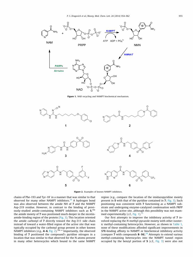
Nicotinamide phosphoribosyltransferase (NAMPT, also knownin the literature as pre-B cell colony-enhancing factor (PBEF) aswell as visfatin; EC 2.4.2.12) plays a critical role in cellular metab-olism.1 The enzyme catalyzes the rate-limiting step in the conver-sion of nicotinamide (NAM) to the important enzyme co-factornicotinamide adenine dinucleotide (NAD).2 This process enablesthe efficient intracellular recycling of NAM, which is produced bythe catalytic action of NAD-consuming enzymes such as the PARPsand Sirtuins, back into NAD (Fig. 1).3 Proper maintenance of NADlevels is known to be critical to sustaining energetics required formany cellular functions.4 Inhibition of NAMPT has thereforeemerged as a novel strategy for impairing the proliferation of tu-mors whose high growth rates may make them more susceptibleto NAMPT disruption relative to non-cancerous cells.5
Multiple examples of NAMPT inhibitors are known in the scien-tific and patent literature, and the most advanced of these agents[GMX-1778 (1)6 and APO-866 (2)7; Fig. 2] have progressed to hu-
man clinical trials.8,9 Our own prior discovery efforts identified po-tent urea and amide-derived NAMPT inhibitors which alsocontained terminal biaryl sulfone moieties (compounds 3–6;Fig. 2).10 In an effort to further diversify these molecules, we alsoconducted a surface plasmon resonance (SPR)-based screen toidentify small, structurally novel NAMPT-binding moieties (‘frag-ments’) which could be combined with our existing compounds.11
In this report, we describe the structure-based elaboration of twoSPR screening hits into potent anti-NAMPT agents that are struc-turally distinct from other known NAMPT inhibitors.
Compound 7 was identified from our SPR-based screeningmethods as a moderately potent NAMPT binder with high ligandefficiency (Table 1).11,12 The compound also demonstrated the abil-ity to inhibit NAMPT in biochemical assessments, although its po-tency was somewhat attenuated relative to its binding properties(Table 1).13 A co-crystal structure of the molecule in complex withNAMPT was subsequently determined and revealed that the mole-cule occupied the nicotinamide-binding region of the protein’sactive site (Fig. 3).14 Not unexpectedly, the pyridine portion of 7formed face-to-face pi-stacking interactions with the side
N
O
NH2 +OO
HO OH
NAM
PO
-O-O
O PO
O-O
P O-O
O-
PRPP
OO
HO OH
PO
-O-O N
O
NH2
NMN
NAMPT
N
O
OH
OH
OPO O-O
PO
O-O
OHO
HO
N
N
N
N
NH2 O
NH2
NAD
ATP ADP + PO43-
NMNAT
PARPs
Sirtuins
Figure 1. NAD recycling and NAMPT biochemical mechanism.
N
NH
N
NH
N
O
Cl
N
O
NH
N
O
O
NH
NH
SO O
N
3
NH
SN
O O
N
O
N
4
NH
SO O
O
5
F
F
N
N
6
1 2
H2N
NH
SO O
O
CF3NNNH
Figure 2. Examples of known NAMPT inhibitors.
P. S. Dragovich et al. / Bioorg. Med. Chem. Lett. 24 (2014) 954–962 955
chains of Phe-193 and Tyr-180 in a manner that was similar to thatobserved for many other NAMPT inhibitors.10 A hydrogen bondwas also observed between the amide NH of 7 and the NAMPTAsp-219 residue. However, in contrast to the binding of previ-ously-studied amide-containing NAMPT inhibitors such as 5,10c
the amide moiety of 7 was positioned much deeper in the nicotin-amide-binding region of the protein (Fig. 3). This location orientedthe amide carbonyl of 7 directly toward the Arg-311 side chaininstead of toward a water-filled region of the active site that wastypically occupied by the carbonyl group present in other knownNAMPT inhibitors (e.g., 4–6, Fig. 2).10c–e Importantly, the observedbinding of 7 positioned the compound’s pyridine nitrogen in alocation that was similar to that observed for the N-atoms presentin many other heterocycles which bound to the same NAMPT
region (e.g., compare the location of the imidazopyridine moietypresent in 5 with that of the pyridine contained in 7; Fig. 3). Suchpositioning was consistent with 7 functioning as a NAMPT sub-strate and undergoing enzyme-catalyzed condensation with PRPPin the NAMPT active site, although this possibility was not exam-ined experimentally (c.f., Fig. 1).15
Our first attempts to improve the inhibitory activity of 7 in-volved replacing the N-methyl-pyrazole moiety with other isoster-ic methyl-containing heterocycles. However, as shown in Table 1,none of these modifications afforded significant improvements inSPR-binding affinity to NAMPT or biochemical inhibitory activity(compare 7 with compounds 8–16).16 Attempts to extend variousmethyl-containing heterocycles into the NAMPT tunnel regionoccupied by the benzyl portion of 5 (c.f., Fig. 3) were also not

Table 1Initial SAR of 3-aminopyridine-amide NAMPT inhibitors
N
NH
O
R
Compd R NAMPT IC50a (uM) NAMPT Kd
b (uM) NAMPT SPR LEc
7N
N 91 5.2 0.47
8 N
HN
>100 176 0.36
9
N
N >100 129 0.35
10S
34 36 0.40
11
NS >100 55 0.38
12O
38 12 0.44
13
NO >100 61 0.38
14
S50 129 0.35
15
SN >100 20 0.42
16
O>100 13 0.44
All biochemical and SPR results are reported as the arithmetic mean of at least 2 separate runs.a NAMPT biochemical inhibition.b NAMPT SPR binding.c Ligand efficiency based on SPR Kd value.
956 P. S. Dragovich et al. / Bioorg. Med. Chem. Lett. 24 (2014) 954–962
successful. This outcome was obtained regardless of whether theextending moieties were appended to a position adjacent to (com-pounds 17–20) or once-removed from (compounds 21–23) theheterocyclic methyl group (Table 2). These results collectively indi-cated that replacement of the methyl-pyrazole present in 7 withnon-isosteric and/or structurally diverse moieties would likely berequired to properly optimize the potency of the 3-aminopyri-dine-amide inhibitor series.
Accordingly, crystal structures of several other NAMPT frag-ment screening hits were determined to identify those with goodpotential for combination with lead compound 7.11 The nitro-substituted benzimidazole 24 (Fig. 4), which exhibited moderateSPR-based NAMPT affinity and good ligand efficiency, emergedfrom this exploration as a particularly attractive possibility. Asshown in Fig. 5, the compound bound to NAMPT in the tunnel re-gion with the nitro group located near where the amide moiety of 7resided in the 7-NAMPT crystal structure. A hydrogen bond wasobserved between one nitro group oxygen atom of 24 and the sidechain of Arg-311 which was shifted from its location in the7-NAMPT crystal structure in order to facilitate this interaction.The benzimidazole portion of 24 formed many favorable van der
Waals contacts with hydrophobic portions of the NAMPT tunnelregion (Ile-309, Ile-351, and the face of His-191) but did not inter-act with any observed water molecules. Somewhat unexpectedly,the benzimidazole of 24 did not bind to NAMPT in a manner thatwas co-planar with the methyl-pyrazole contained in 7. However,analysis of the two overlaid structures suggested that replacementof the latter moiety with a simple phenyl ring would be toleratedand would offer alternate vectors for further elaboration into theNAMPT tunnel region (Fig. 5).
As shown in Table 3, effecting the described substitution affor-ded a molecule (25) with significantly improved biochemicalNAMPT inhibitory activity relative to lead compound 7. Impor-tantly, extension of 25 via modification of the 4-position on thephenyl ring was typically tolerated and often improved biochemi-cal potency as compared to the unsubstituted inhibitor (26–35;Table 3). Two notable exceptions to this trend were the amino-containing molecules 29 and 32 which, based on the 24-NAMPTco-crystal structure, unfavorably positioned their polar aminogroups in the center of the hydrophobic NAMPT tunnel region.Encouragingly, however, transformation of the amine moietiespresent in either compound to the corresponding carbamates

Table 2Elaboration of 3-aminopyridine-amide NAMPT inhibitors
Compd R NAMPT IC50a
(uM)NAMPT Kd
b
(lM)NAMPTSPR LEc
17 N >100 234 0.31
18
NN >100 >500 NA
19
NN >100 177 0.24
20
N
N>100 >500 NA
21NN >100 59 0.36
22NN >100 233 0.29
23 NN
>100 481 0.26
All biochemical and SPR results are reported as the arithmetic mean of at least 2separate runs.
a NAMPT biochemical inhibition.b NAMPT SPR binding.c Ligand efficiency based on SPR Kd value (NA = not applicable).
NH
NO2N
24
NAMPT SPR KD = 126 µMNAMPT SPR LE = 0.44
Figure 4. NAMPT fragment screening hit.
Figure 3. Crystal structure of compound 7 in complex with NAMPT (resolu-tion = 2.86 Å; PDB accession code 4N9B). Protein side chains from the two NAMPTmonomers which form the dimeric active site cleft are depicted in white and grey,respectively. The van der Waals surface of the ligand binding pocket is also shownin grey. The inhibitor is presented as orange tubes with corresponding crystallo-graphic water molecules and hydrogen bonds indicated by orange spheres anddashed orange lines, respectively. The thin green lines depict the location ofcompound 5 in the binding site (PDB accession code 4KFO). Hydrogen bondinteractions between 5 and NAMPT are indicated by the dashed green lines.
P. S. Dragovich et al. / Bioorg. Med. Chem. Lett. 24 (2014) 954–962 957
and/or sulfonamides afforded molecules with more promisingNAMPT inhibition properties (30–31 and 33–35). This observationprompted us to systematically explore appending phenyl-contain-ing acyl and sulfonyl moieties to the amines present in 29 and 32.We anticipated that some of the resulting compounds would suc-cessfully traverse the NAMPT tunnel region and interact with apost-tunnel (solvent-exposed) area of the protein in a manner sim-ilar to that noted for the terminal portions of larger known NAMPTinhibitors (e.g., compound 1–6, Fig. 2; see also Figs. 3 and 6–8).
Table 4 depicts the outcome of this systematic exploration. Mol-ecules incorporating benzoyl-derived amides were moderately po-tent NAMPT inhibitors when the amides were directly attached tothe compounds’ central phenyl rings (36, 40, 44, and 48). However,inclusion of a methylene moiety between the amides and the cen-tral phenyl rings significantly impaired the potencies of the result-ing compounds (37, 41, 45, and 49). In contrast, direct central-ringattachment of phenyl-containing sulfonamides afforded ineffectiveNAMPT inhibitors (38, 42, 46, and 50) while extension of the samesulfonamide moieties by a methylene group provided much morepotent inhibitors (39, 43, 47, 51). The ability of several compoundscontained in Table 4 to inhibit the proliferation of A2780 cells wasalso tested. Most molecules did not exhibit meaningful activity inthis assessment, likely due to insufficient NAMPT inhibition po-tency. However, compounds 47 and 51 displayed measurable anti-proliferation activity and the latter (more potent) molecule wasselected for additional characterization studies.
The co-crystal structure of inhibitor 51 in complex with NAMPTis shown in Figure 6. The molecule’s amino-pyridine fragmentbound to the protein in a manner that was very similar to, althoughnot quite co-planar with, the binding mode described above for theidentical portion of compound 7. Accordingly, the pyridine ring of51 was situated between the side chains of Phe-193 and Tyr-180
and a hydrogen bond was observed between the compound’samide NH moiety and the Asp-219 residue. The central phenyl ringof inhibitor 51 occupied the NAMPT tunnel region and was
positioned similarly to both the methyl-pyrazole present in 7 andthe benzimidazole contained in 24. However, the phenyl group of51 was not precisely co-planar with either of these entities and in-stead filled a space in the tunnel region that resided between them.This orientation was drastically different from the binding modeobserved previously for the central benzyl group of compound 5in the same area of the protein (Fig. 6). In contrast, the terminalphenyl-sulfonamide portion of 51 interacted with NAMPT in amanner that was very similar to that noted for the terminal sulfonepresent in 5 with water-mediated hydrogen bonds being formedwith both sulfonamide oxygen atoms (Fig. 6). The pyridine N-atomof 51 was situated very close to the exposed imidazopyridine nitro-gen of 5 in the NAMPT nicotinamide binding region, and this posi-tioning was consistent with 51 possibly functioning as a NAMPTsubstrate.15 The collective analysis of the 51-NAMPT co-crystalstructure demonstrated how a compound that was structurallydistinct from other known NAMPT inhibitors such as compounds3–6 could effectively and uniquely occupy the protein’s activesite.17
Further analysis of the 24-NAMPT co crystal structure describedabove (Fig. 5) suggested that 6,5-bicyclic aromatic moieties could

Figure 6. Crystal structure of compound 51 in complex with NAMPT (resolu-tion = 1.70 Å; PDB accession code 4N9D). Protein side chains from the two NAMPTmonomers which form the dimeric active site cleft are depicted in white and grey,respectively. The van der Waals surface of the ligand binding pocket is also shownin grey. The inhibitor is presented as blue tubes with corresponding crystallo-graphic water molecules and hydrogen bonds indicated by blue spheres and dashedblue lines, respectively. The thin green lines depict the location of compound 5 inthe binding site (PDB accession code 4KFO). Hydrogen bond interactions between 5and NAMPT are indicated by the dashed green lines.
Figure 7. Crystal structure of compound 24 in complex with NAMPT (resolu-tion = 1.75 Å; PDB accession code 4N9C). Protein side chains from the two NAMPTmonomers which form the dimeric active site cleft are depicted in white and grey,respectively. The van der Waals surface of the ligand binding pocket is also shownin grey. The inhibitor is presented as cyan tubes with corresponding crystallo-graphic water molecules and hydrogen bonds indicated by cyan spheres and dashedcyan lines, respectively. The thin green lines depict the location of compound 2 inthe binding site (PDB accession code 2GVJ). Hydrogen bond interactions between 2and NAMPT are indicated by the dashed green lines.
Table 3Additional elaboration of 3-aminopyridine-amide NAMPT inhibitors
N
NH
O
R
Compd R NAMPT IC50a (lM)
25 H 1326 CH3 1327 Ph 1.028 Cyclohexyl 1.029 NH2 >10030 NHBoc 0.4231 NHSO2CH3 10.332 CH2NH2 >10033 CH2NHBoc 1334 CH2NHCbz 3.335 CH2NHSO2CH3 5.0
All biochemical results are reported as the arithmetic mean of at least 2 separateruns.
a NAMPT biochemical inhibition.
Figure 5. Crystal structure of compound 24 in complex with NAMPT (resolu-tion = 1.75 Å; PDB accession code 4N9C). Protein side chains from the two NAMPTmonomers which form the dimeric active site cleft are depicted in white and grey,respectively. The van der Waals surface of the ligand binding pocket is also shownin grey. The inhibitor is presented as cyan tubes with corresponding crystallo-graphic water molecules and hydrogen bonds indicated by cyan spheres and cyanlines, respectively. The thin orange lines depict the location of compound 7 in thebinding site (PDB accession code 4N9B). Hydrogen bond interactions between 7 andNAMPT are indicated by the dashed orange lines.
958 P. S. Dragovich et al. / Bioorg. Med. Chem. Lett. 24 (2014) 954–962
also be productively utilized as replacements for the methyl-pyrazole present in fragment lead 7. Accordingly, a benzimidazolecontaining an appropriately-positioned 3-amino-pyridine-derivedamide displayed encouraging biochemical NAMPT inhibitory activ-ity (compound 52, Table 5). Replacement of the benzimidazolepresent in 52 with several other 6,5-bicyclic aromatic systems alsoafforded biochemically active NAMPT inhibitors (53–58, Table 5),although several compound potencies were somewhat attenuatedrelative to that exhibited by 52. Unfortunately, all of thesemolecules failed to demonstrate anti-NAMPT activity in cell cul-ture assessments. These outcomes were not entirely surprisingsince the compound concentrations tested in these cell-based
experiments were somewhat low relative to the correspondingbiochemical IC50 values. Attempts to further elaborate the benz-imidazole contained in 52 by derivatizing the nitrogen atom metato the carboxamide substituent were unsuccessful (compounds 59and 60, Table 5). However, analysis of the bound conformations offragment 24 and the very potent NAMPT inhibitor 2 suggested that

Table 53-Aminopyridine-amide NAMPT inhibitors containing 6,5-bicyclic moieties
N
NH
O
R
Compd R NAMPT IC50a
(lM)A2780 IC50
b
(lM)
52NH
N1.5 >2.0
53
N
O9.1 >2.0
54
O
N3.4 >2.0
55
N
S26 >2.0
56
S
N4.1 >2.0
57
O21 >2.0
58
S21 ND
59
N
N>100 ND
60
N
N >100 ND
All biochemical and cell-based results are reported as the arithmetic mean of atleast 2 separate runs.
a NAMPT biochemical inhibition.b Antiproliferation activity determined in cell culture experiments using A2780
cell line. ND = not determined.
Table 4Systematic exploration of amino-containing 3-aminopyridine-amide NAMPT inhibi-tors
N
NH
O
HN
Xn
R
Compd R X n NAMPT IC50a
(lM)A2780 IC50
b
(lM)
36 C@O 0 0.61 >2.037 C@O 1 25 ND38 SO2 0 >100 >2.039 SO2 1 0.21 ND40 C@O 0 1.5 >2.041 C@O 1 40 ND42 SO2 0 83 >2.043 SO2 1 0.014 ND44 Cl C@O 0 0.64 >2.045 C@O 1 3.1 >2.046 SO2 0 62 >2.047 SO2 1 0.019 0.8848 C@O 0 0.29 >2.049 C@O 1 80 ND50 SO2 0 36 >2.051 SO2 1 0.019 0.12
All biochemical and cell-based results are reported as the arithmetic mean of atleast 2 separate runs.
a NAMPT biochemical inhibition.b Antiproliferation activity determined in cell culture experiments using A2780
cell line (ND = not determined). This inhibition can be reversed by addition of0.33 mM of NMN.
Figure 8. Crystal structure of compound 63 in complex with NAMPT (resolu-tion = 1.72 Å; PDB accession code 4N9E). Protein side chains from the two NAMPTmonomers which form the dimeric active site cleft are depicted in white and grey,respectively. The van der Waals surface of the ligand binding pocket is also shownin grey. The inhibitor is presented as salmon tubes with corresponding crystallo-graphic water molecules and hydrogen bonds indicated by salmon spheres anddashed salmon lines, respectively. The thin green lines depict the location ofcompound 2 in the binding site (PDB accession code 2GVJ). Hydrogen bondinteractions between 2 and NAMPT are indicated by the dashed green lines.
P. S. Dragovich et al. / Bioorg. Med. Chem. Lett. 24 (2014) 954–962 959
functionalization of the 52 nitrogen atom para to the carboxamidesubstituent might be more fruitful (Fig. 7). Specifically, it wasenvisioned that introduction of a methyl group at this location in52 would be tolerated and would thereby enable additional
elaboration with molecular fragments derived directly from thestructure of inhibitor 2.
The described methyl-containing derivative of 52 was prepared(compound 61) and it displayed biochemical NAMPT inhibitionproperties that were equivalent to those exhibited by the unme-thylated parent molecule (Table 6). Further elaboration of themethyl group present in 61 with several moieties inspired by thestructure of inhibitor 2 was either tolerated (62) or dramaticallyimproved biochemical NAMPT inhibitory activity (63). However,related derivatization of the 61 benzimidazole 2-positionweakened anti-NAMPT potency with the larger appended groupshaving the most deleterious effects (compounds 64–67, Table 6).Cell-culture antiproliferation effects were assessed with severalof the compounds depicted in Table 6, but only inhibitor 63exhibited significant potency in these experiments. Somewhatsurprisingly, however, the antiproliferative effects associated with63 were not completely reversed/eliminated by the addition ofNMN (the biochemical product of NAMPT catalysis; c.f. Fig. 1) tothe cell culture media. This result suggested that the cell effects

Table 7Antiproliferation effects determined for compounds 51 and 63 in various humantumor cell linesa,b
CyQuant IC50 values in nM
HT1080 PC3 MiaPaCa2c HCT116
51 213 ± 9 217 ± 67 855 ± 77 271 ± 3163 >1000 >1000 >1000 >1000
a CyQuant endpoint; arithmetic mean of 3 separate runs (n = 3, standard devia-tions are also shown).
b All antiproliferation effects were reversed by addition of 330 lM NMN, stronglyimplicating NAMPT inhibition as the causative MOA.
c n = 2. See Supplementary data for experimental details.
N
NH2+
R
O
HO
i 8-11, 13-23, 25-28, 30,33, 34, 52-59, 62, 64-66
+R
O
H3CO
iii 60
ii 61
Scheme 1. Synthesis of compounds containing 3-aminopyridine-derived amides.Reagents and conditions: (i) HATU, (i-Pr)2NEt, DMF, 25–50 �C, 3–18 h, 4–76%; (ii)HATU, (i-Pr)2NEt, DMF, 45 �C, 3 h (forms HOAt adduct), then 3-aminopyridine,microwave, DMF, 95 �C, 1 h, 25%; (iii) AlCl3, 1,2-dichloroethane, 70 �C, 15 h, 35%.
Table 6Optimization of benzimidazole-containing 3-aminopyridine-amide NAMPT inhibitors
N
NH
O
N
N
R1
R2
Compd R1 R2 NAMPT IC50a (lM) A2780 IC50
b (lM)
52 H H 1.5 >2.061 CH3 H 1.4 ND
62 H 3.7 >2.0
63N
O
H 0.015 0.099c
64 CH3 CH3 6.1 ND65 CH3 Ph 14 ND
66 CH3 N Boc >100 ND
67 CH3
NO
>100 ND
All biochemical and cell-based results are reported as the arithmetic mean of at least 2 separate runs.a NAMPT biochemical inhibition.b Antiproliferation activity determined in cell culture experiments using A2780 cell line (ND = not determined).c Antiproliferative effects were only partially reversed by addition of 0.33 mM of NMN.
960 P. S. Dragovich et al. / Bioorg. Med. Chem. Lett. 24 (2014) 954–962
observed for compound 63 might result from inhibition of biolog-ical targets in addition to and/or other than NAMPT. More detailedcharacterization studies of compound 63 were therefore under-taken to better explore this possibility.
A co-crystal structure of compound 63 in complex with NAMPTwas determined and it confirmed that the compound could bio-physically associate with the protein. The terminal portion of themolecule, which bound in the post-tunnel, solvent-exposedNAMPT region, closely mimicked the bound conformation of thecorresponding portion of inhibitor 2 (Fig. 8). Such binding enabledmany favorable van der Waals contacts between the benzoyl moi-ety present in 63 and aliphatic portions of the side chains ofNAMPT residues Arg-349 and Ala-379. Similar favorable van derWaals interactions were also noted between the piperidine frag-ment of 63 and the hydrophobic Val-242 and Ile-309 NAMPT tun-nel-region residues. As was observed for other 3-amino-pyridineamides described in this work, the pyridine portion of 63 formedface-to-face pi-stacking interactions with the side chains of Phe-193 and Tyr-180, a hydrogen bond existed between the compound’samide NH and Asp-219, and the molecule’s amide carbonyl was
oriented directly toward the Arg-311 side chain. As also expectedfrom analysis of many inhibitor-NAMPT co-crystal structures, thepyridine nitrogen atoms of compounds 63 and 2 were located insimilar positions in the NAM-binding region of the protein thatwere consistent with each molecule possibly functioning as aNAMPT substrate.15
In addition to the various studies described above, more exten-sive cell-culture experiments were conducted with both com-pounds 51 and 63 to characterize their anti-NAMPT properties ingreater detail. Compound 51 potently reduced NAD levels inA2780 cells with an IC50 value between 24 and 58 nM (Figs. S1and S2). In contrast, compound 63 exhibited negligible effects onA2780 NAD levels when tested under similar experimental condi-tions (IC50 >1000nM; Fig. S3). The latter observation was consis-tent with the inability of NMN to reverse the A2780antiproliferation effects exhibited by compound 63 and suggestedthat, in spite of the compound’s ability to biophysically associate

68 R = H74 R = CH3
i
ii
O
RO
Br
NO2O
H3CO
NH
R
75 R = NO276 R = NH2
iii
63 viii
Scheme 5. Synthesis of compound 63. Reagents and conditions: (i) EDC�HCl, (i-Pr)carboxylate, K2CO3, DMF, 100 �C, 20 h, 65%; (iii) Raney Ni, H2, MeOH, 25 �C, 0.5 h, 45%; (iv)aminopyridine, HATU, (i-Pr)2NEt, DMF, 25 �C, 20 h, 55%; (vii) TFA, CH2Cl2, 25 �C, 0.5 h, 8
i
O
HO
Br
NO2O
HO
NH
R
69 R = NO270 R = NH2
ii
O
HOiii
N
N
6871
Scheme 2. Synthesis of acid required to prepare compound 62. Reagents and conditions: (i) Cyclohexylmethanamine, K2CO3, DMF, 110 �C, 14 h, 19%; (ii) H2, 10% Pd on C,MeOH, 25 �C, 2 h, 91%; (iii) HC(OEt)3, EtOH, 70 �C, 14 h, 87%.
NH2
NH
EtO
O
N
H
OBoc
N
N
NBoc
RO
Oi
72 R = Et73 R = H
ii
+
Scheme 3. Synthesis of of acid required to prepare compound 66. Reagents andconditions: (i) 10% Pd/C, EtOH, reflux, 48 h, 89%; (ii) 1:5 1 N NaOH:EtOH, 85 �C, 18 h,94%.
i 2930
ii
iii
36, 40, 44, 48
31, 38, 42, 46, 50
iv 3233
ii
iii
37, 41, 45, 49
35, 39, 43, 47, 51
Scheme 4. Alternate synthesis of amide and sulfonamide-containing compounds.Reagents and conditions: (i) 4.0 M HCl, 1,4-dioxane, 25 �C, 6 h, 85%; (ii) RC(O)Cl,Et3N, CH2Cl2, 25 �C, 18 h, 11–26%; (iii) RSO2Cl, (i-Pr)2NEt, CH2Cl2, 25 �C, 18 h, 4–45%;(iv) TFA, CH2Cl2, 25 �C, 3 h, 90%.
P. S. Dragovich et al. / Bioorg. Med. Chem. Lett. 24 (2014) 954–962 961
with the protein and potently inhibit its biochemical activity, theA2780 cell culture outcomes likely result from non-NAMPT-relatedbiological activity. Accordingly, compound 63 did not exhibitmeaningful antiproliferative effects against four other cancer celllines against which it was tested (Table 7). Encouragingly, how-ever, compound 51, which strongly reduced NAD levels in A2780cells, exhibited potent to moderate antiproliferative activityagainst all of these same lines (Table 7). Taken together, the aboveresults indicate that careful biological characterization should beconducted with novel NAMPT inhibitors to confirm that any ob-served antiproliferative effects truly result from inhibition of thetargeted NAMPT enzyme.
The compounds described in this work were either purchasedfrom Sigma–Aldrich (7, 12, and 24) or were prepared by the meth-ods shown in Schemes 1–5.18 Many were synthesized by HATU-mediated coupling of commercially available carboxylic acids with3-aminopyridine (8–11, 13–23, 25–28, 30, 33, 34, 52–58, 62, 64–66; Scheme 1). The acid required for the preparation of compound59 was obtained by hydrolysis of the corresponding (commerciallyavailable) methyl ester while those needed to make 62 and 66were prepared as described in Schemes 2 and 3, respectively. Inthe case of inhibitor 60, the desired amide linkage was formed di-rectly from 3-aminopyridine and the appropriate methyl esterusing an AlCl3-mediated coupling technique (Scheme 1). Removalof the Boc groups present in compounds 30 and 33 using variousacidic conditions respectively afforded the amino-containing mol-ecules 29 and 32 in good yield. These entities were subsequentlytransformed into the corresponding amides and sulfonamides byreaction with various acid- and sulfonyl-chlorides (compounds31, 35–51; Scheme 4). The preparation of compound 63 was
NBoc
O
ROiv
N
N
N Boc
77 R = CH378 R = H
v
O
NH
N
N
N R
N
79 R = Boc80 R = H
vii
vi
2NEt, HOBt, MeOH, 25 �C, 1 h, 87%; (ii) tert-butyl-4-(aminomethyl)piperidine-1-PPTS, HC(OEt)3, CH2Cl2, 25 �C, 20 h, 72%; (v) KOH, EtOH/H2O, 25 �C, 20 h, 82%; (vi) 3-
2%; (viii) PhC(O)Cl, Et3N, CH2Cl2, 25 �C, 0.5 h, 14%.

962 P. S. Dragovich et al. / Bioorg. Med. Chem. Lett. 24 (2014) 954–962
somewhat more complicated and was accomplished by the synthe-sis depicted in Scheme 5.
In this report, we describe the fragment-based identification oftwo potent biochemical inhibitors of the nicotinamide phosphori-bosyltransferase enzyme that are structurally distinct from otherknown anti-NAMPT agents. Each of these new compounds (51and 63) incorporates an amide moiety derived from 3-amino-pyridine, and this entity enables them to bind to NAMPT in amanner that is unique relative to how other NAMPT inhibitorsassociate with the protein. Although both compounds exhibitantiproliferative activity in A2780 cell culture experiments, onlyinhibitor 51 appears to exert these effects via a NAMPT-mediatedmechanism. This molecule also displays antiproliferative proper-ties in several other cancer cell lines. Collectively, our resultsprovide new chemical options for the design of potent NAMPTinhibitors, and also caution regarding the need for careful biologi-cal characterization of any novel molecules so identified.
Acknowledgments
We thank Drs. Krista Bowman and Jiansheng Wu and theirrespective Genentech research groups for performing many pro-tein expression and purification activities. We also thank ShohiniGanguly for generating all the biochemical and cell IC50 results. Fi-nally, we thank Dr. Peter Jackson for many helpful discussionsregarding NAMPT and Dr. Jeff Blaney for many useful suggestionsassociated with fragment-based ligand design.
Supplementary data
Supplementary data associated with this article can be found,in the online version, at http://dx.doi.org/10.1016/j.bmcl.2013.12.062.
References and notes
1. (a) He, J.; Tu, C.; Li, M.; Wang, S.; Guan, X.; Lin, J.; Li, Z. Curr. Pharm. Des. 2012,18, 6123; (b) Burgos, E. S. Curr. Med. Chem. 2011, 18, 1947; (c) Bi, T.-Q.; Che, X.-M. Cancer Biol. Ther. 2010, 10, 119; (d) Garten, A.; Petzold, S.; Körner, A.; Imai,S.-I.; Kiess, W. Trends Endocrin. Metab. 2008, 20, 130.
2. (a) Houtkooper, R. H.; Cantó, C.; Wanders, R. J.; Auwerx, J. Endocrine Rev. 2010,31, 194; (b) Sauve, A. A. J. Pharmacol. Exp. Ther. 2008, 324, 883.
3. The de novo NAD synthesis pathway requires eight enzyme-catalyzed steps toproduce the co-factor from tryptophan. See Ref. 2 for additional details.
4. (a) Schulze, A.; Harris, A. L. Nature 2012, 491, 364; (b) Ward, P. S.; Thompson, C.B. Cancer Cell 2012, 21, 297; (c) Jones, N. P.; Schulze, A. Drug Discovery Today2012, 17, 232; (d) Tennant, D. A.; Durán, R. V.; Gottlieb, E. Nat. Rev. Cancer 2010,10, 267; (e) Vander Heiden, M. G.; Cantley, L. C.; Thompson, C. B. Science 2009,324, 1029.
5. (a) Chiarugi, A.; Dölle, C.; Felici, R.; Ziegler, M. Nat. Rev. Cancer 2012, 12, 741; (b)Zhang, L. Q.; Heruth, D. P.; Ye, S. Q. J. Bioanal. Biomed. 2011, 3, 13; (c) Khan, J. A.;Forouhar, F.; Tao, X.; Tong, L. Exp. Opin. Ther. Targets 2007, 11, 695.
6. (a) Watson, M.; Roulston, A.; Bélec, L.; Billot, X.; Marcellus, R.; Bédard, D.;Bernier, C.; Branchaud, S.; Chan, H.; Dairi, K.; Gilbert, K.; Goulet, D.; Gratton,M.-O.; Isakau, H.; Jang, A.; Khadir, A.; Koch, E.; Lavoie, M.; Lawless, M.; Nguyen,M.; Paquette, D.; Turcotte, E.; Berger, A.; Mitchell, M.; Shore, G. C.; Beauparlant,P. Mol. Cell. Biol. 2009, 29, 5872; (b) Ravaud, A.; Cerny, T.; Terret, C.; Wanders, J.;Bui, B. N.; Hess, D.; Droz, J.-P.; Fumoleau, P.; Twelves, C. Eur. J. Cancer 2005, 41,702; (c) Hovstadium, P.; Larsson, R.; Jonsson, E.; Skov, T.; Kissmeyer, A.-M.;Krasilnikoff, K.; Bergh, J.; Karlsson, M. O.; Lonnebo, A.; Ahlgren, J. Clin. CancerRes. 2002, 8, 2843. Compound 1 is also known in the literature as CHS828.
7. (a) Holen, K.; Saltz, L. B.; Hollywood, E.; Burk, K.; Hanauske, A.-R. Invest. NewDrugs 2008, 26, 45; (b) Hasmann, M.; Schemainda, I. Cancer Res. 2003, 63, 7436.Compound 2 is also known in the literature as FK866.
8. For a recent review of NAMPT and associated inhibitors, see Galli, U.; Travelli,C.; Massarrotti, A.; Fakhfouri, G.; Rahimian, R.; Tron, G. C.; Genazzani, A. A. J.Med. Chem. 2013, 56, 6279.
9. (a) Christensen, M. K.; Erichsen, K. D.; Olesen, U. H.; Tjørnelund, J.; Fristrup, P.;Thougaard, A.; Nielsen, S. J.; Sehested, M.; Jensen, P. B.; Loza, E.; Kalvinsh, I.;Garten, A.; Kiess, W.; Bjökling, F. J. Med. Chem. 2013, 56, 9071; (b) Arigon, J.;Bernhart, C.; Bouaboula, M.; Dimalta, A.; Nardi, F.; Jegham, S. WO 2012038904,2012.; (c) Arigon, J.; Bernhart, C.; Bosch, M.; Bouaboula, M.; Nardi, F.; Jegham,S.; Combet, R. WO 2012038905, 2012.; (d) Curtin, M. L.; Sorensen, B. K.;Heyman, H. R.; Clark, R. F.; Woller, K. R.; Shah, O. J.; Michaelides, M.; Tse, C.;Vasudevan, A.; Mack, H.; Hansen, T. M.; Sweis, R.; Pliushchev, M. A. US
20120122842, 2012.; (e) Curtin, M. L.; Sorensen, B. K.; Heyman, H. R.; Clark, R.F.; Michaelides, M.; Tse, C. US 20120122924, 2012.; (f) Kumar, D. V.; Slattum, P.M.; Yager, K. M.; Shenderovich, M. D.; Tangallapally, R.; Kim, S. WO2012177782, 2012.; (g) Willardsen, A. J.; Lockman, J. W.; Murphy, B. R.; Judd,W. R.; Kim, I. C.; Kim, S.-H.; Zigar, D. F.; Yager, K. M.; Fleischer, T. C.; Terry-Lorenzo, R. T.; Boniface, J. J.; Parker, D. P.; McAlexander, I. A.; Bursavich, M. G.;Dastrup, D. M. WO 2011109441, 2011.; (h) You, H.; Youn, H.-S.; Im, I.; Bae, M.-H.; Lee, S.-K.; Ko, H.; Eom, S. H.; Kim, Y.-C. Eur. J. Med. Chem. 2011, 46, 1153; (i)Lockman, J. W.; Murphy, B. R.; Zigar, D. F.; Judd, W. R.; Slattum, P. M.; Gao, Z.-H.; Ostanin, K.; Green, J.; McKinnon, R.; Terry-Lorenzo, R. T.; Fleischer, T. C.;Boniface, J. J.; Shenderovich, M.; Willardsen, J. A. J. Med. Chem. 2010, 53, 8734;(j) Colombano, G.; Travelli, C.; Galli, U.; Caldarelli, A.; Chini, M. G.; Canonico, P.L.; Sorba, G.; Bifulco, G.; Tron, G. C.; Genazzani, A. A. J. Med. Chem. 2010, 53, 616;(k) Galli, U.; Ercolano, E.; Carraro, L.; Blasi Roman, C. R.; Sorba, G.; Canonico, P.L.; Genazzani, A. A.; Tron, G. C.; Billington, R. A. ChemMedChem 2008, 3, 771.
10. (a) Zheng, X.; Bauer, P.; Baumeister, T.; Buckmelter, A. J.; Caligiuri, M.;Clodfelter, K. H.; Han, B.; Ho, Y.-C.; Kley, N.; Lin, J.; Reynolds, D. J.; Sharma, G.;Smith, C. C.; Wang, Z.; Dragovich, P. S.; Oh, A.; Wang, W.; Zak, M.; Gunzner-Toste, J.; Zhao, G.; Yuen, P.-W.; Bair, K. W. J. Med. Chem. 2013, 56, 4921; (b)Gunzner-Toste, J.; Zhao, G.; Bauer, P.; Baumeister, T.; Buckmelter, A. J.;Caligiuri, M.; Clodfelter, K. H.; Fu, B.; Han, B.; Ho, Y.-C.; Kley, N.; Liang, X.;Liederer, B. M.; Lin, J.; Mukadam, S.; O’Brien, T.; Oh, A.; Reynolds, D. J.; Sharma,G.; Skelton, N.; Smith, C. C.; Sodhi, J.; Wang, W.; Wang, Z.; Xiao, Y.; Yuen, P.-W.;Zak, M.; Zhang, L.; Zheng, X.; Bair, K. W.; Dragovich, P. S. Bioorg. Med. Chem. Lett.2013, 23, 3531; (c) Zheng, X.; Bauer, P.; Baumeister, T.; Buckmelter, A. J.;Caligiuri, M.; Clodfelter, K. H.; Han, B.; Ho, Y.-C.; Kley, N.; Lin, J.; Reynolds, D. J.;Sharma, G.; Smith, C. C.; Wang, Z.; Dragovich, P. S.; Gunzner-Toste, J.; Liederer,B. M.; Ly, J.; O’Brien, T.; Oh, A.; Wang, L.; Wang, W.; Xiao, Y.; Zak, M.; Zhao, G.;Yuen, P.-W.; Bair, K. W. J. Med. Chem. 2013, 56, 6413; (d) Dragovich, P. S.; Bair,K. W.; Baumeister, T.; Ho, Y.-C.; Liederer, B. M.; Liu, X.; Liu, Y.; O’Brien, T.; Oeh,J.; Sampath, D.; Skelton, N.; Wang, L.; Wang, W.; Wu, H.; Xiao, Y.; Yuen, P.-W.;Zak, M.; Zhang, L.; Zheng, X. Bioorg. Med. Chem. Lett. 2013, 23, 4875; (e) Zheng,X.; Bair, K. W.; Bauer, P.; Baumeister, T.; Bowman, K. K.; Buckmelter, A. J.;Caligiuri, M.; Clodfelter, K. H.; Feng, Y.; Han, B.; Ho, Y.-C.; Kley, N.; Li, H.; Liang,X.; Liederer, B. M.; Lin, J.; Ly, J.; O’Brien, T.; Oeh, J.; Oh, A.; Reynolds, D. J.;Sampath, D.; Sharma, G.; Skelton, N.; Smith, C. C.; Tremayne, J.; Wang, L.;Wang, W.; Wang, Z.; Wu, H.; Wu, J.; Xiao, Y.; Yang, G.; Yuen, P.-W.; Zak, M.;Dragovich, P. S. Bioorg. Med. Chem. Lett. 2013, 23, 5488.
11. Giannetti, A. M.; Zheng, X.; Skelton, N. J.; Wang, W.; Bravo, B.; Bair, K. W.;Baumeister, T.; Cheng, E.; Crocker, L.; Feng, Y.; Gunzner-Toste, J.; Ho, Y.-C.; Hua,R.; Liederer, B. M.; Liu, Y.; Ma, X.; O’Brien, T.; Oeh, J.; Sampath, D.; Shen, Y.;Wang, C.; Wang, L.; Wu, H.; Xiao, Y.; Yuen, P.-W.; Zak, M.; Zhao, G.; Zhao, Q.;Dragovich, P. S. submitted to J. Med. Chem.
12. Hopkins, A. L.; Groom, C. R.; Alex, A. Drug Discovery Today 2004, 9, 430.13. The SPR assessments omitted the PRPP co-substrate and ATP additive that
were included in the biochemical inhibition assays (c.f., Fig. 1). The latter entityis known in the literature to improve NAMPT catalytic efficiency (see: Burgos,E. S.; Schramm, V. L. Biochemistry 2008, 47, 11086). Thus, it was not surprisingthat that SPR Kd values did not perfectly correspond to biochemical IC50
measurements. SPR techniques were utilized primarily to identify the novelNAMPT binding fragments and to optimize their affinity in the absence ofmeaningful biochemical inhibitory activity. Once biochemical IC50 values of<20 lM were achieved for a given inhibitor series, biochemical methods werepreferentially used in subsequent optimization activities.
14. NAMPT is believed to function as a symmetrical homodimer with two activesites formed at opposite ends of the dimer interface. Accordingly, the proteincrystalized with such a dimer present in the asymmetric unit. The twomonomer chains in the dimers of the co-crystal structures described in thiswork were very similar, and both active sites contained co-crystallizedmolecules in similar orientations. In the crystallography discussions, theNAMPT residues are designated with prime and non-prime notation (e.g.,Tyr180 , Phe193) to distinguish the monomer chain in which a given residueresides.
15. We currently believe that many cell-potent NAMPT inhibitors form PRPP-derived phosphoribosylated adducts in the protein’s active site which block thefunction of the enzyme. This belief is consistent with the repeated observationof these adducts by mass spectrometry in biochemical and/or crystallographicexperiments (e.g., compounds 5 and 6; see Ref. 10a,c,e). Once formed, thePRPP-adducts may accumulate intracellularly and thereby enhance cell cultureantiproliferation effects (see Ref. 6a for additional information and discussion).However, there are many other factors that also likely influence NAMPTinhibitor cell potency including: biochemical inhibition activity, the ability of agiven inhibitor and/or its corresponding PRPP-derived ribose adduct toeffectively compete with the NAM substrate, cell membrane permeability,and/or protein binding.
16. The improvements in biochemical inhibition exhibited by compounds 10, 12,and 14 relative to 7 were deemed too incremental to warrant additionalfollow-up activities.
17. Compound 51 was previously described in the literature as STF-31, an inhibitorof the GLUT1 glucose transporter: Chan, D. A.; Sutphin, P. D.; Nguyen, P.;Turcotte, S.; Lai, E. W.; Banh, A.; Reynolds, G. E.; Chi, J.-T.; Wu, J.; Solow-Cordero, D. E.; Bonnet, M.; Flanagan, J. U.; Bouley, D. M.; Graves, E. E.; Denny,W. A.; Hay, M. P.; Giaccia, A. J. Sci. Trans. Med. 2011, 3, 94ra70.
18. The chemical purities of all final compounds described in this work were >95%as determined by as determined by LCMS analysis with UV detection at220 nm.
Related Documents