Fragile Histidine Triad Gene Abnormalities in the Pathogenesis of Gallbladder Carcinoma Ignacio I. Wistuba,* Raheela Ashfaq, † Anirban Maitra, ‡ Hector Alvarez,* Erick Riquelme,* and Adi F. Gazdar †§ From the Department of Anatomic Pathology,* Pontificia Universidad Cato ´lica de Chile, Santiago, Chile; the Department of Pathology † and Hamon Center for Therapeutic Oncology Research, § University of Texas Southwestern Medical Center, Dallas, Texas; and the Department of Pathology, ‡ Johns Hopkins Medical Institutions, Baltimore, Maryland There is limited information about the molecular changes involved in the pathogenesis of gallbladder carcinoma (GBC). Our recent allelotyping analyses have indicated that chromosome 3p loss of heterozy- gosity (LOH), including the fragile histidine triad (FHIT) candidate tumor-suppressor gene locus at 3p14.2, is frequently detected in this neoplasm. To investigate the role of the FHIT abnormalities in the multistage sequential development of GBC, 33 forma- lin-fixed paraffin-embedded invasive GBC specimens and 76 accompanying histologically normal (n 43) and dysplastic (n 33) epithelia were examined by immunostaining for expression of Fhit protein. Allele loss at the FHIT gene locus (3p14.2) was studied in all GBCs and in a subset of accompanying gallbladder epithelia by polymerase chain reaction-based LOH analysis, using three 3p14.2 microsatellite markers. In addition, histologically normal epithelium from chronic cholecystitis (n 19) and dysplasia (n 13) from gallbladder specimens without cancer were ex- amined for immunostaining and LOH. There was a progressive increase in both the frequency of loss of Fhit expression and LOH at FHIT with increasing severity of histopathological changes. FHIT abnor- malities were occasionally demonstrated in histolog- ically normal gallbladder epithelium. Dysplastic foci demonstrated frequent reduction or absence of Fhit immunostaining (38 to 55%) and FHIT allelic loss (33 to 46%). In invasive tumors , these abnormalities were even higher, with 79% reduction or absence of Fhit immunostaining and 76% FHIT allele loss. A high correlation (70%) was observed between Fhit immu- nostaining abnormalities and allele loss in GBC spec- imens (P < 0.05). Although a high frequency of FHIT locus breakpoints were detected in both invasive and dysplastic gallbladder specimens, no intronic ho- mozygous deletions on FHIT were detected in GBCs. FHIT gene abnormalities are nearly universal in GBC and these changes are detected early in the sequential development of this neoplasm. Our findings indicate that the FHIT gene is one of the chromosome 3p putative tumor suppressor genes involved in the pathogenesis of this highly malignant neoplasm. (Am J Pathol 2002, 160:2073–2079) Gallbladder carcinoma (GBC) is a relatively uncommon neoplasm that demonstrates considerable geographic and gender variation in incidence. 1 It is one of the most frequent neoplasms in Chile, where it is the leading cause of cancer deaths in females. 1 GBC is a highly malignant neoplasm, which is usually diagnosed at advanced clin- ical stages. It has been well established that invasive GBC is preceded by preinvasive lesions, including dys- plastic changes of the gallbladder epithelium. 2 Although the development of molecular markers for early detection and the prediction of response to adjuvant therapies would be beneficial, there is very limited information about the molecular changes involved in the pathogen- esis of GBC. 3 It is now well recognized that epithelial tumorigenesis is a multistep process resulting from the accumulation of sequential genetic alterations, including the inactivation of one or more tumor suppressor genes (TSGs). 4 Allele loss, manifested as loss of heterozygosity (LOH) at poly- morphic loci flanking TSGs, is recognized as a hallmark of cancers, with inactivation of the second allele by point mutations or by some other mechanism. 5 These changes result in the absence of the normal gene protein in the affected cells. The fragile histidine triad ( FHIT) gene, a candidate TSG, has been identified at the 3p14.2 region, spanning the FRA3B common fragile site. 6 Because the occur- rence of intragenic mutations in FHIT is very rare, some researchers have argued that FHIT abnormalities may only represent alterations in the 3p14.2 FRA3B fragile site. However, frequent allelic losses as well as homozy- gous deletions have been described at the FHIT locus in several human cancers arising from epithelial cells, mak- ing FHIT a strong candidate TSG. 7 Similarly, several groups have demonstrated that introduction of a wild- Supported by grant 1990489 from the Fondo Nacional de Desarrollo Cientifico y Tecnologico (to I. I. W.). Accepted for publication March 6, 2002. Address reprint requests to Ignacio I. Wistuba, M.D., Department of Anatomic Pathology, P. Universidad Catolica de Chile, Marcoleta 367, P.O. Box 114-D, Santiago, Chile. E-mail: [email protected]. American Journal of Pathology, Vol. 160, No. 6, June 2002 Copyright © American Society for Investigative Pathology 2073

Welcome message from author
This document is posted to help you gain knowledge. Please leave a comment to let me know what you think about it! Share it to your friends and learn new things together.
Transcript
Fragile Histidine Triad Gene Abnormalities in thePathogenesis of Gallbladder Carcinoma
Ignacio I. Wistuba,* Raheela Ashfaq,†
Anirban Maitra,‡ Hector Alvarez,* Erick Riquelme,*and Adi F. Gazdar†§
From the Department of Anatomic Pathology,* Pontificia
Universidad Catolica de Chile, Santiago, Chile; the Department
of Pathology † and Hamon Center for Therapeutic Oncology
Research,§ University of Texas Southwestern Medical Center,
Dallas, Texas; and the Department of Pathology,‡ Johns Hopkins
Medical Institutions, Baltimore, Maryland
There is limited information about the molecularchanges involved in the pathogenesis of gallbladdercarcinoma (GBC). Our recent allelotyping analyseshave indicated that chromosome 3p loss of heterozy-gosity (LOH), including the fragile histidine triad(FHIT) candidate tumor-suppressor gene locus at3p14.2, is frequently detected in this neoplasm. Toinvestigate the role of the FHIT abnormalities in themultistage sequential development of GBC, 33 forma-lin-fixed paraffin-embedded invasive GBC specimensand 76 accompanying histologically normal (n � 43)and dysplastic (n � 33) epithelia were examined byimmunostaining for expression of Fhit protein. Alleleloss at the FHIT gene locus (3p14.2) was studied in allGBCs and in a subset of accompanying gallbladderepithelia by polymerase chain reaction-based LOHanalysis, using three 3p14.2 microsatellite markers.In addition, histologically normal epithelium fromchronic cholecystitis (n � 19) and dysplasia (n � 13)from gallbladder specimens without cancer were ex-amined for immunostaining and LOH. There was aprogressive increase in both the frequency of loss ofFhit expression and LOH at FHIT with increasingseverity of histopathological changes. FHIT abnor-malities were occasionally demonstrated in histolog-ically normal gallbladder epithelium. Dysplastic focidemonstrated frequent reduction or absence of Fhitimmunostaining (38 to 55%) and FHIT allelic loss (33to 46%). In invasive tumors, these abnormalities wereeven higher, with 79% reduction or absence of Fhitimmunostaining and 76% FHIT allele loss. A highcorrelation (70%) was observed between Fhit immu-nostaining abnormalities and allele loss in GBC spec-imens (P < 0.05). Although a high frequency of FHITlocus breakpoints were detected in both invasive anddysplastic gallbladder specimens, no intronic ho-mozygous deletions on FHIT were detected in GBCs.FHIT gene abnormalities are nearly universal in GBC
and these changes are detected early in the sequentialdevelopment of this neoplasm. Our findings indicatethat the FHIT gene is one of the chromosome 3pputative tumor suppressor genes involved in thepathogenesis of this highly malignant neoplasm.(Am J Pathol 2002, 160:2073–2079)
Gallbladder carcinoma (GBC) is a relatively uncommonneoplasm that demonstrates considerable geographicand gender variation in incidence.1 It is one of the mostfrequent neoplasms in Chile, where it is the leading causeof cancer deaths in females.1 GBC is a highly malignantneoplasm, which is usually diagnosed at advanced clin-ical stages. It has been well established that invasiveGBC is preceded by preinvasive lesions, including dys-plastic changes of the gallbladder epithelium.2 Althoughthe development of molecular markers for early detectionand the prediction of response to adjuvant therapieswould be beneficial, there is very limited informationabout the molecular changes involved in the pathogen-esis of GBC.3
It is now well recognized that epithelial tumorigenesisis a multistep process resulting from the accumulation ofsequential genetic alterations, including the inactivationof one or more tumor suppressor genes (TSGs).4 Alleleloss, manifested as loss of heterozygosity (LOH) at poly-morphic loci flanking TSGs, is recognized as a hallmarkof cancers, with inactivation of the second allele by pointmutations or by some other mechanism.5 These changesresult in the absence of the normal gene protein in theaffected cells.
The fragile histidine triad (FHIT) gene, a candidateTSG, has been identified at the 3p14.2 region, spanningthe FRA3B common fragile site.6 Because the occur-rence of intragenic mutations in FHIT is very rare, someresearchers have argued that FHIT abnormalities mayonly represent alterations in the 3p14.2 FRA3B fragilesite. However, frequent allelic losses as well as homozy-gous deletions have been described at the FHIT locus inseveral human cancers arising from epithelial cells, mak-ing FHIT a strong candidate TSG.7 Similarly, severalgroups have demonstrated that introduction of a wild-
Supported by grant 1990489 from the Fondo Nacional de DesarrolloCientifico y Tecnologico (to I. I. W.).
Accepted for publication March 6, 2002.
Address reprint requests to Ignacio I. Wistuba, M.D., Department ofAnatomic Pathology, P. Universidad Catolica de Chile, Marcoleta 367,P.O. Box 114-D, Santiago, Chile. E-mail: [email protected].
American Journal of Pathology, Vol. 160, No. 6, June 2002
Copyright © American Society for Investigative Pathology
2073
type FHIT gene suppresses tumorigenicity.8 In addition,tumor-specific promoter methylation and epigenetic in-activation of the FHIT gene that are absent in the corre-sponding normal tissues have also established the im-portant role of FHIT in tumorigenesis.9,10
Although FHIT abnormalities have been observed in avariety of human tumors, there is no information about itsabnormalities in gallbladder tumorigenesis. As a result ofour recent allelotyping analyses on GBC, we identified 3pregions with frequent allelic loss in this tumor, includingthe FHIT (3p14.2) region.11 Based on our previous find-ings, the present study was undertaken to investigate thefrequency of FHIT gene abnormalities during multistagepathogenesis of GBC, and to compare Fhit immunostain-ing expression abnormalities and FHIT locus allele loss ininvasive GBC and corresponding histologically normaland dysplastic epithelium. Using archival paraffin-em-bedded tissue, we studied the immunohistochemical ex-pression of the Fhit protein and the presence of allelicloss at the FHIT gene locus in invasive GBC and accom-panying histologically normal and preinvasive gallblad-der epithelia.
Materials and Methods
Archival Tumor Specimens
Formalin-fixed paraffin-embedded material from 33 pri-mary GBCs was obtained from cholecystectomy speci-mens resected between 1990 and 1998 at the CatholicUniversity Medical School Hospital, Santiago, Chile. Thepatients consisted of 24 women and 9 men ranging inage from 51 to 85 years (mean age, 68 years). All tumorswere invasive GBCs diagnosed using established histo-logical criteria,2 and consisted of 18 (55%) well-differen-tiated, 3 (9%) moderately differentiated, and 12 (36%)poorly differentiated adenocarcinomas. Based on archi-tectural pattern, there were 13 (39%) tubular adenocar-cinomas, 1 (3%) papillary adenocarcinoma, 18 (55%)tubulopapillary adenocarcinomas, and 1 (3%) adeno-squamous carcinoma. Most of the tumors were advancedGBCs with invasion of the gallbladder serosa (6 cases,18%) or subserosa (20 cases, 61%), and the rest of thecases (7 cases, 21%) were early GBCs, with invasion ofthe gallbladder muscularis propia.
Histologically Normal Epithelium and DysplasticLesions
From the 33 GBCs selected for this study we identified 76discrete foci consisting of histologically normal (n � 43)and dysplastic (n � 33) gallbladder epithelium for Fhitimmunohistochemical analysis. A subset of those epithe-lial foci used for immunostaining (26 dysplastic and 16histologically normal epithelia) were selected for FHITLOH analysis, and microdissected as described below.In addition, histologically normal epithelium from chroniccholecystitis (n � 19) and dysplasia (n � 13) from gall-bladder specimens without cancer were examined forboth immunostaining and LOH. All these gallbladder
specimens were histologically mapped to rule out thepresence of invasive carcinoma. The dysplastic lesionswere scored as low- and high-grade lesions using pub-lished criteria for the histopathological identification ofdysplasia arising in the gallbladder epithelium.2
Microdissection and DNA Extraction
Serial 5-�m sections were cut from the archival, formalin-fixed paraffin-embedded tissues. All slides were stainedwith hematoxylin and eosin, and one of the slides wascoverslipped. The coverslipped slide was used as guideto localize regions of interest for microdissection of theother slides. Microdissection from archival paraffin-embedded tissues was precisely performed under micro-scopic visualization using a micromanipulator, as describedpreviously.12 From four sections of each case a total of800 sectioned tumor cells were microdissected. DNAextraction was performed as described previously.12 Dis-sected lymphocytes or normal stromal cells from the sameslide were used as a source of constitutional DNA fromeach case. After DNA extraction, 5 �l of the proteinaseK-digested samples containing DNA from at least 200 cellswere used for each polymerase chain reaction (PCR).
Microsatellite DNA Markers and PCR-LOHAnalysis
To evaluate LOH, we used primers flanking three highlypolymorphic microsatellite markers within the FHIT gene,including D3S1234 (intron 8), D3S4103 (intron 5), andD3S1300 (intron 5). A two-round PCR strategy was usedto amplify each marker, as described previously.12 Thefinal product was separated on a 6% denaturing poly-acrylamide gel and subjected to autoradiography. LOHwas scored by visual detection of complete absence ofone allele. Because artifacts resulting from PCR amplifi-cation may be mistaken for LOH, especially when minuteamounts of input DNA are used, all examples of LOHfrom the histologically normal epithelia and nearly halffrom the dysplasia were repeated for confirmation.
Fhit Protein Immunostaining
One representative paraffin block containing GBC andaccompanying histologically normal and dysplastic epi-thelia was retrieved for each case. Five-�m paraffin sec-tions were reacted with rabbit polyclonal anti-Fhit anti-body (Zymed Laboratories Inc, San Francisco, CA) at a1:200 dilution for 1 hour at room temperature.13 The sec-tions were then incubated with biotinylated anti-rabbitIgG diluted 1:200 followed by streptavidin-biotinylatedperoxidase complex. Diaminobenzidine was used aschromogen, and hematoxylin as counterstain. Normalbreast acinar tissue, which demonstrates strong Fhit im-munoreactivity, was used as positive control whereas theprimary antibody was replaced by normal rabbit serumIgG as negative control.
2074 Wistuba et alAJP June 2002, Vol. 160, No. 6
Both the extent and intensity of immunopositivity wereconsidered when scoring Fhit protein expression, as pre-viously described.13 The extent of positivity was scoredas follows: 0, �5%; 1, �5 to 25%; 2, �25 to 50%: 3, �50to 75%; and 4, �75% of the gallbladder epithelial cells inthe respective lesion. The intensity was scored as follows:0, negative; 1�, weak; 2�, moderate; and 3�, as strongas positive control. The final score was obtained by mul-tiplying the extent of positivity and intensity scores, pro-ducing a range from 0 to 12.13 Scores 9 to 12 weredefined as preserved or strong staining pattern, scores 6to 8 were defined as mildly reduced staining pattern, andscores 0 to 4 were defined as severely reduced or absentexpression.
Identification of FHIT Homozygous Deletions
To identify the presence of FHIT exon 3 to 9 homozygousdeletions we performed multiplex PCR with seven differ-ent primer sets amplifying exons 3, 4, 5, and 8 (four sets)or exons 6, 7, and 9 (three sets) in 10 microdissectedGBC specimens, according to previously publishedmethodology.14
Results
FHIT Abnormalities (Reduction/Loss ofImmunostaining and LOH) in GBC
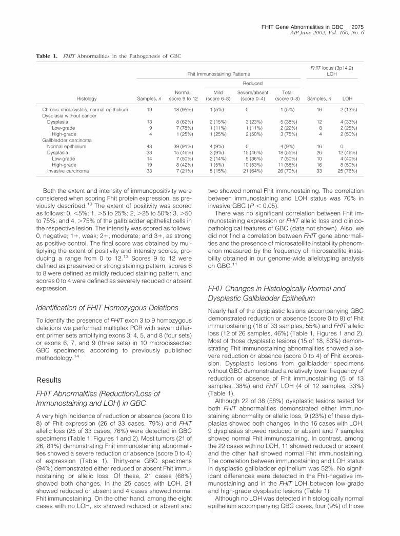
A very high incidence of reduction or absence (score 0 to8) of Fhit expression (26 of 33 cases, 79%) and FHITallelic loss (25 of 33 cases, 76%) were detected in GBCspecimens (Table 1, Figures 1 and 2). Most tumors (21 of26, 81%) demonstrating Fhit immunostaining abnormali-ties showed a severe reduction or absence (score 0 to 4)of expression (Table 1). Thirty-one GBC specimens(94%) demonstrated either reduced or absent Fhit immu-nostaining or allelic loss. Of these, 21 cases (68%)showed both changes. In the 25 cases with LOH, 21showed reduced or absent and 4 cases showed normalFhit immunostaining. On the other hand, among the eightcases with no LOH, six showed reduced or absent and
two showed normal Fhit immunostaining. The correlationbetween immunostaining and LOH status was 70% ininvasive GBC (P � 0.05).
There was no significant correlation between Fhit im-munostaining expression or FHIT allelic loss and clinico-pathological features of GBC (data not shown). Also, wedid not find a correlation between FHIT gene abnormali-ties and the presence of microsatellite instability phenom-enon measured by the frequency of microsatellite insta-bility obtained in our genome-wide allelotyping analysison GBC.11
FHIT Changes in Histologically Normal andDysplastic Gallbladder Epithelium
Nearly half of the dysplastic lesions accompanying GBCdemonstrated reduction or absence (score 0 to 8) of Fhitimmunostaining (18 of 33 samples, 55%) and FHIT allelicloss (12 of 26 samples, 46%) (Table 1, Figures 1 and 2).Most of those dysplastic lesions (15 of 18, 83%) demon-strating Fhit immunostaining abnormalities showed a se-vere reduction or absence (score 0 to 4) of Fhit expres-sion. Dysplastic lesions from gallbladder specimenswithout GBC demonstrated a relatively lower frequency ofreduction or absence of Fhit immunostaining (5 of 13samples, 38%) and FHIT LOH (4 of 12 samples, 33%)(Table 1).
Although 22 of 38 (58%) dysplastic lesions tested forboth FHIT abnormalities demonstrated either immuno-staining abnormality or allelic loss, 9 (23%) of these dys-plasias showed both changes. In the 16 cases with LOH,9 dysplasias showed reduced or absent and 7 samplesshowed normal Fhit immunostaining. In contrast, amongthe 22 cases with no LOH, 11 showed reduced or absentand the other half showed normal Fhit immunostaining.The correlation between immunostaining and LOH statusin dysplastic gallbladder epithelium was 52%. No signif-icant differences were detected in the Fhit-negative im-munostaining and in the FHIT LOH between low-gradeand high-grade dysplastic lesions (Table 1).
Although no LOH was detected in histologically normalepithelium accompanying GBC cases, four (9%) of those
Table 1. FHIT Abnormalities in the Pathogenesis of GBC
Histology
Fhit Immunostaining PatternsFHIT locus (3p14.2)
LOH
Samples, nNormal,
score 9 to 12
Reduced
Samples, n LOHMild
(score 6–8)Severe/absent
(score 0–4)Total
(score 0–8)
Chronic cholecystitis, normal epithelium 19 18 (95%) 1 (5%) 0 1 (5%) 16 2 (13%)Dysplasia without cancer
Dysplasia 13 8 (62%) 2 (15%) 3 (23%) 5 (38%) 12 4 (33%)Low-grade 9 7 (78%) 1 (11%) 1 (11%) 2 (22%) 8 2 (25%)High-grade 4 1 (25%) 1 (25%) 2 (50%) 3 (75%) 4 2 (50%)
Gallbladder carcinomaNormal epithelium 43 39 (91%) 4 (9%) 0 4 (9%) 16 0Dysplasia 33 15 (46%) 3 (9%) 15 (46%) 18 (55%) 26 12 (46%)
Low-grade 14 7 (50%) 2 (14%) 5 (36%) 7 (50%) 10 4 (40%)High-grade 19 8 (42%) 1 (5%) 10 (53%) 11 (58%) 16 8 (50%)
Invasive carcinoma 33 7 (21%) 5 (15%) 21 (64%) 26 (79%) 33 25 (76%)
FHIT Gene Abnormalities in GBC 2075AJP June 2002, Vol. 160, No. 6
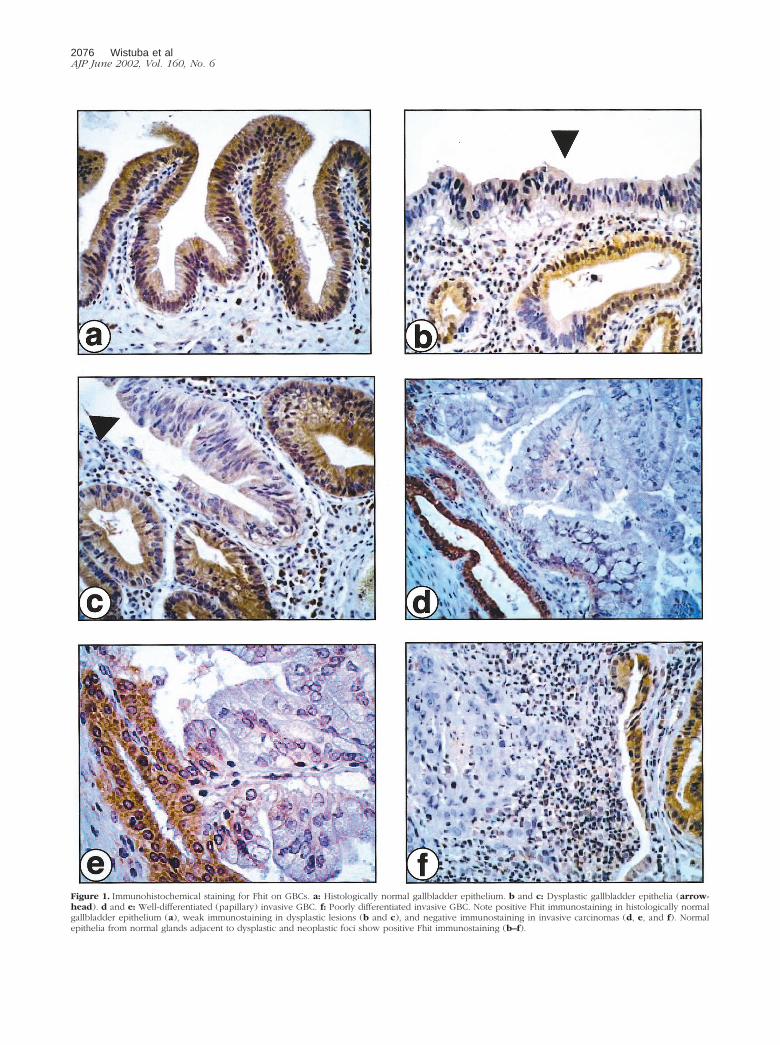
Figure 1. Immunohistochemical staining for Fhit on GBCs. a: Histologically normal gallbladder epithelium. b and c: Dysplastic gallbladder epithelia (arrow-head). d and e: Well-differentiated (papillary) invasive GBC. f: Poorly differentiated invasive GBC. Note positive Fhit immunostaining in histologically normalgallbladder epithelium (a), weak immunostaining in dysplastic lesions (b and c), and negative immunostaining in invasive carcinomas (d, e, and f). Normalepithelia from normal glands adjacent to dysplastic and neoplastic foci show positive Fhit immunostaining (b–f).
2076 Wistuba et alAJP June 2002, Vol. 160, No. 6
normal samples demonstrated mild reduced pattern(score 6 to 8) of the Fhit immunostaining. All those sam-ples were accompanying invasive GBC and correspond-ing dysplastic lesions with markedly reduced or absenceFhit immunostaining. Similarly, histologically normal epi-thelium from chronic cholecystitis without cancer demon-strated very infrequent FHIT immunostaining (5%) andLOH (13%) abnormalities (Table 1).
Patterns of FHIT Abnormalities in GBC andAccompanying Gallbladder Epithelia
In GBC specimens, one or both FHIT abnormalities (re-duced or loss of Fhit immunostaining and FHIT LOH) weredetected in 55 of 75 (73%) normal, dysplastic, and neo-plastic foci examined for both FHIT abnormalities. Weused these changes to determine whether the foci inindividual cases were molecularly related. Of interest, alldysplastic lesions and histologically normal epitheliumshowing negative Fhit immunostaining and FHIT locusLOH demonstrated the same abnormality in the corre-sponding invasive GBC.
Breakpoints at the FHIT Gene Locus
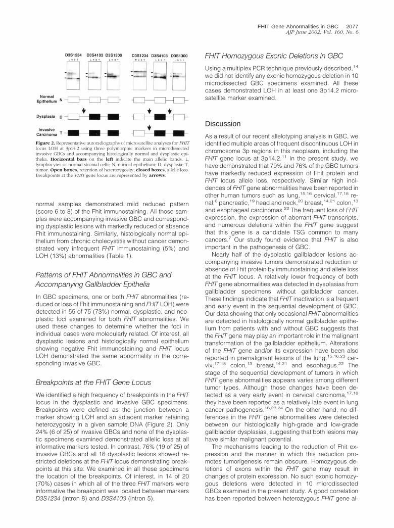
We identified a high frequency of breakpoints in the FHITlocus in the dysplastic and invasive GBC specimens.Breakpoints were defined as the junction between amarker showing LOH and an adjacent marker retainingheterozygosity in a given sample DNA (Figure 2). Only24% (6 of 25) of invasive GBCs and none of the dysplas-tic specimens examined demonstrated allelic loss at allinformative markers tested. In contrast, 76% (19 of 25) ofinvasive GBCs and all 16 dysplastic lesions showed re-stricted deletions at the FHIT locus demonstrating break-points at this site. We examined in all these specimensthe location of the breakpoints. Of interest, in 14 of 20(70%) cases in which all of the three FHIT markers wereinformative the breakpoint was located between markersD3S1234 (intron 8) and D3S4103 (intron 5).
FHIT Homozygous Exonic Deletions in GBC
Using a multiplex PCR technique previously described,14
we did not identify any exonic homozygous deletion in 10microdissected GBC specimens examined. All thesecases demonstrated LOH in at least one 3p14.2 micro-satellite marker examined.
Discussion
As a result of our recent allelotyping analysis in GBC, weidentified multiple areas of frequent discontinuous LOH inchromosome 3p regions in this neoplasm, including theFHIT gene locus at 3p14.2.11 In the present study, wehave demonstrated that 79% and 76% of the GBC tumorshave markedly reduced expression of Fhit protein andFHIT locus allele loss, respectively. Similar high inci-dences of FHIT gene abnormalities have been reported inother human tumors such as lung,15,16 cervical,17,18 re-nal,6 pancreatic,19 head and neck,20 breast,14,21 colon,13
and esophageal carcinomas.22 The frequent loss of FHITexpression, the expression of aberrant FHIT transcripts,and numerous deletions within the FHIT gene suggestthat this gene is a candidate TSG common to manycancers.7 Our study found evidence that FHIT is alsoimportant in the pathogenesis of GBC.
Nearly half of the dysplastic gallbladder lesions ac-companying invasive tumors demonstrated reduction orabsence of Fhit protein by immunostaining and allele lossat the FHIT locus. A relatively lower frequency of bothFHIT gene abnormalities was detected in dysplasias fromgallbladder specimens without gallbladder cancer.These findings indicate that FHIT inactivation is a frequentand early event in the sequential development of GBC.Our data showing that only occasional FHIT abnormalitiesare detected in histologically normal gallbladder epithe-lium from patients with and without GBC suggests thatthe FHIT gene may play an important role in the malignanttransformation of the gallbladder epithelium. Alterationsof the FHIT gene and/or its expression have been alsoreported in premalignant lesions of the lung,15,16,23 cer-vix,17,18 colon,13 breast,14,21 and esophagus.22 Thestage of the sequential development of tumors in whichFHIT gene abnormalities appears varies among differenttumor types. Although those changes have been de-tected as a very early event in cervical carcinoma,17,18
they have been reported as a relatively late event in lungcancer pathogenesis.16,23,24 On the other hand, no dif-ferences in the FHIT gene abnormalities were detectedbetween our histologically high-grade and low-gradegallbladder dysplasias, suggesting that both lesions mayhave similar malignant potential.
The mechanisms leading to the reduction of Fhit ex-pression and the manner in which this reduction pro-motes tumorigenesis remain obscure. Homozygous de-letions of exons within the FHIT gene may result inchanges of protein expression. No such exonic homozy-gous deletions were detected in 10 microdissectedGBCs examined in the present study. A good correlationhas been reported between heterozygous FHIT gene al-
Figure 2. Representative autoradiographs of microsatellite analyses for FHITlocus LOH at 3p14.2 using three polymorphic markers in microdissectedinvasive GBCs and accompanying histologically normal and dysplastic epi-thelia. Horizontal bars on the left indicate the main allelic bands. L,lymphocytes or normal stromal cells; N, normal epithelium; D, dysplasia; T,tumor. Open boxes, retention of heterozygosity; closed boxes, allelic loss.Breakpoints at the FHIT gene locus are represented by arrows.
FHIT Gene Abnormalities in GBC 2077AJP June 2002, Vol. 160, No. 6
terations (allele loss and intronic deletions) and reductionof Fhit protein expression.7 Our invasive GBCs showed agood correlation between both allele loss and reductionor absence of Fhit protein expression. Although alleleloss explains inactivation of one parental homologue, themechanism of inactivation of the second allele is unlikelyto occur by Knudson’s classic “two-hit” mechanism.5
Missense, nonsense, or frame-shift mutations in the FHITgene are rare in primary tumors.7 On the contrary, hyper-methylation of a 5� CpG island seem to inactivate the FHITgene in esophageal9 and lung cancers,10 and hasemerged as the major epigenetic mechanism for genesilencing in FHIT.
The progression in the overall frequency of FHIT ab-normalities with histological progression, as well as thepresence of identical patterns of LOH and Fhit proteinimmunostaining in precursor dysplastic lesions and theircorresponding invasive gallbladder tumors, suggest amolecular relationship through sequential changes be-tween both noninvasive and malignant gallbladder le-sions. Of interest, all dysplastic lesions and histologicallynormal epithelium showing negative Fhit immunostainingand FHIT locus allele loss demonstrated the same abnor-mality in the corresponding invasive GBC. Our recentunpublished findings based on allele loss pattern sug-gest that histologically normal and dysplastic gallbladderepithelia accompanying GBC arise as independentclones. Thus, the identical pattern of FHIT abnormalitiesdetected in the present study may be related to a fieldcancerization phenomenon.25
We identified a high frequency of discontinuous LOHand breakpoints in the FHIT locus in the dysplastic andinvasive GBC specimens. Breakpoints were defined asthe junction between a marker showing LOH and anadjacent marker retaining heterozygosity in a given sam-ple DNA. Seventy-six percent of invasive GBCs and alldysplastic lesions with LOH showed restricted deletions atthe FHIT locus involving breakpoints at this site. The 3p14.2FHIT region harbors the most common known aphidicolin-inducible fragile site in the genome (FRA3B).26 A high inci-dence of such breakpoints in this region have been recentlydemonstrated in invasive lung24 and breast21 tumors aswell as in their corresponding noninvasive epithelia. Ourfindings indicate that the FHIT 3p14.2 site represents ahighly unstable region undergoing frequent allele lossassociated with breakpoints in the gallbladder epitheliumundergoing malignant transformation. Although a corre-lation between high-grade microsatellite instability phe-nomenon and FHIT homozygous deletions has been sug-gested in pancreatic cancer,27 no correlation betweenFHIT gene abnormalities and the presence of microsat-ellite instability was detected in our GBC cases.
In summary, FHIT gene abnormalities, expressed byconsiderable reduction or loss of Fhit immunostainingand allele loss are nearly universal in GBC and thesechanges are detected early in the sequential develop-ment of this neoplasm. Our findings suggest that the FHITgene is one of the chromosome 3p putative TSGs in-volved in the pathogenesis of this highly malignant neo-plasm.
References
1. Lazcano-Ponce E, Miquel JF, Ferrecio C, Wistuba I, Munoz N, Alonsode Ruiz P, Urista G, Herrero R, Nervi F: Epidemiology and molecularpathology of gallbladder cancer. Ca Cancer J Clin 2001, 51:349–364
2. Albores-Saavedra J, Henson DE: Tumors of Gallbladder and Extra-hepatic Bile Ducts, fascicle 23, ed. 3. Washington, DC, Armed ForcesInstitute of Pathology, 2000
3. Wistuba II, Albores-Saavedra J: Genetic abnormalities involved in thepathogenesis of gallbladder carcinoma. J Hepatobiliary PancreatSurg 1999, 6:237–244
4. Fearon ER, Vogelstein B: A genetic model for colorectal tumorigene-sis. Cell 1990, 61:759–767
5. Knudson AG: Hereditary cancer, oncogenes, and antioncogenes.Cancer Res 1985, 45:1437–1443
6. Ohta M, Inoue H, Cotticelli MG, Kastury K, Baffa R, Palazzo J,Siprashvili Z, Mori M, McCue P, Druck T, Croce CM, Huebner K: TheFHIT gene, spanning the chromosome 3p14.2 fragile site and renalcarcinoma-associated t(3;8) breakpoint, is abnormal in digestive tractcancers. Cell 1996, 84:587–597
7. Croce CM, Sozzi G, Huebner K: Role of FHIT in human cancer. J ClinOncol 1999, 17:1618–1624
8. Siprashvili Z, Sozzi G, Barnes LD, McCue P, Robinson AK, Eryomin V,Sard L, Tagliabue E, Greco A, Fusetti L, Schwartz G, Pierotti MA,Croce CM, Huebner K: Replacement of Fhit in cancer cells sup-presses tumorigenicity. Proc Natl Acad Sci USA 1997, 94:13771–13776
9. Tanaka H, Shimada Y, Harada H, Shinoda M, Hatooka S, Imamura M,Ishizaki K: Methylation of the 5� CpG island of the FHIT gene is closelyassociated with transcriptional inactivation in esophageal squamouscell carcinomas. Cancer Res 1998, 58:3429–3434
10. Zochbauer-Muller S, Fong KM, Maitra A, Lam S, Geradts J, Ashfaq R,Virmani AK, Milchgrub S, Gazdar AF, Minna JD: 5� CpG islandmethylation of the FHIT gene is correlated with loss of gene expres-sion in lung and breast cancer. Cancer Res 2001, 61:3581–3585
11. Wistuba II, Tang M, Maitra A, Alvarez H, Troncoso P, Pimentel F,Gazdar AF: Genome-wide allelotyping analysis reveals multiple sitesof allelic loss in gallbladder carcinoma. Cancer Res 2001, 61:3795–3800
12. Wistuba II, Behrens C, Milchgrub S, Virmani AK, Jagirdar J, ThomasB, Ioachim HL, Litzky LA, Brambilla EM, Minna JD, Gazdar AF:Comparison of molecular changes in lung cancers in HIV-positiveand HIV-indeterminate subjects. JAMA 1998, 279:1554–1559
13. Hao XP, Willis JE, Pretlow TG, Rao JS, MacLennan GT, Talbot IC,Pretlow TP: Loss of fragile histidine triad expression in colorectalcarcinomas and premalignant lesions. Cancer Res 2000, 60:18–21
14. Ahmadian M, Wistuba II, Fong KM, Behrens C, Kodagoda DR, Sa-boorian MH, Shay J, Tomlinson GE, Blum J, Minna JD, Gazdar AF:Analysis of the FHIT gene and FRA3B region in sporadic breastcancer, preneoplastic lesions, and familial breast cancer probands.Cancer Res 1997, 57:3664–3668
15. Sozzi G, Pastorino U, Moiraghi L, Tagliabue E, Pezzella F, Ghirelli C,Tornielli S, Sard L, Huebner K, Pierotti MA, Croce CM, Pilotti S: Lossof FHIT function in lung cancer and preinvasive bronchial lesions.Cancer Res 1998, 58:5032–5037
16. Fong KM, Biesterveld EJ, Virmani A, Wistuba I, Sekido Y, Bader SA,Ahmadian M, Tiong Ong S, Rassool FV, Zimmerman PV, Giaccone G,Gazdar AF, Minna JD: FHIT and FRA3B allele loss are common inlung cancer and preneoplastic bronchial lesions and are associatedwith cancer-related FHIT cDNA splicing aberrations. Cancer Res1997, 57:2256–2267
17. Wistuba II, Montellano FD, Milchgrub S, Virmani AK, Behrens C, ChenH, Ahmadian M, Nowak JA, Muller C, Minna JD, Gazdar AF: Deletionsof chromosome 3p are frequent and early events in the pathogenesisof uterine cervical carcinoma. Cancer Res 1997, 57:3154–3158
18. Birrer MJ, Hendricks D, Farley J, Sundborg MJ, Bonome T, Walts MJ,Geradts J: Abnormal Fhit expression in malignant and premalignantlesions of the cervix. Cancer Res 1999, 59:5270–5274
19. Sorio C, Baron A, Orlandini S, Zamboni G, Pederzoli P, Huebner K,Scarpa A: The FHIT gene is expressed in pancreatic ductular cellsand is altered in pancreatic cancers. Cancer Res 1999, 59:1308–1314
20. Mao L, Fan YH, Lotan R, Hong WK: Frequent abnormalities of FHIT, a
2078 Wistuba et alAJP June 2002, Vol. 160, No. 6
candidate tumor suppressor gene, in head and neck cancer celllines. Cancer Res 1996, 56:5128–5131
21. Maitra A, Wistuba II, Washington C, Virmani AK, Ashfaq R, MilchgrubS, Gazdar AF, Minna JD: High-resolution chromosome 3p allelotypingof breast carcinomas and precursor lesions demonstrates frequentloss of heterozygosity and a discontinuous pattern of allele loss. Am JPathol 2001, 159:119–130
22. Michael D, Beer DG, Wilke CW, Miller DE, Glover TW: Frequentdeletions of FHIT and FRA3B in Barrett’s metaplasia and esophagealadenocarcinomas. Oncogene 1997, 15:1653–1659
23. Wistuba II, Behrens C, Milchgrub S, Bryant D, Hung J, Minna JD,Gazdar AF: Sequential molecular abnormalities are involved in themultistage development of squamous cell lung carcinoma. Onco-gene 1999, 18:643–650
24. Wistuba II, Behrens C, Virmani AK, Mele G, Milchgrub S, Girard L,
Fondon III JW, Garner HR, McKay B, Latif F, Lerman MI, Lam S,Gazdar AF, Minna JD: High resolution chromosome 3p allelotyping ofhuman lung cancer and preneoplastic/preinvasive bronchial epithe-lium reveals multiple, discontinuous sites of 3p allele loss and threeregions of frequent breakpoints. Cancer Res 2000, 60:1949–1960
25. Strong MS, Incze J, Vaughan CW: Field cancerization in the aerodi-gestive tract—its etiology, manifestation, and significance. J Otolar-yngol 1984, 13:1–6
26. Boldog F, Gemmill RM, West J, Robinson M, Robinson L, Li EF, RocheJ, Todd S, Waggoner B, Lundstrom R, Jacobson J, Mullokandov MR,Klinger H, Drabkin HA: Chromosome 3p14 homozygous deletionsand sequence analysis of FRA3B. Hum Mol Genet 1997, 6:193–203
27. Hilgers W, Koerkamp BG, Geradts J, Tang DJ, Yeo CJ, Hruban RH,Kern SE: Genomic FHIT analysis in RER� and RER� adenocarcino-mas of the pancreas. Genes Chromosom Cancer 2000, 27:239–243
FHIT Gene Abnormalities in GBC 2079AJP June 2002, Vol. 160, No. 6
Related Documents