FRACTURE HEALING DR.ABHINAV KESARKAR JR1 DEPARTMENT OF ORTHOPEDICS PDMMC, AMRAVATI

Welcome message from author
This document is posted to help you gain knowledge. Please leave a comment to let me know what you think about it! Share it to your friends and learn new things together.
Transcript

FRACTURE HEALING
DR.ABHINAV KESARKAR
JR1
DEPARTMENT OF ORTHOPEDICS
PDMMC, AMRAVATI

CELLS FOUND IN BONE
Osteoprogenitor cells
• Pluripotent mesenchymal stem cells found in bone which differentiate into osteoblasts
•Found throughout the bone and also called as preosteoblasts

Osteoblasts• derived from osteoprogenitor cells• Secrete collagen and calcium salts• Highly active cells depicted by increaseddensity rER on histology• create bone tissue de novo and entrap themselves
in small spaces called lacunae.• long processes from the cell lie in channels called as canaliculi
through which these cells communicate with other cells.• osteoblast within a lacunae surrounded by bone matrix
and further differentiation is called as an osteocyte and is responsible strength and integrity of the bone

Osteoclast• derived from fusion of many
monocytes•They are large phagocytic multi-nucleated cells• They break down bone after senescence of or trauma to osteocyte,normal bone remodelling• cytoplasm is eosinophilic and has a ruffled border• present on the surface of bone being reabsorbed and occasionally free in bone

• Are flattened epithelium in adult skeleton found on resting surfaces.
• Plays active role in differentiation of progenitor cells
• Controls osteoclasts, mineral hemostasis and may secrete collagenase.
• Lines – endosteal surface of marrow cavity
- periosteal surface
- vascular channels within osteons.
5
BONE LINING CELLS

THE BONE
• Bone is essentially a highly vascular, living, constantly changing mineralized connective tissue which makes up body’s skeleton.
• Other functions are:
- Bone provides protection for the vital organs of the body( eg: heart and brain)
- The hematopoietic bone marrow is protected by the surrounding bony tissue.
- Storage of calcium and phosphate


• Woven (Immature bone): Characterized by random arrangement of cells and collagen ,associated with periods of rapid bone formation, such as in initial stage of fracture healing.
• Lamellar bone (Mature bone) : Characterized by an orderly cellular distribution and properly oriented collagen fibres . This constitutes organised bone both cortical and cancellous


HAVERSIAN CANAL
• Haversian canal
Cells, nerves & vessels
• Volkmann’s
canal
Connects osteons
10
osteon
Haversian
canal
osteocyte
Volkmann’s
canal

THE PERIOSTEUM
Is a membrane that lines the outer surface of all bones except at the joints of long bones.
• Is made up of :
• Outer FIBROUS layer : made up of white connective and elastic tissue.
• Inner CAMBIUM layer : which has a looser composition, is more vascular and contains cells with osteogenicpotency.

FUNCTION OF THE PERIOSTEUM
1. Anchors tendons and ligaments to bone.
2. Acts as a limiting membrane.
3. Participates in growth (appositional) and repair through the activities of the osteoprogenitor cells .
4. Periosteum helps in fracture healing by forming periosteal callus.
5. It also lessen the displacement of the # and helps in reduction.
6. Allows passage of blood vessels, lymphatics and nerves into and out of
the bone.
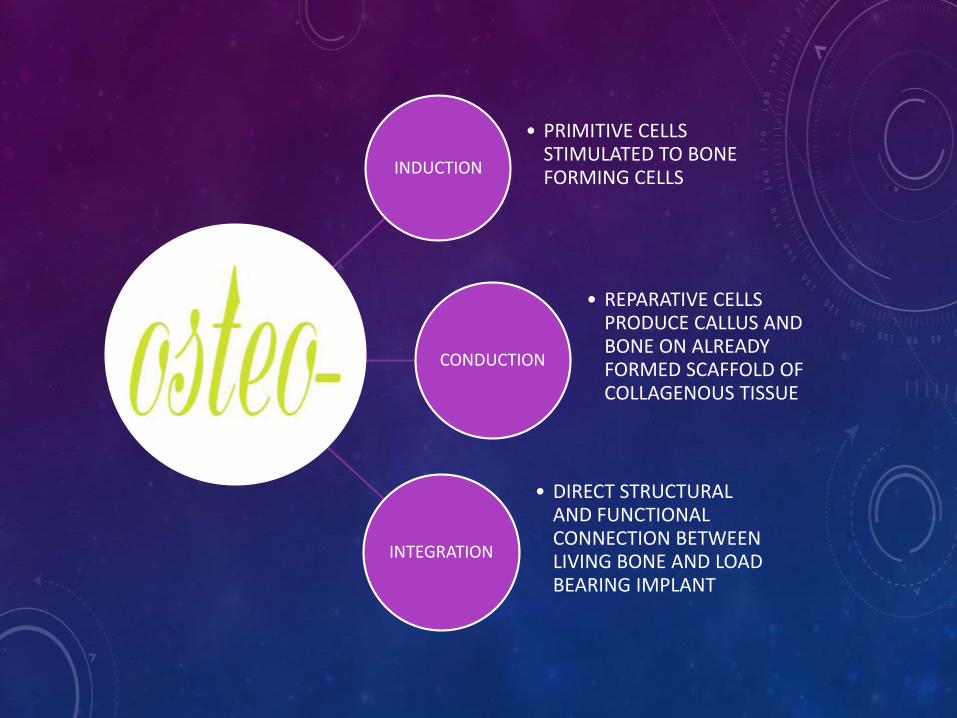
INDUCTION
• PRIMITIVE CELLS STIMULATED TO BONE FORMING CELLS
CONDUCTION
• REPARATIVE CELLS PRODUCE CALLUS AND BONE ON ALREADY FORMED SCAFFOLD OF COLLAGENOUS TISSUE
INTEGRATION
• DIRECT STRUCTURAL AND FUNCTIONAL CONNECTION BETWEEN LIVING BONE AND LOAD BEARING IMPLANT

1. Cutting Cones
2. Intramembranous Bone Formation
3. Endochondral Bone Formation
14
MECHANISM OF BONE FORMATION

CUTTING CONES

• Primarily a mechanism to remodel bone.
• Osteoclasts at the front of the cutting cone remove bone.
• Trailing osteoblasts lay down new bone.
Cutting cone

INTRAMEMBRANOUS BONE FORMATION
17

• Results in the formation of cranial bones of the skull (frontal, perietal, occipital, and temporal bones) and the clavicles.
• All bones formed this way are flat bones
• An ossification center appears in the fibrous connective tissue membrane Bone matrix is secreted within the fibrous membrane Woven bone and periosteum form Bone collar of compact bone forms, and red marrow appears

ENDOCHONDRAL BONE FORMATION

• Results in the formation of all of the rest of the bones
• Begins in the second month of development
• Uses hyaline cartilage “bones” as models for bone construction
• Requires breakdown of hyaline cartilage prior to ossification
• Formation begins at the primary ossification center
• The perichondrium covering the hyaline cartilage “bone”infiltrated with blood vessels converting it to vascularizedperiosteum.
• This change in nutrition causes the underlying mesenchymalcells to specialize into osteoblasts

TYPES OF BONE HEALING
• A] PRIMARY BONE HEALING
• B] SECONDARY BONE HEALING

A] PRIMARY BONE HEALING
• Occurs when fractured bones are approximated properly and stabilised by fixation.
• No callus is formed
• Heals by
contact healing
gap healing

PRIMARY BONE HEALING WITHOUT CALLUS FORMATION

PRIMARY BONE HEALING
1. CONTACT HEALING: When there is direct contact
between the cortical bone ends, lamellar bone forms
directly across the fracture line , parallel to long axis of
the bone, by direct extension of osteons.
2. GAP HEALING: Osteoblasts differentiate and start
depositing osteoids on the exposed surfaces of
fragment ends, mostly without a preceding osteoclastic
resorption which is later converted into the lamellar
bone .

B] SECONDARY BONE HEALING
It is usual type consisting of formation of callus either of
cartilaginous or fibrous.
This callus is later replaced by lamellar bone. It is
comparable to healing of soft tissue by filling of gaps with
vascular granulation tissue.
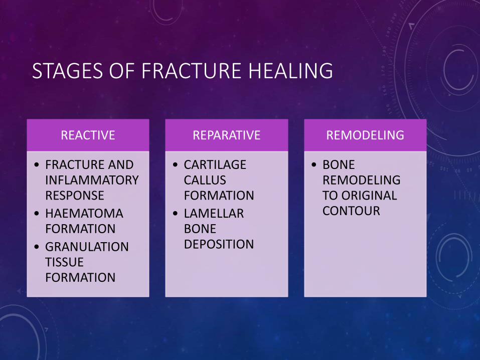
STAGES OF FRACTURE HEALING
REACTIVE
• FRACTURE AND INFLAMMATORY RESPONSE
• HAEMATOMA FORMATION
• GRANULATION TISSUE FORMATION
REPARATIVE
• CARTILAGE CALLUS FORMATION
• LAMELLAR BONE DEPOSITION
REMODELING
• BONE REMODELING TO ORIGINAL CONTOUR

Fracture and inflammatory
ressponse
• presence of blood cells within the tissues which are adjacent to the injury site. Soon after fracture, the blood vessels constrict, stopping any further bleeding
Haematomaformation
• the extravascular blood cells form a blood clot, known as a hematoma. All of the cells within the blood clot degenerate and die.
• The fracture hematoma immobilizes & splints the fracture.
• The fracture haematoma provides a fibrin scaffold that facilitates migration of repair cells.
Granulation tissue formation
• fibroblasts survive and replicate. They form a loose aggregate of cells, interspersed with small blood vessels, known as granulation tissue which grows forward, outside and inside the bone to bridge the fracture.
• They are stimulated by vasoactive mediators like serotonin and histamine.

REPARATIVE STAGE
Days after the # the periosteal cells proximal to the fracture gap and fibroblasts develop into chondroblasts which form hyaline cartilage.
The periosteal cells distal to the fracture gap develop into osteoblasts which form woven bone. These 2 tissues unite with their counterparts and culminate into new mass of heterogenous tissue called Fracture Callus restoring some of its original strength
, the mineralized matrix is penetrated by channels, each containing a microvessel and numerous osteoblasts.
This new lamellar bone is in the form of trabecular bone which restores bone’s original strength.

REMODELING PHASE
The remodeling process substitutes the trabecular bone
with compact bone. The trabecular bone is first resorbed
by osteoclasts, creating a shallow resorption pit known as a
"Howship's lacuna".
Then osteoblasts deposit compact bone within the
resorption pit.
Eventually, the fracture callus is remodelled

FRACTURE HEALING IN CANCELLOUSBONE
•Cancellous bones are porous and have largerSurface area per unit volume• large surface area create many points of bone
contact rich in cells and blood supply.• usually undisplaced and impacted cancellous
fractures heal without visisble callus formationwhereas undisplaced fractures heal by callus
• woven bone is formed along points of contactthereby leading into bone regeneration by osteoblastic activity

FRACTURE HEALING IN CHILDREN
• Compared with the relatively static mature bone of adult, the changing structure and function both physiological and biomechanical of immature bones make them susceptible to different patterns of fracture.
• Fracture in children are more common and are more likely to occur after seemingly insignificant trauma. Damage involving specific growth regions such as the physis or epiphysealossification center may lead to acute and chronic growth disturbances.

HOW IS PAEDIATRIC BONE DIFFERENT ?
1. HIGHER COLLAGEN TO BONE RATIO
2. HIGH CELLULARITY AND POROSITY
3. TRANSITION AREAS IN BONE
4. THICKER PERIOSTEUM
5. PRESENCE OF GROWTH PLATE
6. MORE CANCELLOUS BONE
7. STRONG LIGAMENTS

THE GROWTH PLATE
John Hunter studied growing chickens. He observed bones grew at the ends and thus demonstrated the existence of the epiphysealplates. Hunter is considered the “father of the growth plate’’


ZONES OF GROWTH PLATEEpiphyseal plate zone (from epiphysis to diaphysis
Description
Zone of reserve Quiescent chondrocytes are found at the epiphyseal end
Zone of proliferation Chondrocytes undergo rapid mitosis under influence of growth hormone
Zone of maturation and hypertrophy Chondrocytes stop mitosis, and begin to hypertrophy by accumulating glycogen, lipids, and alkaline phosphatase
Zone of calcification Chondrocytes undergo apoptosis. Cartilagenous matrix begins to calcify.Fractures usually occur through this zone
Zone of ossification Osteoclasts and osteoblasts from the diaphyseal side break down the calcified cartilage and replace with mineralized bone tissue.
FRACTURE REPAIR IN PEDIATRIC BONES
Fracture healing in children follow same pattern of adults but with some peculiarities :
PERIOSTEUM:
• In the contrast to adults the periosteum strips away easily from the underlying bone in children. Allowing fracture haematoma to dissect along the diaphysis and metaphysis and this is evident in the subsequent amount of new bone formation along the shaft.
• Dense attachment of the periosteum into the zone of ranvier limit subperiosteal hematoma formation to the metaphysic and diaphysis.

• Paediatric bone very vascular, therefore inflammatory hyperaemicresponse rapid and marked.
• Periosteum already producing bone as part of growth
• Rapid acceleration of this process, supplements endochondralossification from organising haematoma
• Produces rubbery bone around fracture within 10-14 days, then difficult to manipulate

REMODELING IN CHILDREN
The remodelling phase is the longest phase and in children may continue until skeletal maturation.
Remodelling is better in children compared to adult, this is in response to constantly changing stress Patterns in children during skeletal growth and development.

VARIABLES INFLUENCING FRACTURE HEALING
variables
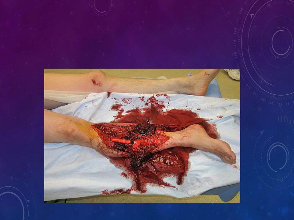
1]INJURY VARIABLES OPEN FRACTURESINJURY SEVERITYINTRAARTICULAR FRACTURESSEGMENTAL FRACTURESVASCULAR INJURYSOFT TISSUE INTERPOSITION
2]PATIENT VARIABLES DISEASE/DISORDERAGENUTRITIONHORMONESNICOTINE AND OTHER AGENTS
3]TISSUE VARIABLES TYPE OF BONE (CANCELLOUS OR CORTICAL)BONE DISEASEBONE NECROSISINFECTION
4] TREATMENT VARIABLES APPOSITION OF FRACTURE ENDSMICROMOTIONFRACTURE STABILITY


• VITAMIN D INSUFFICIENCY (< 32ng/ml)
• VITAMIN D DEFICIENCY (<20ng/ml)
BASIS OF COLLAGEN AND MATRIX LAYDOWN AND OSTEOID FORMATION
IS PROTEIN ‘ THE BUILDING BLOCKS OF BODY TISSUE’
NUTRITIONAL CARE OF PATIENT UNDERGOING ORTHOPEDIC TREATMENT SHOULD NOT BE UNDERESTIMATED.

This x-ray film obtained from a patient with corticosteroid-induced osteoporosis showed osteopenia and multiple vertebral compression fractures.

The hypertrophic fracture pattern indicates motion at the fracture site, despite the fixation. The patient was relatively asymptomatic until a nail broke at the nonunited fracture site (A). Anteroposterior radiograph showing complete healing following compression plating and bone grafting of the nonunion

RECENT ADVANCES:
• GROWTH FACTOR THERAPY
Due to their ability to stimulate proliferation and differentiation of mesenchymal and osteoprogenitor cells they have shown great promise for their ability to promote fracture repair .
• APPLICATION OF PLATELET RICH PLASMA
Injecting platelet rich plasma at fracture site helps in fracture healing .
• TISSUE ENGINEERING, STEM CEELS AND GENE THERAPIES
In past decade tissue culture and stem cells have been implicated in enhancing fracture healing and articular cartilage regeneration.

SUMMARY
• THE PROCESS OF FRACTURE HEALING IS MULTIFACTORIAL
AND DEPENDS UPON VARIABLES LIKE MECHANICAL
STABILITY,HORMONAL INFLUENCES,ELECTRICAL
ACTIVITY,METABOLISM,ETC.
• NECESSITY TO KNOW INTERDEPENDENCE OF THIS FACTORS
ON EACH OTHER AND HEALING PROCESS.
• WARRANTS MORE PENETRANCE OF TECHNOLOGICAL
ADVANCES FOR STUDY OF PHENOMENON OF FRACTURE
HEALING ON MOLECULAR LEVEL
THANK YOU
Related Documents