Published online 12 December 2019 Nucleic Acids Research, 2020, Vol. 48, No. 4 e19 doi: 10.1093/nar/gkz1165 Highly specific enrichment of rare nucleic acid fractions using Thermus thermophilus argonaute with applications in cancer diagnostics Jinzhao Song 1,*,† , Jorrit W. Hegge 2,† , Michael G. Mauk 1 , Junman Chen 1 , Jacob E. Till 3 , Neha Bhagwat 3 , Lotte T. Azink 2 , Jing Peng 1 , Moen Sen 3 , Jazmine Mays 3 , Erica L. Carpenter 3 , John van der Oost 2,* and Haim H. Bau 1,* 1 Department of Mechanical Engineering and Applied Mechanics, University of Pennsylvania, Philadelphia PA, USA, 2 Laboratory of Microbiology, Department of Agrotechnology and Food Sciences, Wageningen University,The Netherlands and 3 Perelman School of Medicine, University of Pennsylvania, Philadelphia, PA, USA Received July 04, 2019; Revised November 12, 2019; Editorial Decision December 01, 2019; Accepted December 03, 2019 ABSTRACT Detection of disease-associated, cell-free nucleic acids in body fluids enables early diagnostics, geno- typing and personalized therapy, but is challenged by the low concentrations of clinically significant nucleic acids and their sequence homology with abundant wild-type nucleic acids. We describe a novel approach, dubbed NAVIGATER, for increas- ing the fractions of Nucleic Acids of clinical inter- est Via DNA-Guided Argonaute from Thermus ther- mophilus (TtAgo). TtAgo cleaves specifically guide- complementary DNA and RNA with single nucleotide precision, greatly increasing the fractions of rare al- leles and, enhancing the sensitivity of downstream detection methods such as ddPCR, sequencing, and clamped enzymatic amplification. We demonstrated 60-fold enrichment of the cancer biomarker KRAS G12D and ∼100-fold increased sensitivity of Pep- tide Nucleic Acid (PNA) and Xenonucleic Acid (XNA) clamp PCR, enabling detection of low-frequency (<0.01%) mutant alleles (∼1 copy) in blood sam- ples of pancreatic cancer patients. NAVIGATER sur- passes Cas9-based assays (e.g. DASH, Depletion of Abundant Sequences by Hybridization), identify- ing more mutation-positive samples when combined with XNA-PCR. Moreover, TtAgo does not require targets to contain any specific protospacer-adjacent motifs (PAM); is a multi-turnover enzyme; cleaves ss- DNA, dsDNA and RNA targets in a single assay; and operates at elevated temperatures, providing high se- lectivity and compatibility with polymerases. INTRODUCTION In recent years, researchers have identified various en- zymes from archaea and bacteria that can be programmed with nucleic acids to cleave complementary strands, among which CRISPR–Cas have attracted considerable atten- tion as genome-editing tools (1–3). In medical diagnostics, CRISPR-associated nucleases have been used to (i) am- plify reporter signals during nucleic acid detection (4–8), (ii) detect unamplified targets immobilized on graphene field- effect transistors (9) and (iii) enrich rare alleles to enhance detection limits of oncogenic sequences (10–12). Despite re- markable progress, the applications of CRISPR–Cas are re- stricted due, among other things, to their reliance on the protospacer-adjacent motif (PAM), which is absent in many sequences of interest (10–12). Analogous to CRISPR–Cas, Argonaute (Ago) proteins (13) are nucleic acid-guided endonucleases. In contrast to Cas nucleases, Ago nucleases do not require the presence of any specific motifs and are, therefore, more versatile. Under appropriate conditions, Ago from the thermophilic bacterium Thermus thermophilus (TtAgo) (14–17) cleaves with high efficiency both DNA and RNA complementary to its small interfering DNA guides (siDNA), but spares nucleic acids with a single nucleotide mismatch at and around its catalytic site. We formulate a new assay (Fig- ure 1) termed NAVIGATER ( Nucleic Acid enrichment Via * To whom correspondence should be addressed. Tel: +1 215 898 1380; Fax: +1 215 573 6334; Email: [email protected] Correspondence may also be addressed to John van der Oost. Email: [email protected] Correspondence may also be addressed to Haim H. Bau. Email: [email protected] † The authors wish it to be known that, in their opinion, the first two authors should be regarded as joint First Authors. C The Author(s) 2019. Published by Oxford University Press on behalf of Nucleic Acids Research. This is an Open Access article distributed under the terms of the Creative Commons Attribution Non-Commercial License (http://creativecommons.org/licenses/by-nc/4.0/), which permits non-commercial re-use, distribution, and reproduction in any medium, provided the original work is properly cited. For commercial re-use, please contact [email protected] Downloaded from https://academic.oup.com/nar/article/48/4/e19/5673627 by guest on 24 January 2022

Welcome message from author
This document is posted to help you gain knowledge. Please leave a comment to let me know what you think about it! Share it to your friends and learn new things together.
Transcript
Published online 12 December 2019 Nucleic Acids Research, 2020, Vol. 48, No. 4 e19doi: 10.1093/nar/gkz1165
Highly specific enrichment of rare nucleic acidfractions using Thermus thermophilus argonaute withapplications in cancer diagnosticsJinzhao Song 1,*,†, Jorrit W. Hegge2,†, Michael G. Mauk1, Junman Chen1, Jacob E. Till3,Neha Bhagwat3, Lotte T. Azink2, Jing Peng1, Moen Sen3, Jazmine Mays3, EricaL. Carpenter3, John van der Oost2,* and Haim H. Bau 1,*
1Department of Mechanical Engineering and Applied Mechanics, University of Pennsylvania, Philadelphia PA, USA,2Laboratory of Microbiology, Department of Agrotechnology and Food Sciences, Wageningen University,TheNetherlands and 3Perelman School of Medicine, University of Pennsylvania, Philadelphia, PA, USA
Received July 04, 2019; Revised November 12, 2019; Editorial Decision December 01, 2019; Accepted December 03, 2019
ABSTRACT
Detection of disease-associated, cell-free nucleicacids in body fluids enables early diagnostics, geno-typing and personalized therapy, but is challengedby the low concentrations of clinically significantnucleic acids and their sequence homology withabundant wild-type nucleic acids. We describe anovel approach, dubbed NAVIGATER, for increas-ing the fractions of Nucleic Acids of clinical inter-est Via DNA-Guided Argonaute from Thermus ther-mophilus (TtAgo). TtAgo cleaves specifically guide-complementary DNA and RNA with single nucleotideprecision, greatly increasing the fractions of rare al-leles and, enhancing the sensitivity of downstreamdetection methods such as ddPCR, sequencing, andclamped enzymatic amplification. We demonstrated60-fold enrichment of the cancer biomarker KRASG12D and ∼100-fold increased sensitivity of Pep-tide Nucleic Acid (PNA) and Xenonucleic Acid (XNA)clamp PCR, enabling detection of low-frequency(<0.01%) mutant alleles (∼1 copy) in blood sam-ples of pancreatic cancer patients. NAVIGATER sur-passes Cas9-based assays (e.g. DASH, Depletionof Abundant Sequences by Hybridization), identify-ing more mutation-positive samples when combinedwith XNA-PCR. Moreover, TtAgo does not requiretargets to contain any specific protospacer-adjacentmotifs (PAM); is a multi-turnover enzyme; cleaves ss-DNA, dsDNA and RNA targets in a single assay; and
operates at elevated temperatures, providing high se-lectivity and compatibility with polymerases.
INTRODUCTION
In recent years, researchers have identified various en-zymes from archaea and bacteria that can be programmedwith nucleic acids to cleave complementary strands, amongwhich CRISPR–Cas have attracted considerable atten-tion as genome-editing tools (1–3). In medical diagnostics,CRISPR-associated nucleases have been used to (i) am-plify reporter signals during nucleic acid detection (4–8), (ii)detect unamplified targets immobilized on graphene field-effect transistors (9) and (iii) enrich rare alleles to enhancedetection limits of oncogenic sequences (10–12). Despite re-markable progress, the applications of CRISPR–Cas are re-stricted due, among other things, to their reliance on theprotospacer-adjacent motif (PAM), which is absent in manysequences of interest (10–12).
Analogous to CRISPR–Cas, Argonaute (Ago) proteins(13) are nucleic acid-guided endonucleases. In contrast toCas nucleases, Ago nucleases do not require the presenceof any specific motifs and are, therefore, more versatile.Under appropriate conditions, Ago from the thermophilicbacterium Thermus thermophilus (TtAgo) (14–17) cleaveswith high efficiency both DNA and RNA complementaryto its small interfering DNA guides (siDNA), but sparesnucleic acids with a single nucleotide mismatch at andaround its catalytic site. We formulate a new assay (Fig-ure 1) termed NAVIGATER (Nucleic Acid enrichment Via
*To whom correspondence should be addressed. Tel: +1 215 898 1380; Fax: +1 215 573 6334; Email: [email protected] may also be addressed to John van der Oost. Email: [email protected] may also be addressed to Haim H. Bau. Email: [email protected]†The authors wish it to be known that, in their opinion, the first two authors should be regarded as joint First Authors.
C© The Author(s) 2019. Published by Oxford University Press on behalf of Nucleic Acids Research.This is an Open Access article distributed under the terms of the Creative Commons Attribution Non-Commercial License(http://creativecommons.org/licenses/by-nc/4.0/), which permits non-commercial re-use, distribution, and reproduction in any medium, provided the original workis properly cited. For commercial re-use, please contact [email protected]
Dow
nloaded from https://academ
ic.oup.com/nar/article/48/4/e19/5673627 by guest on 24 January 2022
e19 Nucleic Acids Research, 2020, Vol. 48, No. 4 PAGE 2 OF 15
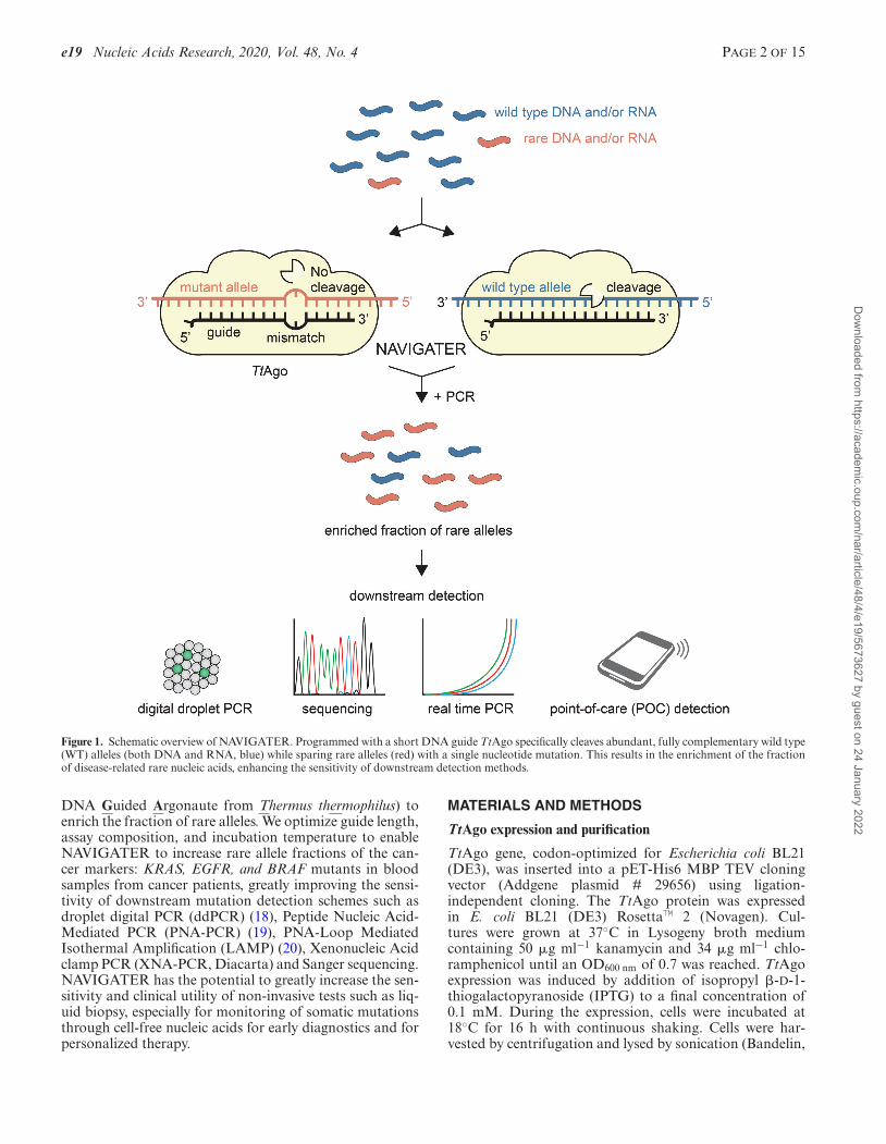
Figure 1. Schematic overview of NAVIGATER. Programmed with a short DNA guide TtAgo specifically cleaves abundant, fully complementary wild type(WT) alleles (both DNA and RNA, blue) while sparing rare alleles (red) with a single nucleotide mutation. This results in the enrichment of the fractionof disease-related rare nucleic acids, enhancing the sensitivity of downstream detection methods.
DNA Guided Argonaute from Thermus thermophilus) toenrich the fraction of rare alleles. We optimize guide length,assay composition, and incubation temperature to enableNAVIGATER to increase rare allele fractions of the can-cer markers: KRAS, EGFR, and BRAF mutants in bloodsamples from cancer patients, greatly improving the sensi-tivity of downstream mutation detection schemes such asdroplet digital PCR (ddPCR) (18), Peptide Nucleic Acid-Mediated PCR (PNA-PCR) (19), PNA-Loop MediatedIsothermal Amplification (LAMP) (20), Xenonucleic Acidclamp PCR (XNA-PCR, Diacarta) and Sanger sequencing.NAVIGATER has the potential to greatly increase the sen-sitivity and clinical utility of non-invasive tests such as liq-uid biopsy, especially for monitoring of somatic mutationsthrough cell-free nucleic acids for early diagnostics and forpersonalized therapy.
MATERIALS AND METHODS
TtAgo expression and purification
TtAgo gene, codon-optimized for Escherichia coli BL21(DE3), was inserted into a pET-His6 MBP TEV cloningvector (Addgene plasmid # 29656) using ligation-independent cloning. The TtAgo protein was expressedin E. coli BL21 (DE3) Rosetta™ 2 (Novagen). Cul-tures were grown at 37◦C in Lysogeny broth mediumcontaining 50 �g ml−1 kanamycin and 34 �g ml−1 chlo-ramphenicol until an OD600 nm of 0.7 was reached. TtAgoexpression was induced by addition of isopropyl �-D-1-thiogalactopyranoside (IPTG) to a final concentration of0.1 mM. During the expression, cells were incubated at18◦C for 16 h with continuous shaking. Cells were har-vested by centrifugation and lysed by sonication (Bandelin,
Dow
nloaded from https://academ
ic.oup.com/nar/article/48/4/e19/5673627 by guest on 24 January 2022
PAGE 3 OF 15 Nucleic Acids Research, 2020, Vol. 48, No. 4 e19
Sonopuls. 30% amplitude, 1s on/2s off for 5 min) in lysisbuffer containing 20 mM Tris–HCl pH 7.5, 250 mM NaCl,5mM imidazole, supplemented with EDTA-free proteaseinhibitor cocktail tablet (Roche). The soluble fraction ofthe lysate was loaded on a nickel column (HisTrap Hp,GE healthcare). The column was extensively washed withbuffer containing 20 mM Tris–HCl pH 7.5, 250 mM NaCland 30 mM imidazole. Bound proteins were eluted byincreasing the concentration of imidazole in the washbuffer to 250 mM. The eluted protein was dialysed at4◦C overnight against 20 mM HEPES pH 7.5, 250 mMKCl, and 1 mM dithiothreitol (DTT) in the presence of1 mg TEV protease (expressed and purified as previouslydescribed (21)) to cleave the His6-MBP tag. Next, thecleaved protein was diluted in 20 mM HEPES pH 7.5 tolower the final salt concentration to 125 mM KCl. Thediluted protein was applied to a heparin column (HiTrapHeparin HP, GE Healthcare), washed with 20mM HEPESpH 7.5, 125 mM KCl and eluted with a linear gradient of0.125–2 M KCl. Next, the eluted protein was loaded ontoa size exclusion column (Superdex 200 16/600 column,GE Healthcare) and eluted with 20 mM HEPES pH 7.5,500 mM KCl and 1 mM DTT (Supplementary FigureS26). Purified TtAgo protein was diluted in a size exclusionbuffer to a final concentration of 5 �M. Aliquots were flashfrozen in liquid nitrogen and stored at –80◦C.
TtAgo-based cleavage assays
5′-Phosphorylated DNA guides and Ultramer® ssDNAand ssRNA targets (100 nt) were synthesized by IDT(Coralville, IA, USA). For ssDNA and ssRNA cleavageexperiments, purified TtAgo, DNA guides, and ssDNA orssRNA targets were mixed with TtAgo and guides at theratios indicated in the buffers listed in Supplementary Ta-ble S1 and incubated at the indicated temperatures. Reac-tions were terminated by adding 1 �l proteinase K (Qiagen,Cat. No. 19131) solution, followed by 15 min incubationat 56◦C. Samples were then mixed with 2× loading buffer(95% (de-ionized) formamide, 5 mM EDTA, 0.025% SDS,0.025% bromophenol blue and 0.025% xylene cyanol) andheated for 10 min at 95◦C before the samples were resolvedon 15% denaturing polyacrylamide gels (7 M urea). Gelswere stained with SYBR gold Nucleic Acid Gel Stain (In-vitrogen) and nucleic acids were visualized using a BioRadGel Doc XR+ imaging system. For dsDNA cleavage, TtAgoand guides were pre-incubated in LAMP Buffer 3 (Supple-mentary Table S1) at 75◦C for 20 min or on ice for 3 min.
CRISPR/Cas9-based dsDNA cleavage
S.p. Cas9 Nuclease V3 (Cas9) and CRISPR–Cas9 sgRNA(sgRNA) were purchased from IDT (Coralville, IA). To cre-ate 10 �M ribonucleoprotein (RNP) complex which con-tains both sgRNA and Cas9 in equimolar amounts, 10�M Cas9 and 10 �M sgRNA were incubated in workingbuffer (30 mM HEPES, 150 mM KCl, pH 7.5) at roomtemperature for 10 min. For dsDNA cleavage experiments,RNP complex and dsDNA were mixed in 10:1 ratio (2.5�M RNP, 0.25 �M dsDNA) in Nuclease Reaction Buffer(20 mM HEPES, 100 mM NaCl, 15 mM MgCl2, 0.1 mM
EDTA, pH6.5) to get 10 �l total volume. The mixture wasincubated at 37◦C for 1 h. 1 �l RNase A (Thermo Scien-tific™, Cat. No. EN0531) was added and incubated at roomtemperature for 10 min to digest the sgRNA. Then, Cas9was digested by adding 1 �l proteinase K (Qiagen, Cat. No.19131) solution, followed by 15 min incubation at 56◦C.Samples were resolved on 15% denaturing polyacrylamidegels with the same procedure as described above.
Samples
Patient cell-free DNA (cfDNA) samples. All pancreaticpatient blood samples were obtained from patients eitherwith metastatic pancreatic cancer (Supplementary TableS2) or at start of therapy (Supplementary Table S4) whohad provided informed consent under the IRB-approvedprotocol (UPCC 02215, IRB# 822028). cfDNA was ex-tracted with QIAamp® Circulating Nucleic Acid kit (Qi-agen, Valencia, CA, USA). Subsequently, the extractedcfDNA was qualified and quantified with multiplex ddPCR(Raindance).
RNA samples. Total RNA was extracted with RNeasy®
mini kit (Qiagen, Valencia, CA, USA) per manufacturer’sprotocol from Human cancer cell lines U87-MG (WTKRAS mRNA) and ASPC1 (KRAS G12D mRNA).
cfDNA pre-amplification. cfDNA pre-amplification wascarried out in 50 �l reaction volumes using 20 ng of cfDNA,1× Q5 Hot Start High-Fidelity Master Mix (22) (New Eng-land Biolabs, Ipswich, MA, USA), and 100 nM each offorward and reverse KRAS ddPCR primers (Supplemen-tary Table S3). Reaction mixes without DNA were includedas no-template (negative) controls (NTCs). Nucleic acidswere preamplified with a BioRad Thermal Cycler (BioRad,Model CFD3240) with a temperature profile of 98◦C for 3min, followed by 30 cycles of amplification (98◦C for 10 s,63◦C for 3 min and 72◦C for 30 s), and a final 72◦C extensionfor 2 min.
mRNA pre-amplification. mRNA pre-amplification wasperformed in 50 �l reactions using 30 ng of total RNA, 1×Q5 Hot Start High-Fidelity Master Mix (New England Bi-olabs, Ipswich, MA, USA), 100 nM each of forward andreverse KRAS RT-PCR primers (Supplementary Table S3),and 1 �l reverse transcriptase (Invitrogen, Carlsbad, CA,USA). The reaction mix was incubated at 55◦C for 30 minand 98◦C for 3 min, followed by 30 cycles of amplification(93◦C for 15 s, 62◦C for 30 s and 72◦C for 30 s), and a final72◦C extension for 4 min.
Mutant allele enrichment (NAVIGATER)
The same setup as for synthetic dsDNA cleavage was usedfor cfDNA and mutant mRNA enrichment. TtAgo, S-guide, and AS-guide were mixed in 1:10:10 ratio (1.25 �MTtAgo, 12.5 �M S-guide, 12.5 �M AS-guide) in Buffer 3and pre-incubated at 75◦C for 20 min or on ice for 3 min. 2�l nucleic acids were added to 8 �l TtAgo-guides complexesand incubated at 83◦C for 1 h. When using preamplified nu-cleic acids, the enriched products were diluted 104-fold be-
Dow
nloaded from https://academ
ic.oup.com/nar/article/48/4/e19/5673627 by guest on 24 January 2022
e19 Nucleic Acids Research, 2020, Vol. 48, No. 4 PAGE 4 OF 15
fore downstream mutation analysis or second-round enrich-ment. TtAgo activity was terminated with the addition of 1�l proteinase K solution, followed by 15 min incubation at56◦C. Alternatively, TtAgo can be deactivated by incubat-ing the assay at 95◦C for over 20 min (Supplementary FigureS27).
NAVIGATER combined with downstream mutation detec-tion methods
Droplet digital PCR (ddPCR). ddPCR was carried outwith the RainDrop Digital PCR system (RainDance Tech-nologies, Inc.) to verify mutation abundance before and af-ter TtAgo enrichment. 2 �l of the 104-fold diluted, TtAgo-treated sample was added to each 30 �l dPCR. Each dPCRcontained 1× TaqMan Genotyping Master Mix (Life Tech-nologies), 400 nM KRAS ddPCR primers, 100 nM KRASwild type probe, 100 nM KRAS mutant probe (Supplemen-tary Table S3) and 1× droplet stabilizer (RainDance Tech-nologies, Inc.). Emulsions of each reaction were preparedon the RainDrop Source instrument (RainDance Technolo-gies, Inc.) to produce 2–7 million, 5-pl-volume dropletsper 25 �l reaction volume. Thereafter, the emulsions wereplaced in a thermal cycler to amplify the target and gen-erate signal. The temperature profile for amplification con-sisted of an activation step at 95◦C for 10 min, followed by45 cycles of amplification [95◦C for 15 s and 60◦C for 45 s].Reaction products were kept at 4◦C before placing them onthe RainDrop Sense instrument (RainDance Technologies,Inc.) for signal detection. RainDrop Analyst (RainDanceTechnologies, Inc.) was used to determine positive signalsfor each allele type. Gates were applied to regions of clus-tered droplets to define positive hits for each allele, accord-ing to manufacturer’s instructions.
PNA-PCR. PNA-PCR was performed in 20 �l reactionvolumes, each containing 4.5 �l of 104-fold diluted TtAgo-treated products, 1× Q5 Hot Start High-Fidelity MasterMix (New England Biolabs, Ipswich, MA, USA), 0.5 �l ofEvaGreen fluorescent dye (Biotium, Hayward, CA, USA),500 nM KRAS PNA clamp (Supplementary Table S3), and100 nM each of forward and reverse KRAS ddPCR primers.Reactions (quantitative PCR) were amplified with a BioRadThermal Cycler (BioRad, Model CFD3240) with a temper-ature profile of 98◦C for 3 min, followed by 40 cycles of am-plification (98◦C for 10 s, 63◦C for 3 min and 72◦C for 30s).
Sanger sequencing. RNA extracted from cell lines werepre-amplified with KRAS RT-PCR primers as describedabove and treated by our TtAgo enrichment system. 2 �lof the 104-fold diluted, TtAgo-treated sample was then am-plified by KRAS PCR protocol (same as for mRNA pre-amplification but without the reverse transcription step) for30 cycles. PCR products were inspected for quality and yieldby running 5 �l in 2.2% agarose Lonza FlashGel DNA Cas-sette and processed for Sanger sequencing at the Penn Ge-nomic Analysis Core.
Point-of-care (POC) mutation detection. PNA-LAMP(SMAP-2) was prepared in 20 �l reaction volumes accord-
ing to the previously described protocol with minor modifi-cations (20). The reaction mix contained 2 �l of the 104-folddiluted TtAgo-treated products (same as used for Sanger se-quencing), 1× LAMP buffer 3 (Eiken LAMP buffer), 1 �lBst DNA polymerase (from Eiken DNA LAMP kit), 2.5 �lof BART reporter (Lot: 1434201; ERBA Molecular, UK)(23), KRAS PNA clamp and LAMP primers (sequences andconcentrations listed in Supplementary Table S3). The pre-pared mixtures were injected into reaction chambers of ourcustom made, multifunctional chip (24,25). The inlet andoutlet ports were then sealed with transparent tape (3M,Scotch brand cellophane tape, St Paul, MN, USA) and thechip was placed in our portable Smart-Connected Cup andprocessed according to previously described protocol (23).
Comparison of TtAgo and CRISPR/Cas9 -based multi-plexed enrichment
Multiplexed pre-amplification. Triplex PCR were carriedout with primers from mutation detection kit (DiaCarta,Inc.). The 10 �l reaction mixture contains 60 ng of cfDNA(reference standard that includes various mutant alleles,Horizon Discovery, HD780), 1× PCR Master Mix, 1 �lof mixed PCR primers (1:1:1) for targets of interest. Nu-cleic acids were pre-amplified with a BioRad Thermal Cy-cler (BioRad, Model CFX96) with the temperature profileof 95◦C for 5 min, followed by 35 cycles of amplification(95◦C for 20 s, 70◦C for 40 s, 60◦C for 30 s and 72◦C for 30s) and a final 72◦C extension for 2 min.
Multiplexed enrichment. For triplex NAVIGATER,guides (1:1:1) for targets of interest were mixed with TtAgoin 10:1 ratio (12.5 �M S-guides, 12.5 �M AS-guides,1.25 �M TtAgo) in Buffer 3 and pre-incubated on icefor 3 min. Samples consisted of 1 �l pre-amplified triplexPCR products mixed with pre-incubated TtAgo-guidecomplexes. The reaction mixes were incubated at 83◦Cfor 1 h. The triplex Cas9-DASH (Depletion of AbundantSequences by Hybridization) (10) used the pre-amplifiedtriplex PCR products as the sample and three sgRNAs in1:1:1 ratio (10 �M sgRNA in total). The products withand without treatment were resolved on 15% denaturingpolyacrylamide gels (7 M urea).
XNA-PCR. NAVIGATER products were tested by themutation detection method XNA-PCR (DiaCarta, Inc.).XNA-PCR was carried out for individual mutants in 10�l reaction volumes, containing 3 �l of the 107-fold di-luted NAVIGATER products, 1× PCR Master Mix, 1 �l ofPCR primer/probe mix, and 1 �l of XNA clamp. Reactions(quantitative PCR) were amplified with a BioRad ThermalCycler (BioRad, Model CFX96) with a temperature profileof 95◦C for 5 min, followed by 45 cycles of amplification(95◦C for 20 s, 70◦C for 40 s, 60◦C for 30 s and 72◦C for 30s).
RESULTS
Betaine, Mg2+ and dNTPs enhance TtAgo’s endonucleolyticactivity
We first examined TtAgo activity in various buffers (Sup-plemental Section 1) and selected a buffer that provided the
Dow
nloaded from https://academ
ic.oup.com/nar/article/48/4/e19/5673627 by guest on 24 January 2022
PAGE 5 OF 15 Nucleic Acids Research, 2020, Vol. 48, No. 4 e19
best performance (Buffer 3, Supplemental Table 1). Signifi-cantly, dNTPs additives greatly enhance cleavage efficiencyof RNA (from 20% to 100%) while betaine and high concen-trations of Mg2+ (≥8 mM) enable high cleavage efficiency(>90%) of DNA at temperatures ≥80◦C without adverselyimpacting selectivity. The same buffer is suitable for cleav-ing both DNA and RNA.
Single guide-target-mismatches curtail target cleavage
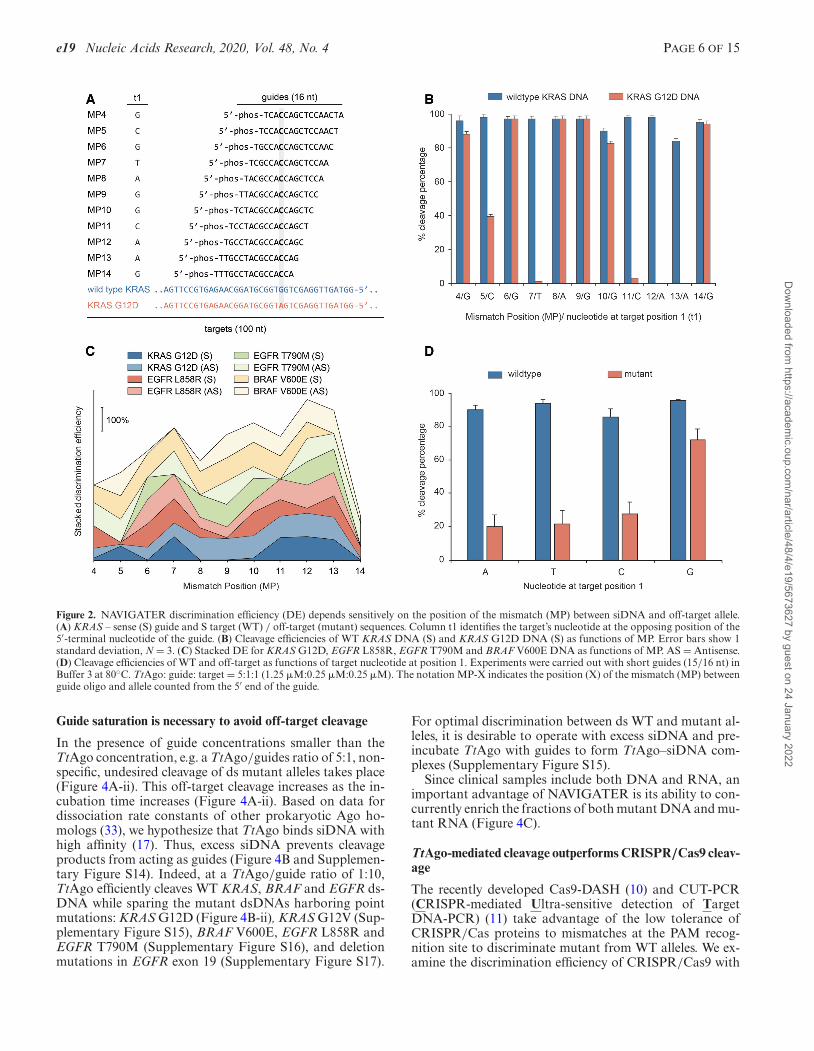
The conformation and, therefore, cleavage efficiency of Agoproteins depend sensitively on the position and type of sin-gle and dinucleotide mismatches between a guide oligo andtarget strand (15,26–28). Since we require efficient cleavageof wild type (WT) alleles while sparing mutant alleles witha single nucleotide mismatch, we examined the effects ofguide nucleotide mismatch position (MP, counted from thesiDNA’s 5′ end) on cleavage efficiency of WT KRAS, BRAF,EGFR and their mutants––both DNA and RNA (Figure 2and Supplementary Section 2). Figure 2A depicts the var-ious siDNAs tested with KRAS G12D. TtAgo-mediatedcleavage of KRAS G12D strand is nearly completely cur-tailed when siDNA has a nucleotide mismatch at MP7 orMP11-13 (Figure 2B). Guides designed for enriching KRASG12D also enrich the fractions of other KRAS G12 mu-tants.
To characterize siDNA performance, we define the dis-crimination efficiency (DE) as the difference in cleavage ef-ficiency of the WT and mutant strand. Figure 2C depictsDE as a function of MP for KRAS, EGFR and BRAF DNAand their mutants. Mismatches MP7 and MP9–MP13, lo-cated around the cleavage site (g10/g11), yielded the bestdiscrimination in most cases (Figure 2C and SupplementaryFigure S10e). The optimal MP depends, however, on the al-lele’s specific sequence. Cleavage of RNA was less tolerantto mismatches than cleavage of DNA. Single mismatchesanywhere between MP4–MP11 near completely preventedRNA cleavage (Supplementary Figures S6e and S8).
Despite being loaded with guides that were fully com-plementarity to the target, WT sequences were cleaved byTtAgo at variable efficiencies (Figure 2B and Supplemen-tary Section 2). This suggests that besides the differentbuffer components, the activity of TtAgo also depends onthe sequences of the guide and target, which might affectthe conformation of the ternary TtAgo–siDNA–DNA andTtAgo–siDNA–RNA complexes (29).
TtAgo encompasses two distinct binding pockets in theMID and PIWI domains to, respectively, accommodate theguide’s 5′-terminal nucleotide (g1) and its opposing tar-get nucleotide (t1) (Figure 2A) (29). Although a t1 guano-sine (t1G) binds preferentially into the pocket of the PIWI-domain (30), this was not reflected in the TtAgo’s cleavageefficiencies of fully complementary targets. That is the cleav-age efficiencies of targets (WT) did not significantly dependon the nucleotide type at target position 1 (SupplementaryNote 1) (30). In contrast, there is a significant increase inthe cleavage of mutant alleles when target position 1 is oc-cupied with guanosine (Figure 2D) even in the presence of amismatch between guide and target (Figure 2B and D), re-sulting in a poor discrimination efficiency. Therefore, whendesigning guides for NAVIGATER, t1G should be avoided.
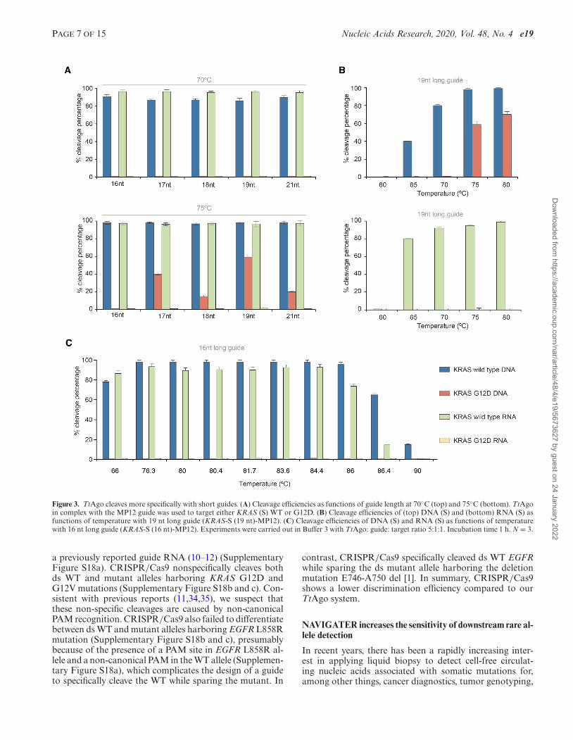
TtAgo cleaves most specifically with short guides (15/16 nt)
Heterologously-expressed TtAgo is typically purified withsiDNAs ranging in length from 13 to 25 nt (17), whereasin vitro, TtAgo operates with siDNAs ranging in lengthfrom 7 to 36 nt (15). Since little is known on the effect ofguide length on TtAgo discrimination efficiency, we exam-ine this effect in our in vitro assay. TtAgo efficiently cleavesWT KRAS (sense, S) with complementary guides, rangingin length from 16 to 21 nt at both 70◦C and 75◦C (Figure3A and Supplementary Figures S11 and S12). Guides 17–21 nt in length with a single nucleotide mismatch at MP12cleave mutant DNA strands at 75◦C, but not at 70◦C (Figure3A). We compared cleavage efficiencies of 16 nt and 19 ntlong guides as functions of temperature (Figure 3B and C).The 19 nt long guide cleaves DNA mutant strands at tem-peratures >75◦C, but not at temperatures below 70◦C (Fig-ure 3B). Cleavage of the mutant allele at ≥75◦C is, however,completely suppressed with the 16 nt guide (Figure 3A, bot-tom and Figure 3C). We observe a similar behavior when a15 nt guide with a single mismatch at MP13 targets the an-tisense (AS) KRAS strand (Supplementary Figure S11a–ii).We hypothesize that short guides form a less stable com-plex with off-targets than longer guides, preventing unde-sired cleavage.
In contrast to mutant DNA, increases in assay tempera-ture and guide length do not increase undesired cleavage ofmutant RNA (Figure 3A and B), likely due to differences inthe effects of ssDNA and ssRNA on TtAgo conformation.In summary, when operating with short guides (15/16 nt),TtAgo efficiently cleaves both WT RNA and DNA targetsin the same buffer while avoiding cleavage of mutant allelesat temperatures ranging from 66 to 86◦C (Figure 3C).
TtAgo efficiently cleaves targeted dsDNA at temperaturesabove the dsDNA’s melting temperature
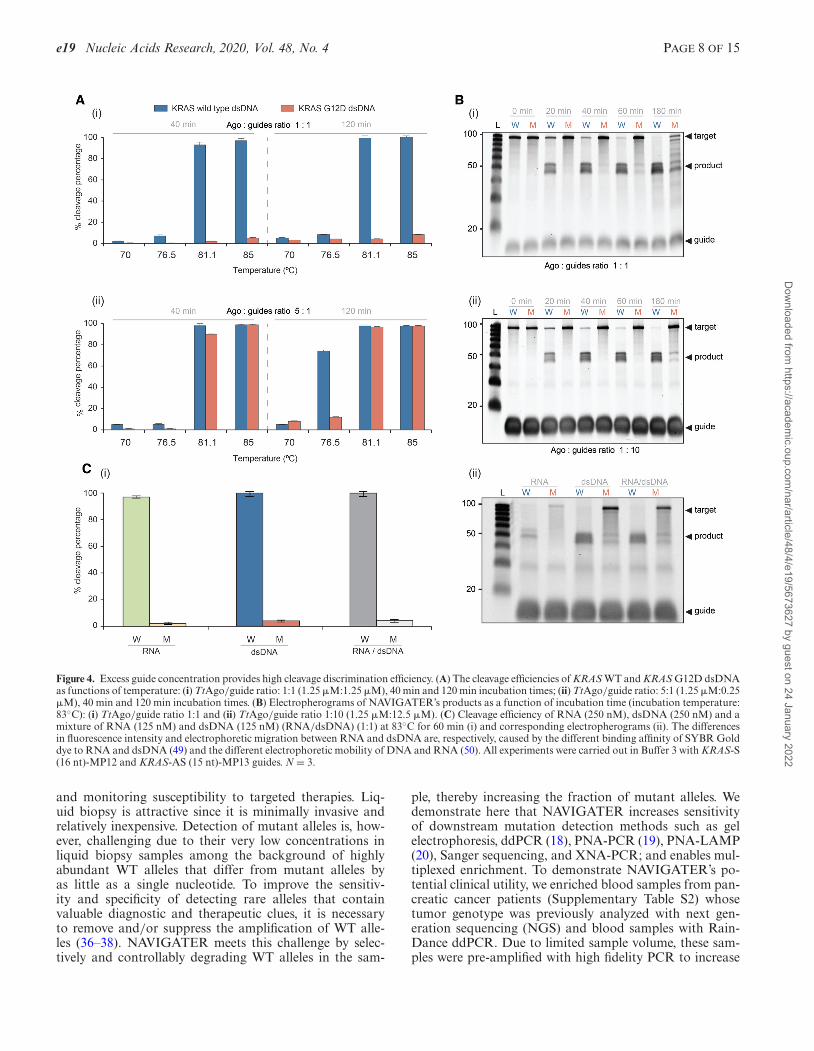
Guide-free (apo-) TtAgo can degrade dsDNA via a ‘chop-ping’ mechanism, autonomously generating functionalsiDNA (30). This is, however, a slow process that takesplace only when the target DNA is rich in AT (<17% GC)(17), suggesting that TtAgo lacks helicase activity and de-pends on dsDNA thermal breathing to enable chopping(13,17,31). In our assays TtAgo is incubated with an excessof siDNAs to suppress chopping of dsDNA. TtAgo’s activ-ity at high temperatures provides NAVIGATER with a clearadvantage since dsDNA unwinds as the incubation temper-ature increases.
We determined the optimal temperature at which TtAgo,saturated with guides, efficiently cleaves dsKRAS WT whilesparing mutant alleles. The estimated melting tempera-ture of 100 bp dsKRAS (S strand sequence listed in Fig-ure 2A) in Buffer 3 is 79.7◦C (IDT-OligoAnalyzer. https://www.idtdna.com/calc/analyzer). Consistent with this esti-mate, very little cleavage of dsDNA takes place at temper-atures below 80◦C, but TtAgo cleaves dsDNA efficiently attemperatures above 80◦C (Figure 4A-i). Cleavage efficiencyincreases as the incubation time increases and nearly sat-urates after about one hour (Figure 4B). TtAgo efficientlycleaves high GC-content (∼70%) dsDNA at 83◦C (Supple-mentary Figure S13), presumably due to the high betaineconcentration of our buffer (32).
Dow
nloaded from https://academ
ic.oup.com/nar/article/48/4/e19/5673627 by guest on 24 January 2022
e19 Nucleic Acids Research, 2020, Vol. 48, No. 4 PAGE 6 OF 15
Figure 2. NAVIGATER discrimination efficiency (DE) depends sensitively on the position of the mismatch (MP) between siDNA and off-target allele.(A) KRAS – sense (S) guide and S target (WT) / off-target (mutant) sequences. Column t1 identifies the target’s nucleotide at the opposing position of the5′-terminal nucleotide of the guide. (B) Cleavage efficiencies of WT KRAS DNA (S) and KRAS G12D DNA (S) as functions of MP. Error bars show 1standard deviation, N = 3. (C) Stacked DE for KRAS G12D, EGFR L858R, EGFR T790M and BRAF V600E DNA as functions of MP. AS = Antisense.(D) Cleavage efficiencies of WT and off-target as functions of target nucleotide at position 1. Experiments were carried out with short guides (15/16 nt) inBuffer 3 at 80◦C. TtAgo: guide: target = 5:1:1 (1.25 �M:0.25 �M:0.25 �M). The notation MP-X indicates the position (X) of the mismatch (MP) betweenguide oligo and allele counted from the 5′ end of the guide.
Guide saturation is necessary to avoid off-target cleavage
In the presence of guide concentrations smaller than theTtAgo concentration, e.g. a TtAgo/guides ratio of 5:1, non-specific, undesired cleavage of ds mutant alleles takes place(Figure 4A-ii). This off-target cleavage increases as the in-cubation time increases (Figure 4A-ii). Based on data fordissociation rate constants of other prokaryotic Ago ho-mologs (33), we hypothesize that TtAgo binds siDNA withhigh affinity (17). Thus, excess siDNA prevents cleavageproducts from acting as guides (Figure 4B and Supplemen-tary Figure S14). Indeed, at a TtAgo/guide ratio of 1:10,TtAgo efficiently cleaves WT KRAS, BRAF and EGFR ds-DNA while sparing the mutant dsDNAs harboring pointmutations: KRAS G12D (Figure 4B-ii), KRAS G12V (Sup-plementary Figure S15), BRAF V600E, EGFR L858R andEGFR T790M (Supplementary Figure S16), and deletionmutations in EGFR exon 19 (Supplementary Figure S17).
For optimal discrimination between ds WT and mutant al-leles, it is desirable to operate with excess siDNA and pre-incubate TtAgo with guides to form TtAgo–siDNA com-plexes (Supplementary Figure S15).
Since clinical samples include both DNA and RNA, animportant advantage of NAVIGATER is its ability to con-currently enrich the fractions of both mutant DNA and mu-tant RNA (Figure 4C).
TtAgo-mediated cleavage outperforms CRISPR/Cas9 cleav-age
The recently developed Cas9-DASH (10) and CUT-PCR(CRISPR-mediated Ultra-sensitive detection of TargetDNA-PCR) (11) take advantage of the low tolerance ofCRISPR/Cas proteins to mismatches at the PAM recog-nition site to discriminate mutant from WT alleles. We ex-amine the discrimination efficiency of CRISPR/Cas9 with
Dow
nloaded from https://academ
ic.oup.com/nar/article/48/4/e19/5673627 by guest on 24 January 2022
PAGE 7 OF 15 Nucleic Acids Research, 2020, Vol. 48, No. 4 e19
Figure 3. TtAgo cleaves more specifically with short guides. (A) Cleavage efficiencies as functions of guide length at 70◦C (top) and 75◦C (bottom). TtAgoin complex with the MP12 guide was used to target either KRAS (S) WT or G12D. (B) Cleavage efficiencies of (top) DNA (S) and (bottom) RNA (S) asfunctions of temperature with 19 nt long guide (KRAS-S (19 nt)-MP12). (C) Cleavage efficiencies of DNA (S) and RNA (S) as functions of temperaturewith 16 nt long guide (KRAS-S (16 nt)-MP12). Experiments were carried out in Buffer 3 with TtAgo: guide: target ratio 5:1:1. Incubation time 1 h. N = 3.
a previously reported guide RNA (10–12) (SupplementaryFigure S18a). CRISPR/Cas9 nonspecifically cleaves bothds WT and mutant alleles harboring KRAS G12D andG12V mutations (Supplementary Figure S18b and c). Con-sistent with previous reports (11,34,35), we suspect thatthese non-specific cleavages are caused by non-canonicalPAM recognition. CRISPR/Cas9 also failed to differentiatebetween ds WT and mutant alleles harboring EGFR L858Rmutation (Supplementary Figure S18b and c), presumablybecause of the presence of a PAM site in EGFR L858R al-lele and a non-canonical PAM in the WT allele (Supplemen-tary Figure S18a), which complicates the design of a guideto specifically cleave the WT while sparing the mutant. In
contrast, CRISPR/Cas9 specifically cleaved ds WT EGFRwhile sparing the ds mutant allele harboring the deletionmutation E746-A750 del [1]. In summary, CRISPR/Cas9shows a lower discrimination efficiency compared to ourTtAgo system.
NAVIGATER increases the sensitivity of downstream rare al-lele detection
In recent years, there has been a rapidly increasing inter-est in applying liquid biopsy to detect cell-free circulat-ing nucleic acids associated with somatic mutations for,among other things, cancer diagnostics, tumor genotyping,
Dow
nloaded from https://academ
ic.oup.com/nar/article/48/4/e19/5673627 by guest on 24 January 2022
e19 Nucleic Acids Research, 2020, Vol. 48, No. 4 PAGE 8 OF 15
Figure 4. Excess guide concentration provides high cleavage discrimination efficiency. (A) The cleavage efficiencies of KRAS WT and KRAS G12D dsDNAas functions of temperature: (i) TtAgo/guide ratio: 1:1 (1.25 �M:1.25 �M), 40 min and 120 min incubation times; (ii) TtAgo/guide ratio: 5:1 (1.25 �M:0.25�M), 40 min and 120 min incubation times. (B) Electropherograms of NAVIGATER’s products as a function of incubation time (incubation temperature:83◦C): (i) TtAgo/guide ratio 1:1 and (ii) TtAgo/guide ratio 1:10 (1.25 �M:12.5 �M). (C) Cleavage efficiency of RNA (250 nM), dsDNA (250 nM) and amixture of RNA (125 nM) and dsDNA (125 nM) (RNA/dsDNA) (1:1) at 83◦C for 60 min (i) and corresponding electropherograms (ii). The differencesin fluorescence intensity and electrophoretic migration between RNA and dsDNA are, respectively, caused by the different binding affinity of SYBR Golddye to RNA and dsDNA (49) and the different electrophoretic mobility of DNA and RNA (50). All experiments were carried out in Buffer 3 with KRAS-S(16 nt)-MP12 and KRAS-AS (15 nt)-MP13 guides. N = 3.
and monitoring susceptibility to targeted therapies. Liq-uid biopsy is attractive since it is minimally invasive andrelatively inexpensive. Detection of mutant alleles is, how-ever, challenging due to their very low concentrations inliquid biopsy samples among the background of highlyabundant WT alleles that differ from mutant alleles byas little as a single nucleotide. To improve the sensitiv-ity and specificity of detecting rare alleles that containvaluable diagnostic and therapeutic clues, it is necessaryto remove and/or suppress the amplification of WT alle-les (36–38). NAVIGATER meets this challenge by selec-tively and controllably degrading WT alleles in the sam-
ple, thereby increasing the fraction of mutant alleles. Wedemonstrate here that NAVIGATER increases sensitivityof downstream mutation detection methods such as gelelectrophoresis, ddPCR (18), PNA-PCR (19), PNA-LAMP(20), Sanger sequencing, and XNA-PCR; and enables mul-tiplexed enrichment. To demonstrate NAVIGATER’s po-tential clinical utility, we enriched blood samples from pan-creatic cancer patients (Supplementary Table S2) whosetumor genotype was previously analyzed with next gen-eration sequencing (NGS) and blood samples with Rain-Dance ddPCR. Due to limited sample volume, these sam-ples were pre-amplified with high fidelity PCR to increase
Dow
nloaded from https://academ
ic.oup.com/nar/article/48/4/e19/5673627 by guest on 24 January 2022
PAGE 9 OF 15 Nucleic Acids Research, 2020, Vol. 48, No. 4 e19
WT and mutant KRAS total content before NAVIGATERenrichment.
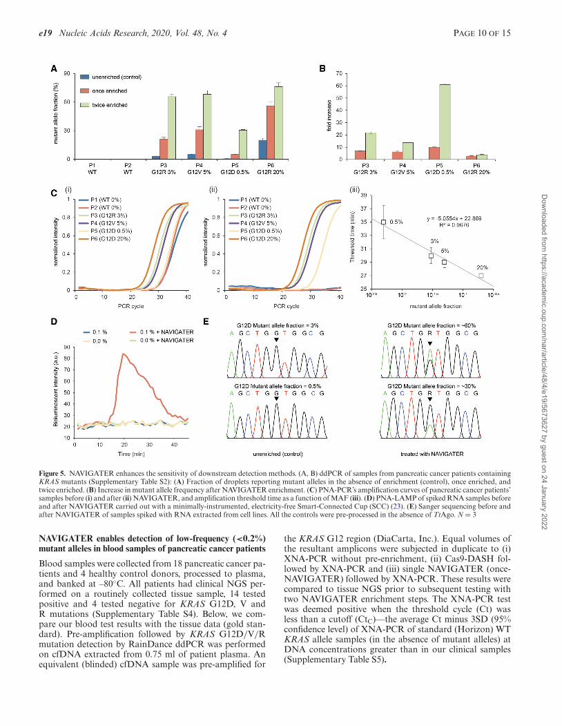
Gel electrophoresis (Supplementary Figure S19). We sub-jected NAVIGATER products to gel eIectrophoresis. In theabsence of enrichment (control), the bands at 80 bp (KRAS)on the electropherogram are dark. After 40 min of TtAgoenrichment, these bands faded, indicating a reduction ofKRAS WT alleles. After 2 h enrichment, all the bands at 80bp, except that of patient P6, have essentially disappeared,suggesting that most WT alleles have been cleaved. Thepresence of an 80 bp band in the P6 lane is attributed tothe relatively high (20%) mutant allele frequency (MAF)that is not susceptible to cleavage. We also PCR amplifiedthe products of a 2-hour NAVIGATER treatment, and sub-jected the amplicons to a second NAVIGATER (2 h). Thecolumns P3, P4 and P6 feature darker bands than P1, P2and P5, indicating the presence of mutant alleles in samplesP3, P4 and P6 (Supplementary Figure S19) and demonstrat-ing that NAVIGATER renders observable otherwise unde-tectable mutant alleles.
Droplet Digital PCR (ddPCR) (Supplementary FigureS20). To quantify our enrichment assay products, we sub-jected them to ddPCR. The detection limit of ddPCR iscontrolled by the number of amplifiable nucleic acids in thesample, which must be a small fraction of the number ofddPCR droplets. The large number of WT alleles in the sam-ple limits the number of pre-ddPCR amplification cyclesthat can be carried out to increase rare alleles’ concentra-tion. Since NAVIGATER drastically reduces the number ofWT alleles in the sample, it enables one to increase the num-ber of pre-amplification cycles, increasing the number ofmutant allele copies, and thus, the ddPCR sensitivity. Whenoperating with a mixture of WT and mutant allele, NAV-IGATER products include residual uncleaved WT (NWT),mutant (NM), and WT-mutant hybrids (NH) alleles. Hybridalleles form during re-hybridization of an ss WT with an ssmutant allele. The MAF is fM = (NM + 1
2 NH)/(NWT + NM+ NH).
We carried out ddPCR of un-enriched (control), once-enriched, and twice-enriched samples (Supplementary Fig-ure S20b), increasing MAF significantly (Figure 5A). Forexample, the MAF increased from 0.5% in the un-enrichedP5 (G12D) sample to ∼30% in the twice-enriched sam-ple. This represents an ∼60-fold increase in the fraction ofdroplets containing the mutant allele (Figure 5B), similarto previously reported methods (10,39,40). The same assayalso enriched G12R, increasing MAF from 3% to ∼66% insample P3 and G12V, increasing MAF from 5% to ∼68%in sample P4 (Figure 5B).
PNA-PCR. PNA-PCR engages a sequence-specific PNAblocker that binds to WT alleles, suppressing WT amplifi-cation and providing a limit of detection of ∼1% MAF (19).To demonstrate NAVIGATER’s utility, we compared theperformance of PNA-PCR when processing pancreatic can-cer patient samples (Supplementary Table S2) before andafter NAVIGATER (Figure 5C). Before enrichment, PNA-PCR real-time amplification curves in the order of appear-ance are P6, P4 and P3, as expected. Samples P1 (MAF
= 0%), P2 (MAF = 0%), and P5 (MAF = 0.5%) nearlyoverlap, consistent with a detection limit of ∼1% (19). En-richment (Figure 5C-ii) significantly increases the thresh-old times of samples P1 and P2 and reveals the presenceof mutant alleles in sample P5. PNA-PCR combined withNAVIGATER provides the linear relationship T1/2 = 22.9– 5 × log(MAF) between threshold time (the time it takesthe amplification curve to reach half its saturation value)and MAF (Figure 5C-iii), allowing one to estimate mutantallele concentration. The data suggests that NAVIGATERcan improve PCR-PNA limit of detection to <0.1%.
PNA-LAMP. Genotyping with PNA blocking oligos canbe combined with the isothermal amplification methodLAMP (20). To demonstrate the feasibility of genotypingat the point of care and in resource-poor settings, we use aminimally-instrumented, electricity-free Smart-ConnectedCup (SCC) (23) with smartphone and bioluminescent dye-based detection to incubate PNA-LAMP and detect re-action products. To demonstrate that we can also detectRNA alleles, we used samples comprised of mixtures ofWT KRAS mRNA and KRAS-G12D mRNA extractedfrom the human cancer cell lines U87-MG and ASPC1. Inthe absence of pre-enrichment, SSC is unable to detect thepresence of 0.1% KRAS-G12D mRNA whereas with pre-enrichment 0.1% KRAS-G12D mRNA is readily detectable(Figure 5D).
Sanger sequencing. In the absence of enrichment, Sangersequencers detect >5% MAF (41). The Sanger sequencerfailed to detect the presence of MAF ∼3% and 0.5% KRAS-G12D mRNA in our un-enriched samples, but readily de-tected these mutant alleles following NAVIGATER enrich-ment (Figure 5E).
Multiplexed enrichments. We carried out triplex NAVI-GATER and compared it with triplex Cas9-based DASH(Cas9-DASH) (10). Our experiment demonstrates thatNAVIGATER can operate as a multiplexed assay, enrich-ing multiple mutant allele fractions in a single sample; it ismore specific than CRISPR/Cas9’s PAM site recognition-based enrichment; and it can be combined with clamped as-say XNA-PCR that is similar to PNA-PCR to significantlyimprove XNA-PCR sensitivity (Supplementary Section 7).NAVIGATER enriches three different KRAS G12 mutantswith a single pair of guides (Figure 5A). When mutationsare proximate such as in the KRAS region, one can enrichfor different mutant allele fractions with a single guide, oth-erwise multiple guides with different sequences in multiplereactions are needed.
NAVIGATER applied directly to cfDNA. TtAgo cleavescfDNA (Horizon) with high efficiency at its optimal op-erating temperature (Supplement Figure 23a) and genomicDNA with reduced efficiency (Supplement Figure 23b). Inappropriately prepared clinical samples, the vast majorityof cfDNA is fragmented with average length of ∼160 bp(42,43), which our assay handles efficiently. Furthermore,our assay also targets RNA that is likely present at muchgreater abundance than DNA fragments. Thus, for enrich-ment of cell-free mutant alleles, pre-amplification is notneeded for the operation of our assay.
Dow
nloaded from https://academ
ic.oup.com/nar/article/48/4/e19/5673627 by guest on 24 January 2022
e19 Nucleic Acids Research, 2020, Vol. 48, No. 4 PAGE 10 OF 15
Figure 5. NAVIGATER enhances the sensitivity of downstream detection methods. (A, B) ddPCR of samples from pancreatic cancer patients containingKRAS mutants (Supplementary Table S2): (A) Fraction of droplets reporting mutant alleles in the absence of enrichment (control), once enriched, andtwice enriched. (B) Increase in mutant allele frequency after NAVIGATER enrichment. (C) PNA-PCR’s amplification curves of pancreatic cancer patients’samples before (i) and after (ii) NAVIGATER, and amplification threshold time as a function of MAF (iii). (D) PNA-LAMP of spiked RNA samples beforeand after NAVIGATER carried out with a minimally-instrumented, electricity-free Smart-Connected Cup (SCC) (23). (E) Sanger sequencing before andafter NAVIGATER of samples spiked with RNA extracted from cell lines. All the controls were pre-processed in the absence of TtAgo. N = 3
NAVIGATER enables detection of low-frequency (<0.2%)mutant alleles in blood samples of pancreatic cancer patients
Blood samples were collected from 18 pancreatic cancer pa-tients and 4 healthy control donors, processed to plasma,and banked at –80◦C. All patients had clinical NGS per-formed on a routinely collected tissue sample, 14 testedpositive and 4 tested negative for KRAS G12D, V andR mutations (Supplementary Table S4). Below, we com-pare our blood test results with the tissue data (gold stan-dard). Pre-amplification followed by KRAS G12D/V/Rmutation detection by RainDance ddPCR was performedon cfDNA extracted from 0.75 ml of patient plasma. Anequivalent (blinded) cfDNA sample was pre-amplified for
the KRAS G12 region (DiaCarta, Inc.). Equal volumes ofthe resultant amplicons were subjected in duplicate to (i)XNA-PCR without pre-enrichment, (ii) Cas9-DASH fol-lowed by XNA-PCR and (iii) single NAVIGATER (once-NAVIGATER) followed by XNA-PCR. These results werecompared to tissue NGS prior to subsequent testing withtwo NAVIGATER enrichment steps. The XNA-PCR testwas deemed positive when the threshold cycle (Ct) wasless than a cutoff (CtC)––the average Ct minus 3SD (95%confidence level) of XNA-PCR of standard (Horizon) WTKRAS allele samples (in the absence of mutant alleles) atDNA concentrations greater than in our clinical samples(Supplementary Table S5).
Dow
nloaded from https://academ
ic.oup.com/nar/article/48/4/e19/5673627 by guest on 24 January 2022
PAGE 11 OF 15 Nucleic Acids Research, 2020, Vol. 48, No. 4 e19
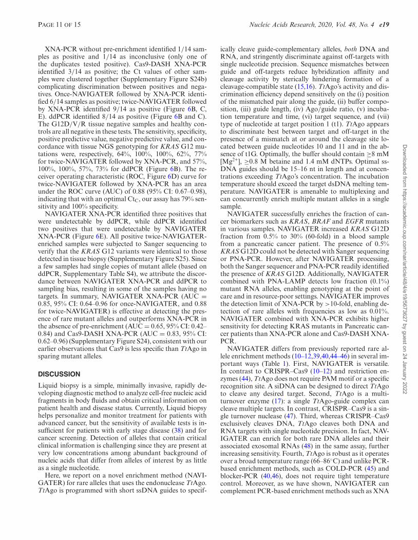
XNA-PCR without pre-enrichment identified 1/14 sam-ples as positive and 1/14 as inconclusive (only one ofthe duplicates tested positive). Cas9-DASH XNA-PCRidentified 3/14 as positive; the Ct values of other sam-ples were clustered together (Supplementary Figure S24b)complicating discrimination between positives and nega-tives. Once-NAVIGATER followed by XNA-PCR identi-fied 6/14 samples as positive; twice-NAVIGATER followedby XNA-PCR identified 9/14 as positive (Figure 6B, C,E). ddPCR identified 8/14 as positive (Figure 6B and C).The G12D/V/R tissue negative samples and healthy con-trols are all negative in these tests. The sensitivity, specificity,positive predictive value, negative predictive value, and con-cordance with tissue NGS genotyping for KRAS G12 mu-tations were, respectively, 64%, 100%, 100%, 62%, 77%for twice-NAVIGATER followed by XNA-PCR, and 57%,100%, 100%, 57%, 73% for ddPCR (Figure 6B). The re-ceiver operating characteristic (ROC, Figure 6D) curve fortwice-NAVIGATER followed by XNA-PCR has an areaunder the ROC curve (AUC) of 0.88 (95% CI: 0.67–0.98),indicating that with an optimal CtC, our assay has 79% sen-sitivity and 100% specificity.
NAVIGATER XNA-PCR identified three positives thatwere undetectable by ddPCR, while ddPCR identifiedtwo positives that were undetectable by NAVIGATERXNA-PCR (Figure 6E). All positive twice-NAVIGATER-enriched samples were subjected to Sanger sequencing toverify that the KRAS G12 variants were identical to thosedetected in tissue biopsy (Supplementary Figure S25). Sincea few samples had single copies of mutant allele (based onddPCR, Supplementary Table S4), we attribute the discor-dance between NAVIGATER XNA-PCR and ddPCR tosampling bias, resulting in some of the samples having notargets. In summary, NAVIGATER XNA-PCR (AUC =0.85, 95% CI: 0.64–0.96 for once-NAVIGATER, and 0.88for twice-NAVIGATER) is effective at detecting the pres-ence of rare mutant alleles and outperforms XNA-PCR inthe absence of pre-enrichment (AUC = 0.65, 95% CI: 0.42–0.84) and Cas9-DASH XNA-PCR (AUC = 0.83, 95% CI:0.62–0.96) (Supplementary Figure S24), consistent with ourearlier observations that Cas9 is less specific than TtAgo insparing mutant alleles.
DISCUSSION
Liquid biopsy is a simple, minimally invasive, rapidly de-veloping diagnostic method to analyze cell-free nucleic acidfragments in body fluids and obtain critical information onpatient health and disease status. Currently, Liquid biopsyhelps personalize and monitor treatment for patients withadvanced cancer, but the sensitivity of available tests is in-sufficient for patients with early stage disease (38) and forcancer screening. Detection of alleles that contain criticalclinical information is challenging since they are present atvery low concentrations among abundant background ofnucleic acids that differ from alleles of interest by as littleas a single nucleotide.
Here, we report on a novel enrichment method (NAVI-GATER) for rare alleles that uses the endonuclease TtAgo.TtAgo is programmed with short ssDNA guides to specif-
ically cleave guide-complementary alleles, both DNA andRNA, and stringently discriminate against off-targets withsingle nucleotide precision. Sequence mismatches betweenguide and off-targets reduce hybridization affinity andcleavage activity by sterically hindering formation of acleavage-compatible state (15,16). TtAgo’s activity and dis-crimination efficiency depend sensitively on the (i) positionof the mismatched pair along the guide, (ii) buffer compo-sition, (iii) guide length, (iv) Ago/guide ratio, (v) incuba-tion temperature and time, (vi) target sequence, and (vii)type of nucleotide at target position 1 (t1). TtAgo appearsto discriminate best between target and off-target in thepresence of a mismatch at or around the cleavage site lo-cated between guide nucleotides 10 and 11 and in the ab-sence of t1G. Optimally, the buffer should contain ≥8 mM[Mg2+], ≥0.8 M betaine and 1.4 mM dNTPs. Optimal ss-DNA guides should be 15–16 nt in length and at concen-trations exceeding TtAgo’s concentration. The incubationtemperature should exceed the target dsDNA melting tem-perature. NAVIGATER is amenable to multiplexing andcan concurrently enrich multiple mutant alleles in a singlesample.
NAVIGATER successfully enriches the fraction of can-cer biomarkers such as KRAS, BRAF and EGFR mutantsin various samples. NAVIGATER increased KRAS G12Dfraction from 0.5% to 30% (60-fold) in a blood samplefrom a pancreatic cancer patient. The presence of 0.5%KRAS G12D could not be detected with Sanger sequencingor PNA-PCR. However, after NAVIGATER processing,both the Sanger sequencer and PNA-PCR readily identifiedthe presence of KRAS G12D. Additionally, NAVIGATERcombined with PNA-LAMP detects low fraction (0.1%)mutant RNA alleles, enabling genotyping at the point ofcare and in resource-poor settings. NAVIGATER improvesthe detection limit of XNA-PCR by >10-fold, enabling de-tection of rare alleles with frequencies as low as 0.01%.NAVIGATER combined with XNA-PCR exhibits highersensitivity for detecting KRAS mutants in Pancreatic can-cer patients than XNA-PCR alone and Cas9-DASH XNA-PCR.
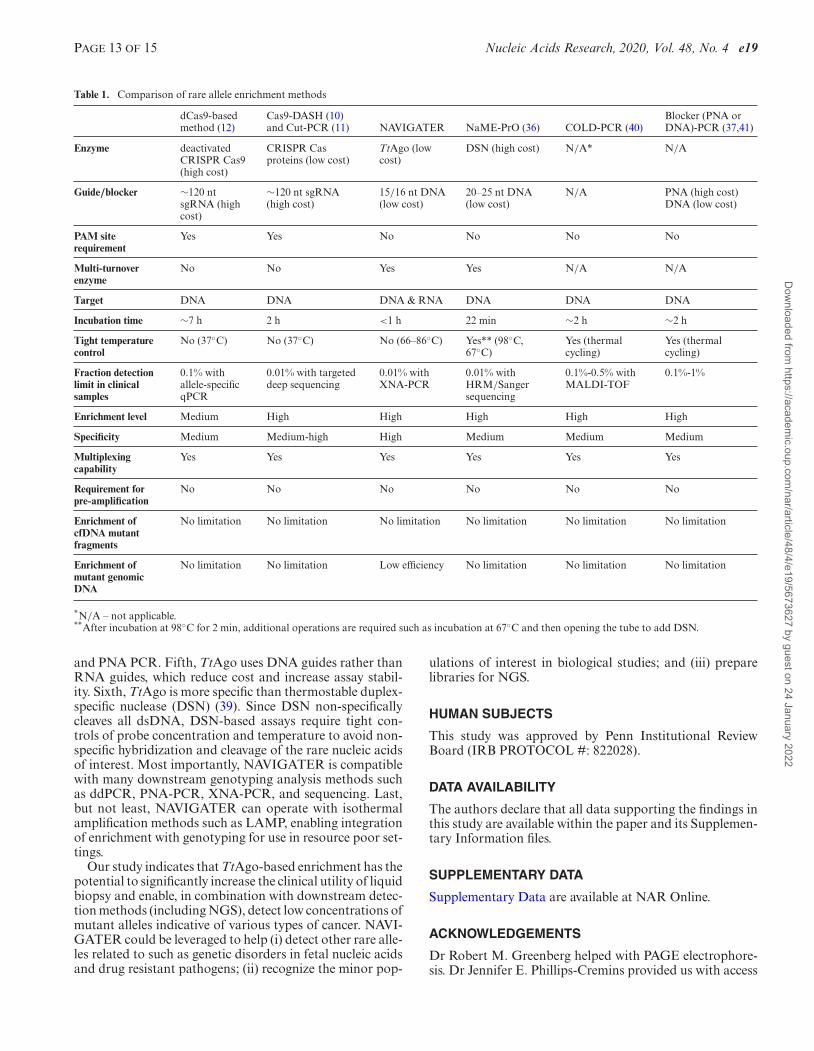
NAVIGATER differs from previously reported rare al-lele enrichment methods (10–12,39,40,44–46) in several im-portant ways (Table 1). First, NAVIGATER is versatile.In contrast to CRISPR–Cas9 (10–12) and restriction en-zymes (44), TtAgo does not require PAM motif or a specificrecognition site. A siDNA can be designed to direct TtAgoto cleave any desired target. Second, TtAgo is a multi-turnover enzyme (17): a single TtAgo-guide complex cancleave multiple targets. In contrast, CRISPR–Cas9 is a sin-gle turnover nuclease (47). Third, whereas CRISPR–Cas9exclusively cleaves DNA, TtAgo cleaves both DNA andRNA targets with single nucleotide precision. In fact, NAV-IGATER can enrich for both rare DNA alleles and theirassociated exosomal RNAs (48) in the same assay, furtherincreasing sensitivity. Fourth, TtAgo is robust as it operatesover a broad temperature range (66–86◦C) and unlike PCR-based enrichment methods, such as COLD-PCR (45) andblocker-PCR (40,46), does not require tight temperaturecontrol. Moreover, as we have shown, NAVIGATER cancomplement PCR-based enrichment methods such as XNA
Dow
nloaded from https://academ
ic.oup.com/nar/article/48/4/e19/5673627 by guest on 24 January 2022
e19 Nucleic Acids Research, 2020, Vol. 48, No. 4 PAGE 12 OF 15
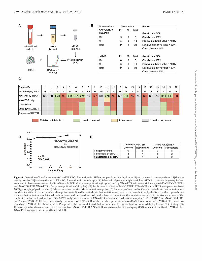
Figure 6. Detection of low-frequency (<0.2%) KRAS G12 mutations in cfDNA samples from healthy donors [4] and pancreatic cancer patients [18] that aretesting positive [14] and negative [4] to KRAS G12 mutations in tissue biopsy. (A) Schematic of patient sample workflow. cfDNA corresponding to equivalentvolumes of plasma were assayed by RainDance ddPCR after pre-amplification (9 cycles) and by XNA-PCR without enrichment, cas9-DASH XNA-PCR,and NAVIGATER XNA-PCR after pre-amplification (35 cycles). (B) Performance of twice-NAVIGATER XNA-PCR and ddPCR compared to tissueNGS genotyping (‘gold standard’). M+ = mutation positive. M– = mutation negative. (C) Summary of test results. Gray boxes indicate that mutation wasnot detected either in tissue or in blood (negative control); red boxes indicate that mutation was detected in tissue but not by the listed method; green boxesindicate that mutation was detected both in tissue and the listed method; and yellow boxes indicate that mutation was detected in tissue and one of theduplicate test by the listed method. ‘XNA-PCR only’ are the results of XNA-PCR of non-enriched patient samples, ‘cas9-DASH’, ‘once-NAVIGATER’,and ‘twice-NAVIGATER’ are, respectively, the results of XNA-PCR of the enriched products of cas9-DASH, one round of NAVIGATER, and tworounds of NAVIGATER. N = negative. P = positive. ND = not detected. NA = not available because healthy donors didn’t get tissue NGS testing. (D)Receiver operator characteristic (ROC) curve of twice-NAVIGATER XNA-PCR versus tissue NGS genotyping. (E) Summary of results of NAVIGATERXNA-PCR compared with RainDance ddPCR.
Dow
nloaded from https://academ
ic.oup.com/nar/article/48/4/e19/5673627 by guest on 24 January 2022
PAGE 13 OF 15 Nucleic Acids Research, 2020, Vol. 48, No. 4 e19
Table 1. Comparison of rare allele enrichment methods
dCas9-basedmethod (12)
Cas9-DASH (10)and Cut-PCR (11) NAVIGATER NaME-PrO (36) COLD-PCR (40)
Blocker (PNA orDNA)-PCR (37,41)
Enzyme deactivatedCRISPR Cas9(high cost)
CRISPR Casproteins (low cost)
TtAgo (lowcost)
DSN (high cost) N/A* N/A
Guide/blocker ∼120 ntsgRNA (highcost)
∼120 nt sgRNA(high cost)
15/16 nt DNA(low cost)
20–25 nt DNA(low cost)
N/A PNA (high cost)DNA (low cost)
PAM siterequirement
Yes Yes No No No No
Multi-turnoverenzyme
No No Yes Yes N/A N/A
Target DNA DNA DNA & RNA DNA DNA DNA
Incubation time ∼7 h 2 h <1 h 22 min ∼2 h ∼2 h
Tight temperaturecontrol
No (37◦C) No (37◦C) No (66–86◦C) Yes** (98◦C,67◦C)
Yes (thermalcycling)
Yes (thermalcycling)
Fraction detectionlimit in clinicalsamples
0.1% withallele-specificqPCR
0.01% with targeteddeep sequencing
0.01% withXNA-PCR
0.01% withHRM/Sangersequencing
0.1%-0.5% withMALDI-TOF
0.1%-1%
Enrichment level Medium High High High High High
Specificity Medium Medium-high High Medium Medium Medium
Multiplexingcapability
Yes Yes Yes Yes Yes Yes
Requirement forpre-amplification
No No No No No No
Enrichment ofcfDNA mutantfragments
No limitation No limitation No limitation No limitation No limitation No limitation
Enrichment ofmutant genomicDNA
No limitation No limitation Low efficiency No limitation No limitation No limitation
*N/A – not applicable.**After incubation at 98◦C for 2 min, additional operations are required such as incubation at 67◦C and then opening the tube to add DSN.
and PNA PCR. Fifth, TtAgo uses DNA guides rather thanRNA guides, which reduce cost and increase assay stabil-ity. Sixth, TtAgo is more specific than thermostable duplex-specific nuclease (DSN) (39). Since DSN non-specificallycleaves all dsDNA, DSN-based assays require tight con-trols of probe concentration and temperature to avoid non-specific hybridization and cleavage of the rare nucleic acidsof interest. Most importantly, NAVIGATER is compatiblewith many downstream genotyping analysis methods suchas ddPCR, PNA-PCR, XNA-PCR, and sequencing. Last,but not least, NAVIGATER can operate with isothermalamplification methods such as LAMP, enabling integrationof enrichment with genotyping for use in resource poor set-tings.
Our study indicates that TtAgo-based enrichment has thepotential to significantly increase the clinical utility of liquidbiopsy and enable, in combination with downstream detec-tion methods (including NGS), detect low concentrations ofmutant alleles indicative of various types of cancer. NAVI-GATER could be leveraged to help (i) detect other rare alle-les related to such as genetic disorders in fetal nucleic acidsand drug resistant pathogens; (ii) recognize the minor pop-
ulations of interest in biological studies; and (iii) preparelibraries for NGS.
HUMAN SUBJECTS
This study was approved by Penn Institutional ReviewBoard (IRB PROTOCOL #: 822028).
DATA AVAILABILITY
The authors declare that all data supporting the findings inthis study are available within the paper and its Supplemen-tary Information files.
SUPPLEMENTARY DATA
Supplementary Data are available at NAR Online.
ACKNOWLEDGEMENTS
Dr Robert M. Greenberg helped with PAGE electrophore-sis. Dr Jennifer E. Phillips-Cremins provided us with access
Dow
nloaded from https://academ
ic.oup.com/nar/article/48/4/e19/5673627 by guest on 24 January 2022
e19 Nucleic Acids Research, 2020, Vol. 48, No. 4 PAGE 14 OF 15
to gel imager. Dr Changchun Liu provided helpful com-ments early in this project. Stephanie Yee and Taylor Blackassisted with ddPCR. Yaguang Fan assisted with statisticalanalysis.Author contributions: J.S. and H.H.B. conceived the projectand designed the experiments. J.W.H. expressed and pu-rified TtAgo protein. J.S., J.W.H., J.P. and L.T.A. carriedout the experiments. M.G.M. and J.C. assisted, respectively,with PNA-PCR and XNA-PCR. N.B. and J.E.T. extractedcfDNA from patients’ blood and quantified KRAS G12mutations with ddPCR. J.E.T. extracted RNA from celllines. N.B. and J.E.T. assisted with ddPCR experiments.M.S. and J.M. cultured cell lines. E.C. supervised N.B.,J.E.T., M.S. and J.M. and advised on patient samples andddPCR experiments. J.S., J.W.H., M.G.M., J.v.d.O. andH.H.B. analyzed the data and wrote the manuscript. All au-thors read and commented on the manuscript.
FUNDING
National Institutes of Health (NIH) [NCI 1R21CA227056-01, FIC 1R21TW011496-01A1, FIC 1K01TW011190-01A1 to the University of Pennsylvania]; Netherlands Or-ganization of Scientific Research [NWO-ECHO 711013002,NWO-TOP 714015001 to J.v.d.O.]. Funding for open accesscharge: NIH [NCI 1R21CA227056-01].Conflict of interest statement. University of Pennsylvaniaand Wageningen University have applied for a patent onNAVIGATER with J.S., J.W.H., M.G.M., J.v.d.O. andH.H.B. listed as co-inventors.
REFERENCES1. Barrangou,R. and Doudna,J.A. (2016) Applications of CRISPR
technologies in research and beyond. Nat. Biotechnol., 34, 933–941.2. Komor,A.C., Badran,A.H. and Liu,D.R. (2017) CRISPR-based
technologies for the manipulation of eukaryotic genomes. Cell, 168,20–36.
3. Wu,W.Y., Lebbink,J.H.G., Kanaar,R., Geijsen,N. and van der Oost,J.(2018) Genome editing by natural and engineeredCRISPR-associated nucleases. Nat. Chem. Biol., 14, 642–651.
4. Gootenberg,J.S., Abudayyeh,O.O., Lee,J.W., Essletzbichler,P.,Dy,A.J., Joung,J., Verdine,V., Donghia,N., Daringer,N.M.,Freije,C.A. et al. (2017) Nucleic acid detection withCRISPR–Cas13a/C2c2. Science, 356, 438–442.
5. Chen,J.S., Ma,E.B., Harrington,L.B., Da Costa,M., Tian,X.R.,Palefsky,J.M. and Doudna,J.A. (2018) CRISPR–Cas12a targetbinding unleashes indiscriminate single-stranded DNase activity.Science, 360, 436–439.
6. Harrington,L.B., Burstein,D., Chen,J.S., Paez-Espino,D., Ma,E.,Witte,I.P., Cofsky,J.C., Kyrpides,N.C., Banfield,J.F. and Doudna,J.A.(2018) Programmed DNA destruction by miniature CRISPR–Cas14enzymes. Science, 362, 839–842.
7. East-Seletsky,A., O’Connell,M.R., Knight,S.C., Burstein,D.,Cate,J.H., Tjian,R. and Doudna,J.A. (2016) Two distinct RNaseactivities of CRISPR-C2c2 enable guide-RNA processing and RNAdetection. Nature, 538, 270–273.
8. Gootenberg,J.S., Abudayyeh,O.O., Kellner,M.J., Joung,J., Collins,J.J.and Zhang,F. (2018) Multiplexed and portable nucleic acid detectionplatform with Cas13, Cas12a, and Csm6. Science, 360, 439–444.
9. Hajian,R., Balderston,S., Tran,T., deBoer,T., Etienne,J., Sandhu,M.,Wauford,N.A., Chung,J.-Y., Nokes,J., Athaiya,M. et al. (2019)Detection of unamplified target genes via CRISPR–Cas9 immobilizedon a graphene field-effect transistor. Nat. Biomed. Eng., 3, 427–437.
10. Gu,W., Crawford,E.D., O’Donovan,B.D., Wilson,M.R., Chow,E.D.,Retallack,H. and DeRisi,J.L. (2016) Depletion of AbundantSequences by Hybridization (DASH): using Cas9 to remove
unwanted high-abundance species in sequencing libraries andmolecular counting applications. Genome Biol., 17, 1–13.
11. Lee,S.H., Yu,J., Hwang,G.H., Kim,S., Kim,H.S., Ye,S., Kim,K.,Park,J., Park,D.Y., Cho,Y.K. et al. (2017) CUT-PCR:CRISPR-mediated, ultrasensitive detection of target DNA usingPCR. Oncogene, 36, 6823–6829.
12. Aalipour,A., Dudley,J.C., Park,S.M., Murty,S., Chabon,J.J.,Boyle,E.A., Diehn,M. and Gambhir,S.S. (2018) Deactivated CRISPRassociated protein 9 for minor-allele enrichment in cell-free DNA.Clin. Chem., 64, 307–316.
13. Hegge,J.W., Swarts,D.C. and van der Oost,J. (2018) ProkaryoticArgonaute proteins: novel genome-editing tools? Nat. Rev.Microbiol., 16, 5–11.
14. Swarts,D.C., Makarova,K., Wang,Y.L., Nakanishi,K., Ketting,R.F.,Koonin,E.V., Patel,D.J. and van der Oost,J. (2014) The evolutionaryjourney of Argonaute proteins. Nat. Struct. Mol. Biol, 21, 743–753.
15. Wang,Y.L., Juranek,S., Li,H.T., Sheng,G., Tuschl,T. and Patel,D.J.(2008) Structure of an argonaute silencing complex with aseed-containing guide DNA and target RNA duplex. Nature, 456,921–926.
16. Wang,Y.L., Juranek,S., Li,H.T., Sheng,G., Wardle,G.S., Tuschl,T. andPatel,D.J. (2009) Nucleation, propagation and cleavage of targetRNAs in Ago silencing complexes. Nature, 461, 754–761.
17. Swarts,D.C., Jore,M.M., Westra,E.R., Zhu,Y.F., Janssen,J.H.,Snijders,A.P., Wang,Y.L., Patel,D.J., Berenguer,J., Brouns,S.J.J. et al.(2014) DNA-guided DNA interference by a prokaryotic Argonaute.Nature, 507, 258–261.
18. Taly,V., Pekin,D., Benhaim,L., Kotsopoulos,S.K., Le Corre,D.,Li,X.Y., Atochin,I., Link,D.R., Griffiths,A.D., Pallier,K. et al. (2013)Multiplex picodroplet digital PCR to detect KRAS mutations incirculating DNA from the plasma of colorectal cancer patients. Clin.Chem., 59, 1722–1731.
19. Choi,J.J., Cho,M., Oh,M., Kim,H., Kil,M.S. and Park,H. (2010)PNA-mediated real-time PCR clamping for detection of EGFRmutations. B. Korean. Chem. Soc., 31, 3525–3529.
20. Tatsumi,K., Mitani,Y., Watanabe,J., Takakura,H., Hoshit,K.,Kawai,Y., Kikuchi,T., Kogo,Y., Oguchi-Katayama,A., Tomaru,Y.et al. (2008) Rapid screening assay for KRAS mutations by themodified smart amplification process. J. Mol. Diagn., 10, 520–526.
21. Tropea,J.E., Cherry,S. and Waugh,D.S. (2009) In: Doyle,SA (ed).High Throughput Protein Expression and Purification: Methods andProtocols. Humana Press, Totowa, pp. 297–307.
22. Potapov,V. and Ong,J.L. (2017) Examining sources of error in PCRby single-molecule sequencing. PLoS One, 12, e0169774.
23. Song,J.Z., Pandian,V., Mauk,M.G., Bau,H.H., Cherry,S., Tisi,L.C.and Liu,C.C. (2018) Smartphone-based mobile detection platform formolecular diagnostics and spatiotemporal disease mapping. Anal.Chem., 90, 4823–4831.
24. Song,J.Z., Liu,C.C., Bais,S., Mauk,M.G., Bau,H.H. andGreenberg,R.M. (2015) Molecular detection of schistosomeinfections with a disposable microfluidic cassette. PloS Neglect. Trop.D., 9, e0004318.
25. Song,J.Z., Mauk,M.G., Hackett,B.A., Cherry,S., Bau,H.H. andLiu,C.C. (2016) Instrument-free point-of-care molecular detection ofzika virus. Anal. Chem., 88, 7289–7294.
26. Kaya,E., Doxzen,K.W., Knoll,K.R., Wilson,R.C., Strutt,S.C.,Kranzusch,P.J. and Doudna,J.A. (2016) A bacterial Argonaute withnoncanonical guide RNA specificity. Proc. Natl. Acad. Sci. U.S.A.,113, 4057–4062.
27. Doxzen,K.W. and Doudna,J.A. (2017) DNA recognition by anRNA-guided bacterial Argonaute. PLoS One, 12, e0177097.
28. Sheng,G., Gogakos,T., Wang,J.Y., Zhao,H.T., Serganov,A.,Juranek,S., Tuschl,T., Patel,D.J. and Wang,Y.L. (2017)Structure/cleavage-based insights into helical perturbations at bulgesites within T. thermophilus Argonaute silencing complexes. NucleicAcids Res., 45, 9149–9163.
29. Sheng,G., Zhao,H.T., Wang,J.Y., Rao,Y., Tian,W.W., Swarts,D.C.,van der Oost,J., Patel,D.J. and Wang,Y.L. (2014) Structure-basedcleavage mechanism of Thermus thermophilus Argonaute DNAguide strand-mediated DNA target cleavage. Proc. Natl. Acad. Sci.U.S.A., 111, 652–657.
30. Swarts,D.C., Szczepaniak,M., Sheng,G., Chandradoss,S.D.,Zhu,Y.F., Timmers,E.M., Zhang,Y., Zhao,H.T., Lou,J.Z., Wang,Y.L.
Dow
nloaded from https://academ
ic.oup.com/nar/article/48/4/e19/5673627 by guest on 24 January 2022
PAGE 15 OF 15 Nucleic Acids Research, 2020, Vol. 48, No. 4 e19
et al. (2017) Autonomous generation and loading of DNA guides bybacterial Argonaute. Mol. Cell, 65, 985–998.
31. Phelps,C., Lee,W., Jose,D., von Hippel,P.H. and Marcus,A.H. (2013)Single-molecule FRET and linear dichroism studies of DNAbreathing and helicase binding at replication fork junctions. Proc.Natl. Acad. Sci. U.S.A., 110, 17320–17325.
32. Rees,W.A., Yager,T.D., Korte,J. and Vonhippel,P.H. (1993) Betainecan eliminate the base pair composition dependence of DNA melting.Biochemistry-US, 32, 137–144.
33. Kuzmenko,A., Yudin,D., Ryazansky,S., Kulbachinskiy,A. andAravin,A.A. (2019) Programmable DNA cleavage by Ago nucleasesfrom mesophilic bacteria Clostridium butyricum and Limnothrixrosea. Nucleic Acids Res., 47, 5822–5836.
34. Zhang,Y., Ge,X., Yang,F., Zhang,L., Zheng,J., Tan,X., Jin,Z.B., Qu,J.and Gu,F. (2014) Comparison of non-canonical PAMs forCRISPR/Cas9-mediated DNA cleavage in human cells. Sci. Rep., 4,5405.
35. Yamano,T., Zetsche,B., Ishitani,R., Zhang,F., Nishimasu,H. andNureki,O. (2017) Structural basis for the canonical and non-canonicalPAM recognition by CRISPR-Cpf1. Mol. Cell, 67, 633–645.
36. Bettegowda,C., Sausen,M., Leary,R.J., Kinde,I., Wang,Y.X.,Agrawal,N., Bartlett,B.R., Wang,H., Luber,B., Alani,R.M. et al.(2014) Detection of circulating tumor DNA in early- and late-stagehuman malignancies. Sci. Transl. Med., 6, 224ra224.
37. Oxnard,G.R., Paweletz,C.P., Kuang,Y.A., Mach,S.L., O’Connell,A.,Messineo,M.M., Luke,J.J., Butaney,M., Kirschmeier,P.,Jackman,D.M. et al. (2014) Noninvasive detection of response andresistance in EGFR-mutant lung cancer using quantitativenext-generation genotyping of cell-free plasma DNA. Clin. CancerRes., 20, 1698–1705.
38. Aggarwal,C., Thompson,J.C., Black,T.A., Katz,S.I., Fan,R., Yee,S.S.,Chien,A.L., Evans,T.L., Bauml,J.M., Alley,E.W. et al. (2018) Clinicalimplications of plasma-based genotyping with the delivery ofpersonalized therapy in metastatic non-small cell lung cancer. JAMAOncol., 5, 173–180.
39. Song,C., Liu,Y.B., Fontana,R., Makrigiorgos,A., Mamon,H.,Kulke,M.H. and Makrigiorgos,G.M. (2016) Elimination of unalteredDNA in mixed clinical samples via nuclease-assisted minor-alleleenrichment. Nucleic Acids Res., 44, e146.
40. Wu,L.R., Chen,S.X., Wu,Y., Patel,A.A. and Zhang,D.Y. (2017)Multiplexed enrichment of rare DNA variants via sequence-selectiveand temperature-robust amplification. Nat. Biomed. Eng., 1, 714–723.
41. Tsiatis,A.C., Norris-Kirby,A., Rich,R.G., Hafez,M.J., Gocke,C.D.,Eshleman,J.R. and Murphy,K.M. (2010) Comparison of sangersequencing, pyrosequencing, and melting curve analysis for thedetection of KRAS mutations diagnostic and clinical implications. J.Mol. Diagn., 12, 425–432.
42. Underhill,H.R., Kitzman,J.O., Hellwig,S., Welker,N.C., Daza,R.,Baker,D.N., Gligorich,K.M., Rostomily,R.C., Bronner,M.P. andShendure,J. (2016) Fragment length of circulating tumor DNA. PLoSGenet., 12, e1006162.
43. Sato,A., Nakashima,C., Abe,T., Kato,J., Hirai,M., Nakamura,T.,Komiya,K., Kimura,S., Sueoka,E. and Sueoka-Aragane,N. (2018)Investigation of appropriate pre-analytical procedure for circulatingfree DNA from liquid biopsy. Oncotarget, 9, 31904–31914.
44. Bielas,J.H. and Loeb,L.A. (2005) Quantification of random genomicmutations. Nat. Methods, 2, 285–290.
45. Li,J., Wang,L., Mamon,H., Kulke,M.H., Berbeco,R. andMakrigiorgos,G.M. (2008) Replacing PCR with COLD-PCRenriches variant DNA sequences and redefines the sensitivity ofgenetic testing. Nat. Med., 14, 579–584.
46. Kim,H.S., Sung,J.S., Yang,S.J., Kwon,N.J., Jin,L., Kim,S.T.,Park,K.H., Shin,S.W., Kim,H.K., Kang,J.H. et al. (2013) Predictiveefficacy of low burden EGFR mutation detected by next-generationsequencing on response to EGFR tyrosine kinase inhibitors innon-small-cell lung carcinoma. PLoS One, 8, e81975.
47. Sternberg,S.H., Redding,S., Jinek,M., Greene,E.C. and Doudna,J.A.(2014) DNA interrogation by the CRISPR RNA-guidedendonuclease Cas9. Nature, 507, 62–67.
48. Krug,A.K., Enderle,D., Karlovich,C., Priewasser,T., Bentink,S.,Spiel,A., Brinkmann,K., Emenegger,J., Grimm,D.G.,Castellanos-Rizaldos,E. et al. (2018) Improved EGFR mutationdetection using combined exosomal RNA and circulating tumorDNA in NSCLC patient plasma. Ann. Oncol., 29, 700–706.
49. Tuma,R.S., Beaudet,M.P., Jin,X.K., Jones,L.J., Cheung,C.Y., Yue,S.and Singer,V.L. (1999) Characterization of SYBR gold nucleic acidgel stain: A dye optimized for use with 300-nm ultraviolettransilluminators. Anal. Biochem., 268, 278–288.
50. Lilley,D.M., Bhattacharyya,A. and McAteer,S. (1992) Gelelectrophoresis and the structure of RNA molecules. Biotechnol.Genet. Eng. Rev., 10, 379–401.
Dow
nloaded from https://academ
ic.oup.com/nar/article/48/4/e19/5673627 by guest on 24 January 2022
Related Documents