SUMMARY Surveys of fields, parks, and forests in the Golestan province of northeast Iran resulted in the record of four- teen rust-induced diseases on weeping willow, black poplar, blackberry, rose, estragon, garlic, wild oat, barley, ryegrass, wheat, peach, plum, alfalfa, and broad bean. Causal agents were identified as Melampsora coleospori- oides, M. allii-populina, Phragmidium bulbosum, Ph. vio- laceum, Ph. rosae-pimpinellifoliae, Ph. tuberculatum, Puc- cinia absinthii, P. allii, P. coronata f.sp. avenae, P. coronata f.sp. hordei, P. graminis f.sp. avenae, P. hordei, P. persistens subsp. triticina, Tranzschelia discolor, Uromyces striatus, and U. viciae-fabae. Weeping willow rust and barley crown rust are reported for the first time from Iran and Asia, respectively. Key words: Rusts, Melampsora, Phragmidium, Puccinia, Tranzschelia, Uromyces. INTRODUCTION With more than 7000 species, rust fungi (Basidiomy- cota, Uredinales) are the largest group of obligate plant pathogens known to date (Aime, 2006). Among them, there are disease agents that severely affect field crops, vegetables, ornamentals, fruit, and forest trees (Agrios, 1997). Wheat rusts, for example, have influenced the course of early civilization by destroying a major source of food (Wiese, 1987) and are still most destructive pathogens in all provinces of Iran where, in 1993, have caused more than 1 million tons yield loss (Okhovat, 1999). About 300 rust species, mostly belonging to the gen- era Melampsora, Phragmidium, Puccinia, Tranzschelia, and Uromyces, have been reported from Iran (Ershad, 1995). The most significant studies on rusts conducted in this country include those on Melampsora on shade Corresponding author: M. Sadravi Fax: +98.171.4420438 E-mail: [email protected] and forest trees (Petrak and Esfandiari, 1941; Viennot- Bourgin, 1958), Phragmidium on Rosa and Rubus (Pe- trak, 1956), Puccinia on cereal crops (Ershad, 1995; Ab- basi and Hejaroude, 2004a, 2004b), Tranzschelia on Prunus (Abbasi and Ershad, 1995; Sadravi, 2001), and Uromyces on Medicago and Vicia (Ershad, 1995; Abbasi and Pooralibaba, 2002). The Iranian province of Golestan (southeastern shore of the Caspian sea) has a temperate humid cli- mate, that favours the growth of a rich and diversified natural flora and timber trees, and the extensive cultiva- tion of field, fruit and horticultural crops. Although rusts are widely represented in this area, no specific surveys have been carried out in the past decades. This prompted the senior author to undertake an investigation to this effect, the results of which are reported in the present paper. MATERIALS AND METHODS From 2003 to 2005, fields, orchards, forests and parks of the Golestan province were surveyed and plants infect- ed with rust fungi were sampled. Spores from sori were mounted in polyvinyl alcohol-lactic acid-glycerol (Koske and Tessier, 1983) and Melzer’s reagent and observed with a bright-field microscope. For scanning electron mi- croscopic examination, urediniospores were fixed in 3% glutaraldhyde in 0.02 M phosphate buffer pH 6, dehy- drated in graded ethanol, critical point dried, mounted on aluminium stubs and coated with gold (Blaszkowski, 1997). Subepidermal telia were studied by preparing freehand sections of infected leaf tissues that were stained as described by Shipton and Brown (1962). RESULTS AND DISCUSSION Fourteen rust diseases caused by sixteen rust fungi were identified in the surveyed area. Weeping willow rust. In late spring, leaves of weep- ing willow (Salix babylonica L.) were covered with yel- low spots and yellowish-orange powdery pustules on Journal of Plant Pathology (2007), 89 (2), 191-202 Edizioni ETS Pisa, 2007 191 FOURTEEN RUSTS FROM NORTHEAST IRAN M. Sadravi 1 , Y. Ono 2 , M. Pei 3 and K. Rahnama 1 1 Department of Plant Protection, Faculty of Agricultural Sciences, Gorgan University of Agricultural Sciences and Natural Resources, Gorgan, Iran 2 Faculty of Education, Ibaraki, Mito, Ibaraki 310-8512, Japan 3 Rothamsted Research, Harpenden, Hertfordshire, AL5 2JQ,UK

Welcome message from author
This document is posted to help you gain knowledge. Please leave a comment to let me know what you think about it! Share it to your friends and learn new things together.
Transcript
SUMMARY
Surveys of fields, parks, and forests in the Golestanprovince of northeast Iran resulted in the record of four-teen rust-induced diseases on weeping willow, blackpoplar, blackberry, rose, estragon, garlic, wild oat, barley,ryegrass, wheat, peach, plum, alfalfa, and broad bean.Causal agents were identified as Melampsora coleospori-oides, M. allii-populina, Phragmidium bulbosum, Ph. vio-laceum, Ph. rosae-pimpinellifoliae, Ph. tuberculatum, Puc-cinia absinthii, P. allii, P. coronata f.sp. avenae, P. coronataf.sp. hordei, P. graminis f.sp. avenae, P. hordei, P. persistenssubsp. triticina, Tranzschelia discolor, Uromyces striatus,and U. viciae-fabae. Weeping willow rust and barleycrown rust are reported for the first time from Iran andAsia, respectively.
Key words: Rusts, Melampsora, Phragmidium, Puccinia,Tranzschelia, Uromyces.
INTRODUCTION
With more than 7000 species, rust fungi (Basidiomy-cota, Uredinales) are the largest group of obligate plantpathogens known to date (Aime, 2006). Among them,there are disease agents that severely affect field crops,vegetables, ornamentals, fruit, and forest trees (Agrios,1997). Wheat rusts, for example, have influenced thecourse of early civilization by destroying a major sourceof food (Wiese, 1987) and are still most destructivepathogens in all provinces of Iran where, in 1993, havecaused more than 1 million tons yield loss (Okhovat,1999).
About 300 rust species, mostly belonging to the gen-era Melampsora, Phragmidium, Puccinia, Tranzschelia,and Uromyces, have been reported from Iran (Ershad,1995). The most significant studies on rusts conductedin this country include those on Melampsora on shade
Corresponding author: M. SadraviFax: +98.171.4420438E-mail: [email protected]
and forest trees (Petrak and Esfandiari, 1941; Viennot-Bourgin, 1958), Phragmidium on Rosa and Rubus (Pe-trak, 1956), Puccinia on cereal crops (Ershad, 1995; Ab-basi and Hejaroude, 2004a, 2004b), Tranzschelia onPrunus (Abbasi and Ershad, 1995; Sadravi, 2001), andUromyces on Medicago and Vicia (Ershad, 1995; Abbasiand Pooralibaba, 2002).
The Iranian province of Golestan (southeasternshore of the Caspian sea) has a temperate humid cli-mate, that favours the growth of a rich and diversifiednatural flora and timber trees, and the extensive cultiva-tion of field, fruit and horticultural crops.
Although rusts are widely represented in this area, nospecific surveys have been carried out in the pastdecades. This prompted the senior author to undertakean investigation to this effect, the results of which arereported in the present paper.
MATERIALS AND METHODS
From 2003 to 2005, fields, orchards, forests and parksof the Golestan province were surveyed and plants infect-ed with rust fungi were sampled. Spores from sori weremounted in polyvinyl alcohol-lactic acid-glycerol (Koskeand Tessier, 1983) and Melzer’s reagent and observedwith a bright-field microscope. For scanning electron mi-croscopic examination, urediniospores were fixed in 3%glutaraldhyde in 0.02 M phosphate buffer pH 6, dehy-drated in graded ethanol, critical point dried, mountedon aluminium stubs and coated with gold (Blaszkowski,1997). Subepidermal telia were studied by preparingfreehand sections of infected leaf tissues that were stainedas described by Shipton and Brown (1962).
RESULTS AND DISCUSSION
Fourteen rust diseases caused by sixteen rust fungiwere identified in the surveyed area.
Weeping willow rust. In late spring, leaves of weep-ing willow (Salix babylonica L.) were covered with yel-low spots and yellowish-orange powdery pustules on
Journal of Plant Pathology (2007), 89 (2), 191-202 Edizioni ETS Pisa, 2007 191
FOURTEEN RUSTS FROM NORTHEAST IRAN
M. Sadravi1, Y. Ono2, M. Pei3 and K. Rahnama1
1 Department of Plant Protection, Faculty of Agricultural Sciences, Gorgan University of Agricultural Sciences and Natural Resources, Gorgan, Iran
2 Faculty of Education, Ibaraki, Mito, Ibaraki 310-8512, Japan3 Rothamsted Research, Harpenden, Hertfordshire, AL5 2JQ,UK
004_TESTO558_191_new 13-06-2007 17:30 Pagina 191
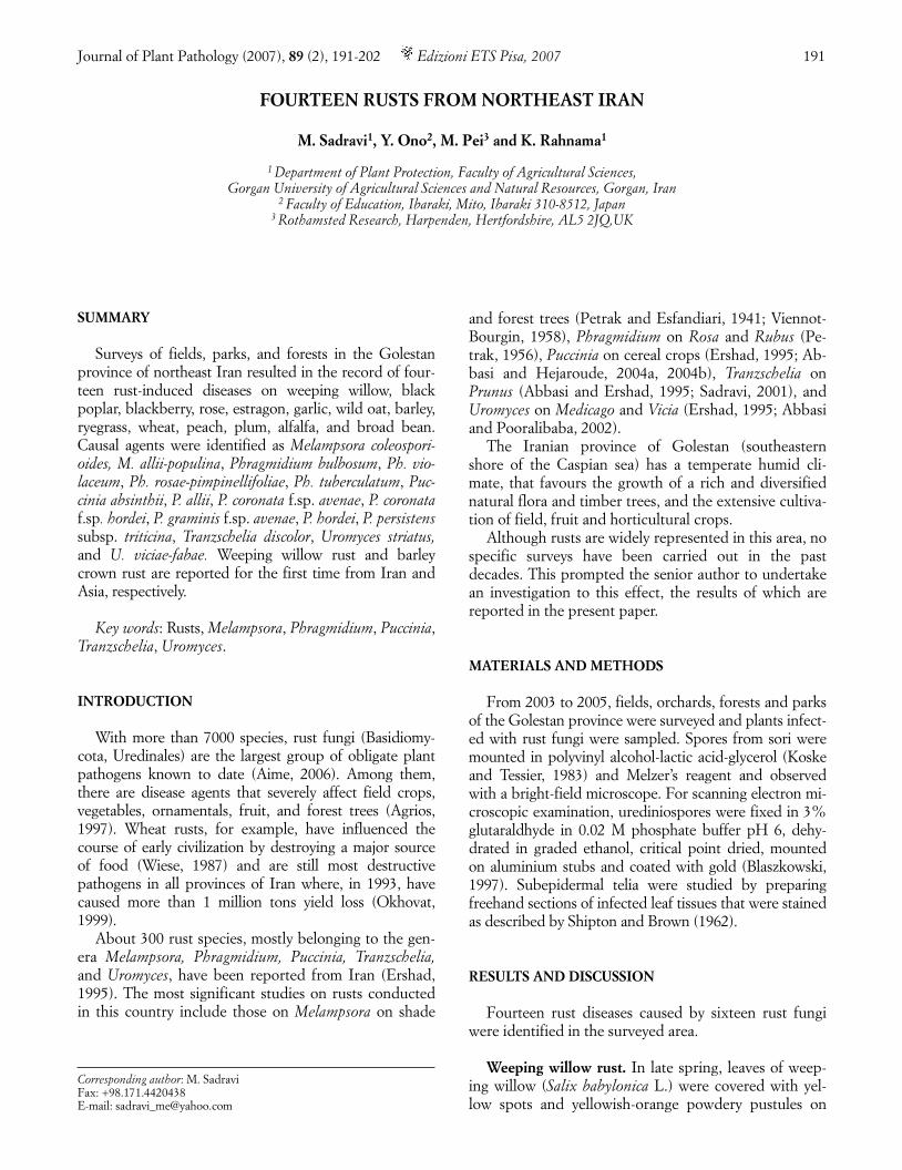
their abaxial surface. These spots became necrotic andthe pustules turned dark-brown in late summer, whilethe leaves desiccated and fell in early autumn. Thepathogen, identified as Melampsora coleosporioides Di-etel had the following characteristics: telia hypophyl-lous, sub-epidermal, dark brown, circular to oval.Teliospores hyaline to pale yellow-brown, one-celled,cylindrical, 21-53 × 4-7 µm in size, arranged in a singlerow (Fig. 1). Uredinia hypophyllous, yellowish-orange,sub-epidermal, erumpent, powdery, surrounded withmany clavate paraphyses (Fig. 2). Urediniospores hya-line to pale-yellow, echinulate, obovoid to pyriform, 16-28 x 12-16 µm in size. (Figs. 3, 4).
Melampsora epitea (Kze. et Schum.) Thüm. is com-mon on various willows (Salix spp.) in Iran (Ershad,1995). M. coleosporioides can be distinguished from M. epitea by the position of uredinia (mainly hypophyl-lous in the former and amphigenous in the latter), largerurediniospores and smaller teliospores, and differenthost specificity (Walker, 1978; Latch, 1980; Spiers andHopcroft, 1988, 1996; Ono et al., 1992). M. coleospori-oides can infect weeping willow and its hybrids, S. fragilisL. (common crack willow) and S. matsudana Koidz.(Peking willow), but S. matsudana × S. alba hybrid culti-vars are highly resistant (Latch, 1980). This rust was re-ported from Russia (Sydow and Sydow, 1915), Japan(Ono et al., 1992), China and Taiwan (Hiratsuka andKaneko, 1982), eastern Australia (Walker, 1978), andNew Zealand (Latch, 1980), but has not been previouslyrecorded in Iran.
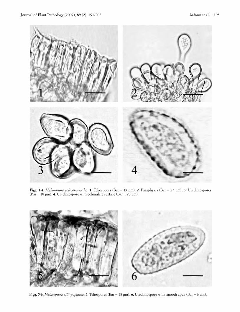
Black poplar rust. Leaves of black poplar (Populusnigra L.) trees showed circular to ellipsoid necroticspots, and dark-orange to black pustules on the abaxialsurface. Affected trees suffered heavy defoliation. Thepathogen, identified as Melampsora allii-populina Kleb.had the following characteristics: telia hypophyllous,black, circular to ellipsoid and sub-epidermal. Telio-spores yellowish-brown, one-celled, cylindrical to pris-matic, rounded at both ends, 30-62 × 7-12 µm in size,arranged in a single row (Fig. 5). Uredinia hypophyl-lous, deep orange, sub-epidermal, erumpent, powdery,surrounded by many clavate paraphyses. Uredin-iospores hyaline, broadly elliptical, echinulate butsmooth at the apex, 30-50 x 12-25 µm in size (Fig. 6).
M. allii-populina can be distinguished from the sixMelampsora species recorded from black poplar by itsteliospore morphology and the evenly thick-walled, api-cally smooth urediniospores. This fungus can also infectP. deltoids Marsh. and P. balsamifera L. Its aecial stage,which was not observed in Iran, develops on Alliumspp. and Arum spp. (Wilson and Henderson, 1966). M.allii-populina was reported from western, central andeastern Iran (Petrak and Esfandiari, 1941; Viennot-Bourgin, 1958), Russia, Africa, Australia, New Zealandand Britain (Sydow and Sydow, 1915; Gäumann, 1959;
Wilson and Henderson, 1966; Viljanen-Rollinson andCromey, 2002).
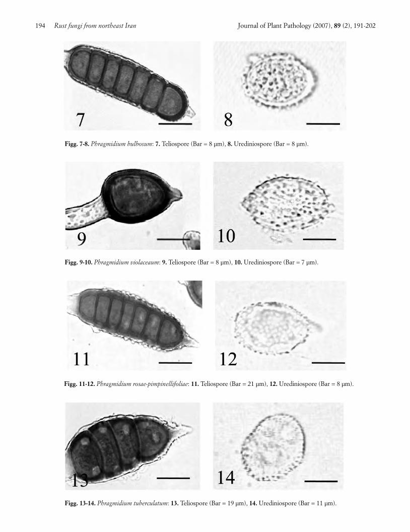
Blackberry rust. Blackberry (Rubus fruticosus L.)leaves showed circular to ellipsoid dark-purple spots onthe upper surface, and yellow, orange-yellow and blackpustules on the abaxial surface. Two Phragmidiumspecies were identified on this host. The first, Phragmid-ium bulbosum (Strauss) Schlechtend, had the followingcharacteristics: telia hypophyllous, black, circular to el-lipsoid, and erumpent. Teliospores orange-brown, witha conical hyaline apical papilla, 2-7-celled, ellipsoid, ver-rucose, 37-113 × 25-35 µm in size (Fig. 7). The hyalinepedicel was swollen, clavate in the lower half. Uredinia,mixed with telia, were yellow, erumpent, powdery, andsurrounded by many hyaline clavate paraphyses. Ure-diniospores hyaline to pale-yellow, globose to ovate,densely echinulate, bearing an indistinct germ pore, 17-25 × 17-20 µm in size (Fig. 8). Ph. bulbosum is new ad-dition to the Iranian mycoflora.
The second species was Phragmidium. violaceum(Schultz) Winter, characterized by hypophyllous, black,and erumpent telia. Teliospores dark-brown, with arounded hyaline apical papilla, 1- to 5-celled, ellipsoid,verrucose, 28-92 × 23-37 µm in size (Fig. 9). The hyalinepedicel was swollen to narrowly clavate in the lowerhalf. Uredinia hypophyllous, orange-yellow, erumpent,powdery, surrounded by many clavate hyaline paraphy-ses. Urediniospores 20-32 × 13-25 µm in size, hyaline,mostly ellipsoid, densely echinulate with long spines,bearing an indistinct germ pore (Fig. 10).
This fungus differs from Ph. bulbosum by the lowernumber of cells in teliospores (1-5 vs. 2-7), the colour ofteliospore wall (dark- brown vs. orange-brown), and therounded vs. conical apical papilla at the teliospore apex.Further differences reside in the urediniospore shapeand their denser, longer, and sharper spines. Ph. vio-laceum was reported earlier from western Iran (Ershad,1995). Both fungi also attack R. laciniatus Willd., andare common throughout Europe, the Middle East,Chile, Australia and New Zealand (Ellis et al., 1991;Bahcecioglu, 2001).
Rose rust. In late spring, rose (Rosa gallica L.) leaveswere densely covered with many circular to ellipsoid or-ange spots and pale yellow pustules 1-2 mm in diameter,that turned dark brown to black in late summer. Symp-tomatic leaves desiccated and dropped in early autumn.Two Phragmidium species were found on diseasedplants, one of them being Phragmidium rosae-pimpinelli-foliae Dietel with the following characteristics: telia hy-pophyllous, black and erumpent, containing chestnut-brown teliospores with a hyaline conical apical papilla,5- to 7-celled, ellipsoid to cylindrical, verrucose, 65-95 x25-33 µm in size (Fig. 11). The hyaline pedicel wasswollen, clavate in the lower half. Uredinia circular, hy-
192 Rust fungi from northeast Iran Journal of Plant Pathology (2007), 89 (2), 191-202
004_TESTO558_191_new 13-06-2007 17:30 Pagina 192
Journal of Plant Pathology (2007), 89 (2), 191-202 Sadravi et al. 193
Figg. 1-4. Melampsora coleosporioides: 1. Teliospores (Bar = 15 µm), 2. Paraphyses (Bar = 27 µm), 3. Urediniospores(Bar = 18 µm), 4. Urediniospore with echinulate surface (Bar = 20 µm).
Figg. 5-6. Melampsora allii-populina: 5. Teliospores (Bar = 18 µm), 6. Urediniospore with smooth apex (Bar = 6 µm).
004_TESTO558_191_new 13-06-2007 17:30 Pagina 193
194 Rust fungi from northeast Iran Journal of Plant Pathology (2007), 89 (2), 191-202
Figg. 9-10. Phragmidium violaceaum: 9. Teliospore (Bar = 8 µm), 10. Urediniospore (Bar = 7 µm).
Figg. 11-12. Phragmidium rosae-pimpinellifoliae: 11. Teliospore (Bar = 21 µm), 12. Urediniospore (Bar = 8 µm).
Figg. 7-8. Phragmidium bulbosum: 7. Teliospore (Bar = 8 µm), 8. Urediniospore (Bar = 8 µm).
Figg. 13-14. Phragmidium tuberculatum: 13. Teliospore (Bar = 19 µm), 14. Urediniospore (Bar = 11 µm).
004_TESTO558_191_new 13-06-2007 17:30 Pagina 194

pophyllous, pale-yellow, erumpent and minute. Uredin-iospores hyaline, ellipsoid to obovate, 19-28 x 19-22 µmin size, densely echinulate, and bearing an indistinctgerm pore (Fig. 12).
The nearest species to this fungus is Ph. montivagumArthur which has teliospores of similar in size but with alower number of cells (5-7 vs. 5-9) and a different colourof the wall (chestnut-brown vs. chocolate-brown) (Cum-mins, 1931; Wahyuno et al., 2001). Ph. rosae-pimpinellifo-liae occurs on Rosa pimpinellifolia L. and cultivated rosesin northwest Iran, North America and Britain (Cummins,1931; Wilson and Henderson, 1966; Ershad, 1995).
The second species, Ph. tuberculatum Muller, had thefollowing characteristics: telia hypophyllous, ellipsoid,dark brown sub-epidermal, becoming erumpent.Teliospores brown, with a pointed, hyaline apical papil-la, 5- to 6-celled, ellipsoid to cylindrical, verrucose, 37-81 × 14-32 µm in size (Fig. 13). The hyaline pedicel wasswollen to broadly clavate in the lower half. Urediniahypophyllous, minute, circular, yellow, subepidermal,erumpent and powdery. Urediniospores hyaline to pale-yellow, ellipsoid to ovate, echinulate, 23-28 × 16-23 µmin size. Germ pores conspicuously intruding into the lu-men (Fig. 14) constitute a unique differential characterof this species (Wilson and Henderson, 1966).
Ph. tuberculatum is common on more than 200 culti-vars of bush rose in England, on Rosa webbiana Wallichex Royle in Pakistan and R. macrophylla Lindl. and R. se-riacea Lindl. in Nepal (Horst, 1989; Ono and Kakishima,1992; Kakishima et al., 1993; Ono et al., 1995). It occursalso in central and western Iran (Petrak, 1956; Ershad,1995), Ukraine (Semina et al., 1991), East Europe(Susuri and Doda-Gashi, 1999), Austria (Bedlan, 1980),Portugal (Dias et al., 1982), Belarus (Gorlenko andPodobnaya, 1981), Estonia (Rumberg and Semenova,1974) and the British Isles (Howden and Jacobs, 1973).
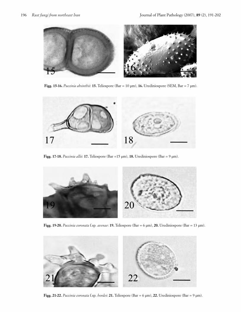
Estragon rust. Leaves of estragon (Artemisia dracun-culus L.) were infected with brown circular to ellipsoidsori mostly confined to the abaxial surface. Dark brown,oblong sori appeared on the stems in the late growingseason. Yield loss caused by this disease were estimatedto ca. 30% in most fields. The pathogen, identified asPuccinia absinthii (Hedw.) DC, had the following char-acteristics: telia dark brown, oblong, and erumpent.Teliospores pale-yellow to brown, two-celled, ellipsoid,finely verrucuose, 28-60 × 7.5-30 µm in size, with wallthickness averaging 6 µm at the apex and 3 µm on thesides (Fig. 15). Uredinia mostly small, hypophyllous,brown, erumpent, circular to ellipsoid. Urediniosporesgolden-yellow to yellow-brown, ellipsoid, 17-43 × 12-28µm in size, echinulate with minute spines, and bearing 2to 3 equatorial germ pores (Fig. 16).
P. absinthii infects A. absinthium L. and A. dubiaWallich ex Besser in Pakistan (Ono and Kakishima,1992; Ono and Okane, 1995), and was recorded from
central Iran (Ershad, 1995), Turkey (Bahcecioglu,2001), and Nepal (Ono et al., 1995).
Garlic rust. Circular to ellipsoid cream-coloured soriwere plentiful on garlic (Allium sativum L.) leaves,which turned yellow and desiccated. Plant mortality washigher than 70% in some fields.
Puccinia allii (DC.) Rud, the identified pathogen, hadthese characters: telia minute circular to ellipsoid, epi-phyllous, black, subepidermal, erumpent, containingbrown, fused paraphyses. Teliospores pale-brown, mostlytwo-celled, ellipsoid to oblong, 34-67 × 13-28 µm in size,slightly constricted in the middle, with a flat to slightlyraised beak (Fig. 17). Uredina minute, circular, epiphyl-lous, cream-coloured, erumpent. Urediniospores pale-yellow, subglobose to ovoid, echinulate, with 5 scatteredgerm pores, and a size of 20-32 × 18-25 µm (Fig. 18). Thetaxonomic position of this fungus is under discussion dueto variation in the host range, morphological charactersand DNA sequence of its isolates (Metcalf and Napier,2002; Anikster et al., 2004; Lupien et al., 2004).
This rust is common in southwest Iran (Ershad,1995), Japan (Ono et al., 1992), China (Liu and Hu,1987), India (Singh and Basandrai, 1988), Australia(Metcalf and Napier, 2002), France (Monnet andThibault, 2001), Germany (Bedlan, 1986), Britain (Wil-son and Henderson, 1966), and North America (Arthur,1934; Griesbach et al., 2001; Koike et al., 2001).
Wild oat crown rust. Leaves of wild oat (Avena fatuaL.) were covered with orange, circular to oblong pus-tules surrounded by black oblong pustules. Thepathogen, identifiedas Puccinia coronata Corda f.sp. ave-nea Erikss was characterized by hypophyllous, black,sub-epidermal, erumpent, and oblong telia. Teliosporesbrown, two-celled, 40-50 × 15-20 µm in size, with 3 to 5fine hyaline apical appendages (Fig. 19). Uredinia circu-lar to oblong, hypophyllous, orange, sub-epidermal,erumpent. Urediniospores hyaline to pale-yellow, spher-ical to ellipsoid, echinulate, 12-30 × 17-28 µm in size,with 2 to 8 scattered germ pores (Fig. 20).
This rust attacks cultivated and wild oats (Avenaspp.) in North America, Canada, Australia, northwestEurope, South Africa and Japan (Wilson and Hender-son, 1966; Gareth Jones and Clifford, 1983; van Niek-erk, 2001; Ono et al., 1992), and Phalaris sp. in Iran(Abbasi and Hejaroude, 2004a).
Barley crown rust. Leaves of barley (Hordeum vul-garis L.) showed orange brown ellipsoid to oblong pus-tules around which dark-brown to black, small, linearpustules arose. Attacks of little economic consequencewere observed in some fields. This is the first record ofthis barley rust in Asia. The pathogen, identified as Puc-cinia coronata Corda f.sp. hordei Jin and Steffenson, hadthe following characteristics: small, linear, hypophyllous,
Journal of Plant Pathology (2007), 89 (2), 191-202 Sadravi et al. 195
004_TESTO558_191_new 13-06-2007 17:30 Pagina 195
196 Rust fungi from northeast Iran Journal of Plant Pathology (2007), 89 (2), 191-202
Figg. 17-18. Puccinia allii: 17. Teliospore (Bar =15 µm), 18. Urediniospore (Bar = 9 µm).
Figg. 19-20. Puccinia coronata f.sp. avenae: 19. Teliospore (Bar = 6 µm), 20. Urediniospore (Bar = 13 µm).
Figg. 21-22. Puccinia coronata f.sp. hordei: 21. Teliospore (Bar = 6 µm), 22. Urediniospore (Bar = 9 µm).
Figg. 15-16. Puccinia absinthii: 15. Teliospore (Bar = 10 µm), 16. Urediniospore (SEM, Bar = 7 µm).
004_TESTO558_191_new 13-06-2007 17:30 Pagina 196
black, sub-epidermal and erumpent telia. Teliosporesbrown, two-celled, 32-63 × 10-27 µm in size, with 3 to 5clearly defined, apical, hyaline and branched ap-pendages (Fig. 21). Uredinia hypophyllous, orange-brown, sub-epidermal, erumpent and ellipsoid. Uredin-iospores pale-yellow, echinulate, subglobose to ellipsoid,17-33 × 15-25 µm in size, with 1 to 6 scattered germpores (Fig. 22). This species can be distinguished fromP. coronata Corda f.sp. avenea because of the longerteliospores with prominent branched appendages.
In the northern Great Plains of the USA, this rustnaturally infects Hordeum vulgare, H. jubatum, Bromustectorum, Elymus canadensis, Elymus trachycaulus, Ely-mus virginicus, Elytrigia intermedia [Elymus hispidus],Elymus repens, Leymus angustus, L. cinerius, L. dahuri-cus, L. racemosus, Pascopyrum smithii, Psathyrostachysjuncea and Secale cereale. Susceptible to artificial inocu-lation are: Aegilops, Agropyron, Elymus, Elytrigia, Ley-mus, Pascopyrum, Psathyrostachys, Secale, and Triticumin the tribe Triticeae, some species of Brachypodium,Bromus, Festuca and Lolium in the tribe Poaceae, andPhalaris in the tribe Avenae (Jin and Steffenson, 1999).It was observed on Bromus tomentosus in Iran (Abbasiand Hejaroude, 2004a).
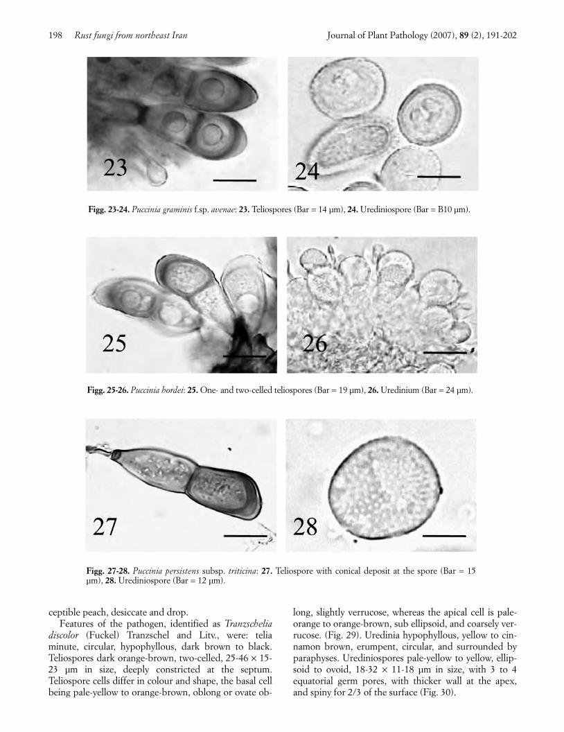
Wild oat black rust. Leaf sheath and stems of wildoat showed orange-brown, oblong, powdery pustulesbecoming dark-brown to black in early autumn. Severe-ly affected plants turn yellow and desiccate. Characteris-tics of the pathogen, identified as Puccinia graminisPers.: Pers. f.sp. avenae Eriks. and Henn. were: telia onleaf sheath and stems dark orange-brown to black, ob-long, and erumpent. Teliospores orange-brown, two-celled, 30-65 × 15-25 µm in size, with apical projectedwall (Fig. 23). Uredinia in the same position, orange-brown, oblong, and erumpent. Urediniospores yellow,oblong ellipsoid, echinulate, 22-43 × 10-25 µm in size,with 1 to 4 equatorial germ pores (Fig. 24). Distinct fea-tures of this fungus are the teliospore’s apical projectedwall, and the oblong ellipsoid shape of urediniosporeswith equatorial germ pores.
P. graminis f.sp. avenae infects Avena sativa, Alopecu-rus pratensis, Arrhenatherum elatius, Dactylis glomerata,Trisetum flavescens and Festuca rubra in Britain (Wilsonand Henderson, 1966), Avena byzantina and A. sterilis inSouth Africa (van Niekerk, 2001) and occurs in Canada(Harder, 1999) and Australia (Adhikari et al., 1999).
Barley and ryegrass leaf rust. Minute circular to ellip-soid pale yellow-brown pustules turning black later in theseason, were present on the leaves of barley and ryegrass(Lolium rigidum Gaudin). Whereas ryegrass leavesturned yellow and desiccated, damage to barley appearednegligible. Relevant features of the pathogen identified asPuccinia hordei Otth., were: telia minute circular, mostlyepiphyllous, black, sub-epidermal, erumpent. Teliospores
yellow, one- to two-celled, 30-63 × 15-25 µm in size (Fig.25). Uredinia epiphyllous, pale yellow-brown, sub-epi-dermal, becoming erumpent and ellipsoid. Uredin-iospores hyaline to pale-yellow, echinulate, ellipsoid toovoid, 15-24 × 10-20 µm in size, with 1 to 3 scatteredgerm pores (Fig. 26).
P. hordei is closely related to P. recondita, from whichit is readily differentiated by the high proportion single-celled teliospores. It also infects Hordeum bulbosum, H.glaucum, H. khaburense, H. leporinum, H. spontaneum,H. violaceum, H. murinum, H. distichon, Lolium per-sicum, L. temulentum, Lophochloa obtusiflora, Lo.phleoides, Bromus tomentellus and Trisetum flavescens.Its aecial stage, which develops on Ornithogalum pyre-naicum and O. umbellatum (Wilson and Henderson,1966) has not been observed in Iran.
This rust is common on barley in Iran (Okhovat,1999), Japan (Ono et al., 1992), Australia (Park et al.,2003), South Africa (van Niekerk et al., 2001), NewZealand, Europe, north Africa, Argentina, and northAmerica (Mathre, 1985).
Wheat leaf rust. Orange-brown, circular pustulesturning black later in the season, were present on the leafblade and sheath of wheat (Triticum aestivum L.). Attackswere sporadic and of little economic importance. Charac-teristics of the pathogen, identified as Puccinia persistensPlow. subsp. triticina (Erikss.) Urban et Markova, were:telia on the leaf blade and sheath, black, erumpent, andcircular. Teliospores pale-yellow to brown, two-celled,42-70 × 15-23 µm in size, with hyaline pedicel and a coni-cal deposit at the base (Fig. 27). Uredinia circular, epi-phyllous, orange-brown, erumpent. Urediniospores hya-line to pale-yellow, oval to oblong, echinulate, 25-45 × 15-30 µm in size, with 4 scattered germ pores (Fig. 28).
The taxonomic classification of this fungus has been influx for more than a century but in recent years DNA se-quence analysis showed that it is distinct from other leafrust species of cereals and grasses, being more related toP. persistens than to the P. recondita complex. Hosts otherthan T. aestivum are T. durum, T. boeoticum, T. urartu, T.dicoccoides, Aegilops longissima, A. ovata, A. sharonensis,A. bicornis, A. speltoides, A. taushii, A. squarrosa, Thalic-trum minus and T. speciosissimum (Goodwin et al., 2001;Abbasi and Hedjaroude, 2004b; Szabo et al., 2004).
Leaf rust has a worldwide distribution (Wiese, 1987).In past decade, it used to be the second importantwheat rust after yellow rust in Iran, but its economic rel-evance has decreased since resistant cultivars are grown(Okhovat, 1999).
Stone fruit trees rust. Leaves of peach (Prunus persi-ca (L.) Batsch) and plum (Prunus domestica L.) showcircular yellow spots, which exhibit on their abaxial sur-face orange-brown to dark-brown or black pustules. Se-verely affected leaves, especially those of the highly sus-
Journal of Plant Pathology (2007), 89 (2), 191-202 Sadravi et al. 197
004_TESTO558_191_new 13-06-2007 17:30 Pagina 197
ceptible peach, desiccate and drop. Features of the pathogen, identified as Tranzschelia
discolor (Fuckel) Tranzschel and Litv., were: teliaminute, circular, hypophyllous, dark brown to black.Teliospores dark orange-brown, two-celled, 25-46 × 15-23 µm in size, deeply constricted at the septum.Teliospore cells differ in colour and shape, the basal cellbeing pale-yellow to orange-brown, oblong or ovate ob-
long, slightly verrucose, whereas the apical cell is pale-orange to orange-brown, sub ellipsoid, and coarsely ver-rucose. (Fig. 29). Uredinia hypophyllous, yellow to cin-namon brown, erumpent, circular, and surrounded byparaphyses. Urediniospores pale-yellow to yellow, ellip-soid to ovoid, 18-32 × 11-18 µm in size, with 3 to 4equatorial germ pores, with thicker wall at the apex,and spiny for 2/3 of the surface (Fig. 30).
198 Rust fungi from northeast Iran Journal of Plant Pathology (2007), 89 (2), 191-202
Figg. 25-26. Puccinia hordei: 25. One- and two-celled teliospores (Bar = 19 µm), 26. Uredinium (Bar = 24 µm).
Figg. 27-28. Puccinia persistens subsp. triticina: 27. Teliospore with conical deposit at the spore (Bar = 15µm), 28. Urediniospore (Bar = 12 µm).
Figg. 23-24. Puccinia graminis f.sp. avenae: 23. Teliospores (Bar = 14 µm), 24. Urediniospore (Bar = B10 µm).
004_TESTO558_191_new 13-06-2007 17:30 Pagina 198
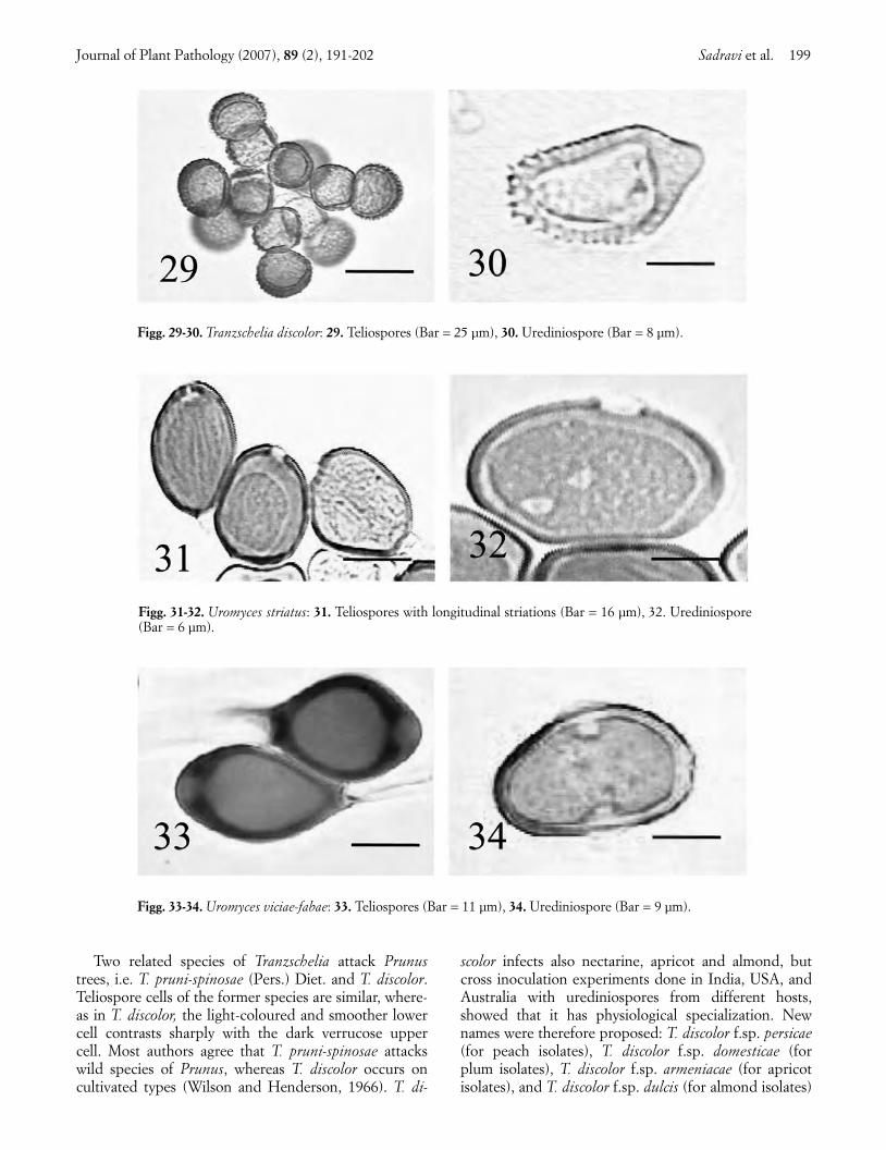
Two related species of Tranzschelia attack Prunustrees, i.e. T. pruni-spinosae (Pers.) Diet. and T. discolor.Teliospore cells of the former species are similar, where-as in T. discolor, the light-coloured and smoother lowercell contrasts sharply with the dark verrucose uppercell. Most authors agree that T. pruni-spinosae attackswild species of Prunus, whereas T. discolor occurs oncultivated types (Wilson and Henderson, 1966). T. di-
scolor infects also nectarine, apricot and almond, butcross inoculation experiments done in India, USA, andAustralia with urediniospores from different hosts,showed that it has physiological specialization. Newnames were therefore proposed: T. discolor f.sp. persicae(for peach isolates), T. discolor f.sp. domesticae (forplum isolates), T. discolor f.sp. armeniacae (for apricotisolates), and T. discolor f.sp. dulcis (for almond isolates)
Journal of Plant Pathology (2007), 89 (2), 191-202 Sadravi et al. 199
Figg. 29-30. Tranzschelia discolor: 29. Teliospores (Bar = 25 µm), 30. Urediniospore (Bar = 8 µm).
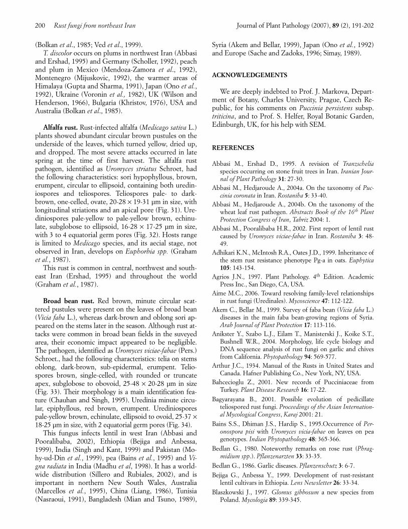
Figg. 31-32. Uromyces striatus: 31. Teliospores with longitudinal striations (Bar = 16 µm), 32. Urediniospore(Bar = 6 µm).
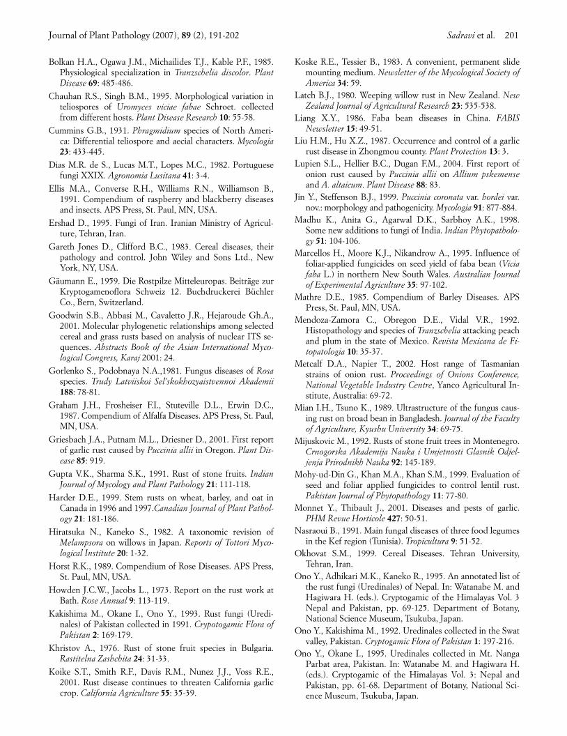
Figg. 33-34. Uromyces viciae-fabae: 33. Teliospores (Bar = 11 µm), 34. Urediniospore (Bar = 9 µm).
004_TESTO558_191_new 13-06-2007 17:30 Pagina 199
(Bolkan et al., 1985; Ved et al., 1999). T. discolor occurs on plums in northwest Iran (Abbasi
and Ershad, 1995) and Germany (Scholler, 1992), peachand plum in Mexico (Mendoza-Zamora et al., 1992),Montenegro (Mijuskovic, 1992), the warmer areas ofHimalaya (Gupta and Sharma, 1991), Japan (Ono et al.,1992), Ukraine (Voronin et al., 1982), UK (Wilson andHenderson, 1966), Bulgaria (Khristov, 1976), USA andAustralia (Bolkan et al., 1985).
Alfalfa rust. Rust-infected alfalfa (Medicago sativa L.)plants showed abundant circular brown pustules on theunderside of the leaves, which turned yellow, dried up,and dropped. The most severe attacks occurred in latespring at the time of first harvest. The alfalfa rustpathogen, identified as Uromyces striatus Schroet, hadthe following characteristics: sori hypophyllous, brown,erumpent, circular to ellipsoid, containing both uredin-iospores and teliospores. Teliospores pale- to dark-brown, one-celled, ovate, 20-28 × 19-31 µm in size, withlongitudinal striations and an apical pore (Fig. 31). Ure-diniospores pale-yellow to pale-yellow brown, echinu-late, subglobose to ellipsoid, 16-28 × 17-25 µm in size,with 3 to 4 equatorial germ pores (Fig. 32). Hosts rangeis limited to Medicago species, and its aecial stage, notobserved in Iran, develops on Euphorbia spp. (Grahamet al., 1987).
This rust is common in central, northwest and south-east Iran (Ershad, 1995) and throughout the world(Graham et al., 1987).
Broad bean rust. Red brown, minute circular scat-tered pustules were present on the leaves of broad bean(Vicia faba L.), whereas dark-brown and oblong sori ap-peared on the stems later in the season. Although rust at-tacks were common in broad bean fields in the suveyedarea, their economic impact appeared to be negligible.The pathogen, identified as Uromyces viciae-fabae (Pers.)Schroet., had the following characteristics: telia on stemsoblong, dark-brown, sub-epidermal, erumpent. Telio-spores brown, single-celled, with rounded or truncateapex, subglobose to obovoid, 25-48 × 20-28 µm in size(Fig. 33). Their morphology is a main identification fea-ture (Chauhan and Singh, 1995). Uredinia minute circu-lar, epiphyllous, red brown, erumpent. Urediniosporespale-yellow brown, echinulate, ellipsoid to ovoid, 25-37 ×18-25 µm in size, with 2 equatorial germ pores (Fig. 34).
This fungus infects lentil in west Iran (Abbasi andPooralibaba, 2002), Ethiopia (Bejiga and Anbessa,1999), India (Singh and Kant, 1999) and Pakistan (Mo-hy-ud-Din et al., 1999), pea (Bains et al., 1995) and Vi-gna radiata in India (Madhu et al, 1998). It has a world-wide distribution (Sillero and Rubiales, 2002), and isimportant in northern New South Wales, Australia(Marcellos et al., 1995), China (Liang, 1986), Tunisia(Nasraoui, 1991), Bangladesh (Mian and Tsuno, 1989),
Syria (Akem and Bellar, 1999), Japan (Ono et al., 1992)and Europe (Sache and Zadoks, 1996; Simay, 1989).
ACKNOWLEDGEMENTS
We are deeply indebted to Prof. J. Markova, Depart-ment of Botany, Charles University, Prague, Czech Re-public, for his comments on Puccinia persistens subsp.triticina, and to Prof. S. Helfer, Royal Botanic Garden,Edinburgh, UK, for his help with SEM.
REFERENCES
Abbasi M., Ershad D., 1995. A revision of Tranzscheliaspecies occurring on stone fruit trees in Iran. Iranian Jour-nal of Plant Pathology 31: 27-30.
Abbasi M., Hedjaroude A., 2004a. On the taxonomy of Puc-cinia coronata in Iran. Rostaniha 5: 33-40.
Abbasi M., Hedjaroude A., 2004b. On the taxonomy of thewheat leaf rust pathogen. Abstracts Book of the 16th PlantProtection Congress of Iran, Tabriz 2004: 1.
Abbasi M., Pooralibaba H.R., 2002. First report of lentil rustcaused by Uromyces viciae-fabae in Iran. Rostaniha 3: 48-49.
Adhikari K.N., McIntosh R.A., Oates J.D., 1999. Inheritance ofthe stem rust resistance phenotype Pg-a in oats. Euphytica105: 143-154.
Agrios J.N., 1997. Plant Pathology. 4th Edition. AcademicPress Inc., San Diego, CA, USA.
Aime M.C., 2006. Toward resolving family-level relationshipsin rust fungi (Uredinales). Mycoscience 47: 112-122.
Akem C., Bellar M., 1999. Survey of faba bean (Vicia faba L.)diseases in the main faba bean-growing regions of Syria.Arab Journal of Plant Protection 17: 113-116.
Anikster Y., Szabo L.J., Eilam T., Manisterski J., Koike S.T.,Bushnell W.R., 2004. Morphology, life cycle biology andDNA sequence analysis of rust fungi on garlic and chivesfrom California. Phytopathology 94: 569-577.
Arthur J.C., 1934. Manual of the Rusts in United States andCanada. Hafner Publishing Co., New York, NY, USA.
Bahcecioglu Z., 2001. New records of Pucciniaceae fromTurkey. Plant Disease Research 16: 17-22.
Bagyarayana B., 2001. Possible evolution of pedicillateteliospored rust fungi. Proceedings of the Asian Internation-al Mycological Congress, Karaj 2001: 21.
Bains S.S., Dhiman J.S., Hardip S., 1995.Occurrence of Per-onospora pisi with Uromyces vicia-fabae on leaves on peagenotypes. Indian Phytopathology 48: 365-366.
Bedlan G., 1980. Noteworthy remarks on rose rust (Phrag-midium spp.). Pflanzenarzten 33: 33-35.
Bedlan G., 1986. Garlic diseases. Pflanzenschutz 3: 6-7.
Bejiga G., Anbessa Y., 1999. Development of rust-resistantlentil cultivars in Ethiopia. Lens Newsletter 26: 33-34.
Blaszkowski J., 1997. Glomus gibbosum a new species fromPoland. Mycologia 89: 339-345.
200 Rust fungi from northeast Iran Journal of Plant Pathology (2007), 89 (2), 191-202
004_TESTO558_191_new 13-06-2007 17:30 Pagina 200
Bolkan H.A., Ogawa J.M., Michailides T.J., Kable P.F., 1985.Physiological specialization in Tranzschelia discolor. PlantDisease 69: 485-486.
Chauhan R.S., Singh B.M., 1995. Morphological variation inteliospores of Uromyces viciae fabae Schroet. collectedfrom different hosts. Plant Disease Research 10: 55-58.
Cummins G.B., 1931. Phragmidium species of North Ameri-ca: Differential teliospore and aecial characters. Mycologia23: 433-445.
Dias M.R. de S., Lucas M.T., Lopes M.C., 1982. Portuguesefungi XXIX. Agronomia Lusitana 41: 3-4.
Ellis M.A., Converse R.H., Williams R.N., Williamson B.,1991. Compendium of raspberry and blackberry diseasesand insects. APS Press, St. Paul, MN, USA.
Ershad D., 1995. Fungi of Iran. Iranian Ministry of Agricul-ture, Tehran, Iran.
Gareth Jones D., Clifford B.C., 1983. Cereal diseases, theirpathology and control. John Wiley and Sons Ltd., NewYork, NY, USA.
Gäumann E., 1959. Die Rostpilze Mitteleuropas. Beiträge zurKryptogamenoflora Schweiz 12. Buchdruckerei BüchlerCo., Bern, Switzerland.
Goodwin S.B., Abbasi M., Cavaletto J.R., Hejaroude Gh.A.,2001. Molecular phylogenetic relationships among selectedcereal and grass rusts based on analysis of nuclear ITS se-quences. Abstracts Book of the Asian International Myco-logical Congress, Karaj 2001: 24.
Gorlenko S., Podobnaya N.A.,1981. Fungus diseases of Rosaspecies. Trudy Latviiskoi Sel’skokhozyaistvennoi Akademii188: 78-81.
Graham J.H., Frosheiser F.I., Stuteville D.L., Erwin D.C.,1987. Compendium of Alfalfa Diseases. APS Press, St. Paul,MN, USA.
Griesbach J.A., Putnam M.L., Driesner D., 2001. First reportof garlic rust caused by Puccinia allii in Oregon. Plant Dis-ease 85: 919.
Gupta V.K., Sharma S.K., 1991. Rust of stone fruits. IndianJournal of Mycology and Plant Pathology 21: 111-118.
Harder D.E., 1999. Stem rusts on wheat, barley, and oat inCanada in 1996 and 1997.Canadian Journal of Plant Pathol-ogy 21: 181-186.
Hiratsuka N., Kaneko S., 1982. A taxonomic revision ofMelampsora on willows in Japan. Reports of Tottori Myco-logical Institute 20: 1-32.
Horst R.K., 1989. Compendium of Rose Diseases. APS Press,St. Paul, MN, USA.
Howden J.C.W., Jacobs L., 1973. Report on the rust work atBath. Rose Annual 9: 113-119.
Kakishima M., Okane I., Ono Y., 1993. Rust fungi (Uredi-nales) of Pakistan collected in 1991. Crypotogamic Flora ofPakistan 2: 169-179.
Khristov A., 1976. Rust of stone fruit species in Bulgaria.Rastitelna Zashchita 24: 31-33.
Koike S.T., Smith R.F., Davis R.M., Nunez J.J., Voss R.E.,2001. Rust disease continues to threaten California garliccrop. California Agriculture 55: 35-39.
Koske R.E., Tessier B., 1983. A convenient, permanent slidemounting medium. Newsletter of the Mycological Society ofAmerica 34: 59.
Latch B.J., 1980. Weeping willow rust in New Zealand. NewZealand Journal of Agricultural Research 23: 535-538.
Liang X.Y., 1986. Faba bean diseases in China. FABISNewsletter 15: 49-51.
Liu H.M., Hu X.Z., 1987. Occurrence and control of a garlicrust disease in Zhongmou county. Plant Protection 13: 3.
Lupien S.L., Hellier B.C., Dugan F.M., 2004. First report ofonion rust caused by Puccinia allii on Allium pskemenseand A. altaicum. Plant Disease 88: 83.
Jin Y., Steffenson B.J., 1999. Puccinia coronata var. hordei var.nov.: morphology and pathogenicity. Mycologia 91: 877-884.
Madhu K., Anita G., Agarwal D.K., Sarbhoy A.K., 1998.Some new additions to fungi of India. Indian Phytopatholo-gy 51: 104-106.
Marcellos H., Moore K.J., Nikandrow A., 1995. Influence offoliar-applied fungicides on seed yield of faba bean (Viciafaba L.) in northern New South Wales. Australian Journalof Experimental Agriculture 35: 97-102.
Mathre D.E., 1985. Compendium of Barley Diseases. APSPress, St. Paul, MN, USA.
Mendoza-Zamora C., Obregon D.E., Vidal V.R., 1992.Histopathology and species of Tranzschelia attacking peachand plum in the state of Mexico. Revista Mexicana de Fi-topatologia 10: 35-37.
Metcalf D.A., Napier T., 2002. Host range of Tasmanianstrains of onion rust. Proceedings of Onions Conference,National Vegetable Industry Centre, Yanco Agricultural In-stitute, Australia: 69-72.
Mian I.H., Tsuno K., 1989. Ultrastructure of the fungus caus-ing rust on broad bean in Bangladesh. Journal of the Facultyof Agriculture, Kyushu University 34: 69-75.
Mijuskovic M., 1992. Rusts of stone fruit trees in Montenegro.Crnogorska Akademija Nauka i Umjetnosti Glasnik Odjel-jenja Prirodnikh Nauka 92: 145-189.
Mohy-ud-Din G., Khan M.A., Khan S.M., 1999. Evaluation ofseed and foliar applied fungicides to control lentil rust.Pakistan Journal of Phytopathology 11: 77-80.
Monnet Y., Thibault J., 2001. Diseases and pests of garlic.PHM Revue Horticole 427: 50-51.
Nasraoui B., 1991. Main fungal diseases of three food legumesin the Kef region (Tunisia). Tropicultura 9: 51-52.
Okhovat S.M., 1999. Cereal Diseases. Tehran University,Tehran, Iran.
Ono Y., Adhikari M.K., Kaneko R., 1995. An annotated list ofthe rust fungi (Uredinales) of Nepal. In: Watanabe M. andHagiwara H. (eds.). Cryptogamic of the Himalayas Vol. 3Nepal and Pakistan, pp. 69-125. Department of Botany,National Science Museum, Tsukuba, Japan.
Ono Y., Kakishima M., 1992. Uredinales collected in the Swatvalley, Pakistan. Cryptogamic Flora of Pakistan 1: 197-216.
Ono Y., Okane I., 1995. Uredinales collected in Mt. NangaParbat area, Pakistan. In: Watanabe M. and Hagiwara H.(eds.). Cryptogamic of the Himalayas Vol. 3: Nepal andPakistan, pp. 61-68. Department of Botany, National Sci-ence Museum, Tsukuba, Japan.
Journal of Plant Pathology (2007), 89 (2), 191-202 Sadravi et al. 201
004_TESTO558_191_new 13-06-2007 17:30 Pagina 201
Ono Y., Uematsu K., Hikita M., 1992. Rust flora of theRyukyu islands, Japan. The Bulletin of the Faculty of Educa-tion, Ibaraki University (Natural Sciences) 41: 127-151.
Park R.F., Poulsen D., Barr A.R., Cakir M., Moody D.B., Ra-man H., Read B.J., 2003. Mapping genes for resistance toPuccinia hordei in barley. Australian Journal of AgriculturalResearch 54: 1323-1333.
Petrak F., 1956. Iranische pilze. Sydowia 10: 1-17.Petrak F., Esfandiari E., 1941. Beitrage zur Kenntnis der
iranischen Plizflora. Annnals of Mycology 39: 204-228.Rumberg V., Semenova V., 1974. Disease resistance of rose va-
rieties under Estonian conditions. Bolezneustoichivost Ras-tenii: 68-120.
Sache I., Zadoks J.C., 1996. Spread of faba bean rust over adiscontinuous field. European Journal of Plant Pathology102: 51-60.
Sadravi M., 2001. Notes on Transzchelia discolor occurring innortheast of Iran. Abstracts Book of the Asian InternationalMycological Congress, Karaj 2001: 42.
Scholler M., 1992. Puccinia cribrata and Tranzschelia discolorfound in Mecklenburg-Vorpommern. Zeitschrift furMykologie 58: 129-134.
Semina S.N., Klimenko Z.K., Timoshenko N.M., Zikov K.I.,1991. Protection of garden roses against diseases on thesouthern coast of the Crimea. Sbornik Nauchnykh TrudovGosudarstvennyi Nikitskii Botanicheskii Sad. 111: 145-154.
Shipton W.A., Brown J.F., 1962. A whole-leaf clearing andstaining technique to demonstrate host- pathogen relation-ships to wheat stem rust. Phytopathology 52: 1313.
Sillero J.C., Rubiales D., 2002. Histological characterizationof resistance to Uromyces viciae-fabae in faba bean. Phy-topathology 92: 294-299.
Simay E.I., 1989. On the mycoflora of uredosori of Uromycesfabae (Pers.) de Bary. Botanikai Kozlemenyek 76: 139-145.
Singh D.P., Kant L., 1999. Preliminary identification ofsources of resistance to rust in lentil. Lens Newsletter 26:32-33.
Singh P.J., Basandrai A.K., 1988. New report of garlic rustfrom Punjab State. Current Science 57: 266-267.
Spiers A.G., Hopcroft D.H., 1988. Ultrastructural studies ofthe telial, basidial, and spermatial stages of the willow rustfungus Melampsora coleosporioides in New Zealand. NewZealand Journal of Botany 26: 423-430.
Spiers A.G., Hopcroft D.H., 1996. Morphological and hostrange studies of Melampsora rusts attacking Salix species inNew Zealand. Mycological Research 100: 1163-1175.
Susuri L.R., Doda-Gashi G.N.,1999. Some morphologicalcharacteristics of Phragmidium spp. Zbornik BiotehniskeFakultete Univerze v Ljubljani. Kmetijstvo. 73: 81-86.
Sydow P., Sydow H., 1915. Monographia Uredinearum III.Fratres Borntraeger : 334-350.
Szabo L.J., Markova J., Anikster Y., Eilam T., Manisterski J.,Yehuda P.B., 2004. In search of the correct name for leafrust of cultivated wheat. Abstracts Book of the 11th Interna-tional Cereal Rusts and Mildews Conference, Norwich 2004:151.
van Niekerk B.D., 2001. Pathogenic variability of Pucciniacoronata f. sp. avenae and P. graminis f. sp. avenae on oat inSouth Africa. Plant Disease 85: 1085-1090.
van Niekerk B.D., Pretorius Z.A., Boshoff W.H.P., 2001. Oc-currence and pathogenicity of Puccinia hordei on barley inSouth Africa. Plant Disease 85: 713-717.
Ved R., Usha,S., Bhardwaj L.N., Sharma R.C., Ram V., Shar-ma U., 1999. Physiological specialization in Tranzscheliadiscolor causing rust of stone fruits. Plant Disease Research14: 45-46.
Viennot-Bourgin G., 1958. Contribution a la connaissance deschampignons parasites de l’Iran. Annales des Epiphyties 9:97-210.
Viljanen-Rollinson S.L.H., Cromey M.G., 2002. Pathways ofentry and spread of rust pathogens: Implications for NewZealand’s biosecurity. New Zealand Plant Protection 55: 42-48.
Voronin E.I., Kartausova V.A., Kopylova N.I. 1982. Plum rustin the Crimea.Trudy po Prikladnoi Botanike Genetike i Se-lektsii 71: 72-74.
Wahyuno D., Kakishima M., Ono Y., 2001. Morphologicalanalyses of urediniospores and teliospores in seven Phrag-midium species parasitic on ornamental roses. Mycoscience42: 519-533.
Walker J.C., 1978: Rust on weeping willow. Australasian PlantPathology 7: 34-42.
Wilson M., Henderson D.M., 1966. British Rust Fungi. Cam-bridge University Press, London, UK.
Wiese M.V., 1987. Compendium of Wheat Diseases. APSPress, St. Paul, MN, USA.
202 Rust fungi from northeast Iran Journal of Plant Pathology (2007), 89 (2), 191-202
Received March 20, 2006Accepted February 20, 2007
004_TESTO558_191_new 13-06-2007 17:30 Pagina 202
Related Documents