Formation of UV-honey guides in Rudbeckia hirta Karin Schlangen a , Silvija Miosic a , Ana Castro a , Karin Freudmann a , Maria Luczkiewicz b , Florian Vitzthum c , Wilfried Schwab c , Sonja Gamsjäger d , Maurizio Musso e , Heidi Halbwirth a, * a Technical University of Vienna, Institute for Chemical Engineering, Getreidemarkt 9/1665, A-1060 Vienna, Austria b Akademia Medyczna w Gdan ´sku, Katedra i Zakład Farmakognozji, Al. Gen. J. Hallera 107, 80-416 Gdansk, Poland c Technical University of Munich, Biomolecular Food Technology, Lise-Meitner-Str. 34, 85354 Freising, Germany d Ludwig Boltzmann Institute of Osteology, The Hanusch Hospital of WGKK and AUVA Trauma Centre Meidling, 4th Medical Department, Heinrich Collin-Str. 30, A-1140 Vienna, Austria e University of Salzburg, Department of Materials Research and Physics, Hellbrunnerstrasse 34, 5020 Salzburg, Austria article info Article history: Received 2 October 2008 Received in revised form 22 April 2009 Available online 26 May 2009 Keywords: Rudbeckia hirta Asteraceae UV-honey guides Flavonoids Methylated flavonols A-ring-specific hydroxylation Flavonoid 6-hydroxylase Methyltransferases Glucosyltransferases abstract The UV-honey guides of Rudbeckia hirta were investigated by UV-photography, reflectance spectroscopy, LC–MS analysis and studies of the enzymes involved in the formation of the UV-absorbing flavonols pres- ent in the petals. It was shown for the first time that the typical bull’s eye pattern is already established at the early stages of flower anthesis on the front side of the petal surface, but is hidden to pollinators until the buds are open and the petals are unfolded. The rear side of the petals remains UV-reflecting during the whole flower anthesis. Studies on the local distribution of 19 flavonols across the petals confirmed that the majority are concentrated in the basal part of the ray flower. However, in contrast to the earlier studies, eupatolitin 3-O-glucoside (6,7-dimethoxyquercetin 3-O-glucoside) was present in both the basal and apical parts of the petals, whereas eupatolin (6,7-dimethoxyquercetin 3-O-rhamnoside) was exclu- sively found in the apical parts. The enzymes involved in the formation of the flavonols in R. hirta were demonstrated for the first time. These include a rare flavonol 6-hydroxylase, which was identified as cytochrome P450-dependent monooxygenase and did not accept any methylated flavonol as substrate. All enzymes were present in the basal and apical parts of the petals, although some of them clearly showed higher activities in the basal part. This indicates that the local accumulation of flavonols in R. hir- ta is not achieved by a locally restricted presence of the enzymes involved in flavonol formation. Ó 2009 Elsevier Ltd. All rights reserved. 1. Introduction Honey guides are part of the pigmentation pattern, which oc- curs particularly frequently in bee-pollinated flowers. Their physi- ological function is to guide pollinators to the reproduction organs and to the nectar in the flower centre (Daumer, 1956, 1958; Kevan and Mulligan, 1973; Harborne, 1993; Kevan et al., 2001). A broad variety of forms are known. They may appear as color contrast – as in snapdragons (Antirrhinum majus) where a yellow spot is lo- cated on the lip of an otherwise red or orange flower – or may take the form of colored dots or lines in particular areas of the petals. Often, honey guides are formed by local accumulation of UV- absorbing substances and are therefore invisible to the human eye. Such UV-patterns on flower petals may be perceived by many pollinating insects because of their specific color sense, which makes them blind to scarlet red coloration but sensitive to the UV-range of the spectrum (Eisner et al., 1969; Silberglied, 1979; McCrea and Levy, 1983; Dyer, 1996; Indsto et al., 2006, 2007). The prime example for the formation of UV-honey guides is Rudbeckia hirta (Fig. 1). In 1972, Thompson et al. were able to show that the flowers, which are uniformly yellow to the human eye, have a characteristic UV-absorbing centre, whilst the outer parts of the ray petals are UV-reflecting. For UV-sensitive insects, the Rudbeckia flower appears as a bull’s eye with two concentric dichromatic circles against the green meadow background (Abra- hamson and McCrea, 1977). The UV-reflecting apical part is responsible for the long-distance orientation and acts as a landing site for the pollinators, whereas the UV-absorbing centre of the flower acts as a ‘honey guide’, helping the pollinators to orient themselves within the flower after landing (Daumer, 1956, 1958; Jokl and Fürnkranz, 1989; Burr et al., 1995). In contrast to the fully developed Rudbeckia flower, small buds were described as being 0031-9422/$ - see front matter Ó 2009 Elsevier Ltd. All rights reserved. doi:10.1016/j.phytochem.2009.04.017 Abbreviations: CHI, chalcone isomerase; CHS, chalcone synthase; DHK, dihydrokaempferol; EGME, ethylene glycol mono methyl ether; EtOAC, ethyl acetate; FHT, flavanone 3-hydroxylase; FLS, flavonol synthase; F3 0 H, flavonoid 3 0 - hydroxylase; F6H, flavonol 6-hydroxylase; F7GT, flavonol 7-O-glucosyltransferase; F3GT, flavonol 3-O-glucosyltransferase; Glu, glucose; Rha, rhamnose; SAM, S- Adenosylmethionine; UDPG, UDP-glucose. * Corresponding author. Tel.: +43 1 58801 17311; fax: +43 1 58801 17399. E-mail address: [email protected] (H. Halbwirth). Phytochemistry 70 (2009) 889–898 Contents lists available at ScienceDirect Phytochemistry journal homepage: www.elsevier.com/locate/phytochem

Welcome message from author
This document is posted to help you gain knowledge. Please leave a comment to let me know what you think about it! Share it to your friends and learn new things together.
Transcript

Phytochemistry 70 (2009) 889–898
Contents lists available at ScienceDirect
Phytochemistry
journal homepage: www.elsevier .com/locate /phytochem
Formation of UV-honey guides in Rudbeckia hirta
Karin Schlangen a, Silvija Miosic a, Ana Castro a, Karin Freudmann a, Maria Luczkiewicz b, Florian Vitzthum c,Wilfried Schwab c, Sonja Gamsjäger d, Maurizio Musso e, Heidi Halbwirth a,*
a Technical University of Vienna, Institute for Chemical Engineering, Getreidemarkt 9/1665, A-1060 Vienna, Austriab Akademia Medyczna w Gdansku, Katedra i Zakład Farmakognozji, Al. Gen. J. Hallera 107, 80-416 Gdansk, Polandc Technical University of Munich, Biomolecular Food Technology, Lise-Meitner-Str. 34, 85354 Freising, Germanyd Ludwig Boltzmann Institute of Osteology, The Hanusch Hospital of WGKK and AUVA Trauma Centre Meidling, 4th Medical Department,Heinrich Collin-Str. 30, A-1140 Vienna, Austriae University of Salzburg, Department of Materials Research and Physics, Hellbrunnerstrasse 34, 5020 Salzburg, Austria
a r t i c l e i n f o a b s t r a c t
Article history:Received 2 October 2008Received in revised form 22 April 2009Available online 26 May 2009
Keywords:Rudbeckia hirtaAsteraceaeUV-honey guidesFlavonoidsMethylated flavonolsA-ring-specific hydroxylationFlavonoid 6-hydroxylaseMethyltransferasesGlucosyltransferases
0031-9422/$ - see front matter � 2009 Elsevier Ltd. Adoi:10.1016/j.phytochem.2009.04.017
Abbreviations: CHI, chalcone isomerase; CHS,dihydrokaempferol; EGME, ethylene glycol monoacetate; FHT, flavanone 3-hydroxylase; FLS, flavonolhydroxylase; F6H, flavonol 6-hydroxylase; F7GT, flavoF3GT, flavonol 3-O-glucosyltransferase; Glu, glucosAdenosylmethionine; UDPG, UDP-glucose.
* Corresponding author. Tel.: +43 1 58801 17311; fE-mail address: [email protected] (H.
The UV-honey guides of Rudbeckia hirta were investigated by UV-photography, reflectance spectroscopy,LC–MS analysis and studies of the enzymes involved in the formation of the UV-absorbing flavonols pres-ent in the petals. It was shown for the first time that the typical bull’s eye pattern is already established atthe early stages of flower anthesis on the front side of the petal surface, but is hidden to pollinators untilthe buds are open and the petals are unfolded. The rear side of the petals remains UV-reflecting duringthe whole flower anthesis. Studies on the local distribution of 19 flavonols across the petals confirmedthat the majority are concentrated in the basal part of the ray flower. However, in contrast to the earlierstudies, eupatolitin 3-O-glucoside (6,7-dimethoxyquercetin 3-O-glucoside) was present in both the basaland apical parts of the petals, whereas eupatolin (6,7-dimethoxyquercetin 3-O-rhamnoside) was exclu-sively found in the apical parts. The enzymes involved in the formation of the flavonols in R. hirta weredemonstrated for the first time. These include a rare flavonol 6-hydroxylase, which was identified ascytochrome P450-dependent monooxygenase and did not accept any methylated flavonol as substrate.All enzymes were present in the basal and apical parts of the petals, although some of them clearlyshowed higher activities in the basal part. This indicates that the local accumulation of flavonols in R. hir-ta is not achieved by a locally restricted presence of the enzymes involved in flavonol formation.
� 2009 Elsevier Ltd. All rights reserved.
1. Introduction
Honey guides are part of the pigmentation pattern, which oc-curs particularly frequently in bee-pollinated flowers. Their physi-ological function is to guide pollinators to the reproduction organsand to the nectar in the flower centre (Daumer, 1956, 1958; Kevanand Mulligan, 1973; Harborne, 1993; Kevan et al., 2001). A broadvariety of forms are known. They may appear as color contrast –as in snapdragons (Antirrhinum majus) where a yellow spot is lo-cated on the lip of an otherwise red or orange flower – or may takethe form of colored dots or lines in particular areas of the petals.Often, honey guides are formed by local accumulation of UV-
ll rights reserved.
chalcone synthase; DHK,methyl ether; EtOAC, ethylsynthase; F30H, flavonoid 30-nol 7-O-glucosyltransferase;
e; Rha, rhamnose; SAM, S-
ax: +43 1 58801 17399.Halbwirth).
absorbing substances and are therefore invisible to the humaneye. Such UV-patterns on flower petals may be perceived by manypollinating insects because of their specific color sense, whichmakes them blind to scarlet red coloration but sensitive to theUV-range of the spectrum (Eisner et al., 1969; Silberglied, 1979;McCrea and Levy, 1983; Dyer, 1996; Indsto et al., 2006, 2007).
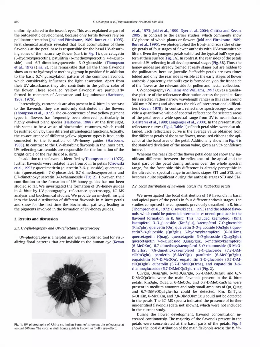
The prime example for the formation of UV-honey guides isRudbeckia hirta (Fig. 1). In 1972, Thompson et al. were able to showthat the flowers, which are uniformly yellow to the human eye,have a characteristic UV-absorbing centre, whilst the outer partsof the ray petals are UV-reflecting. For UV-sensitive insects, theRudbeckia flower appears as a bull’s eye with two concentricdichromatic circles against the green meadow background (Abra-hamson and McCrea, 1977). The UV-reflecting apical part isresponsible for the long-distance orientation and acts as a landingsite for the pollinators, whereas the UV-absorbing centre of theflower acts as a ‘honey guide’, helping the pollinators to orientthemselves within the flower after landing (Daumer, 1956, 1958;Jokl and Fürnkranz, 1989; Burr et al., 1995). In contrast to the fullydeveloped Rudbeckia flower, small buds were described as being
890 K. Schlangen et al. / Phytochemistry 70 (2009) 889–898
uniformly colored to the insect’s eyes. This was explained as part ofthe ontogenetic development, because only fertile flowers rely onpollinator attraction (Jokl and Fürnkranz, 1989; Burr et al., 1995).First chemical analysis revealed that local accumulation of threeflavonols at the petal base is responsible for the basal UV-absorb-ing zones of the mature ray flower (dark in Fig. 1): quercetagetin(6-hydroxyquercetin), patulitrin (6-methoxyquercetin 7-O-gluco-side) and 6,7-dimethoxyquercetin 3-O-glucoside (Thompsonet al., 1972) (Fig. 2). It is remarkable that all the three flavonolsshow an extra hydroxyl or methoxyl group in position 6 in additionto the basic 5,7-hydroxylation pattern of the common flavonols,which considerably influences the light absorption. Apart fromtheir UV-absorbance, they also contribute to the yellow color ofthe flower. These so-called ‘yellow flavonols’ are particularlyformed in members of Asteraceae and Leguminosae (Harborne,1967, 1976).
Interestingly, carotenoids are also present in R. hirta. In contrastto the flavonols, they are uniformly distributed in the flowers(Thompson et al., 1972). The co-occurrence of two yellow pigmenttypes in flowers has frequently been observed, particularly inhighly evolved plant species (Harborne, 1988). At the first sight,this seems to be a waste of biochemical resources, which couldbe justified only by their different physiological functions. Actually,the co-occurrence of different yellow pigment types is frequentlyconnected to the formation of UV-honey guides (Harborne,1988). In contrast to the UV-absorbing flavonols in the inner part,UV-reflecting carotenoids are responsible for the formation of thebright circle of the ray disk of R. hirta.
In addition to the flavonols identified by Thompson et al. (1972),further flavonols were isolated later from R. hirta petals (Cisowskiet al., 1993): quercimeritrin (quercetin 7-O-glucoside), quercetagi-trin (quercetagetin 7-O-glucoside), 6,7-dimethoxyquercetin and6,7-dimethoxyquercetin 3-O-rhamnoside (Fig. 2). However, theircontribution to the formation of UV-honey guides has not beenstudied so far. We investigated the formation of UV-honey guidesin R. hirta by UV-photography, reflectance spectroscopy, LC–MSanalysis and biochemical studies. We provide an in-depth insightinto the local distribution of different flavonols in R. hirta petalsand show for the first time the biochemical pathway leading tothe pigments involved in the formation of UV-honey guides.
2. Results and discussion
2.1. UV-photography and UV-reflectance spectroscopy
UV-photography is a helpful and well-established tool for visu-alizing floral patterns that are invisible to the human eye (Kevan
Fig. 1. UV-photography of R.hirta cv. ‘Indian Summer’, showing the reflectance ataround 360 nm. The circular dark honey guide is known as ‘bull’s eye-effect’.
et al., 1973; Jokl et al., 1999; Dyer et al., 2004; Chittka and Kevan,2005). In contrast to the earlier studies, which commonly showUV-photos of whole plants or flowers (Jokl and Fürnkranz, 1989;Burr et al., 1995), we photographed the front- and rear sides of sin-gle petals of four stages of flower anthesis with UV-transmittablefilters. Even the youngest petals exhibited the typical bull’s eye pat-tern at their surface (Fig. 3A). In contrast, the rear sides of the petalsremain UV-reflecting in all developmental stages (Fig. 3B). Thus, thehoney guides are already formed at early stages but are hidden tothe pollinators, because juvenile Rudbeckia petals are two timesfolded and only the rear side is visible at the early stages of floweranthesis. Apparently, the bull’s eye is formed only on the front sideof the flower as the relevant side for pollen and nectar collection.
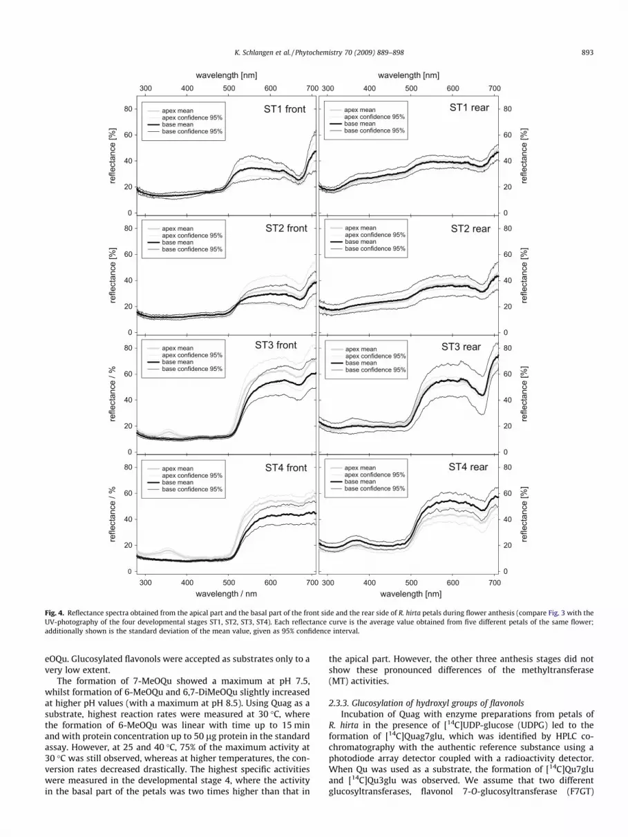
UV-photography (Williams and Williams, 1993) gives a qualita-tive picture of the reflectance distribution across the petal surfacefor a selected, rather narrow wavelength range (in this case around360 nm ± 20 nm) and also runs the risk of interpretational difficul-ties (Kevan, 1979). In contrast, reflectance spectroscopy can pro-vide a quantitative value of spectral reflectance for selected areasof the petal over a wide spectral range from UV to near infrared(Galsterer et al., 1999; Langanger et al., 2000). In the present study,reflectance curves (Fig. 4, Table 1) of both petal sides were also ob-tained. Each reflectance curve is the average value obtained fromfive different petals of the same flower, measured either at the api-cal or at the basal area of the petal. Additionally shown in Fig. 4 isthe standard deviation of the mean value, given as 95% confidenceinterval.
Whereas the rear side of the flower petal does not show any sig-nificant difference between the reflectance of the apical and thebasal part of the petal during anthesis over the whole spectralrange, for the front side this difference is already perceptible inthe ultraviolet spectral range in anthesis stages ST1 and ST2, andbecomes quite significant during the anthesis stages ST3 and ST4.
2.2. Local distribution of flavonols across the Rudbeckia petals
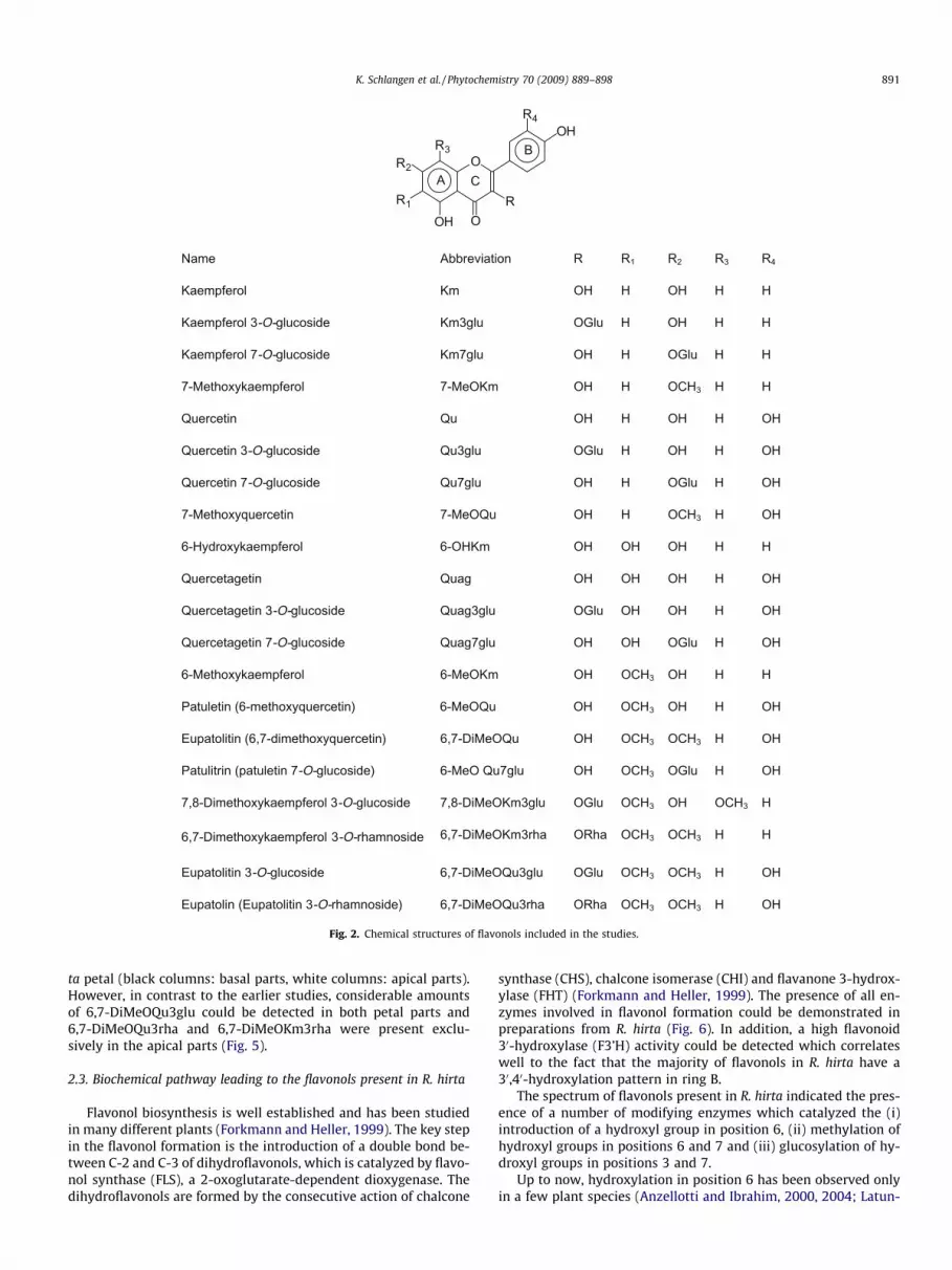
We investigated the local distribution of 19 flavonols in basaland apical parts of the petals in four different anthesis stages. Thestudies comprised the compounds previously described in R. hirta(Thompson et al., 1972; Cisowski et al., 1993) and the related flavo-nols, which could be potential intermediates or end-products in theflavonol formation in R. hirta. This included kaempferol (Km),kaempferol 3-O-glucoside (Km3glu), kaempferol 7-O-glucoside(Km7glu), quercetin (Qu), quercetin 3-O-glucoside (Qu3glu), quer-cetin7-O-glucoside (Qu7glu), 6-hydroxykaempferol (6-OHKm),quercetagetin (Quag), quercetagetin 3-O-glucoside (Quag3glu),quercetagetin 7-O-glucoside (Quag7glu), 6-methoxykaempferol(6-MeOKm), 6,7-dimethoxykaempferol 3-O-rhamnoside (6-MeO-Km3rha), 7,8-dimethoxykaempferol 3-O-glucoside (7,8-DiM-eOKm3glu), patuletin (6-MeOQu), patulitrin (6-MeOQu7glu),eupatolitin (6,7-DiMeOQu), eupatolitin 3-O-glucoside (6,7-DiM-eOQu3glu), eupatolin (6,7-DiMeOQu3rha), and eupatolitin 3-O-rhamnoglucoside (6,7-DiMeOQu3glu-rha) (Fig. 2).
Qu7glu, Quag7glu, 6-MeOQu7glu, 6,7-DiMeOQu3glu and 6,7-DiMeOQu3rha were the main flavonols present in the R. hirtapetals. Km3glu, Qu3glu, 6-MeOQu, and 6,7-DiMeOKm3rha werepresent in medium amounts and only small amounts of Qu, Quagand 6,7-DiMeOQu3glu-rha could be detected. Km, Km7glu,6-OHKm, 6-MeOKm, and 7,8-DiMeOKm3glu could not be detectedin the petals. The LC–MS spectra indicated the presence of furtherunidentified flavonols (data not shown), which were not includedin the current study.
During the flower development, flavonol concentration in-creased continuously. The majority of the flavonols present in thepetals were concentrated at the basal parts of the petals. Fig. 5shows the local distribution of the main flavonols across the R. hir-
RRnoitaiverbbAemaN 1 R2 R3 R4
HHHOHHOmKlorefpmeaK
Kaempferol 3-O-glucoside Km3glu OGlu H OH H H
Kaempferol 7-O-glucoside Km7glu OH H OGlu H H
7-Methoxykaempferol 7-MeOKm OH H OCH3 H H
HOHHOHHOuQnitecreuQ
Quercetin 3-O-glucoside Qu3glu OGlu H OH H OH
Quercetin 7-O-glucoside Qu7glu OH H OGlu H OH
HCOHHOuQOeM-7nitecreuqyxohteM-7 3 H OH
HHHOHOHOmKHO-6lorefpmeakyxordyH-6
HOHHOHOHOgauQnitegatecreuQ
Quercetagetin 3-O-glucoside Quag3glu OGlu OH OH H OH
Quercetagetin 7-O-glucoside Quag7glu OH OH OGlu H OH
6-Methoxykaempferol HCOHOmKOeM-6 3 OH H H
Patuletin (6-methoxyquercetin) 6-MeOQu OH OCH3 OH H OH
Eupatolitin (6,7-dimethoxyquercetin) 6,7-DiMeOQu OH OCH3 OCH3 H OH
Patulitrin (patuletin 7-O-glucoside) 6-MeO Qu7glu OH OCH3 OGlu H OH
7,8-Dimethoxykaempferol 3-O-glucoside 7,8-DiMeOKm3glu OGlu OCH3 OH OCH3 H
6,7-Dimethoxykaempferol 3-O-rhamnoside 6,7-DiMeOKm3rha ORha OCH3 OCH3 H H
Eupatolitin 3-O-glucoside 6,7-DiMeOQu3glu OGlu OCH3 OCH3 H OH
Eupatolin (Eupatolitin 3-O-rhamnoside) 6,7-DiMeOQu3rha ORha OCH3 OCH3 H OH
O
OOHR
OHR4
R1
R2
R3
A
B
C
Fig. 2. Chemical structures of flavonols included in the studies.
K. Schlangen et al. / Phytochemistry 70 (2009) 889–898 891
ta petal (black columns: basal parts, white columns: apical parts).However, in contrast to the earlier studies, considerable amountsof 6,7-DiMeOQu3glu could be detected in both petal parts and6,7-DiMeOQu3rha and 6,7-DiMeOKm3rha were present exclu-sively in the apical parts (Fig. 5).
2.3. Biochemical pathway leading to the flavonols present in R. hirta
Flavonol biosynthesis is well established and has been studiedin many different plants (Forkmann and Heller, 1999). The key stepin the flavonol formation is the introduction of a double bond be-tween C-2 and C-3 of dihydroflavonols, which is catalyzed by flavo-nol synthase (FLS), a 2-oxoglutarate-dependent dioxygenase. Thedihydroflavonols are formed by the consecutive action of chalcone
synthase (CHS), chalcone isomerase (CHI) and flavanone 3-hydrox-ylase (FHT) (Forkmann and Heller, 1999). The presence of all en-zymes involved in flavonol formation could be demonstrated inpreparations from R. hirta (Fig. 6). In addition, a high flavonoid30-hydroxylase (F3’H) activity could be detected which correlateswell to the fact that the majority of flavonols in R. hirta have a30,40-hydroxylation pattern in ring B.
The spectrum of flavonols present in R. hirta indicated the pres-ence of a number of modifying enzymes which catalyzed the (i)introduction of a hydroxyl group in position 6, (ii) methylation ofhydroxyl groups in positions 6 and 7 and (iii) glucosylation of hy-droxyl groups in positions 3 and 7.
Up to now, hydroxylation in position 6 has been observed onlyin a few plant species (Anzellotti and Ibrahim, 2000, 2004; Latun-
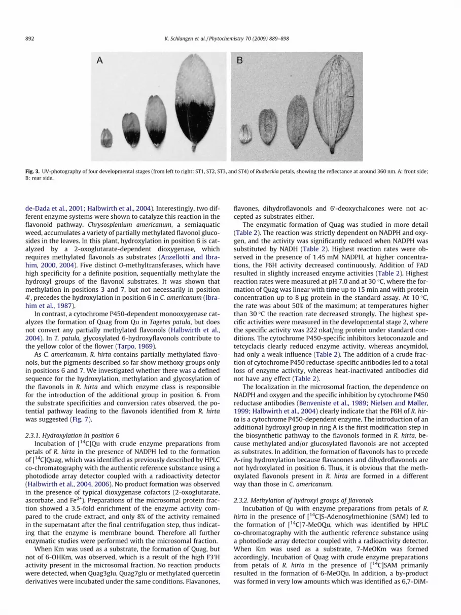
Fig. 3. UV-photography of four developmental stages (from left to right: ST1, ST2, ST3, and ST4) of Rudbeckia petals, showing the reflectance at around 360 nm. A: front side;B: rear side.
892 K. Schlangen et al. / Phytochemistry 70 (2009) 889–898
de-Dada et al., 2001; Halbwirth et al., 2004). Interestingly, two dif-ferent enzyme systems were shown to catalyze this reaction in theflavonoid pathway. Chrysosplenium americanum, a semiaquaticweed, accumulates a variety of partially methylated flavonol gluco-sides in the leaves. In this plant, hydroxylation in position 6 is cat-alyzed by a 2-oxoglutarate-dependent dioxygenase, whichrequires methylated flavonols as substrates (Anzellotti and Ibra-him, 2000, 2004). Five distinct O-methyltransferases, which havehigh specificity for a definite position, sequentially methylate thehydroxyl groups of the flavonol substrates. It was shown thatmethylation in positions 3 and 7, but not necessarily in position40, precedes the hydroxylation in position 6 in C. americanum (Ibra-him et al., 1987).
In contrast, a cytochrome P450-dependent monooxygenase cat-alyzes the formation of Quag from Qu in Tagetes patula, but doesnot convert any partially methylated flavonols (Halbwirth et al.,2004). In T. patula, glycosylated 6-hydroxyflavonols contribute tothe yellow color of the flower (Tarpo, 1969).
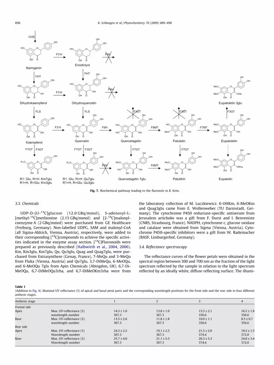
As C. americanum, R. hirta contains partially methylated flavo-nols, but the pigments described so far show methoxy groups onlyin positions 6 and 7. We investigated whether there was a definedsequence for the hydroxylation, methylation and glycosylation ofthe flavonols in R. hirta and which enzyme class is responsiblefor the introduction of the additional group in position 6. Fromthe substrate specificities and conversion rates observed, the po-tential pathway leading to the flavonols identified from R. hirtawas suggested (Fig. 7).
2.3.1. Hydroxylation in position 6Incubation of [14C]Qu with crude enzyme preparations from
petals of R. hirta in the presence of NADPH led to the formationof [14C]Quag, which was identified as previously described by HPLCco-chromatography with the authentic reference substance using aphotodiode array detector coupled with a radioactivity detector(Halbwirth et al., 2004, 2006). No product formation was observedin the presence of typical dioxygenase cofactors (2-oxoglutarate,ascorbate, and Fe2+). Preparations of the microsomal protein frac-tion showed a 3.5-fold enrichment of the enzyme activity com-pared to the crude extract, and only 8% of the activity remainedin the supernatant after the final centrifugation step, thus indicat-ing that the enzyme is membrane bound. Therefore all furtherenzymatic studies were performed with the microsomal fraction.
When Km was used as a substrate, the formation of Quag, butnot of 6-OHKm, was observed, which is a result of the high F30Hactivity present in the microsomal fraction. No reaction productswere detected, when Quag3glu, Quag7glu or methylated quercetinderivatives were incubated under the same conditions. Flavanones,
flavones, dihydroflavonols and 60-deoxychalcones were not ac-cepted as substrates either.
The enzymatic formation of Quag was studied in more detail(Table 2). The reaction was strictly dependent on NADPH and oxy-gen, and the activity was significantly reduced when NADPH wassubstituted by NADH (Table 2). Highest reaction rates were ob-served in the presence of 1.45 mM NADPH, at higher concentra-tions, the F6H activity decreased continuously. Addition of FADresulted in slightly increased enzyme activities (Table 2). Highestreaction rates were measured at pH 7.0 and at 30 �C, where the for-mation of Quag was linear with time up to 15 min and with proteinconcentration up to 8 lg protein in the standard assay. At 10 �C,the rate was about 50% of the maximum; at temperatures higherthan 30 �C the reaction rate decreased strongly. The highest spe-cific activities were measured in the developmental stage 2, wherethe specific activity was 222 nkat/mg protein under standard con-ditions. The cytochrome P450-specific inhibitors ketoconazole andtetcyclacis clearly reduced enzyme activity, whereas ancymidol,had only a weak influence (Table 2). The addition of a crude frac-tion of cytochrome P450 reductase-specific antibodies led to a totalloss of enzyme activity, whereas heat-inactivated antibodies didnot have any effect (Table 2).
The localization in the microsomal fraction, the dependence onNADPH and oxygen and the specific inhibition by cytochrome P450reductase antibodies (Benveniste et al., 1989; Nielsen and Møller,1999; Halbwirth et al., 2004) clearly indicate that the F6H of R. hir-ta is a cytochrome P450-dependent enzyme. The introduction of anadditional hydroxyl group in ring A is the first modification step inthe biosynthetic pathway to the flavonols formed in R. hirta, be-cause methylated and/or glucosylated flavonols are not acceptedas substrates. In addition, the formation of flavonols has to precedeA-ring hydroxylation because flavanones and dihydroflavonols arenot hydroxylated in position 6. Thus, it is obvious that the meth-oxylated flavonols present in R. hirta are formed in a differentway than those in C. americanum.
2.3.2. Methylation of hydroxyl groups of flavonolsIncubation of Qu with enzyme preparations from petals of R.
hirta in the presence of [14C]S-Adenosylmethionine (SAM) led tothe formation of [14C]7-MeOQu, which was identified by HPLCco-chromatography with the authentic reference substance usinga photodiode array detector coupled with a radioactivity detector.When Km was used as a substrate, 7-MeOKm was formedaccordingly. Incubation of Quag with crude enzyme preparationsfrom petals of R. hirta in the presence of [14C]SAM primarilyresulted in the formation of 6-MeOQu. In addition, a by-productwas formed in very low amounts which was identified as 6,7-DiM-
ST3 rear
refle
ctan
ce [%
]
0
20
40
60
80ST3 front
refle
ctan
ce /
%
0
20
40
60
80
ST1 rear
wavelength [nm]300 400 500 600 700
refle
ctan
ce [%
]
0
20
40
60
80ST1 front
wavelength [nm]300 400 500 600 700
refle
ctan
ce [%
]
0
20
40
60
80 apex mean apex confidence 95%base meanbase confidence 95%
apex mean apex confidence 95%base meanbase confidence 95%
apex mean apex confidence 95%base meanbase confidence 95%
ST2 rear
refle
ctan
ce [%
]
0
20
40
60
80ST2 front
refle
ctan
ce [%
]
0
20
40
60
80
ST4 rear
wavelength [nm]300 400 500 600 700
refle
ctan
ce [%
]
0
20
40
60
80ST4 front
wavelength / nm300 400 500 600 700
refle
ctan
ce /
%
0
20
40
60
80 apex mean apex confidence 95%base meanbase confidence 95%
apex mean apex confidence 95%base meanbase confidence 95%
apex mean apex confidence 95%base meanbase confidence 95%
apex mean apex confidence 95%base meanbase confidence 95%
apex mean apex confidence 95%base meanbase confidence 95%
Fig. 4. Reflectance spectra obtained from the apical part and the basal part of the front side and the rear side of R. hirta petals during flower anthesis (compare Fig. 3 with theUV-photography of the four developmental stages ST1, ST2, ST3, ST4). Each reflectance curve is the average value obtained from five different petals of the same flower;additionally shown is the standard deviation of the mean value, given as 95% confidence interval.
K. Schlangen et al. / Phytochemistry 70 (2009) 889–898 893
eOQu. Glucosylated flavonols were accepted as substrates only to avery low extent.
The formation of 7-MeOQu showed a maximum at pH 7.5,whilst formation of 6-MeOQu and 6,7-DiMeOQu slightly increasedat higher pH values (with a maximum at pH 8.5). Using Quag as asubstrate, highest reaction rates were measured at 30 �C, wherethe formation of 6-MeOQu was linear with time up to 15 minand with protein concentration up to 50 lg protein in the standardassay. However, at 25 and 40 �C, 75% of the maximum activity at30 �C was still observed, whereas at higher temperatures, the con-version rates decreased drastically. The highest specific activitieswere measured in the developmental stage 4, where the activityin the basal part of the petals was two times higher than that in
the apical part. However, the other three anthesis stages did notshow these pronounced differences of the methyltransferase(MT) activities.
2.3.3. Glucosylation of hydroxyl groups of flavonolsIncubation of Quag with enzyme preparations from petals of
R. hirta in the presence of [14C]UDP-glucose (UDPG) led to theformation of [14C]Quag7glu, which was identified by HPLC co-chromatography with the authentic reference substance using aphotodiode array detector coupled with a radioactivity detector.When Qu was used as a substrate, the formation of [14C]Qu7gluand [14C]Qu3glu was observed. We assume that two differentglucosyltransferases, flavonol 7-O-glucosyltransferase (F7GT)

Qu7glu
020406080
100
Anthesis stage
% re
l Quag7glu
020406080
100
Anthesis stage
% re
l
6-MeOQu7glu
020406080
100
1 2 3 4 1 2 3 4 1 2 3 4Anthesis stage
Anthesis stage Anthesis stage Anthesis stage
Anthesis stage Anthesis stage Anthesis stage
% re
l
6,7-DiMeOQu
020406080
100
% re
l
6,7-DiMeOQu3gl
020406080
100%
rel
6,7-DiMeOQu3rha
020406080
100
1 2 3 4 1 2 3 4 1 2 3 4
% re
l
Km3glu
020406080
100
% re
l
020
4060
80100
% re
l
6,7-DiMeOKm3rha
020406080
100
1 2 3 4 1 2 3 4 1 2 3 4%
rel
6-MeOQu
Fig. 5. Relative distribution (% rel.) of selected flavonols in basal (black columns) and apical (white columns) parts of R. hirta petals in four anthesis stages. Values werecalculated in relation to the highest amount of each compound present.
894 K. Schlangen et al. / Phytochemistry 70 (2009) 889–898
and flavonol 3-O-glucosyltransferase (F3GT), are present in thepetals of R. hirta, because glucosyltransferases frequently act spe-cifically on a distinct position (Stich et al., 1997; Forkmann andHeller, 1999).
Apart from Qu and Quag, Km, 6-OHKm, 6-MeOKm and 6-MeO-Qu were also glucosylated in position 7. However, the highestreaction rates were observed with Quag and 6-OHKm as sub-strates followed by 6-MeOQu and 6-MeOKm. With Qu and Kmas substrates, 10-fold lower activities were observed. This indi-cates that in the biochemical pathway, glucosylation tends to pre-cede the methylation reaction. For all substrates tested, theglucosylation in position 7 showed pH optima between pH 7.5and 8.25 (Quag, 6-MeOKm: 7.75; Qu: 7.5; Km, 6-OHKm: 8.0; 6-MeOQu: 8.25).
Using Quag as a substrate, the highest reaction rates were mea-sured at 30 �C, where the formation of Quag7glu was linear withtime up to 25 min and with protein concentration up to 15 lg pro-tein in the standard assay. The highest specific activities were mea-sured in anthesis stage 4. Interestingly, F7GT activity was muchhigher in the basal part than that in the apical part of the petals(Fig. 6), which correlates well with the fact that only traces of fla-vonol 7-O-glucosides could be detected in the apical parts (Fig. 2).
Compared to the F7GT activity, glucosylation in position 3 wasdistinctly lower. The highest reaction rates were observed using6,7-DiMeOQu as a substrate. With Km and Qu as substrates, thereaction rates were 10 times lower. No flavonol 3-O-glucosideswere formed, when Quag, 6-MeOQu, 6-OHKm or 6-MeOKm wereincubated with enzyme preparations of R. hirta in the presence ofUDPG. Using 6,7-DiMeOQu as a substrate, the highest reaction
rates were measured at pH 7.5 and at 30 �C, where the formationof 6,7-DiMeOQu3glu was linear with time up to 40 min and withprotein concentration up to 20 lg protein. The highest specificactivities were measured in the developmental stage 3. F3GT didnot show these pronounced activity differences between basaland apical parts of the petals as observed for F7GT (Fig. 6).
2.4. Formation of UV-honey guides
UV-photographs of Rudbeckia flowers suggest the absence offlavonols in the apical part of the petals and actually, several flavo-nols could only be found in the basal parts (Thompson et al., 1972).Thus we assumed a local restriction of the flavonol biosynthesisand the absence of the involved enzyme in the apical parts. Ourinvestigations have confirmed that many flavonols are concen-trated in the basal parts of the flowers (Table 1), whereas carote-noids are present in the basal and apical parts of the petals inthe four developmental stages. However, in contrast to earlierstudies, 6,7-DiMeOQu3glu was surprisingly found in the basaland apical parts and 6,7-DiMeOQu3rha was found exclusively inthe apical parts.
The presence of two flavonol pigments in the apical part of theray flower raises the question of why this does not lead to UV-absorption in the outer part. It could be speculated that the con-centration gradient of flavonols in the different parts of the petalsis high enough to result in the differences in the UV-absorption ob-served. In addition, the flavonol 7-O-glucosides are exclusivelyaccumulated in the basal parts. In contrast to the 3-O-glucosides,they show yellow fluorescence in the UV-light and this could also
CHS
0
200
400
600
800nk
at/k
g pr
otei
nFHT
0
1000
2000
3000
nkat
/kg
prot
ein
FLS
0
1000
2000
3000
1 2 3 4 1 2 3 4 1 2 3 4
nkat
/kg
prot
ein
F3'H
0
1000
2000
3000
nkat
/kg
prot
ein
F6H
0
100
200
300
1000
2000
3000
1 2 3 4 1 2 3 4na
t/kg
prot
eink
01 2 3 4nk
at/k
g pr
otei
n MT
F7GT
0
500
1000
nkat
/kg
prot
ein
F3GT
0
20
40
60
80
1 2 3 4 1 2 3 4
nkat
/kg
prot
ein
anthesis stage
anthesis stage
anthesis stage
anthesis stage
anthesis stage anthesis stage
anthesis stage anthesis stage
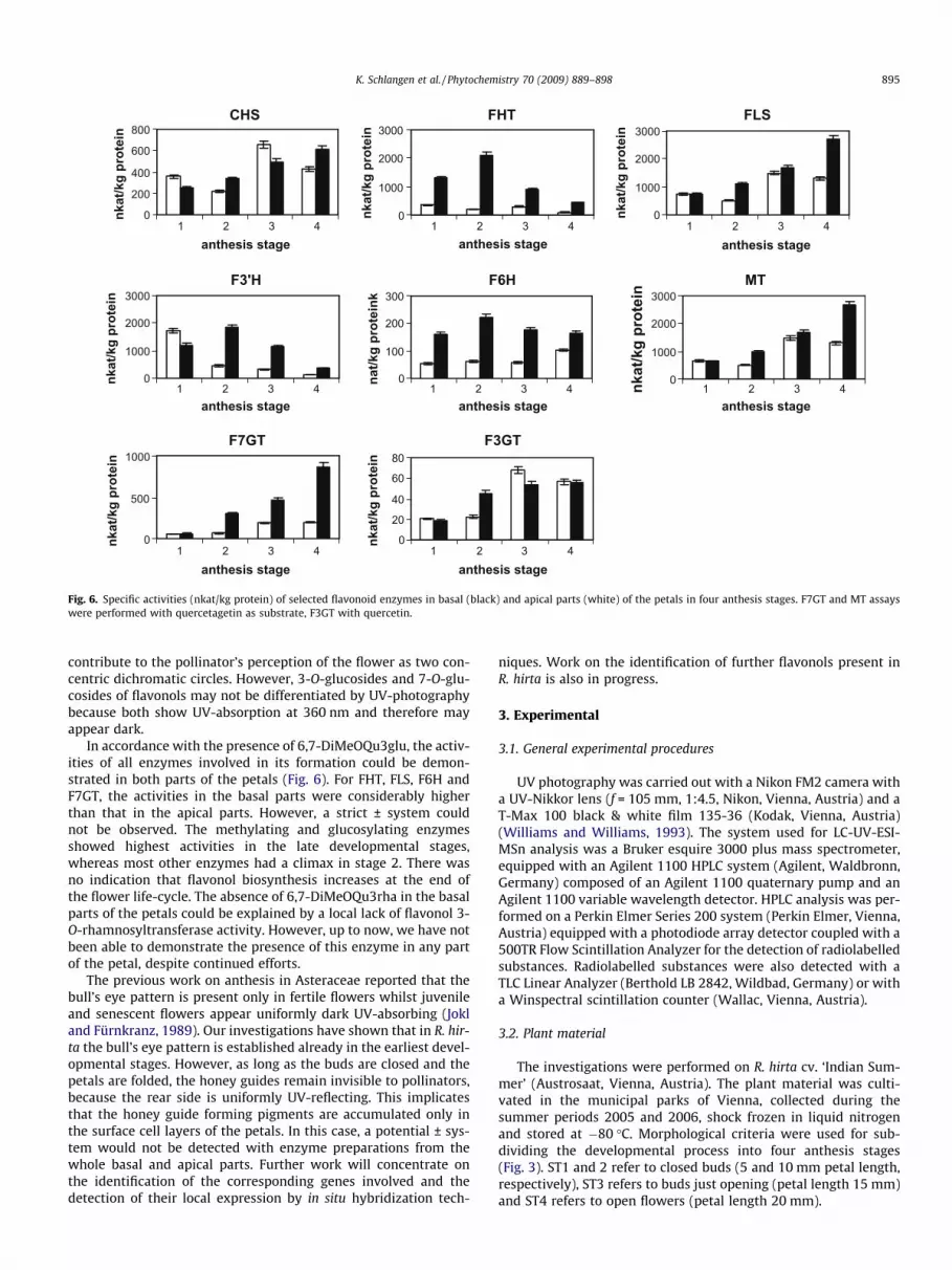
Fig. 6. Specific activities (nkat/kg protein) of selected flavonoid enzymes in basal (black) and apical parts (white) of the petals in four anthesis stages. F7GT and MT assayswere performed with quercetagetin as substrate, F3GT with quercetin.
K. Schlangen et al. / Phytochemistry 70 (2009) 889–898 895
contribute to the pollinator’s perception of the flower as two con-centric dichromatic circles. However, 3-O-glucosides and 7-O-glu-cosides of flavonols may not be differentiated by UV-photographybecause both show UV-absorption at 360 nm and therefore mayappear dark.
In accordance with the presence of 6,7-DiMeOQu3glu, the activ-ities of all enzymes involved in its formation could be demon-strated in both parts of the petals (Fig. 6). For FHT, FLS, F6H andF7GT, the activities in the basal parts were considerably higherthan that in the apical parts. However, a strict ± system couldnot be observed. The methylating and glucosylating enzymesshowed highest activities in the late developmental stages,whereas most other enzymes had a climax in stage 2. There wasno indication that flavonol biosynthesis increases at the end ofthe flower life-cycle. The absence of 6,7-DiMeOQu3rha in the basalparts of the petals could be explained by a local lack of flavonol 3-O-rhamnosyltransferase activity. However, up to now, we have notbeen able to demonstrate the presence of this enzyme in any partof the petal, despite continued efforts.
The previous work on anthesis in Asteraceae reported that thebull’s eye pattern is present only in fertile flowers whilst juvenileand senescent flowers appear uniformly dark UV-absorbing (Jokland Fürnkranz, 1989). Our investigations have shown that in R. hir-ta the bull’s eye pattern is established already in the earliest devel-opmental stages. However, as long as the buds are closed and thepetals are folded, the honey guides remain invisible to pollinators,because the rear side is uniformly UV-reflecting. This implicatesthat the honey guide forming pigments are accumulated only inthe surface cell layers of the petals. In this case, a potential ± sys-tem would not be detected with enzyme preparations from thewhole basal and apical parts. Further work will concentrate onthe identification of the corresponding genes involved and thedetection of their local expression by in situ hybridization tech-
niques. Work on the identification of further flavonols present inR. hirta is also in progress.
3. Experimental
3.1. General experimental procedures
UV photography was carried out with a Nikon FM2 camera witha UV-Nikkor lens (f = 105 mm, 1:4.5, Nikon, Vienna, Austria) and aT-Max 100 black & white film 135-36 (Kodak, Vienna, Austria)(Williams and Williams, 1993). The system used for LC-UV-ESI-MSn analysis was a Bruker esquire 3000 plus mass spectrometer,equipped with an Agilent 1100 HPLC system (Agilent, Waldbronn,Germany) composed of an Agilent 1100 quaternary pump and anAgilent 1100 variable wavelength detector. HPLC analysis was per-formed on a Perkin Elmer Series 200 system (Perkin Elmer, Vienna,Austria) equipped with a photodiode array detector coupled with a500TR Flow Scintillation Analyzer for the detection of radiolabelledsubstances. Radiolabelled substances were also detected with aTLC Linear Analyzer (Berthold LB 2842, Wildbad, Germany) or witha Winspectral scintillation counter (Wallac, Vienna, Austria).
3.2. Plant material
The investigations were performed on R. hirta cv. ‘Indian Sum-mer’ (Austrosaat, Vienna, Austria). The plant material was culti-vated in the municipal parks of Vienna, collected during thesummer periods 2005 and 2006, shock frozen in liquid nitrogenand stored at �80 �C. Morphological criteria were used for sub-dividing the developmental process into four anthesis stages(Fig. 3). ST1 and 2 refer to closed buds (5 and 10 mm petal length,respectively), ST3 refers to buds just opening (petal length 15 mm)and ST4 refers to open flowers (petal length 20 mm).
CHS
O
OOH
OHOH
HOO
O
HO
OH
OH
F3’H
O
O
HO
OH
OH
OH
Kaempferol
F3’H F6H
R1: Glu, R=H: Qu7glu R1=H, R=Glu: Qu3glu
F6MTO
O
HO
OH
OH
OH
OH
MeO
Patuletin
F7GT F7GT
Quercetagetin 7glu
F6MTF6H
Patulitrin
O
OOH
OH
OH
OH
MeO
GluO
F7MTO
OOH
OH
OH
OH
MeO
MeO
Eupatoletin
F3GT F7GT F3GT F7GT
O
OOH
OH
OR
R1O
R1: Glu, R=H: Km7glu R1=H, R=Glu: Km3glu
O
OOH
OHOH
MeO
MeO ORha
Eupatolin
O
OOH
OHOH
OGlu
MeO
MeO
Eupatoletin 3glu
F3GT FLS F3GT F3GT
O
OOH
OH
HO
oH
Dihydrokaempferol
FLS
Dihydroquercetin
O
OOH
OH
HO
OH
OH
F3’H
FHT
Eriodictyol Naringenin
FHT
F6H
F6H
O
O
HO
OH
OH
OH
OH
Quercetin
O
OOH
OHOH
OR
R1O O
OOH
OH
OH
OH
GluO
HO
O
O
HO
OH
OH
OH
OH
HO
Quercetagetin
Fig. 7. Biochemical pathway leading to the flavonols in R. hirta.
896 K. Schlangen et al. / Phytochemistry 70 (2009) 889–898
3.3. Chemicals
UDP-D-[U-14C]glucose (12.0 GBq/mmol), S-adenosyl-L-[methyl-14C]methionine (2.15 GBq/mmol) and [2-14C]malonyl-coenzyme A (2 GBq/mmol) were purchased from GE Healthcare(Freiburg, Germany). Non-labelled UDPG, SAM and malonyl-CoA(all Sigma-Aldrich, Vienna, Austria), respectively, were added totheir corresponding [14C]compounds to achieve the specific activi-ties indicated in the enzyme assay section. [14C]Flavonoids wereprepared as previously described (Halbwirth et al., 2004, 2006).Km, Km3glu, Km7glu, Qu, Qu3glu, Quag and Quag7glu, were pur-chased from Extrasynthese (Genay, France), 7-MeQu and 3-MeQufrom Fluka (Vienna, Austria) and Qu7glu, 3,7-DiMeQu, 6-MeOQu,and 6-MeOQu 7glu from Apin Chemicals (Abingdon, UK). 6,7-Di-MeOQu, 6,7-DiMeOQu3rha, and 6,7-DiMeOKm3rha were from
Table 1(Addition to Fig. 4). Maximal UV reflectance (%) of apical and basal petal parts and the coranthesis stages.
Anthesis stage 1
Frontal sideApex Max. UV-reflectance (%) 14.3 ± 1.0
wavelength number 367.3Base Max. UV-reflectance (%) 13.5 ± 2.6
wavelength number 367.3Rear sideApex Max. UV-reflectance (%) 24.3 ± 2.2
Wavelength number 367.3Base Max. UV-reflectance (%) 25.7 ± 4.0
Wavelength number 367.3
the laboratory collection of M. Luczkiewicz. 6-OHKm, 6-MeOKmand Quag3glu came from E. Wollenweber (TU Darmstadt, Ger-many). The cytochrome P450 reductase-specific antiserum fromJerusalem artichoke was a gift from F. Durst and I. Benveniste(CNRS, Strasbourg, France). NADPH, cytochrome c, glucose oxidaseand catalase were obtained from Sigma (Vienna, Austria). Cyto-chrome P450-specific inhibitors were a gift from W. Rademacher(BASF, Limburgerhof, Germany).
3.4. Reflectance spectroscopy
The reflectance curves of the flower petals were obtained in thespectral region between 300 and 700 nm as the fraction of the lightspectrum reflected by the sample in relation to the light spectrumreflected by an ideally white, diffuse reflecting surface. The illumi-
responding wavelength positions for the front side and the rear side in four different
2 3 4
13.0 ± 1.0 15.5 ± 2.1 16.2 ± 1.8367.3 356.6 356.611.8 ± 1.8 10.0 ± 1.1 8.5 ± 0.7367.3 356.6 356.6
19.1 ± 2.3 21.5 ± 2.0 19.5 ± 1.5367.3 374.4 372.021.1 ± 5.5 20.3 ± 5.3 24.0 ± 3.4367.3 374.4 372.0
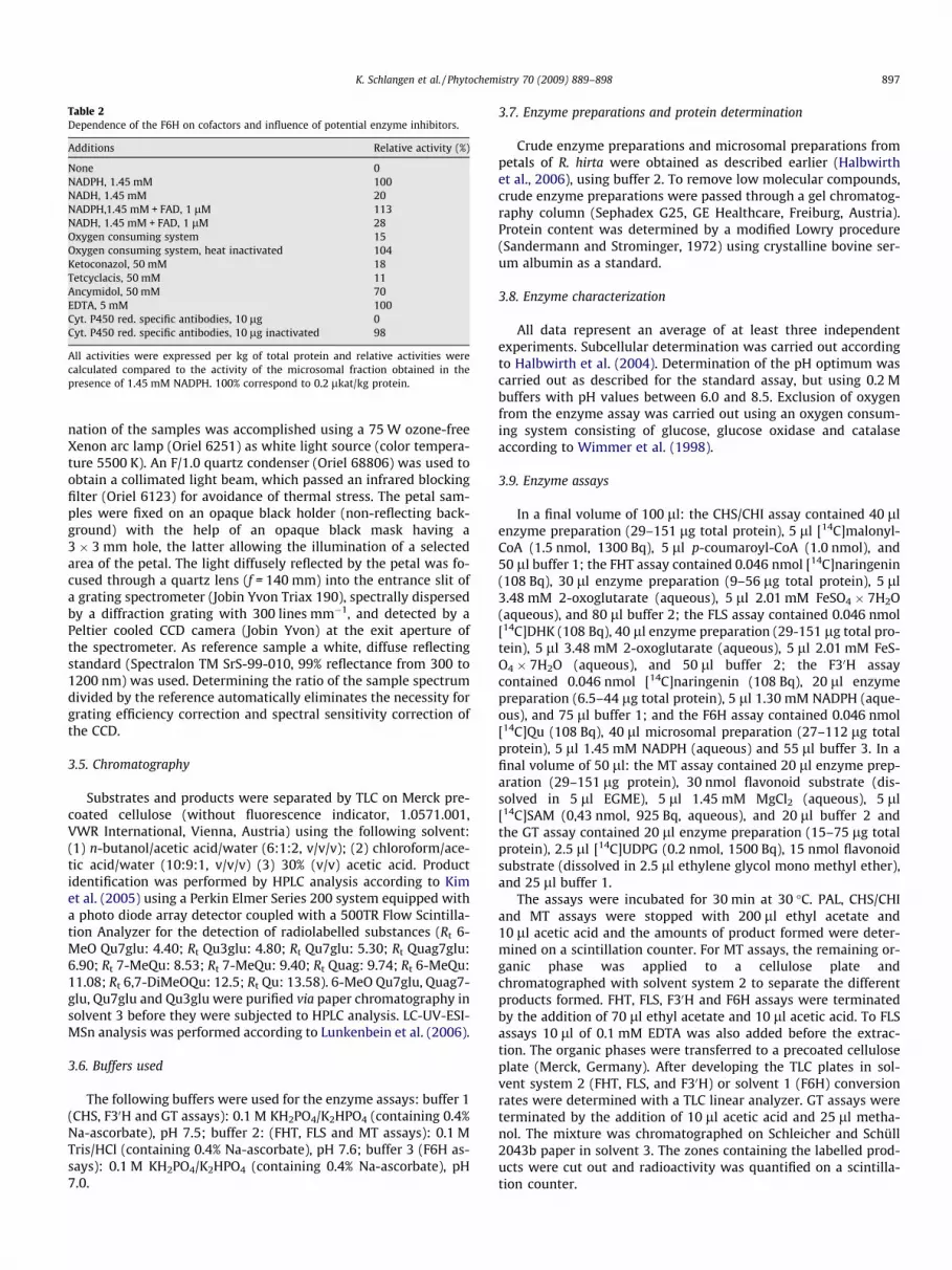
Table 2Dependence of the F6H on cofactors and influence of potential enzyme inhibitors.
Additions Relative activity (%)
None 0NADPH, 1.45 mM 100NADH, 1.45 mM 20NADPH,1.45 mM + FAD, 1 lM 113NADH, 1.45 mM + FAD, 1 lM 28Oxygen consuming system 15Oxygen consuming system, heat inactivated 104Ketoconazol, 50 mM 18Tetcyclacis, 50 mM 11Ancymidol, 50 mM 70EDTA, 5 mM 100Cyt. P450 red. specific antibodies, 10 lg 0Cyt. P450 red. specific antibodies, 10 lg inactivated 98
All activities were expressed per kg of total protein and relative activities werecalculated compared to the activity of the microsomal fraction obtained in thepresence of 1.45 mM NADPH. 100% correspond to 0.2 lkat/kg protein.
K. Schlangen et al. / Phytochemistry 70 (2009) 889–898 897
nation of the samples was accomplished using a 75 W ozone-freeXenon arc lamp (Oriel 6251) as white light source (color tempera-ture 5500 K). An F/1.0 quartz condenser (Oriel 68806) was used toobtain a collimated light beam, which passed an infrared blockingfilter (Oriel 6123) for avoidance of thermal stress. The petal sam-ples were fixed on an opaque black holder (non-reflecting back-ground) with the help of an opaque black mask having a3 � 3 mm hole, the latter allowing the illumination of a selectedarea of the petal. The light diffusely reflected by the petal was fo-cused through a quartz lens (f = 140 mm) into the entrance slit ofa grating spectrometer (Jobin Yvon Triax 190), spectrally dispersedby a diffraction grating with 300 lines mm�1, and detected by aPeltier cooled CCD camera (Jobin Yvon) at the exit aperture ofthe spectrometer. As reference sample a white, diffuse reflectingstandard (Spectralon TM SrS-99-010, 99% reflectance from 300 to1200 nm) was used. Determining the ratio of the sample spectrumdivided by the reference automatically eliminates the necessity forgrating efficiency correction and spectral sensitivity correction ofthe CCD.
3.5. Chromatography
Substrates and products were separated by TLC on Merck pre-coated cellulose (without fluorescence indicator, 1.0571.001,VWR International, Vienna, Austria) using the following solvent:(1) n-butanol/acetic acid/water (6:1:2, v/v/v); (2) chloroform/ace-tic acid/water (10:9:1, v/v/v) (3) 30% (v/v) acetic acid. Productidentification was performed by HPLC analysis according to Kimet al. (2005) using a Perkin Elmer Series 200 system equipped witha photo diode array detector coupled with a 500TR Flow Scintilla-tion Analyzer for the detection of radiolabelled substances (Rt 6-MeO Qu7glu: 4.40; Rt Qu3glu: 4.80; Rt Qu7glu: 5.30; Rt Quag7glu:6.90; Rt 7-MeQu: 8.53; Rt 7-MeQu: 9.40; Rt Quag: 9.74; Rt 6-MeQu:11.08; Rt 6,7-DiMeOQu: 12.5; Rt Qu: 13.58). 6-MeO Qu7glu, Quag7-glu, Qu7glu and Qu3glu were purified via paper chromatography insolvent 3 before they were subjected to HPLC analysis. LC-UV-ESI-MSn analysis was performed according to Lunkenbein et al. (2006).
3.6. Buffers used
The following buffers were used for the enzyme assays: buffer 1(CHS, F30H and GT assays): 0.1 M KH2PO4/K2HPO4 (containing 0.4%Na-ascorbate), pH 7.5; buffer 2: (FHT, FLS and MT assays): 0.1 MTris/HCl (containing 0.4% Na-ascorbate), pH 7.6; buffer 3 (F6H as-says): 0.1 M KH2PO4/K2HPO4 (containing 0.4% Na-ascorbate), pH7.0.
3.7. Enzyme preparations and protein determination
Crude enzyme preparations and microsomal preparations frompetals of R. hirta were obtained as described earlier (Halbwirthet al., 2006), using buffer 2. To remove low molecular compounds,crude enzyme preparations were passed through a gel chromatog-raphy column (Sephadex G25, GE Healthcare, Freiburg, Austria).Protein content was determined by a modified Lowry procedure(Sandermann and Strominger, 1972) using crystalline bovine ser-um albumin as a standard.
3.8. Enzyme characterization
All data represent an average of at least three independentexperiments. Subcellular determination was carried out accordingto Halbwirth et al. (2004). Determination of the pH optimum wascarried out as described for the standard assay, but using 0.2 Mbuffers with pH values between 6.0 and 8.5. Exclusion of oxygenfrom the enzyme assay was carried out using an oxygen consum-ing system consisting of glucose, glucose oxidase and catalaseaccording to Wimmer et al. (1998).
3.9. Enzyme assays
In a final volume of 100 ll: the CHS/CHI assay contained 40 llenzyme preparation (29–151 lg total protein), 5 ll [14C]malonyl-CoA (1.5 nmol, 1300 Bq), 5 ll p-coumaroyl-CoA (1.0 nmol), and50 ll buffer 1; the FHT assay contained 0.046 nmol [14C]naringenin(108 Bq), 30 ll enzyme preparation (9–56 lg total protein), 5 ll3.48 mM 2-oxoglutarate (aqueous), 5 ll 2.01 mM FeSO4 � 7H2O(aqueous), and 80 ll buffer 2; the FLS assay contained 0.046 nmol[14C]DHK (108 Bq), 40 ll enzyme preparation (29-151 lg total pro-tein), 5 ll 3.48 mM 2-oxoglutarate (aqueous), 5 ll 2.01 mM FeS-O4 � 7H2O (aqueous), and 50 ll buffer 2; the F30H assaycontained 0.046 nmol [14C]naringenin (108 Bq), 20 ll enzymepreparation (6.5–44 lg total protein), 5 ll 1.30 mM NADPH (aque-ous), and 75 ll buffer 1; and the F6H assay contained 0.046 nmol[14C]Qu (108 Bq), 40 ll microsomal preparation (27–112 lg totalprotein), 5 ll 1.45 mM NADPH (aqueous) and 55 ll buffer 3. In afinal volume of 50 ll: the MT assay contained 20 ll enzyme prep-aration (29–151 lg protein), 30 nmol flavonoid substrate (dis-solved in 5 ll EGME), 5 ll 1.45 mM MgCl2 (aqueous), 5 ll[14C]SAM (0,43 nmol, 925 Bq, aqueous), and 20 ll buffer 2 andthe GT assay contained 20 ll enzyme preparation (15–75 lg totalprotein), 2.5 ll [14C]UDPG (0.2 nmol, 1500 Bq), 15 nmol flavonoidsubstrate (dissolved in 2.5 ll ethylene glycol mono methyl ether),and 25 ll buffer 1.
The assays were incubated for 30 min at 30 �C. PAL, CHS/CHIand MT assays were stopped with 200 ll ethyl acetate and10 ll acetic acid and the amounts of product formed were deter-mined on a scintillation counter. For MT assays, the remaining or-ganic phase was applied to a cellulose plate andchromatographed with solvent system 2 to separate the differentproducts formed. FHT, FLS, F30H and F6H assays were terminatedby the addition of 70 ll ethyl acetate and 10 ll acetic acid. To FLSassays 10 ll of 0.1 mM EDTA was also added before the extrac-tion. The organic phases were transferred to a precoated celluloseplate (Merck, Germany). After developing the TLC plates in sol-vent system 2 (FHT, FLS, and F30H) or solvent 1 (F6H) conversionrates were determined with a TLC linear analyzer. GT assays wereterminated by the addition of 10 ll acetic acid and 25 ll metha-nol. The mixture was chromatographed on Schleicher and Schüll2043b paper in solvent 3. The zones containing the labelled prod-ucts were cut out and radioactivity was quantified on a scintilla-tion counter.
898 K. Schlangen et al. / Phytochemistry 70 (2009) 889–898
Acknowledgements
These investigations were supported by a grant from the Aus-trian FWF (Project P-17629-B03). H. Halbwirth acknowledges hergrant from the Austrian FWF (Project V18-B03). We are indebtedto E. Wollenweber (TU Darmstadt, Germany) for providing us witha multitude of reference compounds. We are grateful to F. Durstand I. Benveniste (CNRS, Strasbourg, France) for their gift of cyto-chrome P450 reductase-specific antiserum, to Jenny Greenham(University of Reading, UK) for kindly providing us with referencecompounds and to S. Jokl (University of Salzburg, Austria) for gen-erously supplying us with the UV-camera system.
References
Abrahamson, W.G., McCrea, K.D., 1977. Ultraviolet light reflection and absorptionpatterns in populations of Rudbeckia. Rhodora 72, 269–277.
Anzellotti, D., Ibrahim, R.K., 2000. Novel flavonol 2-oxoglutarate dependentdioxygenase: affinity purification, characterization, and kinetic properties.Arch. Biochem. Biophys. 382, 161–172.
Anzellotti, D., Ibrahim, R.K., 2004. Molecular characterization and functionalexpression of flavonol 6-hydroxylase. BMC Plant Biol. 4, 20–26.
Benveniste, I., Lesot, A., Hasenfratz, M.P., Durst, F., 1989. Immunochemicalcharacterization of NADPH-cytochrome P-450 reductase from Jerusalemartichoke and other higher plants. Biochem. J. 259, 847–853.
Burr, B., Rosen, D., Barthlott, W., 1995. Untersuchungen zur Ultraviolettreflexionvon Angiospermenblüten III. Dilleniidae und Asteridae. Trop. u. Subtrop.Pflanzenwelt 93, 1–185.
Cisowski, W., Dembinska-Migas, W., Luczkiewicz, M., 1993. Flavonoids from theRudbeckia hirta L. herb. Pol. J. Chem. 67, 829–836.
Chittka, L., Kevan, P.G., 2005. Flower colour as advertisement. In: Dafni, A., Kevan,P.G., Husband, B.C. (Eds.), Practical Pollination Biology. Enviroquest Ltd.,Cambridge, ON, Canada, pp. 157–196.
Daumer, K., 1956. Reizmetrische Untersuchungen des Farbsehens der Biene. Z.Vergl. Physiol. 38, 413–478.
Daumer, K., 1958. Blumenfarben, wie sie die Bienen sehen. Z. Vergl. Physiol. 41, 49–110.
Dyer, A.G., 1996. The reflection of near ultraviolet radiation from flowers ofAustralian native plants. Aust. J. Bot. 44, 473–488.
Dyer, A.G., Muir, L.L., Muntz, W.R.A., 2004. A calibrated gray scale for forensicultraviolet photography. J. Forensic Sci. 49, 1056.
Eisner, T., Silberglied, R.E., Aneshansley, D., Carrel, J.F., Howlans, H.C., 1969.Ultraviolet video-viewing: the television camera as an insect eye. Science166, 1172–1174.
Forkmann, G., Heller, W., 1999. Biosynthesis of flavonoids. In: Barton, D., Nakanishi,K., Meth-Cohn, O. (Eds.), Comprehensive Natural Products Chemistry, vol. 1.Elsevier, Oxford, pp. 713–748.
Galsterer, S., Musso, M., Asenbaum, A., Fürnkranz, D., 1999. Reflectancemeasurements of glossy petals of Ranunculus lingua (Ranunculaceae) and ofnon-glossy petals of Heliopsis helianthoides (Asteraceae). Plant Biol. 1, 70–78.
Halbwirth, H., Forkmann, G., Stich, K., 2004. The A-ring specific hydroxylation offlavonols in position 6 in Tagetes sp. is catalyzed by a cytochrome P450dependent monooxygenase. Plant Sci. 167, 129–135.
Halbwirth, H., Puhl, I., Haas, U., Jezik, K., Treutter, D., Stich, K., 2006. Two-phaseflavonoid formation in developing strawberry (Fragaria � ananassa) fruit. J.Agric. Food Chem. 54, 1479–1485.
Harborne, J.B., 1967. Comparative Biochemistry of the Flavonoids. Academic Press,London.
Harborne, J.B., 1976. Functions of flavonoids in plants. In: Goodwin, T.W. (Ed.),Chemistry and Biochemistry of Plant Pigments, vol. 1. Academic Press, London,pp. 736–778.
Harborne, J.B., 1988. The flavonoids: recent advances. In: Goodwin, T.H. (Ed.), PlantPigments. Academic Press, London, pp. 299–344.
Harborne, J.B., 1993. Introduction to Ecological Biochemistry. Academic Press,London.
Ibrahim, R.K., De Luca, V., Khouri, H., Latchinian, L., Brisson, L., Charest, P.M., 1987.Enzymology and compartmentation of polymethylated flavonol glucosides inChrysosplenium americanum. Phytochemistry 26, 1237–1245.
Indsto, J.O., Weston, P.H., Clements, M.A., Dyer, A.G., Batley, M., Whelan, R.J., 2006.Pollination of Diuris maculate (Orchidaceae) by male Trichocolletes venustusbees. Aust. J. Bot. 54, 669–679.
Indtso, J.O., Weston, P.H., Clements, M.A., Dyer, A.G., Batley, M., Whelan, R.J., 2007.Diuris alba (Orchidaceae) is pollinated by small bees and wasps and showsevidence of weak pea flower mimicry. Aust. J. Bot. 55, 628–634.
Jokl, S., Fürnkranz, D., 1989. Antheseabhängige UV-Muster in Blütenständen vonAsteraceen. Plant Syst. Evol. 165, 91–94.
Jokl, S., Galsterer, S., Musso, M., Fürnkranz, D., Asenbaum, A., 1999. Ray flowers ofHeliopsis helianthoides (Asteraceae) and their UV-reflectance during anthesis.Bot. Jahrb. Syst. 121, 279–287.
Kevan, P.G., Mulligan, G.A., 1973. Floral characteristics attracting insects. Can. J. Bot.51, 1939–1952.
Kevan, P.G., Grainger, N.D., Mulligan, G.A., Robertson, A.R., 1973. A gray-scale formeasuring reflectance and colour in the insect and human visual spectra.Ecology 54, 924–926.
Kevan, P.G., 1979. Vegetation and floral colors revealed by ultraviolet light:interpretational difficulties for functional significance. Am. J. Bot. 66, 749–751.
Kevan, P.G., Chittka, L., Dyer, A.G., 2001. Limits to the salience of ultraviolet: lessonsfrom colour vision in bees and birds. J. Exp. Biol. 204, 2571–2580.
Kim, D.H., Kim, B.G., Lee, Y., Ryu, J.Y., Lim, Y., Hur, H.G., Ahn, J.H., 2005. Regiospecificmethylation of naringenin to ponciretin by soybean O-methyltransferaseexpressed in Escherichia coli. J. Biotechnol. 119, 155–162.
Langanger, M., Jokl, S., Musso, M., 2000. UV-reflectance in flowers of Nymphea alba L.and Nuphar lutea (L.) Sm. (Nymphaeaceae). Aquat. Bot. 67, 13–21.
Latunde-Dada, A.O., Cabello-Hurtado, F., Czittrich, N., Didierjean, L., Schopfer, C.,Hertkorn, N., Werck-Reichhart, D., Ebel, J., 2001. Flavonoid 6-hydroxylase fromsoybean (Glycine max L.), a novel plant P-450 monooxygenase. J. Biol. Chem.276, 1688–1695.
Lunkenbein, S., Bellido, M., Aharoni, A., Salentijn, E., Kaldenhoff, R., Coiner, H.,Munoz-Blanco, J., Schwab, W., 2006. Cinnamate metabolism in ripening fruit.Characterization of a UDP-glucose:cinnamate glucosyltransferase fromstrawberry. Plant Physiol. 140, 1047–1058.
McCrea, K.D., Levy, M., 1983. Photographic visualization of floral colors as perceivedby honeybee pollinators. Am. J. Bot. 70, 369–375.
Nielsen, J.S., Møller, B.L., 1999. Biosynthesis of cyanogenic glucosides in Triglochinmaritima and the involvement of cytochrome P450 enzymes. Arch. Biochem.Biophys. 368, 121–130.
Silberglied, R.E., 1979. Communication in the ultraviolet. Annu. Rev. Ecol. Syst. 10,373–398.
Stich, K., Halbwirth, H., Wurst, F., Forkmann, G., 1997. UDP-glucose: flavonol 7-O-glucosyltransferase activity in flower extracts of Chrysanthemum segetum. Z.Naturforsch. C 52, 153–158.
Tarpo, E., 1969. Flavones of Tagetes patula. Farmacia 93, 98.Thompson, W.R., Meinwald, J., Aneshansley, D., Eisner, T., 1972. Flavonols:
responsible for ultraviolet absorption in nectar guide of flower. Science 177,528–530.
Williams, A.R., Williams, G.F., 1993. The invisible image – a tutorial on photographywith invisible radiation. Part 1. Introduction and reflected ultraviolettechniques. J. Biol. Photogr. 61, 115–132.
Wimmer, G., Halbwirth, H., Wurst, F., Forkmann, G., Stich, K., 1998. Enzymatichydroxylation of 60-deoxychalcones with protein preparations from petals ofDahlia variabilis. Phytochemistry 47, 1013–1016.
Related Documents