FORMATION OF PEARL-NECKLACE MONOMORPHIC G-QUADRUPLEXES IN THE HUMAN CEB25 MINISATELLITE Michael Adrian 1, *, Samir Amrane 1,2 , Brahim Heddi 1 , Alexandre Serero 3 , William Qinghao Chen 1 , Alain Nicolas 3 , Jean-Louis Mergny 2 and Anh Tuân Phan 1 1 School of Physical and Mathematical Sciences, Nanyang Technological University, 637371 Singapore, 2 INSERM, U869, Bordeaux University, European Institute of Chemistry and Biology, 33600 Pessac, France and 3 Institut Curie, Centre de Recherche, UMR3244 CNRS, Université Pierre et Marie Curie, 75248 Paris, France *Correspondence to: [email protected] ABSTRACT CEB25 is a human minisatellite locus, com- posed of slightly polymorphic 52-nt tandem repeats. We report on the structure of a pro- peller-type parallel-stranded G-quadruplex formed by the conserved 26-nt G-rich frag- ment of the CEB25 motif. Further, we demon- strate that such a monomorphic structure is formed within longer sequence contexts. INTRODUCTION G-quadruplex is a four-stranded nucleic acid structure formed by G-rich DNA or RNA strands, comprising few stacking G-tetrads, each of which being a planar association of four guanines held together by eight hydrogen bonds and stabilized by cations such as K + . These structures show a variety of G-quadruplex folding topologies with respect to strand orientations, glycosidic confor- mations of guanine bases, and intervening loops [1]. The G-quadruplex formation on minisatellite DNA was previously found to stimulate genomic instability in yeast [2]. The G-quadruplex struc- ture adopted by the human CEB25 minisatellite sequence is under the scope of this work. RESULTS The G-quadruplex structure formed by the 26- nt G-rich sequence d[AAGGGTGGGTGTAAGT GTGGGTGGGT] (26CEB) in K + solution was determined on the basis of NMR distance re- straints. The parallel-stranded structure has three double-chain-reversal loops: the first and third loops, each consists of a single nucleotide, while the central loop consists of nine nucleotides. This long loop is anchored to the 5’ end of the se- quence by an A•T Watson-Crick and a potential G•A non-canonical base pairs, contributing to the stability of the overall structure, as measured by an increase of about 17 kcal/mol in enthalpy or 6 °C in melting temperature. The formation of G- quadruplex blocks at different locations of long CEB25 sequences was revealed by NMR spec- troscopy and could be visualized by means of AFM imaging. Based on these results, a ‘pearl- necklace’ model of stable G-quadruplexes inter- connected by non-quadruplex-forming sequences on a long single-stranded CEB25 repeats is pro- posed (Figure 1) [3]. REFERENCES 1. Phan, A.T. FEBS J., 2010, 277, 1107-1117. 2. Lopes, J., Piazza, A., Bermejo, R., Kriegsman, B., Colosio, A., Teulade-Fichou, M.P., Foiani, M., Nicolas, A. EMBO J., 2011, 30, 4033- 4046. 3. Amrane, S., Adrian, M., Heddi, B., Serero, A., Nicolas, A., Mergny, J.L., Phan, A.T. J. Am. Chem. Soc., 2012, 134, 5807-5816. Figure 1. CEB25 minisatellite ‘pearl-necklace’ model. 188

Welcome message from author
This document is posted to help you gain knowledge. Please leave a comment to let me know what you think about it! Share it to your friends and learn new things together.
Transcript
FORMATION OF PEARL-NECKLACE MONOMORPHIC G-QUADRUPLEXES IN THE HUMAN
CEB25 MINISATELLITE
Michael Adrian1,*, Samir Amrane
1,2, Brahim Heddi
1, Alexandre Serero
3, William Qinghao Chen
1,
Alain Nicolas3, Jean-Louis Mergny
2 and Anh Tuân Phan
1
1School of Physical and Mathematical Sciences, Nanyang Technological University, 637371 Singapore,
2INSERM, U869, Bordeaux University, European Institute of Chemistry and Biology, 33600 Pessac, France and 3Institut Curie, Centre de Recherche, UMR3244 CNRS, Université Pierre et Marie Curie, 75248 Paris, France
*Correspondence to: [email protected]
ABSTRACT
CEB25 is a human minisatellite locus, com-
posed of slightly polymorphic 52-nt tandem
repeats. We report on the structure of a pro-
peller-type parallel-stranded G-quadruplex
formed by the conserved 26-nt G-rich frag-
ment of the CEB25 motif. Further, we demon-
strate that such a monomorphic structure is
formed within longer sequence contexts.
INTRODUCTION
G-quadruplex is a four-stranded nucleic acid
structure formed by G-rich DNA or RNA strands,
comprising few stacking G-tetrads, each of which
being a planar association of four guanines held
together by eight hydrogen bonds and stabilized
by cations such as K+. These structures show a
variety of G-quadruplex folding topologies with
respect to strand orientations, glycosidic confor-
mations of guanine bases, and intervening loops
[1]. The G-quadruplex formation on minisatellite
DNA was previously found to stimulate genomic
instability in yeast [2]. The G-quadruplex struc-
ture adopted by the human CEB25 minisatellite
sequence is under the scope of this work.
RESULTS
The G-quadruplex structure formed by the 26-
nt G-rich sequence d[AAGGGTGGGTGTAAGT
GTGGGTGGGT] (26CEB) in K+
solution was
determined on the basis of NMR distance re-
straints. The parallel-stranded structure has three
double-chain-reversal loops: the first and third
loops, each consists of a single nucleotide, while
the central loop consists of nine nucleotides. This
long loop is anchored to the 5’ end of the se-
quence by an A•T Watson-Crick and a potential
G•A non-canonical base pairs, contributing to the
stability of the overall structure, as measured by
an increase of about 17 kcal/mol in enthalpy or
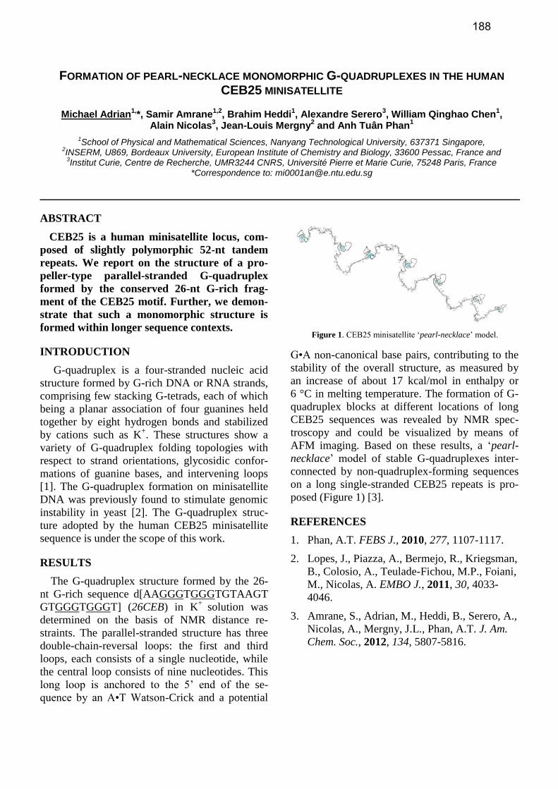
6 °C in melting temperature. The formation of G-
quadruplex blocks at different locations of long
CEB25 sequences was revealed by NMR spec-
troscopy and could be visualized by means of
AFM imaging. Based on these results, a ‘pearl-
necklace’ model of stable G-quadruplexes inter-
connected by non-quadruplex-forming sequences
on a long single-stranded CEB25 repeats is pro-
posed (Figure 1) [3].
REFERENCES
1. Phan, A.T. FEBS J., 2010, 277, 1107-1117.
2. Lopes, J., Piazza, A., Bermejo, R., Kriegsman,
B., Colosio, A., Teulade-Fichou, M.P., Foiani,
M., Nicolas, A. EMBO J., 2011, 30, 4033-
4046.
3. Amrane, S., Adrian, M., Heddi, B., Serero, A.,
Nicolas, A., Mergny, J.L., Phan, A.T. J. Am.
Chem. Soc., 2012, 134, 5807-5816.
Figure 1. CEB25 minisatellite ‘pearl-necklace’ model.
188

CATALYTIC DNAS THAT HARVEST VIOLET LIGHT TO REPAIR THYMINE DIMERS WITHIN A
SUBSTRATE STRAND
Adam Barlev1 and Dipankar Sen
1*
1Simon Fraser University, 8888 University Drive, Burnaby BC, Canada, *Correspondence to: [email protected]
ABSTRACT
We have isolated catalytic DNAs (DNAzymes), that are able to harness light of 300-310 nm wavelength to photo-reactivate cancer-causing thymine dimer lesions in sin-gle-stranded DNA. Recently, we have succeeded in gen-erating pterin-containing DNAzyme mutants, that are able to use visible, rather than UV, light for their DNA repair activity.
INTRODUCTION, RESULTS AND DISCUSSION
In vitro selection from a random-sequence DNA library
was used to investigate whether the nucleic acids (DNA or
RNA) are capable of catalyzing photochemical reactions.
The reaction chosen was photoreactivation of thymine cy-
clobutane dimers in DNA, and the wavelengths chosen were
> 300 nm, at the edge of DNA’s absorption spectrum. A 42-
nucleotide single-stranded DNA sequence, UV1C, was iso-
lated and found to repair a single-stranded DNA substrate
containing a thymine dimer efficiently (kcat/kuncat =
2.5x10⁴) [1]. It thus resembled in some respects naturally
occurring photolyase enzymes. Mechanistic investigation of
UV1C indicated that its catalytic role exceeded the mere
positioning of the substrate in a conformation favorable for
photoreactivation. UV1C was therefore a DNAzyme, with
optimal catalytic activity at 305 nm light. A higher-order
DNA fold, a G-quadruplex, formed by guanine bases within
the DNAzyme, was implicated as serving as a light-
harvesting antenna, with photoreactivation of the thymine
dimer proceeding likely via electron donation from an excit-
ed guanine base [1-3].
Recently, we have shown that mutation of key single
guanine residues within UV1C with the pterin compound,
6MI (a guanine structural analogue), extends the action
spectrum of UV1C into the visible part of the spectrum [4].
The properties of this new ensemble of UV1C point mutants
reveal surprising features about the original UV1C, that it is
a multi-component and surprisingly adaptable catalyst. The
mutants fall into three distinct functional classes, which re-
pair the thymine dimer in different ways. In particular, the
interchangeable properties of no less than six of the G6MI
point mutants suggests a functional flexibility that can be
exploited in the future to create structurally robust and cata-
lytically efficient photolyase DNAzymes that may find utili-
ty in human medicine.
REFERENCES
1. D. Chinnapen; D. Sen, Proc. Natl. Acad. Sci. USA,
2004, 101, 65.
2. D. Chinnapen; D. Sen, J. Mol. Biol. 2007, 365, 1326.
3. G. Sekhon; D. Sen, Biochemistry 2009, 48, 6335.
4. A. Barlev; D. Sen, submitted.
Figure 1. The three functionally distinct classes of 6MI mutant substitutions and their positions in the proposed UV1C structure. Top left: These mutants maintain the same activity as wild-type UV1C. Top right: The 6 interchangeable quadruplex mutants which show activity in the UV-A and reduced activity in the UV-B. Bottom right: The versatile G23 position maintains the activity of wild-type UV1C in the UV-B but extends its activity through the UV-A, ex-tending all the way to the visible.
189

STRUCTURE AND DYNAMICS OF MODIFIED DEOXYOLIGONUCLEOTIDES
Dieter Buyst,1,2
* Bjorn Van Gasse1, Vicky Gheerardijn
2, Annemieke Madder
2 and José C. Martins
1
1NMR and Sructure Analysis Unit, Department of Organic Chemistry, UGent, Belgium
2Laboratory for Organic and Biomimetic Chemistry, Department of Organic Chemistry, UGent, Belgium
* Correspondence to: [email protected]
ABSTRACT
In the work presented here, both NMR and modelling
are used to elucidate the structure and dynamics of both
native and modified DNA structures.
INTRODUCTION, RESULTS AND DISCUSSION
There is considerable interest in the development of arti-
ficial catalysts that mimic enzymatic activity. One approach
in this challenging area of research is the use of nucleic acid
building blocks, whose base-pairs are equipped with func-
tional groups mimicking peptide chains. [1]
Our systems of
interest can be classified as hydrolase like DNAzymes, in-
cluding on one hand a 14mer DNA duplex in which a cata-
lytic triad resembling the active site of α-chymotrypsin is
introduced. Due to reasons of synthetic accessibility, these
modifications are incorporated on thymine bases (TSer
, THis
and TAsp
)., The second type of systems consist of esterase
DNAzymes, inspired by the synthetic enzyme of Baltzer and
coworkers[2]
where the introduction of multiple histidine
residues bearing imidazole functions can afford general ac-
id-base catalysed esterase activity. Here, the same unmodi-
fied DNA scaffold provides 7 possible locations for the in-
troduction of modified thymine bases bearing the required
imidazole mimicking functionality.
Furthermore, due to reasons of synthetic accessibility the
introduction of the amino acid mimicking functionalities is
currently limited to thymines, making the presence of a T•T
mismatch inside the DNA scaffold necessary in order to
obtain the required proximity between the functionalities. It
has been shown recently that the conformational behaviour
of this base pair is dependent on the type of neighbouring
base pairs [3],[4]
. Since its central role in the serine protease
mimic system, a more systematic in depth study regarding
dynamics of this non Watson-Crick base pair is desirable. In
the first stages, AMBER force field modelling methods are
used to get an in-depth understanding of the conformational
behaviour of the T-T mismatch present in the native DNA
scaffold. In order to validate the approach used, the model-
ling of this scaffold is compared to a system already de-
scribed in literature[4]
. In the second stage, these modelling
results will be compared with observations acquired via
NMR. In this context the preferential pairing modes of T•T
mismatches can be investigated by interpreting the imino-
methyl nOe’s of the mismatch.
In a first step towards the NMR study of fully modified
systems, a single THis
modified building block was intro-
duced on four different positions (T6, T7, T8 and T21) of
the DNA scaffold. This allows to make an assessment of the
impact of this non-natural modification on the structure and
stability of the system as a whole. Both UV-VIS and NMR-
measurements independently showed a significant increase
in the melting temperature compared to the native DNA
scaffold, attributed to an increase in stability. Additionally,
in a separate pH-study (poster B. Van Gasse) a big differ-
ence was observed in the pKa value of the T8His
with respect
to the three other modified systems. In order to further clari-
fy this peculiar behaviour, a complete NMR structure eluci-
dation has been performed, thus investigating the hypothesis
of the preferred orientation of the THis
modification inside
the major groove.
CONCLUSION
The methodology and knowledge gained here will prove
to be invaluable for the further guidance in the design and
performance of these new DNAzymes.
REFERENCES
1. Catry M., Madder A., Molecules, 2007, 12(1), 114-129.
2. Broo, K.S., et al., J. Am. Chem. Soc., 1997, 119(47),
11362-11372.
3. He, G.Y., C.K. Kwok, and S.L. Lam, Febs Letters,
2011, 585(24), 3953-3958
4. Gantchev, T.G. and D.J. Hunting, J. Mol. Mod., 2005,
11(2), 141-159.
Figuur 1: Mimicked active site on 14-mer DNA scaffold
5’ – GACCATHisTSerTHisGCAGCG – 3’
3’ – CTGGTA A TAspCGTCGC – 5’
190
STIMULI-RESPONSIVE PATTERNING OF BLOCK COPOLYMERS ON DNA NANOTUBES
Karina M. M. Carneiro,1* Graham D. Hamblin,
1 Georgios Rizis
1 and Hanadi F. Sleiman
1
1Department of Chemistry, McGill University, 801 Sherbrooke St. West, room 400, H2X 0B8, Montreal, Canada. *
Correspondence to: [email protected]
ABSTRACT
DNA nanotubes are an attractive class of materials
that can be assembled with precise control over their size,
shape, length and porosity, and can encapsulate and re-
lease materials in response to specific added molecules.
In parallel, block copolymer assemblies can provide bio-
compatibility, stability, nuclease resistance, the ability to
encapsulate small molecules, long-range assembly and
numerous additional functionalities that can be tuned
with subtle chemical modifications. Herein, we describe
a new class of hybrid materials in which block copoly-
mer assemblies are sequence-specifically and longitudi-
nally positioned on robust DNA nanotubes constructed
using rolling circle amplification.
INTRODUCTION, RESULTS AND
DISCUSSION, CONCLUSION
Hierarchical self-assembly is the driving force for the or-
ganization of all matter in nature; chemists have strived to
mimic nature by designing molecules that are able to self-
assemble though non-covalent forces in a predictable man-
ner, giving rise to the field of supramolecular chemistry.1
Nucleic acids have been proposed as a powerful building
block for advanced materials due to desirable properties
such as sequence programmability and specificity, mono-
dispersity, ease of synthesis and functionalization, and
abundance in nature.2 In particular, DNA nanotubes with
longitudinal control have emerged as promising scaffolds
for the precise patterning of moieties in three-dimensions
(3D) and as stimuli-responsive cargo carriers.3
With this in mind, we here construct a robust DNA nano-
tube architecture with a continuous DNA backbone, via roll-
ing circle amplification (RCA).4 RCA-nanotubes can be
easily manipulated due to the resilient, non-nicked nature of
their backbones in comparison to previous designs. In par-
allel, we conjugated a range of polymers to DNA strands
through copper-catalyzed azide alkyne Huisgen cycloaddi-
tion (‘click’ chemistry) or amide coupling reactions, and
incorporated onto DNA nanotubes.
To test the generality of our approach, we also created a
range of amphiphilic DNA-polymer conjugates that self-
assemble into micelles prior to incorporation onto the RCA-
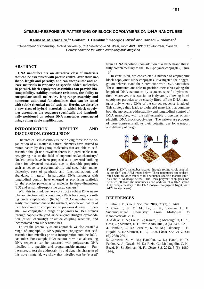
nanotubes. For example, RCA-nanotubes with an alternating
DNA sequence can be patterned with polystyrene-DNA
micelles in a specific, and programmable manner. Fur-
thermore, to test the addressability and dynamic character of
this novel material, we show that micelles can be ‘erased’
from a DNA nanotube upon addition of a DNA strand that is
fully complementary to the DNA-polymer conjugate (Figure
1).5
In conclusion, we constructed a number of amphiphilic
block copolymer-DNA conjugates, investigated their aggre-
gation behaviour and their interaction with DNA nanotubes.
These structures are able to position themselves along the
length of DNA nanotubes by sequence-specific hybridiza-
tion. Moreover, this association is dynamic, allowing block
copolymer particles to be cleanly lifted off the DNA nano-
tubes only when a DNA of the correct sequence is added.
This strategy thus leads to biohybrid materials that combine
both the molecular addressability and longitudinal control of
DNA nanotubes, with the self-assembly properties of am-
phiphilic DNA block copolymers. The write-erase property
of these constructs allows their potential use for transport
and delivery of cargo.
REFERENCES
1. Lehn, J. M., Chem. Soc. Rev. 2007, 36 (2), 151-60.
2. Carneiro, K. M. M.; Lo, P. K.; Sleiman, H. F.,
Supramolecular Chemistry: From Molecules to
Nanomaterials. 2011.
3. Aldaye, F. A.; Lo, P. K.; Karam, P.; McLaughlin, C. K.;
Cosa, G.; Sleiman, H. F., Nat. Nano 2009, 4 (6), 349-352.
4. Hamblin, G. D.; Carneiro, K. M. M.; Fakhoury, J. F.;
Bujold, K. E.; Sleiman, H. F., J. Am. Chem. Soc. 2012, 134
(6), 2888-2891.
5. Carneiro, K. M. M.; Hamblin, G. D.; Hanni, K. D.;
Fakhoury, J.; Nayak, M. K.; Rizis, G.; McLaughlin, C. K.;
Bazzi, H. S.; Sleiman, H. F., Chem. Sci. 2012, 3 (6), 1980-
1986.
Figure 1. DNA nanotubes created through rolling circle amplifi-cation (left) and AFM image below. These nanotubes can be deco-rated with polymer micelles in a sequence specific manner (mid-dle) and AFM image below. The DNA-polymer conjugates can be lifted off from the nanotubes upon addition of a DNA strand fully complementary to the DNA-polymer conjugates (right, with AFM image below).
191

Figure 1. Furan oxidation into reactive aldehydes
TOXICITY INSPIRED CROSSLINKING OF DNA TO ITS BINDING PROTEINS
Carrette, L.L.G.,1* Op de Beeck, M.,1 Morii, T.2 and Madder, A.1 1Laboratory for Organic and Biomimetic Chemistry, UGent, Krijgslaan 281, S4, 9000, Gent, Belgium and
2Laboratory for Biofunctional Science Kyoto University, Uji, Kyoto 611-0011 Japan
* Correspondence to: [email protected]
ABSTRACT
We here present a novel protein-DNA crosslinking
methodology with inducible reactivity based on specific
oxidation of a furan moiety. Positioning of the furan on
the nucleotide is crucial for obtaining high selectivity
and yield. In addition, the ability to specifically locate
and activate a suspected genotoxin within a protein-DNA
complex, will conceivably provide valuable knowledge
on the mode of action.
INTRODUCTION
Crosslinking between DNA and proteins is highly rele-vant both fundamentally, for the identification of their inter-actions, as well as practically, for a wide range of applica-tions from molecular assemblies to therapeutics. Several crosslinking approaches have been described for this pur-pose (1). Externally added bifunctional linkers like glutaral-dehyde, do not require any modification of the DNA or pro-tein, but lack selectivity. Therefore it is often more useful to modify either the DNA or the protein with a reactive group, preferably with inducible reactivity to avoid non-specific side reactions.
In our laboratory a crosslinking strategy was developed, inspired by the toxic liver metabolism of furan. Furan and analogues are oxidised in the liver by Cyt P450 enzymes to butene-dial or 4-oxo-butenals. These very reactive interme-diates quickly react with proximate nucleo-philes from proteins and DNA eliciting a toxic response (2).
RESULTS AND DISCUSSION
We have previously demonstrated the successful applica-tion of this methodology for DNA interstrand crosslinking, resulting in high crosslinking yields with unprecedented selectivity (3). We are now interested in the expansion of the methodology towards the very interesting protein-DNA interface. Design, synthesis and incorporation of alternative furan modified nucleotides into oligonucleotides allowed comparison of the reactivity and selectivity of this reaction, further illustrating the possibility to discriminate in reactivi-ty between the opposite oligonucleotide strand to form interstrand crosslinks and a binding protein to form protein-DNA crosslinks.
The results that will be presented were obtained using a miniaturized transcription factor, based on a non-covalent inclusion complex (4). This greatly simplified the final mass analysis and allowed for a systematic study, modifying both the DNA and the peptide with furan on different positions. Experiments with the full length transcription factor protein and under more biocompatible conditions are in progress.
Figure 2. Furan mediated peptide-DNA crosslinking
Interestingly, the crosslinking obtained with this toxicity inspired method provides feedback on the formation of toxic protein-DNA crosslinks, the most complex and understudied class of DNA damage (5). The positioning and selective activation of furan, keys to the successful crosslinking methodology, constitute an ideal model system.
CONCLUSION
A novel, toxicity inspired crosslinking methodology, originally designed for DNA interstrand crosslinking, is demonstrated to be applicable for protein-DNA crosslinking. Based on an alternative nucleotide design, a selectively crosslinked protein-DNA adduct could be isolated and char-acterized.
REFERENCES
1. Verzele, D., Carrette, L.L.G., Madder, A., Drug Discov.
Today: Tech., 2010, 7, 115 115-123. 2. Peterson, L.A. Drug Metabol. Rev., 2006, 38, 615-626 3. Op de Beeck, M., Madder, A., J. Amer. Chem. Soc.,
2011, 133, 796-807 4. Ueno, M., Murakami, K., Makino, K., Morii, T., J.
Amer. Chem. Soc., 1993, 115, 12575-12576 5. Barker, S., Weinfeld, M., Murray, D., Mutat. Res., 2005,
589, 111-135
ACKNOWLEDGEMENT J. Van den Begin and J. Goeman are acknowledged for technical support. FWO is acknowledged for an aspirant position. We further acknowledge support from COST action TD0905.
192

3D DNA PRISMS: SYNTHESIS, ADDRESSIBILITY, AND RESISITANCE TO ENZYMATIC
DEGRADATION
Justin W. Conway 1, Christopher K. McLaughlin
1 and Hanadi Sleiman
1*
1 McGill University, 801 Sherbrooke St. West, Montreal, Canada. * Correspondence: [email protected]
ABSTRACT
The high fidelity base pairing of DNA has been used to
construct nano-scale triangular prisms from a minimum
number of complementary strands. Prisms formed in
this manner are highly attractive as functional
oligonucleotides because of their bio-compatibility and
potential functionalization. However, their usefulness in
biological environments depends on their stability in
vivo. DNA prisms have been designed to maximize
resistance to enzymatic degradation, while retaining
addressable single stranded (ss) regions. Structural
distinctions include varied vertex regions, hybridization
strength/orientation, and ligation of nicked regions. The
combination of these parameters has produced DNA
prisms with tunable in vitro and in cellulo degradation
profiles.
INTRODUCTION, RESULTS AND DISCUSSION,
CONCLUSION
The programmability and ease of synthesis of DNA
oligomers, make it a valuable building material for many
nano-scale architectures and functions. Applications range
from organizational scaffolds for nano-particles, proteins,
and polymers1, to detection strategies, gene silencing and
more recently to promote cellular uptake2,3
. DNA prisms are
attractive tools because of their bio-compatibility, controlled
shape and ease of functionalization.
The research presented here will describe the synthesis
and design of DNA triangular prisms constructed of three
complementary oligonucleotides. The strands assemble in a
“clip-by-clip” fashion along the vertical edges of a
triangular prism, thereby leaving the top and bottom faces as
ssDNA as seen in Fig.1. This represents a minimum number
of strands for the formation of a closed 3D object while
retaining a significant amount of readily addressable ssDNA
regions for further hybridization.
Figure 1. “Clip-by-clip” assembly of a triangular prism
Using standard automated solid support DNA synthesis a
series of triangular prisms are synthesized in which the
oligomer length (60-84 bp) and vertex components (4
unpaired thymines (T) or 1 hexaethylene glycol (HEG)), are
varied. Fully ligated DNA prisms are synthesized using a
5`-PO43-
modification and T4 Ligase under buffered
conditions. This set of varied structural motifs allows for the
investigation of how rigidity and flexibility influence the
formation of the folded structure, and how these features
affect the enzymatic degradation of DNA prisms.
DNA purification, assembly, ligation, and post-synthesis
addressability are monitored using denaturing and native
polyacrylamide gel electrophoresis (PAGE). The clip-by-
clip assembly of all DNA sequences show discrete product
formation. All closed prisms show a step-wise decrease in
mobility as each ss region is addressed with its
complementary piece. This confirms that vertex variation
and nicked region ligations do not interfere with further
DNA functionalization.
Resistance of the DNA prisms to enzymatic degradation
is determined using incubations of our isolated prisms with
Exonuclease VII (ExoVII) and Fetal Bovine Serum (FBS),
which is a mixture of both endo- and exo- nucleases that
better represents physiological conditions. Samples are
incubated with nucleases and loaded on native and
denaturing PAGE for comparison. It was found that the
ligated samples persisted after 2h at 37°C with ExoVII,
while the non-ligated samples were digested. An FBS assay
on the same samples revealed that ligated prisms persist up
to 2h in the case of both HEG and 4T vertex prisms. Non-
ligated prisms were fully digested after 1h. Extending the
clipping regions by 2 bps was found to have the greatest
contribution to stability. Nicked prisms containing 22 bps
clipping regions show persistence following incubations
with Exo VII, 2.5h at 37°C, and FBS, 4h at 37°C.
In conclusion, we propose that using small structural
changes in DNA assemblies, yields enhanced nuclease
resistance, while retaining a simple assembly scheme and
straightforward post-synthesis functionalization with
complementary DNA strands.
REFERENCES
1. McLaughlin, C. K.; Hamblin G. D.; Hänni, K. D.;
Conway, J. W.; Nayak, M. K.; Carneiro, K. M.; Bazzi,
H. S.; Sleiman, H. F.; J. Am. Chem. Soc. 2012, 139,
4280.
2. Hamblin, G. D.; Carneiro, K. M.; Fakhoury, J. F.;
Bujold, K. E.; Sleiman, H. F. J. Am. Chem. Soc. 2012,
134, 2888.
3. Schuller, V. J.; Heidegger, S.; Sandholzer, N.; Nickels,
P. C.; Suhartha, N. A.; Endres, S.; Bourquin, C.; Liedl,
T. ACS Nano 2011, 5, 9696.
193

FACILE DETECTION OF AMINOGLYCOSIDE ANTIBIOTICS USING RNA APTAMERS AND
GOLD NANOPARTICLE DETECTION
Nicola Derbyshire,1*
Simon J White1, David H J Bunka
1, Lei Song
1,2, Sara Stead
3, Jonathan Tarbin
3,
Matthew Sharman3, Dejian Zhou
1,2 and Peter G Stockley
1
1Astbury Centre for Structural Molecular Biology and
2School of Chemistry, University of Leeds, UK,
3The Food and
Environment Research Agency, Sand Hutton, UK. * Correspondence to: [email protected]
ABSTRACT
The international transfer of food produce has gener-
ated a need for simple, rapid methods for detecting po-
tentially damaging residues, such as aminoglycoside an-
tibiotics (AMGs). By physi-sorbing nucleic acid ap-
tamers selected against AMGs onto gold nanoparticles
(GNPs) we have developed a quick, colourimetric sensor
for AMG residues, with nM sensitivity. This simple as-
say is adaptable to a variety of targets by simply ex-
changing the aptamer, meaning it could become a valua-
ble tool in the field for rapidly screening food produce.
INTRODUCTION, RESULTS AND DISCUSSION,
CONCLUSION
Aminoglycosides (AMGs) are RNA binding antibiotics
whose use in humans have been restricted due their toxic
side effects, though they are still widely used in animal hus-
bandry owing to their low price. As such, maximum residue
limits (MRLs) are dictated by governing bodies for various
AMG/matrices combinations. Detection of AMGs is diffi-
cult owing to a lack of chromophores or fluorophores. Cur-
rent detection relies on LC-MS-MS which requires exten-
sive sample preparation and purification prior to analysis.
For these reasons there is a need for a rapid, simple and
cheap detection method. Here we report the development of
such a detection method, based on nucleic acid aptamers
selected in vitro from degenerate libraries of oligonucleo-
tides against four pairs of AMGs and gold nanoparticles
(GNPs).
Aptamer selection toggled against pairs of AMGs, em-
ploying 2’-fluoro (2’-F) modified transcripts to infer nucle-
ase resistance resulted in aptamers that appeared to bind to a
broad range of AMGs not just their selection targets. This
binding promiscuity was mirrored by two control aptamers
previously selected as specific for tobramycin[1] and strep-
tomycin[2] when used as 2’-F modified RNAs[3].
Development of the GNP assay revealed that 2’-F modi-
fied RNA but not natural RNA can stabilise GNPs against
salt induced aggregation; a phenomenon that results in a
rapid pink to blue colour change due to plasmon resonance
effects. In the presence of AMGs the aptamers preferentially
bind to the AMGs leaving the GNPs susceptible to aggrega-
tion (Figure 1). With this simple method we were able to
detect all eight target AMGs and six of these at levels below
their stipulated MRLs (Figure 2)[3]. The two streptomycins
were only detectable above µM concentrations. The assay is
specific for AMGs showing no cross-reactivity to a panel of
other agents used in animal husbandry.
Since the majority of aptamers currently described in the
literature are RNA oligonucleotides, this assay opens the
door for rapid screening of a host of small molecule com-
pounds.
REFERENCES 1. Goertz, P.W., J. Colin Cox, and A.D. Ellington,
Journal of the Association for Laboratory
Automation, 2004. 9(3): p. 150-154.
2. Wallace, S.T. and R. Schroeder, RNA, 1998. 4(1):
p. 112-123.
3. Derbyshire et al, submitted 22nd
March 2012
Figure 2. Aminoglycoside detection. Using the aptamer-GNP biosensor most AMGs were detectable at low nM concentration. Dashed line indicates the point of colour change, below it GNPs are red, i.e. stabilised therefore no AMG detection, above it GNPs are blue indicating the presence of AMGs.
Figure 1. Aptamer-GNP biosensor mechanism. A) GNPs change from pink to blue when aggregation is induced with salt. B) 2’-fluoro aptamers (black lines) protect the GNPs from aggregation. C) Aptamers bind preferentially to their targets leaving the GNPs sus-ceptible to aggregation.
194

APTAMER POLYELECTROLYTE MULTILAYER FILMS FOR USE IN SMART MATERIALS
Yasir Sultan, Amanda Foster, Emily Mastronardi, Carlos Monreal, and Maria C. DeRosa*
Carleton University, 1125 Colonel By Dr, Ottawa, Canada K1S5B6.* Correspondence to: [email protected]
ABSTRACT
Aptamer-polyelectrolyte multilayer films have been
developed for use in controlled delivery. Characteriza-
tion of the effect of aptamer-target binding on the per-
meability of these films and on microcapsules will be
discussed. Efforts to make fully biodegradable systems
and to incorporate aptamers of biological relevance will
be reported.
INTRODUCTION
Smart materials that are sensitive to specific mo-
lecular stimuli are finding applications in areas such as
sensing and drug delivery. These systems require the
integration of a molecular recognition probe specific to
the target molecule of interest. Aptamers are synthetic,
nucleic-acid based receptors that fold into unique ter-
tiary structures capable of binding tightly and selec-
tively to a target of interest. The ease of synthesis and
labelling, low cost, and stability of DNA aptamers
make them uniquely suited to effectively serve as mo-
lecular recognition probes in novel smart material sys-
tems.
Methods to change the permeability of multilayered
polyelectrolyte films or of hollow polyelectrolyte mi-
crocapsules are generating increased interest in the
development of smart materials for controlled release
of a molecular payload. Our research group has been
interested in designing systems whereby the detection
of a target molecule will lead to changes in the poly-
electrolyte film and concomitant release of payload.
As aptamers are negatively charged biopolymers, we
are exploring the potential for their incorporation into
polyelectrolyte films and microcapsules as molecular
recognition elements.
RESULTS and DISCUSSION
We have successfully incorporated a DNA aptamer
into a multilayered polyelectrolyte thin film.[1] We
found that the matrix was flexible enough to permit a
model aptamer to fold into its active conformation and
to bind the target strongly and specifically, confirming
that an aptamer can confer its affinity and specificity
for its cognate target to the nanoscale polyelectrolyte
film. We have also incorporated aptamers into the
walls of hollow polyelectrolyte microcapsules in order
to gauge the effect of aptamer-target binding on the
permeability of the capsule walls.[2] We monitored
the diffusion of the dye sulforhodamine B (SB)
through the walls of a series of polyelectrolyte micro-
capsules. In microcapsules containing the SB aptamer
within the multilayers, the diffusion coefficient for the
dye was nearly an order of magnitude greater than mi-
crocapsules containing either a random DNA oligonu-
cleotide, or those comprised of synthetic polyelectro-
lytes alone. (Figure 1) More recent work suggests
that this permeability effect is general and not limited
to the aptamer’s cognate target.
CONCLUSIONS
Aptamer-polyelectrolyte films display a target-
sensitive change in permeability that may prove useful
in controlled delivery. This presentation will focus on
these results as well as our more recent work looking
at developing fully biodegradable systems as well as
those incorporating biologically relevant aptamer sys-
tems. REFERENCES
1. Sultan, Y. Walsh, R. Monreal, C., DeRosa, M. C. Bi-
omacromolecules 2009, 10, 1149-1154.
2. Sultan, Y. and DeRosa, M. C. Small 2011, 7, 1219-
1226.
Figure 1. Sample Fluorescence Recovery After Photobleaching
(FRAP) experiment for a hollow microcapsule showing complete
recovery over time. Overlaid on the fluorescence recovery data are
confocal microscope images from the capsule under investigation,
taken at the appropriate time points. Inset: Recovery rate constant
data obtained by FRAP experiments on the microcapsules. The
aptamer microcapsules display nearly an order of magnitude higher
diffusion coefficient for the target dye over control systems. Modi-
fied from reference 2.
0
50
100
150
200
250
0 100 200 300 400 500
Flu
ore
scen
ce In
ten
sity
/A
U
Time /s
Microcapsule composition Diffusion coefficient (m2/s)
Polyelectrolytes alone 2.9(± 1.9) × 10-15
Polyelectrolytes + Random DNA 2.4(± 0.7) × 10-15
Polyelectrolytes + Aptamer 19(± 11) × 10-15
195

INTEGRATION OF DNA NANOSTRUCTURES WITH LIPID BILAYERS
Thomas G.W. Edwardson,1 Karina M.M. Carneiro,1 Christopher K. McLaughlin1 and Hanadi F. Sleiman1* 1 Chemistry Department, McGill University, 801 Sherbrooke St. West, Montreal, Canada
* Correspondence by email to: [email protected]
ABSTRACT
Covalent modification of nucleic acids with hydro-phobic moieties for improved pharmacokinetic proper-ties has been well established in the area of nucleic acid delivery. Due to their amphiphilic character and se-quence addressability these conjugates also have great potential in the field of functional and structural DNA nanotechnology. The assembly of lipid-DNA conjugates with self-assembled DNA nanostructures will be dis-cussed as well as the properties and applications of these hybrid structures.
INTRODUCTION, RESULTS AND DISCUSSION, CONCLUSION
An important goal in nanomedicine is the development of systems which can be pre-programmed to load and release cargo specifically. The potential use of DNA nanostructures for this purpose is widely reported.1 However some chal-lenges which concern DNA are degradation by nucleases in vivo, overall negative charge of a DNA structure which hin-ders cell permeability and the careful sequence choice re-quired to avoid immunogenicity. The conjugation of nucleic acids with lipid molecules is well documented to address these problems in the context of non-viral vectors for nucle-ic acid delivery.2 Lipid-DNAs have also gathered interest recently due to their unique self-assembly properties.3 Inte-gration of these conjugates with well-defined 3D DNA scaf-folds allows the precise organisation of lipid moieties as well as providing functionality to the underlying scaffold.
A novel modular strategy to create three dimensional DNA cages of different sizes, shapes and geometries is used to give a tunable scaffold which can be easily functionalised. Decoration of the scaffold with lipid-DNA conjugates gives way to hybrid structures, which combine the highly pro-grammable nature of DNA with the dynamic assembly properties of lipid amphiphiles. The synthesis of these con-jugates using a pre-synthetic phosphoramidite approach and subsequent characterisation is described. The ability of li-pid-DNA conjugates to act as robust anchors in membranes has been demonstrated previously.4 The properties and be-haviour of the resulting hybrid structures are interesting not only in their unique assembly properties but in the contexts of nucleic acid, drug delivery and tissue engineering. The introduction of this type of functionality to DNA nanostruc-tures gives a new platform for interfacing deliberately de-signed nano-architectures with biological structures. By developing new methods for creating tuneable DNA-based molecules that can better interact with biological systems we will be one step closer to practical biomedical applica-tions.
REFERENCES
1. a) Seeman N.C. Annu. Rev. Biochem. 2010, 79, 65-87 b) Lo, P.K., Karam, P., Aldaye, F.A., McLaughlin, C.K., Hamblin, G.D., Cosa, G., Sleiman, H.F. Nat. Chem. 2010, 2, 319-328 c) Hamblin, G.D., Carneiro, K.M.M., Fakhoury, J.F., Bujold, K.E., Sleiman, H.F. J. Am. Chem. Soc. 2012, 134, 2888–2891
2. Raouane, M., Desmaёle, D., Urbinati, G., Massaad-Massade, L., Couvreur, P. Bioconjugate Chem., 2012, DOI: 10.1021/bc200422w
3. a) Patwa, A., Gissot, A., Bestel, I., Barthélémy P. Chem. Soc. Rev. 2011, 40, 5844-5854. b) Kwak, M., Herrmann, A. Chem. Soc. Rev. 2011, 40, 5745-5755.
4. a) Chung, M., Boxer, S.G. Langmuir, 2011, 27, 5492–5497 b) Börjesson, K., Lundberg, E.P., Woller, J.G., Nordén, B., Albinsson B. Angew. Chem. Int. Ed. 2011, 50, 8312 –8315
5. McLaughlin, C.K., Hamblin, G.D., Hänni, K.D. Con-way, J.W., Nayak, M.K., Carneiro, K.M.M., Bazzi, H.S., Sleiman, H.F. J. Am. Chem. Soc. 2012, 134, 4280-4286
Figure 1. a) Simple assembly of a DNA cube from four starting strands and b) subsequent decoration with lipid-DNA conju-gates.5
a)
b)
196

SITE-DIRECTED ETHENO-ADENINE FORMATION
David Egloff1* and Eva Freisinger
1
1University of Zurich, Institute of Inorganic Chemistry, Winterthurerstr. 190, 8057 Zurich, Switzerland.
* Correspondence to: [email protected]
ABSTRACT
By applying a new method it was possible to induce
the site-directed conversion of adenine to etheno-adenine
(ε-adenine) in single stranded deoxyoligonucleotides.
The successful outcome was confirmed by fluorescence
spectroscopy, polymerase stop assay and ESI-MS.
INTRODUCTION, RESULTS AND DISCUSSION,
CONCLUSION
Among the manifold DNA adducts known today the so-
called ε-bases belong to the ones most often mentioned in
literature mainly due to their critical role in carcinogenesis.
These modified nucleobases are found in genomes of organ-
isms that have been exposed to carcinogens such as vinyl
chloride or urethane and can lead to base mispairing during
replication and hence induce base pair substitutions in ge-
nomic DNA. Intensive research has revealed that ε-bridged
nucleobases are also formed endogenously in various organ-
isms, including humans, as a consequence of increased oxi-
dative stress [1]. A second, well-known feature especially of
ε-adenine is its strong fluorescence that has been exploited
for different applications like enzyme reaction and protein
binding studies or smFret [2, 3].
The state of the art to form short oligodeoxynucleotides
containing ε-adenine in specific positions is solid phase syn-
thesis using phosphoramidite chemistry [4]. In this study,
however, we report the successful development of a new
method starting from unmodified ssDNA and thus allowing
the facile site-directed introduction of ε-adenine also into
longer strands (Figure 1). The site-directed conversion of
adenine to its corresponding ε-base in a target sequence is
induced by a so-called reactive sequence. This reactive se-
quence combines a complementary DNA template and a
chemically reactive group derived from the ε-forming chem-
ical chloroacetaldehyde. Due to the intrinsic base pairing
properties of nucleic acids the complementary target strand
will then hybridize with the reactive sequence. Thus, the
reactive group will be escorted into close proximity of its
target rendering the site-specific ε-adenine formation possi-
ble.
First of all, viable candidates for the reactive group were
screened in model reactions with adenine monomers. The
most promising of them were selected and used for the syn-
thesis of the corresponding reactive sequences by linking
the reactive groups to commercially available 3’-
phosphorylated DNA templates via an ethylenediamine
linker. Eventually, it was possible to generate ε-adenine in
different target sequences using this new methodology. The
site-specifically modified target sequences produced this
way were identified and characterized by fluorescence spec-
troscopy, Taq polymerase stop assay and enzymatic diges-
tion followed by ESI-MS.
The results obtained so far indicate that annealing of re-
active and target sequences represents a crucial requirement
for the successful ε-adenine formation as no conversion is
observed for the reaction with uncomplementary target se-
quences. The activity of the reactive group exhibits a strong
preference for positions n+1 and n+2 in the overhang of the
target sequence and is rapidly decreasing for nucleotides
located further away. However, the residues of the target
sequence placed inside the duplex are not affected. The
yields determined by Taq polymerase stop assay are re-
markably high compared to similar DNA templated reac-
tions [5].
Financial support from the Swiss National Science Foun-
dation is gratefully acknowledged (SNSF-Professorship
PP002-119106/1 to EF).
REFERENCES
1. Barbin, A. Mutat. Res. 2000, 462, 55-69.
2. Leonard, N. J. Chemtracts Biochem. Mol. Biol. 1993, 4,
251-284.
3. Masuda, T., Ling, F., Shibata, T., Mikawa, T. FEBS J.
2010, 277, 1440-1452.
4. Srivastava, S. C., Raza, S. K., Misra, R. Nucleic Acids
Res. 1994, 22, 1296-1304.
5. Li, X., Liu, D. Angew. Chem. Int. Ed. 2004, 43, 4848-
4870.
Figure 1. Annealing of reactive sequence (orange) and target sequence (black) will induce the site-directed conversion of ade-nine (A) to ε-adenine (B) due to the favourable placement of the reactive group (green) close to the target site (in bold).
197

MULTIPLE LABELLING OF PNA OLIGOMERS BY SEQUENTIAL AZIDE-ALKYNE
CYCLOADDITION REACTIONS
Christie Ettles1* and Robert H. E. Hudson
1
1Department of Chemistry, The University of Western Ontario, London, Ontario, CANADA N6A 5B7
* Correspondence to: Email address [email protected]
ABSTRACT
PNA possessing azide labels has been prepared via in-
corporation of a novel monomer by Fmoc-based chemis-
try. Azide-containing PNAs are competent to undergo
copper-catalyzed azide-alkyne cycloaddition (CuAAC)
reactions. In order to differentially label an oligomer,
sequential, on-resin “click” CuAAC reactions are per-
formed. To demonstrate this approach, molecular bea-
cons have been constructed from alkyne-containing
fluorophores and quenchers.
INTRODUCTION
Peptide nucleic acid (PNA) is an attractive framework
for molecular beacons owing to its neutral, non-natural pep-
tide backbone.1 In order to act as a reporting probe, PNA
requires derivatization with some kind of label. Recently,
we have investigated the labelling of PNA via copper(I)-
catalyzed azide-alkyne cycloaddition (the “click” reaction)
after incorporation of an azide-containing monomer
(Figure 1).
Figure 1. Structure of azide-containing PNA monomer 1.
Previous work form our group,2 and others,
3,4 has shown
the utility of the universal quencher DABCYL in the context
of PNA. Interest has now turned to the preparation of
“clickable” fluorophores that may function as suitable
Förster resonance energy transfer (FRET) partners with this
quencher.
RESULTS AND DISCUSSION
We have demonstrated that multiple azide labels maybe
incorporated into an oligomer via standard Fmoc-based
chemistry using 1. Multiple labels can be simultaneously be
converted to substituted 1,2,3-triazoles by on-resin applica-
tion of the CuAAC. In order to differentially label an oligo-
mer, sequential incorporation of azido monomer 1 and on-
resin CuAAC reactions must be performed. To test this
methodology, both fluorophore and quencher alkynes have
been targeted for synthesis. For example, the following
molecules are currently being explored as potential “clicka-
ble” fluorophores for incorporation into PNA molecular
beacons, Figure 2.
Figure 2. Potential “clickable” fluorophores derived from acridine,
acridone, and NBD.
Acridine,5 acridone,
6 and 4-chloro-7-nitrobenzofurazan
(NBD-Cl)7 can be derivatized with alkyne moieties that en-
able CuAAC with the azide-containing PNA. Acridine and
acridone are fluorescent and may also function as intercala-
tors, while NBD-Cl gains fluorescence on substitution of Cl
with an amine (Figure 2).
CONCLUSION
The ease and utility of “click” chemistry make it a promis-
ing technology for the synthesis of PNA-based molecular
beacons. Future work will include synthesis of PNA incor-
porating various fluorophores or fluorophore/quencher pairs
and evaluation of their utility as molecular beacons.
REFERENCES
1. Zhing, N., and Appella, D. J. Infect. Dis., 2010, 201,
S42-S45.
2. Moustafa, M., and Hudson, R. Nucleos. Nucleot. Nucl.,
2011, 30(9), 740-751.
3. Ortiz, E., Estrada, G., Lizardi, P. Mol. Cell. Probes,
1998, 12(4), 219-26.
4. Xi, C., Balberg, M., Boppart, S., and Raskin, L. Appl.
Environ. Microbiol., 2003, 69, 5673-5678.
5. Reisch, J., and Gunaherath, G. J. Heterocyclic Chem.,
1993, 30, 1677-1678.
6. Katritzky, A., and Ramer, W. J. Org. Chem., 1985, 50,
852-856.
7. Li, C., Henry, E., Mani, N., Tang, J., Brochon, J., De-
prez, E, Xian, J. Eur. J. Org. Chem., 2010, 12, 2395-
2405.
198

Kinetic analysis of the self-sustained exponential amplification of RNA enzymes
Antonio C. Ferretti* and Gerald F. Joyce
Department of Molecular Biology and Chemistry. The Scripps Research Institute. 10550 North Torrey Pines Road, La Jolla, CA 92037 (USA) * Correspondence to: [email protected]
ABSTRACT
Kinetic and mechanistic studies were carried out on a
system of cross-replicating RNA enzymes that are capa-
ble of exponential amplification under isothermal condi-
tions. Rate constants were measured and implemented in
a kinetic model. Computer simulations based on the
model suggest strategies to improve the exponential
growth rate.
INTRODUCTION, RESULTS AND DISCUSSION,
CONCLUSION
RNA enzymes that undergo self-sustained exponential am-
plification have been developed by the Joyce laboratory in
recent years1. This system does not employ protein enzymes
and is the first artificial chemical system that can transfer
genetic information and replicate in a self-sustained manner.
It has the potential to serve as a model of an RNA world to
investigate questions regarding the origin of life, and it has
been also engineered to be used as a tool in medical diag-
nostics2. In order to spearhead further development, a de-
tailed kinetic and mechanistic investigation, which we pre-
sent here, was carried out.
Cross-catalytic replication involves a plus-strand enzyme
(E) that catalyzes the joining reaction of two oligo RNAs
(A and B), forming a minus-strand enzyme (E); the minus-
strand enzyme E in turn catalyzes the joining reaction of A
and B, forming E. By this design, nonproductive complexes
AB and AB are also reversibly formed. Individual steps
(substrate binding, chemical step and product release) were
studied and rate constants were measured.
Cross-replication reactions were monitored at varying initial
concentration of substrates. It was found that increasing the
initial concentration of one of the substrates involved in the
plus-reaction (A+BE) caused the reaction rate of the mi-
nus-reaction (A+BE) to decrease. Moreover, the fastest
overall rate was found when the initial concentrations of
complementary substrates A and B (or A and B) were sim-
ultaneously increased relative to standard conditions. (Fig-
ure 1). These results indicate that formation of the nonpro-
ductive complexes AB and AB has a significant effect on
the reaction rate. The Kd values of AB and AB were de-
termined and found to be considerably smaller than the cor-
responding Km values of the two half reactions (A+BE
and A+ B E).
Figure 1: Time courses of cross-replication reactions monitored at different
initial substrates concentrations. Standard conditions: [A]0, [B]0, [A]0,
[B]0=5 µM. [E]0, [E]0 =0.1 µM; 25 mM MgCl2, 50 mM EPPS (pH 8.5), at
44 ºC. Blue curve: growth of E; red curve: growth of E. Initial concentra-
tions of substrates are specified on graph.
The data were implemented using a kinetic modeling soft-
ware (COPASI). A good fit between the experimental data
and a theoretical model was found. The model accounts for
all experimental observations. Simulations were then carried
out: a plot describing the concentration of substrates and
complexes involved in cross-replication vs time showed that
the substrates are mainly present as nonproductive complex-
es, hindering the overall reaction rate. Simulations were run
in order to devise a strategy to improve the exponential
growth rate of E and E. It was shown that this could be
achieved if the Km values of the two reactions relative to
both substrates could be lowered, without concurrently de-
creasing the Kd of AB and AB. This work can provide the
basis for further developing this system towards faster kinet-
ics and higher complexity.
REFERENCES 1. Lincoln, T. A.; Joyce, G. F. Science 2009, 323, 229−1232. 2. Lam, B. J.; Joyce, G. F. Nat. Biotechnol. 2009, 27, 88−292
199

On Beads Fluorescent Assays based on Functionalized Oligonucleotides to Monitor Specific DNA Repair Activities
Guillaume Gines, Christine Saint-Pierre, Didier Gasparutto * SCIB - UMR E3 CEA / UJF Grenoble 1, INAC, CEA Grenoble, 38054 Grenoble Cedex 9, France
* Correspondence to: [email protected]
ABSTRACT
The removal of DNA damages by dedicated repair pathways plays a key role in the maintenance of the in-tegrity of genomes and is involved in several pathologies (cancer, neurodegenerative disease…). In order to detect enzymatic activities of the base excision repair pathway (BER), we devised a new tool based on a set of fluores-cent and hairpin-shaped DNA probes immobilized on magnetic microbeads.
INTRODUCTION
The base excision repair (BER) pathway takes in charge bit bulky DNA adducts, mainly resulting from alkylation, oxidation or desamination processes of nucleobases [1]. Briefly, a DNA N-glycosylase recognizes the damaged base and cleaves the N-glycosidic bond forming an abasic site (AP site). Then, a second enzyme, namely an AP-endonuclease, incises the phosphodiester bond close to the AP-site, generating a nick in the strand. Some DNA N-glycosylases are bifunctional and possess an additional AP-lyase activity. The study of the processing of each lesion and the expression of the corresponding enzymatic activities is essential to better understand molecular mechanisms of several diseases and resistance phenomena in cancer chemo- and radiotherapy [2]. To achieve this goal there is a need to develop new devices, such as molecular beacons, aim at monitoring DNA repair activities in a quick, easy and spe-cific manner [3]. Moreover, such tools can be useful to de-velop HTS assays to search for specific inhibitors. In the current work we have designed and then prepared original on beads DNA biosensors able to detect specific BER ac-tivities i.e. DNA N-glycosylases and AP-endonucleases. RESULTS AND DISCUSSION
Our current devices are based on a set of fluorescent and hairpin-shaped DNA probes, each of them being a substrate for a specific BER enzymatic activity by incorporating a defined lesion in the double strand. In the present work, several oligonucleotides that contain one lesion have been synthesized, namely uracile, inosine, 8-oxo-guanine and AP site analog (THF), aim at targeting Uracile N-glycosylase (UNG), Alkyl-Adenine N-glycosylase (Aag), 8-oxo-guanine N-glycosylase (Ogg1) and AP-endonucleases respectively. These DNA probes are also functionalized at one end so that they can be immobilized on magnetic microbeads. The exci-sion/incision activity of targeted enzymes, leading to the
cleavage of the probe, is detected and quantified by a fluo-rescent measurement (scheme) [4].
BER enzymes
0
100
200
0 1 2 3 4 5APE1 (u)
flu
o (
RFU
)
Spectrofluorometric analysis of the supernantant
Scheme. Principle of detection of BER activities by using on beads immobilized fluorescent DNA probes
This device allows the activities’ detection of purified enzymes and within nuclear extracts. We have shown that the immobilized platform provides a benefit in comparison with the classical in solution format towards the non-specific degradation by nucleases present in biological sam-ples. Likewise, application of such molecular tools to search for inhibitors of DNA repair enzymes was recently investi-gated.
CONCLUSION
Altogether, these results validated our new bioanalytical device and the corresponding functional assays to analyze DNA repair activities in a parallelized and miniaturized fluorescent on support format.
REFERENCES
1. Seeberg, E., Eide, L., Bjoras, M. Trends Biochem Sci. 1995, 20, 391-397.
2. Wang, D., Xiang, D.B., Yang, X.Q., Chen, L.S., Li, M.X., Zhong, Z.Y., Zhang, Y.S. Lung Cancer. 2009, 66, 298-304.
3. a) Kundu, L.M., Burgdorf, L.T., Kleiner, O., Batschauer, A., Carell, T. Chembiochem. 2002, 3, 1053-1060. b) Gasparutto, D. & Cadet, J., Patent 0207358, 2002. c) Maksimenko, A., Ishchenko, A.A., Sanz, G., Laval, J., Elder, R.H., Saparbaev, M.K. Biochem Biophys Res Commun. 2004, 319, 240-246. d) Chollat-Namy, A., Gasparutto, D., Cadet, J., Favier, A., Chemistry of Nucleic Acid Components 2005, 7, 397-399. e) Mirbahai, L., Kershaw, R.M., Green, R.M., Hayden, R.E., Meldrum, R.A., Hodges, N.J. DNA Repair. 2010, 9, 144-152. f) Matsumoto, N., Toga, T., Hayashi, R., Sugasawa, K., Katayanagi, K., Ide, H., Kuraoka, I., Iwai, S. Nucleic Acids Res. 2010, 38, e101.
4. Gines, G., Saint-Pierre, C., Gasparutto, D., (manuscript in preparation).
200

FUNCTIONALIZED OLIGONUCLEOTIDES AS TOOLS IN CATALYSIS
Vicky Gheerardijn,1* Bjorn Van Gasse,1 Dieter Buyst,1 Simone Di Micco,2 Guiseppe Bifulco,2 José Martins1 and Annemieke Madder1
1Ghent University, Krijgslaan 281 S4, 9000 Gent, Belgium and 2University of Salerno, via Ponte don Melillo, 84084 Salerno, Italy. * Correspondence to: [email protected]
ABSTRACT DNA based catalysts are conceived through carefully
designed introduction of enzyme-like catalytic function-alities in the major groove. The synthesis of a series of multiply functionalized double helices and preliminary results of the structure determination will be discussed.
INTRODUCTION Catalysis, playing a central role in as well synthetic or-
ganic chemistry as the chemistry of life, allows obtaining complex molecular architectures and carrying out difficult transformations at high speed and with high selectivity. The development of robust catalytic systems as alternatives for nature’s enzymes has therefore been and still is a thriving field of research.
Despite its predictable and well-investigated structure, DNA has mostly been ignored as a scaffold for the design of artificial enzymes. Only recently, examples of reactions have been illustrated in literature where ligands intercalated in a DNA double helix are used to carry out asymmetric transformations [1]. These experiments have convincingly illustrated the power of the DNA scaffold in providing a hydrophobic and chiral environment that can be used for acceleration of a range of organic reactions. Whereas mostly unmodified DNA has been used, it is clear that even higher activities and selectivities will come within reach by de-signed site-selective grafting of catalytic functionalities onto the DNA double helix structure.
RESULTS AND DISCUSSION We here describe the development of DNA double helix
based systems with potential catalytic activity, designed so as to contain an active site modelled after the serine protease catalytic triad [2]. The drawback of using natural nucleic acids in the design of synthetic catalysts is the lack of acid and base functionalities as compared to peptides. For this reason, nucleoside building blocks have been equipped with the desired functional groups, more specifically alcohol, imidazole and carboxylate groups in accordance with the functionalities of the catalytic triad of α-chymotrypsin, and incorporated into complementary oligonucleotides (Figure 1).
We have chosen to synthesize a series of functional nu-cleosides modified on position 5 through the use of 5-I-2’-deoxyuridine. These modified nucleosides are then incorpo-
rated via standard protocols into suitable oligonucleotides by using an automated DNA synthesizer.
Figure 1. Creation of functionalized oligonucleotides.
Through molecular modeling studies suitable positioning of the catalytic groups was verified and visualized after which the following oligonucleotides were synthesized:
5’ - G A C C A T THis TSer G C A G C G - 3’ 3’ - C T G G T A A TAsp C G T C G C - 5’
Though introduction of several modified building blocks is far from trivial and their incorporation often destabilizes the duplex structure, the obtained DNA duplex wherein these 3 modified building blocks have been incorporated shows an unaltered or even enhanced stability.
The key to successful design of a catalytic system is to secure detailed information on the conformation and dynam-ics of these DNAzymes. Therefore, we have further gath-ered information at the molecular level through an inte-grated approach via NMR and molecular modeling. As no other technique, NMR allows verification of adequate func-tional group positioning.
Catalytic profiling is possible through an approach based on activity-based probes.
CONCLUSION Cycles of analysis of catalytic activity, NMR based struc-
ture determination of the engineered active site and redesign should allow development of the first DNA based serine protease mimic.
REFERENCES 1. Boersma, A., Megens, R., Feringa, B., Roelfes, G.
Chem. Soc. Rev., 2010, 39, 2083-2092.
2. Catry, M., Gheerardijn, V., Madder, A. Bioorganic Chemistry, 2010, 38, 92-97.
201

THE ROLE OF RIGID ORGANIC LINKERS IN DIRECTING DNA SELF-ASSEMBLY AND
STABILIZING DNA DUPLEXES†
Andrea Greschner1* and Hanadi Sleiman
1
1McGill University, 801 Sherbrooke St. Ouest, Montreal, Canada
* Correspondence to: [email protected]
ABSTRACT
By integrating synthetic vertices of varying flexibility
into short DNA strands, a simple method of controlling
the self-assembly and stability of nanostructures is re-
vealed. Varying the connectivity between the DNA
strand and the synthetic linker is demonstrated to have
an additional effect on both assembly and stability. In-
corporating rigid m-triphenylene linkers between two 17
base-pair DNA duplexes demonstrates an increase in
thermal denaturation temperature of 10°C over that
predicted by computational methods.
INTRODUCTION, RESULTS AND DISCUSSION,
CONCLUSION
DNA is a powerful template for organizing nanomateri-
als with precisely programmed features. Most current ap-
proaches in DNA nanotechnology, such as DNA tile assem-
bly1 or DNA origami,_ENREF_2
2 use DNA strands as the
sole guide for the assembly process. We_ENREF_33 and
others4,5
have demonstrated an alternative strategy that uses
synthetic molecules as corner units, and DNA strands as
arms.
For this study, two 17 base DNA strands were connected by one of three linkers - a rigid m-tripheynlyene linker, a four-thymine (T4) linker, or a C6 linker. Each linker lends different assembly and stability characteristics to the final structures.
The rigid organic linker provides the ability to dramati-cally modify the self-assembly outcome, depending on the connectivity of the DNA strands. 5’-3’ connectivity (where one DNA arm is connected to the linker through its 5' end and the other through its 3' end) leads to clean dimer for-mation, while 5’-5’ and 3’-3’ connectivities do not give di-mer at all, but instead lead to oligomeric mixtures and high-er-order assemblies. When the flexible T4 and C6 linkers are
used, dimers are obtained for all connectivities. We pro-pose a mechanism where the assembly is directed by strand-end orientation. This mechanism is based on the linkers' ability to distort to relieve duplex strain when strand-ends are unfavorably oriented, and we use this mechanism to successfully predict the assembly outcome of a system with shorter DNA strands.
Stability studies - consisting of PAGE, thermal denatura-tion (TM), and cooperativity calculations - reveal that when strand-end orientation is not a factor, the rigid m-triphenylene linker imparts the most stability. In compari-son to the single 17 base-pair duplex, the rigid linker pro-vides an increase in TM of 10oC. The T4 and C6 linkers stabi-lized the duplexes by 8 and 7oC respectively.
In conclusion, we have shown a simple method to con-trol both the stability and the self-assembly behavior of DNA structures. By using small synthetic linkers that con-nect two adjacent duplexes, factors such as linker rigidity and connectivity are shown to increase the TM of 17-base pair duplexes by up to 10oC. For the same DNA sequence, one can now tune the melting temperature to vastly differ-ent values by selecting the linker structure and DNA-to-linker connectivity.
Furthermore, a small rigid linker can be used to directly affect the self-assembly product distribution. Because of the strict requirements that it imposes, subtle changes in the orientation of the linked strands (eg, 5’3’ vs. 3’3’ ) can now lead to dramatic changes in the self-assembly behav-ior. These variations can be readily predicted using a sim-ple strand end-alignment model.
REFERENCES
† J Am Chem Soc, 2012, in revision
(1) Seeman, N. C. J. Theor. Biol. 1982, 99, 237.
(2) Andersen, E. S.; Dong, M.; Nielsen, M. M.; Jahn,
K.; Subramani, R.; Mamdouh, W.; Golas, M. M.; Sander,
B.; Stark, H.; Oliveira, C. L. P.; Pedersen, J. S.; Birkedal,
V.; Besenbacher, F.; Gothelf, K. V.; Kjems, J. Nature 2009,
459, 73.
(3) Aldaye, F. A.; Lo, P. K.; Karam, P.; McLaughlin,
C. K.; Cosa, G.; Sleiman, H. F. Nat Nanotechnol 2009, 4,
349.
(4) Eryazici, I.; Prytkova, T. R.; Schatz, G. C.; Nguyen,
S. T. J Am Chem Soc 2010, 132, 17068.
(5) Zimmermann, J.; Cebulla, M. P. J.; Mönninghoff,
S.; von Kiedrowski, G. Angew Chem Int Ed 2008, 47, 3626.
Figure 1. Left: Self-assembly outcomes are controlled by ad-justing the connectivity between the rigid linker and each DNA arm. Right: Varying the linker between DNA arms increases the stability of the resulting nanostructures.
202

ROLLING CIRCLE AMPLIFICATION-TEMPLATED DNA NANOTUBES SHOW INCREASED
STABILITY AND CELL PENETRATION ABILITY
Graham D. Hamblin, Karina M. M. Carneiro, Johans F. Fakhoury, Katherine E. Bujold and Hanadi F. Sleiman*
Department of Chemistry, McGill University, 801 Sherbrooke St. West, Montreal, Canada
* Correspondence to: [email protected]
ABSTRACT
We present an augmented approach to construct
modular DNA nanotubes, using enzymatic polymeriza-
tion to produce a continuous backbone along their
length. The resulting products have templated length,
increased serum stability, and can enter HeLa cells with
high efficiency.
INTRODUCTION, RESULTS AND DISCUSSION,
CONCLUSION
Well-defined nanoscale building blocks have become in-
creasingly attractive in current technologies, such as nanoe-
lectronics, biomedical engineering, drug delivery, and nano-
robotics. While current nanoobjects tend to be built from
many separate building blocks, nature often relies on pro-
cessive cycles to produce highly complex machinery from a
template, as with protein biosynthesis. This allows the pro-
duction of intricate, precise structures from simple starting
materials via a single assembly pathway.
DNA has recently emerged as a versatile material for na-
noscale construction, resulting in a wide variety of well-
defined and functional objects.1 Because of its highly pre-
dictable Watson-Crick base-pairing behavior, structural def-
inition, and ease of synthesis, it is an ideal molecule for self-
assembly. A distinct advantage of DNA as a biological mol-
ecule is its ability to be used as a template in cellular pro-
cesses. As such, it should be amenable to construction strat-
egies that draw on biological mechanisms. Indeed, several
groups have reported the use of polymerases, ligases, and
even bacteria to produce DNA nanostructures enzymatically
or augment their properties.2
In this work, we use a synthetic design that yields robust,
structurally uniform DNA nanotubes, capable of resisting
nuclease degradation and entering HeLa cells. Rolling circle
amplification (RCA) is an enzymatic process that generates
long, single-stranded DNA of periodic sequence from a
template. It was used to create a continuous, non-nicked
backbone strand that guides nanotube assembly, as outlined
in Figure 1.
The resulting products are robust, with a serum-stability
four times greater than our previous DNA nanotube designs,
and five times greater than double-stranded DNA. Their
length is templated, allowing a greater degree of control
over their size. RCA nanotubes should retain the encapsula-
tion and release properties that we have established else-
where,4 and exhibit more efficient cellular uptake of an un-
labaled DNA nanostructure than any previous report. With
multiple addressable and repeating sites, these nanotubes
should also be compatible with labelling for targeted uptake,
decreased immune response, or further protection against
degradation. Together, this gives them promising potential
for applications as cellular probes as well as drug delivery
and imaging tools.
REFERENCES
1. Lin, C., Liu, Y., Yan, H. Biochemistry, 2009, 48, 1663-
1674.
2. (a) Lin, C., Rinker, S., Wang, X., Liu, Y., Seeman, N.
C., Yan, H. Proc. Natl. Acad. Sci. USA, 2008, 105,
17626-17631. (b) O’Neill, P., Rothemund, P. W. K.,
Kumar, A., Fygenson, D. K. Nano Lett., 2006, 6, 1379-
1383. (c) Goodman, R. P., Schapp, I. A. T., Tardin, C.
F., Erben, C. M., Berry, R. M., Schmidt, C. F., Turber-
field, A. J. Science, 2005, 310, 1661-1665.
3. (a) Aldaye, F. A., Lo, P. K., Karam, P., McLaughlin, C.
K., Cosa, G., Sleiman, H. F. Nature Nanotechnol., 2009,
4, 349-352. (b) Lo, P. K., Karam, P., Aldaye, F. A.,
McLaughlin, C. K., Hamblin, G. D., Cosa, G., Sleiman,
H. F. Nat. Chem., 2010, 2, 319-328.
Figure 1. Design scheme for RCA nanotubes.
203

FLEXCIRCLESTM
- HETERODIRECTIONAL CIRCULAR OLIGONUCLEOTIDES
Klaus Hellmuth1,*
, Ruud Out1 , Xia Teng
1 and Stefan Matysiak
1,2
1Flexgen BV, Wassenaarseweg 72, 2333 Al Leiden, The Netherlands, www.flexgen.nl
2Chemgenes Corporation, 33 Industrial Way, Wilmington MA 01887, USA, www.chemgenes.com
* Correspondence to: [email protected]
ABSTRACT
FlexCircles™ are heterodirectional circular oligo-
nucleotides. Their increased enzymatic stability and
compact structure is interesting for various applications,
like antisense technology as well as next generation ap-
tamers.
INTRODUCTION, RESULTS AND DISCUSSION,
CONCLUSION
Unidirectional nucleotide synthesis results in polynu-
cleotides having both a 5’ and a 3’ end. In contrast, bidirec-
tional synthesis1 including one switch in synthesis orienta-
tion at the terminus of a growing oligomer results in hetero-
directional molecules having either two 5’ or two 3’ ends.
We describe here the design and synthesis of hetero-
directional polynucleotides with two 5’ ends, which are re-
verse-complement to each other. As expected1 these se-
quences are more stable against 3’ exonuclease acitivity not
only due to the missing 3' end, but also by hybridization of
their reverse-complement ends to form a compact circular
structure. This also explains their significantly increased
stability in serum. In this study we investigate biochemical
and pharmacological properties of these novel compounds
for a limited set of DNA sequences.
REFERENCES
1 Chemgenes Corp. ANP 4671-4674
2 Keefe, A. D., Pai, S., & Ellington, A. Nature reviews.
Drug discovery 2010, 9(7), 537-50.
204
A TRANSAMINASE RIBOZYME—RNA CATALYSED PROTON TRANSFER FROM CARBON.
Mark Skipsey1, David R. W. Hodgson1* and Hiroaki Suga2. 1Department of Chemistry, Durham University, Durham, DH1 3LE, United Kingdom. 2Univeristy of Tokyo, Department of
Chemistry, Tokyo 113, Japan. * Correspondence to: Email address [email protected]
ABSTRACT
We have used in vitro selection techniques to allow for the selection of RNA catalysts that perform transamina-tion. This work demonstrates that RNA catalysts are capable of performing the intrinsically difficult proc-esses of proton transfer to and from carbon.
INTRODUCTION
The development of life on Earth is thought to have passed through an “RNA World” where RNA carried out both genetic and functional catalytic roles.1 With this in mind, several research groups have tried to explore the scope and limitations of RNA catalysis in naturally occur-ring ribozymes and ribozymes generated through the use of in vitro selection techniques.2,3
Recently, there has been much focus on the ability of naturally occurring ribozymes to mediate acid-base catalysis of phosphoryl transfers and transpeptidation.4,5
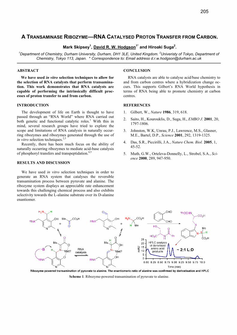
RESULTS AND DISCUSSION
We have used in vitro selection techniques in order to generate an RNA system that catalyses the reversible transamination process between pyruvate and alanine. The ribozyme system displays an appreciable rate enhancement towards this challenging chemical process and also exhibits selectivity towards the L-alanine substrate over its D-alanine enantiomer.
CONCLUSION
RNA catalysts are able to catalyse acid/base chemistry to and from carbon centres where a hybridization change oc-curs. This supports Gilbert’s RNA World hypothesis in terms of RNA being able to promote chemistry at carbon centres.
REFERENCES
1. Gilbert, W., Nature 1986, 319, 618.
2. Saito, H., Kourouklis, D., Suga, H., EMBO J. 2001, 20, 1797-1806.
3. Johnston, W.K, Unrau, P.J., Lawrence, M.S., Glasner, M.E., Bartel, D.P., Science 2001, 292, 1319-1325.
4. Das, S.R., Piccirilli, J.A., Nature Chem. Biol. 2005, 1, 45-52.
5. Muth, G.W., Orteleva-Donnelly, L., Strobel, S.A., Sci-ence 2000, 289, 947-950.
Scheme 1. Ribozyme-powered transamination of pyruvate to alanine.
205

ENZYMATIC POLYMERIZATION OF NUCLEOSIDE TRIPHOSPHATES BEARING ORGANOCATALYTIC FUNCTIONALITIES
Marcel Hollenstein* 1Department of Chemistry and Biochemistry, University of Bern, Freiestrasse 3, CH-3012 Bern, Switzerland
* Correspondence to: Email address [email protected]
ABSTRACT
Five modified nucleoside triphosphates (dNTPs) adorned with side-chains capable of organocatalysis have been synthesized. These dNTPs were shown to be fully compatible with in vitro selection methods, and could supplement the chemical armamentarium of DNA enzymes.
INTRODUCTION
Nucleoside triphosphates (dNTPs) have advanced as a very convenient platform for the generation of functional-ized oligonucleotides that can rival with the standard auto-mated solid-phase synthesis of nucleic acids.1-3 Consequent-ly, triphosphates modified with a wealth of side-chains rang-ing from amino acids4 to nucleic acids have appeared.5 In this context, the modified dNTPs 1-5 (Figure 1) bearing residues of relevance in organocatalysis have been synthe-sized in order to potentially replenish the reservoir of func-tional groups available to DNAzymes.6
RESULTS AND DISCUSSION
The modified dNTPs were obtained first by coupling the various side-chain precursors to a suitably protected 5-allyamino-2'-deoxyuridine derivative, either by straightfor-ward amide bond formation, or by reaction of an isocyanate or a sulfamide with the free amine moiety. Following deblocking of the trityl groups, the intermediates were then
converted to the corresponding triphosphates by application of a known triphosphorylation protocol. All the modified dNTPs were then tested for their substrate capacities in pri-mer extension reactions (Figure 2).
Figure 2. Gel image (PAGE 20%) of primer extension reac-
tions with the Pwo DNA polymerase. A) Standing start experi-ment; B) Running start experiment; C) Incorporation of three mod-ified dNTPs in a row.
All of the modified dNTPs revealed to be good substrates
for various DNA polymerases, independently of the se-quence context since only full length products were ob-served. In addition, all the triphosphates but dUBpuTP 4 could be incorporated into DNA via PCR and the resulting functionalized oligonucleotides could be converted back to natural DNA, again by PCR.
CONCLUSION
Five modified dNTPs have been synthesized and their bio-chemical characterization showed that they are good candi-dates for in vitro selection experiments that could unravel DNAzymes capable of organocatalysis.
REFERENCES
1. Hocek, M., Fojta, M. Org. Biomol. Chem. 2008, 6,
2233-2241. 2. Weisbrod, S. H., Marx, A. Chem. Commun. 2008,
5675-5685. 3. Hollenstein, M. Chimia 2011, 65, 770-775. 4. Raindlová, V., Pohl, R., Hocek, M. Chem. Eur. J.
2012, 18, 4080-4087. 5. Baccaro, A., Steck, A.-L., Marx, A. Angew. Chem.
Int. Ed. 2012, 51, 254-257. 6. Hollenstein, M., Leumann, C. J. Chem. Eur. J.
2012, submitted for publication.
Figure 1. Chemical structures of dUtPTP 1, dUcPTP 2, dUFPTP 3, dUBpuTP 4, and dUBsTP 5.
206

LIPOSOME ASSEMBLY CONTROLLED BY LIPID-MODIFIED OLIGONUCLEOTIDES
Ulla Jakobsen* and Stefan Vogel
Nucleic Acid Center, Department of Physics, Chemistry and Pharmacy, University of Southern Denmark, Campusvej 55, 5230 Odense M, Denmark. * Correspondence to: [email protected]
ABSTRACT
The formation of liposome aggregates can be con-
trolled by lipid-modified oligonucleotides. The assembly
is reversible and dependent on the design of the oligonu-
cleotides forming the DNA duplex.
INTRODUCTION, RESULTS AND DISCUSSION,
CONCLUSION
Liposome aggregates have previously been reported to be
formed when liposomes are mixed with a DNA duplex con-
sisting of one strand modified at both ends with lipophilic
substituents (membrane anchors) and an unmodified strand.
The membrane anchors adhere to the surface of the lipo-
somes and the rigid duplex prevents the ends of one modi-
fied strand to adhere to the same liposome (Figure 1).1
The assembly can be detected e.g. by monitoring the ab-
sorbance as a function of temperature; at temperatures be-
low the melting temperature (Tm) of the duplex linking the
liposomes, aggregates are formed and give rise to a large
apparent absorbance. At temperatures above Tm, disassem-
bly of the aggregates to individual liposomes results in a
significantly lower apparent absorbance.1b,c
Figure 1. Schematic representation of DNA-controlled assem-
bly of liposomes. DNA strands (red and blue ribbons) and lipo-
somes (grey spheres) are not drawn to scale.
Also oligonucleotides with lipophilic modifications in
only one end could in principle be used for DNA-controlled
liposome assembly.2 However, for a system consisting of
two complementary oligonucleotides both modified in either
the 3’- or the 5’-end (Figure 2a), no (5’-end modification) or
significantly reduced (3’-modification) assembly was ob-
served.
Figure 2. Schematic representation of (a) two and (b) three strand system for liposome assembly with oligonucleotides modi-fied with a single membrane anchor.
Liposome assembly was seen (Figure 3) when two non-
complementary oligonucleotides with a single lipid modifi-
cation at either end were mixed with an unmodified target
strand with regions complementary to each of the modified
oligonucleotides (Figure 2b).
Figure 3. Liposome assembly monitored by thermal denatura-tion experiments. 60 nM oligonucleotide concentration.
In conclusion, two complementary oligonucleotides with
a single lipid-modification are not sufficient to induce lipo-
some assembly, but two non-complementary strands can
control assembly by hybridization to an unmodified target
strand. These results show the importance of the duplex
design for assembly efficiency and the impact of sequence
design.
REFERENCES
1. a) Zhang, G. R., Farooqui, F., Kinstler, O., Letsinger, R.
L. Tetrahedron Lett., 1996, 37, 6243-6246, b) Jakobsen,
U., Simonsen, A. C., Vogel, S. J. Am. Chem. Soc., 2008,
130, 10462-10463, c) Jakobsen, U., Vogel, S. Methods
Enzymol,, 2009, 464, 233-248
2. a) Yoshina-Ishii, C., Boxer, S. G. J. Am. Chem. Soc.,
2003, 125, 3696-3697, b) Pfeiffer, I., Höök, F. J. Am.
Chem. Soc., 2004, 126, 10224-10225.
207

CELL-BASED SELECTION OF RNA APTAMERS AGAINST HUMAN IGF-I RECEPTOR
Davydova Anna1, Krasheninina Olga
1*, Vorobjeva Maria
1, Jean-Christophe Francois
2 and
Venyaminova Alya1
1Institute of Chemical Biology and Fundamental Medicine SB RAS, Lavrentieva ave. 8, Novosibirsk, 630090, Russia
and 2Centre de Recherche Saint-Antoine INSERM - UPMC, UMR S 938, 27 rue Chaligny F-75571 PARIS 12
* Correspondence to: [email protected]
ABSTRACT
Specific cell targeting is one of the most acute tasks
facing the modern fundamental medicine. Using of
escort RNA aptamers with a high affinity to the certain
proteins on cell surface represents a promising approach
to address this problem. The goal of this study was to
optimize the selection protocol for escort RNA aptamers
as well as to evolve new RNA aptamers against human
insulin-like growth factor I receptor (IGF-IR).
INTRODUCTION, RESULTS AND DISCUSSION,
CONCLUSION
Aptamers are in vitro selected DNA or RNA molecules that
can specifically bind to a wide variety of targets from small
molecules to proteins. Escort aptamers which recognize
specifically the cells of certain type have great potential for
addressed cell delivery of different agents for diagnostic or
therapeutic purposes. The goal of this study was to identify
specific RNA aptamers against human insulin-like growth
factor I receptor (IGF-IR). Insulin-like growth factor I (IGF
I) and its receptor (IGF-IR) are involved in cell
differentiation, proliferation and apoptosis. Taking part also
in cell malignant transformation, IGF-IR is considered now
as a promising cancer target [1].
Escort RNA aptamers against IGF-IR were obtained by cell-
based SELEX method using whole living cells as a target.
To improve biological stability of aptamers we replaced all
pyrimidine nucleotides by their 2'-F analogs. One of the key
stages of cell SELEX protocol is an isolation of total
cellular RNA after the selection, followed by separation of
aptamer sequences. We proposed a new method of
separation of modified RNA aptamers from total RNA
based on using of artificial ribonucleases (small RNA-
cleaving compounds mimicking RNase A) instead of
ribonuclease A [2]. The method allows to avoid an
accidental RNase A contamination as well as to exclude an
additional step for enzyme inactivation on every selection
round. It was shown in a model system that the treatment of
total cellular RNA by artificial ribonucleases results in the
formation of short RNA fragments while 2’-F-modified
RNA aptamer remain intact and can be subjected then to
specific RT-PCR. Ribonuclease Dp12 built of two
1, 4-diazabicyclo[2.2.2]octane residues [3, 4] was found to
be the most efficient for this purpose.
Then we established the feasibility of the newly developed
method in cell-based SELEX protocol. Two mice fibroblast
lines were used for selection: R+ and R- cells, expressing
and non-expressing human IGF-IR, correspondingly. Each
SELEX round included two sequential steps: 1) negative
selection using R- cells; 2) positive selection with the R+
cells. Thus we discarded the aptamers nonspecifically
recognizing any other cell surface proteins.
Fig. 1. Binding of [32P]-labeled RNA libraries to R+ cells and R-
cells
After 10 rounds we obtained an enriched RNA library
possessing an increased affinity and specificity to target R+
cells (fig. 1). The library was then cloned and sequenced,
and individual aptamers were characterized regarding their
structure and binding properties.
The obtained escort aptamers can be considered as
promising delivery tools in tumour diagnostics and therapy.
This work was supported by RFBR grant N 11-04-01014.
We acknowledge Dr. R. Baserga for providing R+ and R-
cells.
REFERENCES
1. Tao, Y., Pinzi, V., Bourhis, J., Deutsch, E., Nat. Clin.
Prac. Oncol., 2007, 4, 591-602.
2. Davydova A., Vorobjeva M., Zenkova M., François
J.-C. and Venyaminova A., International Roundtable
on Nucleosides, Nucleotides and Nucleic Acids.
Abstracts, 2010, P.577-578.
3. Koroleva, L.S., Serpokrylova, I.Y., Vlassov, V.V.,
Silnikov, V.N., Protein Pept. Lett., 2007, 14, 151-163.
4. See web-site www.nanotex-c.ru
208

DEVELOPEMENT OF THE ACYCLIC PYRIMIDINE DERIVATIVE FOR SELECTIVE CROSS-LINKING REACTION TO GUANINE
Shuhei Kusano, Shinya Hagihara, Takuma Moki and Fumi Nagatsugi*
Insitute of Multidisciplinary Research for Advanced Materials, Tohoku University, 2-1-1 Katahira, Aoba-ku, Sendai-shi, Miyagi, Japan* Correspondence to: Email [email protected]
ABSTRACT
Interstrand cross-link forming oligodeoxynucleotides
have been expected to ensure the inhibition of gene ex-
pression. In this presentation, we report the 4-amino-6-
oxo-2-vinylpyrimidine analogue, designed for the selec-
tive cross-linking reaction to guanine.
INTRODUCTION, RESULTS AND DISCUSSION,
CONCLUSION
Oligodeoxynucleotides (ODNs) are an attractive tool to
inhibit gene expression. A number of chemically modified
ODNs have been developed. To improve the efficiency of
gene regulation, one of the approaches is an interstrand
cross-linking (ICL) to the target gene. Previously, we have
developed a highly selective and very fast ICL reaction of 4-
amino-6-oxo-2-vinylpyrimidine (AOVP) derivative (1) to
uracil under neutral condition1)
. The ICL reaction of 1 is
activated by the hydrogen bonds between 1 and uracil (Fig-
ure 1). In this study, we designed the AVOP analogue (2)
with a flexible linker. 2 was expected to react with guanine,
because the distance from backbone of 2 is shorter than that
of 1, and hence 2 would form more stable base pair with
guanine than thymine.
The linker part (3) was synthesized in 9 steps from 2’-
deoxy-D-ribose. The base part (4) was synthesized as de-
scribed previously1)
. The coupling reaction between 3 and 4
was carried out using LiH as a base, providing nucleoside
analogue (5). The structure of 5 was determined by 2D-
NMR spectroscopy. The conventional method produced the
phosphoramidite (6) in 4 steps (20%) from 5. The sulfide-
protected ODN was obtained in good yield by applying 6 to
an automated DNA synthesizer and purification with RP-
HPLC. The sulfide in the obtained ODN was then smoothly
converted to sulfoxide by oxidation with magnesium mon-
operphthalate, and following elimination of the sulfoxide
under alkaline conditions afforded ODN (7) (Scheme 1).
The ICL reaction of ODN (7) with its complementary
RNAs (Y = A, G, C, U) was conducted under physiological
conditions. Figure 2A shows that 2 reacts selectively with
G in the target RNA.
The low reactivity (33%) of ODN (7) might be owing to
high flexibility of the linker. Next, we investigated the effect
of metal ions on ICL reaction of 2. In the presence of 1 mM
ZnCl2 or NiCl2, ICL yield of ODN (7) toward guanine was
increased to 90% (Figure 2B). These metal ions are pre-
sumed to enhance the ICL formation by changing the duplex
structure or directly activating the ICL reaction as lewis
acids. Finally, the cross-linked adduct was purified by RP-
HPLC and then enzymatically digested into nucleotides.
The sturucture of 2-dG adduct was confirmed as shown in
Figure 2C by MALDI-TOF MS and 1H-NMR spectroscopy.
This result indicated that the ICL reaction between 2 and
guanine would occur at N1 of guanine. Because of steric
hindrance between vinyl group of AOVP and amino group
of guanine, the vinyl group would be reacted to guanine at
N1 positon.
In conclusion, we have synthesized ODN (7) containing
acyclic nucleoside analogue of AOVP and ODN 7 demon-
strated high reactivity to guanine in the presence of ZnCl2 or
NiCl2. Furthermore, the structural analysis of cross-linking
adduct showed the ICL reaction occurred at N1 of guanine.
REFERENCES
1. Hattori, K., Hirohama, T., Imoto, S., Kusano, S., Na-
gatsugi, F. Chem. Commun. 2009, 42, 6463
Scheme 1. Synthesis of ODN 7: a) LiH, 1,4-dioxane, 100 ºC, 3 days, 23%; b) (i) 1-HOBt,
CH3CN, pyridine, 63%, (ii) H2, Pd(OH)2, MeOH, 56%, (iii) DMTrCl, pyridine, 76%, (iv) iPr2NP(Cl)OC2H4CN, DIPEA, CH2Cl2, 76%; c) (i) Automated DNA synthesizer, (ii) magne-sium monoperphthalate, (iii) 4 M NaOH
7: 5'-d(CCGCGT-2-TCGCCG)-3'
3'-r(GGCGCA-Y-AGCGGC)-FAM-5'
5'-d(CCGCGT-2-TCGCCG)-3'
3'-r(GGCGCA-Y-AGCGGC)-FAM-5'
N N
N
O
H2N
OH
HON
N
O
NH2
N
O
OH
HO
1
2
(B) Effect of metal ions (C) Structure of
cross-link adduct
Figure 2. cross-linking reaction of ODN 7 and structure of cross-link adduct; the reaction
was performed with 10 M ODN 7 and 5 M target ON in 50 mM MES, 100 mM NaCl, pH
7, 37 ºC
(A) Gel electrophoresis
BnO
OMs
BnON
N
O
NH2
SC8H176
1'
4
1
H
HMBC
DMTrON
N
O
NHPac
SC8H17
OP
Ni-Pr2
OCN
ON
N
O
NH2
X =
5'-d(CCGCGT-X-TCGCCG)-3'
O
OBnOBn
35
6 7
+
NH
N
SC8H17O
NH2
4
a
b c
O
O N
N
N
O
H
H
N
N
N
NO
N
H
H
H
RNA
2
G
N
NO
N
N
N
O
O RNA
H
H
H
O
O
O
U
1Expansion of Target Base
1
2
AOVP
Figure 1. Design of New Cross-linking Reagent
209

“SINGLE-MOLECULE” BEACONS: AN APPLICATION OF NANOMECHANICAL DNA
ORIGAMI DEVICES
Akinori Kuzuya,1* Yusuke Sakai,2 Takahiro Yamazaki,2 Yusei Yamanaka,1 Yan Xu,2 Yuichi Ohya,1 and Makoto Komiyama2*
1Dept. Chem. Mater. Eng., Kansai University, 3-3-35 Yamate, Suita, Osaka 565-8680, Japan and 2RCAST, The Uni-versity of Tokyo, 4-6-1 Komaba, Meguro, Tokyo 153-8901, Japan. * Correspondence to: [email protected] or
ABSTRACT
We propose versatile sensing systems for a variety of chemical and biological targets at molecular resolution. We have designed functional nanomechanical DNA ori-gami devices that can be used as "single-molecule bea-cons", which consist of two levers approximately 170 nm long connected at a fulcrum. Various single-molecule inorganic/organic targets ranging from metal ions to proteins can be visually detected on AFM by a shape transition of the origami devices.
INTRODUCTION
Rapid development of nanotechnology has enabled pre-cise manipulation of nanomaterial. However, typical ana-lytical methods for chemical or biochemical targets are still based on spectroscopic principles, which treat average be-havior of vast molecules. To analyze individual molecule's behavior, nanomechanical devices that can work with target molecules in single-molecule manner are required. DNA origami [1,2], in which long single-stranded DNA is folded into designed nanostructure with the aid of many short sta-ple strands, is a promising candidate to provide such nanomechanical devices. Here we present functional nanomechanical DNA origami devices that can be used as versatile and visible “single-molecule” beacons (DNA ori-gami beacons) [3].
RESULTS AND DISCUSSION
Figure 1 shows the structure of “DNA origami pliers” used in this study. It consists of two ca. 170-nm long lever domains, which are made of six parallel DNA helices of
M13 scaffold and 117 staple strands each. These levers are joined together at a fulcrum via two phosphodiester linkages in the M13 scaffold. Figure 2 shows typical AFM images of selective and single-molecular pinching of a streptavidin tetramer (SA) by DNA origami pliers. Here, DNA pliers were modified with a biotin group in each of the jaws on the levers. The dominant species initially observed without SA (i) was DNA pliers in cross. When SA was added to the solution, on the other hand, the population of each forms drastically altered. The major form of DNA pliers was par-allel closed form (Figure 1) in the presence of SA (ii). In addition, a bright spot of 5-nm height that corresponds to the expected diameter of the pinched SA molecule was found in the jaws of most of the parallel pliers, showing that DNA pliers indeed can pinch exactly one SA molecule in the jaws. Selective opening of the closed DNA pliers was possible by detaching the biotinylated anchor strands from DNA pliers utilizing DNA strand displacement technique. Similar detec-tion of anti-fluorescein IgG was also successful by using FAM-modified DNA pliers.
The present origami beacons can detect various targets, not only proteins. For example, Na+ ion sensing was possi-ble utilizing G-quadruplex formation. Similarly, the pres-ence of human micro RNA (miRNA) or ATP could be clearly detected with origami beacons.
REFERENCES
1. Rothemund, P.W.K. Nature, 2006, 440, 297-302.
2. Kuzuya, A., Komiyama, M., Nanoscale, 2010, 2, 310-322.
3. Kuzuya, A., Sakai, Y., Yamazaki, T., Xu, Y., Komi-yama, M. Nature Commun., 2011, 2, 449.
Figure 1. The structure and possible conformation of DNA origami pliers used in this study.
Figure 2. Typical AFM images for selective pinching of SA.
210

PHOTOCHROMIC CONTROL OF DNA BINDING EVENTS THROUGH ANTHRACENE
DIMERISATION
Jack Manchester,1* John Zhao
1 and James H. R. Tucker
1
1School of Chemistry, University of Birmingham, Edgbaston, Birmingham, B15 2TT United Kingdom.
* Correspondence to: [email protected], Website: http://tiny.cc/dtcnz
ABSTRACT
With the incorporation of two non-nucleosidic anthra-
cene tags into an oligonucleotide, the resulting intramo-
lecular photochemical reaction produces a dimer with
markedly different binding behaviour towards the com-
plementary sequence. The extent of this effect is a direct
result of the number of bases between the anthracene
tags.
INTRODUCTION
The regulation of DNA and its associated bio-reactions
by external stimuli has been an important theme of research
in recent years.1,2
The focus of this research is to alter the
stability of DNA structures through the photochemical di-
merisation of anthracene. It is proposed that by tagging a
DNA strand with two anthracene groups, in a manner that
allows sufficient proximity to enable them to dimerise once
irradiated at the required wavelength, a structural change
will be induced that reduces the amount of effective base
pairing within the system, Figure 1. The reverse process can
be initiated by the application of heat or UV light at a short-
er wavelength to give photochromic control.3
Based on our recent work on detecting base changes and
epigenetic modifications in DNA,4 the anthracene is tagged
to the oligonucleotide through a non-nucleosidic linker with
a long carbon chain, which allows the anthracene sufficient
flexibility. This study focuses on variations in the number of
bases separating the anthracene tags and observing the de-
gree of structural change induced upon the oligonucleotide
and how it affects base pairing.
RESULTS AND DISCUSSION
A series of anthracene tagged oligonucleotides were syn-
thesised via standard automated synthesis, as shown in Ta-
ble 1. Upon photo-irradiation the characteristic absorption
band of anthracene (360nm) was observed to decrease sig-
nificantly over a 40-minute period, with HPLC analysis in-
dicating the appearance of new photoproducts in a clean
reaction. Each product was characterised by mass spectros-
copy and was confirmed to be identical to the starting mate-
rial, indicating an intramolecular process.
Name Sequence
S1 TGGACTXTXTCAATG
S2 TGGACXCTCXCAATG
S3 TGGAXTCTCTXAATG Table 1. Modified oligo sequences, X = Anthracene.
The isolated photodimer was analysed by variable tem-
perature UV spectroscopy in order to obtain a melting tem-
perature (Tm), an indication of duplex stability. The results
indicated that with increasing numbers of bases between the
anthracene tags the more destabilised the duplex system
becomes, as signified by the increase in the ΔTm, Table 2.
For sequence S3 no observable Tm was recorded, indicating
a total loss of base pairing.
Name Initial Tm Dimer Tm Δ Tm
S1 37.5 37 0.5
S2 45 25 20
S3 35 < 5 > 30 Table 2. Tm measurements of modified oligo sequences
These results were further investigated through a number
of tests including Circular Dichroism (CD) spectroscopy
and native PAGE gel electrophoresis. The results of these
tests supported the data collected from Tm analysis.
CONCLUSION
Through various techniques, it has been demonstrated
that the duplex binding of modified DNA can be altered
through the photochemical dimerisation of anthracene. The
research will progress by applying the technique to more
diverse DNA systems and studying their behaviour.
REFERENCES
1. Asanuma, H., Komiyama, M. et al. Angew. Chem. Int.
Ed., 2001, 40, 2671-2673.
2. Ogasawara, S., Maeda M. Angew. Chem. Int. Ed., 2008,
47, 8839-8842.
3. Bouas-Laurent, H., Castellan, A., Desvergne, J.P., La-
pouyade, R. Chem. Soc. Rev., 2001, 30, 248-263.
4. Duprey, J.L., Zhao, Z., Bassani, D.M., Manchester, J.,
Vyle, J.S., Tucker, J.H.R. Chem. Commun., 2011, 47,
6629-6631.
hυ hυ
Δ Δ
Figure 1. Anthracene modified DNA and photochemical reaction.
211

ANNEALING ACTIVITY OF CATIONIC COPOLYMERS FOR QUADRUPLEX ASSEMBLY
Atsushi Maruyama, Rui Moriyama, Naohiko Shimada, Arihiro Kano Institute for Materials Chemistry and Engineering, Kyushu University, Moto-oka 744-CE11, Nishi, Fukuoka, Japan. *
Correspondence to: [email protected]
ABSTRACT
We showed that the cationic comb-type copolymer ac-celerates the association of tetramolecular G-quadruplex DNA three orders of magnitude. We also found that that the copolymer promotes the rearrangement to canonical quadruplexes from kinetically trapped structures.
INTRODUCTION
The hybridization and folding of nucleic acid into the thermodynamically most stable structure is often impeded by kinetic traps (i.e., local energy minima) due to formation of local structures that are stable enough to halt the hybridi-zation for a physiologically significant amount of time. Partially hybridized or misfolded intermediates can be over-come by the aid of nucleic acid chaperone proteins that an-nealed the intermediates to the thermodynamically most stable structures. We have previously reported that cationic comb-type copolymers composed of a cationic polymer backbone (<20 wt%) and abundant hydrophilic graft side chains (>80 wt%) significantly accelerates the associations of duplex, triplex and quadruplex DNA1. Moreover, the copolymer activates the strand exchange reaction of duplex with homologous single-stranded DNA (ssDNA).2 Thus, the copolymer, like protein chaperones, facilitates DNA assembly into the thermodynamically most stable structures.
G-quadruplex structures can be formed by guanine-rich sequences. G-quadruplexes have intrinsic conformational polymorphism as different strand orientations and glycosyl bond angles are possibel and G-quadruplex assemblies tend to fall into kinetically trapped structures of non-optimal sta-bility. In this study we characterized the annealing activity of the copolymers for intra molecular quadruplexes.
RESULTS AND DISCUSSION
We evaluated the influence of PLL-g-Dex on recombi-nation of heteroquadruplexes to the homoquadruplex at a constant temperature. A heterogeneous mixture composed of [TG4T]4, [TG5T]4, [(TG4T)3▪TG5T], [(TG4T)2▪(TG5T)2] and [TG4T▪(TG5T)3] quadruplexes were incubated at 25◦C with or without PLL-g-Dex. In the absence of PLL-g-Dex, no change in composition was observed after three days (Figure 1a). In the presence of PLL-g-Dex (N/P = 4), peaks due to heteroquadruplexes at retention times ranging from 86 to 108 min disappeared and [TG4T]4 and [TG5T]4 homoquadruplex peaks at retention times of 71 and 148 min, respectively increased in area (Figure 1b). After three days, only homoquadruplexes were observed, indicating that PLL-
g-Dex facilitated the recombination of heteroquadruplexes to homoquadruplex.3 The effect of the copolymer was en-hanced with increasing N/P ratios and reached a plateau at N/P = 2 (data not shown).
Figure 3. Recombination of heterogeneous quadruplexes at constant temperature.
CONCLUSION
The copolymer activated the recombination of metastable quadruplex structures into the most stable conformation. Though mechanisms underlying acceleration of quadruplex assemlby are not fully understood, the copolymer will find application as a DNA annealer in nano-applications.
REFERENCES
1. Torigoe, H., Ferdous, A., Watanabe, H., Akaike, T., Maruyama, A., J. Biol. Chem., 1999, 274, 6161-6167, Wu, L., Shimada, N., Kano, A., Maruyama, A., Soft Matter, 2008, 4, 744-747, Moriyama, R., Shimada, N., Kano, A., Maruyama, A., Biomaterials, 2011, 32, 2351-2358.
2. Kim WJ, Akaike T, Maruyama A. J Am Chem Soc, 2002, 124, 12676-12677.
3. Moriyama, R., Shimada, N., Kano, A., Maruyama, A., Biomaterials, 2011, 32, 7671-7676.
212

PREPARATION OF MODIFIED OLIGONUCLEOTIDES BY NICKING ENZYME AMPLIFICATION
REACTION
Petra Ménová1* and Michal Hocek
1
1Institute of Organic Chemistry and Biochemistry, Academy of Sciences of the Czech Republic, Flemingovo nám. 2,
166 10, Prague 6, Czech Republic. * Correspondence to: Email address [email protected]
ABSTRACT
A novel method for the enzymatic preparation of
modified oligonucleotides based on the Nicking Enzyme
Amplification Reaction was developed. It was successful-
ly used for the incorporation of modified deoxycytidine
triphosphates. The synthesis was scaled-up to nanomolar
amounts.
INTRODUCTION, RESULTS AND DISCUSSION,
CONCLUSION
Nicking enzymes are a class of restriction enzymes
which cut one strand of the DNA duplex to produce shorter
DNA fragments. One of the major applications of nicking
enzymes is in Nicking Enzyme Amplification Reaction
(NEAR), which has been reported to rapidly synthetize short
oligonucleotides under mild isothermal conditions.[1] Since
then it has been used in biochemical research and diagnos-
tics. Unlike PCR, which requires temperature cycling,
NEAR proceeds at a constant temperature (55 °C). The am-
plification is based on the continuous repetition of two
steps: polymerase-mediated primer extension and nicking
enzyme-mediated cleavage. The cleaved oligonucleotide is
too short to form a stable duplex with the template and
leaves the duplex, thus regenerating the initial dimer primer-
template.
NEAR has so far been used on an analytical scale for the
detection and quantification of DNA. Here, we present a
method based on NEAR developed for the preparation of
modified oligonucleotides. In the initial experiments the
most efficient combination of DNA polymerase and nicking
enzyme was established. These enzymes were used for the
incorporation of m-aminophenyl-modified nucleobases. The
reaction temperature, time, and amounts as well as the ratio
of both enzymes were also optimized. To evaluate the scope
of the optimized procedure, a series of modified dCXTPs
bearing various modifications used previously in DNA la-
belling, protection and bioconjugation was incorporated
using three different templates. Thus, three different se-
quences of 12-mer ONs bearing either one or three modifi-
cations were prepared.
Apart from working on an analytical scale, we also trans-
ferred the procedure to nanomolar amounts. A protocol for
the isolation of pure modified oligonucleotides including a
separation on HPLC was developed. The products were
characterized by HPLC retention time, UV-VIS spectrosco-
py and MALDI spectrometry.
Our methodology is complementary to the synthesis of
medium-length single-stranded oligonucleotides by PEX
followed by magnetoseparation on streptavidine-coated
beads.[2] Thus prepared short modified oligonucleotides can
find applications as modified primers for PEX and PCR as
well as in diagnostics.
REFERENCES
1. Van Ness, J., Van Ness, L.K., Galas, D.J. Proc. Nat. Ac.
Sci., 2003, 100, 4504-4509.
2. (a) Brázdilová, P., Vrábel, M., Pohl, R., Pivoňková, H.,
Havran, L., Hocek, M., Fojta, M. Chem. Eur. J., 2007,
13, 9527-9533; (b) Cahová, H., Havran, L., Brázdilová,
P., Pivoňková H., Pohl, R., Fojta, M., Hocek, M. Angew.
Chem. Int. Ed., 2008, 47, 2059-2062; (c) Balintová, J.,
Pohl, R., Horáková, P., Vidláková, P., Havran, L., Fojta,
M., Hocek, M. Chem. Eur. J., 2011, 17, 14063-14073.
This work was supported by the Grant Agency of the Acad-
emy of Sciences of the Czech Republic (IAA400040901), by
the Czech Science Foundation (203/09/0317) and by Gilead
Sciences, Inc. (Foster City, CA, USA).
Figure 1. Schematic representation of the linear NEAR with modified dCXTP.
213

SYNTHESIS OF MONO AND MULTIPLE CONJUGATED OLIGONUCLEOTIDES BY "CLICK
THIOL" CHEMISTRY AND COMBINATION WITH CUAAC "CLICK CHEMISTRY"
Albert Meyer, Jean-Jacques Vasseur and François Morvan*
Institut des Biomolécules Max Mousseron, UMR 5247 CNRS UM1 UM2, Université Montpellier 2, Place E. Bataillon, 34095 Montpellier cedex 5, France. *Correspondence to: [email protected]
ABSTRACT
Oligonucleotide conjugates were efficiently synthe-
sized by thiol click chemistry starting from a mono- or
poly-thiol oligonucleotides and different acrylamide de-
rivatives. This strategy was applied to form glycoclusters
and was also combined with CuAAC for bis-conjugation
through a sequential protocol.
INTRODUCTION
Oligonucleotide conjugates are widely used for various ap-
plications in biology, biotechnology, and medicine. Most
applications require labelling with dyes, redox tag or other
biomolecules such as biotin or carbohydrates.
We explored the use of thiol Michael-type addition (TMTA)
to prepare oligonucleotide conjugates. This addition corre-
sponds to the reaction of a thiol on an electron-deficient ene
forming a thioether linkage. This reaction is usually restrict-
ed to the reaction of thiol-oligonucleotides with maleimide
derivatives [1]. Herein we present the TMTA using acryla-
mide derivatives. Finally, TMTA was combined sequential-
ly with the Copper (I) catalyzed Azide Alkyne Cycloaddi-
tion (CuAAC) to synthesized bis conjugates.
RESULTS AND DISCUSSION
A 5'-S-acetyl-thiohexyl oligonucleotide was synthesized
using commercially available amidites on a DNA synthesiz-
er according to the phosphoramidite chemistry. Different
acrylamide derivatives exhibiting a phenyl, mannose, ferro-
cene, dansyl, biotin or deoxycholic moiety were prepared.
After the removal of cyanoethyl group by piperidine treat-
ment, the solid-supported thio-oligonucleotide was treated
with an acrylamide derivative in presence of TCEP and
K2CO3 methanol leading to the deprotection, release from
solid support and TMTA (Figure 1). Hence the monoconju-
gate oligonucleotide was obtained with a quantitative con-
version of the thiol-oligonucleotide. A size exclusion chro-
matography on cartridge allowed the isolation of almost
pure conjugate. The same strategy was applied to synthesize
a mannose-centered tetramannose oligonucleotide allowing
the formation of a glycocluster oligonucleotide conjugate
with high efficiency starting from a tetra-thiolhexyl oligo-
nucleotide.
Finally the TMTA was combined with the CuAAC ac-
cording to a sequentially protocol to synthesize bis-
conjugated oligonucleotides exhibiting biotin and carbohy-
drate, dansyl and mannose or mannose and galactose.
R
NH
NCH3H3C
SO
O
HN NH
O
S C
O
HN
NHFe
HN
H3C H
CH3HO
HO
CH3
H
H
O
H
OHOHO
OH
O
HO
DNAOP
O
O
OSN
H
O
R
DNAOP
O
O
OAcSN
H
O
R
Thiol Click
OH
Figure 1. Structure of oligonucleotide conjugates synthesized
by "click thiol" chemistry
CONCLUSION
The TMTA is a very efficient click reaction to synthesize
oligonucleotides conjugated with various molecules. The
great advantages of this reaction are that it does not require
radical initiator and uv irradiation, and it occurs during the
deprotection and release of the oligonucleotide from solid
support affording the expected conjugates as the unique
molecule. TMTA could be applied to the synthesis of multi-
labelled oligonucleotides. Eventually, the sequential combi-
nation of the TMTA with the very popular CuAAC allowed
the synthesis of bis-conjugated oligonucleotides starting
either from the CuAAC and then TMTA or reverse.
REFERENCES 1. Singh, Y.; Murat, P.; Defrancq, E. Chem. Soc. Rev.
2010, 39, 2054-2070.
214

NEW SWNT HYBRYDS FOR DELIVERY OF FUNCTIONAL NUCLEIC ACIDS INTO CELLS
Darya Novopashina,1* Evgeny Apartsin,
1 Marina Buyanova,
1,2 Alevtina Baturina
1,2 and
Alya Venyaminova1
1Institute of Chemical Biology and Fundamental Medicine, akad. Lavrentiev ave, 8, Novosibirsk, Russia and
2Novosibirsk State University, ul.Pirogova, 2, Novosibirsk, Russia. * Correspondence to: [email protected]
ABSTRACT
The preparation of novel multifunctional hybrids of fluorescent single walled carbon nanotubes with nucleic acid fragments as prospective cell penetrating construc-tions was described. Structure and physico-chemical properties of modified carbon nanotubes and their hy-brids with oligonucleotides were investigated.
INTRODUCTION, RESULTS AND DISCUSSION,
CONCLUSION
Carbon nanotubes have attracted attention due to their
unique properties as one of the most promising nanomateri-
als for a variety of biomedical applications. Application of
carbon nanotubes for the delivery of therapeutic nucleic
acids (NA) to the site of action has become one of the main
areas of interest [1]. Nanomaterials should contain reporter
groups to permit the monitoring of transfection and elimina-
tion processes.
We prepared three types of fluorescent single walled
nanotubes (SWNTs) containing covalently attached fluores-
cein using hexamethylenediamine or bis-amino-modified
polyethylene glycol (PEG) linkers or polyamidoamine den-
drimer G3.0 applying the strategy we reported recently [2].
Fluorescein-modified nanotubes were characterized by in-
frared spectroscopy, thermogravimetric analysis, elemental
analysis, Raman spectroscopy, transmission and scanning
electron microscopy.
The pyrene conjugates of the model oligodeoxyribonu-
cleotides containing 5'-phosphate and 5'-hexaethyleneglycol
phosphate were synthesized by analogy with [3]. The im-
mobilization of the oligonucleotides on the nanotube surface
occurred due to the stacking interaction of the pyrene aro-
matic system with the SWNTs and lead to the solubilization
of the latter one. The influence of oligonucleotide length on
the immobilization efficacy was revealed using model ribo-
and deoxyribo- homo-oligonucleotides. The noncovalent
functionalization of the fluorescein-modified SWNTs was
performed by the ultrasound treatment of nanotubes in the
pyrene oligonucleotide conjugate aqueous solution (Fig.1).
An original method was proposed to estimate the effi-
ciency of the pyrene oligonucleotide conjugate adsorption
on the surface of CNTs based on the quenching of the py-
rene residue fluorescence upon interacting with the nano-
tube surface. The adsorption isotherms of the pyrene oligo-
nucleotide conjugates on the surface of SWNTs were con-
structed. The capacity of carbon nanotubes as potential oli-
gonucleotide transporters was estimated from adsorption
isotherms and amounted to about 20-100 μmol/g depending
on oligonucleotide structure and type of SWNT functionali-
zation.
The -potential values changing profiles plotted for hy-
brids were shown to have minima at the points likely corre-
sponding to the highest amount of oligonucleotide adsorbed
on individual SWNTs. Transmission electron microscopy
images of non-covalent hybrids of oligonucleotides with
functionalized SWNTs demonstrated simultaneous presence
of functional groups and oligonucleotide in hybrid structures.
Oligonucleotides were visualized as folded nanosized struc-
tures on the SWNT surface.
Our results allow for the rational design of multifunction-
al nanotube hybrids with nucleic acids to be conducted.
These hybrids could find wide application in functional nu-
cleic acids delivery (NAzymes, aptamers, siRNA, and oth-
ers) into the cells.
REFERENCES
1. Madani, S.Y., Naderi, N., Dissanayake, O., Tan, A.,
Seifalian, A.M. Int. J. Nanomed., 2011, 6, 2963-2979.
2. Apartsin, E.K., Novopashina, D.S., Nastaushev, Yu.V.,
Ven'yaminova, A.G. Nanotechnol. Russ., 2012, 7, 99-
109.
3. Novopashina, D.S., Totskaya, O.S., Kholodar, S.A.,
Meshchaninova, M.I., Venyaminova ,A.G. Russ. J.
Bioorg. Chem., 2008, 34, 602-612.
Figure 1. Structure of fluorescein-modified SWNT hybrids with 5'-pyrene modified oligonucleotide.
215

RNA APTAMERS AGAINST ANTI-MBP AUTOANTIBODIES ASSOCIATED WITH MULTIPLE SCLEROSIS
Darya Novopashina,1 Maria Vorobjeva
1*, Valentina Timoshenko,
1 Anastasia Popovetskaya,
1 Alesya
Fokina, 1
Anna Bezuglova, 1 Georgiy Nevinsky
1 and Alya Venyaminova
1
1Institute of Chemical Biology and Fundamental Medicine SB RAS, Lavrentieva ave., 8, Novosibirsk, Russia
*Correspondence to: [email protected]
ABSTRACT
A SELEX of 2’-F-modified RNA aptamers against
pathogenic autoantibodies associated with multiple scle-
rosis was performed. An enriched RNA library was
cloned and sequenced. A series of individual aptamers
was synthesized and their binding affinity and specificity
were characterized.
INTRODUCTION
Multiple sclerosis (MS) is a chronic progressive demyelinat-ing disease of the central nervous system. It is now general-ly considered to be an autoimmune disorder characterized by presence of autoantibodies against myelin basic protein (MBP). These antibodies were shown to possess a proteolyt-ic activity against MBP [1, 2] thus contributing to the patho-logical destruction of myelin sheath. A development of new approaches to the specific detection of anti-MBP autoanti-bodies and inhibition of their proteolytic activity would be of importance for diagnostic and therapeutic purposes. Aptamers are short single stranded DNA or RNA molecules, binding with high affinity to a large variety of targets, from small molecules to complex mixtures, due to their specific three-dimensional shapes. The objective of the present work was the in vitro selection of 2′-fluoro modified RNA ap-tamers for the specific targeting of anti-MBP autoantibodies.
RESULTS AND DISCUSSION
Polyclonal IgGs were isolated from sera of patients with
multiple sclerosis, the fraction containing MBP-specific
antibodies was purified using MBP affinity chromatography,
and used as a target in SELEX procedure. To produce RNA
aptamers, a SELEX protocol including immunoglobulins’
adsorption to the PCR tube walls and the performance of the
whole selection cycle in one PCR tube was employed [3].
After 10 rounds of selection, we obtained an enriched 2′-F-
pyrimidine RNA library which bound with high affinity to
anti-MBP IgGs and inhibited their proteolytic activity. The
library was then cloned and sequenced. The clones were
divided into groups according to their sequence similarities.
Individual 2′-F-pyrimidine RNA aptamers from each group
were synthesized and characterized in terms of their second-
ary structure, binding affinity and ability to inhibit the deg-
radation of human MBP by MS-associated polyclonal IgGs.
Figure 1. Binding of [32P]-labeled aptamer II-9 with polyclonal
anti-MBP pathogenic autoantibodies from MS patients (●) and
total IgG from healthy donors (▲).
The specificity of aptamers towards pathogenic MS-
related IgGs was tested using total IgG from healthy donors
as a control (see Fig. 1 for example). Aptamers possessing
an optimal set of properties were chosen for the subsequent
sequence minimization and design of MS-specific aptasen-
sors.
CONCLUSION
The obtained 2′-F-RNA aptamers to MBP-specific autoanti-
bodies provide a novel opportunity to develop precise tools
for diagnostics and therapy of multiple sclerosis.
This work was supported by RFBR grant N 11-04-01014.
REFERENCES
1. Ponomarenko, N.A., Durova, O.M., Vorobiev, I.I., Ale-
ksandrova, E.S., Telegin, G.B., Chamborant, O.G., Si-
dorik, L.L., Suchkov, S.V., Alekberova, Z.S., Gnuchev,
N.V., Gabibov, A.G. J. Immunol. Meth., 2002, 269,
197-211.
2. Polosukhina, D.I., Kanyshkova, T.G., Doronin, B.M.,
Tyshkevich, O.B., Buneva, V.N., Boiko, A.N.,
Gusev, E.I., Favorova, O.O., Nevinsky, G.A. J. Cell.
Mol. Med., 2004, 8, 359-368.
3. Missailidis, S. Methods Mol. Biol., 2004, 248, 547-555.
216

A MODULAR SYSTEM FOR THE INCORPORATION OF POINT-SPECIFIC MODIFICATIONS INTO
LARGER NUCLEIC ACIDS
Igor A. Oleinich* and Eva Freisinger
1University of Zurich, Institute of Inorganic Chemistry, Winterthurerstr. 190, 8057 Zurich, Switzerland
* Correspondence to: [email protected]
ABSTRACT
A new synthetic strategy for the point-specific label-
ling of natural single and double stranded oligonucleo-
tides with bioorthogonal reactive groups was designed.
The successful modifications were confirmed by
MALDI-TOF, PAGE and LC-MS analysis of enzymatic
digestions.
INTRODUCTION, RESULTS AND DISCUSSION,
CONCLUSION
Non-natural modifications of oligonucleotides have
unusual properties and can be used for a range of therapeu-
tic and analytical purposes, including the treatment of dis-
eases, the regulation of gene expression, as well as investi-
gation of DNA and RNA tertiary structures, DNA-peptide
interaction, monitoring concentration of RNA in vivo and
detection of single nucleotide polymorphism [1]. Modifica-
tion of nucleic acid up to now is not specific and can go in
two approaches, complete modification of one nucleotide
through the whole sequence, or during solid-phase synthesis.
Restriction of the lengths of such modified oligonucleotides
accessible by second approach made us follow alternative
approach.
Our objective in the present study was to design con-
struct for the point-specific modification of target nucleotide
at a specific position. The basic principle of this approach
relies on the annealing of the donor molecule with the target
oligonucleotide to localize the reactive group close in space
to the target nucleotide [2]. The donor molecule contains the
complementary oligonucleotide sequence required to sense
the specific position in the target DNA, a reactive group to
generate the desired modification, and a cleavage part to
cleaved the intermediately formed covalent cross-link be-
tween donor molecule and target sequence (Figure 1).
The reactive group was designed in such a way that it
can selectively modify a specific nucleotide (cytosine, gua-
nine or thymine [4]) in the presence of the three others. In
principle different position for a modification with nucleo-
tides are possible. Depending on the site, the modification
can preserve, with or without distortion, or hinder Watson-
Crick base pairing.
First of all different synthetic strategies to introduce the
reactive group into the donor molecule via a linker sequence
were tested, compared and improved. A library of linkers
with different sizes, dipolar moments and flexibility was
created. The most promising candidates were incorporated
into the donor molecule and tested with various numbers of
different target oligonucleotides. The site-specific modifica-
tions in the target sequences were detected by PAGE,
MALDI-TOF, ESI-MS and LC-MS analysis of enzymatic
digestions.
The results show that the recognition and duplex for-
mation between donor and target sequence is the major driv-
ing force in this approach. In the absence of duplex or tri-
plex formation between complementary sequences no modi-
fications take place [5].
REFERENCES
1. Milligan, J.F., Groebe, D.R., Witherell, G.W., Uhlen-
beck, O.C. Nucleic Acids Res. 1987, 15, 8783-8798.
2. Molander J, Hurskainen P. Bioconjugate Chem. 1993,
4, 362-365.
3. Taylor M.J., Dervan P.B. Bioconjugate Chem. 1997, 8,
354-364.
4. Kellner S., Seidu-Larry S., Burhenne J., Motorin Y.,
Helm M. Nucleic Acids Res. 2011, 39, 7348-7360.
5. Gartner Z.J., Kanan M.W., Liu D.R. Angew. Chem. Int.
Ed. 2002, 41, 1796-1800.
N
NO
NH
H2N
ODN
HN
NN
N
O
H2N
R
O
ODN
N
O
O
R O
ODN
Figure 1. Annealing donor molecule (blue) and target sequence (black) will induce site-specific nucleotide modification and intro-duce bioorthogonal reactive group in desire position.
217
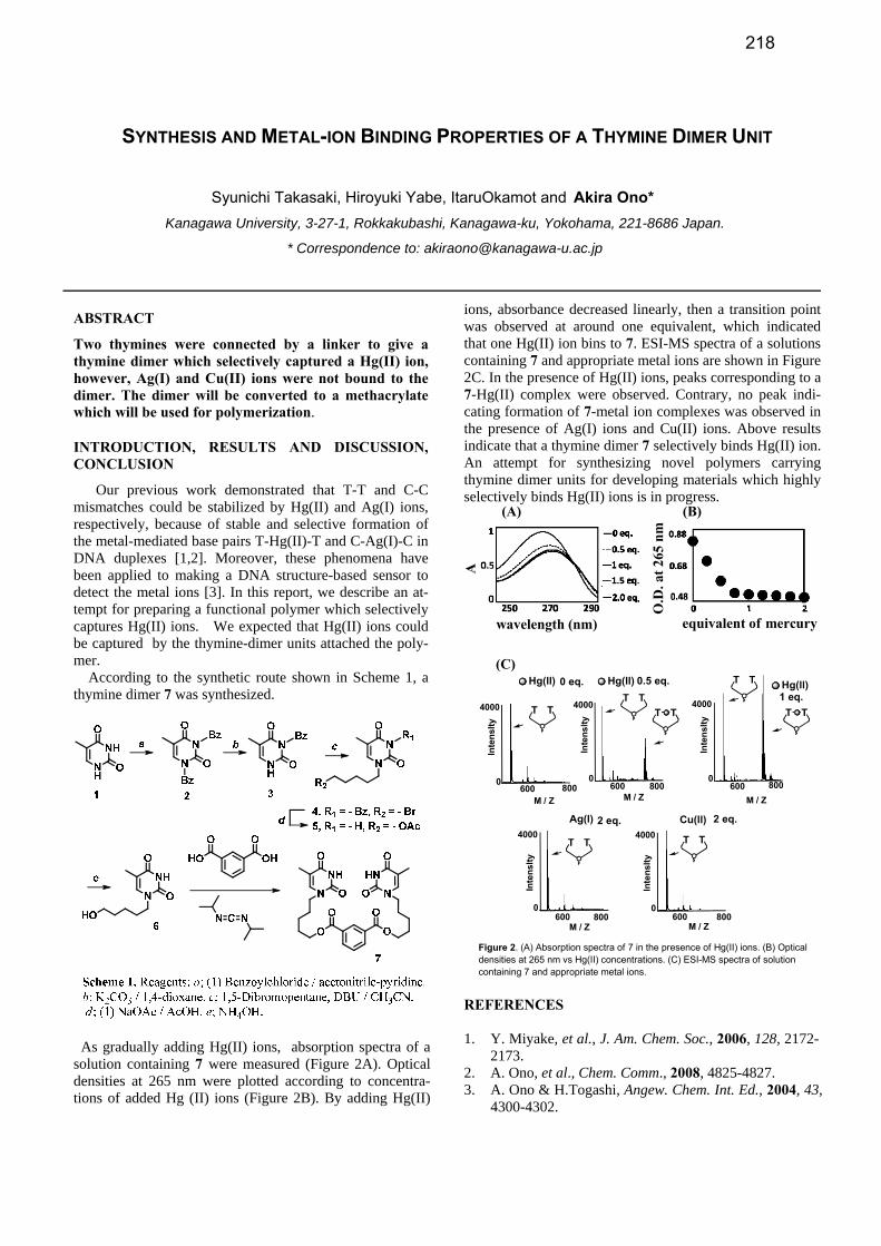
SYNTHESIS AND METAL-ION BINDING PROPERTIES OF A THYMINE DIMER UNIT
Syunichi Takasaki, Hiroyuki Yabe, ItaruOkamot and Akira Ono*
Kanagawa University, 3-27-1, Rokkakubashi, Kanagawa-ku, Yokohama, 221-8686 Japan.
* Correspondence to: [email protected]
ABSTRACT
Two thymines were connected by a linker to give a thymine dimer which selectively captured a Hg(II) ion, however, Ag(I) and Cu(II) ions were not bound to the dimer. The dimer will be converted to a methacrylate which will be used for polymerization.
INTRODUCTION, RESULTS AND DISCUSSION, CONCLUSION
Our previous work demonstrated that T-T and C-C mismatches could be stabilized by Hg(II) and Ag(I) ions, respectively, because of stable and selective formation of the metal-mediated base pairs T-Hg(II)-T and C-Ag(I)-C in DNA duplexes [1,2]. Moreover, these phenomena have been applied to making a DNA structure-based sensor to detect the metal ions [3]. In this report, we describe an at-tempt for preparing a functional polymer which selectively captures Hg(II) ions. We expected that Hg(II) ions could be captured by the thymine-dimer units attached the poly-mer.
According to the synthetic route shown in Scheme 1, a thymine dimer 7 was synthesized.
As gradually adding Hg(II) ions, absorption spectra of a solution containing 7 were measured (Figure 2A). Optical densities at 265 nm were plotted according to concentra-tions of added Hg (II) ions (Figure 2B). By adding Hg(II)
ions, absorbance decreased linearly, then a transition point was observed at around one equivalent, which indicated that one Hg(II) ion bins to 7. ESI-MS spectra of a solutions containing 7 and appropriate metal ions are shown in Figure 2C. In the presence of Hg(II) ions, peaks corresponding to a 7-Hg(II) complex were observed. Contrary, no peak indi-cating formation of 7-metal ion complexes was observed in the presence of Ag(I) ions and Cu(II) ions. Above results indicate that a thymine dimer 7 selectively binds Hg(II) ion. An attempt for synthesizing novel polymers carrying thymine dimer units for developing materials which highly selectively binds Hg(II) ions is in progress.
A
wavelength (nm)
O.D
.at2
65nm
equivalent of mercury
4000
0600
Inte
nslty
M / Z
4000
0600
Inte
nslty
M / Z
Ag(l) Cu(ll)2 eq.
TTTT
800 800
4000
0600
Inte
nslty
M / Z
4000
0600
Inte
nslty
M / Z
4000
0600
Inte
nslty
800 800
0 eq. 0.5 eq.1 eq.
Hg(ll)
TTTT
TT
TT
TT
M / Z800
(A) (B)
Figure 2. (A) Absorption spectra of 7 in the presence of Hg(II) ions. (B) Opticaldensities at 265 nm vs Hg(II) concentrations. (C) ESI-MS spectra of solutioncontaining 7 and appropriate metal ions.
2 eq.
Hg(ll) Hg(ll)
(C)
REFERENCES 1. Y. Miyake, et al., J. Am. Chem. Soc., 2006, 128, 2172-
2173. 2. A. Ono, et al., Chem. Comm., 2008, 4825-4827. 3. A. Ono & H.Togashi, Angew. Chem. Int. Ed., 2004, 43,
4300-4302.
218

ASSEMBLY OF METALLATED STRUCTURES FROM DNA-BASED BUILDING BLOCKS
Fiora Rosati,1* Hua Yang and Hanadi F. Sleiman
1
Department of Chemistry, McGill University, 801 Sherbrooke St. W., Montreal, Quebec, Canada * E-mail address: [email protected]
ABSTRACT
Chiral DNA junctions with a transition metal at the
branch point of different DNA single-stranded arms
were assembled by a combination of DNA templating
and metal coordination. It has been proved that the chi-
rality is transferred from DNA to the metal center; addi-
tionally, the high stability of the resulting assemblies
makes them effective building blocks for 3D metal–DNA
structures.
INTRODUCTION, RESULTS AND DISCUSSION,
CONCLUSION
The use of DNA-based building blocks represents a valua-
ble approach for the assembly of nanostructures, and holds
promise for numerous applications from biological probes,
drug delivery tools to organizational scaffolds.1 Recently,
the versatility of these structures has been enhanced by the
site-specific incorporation within the DNA-backbone of
organic molecules which effectively bind transition metals.2
This allows to combine the features of DNA (programma-
bility, binding specificity, structural definition on nanometer
scale) with those provided by metals; for instance, dramati-
cally increased stability, but also the possibility to create
new structural motifs and to endow the resulting constructs
with new redox, magnetic, photophysical and catalytic
properties.
Here we show a DNA-templated method for the introduc-
tion of a metal junction, containing a single copper(I)–
bisphenanthroline unit, at different sites within a DNA du-
plex made of four or more different single strands of differ-
ent sequences (Figure 1). The template strand can be re-
moved to generate a single-stranded n-arm DNA junction.
Figure 1. Templated synthesis of a DNA–metal junction with four
different single-stranded arms.
The resulting assemblies are stable and the metal coordina-
tion ensures the transfer of chirality from the DNA-helix to
the junction.
These junctions can be used for different purposes, for in-
stance as building blocks to generate metal–DNA nanotubu-
lar assemblies (Figure 2).
Figure 2. Assembly of a metal–DNA nanotubular structure
REFERENCES
1. F. A. Aldaye, A. Palmer, H. F. Sleiman; Science 2008, 321,1795-1799.
2. H. Yang, F. Altvater, A. D. de Bruijn, C. K. McLaugh-
lin, P. K. Lo, H. F. Sleiman, Angew. Chem. In. Ed.,
2011, 50, 4620-4623.
219
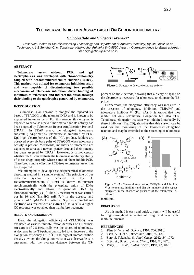
TELOMERASE INHIBITION ASSAY BASED ON CHRONOCOULOMETRY
Shinobu Sato and Shigeori Takenaka*
Research Center for Bio-microsensing Technology and Department of Applied Chemistry, Kyushu Institute of Technology, 1-1 Senshui-Cho, Tobata-ku, Kitakyushu, Fukuoka 840-8550 Japan. * Correspondence to: Email address
ABSTRACT
Telomerase assay without PCR and gel
electrophoresis was developed with chronocoulometry
coupled with hexaammineruthenium chloride (RuHex).
This method was utilized for telomerase inhibition assay
and was capable of discriminating two possible
mechanism of telomerase inhibition: direct binding of
inhibitors to telomerase and indirect inhibition through
their binding to the quadruplex generated by telomerase.
INTRODUCTION
Telomerase is an enzyme to elongate the repeated six bases of TTAGGG of the telomere DNA and is known to be expressed in tumor cells. For this reason, this enzyme is expected to serve as a new tumor marker and its activity has been detected by Telomerase Repeat Amplification Protocol (TRAP).
1 In TRAP assay, the elongated telomerase
substrate (TS)-primer by telomerase is amplified by PCR. Upon gel electrophoresis of the PCR product, ladders are observed every six base pairs of TTAGGG when telomerase activity is present. Meanwhile, inhibitors of telomerase are expected to serve as a new anticancer drug and their potency has been assessed by TRAP.
2 However, it is not certain
whether TRAP can evaluate the telomerase inhibitory ability of these drugs properly where some of them inhibit PCR. Therefore, a more effective PCR-free telomerase assay has been required.
We attempted to develop an electrochemical telomerase detecting method in a simple system.
3 The principle of our
detection system is depicted in Fig. 1. Hexaammineruthenium (RuHex) is known to interact stoichiometrically with the phosphate anion of DNA electrostatically and allows to quantitate DNA by chronocoulometry (CC).
4 The CC measurement was carried
out in 10 mM Tris-HCl (pH 7.4) in the absence and presence of 50 μM RuHex. After a TS primer- immobilized electrode was treated with an extract of HeLa cells, a higher CC response was obtained than that before treatment.
RESULTS AND DISCUSSION
Here, the elongation efficiency of (TTAGGG)n was evaluated at various immobilization densities of TS-primer. An extract of 2.5 HeLa cells was the source of telomerase. A decrease in the TS-primer density led to an increase in the elongation efficiency at 37
°C. The highest immobilization
density at which the elongation reaction was observable is in agreement with the average distance between the TS-
primers on the electrode, showing that a plenty of space on the electrode is necessary for telomerase to elongate the TS-primer.
Furthermore, the elongation efficiency was measured in the presence of telomerase inhibitors, TMPyP4
2 and
telomerase Inhibitor V5 (Fig. 2A). It is known that they
inhibit not only telomerase elongation but also PCR. Telomerase elongation reaction was inhibited markedly by these inhibitors (Fig. 2B), showing that this system can be used for the monitoring of the telomerase elongation reaction and may be extended to the screening of telomerase
inhibitors.
CONCLUSION
As this method is easy and quick to run, it will be useful for high-throughput screening of drug candidates which inhibit telomerase.
REFERENCES 1. Kim, N. W. et al., Science, 1994, 266, 2011. 2. Cian, A. D. et al., Biochimie, 2008, 90, 131. 3. Sato, S, Takenaka, S., Anal. Chem., 2012, 84, 1772. 4. Steel, A., B. et al., Anal. Chem., 1998, 70, 4670. 5. Perry, P. J. et al., J. Med. Chem., 1998, 41, 4873.
Figure 1. Strategy to detect telomerase activity.
Figure 2. (A) Chemical structure of TMPyP4 and Inhibitor V as telomerase inhibitor and (B) the number of the repeat elongated in the absence or presence of the telomerase in-hibitor.
(A) (B)NH
N NHN
N
NN
NCH3
CH3H3C
H3C
4(pCH3C6H4SO3-)
TMPyP4
O
O
HN N
NH
ON
O
Inhibitor V
0
2
4
6
8
10
12
(TT
AG
GG
)n
-In
hib
ito
r
TM
PyP
4
Inh
ibito
rV
220
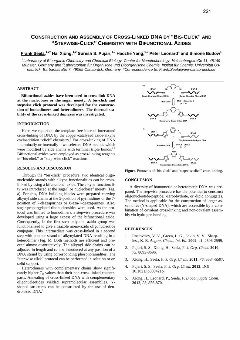
CONSTRUCTION AND ASSEMBLY OF CROSS-LINKED DNA BY “BIS-CLICK” AND
“STEPWISE-CLICK” CHEMISTRY WITH BIFUNCTIONAL AZIDES
Frank Seela,1,2*
Hai Xiong,1,2
Suresh S. Pujari,1,2
Haozhe Yang,1,2
Peter Leonard1 and Simone Budow
1
1Laboratory of Bioorganic Chemistry and Chemical Biology, Center for Nanotechnology, Heisenbergstraße 11, 48149
Münster, Germany and 2Laboratorium für Organische und Bioorganische Chemie, Institut für Chemie, Universität Os-
nabrück, Barbarastraße 7, 49069 Osnabrück, Germany. *Correspondence to: [email protected]
ABSTRACT
Bifunctional azides have been used to cross-link DNA
at the nucleobase or the sugar moiety. A bis-click and
stepwise click protocol was developed for the construc-
tion of homodimers and heterodimers. The thermal sta-
bility of the cross-linked duplexes was investigated.
INTRODUCTION
Here, we report on the template-free internal interstrand
cross-linking of DNA by the copper-catalyzed azide-alkyne
cycloaddition “click” chemistry.1 For cross-linking of DNA
– terminally or internally – we selected DNA strands which
were modified by side chains with terminal triple bonds.2-4
Bifunctional azides were employed as cross-linking reagents
in “bis-click” or “step-wise click” reactions.
RESULTS AND DISCUSSION
Through the “bis-click” procedure, two identical oligo-
nucleotide strands with alkyne functionalities can be cross-
linked by using a bifunctional azide. The alkyne functionali-
ty was introduced at the sugar4 or nucleobase
2 moiety (Fig.
a). For this, DNA building blocks were prepared carrying
alkynyl side chains at the 5-position of pyrimidines or the 7-
position of 7-deazapurines or 8-aza-7-deazapurines. Also,
sugar propargylated ribonucleosides were used. As the pro-
tocol was limited to homodimers, a stepwise procedure was
developed using a large excess of the bifunctional azide.
Consequently, in the first step only one azido group was
functionalized to give a triazole mono-azido oligonucleotide
conjugate. This intermediate was cross-linked in a second
step with another strand of alkynylated DNA resulting in a
heterodimer (Fig. b). Both methods are efficient and pro-
ceed almost quantitatively. The alkynyl side chains can be
adjusted in length and can be introduced at any position of a
DNA strand by using corresponding phosphoramidites. The
“stepwise click” protocol can be performed in solution or on
solid support. Heterodimers with complementary chains show signifi-
cantly higher Tm values than their non-cross-linked counter-
parts. Annealing of cross-linked DNA with complementary
oligonucleotides yielded supramolecular assemblies. Y-
shaped structures can be constructed by the use of den-
dronized DNA.5
Figure. Protocols of “bis-click” and “stepwise click” cross-linking.
CONCLUSION
A diversity of homomeric or heteromeric DNA was pre-
pared. The stepwise procedure has the potential to construct
oligonucleotide-peptide, -carbohydrate, or -lipid conjugates.
The method is applicable for the construction of larger as-
semblies (Y-shaped DNA), which are accessible by a com-
bination of covalent cross-linking and non-covalent assem-
bly via hydrogen bonding.
REFERENCES
1. Rostovtsev, V. V., Green, L. G., Fokin, V. V., Sharp-
less, K. B. Angew. Chem., Int. Ed. 2002, 41, 2596-2599.
2. Pujari, S. S., Xiong, H., Seela, F. J. Org. Chem. 2010,
75, 8693-8696.
3. Xiong, H., Seela, F. J. Org. Chem. 2011, 76, 5584-5597.
4. Pujari, S. S., Seela, F. J. Org. Chem. 2012, DOI
10.1021/jo300421p.
5. Xiong, H., Leonard, P., Seela, F. Bioconjugate Chem.
2012, 23, 856-870.
221
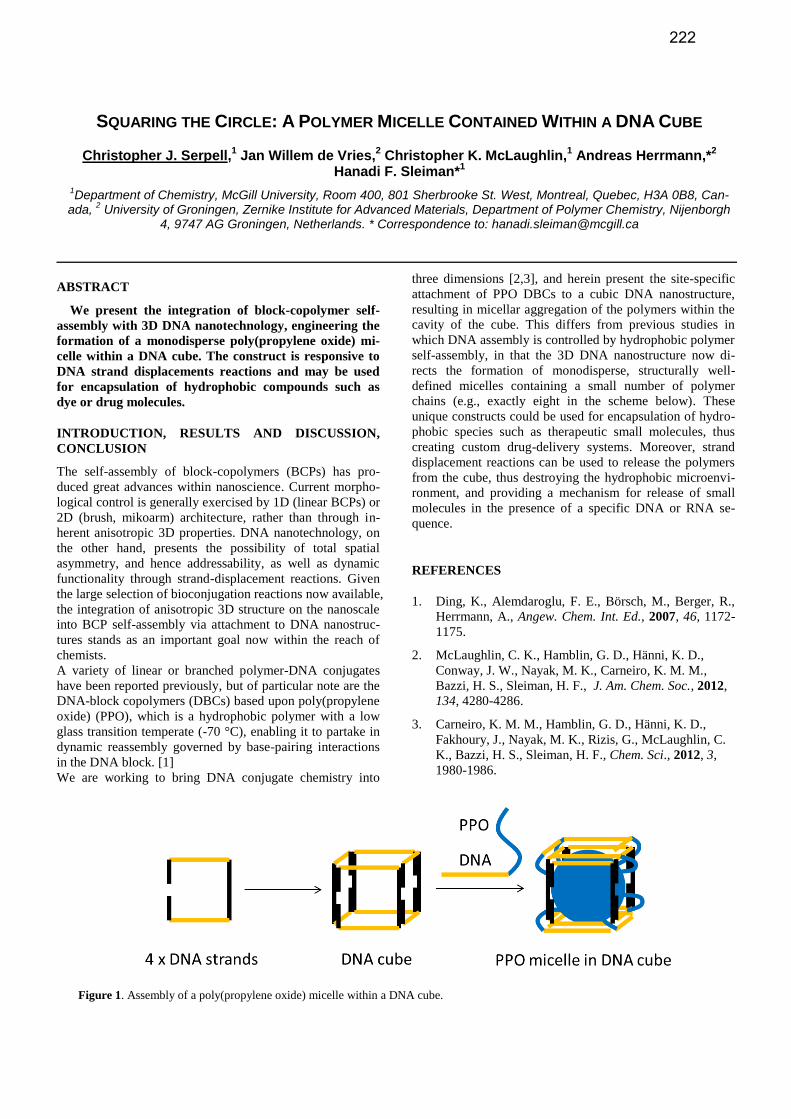
SQUARING THE CIRCLE: A POLYMER MICELLE CONTAINED WITHIN A DNA CUBE
Christopher J. Serpell,1 Jan Willem de Vries,
2 Christopher K. McLaughlin,
1 Andreas Herrmann,*
2
Hanadi F. Sleiman*1
1Department of Chemistry, McGill University, Room 400, 801 Sherbrooke St. West, Montreal, Quebec, H3A 0B8, Can-
ada, 2 University of Groningen, Zernike Institute for Advanced Materials, Department of Polymer Chemistry, Nijenborgh
4, 9747 AG Groningen, Netherlands. * Correspondence to: [email protected]
ABSTRACT
We present the integration of block-copolymer self-
assembly with 3D DNA nanotechnology, engineering the
formation of a monodisperse poly(propylene oxide) mi-
celle within a DNA cube. The construct is responsive to
DNA strand displacements reactions and may be used
for encapsulation of hydrophobic compounds such as
dye or drug molecules.
INTRODUCTION, RESULTS AND DISCUSSION,
CONCLUSION
The self-assembly of block-copolymers (BCPs) has pro-
duced great advances within nanoscience. Current morpho-
logical control is generally exercised by 1D (linear BCPs) or
2D (brush, mikoarm) architecture, rather than through in-
herent anisotropic 3D properties. DNA nanotechnology, on
the other hand, presents the possibility of total spatial
asymmetry, and hence addressability, as well as dynamic
functionality through strand-displacement reactions. Given
the large selection of bioconjugation reactions now available,
the integration of anisotropic 3D structure on the nanoscale
into BCP self-assembly via attachment to DNA nanostruc-
tures stands as an important goal now within the reach of
chemists.
A variety of linear or branched polymer-DNA conjugates
have been reported previously, but of particular note are the
DNA-block copolymers (DBCs) based upon poly(propylene
oxide) (PPO), which is a hydrophobic polymer with a low
glass transition temperate (-70 °C), enabling it to partake in
dynamic reassembly governed by base-pairing interactions
in the DNA block. [1]
We are working to bring DNA conjugate chemistry into
three dimensions [2,3], and herein present the site-specific
attachment of PPO DBCs to a cubic DNA nanostructure,
resulting in micellar aggregation of the polymers within the
cavity of the cube. This differs from previous studies in
which DNA assembly is controlled by hydrophobic polymer
self-assembly, in that the 3D DNA nanostructure now di-
rects the formation of monodisperse, structurally well-
defined micelles containing a small number of polymer
chains (e.g., exactly eight in the scheme below). These
unique constructs could be used for encapsulation of hydro-
phobic species such as therapeutic small molecules, thus
creating custom drug-delivery systems. Moreover, strand
displacement reactions can be used to release the polymers
from the cube, thus destroying the hydrophobic microenvi-
ronment, and providing a mechanism for release of small
molecules in the presence of a specific DNA or RNA se-
quence.
REFERENCES
1. Ding, K., Alemdaroglu, F. E., Börsch, M., Berger, R.,
Herrmann, A., Angew. Chem. Int. Ed., 2007, 46, 1172-
1175.
2. McLaughlin, C. K., Hamblin, G. D., Hänni, K. D.,
Conway, J. W., Nayak, M. K., Carneiro, K. M. M.,
Bazzi, H. S., Sleiman, H. F., J. Am. Chem. Soc., 2012,
134, 4280-4286.
3. Carneiro, K. M. M., Hamblin, G. D., Hänni, K. D.,
Fakhoury, J., Nayak, M. K., Rizis, G., McLaughlin, C.
K., Bazzi, H. S., Sleiman, H. F., Chem. Sci., 2012, 3,
1980-1986.
Figure 1. Assembly of a poly(propylene oxide) micelle within a DNA cube.
222

DNA STABILITY IN A HYDRATED IONIC LIQUID
H. Tateishi-Karimata1 and N. Sugimoto
1,2*
1Frontier Institute for Biomolecular Engineering Research (FIBER), and
2Faculty of Frontiers of Innovative Research
in Science and Technology (FIRST), Konan University,
7–1–20 Minatojima-minamimachi, Chuo-ku, Kobe 650–0047, Japan
ABSTRACT
In order to develop the DNA materials, we
investigated the stabilities of DNA duplexes in the
hydrated ionic liquid of choline dihydrogenphosphate
(choline dhp). Our results demonstrate that A–T base
pairs are more stable than G–C base pairs in a solution
containing choline dhp. This trend is the reverse of the
trend observed in the stability of DNA duplexes in a
standard biochemical solution. The unique interaction
between DNA and the hydrated IL is expected to be
useful for the development of environmentally friendly
advanced materials.
INTRODUCTION DNA has enormous potential for applications in the field
of nanobiotechnology and biomedical technology due to the
ability of single strands of DNA to recognize and hybridize
with their complementary sequences through highly specific
base-pairing interactions. An understanding of base pair
stability under various conditions is necessary for
development of DNA materials [1,2]. Room-temperature
ionic liquids (ILs) have generated tremendous interest as
non-volatile media that provide favorable environments for
a wide range of chemical reactions and a variety of
bioapplications. Choline dihydrogenphosphate (choline dhp)
with dissolved water is one of the hydrated ILs (Figure 1a),
which has been demonstrated to be a good solvent for
proteins and DNAs for ensuring long-term stability. It is
considered that the hydrated IL should be useful as a
medium developing “green” DNA materials. However, its
detail effect on the DNAs remains unclear.
RESULTS AND DISCUSSION
We investigated the stability of non-self-complementary
DNA duplexes by ultraviolet (UV) melting. The
thermodynamic parameters for DNA duplexes with different
sequences, Duplex1 (A-T rich sequence) and Duplex2 (G-C
rich sequence) (Figure 1b), were measured in solutions
containing 4 M choline dhp (80 wt%) or NaCl. NaCl was
used as the reference, because NaCl solution is a standard to
measure DNA stability. Our results showed that Duplex1
was more stable in the choline dhp solution than that in the
NaCl solution, whereas Duplex2 was instabilized in choline
dhp relative to the NaCl solution. To understand the
interaction between choline dhp and DNA, we carried out
thermodynamic analyses and molecular dynamics
calculations. Our results revealed the choline ions bound to
A–T rich regions, especially in the major groove of DNA
duplexes, and stabilized the A–T base pairs, whereas the G–
C base pairs were instabilized due to specific binding of
choline ions to G base in single strands. [2]
In the presentation, we will also investigate the stabilities
of triplexes and G-quadruplexes in the solution containing
the choline dhp and discuss the detail interaction between
choline ions and DNAs using thermodynamic analyses and
molecular dynamic calculations.
CONCLUSION
Our results indicate that A–T base pairs can be stabilized
relative to G–C base pairs in choline dhp solutions. The
DNA behavior in hydrated ILs shown here will be useful in
the design of oligonucleotides for use in nanomaterials.
ACKNOWLEDGEMENTS
This work was partly supported by the Grants-in-Aid for
Scientific Research and the ‘Core research’ project (2009–
2014), from the Ministry of Education, Culture, Sports,
Science and Technology, Japan.
REFERENCES
1. Pramanik, S., Nagatoishi, S., Saxena, S., Bhattacharyya,
J., Sugimoto, N., J. Phys. Chem. B,, 2011, 115, 13862-
13872.
2. Tateishi-Karimata, H., Sugimoto, N., Angew. Chem. Int.
Ed., 2012, 51, 1416-1419.
Figure 1. (a) The chemical structure of choline dihydrogenphosphate. (b) Sequence of the DNA duplexes used in this study.
223

FLUOROMETRIC IMAGING OF INTERCELLULAR POTASSIUM ION USING A DNA-PEPTIDE
CONJUGATE
Shigeori Takenaka,1* Shinsuke Ohzawa,
1 Kojirou Sota,
1 Shinobu Sato,
1 Tomoki Matsuda,
2 Yukiko
Yoshiura,3 Koji Nakazawa,
3 and Takeharu Nagai
2
1Research Center for Bio-microsensing Technology and Department of Applied Chemistry, Kyushu Institute of
Technology, Fukuoka 840-8550, Japan, 2The Institute of Scientific and Industrial Research, Osaka University, Osaka
567-0047, Japan and 3The Faculty of Environmental Engineering, the University of Kitakyushu, Fukuoka 808-0135.
*Correspondence to (Email address):[email protected]
ABSTRACT
Fluorescent potassium ion (K+) imaging probe PSO-5
was synthesized by conjugating a thrombin binding
aptamer (TBA) carrying FAM at its 5’-end with a
peptide carrying TAMRA and biotin in the middle and
at the C-terminus, respectively Upon binding of PSO-5
to the membrane surface through the ternary complex
consisting of sugar chain, biotinyl concanavalin A (Con
A) and streptavidin (StAv), FRET was observed which is
associated with K+ efflux from the cell as well as
apoptosis.
INTRODUCTION
To understand life it is important to clarify various phenomena occurring in the cell at the molecular level and several methods have been developed to monitor celluar processes. Especially, imaging of specific signaling molecules in the cell including metal ions may provide spatiotemporal information on their location in the cell. Since K
+ plays an important role in many physiological
events such as homeostasis in the heart muscle and hyperpolarization of neurons, it is important to develop not only a detection method for this cation, but also a fluorescence imaging technique. However, few papers that concerned with potassium ion sensing in aqueous media have been reported. Oligonucleotides with sequences of human telomere DNA or TBA are known to form tetraplex
structures upon K+ ion binding. Structural changes
associated with formation of tetraplex assemblies led to the development of potassium sensing oligonucleotide (PSO) probes, in which two fluorescent dyes were attached to either termini of a particular oligonucleotide. The combination of dyes included FRET and excimer emission approaches and the structural changes upon binding K
+ ion
could be monitored by a fluorescence technique. These systems showed very high binding preference for K
+ over
sodium ion (Na+).
1
To realize fluorescence imaging of intercellular K+
concentration gradient, PSO-5 was synthesized by the conjugation of TBA carrying FAM at the 5’-end with a peptide carrying TAMRA and biotin in the middle and at the C-terminus, respectively (Fig. 1A).
2 We tried to locate
PSO-5 on the cell surface through sugar chains of the cell surface, biotinyl ConA, and streptavidin as shown in Fig. 1B.
RESULTS AND DISCUSSION
To localize PSO-5 on the membrane surface, the follow-
ing reagents were added to HeLa cells in DMEM, biotinyl
ConA, StAv, and PSO-5 in this order, then incubated for 30
min at 37 oC in the CO2 incubator and washed with PBS.
After this treatment, fluorescence images of the cell were
measured by confocal laser fluorescence inverted microsco-
py. Fluorescence based on TAMRA was observed only on
the cell surface (Fig. 1C). Fluorescence based on TAMRA
decreased and that based on FAM increased upon addition
of K+ in the medium, showing that PSO-5 can be localized
on the cell surface and that K+ concentration gradient can be
monitored. Fluorometric imaging of the K+ concentration
change in the cell was monitored upon addition of ampho-
tericin B, ouabain, and bumetanide to induce K+ efflux from
the cell. The K+ concentration in the cell increased upon
addition of these drugs, as revealed by a F585/F517 change.
CONCLUSION
We successfully localized PSO-5 on the cell surface and monitored a K
+ concentration gradient upon addition of an
apoptosis-inducing drug.
REFERENCES 1. Ueyama, H. et al., J. Am. Chem. Soc., 2002, 124, 14286. 2. Ohtsuka, K. et al., Chem. Commun., 2012, 48, 4740..
Figure 1. Chemical Structure of PSO-5, (B) concept of localiza-
tion of PSO-5 in cell membrane and fluorescence change by K+
ion, and (C) Fluorescence image of HeLa cells based on the
TAMRA fluorescence.
224
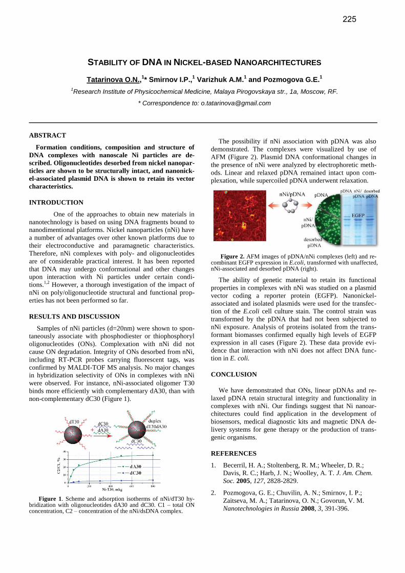
STABILITY OF DNA IN NICKEL-BASED NANOARCHITECTURES
Tatarinova O.N.,1* Smirnov I.P.,
1 Varizhuk A.M.
1 and Pozmogova G.E.
1
1Research Institute of Physicochemical Medicine, Malaya Pirogovskaya str., 1a, Moscow, RF.
* Correspondence to: [email protected]
ABSTRACT
Formation conditions, composition and structure of
DNA complexes with nanoscale Ni particles are de-
scribed. Oligonucleotides desorbed from nickel nanopar-
ticles are shown to be structurally intact, and nanonick-
el-associated plasmid DNA is shown to retain its vector
characteristics.
INTRODUCTION
One of the approaches to obtain new materials in
nanotechnology is based on using DNA fragments bound to
nanodimentional platforms. Nickel nanoparticles (nNi) have
a number of advantages over other known platforms due to
their electroconductive and paramagnetic characteristics.
Therefore, nNi complexes with poly- and oligonucleotides
are of considerable practical interest. It has been reported
that DNA may undergo conformational and other changes
upon interaction with Ni particles under certain condi-
tions.1,2
However, a thorough investigation of the impact of
nNi on poly/oligonucleotide structural and functional prop-
erties has not been performed so far.
RESULTS AND DISCUSSION
Samples of nNi particles (d=20nm) were shown to spon-
taneously associate with phosphodiester or thiophosphoryl
oligonucleotides (ONs). Complexation with nNi did not
cause ON degradation. Integrity of ONs desorbed from nNi,
including RT-PCR probes carrying fluorescent tags, was
confirmed by MALDI-TOF MS analysis. No major changes
in hybridization selectivity of ONs in complexes with nNi
were observed. For instance, nNi-associated oligomer T30
binds more efficiently with complementary dA30, than with
non-complementary dC30 (Figure 1).
Figure 1. Scheme and adsorption isotherms of nNi/dT30 hy-bridization with oligonucleotides dA30 and dC30. C1 – total ON concentration, C2 – concentration of the nNi/dsDNA complex.
The possibility if nNi association with pDNA was also
demonstrated. The complexes were visualized by use of
AFM (Figure 2). Plasmid DNA conformational changes in
the presence of nNi were analyzed by electrophoretic meth-
ods. Linear and relaxed pDNA remained intact upon com-
plexation, while supercoiled pDNA underwent relaxation.
Figure 2. AFM images of pDNA/nNi complexes (left) and re-combinant EGFP expression in E.coli, transformed with unaffected, nNi-associated and desorbed pDNA (right).
The ability of genetic material to retain its functional
properties in complexes with nNi was studied on a plasmid
vector coding a reporter protein (EGFP). Nanonickel-
associated and isolated plasmids were used for the transfec-
tion of the E.coli cell culture stain. The control strain was
transformed by the pDNA that had not been subjected to
nNi exposure. Analysis of proteins isolated from the trans-
formant biomasses confirmed equally high levels of EGFP
expression in all cases (Figure 2). These data provide evi-
dence that interaction with nNi does not affect DNA func-
tion in E. coli.
CONCLUSION
We have demonstrated that ONs, linear pDNAs and re-
laxed pDNA retain structural integrity and functionality in
complexes with nNi. Our findings suggest that Ni nanoar-
chitectures could find application in the development of
biosensors, medical diagnostic kits and magnetic DNA de-
livery systems for gene therapy or the production of trans-
genic organisms.
REFERENCES
1. Becerril, H. A.; Stoltenberg, R. M.; Wheeler, D. R.;
Davis, R. C.; Harb, J. N.; Woolley, A. T. J. Am. Chem.
Soc. 2005, 127, 2828-2829.
2. Pozmogova, G. E.; Chuvilin, A. N.; Smirnov, I. P.;
Zaitseva, M. A.; Tatarinova, O. N.; Govorun, V. M.
Nanotechnologies in Russia 2008, 3, 391-396.
225

SOLID-PHASE SYNTHESIS OF BORON-CONTAINING DNA ANALOGUES BY THE H-BORANOPHOSPHONATE METHOD
Sho Uehara,1 Shingo Hiura,1 Renpei Higashida,1 Natsuhisa Oka,2 and Takeshi Wada1*
1Department of Medical Genome Sciences, Graduate School of Frontier Sciences, The University of Tokyo, Biosci-
ence Building 702, 5-1-5 Kashiwanoha, Kashiwa, Chiba 277-8562, Japan and 2Department of Chemistry, Faculty of
Engineering, Gifu University, Yanagito, Gifu, Gifu 501-1193, Japan. * Correspondence to: [email protected]
ABSTRACT
H-Boranophosphonate DNA, which contains a P→BH3 and a P-H group, is a useful intermediate for the
synthesis of various boron-containing DNA analogues.
Oligomers of boranophosphate DNA, boranophos-
phorothioate DNA and boranophosphoramidate DNA
were successfully synthesized on solid support via H-
boranophosphonate DNA.
INTRODUCTION
The phosphorus atom-modified oligonuleotides have been widely used as therapeutic agents for numerous dis-eases because of their high stability toward nucleases and the relatively high cellular uptake. Recently, boron-containing oligonucleotides have been drawn attention due to their potential of medicinal applications. Boranophos-phate DNA (PBO-DNA) has higher nuclease resistance, lipophilicity and RNase H activity compared with those of natural DNA and has lower cytotoxicity than phos-phorothioate DNA [1]. Boranophosphorothioate DNA (PBS-DNA) has higher nuclease resistance and lipophilicity than those of PBO-DNA. Therefore, PBS-DNA is also ex-pected to be useful as a therapeutic agent [2]. We have de-veloped a new class of boron-containing DNA analogue, H-boranophosphonate DNA (PBH-DNA). PBH-DNA is an efficient intermediate to synthesize various boron-containing DNA analogues via the P-H activation and the following reaction with electrophiles [3]. Here, we report the solid-phase synthesis of oligonulceotides of PBO-DNA, PBS-DNA and a novel boron-containing DNA analogue, boranophosphoramidate DNA (PBN-DNA) via PBH-DNA.
RESULTS AND DISCUSSION
As shown in Scheme 1, thymidylate 12mers of PBO-DNA 4, PBS-DNA 5 and PBN-DNA 6 were synthesized on solid support. First, the reaction conditions were optimized for the dithymidine derivatives. Especially, the yields of the desired dimers were dependent on the conditions of conden-sation. The use of MNTP as a condensing agent and 2,6-lutidine as a base gave the dimers in excellent yields (96-98%). It was noteworthy that the reaction was completed within 1 minute. The dimers containing other nucleobases, A, C and G were also obtained in high yields as well as dithymidine derivatives.
Scheme 1. Solid-phase synthesis of thymidylate 12mers of PBO-DNA 4, PBS-DNA 5 and PBN-DNA 6.
Reaction conditions for the synthesis of the dimers were
applied to the synthesis of thymidylate 12mers. After the chain elongation by repeating the condensation and detrity-lation, conversions were carried out by treatment with elec-trophiles. Islated yields of thymidylate 12mers of PBO-DNA 4, PBS-DNA 5 and PBN-DNA 6 were 28%, 31% and 10%, respectively. Synthesis of oligonucletides containing all nucleobases and evaluation of physicochemical and physiological properties of them are in progress.
CONCLUSION
The thymidylate 12mers of PBO-DNA, PBS-DNA and PBN-DNA were successfully synthesized on solid support. The H-boranophosphonate method was found to be a prominent method for the synthesis of a variety of boron-containing oligonucleotides.
REFERENCES 1. Summers, J. S., Shaw, B. R. Curr. Med. Chem., 2001, 8,
1147-1155.
2. a) Lin, J., Shaw, B. R. Chem. Commun., 1999, 1517-1518. b) Lin, J., Shaw, B. R. Nucleosides, Nucleotides
Nucleic Acids, 2001, 20, 587-596.
3. Higashida, R., Oka, N., Kawanaka, T., Wada, T. Chem.
Commun., 2009, 2466-2468.
226

NMR INVESTIGATION OF THE STRUCTURE AND STABILITY OF MODIFIED DNA DUPLEXES
B. Van Gasse,1* V. Gheerardijn
1 ,D. Buyst
1, A. Madder
1 and J.C. Martins
1
1Department of Organic Chemistry, University of Ghent, Krijgslaan 281(S4), Ghent, Belgium
* Correspondence to: [email protected]
Here, we present an NMR study of the structure and
stability of several modified DNA duplexes, equipped
with histidine-like functionalities.
There is considerable interest in the development of en-
zyme mimics. These should retain the catalytic functionali-
ties of the original enzymes, but be incorporated into a sim-
pler template structure [1]. The enzyme of interest in our
research is α-chymotrypsine, which catalyses the cleavage
of peptide bonds. Its catalytic site consists out of three ami-
no acids; an aspartate, a histidine and a serine. To obtain a
serine protease mimic, these three functionalities should be
introduced into a suitable template structure. Recently,
Madder et al have developed the use of DNA duplexes as
template structures. The necessary functional groups can be
introduced through the incorporation of modified thymidine
nucleotides, in which the methyl group of the thymine base
is replaced with one of the three different functionalities,
necessary to form the catalytic site (Figure 1). Following a
preliminary molecular modelling study, aimed at determin-
ing the optimal position of the modified nucleotides in the
DNA duplex, a 14 base-pair duplex was chosen as a tem-
plate structure:
1 2 3 4 5 6 7 8 9 10 11 12 13 14
5’- G A C C A T T T G C A G C G -3’
3’- C T G G T A A T C G T C G C -5’
28 27 26 25 24 23 22 21 20 19 18 17 16 15
The different modified nucleotides need to be incorpo-
rated on the positions 6, 7, 8 and 21. Since only modified
thymidine nucleotides can be synthesized, this approach
does require the insertion of a T8.T21 mismatched base-pair.
Before engaging in the study of the completely modified
duplexes, the position-dependant impact of the insertion of a
single modified thymine on the structure and stability of that
duplex was investigated. The THis
nucleotide features a pH
sensitive imidazole group attached to the thymine base via a
flexible linker and with a pKa of 7.17. Four such modified
duplexes were compared against each other and the original
non-modified DNA duplex (depicted above), which is re-
ferred to as the wild type duplex. The four modified duplex-
es each contain one modified nucleotide, and differ with
respect to the position of the THis
, which was inserted at po-
sition 6, 7, 8 or 21 resulting in the T6His
, T7His
, T8His
, and
T21His
duplexes.
With NMR, we first studied the behaviour of the imino
proton resonances, which can serve as indicators of duplex
stability at the individual base-pair level. The imino proton
resonances were assigned via 2D NOESY spectroscopy and
through a partial 15
N-labelling approach [2]. The chemical
shift differences between the imino protons in the wild type
duplex and the modified ones, indicated the the introduction
of the modification only causes localized perturbations of
the duplex structure.
In addition, a stabilization of up to 5°C could be observed
for the T8His
and T21His
duplexes, i.e. when the histidine-
modification is introduced in the T.T mismatched base-pair.
This behaviour was confirmed by the UV-VIS based melt-
ing temperatures.
The pKa value of the imidazole ring in the various du-
plexes, determined from its pH dependent 1H chemical shift
through titration studies, was found to increase by at least
0.5 units for all modified duplexes and even 1.5 units for
T8His
, suggesting stabilising electrostatic interactions be-
tween the charged imidazole and the negatively charged
phosphate backbone. However, the much larger stabilisation
of T8His
compared to all other modified duplexes most prob-
ably results from additional specific interactions between
the imidazole group and neighbouring base-pairs, as evi-
denced from contacts in 2D NOESY NMR spectra. These
and other aspects relating to the impact of inserting such
THis
residues into the DNA backbone will be discussed in
more detail.
REFERENCES
1. Catry, M., Madder, A. Molecules, 2007, 12(1), 114-129.
2. Phan, A.T., Patel, D.J. J. Biomol. NMR, 2002, 124(7),
257-262.
His SerAsp
Ser
Figure 1. Image of the DNA template structure with the modified nucleotides (left), and the structure of the modified nucleotide used in the comparative study (right).
227

FACILE DERIVATIZATION OF PNA OLIGOMERS BY ON-RESIN CLICK CHEMISTRY
Rachael (Xiaoxiao) Wang1*
and Robert H.E. Hudson1
1Department of Chemistry, The University of Western Ontario, London, Ontario, CANADA N6A 5B7
* Correspondence to: [email protected]
ABSTRACT
An azido-containing PNA monomer has been devel-
oped that is compatible with Fmoc-oligomerization
chemistry. Incorporation of single or multiple azide la-
bels is possible and their subsequent reaction in the cop-
per-catalyzed azide-alkyne cycloaddition while attached
to an insouble resin has been demonstrated. A colour
changing molecular beacon construct based on the py-
rene chromophore has been prepared using this method.
INTRODUCTION
As our understanding of the genetic basis of disease has
grown, there has been an increasing demand for biomolecu-
lar tools and methods for nucleic acid detection. Molecular
beacons (MBs), in which a synthetic oligonucleotide is
functionalized with two chromophores, have great potential
in such applications.1 Hybridization of MBs with a comple-
mentary nucleic acid induces a conformational change and
gives rise to a specific spectroscopic signal that can be uti-
lized for detection. Herein, we present a novel chemical
approach to a PNA-based color changing molecular beacon2
assembled utilizing the well-known copper(I)-catalyzed
alkyne-azide Huisgen cycloaddition.3
RESULTS AND DISCUSSION
We have designed a convertible PNA monomer wherein
the azido group replaces the nucleobase (Fmoc-1-OH, Fig-
ure 1). This monomer may be inserted at any place within a
PNA sequence by Fmoc-based solid phase peptide synthesis
(SPPS) without degradation. It has been found to be com-
patible with both commercial PNA monomers and our pre-
viously reported Boc-protected monomers.4
Figure 1. Structure of azide PNA monomer, Fmoc-1-OH.
By application of Fmoc-1-OH, we present the first exam-
ple of the incorporation of pyrene functionalities into a PNA
oligomer using the click methodology (Scheme 1). Pyrene
chromophores were installed at the terminal ends of the oli-
gomer via 1,2,3-triazole moieties and were intended to im-
part colour-changing molecular beacon properties based on
presence of excimers versus monomers.
Scheme 1. The on-resin click reaction of ethynylpyrene with an azide-labelled PNA oligomer.
Hybridization properties with its cDNA strand were in-
vestigated by fluorescence, UV-Vis and CD studies and will
be reported.
CONCLUSION
In summary, a pyrene-functionalized molecular beacon
was synthesized exploiting the solid phase “click” reaction.
This versatile strategy is useful for singly or multiply label-
ling PNA oligomers in one step by an on-resin “click” reac-
tion. The PNA oligomers prepared by this method show no
evidence of damage by using a Cu(I) catalyst.
REFERENCES
1. (a) Tyagi, S., Kramer, F. R., Nature Biotechnology,
1996, 14, 303-308. (b) Wang, K., Tang, Z., Chaoyong,
J. Y., Kim, Y., Fang, X., Li, W., Wu, Y., Medley, C.
D., Cao, Z., Li, J., Colon, P., Lin, H., Tan, W. Angew.
Chem. Int. Ed., 2009, 48, 856-870.
2. Seo, Y. J., Hwang, G. T., Kim, B. H. Tetrahedron Lett.
2006, 47, 4037–4039.
3. Rostovtsev, V. V., Green, L. G., Fokin, V. V., Sharp-
less, K. B. Angew. Chem. Int. Ed., 2002, 41, 2596-
2599.
4. Wojciechowski, F., Hudson, R. H. E. J. Org. Chem.
2008, 73, 3807-3816.
228

THE THIRD BASE PAIR ‘DS-PX’ IN PCR AMPLIFICATION
Rie Yamashige,1* Michiko Kimoto
1,2, Yusuke Takezawa
1, Akira Sato
1, Tsuneo Mitsui
2,
Shigeyuki Yokoyama1,3
, Ichiro Hirao1,2
1RIKEN systems and Structural Biology Center (SSBC), Kanagawa, Japan,
2TagCyx Biotechnologies, Kanagawa, Ja-
pan and 3Deperment of Biophysics and Biochemistry, Graduate School of Science, The University of Tokyo, Tokyo,
Japan. * Correspondence to: [email protected]
ABSTRACT
Toward the expansion of the genetic alphabet of DNA,
we present a highly-specific artificial PCR amplification
system using the unnatural ‘Ds–Px’ pair.
INTRODUCTION
An artificial extra base pair (unnatural base pair) that
functions as a third base pair in replication, transcription,
and/or translation enables the site-specific incorporation of
functional groups into nucleic acids and proteins at desired
positions. This technology is applicable to a wide range of
biotechnologies. We have developed several unnatural base
pairs that exhibit high specificity in polymerase reactions.
Among them, an unnatural base pair between 7-(2-
thienyl)-imidazo[4,5-b]pyridine (Ds) and 2-nitro-4-
propynylpyrrole (Px) functions in PCR amplification with
high efficiency and selectivity (Fig. 1A). DNA fragments
containing the Ds–Px pair can be amplified 107–10
8-fold by
30–40 cycles of PCR, supplemented with the Ds and Px
substrates, using an exonuclease-proficient DNA polymer-
ase (Deep Vent DNA polymerase). More than 97% of the
Ds–Px pair retains in the amplified DNA. In addition, the
Px base can be modified with any functional group (R in Fig.
1A). Thus, a site-specific labelling of the amplified DNA
with desired functional groups is possible by PCR involving
the Ds–Px alongside natural A–T and G–C base pairs.
However, it is still unknown how high the specificity of the
Ds–Px pair is in PCR.
Here, to know the abilities and limitations of the Ds–Px
pair, we preformed 100 cycles of PCR using DNA frag-
ments containing the Ds–Px pair and further optimized the
PCR conditions. Then, we created a highly-specific artificial
PCR amplification system in which the Ds–Px pair exhibits
extremely high selectivity (selectivity of Ds–Px pairing:
>99.92%, misincorporation of unnatural bases opposite nat-
ural bases: 0.005%/bp/replication).
RESULTS AND DISCUSSION, CONCLUSION
To maintain an exponential amplification through the 100
cycle-PCR, the PCR solution was diluted after each 10 cycle
-PCR, and the amplification and dilution process was per-
formed 10 times. After each 10 cycle-PCR, the amplified
DNA fragments were analysed by PAGE to determine the
amplification fold, and were purified for further experiments.
The retention rate of the Ds–Px pair in the amplified frag-
ments was determined by sequencing methods supplement-
ed with unnatural base substrates, which we reported on
before (1). We also established a method to assess the mis-
incorporation rate of unnatural bases opposite natural bases
by fluorescent labelling of PCR products with a modified Px
substrate, Cy5-hx-dPxTP (2).
After 100 cycles of PCR, DNA fragments containing Ds–
Px (R = NH2-(CH2)5-CO-NH-) were amplified 1028
-fold,
and at least 97% of the Ds–Px pair still retained. The selec-
tivity of the Ds–Px pairing reached 99.97% per replication.
We also determined the misincorporation rate of unnatural
bases opposite natural bases, which was as low as 0.007%
/bp/replication. Furthermore, Diol substitution of the Px
base (Diol-Px, Fig.1B) reduced the misincorporation rate to
0.005% /bp/replication while preserving high selectivity of
the Ds–Px pairing (>99.92%). Thus, we found that the se-
lectivity of Ds–Px pairing in PCR under the optimized con-
ditions is very close to that of a natural base pair (error rate
of natural base paring under the same conditions is
~0.002%/bp/replication).
By using these optimized PCR conditions involving the
Ds–Diol-Px pair, we recently succeeded in generating DNA
aptamers containing the unnatural bases which bind to their
targets with higher affinity than those of existing aptamers
comprising only the natural bases. In this aptamer selection,
the Ds–Diol-Px pair survived though more than 150 cycles
of PCR, which verifies the Ds–Px pair’s capability as the
third base pair in PCR amplification. This highly-specific
artificial PCR amplification system can be applied not only
to aptamer selection but also a wide variety of applications
in new biotechnologies.
REFERENCES
1. Kimoto, M., Kawai, R., Mitsui, T., Yokoyama, S.,
Hirao, I. Nucleic Acids Res., 2009, 37, e34.
2. Yamashige, R., Kimoto, M., Takeawa, Y., Sato, A.,
Mitsui, T., Yokoyama, S., Hirao, I. Nucleic Acids Res.,
2012, 40, 2793. Fig. 1 The unnatural Ds-Px pair
229

SYNTHESIS OF ENEDIYNES FOR INCORPORATION INTO OLIGONUCLEOTIDES
Ningzhang Zhou, Ravi Shekar Yalagala and Hongbin (Tony) Yan*
Department of Chemistry, Brock University, 500 Glenridge Ave., St. Catharines, Ontario, Canada *Correspondence to: [email protected]
ABSTRACT
Compounds containing the enediyne moiety were syn-
thesized for incorporation into oligonucleotides. The
enediyne-modified oligonucleotides will be used to trig-
ger sequence-specific nucleic acid chain cleavage
through the Bergman cyclization.
INTRODUCTION
The enediyne moiety is found in a number of natural
products and can undergo the Bergman cyclization reaction,
leading to the formation of biradical intermediates.1 Some of
these compounds, such as calicheamycin,2 possess extraor-
dinary ability to cause DNA damage through double strand
cleavages involving the biradical intermediate.
Using an oligonucleotide tethered with the enediyne moi-
ety, we aim to enable sequence-specific nucleic acid chain
cleavages through in situ Bergman cyclization. We report
here the synthesis of enediyne moieties that can be incorpo-
rated into oligonucleotides through either direct phospho-
ramidite chemistry-based solid phase synthesis or post-
synthetic conjugation reactions.
RESULTS AND DISCUSSION
The synthesis of these compounds begin with the prepa-
ration of 1,2-cis-dihaloalkenes 4, the thermodynamically
less stable isomer of 1,2-dihaloalkenes. This task was
achieved through a three-step regiospecific sequence start-
ing from the corresponding terminal alkynes 1 (Scheme 1).
The cis-geometry was confirmed by NOE.3
Scheme 1. Synthesis of 1,2-cis-dihaloalkenes from terminal al-
kynes.
Subsequent Sonogashira couplings (Scheme 2) of the 1,2-
cis-dihaloalkenes 4 with suitable alkynes gave correspond-
ing functionalized enediynes 5.
R' R'
R
ClX
4R
Sonogashira coupling
5 Scheme 2. Preparation of functionalized enediynes through the
Sonogashira coupling reactions.
Compounds 5 are being incorporated into oligonucleo-
tides (as in 6) to mediate site-specific nucleic acid chain
cleavages via the Bergman cyclization reaction.
R' R'
6ODN
CONCLUSION
Functionalized enediynes are synthesized by So-
nogashira couplings of appropriate alkynes with 1,2-cis-
dihaloalkenes, which were readily prepared from the corre-
sponding terminal alkynes. These functionalized enediynes
will be incorporated into oligonucleotides to effect site-
specific nucleic acid chain cleavages via in situ Bergman
cyclization.
REFERENCES
1. Basak, A., Mandal, S., Bag, S. S. Chem. Rev. 2003, 103,
4077-4094.
2. Lee, M. D., Dunne, T. S., Siegel, M. M., Chang, C. C.,
Morton, G. O. and Borders, D. B. J. Am. Chem. Soc.
1987, 109, 3464-3466.
3. Zhou, N., Wang, Q., Lough, A., Yan, H. manuscript
submitted for publication.
230
EFFICIENT AND HIGH-FIDELITY COPYING OF AN RNA-LIKE MODEL PREBIOTIC SYSTEM
Shenglong Zhang,1,2,3
* Sergei M. Gryaznov4 and Jack W. Szostak
1,2,3
1Howard Hughes Medical Institute,
2Harvard Medical School, and
3Department of Molecular Biology and Center for
Computational and Integrative Biology, Massachusetts General Hospital, 185 Cambridge Street, Boston, Massachu-setts 02114, USA, and
4Geron Corporation, 230 Constitution Drive, Menlo Park, CA 94025, USA.
* Correspondence to: Email address [email protected]
ABSTRACT
Non-enzymatic RNA replication would provide an
important bridge to the RNA world. However, the
demonstration of efficient and high-fidelity copying
chemistry remains a great experimental challenge. Our
results show that a high rate of polymerization and a
high degree of fidelity are not mutually exclusive, but
can be achieved simultaneously in non-enzymatic copy-
ing of N3′-P5′-linked phosphoramidate DNA. The struc-
tural similarity of NP-DNA to RNA suggests that these
results could be translated to an RNA-only system.
INTRODUCTION, RESULTS AND DISCUSSION,
CONCLUSION
Experimental demonstration of non-enzymatic RNA rep-
lication requires an efficient mechanism that can lead to
both a high rate of polymerization and a high degree of fi-
delity in the copying chemistry. Previous experiments con-
cerning non-enzymatic template-directed synthesis of RNA
with activated monomers have led to the copying of short
RNA templates, but these reactions are generally slow (tak-
ing days to weeks) and highly error-prone. Therefore, the
ability to efficiently and accurately copy arbitrary template
sequences remains frustratingly out of reach. N3′-P5′-linked
phosphoramidate DNA is a highly reactive model for self-
replicating genetic materials and has been used for studies
of non-enzymatic RNA self-replication. It is also an excel-
lent RNA mimic, due to its similar overall duplex structure,
rigidity, and level of hydration [1]. Our experiments show
that the high reactivity imparted by the presence of an ami-
no nucleophile allows rapid and efficient copying of all four
nucleobases on both homopolymeric and mixed templates.
On the other hand, G:T wobble pairing leads to a high error
rate. We have therefore investigated the use of the modified
nucleobase, 2-thio T (Ts) [2], to suppress formation of the
G:T wobble base-pair.
Our results illustrate that the 2-thio modification can both
increase polymerization rate and enhance fidelity in this
self-replicating N3′-P5′-DNA system. These results suggest
that this simple nucleobase modification may have played a
role in primordial RNA (or proto-RNA) replication. In addi-
tion to suppressing the G:T mismatch, an additional benefit
gained from its stronger base-pairing with A is that it also
reduces A:C mismatch formation. Thus simple modifica-
tions of nucleobases might provide a means of suppressing
mismatches to yield better fidelity.
REFERENCES
1. Tereshko, V., Gryaznov, S., Egli, M. J. Am. Chem. Soc.,
1998, 120, 269-283.
2. Sintim, H. O., and Kool, E. T. J. Am. Chem. Soc., 2006,
128, 396-397.
231
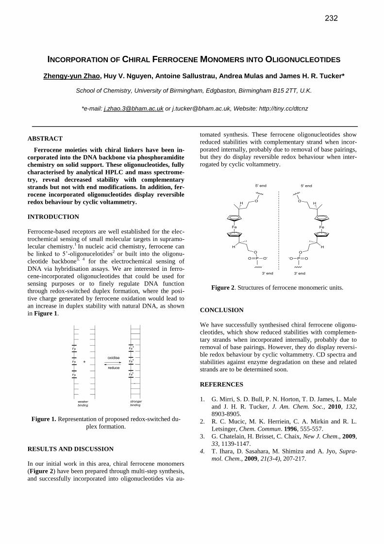
INCORPORATION OF CHIRAL FERROCENE MONOMERS INTO OLIGONUCLEOTIDES
Zhengy-yun Zhao, Huy V. Nguyen, Antoine Sallustrau, Andrea Mulas and James H. R. Tucker*
School of Chemistry, University of Birmingham, Edgbaston, Birmingham B15 2TT, U.K.
*e-mail: [email protected] or [email protected], Website: http://tiny.cc/dtcnz
ABSTRACT
Ferrocene moieties with chiral linkers have been in-
corporated into the DNA backbone via phosphoramidite
chemistry on solid support. These oligonucleotides, fully
characterised by analytical HPLC and mass spectrome-
try, reveal decreased stability with complementary
strands but not with end modifications. In addition, fer-
rocene incorporated oligonucleotides display reversible
redox behaviour by cyclic voltammetry.
INTRODUCTION
Ferrocene-based receptors are well established for the elec-
trochemical sensing of small molecular targets in supramo-
lecular chemistry.1
In nucleic acid chemistry, ferrocene can
be linked to 5’-oligonucelotides2 or built into the oligonu-
cleotide backbone3, 4
for the electrochemical sensing of
DNA via hybridisation assays. We are interested in ferro-
cene-incorporated oligonucleotides that could be used for
sensing purposes or to finely regulate DNA function
through redox-switched duplex formation, where the posi-
tive charge generated by ferrocene oxidation would lead to
an increase in duplex stability with natural DNA, as shown
in Figure 1.
Figure 1. Representation of proposed redox-switched du-
plex formation.
RESULTS AND DISCUSSION
In our initial work in this area, chiral ferrocene monomers
(Figure 2) have been prepared through multi-step synthesis,
and successfully incorporated into oligonucleotides via au-
tomated synthesis. These ferrocene oligonucleotides show
reduced stabilities with complementary strand when incor-
porated internally, probably due to removal of base pairings,
but they do display reversible redox behaviour when inter-
rogated by cyclic voltammetry.
Figure 2. Structures of ferrocene monomeric units.
CONCLUSION
We have successfully synthesised chiral ferrocene oligonu-
cleotides, which show reduced stabilities with complemen-
tary strands when incorporated internally, probably due to
removal of base pairings. However, they do display reversi-
ble redox behaviour by cyclic voltammetry. CD spectra and
stabilities against enzyme degradation on these and related
strands are to be determined soon.
REFERENCES
1. G. Mirri, S. D. Bull, P. N. Horton, T. D. James, L. Male
and J. H. R. Tucker, J. Am. Chem. Soc., 2010, 132,
8903-8905.
2. R. C. Mucic, M. K. Herriein, C. A. Mirkin and R. L.
Letsinger, Chem. Commun. 1996, 555-557.
3. G. Chatelain, H. Brisset, C. Chaix, New J. Chem., 2009,
33, 1139-1147.
4. T. Ihara, D. Sasahara, M. Shimizu and A. Jyo, Supra-
mol. Chem., 2009, 21(3-4), 207-217.
232
Related Documents