A peer reviewed journal. Editor Dr. Induwara Gooneratne Dept. of Forensic Medicine Faculty of Medicine University of Peradeniya Tel. 094-81-2388083 / 2396400 Sri Lanka E-mail : [email protected] Editorial Board Prof. Ravindra Fernando, MBBS, MD, FCCP, FCGP, FRCP (London), FRCP (Glasgow), FRCP (Edinburgh), FRCPath (UK), DMJ (London) Senior Professor Dept. of Forensic Medicine & Toxicology Faculty of Medicine, University of Colombo Dr. L.B.L. De Alwis, MB, BS (Cey), DLM (Colombo), MD (Colombo) Chief Consultant JMO (Retired) Colombo Dr. Colin Seneviratne, BSc, MSc, PhD (UK) Centre for Forensic & Legal Medicine University of Dundee, UK Dr. Induwara Gooneratne, BDS, Dip. in Forensic Medicine, MSc, MPhil (For.Med), LLM (USA), DTox, DHR, Attorney-at-Law Dept. of Forensic Medicine, Faculty of Medicine, University of Peradeniya Dr. Dinesh Fernando, MBBS, MD, DLM, DMJ (Lon.) Dept. of Forensic Medicine, Faculty of Medicine, University of Peradeniya Dr.(Mrs) D.H. Edussuriya, MBBS, MPhil (For.Med.) Dept. of Forensic Medicine, Faculty of Medicine, University of Peradeniya Dr. Amal Vadysinghe, MBBS, DLM, MD (Col.), D-ABMDI (USA) Dept. of Forensic Medicine, Faculty of Medicine, University of Peradeniya Dr. K.A.S. Kodikara, MBBS, MD, DLM, Attorney-at-Law Dept. of Forensic Medicine, Faculty of Medicine, University of Peradeniya International Advisory Board Prof. Corrine Parver, JD Professor of Health Law & Director, Health Law & Bio Ethics Project American University Washington DC, U.S.A. Prof. Derrick Pounder, MB, ChB, FRCPA, FFPathRCPI, MRCPath, FHKCPath Professor & Director Centre for Forensic & Legal Medicine University of Dundee, UK Prof. D. Ubelaker, PhD, DABFA Consultant to FBI & Adjunct Professor Smithsonian Institute Washington DC, U.S.A. Medicine Sri Lanka Journal of Science & Law Forensic

Welcome message from author
This document is posted to help you gain knowledge. Please leave a comment to let me know what you think about it! Share it to your friends and learn new things together.
Transcript

A peer reviewed journal.
Editor
Dr. Induwara Gooneratne Dept. of Forensic Medicine Faculty of Medicine University of Peradeniya Tel. 094-81-2388083 / 2396400
Sri Lanka E-mail : [email protected]
Editorial Board
Prof. Ravindra Fernando, MBBS, MD, FCCP, FCGP, FRCP (London), FRCP (Glasgow), FRCP (Edinburgh), FRCPath (UK), DMJ (London)
Senior Professor Dept. of Forensic Medicine & Toxicology Faculty of Medicine, University of Colombo Dr. L.B.L. De Alwis, MB, BS (Cey), DLM (Colombo), MD (Colombo) Chief Consultant JMO (Retired) Colombo Dr. Colin Seneviratne, BSc, MSc, PhD (UK) Centre for Forensic & Legal Medicine University of Dundee, UK Dr. Induwara Gooneratne, BDS, Dip. in Forensic Medicine, MSc, MPhil (For.Med),
LLM (USA), DTox, DHR, Attorney-at-Law Dept. of Forensic Medicine, Faculty of Medicine, University of Peradeniya Dr. Dinesh Fernando, MBBS, MD, DLM, DMJ (Lon.) Dept. of Forensic Medicine, Faculty of Medicine, University of Peradeniya Dr.(Mrs) D.H. Edussuriya, MBBS, MPhil (For.Med.) Dept. of Forensic Medicine, Faculty of Medicine, University of Peradeniya Dr. Amal Vadysinghe, MBBS, DLM, MD (Col.), D-ABMDI (USA) Dept. of Forensic Medicine, Faculty of Medicine, University of Peradeniya Dr. K.A.S. Kodikara, MBBS, MD, DLM, Attorney-at-Law Dept. of Forensic Medicine, Faculty of Medicine, University of Peradeniya
International Advisory Board
Prof. Corrine Parver, JD Professor of Health Law & Director, Health Law & Bio Ethics Project American University Washington DC, U.S.A. Prof. Derrick Pounder, MB, ChB, FRCPA, FFPathRCPI, MRCPath, FHKCPath Professor & Director Centre for Forensic & Legal Medicine University of Dundee, UK Prof. D. Ubelaker, PhD, DABFA Consultant to FBI & Adjunct Professor Smithsonian Institute Washington DC, U.S.A.
Medicine Sri Lanka Journal of Science & Law
Forensic

Sri Lanka Journal of Forensic Medicine, Science & Law 2010 Vol.1 No.2 1
Delivering a Forensic Expert Testimony for the Defense : Relevance, Hesitations and Reservations amongst Professionals in Sri Lanka. This article attempts to examine the existing medico legal structure and practice in Sri Lanka with a view to identify a) the space and opportunity available for a forensic expert to appear as an expert witness for the defense in a criminal court b) the challenges and obstacles such experts would have to face in appearing for the defense. This submission, in essence, illustrates the relevance of defense expert evidence in Criminal Jurisprudence and elucidates the challenges exist in the Sri Lankan Medico-Legal Framework. Towards the later part of this paper, several foreign cases are used to demonstrate the need and the bearing of defense testimony in criminal trials. Although the term “ forensic expert” is used for the purposes of this article to denote all such experts as identified as ‘expert witnesses ‘in accordance to the evidence ordinance of Sri Lanka, special reference has been made to the medical expert and expert scientific witness . Expert forensic witnesses can and are attending civil courts in matters connected to civil disputes, but, this article limits and focuses the discussion for criminal court room appearances. Law concerning an “expert witness” is clear in Sri Lanka. The term is defined in the section 45 of the evidence ordinance. The law does not define expert witnesses for the prosecution and defense separately. The Sri Lankan law introduces the “concept” of expert witness, and, depending on the perceived needs and pertinence of the court, the court may summon an expert either for the defense or for prosecution. However it is apparent that the appearances of expert witnesses for defense are very limited and scarce in Sri Lanka. In contrast, in developed countries almost all criminal trials have a defense forensic expert. Having a defense expert witness for both sides provides a unique platform with apparent balance of not only power, but intellectual-scientific commitment and bias in the court room. Therefore it is important to examine the situation in Sri Lanka and identify determinants for avoidance or hesitations for defense testimony by forensic experts in Sri Lanka.
The general practice in Sri Lanka involving medical/scientific –legal issues are that they are directed to a judicial medical officer by police (live cases) or coroner / magistrate (concerning a sudden / unnatural death). Cases of suspected murder, rape, assault are common examples. Other productions or biological samples collected from a scene of a crime are usually directed to the Government analyst for an opinion. As a result both the forensic medical officer and the Government Analyst send their reports to courts / relevant legal offices for administration of justice. When/If the trial comes up, the forensic experts who submitted reports are usually summoned by the state / prosecution for evidence. In the general practice of forensic medicine or science there is no involvement of a defense: nor defense attorney, nor defense scientific officer. It is ironically presumed that the state officers are handling such evidence so diligently and so perfectly. It is also ironically presumed that there would be absolutely no bias or no doubt about integrity or honesty of the government officers in handling medico-legal work. It is presumed that the state could prosecute, state could investigate, state could perform scientific tests, with no defense involvement prior to trial. However there is yet a tiny conflict with the traditional legal philosophical norms of natural justice here. The common suspect, in court has to defense himself with the hard earned nickels and dimes, challenging all these “state power”. This tradition, being long existed, many scientific experts are either oblivious or un engrossed to learn another better way of “doing things”. There are several pertinent issues that needs discussion concerning the above predicament scenario. According to the law, the state prosecutes against crime charges, the Attorney General Department, a state agency is handling this with the assistance of the police. The police, a state agency is investigating the crime. The Judicial medical officer and the government Analyst, both state employees manage investigations and sending reports. The judicial medical officer and the analyst works as a member of a team along with the police in investigating and visiting the scene. At the end of investigations,
EDITORIAL

Sri Lanka Journal of Forensic Medicine, Science & Law 2010 Vol.1 No.2 2
an indictment is filed and the State counsel calls the forensic expert to courts in his witness list. The forensic expert usually visits the state counsel’s chambers before the trial and at times exchange documents and a friendly smiles. The lay accused observes all these movements and interactions. In his mind a reasonable doubt, a negative opinion could develop concerning the integrity and biasness of forensic experts summoned by the state. The accused observes from the dock that all witnesses summoned by the prosecution had been against the accused. How could he assert only the scientific witness otherwise ? Justice studies unanimously endorse the notion that “ justice must not only be done, it must be seen to being done.” In the minds of the lay accused, whether this presumption adheres, is a question? Do we consider this an important idea or do we have other more important things to attend? The law seem to endorse a great trust in the state. The law assumes that the state is honest, prudent, integral, just, rational and unbiased. The state is an ideological entity but in practice, the state are the people: functions of the officers of the state. Could the officers of the state act/behave to the expectations of the law to such level of honesty and integrity. Are they different from the accused who is supposed to have been dishonest? Is it because of the state office? In other words, if the accused also held a state office, would he be honest and would not break law? Then, why are many state officers being indicted and punished for dishonesty and crimes? One may argue why not the accused not retain a top lawyer? How could this be possible: crimes are usually associated with poverty!. The relevance of a defense expert witness is numerous. It not only builds psychological esteem in the lay mind of the suspect, but also proposes an alternate insight to the case. It can also highlight technical and scientific insufficiencies and pit falls of the primary expert. The defense expert can also bring forth new evidence from the primary source of evidence presented in court. The defense expert can provide a scientific or rather technical defense for the accused based on sound scientific evidence, it can also demonstrate alternate or accepted but viewpoints different from prosecution witness . The defense expert with the help of the defense lawyer could create a valid doubt, also provide sufficient scientific evidence if available to discredit the prosecution expert
witness. Thus, the court room enlightens with academia and wisdom. Balancing all presented facts and opinions, then it is up to the court and the jury to decide which position is the most appropriate, given the specific context. In this approach, justice is not only being done: it is seen to be done. Unfortunately most of the time, the opinion and the position of the expert witness summoned by the prosecution is accepted without any challenge!. There is absolutely no barrier in the law for calling a defense expert witness. However, this costs money. The defense experts have to be paid for their services and travelling. On the other hand there are logistical and administrative barriers for a government officer to appear for defense and charge a fee. However retired experts do not have such an obstacle. There are many experts who are willing to provide an expert evidence for the defense. However in practice not many engage due to several socio-cultural and other reasons. One reason is that the expert who appear for the defense is culturally belittled by other collogues as “money minded etc”. The ironic fact is that the prosecution expert witness also appear for a fee in the form of a government approved rates, although it is minimal, including a traveling fee etc. Also for postmortem examinations, there is a payment on top of the government salary. As the existing practice has been going on for some generations, appearing for defense is not accepted by peers who follow the traditional professional culture. Second reason is that all forensic experts in Sri Lanka are known to each other well. Giving an opinion against another can create foe. A defense opinion can become a personal threat. One would not want to make an “enemy” in the name of justice!. The third reason is that a lot of experts in Sri Lanka are administratively subordinate to others as for hierarchy, although educationally equal. In this set up, no expert would want to provide an opinion against one of their teachers or administratively higher senior colleague and get their career ruined. Training , imbibing and berth under one roof allow stagnating similar attitudes, discarding alternate and differential views. On the other hand, the lawyers and judges aren’t familiar with forensic scientific content, therefore, the court do not go in to deeper scientific analysis of the case. They rather depend on ordinary witnesses or other legal techniques to acquit or to convict. This is an unfortunate situation in Sri Lanka.

Sri Lanka Journal of Forensic Medicine, Science & Law 2010 Vol.1 No.2 3
In many situations in Sri Lanka it is observed that the expert witness attempts to be or almost act as an “eye witness”. They tend to makes very strong opinions and positions as if he was at the vicinity of the incident and observed the entire event. It illustrates that such experts deny any other possibility other than theirs. Experts must realize that they provide facts and opinions to courts based on what they examined subsequent to the event concerned, and therefore what exactly happened could be distant to the opinion possessed. Although it is accepted that it is the responsibility of the defense lawyer to elucidate a defense from the existing expert, there are practical reasons why he cannot. Is it fair to consider the expert summoned by the state to call the “expert for the court” including for the defense?. The expert summoned by the prosecution engaged in the investigations with the police, usually have discussions with the state attorney about the case prior to the hearing. They hear histories, stories of victims through victims family. The forensic expert working with the police can develop a “convicting” mentality. In this context, is it possible to have a totally unbiased opinion? The flip side is that in the absence of defense experts , the opinion of the expert presented prevails. Practicing in a geographical region for a long time, association with the police, state attorneys and judges etc., predispose an expert witness to develop an unwarranted “power” in the court house that results in adamant and strong opinions. Although the court could dismiss an opinion of an expert theoretically, in practice, it is highly unlikely in the absence of a second opinion. Such developments are seen as negative attributes which could be negated with the use of suitable defense expert in the name of justice. The other central issue having one expert opinion is that it could only demonstrate one side of the science to a non- scientific court room. This opinion can be pre-decided or non evidence based: could even be an assertion. There is no practice of a verification of the photos, slides etc., shown by the expert witness, in Sri Lanka. They are presumed to be very honest and integral. Eventually, everything comes down to the personal integrity of the expert: Why should the suspect or the court trust the expert witness, amidst all sorts of allegations and corruption charges against many government officials? Could the scientific expert be different because he is trained in science? Although there are no legal restrictions, in routine practice, a defense medical officer or a defense
scientist are not allowed in a post mortem examination or in a clinical examination generally. There had been instances where such defense experts’ presence were allowed but those were VIP cases. To the commoner, those approaches are not possible. If there is a defense expert on behalf of the accused present in all scientific examinations, he could document the entire procedure and ensure that justice to the accused is protected. The government analyst is the sole authority on certain aspects of forensics in our country. In this context how a defense expert could be made available? Will a defense expert be allowed to observe proceedings in the government analyst department? Would such an approach generate a chaos? What type of chaos? How do you ensure all standards and protocols are followed in state labs which are on budget restrictions? Is examining documents produced alone enough in this respect? What could be the most appropriate way? What are the ways other countries do such functions? In Sri Lankan set up, forensic toxicologists could only exist in the Government analyst department, no other units are recognized by law as service units for forensic toxicology. These barriers have resulted forensic toxicologists well trained who used to work in other units to leave the country and lead fully fledged forensic toxicology labs in foreign university setups. Same applies to finger and palm print analysis and document analysis. Unless these restrictions are reformed no parallel development of these sciences could be envisaged. Many believe that the Sri Lankan police is not sufficiently trained to handle a serious forensic case. So are the inquires in to sudden deaths. Many serious crimes are dismissed in courts due to mishandling of evidence, and mal procedures. How could these impact the expert testimony of the scientific witness? It must be mentioned that in many countries, number of exonerations are done, based on defense expert testimony. Many accused are acquitted merely on the basis of the defense experts evidence and illustrations. Also, it must be mentioned that unscientific evidence and stubborn opinions provided in the past have incarcerated innocent people in many countries, many years after . Those suspects are exonerated using defense expert testimony. When will such a move appear in Sri Lanka?

Sri Lanka Journal of Forensic Medicine, Science & Law 2010 Vol.1 No.2 4
There is very little or no evidence concerning action on perjury by expert witnesses in Sri Lanka. There are no means to make checks and balances of the practice and evidence of an expert witness. In short there is no quality assurance mechanism for expert testimony in Sri Lanka. If there is a defense expert available, at least then there is arguably a balance of scientific power that could minimize bias, prejudice, adamancy, dishonesty of the expert testimony which in turn will illuminate justice. It is high time that a team of forensic scientists come forward in Sri Lanka for providing defense opinions, for the interests of justice. This not only improves quality of expert testimony and justice administration in the country but also facilitates advancement of forensic sciences.
Dr. Induwara Goonerathne Editor
Sri Lanka Journal of Forensic Medicine, Science and Law
& Senior Lecturer
Department of Forensic Medicine Faculty of Medicine
University of Peradeniya, Sri Lanka.

Sri Lanka Journal of Forensic Medicine, Science & Law 2010 Vol.1 No.2 5
D.M.G. Fernando & *L.B.L. De Alwis
Department of Forensic Medicine, University of Peradeniya and *Retired Chief Judicial Medical
Officer, Colombo, Sri Lanka.
The use of chemical1 and biological weapons in war is prohibited by the Geneva Protocol of 1925 and the Biological weapons convention of 1992.2
A) CHEMICAL WARFARE
A chemical used in warfare is called a chemical warfare agent (CWA) and involves using toxic properties of chemical substances to kill, injure or incapacitate an enemy. Chemical warfare agents include gases, liquids and solids.3 Effects of these “CWA” include local irritant effects and systemic toxic effects. Chemical Warfare Agents (CWA) can be classified as persistent or non-persistent. Agents classified as “non-persistent” lose their effectiveness after a few minutes or hours. These include chlorine, sarin and other nerve gases. “Persistent agents” include those that remain in the environment for several weeks. Medical personnel involved in decontamination and treatment must protect themselves using special suits such as “HAZMAT” suits. Modern chemical warfare began during World War I and Germany was the first country to employ chemical warfare in the battle field. For many terrorist organizations, chemical warfare might be considered an ideal choice for a mode of attack as they are cheap, has a long shelf life, easy to transport, difficult to detect and effects (death and disability) are immediate. The first successful use of chemical agents by terrorists against a civilian population was on 20th March 1995 where “Aum Shinrikyo’, an apocalyptic group based in Japan released “Sarin” into the Tokyo subway system, killing twelve (12) and injuring over 5000 people. About seventy (70) different chemicals have been used or stockpiled as chemical warfare agents (CWA) during the 20th and 21st century. 01. Mustard Gas4
It is chemically referred to as “Dichloroethyl Sulphide”. It is a vesicant or blister forming gas and it is also referred to as “Sulphur Mustard” gas. It is alleged that it was used during World War I.5,6
During World War II, it was used by the Japanese Imperial Army. It was also used in the war between Iran and Iraq, in 1980 to 1988. It is a volatile liquid contained in “shells” which are fired into the enemy territory. Others include “Nitrogen Mustard” which is similar in action. Clinical Features i. Skin blisters and vescicles leading to
ulceration and infection.
ii. Irritation of the eyes causing conjunctivitis, corneal ulcerations and erosions leading to scarring, impairment of vision and blindness in survivors.
iii. Irritation of the mucosal surfaces of the nose,
larynx and upper and lower respiratory passages causing cough and dyspnoea due to chemical bronchitis, bronchiolitis, alveolitis and fatal acute pulmonary oedema. In survivors there can be bacterial broncho-pneumonia which again can be fatal.
iv. Irritation of the larynx often leads to laryngeal
oedema and possible death due to asphyxiation.
v. Irritation of the mouth, throat and oesophagus
causes nausea, vomiting and epigastric pain. Mustard gas is a persistent warfare agent and a contact hazard.
02. Phosgene It is chemically referred to as carbonyl chloride. Like chlorine and ammonia it is a very irritant gas and also has a corrosive effect. It is mainly a pulmonary irritant. It is available in ‘canisters’. The French modified artillery ammunitions to contain phosgene in response to chlorine canisters used by the Germans.
CHEMICAL AND BIOLOGICAL WARFARE

Sri Lanka Journal of Forensic Medicine, Science & Law 2010 Vol.1 No.2 6
Clinical features i. Irritation of the eyes, causing burns and
ulceration, leading to scarring, impairment of vision and blindness in survivors.
ii. Irritation of mucosal surfaces of the nose, upper and lower respiratory tracts causing cough and dyspnoea due to chemical bronchitis, bronchiolitis, alveolitis and fatal pulmonary oedema. In survivors there can be lung fibrosis or bacterial broncho-pneumonia which can be fatal.
iii. Exposure to high concentrations may result in death.7
03. Sarin Gas It is a nerve gas falling into the same category was ‘Tabun’, ‘Soman’, ‘VX’, ‘VR’, etc. Sarin, Tabun and Soman were the three ‘German nerve agents” of the day. Sarin gas is an organic ester of phosphoric acid and is chemically an organophosphate. Sarin gas was released into a Tokyo subway by a terrorist group killing 12 and injuring over 5000 as mentioned earlier. The organophosphates used in chemical warfare are more toxic than those used in other pesticides and capable of causing rapid death. Clinical features Poisoning occurs from inhalation and skin absorption. i. Nicotinic actions at neuro-muscular junctions ii. Muscarinic actions (DUMBELS) iii. Direct effect on the central nervous system
Death usually occurs from respiratory failure. Of the nerve gases “VX’ is a persistent chemical warfare agent (CWA) and a contact hazard. 04. Arsine8 There are organic compounds of Arsine and fired in artillery shells. They include :- i. Diphenyl chlorascine ii. Diphenyl amyl chlorascine iii. Diphenyl cynarascine
Inhaled arsine is extremely toxic. It has a powerful effect of destroying red blood cells (intra-vascular haemolysis) leading to haemoglobin uria, acute tubular necrosis and renal failure.
05. Chlorine Germans used chlorine in World War I. They simply opened canisters of chlorine upwind of the opposing side and let the prevailing winds do the dissemination. In early 2007, multiple terrorist bombings have been reported in Iraq using chlorine gas. As mentioned earlier it is mainly an irritant to the skin and eyes. But the main irritant effects are observed in the respiratory system causing severe breathing problems and also death similar to ‘Mustard gas’, phosgene’ etc. 06. Hydrogen cyanide This group also includes cyanogen chloride. They are cytotoxic agents and causes death by ‘histotoxic anoxia’. German dictator and ‘mass murderer’, Adolph Hitler used cyanide gas in his famous gas chambers to exterminate millions of Jews. (Refer chapter 8 on ‘cyanide poisoning’). 07. Sensory irritants9
They are also referred to as ‘Lachrymators’. They are fired in artillery shells or canisters. They are commonly used by law enforcement authorities mainly in riot control or to disperse people voicing protests against the government in power. 7.1 Chloracetaphenone – CN10
It is referred to as ‘tear gas’. It causes irritation of the eyes causing lachrymation, blurring of vision and temporary blindness. It also irritates the skin, nasal mucosa and respiratory tract mucosa causing bronchospasm. Deaths have been reported.10
7.2 Chlorobenzylidene malonitrite – CS It is more irritant but less toxic than chloracetaphenone.11 However it causes skin and eye irritation, irritation of the nasal and respiratory mucosa with lachrymation, rhinorrhoea and difficulty in breathing.
7.3 Dibenzoxazipine – CR
It is more potent and less toxic than CN or CS.12
08. Agent 15 (B2) It is an incapacitating poison and extremely persistent in soil and water. Actions are similar to atropine. Erratic behaviour, confusion, hallucinations, incoordination and blurring of vision due to mydriasis are the common clinical manifestation. These will be helpful to defeat enemy forces.

Sri Lanka Journal of Forensic Medicine, Science & Law 2010 Vol.1 No.2 7
09. Fentanyl Derivatives On 26th October 2002, Russian Special Forces used a chemical agent (Presumably KOLOKOL-1) an aerosolized fentanyl derivative as a precursor to an assault on Chechen terrorists ending the Moscow theatre hostage crisis. 10. Herbicides It is recorded that between 1961 to 1967, the US Air-force sprayed 12 million US gallons of concentrated herbicides, mainly “Agent Orange” (containing dioxin as an impurity in the manufacturing process) to destroy vegetation in South Vietnam. This caused about half a million (500,000) children to be born with dioxin related deformities. 11. Lewisite It is a blister agent like sulphur mustard etc. It was used by the imperialist Japanese army during World War II. But unlike sulphur mustard which take a few hours, the actions of the Lewisite are immediate. Its actions are persistent and also a contact hazard. Clinical features are similar to those of ‘sulphur mustard’ and ‘phosgene’. 12. Other gases
12.1 Carbon monoxide 12.2 Hydrogen sulphide 12.3 Hydrogen chloride 12.4 Oxides of nitrogen
13. Toxins
13.1 Botulinum toxin 13.2 Ricin 13.3 Saxitoxin 13.4 Abrin 13.5 Mycotoxins
B) BIOLOGICAL WARFARE Offensive use of infective living organisms against enemies and civilians around them during a war is considered to be biological warfare. Even bacterial exo-toxins propagated through food, water and air which could incapacitate enemies and civilians are also considered to be weapons of biological warfare.
Biological weapons takes three major forms. a. Deliberate poisoning of food and water with
infectious material. b. Use of micro-organisms, toxins or animals,
living or dead, in a weapon system. c. Use of biologically inoculated fabrics. A successful biological attack will have a devastating impact and could result in millions or even billions of deaths and cause severe disruption to societies and economies. Ideal characteristics of biological weapons are high infectivity, high potency, non-availability of vaccines and delivering as an aerosol. Diseases considered for weaponization or known to have been weaponized include Anthrax, Plague, Ebola, Tularaemia, Cholera, Marburg virus, Brucellosis, Q fever, Machupo, coccidiodes mycosis, Glanders, Melioidosis, Shigella, Rocky mountain spotted fever, Psittacosis, Yellow fever, Japanese B encephalitis, Rift valley fever and small pox. (A minimum of 20 diseases). As mentioned earlier naturally occurring toxins like Ricin, Abrin, Botulinum toxin, saxitoxin and mycotoxins can also be used in Biological warfare. Biological warfare can also specifically target plants to destroy crops or defoliate vegetation. Attacking animals is another area of biological warfare intended to eliminate animal resources for transportation and food. It is important to note that all of the classical and modern biological warfare are diseases of animals, the only exception being small-pox. Therefore it is most likely that such animals will become ill earlier than humans. Today, at least 17 nations are believed to have offensive biological weapons programs.13
1. Plague The earliest documented incident of the intention to use biological weapons is recorded in the Hittite texts of 1500-1200 BC, in which victims of plague were driven into enemy lands. During the middle ages victims of bubonic plague were used for biological attacks. This was either by flinging corpses (dead bodies) or the excrement of victims using catapults over walls into castles. In the Second World War, Imperial Japanese Airforce bombed Ningbo with ceramic bombs full of fleas carrying bubonic plague.14 Plague is caused by Yersinia pestis a gram negative bacillus. The vector is the rat flea Xenopsyllacheopis. The fleas

Sri Lanka Journal of Forensic Medicine, Science & Law 2010 Vol.1 No.2 8
bite humans causing plague. Clinical features are attributed to an endo-toxin.15
2. Anthrax During the First World War, Germany pursued an ambitious biological warfare programme and Anthrax was used. Field testing carried out in the United Kingdom during World War II left Gruinard Island in Scotland contaminated with anthrax for the next 48 years. Anthrax is produced by Bacillus anthracis. It produces a toxin which is very virulent. Spores are used in warfare as they can withstand extremes of temperature and humidity. The spores are further perfect for disposal by aerosols. Inhalation of such spores results in dyspnoea, marked cyanosis and death (wool sorters disease). Fatality rate is 90% or higher. In the largest biological weapon accident known, the accidental aerosolized release of antrax spores caused the anthrax outbreak in Sverdlovsk in the Soviet Union in 1979, resulted in 68 deaths and sheep became ill as far as 200 km from the release point of the organism from a military facility. This area is still out of bounds for visitors.16 An anthrax aerosol is odorless. On September 18, 2001, and a few days after, several letters were received by members of the U.S. Congress and media outlets containing anthrax spores. The attack killed five persons.17 In the case of Anthrax, it is likely that by 24-36 hours after an attack those with compromised immune system or those who have received a large dose of the organism due to proximity to the release point will become ill with classical signs and symptoms. When diagnosed early, about 80% can be treated, with antibiotics. If not detected ‘early’, the mortality rate is moderately high. A 993 report by the US congressional Office of Technology Assessment estimated that between 130,000 and 3 million deaths could follow the aerosolized release of 100kg of Anthrax spores upwind of the Washington, DC, area – lethally matching or exceeding that of a hydrogen bomb.18 Iraq has acknowledged producing and weaponizing Anthrax.19 During a 1945 outbreak in Iran, 1 million sheep died. The terrorist group Aum Shinrikyo (responsible for releasing sarin gas in a Tokyo, Japan subway station in 1995) also dispersed aerosols of anthrax and botulism throughout Tokyo on at least 8 occasions. For unclear reasons attacks failed to produce illness.20
3. Clostridium botulinum The bacterium produces neurotoxins A, B and E causing marked neuromuscular blockage. It is a form of food poisoning. Botulinum toxin is also used in biological warfare. After the 1991 Persian Gulf War, Iraq admitted to the United Nations inspection team of having produced 19,000 litres of concentrated botulium toxin, of which 10,000 litres were loaded into military weapons. These 19,000 litres have never been fully accounted for. This is approximately three (3) times the amount needed to kill the entire current human population by inhalation.21 4. Tularaemia This is due to infection by Franciscella tularensis a gram negative organism. Vectors are ticks and blood sucking flies whose bites cause infection in humans. It can devastate and incapacitate families, communities, civilians and combatants in times of civil war. 5. Cholera It is caused by a gram negative bacillus called Vibrio cholerae. The infection results in severe diarrhoea leading to dehydration, hypovolaemia, electrolyte imbalance, hypotension, circulatory collapse and death. Like Tularaemia it can devastate and incapacitate families, communities, civilians and combatants in times of civil war.

Sri Lanka Journal of Forensic Medicine, Science & Law 2010 Vol.1 No.2 9
REFRENCES
1. Lockwood AH, Nerve Gases (1991), The physicians for social responsibility quarterly; 2:69 76
2. Mason JK and Purdue BN (200), Pathology of
Trauma, 3rd edition, Arnold. ch 7:p97 3. Lockwood AH, Nerve Gases (1991), The
physicians for social responsibility quarterly; 2:69 76
4. Mason JK and Purdue BN (200), Pathology of
Trauma, 3rd edition, Arnold. ch 7:p97 5. Reddy KSN (1995), The Essentials of Forensic
Medicine and Toxicology. 9th edition, ch:37; p485 6. Williams JL (1993), Pathological and clinical
aspect of mustard gas intoxications, Intensive and Critical Care Digest, 12:1-2
7. Eeisenmenger et al (1991), Clinical and
Morphological findings in mustard gas intoxications.
8. Dacre JC and Goldaman M (1966), Toxicological
and Pharmacology of chemical warfare agent ‘Sulphur mustard’, Pharmacological review, 48:228-326
9. Fernando R., Management of Poisoning (2007),
Natural Poison Information centre, National Hospital, Colombo. 3rd edition, p93.
10. Smith Sydney and Fiddes FS (1949), Forensic
Medicine, 9th edition, London Churchill: p466. 11. Mason JK and Purdue BN (2000), Pathology of
trauma, 3rd edition, Arnold. Ch;6:p76. 12. Stein AA and Kirwan WE (1964),
Chloracetaphenone poisoning, A clinical pathological report, J. Forensic Science, 9:374-382.
13. Himsworth H, 1969 and 1971, Report of Inquiring
into the medical and toxicological aspects of C.S, Parts 1 and 2, London HMSO.
14. Ballantyne B and Swanston DN (1974), The irritant
effect of dilute solutions of Dibenzoxapine on eyes and tongue, Acta Pharmacological Toxicologica, 35:412-413.
15. Cole LA. The spectre of biological weapons. Sci
Am. December 1996:60-65 16. Daniel Barenblatt (2004), A plague upon humanity,
p32. 17. Kumar and Clark, Clinical Medicine, 4th edition,
WB Saunders, ch 1: p36. 18. Meselson et al (1994), The Sverdlovsk Anthrax
outbreak in 1979, Science 266:1202-1208. 19. Gray, Collin (2007), Another bloody century:
Future welfare, P 265-269, Phoenix ISBN 0304367346.
20. Office of Technology Assessment, US Congress,
1993 21. Zilinkas RA. Iraq’s biological weapons: the past as
future? JAMA. 1997;278:418-424 22. WUDUnnS, Miller J, Broad W. How Japan germ
terror alerted world. New York Times, May 26, 1998:1-6
23. Rheinhart, Courtney Elizabeth, “Clostridium
botulinum toxin development in refrigerated reduced oxygen packaged Atlantic croaker.
Sri Lanka Journal of Forensic Medicine, Science & Law 2010 Vol.1 No.2 10
IS KNOWLEDGE ON “MEDICO-LEGAL ASPECTS OF CHILD ABUSE” ADEQUATE AMONG POLICE OFFICERS?
A.N. Vadysinghe, U.S.N. Ranmohottige, I.R. Weerakkody & R. Aluthgedara
Dept. of Forensic Medicine, Faculty of Medicine, University of Peradeniya
INTRODUCTION Child abuse (CA), which is a worldwide phenomina1, continues to be a major crisis in the current society. It is defined as child maltreatment including any act or series of acts of commission or omission by a parent or other caregiver that result in harm, potential for harm, or threat of harm to a child2. Abuse of children can be divided into 4 main types: physical abuse, neglect, sexual abuse and emotional and psychological abuse which may coexist 3. Kempe, identified physical abuse of children and assigned the specific name “battered child syndrome” in 19624. Nonetheless it was only in 1980s that sexual abuse of a child, which is not a novel problem5 was identified as a problem in the western society6. In response, law-makers throughout the world have responded in different ways to bring the perpetrators of such crimes to justice5. Diagnosis and management of child abuse is difficult5 and has to be done with a multidisciplinary approach involving police officers, medical professionals, as well as legal professionals7, 8. Therefore, precise knowledge on the subject of child abuse among these personnel is essential. In our experience, police officers play a vital role in management of child abuse in Sri Lanka. Interactions between the abused child, Judicial Medical Officer (JMO) and the court have almost always been handled by a police officer. Hence it is exceedingly important for police officers to be thoroughly acquainted on medico-legal aspects of child abuse. Incidents of child abuse and neglect often said to go undetected because police or other first responders coming into contact with children do not identify injuries, conditions, or behaviors as suspicious9. When no intervention is offered, an abused child has up to a 10 per cent risk of having eventually fatal injuries10. The unavailability of Sri Lankan research literature with reference to awareness of police officers about medico-legal issues of child abuse together
with apparent lack of their knowledge on the subject have inspired us to conduct this study. OBJECTIVES The objective of this study was to ascertain the knowledge among police officers, who are more than 10 years in police service, in central province, regarding the essential medico-legal aspects of child abuse. METHODOLOGY We have identified 3 major areas with reference to medico-legal aspects of child abuse in which police officers need to be aware of adequately. Hence we have structured questions to cover those broad areas beginning from legally accepted age limit for definition of a child since “age” is a key element in several offences concerning children. Second aspect is types and features of child abuse which includes awareness of physical, sexual abuse, neglect and how to look for evidence during investigation of a case. Thirdly with regards to his duty as a police officer and the duty towards JMO, he needs to be thoroughly familiar with procedures including obtaining the statements, documentation, collecting evidence and informing relevant professionals who are involved in management of child abuse, to handle the process early and effectively. A randomly selected sample of 196 police officers was provided with a questionnaire on above aspects, following a brief introduction and subsequent to obtaining their consent. Marks were given out of hundred for each area and the results were analyzed.

Sri Lanka Journal of Forensic Medicine, Science & Law 2010 Vol.1 No.2 11
RESULTS The study sample consisted of 60 male police officers, 89 female police officers and rest 47 were unmarked. Their number according to their ranks is as follows:
CI = Chief Inspector IP = Inspector SI = Sub Inspector PS = Police Sergeant PC = Police Constable.
First aspect of our study was to ascertain the awareness of age limit. Out of the total study sample of 196, 75.5% have responded with the exact age limit to be known as a “child”, which is shown on the figure 01. However 24.5% of the sample was not aware of the correct age limit.
Figure 01: Knowledge on Age Limit of a child
Out of the total study sample of 196, 95% got less than 75% and 59% had marks 50% and below for knowledge on types of child abuse. Remarkably 7% got marks between 0 to 25% which is pointed up in figure 02. Only 5% managed to get marks above 75%.
Figure 02: Knowledge on Type of Abuse
Out of the total study sample of 196, 86% got 75% and below marks for features of child abuse as shown in figure 03. 54% were 50% and less aware of the features of child abuse. 4% received zero marks for knowledge on features of child abuse.
48
148
0
50
100
150
200
FALSE CORRECT
No
of P
olic
e O
ffice
rs
Response
Knowledge on Age Limit of a Child
14
102
70
10
0
20
40
60
80
100
120
0-25 26-50 51-75 76-100
No
of P
olic
e O
ffice
rs
Marks
Knowledge on Type of Abuse
Post CI IP SI PS PC Unmarked Number 8 22 32 26 61 47
Sri Lanka Journal of Forensic Medicine, Science & Law 2010 Vol.1 No.2 12
Figure 03: Knowledge on Features of Child Abuse
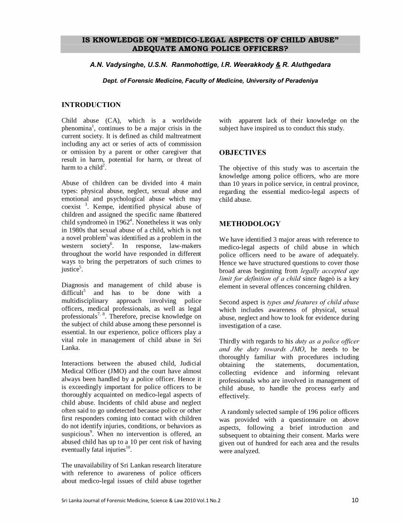
Out of the total study sample of 196, 31% scored equal and less than 75% marks for their duty in management of child abuse. 3% were 50% and less aware of their own duty as illustrated on figure 04. 1% was 25% and less aware of their duty in handling child abuse.
Figure 04: Knowledge on Duty of Police Officers
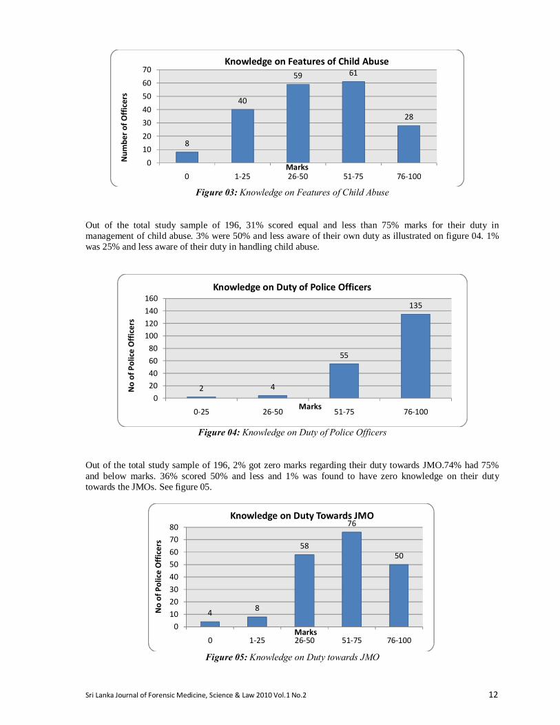
Out of the total study sample of 196, 2% got zero marks regarding their duty towards JMO.74% had 75% and below marks. 36% scored 50% and less and 1% was found to have zero knowledge on their duty towards the JMOs. See figure 05.
Figure 05: Knowledge on Duty towards JMO
8
40
59 61
28
010203040506070
0 1-25 26-50 51-75 76-100
Num
ber o
f Offi
cers
Marks
Knowledge on Features of Child Abuse
2 4
55
135
020406080
100120140160
0-25 26-50 51-75 76-100
No
of P
olic
e O
ffice
rs
Marks
Knowledge on Duty of Police Officers
4 8
58
76
50
01020304050607080
0 1-25 26-50 51-75 76-100
No
of P
olic
e O
ffice
rs
Marks
Knowledge on Duty Towards JMO

Sri Lanka Journal of Forensic Medicine, Science & Law 2010 Vol.1 No.2 13
DISCUSSION It is mandatory for the people who are involved in management of child abuse to be thoroughly aware of medico-legal aspects to execute justice. Deficiency of knowledge may ultimately lead to injustice to assailant as well as the victim. Out of the total study sample of 196, 75.5% had responded with the exact age limit to be known as a “child”. 24.5% were not aware of the correct age limit, which can be considered as a serious imperfection in their career. Out of the total study sample of 196, 95% got less than 75% marks and 59% had marks 50% and below for knowledge on types of child abuse; 86% got 75% and below marks for features of child abuse. 54% were 50% and less aware of the features of child abuse. Out of the total study sample of 196, 31% scored equal and less than 75% marks for their duty in investigation of child abuse. 3% were 50% and less aware of their own duty: 2% got zero marks regarding their duty towards JMO.74% had 75% and below marks. 36% scored 50% and less. CONCLUSION In conclusion, although more than 50% of the study group had above 50% knowledge in all the aspects of child abuse, more than half of the total study sample had below 75% knowledge regarding the essential aspects of CA. SUGGESTIONS Following our study we suggest the need to emphasize this topic in the basic training curriculum of police officers especially with regard to types and features of child abuse and the police officers’ duty towards JMO’s. However to be worthwhile, it is better to conduct continuous education programs or workshops on awareness of medico-legal issues concerning child abuse to the police officers who are frequently involved in management of child abuse.
REFERENCES 1. Wynne J. The Physical and Emotional Abuse of
Children. In: Forensic medicine: Clinical and Pathological aspects 2003: 469-485.
2. http://www.cdc.gov/ViolencePrevention/childmaltreatment/definitions.html
3. Nathanson M. The Physically and Emotionally
Abused Child. In: The Pathology of Trauma, 3rd ed. New York: Oxford University press, 2000:155-175.
4. Kempe CH, Silverman FN, Steele BF, et al The
Battered Child Syndrome. JAMA 1962; 181:17-24. 5. McCann J, Rosas A, Boos S. Child and Adolescent
Sexual Assaults (Childhood Sexual Abuse). In: Forensic medicine: Clinical and Pathological aspects 2003: 453-468.
6. Kempe CH. Sexual Abuse. Another Hidden
Paediatric Problem: The 1977 C; Anderson Aldrich Lecture; Paediatrics 1978; 62: 382-389.
7. Mitchels B; (1993); Protecting Children, ABC of
Child Abuse, BMJ Publishing Group; 02: 56-60. 8. Werner US, Daniel JS. The Abused Child and
Adolescent. Medico-legal Investigation of Death, 4th ed. Charles Thomas Publisher, 2006: 357-368.
9. http://www.popcenter.org/problems/child_abu
se/3 10. Knight B, Saukko P. Fatal Child Abuse. In:
Knight’s Forensic pathology, 2004:461-479.

Sri Lanka Journal of Forensic Medicine, Science & Law 2010 Vol.1 No.2 14
SUBARACHNOID HAEMORRHAGE AS A CAUSE OF DEATH : A REVIEW OF FORENSIC AUTOPSIES CONDUCTED IN EDINBURGH
P.A.S. Edirisinghe
Senior Lecturer, Department of Forensic Medicine, Faculty of Medicine,
University of Kelaniya, Sri Lanka. INTRODUCTION Subarachnoid haemorrhage (SAH) as a cause of death in autopsies where death occurred before reaching medical attention has not changed over the years1. Although majority of the cases are spontaneous in nature due to natural causes, occasional SAH due to trauma is encountered in forensic practice. Subarachnoid haemorrhage (SAH) comprises 1% to 7% of all strokes, therefore it is important in morbidity and mortality, especially in the elderly. Although the diagnosis of SAH has advanced due to invent of imaging technology and campaign on preventive measures, the familial preponderance suggesting a genetic influence associated with harmful lifestyles has attributed to sudden deaths2,3. Macroscopic diagnosis of SAH at the autopsy is easy, but finding the underlying cause is at times difficult and time consuming. It is important to seek an easier and practical method in the investigation and diagnosis of underlying causes of SAH at the autopsy. Also it is pertinent to educate the relatives and the public about this condition.
METHOD All forensic cases at Edinburgh University, pathology division, in which death was certified as Subarachnoid Haemorrhage (SAH) as 1a or 1b in the death certificate between 2000-2005 were reviewed. All these cases had been referred by Lothian and Borders Procurator Fiscal. They were autopsied and investigated at the Section of Forensic Medicine, division of Pathology, University of Edinburgh. Data were collected using a proforma, by reviewing the post-mortem examination reports and the case notes provided by the police. Descriptive analysis was done with a pre-tested protocol.
Past medical history was obtained from the police notes provided by the GP. Immediate complaints were divided into three main groups according to the available data. “Complaint of headache” and “collapse” was taken when witnesses are available to give information while the category ‘found dead’ was taken when no witness were available to comment on the period prior to death. RESULTS Sixty seven (67) (0.8%) cases were diagnosed as SAH as the cause of death out of 8,264 post mortem examinations done during this period. There was a slight female preponderance in the sample where 37 were females while 30 were males. The majority was distributed in their fifth or sixth decade of life. The medium age of the sample was 59.3 years, the youngest being 19 years old while the oldest being 99 years old.
The cause of death (1a) was given as Acute SAH or Spontaneous SAH in majority of the cases (87%) while the remaining varied between acute hydrocephalus, increased intracranial pressure, hypostatic bronchopneumonia, subdural hamemorrghage associated with SAH. There was only one case of traumatic subarachnoid haemorrhage in a young male due to blunt force trauma to the neck. There were 5 cases of SAH associated with trauma due to falls. Out of 67 cases 51 (76%) cases had ‘rupture of cerebral artery aneurysm’ as the underlying cause for spontaneous or acute subarachnoid haemorrhage as 1b in the death certificate. 15 (22%) of them had ‘rupture of cerebral berry aneurysm’ while one case has the diagnosis of rupture of cerebral atheromatic aneurysm. Three
<20 20- 25- 30- 35- 40- 45- 50- 55- 60 65 70 75 80+
Male 2 2 2 1 1 5 5 1 2 3 3 2 1
Female 1 2 2 3 11 1 7 3 7

Sri Lanka Journal of Forensic Medicine, Science & Law 2010 Vol.1 No.2 15
cases (4.5%) had the diagnosis of rupture of cerebral artery atheroma as the underlying cause for acute/spontaneous SAH. All three cases were above 70 years and had generalized atheroma as an associated disease in the death certificate. Cause for the SAH Number of deaths
in the study
SAH due to aneurismal rupture
51
SAH associated with trauma
5
Traumatic SAH 1 SAH due to other causes/no cause
5
Total 67 15 (22%) cases had hypertension as an associated cause of death in death certificate and only 9 of them were on medication. 9 out of 67 had a diagnosis of generalized atheroma / coronary artery disease as an associated cause of death while 4 cases only had an associated diagnosis of fatty liver/cirrhosis of liver, where there was a history of high intake of alcohol. 21(40%) had a past medical history of hypertension, ischemic heart disease or stroke while only two had a past history of SAH with aneurismal clipping. 26 had documentary evidence of a complaint of headache/ and or collapse while 33 were found dead beyond the state of resuscitation. Although, rupture of aneurysm causing sudden death in young while having sex/intercourse is mentioned in forensic texts, only 2 males in our series (53 and 36 years) had a similar history. Since use of prescribed drugs or self-medication for minor ailments is common in any society, 33 (49%) had evidence of medication at the time of the death. One person had a history of drug abuse. 19 cases recoded presence un-ruptured aneurysms varying from 2mm to 10mm. The majority of ruptured and un-ruptured aneurysms were located in the anterior part of the circle of Willis. Multiple aneurysms were recorded in 12 cases. Most of SAH were basal (37); nonetheless 11 cases had an associated intra ventricular haemorrhage. Toxicological examination was done on 8 cases, which was associated with trauma where alcohol was found in 3 cases, benzodiazepine in one and Ibuprofen in one case. All trauma associated SAH had a history of a fall. Two of them had contusions in the scalp and one had a fracture in the occipital area of the skull, while another had a dislocation of
the shoulder. Histology was done in 5 cases, which were associated with trauma. DISCUSSION According to ICD 10 (International Classification of Diseases) SAH is classified as number 160. Although incidence of SAH has been estimated for many years at 10 to 15 per 100 000 person-years, most recent studies show lower figures such as 6 to 8 per 100 000 person-years4. However, Pobereskin report that the incidence rates are higher than those previously reported in the United Kingdom and the age standardized incidence rate for females is 11.9, while the rate for males is 7.45. Scotland recorded 93 male deaths and 163 female deaths due to SAH in the year 2000 for a population of just over 5million 6. A death certificate has several purposes. It gives the necessary information as to the cause of death of an individual, which is in turn helps to fulfill legal rights of the individual, his family and the country he lives. Statistics on death also helps a government to plan health polices of a country. However, government statistics, which are categorized according to the ICD 10, does not divide SAH as that of acute or spontaneous. In our study most of the death certificates i.e. 87% carried this descriptive data. Although, it is important to the relative to understand the suddenness and the nature of the illness it’s relevance as a standard document is a matter for debate. The meaning of the word ‘acute’ according to oxford dictionary in medical context is a disease coming sharply to a crisis, whereas spontaneous means acting or occurring without external cause, which describe the development of the illness. Therefore, when using this word in the death certificate the pathologist should be cautious because this can lead to many misunderstanding and misinterpretations regarding the peri-mortem period. Earlier autopsy studies on SAH from Texas, U.S.A. (autopsies carried out during 1977-1997) states that SAH due to ruptured cerebral artery aneurysms accounted for 1.5% of natural deaths and 0.4% of all deaths during this period7. However the our study showed 0.6% cases of all deaths investigated during this period was due to rupture of a cerebral aneurysm. The high rate encountered in Edinburgh may be due to the poor health record for vascular disease in Scotland8,9. The incidence of intracranial aneurysms is unknown but autopsy studies reveal that approximately 4-5% of sudden natural deaths have

Sri Lanka Journal of Forensic Medicine, Science & Law 2010 Vol.1 No.2 16
a cerebral aneurysm10. However, more than 50% of aneurysms identified at post-mortem examinations are asymptomatic and previously unrecognized10. In a meta-analysis of relevant studies, the pooled incidence rate of SAH due to ruptured aneurysms is 10.5 per 100 000 person years4.
Approximately 85% of all spontaneous haemorrhages into the subarachnoid space arise from rupture of saccular aneurysms at the base of
the brain 11. Other causes of SAH are ruptured arteriorvenous malformations, intracranial artery dissections, mycotic aneurysms, trauma, bleeding disorders in patients whom no aneurysm is discovered on initial angiogram12,13. Saccular aneurysms that arise at arterial bifurcations in the subarachnoid space are also known as berry aneurysms. 15 cases in our study had the underlying cause as a rupture of a berry aneurysm while 51 cases had the underlying cause as rupture of a cerebral artery aneurysm. It is unknown why some persons develop aneurysms at arterial bifurcations and others do not. The once popular notion of a congenital defect in the muscle layer of the wall (tunica media) being a weak spot through which the inner layers of the
arterial wall would bulge has been largely dispelled by a number of contradictory observations13, 14. With this background we suggest unless a careful macroscopic and histological diagnosis is made to identify the type of aneurysm (saccular, fusiform, mycotic or tramatic) it is better to have the phrase ‘rupture of intracranial aneurysm’ in the death certificate to avoid a medical controversy. Two hypotheses have been put forward to explain the development of intracranial aneurysms15. One theory implies acquired factors such as mural atrophy due to hemodynamic disturbances associated with hypertension; this theory holds that the berry/saccular aneurysm is an acquired lesion arising from a weakness in the arterial wall
associated with a high intraluminal pressure. Another theory cites congenital defects of the arterial wall similar to those seen in hereditary connective tissue disorders such as Marfan’s syndrome, Ehlers-Danlos syndrome, and pseudoxanthoma elasticum. Associated congenital abnormalities such as aortic coarctation, polycystic disease of the kidneys, and arteriovenous malformations, support this theory. Although several risk factors are known to predispose a person for a SAH, cigarette smoking, hypertension and alcohol abuse are the most important16,17. In keeping with common trend 40% of our study had documentary evidence of a past history of hypertension and 86% of them were confirmed at the autopsy. However autopsy studies on rupture of cerebral aneurysm from Belgrade and Texas showed higher rates of a history of hypertension (65-75%)18,7. This indicates the need of screening for hypertension to be more strengthened at the primary care level in Scotland. We did not study about the smoking and alcohol in this group since the information provided by the police about those habits were not mentioned in every case. Another interesting factor in this study was half of the people who died of SAH were taking some medicinal drug at the time of death. This indicates that although majority of diagnosed hypertensives were on medication it was not controlled adequately. However, SAH tend to occur in ambulatory patients and following physical exertion; therefore sudden rise in blood pressure may play a part in rupture of aneurysm rather than high blood pressure14. Prescribed and non-prescribed medication of non-steroidal anti-inflammatory drugs and anticoagulants used by elderly may also contribute to a SAH. Although the development of imaging technology and neuro-surgery in past two decades was rapid, we had encountered only two cases of previously diagnosed SAH, and aneurysmal clipping 2-3 years ago. The main reason for not encountering this group in forensic autopsy population is that these deaths may have been investigated as a pathological post-mortems rather than an investigation of sudden death. The sudden nature of this condition is again highlighted in this study. 26 had documentary evidenced of development of headache and collapse prior to death, while 33 were found dead which adds to 85% from the total who die before reaching hospital or medical attention. The combined overall risk of sudden death before medical attention according eighteen population-based studies between 1965 and 2001 to was

Sri Lanka Journal of Forensic Medicine, Science & Law 2010 Vol.1 No.2 17
12.4% for aneurysmal SAH (95% confidence interval, 11-14%) and 44.7% for posterior circulation aneurysms.19 However, our autopsy study showed the majority of the aneurysms were located in the anterior part of the circle of Willis that is similar to other autopsy studies 7,18. Most intracranial aneurysms increase in size and/or rupture with time. Aneurysms greater than 10mm in diameter have a roughly 50% risk of bleeding per year9. Ruptured aneurysms are usually larger than un-ruptured ones, and it is now known when aneurysms reach a critical size (7-10mm), the probability of hemorrhage increases20. Our autopsy study too showed that the presence of un-ruptured aneurysms could be varied in sizes. Multiple aneurysms were found in 17% of cases compared to 12% in Texas study. SAH can cause virtually instantaneous death even though the mechanism is obscure. The location of the ruptured aneurysm is significant in regard to survival time. Patients whose aneurysm is located on the internal carotid artery or on the posterior part of the circle of Willis have a greater chance of dying at the time of rupture (69% to 79%) than those with an aneurysm on the middle cerebral, anterior cerebral, and anterior communicating arteries (49% to 55%)19. Exertion has been linked to aneurysmal rupture. However studies report that nearly half of the patients are engaged in non-strenuous activities such as resting or sleeping at the time of rupture while the others are engaged in stressful activities just before rupture (eg, heavy work, exercise, sexual intercourse) or have a heightened degree of emotional tension21. Our study revealed only two cases of such heightened degree of emotional tension where as almost 50% (33) were found dead where there was no reliable evidence or records to comment about the immediate activity prior to death. Mortality rate of SAH is proportional to the amount of blood released to the subarachnoid space. Those who have a haemorrhage of 15cm 3
or less have a better prognosis22. However, a significant proportion of SAH deaths have a massive intraventicular haemorrhage usually from ruptured anterior communicating artery aneurysm or intra-cerebral bleeding form middle cerebral aneurysms23. Rupture of an aneurysm usually occurs at the fundus and rarely at the sides or the neck of the aneurysm. If the aneurysm is in the anterior half of the circle of Willis, SAH will be basal, extending into the inter-peduncular fossa and over the convexities of the frontal and parietal lobes. Although exact cause of death when blood
is confined to the subarachnoid space is unclear, death may be due to a sudden rush of blood at arterial pressure into the subarachnoid space leading to meningeal irritation with cortical oedema. If the aneurysm is embedded in the cortex, there may be intra-cerebral or intraventricular hemorrhage and obstruction of the aqueduct by blood, with subsequent hydrocephalus. Accumulation of a large hematoma will produce increased mass effect, possibly with secondary herniations and ischemia24. In this study most of the SAH were basal while there was a considerable amount of intraventicular haemorrhages indicating the lethality of the episode. Another form of severe basal SAH is known to occur with laceration of the vertebral/ basilar artery due to trauma. Hyperextension of the head can cause injury to the vessels at the base of the brain. These lacerations are believed to be caused by stretching of the vessels due to the abrupt movement of the head and rotational acceleration of the brain within the cranium. They usually occur in the intracranial portions of the vessels, producing a basal SAH. Most of these deaths have evidence of trauma to the neck or side of the face or jaw25. In this study only one case had the diagnosis of a traumatic SAH with a punch to the side of the neck while 5 cases had an association with trauma. Hence, the importance of doing a careful neck dissection is highlighted in suspected cases. In trauma associated SAH, it is essential to have a careful dissection of the Circle of Willis to avoid missing an underlying aneurysm. There are two choices a pathologist can have for examination of the brain at the autopsy. One method is to dissect the circle of Willis and its major branches free from the brain and identification of the aneurysm on exposed vessels. The other is to section the fixed brain coronally in 1cm and examine macroscopically and microscopically. Whatever the choice is made, if results of prior angiography are available it greatly facilitate the location of the aneurysm. Although fixing the brain is time consuming, it is a good method if the ruptured aneurysm is very small. In similar to other autopsy studies on SAH, this study also showed that the patterns and the prior diagnosis have not changed despised the improvement of the imaging technology and increase availability to access to medical information and advice. This indicates that awareness of this condition among the common man is not adequate. A significant proportion of this condition has a familial preponderance

Sri Lanka Journal of Forensic Medicine, Science & Law 2010 Vol.1 No.2 18
suggesting a genetic influence with harmful lifestyles; therefore it is our duty to learn lessons from the dead and to strengthen the preventive measures. In the advent of non-invasive methods of imaging intracranial blood vessels has facilitated screening for intracranial aneurysms in people who are at risk. A strong risk factor for SAH from an aneurysm is a positive family history, defined as two or more first-degree relatives with SAH (relative risk [RR]=6.6). The greatest familial risk is associated with an affected sibling. Another strong risk factor is polycystic kidney disease (RR=4.4). People who have these risk factors are potential candidates for screening26. As forensic pathologists who investigate sudden deaths it is our duty to inform the family of the deceased about the possibility of screening particularly if there have been any symptoms and advice on hypertension control, smoking and alcohol through their own GP if we are to prevent another sudden death from SAH. ACKNOWLEDGEMENT Professor A Busuttil, Forensic Medicine: Division of Pathology, University of Edinburgh for guidance.
REFERENCE 1. Schievink WI, Wijdicks EP, Piepgras DG,
Whisnant JP. Sudden death from aneurismal subarachnoid haemorrhage. Neurology 1995;45(5):871-4.
2. Ruigrok YM, Buskens E, Rinkel GJ. Attributable
risk of common and rare determinants of subarachnoid hemorrhage. Stroke. 2001; 32: 1173–1175.
3. Feigin VL, Lawes CM, Bennett DA, Anderson CS. Stroke epidemiology: a review of population-based studies of incidence, prevalence, and case-fatality in the late 20th century. Lancet Neurol. 2003; 2: 43–53
4. Linn FH, Rinkel GJ, Algra A, van Gijn J. Incidence
of subarachnoid haemorrhage: role of region, year and rate of computed tomography: a meta analysis. Stroke 1996:27:625-9
5. Pobereskin L H. Incidence and outcome of
subarachnoid haemorrhage: a retrospective population based study. J Neurol Neurosurg & Psych 2001;70:340-343
6. Annual report of the Register General for Scotland 2000. http://gro-scotland.gov.uk/statistics/library/annrep/index.html Web site consulted on 3.2.2006
7. Gonsoulin M, Barnard Jeffrey J, Prahlow J A.,
Death Resulting from Ruptured Cerebral Artery Aneurysm: 219 Cases. Ame J Forensic Medicine & Pathology 2002;23(1):5-14.
8. Elford J, Phillips AN, Thomson AG, Shaper AG.
Migration and geographic variations in ischaemic heart disease in Great Britain. Lancet 1989;1:343–346.
9. Syme P D., Byrne A W., Chen R, Devenny R,
Forbes J F. Community-Based Stroke Incidence in a Scottish Population. Stroke 2005;36:1837
10. Vinas FC, Wilner HI, Brain, Aneurysm
http://www.emedicine.com/consulted 27.1.2005
11. Van Gijn J, van Dongen KJ. Computed tomography in the diagnosis of subarachnoid haemorrhage and ruptured aneurysm. Clin Neurol Neurosurg 1980; 82: 11–24.
12. Rinkel GJE, J. van Gijn, Wijdeks Subarachnoid
haemorrhage without detectable aneurysm. A review of causes. Stroke 1993;24:1403-
13. J. van Gijn and G. J. E. Rinkel, Subarachnoid
haemorrhage: diagnosis, causes and management. Brain 2001; 124: 249-278
14. Weller RO. Subarachnoid hemorrhage and myths
about saccular aneurysm. J Clin Pathol 1995; 48: 1078-81.

Sri Lanka Journal of Forensic Medicine, Science & Law 2010 Vol.1 No.2 19
15. Ostergaard JR: Risk factors in intracranial saccular aneurysms: aspects on the formation and rupture of aneurysm, and development of cerebral vasospasm. Acta Neurol Scand 1989; 80:81-98
16. Teunissen LL, Rinkel GJE, Algra A, van Gijn J. Risk factors for subarachnoid hemorrhage – a systematic review. Stroke 1996; 27: 544–9.
17. Feigin VL.; Rinke G J.E. l, Lawes C M.M., Algra A, Bennett D A., J van Gijn, Anderson C S., Risk Factors for Subarachnoid Hemorrhage An Updated Systematic Review of Epidemiological Studies. Stroke 2005;36:2773
18. Nikolic S, Banjanin I, Stanojevic A. Subarachnoidal hemorrhage from saccular aneurysms as a cause of natural death. Srp Arh Celok Lek. 132(7-8):236-9.
19. Huang J,van Gelder, J M. The Probability of
Sudden Death from Rupture of Intracranial Aneurysms: A Meta-analysis. Neurosurgery. 2002;51(5):1101-1107.
20. Juvela S, Porras M, Poussa K: Natural history of
unruptured intracranial aneurysms: probability of and risk factors for aneurysm rupture. J Neurosurg, 2000; 93(3): 379-87
21. Mocco J, Komotar RJ, Lavine SD, Meyers PM, Connolly ES, Solomon RA. The natural history of unruptured intracranial aneurysms. Neurosurg Focus. 2004 15;17(5):E3.
22. Sacco RI, Wolf PA, Kannel WB, McNamara PM. Survival and recurrence following stroke. The Framingham Study. Stroke 1982;13:290-95
23. Inagawa T, Hirano A, Ruptured intracranial
aneurysms: an autopsy study of 133 patients. Surg Neurol 1990;33:117-23
24. Stehbens WE: Aneurysms. Vascular Pathology. Chapman and Hall Medl Lond, 1995, pp 379-400
25. Kindelberger D, Gilmore K, Catanese CA,
Armbrustmacher VW. Hyperextension and rotation of head causing internal carotid artery laceration with basilar subarachnoid hematoma. J Forensic Sci 2003;48(6):1366-8.
26. Rinkel GJ. Intracranial aneurysm screening:
indications and advice for practice. Lancet Neuro. 2005;4(2):122-8.

REPEATED EPISODES OF PHYSICAL CHILD ABUSE: IS THE EXISTING SYSTEM ON MANAGING CHILD ABUSE DEFECTIVE IN SRI LANKA?
D.M.G. Fernando, D.H. Edussuriya & K.M.P.L. Dayaratne
Department of Forensic Medicine, Faculty of Medicine, University of Peradeniya
INTRODUCTION According to Bernard Knight the ‘child abuse syndrome’ exists when an infant or child suffers repetitive physical injuries inflicted by a parent or guardian, in circumstances that exclude accident.1
Physical abuse represents approximately 70% of child abuse cases and may range from minor bruises to fatal haematomas.2
We report a case where the child has been repeatedly released into the same hazardous environment, in spite of a diagnosis of physical child abuse being made by pediatricians and judicial medical officers. The child has been subjected to repeated abuse resulting in multiple hospital admissions with 7 long bone fractures, multiple rib fractures and other life threatening injuries in her short life span of 20 months. Case report A five month old baby girl was brought to the General Hospital Nuwara-Eliya by her parents, who were estate laborers, with swelling of the left arm and excessive crying. There was no history of fever, trauma or fall. On examination the child was a febrile and the left arm was swollen and deformed. X-ray revealed a recent fracture of the mid shaft of left humerus (Figure:1) and healing fractures of mid shaft of ulna and radius of the same side (Figure:2). The baby was transferred to the Teaching Hospital Kandy for specialized orthopaedic management. The Judicial Medical Officer (JMO) referred the baby to the Eye surgeon, Neuro surgeon and Radiologist. Subsequently a diagnosis of non accidental injury was made and the Police and the Department of Probation and Child Care were informed. However, the baby was discharged and sent back home. Six months later, at the age of 11 months the same child was transferred to the Teaching Hospital Peradeniya with swelling of left lower limb and reduced movements. There was no history of trauma. X-ray revealed a recent spiral fracture in mid shaft of left femur (Figure:3).
Figure: 1 Figure: 2 The JMO was informed and a skeletal survey revealed multiple healing rib fractures and a transverse fracture with callus in the mid shaft of right ulna (Figure:4). A diagnosis of physical child abuse was made for the second time and the Police was informed, but the child was discharged, once again, without further action.

Figure: 3 Figure : 4 Nine months later, at the age of 20 months, the child was again transferred to Teaching Hospital Peradeniya with focal fits and swelling of the left thigh. On examination, she was found to be drowsy and had a spastic right upper limb. CT scan of the brain revealed multiple haemorrhagic infarcts (Figure:5). The child was referred to the Department of Forensic Medicine and a skeletal survey revealed a recent fracture of the upper third of left femur (Figure:6) and evidence of previous fractures noted above in different stages of healing.3 In addition, a healed right tibial metaphyseal fracture was detected. Spine and skull X-rays were unremarkable. The baby was referred to the Eye Surgeon, Neurosurgeon, Cardiologist, Psychiatrist and Radiologist. Extensive investigations, including DEXA scan, was done to exclude pathological conditions.4
Figure: 5
Figure : 6 The mother and sibling were investigated to exclude hereditary bone diseases. A diagnosis of physical child abuse was made and the Police was informed. The mother denied any form of abuse at home, but said that she keeps the baby in an estate crèche during day time since she was working. This estate day care center was usually managed by two attendants who looked after forty children. A case conference was organized by the Department of Forensic Medicine, University of Peradeniya. This was facilitated by the Society against Child Abuse and Neglect (SCAN) which is a society consisting of representatives from Departments of Probation and Child Care, Social services, Labour, Education, police etc.. Members of SCAN, both parents of the child, the investigating Police officer, Estate Medical Assistant, both crèche attendants, Consultants in Paediatrics, Forensic Medicine, Radiology and

Sri Lanka Journal of Forensic Medicine, Science & Law 2010 Vol.1 No.2 22
Community Medicine, Senior Registrar in Paediatrics and the academic staff of the Department of Forensic Medicine participated at the case conference. The crèche attendants stated that the child sustained no injuries whilst under their care. The Estate Medical Assistant stated that there had not been reports of similar cases in the estate since he commenced working there in January 2004, and the 4 year old sibling of this child did not have any notable injuries. The participants, including the parents, unanimously agreed that the child should not be sent back to the same environment. The child was sent to a “home” for safety. The weight at the time of admission was way below the 3rd centile and the height was between the 3rd and the 10th centiles. The progress of the child was monitored and 1 ½ years later, the weight was above the 3rd centile and the height was between the 10th and 25th centiles showing an upward curve across the centile lines. Since admission, the child has not had any other fractures, significant injuries or further hospital admissions. However, both limbs on the right side were spastic with an increased tone and reflexes were exaggerated. DISCUSSION Recognition and management of child abuse by the physician demands a full measure of clinical acumen, skill and diplomacy. If not diagnosed, it may lead to continuous suffering of an innocent child, which might result in a permanent handicap or death. Mere diagnosis does not suffice, as illustrated in this case, but relevant authorities must take necessary action to prevent further harm being inflicted on the child. The reason for the repeated abuse suffered by this child, in spite of a correct diagnosis, has been the lack of coordination between doctors, police, and the department of probations and child care. Even though many argue that institutionalization of an abused child results in further abuse of that child, the release of children into unsafe environments may lead to permanent physical or psychological damage to the child, or even death. Even if a correct diagnosis is made and the authorities are informed, it is important for us to look at each child individually in order to do what is in the best interest of that particular child. Therefore, development of lines of communication among those handling abused children is mandatory.
This case illustrates the lapses in our system for referral and follow up in cases of child abuse. The importance of linking services available for abused children is clearly seen. RECOMMENDATIONS The Departments of Forensic Medicine and Psychiatry, University of Peradeniya identified the need for linking of services related to children 5 years ago. Therefore, SCAN was created to facilitate the above. An already established organization of people working for the benefit of children is useful in situations relating to children, as links of communication are already established, and therefore a quicker and a more efficient service can be provided. It is recommended that all Departments of Forensic Medicine and offices of Judicial Medical Officers form such links with the relevant departments associated with the care and protection of children, so that the children of today will grow up in a safer environment with prompt, efficient and quality care readily available for them for a better tomorrow. ACKNOWLEDGEMENTS We would like to acknowledge Dr. (Mrs) Badra Hevavithana, Consultant Radiologist, Faculty of Medicine, University of Peradeniya for reporting on X-rays & CT. REFERENCES
1. Knight B, Saukko P. Knight’s Forensic Pathology.
3rd edition.p.461-479. 2. Elkerdany A.A.F, Buhalqa A.A. Fatal physical
child abuse in two children of a family. Annals of Saudi Medicine.1999;19(2):120-123.
3. Carty H.M.L. The radiological features of child
abuse. Current Paediatrics. paediatrics.1995;5:230-235. 4. De Silva D.G. Child abuse: a manual for Medical
Officers in Sri Lanka.2000

Sri Lanka Journal of Forensic Medicine, Science & Law 2010 Vol.1 No.2 23
P.G.L. Gunatilake & Induwara Gooneratne
Department of Forensic Medicine, Faculty of Medicine, University of Peradeniya, Sri Lanka.
Drowning can cause death by submersion in a fluid. Each year many people die in Sri Lanka due to drowning. There are different ways and circumstances people drown in fluids, the most common fluid being water. There can be instances where victims are drowned / disposed to water subsequent to “killing”. In these instances it is important to identify whether one died because of drowning or due to some other cause. Diatom studies have been helpful in determining the cause of death in drowning associated deaths.
Diatoms are among the well known water planktons. They are autotrophic in nature and different genera are found in both marine and fresh habitats. When drowning takes place, diatoms enter into the lung cavity of a person through the aspirated water. The water exerts a pressure on lung cavity and facilitates entering diatoms into the blood. Thus diatoms can enter into heart, liver, kidney, brain and bone marrow. As diameter and thickness of lung alveoli remains very small it is not possible for all the diatoms to penetrate into the body organs through the lung cavity. The diatoms which can penetrate through this capillary network are called “Drowning Associated Diatoms” (DAD). Analysis of such DAD present in the lungs, liver, spleen, blood and bone marrow has for many years been undertaken as a reliable test in forensic practice in suspected downing cases. Research on forensically significant diatoms in Sri Lanka are very little and scanty. Although it is well known that diatoms are associated with drowning, which types of diatoms are commonly found in drowning in a Sri Lankan context is not documented. The objective of this study was to identify the DADs (the genus) amongst the samples referred to the department for diatom analysis and document them. The Department of forensic Medicine at University of Peradeniya receives referrals for diatom analysis from around the country and this documentation serves as an initial step in identifying which diatom types are commonly associated with drowning cases.
Thirty five tissue samples (belonging to thirty five different suspected drowning cases) sent to the department with a history of drowning were analyzed with a view to identify the diatom present. The samples included lungs, kidneys and bone marrow. A routine slide preparation was done adhering to nitric acid digestion and centrifuge method, (Peabody, A.J -1977). The diatoms were identified through a light microscope. In this study Genus Cyclotella was found to be the most frequent (more than 50%) and then Navicula and Nitzschia were found to be frequent respectively. In addition Frustulia, Gomphonema, Pinnularia, Cymbella and Cocconeis were also identified in the tissues.
It appears that the shape of the diatom plays a role in drowning. When the shape is more elongated thin and small in size it is presumed to penetrate lung tissues and enter into blood stream easily. The other consideration would be the concentration of the specific type of diatom in the water where the drowning took place. It can be asserted that more the concentration of a specific type of a diatom, better the chance of getting in to the lungs and thereafter to the blood stream. However the concentration of specific type of diatoms are not routinely estimated. The nature and flexibility of the cell wall of the diatom cell is another factor that facilitates this process. However with physical force almost all types of diatoms can be associated in the blood stream. It must be also noted that there can be diatoms in our blood or in our tissues under normal condition due to the availability of diatoms in drinking water. It can be common among those who drink water from wells, rivers and tanks etc. Therefore it is important to assess the victims’ routine water sample also in order to provide a comprehensive opinion. However under such condition the presence of diatoms can be minimal. In conclusion it can be stated that the following genus are DADs in Sri Lanka; Cyclotella sp., Navicula sp., Nitzschia sp., Frustulia sp., Gomphonema sp., Pinnularia sp., Cymbella sp. and Cocconeis sp.
DROWNING ASSOCIATED DIATOMS IN SRI LANKA

Sri Lanka Journal of Forensic Medicine, Science & Law 2010 Vol.1 No.2 24
Some identified DADs in Sri Lanka.
Cyclotella sp. Gomphonema sp.
Cocconeis sp. Navicular sp.
REFERENCES 1. F. E. Round , R. M. Crawford and D. G. Mann.
(1990)Diatoms: Biology and Morphology of the Genera. Cambridge University Press. London
2. Peabody, A.J. (1977)Diatoms in Forensic
Science. Journel of Forensic Science Society. Vol 17,Issue 2-3, PP 81-7.
3. Singh R, Singh R, Kumar S, Thakar
MK. Forensic Analysis of Diatoms- A Review. Anil Aggrawal's Internet Journal of Forensic Medicine and Toxicology 2006; Vol. 7, No. 2 (July - December 2006):

Sri Lanka Journal of Forensic Medicine, Science & Law 2010 Vol.1 No.2 25
POSTMORTEM EXAMINATIONS OF DEATHS IN POLICE CUSTODY- CHALLENGES FACED BY FORENSIC PATHOLOGISTS IN SRI LANKA IN
RELATION TO CAUSAL FACTORS
P.A.S. Edirisinghe
Senior Lecturer, Department of Forensic Medicine, Faculty of Medicine, University of Kelaniya, Sri Lanka
INTRODUCTION Postmortem examination of a death in police custody requires prior organization and planning. Such a post mortem examination is not only aimed at finding the cause of death, but also aimed at addressing other medico-legal issues related to arrest/custody. Although the detained individuals are “suspected” to have broken the existing law of the land, all investigators have a legal and an ethical duty, to safeguard and ensure their human rights. Some believe this dual responsibility of the investigators, especially, of the police makes any death in police custody potentially controversial.1
This paper attempts to highlight some challenges faced by forensic pathologists in Sri Lanka in dealing with postmortem examinations concerning deaths in custody, using two cases. Case - 1 A 71-year-old male, who actively engaged in cultivation, collapsed while being arrested by the police for possession of illicit liquor. He was pronounced dead on admission to hospital. The subsequent postmortem examination revealed that the coronary arteries were 60-80% occluded with subendocardial and myocardial fibrosis (Figure 1-2). Minor injuries (abrasions and contusions) found in the forearms and buttocks (Figure 3) were diagnosed as injuries that could have resulted from an encounter with the police, but they were not sufficient to cause the death. A retrospective scene visit examination was conducted to find the extent of exertion prior to death. The final medical opinion concerning the cause of death of this victim was that “injuries, exertions, and excitement during the arrest had created an acute coronary insufficiency which caused the death”. The police officers were subsequently charged for causing “grievous hurt”.
Case – 2 A 25- year-old soldier who was arrested one night by the police, on suspicion of robbery, was found dead in the police cell hanging with the belt of his trousers, the following day morning. The mother of the victim alleged that the victim was tortured by the police prior to death. Scene visit examination revealed that, it was a complete hanging, and the point of suspension could have been reached by the individual (Figure 4). The autopsy revealed that there were no injuries except the ligature mark, in the upper neck which was directing upwards (Figure 5 & 6). The cause of death was provided as “ligature suspension of the neck”. The opinion on the analysis of the scene visit findings was that the death was compatible with self-suspension. Although a second postmortem examination was carried out after 1 year later, there was no change made to the cause of death or the circumstance of the death. DISCUSSION Although the statistics of the causes of deaths in police custody varies from country to country and from region to region [1-4], they can be basically categorized into three types of deaths, according to the causal factors. (a) deceased on actions (b) deceased on medical condition, and (c) another person’s action that may have
been associated. A study conducted in the UK states that 63% of police custody deaths during 1990-1996 were due to the deceased on actions where deliberate self harm from hanging was the number one cause.1, 2
Although there are no studies from Sri Lanka, categorizing the deaths in custody according to their causal factors, forensic pathologists frequently encounter custodial deaths in routine autopsy examinations. The opinion expressed by a pathologist in the investigation of police

Sri Lanka Journal of Forensic Medicine, Science & Law 2010 Vol.1 No.2 26
custodial deaths plays a crucial role in finding the culpability especially those related to torture. In Sri Lanka ‘Wijedasa Liyanarachchi’ murder trial in 1990 was an example.5
Although the cause of death was natural in case no:1, described above, aggravation of the natural disease was due to events related to the arrest. Therefore the contribution of the arrest to the causal factor has to be accounted for. The stress and adrenergic reactions leading to death in cardiac compensated individuals are well documented in medical literature. Such example was an unexpected increase of individuals suffering from sudden cardiac deaths in Northridge (Los Angeles) earthquake in 1994 which has been attributed to emotional stress related adrenergic stimulation.6 Deliberate self harm is a well known phenomenon in offenders in jails or police custody.7, 8 Suicide by hanging is the commonest.1, 7, 8 The second case study highlights the issue of difficulty in accepting this common phenomenon although statistics throughout the world show similar trends. Although comprehensive postmortem reports and scene visit reports were given to courts, an exhumation and 2nd examination resulted in the second case where neither the cause of death nor the number of injuries changed highlighting the issue of unnecessary expenses incurred to the government for mere satisfaction of the family. Although the challenges in causal factors in custodial deaths are there for the pathologist, the logistic issues are the main concerns. Lack of facilities such as equipments, radiology, photography, histology (special stains), biochemistry, microbiology and DNA are some hindrance in Sri Lanka that can be named in forensic investigations. The other factor is the socio-cultural practices of Sri Lankans related to death where the relatives demand for quick postmortems/release the bodies without postmortem examinations to organize the funeral.
ACKNOWLEDGEMENT Dr.Ananda Samarasekera, Consultant JMO, Teaching Hospital Ragama, who supervised the investigation of these two cases. REFERENCE 1. Leigh A, Johnson G, Ingram A. Deaths in Police
Custody: Learning the lessons , Eds Laycock G. Police Research Series paper 26, Home Office London UK, 1998
2. Norfolk GA, Death in police custody during 1994: a retrospective analysis, J of Clin Foren Med, 1998; 5: 49-54
3. Wobeser WL, Datema J, Bechard B, Ford P,
Causes of death among people in custody in Ontario, 1990-1999. CMAJ, 2002; 167:10-18
4. Curnow J and Joudo J 2009. Deaths in custody in
Australia: National Deaths in Custody Program 2007. Monitoring reports series , no. 03. Canberra: Australian Institute of Criminology. http://www.aic.gov.au/en/statistics/deathscustody/police.aspx Web site contacted on 2.10.2009
5. Abdication of Responsibility – The
Commonwealth and Human Rights, A human Rights watch report 1991- http://www.hrw.org/reports/pdfs/g/general/general291o.pdf web site contacted on 2.10.2009
6. Leor J et al: Sudden cardiac death triggered by an
earthquake. N Engl J Med, 1996;334:413-17. 7. Lohner J, Konrad N. Deliberate self-harm and
suicide attempt in custody: Distinguishing features in male inmates' self-injurious behavior International Journal of Law and Psychiatry. 2006;29: 370-385.
8. Inch H, Rowlands P, Soliman Deliberate self-harm
in a young offenders' institution A. Journal of Forensic Psychiatry & Psychology.1995;6: 161 – 171

Case 1
Case 2
Figure 4: Scene visit photographs showing the accessibility and the suspension.
Figure 5: Imprint abrasions Figure 6& 7: Absence of injuries
Figure 1: Sub-endocardial fibrosis Figure 2: Myocardial fibrosis Figure 3: minor injuries (contusions)

Sri Lanka Journal of Forensic Medicine, Science & Law 2010 Vol.1 No.2 28
DEFINING AND COMPREHENDING CHILD ABUSE AT PRESENT TIMES. - AN APPRAISAL -
S. T. Kudagammana
Department of Paediatrics, Faculty of Medicine, University of Peradeniya, Sri Lanka.
Child abuse is generally defined as “any act or series of acts of commission or omission by a parent or another caregiver of a child that results in harm/ potential harm, or threat of harm to a child”. According to Herrenkohl, R.C. (2005), child abuse is “any recent act or failure to act on the part of a parent or caretaker which results in death, serious physical or emotional harm, sexual abuse or exploitation, an act or failure to act which presents an imminent risk of serious harm to a child”.1 These two definitions show that the idea of child abuse is a very broad one and involves several academic disciplines such as law, sociology, psychology, medicine etc ,in addressing the. Also there is no uniformity among scholars in defining child abuse. Abuse of children is more than bruises and broken bones. While physical abuse might be the most visible sign, other types of child abuse such as emotional abuse or neglect, also leave deep, long lasting scars. When abused children get help early, their chances of recovery and heal from their abuse is greater: limiting the cycle to perpetuate. While physical abuse is shocking due to the injuries and scars it leave, not all child abuse is as obvious as expected.
Children should be able to predict, understand the structure and clear boundaries, and have knowledge that their parents are looking after their safety. Research has shown that abused children could not have predicted how their parents/ caretakers would act. Their world is an unpredictable, frightening one with no rules. Whether the abuse is a slap, a harsh comment, stony silence, or not knowing if there will be dinner on the table at night, the end result is a child that feels unsafe, uncared for, and alone. The incidence of child abuse in real life is difficult to ascertain as many cases are concealed. It is reported that one third of child abuses occur during first 6 months of life and another third during 6 months to 3 years2. The attempt of this communication is to highlight some up-to-date key ideas concerning child abuse.
WHO ABUSES CHILDREN? While abuse by strangers does happen, most abusers are family members or others close to the family. It is true that abused children are more likely to repeat the cycle of abuse as adults: unconsciously imitating what they experienced as children. Ironically, many adult survivors of child abuse have a strong motivation to protect their children against what they went through as children and become excellent parents. Not all abusers are intentionally harming their children. Many have been victims of abuse themselves, and don’t know any other way to parent other than abusive modes. It crosses all racial, economic, and cultural lines. CAUSES OF CHILD ABUSE Child abuse is a complex phenomenon with multiple causes.3 Understanding the causes of abuse is crucial to addressing the problem of child abuse. Parents who physically abuse their spouses are more likely than others to physically abuse their children 4 .However, it is impossible to know whether marital strife is a cause of child abuse, or if both the marital strife and the abuse are caused by tendencies in the abuser. What are the different types of child abuse? For purpose of classification, abuse or ill-treatment of children can be classified into four types, though more than one type can be found in each case. 1. Physical abuse Physical abuse or Non Accidental Injury (NAI) is physical aggression directed at a child by an adult. It can involve striking, burning, bruising, choking or shaking a child. Any part of the body can be injured; some can lead to permanent disability and even death. Most of the children who have been fatally injured are likely to have been assaulted several times. Thus

Sri Lanka Journal of Forensic Medicine, Science & Law 2010 Vol.1 No.2 29
recognition of minor injuries and prompt action are very important. COMMON INJURIES IN PHYSICAL ABUSE Injuries of physical abuse can be very non specific. The following may act as pointers to physical abuse than of accidental nature. 1. Superficial injuries Abrasions- linier abrasions are seen around ankles and wrists made due to ligature, imprinted abrasions made by common objects like belts are seen on the trunk. Bruises- these are caused by extravasations of blood from broken capillaries under the skin. Finger print bruises are seen around the mouth incurred while trying to stop the child from crying; around the arms when trying to restrain or during shaking the child. These are not associated with swelling compared to bruises caused by blunt objects which usually associated with swelling that can last for few days. Pinches and bites cause typical marks on the skin. 2. Intracranial injuries These may be caused by either direct trauma or by violent shaking. Multiple fractures involving the skull bones which are irregular rather than single linear cracks are suggestive of physical abuse. Growing fractures, subdural haematomata and cerebral edema are commoner in non accidentally injured than the others. Shaking a child can cause shaken baby syndrome, which can lead to increased intracranial pressure with cerebral edema and diffuse axonal injury. The presentation can be with failure to thrive, vomiting, lethargy, seizures, bulging or tense fontanelles, altered breathing, and dilated pupils. It may be the result of a deliberate attempt to hurt the child. 3. Bony injuries Accidental fractures of bones are likely to involve the arms and likely to be of greenstick type. In NAI they are more in the legs, spiral and multiple.
4. Burns Burns due to NAI can be of different types. The following are common types. Cigarette burns- circular burns with blistering in a body part easily immobilized are likely due to non accidental in origin. Immersion burns- scalds up to the level of immersion of hot water are likely to be due to non accidental. 5. Visceral injuries Most common are tears of mesentery and perforation of small intestine. Perforation of the stomach and bladder are unusual so are the tears of the liver and the spleen 3
Many physically abusive parents and caregivers insist that their actions are simply forms of discipline—ways to make children learn to behave. But there is a big difference between using physical punishment to discipline and physical abuse. PHYSICAL ABUSE VS. DISCIPLINE In physical abuse, unlike physical forms of discipline, the following elements are present: • Unpredictability. The child never knows what
is going to set the parent off. There are no clear boundaries or rules. The child is constantly walking on eggshells, never sure what behavior will trigger a physical assault.
• Lashing out in anger. Physically abusive parents act out of anger and the desire to assert control, not the motivation to lovingly teach the child. The angrier the parent, the more intense the abuse.
• Using fear to control behavior. Parents who
are physically abusive may believe that their children need to fear them in order to behave, so they use physical abuse to “keep their child in line.” However, what children are really learning is how to avoid being hit, not how to behave or grow as individuals.
2. Emotional child abuse “Sticks and stones may break my bones but words will never hurt me”. Contrary to this old saying, emotional abuse can severely damage a child’s

Sri Lanka Journal of Forensic Medicine, Science & Law 2010 Vol.1 No.2 30
mental health or social development, leaving lifelong psychological scars. Examples of emotional child abuse include: • Constant belittling, shaming, and humiliating a
child • Calling names and making negative
comparisons to others • Telling a child he or she is “no good,"
"worthless," "bad," or "a mistake." • Frequent yelling, threatening, or bullying. • Ignoring or rejecting a child as punishment,
giving him or her silent treatment. • Limited physical contact with the child—no
hugs, kisses, or other signs of affection. • Exposing the child to violence or the abuse of
others, whether it is the abuse of a parent, a sibling, or even a pet.
3. Child neglect Child neglect—a very common type of child abuse is a pattern of failing to provide for a child's basic needs, whether it be adequate food, clothing, hygiene, or supervision. Child neglect is not always easy to spot. Sometimes, a parent might become physically or mentally unable to care for a child, such as with a serious injury, untreated depression, or anxiety. Other times, alcohol or drug abuse may seriously impair judgment and the ability to keep a child safe. Older children might not show outward signs of neglect, becoming used to presenting a competent face to the outside world, and even taking on the role of the parent. But at the end of the day, neglected children are not getting their physical and emotional needs met. 4. Child sexual abuse Child sexual abuse is an especially complicated form of abuse because of its layers of guilt and shame. It's important to recognize that sexual abuse doesn't always involve body contact. Exposing a child to sexual situations or material is sexually abusive, whether or not touching is involved. While media stories of sexual predators are scary, what is even more frightening is that sexual abuse usually occurs at the hands of someone the child knows and should be able to trust—most often close relatives. In contrary to what many believe, it’s not just girls who are at risk. Boys and girls both suffer from sexual abuse. In fact, sexual abuse of boys may be underreported due to shame and stigma.
Aside from the physical damage that sexual abuse can cause, the emotional component is powerful and far-reaching. Sexually abused children are tormented by shame and guilt. They may feel that they are responsible for the abuse or somehow brought it upon themselves. This can lead to self-loathing and sexual problems as they grow older; often either excessive promiscuity or an inability to have intimate relations. The shame of sexual abuse makes it very difficult for children to come forward. They may worry that others won’t believe them, will be angry with them, or that it will split their family apart. Because of these difficulties, false accusations of sexual abuse are not common. How do we identify child abuse early? Earlier the child abuse is caught, better the chance of recovery and opportunity for appropriate treatment for the child. Child abuse is not always obvious. By learning some of the common warning signs of child abuse and neglect, one can catch the problem as early as possible and get both the child and the abuser the help that they need. Of course, just because you see a warning sign doesn’t automatically mean a child is being abused. It’s important to dig deeper, looking for a pattern of abusive behavior and warning signs. Warning signs of physical abuse in children • Frequent injuries or unexplained bruises,
welts, or cuts. • Is always watchful and “on alert,” as if waiting
for something bad to happen. • Injuries appear to have a pattern such as marks
from a hand or belt. • Shies away from touch, flinches at sudden
movements, or seems afraid to go home. Wears inappropriate clothing to cover up injuries, such as long-sleeved shirts on hot days.
Warning signs of emotional abuse in children • Excessively withdrawn, fearful, or anxious
about doing something wrong. • Shows extremes in behavior (extremely
compliant or extremely demanding; extremely passive or extremely aggressive).
• Doesn’t seem to be attached to the parent or caregiver.
• Acts either inappropriately adult (taking care of other children) or inappropriately infantile (rocking, thumb-sucking, tantruming).

Sri Lanka Journal of Forensic Medicine, Science & Law 2010 Vol.1 No.2 31
Warning signs of neglect in children • Clothes are ill-fitting, filthy, or inappropriate
for the weather. • Hygiene is consistently bad (unbathed, matted
and unwashed hair, noticeable body odor). • Untreated illnesses and physical injuries. • Is frequently unsupervised or left alone or
allowed to play in unsafe situations and environments.
• Is frequently late or missing from school. Warning signs of sexual abuse in children • Trouble walking or sitting. • Displays knowledge or interest in sexual acts
inappropriate to his or her age, or even seductive behavior.
• Makes strong efforts to avoid a specific person, without an obvious reason.
• Doesn’t want to change clothes in front of others or participate in physical activities.
• An STD or pregnancy, especially under the age of 14.
• Runs away from home.
Risk factors for child abuse and neglect While child abuse and neglect occurs in all types of families—even in those that look happy from the outside—children are at a much greater risk in certain situations. • Domestic violence: Witnessing domestic violence is terrifying to children and emotionally abusive. Even if the mother does her best to protect her children and keeps them from being physically abused, the situation is still extremely damaging. • Alcohol and drug abuse: Living with an alcoholic or addict is very difficult for children and can easily lead to abuse and neglect. Parents who are drunk are unable to care for their children, make good parenting decisions, and control often-dangerous impulses. Substance abuse also commonly leads to physical abuse. • Untreated mental illness: Parents who suffer from depression, an anxiety disorder, bipolar disorder, or another mental illness have trouble taking care of themselves, much less their children. A mentally ill or traumatized parent may be distant and withdrawn from his or her children, or quick to anger without understanding why. Treatment for the caregiver means better care for the children.
• Lack of parenting skills: Some caregivers never learned the skills necessary for good parenting. Teen parents, for example, might have unrealistic expectations about how much care babies and small children need. Or parents who where themselves victims of child abuse may only know how to raise their children the way they were raised. In such cases, parenting classes, therapy, and caregiver support groups are great resources for learning better parenting skills. • Stress and lack of support: Parenting can be a very time-intensive, difficult job, especially if you’re raising children without support from family, friends, or the community or you’re dealing with relationship problems or financial difficulties. Caring for a child with a disability, special needs, or difficult behaviors is also a challenge. It’s important to get the support you need, so you are emotionally and physically able to support your child. Talking to suspected abused children • Avoid denial and remain calm: A common reaction to news as unpleasant and shocking as child abuse is denial. However, if you display denial to a child, or show shock or disgust at what they are saying, the child may be afraid to continue and will shut down. As hard as it may be, remain as calm and reassuring as you can. • Don’t interrogate: Let the child explain to you in his or her own words what happened, but don’t interrogate the child or ask leading questions. This may confuse and fluster the child and make it harder for them to continue their story. • Reassure the child that they did nothing wrong: It takes a lot for a child to come forward about abuse. Reassure him or her that you take what is said seriously, and that it is not the child’s fault. • Safety comes first: If you feel that your safety or the safety of the child would be threatened if you try to intervene, leave it to the professionals. You may be able to provide more support later after the initial professional intervention.

Sri Lanka Journal of Forensic Medicine, Science & Law 2010 Vol.1 No.2 32
REFERENCES 1. Herrenkohl, R.C. (2005). "The definition of child
maltreatment: from case study to construct". Child Abuse and Neglect, 29(5),413-24
2. 2 Leeb, R.T.; Paulozzi, L.J.; Melanson, C.; Simon, T.R.; Arias, I. (1 January 2008). "Child Maltreatment Surveillance: Uniform Definitions for Public Health and Recommended Data Elements". Centers for Disease Control and Prevention
3. Fontana, V.J. (October 1984). "The maltreatment
syndrome of children". Pediatric Annals 13 (10): 736–44.
4. Ross, S. (1996). "Risk of physical abuse to children
of spouse abusing parents". Child Abuse & Neglect 20: 589
5. Text book of Paediatrics by Forfar and Arneils, 4th
edition,1896-1898

Sri Lanka Journal of Forensic Medicine, Science & Law 2010 Vol.1 No.2 33
A HISTOLOGICAL AND A FORENSIC ODONTOLOGICAL APPROACH TO IDENTIFY IVORY AND IVORY SUBSTITUTES FOR FORENSIC PURPOSES.
- A CASE STUDY -
Induwara Goonerathne & P. G. L. Gunatilake
Department of Forensic Medicine, Faculty of Medicine, University of Peradeniya, Sri Lanka.
INTRODUCTION
The word "ivory" is traditionally applied to the tusks of elephants. Ivory can be carved into an almost infinite variety of shapes and objects. Ornaments made out of ivory are expensive. There are antique and historical ornaments made out of ivory that are of high value in terms of money and culture. Ivory in form of carved ornaments or in natural form of “ pulp stones” or as natural form of tusks itself can be available amongst people.
The law of Sri Lanka prohibits selling, purchasing and possessing “ivory” without due permission. It is an offense to kill elephants in order to obtain tusks. On the other hand, there are instances of forged “ivory”. Many people are being cheated by substituting “ivory like substances”. Thus, detecting ivory on a scientific basis is essential for legal and forensic purposes.
There is growing concern over killing of wild elephants for commercial purposes in Sri Lanka especially in obtaining their tusks for jewelry and other trades.
The identification of ivory and ivory substitutes is based on the physical, morphological, histological, radiological and chemical characteristics of “tusks”. Tusks in elephants and others are of dental origin. Artificial material can be excluded in the absence of accepted specific dental histology that is unique to elephant tusks or ivory. The case A sample was referred from courts to the department of Forensic Medicine at University of Peradeniya to ascertain whether the sample contained genuine “ivory”. The police have arrested a suspect with “ivory” like material in possession. The man was arrested by the police after having received some reliable information through an informant. The request from courts and the police was to ascertain if the production sample was “genuine ivory”. In other words the courts wanted to know
if the sample contained some material that derived from elephant’s tusks or they were artificial ivory substitutes. The accused is reported to have made a statement to the effect that they were ivory substitutes which mimic ivory. It is not an offense to possess ivory substitutes that are actually not made out of ivory. However it is an offense under the prevailing law to possess ivory or any item made out of ivory without due permission. In identification and ascertaining the sample, first a careful visual examination was made. The morphology, the appearance, physical characteristics the colour, texture, weight, size, shape were examined and documented. An x ray examination was performed to visualize the interior of the substance. By this, the possibility of a substitute was excluded and the appearance of dental hard tissue was verified. Next, ground sections were prepared of 15 – 20 μ m thickness and the details of tissues were visualized through a light microscope. In obtaining sections for histology, care was taken not to damage the sample excessively and to preserve the value of the sample. As results in the cross section of the putative “ivory” prepared as a ground section showed evidence of dental origin. The characteristic Schreger lines in the dentine were also observed. The intersections of Schreger lines formed an angles > 1150. This confirmed that the sample was Ivory and it originated from an elephant. It must be noted that forensic odontological approach helped in the identification of the ivory sample and help in administering justice. In identifying ivory, it is important that the analyst has a satisfactory training in dentistry and preferably forensic dentistry. Also it is important to keep the value of the sample without subject it to unnecessary sampling for histology for which ample training is necessary in dental histology.

Sri Lanka Journal of Forensic Medicine, Science & Law 2010 Vol.1 No.2 34
It was revealed by the police later that the accused admitted that he was guilty of the offense of possession of unauthorized ivory. There have been no previous reported cases of ivory detection using the above comprehensive method in the Sri Lankan literature and therefore this case is important academically.
Ground Section (10x40)
The Sample Ready for Investigation.
Ground Section (10x10)
The X ray View
REFERENCES
1. Harvey Shell (2004) Is it Ivory ? Boone Trading Company.
2. Ivory Detection (2002) Manual National Wild Life
Forensic Laboratory USA

Sri Lanka Journal of Forensic Medicine, Science & Law 2010 Vol.1 No.2 35
DEVELOPMENT OF A LOW COST IN-HOUSE METHOD FOR THE ANALYSIS OF HUMAN Y-CHROMOSOMAL MINI STR LOCI DYS19, DYS390 and DYS388
Anushika T. Gajanayaka1*, Dinuka Markalanda2, Priyani Hettiarachchi1, Neil D.
Fernandopulle2 & Ruwan J. Illepruma2.
1Department of Botany, Faculty of Applied Science, University of Sri Jayewardenepura. 2Genetech, Molecular Diagnostics and School of Gene Technology, Colombo.
INTRODUCTION
It is well known in the Forensic field that Short Tandem Repeat (STR) DNA on the human genome is one of the powerful markers for accurate identification of a criminal from an evidentiary sample. In sexual assault of a female individual by a male, the evidentiary sample submitted for DNA based identification of the male perpetrator is often a swab containing semen taken from the vagina or anus of the victim. However in most instances the vaginal or anal swabs consist of mixed DNA from the female victim and the male perpetrator. Depending on how the area was swabbed, the samples could contain a large proportion of female component which will eventually masks the DNA profile of the male perpetrator. In such situations accurate identification of the male profile is often not achieved (Cerri et al., 2003). Analyzing DNA markers on the Y chromosome will target only the male DNA component containing in the swab making accurate identification of male assailant feasible. If such a mixed evidentiary material contain partially degraded male DNA, identification of the male assailant is even more challenging because of fragmentation of the Y-chromosomal DNA due to degradation, which could result in the failure of PCR amplification of the full length of the Y-chromosomal STR region of interest. Therefore Y STRs that make shorter amplicons in PCR (Y-mini STR) are a reliable way to analyse partially degraded male DNA(Park et al., 2007; Asamura et al., 2007). Commercially available kits such as the Promega power plex Y STR (Promega, USA) are costly, and would render the test unaffordable in Sri Lankan situation. Further this method needs an Automated DNA Genetic analyzer to genotype the Y-STR markers. However genotyping using a manual silver staining detection system is a relatively rapid, inexpensive alternative to automated genotyping techniques (Benbouza etal.,2006). Therefore the development of a low
cost genotyping method for Y-STR’s based on silver staining detection method to analyze partially degraded male DNA is crucial to make the identification of the male assailant using sexual assault swabs containing partially degraded male DNA. The objective of this study was to develop a low cost in-house method to analyse short amplicons of Y-STR DNA markers for the DYS19, DYS388 and DYS390 loci. MATERIALS AND METHODS The first stage of the study was to develop in-house Y STR standard size markers (standard allelic markers) for DYS19, DYS388 and DYS390 loci. Nine male DNA samples that can contribute the maximum no of polymorphic alleles to each locus were selected. The selected male samples (of which the alleles were known by validating against a Promega PowerPlex Y STR kit) were pooled to construct allelic ladders. DNA extractions were done using a genomic DNA extraction kit (Geneshun, China). 5 µL of the DNA mixture was used for PCR in a total reaction volume of 50 µl. The PCR reaction consisted of; § 10 X STR buffer 5 µL § 10 mM dNTP 5 µL § 2.5 µM STR Primer (F) 2.5 µL § 2.5 µM STR primer (R) 2.5 µL § 5 u/µL Taq DNA polymerase 0.2 µL § Sterile Distilled water 29.8 µL § DNA Template 5µL
The PCR reaction was carried out in a GeneAmp 9600 (Applied Biosystem, USA) thermal cycler under the following PCR protocol: 96 0C for 2 min; 38 cycles of 94 0C for 20 s, 58 0C for 45 s, 72 0C for 90 s; a final extension at 72 0C for 7min. Subsequent to PCR, the products were subjected to 3% w/v Agarose gel electrophoresis (Figure:1) to verify the success of the PCR reaction. The samples amplified in the 1st PCR reaction were

then subjected to 4% denaturing Polyacrylamide gel electrophoresis (Figure:2). The Primers were newly designed (Table 1) targeting the three polymorphic loci DYS19, DYS388 and DYS390 on the Y chromosome in order to reduce the amplicon length using the Primer3 software and electronic PCR (Abd-Elsalam, 2003). Using the Y-STR ladders (developed in the 1st stage of this study) and custom primers, Y mini STR ladders were developed (Figure:3) and evaluated against male samples of which the alleles had been analyzed by Promega PowerPlex Y STR kit. PCR reaction components for Y mini STR amplification were as same as the full leagnth Y-STR amplification except the template DNA. 2 µL of PCR products of the Y-STR ladders were used as DNA template to re-amplify Y Mini STR ladders. PCR conditions were also similar to Y-STR amplification, only the annealing temperature of the PCR protocol was increased from 570C to 580C to increase the specificity of primer binding. 2.5 µl of each PCR products was ran in 4% denaturing PAGE (Figure:4) along with a male positive control amplicon of which the allelic size was known from Promega Power Plex Y-STR system. RESULTS Newly make custom Y-STR ladders were visualized by 3% agarose gel electrophoresis followed by 4% polyacrylamide gel electrophoresis (Figure:1 & 2). Then primers were newly designed to reduce the amplicon length (Table:1).Subsequently the Y mini STR ladders were developed with the custom made primers (Figure:3) and analyzed against degraded male DNA (Figure:4). DISCUSSION The proportion of the amplicon size reduction achieved by the present study compared to conventional Y-STR systems for DYS19, DYS388 and DYS390 were 41%, 24% and 21% respectively (Table: 2). The allelic ladders constructed for each of the three systems enabled their use as size markers in polyacrylamide based analysis of Y-STR mini loci. The integration of these systems to the silver staining procedure enabled their analysis at significantly lower cost (50%) compared to a commercially available kit. The findings of the present study would be an initial step towards developing a system of which the entire Y-chromosomal STR test panel that
consist of 12 loci will be analyzed using a single low cost methodology in Sri Lanka in the future. CONCLUSION Y- Chromosome mini STR systems developed herein make significantly reduced amplicon sizes of the three Y-STR markers analyzed and thereby enabling the analysis of a post coital samples of sexual assault containing degraded male DNA. Custom made primers and allelic ladders of the present study can be used to analyse Y chromosome mini STR system consisting of the three loci DYS19, DYS388 and DYS390 cost effectively using a polyacrylamide based silver staining procedure.
Figure 1: Agarose Gel electrophoresis of DYS19, DYS390 and DYS388 Y STR standard size markers.
50bp Size marker
Primer dimer
DYS 19 DYS390 (186-202bp) (203-219bp)
Primer dimer
DYS388 (121-136 ) bp

Figure 2:Polyacrylamide (4%) gel electrophoresis of DYS19, DYS390 and DYS388 conventional Y STR amplicons with the standard size markers.
Table 1: Details of Y chromosome mini STR primers designed for the present study.
Figure 3: Agarose Gel electrophoresis of DYS19, DYS388, and DYS390 Y chromosome mini STR ladders
Figure 4:Polyacrylamide (4%) gel electrophoresis of PCR amplicons of DYS19, DYS388 and DYS390 Y Chromosome Mini STR ladders
Table 2: Proportion of the amplicon size reduction of Y chromosome mini STR primers designed for the present study with compared to Y STR system. REFERENCES 1) Cerri N, Ricci U, Sani I, Verzeletti A, Ferrari FD.
Mixed stains from Sexual Assault cases: Autosomal or Y chromosome Short Tandem Repeats. Forensic sci. Int. 2003;44(3):289-292.
2) Park JM, Lee HY, Chung U, Kang SC, Shin KJ. Y
STR analysis of degraded DNA using reduced-size amplicon. Int. J. Legal Med. 2007;121:152-157.
3) Asamura H, Saki H, Ota M, Fukushima H. Mini Y-
STR quardruplex systems with short amplicon lengths for analysis of degraded DNA samples. Forensic Sci. Int. Genetics 2007;1:56-61.
4) Benbouza H, Jacquemin JM, Baudoin JP, Mergeai
G. Optimization of a reliable, fast, cheap, and sensitive silver staining method to detect SSR markers in polyacrylamide gels. Biotechnol. Agron. Soc. Environ. 2006;10(2):77-81.
5) Abd-Elsalam KA. Bioinformatic tools and
guideline for PCR primer design. African journal of Biotechnology 2003;2(5):91-95.
Y
Chromosome Loci
Custom Y chromosome Mini Primers
For the current study (Forward and Reverse)
Annealing temp.
0C
DYS19 3’TGGTCTTCTACTTGTGTCAATACAGA 5’ 3’AAAATGAGGTATGTCTCATAGAAAAGA5’
59.30 58.05
DYS388 3’TCATGTGAGTTAGCCGTTTAGC5’ 3’AGCGAGAGTCCGTCTCAAAT5’
59.43 59.04
DYS390 3’CTGCATTTTGGTACCCCATA5’ 3’GCAATGTGTATACTCAGAAACAAGG5’
58.36 59.17
Y chromosomeLoci
Y STR amplicon Size (bp)
Y mini STR amplicon size
Band Size Reduction
as a percentage
DYS19 182-206 114-138 37%
DYS388 121-136 91-106 25%
DYS390 195- 227 151-183 23% 50
bp si
ze m
arke
r
17 16 15 14 13 50
bp si
ze m
arke
r
1 2 3 4
50bp
size
mar
ker
17 16 15 14 13 50
bp si
ze m
arke
r
1 2 3 4
DYS390
DYS 19 DYS 388
25 23
22 21 50
bp si
ze m
arke
r DYS390 Mini STR ladder DYS19 Mini STR ladder DYS388 Mini STR ladder
DYS390 male sample DYS390 male sample DYS390 male sample
DYS19 DYS390 DYS388 (118-134)bp (157-175)bp ( 91-106)bp
100bp size marker
400 300 200
100

Sri Lanka Journal of Forensic Medicine, Science & Law 2010 Vol.1 No.2 38
INSTRUCTIONS TO AUTHORS Sri Lanka Journal of Forensic Medicine, Science & Law publishes original papers, reviews, points of view, case reports, and letters to the editor, in all fields of Forensic Medicine, Forensic Sciences & relevant Law & Ethics. Material received is assumed to be submitted exclusively to the journal. All papers will be peer reviewed. The editors reserve the right to amend style and shorten articles where necessary, and determine priority and time of publication. When submitting papers, authors are advised to keep copies of the manuscripts and to include a covering letter in which all authors have consented for the publication of the article in the Sri Lanka Journal of Medicine. MANUSCRIPTS Two copies of the manuscript, including figures and tables, should be submitted to the editor: Dr. Induwara Gooneratne, Editor, Dept. of Forensic Medicine, Faculty of Medicine, University of Peradeniya. The paper should be typewritten in double spacing on one side of A4 paper. All pages should be numbered. Papers should be divided into the following sections, each of which should begin on a separate page: Title Page, Summary, Text, Acknowledgements, References, Tables, Figures and Legends. ELECTRONIC MANUSCRIPTS If accepted for publication the authors will be requested to submit an electronic manuscript on a CD in “word” format and an exactly matching printout. Please specify the word processing package used, in the covering letter. The title page should give the full title, names of authors with qualifications, posts held during the study, department(s) and institution(s) where the work was carried out, and the name and full address (including telephone number, emails) of the author for correspondence. The summary should not exceed 250 words and should set out what was done, the main findings and conclusions. Upto five Key words should be given under the Summary. The text of full papers should be divided into Introduction, Materials and Methods, Results, and Discussion. Only generic names of drugs should be given. Abbreviations should be spelt out when first used in the text. Scientific measurements should be given in SI units. Statistical methods should be specified in the Methods section and any which are not in common usage should be referenced. Tables and figures should be referred to in the order of appearance in the text in Arabic numerals within parentheses, e.g. (Fig. 1). Tables with brief titles should be typed on separate pages. Figures should be used only
when data cannot be expressed clearly in any other form. They should not be mounted. Line drawings should be in Indian ink on heavy white paper or card. Photographs should be glossy prints, and the reverse should give the figure number, title of paper, principal author’s name and have a mark indicating the top. The cost of reproducing photographs and illustrations may be charged to the author. References should be in the Vancouver style, and numbered consecutively using Arabic numerals within parentheses in the order in which they appear in the text. Reference to journal articles should give name(s) of the author(s), title of the article, title of the journal. Note that this journal requires the complete name of the journal and not its abbreviation. References from books and monographs should include name(s) of the author(s), title of book, edition, place of publication, publisher’s name and year of publication. e.g. Journal article: 1. Khong TY, Healy DL, McCloud P1. Pregnancies complicated by abnormally adherent placenta and sex ratio at birth. British Medical Journal 1991;302:625-6. Book: 2. Sherlock S. Diseases of the liver & biliary system. Oxford: Oxford University Press, 1985. Article in book: 3. Blumgart LH. Benign biliary strictures. In: Tandon BN, Nayak NC, Nundy S, eds. Advances in liver diseases. Delhi: Macmillan India Ltd, 1989:164-82. Articles accepted for publication but not yet published can be included as references followed by ‘(in press)’. Using abstracts as references should be avoided. ‘Unpublished data’ and ‘personal communications’ should not be used as references, but may be mentioned as such in the text within parentheses. Proofs will be sent to the corresponding author. Corrections should be kept to a minimum, and the corrected proofs returned within one week. Reprints of articles will be supplied at cost. Details will be sent to authors with the proofs. Inquiries regarding advertisements & subscription should be addressed to, Dr. Induwara Gooneratne, Editor, Dept. of Forensic Medicine Faculty of Medicine, University of Peradeniya Sri Lanka. Individual copies of the Journal are available at Rs.250/- each. All cheques and drafts should be drawn in favour of the ‘Sri Lanka Journal of Forensic Medicine, Science & Law, Dept. of Forensic Medicine, Faculty of Medicine. A soft copy of your article has to be sent to the Editor. E-mail: [email protected]
Related Documents