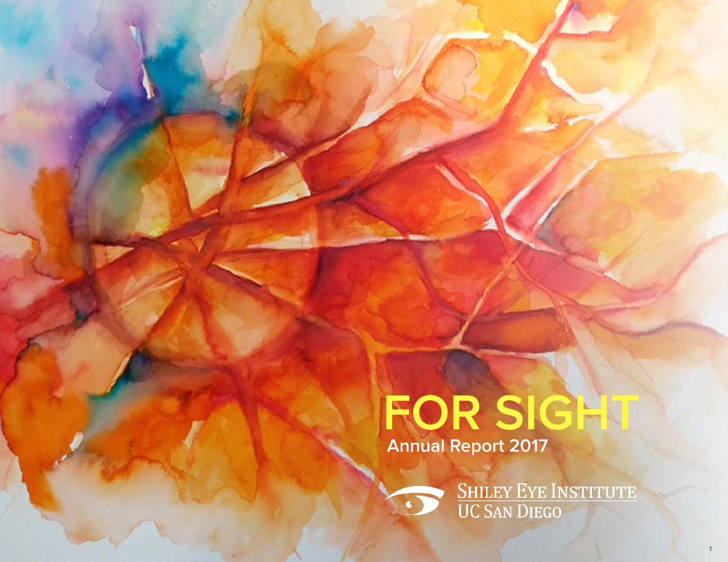
1 FOR SIGHT Annual Report 2017

Welcome message from author
This document is posted to help you gain knowledge. Please leave a comment to let me know what you think about it! Share it to your friends and learn new things together.
Transcript
1
FOR SIGHTAnnual Report 2017
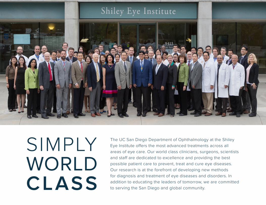
SIMPLY WORLD CLASS
The UC San Diego Department of Ophthalmology at the Shiley Eye Institute offers the most advanced treatments across all areas of eye care. Our world class clinicians, surgeons, scientists and staff are dedicated to excellence and providing the best possible patient care to prevent, treat and cure eye diseases. Our research is at the forefront of developing new methods for diagnosis and treatment of eye diseases and disorders. In addition to educating the leaders of tomorrow, we are committed to serving the San Diego and global community.
3
04 Letters From Leaders
07 Shiley Year in Review
08 Clinical, Research and Patient Highlights
30 Faculty
46 Education
49 Residents and Fellows
52 Publications, Clinical Trials, Grants, Lectures
76 Giving
78 Honor Roll of Donors
81 Special Gift
Contents
CHANCELLOR, UC SAN DIEGOPradeep K. Khosla, PhD
DEAN, UC SAN DIEGO SCHOOL OF MEDICINEDavid Brenner, MD
CEO, UC SAN DIEGO HEALTHPatty Maysent, MPH, MBA
DIRECTOR, SHILEY EYE INSTITUTERobert N. Weinreb, MD
CONTRIBUTORSJo AdamcikNatalie A. Afshari, MDGrace ChangDaniel Chao, MD, PhDWilliam R. Freeman, MDLilian GischlerWeldon Haw, MDNatasha Josefowitz, PhDDon O. Kikkawa, MDCraig Kishaba, MBADorota Skowronska-Krawczyk, PhD, MScScott LaFeeNadya LevHelen LiuIliana Molina, MBAShira Robbins, MDKaren Anisko Ryan, MSKristin SchafgansKarl Wahlin, PhD
EDITORSKaren Anisko Ryan, MSRobert N. Weinreb, MD
CREATIVEGrace ChangReynaldo Moreno
PHOTOGRAPHYGrace ChangRobyn AustinPeter DurdallerChris Park PhotographyChad Michael Ward
PRINTINGTu’s Printing and Graphics
On the cover: An artist’s representation of an optic nerve head. See article on page 12.
4
Dear Friends,
Partnerships and collaborations based on trust and common goals can become enduring relationships. This past year the Shiley Eye Institute (SEI) has added partnerships and deepened relationships with our patients and donors, the greater San Diego community, and colleagues within the UC San Diego campus and beyond. We could not have made it this far without you, and will share some of the exciting news.
Just a few months ago, we strengthened our partnership with the Downtown San Diego Lions Club. The club supported our Shiley EyeMobile for Children for many years. This year, however, they reaffirmed the partnership by funding our newly named Downtown San Diego Lions Club BioBank for Vision. Already, their support is making a difference and ensuring SEI leadership in ophthalmic genetics.
During the past year, generous donations from grateful patients and friends have had a major impact on and accelerated the innovative research being conducted at the Richard C. Atkinson Laboratory for Regenerative Ophthalmology team. Collaborating with world leading scientists from the UC San Diego Department of Neurosciences, as one example, the Atkinson Laboratory team is seeking to develop gene therapy to rescue and restore vision that has been lost in patients with glaucoma. This effort complements their ongoing work to develop stem cell therapies to prevent and cure glaucoma, retinal degenerations and other blinding eye diseases. Such cross-disciplinary partnerships are essential for discovery and advancing discovery to the patient.
As another example of our partnering, Daniel Chao, MD, PhD, who oversees our medical student education, is working with the UC San Diego Student Free Clinic to implement use of the cell phone for examination and management of the underserved community in San Diego. And a local Girl Scout Troup is partnering with us to collect eyeglasses for these individuals at the Lions Optometric Clinic in downtown San Diego. As still another example of the partnership, this one between UC San Diego Health and the San Diego Padres, the Shiley EyeMobile traveled to spring training in Peoria where SEI staff performed eye examinations for the Padres players and staff.
With the UC San Diego Rady School of Management, we initiated a Leadership and Innovation lecture series to provide our faculty, residents and fellows with leadership and entrepreneurial skills to succeed in the changing landscape of vision research and healthcare.
I am invigorated daily by the clinical excellence, world leading research and dedication of our faculty and staff. It is a privilege to partner with our patients, colleagues and big-hearted advocates.
Sincerely,
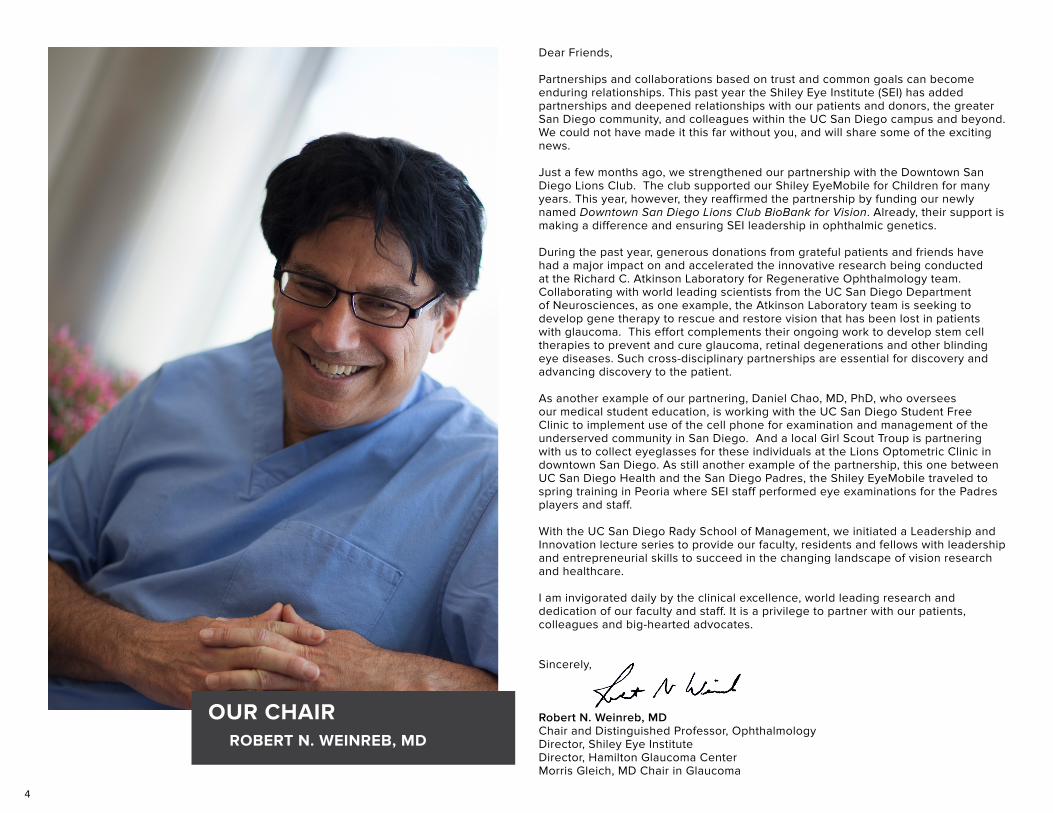
Robert N. Weinreb, MDChair and Distinguished Professor, OphthalmologyDirector, Shiley Eye InstituteDirector, Hamilton Glaucoma CenterMorris Gleich, MD Chair in Glaucoma
OUR CHAIRROBERT N. WEINREB, MD
5
Dear Friends,
The Shiley Eye Institute at UC San Diego Health is a shining example of the university’s mission to advance education, research, patient care and public service. Our clinical and research faculty are world leaders who train tomorrow’s innovators in ophthalmology, lead groundbreaking investigations, and bring new breakthroughs into the clinic to benefit our patients, including those in underserved communities.
These achievements are due to the hard work and collaborations of our outstanding faculty, residents, fellows and staff, who strive for excellence and make UC San Diego a global destination for health. This quality of care benefits the citizens of our region, our global community, and UC San Diego’s mission and standing. UC San Diego is ranked as a top public university in the nation based on our research, service and social mobility, for the eighth consecutive year, as well as the 14th best university in the world. These rankings are a reflection of our talented campus members and generous supporters. It is because of you that UC San Diego and the Shiley Eye Institute have grown and flourished over the decades.
Thank you for your support and continued partnership with UC San Diego and the Shiley Eye Institute.
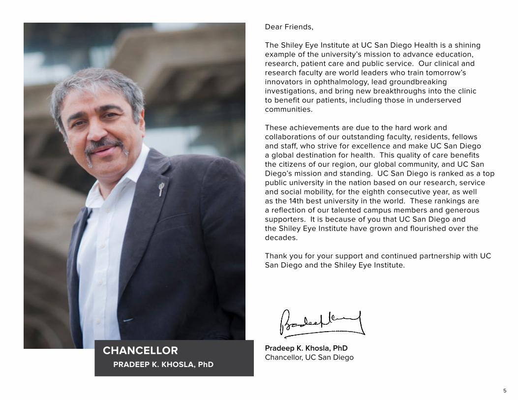
Pradeep K. Khosla, PhDChancellor, UC San Diego
CHANCELLORPRADEEP K. KHOSLA, PhD
6
Dear Friends of the Shiley Eye Institute,
With the close of 2017 approaching, let’s pause to celebrate – again – another year of achievement and distinguished success by the faculty and staff in the Department of Ophthalmology and at Shiley Eye Institute. Both continue to be innovative and vital hubs within UC San Diego School of Medicine and UC San Diego Health, the region’s only academic health care system.
The researchers, physicians and staff at Shiley have always seen farther and better than most. Long before “big data” became a buzzword, Shiley doctors were observing and documenting patients through a longitudinal glaucoma study known as Diagnostic Innovations in Glaucoma – now decades old but still going strong. Shiley’s BioBank is a repository of tissue samples from more than 5,000 patients with myriad eye conditions, each linked to highly detailed clinical histories, each a potential goldmine in the search for new insights and therapies.
Every year at Shiley is marked by significant news and progress. A few examples for 2017:
Using the gene-editing tool CRISPR/Cas9, Shiley scientists with colleagues in China announced they had reprogrammed mutated rod photoreceptors to become functional cone photoreceptors, reversing cellular degeneration and restoring visual function in two mouse models of retinitis pigmentosa.
With colleagues across the country, Shiley scientists identified three distinct stretches of genetic material on chromosomes linked to Fuchs endothelial corneal dystrophy, an inherited, progressive vision disorder.
And working with industry, Shiley scientists and engineers have developed the nanotechnology and wireless electronics for a new type of retinal prosthesis, bringing research one step closer to restoring the ability of neurons in the retina to respond to light.
These are just three remarkable examples from a place where remarkable is the norm. Other researchers are working with stem cells to grow new retinas or use machine learning to improve clinicians’ abilities to spot the smallest tell-tale signs of vision problems.
All of this happens at Shiley and across campus, an epitome of community and collaboration where the benefits ultimately extend far beyond. I celebrate this year’s work and achievements at Shiley. I look forward to more in the next year – and in the many years to come.
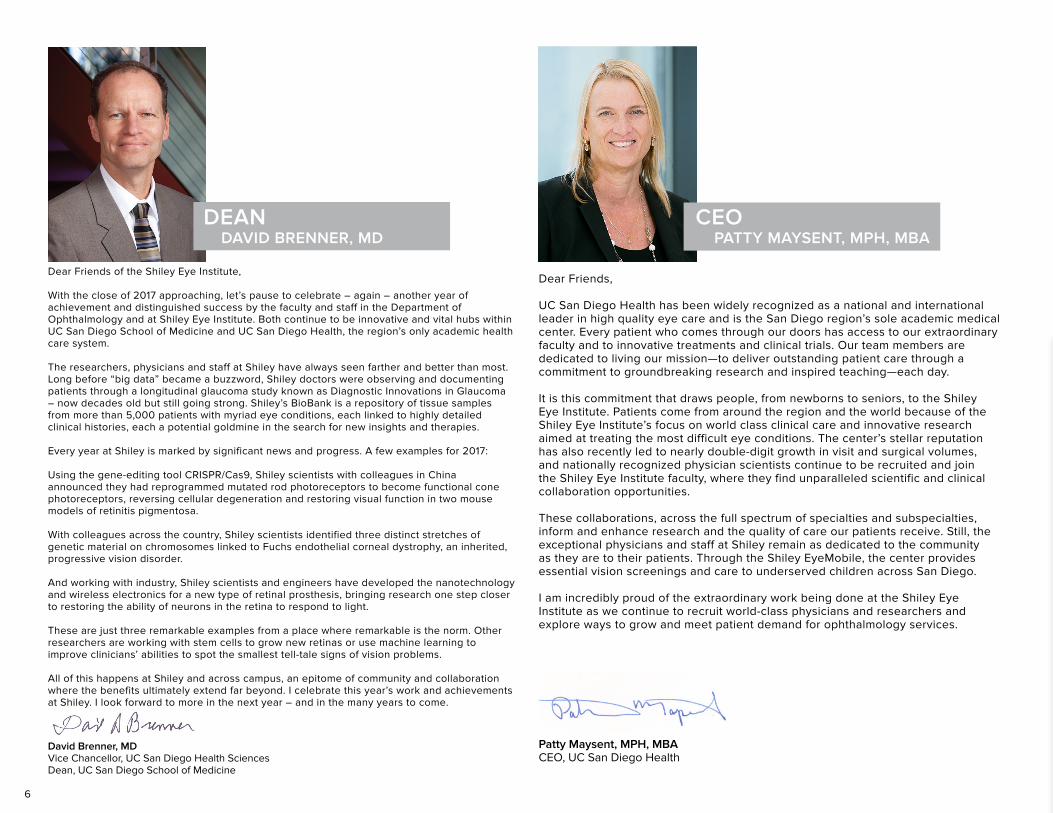
David Brenner, MDVice Chancellor, UC San Diego Health SciencesDean, UC San Diego School of Medicine
Dear Friends,
UC San Diego Health has been widely recognized as a national and international leader in high quality eye care and is the San Diego region’s sole academic medical center. Every patient who comes through our doors has access to our extraordinary faculty and to innovative treatments and clinical trials. Our team members are dedicated to living our mission—to deliver outstanding patient care through a commitment to groundbreaking research and inspired teaching—each day.
It is this commitment that draws people, from newborns to seniors, to the Shiley Eye Institute. Patients come from around the region and the world because of the Shiley Eye Institute’s focus on world class clinical care and innovative research aimed at treating the most difficult eye conditions. The center’s stellar reputation has also recently led to nearly double-digit growth in visit and surgical volumes, and nationally recognized physician scientists continue to be recruited and join the Shiley Eye Institute faculty, where they find unparalleled scientific and clinical collaboration opportunities.
These collaborations, across the full spectrum of specialties and subspecialties, inform and enhance research and the quality of care our patients receive. Still, the exceptional physicians and staff at Shiley remain as dedicated to the community as they are to their patients. Through the Shiley EyeMobile, the center provides essential vision screenings and care to underserved children across San Diego.
I am incredibly proud of the extraordinary work being done at the Shiley Eye Institute as we continue to recruit world-class physicians and researchers and explore ways to grow and meet patient demand for ophthalmology services.
Patty Maysent, MPH, MBACEO, UC San Diego Health
DEAN CEODAVID BRENNER, MD PATTY MAYSENT, MPH, MBA
7
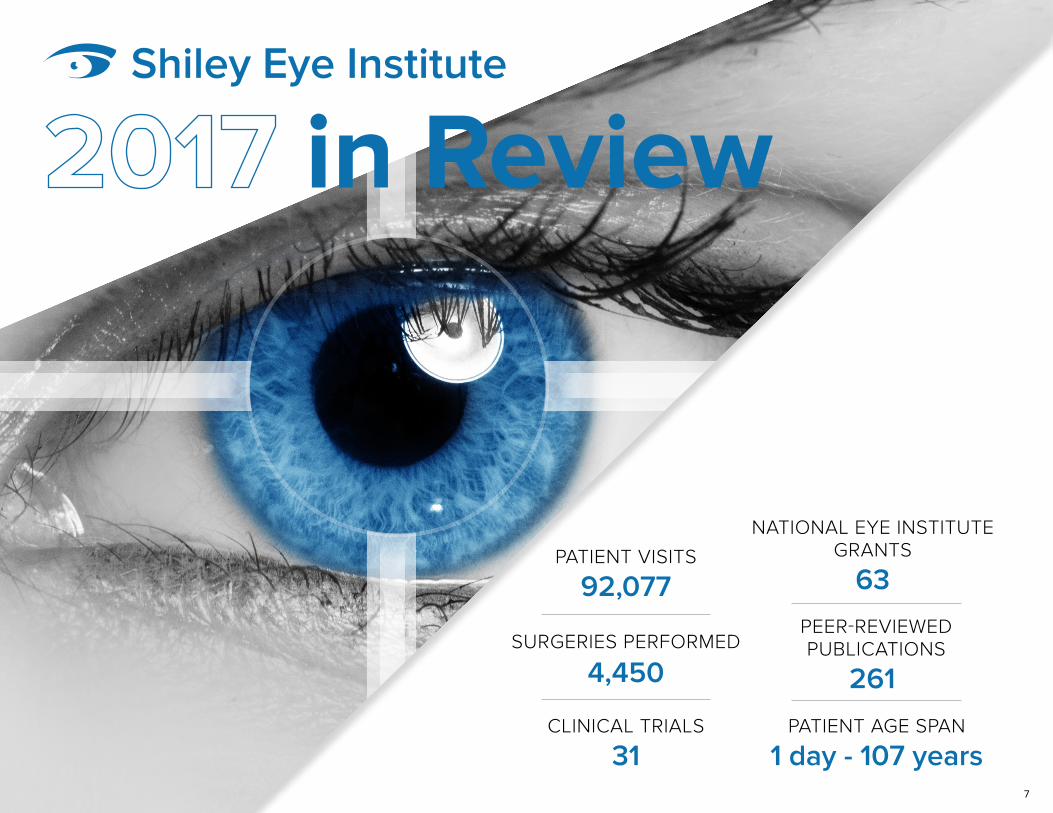
in Review
1 day - 107 yearsPATIENT AGE SPAN
261
PEER-REVIEWED PUBLICATIONS
92,077PATIENT VISITS
4,450SURGERIES PERFORMED
31CLINICAL TRIALS
63
NATIONAL EYE INSTITUTEGRANTS
Shiley Eye Institute
8
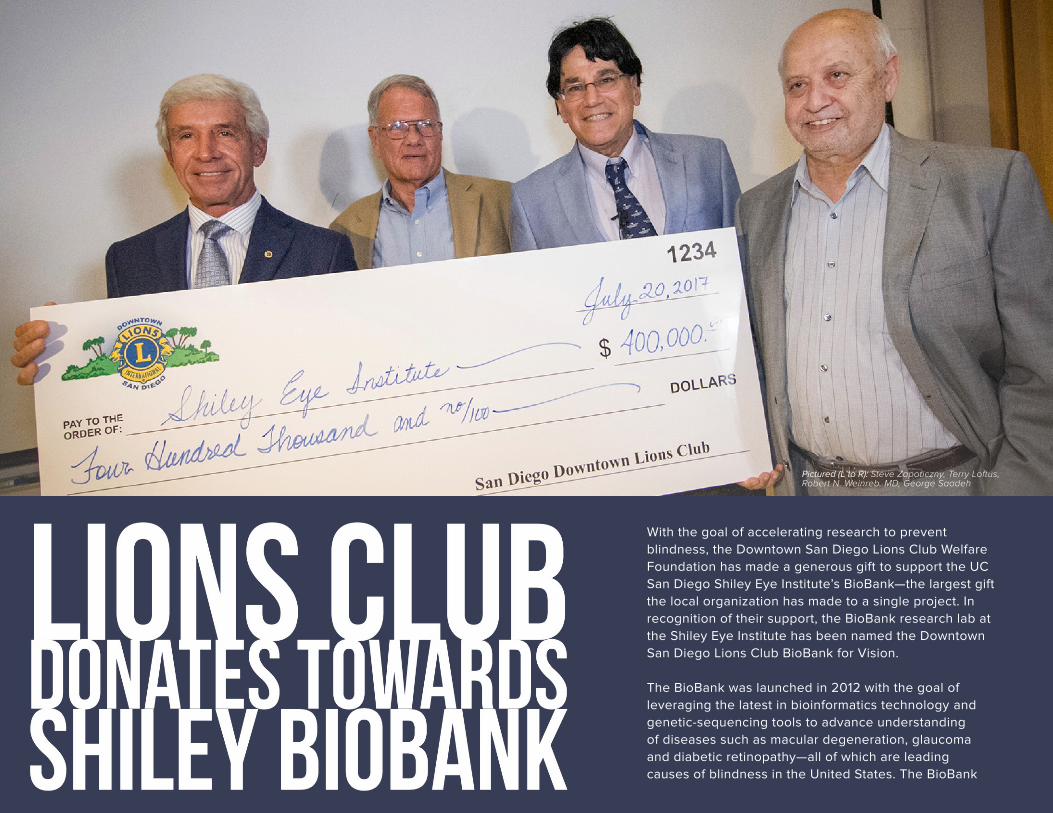
With the goal of accelerating research to prevent blindness, the Downtown San Diego Lions Club Welfare Foundation has made a generous gift to support the UC San Diego Shiley Eye Institute’s BioBank—the largest gift the local organization has made to a single project. In recognition of their support, the BioBank research lab at the Shiley Eye Institute has been named the Downtown San Diego Lions Club BioBank for Vision.
The BioBank was launched in 2012 with the goal of leveraging the latest in bioinformatics technology and genetic-sequencing tools to advance understanding of diseases such as macular degeneration, glaucoma and diabetic retinopathy—all of which are leading causes of blindness in the United States. The BioBank
Pictured (L to R): Steve Zapoticzny, Terry Loftus, Robert N. Weinreb, MD, George Saadeh
9
provides a library of biological samples with complete medical and family history and other demographic information that researchers can utilize to learn about predictors for diseases (biomarkers) and effectiveness of therapies.
“The BioBank allows our ophthalmology team to make remarkable advances in understanding the biology of diseases and the promise of personalized medicine,” said Robert N. Weinreb, MD, chair and Distinguished Professor of Ophthalmology and director of the Shiley Eye Institute. “The support from the San Diego Lions Club Welfare Foundation aids our research and
helps us to better diagnose, prevent and treat eye diseases.” Celebrating 100 years, the Lions International has a long history of service to prevent blindness, restore eyesight and improve eye health. In 1925, Helen Keller urged Lions everywhere to become “Knights of the Blind.” Since then, sight programs have been a hallmark of Lions Clubs around the world, including the Downtown San Diego club.
In keeping with this focus, the local club came to UC San Diego with the goal of supporting a major project that leverages the latest technology and research to help patients with
The BioBank has the potential to make a significant difference for the thousands of patients in San Diego with blinding eye diseases.
blinding eye diseases. “The BioBank has the potential to make a significant difference for the thousands of patients in San Diego with blinding eye diseases, as well as patients around the world,” said Steve Zapoticzny, president of the Downtown San Diego Lions Club. “We are thrilled to join the Shiley Eye Institute—the nation’s leading institute for efforts to prevent and cure blindness—in this partnership.”
Terry Loftus, past-president for the Downtown San Diego Lions Club, led the partnership proposal along with fellow Lion and past-president George Saadeh. “We were looking into stem cell and genetic research that is making strides toward curing these diseases,” said Loftus. “When we came across an article about what UC San Diego is doing in this area, the pieces started to fall into place.” More than 150,000 individuals in San Diego County suffer from retinal degeneration, glaucoma or diabetic retinopathy. At the Shiley Eye Institute, doctors and researchers have made measurable progress in addressing these and other primary diseases of vision.
“Our physician-scientists are involved in diverse and collaborative research projects that aim to improve treatments for eye diseases and hasten the day when blindness is entirely preventable,” said Dr. Weinreb. “The BioBank provides a critical resource that they can draw upon to accelerate the translation of research into treatments.” Partnering with the Shiley Eye Institute at UC San Diego is a great way to celebrate the Downtown San Diego Lions Club’s 95th Anniversary!
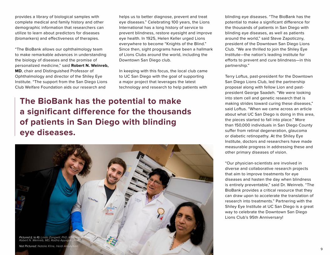
Pictured (L to R): Linda Zangwill, PhD, Matthew Holman,Robert N. Weinreb, MD, Radha Ayyagari, PhD
Not Pictured: Natalie Kline, Heidi Amundson
10
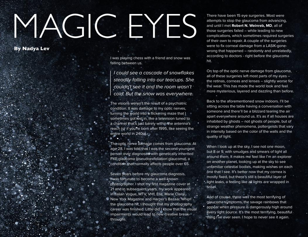
I was playing chess with a friend and snow was falling between us.
The visions weren’t the result of a psychiatric condition. It was damage to my optic nerves, turning the world into a flickering maze that I sometimes got lost in, like a television tuned to a channel that’s just barely within the antenna’s reach (or if you’re born after 1995, like seeing the entire world in 240p).
The optic nerve damage comes from glaucoma. At age 28, I was told that I was the second-youngest person ever diagnosed with genetically inherited PXF glaucoma (pseudoexfoliation glaucoma), a condition that normally affects people over 65.
Seven years before my glaucoma diagnosis, I was fortunate to become a well-known photographer. I shot my first magazine cover at 21 and in subsequent years, my work appeared in Italian Vogue, MTV, VH1, Elle, Marie Claire, New York Magazine and Harper’s Bazaar. When the glaucoma hit, I thought that my photography career was finished. Little did I know that the visual impairments would lead to new creative break-throughs.
There have been 15 eye surgeries. Most were attempts to stop the glaucoma from advancing, and until I met Robert N. Weinreb, MD, all of those surgeries failed – while leading to new complications, which sometimes required surgeries of their own to repair. A couple of the surgeries were to fix corneal damage from a LASIK-gone-wrong that happened – randomly and unrelatedly, according to doctors - right before the glaucoma hit.
On top of the optic nerve damage from glaucoma, all of these surgeries left most parts of my eyes – the retinas, corneas and lenses – slightly worse for the wear. This has made the world look and feel more mysterious, layered and dazzling than before.
Back to the aforementioned snow indoors. I’ll be sitting across the table having a conversation with someone and there’ll be a blizzard tearing the air apart everywhere around us. It’s as if all houses are inhabited by ghosts – not ghosts of people, but of strange weather phenomena, poltergeists that vary in intensity based on the color of the walls and the quality of light.
When I look up at the sky, I see not one moon, but 8 or 9, with smudges and smears of light all around them. It makes me feel like I’m an explorer on another planet, looking up at the sky to see unfamiliar celestial bodies, making wishes on each one that I see. It’s better now that my cornea is mostly fixed, but there’s still a beautiful layer of light leaks, a feeling like all lights are wrapped in tinsel.
And of course, there was the most terrifying of glaucoma symptoms, the savage rainbows that appear when pressure is dangerously high around every light source. It’s the most terrifying, beautiful thing I’ve ever seen. I hope to never see it again.
MAGIC EYESBy Nadya Lev
I could see a cascade of snowflakes steadily falling into our teacups. She couldn’t see it and the room wasn’t cold. But the snow was everywhere.
11
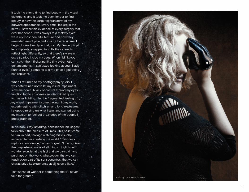
It took me a long time to find beauty in the visual distortions, and it took me even longer to find beauty in how the surgeries transformed my outward appearance. Every time I looked in the mirror, I saw all this evidence of every surgery that ever happened. I was always told that my eyes were my most beautiful feature and now they reminded me of pain and loss. But after a time, I began to see beauty in that, too. My new artificial lens implants, swapped in to fix the cataracts, reflect light differently, so that there’s always an extra sparkle inside my eyes. When I blink, you can catch them flickering like tiny cybernetic enhancements. “I can’t stop looking at your Blade Runner eyes,” someone told me once. I like being half-replicant.
When I returned to my photography studio, I was determined not to let my visual impairment slow me down. A lack of control around my eyes’ function led to an obsessive, disciplined quest to master lighting. I let the fragmented feeling of my visual impairment come through in my work, experimenting with glitch art and long exposures. I stopped relying on what I saw, and started using my intuition to feel out the stories of the people I photographed.
In his book Play Anything, philosopher Ian Bogost talks about the pleasure of limits. This belief came to him, in part, through watching his visually-impaired father interface the world. “Blindness ruptures confidence,” writes Bogost. “It recognizes the preposterousness of all things… it glints with wonder, wonder at the fact that we can gain any purchase on the world whatsoever, that we can touch even part of its sensuousness, that we can characterize its experience at all, even a little.”
That sense of wonder is something that I’ll never take for granted.
Photo by Chad Michael Ward
12
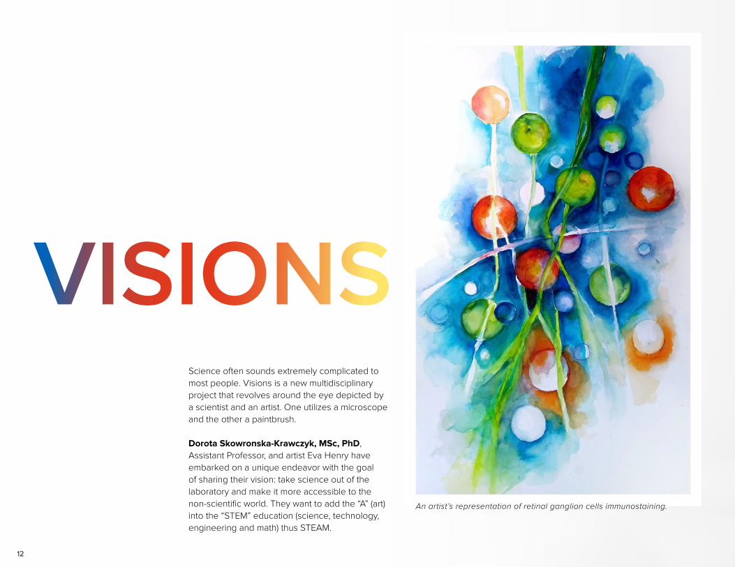
Science often sounds extremely complicated to most people. Visions is a new multidisciplinary project that revolves around the eye depicted by a scientist and an artist. One utilizes a microscope and the other a paintbrush.
Dorota Skowronska-Krawczyk, MSc, PhD, Assistant Professor, and artist Eva Henry have embarked on a unique endeavor with the goal of sharing their vision: take science out of the laboratory and make it more accessible to the non-scientific world. They want to add the “A” (art) into the “STEM” education (science, technology, engineering and math) thus STEAM.
VISIONS
An artist’s representation of retinal ganglion cells immunostaining.
13
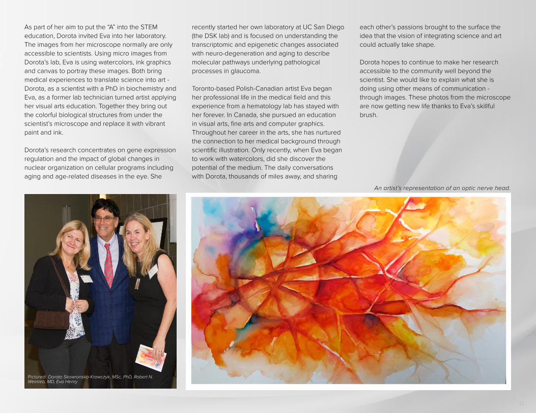
As part of her aim to put the “A” into the STEM education, Dorota invited Eva into her laboratory. The images from her microscope normally are only accessible to scientists. Using micro images from Dorota’s lab, Eva is using watercolors, ink graphics and canvas to portray these images. Both bring medical experiences to translate science into art - Dorota, as a scientist with a PhD in biochemistry and Eva, as a former lab technician turned artist applying her visual arts education. Together they bring out the colorful biological structures from under the scientist’s microscope and replace it with vibrant paint and ink.
Dorota’s research concentrates on gene expression regulation and the impact of global changes in nuclear organization on cellular programs including aging and age-related diseases in the eye. She
recently started her own laboratory at UC San Diego (the DSK lab) and is focused on understanding the transcriptomic and epigenetic changes associated with neuro-degeneration and aging to describe molecular pathways underlying pathological processes in glaucoma.
Toronto-based Polish-Canadian artist Eva began her professional life in the medical field and this experience from a hematology lab has stayed with her forever. In Canada, she pursued an education in visual arts, fine arts and computer graphics. Throughout her career in the arts, she has nurtured the connection to her medical background through scientific illustration. Only recently, when Eva began to work with watercolors, did she discover the potential of the medium. The daily conversations with Dorota, thousands of miles away, and sharing
each other’s passions brought to the surface the idea that the vision of integrating science and art could actually take shape.
Dorota hopes to continue to make her research accessible to the community well beyond the scientist. She would like to explain what she is doing using other means of communication - through images. These photos from the microscope are now getting new life thanks to Eva’s skillful brush.
Pictured: Dorota Skowronska-Krawczyk, MSc, PhD, Robert N. Weinreb, MD, Eva Henry
An artist’s representation of an optic nerve head.
14
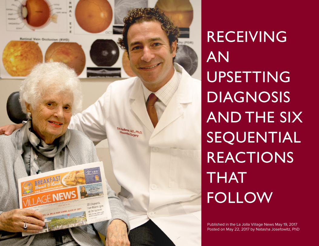
RECEIVINGANUPSETTING DIAGNOSIS AND THE SIX SEQUENTIAL REACTIONS THAT FOLLOW Published in the La Jolla Village News May 19, 2017Posted on May 22, 2017 by Natasha Josefowitz, PhD
15
Recently I noticed that things were getting a bit blurry, so I went to the Shiley Eye Institute at UC San Diego to have it checked out. Guess what? Dr. William Freeman, Director of the Jacobs Retina Center, said I have macular degeneration! He was helpful in explaining what it is and the treatment for it, trying to be reassuring, but it didn’t sound good. I have the dry kind in my left eye and the wet kind in my right.
I am 90 years old and often wondered which of my organs would fail first. In my age group whatever doesn’t dry out, leaks. Little did I know that my first failed organ would be leaking blood vessels in my eye! “Intra-retinal fluid hemorrhaging in my retina.”
When things like this happen to me, I become interested in observing myself to study my reactions to the news, assuming that I am not the only one to react this way and hoping that it might be helpful to others. The stages of reactions I went through upon receiving this upsetting diagnosis:
STAGE 1Shock: Somehow this does not fit the image I have of myself as invulnerable. It’s hard to believe that what I have seen happen to others is now happening to me. There is an element of fright; I am scared of the unknown.
STAGE 2In a funk! Feeling sorry for myself. On my way home I bumped into a couple of friends; I was very teary-eyed as I told them my bad news.
STAGE 3Catastrophising: Worst-case scenario—I’m going to lose my sight; I will go blind. There’s a white cane in my future. I am a writer, a researcher; I read a lot. This is the end of my productive life!
STAGE 4Research: Checking out WebMD and Mayo Clinic online, talking to friends who have it…gathering information. Looking into available resources such as equipment that magnifies print, books on tape, speech recognition software on computers (none of which I will need in the foreseeable future).
The scientist who discovered the substance that causes the leaky blood vessels is Napoleon Ferrara, MD, who also invented the most commonly used class of drugs to treat it (anti-VEGF agents). He is appointed to the UCSD Departments of Ophthalmology and Pathology, the Moores Cancer Center, and the Shiley Eye Institute. As a result, I feel reassured that I will be able to keep writing for another few years (I’m betting on making it to 100).
While waiting to be tested on the latest equipment at Shiley, I saw Eric Cabezas, who was part of a research team at the Hamilton Glaucoma Center when I worked as a tester for
their new machines for about 10 years. He told me that 20 years ago macular degeneration was a sentence for eventual blindness; now there is treatment to prevent further deterioration. He cautioned me that for some people there is no best-case scenario. What helps in these circumstances is support. What is most useful is being part of a group of people with similar diagnoses where coping strategies can be shared.
STAGE 5Treatment: My treatment consists of specially formulated vitamins for the dry kind of macular degeneration in my left eye and a monthly injection in my right eye, which will slow progression of the disease there. The idea of an injection in my eye made me anxious; there was no need to be. Shiley retina specialist, Eric Nudleman, MD, PhD, began by numbing my eye. When I asked him when he was going to proceed with the injection, he replied he was already done. I was surprised as I felt no pain.
STAGE 6Best-case scenario: This isn’t so bad. My father used to say: “If this is the worst that will happen to me, I‘m signing the contract.” I am writing this with my eyes still dilated, but I couldn’t wait to tell my readers what is happening to me as a possible roadmap for others. Yes, we can all go from funk through let’s explore all options to celebrate what still works (my head, more or less), and re-invent ourselves with new disabilities, new coping mechanisms, and maybe the opportunity for new adventures. I’m game!
The incredible luck is that I live next to some of the top eye-care facilities in the world—the Shiley Eye Institute, the Joan and Irwin Jacobs Retina Center, and the Hamilton Glaucoma Center — so I could not be in better hands.
16
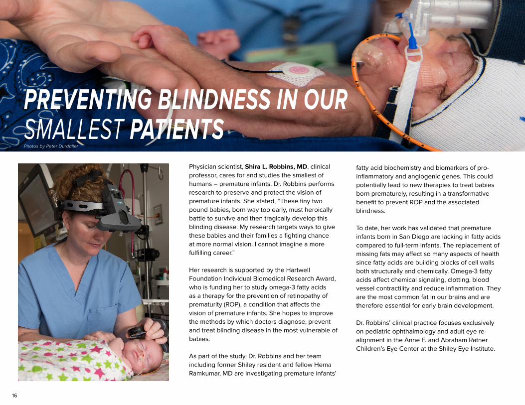
Physician scientist, Shira L. Robbins, MD, clinical professor, cares for and studies the smallest of humans – premature infants. Dr. Robbins performs research to preserve and protect the vision of premature infants. She stated, “These tiny two pound babies, born way too early, must heroically battle to survive and then tragically develop this blinding disease. My research targets ways to give these babies and their families a fighting chance at more normal vision. I cannot imagine a more fulfilling career.”
Her research is supported by the Hartwell Foundation Individual Biomedical Research Award, who is funding her to study omega-3 fatty acids as a therapy for the prevention of retinopathy of prematurity (ROP), a condition that affects the vision of premature infants. She hopes to improve the methods by which doctors diagnose, prevent and treat blinding disease in the most vulnerable of babies.
As part of the study, Dr. Robbins and her team including former Shiley resident and fellow Hema Ramkumar, MD are investigating premature infants’
fatty acid biochemistry and biomarkers of pro-inflammatory and angiogenic genes. This could potentially lead to new therapies to treat babies born prematurely, resulting in a transformative benefit to prevent ROP and the associated blindness.
To date, her work has validated that premature infants born in San Diego are lacking in fatty acids compared to full-term infants. The replacement of missing fats may affect so many aspects of health since fatty acids are building blocks of cell walls both structurally and chemically. Omega-3 fatty acids affect chemical signaling, clotting, blood vessel contractility and reduce inflammation. They are the most common fat in our brains and are therefore essential for early brain development.
Dr. Robbins’ clinical practice focuses exclusively on pediatric ophthalmology and adult eye re-alignment in the Anne F. and Abraham Ratner Children’s Eye Center at the Shiley Eye Institute.
PREVENTING BLINDNESS IN OURSMALLEST PATIENTSPhotos by Peter Durdaller
17
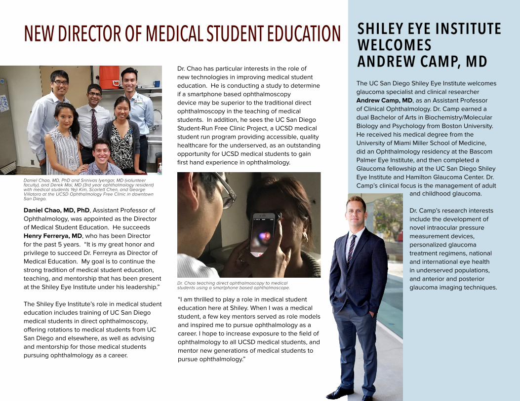
The UC San Diego Shiley Eye Institute welcomes glaucoma specialist and clinical researcher Andrew Camp, MD, as an Assistant Professor of Clinical Ophthalmology. Dr. Camp earned a dual Bachelor of Arts in Biochemistry/Molecular Biology and Psychology from Boston University. He received his medical degree from the University of Miami Miller School of Medicine, did an Ophthalmology residency at the Bascom Palmer Eye Institute, and then completed a Glaucoma fellowship at the UC San Diego Shiley Eye Institute and Hamilton Glaucoma Center. Dr. Camp’s clinical focus is the management of adult
and childhood glaucoma.
Dr. Camp’s research interests include the development of novel intraocular pressure measurement devices, personalized glaucoma treatment regimens, national and international eye health in underserved populations, and anterior and posterior glaucoma imaging techniques.
SHILEY EYE INSTITUTE WELCOMESANDREW CAMP, MDDr. Chao has particular interests in the role of
new technologies in improving medical student education. He is conducting a study to determine if a smartphone based ophthalmoscopy device may be superior to the traditional direct ophthalmoscopy in the teaching of medical students. In addition, he sees the UC San Diego Student-Run Free Clinic Project, a UCSD medical student run program providing accessible, quality healthcare for the underserved, as an outstanding opportunity for UCSD medical students to gain first hand experience in ophthalmology.
Daniel Chao, MD, PhD, Assistant Professor of Ophthalmology, was appointed as the Director of Medical Student Education. He succeeds Henry Ferrerya, MD, who has been Director for the past 5 years. “It is my great honor and privilege to succeed Dr. Ferreyra as Director of Medical Education. My goal is to continue the strong tradition of medical student education, teaching, and mentorship that has been present at the Shiley Eye Institute under his leadership.”
The Shiley Eye Institute’s role in medical student education includes training of UC San Diego medical students in direct ophthalmoscopy, offering rotations to medical students from UC San Diego and elsewhere, as well as advising and mentorship for those medical students pursuing ophthalmology as a career.
Daniel Chao, MD, PhD and Srinivas Iyengar, MD (volunteer faculty), and Derek Mai, MD (3rd year ophthalmology resident) with medical students Yeji Kim, Scarlett Chen, and George Villatoro at the UCSD Ophthalmology Free Clinic in downtown San Diego.
Dr. Chao teaching direct ophthalmoscopy to medical students using a smartphone based ophthalmoscope.
“I am thrilled to play a role in medical student education here at Shiley. When I was a medical student, a few key mentors served as role models and inspired me to pursue ophthalmology as a career. I hope to increase exposure to the field of ophthalmology to all UCSD medical students, and mentor new generations of medical students to pursue ophthalmology.”
NEW DIRECTOR OF MEDICAL STUDENT EDUCATION
18
Shiley EyeMobile Partners with San Diego Padres
Since its founding in 2000, the Shiley EyeMobile program has screened almost 200,000 children across San Diego County. The mission of the Shiley EyeMobile is to give vulnerable populations of children the vision and eye care they need to succeed in school and in life by identifying, treating, and preventing vision disorders. Additionally, the program educates and provides community outreach about the importance for kids to receive vital eye care as well as routine eye exams while removing barriers to accessing comprehensive vision care. The EyeMobile program provides children with the best sight to allow them to be “school ready” so they can learn at their maximum potential.
Keys to the Shiley EyeMobile program’s success include the use of the latest information technology – electronic medical records, a dedicated multilingual staff, students and community volunteers. It is estimated that 1 in 4 children have vision problems. Early detection and treatment have proven to reduce the negative impact vision problems may have on a child’s learning ability and development. If left untreated, conditions such as amblyopia, could lead to irreversible vision loss and psychosocial effects.
During the past year, UC San Diego Health and the San Diego Padres formed a partnership to provide the baseball team with expertise in all medical fields. One of the first medical specialties to move forward was the Department of Ophthalmology through the EyeMobile. On a regular basis, the Shiley EyeMobile successfully addresses the need for vision care of underserved children in San Diego County. Through this new partnership, the eye doctors on the EyeMobile examined the entire team and staff at the Padres training camp in Peoria, Arizona.
The entire Padres team and personnel were examined and those with eye disorders were then referred to Shiley Eye Institute ophthalmologists for further evaluation and possible treatment. The Padres players are slightly taller than the children regularly seen on the EyeMobile!
As partners, the Padres will collaborate with UC San Diego Health on awareness efforts to promote healthy living and disease prevention in San Diego. The Department of Ophthalmology looks forward to collaborating with the Padres on future outreach programs.
As partners, the Padres will be collaborating with UC San Diego Health on awareness efforts to promote healthy living and disease prevention in San Diego.
19
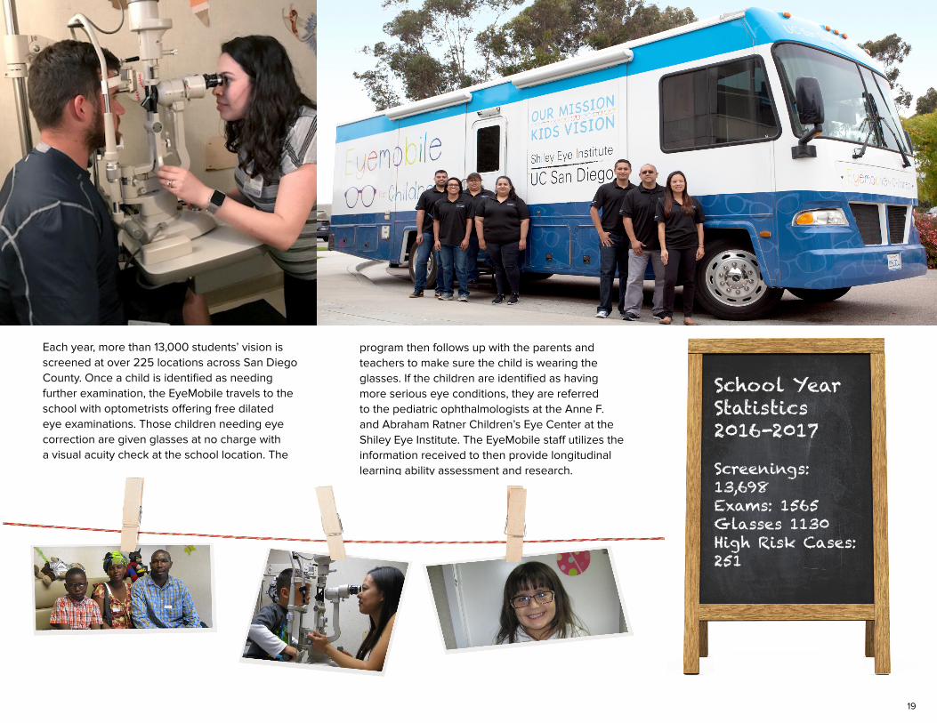
Each year, more than 13,000 students’ vision is screened at over 225 locations across San Diego County. Once a child is identified as needing further examination, the EyeMobile travels to the school with optometrists offering free dilated eye examinations. Those children needing eye correction are given glasses at no charge with a visual acuity check at the school location. The
program then follows up with the parents and teachers to make sure the child is wearing the glasses. If the children are identified as having more serious eye conditions, they are referred to the pediatric ophthalmologists at the Anne F. and Abraham Ratner Children’s Eye Center at the Shiley Eye Institute. The EyeMobile staff utilizes the information received to then provide longitudinal learning ability assessment and research.
School Year Statistics2016-2017
Screenings: 13,698Exams: 1565Glasses 1130High Risk Cases: 251
20
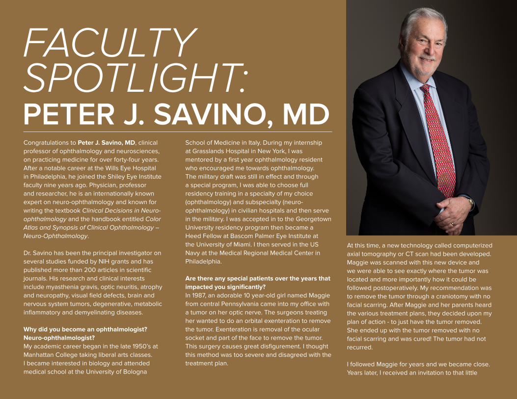
Congratulations to Peter J. Savino, MD, clinical professor of ophthalmology and neurosciences, on practicing medicine for over forty-four years. After a notable career at the Wills Eye Hospital in Philadelphia, he joined the Shiley Eye Institute faculty nine years ago. Physician, professor and researcher, he is an internationally known expert on neuro-ophthalmology and known for writing the textbook Clinical Decisions in Neuro-ophthalmology and the handbook entitled Color Atlas and Synopsis of Clinical Ophthalmology – Neuro-Ophthalmology.
Dr. Savino has been the principal investigator on several studies funded by NIH grants and has published more than 200 articles in scientific journals. His research and clinical interests include myasthenia gravis, optic neuritis, atrophy and neuropathy, visual field defects, brain and nervous system tumors, degenerative, metabolic inflammatory and demyelinating diseases.
Why did you become an ophthalmologist? Neuro-ophthalmologist? My academic career began in the late 1950’s at Manhattan College taking liberal arts classes. I became interested in biology and attended medical school at the University of Bologna
School of Medicine in Italy. During my internship at Grasslands Hospital in New York, I was mentored by a first year ophthalmology resident who encouraged me towards ophthalmology. The military draft was still in effect and through a special program, I was able to choose full residency training in a specialty of my choice (ophthalmology) and subspecialty (neuro-ophthalmology) in civilian hospitals and then serve in the military. I was accepted in to the Georgetown University residency program then became a Heed Fellow at Bascom Palmer Eye Institute at the University of Miami. I then served in the US Navy at the Medical Regional Medical Center in Philadelphia.
Are there any special patients over the years that impacted you significantly?In 1987, an adorable 10 year-old girl named Maggie from central Pennsylvania came into my office with a tumor on her optic nerve. The surgeons treating her wanted to do an orbital exenteration to remove the tumor. Exenteration is removal of the ocular socket and part of the face to remove the tumor. This surgery causes great disfigurement. I thought this method was too severe and disagreed with the treatment plan.
At this time, a new technology called computerized axial tomography or CT scan had been developed. Maggie was scanned with this new device and we were able to see exactly where the tumor was located and more importantly how it could be followed postoperatively. My recommendation was to remove the tumor through a craniotomy with no facial scarring. After Maggie and her parents heard the various treatment plans, they decided upon my plan of action - to just have the tumor removed. She ended up with the tumor removed with no facial scarring and was cured! The tumor had not recurred.
I followed Maggie for years and we became close. Years later, I received an invitation to that little
FACULTY SPOTLIGHT: PETER J. SAVINO, MD
21
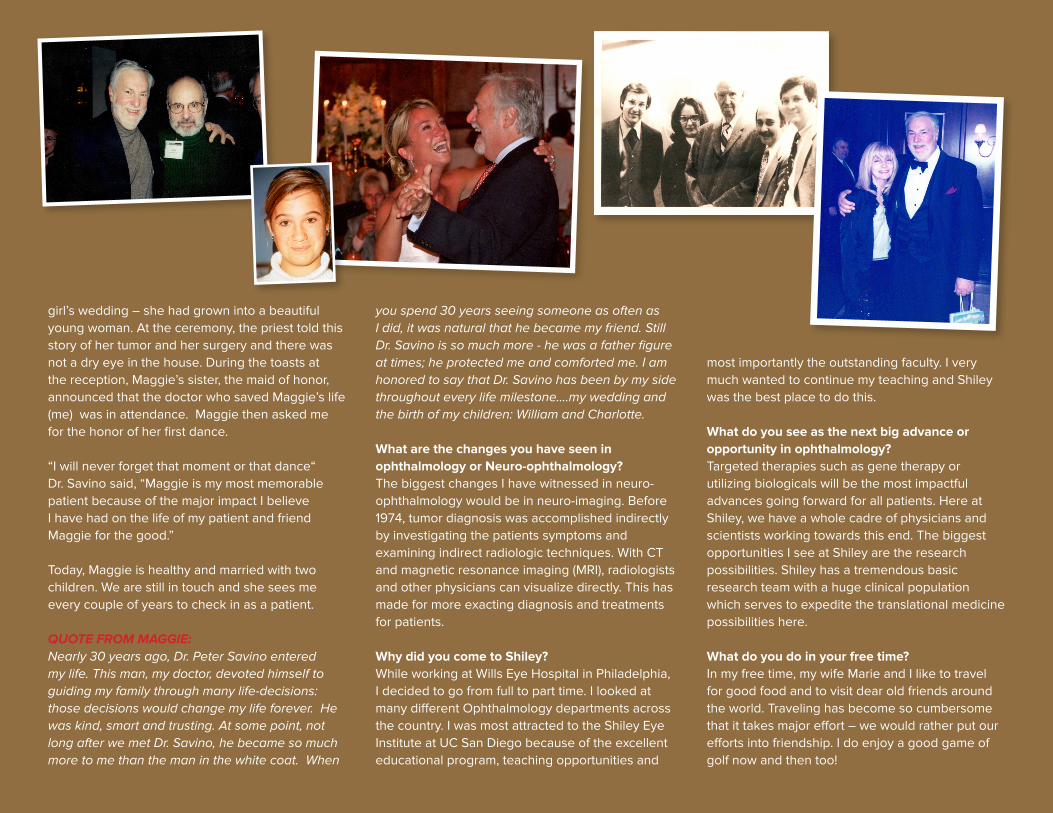
girl’s wedding – she had grown into a beautiful young woman. At the ceremony, the priest told this story of her tumor and her surgery and there was not a dry eye in the house. During the toasts at the reception, Maggie’s sister, the maid of honor, announced that the doctor who saved Maggie’s life (me) was in attendance. Maggie then asked me for the honor of her first dance.
“I will never forget that moment or that dance“ Dr. Savino said, “Maggie is my most memorable patient because of the major impact I believe I have had on the life of my patient and friend Maggie for the good.”
Today, Maggie is healthy and married with two children. We are still in touch and she sees me every couple of years to check in as a patient.
QUOTE FROM MAGGIE:Nearly 30 years ago, Dr. Peter Savino entered my life. This man, my doctor, devoted himself to guiding my family through many life-decisions: those decisions would change my life forever. He was kind, smart and trusting. At some point, not long after we met Dr. Savino, he became so much more to me than the man in the white coat. When
you spend 30 years seeing someone as often as I did, it was natural that he became my friend. Still Dr. Savino is so much more - he was a father figure at times; he protected me and comforted me. I am honored to say that Dr. Savino has been by my side throughout every life milestone….my wedding and the birth of my children: William and Charlotte.
What are the changes you have seen in ophthalmology or Neuro-ophthalmology?The biggest changes I have witnessed in neuro-ophthalmology would be in neuro-imaging. Before 1974, tumor diagnosis was accomplished indirectly by investigating the patients symptoms and examining indirect radiologic techniques. With CT and magnetic resonance imaging (MRI), radiologists and other physicians can visualize directly. This has made for more exacting diagnosis and treatments for patients.
Why did you come to Shiley?While working at Wills Eye Hospital in Philadelphia, I decided to go from full to part time. I looked at many different Ophthalmology departments across the country. I was most attracted to the Shiley Eye Institute at UC San Diego because of the excellent educational program, teaching opportunities and
most importantly the outstanding faculty. I very much wanted to continue my teaching and Shiley was the best place to do this.
What do you see as the next big advance or opportunity in ophthalmology?Targeted therapies such as gene therapy or utilizing biologicals will be the most impactful advances going forward for all patients. Here at Shiley, we have a whole cadre of physicians and scientists working towards this end. The biggest opportunities I see at Shiley are the research possibilities. Shiley has a tremendous basic research team with a huge clinical population which serves to expedite the translational medicine possibilities here.
What do you do in your free time?In my free time, my wife Marie and I like to travel for good food and to visit dear old friends around the world. Traveling has become so cumbersome that it takes major effort – we would rather put our efforts into friendship. I do enjoy a good game of golf now and then too!
22
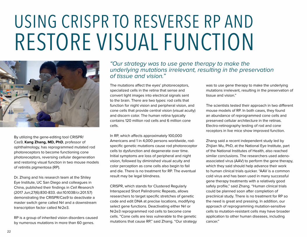
By utilizing the gene-editing tool CRISPR/Cas9, Kang Zhang, MD, PhD, professor of ophthalmology, has reprogrammed mutated rod photoreceptors to become functioning cone photoreceptors, reversing cellular degeneration and restoring visual function in two mouse models of retinitis pigmentosa (RP).
Dr. Zhang and his research team at the Shiley Eye Institute, UC San Diego and colleagues in China, published their findings in Cell Research (2017 Jun;27(6):830-833. doi:10.1038/cr.201.57) demonstrating the CRISPR/Cas9 to deactivate a master switch gene called Nrl and a downstream transcription factor called Nr2e3.
RP is a group of inherited vision disorders caused by numerous mutations in more than 60 genes.
The mutations affect the eyes’ photoreceptors, specialized cells in the retina that sense and convert light images into electrical signals sent to the brain. There are two types: rod cells that function for night vision and peripheral vision, and cone cells that provide central vision (visual acuity) and discern color. The human retina typically contains 120 million rod cells and 6 million cone cells.
In RP, which affects approximately 100,000 Americans and 1 in 4,000 persons worldwide, rod-specific genetic mutations cause rod photoreceptor cells to dysfunction and degenerate over time. Initial symptoms are loss of peripheral and night vision, followed by diminished visual acuity and color perception as cone cells also begin to fail and die. There is no treatment for RP. The eventual result may be legal blindness.
CRISPR, which stands for Clustered Regularly Interspaced Short Palindromic Repeats, allows researchers to target specific stretches of genetic code and edit DNA at precise locations, modifying select gene functions. Deactivating either Nrl or Nr2e3 reprogrammed rod cells to become cone cells. “Cone cells are less vulnerable to the genetic mutations that cause RP,” said Zhang. “Our strategy
was to use gene therapy to make the underlying mutations irrelevant, resulting in the preservation of tissue and vision.”
The scientists tested their approach in two different mouse models of RP. In both cases, they found an abundance of reprogrammed cone cells and preserved cellular architecture in the retinas. Electro-retinography testing of rod and cone receptors in live mice show improved function.
Zhang said a recent independent study led by Zhijian Wu, PhD, at the National Eye Institute, part of the National Institutes of Health, also reached similar conclusions. The researchers used adeno-associated virus (AAV) to perform the gene therapy, which they said should help advance their work to human clinical trials quicker. “AAV is a common cold virus and has been used in many successful gene therapy treatments with a relatively good safely profile,” said Zhang. “Human clinical trials could be planned soon after completion of preclinical study. There is no treatment for RP so the need is great and pressing. In addition, our approach of reprogramming mutation-sensitive cells to mutation-resistant cells may have broader application to other human diseases, including cancer.”
“Our strategy was to use gene therapy to make the underlying mutations irrelevant, resulting in the preservation of tissue and vision.”
USING CRISPR TO RESVERSE RP ANDRESTORE VISUAL FUNCTION
23
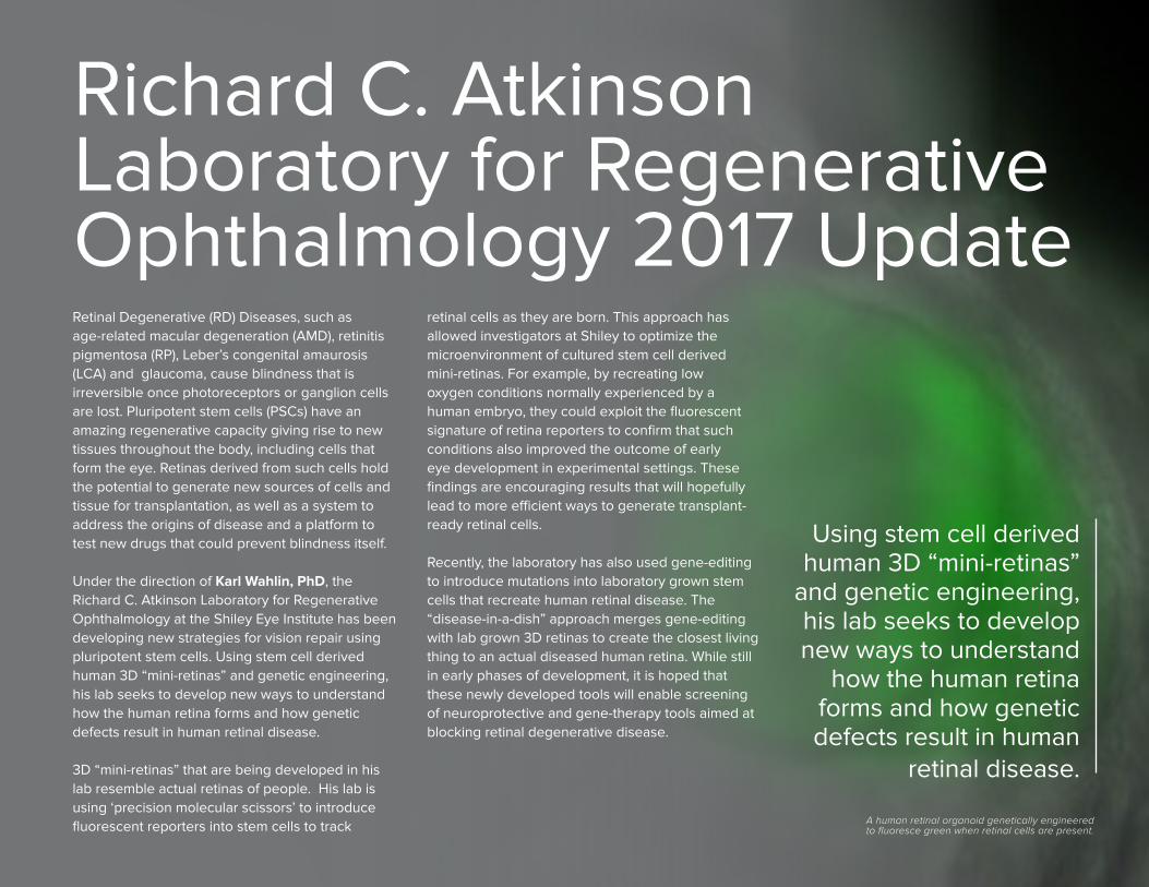
retinal cells as they are born. This approach has allowed investigators at Shiley to optimize the microenvironment of cultured stem cell derived mini-retinas. For example, by recreating low oxygen conditions normally experienced by a human embryo, they could exploit the fluorescent signature of retina reporters to confirm that such conditions also improved the outcome of early eye development in experimental settings. These findings are encouraging results that will hopefully lead to more efficient ways to generate transplant-ready retinal cells.
Recently, the laboratory has also used gene-editing to introduce mutations into laboratory grown stem cells that recreate human retinal disease. The “disease-in-a-dish” approach merges gene-editing with lab grown 3D retinas to create the closest living thing to an actual diseased human retina. While still in early phases of development, it is hoped that these newly developed tools will enable screening of neuroprotective and gene-therapy tools aimed at blocking retinal degenerative disease.
Retinal Degenerative (RD) Diseases, such as age-related macular degeneration (AMD), retinitis pigmentosa (RP), Leber’s congenital amaurosis (LCA) and glaucoma, cause blindness that is irreversible once photoreceptors or ganglion cells are lost. Pluripotent stem cells (PSCs) have an amazing regenerative capacity giving rise to new tissues throughout the body, including cells that form the eye. Retinas derived from such cells hold the potential to generate new sources of cells and tissue for transplantation, as well as a system to address the origins of disease and a platform to test new drugs that could prevent blindness itself.
Under the direction of Karl Wahlin, PhD, the Richard C. Atkinson Laboratory for Regenerative Ophthalmology at the Shiley Eye Institute has been developing new strategies for vision repair using pluripotent stem cells. Using stem cell derived human 3D “mini-retinas” and genetic engineering, his lab seeks to develop new ways to understand how the human retina forms and how genetic defects result in human retinal disease.
3D “mini-retinas” that are being developed in his lab resemble actual retinas of people. His lab is using ‘precision molecular scissors’ to introduce fluorescent reporters into stem cells to track
Richard C. Atkinson Laboratory for Regenerative Ophthalmology 2017 Update
A human retinal organoid genetically engineered to fluoresce green when retinal cells are present.
Using stem cell derived human 3D “mini-retinas”
and genetic engineering, his lab seeks to develop new ways to understand
how the human retina forms and how genetic defects result in human
retinal disease.
24
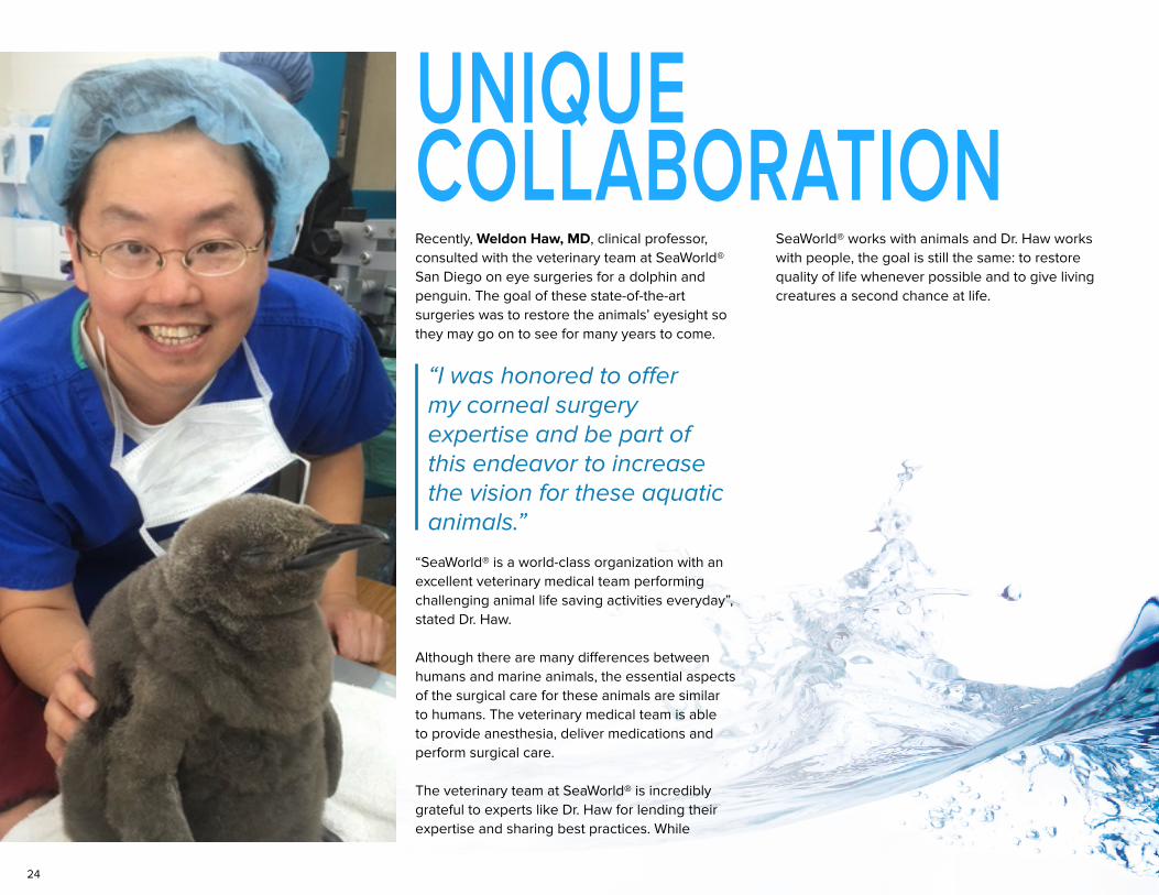
Recently, Weldon Haw, MD, clinical professor, consulted with the veterinary team at SeaWorld® San Diego on eye surgeries for a dolphin and penguin. The goal of these state-of-the-art surgeries was to restore the animals’ eyesight so they may go on to see for many years to come.
“SeaWorld® is a world-class organization with an excellent veterinary medical team performing challenging animal life saving activities everyday”, stated Dr. Haw.
Although there are many differences between humans and marine animals, the essential aspects of the surgical care for these animals are similar to humans. The veterinary medical team is able to provide anesthesia, deliver medications and perform surgical care.
The veterinary team at SeaWorld® is incredibly grateful to experts like Dr. Haw for lending their expertise and sharing best practices. While
SeaWorld® works with animals and Dr. Haw works with people, the goal is still the same: to restore quality of life whenever possible and to give living creatures a second chance at life.
UNIQUE COLLABORATION
“I was honored to offer my corneal surgery expertise and be part of this endeavor to increase the vision for these aquatic animals.”
25
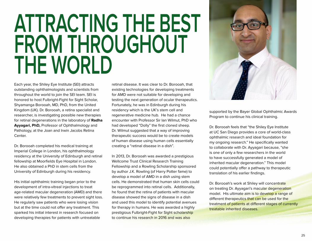
Each year, the Shiley Eye Institute (SEI) attracts outstanding ophthalmologists and scientists from throughout the world to join the SEI team. SEI is honored to host Fulbright-Fight for Sight Scholar, Shyamanga Borooah, MD, PhD, from the United Kingdom (UK). Dr. Borooah, a retina specialist and researcher, is investigating possible new therapies for retinal degenerations in the laboratory of Radha Ayyagari, PhD, Professor of Ophthalmology and Pathology, at the Joan and Irwin Jacobs Retina Center.
Dr. Borooah completed his medical training at Imperial College in London, his ophthalmology residency at the University of Edinburgh and retinal fellowship at Moorfields Eye Hospital in London. He also obtained a PhD in stem cells from the University of Edinburgh during his residency.
His initial ophthalmic training began prior to the development of intra-vitreal injections to treat age-related macular degeneration (AMD) and there were relatively few treatments to prevent sight loss. He regularly saw patients who were losing vision but at the time could not offer any treatment. This sparked his initial interest in research focused on developing therapies for patients with untreatable
supported by the Bayer Global Ophthalmic Awards Program to continue his clinical training.
Dr. Borooah feels that “the Shiley Eye Institute at UC San Diego provides a core of world-class ophthalmic research and ideal foundation for my ongoing research.” He specifically wanted to collaborate with Dr. Ayyagari because, “she is one of only a few researchers in the world to have successfully generated a model of inherited macular degeneration.” This model could potentially offer a pathway to therapeutic translation of his earlier findings.
Dr. Borooah’s work at Shiley will concentrate on treating Dr. Ayyagari’s macular degeneration model. His ultimate aim is to develop a range of different therapeutics that can be used for the treatment of patients at different stages of currently treatable inherited diseases.
retinal disease. It was clear to Dr. Borooah, that existing technologies for developing treatments for AMD were not suitable for developing and testing the next generation of ocular therapeutics. Fortunately, he was in Edinburgh during his residency which is the UK’s stem cell and regenerative medicine hub. He had a chance encounter with Professor Sir Ian Wilmut, PhD who had developed “Dolly” the first cloned sheep. Dr. Wilmut suggested that a way of improving therapeutic success would be to create models of human disease using human cells essentially creating a “retinal disease in a dish”.
In 2013, Dr. Borooah was awarded a prestigious Wellcome Trust Clinical Research Training Fellowship and a Rowling Scholarship sponsored by author J.K. Rowling (of Harry Potter fame) to develop a model of AMD in a dish using stem cells. He demonstrated that human skin cells could be reprogrammed into retinal cells. Additionally, he found that the retina of patients with macular disease showed the signs of disease in a dish and used this model to identify potential avenues for therapy in humans. He was awarded a highly prestigious Fulbright-Fight for Sight scholarship to continue his research in 2016 and was also
ATTRACTING THE BEST FROM THROUGHOUTTHE WORLD
26
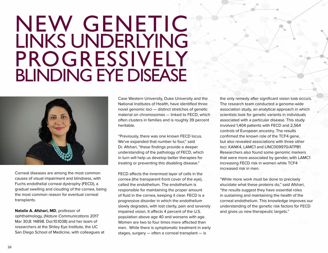
Corneal diseases are among the most common causes of visual impairment and blindness, with Fuchs endothelial corneal dystrophy (FECD), a gradual swelling and clouding of the cornea, being the most common reason for eventual corneal transplants.
Natalie A. Afshari, MD, professor of ophthalmology, (Nature Communications 2017 Mar 30;8: 14898. Doi:10.1038) and her team of researchers at the Shiley Eye Institute, the UC San Diego School of Medicine, with colleagues at
Case Western University, Duke University and the National Institutes of Health, have identified three novel genomic loci — distinct stretches of genetic material on chromosomes — linked to FECD, which often clusters in families and is roughly 39 percent heritable.
“Previously, there was one known FECD locus. We’ve expanded that number to four,” said Dr. Afshari, “these findings provide a deeper understanding of the pathology of FECD, which in turn will help us develop better therapies for treating or preventing this disabling disease.”
FECD affects the innermost layer of cells in the cornea (the transparent front cover of the eye), called the endothelium. The endothelium is responsible for maintaining the proper amount of fluid in the cornea, keeping it clear. FECD is a progressive disorder in which the endothelium slowly degrades, with lost clarity, pain and severely impaired vision. It affects 4 percent of the U.S. population above age 40 and worsens with age. Women are two to four times more affected than men. While there is symptomatic treatment in early stages, surgery — often a corneal transplant — is
the only remedy after significant vision loss occurs.The research team conducted a genome-wide association study, an analytical approach in which scientists look for genetic variants in individuals associated with a particular disease. This study involved 1,404 patients with FECD and 2,564 controls of European ancestry. The results confirmed the known role of the TCF4 gene, but also revealed associations with three other loci: KANK4, LAMC1 and LINC009970/ATPB1. Researchers also found some genomic markers that were more associated by gender, with LAMC1 increasing FECD risk in women while TCF4 increased risk in men.
“While more work must be done to precisely elucidate what these proteins do,” said Afshari, “the results suggest they have essential roles in sustaining and maintaining the health of the corneal endothelium. This knowledge improves our understanding of the genetic risk factors for FECD and gives us new therapeutic targets.”
NEW GENETIC
BLINDING EYE DISEASE
LINKS UNDERLYINGPROGRESSIVELY
27
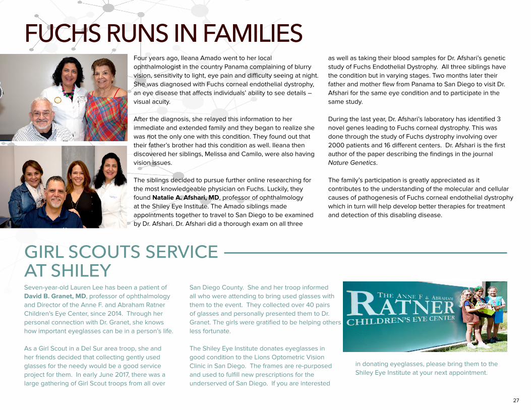
FUCHS RUNS IN FAMILIESFour years ago, Ileana Amado went to her local ophthalmologist in the country Panama complaining of blurry vision, sensitivity to light, eye pain and difficulty seeing at night. She was diagnosed with Fuchs corneal endothelial dystrophy, an eye disease that affects individuals’ ability to see details – visual acuity. After the diagnosis, she relayed this information to her immediate and extended family and they began to realize she was not the only one with this condition. They found out that their father’s brother had this condition as well. Ileana then discovered her siblings, Melissa and Camilo, were also having vision issues. The siblings decided to pursue further online researching for the most knowledgeable physician on Fuchs. Luckily, they found Natalie A. Afshari, MD, professor of ophthalmology at the Shiley Eye Institute. The Amado siblings made appointments together to travel to San Diego to be examined by Dr. Afshari. Dr. Afshari did a thorough exam on all three
as well as taking their blood samples for Dr. Afshari’s genetic study of Fuchs Endothelial Dystrophy. All three siblings have the condition but in varying stages. Two months later their father and mother flew from Panama to San Diego to visit Dr. Afshari for the same eye condition and to participate in the same study.
During the last year, Dr. Afshari’s laboratory has identified 3 novel genes leading to Fuchs corneal dystrophy. This was done through the study of Fuchs dystrophy involving over 2000 patients and 16 different centers. Dr. Afshari is the first author of the paper describing the findings in the journal Nature Genetics.
The family’s participation is greatly appreciated as it contributes to the understanding of the molecular and cellular causes of pathogenesis of Fuchs corneal endothelial dystrophy which in turn will help develop better therapies for treatment and detection of this disabling disease.
Seven-year-old Lauren Lee has been a patient of David B. Granet, MD, professor of ophthalmology and Director of the Anne F. and Abraham Ratner Children’s Eye Center, since 2014. Through her personal connection with Dr. Granet, she knows how important eyeglasses can be in a person’s life.
As a Girl Scout in a Del Sur area troop, she and her friends decided that collecting gently used glasses for the needy would be a good service project for them. In early June 2017, there was a large gathering of Girl Scout troops from all over
GIRL SCOUTS SERVICE AT SHILEY
San Diego County. She and her troop informed all who were attending to bring used glasses with them to the event. They collected over 40 pairs of glasses and personally presented them to Dr. Granet. The girls were gratified to be helping others less fortunate.
The Shiley Eye Institute donates eyeglasses in good condition to the Lions Optometric Vision Clinic in San Diego. The frames are re-purposed and used to fulfill new prescriptions for the underserved of San Diego. If you are interested
in donating eyeglasses, please bring them to the Shiley Eye Institute at your next appointment.
28
Twelve-year-old Mary* has night blindness. Even though her vision is normal during the day, at night, she can’t see and becomes easily disoriented.
Her night blindness has made her feel self-conscious and different. When she visits amusement parks, she has to leave early and misses all the evening activities because she can’t see in the dark. During a recent middle school over-night camping trip, she needed special assistance to guide her during night-time activities.
Her parents have known that she had a retina condition for the past seven years, but no one has been able to make a definitive diagnosis. The retina is the light-sensitive layer of tissue which lines the back of the eye that captures light and sends visual messages through the optic nerve to the brain. Mary’s parents worried
her condition could be a progressive retinal disease that would lead to blindness later in life.
Mary’s doctor referred them to Eric Nudleman, MD, PhD, assistant professor of ophthalmology and pediatric retina specialist at the Shiley Eye Institute, for diagnosis and treatment. After a full retina examination and specialized imaging, Dr. Nudleman suspected an inherited retinal dystrophy. He suggested that Mary see his colleague, Henry A. Ferreyra, MD, associate professor of ophthalmology and a retina specialist at Shiley and an expert in the field of hereditary retinal degenerations.
to distinguish. Dr. Ferreyra ordered more testing for Mary’s eyes, including electroretinography (ERG). An ERG is a test used to measure the function of the retina. Although an ERG was done previously and was abnormal suggesting retinitis punctata albescens, he suspected fundus albipunctatus and performed a modified ERG to evaluate for prolonged dark adaptation.
After examining the results of her testing, Dr. Ferreyra believed that Mary had fundus albipunctatus since her ERG responses normalized with prolonged dark adaptation. In order to make a definitive diagnosis, Dr. Ferreyra ordered genetic testing to look for mutations in the gene 11-cis retinol dehydrogenase 5 (RDH5). RDH5 is a gene that encodes for an enzyme involved in the conversion of 11-cis retinol to 11-cis retinal and necessary for normal vision, especially in low-light conditions. Mutations in the RDH5 are responsible for fundus albipunctatus.
After ordering the genetic testing, she was found to have two disease causing mutations in the RDH5 gene. Additional genetic testing of her parents confirmed that she had autosomal recessive fundus albipunctatus. Mary was so relieved that Dr. Ferreyra had finally diagnosed her with a non-progressive disease and she was not going blind. She can now go on to achieve her dream of becoming an architect when she grows up. She loves to draw and paint.
Sometimes our doctors at the Shiley Eye Institute have to be detectives. This teamwork by our specialists demonstrates how personalized medicine can change lives, especially for the young.
Mary* is a fictitious name; patient wants anonymity.
PERSONALIZED MEDICINE HELPS CHILD
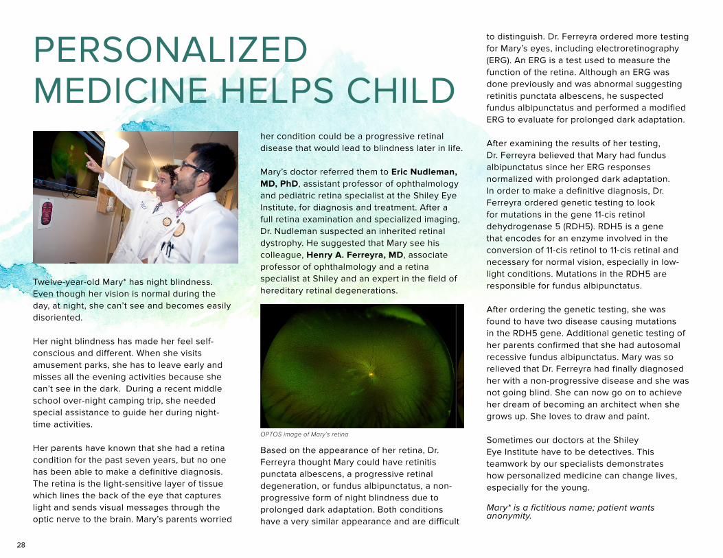
Based on the appearance of her retina, Dr. Ferreyra thought Mary could have retinitis punctata albescens, a progressive retinal degeneration, or fundus albipunctatus, a non-progressive form of night blindness due to prolonged dark adaptation. Both conditions have a very similar appearance and are difficult
OPTOS image of Mary’s retina
29
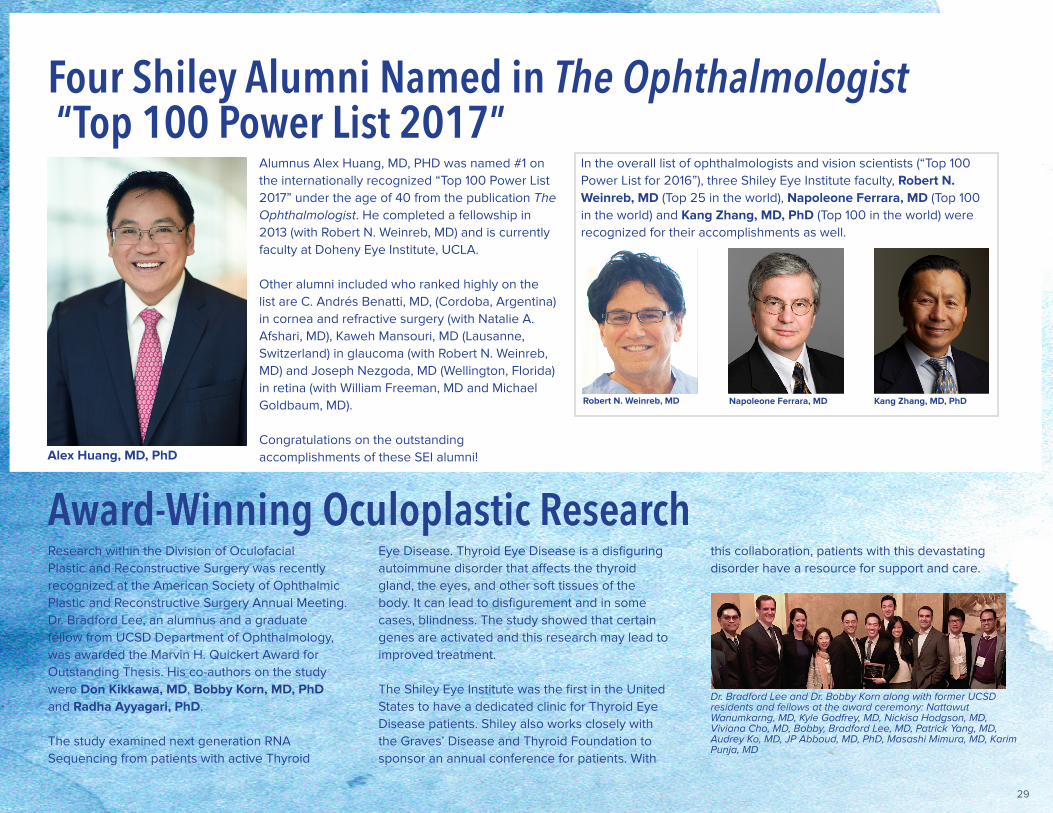
Alumnus Alex Huang, MD, PHD was named #1 on the internationally recognized “Top 100 Power List 2017” under the age of 40 from the publication The Ophthalmologist. He completed a fellowship in 2013 (with Robert N. Weinreb, MD) and is currently faculty at Doheny Eye Institute, UCLA.
Other alumni included who ranked highly on the list are C. Andrés Benatti, MD, (Cordoba, Argentina) in cornea and refractive surgery (with Natalie A. Afshari, MD), Kaweh Mansouri, MD (Lausanne, Switzerland) in glaucoma (with Robert N. Weinreb, MD) and Joseph Nezgoda, MD (Wellington, Florida) in retina (with William Freeman, MD and Michael Goldbaum, MD).
Congratulations on the outstanding accomplishments of these SEI alumni!Alex Huang, MD, PhD
Four Shiley Alumni Named in The Ophthalmologist “Top 100 Power List 2017”
In the overall list of ophthalmologists and vision scientists (“Top 100 Power List for 2016”), three Shiley Eye Institute faculty, Robert N. Weinreb, MD (Top 25 in the world), Napoleone Ferrara, MD (Top 100 in the world) and Kang Zhang, MD, PhD (Top 100 in the world) were recognized for their accomplishments as well.
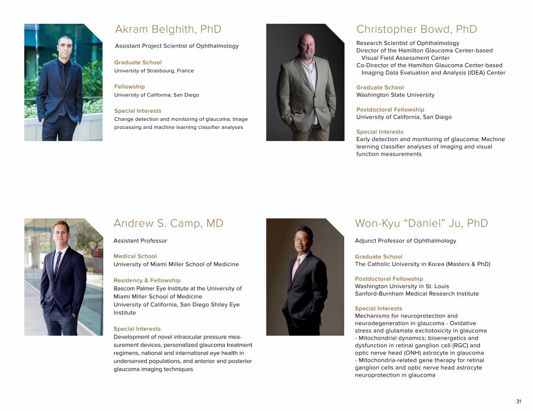
Research within the Division of Oculofacial Plastic and Reconstructive Surgery was recently recognized at the American Society of Ophthalmic Plastic and Reconstructive Surgery Annual Meeting. Dr. Bradford Lee, an alumnus and a graduate fellow from UCSD Department of Ophthalmology, was awarded the Marvin H. Quickert Award for Outstanding Thesis. His co-authors on the study were Don Kikkawa, MD, Bobby Korn, MD, PhD and Radha Ayyagari, PhD.
The study examined next generation RNA Sequencing from patients with active Thyroid
Dr. Bradford Lee and Dr. Bobby Korn along with former UCSD residents and fellows at the award ceremony: Nattawut Wanumkarng, MD, Kyle Godfrey, MD, Nickisa Hodgson, MD, Viviana Cho, MD, Bobby, Bradford Lee, MD, Patrick Yang, MD, Audrey Ko, MD, JP Abboud, MD, PhD, Masashi Mimura, MD, Karim Punja, MD
Award-Winning Oculoplastic ResearchEye Disease. Thyroid Eye Disease is a disfiguring autoimmune disorder that affects the thyroid gland, the eyes, and other soft tissues of the body. It can lead to disfigurement and in some cases, blindness. The study showed that certain genes are activated and this research may lead to improved treatment.
The Shiley Eye Institute was the first in the United States to have a dedicated clinic for Thyroid Eye Disease patients. Shiley also works closely with the Graves’ Disease and Thyroid Foundation to sponsor an annual conference for patients. With
this collaboration, patients with this devastating disorder have a resource for support and care.
Kang Zhang, MD, PhDRobert N. Weinreb, MD Napoleone Ferrara, MD

30
Glaucoma can cause blindness if untreated and is the second leading cause of blindness in the United States. As many as 3 million Americans have glaucoma, and at least one half do not know it. Although there is no cure yet, loss of vision can be slowed or halted with medical and/or surgical treatment. The best way to protect your sight from glaucoma is to get tested. Early diagnosis and appropriate treatment are the keys to preserving vision.
GLAUCOMA
Professor of Ophthalmology in ResidenceCo-Director of Clinical Research, Hamilton Glaucoma CenterDirector, Hamilton Glaucoma Center, Data Coordinating Center
Graduate SchoolHarvard School of Public Health (MS)Ben-Gurion University of the Negev (PhD)
Postdoctoral Fellowship University of Waterloo, Waterloo, Ontario, Canada
Special InterestsTo improve our understanding of the complex relationship between structural and functional change over time in the aging and glaucoma eye; To develop computational and statistical techniques to improve glaucomatous change detection, reduce the number of visits and optimize the type of testing required; To identify risk factors that can predict glaucomatous progression and rapidly progressing glaucoma
Linda M. Zangwill, PhD
Chairman & Distinguished Professor of OphthalmologyDirector of the Shiley Eye InstituteDirector of the Hamilton Glaucoma CenterMorris Gleich, MD Chair in Glaucoma
Medical SchoolHarvard Medical School
Residency & FellowshipUniversity of California, San Francisco
Special InterestsGlaucoma surgery and minimally invasive glaucoma surgery; Optic neuropathy and aging of the eye; Glaucoma genetics; Imaging of the optic disc and retinal nerve fiber layer and macula; Optical Coherence Angiography; Mechanisms of optic nerve damage in glaucoma; Neuroprotection in glaucoma; Measurement of intraocular pressure; Drug delivery to eye; Cataract surgery; Mentoring the next generation of world leaders in glaucoma
Notables & Awards2017 America’s Top Doctors/Castle Connolly Top Doctors, 2017 San Diego Magazine Top Doctors, 2017 Super Doctors
Robert N. Weinreb, MD
31
Graduate SchoolUniversity of Strasbourg, France
Fellowship University of California, San Diego
Special InterestsChange detection and monitoring of glaucoma; Image
processing and machine learning classifier analyses
Akram Belghith, PhDAssistant Project Scientist of Ophthalmology Research Scientist of Ophthalmology
Director of the Hamilton Glaucoma Center-based Visual Field Assessment CenterCo-Director of the Hamilton Glaucoma Center-based Imaging Data Evaluation and Analysis (IDEA) Center
Graduate SchoolWashington State University
Postdoctoral Fellowship University of California, San Diego
Special InterestsEarly detection and monitoring of glaucoma; Machine learning classifier analyses of imaging and visual function measurements
Christopher Bowd, PhD
Andrew S. Camp, MDAssistant Professor
Medical SchoolUniversity of Miami Miller School of Medicine
Residency & FellowshipBascom Palmer Eye Institute at the University of Miami Miller School of MedicineUniversity of California, San Diego Shiley Eye Institute
Special InterestsDevelopment of novel intraocular pressure mea-surement devices, personalized glaucoma treatment regimens, national and international eye health in underserved populations, and anterior and posterior glaucoma imaging techniques
Adjunct Professor of Ophthalmology
Won-Kyu “Daniel” Ju, PhD
Graduate SchoolThe Catholic University in Korea (Masters & PhD)
Postdoctoral Fellowship Washington University in St. LouisSanford-Burnham Medical Research Institute
Special InterestsMechanisms for neuroprotection and neurodegeneration in glaucoma - Oxidative stress and glutamate excitotoxicity in glaucoma - Mitochondrial dynamics; bioenergetics and dysfunction in retinal ganglion cell (RGC) and optic nerve head (ONH) astrocyte in glaucoma - Mitochondria-related gene therapy for retinal ganglion cells and optic nerve head astrocyte neuroprotection in glaucoma
32
Associate Clinical Professor of Ophthalmology
Medical SchoolUniversity of Oklahoma; Internship at UCLA
Residency University of Southern California
Special InterestsClinical research in glaucoma; UC San Diego Optic Disc Reading Center
Rigby Slight, MD Derek S. Welsbie, MD, PhDAssistant Professor of Ophthalmology
Medical SchoolUniversity of California, Los Angeles
Residency & FellowshipThe Johns Hopkins University School of Medicine / Wilmer Eye Institute
Special InterestsNeuroprotection in glaucoma and other optic neuropathies; Use of functional genomic technologies to identify novel mediators of axon injury signaling in neurons; Development of dual leucine zipper kinase inhibitors; Role of dual leucine zipper kinase in traumatic brain injury
Notables & Awards2017 OKAP Teaching Award, Shiley Eye Institute, 2017 Whitehill Prize for Excellence, UC San Diego Academy of Clinician Scholar, 2017 Douglas H. Johnson Award for Glaucoma Research, Brightfocus Foundation
Adjunct Professor of Ophthalmology Director, Glaucoma Molecular Pharmacology Laboratory
Graduate SchoolNational Tsing Hua University (MS Molecular Biology) Texas A&M University (PhD Pharmacology)
Postdoctoral Fellowship Harvard Medical School
Special InterestsRegulation of intraocular pressure and ocular blood flow; 24-hour sleep laboratory for glaucoma and other eye diseases
John H.K. Liu, PhD
Adjunct Professor of Ophthalmology
Dorota Skowronska-Krawczyk, PhD
Graduate SchoolUniversity of Geneva, Switzerland
Postdoctoral Fellowship Eye Hospital Jules Gonin, Lausanne, SwitzerlandUniversity of California, San Diego
Special InterestsMolecular mechanisms in retina development and in genetic and age related eye diseases, including glaucoma
33
NEURO-OPHTHALMOLOGYNeuro-ophthalmologists diagnose and treat neuro-sensory disorders
including brain tumors, double vision, giant cell arteritis, ischemic optic
neuropathy, optic neuritis, papilledema, pseudotumor cerebri, thyroid
eye disease and visual field defects. Shiley Eye Institute’s skilled neuro-
ophthalmologists conduct routine diagnostic tests and a thorough evaluation
while working with the referring physician to manage the condition or illness.
Peter J. Savino, MDClinical Professor of Ophthalmology & Neurosciences
Medical SchoolUniversity of Bologna School of Medicine
ResidencyGeorgetown University Medical Center
Fellowship University of Miami
Special InterestsMyasthenia gravis optic neuritis, atrophy and neuropathy brain and nervous system tumors visual field defects; Degenerative, metabolic, inflammatory & demyelinating diseases vascular disorders
Notables & Awards2017 America’s Top Doctors/Castle Connolly Top Doctors
34
34
CORNEA &
REFRACTIVEThe Shiley Eye Institute Cornea and Refractive specialty is dedicated to the health and functioning of the cornea and combines unparalleled care, expertise, and state-of-the-art equipment to ensure the best experience for patients. The Shiley Eye Institute provides comprehensive eye care for a range of routine, complex and high-risk corneal, cataract and external diseases, as well as the most current vision correction procedures.
Natalie A. Afshari, MD, FACSProfessor of OphthalmologyStuart I. Brown MD Chair in Ophthalmology in Memory of Donald P. ShileyChief, Division of Cornea and Refractive SurgeryVice-Chair for Education, Department of Ophthalmology
Medical SchoolStanford University Medical School
Residency & FellowshipHarvard University, Massachusetts Eye and Ear Infirmary
Special InterestsCorneal surgery; Fuchs Dystrophy; Corneal transplantation; Endothelial keratoplasty (DSAEK & DMEK); Intacs and collagen crosslinking for keratoconus; Laser refractive surgery, including LASIK and PRK, Surgical and medical diseases of cornea; Cataract surgery
Notables & Awards2017 America’s Top Doctors/Castle Connolly Top Doctors, 2017 San Diego Magazine Top Doctors
35
Jiagang “Jack” Zhao, PhDAssociate Project Scientist of Ophthalmology
Graduate SchoolMount Sinai School of Medicine
Postdoctoral Fellowship Salk Institute for Biological Studies, La Jolla, California
Special InterestsAge related macular degeneration; Diabetic retinopathy; Inherited retinal degeneration
Chris W. Heichel, MDAssociate Clinical Professor of Ophthalmology
Medical SchoolChicago Medical School
ResidencyUniversity of California, San Diego (Chief Resident)
FellowshipUniversity of California, San Diego
Special InterestsCorneal transplantations and Keratoprostheses; Challenging cataract and IOL surgeries; LASIK; Intacs, & Visian ICL; Advanced techniques in laser & refractive surgery; Keratoconus; Ocular Surface Tumors; Limbal Stem Cell Transplantation
Notables & Awards2017 America’s Top Doctors/Castle Connolly Top Doctors
Clinical Professor of OphthalmologyChief of Ophthalmology at Veterans Administration Medical Hospital
Medical SchoolUniversity of California, Los Angeles School of Medicine
ResidencyStanford University School of Medicine (Chief Resident)
Fellowship Stanford University School of Medicine (Chief Fellow)
Special InterestsCorneal and cataract surgery, Intraocular lenses, Dry Eye/Pterygium, Cornea transplantation, Refractive surgery/LASIK
Notables & Awards2017 America’s Top Doctors/Castle Connolly Top Doctors
Weldon W. Haw, MDStuart I. Brown, MDProfessor of OphthalmologyDr. Richard and Tatiana Lansche Chair in Ophthalmology
Medical SchoolUniversity of Illinois Medical School
ResidencyTulane Medical School
Fellowship Harvard University, Massachusetts Eye and Ear Infirmary
Special InterestsCornea and external diseases
Notables & Awards2017 Super Doctors
36
RETINA & VITREOUS
Diseases of the retina cause severe and debilitating vision loss. Our retina physicians diagnose and treat macular degeneration, diabetic retinopathy, tumors, inherited retinal disease, retinal detachment, macular holes, and other important retinal diseases.
William R. Freeman, MDDistinguished Professor of OphthalmologyVice-Chair, Department of OphthalmologyDirector, Jacobs Retina CenterCo-Director, Retina Division
Medical SchoolMount Sinai School of Medicine, New York, NY
Residency Lenox Hill Hospital, New York, NY
Fellowship University of California, San Francisco, CA (Uveitis & Immunology) University of Southern California, Los Angeles, CA (Vitreo-Retinal Surgery)
Special InterestsComplicated retinal detachment; Diabetic retinopathy; Macular holes & age related macular degeneration
Notables & Awards2017 America’s Top Doctors/Castle Connolly Top Doctors, 2017 Super Doctors
Michael H. Goldbaum, MD, MSProfessor of Ophthalmology in ResidenceCo-Director, Retina Division
Medical SchoolTulane University School of Medicine (MD)Stanford University (MS)
Residency Tulane University School of Postgraduate Medicine & U.S. Naval Hospital
Fellowship Cornell University Medical Center and New York Hospital
Special InterestsSurgical & medical treatment of the retina and vitreous; Macular degeneration; Pediatric retina; Ocular tumors; Glaucoma informatics
Notables & Awards2017 America’s Top Doctors/Castle Connolly Top Doctors, 2017 Super Doctors
37
Radha Ayyagari, PhDProfessor of Ophthalmology and PathologyChief of Ophthalmic Molecular Diagnostic Laboratory (CLIA certified)Director of Shiley Eye Institute BioBank
Graduate SchoolOsmania University, Hyderabad, India
Postdoctoral Fellowship Molecular Genetics at the National Eye Institute, NIH, Bethesda
Special InterestsMolecular genetics of macular and retinal dystrophy; Biological mechanisms underlying retinal diseases; Age-related macular degeneration; Diabetic retinopathy; and Glaucoma
Notables & Awards2017 Gold Fellow, The Association for Research in Vision and Ophthalmology
Dirk-Uwe Bartsch, PhDAssociate Adjunct Professor of Ophthalmology Co-Director, Jacobs Retina Center
Graduate SchoolUniversity of California, San Diego
Postdoctoral Fellowship University of California, San Diego
Special InterestsRetinal Imaging Scanning Laser Imaging - confocal / non-confocal; Optical Coherence Tomography (OCT); Indocyanine Green and Fluorescein Angiography; Tomographic Reconstruction of the Posterior Pole
Assistant Clinical Professor of Ophthalmology
Medical SchoolStanford University (MD and PhD)
ResidencyBascom Palmer Eye Institute, University of Miami
Fellowship University of California, San Francisco
Special InterestsSurgical and medical management of retinal diseases, diabetic retinopathy, age related macular degeneration; Translational research; Scientific focus on developing zebrafish as a model for retinal diseases; Technology development for new treatments and diagnostics for retinal disease
Daniel L. Chao, MD, PhD Lingyun Cheng, MDAdjunct Professor of Ophthalmology
Medical SchoolShanxi Medical University, China
ResidencyThe First Teaching Hospital of Shanxi Medical University, China
Fellowship University of California, San DiegoIdeta Eye Hospital, Japan
Special InterestsOcular drug delivery and vitreoretinal diseases
38
Henry A. Ferreyra, MDAssociate Clinical Professor of Ophthalmology
Medical SchoolUniversity of California, San Diego
ResidencyUniversity of California, San Diego
Fellowship University of California, San Diego
Special InterestsElectrophysiology Inherited disorders of the retina; Age-related macular degeneration; Diabetic retinopathy; Retinopathy of prematurity
Notables & Awards2017 Outstanding Teacher Award (Resident Award), 2017 America’s Top Doctors/Castle Connolly Top Doctors
Assistant Clinical Professor of Ophthalmology
Medical SchoolAlbert Einstein College of Medicine (MD) Stanford University (PhD)
Residency Washington University in St. Louis
Fellowship Associated Retinal Consultants / William Beaumont Hospital
Special InterestsAdult and pediatric vitreoretinal diseases, including macular degeneration, diabetic eye disease, retinal vein occlusions, retinal detachments, proliferative vitreoretinopathy, macular holes and epiretinal membranes; Specialty interest in pediatric vitreoretinal diseases, including the surgical management of advanced retinopathy of prematurity, familial exudative vitreoretinopathy, Coats disease, persistent fetal vascular syndrome, and intraocular trauma; Scientific focus on developmental angiogenesis, with emphasis on the role of the Wnt Signaling pathway in developmental vascular diseases
Eric Nudleman, MD, PhD
THYROID EYECLINIC
Thyroid Eye Disease is a complex autoimmune
disease that affects not only vision but also
causes pain and deformity. Drs. Granet,
Kikkawa, and Korn have helped hundreds of
patients with this disfiguring disorder and have
published extensively on its characteristics and
treatment.
Don O. Kikkawa, MD
David B. Granet, MD
Bobby S. Korn, MD, PhD
39
OPHTHALMIC PLASTIC & RECONSTRUCTIVE SURGERY
Don O. Kikkawa, MD, FACSProfessor of Clinical Ophthalmology and Plastic SurgeryVice-Chair for Clinical Services, Department of OphthalmologyChief, Division of Oculofacial Plastic and Reconstructive Surgery
Medical SchoolSt. Louis University School of Medicine
ResidencyUniversity of California, Los Angeles
Fellowship University of Wisconsin, Madison
Special InterestsOculofacial surgery; Eyelid, lacrimal and orbital surgery; Thyroid eye disease (orbital decompression and eyelid surgery); Craniofacial disorders involving the eyelids and orbits; Orbital and eyelid tumors; Facial aesthetics - soft tissue fillers and injectables
Notables & Awards2017 America’s Top Doctors/Castle Connolly Top Doctors, 2017 San Diego Magazine Top Doctors, 2017 Super Doctors
Orbits. Eyelids. Face. Lacrimal system. These are the domains of oculofacial plastic surgery. Birth defects, cancer, trauma and the aging process can all alter the periorbital region. These surgeons rebuild, reconstruct, renew and make whole again. The UCSD Division of Ophthalmic Plastic and Reconstructive Surgery is an internationally recognized leader in patient care, teaching and research. Dr. Kikkawa and Dr. Korn have pioneered innovative operations and techniques that have become the standard.
Bobby S. Korn, MD, PhD, FACSProfessor of Clinical Ophthalmology and Plastic Surgery
Medical SchoolUniversity of Texas, Southwestern Medical School (MD and PhD)
Residency & FellowshipUniversity of California, San Diego (Chief Resident)
Special InterestsCosmetic & reconstructive surgery (eyelid & face); Blepharoplasty (eyelid lift surgery); Ptosis surgery (droopy lid surgery); Asian Blepharoplasty (double eyelid surgery); Congenital birth defects; Endoscopic forehead lifting; Thyroid eye disease management; Eyelid & orbital tumors & cancers; Lacrimal/tear outflow system disorders; Bulging or proptosis of eyes; Reconstruction of eyelids post cancer removal; Reconstruction after trauma / eye injuries; Botox, Restylane, Juvederm & facial fillers; Skin rejuvenation – chemical peel
Notables & Awards2017 America’s Top Doctors/Castle Connolly Top Doctors, 2017 San Diego Magazine Top Doctors, 2017 Best Doctors of America
40
David B. Granet, MD, FAAO, FACS, FAAPProfessor of Ophthalmology & PediatricsAnne Ratner Chair of Pediatric OphthalmologyDirector, Anne F. and Abraham Ratner Children’s Eye CenterDirector, Division of Pediatric Ophthalmology & Eye Alignment Disorders
Medical SchoolYale University School of Medicine
ResidencyNew York University Medical Center (Chief Resident)
Fellowship Children’s Hospital of Philadelphia University of Pennsylvania Medical Center Scheie Eye Institute
Special InterestsPediatric ophthalmology & strabismus; Adult eye movement problems; State-of-the-art adjustable suture strabismus surgery; Childhood eye misalignments & disorders; Nystagmus Learning disorders & role of vision
Notables & Awards2017 America’s Top Doctors/Castle Connolly Top Doctors, 2017 Super Doctors
Shira L. Robbins, MD, FAAO, FAAPClinical Professor of OphthalmologyEducational Director of Pediatric Ophthalmology / Strabismus Division
Medical SchoolMedical College of Pennsylvania Hospital
Residency Hahnemann University Hospital
Fellowship University of California San Diego & Naval Medical Center
Special InterestsStrabismus/eye misalignment/double vision; Amblyopia; Retinopathy of prematurity; Pediatric glaucoma & cataracts; including intraocular lens placement; Nasolacrimal duct disorders; Congenital eye syndromes; Craniofacial syndromes; Systemic diseases affecting the eyes; Nystagmus
Notables & Awards2017 America’s Top Doctors/Castle Connolly Top Doctors
Preventing and treating vision loss/ocular problems in children is the highest priority at the Ratner Children’s Eye Center. Dr. David Granet and Dr. Shira Robbins are world-renowned specialists in helping children with eye misalignments (strabismus), nystagmus, congenital diseases like pediatric cataracts and glaucoma, acquired problems from blocked tear ducts to “lazy eye” (amblyopia) as well as trauma. From premature babies to teenagers our team ensures that each child seen at the family oriented Ratner Children’s Eye Center is given the attention and personal medical care they deserve in a child- friendly atmosphere.
Adults with strabismus suffer from an old childhood problem, trauma or a disease causing eye misalignment and require individualized intervention. Recognized worldwide for their teaching & developments in this field, the specialized surgeons at the Ratner Eye Center can help virtually everyone– regardless of age - suffering from various ocular misalignments and their consequences.
& ADULT EYEREALIGNMENT
PEDIATRICOPHTHALMOLOGY
41
Professor of OphthalmologyFounding Director, UCSD Institute for Genomic MedicineCo-Director, Biomaterials and Tissue Engineering, Institute for Engineering in Medicine
Medical SchoolHarvard Medical School / Massachusetts Institute of Technology (MD / PhD Program)
ResidencyThe Johns Hopkins University School of Medicine / Wilmer Eye Institute
Postdoctoral Fellowship University of Utah School of Medicine
Special InterestsAge related macular degeneration; Diabetic retinopathy; Inherited retinal degeneration
Kang Zhang, MD, PhD
Peter Shaw, PhDAssistant Adjunct Professor of OphthalmologyClinical and Translational Research InstituteSchool of Medicine Recruitment and Admission Committee
Graduate SchoolMcMaster University, Ontario, Canada
Postdoctoral Fellowship University of California, San Francisco
Special InterestsEvaluation and diagnosis of eye diseases including macular degeneration, diabetic retinopathy, glaucoma and inherited retinal degenerations by genetic variants and plasma biomarkers; Investigation of how genetic and oxidative stress risk factors impact on disease pathology; Development of molecular and gene therapy meth-ods to treat eye diseases
GENETICSGenetics is the study of genes, genetic variation, and heredity in living organisms. It is generally considered a field of biology, but intersects frequently with many other life sciences and is strongly linked with the study of information systems.
42
REGENERATIVE OPHTHALMOLOGYRetinal degeneration (RD) disease, such as age-related
macular degeneration (AMD), retinitis pigmentosa
(RP),Leber’s congenital amaurosis (LCA) and glaucoma
are blinding disorders, that unfortunately, are untreatable
once photoreceptors or retinal ganglion cells are lost.
Pluripotent stem cells (PSCs) are remarkable cells that can
give rise to virtually every cell type in the body including
cells that form the eye. Retinas derived from such stem
cells offer a potential means to generate new cells and
tissue for transformation, a system to address the origins
Karl Wahlin, PhDAssistant Professor of OphthalmologyDirector, Richard C. Atkinson Laboratory for Regenerative Ophthalmology
Graduate SchoolThe Johns Hopkins School of Medicine (Neuroscience)
Fellowship The Johns Hopkins School of Medicine / Wilmer Eye Institute
Special InterestsDirected differentiation of pluripotent stem cells and their application towards the study of retinal development and eye disease; Photoreceptor cell development and retinal connectivity; Retinal and optic nerve regeneration
of disease and a platform to screen for drugs that could
block the disease process.
Researchers at the Shiley Eye Institute are using stem cell
derived human 3D ‘mini-retinas’, genetic engineering and
drug screening to better understand how the human retina
forms and to understand the complex biology of human
retinal disease and explore potential cures. Combining
the power of stem cells and genetic engineering we are
developing so-called ‘disease-in-a-dish’ models to explore
totally new ways to treat retinal disease.
43
OCULARPATHOLOGYOphthalmic pathology focuses on diseases of
the eye and its neighboring tissues. Precision
diagnosis of diseases is provided by the
ophthalmic pathology service. Diseased tissues
are examined macroscopically, microscopically
and on the ultrastructural level. Advanced
genomic, proteomic, and cytogenetic techniques
can be utilized to diagnose diseases at a
molecular level. The pathologic diagnosis of the
disease plays a vital role in patient care.
Jonathan H. Lin, MD, PhD, FCAPAssociate Professor
Medical SchoolColumbia University College of Physicians & Surgeons (MD & PhD)
Residency Brigham Women’s Hospital (Anatomic Pathology)
Fellowship University of California, San Francisco (Ophthalmic Pathology)
Special InterestsOphthamic Pathology including pigmented ocular lesions (uveal melanoma, primary acquired melanosis), basal cell carcinoma, sebaceous gland lesions, inflammatory lesions (sclerosing orbital inflammatory pseudotumor, IgG4 disease), MALToma, corneas (PKPs, DSAEKs), conjunctival biopsies (conjunctival intraepithelial neoplasia - CIN), orbital lesions, intraocular fine needle aspirates/vitrectomy specimens; Cellular and molecular mechanisms of retinal degeneration; RPE and ocular stem cells
Napoleone Ferrara, MDDistinguished Professor of Ophthalmology and PathologySenior Deputy Director for Basic Sciences, UCSD Moores Cancer Center
Medical School & ResidencyUniversity of Catania Medical School, Catania, Italy
Fellowship University of California, San Francisco
Special InterestsRegulation of angiogenesis (the formation of new blood vessels) and the role of VEGF (vascular endothelial growth factor); Continue to develop new therapies to treat age related macular degeneration building upon past development of Avastin® and Lucentis®.
44
COMPREHENSIVEOPHTHALMOLOGY
Jeffrey E. Lee, MDAssistant Professor of Clinical Ophthalmology Program Director, Ophthalmology Residency
Medical School
University of California, San Diego
Residency
University of California, San Diego
Special Interests
Facial burns; Orbital trauma; Ocular manifestations of
HIV, Optimizing residency cataract surgery education
Thao P. Nguyen, MDAssistant Clinical Professor of Ophthalmology
Medical School
University of Oklahoma, Tulsa
Residency
University of Rochester, New York
Fellowship
University of California, San Diego
Shiley’s comprehensive ophthalmology division provides
primary eye care for all conditions of the eye and
surrounding structures, both routine and urgent. This
specialty offers routine ophthalmic evaluations which
screen and treat a wide range of ophthalmic conditions
including cataracts, ocular surface disorders, glaucoma,
diabetic retinopathy and macular degeneration. Diagnostic
services include evaluation of visual acuity, refraction, slit
lamp evaluation, ophthalmoscopy, tonometry, gonioscopy
and visual field testing.
Typical treatments include prescription medications and/or
optical correction, however, occasional surgical intervention
for cataracts and laser treatments are offered as part
of comprehensive ophthalmology. The comprehensive
ophthalmology division refers patients to the Department’s
subspecialty ophthalmologists for evaluations
regarding eye surgery and/or long-term treatment. The
comprehensive service handles most of the general ocular
problems and those patients desiring screenings for
cataracts, glaucoma and diabetic retinopathy.
45
OPTOMETRY &LOW VISION
John F. Kulischak, OD
Pamela A. Hoo, OD
Anne B. Lam, OD
Lianne Mizoguchi, OD
Lara D. Hustana, OD
Esmeralda McClean, OD
Maria Laura Gomez, MD, OD
Jessica A. Tasto, OD Alicia Lau, OD
Shiley Eye Institute optometrists are
eye care professionals who perform
comprehensive eye exams and are experts
at fitting all types of contact lenses and
glasses. Visual impairment from inherited
diseases to diabetic retinopathy and
macular degeneration can result in profound
vision loss. Using the latest technological
advancements in optical aids, optometrists
provide much needed care for our low
vision patients. Working hand in hand with
Shiley ophthalmologists, the optometry
service strives to deliver the best possible
care to each patient.
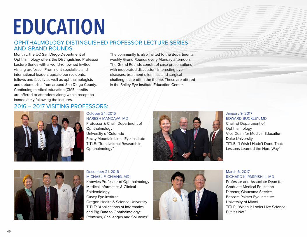
46
EDUCATIONOPHTHALMOLOGY DISTINGUISHED PROFESSOR LECTURE SERIES AND GRAND ROUNDSMonthly, the UC San Diego Department of Ophthalmology offers the Distinguished Professor Lecture Series with a world-renowned invited visiting professor. Prominent specialists and international leaders update our residents, fellows and faculty as well as ophthalmologists and optometrists from around San Diego County. Continuing medical education (CME) credits are offered to attendees along with a reception immediately following the lectures.
The community is also invited to the departmental weekly Grand Rounds every Monday afternoon. The Grand Rounds consist of case presentations with moderated discussion. Interesting eye diseases, treatment dilemmas and surgical challenges are often the theme. These are offered in the Shiley Eye Institute Education Center.
December 21, 2016 MICHAEL F. CHIANG, MDKnowles Professor of OphthalmologyMedical Informatics & Clinical EpidemiologyCasey Eye InstituteOregon Health & Science UniversityTITLE: “Applications of Informatics and Big Data to Ophthalmology: Promises, Challenges and Solutions”
January 9, 2017 EDWARD BUCKLEY, MDChair of Department of OphthalmologyVice Dean for Medical EducationDuke UniversityTITLE: “I Wish I Hadn’t Done That: Lessons Learned the Hard Way”
March 6, 2017 RICHARD K. PARRISH, II, MDProfessor and Associate Dean for Graduate Medical EducationDirector, Glaucoma ServiceBascom Palmer Eye InstituteUniversity of MiamiTITLE: “When It Looks Like Science, But It’s Not”
October 24, 2016 NARESH MANDAVA, MDProfessor & Chair, Department of OphthalmologyUniversity of ColoradoRocky Mountain Lions Eye InstituteTITLE: “Translational Research in Ophthalmology”
2016 – 2017 VISITING PROFESSORS:
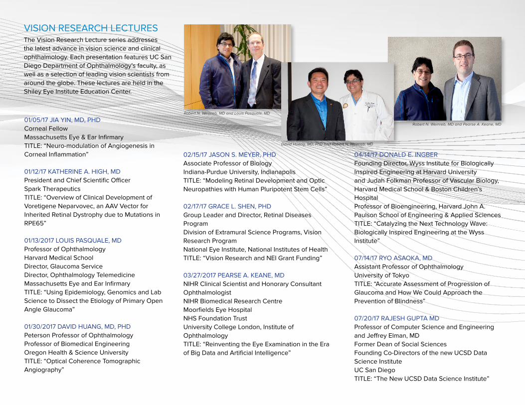
47
VISION RESEARCH LECTURES The Vision Research Lecture series addresses the latest advance in vision science and clinical ophthalmology. Each presentation features UC San Diego Department of Ophthalmology’s faculty, as well as a selection of leading vision scientists from around the globe. These lectures are held in the Shiley Eye Institute Education Center.
01/05/17 JIA YIN, MD, PHDCorneal FellowMassachusetts Eye & Ear Infirmary TITLE: “Neuro-modulation of Angiogenesis in Corneal Inflammation”
01/12/17 KATHERINE A. HIGH, MDPresident and Chief Scientific OfficerSpark Therapeutics TITLE: “Overview of Clinical Development of Voretigene Neparvovec, an AAV Vector for Inherited Retinal Dystrophy due to Mutations in RPE65”
01/13/2017 LOUIS PASQUALE, MDProfessor of OphthalmologyHarvard Medical SchoolDirector, Glaucoma ServiceDirector, Ophthalmology TelemedicineMassachusetts Eye and Ear InfirmaryTITLE: “Using Epidemiology, Genomics and Lab Science to Dissect the Etiology of Primary Open Angle Glaucoma”
01/30/2017 DAVID HUANG, MD, PHDPeterson Professor of OphthalmologyProfessor of Biomedical EngineeringOregon Health & Science UniversityTITLE: “Optical Coherence Tomographic Angiography”
02/15/17 JASON S. MEYER, PHDAssociate Professor of Biology Indiana-Purdue University, Indianapolis TITLE: “Modeling Retinal Development and Optic Neuropathies with Human Pluripotent Stem Cells”
02/17/17 GRACE L. SHEN, PHDGroup Leader and Director, Retinal Diseases ProgramDivision of Extramural Science Programs, Vision Research ProgramNational Eye Institute, National Institutes of HealthTITLE: “Vision Research and NEI Grant Funding”
03/27/2017 PEARSE A. KEANE, MDNIHR Clinical Scientist and Honorary Consultant OphthalmologistNIHR Biomedical Research CentreMoorfields Eye HospitalNHS Foundation TrustUniversity College London, Institute of OphthalmologyTITLE: “Reinventing the Eye Examination in the Era of Big Data and Artificial Intelligence”
04/14/17 DONALD E. INGBERFounding Director, Wyss Institute for Biologically Inspired Engineering at Harvard Universityand Judah Folkman Professor of Vascular Biology, Harvard Medical School & Boston Children’s HospitalProfessor of Bioengineering, Harvard John A. Paulson School of Engineering & Applied Sciences TITLE: “Catalyzing the Next Technology Wave: Biologically Inspired Engineering at the Wyss Institute”
07/14/17 RYO ASAOKA, MDAssistant Professor of Ophthalmology University of Tokyo TITLE: “Accurate Assessment of Progression of Glaucoma and How We Could Approach the Prevention of Blindness”
07/20/17 RAJESH GUPTA MDProfessor of Computer Science and Engineeringand Jeffrey Elman, MDFormer Dean of Social SciencesFounding Co-Directors of the new UCSD Data Science Institute UC San DiegoTITLE: “The New UCSD Data Science Institute”
Robert N. Weinreb, MD and Louis Pasquale, MD
David Huang, MD, PhD and Robert N. Weinreb, MD
Robert N. Weinreb, MD and Pearse A. Keane, MD
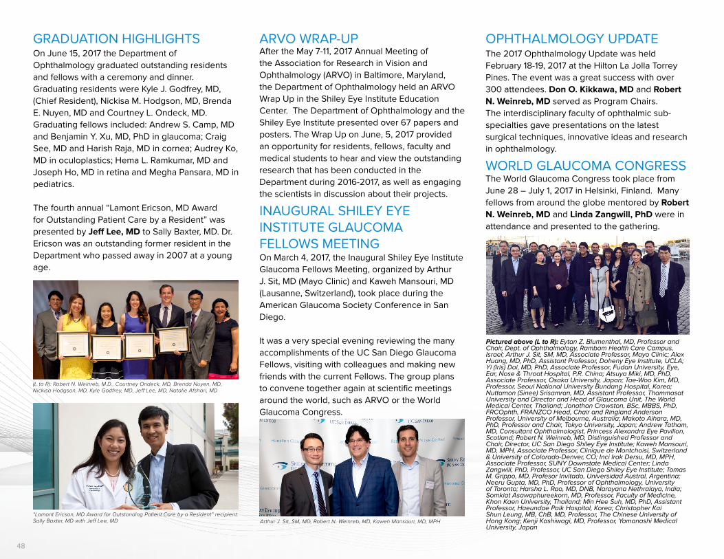
48
OPHTHALMOLOGY UPDATEThe 2017 Ophthalmology Update was held February 18-19, 2017 at the Hilton La Jolla Torrey Pines. The event was a great success with over 300 attendees. Don O. Kikkawa, MD and Robert N. Weinreb, MD served as Program Chairs. The interdisciplinary faculty of ophthalmic sub-specialties gave presentations on the latest surgical techniques, innovative ideas and research in ophthalmology.
ARVO WRAP-UPAfter the May 7-11, 2017 Annual Meeting of the Association for Research in Vision and Ophthalmology (ARVO) in Baltimore, Maryland, the Department of Ophthalmology held an ARVO Wrap Up in the Shiley Eye Institute Education Center. The Department of Ophthalmology and the Shiley Eye Institute presented over 67 papers and posters. The Wrap Up on June, 5, 2017 provided an opportunity for residents, fellows, faculty and medical students to hear and view the outstanding research that has been conducted in the Department during 2016-2017, as well as engaging the scientists in discussion about their projects.
INAUGURAL SHILEY EYE INSTITUTE GLAUCOMA FELLOWS MEETINGOn March 4, 2017, the Inaugural Shiley Eye Institute Glaucoma Fellows Meeting, organized by Arthur J. Sit, MD (Mayo Clinic) and Kaweh Mansouri, MD (Lausanne, Switzerland), took place during the American Glaucoma Society Conference in San Diego.
It was a very special evening reviewing the many accomplishments of the UC San Diego Glaucoma Fellows, visiting with colleagues and making new friends with the current Fellows. The group plans to convene together again at scientific meetings around the world, such as ARVO or the World Glaucoma Congress.
GRADUATION HIGHLIGHTSOn June 15, 2017 the Department of Ophthalmology graduated outstanding residents and fellows with a ceremony and dinner. Graduating residents were Kyle J. Godfrey, MD, (Chief Resident), Nickisa M. Hodgson, MD, Brenda E. Nuyen, MD and Courtney L. Ondeck, MD. Graduating fellows included: Andrew S. Camp, MD and Benjamin Y. Xu, MD, PhD in glaucoma; Craig See, MD and Harish Raja, MD in cornea; Audrey Ko, MD in oculoplastics; Hema L. Ramkumar, MD and Joseph Ho, MD in retina and Megha Pansara, MD in pediatrics.
The fourth annual “Lamont Ericson, MD Award for Outstanding Patient Care by a Resident” was presented by Jeff Lee, MD to Sally Baxter, MD. Dr. Ericson was an outstanding former resident in the Department who passed away in 2007 at a young age.
(L to R): Robert N. Weinreb, M.D., Courtney Ondeck, MD, Brenda Nuyen, MD, Nickisa Hodgson, MD, Kyle Godfrey, MD, Jeff Lee, MD, Natalie Afshari, MD
“Lamont Ericson, MD Award for Outstanding Patient Care by a Resident” recipient: Sally Baxter, MD with Jeff Lee, MD Arthur J. Sit, SM, MD, Robert N. Weinreb, MD, Kaweh Mansouri, MD, MPH
WORLD GLAUCOMA CONGRESSThe World Glaucoma Congress took place from June 28 – July 1, 2017 in Helsinki, Finland. Many fellows from around the globe mentored by Robert N. Weinreb, MD and Linda Zangwill, PhD were in attendance and presented to the gathering.
Pictured above (L to R): Eytan Z. Blumenthal, MD, Professor and Chair, Dept. of Ophthalmology, Rambam Health Care Campus, Israel; Arthur J. Sit, SM, MD, Associate Professor, Mayo Clinic; Alex Huang, MD, PhD, Assistant Professor, Doheny Eye Institute, UCLA; Yi (Iris) Dai, MD, PhD, Associate Professor, Fudan University, Eye, Ear, Nose & Throat Hospital, P.R. China; Atsuya Miki, MD, PhD, Associate Professor, Osaka University, Japan; Tae-Woo Kim, MD, Professor, Seoul National University Bundang Hospital, Korea; Nuttamon (Sinee) Srisamran, MD, Assistant Professor, Thammasat University and Director and Head of Glaucoma Unit, The World Medical Center, Thailand; Jonathan Crowston, BSc, MBBS, PhD, FRCOphth, FRANZCO Head, Chair and Ringland Anderson Professor, University of Melbourne, Australia; Makoto Aihara, MD, PhD, Professor and Chair, Tokyo University, Japan; Andrew Tatham, MD, Consultant Ophthalmologist, Princess Alexandra Eye Pavilion, Scotland; Robert N. Weinreb, MD, Distinguished Professor and Chair, Director, UC San Diego Shiley Eye Institute; Kaweh Mansouri, MD, MPH, Associate Professor, Clinique de Montchoisi, Switzerland & University of Colorado-Denver, CO; Inci Irak Dersu, MD, MPH, Associate Professor, SUNY Downstate Medical Center; Linda Zangwill, PhD, Professor, UC San Diego Shiley Eye Institute; Tomas M. Grippo, MD, Profesor Invitado, Universidad Austral, Argentina; Neeru Gupta, MD, PhD, Professor of Ophthalmology, University of Toronto; Harsha L. Rao, MD, DNB, Narayana Nethralaya, India; Somkiat Asawaphureekorn, MD, Professor, Faculty of Medicine, Khon Kaen University, Thailand; Min Hee Suh, MD, PhD, Assistant Professor, Haeundae Paik Hospital, Korea; Christopher Kai Shun Leung, MB, ChB, MD, Professor, The Chinese University of Hong Kong; Kenji Kashiwagi, MD, Professor, Yamanashi Medical University, Japan
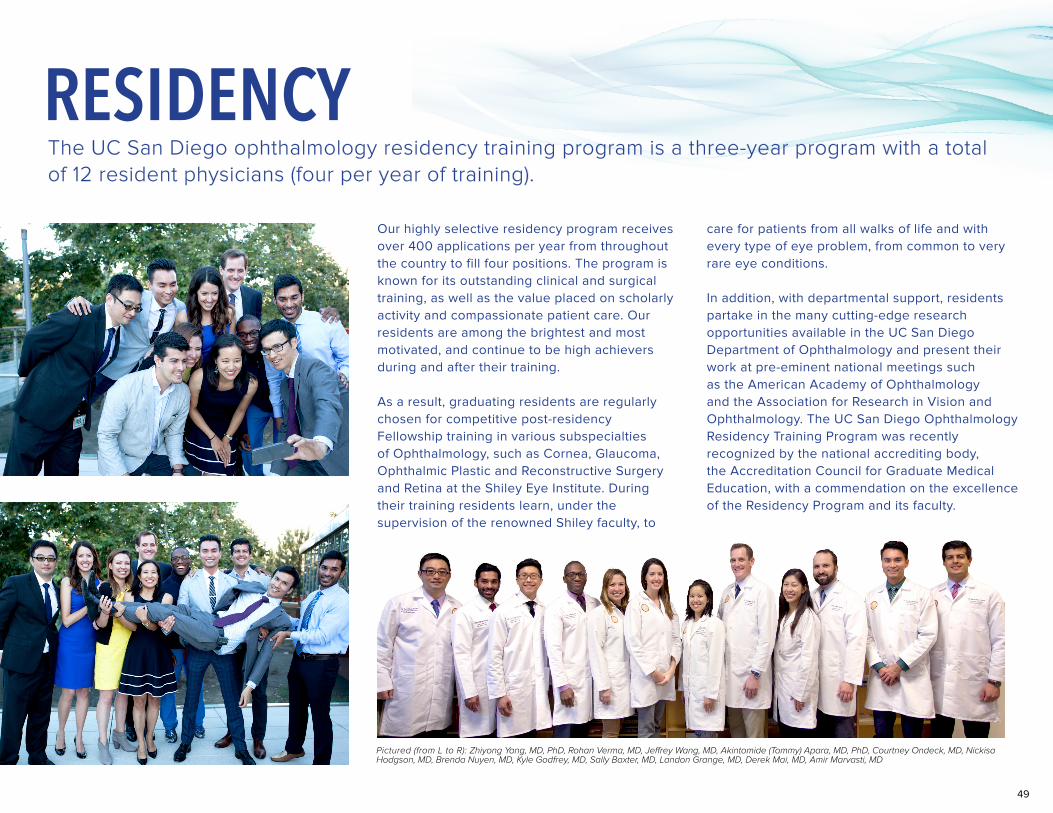
49
Our highly selective residency program receives over 400 applications per year from throughout the country to fill four positions. The program is known for its outstanding clinical and surgical training, as well as the value placed on scholarly activity and compassionate patient care. Our residents are among the brightest and most motivated, and continue to be high achievers during and after their training.
As a result, graduating residents are regularly chosen for competitive post-residency Fellowship training in various subspecialties of Ophthalmology, such as Cornea, Glaucoma, Ophthalmic Plastic and Reconstructive Surgery and Retina at the Shiley Eye Institute. During their training residents learn, under the supervision of the renowned Shiley faculty, to
care for patients from all walks of life and with every type of eye problem, from common to very rare eye conditions.
In addition, with departmental support, residents partake in the many cutting-edge research opportunities available in the UC San Diego Department of Ophthalmology and present their work at pre-eminent national meetings such as the American Academy of Ophthalmology and the Association for Research in Vision and Ophthalmology. The UC San Diego Ophthalmology Residency Training Program was recently recognized by the national accrediting body, the Accreditation Council for Graduate Medical Education, with a commendation on the excellence of the Residency Program and its faculty.
The UC San Diego ophthalmology residency training program is a three-year program with a total of 12 resident physicians (four per year of training).
Pictured (from L to R): Zhiyong Yang, MD, PhD, Rohan Verma, MD, Jeffrey Wang, MD, Akintomide (Tommy) Apara, MD, PhD, Courtney Ondeck, MD, Nickisa Hodgson, MD, Brenda Nuyen, MD, Kyle Godfrey, MD, Sally Baxter, MD, Landon Grange, MD, Derek Mai, MD, Amir Marvasti, MD
RESIDENCY
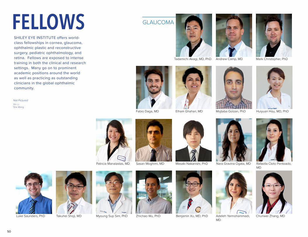
50
Patricia Manalastas, MD
Elham Ghahari, MD Mojtaba Golzan, PhD Huiyuan Hou, MD, PhD
Masaki Nakanishi, PhD Nara Gravina Ogata, MD Rafaella Cleto Penteado, MD
Luke Saunders, PhD Zhichao Wu, PhD Benjamin Xu, MD, PhDTakuhei Shoji, MD Myoung Sup Sim, PhD Adeleh Yarmohammadi, MD
Chunwei Zhang, MD
SHILEY EYE INSTITUTE offers world-class fellowships in cornea, glaucoma, ophthalmic plastic and reconstructive surgery, pediatric ophthalmology, and retina. Fellows are exposed to intense training in both the clinical and research settings. Many go on to prominent academic positions around the world as well as practicing as outstanding clinicians in the global ophthalmic community.
Not Pictured:Xin LiSha Wang
Fabio Daga, MD
Tadamichi Akagi, MD, PhD Mark Christopher, PhDAndrew Camp, MD
GLAUCOMAFELLOWS
Sasan Moghimi, MD
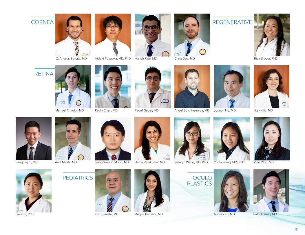
51
Angel Soto Hermida, MD Joseph Ho, MD Ilkay Kilic, MD
Sang Woong Moon, MDFangting Li, MD Amit Meshi, MD Hema Ramkumar, MD Wenqiu Wang, MD, PhD Yuqin Wang, MD, PhD Xiao Ying, MD
Jie Zhu, PhD
C. Andres Benatti, MD Hideki Fukuoka, MD, PhD Harish Raja, MD Craig See, MD
CORNEA
Kim Evaristo, MD Megha Pansara, MD
PEDIATRICS
Manuel Amador, MD Kevin Chen, MD Raouf Gaber, MD
RETINA
Risa Broyer, PhD
REGENERATIVE
Patrick Yang, MDAudrey Ko, MD
OCULOPLASTICS
52
PUBLICATIONS
CORNEAMorlock R, Wirth RJ, Tally S, Garufis C, Heichel CWD. 2017 Patient-Reported Spectacle Independence Questionnaire (PRSIQ): Development and Validation. American Journal of Ophthalmology 2017 June; 178:101-14
Afshari NA, Igo RP Jr, Morris NJ, Stambolian D, Sharma S, Pulagam VL, Dunn S, Stamler JF, Truitt BJ, Rimmler J, Kuot A, Croasdale CR, Qin X, Burdon KP, Riazuddin SA, Mills R, Klebe S, Minear MA, Zhao J, Balajonda E, Rosenwasser GO, Baratz KH, Mootha VV, Patel SV, Gregory SG, Bailey-Wilson JE, Price MO, Price FW Jr, Craig JE, Fingert JH, Gottsch JD, Aldave AJ, Klintworth GK, Lass JH, Li YJ, Iyengar SK. Genome-Wide Association Study
Identifies Three Novel Loci in Fuchs Endothelial Corneal Dystrophy. Nat Commun. 2017 Mar 30;8:14898. doi: 10.1038/ncomms14898. [PMCID: PMC5379100]
Meekins LC, Rosado-Adames N, Maddala R, Zhao J, Rao PV, Afshari NA. Corneal Endothelial Cell Migration and Proliferation Enhanced by Rho Kinase (ROCK) Inhibitors in In Vitro and In Vivo Models. Invest Ophthalmol Vis Sci. 2016 Dec 1;57(15):6731-6738. doi: 10.1167/iovs.16-20414. [PubMed PMID: 27951595]
Zhao J, Afshari NA. Generation of Human Corneal Endothelial Cells via In Vitro Ocular Lineage Restriction of Pluripotent Stem Cells. Invest Ophthalmol Vis Sci. 2016 Dec
1;57(15):6878-6884. doi: 10.1167/iovs.16-20024. [PMCID: PMC5215466]
Haw W. Recurrent Corneal Erosion/Epithelial Basement Membrane Dystrophy. In: K Singh, W Smiddy, A Lee, eds. Ophthalmology Review: A Case-Study Approach. 2nd edition. New York, NY: Thieme. (accepted)
Meekin LC, Rosado-Adames N, Maddala R, Epstein DL, Zhao JJ, Rao PV, Afshari NA. Corneal Endothelial Cell Migration and Proliferation Enhanced by Rho Kinase (ROCK) Inhibitors in In Vitro and In Vivo Models. Invest Ophthalmol Vis Sci. 2016; 57:6731-6738.
Zhao JJ, Afshari NA. Generation of Human Corneal Endothelial Cells via In Vitro Ocular Lineage Restriction of Pluripotent Stem Cells. Invest Ophthalmol Vis Sci. 2016; 57:6878-6884. [PMCID: PMC5215466]
Afshari NA, Igo RP Jr, Morris NJ, Stambolian D, Sharma S, Pulagam VL, Dunn S, Stamler JF, Truitt BJ, Rimmler J, Kuot A, Croasdale CR, Qin X, Burdon KP, Riazuddin SA, Mills R, Klebe S, Minear MA, Zhao J, Balajonda E, Rosenwasser GO, Baratz KH, Mootha VV, Patel SV, Gregory SG, Bailey-Wilson JE, Price MO, Price FW Jr, Craig JE, Fingert JH, Gottsch JD, Aldave AJ, Klintworth GK, Lass JH, Li YJ, Iyengar SK. Genome-Wide Association Study Identifies Three Novel Loci in Fuchs Endothelial Corneal Dystrophy. Nat Commun. 2017; 8: 14898. [PMCID: PMC5379100]
Rudnisky CJ, Belin MW, Guo R, Ciolino JB, Boston Type 1 Keratoprosthesis Study Group. Visual Acuity Outcomes of the Boston Keratoprosthesis Type 1: Multicenter Study Results. 2016. Am J Ophthalmol. 2016; 162:89-98
Afshari NA. October Consultation #6. J Cataract Ref Surg. 2016; 42:1539.
World ScientificWorld Scientificwww.worldscientific.com9545 hc
ISBN 978-981-4667-10-4
Intracranial Pressure and its Effect on
Intracranial Pressure and its Effect on Vision in Space and on Earth
Fluid distribution in space flight and impact on brain and vision health is an emerging field of high-priority research in the NASA human space program. American astronauts have developed ocular refraction change after space flight on the International Space Station (ISS), in addition to other physio-logical and anatomical changes in the eye, optic nerve, and brain. Within this book, experts review current data related to fluid shifts during microgravity exposure and the impact of fluid shifts on astronaut health.
This work also compares current astronaut health problems with Earth-based health conditions such as elevated ICP and glaucoma. Chapters include alterations in fluid distribution, including intra/extracellular and intra/extravascular fluid shifts, eye morphology and vision disturbances, and intraocular pres-sure as well as a discussion of modern advanced non-invasive assessment technologies to investigate the abovementioned fluid alterations.
As such, the book aims to bridge clinicians, eye researchers, and science professionals with present human space program research, thus providing new perspectives to solve emerging brain and eye disease on Earth and in Space.
Brandon R MaciasJohn HK Liu
Christian OttoAlan R Hargens
Editors
Macias • Liu
Otto • H
argens
Vision Impairment in Space
VISIONin Space and on Earth
Intracranial Pressure and its Effect on
Vision Impairment in Space
VISIONin Space and on Earth
Cover image: Jojo Sayson (designer) and Max Nepomuceno (artist)
John HK Liu, PhD and his collaborators published Intracranial Pressure and its Effect on Vision in Space and on Earth: Vision Impairment in Space detailing our understanding of refraction change and other ocular problems in astronauts after their missions in the International Space Station.
53
Lee CM, Afshari NA. The Global State of Cataract Blindness. Curr Opin Ophthalmol. 2017; 28:98-103.
Gali HE, Afshari NA. Patient-Centered Advancements in Cataract Surgery. Curr Opin Ophthalmol. 2017; 28:1-2. [PMCID: PMC5439981]
Fukuoka H, Afshari NA. The Impact of Age Related Cataract on Measures of Frailty in an Aging Global Population. Curr Opin Ophthalmol. 2017; 28:93-37.
Benatti CA, Tsao JZ, Afshari NA. Descemet Membrane Detachment During Cataract Surgery: Etiology and Management. Curr Opin Ophthalmol. 2017; 28:35-41.
GENERALLiu JHK. A Pressure Theory Links the VIIP Syndrome and Eye Diseases. In Intracranial Pressure and Its Effect on Vision in Space and on Earth: Vision impairment in Space (Eds. Macias BR, Liu JHK, Otto C, Hargens AR). pp. 273-287. World Scientific Publishing, Singapore, 2017.
Guluma K, Lee J. Walls, Hockberger, and Gausche-Hill: Rosen’s Emergency Medicine: Concepts and Clinical Practice, 9th edition, Chapter 61, Pages 790 – 819. Ophthalmology 2016.
GENETICSHao X, Luo H, Krawczyk M, Wei W, Wang W, Wang J, Flagg K, Hou J, Zhang H, Yi S, Jafari M, Lin D, Chung C, Caughey BA, Li G, Dhar D, Shi W, Zheng L, Hou R, Zhu J, Zhao L, Fu X, Zhang E, Zhang C, Zhu JK, Karin M, Xu RH, Zhang K. DNA Methylation Markers for Diagnosis and Prognosis of Common Cancers. Proc Natl Acad Sci USA. 2017 Jul 11; 7414-7419. doi: 10.1073/pnas.1703577114. Epub 2017 Jun 26. [PMID: 28652331]
Zhu J, Ming C, Fu X, Duan Y, Hoang DA, Rutgard J, Zhang R, Wang W, Hou R, Zhang D, Zhang E, Zhang C, Hao X, Xiong W, Zhang K. Gene and Mutation Independent Therapy via CRISPR-Cas9 Mediated Cellular Reprogramming in Rod Photoreceptors. Cell Res. 2017 Jun;27(6):830-833. doi: 10.1038/cr.2017.57. Epub 2017 Apr 21. [PMID: 28429769]
Grassmann F1, Kiel C1, Zimmermann ME2, Gorski M2, Grassmann V3, Stark K2; International AMD Genomics Consortium (IAMDGC), Heid IM2, Weber BH4. Genetic Pleiotropy Between Age-Related Macular Degeneration and 16 Complex Diseases and Traits. Genome Med. 2017 Mar 27;9(1):29. doi: 10.1186/s13073-017-0418-0. [PMID: 28347358]
Li K, Duan Y, Ting MYL, Ou Z, Liu L, Zhang K, Fu X. Future Science Prize Goes to Non-Invasive Prenatal Testing. Sci China Life Sci. 2017 Apr;60(4):429-431. doi: 10.1007/s11427-016-0411-8. Epub 2017 Mar 13. [PMID: 28299578].
Lin H, Ouyang H, Zhu J, Huang S, Liu Z, Chen S, Cao G, Li G, Signer RA, Xu Y, Chung C, Zhang Y, Lin D, Patel S, Wu F, Cai H, Hou J, Wen C, Jafari M, Liu X, Luo L, Zhu J, Qiu A, Hou R, Chen B, Chen J, Granet D, Heichel C, Shang F, Li X, Krawczyk M, Skowronska-Krawczyk D, Wang Y, Shi W, Chen D, Zhong Z, Zhong S, Zhang L, Chen S, Morrison SJ, Maas RL, Zhang K, Liu Y. Corrigendum: Lens Regeneration Using Endogenous Stem Cells with Gain of Visual Function. Nature. 2017 Jan 26;541(7638):558. doi: 10.1038/nature19831. Epub 2016 Nov 30. [PMID: 27919080]
Zhang YQ, Wang WY, Xue JX, Xu Y, Fan P, Caughey BA, Tan WW, Cao GQ, Jiang LL, Lu Y, Zhang K, Hu X. MicroRNA Expression Profile on Solid Subtype of Invasive Lung Adenocarcinoma Reveals a Panel of Four miRNAs to be Associated with Poor Prognosis in Chinese
Patients. J Cancer. 2016 Jul 18; 7 (12): 1610-1620. eCollection 2016. [PMID: 27698898]
Khawaja AP, Cooke Bailey JN, Kang JH, Allingham RR, Hauser MA, Brilliant M, Budenz DL, Christen WG, Fingert J, Gaasterland D, Gaasterland T, Kraft P, Lee RK, Lichter PR, Liu Y, Medeiros F, Moroi SE, Richards JE, Realini T, Ritch R, Schuman JS, Scott WK, Singh K, Sit AJ, Vollrath D, Wollstein G, Zack DJ, Zhang K, Pericak-Vance M, Weinreb RN, Haines JL, Pasquale LR, Wiggs JL. Assessing the Association of Mitochondrial Genetic Variation with Primary Open-Angle Glaucoma Using Gene-Set Analyses. Invest Ophthalmol Vis Sci. 2016 Sep 1;57(11):5046-5052. doi: 10.1167/iovs.16-20017. [PMID: 27661856]
Liu Y, Bailey JC, Helwa I, Dismuke WM, Cai J, Drewry M, Brilliant MH, Budenz DL, Christen WG, Chasman DI, Fingert JH, Gaasterland D, Gaasterland T, Gordon MO, Igo RP Jr, Kang JH, Kass MA, Kraft P, Lee RK, Lichter P, Moroi SE, Realini A, Richards JE, Ritch R, Schuman JS, Scott WK, Singh K, Sit AJ, Song YE, Vollrath D, Weinreb R, Medeiros F, Wollstein G, Zack DJ, Zhang K, Pericak-Vance MA, Gonzalez P, Stamer WD, Kuchtey J, Kuchtey RW, Allingham RR, Hauser MA, Pasquale LR, Haines JL, Wiggs JL. A Common Variant in MIR182 Is Associated with Primary Open-Angle Glaucoma in the NEIGHBORHOOD Consortium. Invest Ophthalmol Vis Sci. 2016 Aug 1;57(10):3974-81. doi: 10.1167/iovs.16-19688. [PMID: 27537254]
Gross AM, Jaeger PA, Kreisberg JF, Licon K, Jepsen KL, Khosroheidari M, Morsey BM, Swindells S, Shen H, Ng CT, Flagg K, Chen D, Zhang K, Fox HS, Ideker T. Methylome-Wide Analysis of Chronic HIV Infection Reveals Five-Year Increase in Biological Age and Epigenetic Targeting of HLA. Mol Cell. 2016 Apr 21;62(2):157-68. doi: 10.1016/j.molcel.2016.03.019. [PMID: 27105112]
54
Ouyang H, Goldberg JL, Chen S, Li W, Xu GT, Li W, Zhang K, Nussenblatt RB, Liu Y, Xie T, Chan CC, Zack DJ. Ocular Stem Cell Research from Basic Science to Clinical Application: A Report from Zhongshan Ophthalmic Center Ocular Stem Cell Symposium. Int J Mol Sci. 2016 Mar 22;17(3):415. doi: 10.3390/ijms17030415. [PMID: 27102165]
Wang WQ, Wang FH, Qin WX, Liu HY, Lu B, Chung C, Zhu J, Gu Q, Shi W, Wen C, Wu F, Zhang K, Sun XD. Joint Antiangiogenic Effect of ATN-161 and Anti-VEGF Antibody in a Rat Model of Early Wet Age-Related Macular Degeneration. Mol Pharm. 2016 Sep 6;13(9):2881-90. doi: 10.1021/acs.molpharmaceut.6b00056. Epub 2016 May 9. [PMID: 27089240
Shim MS, Takihara Y, Kim KY, Iwata T, Yue BYJT, Inatani M, Weinreb RN, Perkins GA, Ju WK. Mitochondrial Pathogenic Mechamism and Degradation in Optineurin E50K Mutation-Mediated Retinal Ganglion Cell Degeneration. Scientific Reports. 6:33830. [PMCID: PMC5031982]
GLAUCOMASkaat A, De Moraes CG, Bowd C, Girkin CA, Medeiros FA, Ritch R, Weinreb RN, Zangwill LM, Liebmann JM. For the Diagnostic Innovations in Glaucoma Study and African Descent and Glaucoma Evaluation Study Groups. African Descent and Glaucoma Evaluation Study (ADAGES): Racial differences in Optic Disc Hemorrhage and Beta-Zone Parapapillary Atrophy. Ophthalmology, 2016;123(7), 476-483. [PMID: 27117781]
Belghith A, Medeiros FA, Bowd C, Liebmann JM, Girkin CA, Weinreb RN, Zangwill LM. Structural Change can be Detected in Advanced Glaucoma Eyes. Investigative Ophthalmology and Visual Science, 2016;57(9), OCT511-OCT518. [PMID: 27454660]
Bowd C, Zangwill LM, Weinreb RN, Medeiros FA, Belghith A. Estimating OCT Structural Measurement Floors to Improve Detection of Progression in Advanced Glaucoma. American Journal of Ophthalmology, 2017;175, 37-44. [PMID: 27914978]
Rodrigues T, Krawczyk M, Skowronska-Krawczyk D, Matter-Sadzinski L, Matter JM. (2016) Delayed Neurogenesis with Respect to Eye Growth Shapes the Pigeon Retina for High Visual Acuity. Development 143: 4701-4712; doi: 10.1242/dev.138719
Scully KM, Skowronska-Krawczyk D, Krawczyk M, Merkurjev D, Taylor H, Livolsi A, Tollkuhn J, Stan RV, Rosenfeld MG. (2016) Epithelial Cell Integrin β1 is Required for Developmental Angiogenesis in the Pituitary Gland. PNAS doi: 10.1073/pnas.1614970113
Liu JHK, Slight JR, Vittitow JL, Scassellati Sforzolini B, Weinreb RN. Efficacy of Latanoprostene Bunod 0.024% Compared with Timolol 0.5% in Lowering Intraocular Pressure Over 24 Hours. Am J Ophthalmol 2016;169:249-257. [PMID: 27457257]
Manalastas PIC, Zangwill LM, Saunders LJ, Mansouri K., Belghith A, Suh MH, Yarmohammadi A, Penteado RC, Akagi T, Shoji T, Weinreb RN. Reproducibility of Optical Coherence Tomography Angiography Macular and Optic Nerve Head Vascular Density in Glaucoma and Healthy Eyes. Journal of Glaucoma. 2017 IN PRESS
Kabbara SW, Zangwill LM, Mundae R, Hammel N, Bowd C, Medeiros FA, Weinreb RN, Belghith A. Comparing Optical Coherence Tomography Radial and Cube Scan Patterns for Measuring Bruch’s Membrane Opening Minimum Rim Width (BMO-MRW) in Glaucoma and Healthy Eyes: Cross-Sectional and Longitudinal Analysis. Br J Ophthalmol. 2017. IN PRESS
Shoji T, Zangwill LM, Akagi T, Saunders LJ, Yarmohammadi A, Manalastas PIC, Penteado RC, Weinreb RN. Progressive Macula Vessel Density Loss in Primary Open Angle Glaucoma: A Longitudinal Study. Am J Ophthalmol. 2017. IN PRESS
Huang AS, Belghith A, Dastiridou A, Chopra V, Zangwill LM, Weinreb RN. Automated Circumferential Construction of First-Order Aqueous Humor Outflow Pathways Using Spectral-Domain Optical Coherence Tomography. J Biomed Opt. 2017;22:66010. PMCID: PMC5472236
De Moraes CG, Hood DC, Thenappan A, Girkin CA, Medeiros FA, Weinreb RN, Zangwill LM, Liebmann JM. 24-2 Visual Fields Miss Central Defects Shown on 10-2 Tests in Glaucoma Suspects, Ocular Hypertensives, and Early Glaucoma. Ophthalmology. 2017. IN PRESS
Akagi T, Zangwill LM, Saunders LJ, Yarmohammadi A, Manalastas PIC, Suh MH, Girkin CA, Liebmann JM, Weinreb RN. Rates of Local Retinal Nerve Fiber Layer Thinning before and after Disc Hemorrhage in Glaucoma. Ophthalmology. 2017;124:1403-141. [PMCID: PMC5565682]
De Moraes CG, Murphy JT, Kaplan CM, Reimann JJ, Skaat A, Blumberg DM, Al-Asward L, Cioffi GA, Girkin CA, Medeiros FA, Weinreb RN, Zangwill LM, Liebmann JM. Beta-Zone Parapapillary Atrophy and Rates of Glaucomatous Visual Field Progression: African Descent and Glaucoma Evaluation Study. JAMA Ophthalmol. 2017;135:617-623
Yarmohammadi A, Zangwill LM, Weinreb RN. Reply. Ophthalmology. 2017;124:e51Hammel N, Belghith A, Weinreb RN, Medeiros FA, Mendoza N, Zangwill LM. Comparing the Rates of Retinal Nerve Fiber Layer and Ganglion Cell-Inner Plexiform Layer Loss in Healthy
55
Eyes and in Glaucoma Eyes. Am J Ophthalmol. 2017;178:38-50
Yarmohammadi A, Zangwill LM, Diniz-Filho A, Saunders LJ, Suh MH, Wu Z, Manalastas PIC, Akagi T, Medeiros FA, Weinreb RN. Peripapillary and Macular Vessel Density in Patients with Glaucoma and Single-Hemifield Visual Field Defect. Ophthalmology. 2017;124:709-719. PMCID: PMC5499385
You Q, Freeman WR, Weinreb RN, Zangwill LM, Manalastas PIC, Saunders LJ, Nudleman E. Reproducibility of Vessel Density Measurement with Optical Coherence Tomography Angiography in Eyes with and without Retinopathy. Retina. 2017;37:1475-1482
Bowd C, Zangwill LM, Weinreb RN, Medeiros FA, Belghith A. Estimating Optical Coherence Tomography Structural Measurement Floors to Improve Detection of Progression in Advanced Glaucoma. Am J Ophthalmol. 2017;175:37-44. PMCID: PMC5337134
Suh MH, Zangwill LM, Manalastas PIC, Belghith A, Yarmohammadi A, Medeiros FA, Diniz-Filho A, Saunders LJ, Weinreb RN. Deep Retinal Layer Microvasculature Dropout Detected by the Optical Coherence Tomography Angiography in Glaucoma. Ophthalmology. 2016;123:2509-2518. PMCID: PMC5360450
Yarmohammadi A, Zangwill LM, Diniz-Filho A, Suh MH, Yousefi S, Saunders LJ, Belghith A, Manalastas PIC, Medeiros FA, Weinreb RN. Relationship between Optical Coherence Tomography Angiography Vessel Density and Severity of Visual Field Loss in Glaucoma. Ophthalmology. 2016;123:2498-2508. PMCID: PMC5362128
Diniz-Filho A, Abe RY, Zangwill LM, Gracitelli CP, Weinreb RN, Girkin CA, Liebmann JM, Medeiros FA. Association Between Intraocular Pressure
and Rates of Retinal Nerve Fiber Layer Loss Measured by Optical Coherence Tomography. Ophthalmology. 2016;123:2058-65
Zhang C, Tatham AJ, Abe RY, Hammel N, Belghith A, Weinreb RN, Medeiros FA, Liebmann JM, Girkin CA, Zangwill LM. Macular Ganglion Cell Inner Plexiform Layer Thickness in Glaucomatous Eyes with Localized Retinal Nerve Fiber Layer Defects. PLoS One. 2016;11:e0160549. PMCID: PMC4990273
Belghith A, Medeiros FA, Bowd C, Liebmann JM, Girkin CA, Weinreb RN, Zangwill LM. Structural Change can be Detected in Advanced-Glaucoma Eyes. Invest Ophthalmol Vis Sci. 2016;57:OCT511-8. PMCID: PMC4968923
Yarmohammadi A, Zangwill LM, Diniz-Filho A, Suh MH, Manalastas PIC, Fatehee N, Yousefi S, Belghith A, Saunders LJ, Medeiros FA, Huang D, Weinreb RN. Optical Coherence Tomography Angiography Vessel Density in Healthy, Glaucoma Suspect, and Glaucoma Eyes. Invest Ophthalmol Vis Sci. 2016;57:OCT451-9. PMCID: PMC4968912
Abe RY, Diniz-Filho A, Zangwill LM, Gracitelli CP, Marvasti AH, Weinreb RN, Baig S, Medeiros FA. The Relative Odds of Progressing by Structural and Functional Tests in Glaucoma. Invest Ophthalmol Vis Sci. 2016;57:OCT421-8. PMCID: PMC4968922
Shim MS, Kim KY, Ju WK. Role of Cyclic AMP in the Eye with Glaucoma. BMB Rep. 50:60-70. [PMCID: PMC27916026]
Weinreb RN, Realini T, Varma R. Latanoprostene Bunod, A Dual-Acting Nitric Oxide Donating Prostaglandin Analog for Lowering of Intraocular Pressure. US Ophthalmic Review. 2016;9:80-7.
Hammel N, Belghith A, Bowd C, Medeiros FA, Sharpsten L, Mendoza N, Tatham A, Khachatryan
N, Liebmann JM, Girkin CA, Weinreb RN, Zangwill LM. Rate and Pattern of Rim Area Loss in Healthy and Progressing Glaucoma Eyes. Ophthalmology. 2016;123:760-70. [PMCID: PMC4808425]
Wang S, Moonasar N, Xiao X, Yin T, Weinreb RN, Sun X. Effect of Resveratrol-Based Nutritional Supplement on Choroidal Thickness: A Pilot Study. Curr Eye Res. 2016;41:1339-45.
Yu M, Lin C, Weinreb RN, Lai G, Chiu V, Leung CK. Risk of Visual Field Progression in Glaucoma Patients with Progressive Retinal Nerve Fiber Layer Thinning: A 5-Year Prospective Study. Ophthalmology. 2016;123:1201-10.
de Moraes CG, Liebmann JM, Medeiros FA, Weinreb RN. Management of Advanced Glaucoma: Characterization and Monitoring. Surv Ophthalmol. 2016;61:597-615.
Medeiros FA, Martin KR, Peace J, Scassellati Sforzolini B, Vittitow JL, Weinreb RN. Comparison of Latanoprostene Bunod 0.024% and Timolol Maleate 0.5% in Open-Angle Glaucoma or Ocular Hypertension: The LUNAR Study. Am J Ophthalmol. 2016;168:250-9.
Kuang T, Zhang C, Zangwill LM, Weinreb RN, Medeiros FA. Reply. Ophthalmology. 2016;123:e38.
Belghith A, Medeiros FA, Bowd C, Liebmann JM, Girkin CA, Weinreb RN, Zangwill LM. Structural Change can be Detected in Advanced-Glaucoma Eyes. Invest Ophthalmol Vis Sci. 2016:57:OCT511-8. [PMCID: PMC4968923]
Liu JHK, Slight JR, Vittitow JL, Weinreb RN. Efficacy of Latanoprostene Bunod 0.024% Compared with Timolol 0.5% in Lowering Intraocular Pressure Over 24 Hours. Am J Ophthalmol. 2016;169:249-57.
56
Kawase K, Vittitow JL, Weinreb RN, Araie M. Long-Term Safety and Efficacy of Latanoprostene Bunod 0.024% in Japanese Subjects with Open-Angle Glaucoma or Ocular Hypertension: The JUPITER study. 2016;33:1612-27. [PMCID: PMC5020123]
Aptel F, Weinreb RN, Chiquet C, Mansouri K. 24-h Monitoring Devices and Nyctohemeral Rhythms of Intraocular Pressure. Prog Ret Eye Res. 2016;55:108-48.
Mauri L, Uebe, S, Vossmerbaeurmer U, Weisschuh N, Manfredini E, Maselli E, Patrosso MC, Penco S, Weinreb RN, Reis A, Pasutto F. Expanding the Clinical Spectrum of COL1A1 Mutations in Different Forms of Glaucoma. Orphanet J Rare Dis. 2016;11:108. [PMCID: PMC4970237]
Zhang C, Tatham AJ, Abe RY, Hammel N, Belghith A, Weinreb RN, Medeiros FA, Liebmann JM, Girkin CA, Zangwill LM. Macular Ganglion Cell Inner Plexiform Layer Thickness in Glaucomatous Eyes with Localized Retinal Nerve Fiber Layer Defects. PLoS One. 2016;11:e0160549. [PMCID: PMC4990273]
Liu Y, Bailey JC, Helwa I, Dismuke WM, Cai J, Drewry M, Brilliant MH, Budenz DL, Christen WG, Chasman DI, Fingert JH, Gaasterland D, Gaasterland T, Gordon MO, Igo RP Jr, Kang JH, Kass MA, Kraft P, Lee RK, Lichter P, Moroi SE, Realini A, Richards JE, Ritch R, Schuman JS, Scott WK, Singh K, Sit AJ, Song YE, Vollrath D, Weinreb RN, Medeiros, F, Wollstein G, Zack DJ, Zhang K, Pericak-Vance MA, Gonzalez P, Stamer WD, Kuchtey J, Kuchtey RW, Allingham RR, Hauser MA, Pasquale LR, Haines JL, Wiggs JL. A Common Variant in MIR182 is Associated with Primary Open-Angle Glaucoma in the NEIGHBORHOOD Consortium. Invest Ophthalmol Vis Sci. 2016;57:3974-81.
Diniz-Filho A, Abe RY, Zangwill LM, Gracitelli CPB, Weinreb RN, Girkin CA, Liebmann JM, Medeiros FA. Association Between Intraocular Pressure and Rates of Retinal Nerve Fiber Layer Loss Measured by Optical Coherence Tomography. Ophthalmology. 2016;123:2058-65.
Rao HL, Pradhan ZS, Weinreb RN, Reddy HB, Riyazuddin M, Dasari S, Palakurthy M, Puttaiah NK, Rao, DAS, Webers CAB. Regional Comparisons of Optical Coherence Tomography Angiography Vessel Density in Glaucoma. 2016;171:75-83.
Huang AS, Saraswathy S, Dastiridou A, Begian A, Mohindroo C, Tan JC, Francis BA, Hinton DR, Weinreb RN. Aqueous Angiography-Mediated Guidance of Trabecular Bypass Improves Angiographic Outflow in Human Enucleated Eyes. Invest Ophthalmol Vis Sci. 2016;57:4558-65. [PMCID: PMC5017267]
Weinreb RN, Leung CK, Crowston JG, Medeiros FA, Fredman DS, Wiggs JL, Martin KR. Primary Open-Angle Glaucoma. Nat Rev Dis Primers. 2016;2:16067.
Huang AS, Saraswathy S, Dastiridou A, Begian A, Legaspi H, Mohindroo C, Tan JCH, Francis BA, Caprioli J, Hinton D, Weinreb RN. Aqueous Angiography with Fluorescein and Indocyanine Green in Bovine Eyes. Transl Vis Sci Technol. 2016;5:5. [PMCID: PMC5106193]
Shim MS, Takihara Y, Kim K-Y, Iwata T, Yue BYJT, Inatani M, Weinreb RN, Perkins GA, Ju W-K. Mitochondrial Pathogenic Mechanism and Degradation in Optineurin E50K Mutation-Mediated Retinal Ganglion Cell Degeneration. Sci Rep. 2016;6:33830. [PMCID: PMC5031982]
Huang AS, Mohindroo C, Weinreb RN. Aqueous Humor Outflow Structure and Function Imaging at the Bench and Bedside: A Review. J Clin Exp Ophthalmol. 2016;7:578. [PMCID: PMC5079182]
Khawaja AP, Cooke Bailey JN, Kang JH, Allingham RR, Hauser MA, Brilliant M, Budenz DL, Christen WG, Fingert J, Gaasterland D, Gaasterland T, Kraft P, Lee RK, Lichter PR, Liu Y, Medeiros F, Moroi SE, Richards JE, Realini T, Ritch R, Schuman JS, Scott WK, Singh K, Sit AJ, Vollrath D, Wollstein G, Zack DJ, Zhang K, Pericak-Vance M, Weinreb RN, Haines JL, Pasquale LR, Wiggs JL. Assessing the Association of Mitochondrial Genetic Variation with Primary Open-Angle Glaucoma Using Gene-Set Analyses. Invest Ophthalmol Vis Sci. 2016;57:5046-52. [PMCID: PMC5040191]
Suh MH, Zangwill LM, Manalastas PI, Belghith A, Yarmohammadi A, Medeiros FA, Diniz-Filho A, Saunders LJ, Weinreb RN. Deep Retinal Layer Microvasculature Dropout Detected by the Optical Coherence Tomography Angiography in Glaucoma. Ophthalmology. 2016;123:2509-18.
Suh MH, Zangwill LM, Manalastas PIC, Belghith A, Yarmohammadi A, Medeiros F, Diniz-Filho A, Saunders L, Yousefi S, Weinreb RN. Optical Coherence Tomography Angiography Vessel Density in Glaucomatous Eyes with Focal Lamina Cribrosa Defects. Ophthalmology. 2016;123:2309-17.
Ianchulev T, Chayet A, Kahook M, Packer M, Pasquale L, Weinreb RN. Pharmacodynamic Profile of Mydriatic Agents Delivered by Ocular Piezo-Ejection Microdosing Compared with Conventional Eyedropper. Ther Deliv. 2016;7:751-60.
Budenz DL, Huecker JB, Gedde SJ, Gordon M, Kass M, Ocular Hypertension Treatment Study (Weinreb RN). Thirteen-Year Follow-Up of Optic Disc Hemorrhages in the Ocular Hypertension Treatment Study. Am J Ophthalmol. 2017;174:126-133.
57
Wang C, Dang Y, Waxman S, Xia X, Weinreb RN, Loewen NA. Angle Stability and Outflow in Excisional Ab Interno Trabeculectomy with Active Versus Passive Chamber Management. PeerJ Preprints. 2017;5:e2762v2.
Shim MS, Takihara Y, Kim K-Y, Iwata T, Yue BYJT, Inatani M, Weinreb RN, Perkins GA, Ju W-K. Corrigendum: Mitochondrial Pathogenic Mechanism and Degradation in Optineurin E50K Mutation-Mediated Retinal Ganglion Cell Degeneration. Sci Rep. 2017;7:40460. [PMCID: PMC5244623]
Pasquale LR, Aschard H, Kang JH, Cooke Bailey JN, Lindstrom S, Chasman DI, Christen WG, Allingham RR, Ashley-Koch A, Lee RK, Moroi SE, Lichter PR, Brilliant M, Wollstein G, Schuman JS, Fingert J, Budenz DL, Realini T, Gaasterland T, Gaasterland D, Scott WK, Singh K, Sit AJ, Igo RP, Song YE, Hark L, Ritch R, Rhee DJ, Gulati V, Havens S, Vollrath D, Zack DJ, Zhang K, Weinreb RN, Pericak-Vance MA, Liu Y, Kraft P, Richards JE, Rosner BA, Hauser MA, Haines JL, Wiggs JL. Age at Natural Menopause Genetic Risk Score in Relation to Age at Natural Menopause and Primary Open-Angle Glaucoma is US-Based Sample. Menopause. 2017;24:150-6. [PMCID: PMC5266624]
Miki A, Ikuno Y, Weinreb RN, Yokoyama J, Asai T, Usui S, Nishida K. Measurements of the Parapapillary Atrophy Zones in En Face Optical Coherence Tomography Images. PLoS One. 2017;12:e0175347.
Rao HL, Pradhan ZS, Weinreb RN, Riyazuddin M, Dasari S, Venugopal JP, Puttaiah NK, Rao DA, Devi S, Mansouri K, Webers CA. A Comparison of the Diagnostic Ability of Vessel Density and Structural Measurements of Optical Coherence Tomography in Primary Open Angle Glaucoma. PLoS One. 2017;12:e0173930.
Nuyen B, Weinreb RN, Robbins RL. Steroid-Induced Glaucoma in the Pediatric Population. J AAPO. 2017;21:1-6.
Wang SS, Wang Z, Yang S, Yin T, Zhang Y, Qin Y, Weinreb RN, Sun X. Tissue Distribution of Trans-Resveratrol and its Metabolites after Oral Administration in Human Eyes. J Ophthalmol. 2017;2017:4052094. [PMCID: PMC5377058]
Bowd C, Zangwill LM, Weinreb RN, Medeiros FA, Belghith A. Estimating OCT Structural Measurement Floors to Improve Detection of Progression in Advanced Glaucoma. Am J Ophthalmol. 2017;175:37-44. [PMCID: PMC5337134]
Yarmohammadi A, Zangwill LM, Weinreb RN. Reply. Ophthalmology. 2017;124:e51.
Rao HL, Pradhan ZS, Weinreb RN, Reddy HB, Riyazuddin M, Sachdeva S, Puttaiah NK, Jayadev C, Webers CAB. Determinants of Peripapillary and Macular Vessel Densities Measured by Optical Coherence Tomography Angiography in Normal Eyes. J Glaucoma. 2017;26:491-7.
Wang C, Dang Y, Waxman S, Xia X, Weinreb RN, Loewen NA. Angle Stability and Outflow in Dual Blade Ab Interno Trabeculectomy with Active Versus Passive Chamber Management. PLoS One. 2017;12:e0177238. [PMCID: PMC5423641]
Huang AS, Li M, Yang D, Wang H, Wang N, Weinreb RN. Aqueous Angiography in Living Non-Human Primates Show Segmental, Pulsatile, and Dynamic Angiographic Aqueous Humor Outflow. Ophthalmology. 2017;124:793-803.
Huang AS, Minasyan L, Weinreb RN. Glaucoma-Intraocular Pressure Reduction. Handb Exp Pharmacol. 2017;242:181-207.
You Q, Freeman WR, Weinreb RN, Zangwill L, Manalastas PI, Saunders LJ, Nudleman E. Reproducibility of Vessel Density Measurement with Optical Coherence Tomography Aniography in Eyes with and without Retinopathy. Retina. 2017;37:1475-82.
Rao HL, Kadambi SV, Weinreb RN, Puttaiah Nk, Pradhan ZS, Rao DA, Kumar RS, Webers CA, Shetty R. Diagnostic Ability of Peripapillary Vessel Density Measurements of Optical Coherence Tomography Angiography in Primary Open-Angle and Angle-Closure Glaucoma. Br J Ophthalmol. 2017;101:1066-70.
Yarmohammadi A, Zangwill LM, Diniz-Filho A, Saunders LJ, Suh MH, Wu Z, Manalastas PIC, Akagi T, Medeiros FA, Weinreb RN. Peripapillary and Macular Vessel Density in Patients with Glaucoma and Single-Hemifield Visual Field Defect. Ophthalmology. 2017;124:709-19. [PMCID: PMC5499385]
Rao HL, Pradhan ZS, Weinreb RN, Dasari S, Riyazuddin M, Raveendran S, Puttaiah NK, Venugopal JP, Rao DAS, Devi S, Mansouri K, Webers CAB. Relationship of Optic Nerve Structure and Function to Peripapillary Vessel Density Measurements of Optical Coherence Tomography Angiography in Glaucoma. J Glaucoma. 201726:548-54.
De Moraes CG, Murphy JT, Kaplan CM, Reimann JJ, Skaat A, Blumberg DM, Al-aswad L, Cioffi GA, Girkin CA, Medeiros FA, Weinreb RN, Zangwill L, Liebmann JM. Beta-Zone Parapapillary Atrophy and Rates of Glaucomatous Visual Field Progression: African Descent and Glaucoma Evaluation Study. JAMA Ophthalmol. 2017;135:617-23.
Huang AS, Camp A, Xu B, Penteado R, Weinreb RN. Aqueous Angiography: Aqueous Humor Outflow Imaging in Live Human Subjects. Ophthalmology. 2017;124:1249-51.
58
Huang A, Belghith A, Dastiridou A, Chopra V, Zangwill L, Weinreb RN. Automated Circumferential Construction of First Order Aqueous Humor Outflow Pathways Using Spectral Domain Optical Coherence Tomography. J Biomed Opt. 2017;22:66010.
Aung T, Ozaki M, Lee MC, and Weinreb RN, et al. Worldwide Genetic Association Study of Exfoliation Syndrome Identifies Highly Protective Rare Variants at LOXL1 and Five New Common-Variant Susceptibility Loci. Nat Genet. 2017;49:993-1004.
Rao HL, Pradhan ZS, Weinreb RN, Riyazuddin M, Dasari S, Venugopal JP, Puttaiah NK, Rao DAS, Devi S, Mansouri K, Webers CAB. A Comparison of the Diagnostic Ability of Vessel Density and Structural Measurements in Optical Coherence Tomography in Primary Open Angle Closure Disease. Am J Ophthalmol. 2017. IN PRESS
Akagi T, Zangwill LM, Saunders LJ, Yarmohammadi A, Manalastas PIC, Suh MH, Girkin CA, Liebmann JM, Weinreb RN. Rates of Local Retinal Nerve Fiber Layer Thinning Before and After Disc Hemorrhage in Glaucoma. Ophthalmology. 2017. IN PRESS
De Moraes CG, Hood DC, Thenappan A, Girkin CA, Medeiros FA, Weinreb RN, Zangwill LM, Liebmann JM. 24-2 Visual Fields Miss Central Defects Shown on 10-2 Tests in Glaucoma Suspects and Early Glaucoma. Ophthalmology. 2017. IN PRESS
Aschard H, Kang JH, Iglesias AI, Hysi P, Cooke Baily JN, Khawaja A, Allingham RR, Ashley-Koch A, Lee RK, Moroi SE, Brilliant MH, Wollstein G, Schuman JS, Fingert J, Budenz DL, Realini T, Gaasterland T, Scott WK, Singh K, Sit AJ, Igo RP Jr., Song YE, Hark L, Ritch R, Rhee DJ, Gulati V, Havens S, Vollrath D, Zack DJ, Medeiros F, Weinreb RN, Cheng C-Y, Chasman DI, Christen WG, Pericak-Vance MA, Liu Y, Kraft P, Richards
JE, Rosner BA, Hauser MA, International Glaucoma Genetics Consortium, Klaver C, vanDuijn CM, Haines J, Wiggs JL, Pasquale LR. Genetic Correlations Between Intraocular Pressure, Blood Pressure and Primary Open-Angle Glaucoma: A Multi-Cohort Analysis. Eur J Hum Genet. 2017. IN PRESS
Moghimi S, Mazloumi M, Johari MK, Fard MA, Chen R, Weinreb RN, Nouri-Mahdavi K. Comparison of Macular Choroidal Thickness in Patients with Pseudoexfoliation Syndrome to Normal Control Subjects with Enhanced-Depth SD-OCT imaging. J Curr Ophthalmol. 2017. ePub Ahead of Print.
Realini T, Gurka MJ, Weinreb RN. Reproducibility of Central Corneal Thickness Measurements in Healthy and Glaucomatous Eyes. J Glaucoma. 2017 Sep;26(9):787-791.
Kabbara SW, Mundae R, Bowd C, Hammel N, Medeiros FA, Weinreb RN, Zangwill LM, Belghith A. Comparing Optical Coherence Tomography Radial and Cube Scan Patterns for Measuring Bruch’s Membrane Opening Minimum Rim Width (BMO-MRW) in Glaucoma and Healthy Eyes: Cross-Sectional and Longitudinal Analysis. Br J Ophthalmol. 2017. IN PRESS
Rao HL, Weinreb RN, Webers CAB, Mansouri K. Optical Coherence Tomography Angiography Vessel Density Measurements in Eyes with Primary Open Angle Glaucoma and Disc Hemorrhage. J Glaucoma. 2017. ePub Ahead of Print.
Manalastas PI, Zangwill L, Saunders L, Mansouri K, Belghith A, Suh M, Yarmohammadi A, Penteado RC, Akagi T, Shoji T, Weinreb RN. Reproducibility of Optical Coherence Tomography Angiography Macular and Optic Nerve Head Vascular Density in Glaucoma and Health Eyes. J Glaucoma. 2017. ePub Ahead of Print.
Venugopal JP, Rao HL, Weinreb RN, Pradhan ZS, Dasari S, Riyazuddin M, Puttaiah NK, Rao DAS, Devi S, Mansouri K, Webers CAB. Repeatability of Vessel Density Measurements of Optical Coherence Tomography Angiography in Normal and Glaucoma Eyes. Br J Ophthalmol. 2017.
Belghith A, Medeiros FA, Bowd C, Liebmann JM, Girkin CA, Weinreb RN, Zangwill LM. Structural Change Can Be Detected in Advanced-Glaucoma Eyes. Investigative Ophthalmology & Visual Science July 2016, Vol.57, OCT511-OCT518. doi:10.1167/iovs.15-18929
Zhang C, Tatham AJ, R Y A, Hammel N, Belghith A, Weinreb RN, Medeiros FA, Liebmann JM, Girkin CA, Zangwill LM. Macular Ganglion Cell Inner Plexiform Layer Thickness in Glaucomatous Eyes with Localized Retinal Nerve Fiber Layer Defects. PLOS ONE 11(8): e0160549. 2016.
Suh MH, Zangwill LM, Manalastas PIC, Belghith A, Yarmohammadi A, Medeiros FA, Diniz-Filho A, Saunders LJ, Yousefi A, Weinreb RN. Optical Coherence Tomography Angiography Vessel Density in Glaucomatous Eyes with Focal Lamina Cribrosa Defects. Ophthalmology, Volume 123, Issue 11, 2016, 2309-2317, ISSN 0161-6420.
Yarmohammadi A, Zangwill LM, Diniz-Filho A, Suh MH, Yousefi S, Saunders LJ, Belghith A, Isabel CP, Manalastas PIC, Medeiros FA, Weinreb RN. Relationship between Optical Coherence Tomography Angiography Vessel Density and Severity of Visual Field Loss in Glaucoma. Ophthalmology, Volume 123, Issue 12, 2016, 2498-2508, ISSN 0161-6420.
Suh MH, Zangwill LM, Manalastas PIC, Belghith A, Yarmohammadi A, Medeiros FA, Diniz-Filho A, Saunders LJ, Weinreb RN. Deep Retinal Layer Microvasculature Dropout Detected by the Optical Coherence Tomography Angiography in Glaucoma. Ophthalmology,
59
Volume 123, Issue 12, 2016, Pages 2509-2518, ISSN 0161-6420.
Bowd C, Zangwill LM, Weinreb RN, Medeiros FA, Belghith A. Estimating Optical Coherence Tomography Structural Measurement Floors to Improve Detection of Progression in Advanced Glaucoma. American Journal of Ophthalmology, Volume 175, March 2017, 37-44, ISSN 0002-9394.
Huang AS, Belghith A, Dastiridou A, Chopra V, Zangwill LM, Weinreb RN; Automated Circumferential Construction of First-Order Aqueous Humor Outflow Pathways Using Spectral-Domain Optical Coherence Tomography. J. Biomed. Opt. 0001;22(6):066010. doi:10.1117/1.JBO.22.6.066010. 7061
Hammel N, Belghith A, Weinreb RN, Medeiros FA, Mendoza N, Zangwill LM. Comparing the Rates of Retinal Nerve Fiber Layer and Ganglion Cell–Inner Plexiform Layer Loss in Healthy Eyes and in Glaucoma Eyes. American Journal of Ophthalmology, Volume 178, June 2017, 38-50, ISSN 0002-9394.
OCULOPLASTICSCho WK, Ko AC, Eatamadi H, Al-Ali A, Abboud JP, Kikkawa DO, Korn BS. Orbital and Orbitocranial Trauma From Pencil Fragments: Role of Timely Diagnosis and Management. Am J Ophthalmol. 2017 May 26.
Wu PS, Matoo R, Sun H, Song LY, Kikkawa DO, Lu W. Single-Stage Soft Tissue Reconstruction and Orbital Fracture Repair for Complex Facial Injuries. J Plast Reconstr Aesthet Surg. 2017 Feb;70(2):e1-e6.
Yang P, Ko AC, Kikkawa DO, Korn BS. Upper Eyelid Blepharoplasty: Evaluation, Treatment, and Complication Minimization. Semin Plast Surg. 2017 Feb;31(1):51-57.
Ko AC, Korn BS, Kikkawa DO. The Aging Face. Surv Ophthalmol. 2017 Mar - Apr;62(2):190-202.Korn BS, Kikkawa DO. Video Atlas of Oculofacial Plastic and Reconstructive Surgery (2nd Edition). Elsevier. Oct 2016.
Kikkawa DO (Contributing Editor - Brow lifting, Thyroid Eye Disease, Lacrimal Sections). In. Li Dong Mei Eye Plastic Surgery Atlas (Second Edition). People’s Medical Publishing House. ISBN: 9787117221146. April 2016
Wu, F, Korn BS, Kikkawa DO. Chapter 22. Benign and Premalignant Tumors of the Eyelid. In: Dolman P and Fay A, eds. Diseases and Disorders of the Orbit and Ocular Adnexa 1st Edition, 2017, pp 401-418.
Verma R, Lee BW, Alameddine RM, Ko AC, Khanna PC, Kikkawa DO, Korn BS. Orbital Perivenous Abscess Complicating the Diagnosis and Management of Orbital Cellulitis. Ophthal Plast Reconstr Surg. 2017 May 5.
Cho WK, Ko AC, Korn BS, Kikkawa DO. Orbitocutaneous Fistula Secondary to Buried Polyethylene Mesh Implant 12 Years After Injury. Ophthal Plast Reconstr Surg. 2016 Nov
Ko AC, Satterfield KR, Lee BW, Alameddine RM, Korn BS, Kikkawa DO. Unilateral Alacrima as a Presenting Symptom of Nasopharyngeal Carcinoma. Ophthal Plast Reconstr Surg. 2017 Mar/Apr;33(2):e41-e42.
Tran AQ, Lee BW, Alameddine RM, Korn BS, Kikkawa DO. Reconstruction of Unilateral Incomplete Cryptophthalmos in Fraser Syndrome. Ophthal Plast Reconstr Surg. 2017 May/Jun;33(3S Suppl 1):S73-S75.
Kim JS, Lee BW, Scawn RL, Korn BS, Kikkawa DO. Re: “Secondary Orbital Reconstruction in Patients with Prior Orbital Fracture Repair”. Ophthal Plast Reconstr Surg. 2017 May/Jun;33(3):228.
Ko AC, Korn BS, Kikkawa DO. The Aging Face. Surv Ophthalmol. 2016 Sep 28
Godfrey KJ, Harrison AR, Korn BS, Kikkawa DO. The Glue that Holds the Situation Together. Surv Ophthalmol. 2016 Nov 12.
Cho WK, Ko AC, Korn BS, Kikkawa DO. Orbitocutaneous Fistula Secondary to Buried Polyethylene Mesh Implant 12 Years After Injury. Ophthal Plast Reconstr Surg. 2016 Nov 15.
Verma R, Lee BW, Alameddine RM, Ko AC, Khanna PC, Kikkawa DO, Korn BS. Orbital Perivenous Abscess Complicating the Diagnosis and Management of Orbital Cellulitis. Ophthal Plast Reconstr Surg. 2017
Yang P, Ko AC, Kikkawa DO, Korn BS. Upper Eyelid Blepharoplasty: Evaluation, Treatment, and Complication Minimization. Semin Plast Surg. 2017 Feb;31(1):51-57.
Hodgson NM, King H, Lee BW, Amaro D, Kikkawa DO, Lin JH, Korn BS, Taplitz RA. Scedosporium Prolificans Sclerokeratitis Following Pterygium Excision with Mitomycin C: A Case Requiring Enucleation. J Emerg Infect Disease. 2017 March 2:1.
Cho WK, Ko AC, Eatamadi H, Al-Ali A, Abboud JP, Kikkawa DO and Korn BS. Orbital and Orbitocranial Trauma From Pencil Fragments: Role of Timely Diagnosis and Management. Am Journ Ophthalmology in press, 2017.
PATHOLOGYGodfrey KJ, Kinori M, Lin JH, Snyder VS, Granet DB, Heichel CW, Robbins SL. 2016. Large Benign De Novo Conjunctival Hemangioma in an 11-Year-Old Boy: Case Report and Literature Review. J AAPOS. doi: 10.1016/j.jaapos.2016.06.002. PMID:27647119.
60
Chiang W, Chan P, Wissinger B, Vincent A, Skorczyk-Werner A, Krawczynski MR, Kaufman RJ, Tsang SH, Heon E, Kohl S, Lin JH. 2016. Achromatopsia Mutations Target Sequential Steps of ATF6 Activation. Proc Natl Acad Sci U S A. 2016 Dec 27. pii: 201606387. doi: 10.1073/pnas.1606387114. [Epub ahead of print]. PMID:28028229.
Biswas P, Duncan JD, Maranhao B, Kozak I, Branham K, Gabriel L, Lin JH, Barteselli G, Navani M, Suk J, Parke M, Schlechter C, Weleber R, Hecklenlively JR, Dagnelie G, Lee P, Riazuddin SA, Ayyagari R. Genetic Analysis of Ten Pedigrees with Inherited Retinal Degeneration (IRD) by Exome Sequencing and Phenotype-Genotype Association. 2017. Physiol Genomics. 2017 Jan 27:physiolgenomics.00096.2016. doi:10.1152/physiolgenomics.00096.2016. [Epub ahead of print]. PMID:28130426.
Rodvold JJ, Chiu KT, Hiramatsu N, Nussbacher JK, Galimberti V, Mahadevan NR, Willert K, Lin JH, Zanetti M. Intercellular Transmission of the Unfolded Protein Response Promotes Survival and Drug Resistance in Cancer Cells. Sci Signal. 2017. Jun 6;10(482). pii: eaah7177. doi: 10.1126/scisignal.aah7177. PMID:28588081.
Hodgson NM, King H, Lee BW, Amaro D, Kikkawa DO, Lin JH, Korn BS, Taplitz RA. 2017. Scedosporium Prolificans Sclerokeratitis Following Pterygium Excision with Mitomycin C: A Case Requiring Enucleation. J Emerg Infect Dis. 2:1. doi: 10.4172/2472-4998.1000125.
Skorczyk-Werner A, Chiang WC, Wicker K, Jarmuz-Szymczak M, Kostrzewska-Poczekaj M, Jamsheer A, Ploski R, Rydzanicz M, Weisschuh N, Wissinger B, Kohl S, Lin JH, Krawczynski M. 2017. Autosomal Recessive Cone-Rod Dystrophy can be Caused by Mutations in the ATF6 Gene. (in press, Eur J Hum Genet).
PEDIATRIC OPHTHALMOLOGYGranet DB, Wilson ME, Wagner RS. Minimally Invasive Strabismus Surgery. J Pediatric Ophthalmol Strabismus 2016 Sep 1;53(5):262-265
Ellis EM, Kinori M, Robbins SL, Granet DB. Pulled-in-Two Syndrome: A Multi-Center Survey of Risk Factors, Management and Outcomes. J AAPOS 2016 Oct;20(5):387-391
Godfrey KJ, Kinori M, Lin JH, Snyder VS, Granet DB, Heichel CW, Robbins SL. Large Benign De Novo Conjunctival Hemangioma in an 11 Year Old Boy: Case Report and Literature Review. J AAPOS 2016 Oct;20(5):462-464
El Sahn MF, Granet DB, Marvasti A, Roa A, Kinori M. Strabismus in Adults Older Than 60 Years. J Pediatric Ophthalmol Strabismus 2016 Nov 1;53(6):365-368
Schallhorn CS, Granet DB, Ferreyra HA. Electronegative Electroretinogram in Achromatopsia. Retina Cases Brief Rep 2016 Nov 3
Kinori M. Gomi CF, Ondeck CL, Schanzlin DJ, Robbins SL, Granet DB. Usefulness of Refractive Measurement of Wavefront Autorefraction in Patients with Difficult Retinoscopy. JAAPOS 2016 Dec;20(6):493-495
Berger WE, Granet DB, Kabat AG. Diagnosis and Management of Allergic Conjunctivitis in Pediatric Patients. Allergy Asthma Proc. 2017 Jan 1;38(1):16-27
Hertle RW, DellOsso LF, Granet DB, Abel LA, Jacobs JB. Treatment of Horizontal Nystagmus using Myectomy without Reattachment. J Pediatric Ophthalmol Strabismus 2017 Jan 1;54(1):59
Lin H, Ouyang H, Zhu J, Huang S, Liu Z, Chen S, Cao G, Li G, Signer RA, Xu Y, Chung C, Zhang Y, Lin D, Patel S, Wu F, Cai H, Hou J, Wen C, Jafari M, Liu X, Luo L, Zhu, J, Qiu A, Hou R, Chen B, Chen J, Granet D, Heichel C, Shang F, Li X, Krawczyk M, Skowronska-Krawczyk D, Wang Y, Shi W, Chen D, Zhong Z, Zhong S, Zhang L, Chen S, Morrison SJ, Maas RL, Zhang K, Liu Y. Corrigendum: Lens Regeneration using Endogenous Stem Cells with Gain of Visual Function. Nature 2017 Jan 26;541(7638):558
Kinori M, Godfrey KJ, Whipple KM, Kikkawa DO, Granet DB. Refractive Changes Following Corrective Surgery for Thyroid-Related Orbitopathy. J AAPOS 2017 Feb;21(1):67-68
Kinori M, Hashem F, Granet DB. The Retinal Lesion is Moving. J Pediatric Ophthalmol Strabismus. 2017 Mar 1;54(2):128
Kinori M, Ben Simon G, Zehavi-Dorin T, Robbins SL, Siatkowski RM. Ptosis Shmosis. Surv Ophthalmol. 2017 Mar-Apr;62(2)237-240.
Nuyen B, Weinreb RN, Robbins SL. Steroid Induced Glaucoma in the Pediatric Population. J AAPOS. 2017 Feb;21(1):1-6.
Hodgson N, Kinori M, Goldbaum MH, Robbins SL. Ophthalmic Manifestations of Tuberous Sclerosis: A Review. Clin Exp Ophthalmol. 2017 Jan;45(1):81-86.
Kinori M, Gomi C, Ondeck C, Schanzlin D, Robbins SL, Granet DB. Usefulness of Refractive Measurement of Wavefront Autorefraction in Patients with Difficult Retinoscopy. J AAPOS 2016 Dec;20(6):493-495.e1.
Holmes JM, Manh VM, Lazar EL, Beck RW, Birch EE, Kraker RT, Crouch ER, Erzurum SA, Khuddus N, Summers AI, Wallace DK, for the Pediatric Eye Disease Investigator Group. Effect of a Binocular iPad Game vs Part-time Patching in
61
Children Aged 5 to 12 Years with Amblyopia A Randomized Clinical Trial. JAMA Ophthalmol. 2016 Dec 1;134(12):1391-1400.
Hilkert SM, Cebulla CM, Jain SG, Pfeil SA, Benes SC, Robbins SL. Breaking Bad News: A Communication Competency for Ophthalmology Training Programs. Surv Ophthalmol. 2016 Nov-Dec; 61(6):791-798.
Ellis EM, Kinori M, Robbins SL, Granet DB. Pulled-in-Two Syndrome: A Multicenter Survey of Risk Factors, Management and Outcomes. J AAPOS. 2016 Oct; 20(5):387-391.
Godfrey KJ, Kinori M, Lin JH, Snyder VS, Granet DB, Heichel CW, Robbins SL. Large Benign De Novo Conjunctival Hemangioma in an 11-Year-Old Boy: Case Report and Literature Review. J AAPOS. 2016 Oct; 20(5):462-464.
Ramkumar HL, Verma R, Crow J, Robbins SL, Granet DB, Sheldon CA, Henretig FM, Liu GT. A Baby with a lot of Nerve. Survey of Ophthalmology, 2016 Jul-Aug; 61(4):506-11.
REGENERATIVE OPHTHALMOLOGYVergara NM, Flores-Bellver M, Aparicio-Domingo S, McNally M, Wahlin KJ, Saxena, M, Mumm J, Canto-Soler MV. Enabling Quantitative Screening in Retinal Organoids: 3D Automated Reporter Quantification Technology (3D-ARQ) Development 2017. Accepted.
Wilson D, Routkevitch D, Mosenia A, Wahlin KJ, Zack DJ, Quinones-Hinojosa A, Green J. A Triple-Fluorophore Labeled Nucleic Acid pH Nanosensor to Investigate Non-Viral Gene Delivery. Molecular Therapy. 2017 In press.
Wahlin KJ, Maruotti J, Sripathi SR, Ball J, Angueyra JM, Kim C, Greebe R, Li W, Jones BW, Zack DJ. Photoreceptor Outer Segments in Long-Term 3D Retinas from Human Pluripotent Stem Cells. Scientific Reports 2017.
Foster J, Wahlin KJ, Adams S, Birk D, Zack DJ, Shakravarkti S. Cornea Organoids from Human Induced Pluripotent Stem Cells. Scientific Reports 2017.
Langouet-Astrie CJ, Yang Z, Polisetti SM, Welsbie DS, Hauswirth WW, Zack DJ, Merbs SL, Enke RA. Characterization of Intravitreally Delivered Capsid Mutant AAV2-Cre Vector to Induce Tissue-Specific Mutations in Murine Retinal Ganglion Cells. Exp Eye Res 115:61-7.
Welsbie DS, Mitchell KL, Jaskula-Ranga V, Sluch VM, Yang Z, Kim J, Buehler E, Patel A, Martin SE, Zhang PW, Ge Y, Duan Y, Fuller J, Kim B.J, Hamed E, Chamling X, Lei L, Fraser IDC, Ronai ZA, Berlinicke CA, Zack DJ. Enhanced Functional Genomic Screening Identifies Novel Mediators of Dual Leucine Zipper Kinase-Dependent Injury Signaling in Neurons. Neuron 94(6):1142-1154.
Sluch VM, Chamling X, Liu M, Berlinicke CA, Cheng J, Mitchell KL, Welsbie DS, and DJ Zack. Enhanced Stem Cell Differentiation and Immunopurification of Genome Engineered Human Retinal Ganglion Cells. Stem Cell Translational Medicine 2017 IN PRESS
RETINALi J, Lan B, Li X, Sun S, Lu P, Cheng L. Effect of Intraocular Pressure (IOP) and Choroidal Circulation on Controlled Episcleral Drug Delivery to Retina/Vitreous. J Control Release. 2016 Dec 10;243:78-85. doi: 10.1016/j.jconrel.2016.10.001. Epub 2016 Oct 4. PMID:27717742
Sun S, Li J, Li X, Lan B, Zhou S, Meng Y, Cheng L. Episcleral Drug Film for Better-Targeted Ocular Drug Delivery and Controlled Release Using Multilayered Poly-ε-Caprolactone (PCL). Acta Biomater. 2016 Jun;37:143-54. doi: 10.1016/j.actbio.2016.04.014. Epub 2016 Apr 9. PMID:27071973
Klingenstein A, Schaumberger MM, Freeman WR, Folberg, Mueller AJ, Schaller UC. MuSIC Report III: Tumor Microcirculation Patterns and Development of Metastasis in Long-Term Follow-Up of Melanocytic Uveal Tumors. Acta Ophthalmologica. 2016;94(2):175-181.
Holbrook JT, Sugar EA, Burke AE, Vitale AT, Thorne JE, Davis JL, Jabs DA. Multicenter Uveitis Steroid Treatment (MUST) Trial Research Group: Dissociations of the Fluocinolone Acetoide Implant: The Multicenter Uveitis Steroid Treatment (MUST) Trial and Follow-Up Study. American Journal of Ophthalmology. 2016;164:29-36.
Muftuoglu-Kilic I, Bartsch DU, Freeman WR. Laser Pointers Revisited. Editorial Retina, 2016 Sep;36(9):1611-3.
Muftuoglu-Kilic, Espina M, Tsai F, Ramkumar H, Goldbaum M, Freeman WR. Sutured Versus Sutureless Sclerotomies after 25 Gauge Vitrectomy without an Internal Tamponade. Journal of Clinical and Experimental Ophthalmology 2016: 8: 650.
Gaber R, Nudleman EN, Freeman WR. PhD Thesis: Retinopathy of Prematurity, Tanta University School of Medicine, Tanta, Egypt 2017.
Gaber R, You Q, Freeman WR. Characteristics of Epiretinal Membrane Remnant Edge by Optical Coherence Tomography after Pars Plana Vitrectomy. Retina In Press 2017.
You QS, MD, PhD, William R Freeman, WR, Weinreb RN, Zangwill L, Isabel P, Manalastas C, Sanders LJ, Nudleman E. Reproducibility of Vessel Density Measurement with Optical Coherence Tomography Angiography in Eyes with and without Retinopathy. Submitted Retina Qi Sheng; Retina 2017 In Press.
62
Muftuoglu-Kilic I, Tsai F, Gaber R, Meshi A, You Q, Moragreb M, Freeman WR. High Frequency Aflibercept Injections in Persistent Choroidal Neovascularization in Age Related Macular Degeneration. Graefe’s Archive for Clinical and Experimental Ophthalmology 2017:, 255(4), 709-717.
Muftuoglu-Kilic I, Gaber R, Mendoza N, Alam M, You Q, Freeman WR. Integrity of Outer Retinal Layers After Resolution of Diabetic Macular Edema. Retina In Press 2017.
Muftuoglu-Kilic IK, Alam, M, You QS, Gaber R, Ramkumar HL, Mendoza N, Meshi A, Freeman WR. Long Term Remission of Neovascular Age Related Macular Degeneration with as Needed Anti-Vascular Endothelial Growth Factor Therapy. Retina In Press 2017.
Muftuoglu-Kilic IK, Bartsh DB, Nezgoda JN, Barteselli G, Gaber R, Freeman WR. Imaging of Epiretinal Membranes: A Comparative Study between MultiColor Imaging and Conventional Color Fundus Photography. Retina In Press Dec 2016.
You QS, Gaber R, Meshi A, Ramkumar HL, Alam M, Kilic I, Freeman WR. The Efficacy and Safety of High Dose High Frequency Aflibercept for Recalcitrant Neovascular Age-Related Macular Degeneration. Retina In Press 2017.
Muftuoglu-Kilic I, Ramkumar…. Freeman WR. Quantative Analyses of Inner Retinal Layers in Age-Related Macular Degeneration Using Corrected Optical Coherence Tomography Segmentation. Retina In Press 2017.
Ramkumar H, Nguyen B, Bartsch DU, Sanders LJ, Muftuoglu-Kilic I, Freeman WR. Reduced Ganglion Cell Volume on Optical Coherence Tomography in Patients with Geographic Atrophy. Retina In Press 2017.
Diaz-Coranguez M, Chao DL, Salero EL, Goldberg JL, Antonetti DA. Cell Autonomous Sonic Hedgehog Signaling Contributes to Maintenance of Retinal Endothelial Tight Junctions. Exp Eye Res. 2017 Jul 22.
Chao DL, Lin SC, Chen R, Lin SC. Myopia is Inversely Associated with the Prevalence of Diabetic Retinopathy in the South Korean Population. Am J Ophthalmol. 2016 Dec;172:39-44.
Walter SD, Chao DL, Feuer W, Schiffman J, Char DH, Harbour JW. Prognostic Implications of Tumor Diameter in Association with Gene Expression Profile for Uveal Melanoma. JAMA Ophthalmol. 2016 Jul 1;134(7):734-40.
Gustafson K, Duncan JL, Biswas P, Soto-Hermida A, Matsui H, Jakubosky D, Suk J, Telenti A, Frazer KA, Ayyagari R. Whole Genome Sequencing Revealed Mutations in Two Independent Genes as the Underlying Cause of Retinal Degeneration in an Ashkenazi Jewish Pedigree. Genes (Basel). 2017 8(9). pii: E210. doi: 10.3390/genes8090210. PMID:28837078
Ramkumar HL, Gudiseva HV, Kishaba KT, Suk J, Verma R, Tadimeti K, Thorson JA, Ayyagari R. A Report on Molecular Diagnostic Testing for Inherited Retinal Dystrophies by Targeted Genetic Analyses. Genet Test Mol Biomarkers. 2017 Feb;21(2):66-73. PMID: 28005406
Li L, Chen Y, Jiao X, Jin C, Jiang D, Tanwar M, Ma Z, Huang L, Ma X, Sun W, Chen J, Ma Y, M’hamdi O, Govindarajan G, Cabrera PE, Li J, Gupta N, Naeem MA, Khan SN, Riazuddin S, Akram J, Ayyagari R, Sieving PA, Riazuddin SA, Hejtmancik JF. Homozygosity Mapping and Genetic Analysis of Autosomal Recessive Retinal Dystrophies in 144 Consanguineous Pakistani Families. Invest Ophthalmol Vis Sci. 2017 58(4):2218-2238. PMID:28418496
Biswas P, Duncan JL, Maranhao B, Kozak I, Branham K, Gabriel L, Lin JH, Barteselli G, Navani M, Suk J, Parke M, Schlechter C, Weleber RG, Heckenlively JR, Dagnelie G, Lee P, Riazuddin SA, Ayyagari R. Genetic Analysis of 10 Pedigrees with Inherited Retinal Degeneration by Exome Sequencing and Phenotype-Genotype Association. Physiol Genomics. 2017 Apr 1;49(4):216-229. PMID:28130426
Ullah I, Kabir F, Gottsch CB, Naeem MA, Guru AA, Ayyagari R, Khan SN, Riazuddin S, Akram J, Riazuddin SA. Mutations in Phosphodiesterase 6 Identified in Familial Cases of Retinitis Pigmentosa. Hum Genome Var. 2016, 3:16036. eCollection PMID:2791729
Branham K, Matsui H, Biswas P, Guru AA, Hicks M, Suk JJ, Li H, Jakubosky D, Long T, Telenti A, Nariai N, Heckenlively JR, Frazer KA, Sieving PA, Ayyagari R. Establishing the Involvement of the Novel Gene AGBL5 in Retinitis Pigmentosa by Whole Genome Sequencing.Physiol Genomics. 2016 Dec 1;48(12):922-927. PMID:27764769
Ullah I, Kabir F, Iqbal M, Gottsch CB, Naeem MA, Assir MZ, Khan SN, Akram J, Riazuddin S, Ayyagari R, Hejtmancik JF, Riazuddin SA. Pathogenic Mutations in TULP1 Responsible for Retinitis Pigmentosa Identified in Consanguineous Familial Cases. Mol Vis. 2016 Jul 16;22:797-815. PMID: 27440997
Shaw PX, Sang A, Wang Y, Ho D, Douglas C, Dia L, Goldberg JL. Topical Administration of a Rock/Net Inhibitor Promotes Retinal Ganglion Cell Survival and Axon Regeneration after Optic Nerve Injury. Experimental Eye Research 158, 33-42.
Chang KC, Hertz J, Zhang X, Jin XL, Shaw P, Derosa BA, Li JY. Novel Regulatory Mechanisms for the SoxC Transcriptional Network Required for Visual Pathway Development. Journal of Neuroscience 37 (19), 4967-4981.
63
Apara A, Galvao J, Wang Y, Blackmore M, Trillo A, Iwao K, Brown DP Jr, Fernandes KA, Huang A, Nguyen T, Ashouri M, Zhang X, Shaw PX, Kunzevitzky NJ, Moore DL, Libby RT, Goldberg JL. KLF9 and JNK3 Interact to Suppress Axon Regeneration in the Adult CNS. J Neurosci. 2017 Sep 4. [Epub ahead of print] PMID: 28871032.
Muftuoglu IK, Ramkumar HL, Bartsch DU, Meshi A, Gaber R, Freeman WR. Quantitative Analysis of the Inner Retinal Layer Thicknesses in Age-Related Macular Degeneration Using Corrected Optical Coherence Tomography Segmentation. Retina. 2017. [Epub ahead of print]
Kilic Muftuoglu I, Bartsch DU, Barteselli G, Gaber R, Nezgoda J, Freeman WR. Visualization of Macular Pucker by Multicolor Scanning Laser Imaging. Retina. 2017. [Epub ahead of print]
Bartsch DU, Muftuoglu IK, Freeman WR. Laser Pointers Revisited. Retina. 2016 Sep;36(9):1611-3. [PMCID: PMC4993644]
You QS, Bartsch DU, Espina M, Alam M, Camacho N, Mendoza N, Freeman WR. Reproducibility of Macular Pigment Optical Density Measurement by Two-Wavelength Autofluorescence in a Clinical Setting. Retina. 2016 Jul;36(7):1381-7. [PMCID: PMC4903092]
Pitulescu ME, Schmidt I, Giaimo BD, Antoine T, Berkenfeld F, Ferrante F, Park H, Ehling M, Biljes D, Rocha SF, Langen UH, Stehling M, Nagasawa T, Ferrara N, Borggrefe T, Adams RH. Dll4 and Notch Signalling Couples Sprouting Angiogenesis and Artery Formation. Nat Cell Biol. 2017 Aug;19(8):915-927.
Xin H, Zhong C, Nudleman E, Ferrara N. Evidence for Pro-angiogenic Functions of VEGF-Ax. Cell. 2016 Sep 22;167(1):275-284
Ferrara N. Commentary on “Humanization of an Anti-VEGF Monoclonal Antibody for the Therapy of Solid Tumors and Other Disorders”. Cancer Res. 2016 Sep 1;76(17):4913-5.
Jais A, Solas M, Backes H, Chaurasia B, Kleinridders A, Theurich S, Mauer J, Steculorum SM, Hampel B, Goldau J, Alber J, Förster CY, Eming SA, Schwaninger M, Ferrara N, Karsenty G, Brüning JC. Myeloid-Cell-Derived VEGF Maintains Brain Glucose Uptake and Limits Cognitive Impairment in Obesity. Cell. 2016 Aug 25;166(5):1338-1340.
Schallhorn CS, Granet DB, Ferreyra HA. Electronegative Electroretinogram in Chromatopsia. Retin Cases Brief Rep. 2016 Nov 3. [Epub ahead of print] PMID: 27820752 [PubMed - as supplied by publisher].
Pellegrini F, Interlandi E, Pavesio C, Ferreyra HA. We Cannot See What She Cannot Ignore. Surv Ophthalmol. 2017 Feb 11. pii: S0039-6257(17)30042-5. doi: 10.1016/j.survophthal.2017.02.001. [Epub ahead of print] PMID: 28193472 [PubMed - as supplied by publisher].
Marvasti AH, Chen KC, Ferreyra HA, Falardeau J. Acquired Unilateral Scotoma. Surv Ophthalmol. 2017 Jul 4. pii: S0039- 6257(17)30174-1. doi: 10.1016/j.survophthal.2017.06.006. [Epub ahead of print]. PMID: 28684101 [PubMed - as supplied by publisher].
Hodgson N, Kinori M, Goldbaum MH, Robbins SL. Ophthalmic Manifestations of Tuberous Sclerosis: A Review. Clin Exp Ophthalmol. 2017 Jan;45(1):81-86.
Varnousfaderani ES, Belghith A, Yousefi S,…, Goldbaum MH. Diabetic Retinopathy Detection from Image to Classification Using Deep
Convolutional Neural Network. 2016; abstract 5961, Seattle
Christopher M, Belghith A, Bowd C, Goldbaum MH, Saunders LJ, Medeiros F, Weinreb RN, Zangwill LM. Computational Features Derived from Retinal Nerve Fiber Layer (RNFL) Thickness Maps in Detecting and Monitoring Glaucoma. Invest Ophthalmol Vis Sci 2017; abstract 3999
Freeman W, Ho J, Chao D, Goldbaum M, Ferreyra H, Tripathi B, Nudleman E. Digitally Assisted Vitreoretinal Surgery Using the NGENUITY 3-D System. Invest Ophthalmol Vis Sci 2017; abstract 1186
Hashem F, Hariri N, Solomon J, Thorson J, Del’Aquila M, Korn B, Kikkawa D, Goldbaum M, Lin J. Comparison of FISH and Cytogenomic Microarray Analysis for Detection of Chromosomal Abnormalities in Uveal Melanoma. Invest Ophthalmol Vis Sci 2017; abstract 3962
Campbell JP, Nudleman E, Yang J, Tan O, Chan RVP, Chiang MF, Huang D, Liu G. Handheld Optical Coherence Tomography Angiography and Ultra-Wide-Field Optical Coherence Tomography in Retinopathy of Prematurity. JAMA Ophthalmol. 2017. [Epub ahead of print]
Todorich B, Thanos A, Yonekawa Y, Thomas BJ, Faia LJ, Chang E, Shulman J, Olsen KR, Blair MP, Shapiro MP, Ferrone P, Vajzovic L, Toth CA, Lee TC, Robinson J, Hubbard B, Kondo H, Besirli CG, Nudleman E, Wong SC, Kusaka S, Walsh M, Chan RVP, Berrocal A, Caputo G, Murray TG, Sears J, Schunemann R, Harper CA 3rd, Kychental A, Dorta P, Cernichiaro-Espinosa LA, Wu WC, Campbell JP, Martinez-Castellanos MA, Quiroz-Mercado H, Hayashi H, Quiram P, Amphornphruet A, Hartnett ME, Tsui I, Ells A, John V, Moshfeghi D, Capone A Jr, Drenser
64
KA, Trese MT. Correspondence. Retina. 2017 May;37(5):e52-e54.
You Q, Freeman WR, Weinreb RN, Zangwill L, Manalastas PIC, Saunders LJ, Nudleman E. Reproductibility of Vessel Density Measurement with Optical Coherence Tomography Angiography in Eyes with and without Retinopathy. Retina. 2017 Aug;37(8):1475-1482.
Xin H, Zhong C, Nudleman E, Ferrara N. Evidence for Pro-angiogenic Functions of VEGF-Ax. Cell. 2016 Sep 22;167(1):275-284.
Reddy DN, Yonekawa Y, Thomas BJ, Nudleman ED, Williams GA. Long-term Surgical Outcomes of Retinal Detachment in Patients with Stickler Syndrome. Clin Ophthalmol. 2016 Aug 16;10:1531-4. [PMCID: PMC4993392]
Wood EH, Moshfeghi AA, Nudleman ED, Moshfeghi DM. Evaluation of Visunex Medical’s PanoCam(TM) LT and PanoCam(TM) Pro Wide-field Imaging Systems for the Screening of ROP in Newborn Infants. Expert Rev Med Devices. 2016 Aug;13(8):705-12.
Osterberg N, Ferrara N, Vacher J S, Gaedicke G, Niedermann, Weyerbrock A, Doostkam S, Schaefer HE, Plate KH, Machein MR. Decrease of VEGF-A in Myeloid Cells Attenuates Glioma Progression and Prolongs Survival in an Experimental Glioma Model. Neuro Oncol. 18, 939-949, 2016.
Itatani Y, Zhong C, Yamamoto M, Molinolo AA, Ruppel J, Hegde P, Taketo MM, Ferrara N. Suppressing Neutrophil-dependent Angiogenesis, but not Anti-PD-L1 Treatment, Abrogates Resistance to Anti-VEGF Antibody in a Genetic Model of Colorectal Cancer. Submitted.
THYROID EYE DISEASEKinori M, Godfrey KJ, Whipple KM, Kikkawa DO, Granet DB. Refractive Changes Following Corrective Surgery for Thyroid-Related Orbitopathy. J AAPOS. 2017 Feb;21(1):67-68.
Every year our specialists have been honored as being “The Best” by every major national and local organization:
Shiley Eye Institute Recent Accolades
America’s Top Doctors
Best Doctors
Castle Connolly Top Doctors
Expertscape
San Diego Magazine’s Top Doctors
Super Doctors
The Ophthalmologist
U.S. News & World Report Best Doctors
65
CORNEASHP640-301: A Phase 3, Multi-center, Randomized, Double-Masked Study to Evaluate the Clinical Efficacy and Safety of SHP640 (PVP-Iodine 0.6% and Dexamethasone 0.1%) Ophthalmic Suspension Compared to Placebo in the Treatment of Adenoviral Conjunctivitis. PI: Weldon W. Haw, MDShire Human Genetic Therapies, Inc.
SHP640-303: A Phase 3, Multi-center, Randomized, Double-Masked Study to Evaluate the Clinical Efficacy and Safety of SHP640 (PVP-Iodine 0.6% and Dexamethasone 0.1%) Ophthalmic Suspension Compared to Placebo in the Treatment of Bacterial Conjunctivitis.PI: Weldon W. Haw, MDShire Human Genetic Therapies, Inc.
Evaluation of Efficacy of 20 μg/ml rhNGF New Formulation (With Anti-Oxidant) in Patients With Stage 2 and 3 Neurotrophic Keratitis. PI: Natalie A. Afshari, MD Dompe Farmaceutici, Spa. 2016 - Present
Randomized Double-Blind Phase II Study of Radioactive Iodine (RAI) in Combination with Placebo or Selumetinib for the Treatment of RAI-Avid Recurrent/Metastatic Thyroid Cancers. Sub-I: Natalie A. Afshari, MD
CLINICALTRIALS
Academic and Community Cancer Research United. May 2015 - Present
A Phase I/IIa Study of BMS 986148, a Mesothelin Directed Antibody Drug Conjugate, in Subjects with Select Advanced Solid Tumors. Sub-I: Natalie A. Afshari, MDBristol-Myers Squibb. November 2016 - Present
GENETICSEvaluating Genotypes Using Intravitreal Aflibercept Injection. RegeneronPI: Kang Zhang, MD, PhD
GLAUCOMAA0081096 Prospective Randomized 12 233k Controlled Study of Visual Field Change in Subjects with Partial seizures Receiving Pregabalin or Placebo .PI: Christopher J. Bowd, PhDPfizer Inc.
The Efficacy and Safety of Bimatoprost SR in Patients With Open-angle Glaucoma or Ocular Hypertension.PI: Andrew S. Camp MDAllergan
Myopic and Glaucomatous Optic Neuropathy Changes Using Swept Source OCT. PI: Linda Zangwill, PhDTopcon Inc.
Evaluation of the Repeatability and Reproducibility of AngioVue. PI: Robert N. Weinreb, MD
Retinal Amyloid.PI: Robert N. Weinreb, MDNeurovision
Progression in Glaucoma. PI: Robert N. Weinreb, MDGenentech
STARFISH Genotyping. PI: Robert N. Weinreb, MDGenentech
iVue ETM Study. PI: Robert N. Weinreb, MDOptovue
PEDIATRICSAn Observational, Multi-center Study of the Prevalence of Cerebrotendinous Xanthomatosis (CTX) in Patient Populations Diagnosed with Early Onset Idiopathic Bilateral Cataracts.PI: Shira L. Robbins, MD
RETINASuprachoroidal Injection of Triamcinolone Acetonide With IVT Aflibercept in Subjects With Macular Edema Following RVO (SAPPHIRE). PI: Daniel L. Chao, MD, PhD
Testing and Evaluation of a Retinal Prosthesis. PI: Dirk Bartsch, PhDNanovision, BioSciences, Inc.
Safety and Pharmacokinetics Following Intravitreal Injection of Daunorubicin Loaded Porous Silicon Particles. PI: Eric Nudleman, MD, PhD
A Two-Year, Randomized, Double-Masked, Multicenter, Three-Arm Study Comparing the Efficacy and Safety of RTH258 versus Aflibercept in Subjects with Neovascular Age-Related Macular Degeneration. PI: William R. Freeman, MD Novartis
Safety and Efficacy of Brimonidine Posterior Segment Drug Delivery System in Patients with
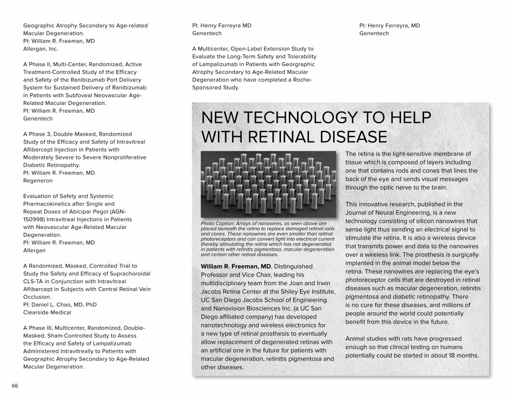
66
Geographic Atrophy Secondary to Age-related Macular Degeneration. PI: William R. Freeman, MD Allergan, Inc.
A Phase II, Multi-Center, Randomized, Active Treatment-Controlled Study of the Efficacy and Safety of the Ranibizumab Port Delivery System for Sustained Delivery of Ranibizumab in Patients with Subfoveal Neovascular Age-Related Macular Degeneration.PI: William R. Freeman, MD Genentech
A Phase 3, Double-Masked, Randomized Study of the Efficacy and Safety of Intravitreal Aflibercept Injection in Patients with Moderately Severe to Severe Nonproliferative Diabetic Retinopathy.PI: William R. Freeman, MD Regeneron
Evaluation of Safety and Systemic Pharmacokinetics after Single and Repeat Doses of Abicipar Pegol (AGN-150998) Intravitreal Injections in Patients with Neovascular Age-Related Macular Degeneration.PI: William R. Freeman, MD Allergan
A Randomized, Masked, Controlled Trial to Study the Safety and Efficacy of Suprachoroidal CLS-TA in Conjunction with Intravitreal Aflibercept in Subjects with Central Retinal Vein Occlusion. PI: Daniel L. Chao, MD, PhD Clearside Medical
A Phase III, Multicenter, Randomized, Double-Masked, Sham-Controlled Study to Assess the Efficacy and Safety of Lampalizumab Administered Intravitreally to Patients with Geographic Atrophy Secondary to Age-Related Macular Degeneration.
PI: Henry Ferreyra MDGenentech
A Multicenter, Open-Label Extension Study to Evaluate the Long-Term Safety and Tolerability of Lampalizumab in Patients with Georgraphic Atrophy Secondary to Age-Related Macular Degeneration who have completed a Roche-Sponsored Study.
Photo Caption: Arrays of nanowires, as seen above are placed beneath the retina to replace damaged retinal rods and cones. These nanowires are even smaller than retinal photoreceptors and can convert light into electrical current thereby stimulating the retina which has not degenerated in patients with retinitis pigmentosa, macular degeneration and certain other retinal diseases.
William R. Freeman, MD, Distinguished Professor and Vice Chair, leading his multidisciplinary team from the Joan and Irwin Jacobs Retina Center at the Shiley Eye Institute, UC San Diego Jacobs School of Engineering and Nanovision Biosciences Inc. (a UC San Diego affiliated company) has developed nanotechnology and wireless electronics for a new type of retinal prosthesis to eventually allow replacement of degenerated retinas with an artificial one in the future for patients with macular degeneration, retinitis pigmentosa and other diseases.
NEW TECHNOLOGY TO HELP WITH RETINAL DISEASE
The retina is the light-sensitive membrane of tissue which is composed of layers including one that contains rods and cones that lines the back of the eye and sends visual messages through the optic nerve to the brain. This innovative research, published in the Journal of Neural Engineering, is a new technology consisting of silicon nanowires that sense light thus sending an electrical signal to stimulate the retina. It is also a wireless device that transmits power and data to the nanowires over a wireless link. The prosthesis is surgically implanted in the animal model below the retina. These nanowires are replacing the eye’s photoreceptor cells that are destroyed in retinal diseases such as macular degeneration, retinitis pigmentosa and diabetic retinopathy. There is no cure for these diseases, and millions of people around the world could potentially benefit from this device in the future. Animal studies with rats have progressed enough so that clinical testing on humans potentially could be started in about 18 months.
PI: Henry Ferreyra, MD Genentech
67
GRANTSNATALIE A. AFSHARI, MDIntegrative Genetic Analyses in Fuchs Endothelial Corneal Dystrophy PI: Natalie A. Afshari, MD NIH, March 2013 – February 2017
A Genome Wide Association Study of Fuchs Endothelial Corneal Dystrophy PI: Natalie A. Afshari, MD Center for Inherited Disease Research (CIDR), NIH X01 HG006619
RADHA AYYAGARI, PHDGenetics of Hereditary Retinal DegenerationsPI: Radha Ayyagari, PhDThe Foundation Fighting Blindness, July 2011 - July 2017
Insights into AMD Derived from the Genetic Mechanisms in Late Onset Retinal Macular Degeneration (L-ORMD)PI: Radha Ayyagari, PhDThome Memorial Foundation, December 2014 - December 2017
Molecular Pathology Underlying Retinal Degeneration due to the Involvement of CTRP5/C1QTNF5 and MFRP GenesPI: Radha Ayyagari, PhDThe Foundation Fighting Blindness, June 2015 - June 2018
Molecular Basis of Hereditary Retinal DegenerationsPI: Radha Ayyagari, PhDNIH/NEI, June 2016 - May 2021
DIRK-UWE G. BARTSCH, PHDMechanistic-Based Non-Invasive Assessment of Retinal Damage in HAART EraPI: Dirk-Uwe G. Bartsch, PhDNIH, June 2006 - August 2021
Animal Structure and FunctionPI: Dirk-Uwe G. Bartsch, PhDNIH, July 2012 - June 2018
Test Intravitreal Injection Drug DevicesPI: Dirk-Uwe G. Bartsch, PhDNanovision Biosciences, Inc. , March 2014 - May 2018
CHRISTOPHER BOWD, PHDPredicting and Detecting Glaucomatous Progression Using Pattern Recognition PI: Christopher Bowd, PhD NIH, February 2012 - July 2016
Machine Learning Methods for Detecting Disease-Related Functional and Structural Change in Glaucoma PI: Christopher Bowd, PhDNIH, July 2017 - June 2019
DANIEL L. CHAO, MD, PHDA Zebrafish Model of Wet Macular Degeneration PI: Daniel L. Chao, MD, PhD Bright Focus Foundation, July 2017 - July 2019
MICHAEL H. GOLDBAUM, MDEvaluating Phenotypes using Intravitreal Aflibercept Injection PI: Michael H. Goldbaum, MD Regeneron, 2014-2018
DAVID B. GRANET, MDAn Observational, Multi-center Study of the Prevalence of Cerebrotendinous Xanthomatosis (CTX) in Patient Populations Diagnosed with Early Onset Idiopathic Bilateral Cataracts PI: Shira L. Robbins, MD Retrophin, Inc., 2016 - Present
WON-KYU JU, PHDMitochondrial Dysfunction in Glaucomatous Optic Neuropathy PI: Won-Kyu Ju, PhDNIH, September 2009 – August 2018
JONATHAN H. LIN, MD, PHDProtein Homeostasis and Proteotoxicity Mechanisms PI: Jonathan H. Lin, MD, PhD NIH/NINDS, February 2015 - January 2020
Cellular and Molecular Mechanisms of Age-Related Retinal Degeneration PI: Jonathan H. Lin, MD, PhD VA/BLR&D, April 2014 - March 2018
Endoplasmic Reticulum Stress in Neurodegeneration PI: Jonathan H. Lin, MD, PhD VA/RR&D. February 2017 - January 2021
The Jackson Laboratory Center for Precision Genetics: New Models to Novel Therapeutics PI: Burgess, NishinaSubcontract: Jonathan H. Lin, MD, PhD NIH/OD, August 2015 - June 2020
UCSD Alzheimer Disease Research Center Pilot Project 3 PI: GalaskoPilot Project: Jonathan H. Lin, MD, PhD NIH/AG, September 2016 - August 2017
68
Amydis Diagnostics Ocular Tissue Project PI: Jonathan H. Lin, MD, PhD Amydis Diagnostics, Inc, September 2016 - August 2017
ERIC D. NUDLEMAN, MD, PHDTesting Intravitreal Injection Drug Particles PI: Eric D. Nudleman, MD, PhDBiospinnaker Sciences, Inc., April 2017 - June 2018
SHIRA ROBBINS, MDAmblyopia Treatment Study: Study of Binocular Computer Activities for Treatment of Amblyopia (ATS18) PI: Shira Robbins, MDPEDIG JAEB Center for Health Research, October 2014 - December 2018
Omega-3 Fatty Acids as a Therapy for the Prevention of Retinopathy of Prematurity PI: Shira Robbins, MDHartwell Foundation, April 2015 - Mar 2018
PETER SHAW, PHDHTRA1 as a Therapeutic Target in the Treatment of Wet AMD PI: Peter Shaw, PhD NIH/NEI, August 2015 – August 2020
DOROTA SKOWRONSKA-KRAWCZYK, PHDMolecular Mechanism of Glaucoma PI: Dorota Skowronska-Krawczyk, PhD NIH, March 2017-February 2022
ROBERT N. WEINREB, MDADAGES III: Contribution of Genotype to Glaucoma Phenotype in African Americans PI: Robert N. Weinreb, MDNIH, September 2013 – August 2018
Ophthalmology and Visual Sciences Career Development K12 Program PI: Robert N. Weinreb, MDNIH, April 2015 – March 2020
Ocular Hypertension Treatment Study 20-Year Follow-Up: Clinic Center PI: Robert N. Weinreb, MD NIH, April 2015 – March 2018
Unrestricted and Challenge Grant - Research to Prevent Blindness PI: Robert N. Weinreb, MD
KARL J. WAHLIN, PHDModeling Photoreceptor Development and Disease Using Human Pluripotent Stem Cells PI: Karl J. Wahlin, PhD NIH, August 2016 - July 2019
Microenvironment Based Optimization of Retinal Induction Using CRISPR-CAS9 Reporter Pluripotent Stem Cells as an Expandable Source of Retinal Progenitors and PhotoreceptorsPI: Karl J. Wahlin, PhD CIRM, October 2016 - September 2019
DEREK WELSBIE, MD, PHDHigh-Content High-Throughput Functional Genomics in Rodent Retinal Ganglion Cells PI: Derek Welsbie, MD, PhD Research to Prevent Blindness (RPB) Career Development Award, July 2013 - June 2017
Dual Leucine Zipper Kinase (DLK) as a Mediator of Retinal Ganglion Cell Injury Co-I: Derek Welsbie, MD, PhD NEI/NIH, July 2013 - June 2017
Developing Dual Leucine Zipper Kinase Inhibitors for Glaucoma PI: Derek Welsbie, MD, PhD Brightfocus Foundation Glaucoma Research Award, July 2014 - June 2016
Targeting Dual Leucine Zipper Kinase as a Therapeutic Strategy for Traumatic Optic Neuropathy and Brain Injury PI: Derek Welsbie, MD, PhD Dept. of Defense Vision Research Program Translational Research Award, July 2014 - June 2017
Glaucoma Neuroprotection: Rho-associated Kinase 2 (ROCK2) as an Upstream Activator of Dual-leucine Zipper Kinase (DLK) in Response to Axonal Injury PI: Derek Welsbie, MD, PhD Ziegler Foundation Career Development Award, November 2014 - October 2017
Novel AAV/CRISPR Therapeutic for DLK Inhibition PI: Derek Welsbie, MD, PhD Brightfocus Foundation Glaucoma Research Award, July 2017 - June 2018
High-Throughput Screening in a Mouse Model of Leber Hereditary Optic Neuropathy PI: Derek Welsbie, MD, PhD NIH/NEI Loan Repayment Plan (LRP), July 2014 - June 2016
LINDA ZANGWILL, PHDDiagnostic Innovations in Glaucoma Study (DIGS): High Myopia and Advanced Diseases PI: Linda Zangwill, PhDNIH, March 2017 – February 2022
African Descent and Glaucoma Evaluation (ADAGES) IV: Alterations of the Lamina Cribrosa in Progression PI: Linda Zangwill, PhDNIH, April 2017 – March 2021
P30-Center Core Grant for Vision Research PI: Linda Zangwill, PhDNIH, July 2012 – June 2018Translational Vision Research Training at UCSD PI: Linda Zangwill, PhDNIH, April 2016 – March 2018
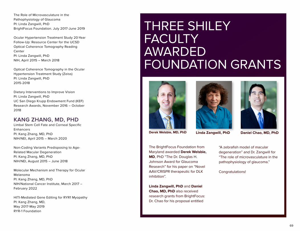
69
The Role of Microvasculature in the Pathophysiology of Glaucoma PI: Linda Zangwill, PhD BrightFocus Foundation. July 2017-June 2019
Ocular Hypertension Treatment Study 20-Year Follow-Up: Resource Center for the UCSD Optical Coherence Tomography Reading Center PI: Linda Zangwill, PhD NIH, April 2015 – March 2018
Optical Coherence Tomography in the Ocular Hypertension Treatment Study (Zeiss) PI: Linda Zangwill, PhD 2015-2018
Dietary Interventions to Improve Vision PI: Linda Zangwill, PhDUC San Diego Krupp Endowment Fund (KEF) Research Awards, November 2016 – October 2018
KANG ZHANG, MD, PHDLimbal Stem Cell Fate and Corneal Specific Enhancers PI: Kang Zhang, MD, PhDNIH/NEI, April 2015 – March 2020
Non-Coding Variants Predisposing to Age-Related Macular Degeneration PI: Kang Zhang, MD, PhD NIH/NEI, August 2015 – June 2018
Molecular Mechanism and Therapy for Ocular Melanoma PI: Kang Zhang, MD, PhD NIH/National Cancer Institute, March 2017 – February 2022
HITI-Mediated Gene Editing for RYR1 Myopathy PI: Kang Zhang, MD,May 2017-May 2019RYR-1 Foundation
THREE SHILEY FACULTY AWARDED FOUNDATION GRANTS
The BrightFocus Foundation from Maryland awarded Derek Welsbie, MD, PhD “The Dr. Douglas H. Johnson Award for Glaucoma Research” for his paper on “Novel AAV/CRISPR therapeutic for DLK inhibition”.
Linda Zangwill, PhD and Daniel Chao, MD, PhD also received research grants from BrightFocus: Dr. Chao for his proposal entitled
Derek Welsbie, MD, PhD Linda Zangwill, PhD Daniel Chao, MD, PhD
“A zebrafish model of macular degeneration” and Dr. Zangwill for “The role of microvasculature in the pathophysiology of glaucoma.”
Congratulations!
70
NATALIE A. AFSHARI, MDAugust 2016, Invited Speaker, National Medical Association – Ophthalmology Section, Los Angeles, CA “Challenging Cases in Cornea and Cataract Surgery”
September 2016, Invited Speaker, Kaiser Permanente 33rd Annual Ophthalmology Symposium, Anaheim, CA “Update on Diagnosis and Management of Challenging Cornea Cases”
September 2016, Invited Speaker, Kaiser Permanente 33rd Annual Ophthalmology Symposium, Anaheim, CA “ROCK Inhibitors and Cornea”
October 2016, International Cornea Course Anterior Segment Reconstruction 2016, Asociación para Evitar la Ceguera en México, Mexico City, Mexico “Medical Treatment for Meibomian Gland Dysfunction, Lipiflow”
October 2016, International Cornea Course Anterior Segment Reconstruction 2016, Asociación para Evitar la Ceguera en México, Mexico City, Mexico “Amniotic Membrane: Indications and Surgical Technique, Fibrin Glue” “Amniotic Membrane: Indications and Surgical Technique, Fibrin Glue”
October 2016, International Cornea Course Anterior Segment Reconstruction 2016, Asociación para Evitar la Ceguera en México, Mexico City, Mexico “Acute and Chronic Management of Stevens-Johnson Syndrome”
October 2016, International Cornea Course Anterior Segment Reconstruction 2016, Asociación para Evitar la Ceguera en México, Mexico City, Mexico “Indications and Techniques for Corneal and Corneoscleral Patch”
February 2017, Ophthalmology Update 2017, La Jolla, CA “Vitrectomy for the Anterior Segment Surgeon”
February 2017, Ophthalmology Update 2017, La Jolla, CA “Grand Rounds of Challenging Cataract Cases and Lens Surgery”
February 2017, Ophthalmology Update 2017, La Jolla, CA “All About Amniotic Membrane”
April 2017, Lorand V Johnson Lecture, Cleveland Ophthalmological Society Meeting, Cleveland, OH “Corneal Transplants, Flaps, Lasers, Bubbles: Recent Advances in Cornea and Refractive Surgery”
April 2017, Invited Lecture, Cleveland Ophthalmological Society Meeting, Cleveland, OH “All About Amniotic Membrane”
April 2017, Grand Rounds Invited Lecture, Case Western Reserve University, Cleveland, OH “Clinical Challenges in Cornea: When the Patient Hasn’t Read the Textbook”
May 2017, Invited Lecture, 13th Annual Harvard Medical School Intensive Cataract Surgery Training Course, Harvard University, Boston, MA “Practical IOL Calculations”
June 2017, Invited Speaker, The Pacific Coast Oto-Ophthalmological Society 101st Meeting, Waimea, HI “All About Amniotic Membranes”
June 2017, Invited Speaker, The Pacific Coast Oto-Ophthalmological Society 101st Meeting, Waimea, HI “ROCK Inhibitors, Stem Cells and Beyond”
June 2017, 20th Annual US Navy Refractive Surgery Course, Navy Medical Center, San Diego, CA “Epithelial Ingrowth”
RADHA AYYAGARI, PHDJuly 2017, National Eye Institute, NIH, Bethesda, MD “Finding Elusive IRD Causing Mutations by Whole Genome Sequencing”
CHRISTOPHER BOWD, PHD22nd International Visual Field and Imaging Symposium, Udine, Italy “Structural Change can be Detected in Advanced Glaucoma”
Association for Research in Vision and Ophthalmology 2017 Imaging in the Eye Conference, Baltimore, MD “Feature Transfer in Deep Learning Models to Detect Glaucomatous Optic Disc Changes”
DANIEL L. CHAO, MD, PHD2017 Chang Gung Memorial Hospital, Taipei, Taiwan “Clinical Approaches to Restoration of Vision in the Retina”
2017 Taiwan Veterans General Hospital, Taipei, Taiwan “Translational Approaches to Retinal Vascular Disease”
2017 National Taiwan University, Taipei, Taiwan “Translational Approaches to Retinal Diseases”
NAPOLEONE FERRERA, MDChinese Ophthalmology Society Meeting, Beijing, People’s Republic of China, Sept. 7-9, 2016
LECTURES
71
Kloster Seeon Young Investigator Meeting & 9th International Kloster Seeon Meeting, Munich, Germany, September 15-20, 2016 “Angiogenesis”
Johns Hopkins University, Wilmer Science Seminar Series, Baltimore, Maryland, October 17, 2016
Japanese Clinical Opthalmology Meeting, Kyoto, Japan, November 3-6, 2016
Mayo Clinic Angiogenesis Symposium, Jacksonville, FL, November 17-20, 2016
Pharmacology Seminar Series, University of California San Diego, La Jolla, CA, 92093
San Raffaele Institute Retreat, Baveno, Italy, March 10-12, 2017
AACR Annual Meeting, Washington, DC, April 2-5, 2017
2017 T. K. Hunt Lecture, The Wound Healing Foundation, San Diego, CA, April 5, 2017
Chinese Vascular Biology Organization (CVBO) Meeting, Guangzhou, People’s Republic of China, June 24-25, 2017
MICHAEL H. GOLDBAUM, MDMidwest Ocular Angiography Conference, Waikea Beach, Hawaii, July 28, 2016 “Positive Scotoma from Paracentral Acute Middle Maculopathy”
Midwest Ocular Angiography Conference, Waikea Beach, Hawaii, July 30, 2016 “Distinguishing between Chronic Central Serous Chorioretinopathy and Polypoidal Choroidal Vasculopathy”
International Society of Ocular Oncology, Syndey, Australia, March 2017 “Vogt Koyanagi Harada Syndrome and Other Immune Responses to Ocular and Cutaneous Melanomas”
Midwest Ocular Angiography Conference, Tallin, Estonia, August 2017 “Breast Carcinoma Metastatic to Optic Nerve Head”
Midwest Ocular Angiography Conference, Tallin, Estonia, August 2017 “Vogt Koyanagi Harada Syndrome and Other Immune Responses to Ocular and Cutaneous Melanomas”
DAVID B. GRANET, MDAugust 2016, Orange County Medical Association, Irvine, CA “10 Things Pediatricians Need to Know About Pedatric Ophthalmology”
September 2016, Table Rock Regional Round Up for the Arkansas Ophthalmological Society/Kansas Society of Eye Phyisicans & Surgeons/Missouri Socciety of Eye Physicians and Surgeons/Oklahoma Academy of Ophthalmology Annual Meeting, Ridgedale, MO “Pediatric Ophthalmology for the Ophthalmologist”, and “Communicating with Intent”
October 2016, Department Grand Rounds for Henry Ford Hospital & Medical Centers, Detroit, MI “Pearls for the Ophthalmologist”, and “Pediatric Ophthalmology for the Ophthalmologist”
October 2016, American Academy of Ophthalmology Annual Meeting, Chicago, IL “I-Phones, Electronic Devices and Eye Apps”
November 2016, San Diego County Optometric Society, San Diego, CA “Reading, Learning and Vision”, and “Adult Strabismus”
January 2017, UC San Diego, Department of Ophthalmology, La Jolla, CA “Nystagmus”
January 2017, UC San Diego, Department of Ophthalmology, La Jolla, CA “All Things Related to Pediatric Ophthalmology”
March 2017, Oregon Academy of Ophthalmology Post Graduate Convention, World Forestry Center, Portland, OR “Muscling in on Refractive Surgery and Myopia”, “Advances in Strabismus”, “Reading, Learning and Vision”, and “Geeking Out on Cases in Pediatric Ophthalmology”
March 2017, UC San Diego, Department of Ophthalmology, La Jolla, CA “Strabismus Surgery Review”
WELDON W. HAW, MDAugust 2016, UC at San Diego School of Medicine & Shiley Eye Institute Cataract Surgery Wet Laboratory Workshop. San Diego
September 2016, Continuing Specialized Education (CSE) Advance Cataract Surgery Course. Jobson. Dallas, Texas “Managing Cornea Astigmatism – Optimizing Cataract Surgery Outcomes. A Case Based Interactive Discussion”
September 2016, Continuing Specialized Education (CSE) Advance Cataract Surgery Course. Jobson. Dallas, Texas “Advanced Cataract Surgery Techniques- A Case Based Interactive Discussion”
September 2016, Continuing Specialized Education (CSE) Advance Cataract Surgery Techniques Course. Jobson. Dallas, TX
September 2016, Continuing Specialized Education (CSE) Advance Cataract Surgery Course. Jobson. Dallas, Texas “Advanced
72
Cataract Surgery- Improving Speed and Efficiency without Compromising Safety”
October 2016, UCSD Department of Ophthalmology & Shiley Eye Institute, La Jolla, CA “Corneal Dystrophies”
October 2016, UCSD Department of Ophthalmology & Shiley Eye Institute., La Jolla, CA “Inflammatory Disorders of the Anterior Segment”
November 2016, Newport Beach, CA”Update on Treatment of Pain and Inflammation following Ocular Surgery. Treatment of Endogenous Anterior Uveitis”
December 2016, Continuing Specialized Education Cataract Surgery and Wet Laboratory. Jobson. Dallas-Fort Worth, TX “Efficient Nucleus Disassembly and I/A Techniques”
December 2016, Continuing Specialized Education. Core Ophthalmic Resident Education (CORE) – Cataract Surgery and Wet Laboratory. Jobson. Dallas-Fort Worth, TX
December 2016, Continuing Specialized Education. Core Ophthalmic Resident Education (CORE) – Cataract Surgery and Wet Laboratory. Jobson. Dallas-Fort Worth, TX
December 2016, Continuing Specialized Education Cataract Surgery and Wet Laboratory. Jobson. Dallas-Fort Worth, TX “Top 3 Surgical Pearls for Surgical Excellence-Cataract Surgery”
January 2017, Association of University Professors of Ophthalmology (AUPO) Annual Meeting. San Diego, CA “Complication Rates of Resident Performed Cataract Surgery. Impact of Early Introduction of Cataract Surgery Training”
February 2017, Continuing Specialized Education. Cataract Surgery and Wet Laboratory. Jobson. Dallas-Fort Worth, TX
February 2017, Jobson Publishing. La Jolla, CA “Cataract & Cornea Section. Ophthalmology Update 2017: New Approaches to Medical and Surgical Therapies.”
February 2017, Continuing Specialized Education Cataract Surgery and Wet Laboratory. Jobson. Dallas-Fort Worth, TX “Cataract Surgery Techniques - Efficient Nucleus and Cortical Removal Techniques.”
February 2017 Jobson Publishing. La Jolla, CA “Update on Ocular Surface Diseases: Dry Eye Disease. Ophthalmology Update 2017: New Approaches to Medical and Surgical Therapies”
February 2017, Jobson Publishing. La Jolla, CA “Surgical Management of Corneal Astigmatism in Cataract Surgery. Ophthalmology Update 2017: New Approaches to Medical and Surgical Therapies”
March 2017, University of Southern California Keck School of Medicine. Los Angeles, CA “Optimizing Outcomes of Cataract Surgery: Management and Prevention of Inflammation & Pain Following Intraocular Surgery”
March 2017, University of Arizona, Tucson, AZ “Modern Cataract Surgery”
March 2017, Southern California College of Optometry – Marshall B. Ketchum University. Fullerton, CA “Managing Postoperative Inflammation & Pain Following Anterior Segment Surgery”
April 2017, University of New Mexico, School of Medicine, Department of Ophthalmology. Albuquerque, NM “Optimizing Outcomes of Modern Day Cataract Surgery”
CHRISTOPHER W. HEICHEL, MDFebruary 2017, UCSD Shiley Eye Institute, UCSD School of Medicine, La Jolla, CA “Update in DMEK and DSEK”
February 2017, Ophthalmology Update 2017 UCSD Shiley Eye Institute, UCSD School of Medicine, La Jolla, CA “Allergic Ocular Disease”
February 2017, Ophthalmology Update 2017 UCSD Shiley Eye Institute, UCSD School of Medicine, La Jolla, CA “Capsular Tension Rings”
DON O. KIKKAWA, MDJuly 2016, 33rd Annual Superficial Anatomy and Cutaneous Surgery, Marriott Del Mar, CA “Periocular Reconstruction and Blepharoplasty”
August 27, 2016, Joint Meeting of the APSOPRS and JSOPRS, Osaka, Japan “Ptosis and Entropion”
September 30, 2016, Oculofacial Conference and Celebration, University of Wisconsin, Madison “Oculoplastics and Life Lessons from Dortz”
December 2, 2016, USC Oculoplastic Surgery Course, Los Angeles, CA “Upper Blepharoplasty and Asian Blepharoplasty”
March 2017 International Symposium Ophthalmology, Dutch Society of Ophthalmology (Nederlands Oogheelkundig Gezelschap), Maastrich, Netherlands “Thyroid Orbitopathy: Have We Learned Anything in the Last Twenty Years?”
June 2017, ASOPRS International Thyroid Eye Disease Symposium, Vancouver, Canada “Surgery for Thyroid Eye Disease”
73
June 2, 2017, USC Roski Eye Institute 42nd Annual Symposium, Pasadena, CA “Update in Oculoplastic Surgery: Better Solutions for Old Problems”
July 2017, Dalian Medical University, Dalian, China “Soft Tissue Fillers and Neurotoxins”
BOBBY S. KORN, MD, PHDJuly 29, 2016, Vision China 2016, Shenzhen, China “Optimizing DCR to a 10 Minute Procedure”
July 30, 2016, Vision China 2016, Shenzhen, China “Minimally Invasive Optic Nerve Sheath Fenestration”
August 27, 2016, Japan Society of Ophthalmic Plastic and Reconstructive Surgery and Asian Pacific Society of Ophthalmic Plastic and Reconstructive Surgery Annual Meeting, Osaka, Japan “EndoDCR My Approach”
September 11, 2016, San Diego County Optometric Society, San Diego, CA “Pearls and Pitfalls in Oculoplastics”
January 19, 2017 Southeastern Society of Plastic and Reconstructive Surgeons (SESPRS), Atlanta, GA “A Postoperative Day #0 Surprise”
February 18, 2017, Ophthalmology Update 2017, San Diego, CA “Fillers and Neurotoxins”
April 29, 2017, International Congress of the Brazilian Society of Oculoplastics, Belo Horizonte, Brazil “Aesthetic Surgery - How to Achieve the Best Results”
April 29, 2017, International Congress of the Brazilian Society of Oculoplastics, Belo Horizonte, Brazil “Lower Blepharoplasty with Fat Repositing”
April 29, 2017, International Congress of the Brazilian Society of Oculoplastics, Belo Horizonte, Brazil “Nonsurgical Asian Eyelid Rejuvenation”
April 30, 2017, International Congress of the Brazilian Society of Oculoplastics, Belo Horizonte, Brazil “How to Restore an Almond Shaped Eyelid”
April 30, 2017, International Congress of the Brazilian Society of Oculoplastics, Belo Horizonte, Brazil “My Algorithm for Management of Lower Eyelid Retraction”
May 11, 2017, Grand Rounds, Department of Dermatology, UC San Diego “Oculoplastic Reconstructions after Mohs Surgery”
June 3, 2017, Annual Meeting of the Japan Society of Ophthalmic Plastic and Reconstructive Surgeons, Kokura, Japan “Endoscopic DCR, Maximizing Hemostasis - My Way”
June 7, 2017, Vegas Cosmetic Surgery Meeting, Las Vegas, NV “Eyelid Retraction Repair - My Algorithm”
June 7, 2017, Vegas Cosmetic Surgery Meeting, Las Vegas, NV “Integrating Ptosis Repair with Aesthetic Blepharoplasty”
June 7, 2017, Vegas Cosmetic Surgery Meeting, Las Vegas, NV “Minimizing Complications in Blepharoplasty: Early Interventions”
JEFFREY E. LEE, MDFebruary 2017, Ophthalmology Update, San Diego, CA “Changing Trends in Ophthalmic Resident Education”
JONATHAN H. LIN, MD, PHDAugust 2016, Jackson Laboratory, Center for Precision Genetics Bar Harbor, ME “ER Stress in Neurodegeneration”
September 2016, International Society for Eye Research Tokyo, Japan “Oxidative and ER Stress in Eye Diseases”
October 2016, Osler Institute South San Francisco, CA “Eye Pathology”
November 2016, China Rare Diseases Consortium 4th Annual Conference Wuhan, China “Unfolded Protein Response Regulator, ATF6, is a Novel Achromatopsia Gene”
February 2017, Medical College of Wisconsin, Vision Sciences Distinguished Lecture Series Milwaukee, WI “ATF6 in Achromatopsia” March 2017, Osler Institute Chicago, IL “Eye Pathology”
March 2017, American Society for Neurochemistry Little Rock, AR “ER Stress in Neurodegeneration”
April 2017, American Society for Investigative Pathology Chicago, IL “Diseases of the Endoplasmic Reticulum”
May 2017, Association for Research Vision Ophthalmology Baltimore, MD “ER Stress and Unfolded Protein Response in Ocular Health and Disease”
JOHN HK LIU, PHDAugust 2016, Intraocular and Intracranial Pressure Gradient Related Diseases International Summit, Beijing, China “The Importance of Habitual 24-Hour IOP Measurement”
74
October 2016, Industrial Technology Research Institute International, San Jose, California “Experience in Clinical Trial of a Medical Device”
October 2016, The Gravity of Medicine in Space: From Recent Results to Earth Benefits Symposium. Southwest Chapter of the American College of Sports Medicine 2016 Meeting, Costa Mesa, CA “Treating Crew Member’s Vision Impairment Syndrome in Space and Glaucoma on Earth”
ERIC D. NUDLEMAN, MD, PHDMarch 2017 Osher Lecture, UC San Diego, La Jolla, CA “Emerging Therapies for Age-Related Macular Degeneration”
March 2017 Advances in Pediatric Retina (APR) Course, Duke University Eye Center, Durham, NC “Treatment of Adults with History of Pediatric Retinal Diseases”
SHIRA L. ROBBINS, MDJuly 2016, UC San Diego Ophthalmology Residents “Retinopathy of Prematurity”
October 2016, American Academy of Pediatrics, San Francisco, CA (2 sessions) ”Everything You Wanted To Know About Ophthalmology but Were Afraid To Ask”
October 2016, American Academy of Ophthalmology Annual Meeting, Chicago, IL “Pediatric Eye Emergencies You Don’t Want To Miss!”
November 2016, Child Health and Disability Prevention Program, Pediatricians/Optometrists/Nurses/Medical Assistants, San Diego, CA ”Vision Screening in Young Children”
February 2017, Ophthalmology Residents, UC San Diego, “OKAP Review-Pediatric Ophthalmology”
February 2017, Ophthalmology Residents, UC San Diego “Didactic: Ocular Development”
February 2017, Ophthalmology Residents, UC San Diego “Didactic: Pediatric Glaucoma and Cataracts”
February 2017, Ophthalmology Residents, UC San Diego “Didactic: Phakomatoses”November 2016, Ophthalmology Skills Session, UC San Diego Medical Students MS2 “Ophthalmology Skills Session”
April 2017, AAPOS, Nashville TN “Ligneous Conjunctivitis Related to Plasminogen Deficiency: Can We Finally Treat It?”
April 2017, Child Health and Disability Prevention Program, Pediatricians/Optometrists/Nurses/Medical Assistants, San Diego, CA “Vision Screening in Young Children”
July 2017, UC San Diego Ophthalmology Residents “Retinopathy of Prematurity”
PETER SHAW, PHD2017 ARVO Meeting, Baltimore, MD “Oxidative Stress Impacts the AMD Pathology through Modulating Wnt Signaling Pathway”
DOROTA SKOWRONSKA-KRAWCZYK, PHD2016 University of Geneva, Switzerland “Molecular Mechanism of Neuronal Degeneration in Glaucoma”
KARL WAHLIN, PHD2016 Sanford Consortium Southern California Research Talk, La Jolla, CA “Gene-Editing and 3D Retinal Organoids”
2016 Shiley Eye Institute Lunch and Learn, La Jolla, CA “Stem Cell Session”
2016 FASEB, Keystone, Colorado “Retinal Connectivity”
2017 UC San Diego Cell and Molecular Medicine Department, LA Jolla, CA “Stem Cells and Retinal Disease Modeling”
2017 Plenary speaker for 2017 Annual Meeting of the Chinese Medical Association - Taipei, Taiwan
2017 UC San Diego Ophthalmology Update, La Jolla, CA “Stem Cells and Ocular Regeneration”
ROBERT N. WEINREB, MD2017 Bruno Boles Carenini Lecture, 1st International Congress of the Associazione Italiana Studio Glaucoma, Rome, Italy “Enhancing Angle Surgery for Glaucoma with Anterior Segment Optical Coherence Tomography”
2017 Glaucoma Update Manchester Royal Eye Hospital, Manchester, UK “Smart Glaucoma - Personalizing Glaucoma Care”
DEREK WELSBIE, MD, PHD2016 Optometric Glaucoma Society Meeting, American Academy of Optometry “What Damages the Optic Nerve in Glaucoma,” and “Alternative Medicine in Glaucoma”
2016 West Coast Optometric Glaucoma Society Meeting “Glaucoma Pathophysiology”
LINDA ZANGWILL, PHD2016 The 11th Chinese Glaucoma Society Congress, Changsha, China “Measuring the Rate of Structural Change in Glaucoma: A Better Way to Identify Progression”
75
2016 The 11th Chinese Glaucoma Society Congress, Changsha, China “OCT Angiography and Glaucoma”
2017 World Glaucoma Congress, Helsinki, Finland “Structure: How to Determine Glaucoma Progression by OCT / HRT”
2017 World Glaucoma Congress, Helsinki, Finland “The Role of Microvasculature in the Pathophysiology of Glaucoma: Evidence from OCT Angiography”
2017 World Glaucoma Congress, Helsinki, Finland “Utility of OCT for Detection of Localized Retinal Damage in Glaucoma”
2017 World Glaucoma Congress, Helsinki, Finland “AngioVue OCT Angiography in Glaucoma Research”
2017 Optical Coherence Tomography Angiography Summit, Portland, Oregon “Microvasculature Dropout in Glaucoma: Evidence from OCTA”
KANG ZHANG, MD, PHD2016 Gene Therapy Session Speaker at the Targeting Ocular Disorders Conference, Boston, MA “Translating Genomics and Stem Cell Technology into Therapy for Major Blinding Diseases”
2016 Special Interest Symposium Speaker at the European Association for Vision and Eye Research, Nice, France “Lanosterol Reversal of Protein Aggregation in Cataract”
2016 Vision Science Seminar Speaker, University of Colorado, Denver, CO “Translating Genomics and Stem Cell Technology into Therapies for Blinding Eye Diseases”
2017 Tissue Engineering and Clinical Applications Speaker at the International
Society for Stem Cell Research, Boston, MA “Lens Regeneration Using Endogenous Stem Cells with Gain of Visual Function”
2017 Clinical Advances in Stem Cell Research Speaker at the International Society for Stem Cell Research, Boston, MA “A Cell Therapy Approach to Treat Cataract”
2017 Brazilian Glaucoma Society, Rio de Janeiro, Brazil “Innovations in Ophthalmology”
2017 ARVO Conference, Baltimore, MD “Current Advances in Stem Cell Therapies and 3D Culture Systems”
2017 Chancellor’s Associates Colloquium, UC San Diego, La Jolla, CA “Aging, Age-Related Diseases, Biological Clock, and Reversal of Aging”
2017 Founders Symposium, UC San Diego, La Jolla, CA “Re-seeing the Unseen”
2017 UC Los Angeles, Westwood, CA “Translating Genomics and Stem Cell Technology into Eye Care”
Educating the future leaders in medicine requires not only the best instruction in ophthalmology but also equipping residents and fellows with the tools they need to succeed in the changing landscape of healthcare. These tools include formal leadership and business training.
This year, the Department in conjunction with the UC San Diego Rady School of Management, initiated a Leadership and Innovation Series. The lectures included topics such as “Skillful Leadership in Turbulent Environments” and “Actionable Leadership.” Shiley also hosted a session featuring possible entrepreneurial ideas for business development.
BUILDING FUTURE ENTREPRENEURS AND LEADERS
Many prominent San Diego business leaders offered their expertise in the sessions. The speakers included: Vish Krishnan, PhD, Steve Poizner, Chris Oveis, PhD, Pam Smith, PhD and Mary McKay, EdD (Rady School of Management), Ruben D. Florez-Saaib, PhD (Office of Innovation and Commercialization), David Guss, MD (Professor Emeritus), Donald Kearns, MD (Rady Children’s Hospital), Blair Sadler (formerly Children’s Hospital) and Neal E. Schmale (retired, Sempra Energy).
76
EVERY GIFT HAS AN IMPACTFor over 30 years, the philanthropic support from generous individuals, foundations and corporations has provided the Department of Ophthalmology with valuable resources for patient care, research, education and community service.
As a friend of the Department of Ophthalmology, there are several giving options for those who wish to contribute to our tradition of excellence. Every donation makes an impact on our patients, faculty, and staff, as well as the field of Ophthalmology. We cherish the partnership that we have developed with those generous members of the community and beyond who invest in us. There are also naming opportunities for gifts including: endowed chairs, laboratories, specialized ophthalmic clinics and research initiatives. We would welcome the opportunity to have a confidential conversation with you, so we clearly understand how you want your donation to be utilized.
VISIONARY CIRCLEMembers of the Visionary Circle are cumulative lifetime contributors of one million dollars or more to the Department of Ophthalmology. We appreciate their generosity.
AnonymousEleanor & John Barbey, Jr.David J. DunnMartin* & Enid* GleichJean Hahn HardyHildyard Family TrustDr. Trude K. HollanderJoan & Irwin JacobsDorothy R. Kerrigan TrustTatiana A. and Richard Kindell* LanscheArthur Murray & Carol-Faith Murray TrustIrvin Olson & Rita A. Olson TrustMarc PaskinAnne F. Ratner*Research to Prevent BlindnessRuth L. Schulman*Shiley Awards in Health Education and the Arts funded by Donald* & Darlene ShileyMichael* & Rebecca ShileyKenneth & Dixie UnruhFrances Hamilton White
* Deceased
77
GIVING OPPORTUNITIES
A gift of endowment demonstrates your long-term commitment to the Department of Ophthalmology since the fund is maintained in perpetuity. Your gift can support programs, lectures, awards, fellowships and Chairs. An endowment serves as an enduring legacy since it often bears the name of a donor or loved one.
ENDOWMENTS- GIFTS IN PERPETUITY
Many employers offer a matching gift program to their employees meaning that your donations are worth even more. All you need is a Matching Gift Form from your employer.
MATCHING GIFTS- DOUBLE OR TRIPLE YOUR GIFT
TRIBUTE GIFTS- ACKNOWLEDGE SOMEONE SPECIAL
Contributions can be made in memory, honor or in celebration of a loved one or to commemorate a special occasion. Gifts can be made to honor a special physician, for example, who has played a significant role in your eye health. Such a gift creates a legacy and memorializes the person by providing direct support to the Department.
ANNUAL GIFTS- CIRCLE OF SIGHT
Started in 1996, the Circle of Sight is the Shiley Eye Institute’s recognition program that acknowledges donors who make annual gifts of $250 or more to support the greatest needs of the Department. Several times a year, the Shiley Eye Institute’s Circle of Sight members are invited to attend Vision Research Lectures and receptions where members get to personally know our faculty. The members are also ambassadors for the Shiley Eye Institute within the San Diego community. The Circle of Sight group is the backbone of many of our successful initiatives.
OUTRIGHT GIFTS - IMMEDIATE IMPACTOutright gifts of all sizes made with cash, check, credit cards, savings bonds, stocks, marketable securities or property provide immediate impact to our faculty and facility. If writing a check, please make payable to the “UC San Diego Foundation” and put the Shiley Eye Institute in the memo section. The check should be accompanied with a letter stating the focus of your donation and mailed to The Shiley Eye Institute, Mailcode 0946, 9415 Campus Point Drive, Room 241B, La Jolla, CA, 92093-0946.
Many of our friends have found they can make a big impact with a gift in their will, a beneficiary designation on a retirement account or life insurance policy, or a gift that pays income. These are just some examples of innovative ways in helping our physician-scientists advance patient care and research in eye diseases. Some giving vehicles may even provide you with special tax advantages.
We would be pleased to provide you, your attorney, accountant or tax advisor with specific bequest language for inclusion in your will, trust or as a beneficiary of your retirement account.
PLANNED GIFTS- YOUR VISION FOR TOMORROW
For further information about making a donation, please contact:
KAREN ANISKO RYANPhone: 858-534-8017Email: [email protected]
78
Katherine FinnellLinda FortierGerald FreedmanDrs. William & Laura FreemanAlexander L. GascoigneThe Morris & Phyllis Gold Fund of the Jewish Community FoundationJane GoodwinMr. & Mrs. Joseph M. GrantDr. & Mrs. Harold R. HallElizabeth HansenLeonard HartJanette & Robert HodgesHerbert B. Hoffman & Janis SammartinoNatasha Josefowitz, PhDPaul & Rosemarie KalemkiarianThe Kelly Family FoundationRosemarie A. KubesBernard Kulchin & Paula TaylorSeth S. LevineFrieda L. LevinskyJeff & Patty MaysentJeffrey T. MillerThe Theodore & Anabel Mintz Fund of the Jewish Community FoundationNancy & Roger MooreLois M. MulcaheyMr. & Mrs. NachtomiJoan and Barry NugentFran Osborn & Tom RyanFrank & Mary PisciottaJohn & Diane Prewitt Family FoundationCarol PriorDr. Arnold & Doris RolandDavid & Ann SakaiTodd D. SchaferNiki ShafferDrs. Jonathan E. Shelton & Nicole H. PurcellMargaret SimonsenJohn & Corrine SoSouthwest San Diego County Lions Club
Esao & Glenda SumidaNessim A. TianoIrving TragenThe Viterbi Family Fund of the Jewish Community FoundationMargot Walk & Jerry FreelandWalmartStephen Wax* & Laurie PriceJo & Howard WeinerRoger & Carolyn WilliamsToby WolfBetty Wong
Gifts of $500 to $999John Adey & Marianne Banning AdeyAnonymous (2)John & Elizabeth BagbyDirk-Uwe Bartsch, PhD & Ann Lopez, MDEvelyn V. BergmannArun & Neera BhatiaJohn H. BlairMiles & Patricia BowlerWayne ComfortMargaret & Oren EisnerJean FreiburgerJ. M. Fuster, MDLouis GalperThe Lucy Goldman Fund of the Jewish Community FoundationWilliam & Ruth* GraulMr. & Mrs. Stuart GreenbaumJohn & Mary GriffinMr. & Mrs. Eugene B. Haignere, Jr.Steve HardenThe Marcia Foster Hazan Fund of the Jewish Community FoundationMargaret J. HeiserJerome HickmondNancy J. Holbrook & William D. Gay, Jr.Robert HymanNick A. JohnstonViktor Kerzhanovich
Gifts of $100,000 & AboveAnonymousDavid J. DunnLanna C. LewinJune & Connant Winton Reed EndowmentResearch to Prevent BlindnessWenc Trust
Gifts of $50,000 to $99,999Dr. & Mrs. David H. AndersonRoland & BeAnn ArthurWayne GreenPrice PhilanthropiesJack & Sherron Schuster
Gifts of $25,000 to $49,999AnonymousFarrell Family FoundationHeidelberg Engineering GmbH
Gifts of $10,000 to $24,999Steven & Sheri AltieriAnonymousRustom & Daneesh AppooThe Donald August TrustGermaine Hope Brennan Charitable FoundationLa Verne & Blaine BriggsGiovanni Bucolo Family FoundationElisabeth A. EwingKathleen & Steven M. Flynn / Bell Charitable FoundationAlbert & Kathleen FredmanThe Milton & Faiya Fredman Family Fund of the Jewish Community FoundationCaroline & Daniel HamlinTsui Pak KimHalpin & Helen O’Reilly Charitable TrustWilson & Jean SextonCapt. & Mrs. Jerome R. Strayve, USN (Ret.)Rudi Urlau
Carol & Larry VeitRobert N. Weinreb, MDJames & Josephine ZolinMr. & Mrs. Eric Von Zwisler
Gifts of $5,000 to $9,999Natalie Afshari, MDAllerganMr. Austin Bernstein & Dr. April TeitelbaumToby & Howard CohenAnne & Walter DempseyMr. & Mrs. Thompson Fetter / 52 & Convoy Corp.Carole & Howard GoldfederSeth & Margaret HerndonThe Howard G. & Samita B. Jacobs FoundationDr. & Mrs. Don O. KikkawaAl & Sharon RappaportSage Research Consulting, LLCShumway Survivors TrustKay* & Don StoneSuzanne SuchardJeanne M. SullivanJocelyn & Dick VortmannPam Wenig
Gifts of $1,000 to $4,999Anonymous (3)Farooq AzamJohn BaileyCharles M. BrownSidney Chang, MDDr. & Mrs. William T. ComerDorothy R. ContePrescott Crafts*Sabina DavidsonWilliam M. DumkaKenneth DunstEugenio Elorduy WaltherElsa & George Feher
The Honor Roll for the Department of Ophthalmology gratefully acknowledges donations from July 1, 2016
through June 30, 2017. Thank you to all of the individuals, foundations and corporations listed below.
79
Elizabeth & Craig C. Kishaba, MBA, CMPEJoan & Howard* KontjeTony & Mary LovettSally McMurrayChrista McReynolds Fund Patricia J. MeridethDennis & Barbara MoranVictoria PappasMichael G. QuallsJoyce Rakowski & Lowell FisherParvin RezainiaHal & Mary Sadler Family Foundation San Diego Unified School District - Community Service AssociationD. P. SchnorrGeorgina Serrano RomeroMr. & Mrs. Clinton R. SpanglerMr. & Mrs. Irving TashlickR. J. & Joy Urich
Gifts of $250 to $499Steve & Betty AllmanAnonymous (2)Fred & Helen ArbuckleRichard & Jessica AttiyehFrancine & Marvin BartLorrie BernsteinShiu-Chu ChiuBruce L. DeckMischelle DevosMr. & Mrs. Joseph DohnPenny DuermeyerEdward J. FarrellAlan & Pauline FatayerjiNicholas Fintzelberg, PhDCarlos FloresCharles Gibb, PhDFrancesco GrilliJaye & Bill HanleyMr. & Mrs. John J. HarrisRobert F. KibbleDick & Linda KintzMaxine KleinbergLeonard M. LaFebreMary LawlerKaren L. LeePaul & Petrina LibbyMr. & Mrs. Frederick MandabachBob & Susan Mardon
Mr. & Mrs. Anthony C. McKoyGary McMullenBud & Nancy MeerchaumLinda MilnerJonathan, Suzanne & Andrew MorrisElspeth & Jim MyerArthur P. Padilla*Betty L. PeabodyJanet T. PodellMary & Herb RabeRicardo J. & Annie C. RasinesMrs. Sally F. SaltzsteinEdwina SchatzJohn SchulerRonald & Diane SeditaLeonard ShulmanSharon E. SingerMr. & Mrs. Harold F. SkellyBradley M. SmithIvar StakgoldMr. & Mrs. Daniel SylvesterFredricka Taubitz & Dennis Martin GriffinJohn TisoCarlos TomaszewskiJill UnderwoodHilda van Neck-YoderWilliam A. & Susie M. VidrihTara & Charles WegnerJohn WeisRobert E. WelkDouglas & Olive WithallDr. Gloria Wolk
Gifts of $100 to $249Diane AmatangeloAnonymous (4)Glenn & Diana ArmstrongEva AvenellJudy BaileyDr. William BayliffBarry BerkovJ. C. Black & Wendy J. BlackSylvia A. BodeJames BoilyBetsy C. BowneKathy Bisceglie imo Henry SuchardChip BrownMarlena G. BrownCarol V. Buuck
Ben CagleZhizhong ChangMicaela ChellMr. & Mrs. William J. ClearyClint & Greta ConewayNancy & Brian CookDr. & Mrs. Olgard DabbertSylvia & Ron DamronLeonard DeSantiHudson B. DrakeMr. & Mrs. Laurence DuclosEstelle EbertJeffrey Ebstein & Cynthia BeckerLeonard Epstein/Empire FoodsMartin FentonDennis J. FischerRobert L. FisherSally FisherMaureen I. FritzerBarbara & Frank FromerCauleen & Michael GlassKari L. Grabowsky, MD & David L. Levin, MD, PhDVicki D. GranowitzW. E. Greer, IIIJerry & Jacquie GrinsteadBill & Sharon GriswoldMarjorie HentonHarriet Jill HerndonNorma Hidalgo-Del RioChristine HottingerHenry & Pauline ImusCarol A. JacksonDiana W. JacobsRuth JamesMichele JannetteMargareta JanssonWilliam JerniganAndrea JohnsonArthur C. Johnson, MDIngrid KamrathJames & Mary KerrHisako KoikeMr. & Mrs. James LangfordJanice LaxagueAnne & Thomas C. LeeNancy & James LeitnerRichard LentzMarley Lima
Eduardo LinaresNoemi G. LizarragaPatricia & Albert* LubarskyRudolf LustiGraham & Joanne MacHutchinDiane MartinJoseph & Sandy McKennaSonja D. MetscherHelene & Harvey MillerMuriel D. MitchellMr. & Mrs. Ronald L. MoeRena C. MongeMs. Sharon S. MoonSusan W. MurphyToni NickellPaul NivenSallee O’BrienVirginia & Kevin O’KeefeMarcus L. OvertonRobert A. PeppingJanet PiskorDr. & Mrs. John ProakisLora PrudenMary Jo Lagerlof QuinbyRomeo & Rose Marie RamiroJames & Janet RespessRita B. RivasRebeca RodkinGisela B. RohaZita RudvalisDr. Blair L. Sadler & Dr. Georgia R. SadlerLCDR John R. Savory, USN (Ret.)Madelyn SheetsLia Y. ShenBernice & George SicklickCarol SmithMr. A. E. SoderbergOleg & Courtney SorokoumovDawn StetskoElizabeth & Lester StielMaria Swann, PhDJohn & Judy TurnerLisa J. Wastila, MD & Michael WastilaDr. & Mrs. Richard T. WoldSusie WolfeFlora H. YoungRicardo R. YumulRoberta ZerdaTheresa Zysk
80
Gifts up to $99MiCarmen AlmaguerMr. & Mrs. Cheem S. AngAnonymous (9)Mr. & Mrs. Maurice J. ArgoudMrs. Concepcion A. BandayrelMr. & Mrs. George F. BartonYouhanna BasmadjianByron C. BeckLibby BentleyMrs. Barbara J. BergMr. & Mrs. Dwight E. BishopMrs. Elaine Bleau-RichardsFilomena Q. BondocSandy & Larry BornsteinJack & Charmian BowsherHelene BrottmanBeatrix BrownfieldJoan L. BuchholzEmerlita BundalianJesse L. CarmanFlorence Y. CarrKarin M. CarriveauBertha V. CastroEstella A. CedenoMr. & Mrs. Peter S. ChanKenneth ChaseKenneth T. CodyDr. Phyllis ColterMr. Hubert CoxYolanda CruzWilliam & Suzanne CulvinerMr. & Mrs. Timothy M. CunninghamJurgen H. DanielMrs. Eve DavidsonMr. & Mrs. Jerry I. DelCastilloLucien R. DesLauriersFredrick J. Di CarloEdna DiazKristen A. DostieJohn Eugene DrastalRoger & Isabel EscalleMs. Ruth Fajarit-DavisMr. & Mrs. Roldolfo V. FalloranCapt. & Mrs. Dan E. FennJosie FoulksTamara & Timothy Fry
Leonardo & Irinea GallardoRita GellerBarbara GenoveseJan GlennDavid & Bonnie GouldNikki Allyn GrossoAnita K. HaltonJeanne Y. HamblinMarcella E. HamlinMr. & Mrs. M. Dwight HamroPia Harris-EbertNewton & Helen HarrisonDavid A. S. HillRussell F. Himmer, CLUGuadalupe HuieMuriel JusterFrancisca KandelHerbert & Leah KarpJohn T. KastenAbu KaziMax & Julie KeyteKenneth KinmanSanford LakoffDavid A. LangVerjean LawsonMrs. Mary K. LemmonAmelia D. LindlarMargaret & Vincent LombardiJune & Will LowClaudia LowensteinMr. Minh Chi LuMr. & Mrs. Quintin MabantaMarcella E. MahanArline MalkasianJohn & Ruth MarcinkeviczJulia MarineAli MaronesyMadeleine MarshRobert W. McNamaraAgha S. MehdiNelda D. MendozaGhanim (Guy) MichaelRosemary MoffettBrian K. & Luisa D. MonsonThelma MontagDavid J. MoralesPat L. MummertHonore MurphyKevin R. Murphy
Mohammad Reza NaghavianLisa Janine NevilleKatya Newmark & Danya CostelloMr. & Mrs. Philip NordhuesMr. & Mrs. Andres C. OlivaLe M. OngAida OrtegaLesley OvertonMr. & Mrs. Prachak PannarajMr. & Mrs. D. J. PapenbrookEdith PartiAngela PiccilloDebbie Powers-CookGrace PurpuraCharles QuistSusan Wish RaffeeHermila RamirezKenneth M. RappaportMartha RobinsonMarcel RogallaBernd J. RossMarvin RuebelMina SalamiJohn S. SalcidoWalter S. Scheib, Jr.William SchlohNikki SchweitzerDr. Gideon SeamanLauren SerwitzKobi SethnaGeraldine SevierMr. & Mrs. Donald ShaevelHarry & Linda SheetsKurt E. ShulerWayne & Marian SiegfriedAnne & Ronald SimonVilma Simon OghliMrs. Patricia A. SmithRobert H. SolsbakPhyllis SpeerTania SpireHarry SuhlZoltan & Anda SztankayJorge & Ellen TangonanMr. & Mrs. John J. ThomersonFrederick H. ThompsonRose Tran / San Diego Vietnamese American Lions ClubBob Treitler
This is a partial list. We have made every effort to be accurate in our listing and apologize if any mistake or omission has been made. Should you find an error or want to change your listing, please contact us at (858) 534-4981.
Anthony S. TubolinoBenjamin R. ValdezPraxedes ValdezJosina & Andy Van DieJason & Katherine VercelliLoc VoMarlene WalkerG. Thomas WaltersAnn WatanabeFrancine R. WelchezTed WitteConnie Wood
* Deceased
MemorialsThe Department of Ophthalmology sadly acknowledges friends and key supporters who have passed away during the past year. They remain in our thoughts. Richard Butcher, MDPrescott CraftsNicholas FrazeeGeorge W. HouckAlbert R. LubarskyWilliam S. McConnorEthel ShelleyCatherine (Kay) M. StoneJudge Ross TharpStephen R. Wax
81
The gifts in this annual report are from our friends who have contributed over the past 12 months to bolster Shiley patient care, vision research, medical training and community outreach. No matter the size, each donation is equal in its importance and receives equal amount of gratitude from all of us at Shiley Eye Institute and on their behalf, Shiley patients and families.
One example of your philanthropy is a generous gift donated by Michael Luzich to honor his parents, Norman and Carol Luzich. The fund’s purpose is to honor Mr. and Mrs. Luzich, to accelerate regenerative research in glaucoma and to benefit patient care.
The donor champions a novel strategy of converting Müller glia cells in the retina into optic nerve fibers (the damaged fibers in glaucoma patients). We already know that Müller glia cells are not damaged by glaucoma. Further, in many organisms like fish and amphibians, injured Müller glia cells can turn into retinal cells similar to optic nerve cells.
The Norman and Carol Luzich Glaucoma Fund is providing four-year funding for an interdisciplinary team of 10 scientists from the Richard C. Atkinson Laboratory for Regenerative Ophthalmology in the Department of Ophthalmology (Derek Welsbie, MD, PhD and Karl Wahlin, PhD), the Shiley Eye Institute (Robert N. Weinreb, MD), and the Department of Neurosciences (Dan Gibbs, PhD and Mark Tuszynski, MD, PhD) to conduct this innovative research.
According to Robert N. Weinreb, MD, project leader and Distinguished Professor of Ophthalmology, “this exciting project will benefit greatly from the collaboration with Drs. Tuszynski and Gibbs, leaders in the science of spinal cord repair and regeneration.”
During the next two years, the Norman and Carol Luzich glaucoma research team seek to validate that this strategy can produce a meaningful regeneration of optic nerve cells in models of glaucoma. The project’s goal is to initiate human clinical trials in glaucoma patients by the end of 2021. If successful, this funding could change
the paradigm of glaucoma treatment from the prevention of vision loss to restoration of vision and visual improvement.
To learn more about The Norman and Carol Luzich glaucoma research project, please contact:Karen Anisko Ryan Director, Business Development & [email protected]
For a private consultation to learn about ways to tailor your gift in a way that is most meaningful for you, please contact:Helen Z. LiuSenior Director of [email protected]
The Norman and Carol Luzich Glaucoma Fund
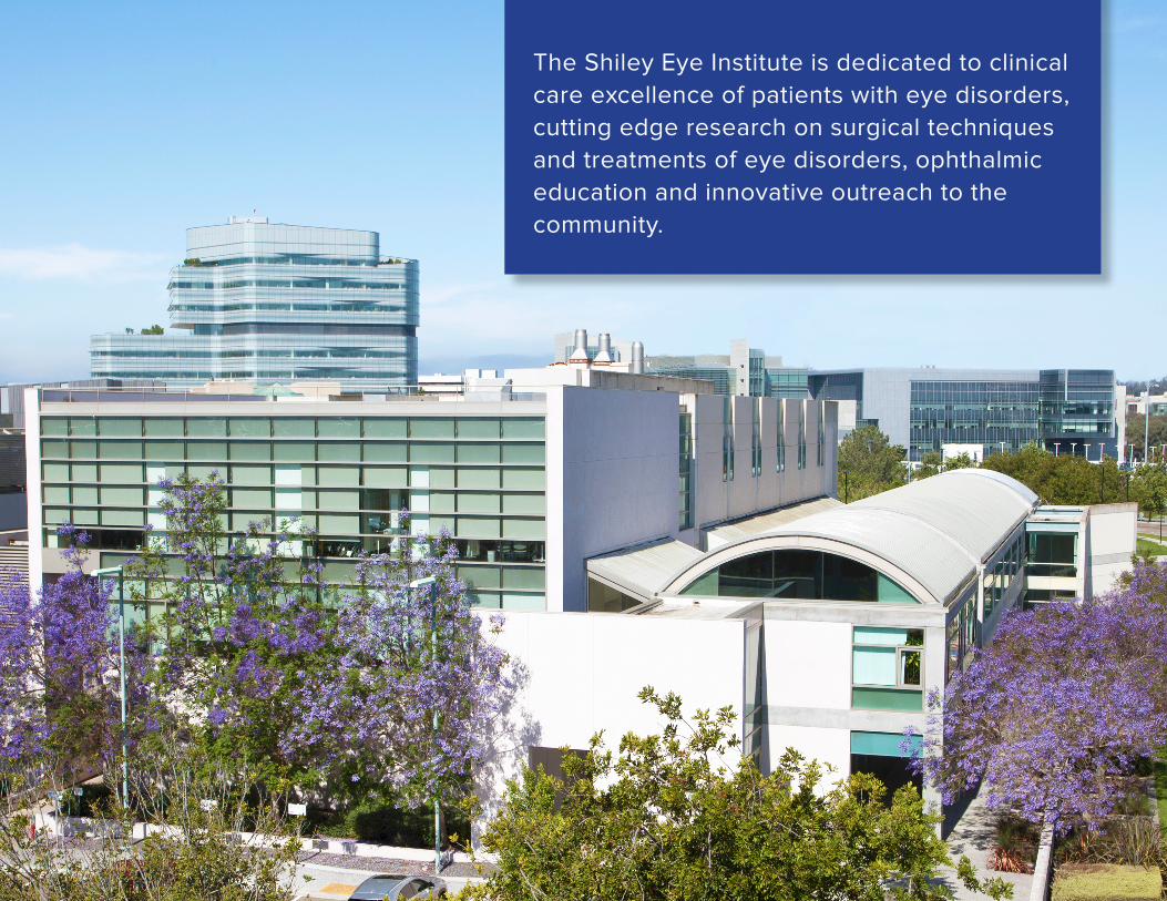
82
The Shiley Eye Institute is dedicated to clinical care excellence of patients with eye disorders, cutting edge research on surgical techniques and treatments of eye disorders, ophthalmic education and innovative outreach to the community.
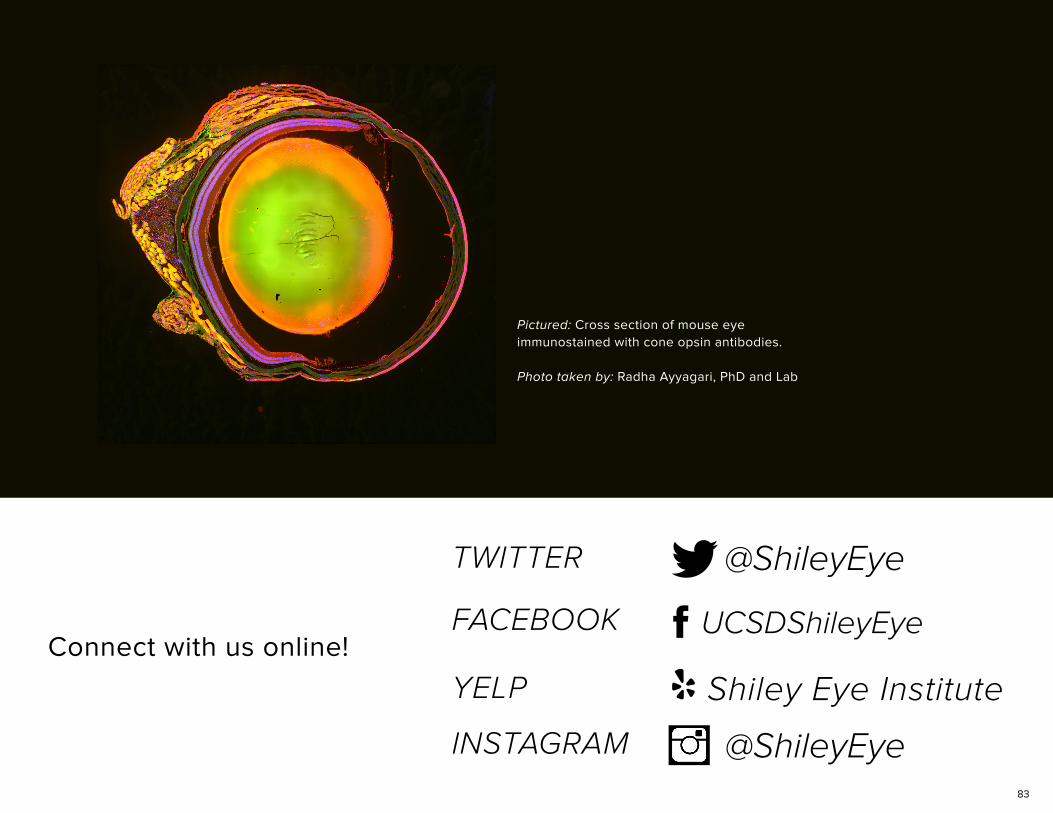
83
@ShileyEye
UCSDShileyEye
Shiley Eye Institute
YELP
INSTAGRAM @ShileyEye
Connect with us online!
Pictured: Cross section of mouse eye immunostained with cone opsin antibodies.
Photo taken by: Radha Ayyagari, PhD and Lab
Related Documents