Journal Pre-proof Foliar pathogens of eucalypts P.W. Crous, M.J. Wingfield, R. Cheewangkoon, A.J. Carnegie, T. Burgess, B.A. Summerell, J. Edwards, P.W.J. Taylor, J.Z. Groenewald PII: S0166-0616(19)30009-0 DOI: https://doi.org/10.1016/j.simyco.2019.08.001 Reference: SIMYCO 90 To appear in: Studies in Mycology Please cite this article as: Crous PW, Wingfield MJ, Cheewangkoon R, Carnegie AJ, Burgess T, Summerell BA, Edwards J, Taylor PWJ, Groenewald JZ, Foliar pathogens of eucalypts, Studies in Mycology, https://doi.org/10.1016/j.simyco.2019.08.001. This is a PDF file of an article that has undergone enhancements after acceptance, such as the addition of a cover page and metadata, and formatting for readability, but it is not yet the definitive version of record. This version will undergo additional copyediting, typesetting and review before it is published in its final form, but we are providing this version to give early visibility of the article. Please note that, during the production process, errors may be discovered which could affect the content, and all legal disclaimers that apply to the journal pertain. © 2019 Westerdijk Fungal Biodiversity Institute. Production and hosting by ELSEVIER B.V. All rights reserved.

Welcome message from author
This document is posted to help you gain knowledge. Please leave a comment to let me know what you think about it! Share it to your friends and learn new things together.
Transcript

Journal Pre-proof
Foliar pathogens of eucalypts
P.W. Crous, M.J. Wingfield, R. Cheewangkoon, A.J. Carnegie, T. Burgess, B.A.Summerell, J. Edwards, P.W.J. Taylor, J.Z. Groenewald
PII: S0166-0616(19)30009-0
DOI: https://doi.org/10.1016/j.simyco.2019.08.001
Reference: SIMYCO 90
To appear in: Studies in Mycology
Please cite this article as: Crous PW, Wingfield MJ, Cheewangkoon R, Carnegie AJ, Burgess T,Summerell BA, Edwards J, Taylor PWJ, Groenewald JZ, Foliar pathogens of eucalypts, Studies inMycology, https://doi.org/10.1016/j.simyco.2019.08.001.
This is a PDF file of an article that has undergone enhancements after acceptance, such as the additionof a cover page and metadata, and formatting for readability, but it is not yet the definitive version ofrecord. This version will undergo additional copyediting, typesetting and review before it is publishedin its final form, but we are providing this version to give early visibility of the article. Please note that,during the production process, errors may be discovered which could affect the content, and all legaldisclaimers that apply to the journal pertain.
© 2019 Westerdijk Fungal Biodiversity Institute. Production and hosting by ELSEVIER B.V. All rightsreserved.

Foliar pathogens of eucalypts P.W. Crous1,2, M.J. Wingfield2,3, R. Cheewangkoon4, A.J. Carnegie5,6, T. Burgess2,7, B.A. Summerell8, J. Edwards9,10, P.W.J. Taylor11 and J.Z. Groenewald1 1Westerdijk Fungal Biodiversity Institute, P.O. Box 85167, 3508 AD Utrecht, The Netherlands; 2Department of Genetics, Biochemistry and Microbiology, Forestry and Agricultural Biotechnology Institute (FABI), University of Pretoria, Pretoria, 0002, South Africa; 3Forestry and Agricultural Biotechnology Institute (FABI), University of Pretoria, Pretoria, 0002, South Africa; 4Department of Plant Pathology, Faculty of Agriculture, Chiang Mai University, Chiang Mai 50200, Thailand; 5Forest Health & Biosecurity, Forest Science, NSW Department of Primary Industries - Forestry, Level 12, 10 Valentine Ave, Parramatta NSW 2150, Australia; 6School of Environment Science and Engineering, Southern Cross University, Lismore, NSW 2480; 7Environmental and Conservation Sciences, Murdoch University, 90 South Street, Murdoch, WA 6150, Australia; 8Royal Botanic Gardens and Domain Trust, Mrs Macquaries Rd, Sydney, NSW 2000, Australia; 9Agriculture Victoria Research, Department of Jobs, Precincts and Regions, AgriBio Centre, 5 Ring Road, LaTrobe University, Bundoora, Victoria 3083 Australia; 10School of Applied Systems Biology, LaTrobe University, Bundoora, Victoria 3083 Australia; 11Faculty of Veterinary and Agricultural Sciences, The University of Melbourne, Parkville, VIC, Australia *Correspondence: P.W. Crous, [email protected]
Abstract: Species of eucalypts (Eucalyptus, Corymbia) are commonly cultivated for solid wood and pulp products. The expansion of commercially managed eucalypt plantations has chiefly been driven by their rapid growth and suitability for propagation across a very wide variety of sites and climatic conditions. Infection of foliar fungal pathogens of eucalypts is resulting in increasingly negative impacts on commercial forest industries globally. To assist in evaluating this threat, the present study provides a global perspective on foliar pathogens of eucalypts. We treat 110 different genera including species associated with foliar disease symptoms of these hosts. The vast majority of these fungi have been grown in axenic culture, and subjected to DNA sequence analysis, resolving their phylogeny. During the course of this study several new genera and species were encountered, and these are described. New genera include: Lembosiniella (L. eucalyptorum on E. dunnii, Australia), Neosonderhenia (N. eucalypti on E. costata, Australia), Neothyriopsis (N. sphaerospora on E. camaldulensis, South Africa), Neotrichosphaeria (N. eucalypticola on E. deglupta, Australia), Nothotrimmatostroma (N. bifarium on E. dalrympleana, Australia), Nowamyces (incl. Nowamycetaceae fam. nov., N. globulus on E. globulus, Australia), and Walkaminomyces (W. medusae on E. alba, Australia). New species include (all from Australia): Disculoides fraxinoides on E. fraxinoides, Elsinoe piperitae on E. piperita, Fusculina regnans on E. regnans, Marthamyces johnstonii on E. dunnii, Neofusicoccum corticosae on E. corticosa, Neotrimmatostroma dalrympleanae on E. dalrympleana, Nowamyces piperitae on E. piperita, Phaeothyriolum dunnii on E. dunnii, Pseudophloeospora eucalyptigena on E. obliqua, Pseudophloeospora jollyi on Eucalyptus sp., Quambalaria tasmaniae on Eucalyptus sp., Q. rugosae on E. rugosa, Sonderhenia radiata on E. radiata, Teratosphaeria pseudonubilosa on E. globulus and Thyrinula dunnii on E. dunnii. A new name is also proposed for Heteroconium eucalypti as Thyrinula uruguayensis on E. dunnii, Uruguay. Although many of these genera and species are commonly associated with disease problems, several appear to be opportunists developing on stressed or dying tissues. For the majority of these fungi, pathogenicity remains to be determined. This represents an important goal for forest pathologists and biologists in the future. Consequently, this study will promote renewed interest in foliar pathogens of eucalypts, leading to investigations that will provide an improved understanding of the biology of these fungi.
Key words: Eucalyptus, Corymbia, foliar pathogen, new taxa, taxonomy. Running head: Eucalypt foliar pathogens
Taxonomic novelties: New family: Nowamycetaceae Crous. New genera: Lembosiniella Crous, Neosonderhenia Crous, Neothyriopsis Crous, Neotrichosphaeria Crous & Carnegie, Nothotrimmatostroma Crous, Nowamyces Crous, Walkaminomyces Crous & Carnegie. New species: Disculoides fraxinoides Crous, Elsinoe piperitae Crous, Fusculina regnans Crous, Lembosiniella eucalyptorum Crous & Carnegie, Marthamyces johnstonii Crous & Carnegie, Neofusicoccum corticosae Crous & Summerell, Neosonderhenia eucalypti Crous, Neotrimmatostroma dalrympleanae Crous, Nowamyces globulus Crous, Nowamyces piperitae Crous, Phaeothyriolum dunnii Crous & Carnegie, Pseudophloeospora eucalyptigena Crous, Pseudophloeospora jollyi Crous, Quambalaria tasmaniae Crous, Quambalaria rugosae Crous, Sonderhenia radiata Crous, Teratosphaeria pseudonubilosa G. Pérez & Carnegie, Thyrinula dunnii Crous & Carnegie. New combinations: Allelochaeta brevilata (H.J. Swart & D.A. Griffiths) Crous, Blastacervulus robbenensis (Crous et al.) Crous, Lembosiniella eucalypti (Sivan. & R.G. Shivas) Crous, Neofusicoccum versiforme (Z.Q. Yuan et al.) Crous, Neosonderhenia foliorum (Cooke) Crous, Neothyriopsis sphaerospora (Marasas) Crous, Neotrichosphaeria eucalypticola (Sivan. & R.G. Shivas) Crous & Carnegie, Nothotrimmatostroma bifarium (Gadgil & M.A. Dick) Crous, Nothotrimmatostroma eucalyptorum (Crous & Carnegie) Crous, Phacidium innumerum (Massee) Crous, Phaeothyriolum amygdalinum (Cooke & Massee) Crous & Carnegie, Phaeothyriolum corymbiae (Crous) Crous, Phaeothyriolum eucalyptorum (Crous & W.B. Kendr.) Crous, Pseudocercospora tumulosa (Carnegie & Beilharz) Carnegie & Crous, Teratosphaeria delegatensis (R.F. Park & Keane) Crous, Thyrinula parasitica (Crous) Crous, Walkaminomyces medusae (Carnegie & G.S. Pegg) Crous & Carnegie. New name: Thyrinula uruguayensis Crous for Heteroconium eucalypti Crous & M.J. Wingf. Epitypes: Arnaudiella eucalyptorum Crous & W.B. Kendr., Lembosiopsis eucalyptina Petr. & Syd., Leptostromella eucalypti Cooke & Massee, Microthyrium amygdalinum Cooke & Massee, Mycosphaerella marksii Carnegie & Keane, Sphaerella cryptica Cooke, Stigmina robbenensis Crous et al., Thyriopsis sphaerospora Marasas, Trichosphaeria eucalypticola Sivan. & R.G. Shivas, Trimmatostroma bifarium Gadgil & M.A. Dick. Available online xxx Dedication: This paper is dedicated to Dr H.J. Swart (1922–1993). Harry Swart was an extraordinary mycologist with an exceptional gift for detailed mycological observations. His series of papers on the

“Australian leaf-inhabiting fungi” published in the then Transactions of the British Mycological Society, inspired the first author to dedicate much of his career to collecting and culturing these incredible foliicolous fungi (see Australas. Pl. Pathol. 23: 29–35. 1994). INTRODUCTION The tree genera Angophora, Corymbia and Eucalyptus (Myrtaceae), colloquially termed eucalypts, accommodate more than 800 species, the majority endemic to Australia (Pryor & Johnson 1971, Boland et al. 1992, Ladiges et al. 2003, Thornhill et al. 2019). Many species of Corymbia and Eucalyptus are grown globally for commercial purposes and as amenity trees. To meet the world’s demand for paper and pulp, there has in recent years been a rapid expansion in areas under afforestation, particularly in South-East Asia and in the Southern Hemisphere (Eldridge et al. 1994, Cotterill & Brolin 1997, Martin 2003, Qi 2003). Importantly eucalypts, which make up 74 % of forested land in Australia (ABARES 2017), are the key components of most ecosystems in Australia and provide food and habitat for a myriad of animals as well as a range of micro-organisms including fungi.
The genus Eucalyptus is one of the most remarkable genera of trees. It includes at least 600 species, almost all of which are native to Australia (Ladiges et al. 2003). The remaining species, including Eucalyptus deglupta and E. urophylla, are endemic to Indonesia, New Guinea and the Philippines. All but one (Corymbia papuana) of the 113 species in Corymbia are also native to Australia. These trees occur across a vast diversity of habitats including moist forests, open woodlands, deserts, rocky outcrops and swamplands (Potts & Pederick 2000). Likewise, they occur in a wide range of climatic conditions with adaptations to high or moderate rainfall, drought, bushfire, high and low temperatures and even snow during winter.
In the last comprehensive review of fungi occurring on these hosts, Sankaran et al. (1995) recorded more than 500 species of leaf-infecting fungi on eucalypts (including the genera Corymbia and Eucalyptus). There have been many other studies on the fungi occurring on eucalypts (Park et al. 2000). These date back to descriptions of “Sphaerella” nubilosa and “S.” cryptica in Cooke’s Handbook of Australian Fungi from samples sent to England from Australia (Cooke 1892). Interest in these fungi has primarily been linked to the fact that Eucalyptus spp. (and to a lesser degree Corymbia spp.) have been extensively planted outside their native range for more than 100 years (Turnbull 2000). During this time, they have reached a point of being amongst few tree genera most commonly propagated in plantations, with almost 20 M ha now planted globally (http://git-forestry-blog.blogspot.com/2008/09/eucalyptus-global-map-2008-cultivated.html). These plantations sustain some of the world’s most important timber industries, which produce solid wood and pulp products. Consequently, the health of eucalypt trees is important to the sustainability of major forest industries, and fungi are amongst the most common causes of diseases of these trees.
Fungal pathogens have been recognised as threats to both native-grown and plantation-grown eucalypt trees for many years (Browne 1968, Dick & Gadgil 1983, Ferreira 1989, Sharma et al. 1985, Lundquist 1987, Crous et al. 1989, Carnegie et al. 1994, Andjic et al. 2019). This threat has become more obvious as eucalypt plantation areas have increased, particularly during the course of the last 30 years. This expansion has been driven by their rapid growth and suitability for propagation across a very wide variety of sites and climatic conditions. The expansion of eucalypt plantings can also be attributed to the growth of the paper and dissolving pulp industries, where the short-fibres of Eucalyptus spp. are required.
The negative impact of foliar pathogens of eucalypts dates back to the early propagation of these trees in plantations outside Australia. One of the first species to be extensively planted was E. globulus, suited particularly to areas of the world with Mediterranean climates (Florence 1986). Many plantations of these trees, and the closely related E. nitens, suited to cool temperate climates, were very seriously damaged by leaf blotch disease caused by species originally treated in the genus Mycosphaerella (Browne 1968, Lundquist 1987, Lundquist & Purnell 1987). Plantations of E. globulus in certain areas in South Africa in the 1930’s were abandoned due to severe damage from Teratosphaeria (= Mycosphaerella) nubilosa (Lundquist 1987). When these species began to be commercially planted in Australia from the 1990’s they also experienced severe damage from T. nubilosa and also T. cryptica (Mohammed et al. 2003, Milgate et al. 2005, Carnegie 2007a, Barber et al. 2008, Hunter et al. 2009, Andjic et al. 2019). As Eucalyptus spp. such as E. camaldulensis, E. grandis and E. urophylla generally suited to warmer summer rainfall areas of the world were deployed in parts of the tropics and Southern Hemisphere, leaf diseases caused by species of Calonectria (Cylindrocladium) were found to cause serious leaf blight problems (Sharma & Mohanan 1982, Sharma et al. 1985, Blum et al. 1992, Park et al. 2000, Crous 2002, Old et al. 2003). Subsequently, species of Teratosphaeria (Kirramyces) emerged as significant diseases on these species in the tropics and sub-tropics (Andjic et al. 2019).
Where eucalypts have been planted for the first time in new areas, they have tended not to be affected by diseases, including those affecting their shoots and leaves. But, as is true for diseases of all plants, this situation has changed over time with new disease (and insect) problems becoming increasingly problematic.

These diseases are caused by two distinct categories of pathogens. They include native pathogens that have moved onto eucalypts (host jumps) and those that have been accidentally introduced from Australia into the areas where the eucalypts are planted (Burgess & Wingfield 2017). Native pathogens of the foliage include for example species of Calonectria that can occur naturally in soils (Lombard et al. 2010a–c, Alfenas et al. 2015). The larger number of non-native pathogens are eucalypt-specific such as species of Readeriella and Teratosphaeria.
Very little is known regarding the majority of fungi occurring on the foliage of eucalypts (Sankaran et al. 1995). A relatively small number of species are recognised as primary pathogens. These have mostly emerged where species of eucalypts are intensively managed in plantations (Andjic et al. 2019). Others occurring on the leaves of trees in native stands are either recognised as primary pathogens either because they belong to groups of fungi known to cause disease or due to the nature of the symptoms with which they are associated. But it is important to realise that even for those fungi considered to cause leaf diseases, the majority have not been subjected to pathogenicity tests and thus proven to be pathogens based on Koch’s postulates.
The areas of origin of most leaf pathogens found on species of eucalypts outside the native range of these trees remains unknown. For example, the very serious leaf and shoot pathogen, Teratosphaeria destructans was first discovered on Eucalyptus planted as non-natives in north Sumatra (Wingfield et al. 1996). The pathogen has spread to many other areas of South East Asia and most recently to South Africa (Andjic et al. 2011, Greyling et al. 2016), although it has never been recorded in areas where these trees are native. Yet its highly host-specific nature and the fact that population genetic studies have shown strong clonality in invaded ranges suggest that it is native to Eucalyptus somewhere in the native range of these trees (Burgess & Wingfield 2017). One suggestion has been that it is native to Timor but this has yet to be proven scientifically (Andjic et al. 2019).
There are numerous instances of foliar fungi being originally described from exotic eucalypts then later found in Australia (e.g. Carnegie et al. 1997, Maxwell et al. 2000, 2003, Jackson et al. 2005, Crous et al. 2007c, Burgess & Wingfield 2017). Many of these are likely to be endemic to Australia but found overseas first due to intensive interest in exotic eucalypt plantations. There are, however, still many species of, for example Teratosphaeria, that have not been found in Australia (Carnegie et al. 2007). With more intensive fungal forays in Australia, many new species are now being discovered (Crous et al. 2017a, b, 2018a–c, present study).
Diseases of eucalypts are emerging via host shifts from native plants to these trees where they are planted as exotics (Slippers et al. 2005). There are now many instances where stem canker pathogens in the Cryphonectriaceae have been shown to have undergone host shifts from native Myrtales (including Myrtaceae, Melastomataceae, etc) to eucalypt (Wingfield 1999, Wingfield 2003). These pathogens, new to eucalypts, can be very damaging and they now threaten the trees in other areas where they are propagated, as well as in the native ranges of these trees. A key driver of native pathogens moving to non-native eucalypts propagated in plantations appears to be the presence of native trees in the Myrtales and thus those relatively closely related to eucalypts facilitating a host shift (Burgess & Wingfield 2017, Crous et al. 2016a).
A classic example of a primary eucalypt leaf and shoot pathogen that is not native on these trees but has adapted to infect them is the rust fungus Austropuccinia psidii. This pathogen has a wide host range on many genera of the Myrtaceae in South America and was first recorded causing significant damage to Eucalyptus in Brazil (Ferreira 1983), where South African seed sources of E. grandis were shown to be particularly susceptible (Coutinho et al. 1998, Glen et al. 2007). More recently, A. psidii has appeared in new areas including Hawaii, Japan, China, Australia, South Africa, New Caledonia, Indonesia, Singapore and most recently in New Zealand (Carnegie & Pegg 2018). The same genotype of the rust occurs in most of these areas and this is known as the pandemic genotype (Stewart et al. 2017, Carnegie & Pegg 2018). This is different to the genotype of A. psidii that occurs in South Africa, which has not been found on these trees in plantations but has been shown to infect Eucalyptus and Corymbia in greenhouse pathogenicity studies (Roux et al. 2013, 2016).
Many fungi occurring on eucalypt foliage are well-known opportunists or latent pathogens. The best-studied are species in the Botryosphaeriaceae and more specifically Neofusicoccum. The Botryosphaeriaceae include 24 genera, the majority of which occur on trees or woody shrubs (Yang et al. 2017). Most are found on the bark of branches and stems where they also cause die-back symptoms. A much smaller number of these fungi are known from leaves including those of Eucalyptus spp. Within Australia only 24 species, mostly Neofusicoccum species, are found associated with eucalypts (Burgess et al. 2019). It is reasonable to assume that, like other Botryosphaeriaceae, they infect healthy tissues without causing symptoms and that they appear when these tissues are subjected to stress (Schoeneweiss 1980, Slippers & Wingfield, 2007). In the case of leaves, this stress might simply be related to senescence. Pathogenicity studies are required to better understand the role of these fungi as foliage pathogens.
An important group of eucalypt leaf pathogens reside in the genus Quambalaria (de Beer et al. 2006). These pathogens are apparently specifically native to Australia and they include at least one species, Q. coyrecup that is an aggressive stem canker pathogen of Corymbia (Paap et al. 2008). The two best known

species on leaves and shoots are Q. eucalypti that causes leaf and shoot diseases of Eucalyptus spp. and Q. pitereka that is specific to leaves and shoots of Corymbia spp. Both species are now found in plantations outside the native range of Eucalyptus and Corymbia with Q. eucalypti having the most extensive global distribution. Interestingly, Q. eucalypti was first found and described in South Africa (Wingfield et al. 1993), then found in Australia more than a decade later (Carnegie 2007a, Pegg et al. 2008).
There are likely numerous records of eucalypt leaf fungi on species previously treated as Eucalyptus that are now known to actually represent species of Corymbia. Those collected from hosts only identified to genus (“Eucalyptus sp.”) would thus now be indistinguishable as being from Corymbia, cf. those collected from distinct species (e.g. Eucalyptus maculata = Corymbia maculata). It is reasonable to presume that the fungi on Corymbia spp. are distinct from those on Eucalyptus, as is found with Q. pitereka and Q. eucalypti. In recent years, Corymbia spp. have been increasingly deployed outside their native range for plantation development. This would justify a much greater focus on making comparisons of pathogens and other fungi that occur on leaves specifically of Corymbia spp. It is relevant to recognise that some records in the present study for “Eucalyptus” are also likely to include some for Corymbia, because the two tree genera were collectively treated as Eucalyptus until relatively recently (Hill & Johnson 1995).
Many of the fungi listed in this study have been described from dead leaves found in the litter below trees. Most are probably saprobes that grow and sporulate once leaves begin to die. Here it is important to recognise that like other plants, eucalypt leaves have a vast microbiome and that many of the fungi that form part of this community are seen only when leaves die. For example, Kemler et al. (2013) used a metagenomics approach to show that living tissues of Eucalyptus trees include a very large number of fungal taxa, the most common of which occur in groups that include important pathogens such as those in the Capnodiales (Dothideomycetes).
The primary aim of this study was to reconsider all the fungi that have been recorded on diseased eucalypt foliage. Species simply associated with eucalypts, and assumed to be saprobic or of minor importance, are excluded. Various new species are described and the taxonomy of previously described species and genera is re-evaluated. Species associated with leaf spots representing 110 genera are listed. For the majority of these fungi, nothing is known regarding their biology or ecological importance. There are several genera that include well recognised primary pathogens, known from experimental evidence to be able to infect unwounded healthy plant tissue. These include (among others) Austropuccinia and species of Calonectria, Coniella, Elsinoë, Pseudocercospora, Quambalaria and Teratosphaeria. Species in various genera such as Alternaria, Botrytis and Neofusicoccum are commonly associated with disease problems and are most likely opportunists that develop on stressed or dying tissues. Many others are putative pathogens or have other important roles, but these have yet to be studied. This presents an important goal for fungal biologists in the future. It is hoped that this study will promote renewed interest in eucalypt leaf fungi, leading to investigations that will provide a better understanding of these fascinating and arguably, insufficiently appreciated fungi. MATERIALS AND METHODS Isolates Isolates were obtained from the culture collection (CBS) of the Westerdijk Fungal Biodiversity Institute, Utrecht, the Netherlands (WI), and the working collection of Pedro Crous (CPC), housed at the Westerdijk Institute, or were freshly isolated (Supplementary Table S1). Single-conidial and ascospore cultures were obtained using the techniques described previously (Crous et al. 1991, Crous 1998). DNA extraction, amplification and sequencing Fungal mycelium of strains (Supplementary Table S1) was harvested with a sterile scalpel and the genomic DNA was isolated using the UltraClean Microbial DNA Isolation Kit (MoBio Laboratories, Inc., Solana Beach, CA, USA) following the manufacturers’ protocols. A variety of loci were amplified and sequenced, depending on which loci are commonly used for a specific genus. The “Genera of phytopathogenic fungi: GOPHY” (Marin-Felix et al. 2017, 2019) series provided a good starting point for references pertaining to commonly used loci and their amplification conditions. The resulting fragments were sequenced in both directions using the PCR primers and the BigDye Terminator Cycle Sequencing Kit v. 3.1 (Applied Biosystems Life Technologies, Carlsbad, CA, USA). DNA sequencing amplicons were purified through Sephadex G-50 Superfine columns (Sigma-Aldrich, St. Louis, MO) in MultiScreen HV plates (Millipore, Billerica, MA). Purified sequence reactions were analysed on an Applied Biosystems 3730xl DNA Analyzer (Life Technologies, Carlsbad, CA, USA). The DNA sequences generated were analysed and consensus sequences were computed using SeqMan v. 7.0 (Lasergene, Madison, WI, USA).

Phylogenetic analysis Additional sequences were downloaded from the NCBI’s GenBank nucleotide database as needed (Supplementary Tables S1 and S2). The downloaded and generated sequences for each gene were aligned with the online version of MAFFT v. 7 (Katoh & Standley 2013). The alignments were manually checked and improved where necessary using MEGA v. 7 (Kumar et al. 2016) and were concatenated using the same software.
The phylogenetic methods used in this study included a Bayesian analysis performed with MrBayes v. 3.2.6 (Ronquist et al. 2012), and Parsimony and distance analyses performed with PAUP v. 4.0b10 (Swofford 2003). Missing sequences, when present, were treated as missing data in the analyses. MrModeltest v. 2.2 (Nylander 2004) was used to determine the best nucleotide substitution model settings for each data partition in order to perform a model-optimised Bayesian phylogenetic reconstruction. The Markov Chain Monte Carlo (MCMC) analysis of four chains started in parallel from a random tree topology, the heat parameter was set at 0.25 and trees were saved every 10 generations until the average standard deviation of split frequencies reached 0.01 (stop value). Burn-in was set to 25 % after which the likelihood values were stationary. The Neighbour-Joining (NJ) phylogenies performed with PAUP executed 1 000 bootstrap replicates using the HKY85 model. In the Maximum Parsimony analyses, alignment gaps were treated as fifth character state and all characters were unordered and of unequal weight. A heuristic search option with 100 random taxon additions and tree bisection and reconnection (TBR) as the branch-swapping algorithm was used. Branches of zero length were collapsed and all multiple, equally most parsimonious trees were saved. The robustness of the trees obtained was evaluated by 1 000 bootstrap replications (Hillis & Bull 1993). Other measures calculated included tree length (TL), consistency index (CI), retention index (RI) and rescaled consistency index (RC).
All resulting trees were printed with Geneious v. 7.0.6 (http://www.geneious.com, Kearse et al. 2012). All new sequences generated in this study were deposited in NCBI’s GenBank nucleotide database (www.ncbi.nlm.nih.gov) and the accession numbers are listed in Supplementary Table S1. The alignments and respective phylogenetic trees were deposited in TreeBASE, study number S24522. Morphology Slide preparations were mounted in lactic acid, Shear’s mounting fluid or water, from colonies sporulating on 2 % malt extract agar (MEA), 2 % potato-dextrose agar (PDA), oatmeal agar (OA), MEA (Crous et al. 2019c), autoclaved pine needles on 2 % tap water agar (PNA) (Smith et al. 1996), or autoclaved banana leaves (BLA), and incubated at 25 °C under continuous near-ultraviolet light to promote sporulation. Reference strains and specimens are maintained in the CBS culture collection. Sections through conidiomata were made by hand. Observations were made with a Nikon SMZ25 dissection-microscope, and with a Zeiss Axio Imager 2 light microscope using differential interference contrast (DIC) illumination and images recorded on a Nikon DS-Ri2 camera with associated software. Colony characters and pigment production were noted after 2–4 wk of growth on MEA, PDA and OA (Crous et al. 2009b) incubated at 25 ºC. Colony colours (surface and reverse) were scored using the colour charts of Rayner (1970). Taxonomic novelties were deposited in MycoBank (www.MycoBank.org; Crous et al. 2004a). RESULTS DNA amplification and phylogenetic analyses The identity of generated sequences was confirmed against the NCBI GenBank nucleotide database. Several novelties were identified through the blast searches, which are described below. For some novelties, the blast results are discussed, whereas for others a phylogenetic tree was generated. Those trees are discussed in the species notes of the respective species and in the legends belonging to the trees. Taxonomy Allelochaeta Petr., Sydowia 9: 464. 1955. Fig. 1. Synonyms: Vermisporium H.J. Swart & M.A. Will., Trans. Brit. Mycol. Soc. 81: 491. 1983. Discostromopsis H.J. Swart, Trans. Brit. Mycol. Soc. 73: 217. 1979.

Foliicolous. Conidiomata stromatic and acervular, erumpent, unilocular to plurilocular, glabrous, dark brown to black; basal stroma of textura angularis; dehiscing via an irregular split in the overlying host tissue. Conidiophores arising from the upper cells of the basal stroma or lining the cavity of the conidioma, reduced to conidiogenous cells or branched and septate, hyaline or pale brown at base, smooth, invested in mucus. Conidiogenous cells discrete or integrated, ampulliform, lageniform, cylindrical, subcylindrical, mostly hyaline, or pale brown below, smooth, proliferating percurrently. Conidia fusoid, naviculate, subcylindrical or acerose, straight or curved, euseptate, wall thin and with or without slight constrictions at the septa, smooth; median cells hyaline to medium brown; end cells hyaline, bearing mostly a single appendage at each end, which could be branched; appendages continuous with the conidium body or cellular, not separated from the conidium body by septa, branched or unbranched, branches filiform or attenuated and flexuous; basal appendage excentric, narrowly cuneiform to spike-like, podiform or cellular. Microconidia present in some species, acerose, unicellular, hyaline, smooth. Ascomata perithecial, immersed in host tissue, solitary or aggregated in cultures, covered by a small clypeus, obpyriform; wall composed of several layers of thin-walled, brown cells. Asci basal, intermingled among paraphyses that degenerate at maturity, long elliptical, unitunicate with thickened apex and indistinct apical structures. Ascospores biseriate, hyaline, 3-septate, ellipsoid, straight to inaequilateral. Type species: Allelochaeta gaubae Petr. [= A. dilophospora (Cooke) Crous] Symptoms: Leaf spots round to angular, amphigenous, often vein-limited, with distinct dark brown to purple-brown margin, and erumpent brown acervuli visible under the hand lens, frequently exuding a slimy, pale brown to creamy or pinkish conidial mass. Notes: Based on a limited number of isolates and LSU nrDNA sequence data, Barber et al. (2011) concluded that Vermisporium should be reduced to synonymy under Seimatosporium. However, additional isolates and gene loci have revealed that this is a generic complex, and that Vermisporium represents a separate genus, for which the oldest available name is Allelochaeta (Crous et al. 2018a). Allelochaeta appears to be a common foliar pathogen of eucalypts, and many more undescribed species await to be named. Allelochaeta acuta (H.J. Swart & M.A. Will.) Crous, Fungal Syst. Evol. 2: 282. 2018. Basionym: Vermisporium acutum H.J. Swart & M.A. Will., Trans. Brit. Mycol. Soc. 81: 495. 1983. Synonym: Seimatosporium acutum (H.J. Swart & M.A. Will.) P.A. Barber & Crous, Persoonia 27: 93. 2011. Diagnosis: Conidia narrowly fusiform, straight or curved, (2–)3(–4)-septate, hyaline, orange in mass, slightly or not constricted at septa, (39–)45–61(–66) × 3–4.5(–5) µm; apical cell narrowly conical, attenuated to an acute apex, (11–)13–22 µm long; second cell from apex cylindrical to sub-cylindrical, (10–)11–17 µm long; third cell from apex cylindrical to sub-cylindrical, 9–15 (av. = 11.7) µm long; basal cell with a truncate base, (7–)9–12(–13) µm long; basal appendage excentric, single, narrowly cuneiform, 2–7 µm long. Description and illustration: Crous et al. (2018a). Typus: Australia , Victoria, Rutherglen, on Eucalyptus sp., 1903, G.H. Robinson (holotype VPRI 2156); Victoria, Woorndoo, S37°53’29.3” E 142°47’51.1”, on E. viminalis, 21 Oct. 2009, P.W. Crous (epitype CBS H-23434, culture ex-epitype CBS 144168 = CPC 17646). Allelochaeta biseptata (H.J. Swart & M.A. Will.) Crous, Fungal Syst. Evol. 2: 283. 2018. Basionym: Vermisporium biseptatum H.J. Swart & M.A. Will., Trans. Brit. Mycol. Soc. 81: 492. 1983. Synonym: Seimatosporium biseptatum (H.J. Swart & M.A. Will.) P.A. Barber & Crous, Persoonia 27: 94. 2011. Diagnosis: Conidia narrowly cylindrical to acerose, straight, curved or sigmoid, 2-septate, hyaline, orange in mass, not constricted at the septa, septa faint, 45–62(–69) × (1.5–)2–2.5 (av. = 54.6 × 2.0) µm; apical cell subcylindrical, tapering to a blunt apex, (17–)20–27 (av. = 23.2) µm long; middle cell short cylindrical, (6–)7–11(–13) (av. = 9.3) µm long; basal cell cylindrical with a truncate base, (17–)20–29 (av. = 23.5) µm long; basal appendage excentric, single, podiform, 1.5–3 (av. = 2.2) µm long. Description and illustration: Swart & Williamson (1983). Typus: Australia , Victoria, Melbourne, Box Hill, on E. melliodora, 11 Oct. 1903, C. French Jr. (holotype VPRI 2168); New South Wales, Northern Tablelands, 7.5 km E of Nundle on road to Hanging Rock (c. 100 m E of Hanging Rock track turnoff), S31º28’31” E151º10’59”, alt. 1 090 m, on E. oresbia, 20 Jul. 2006, A.E. Orme &

R. Johnstone, 732739 (epitype CBS H-20743, cultures ex-epitype CBS 131116 = CPC 13584, CPC 13585, 13586). Allelochaeta brevicentra (H.J. Swart & M.A. Will.) Crous, Fungal Syst. Evol. 2: 284. 2018. Basionym: Vermisporium brevicentrum H.J. Swart & M.A. Will., Trans. Brit. Mycol. Soc. 81: 493. 1983. Synonym: Seimatosporium brevicentrum (H.J. Swart & M.A. Will.) P.A. Barber & Crous, Persoonia 27: 96. 2011. Diagnosis: Conidia narrowly cylindrical, straight, curved or sigmoid, 3-septate, hyaline, smooth walled, hyaline or almost hyaline in mass, not constricted at the septa, 39–70 × 2–3.5 (av. = 54.3 × 2.8) µm; apical cell cylindrical with an obtuse apex, (13–)18–25(–31) (av. = 21.3) µm long; 2nd cell from apex cylindrical, 5–10(–12) (av. = 7.3) µm long; 3rd cell from apex cylindrical, (4–)5–9(–10) (av. = 6.7) µm long; basal cell with a truncate base, (9–)13–25(–28) (av. = 19.1) µm long; basal appendage excentric, single, cuneiform to podiform with an obtuse tip, 1–3(–3.5) (av. = 2.1) µm long. Description and illustration: Swart & Williamson (1983). Typus: Australia , Victoria, Mt Macedon, on leaves of E. ovata, 1978, M.A. Williamson (holotype AW 78.09 ex Herb. MELU). Materials examined: Australia , Victoria, Whittlesea, on E. ovata, 11 Oct. 1999, P.A. Barber, PAB99.11; Mt. Burchell, Grampians National Park, on E. serraensis, 12 Aug. 2000, P.A. Barber, GR0.02. New Zealand, Wellington, Karori Cemetery, on Eucalyptus sp., 14 Nov. 1996, B.J. Rogan, NZFRI-M 3645; Catchpool Forest, on E. fastigata, 16 Oct. 1997, B.J. Rogan, NZFRI-M 3756. Allelochaeta brevilata (H.J. Swart & D.A. Griffiths) Crous, comb. nov. MycoBank MB832013. Basionym: Seimatosporium brevilatum H.J. Swart & D.A. Griffiths, Trans. Brit. Mycol. Soc. 62: 360. 1974. Synonym: Sarcostroma brevilatum (H.J. Swart & D.A. Griffiths) Nag Raj, Coelomycetous Anamorphs with Appendage-bearing Conidia (Ontario): 780. 1993. Diagnosis: Acervuli originating within the epidermis and breaking through the outer epidermal wall and cuticle to form an irregular rupture in the centre of the lesion; conidia 4-celled, short fusiform, 13–21 × 5–7 µm, two median cells brown, thick-walled, together 8.5–14 um long, and end cells paler with long, thin hyaline basal (6–16 µm long) and apical (7–18 µm long) appendages. Description and illustration: Swart & Griffiths (1974). Typus: Australia , New South Wales, Rutherford, on leaves of E. globulus, 3 Mar. 1970, F. McKeowen (holotype DAR 19582). Not known from culture or DNA. Notes: This species was regarded as a synonym of Seimatosporium fusisporum by Nag Raj (1993). However, because distinct species in this genus are frequently morphologically similar, these are retained as two separate taxa, based on the features as discussed by Swart & Griffiths (1974). Allelochaeta cylindrospora (H.J. Swart) Crous, Fungal Syst. Evol. 2: 284. 2018. Basionym: Seimatosporium cylindrosporum H.J. Swart, Trans. Brit. Mycol. Soc. 78: 267. 1982. Synonym: Vermisporium cylindrosporum (H.J. Swart) Nag Raj, in Nag Raj, Coelomycetous anamorphs with appendage-bearing conidia (Ontario): 965. 1993. Diagnosis: Conidia falcate, fusoid, 3-septate, slightly constricted at septa, median cells pale brown, end cells hyaline, smooth-walled, (28–)35–45(–50) × (3.5–)4(–5) µm; apical cell narrowly conical, attenuating toward apex with tubular appendage, 13–15(–19) µm long; second cell from apex cylindrical to subcylindrical, (7–)8–10(–12) µm long; third cell from apex cylindrical to subcylindrical, (7–)8–9 µm long; basal cell with narrowly truncate base, 6–7 µm long; basal appendage excentric, single, tubular to cuneiform, (7–)8–12(–17) µm long. Description and illustration: Nag Raj (1993). Typus: Australia , Victoria, Melton, on E. behriana, 19 Mar. 1977, I. Pascoe (isotype MELU 2002-5-3); Victoria, Djerriwarrh Creek, on E. behriana, 12 May 1972, H.J. Swart (epitype CBS H-23435, culture ex-epitype CBS 144169 = CPC 28302 = VPRI 15692).

Allelochaeta euabalongensis Crous, Fungal Syst. Evol. 2: 286. 2018. Diagnosis: Conidia subcylindrical to narrowly fusoid, hyaline, smooth, guttulate, flexuous, 3-septate, constricted at septa or not, with prominent taper in apical cell to flexuous appendage, (55–)60–70(–75) × (3–)3.5(–4) µm; apical cell attenuating toward a long thin apical appendage with subobtuse apex, (17–)20–26(–30) µm long; second cell from apex cylindrical to subcylindrical, (12–)13–15(–17) µm long; third cell from apex cylindrical to subcylindrical, (10–)11–12(–13) µm long; basal cell cylindrical to narrowly obconic with narrowly truncate base, (10–)11–12 µm long; basal appendage excentric, single, cuneiform with subobtuse tip, (7–)10–13 µm long. Description and illustration: Crous et al. (2018a). Typus: Australia , New South Wales, Euabalong, on leaves of Eucalyptus sp., 1999, unknown collector (holotype CBS H-23436, cultures ex-type CBS 112504 = CPC 3777, CBS 112332 = CPC 3776). Allelochaeta eucalypti (McAlpine) Crous, Fungal Syst. Evol. 2: 287. 2018. Basionym: Cylindrosporium eucalypti McAlpine, Proc. Linn. Soc. N.S.W. 28: 97. 1903. Synonyms: Seimatosporium eucalypti (McAlpine) H.J. Swart, Trans. Brit. Mycol. Soc. 78: 268. 1982. Vermisporium eucalypti (McAlpine) Nag Raj, in Nag Raj, Coelomycetous anamorphs with appendage-bearing conidia (Ontario): 966. 1993. Diagnosis: Conidia falcate, fusoid to sigmoid, 3-septate, slightly constricted at septa, guttulate, median cells pale brown, smooth, (43–)55–65(–70) × (4–)4.5–5(–6) µm; apical cell cuneiform, tapering prominently to a tubular apical appendage, up to 20 µm long; apical cell including appendage (16–)20–26(–30) µm; second cell cylindrical, hyaline to pale brown, (10–)11–14(–16) µm long; third cell cylindrical, hyaline to pale brown, (10–)11–13 µm long; basal cell subcylindrical to elongate-obconical, with a small truncate base, 8–10(–12) µm; basal appendage single, excentric, tubular and flexuous, attenuating to a rounded apex, 10–20 µm long. Descriptions and illustrations: Nag Raj (1993), Crous et al. (2018a). Typus: Australia , Victoria, Dandenong Creek, on E. melliodora, 16 Nov. 1902, C. French Jr. (holotype VPRI 5927a); Tasmania, Lake St. Claire, on E. delegatensis, Mar. 2011, C. Mohammed (epitype CBS H-23439, culture ex-epitype CBS 144170 = CPC 12458). Allelochaeta falcata (B. Sutton) Crous, Fungal Syst. Evol. 2: 288. 2018. Basionym. Cryptostictis falcata B. Sutton, Mycol. Pap. 88: 25. 1963. Synonyms: Seimatosporium falcatum (B. Sutton) Shoemaker, Canad. J. Bot. 42: 416. 1964. Vermisporium falcatum (B. Sutton) Nag Raj, in Nag Raj, Coelomycetous anamorphs with appendage-bearing conidia (Ontario): 969. 1993. Diagnosis: Conidia falcate, fusiform or sigmoid, 3(–4)-septate, rarely 5-septate, distinct, slightly to strongly constricted at the septa, guttulate or not guttulate, median cells brown to pale brown, apical and basal cells pale brown to almost hyaline to hyaline, periclinal wall verruculose or minutely verruculose, slightly thicker in the median cells, pale brown to dark brown in mass, (31–)34–51(–59) × 4–6 µm; apical cell sub-cylindrical to narrowly conic, pale brown to almost hyaline, upper half hyaline, attenuated into a discernible conical or tubular appendage up to 25 µm, total length including the appendage, (9–)11–24(–31) µm long; second cell from apex cylindrical to sub-cylindrical, brown to pale brown, (6–)7–12(–13) µm long; third cell from apex cylindrical to sub-cylindrical, brown to pale brown, (5–)7–12(–13) µm long; basal cell obconic with a truncate base and basal appendage, pale brown to almost hyaline, lower half hyaline, 5–9(–10) µm long; basal appendage single, excentric, plectronoid to tubular and flexuous, attenuated to a point, (3–)4–14(–17) µm long. Descriptions and illustrations: Nag Raj (1993), Crous et al. (2018a). Typus: Australia , Victoria, on Eucalyptus sp., 1963, collector unknown (holotype IMI 59166); New South Wales, Central Tablelands, ca. 200 metres WSW of ‘Coomber’ homestead, on Coomber property, ca. 8 km SW of Rylstone, S32º50’04” E149º56’13”, alt. 600 ± 10 m, on E. alligatrix, 17 Aug. 2006, R. Johnstone & A.E. Orme, 734259 (epitype CBS H-20744, cultures ex-epitype CBS 131117 = CPC 13578, CPC 13579, 13580). Allelochaeta flexuosa Crous, Fungal Syst. Evol. 2: 289. 2018.

Diagnosis: Conidia acerose, straight to curved, 3-septate, hyaline, slightly constricted at septa, (70–)75–80(–85) × (3.5–)4 µm; apical cell long conical, attenuating toward apex, (25–)30–36 µm long; second cell from apex cylindrical to subcylindrical, (15–)17–19(–20) µm long; third cell from apex cylindrical to subcylindrical, 15–16 µm long; basal cell with narrowly truncate base, (9–)10–12 µm long; basal appendage excentric, single, tubular with acute apex, (12–)15–18(–25) µm long. Description and illustration: Crous et al. (2018a). Typus: Australia , South Australia, Kangaroo Island, Ravine des Casours Walk, on E. rugosa, Dec. 2011, W. Quaedvlieg (holotype CBS H-23440, culture ex-type CBS 144171 = CPC 20173). Allelochaeta fusispora (H.J. Swart & D.A. Griffiths) Crous, Fungal Syst. Evol. 2: 289. 2018. Basionym: Seimatosporium fusisporum H.J. Swart & D.A. Griffiths, Trans. Brit. Mycol. Soc. 62: 360. 1974. Diagnosis: Conidia fusoid, 3-septate, brown, central cells verruculose, constricted at septa, (18–)20–22(–26) × 5(–6) µm; basal cell obconical, with truncate base, hyaline, smooth, 3–4 µm long; two central cells thick-walled, medium brown, verruculose, 12–17 µm long; apical cell short-conical, 3–4 µm long, with acute apex extending into apical tubular appendage, 10–20 µm long; basal appendage excentric, tubular, 15–20 µm long. Description and illustration: Crous et al. (2018a). Typus: Australia , Victoria, near Lake Merrimu, on leaf of E. polyanthemos, 30 Aug. 1973, H.J. Swart (isotype of Seimatosporium fusisporum, culture CBS 810.73, specimen CBS H-17996) = ATCC 26928 = IMI 163446 (culture and specimen), culture C 73.22. Allelochaeta minor Crous, Fungal Syst. Evol. 2: 291. 2018. Diagnosis: Conidia acerose, straight to curved, 3-septate, hyaline, slightly constricted at septa, (25–)29–32(–35) × (3–)3.5(–4) µm; apical cell narrowly conical, attenuating toward apex, (10–)12–13(–16) µm long; second cell from apex cylindrical to subcylindrical, 6–7 µm long; third cell from apex cylindrical to subcylindrical, (6–)7(–8) µm long; basal cell with narrowly truncate base, 6–7 µm long; basal appendage excentric, single, cuneiform, (4–)5–6(–7) µm long. Description and illustration: Crous et al. (2018a). Typus: New Zealand, Auckland, Warkworth, Kaipara coast road, Eucalyptus sp., 2015, R. Thangavel (holotype CBS H-23446, cultures ex-type CBS 144175 = CPC 29354 = MPI T15_06344A, CPC 29353). Allelochaeta neoacuta Crous, Fungal Syst. Evol. 2: 292. 2018. Diagnosis: Conidia fusoid, falcate, (2–)3(–5)-septate, hyaline, orange in mass, slightly or not constricted at septa, (43–)49–55(–65) × 3(–4) (av. = 50 × 3) µm; apical cell narrowly conical, attenuated to an acute apex, (11–)16–19(–22) µm long; second cell from apex cylindrical to sub-cylindrical, (11–)12–14(–16) µm long; third cell from apex cylindrical to sub-cylindrical, (8–)10–12 µm long; basal cell with a truncate base, 8–10(–11) µm long; basal appendage excentric, single, cuneiform, (3–)6–8(–10) µm long. Description and illustration: Crous et al. (2018a). Typus: South Africa, Mpumalanga, Sabie, Sabie Forest Station, on leaves of E. smithii, 28 Sep. 1989, P.W. Crous (holotype PREM 50457, isotype CBS H-23448, cultures ex-type CBS 115131 = CPC 156, CBS 110733 = CPC 157, CBS 110734 = CPC 158, CBS 114876 = CPC 159). Allelochaeta neocylindrospora Crous, Fungal Syst. Evol. 2: 292. 2018. Diagnosis: Conidia narrowly fusoid, straight to curved, 3-septate, hyaline, orange in mass, not to slightly constricted at septa, (42–)50–55(–60) × (3–)3.5(–4) µm; apical cell narrowly conical, attenuating toward apex, (15–)20–22 µm long; second cell from apex cylindrical to subcylindrical, 10–13(–16) µm long; third cell from apex cylindrical to subcylindrical, 10–12(–15) µm long; basal cell with narrowly truncate base, 8–9(–10) µm long; basal appendage excentric, single, narrowly cuneiform with subobtuse tip, (7–)9–10(–15) µm long.

Description and illustration: Crous et al. (2018a). Typus: Australia , South Australia, Kangaroo Island, Ravine des Casours Walk, on E. rugosa, Dec. 2011, W. Quaedvlieg (holotype CBS H-23447, culture ex-type CBS 144176 = CPC 20115). Allelochaeta neofalcata Crous, Fungal Syst. Evol. 2: 294. 2018. Diagnosis: Conidia falcate, fusoid, 3-septate, pale brown, end cells subhyaline, finely roughened, constricted at septa, (46–)48–55(–60) × 4–5 µm; apical cell subcylindrical to narrowly conical, subhyaline, attenuating toward conical or tubular appendage, 20–22(–23) µm long; second cell from apex cylindrical to subcylindrical, subhyaline, (11–)12–13(–14) µm long; third cell from apex cylindrical to subcylindrical, (12–)13–14(–16) µm long; basal cell obconical with narrowly truncate base, subhyaline, (7–)8–9(–10) µm long; basal appendage excentric, single, tubular, flexuous, (10–)11–13(–15) µm long. Description and illustration: Crous et al. (2018a). Typus: Australia , Western Australia, Gerby, on E. radiata, 7 Nov. 2014, P.W. Crous (holotype CBS H-23450, culture ex-type CBS 144178 = CPC 25455). Allelochaeta neoorbicularis Crous, Fungal Syst. Evol. 2: 294. 2018. Diagnosis: Conidia acerose, curved, 3-septate, hyaline, smooth, constricted at septa, (45–)48–52(–55) × (4–)4.5(–5) µm; apical cell attenuating toward conical, acute apex, (15–)16–17(–18) µm long; second cell from apex cylindrical to subcylindrical, (11–)12–13(–14) µm long; third cell from apex cylindrical to subcylindrical, (9–)10–11(–12) µm long; basal cell with narrowly truncate base, (7–)8–9 µm long; basal appendage excentric, single, cuneiform, (5–)6–7(–8) µm long. Description and illustration: Crous et al. (2018a). Typus: Australia , Tasmania, Rosenberg, Eucalyptus sp., Dec. 2011, W. Quaedvlieg (holotype CBS H-23451, culture ex-type CBS 144179 = CPC 20140). Allelochaeta neowalkeri Crous, Fungal Syst. Evol. 2: 294. 2018. Diagnosis: Conidia acerose, hyaline, smooth, slightly curved, 3-septate, constricted at septa, (50–)55–60(–65) × (3–)3.5(–4) µm; apical cell narrowly conical, attenuating toward apex, (16–)20–22 µm long; second cell from apex cylindrical to subcylindrical, (13–)15–16(–17) µm long; third cell from apex cylindrical to subcylindrical, (12–)13–14(–15) µm long; basal cell with narrowly truncate base, 10–12 µm long; basal appendage excentric, single, cuneiform, (4–)6–7 µm long. Description and illustration: Crous et al. (2018a). Typus: Australia , Tasmania, Lovershill, on E. regnans, Dec. 2011, W. Quaedvlieg (holotype CBS H-23453, culture ex-type CBS 144181 = CPC 20144). Allelochaeta obliquae Crous, Fungal Syst. Evol. 2: 295. 2018. Diagnosis: Conidia subcylindrical, hyaline, smooth, guttulate, slightly curved, 3-septate, constricted at septa or not, (43–)45–50(–55) × (3–)4 µm; apical cell narrowly conical, attenuating toward a knob-like apex, (13–)15–17 µm long; second cell from apex cylindrical to subcylindrical, (12–)13–14(–15) µm long; third cell from apex cylindrical to subcylindrical, (10–)11–12(–14) µm long; basal cell with narrowly truncate base, (9–)10–11 µm long; basal appendage excentric, single, cuneiform with obtuse apex, 2–3 µm long. Description and illustration: Crous et al. (2018a). Typus: Australia , South Australia, Cape Jervis, on E. obliqua, 25 Nov. 2011, W. Quaedvlieg (holotype CBS H-23454, culture ex-type CBS 144182 = CPC 20191). Allelochaeta obtusa (H.J. Swart & M.A. Will.) Crous, Fungal Syst. Evol. 2: 296. 2018. Basionym: Vermisporium obtusum H.J. Swart & M.A. Will., Trans. Brit. Mycol. Soc. 81: 499. 1983.

Synonym: Seimatosporium obtusum (H.J. Swart & M.A. Will.) P.A. Barber & Crous, Persoonia 27: 107. 2011. Diagnosis: Conidia ellipsoid-fusiform to subcylindrical, straight, slightly curved or slightly sigmoid, 3(–4)-septate, hyaline, smooth-walled, hyaline in mass, not or slightly constricted at the septa, (49–)54–92(–103) × (2.5–)3–4(–4.5) (av. = 70.6 × 3.3) µm; apical cell subcylindrical, slightly attenuated to an obtuse tip, (14–)20–31 (av. = 21.8) µm; second cell from apex cylindrical, 13–25(–27) (av. = 17.6) µm; third cell from apex cylindrical, (10–)12–21(–25) (av. = 16.3) µm; basal cell subcylindrical with a narrow, truncate base, (8–)11–20(–24) (av. = 15) µm; basal appendage tubular, single, excentric, cuneiform to podiform, often somewhat swollen in the middle, 2–9(–11) (av. = 5.1) µm long. Description and illustration: Barber et al. (2011). Typus: Australia , Sherbrooke Forest, Kallista, on E. regnans, 10 Aug. 1976, H.J. Swart (holotype DAR 43110). Materials examined: Australia , Victoria, Toolangi, on E. regnans, 16 July 1999, P.A. Barber, PAB99.13; Victoria, Gellibrand, Otway State Forest, on E. obliqua, 1 May 2002, P.A. Barber, PAB02.31. Allelochaeta orbicularis (Cooke) Crous, Fungal Syst. Evol. 2: 296. 2018. Basionym: Stagonospora orbicularis Cooke, Grevillea 20: 6. 1891. Synonyms: Seimatosporium orbiculare (Cooke) P.A. Barber & Crous, Persoonia 27: 109. 2011. Vermisporium orbiculare (Cooke) H.J. Swart & M.A. Will., Trans. Brit. Mycol. Soc. 81: 497. 1983. Diagnosis: Conidia solitary, ellipsoid-fusoid to subcylindrical, gently curved or sigmoid, guttulate, 3-septate, hyaline, smooth (orange in mass on leaves and in culture), (40–)50–55(–60) × (3–)3.5–4 µm; apical cell subcylindrical, tapering toward an acutely rounded apex, 15–18(–23) µm long; second cell from apex cylindrical, (11–)12–14(–15) µm long; third cell from apex cylindrical, (10–)11–13 µm long; basal cell subcylindrical with narrow, truncate base, (8–)10–11(–12) µm; basal appendage tubular, single, excentric, cuneiform to podiform, tapering towards a subobtuse apex, (2–)3–5(–6) µm long. Description and illustration: Crous et al. (2018a). Typus: Australia : Victoria, on Eucalyptus sp., 24 May 1886, F.M.C. (holotype K(M) 104759); New South Wales, Australian Botanic Garden, Mount Annan, on Corymbia henryi, 3 Mar. 2006, B.A. Summerell (epitype CBS H-20745, culture ex-epitype CBS 131118 = CPC 12935) (treated as Seimatosporium obtusum in Barber et al. 2011). Allelochaeta paracylindrospora Crous, Fungal Syst. Evol. 2: 297. 2018. Diagnosis: Conidia cylindrical to acerose, straight to curved, 3-septate, not or slightly constricted at septa, pale brown in mass, smooth-walled, (34–)38–45(–50) × 3(–3.5) µm; apical cell narrowly conical, attenuating toward apex with tubular appendage, (12–)13–14(–15) µm long; second cell from apex cylindrical to subcylindrical, (8–)9–10 µm long; third cell from apex cylindrical to subcylindrical, 8–9(–10) µm long; basal cell with narrowly truncate base, 8–9 µm long; basal appendage excentric, single, tubular to cuneiform with subobtuse apex, 3–4 µm long. Description and illustration: Crous et al. (2018a). Typus: Australia , South Australia, Cape Jervis, on Eucalyptus sp., 25 Nov. 2011, W. Quaedvlieg (holotype CBS H-23457, culture ex-type CBS 144184 = CPC 20189). Allelochaeta parafalcata Crous, Fungal Syst. Evol. 2: 298. 2018. Diagnosis: Conidia subcylindrical to fusoid, straight to curved, subhyaline to pale brown, finely verruculose, 3(–4)-septate, slightly constricted at septa, (42–)45–55(–70) × (3–)3.5(–4) µm; apical cell narrowly conical, attenuating toward apex, (11–)14–15(–17) µm long; second cell from apex cylindrical to subcylindrical, (10–)11–12(–13) µm long; third cell from apex cylindrical to subcylindrical, (10–)11–12 µm long; basal cell with narrowly truncate base, (5–)6–7(–8) µm long; basal appendage excentric, single, cuneiform, (3–)6–7(–8) µm long. Description and illustration: Crous et al. (2018a).
Typus: Australia , Western Australia, Wandoo National Park, on Corymbia sp., 13 Jul. 2011, W. Gams (holotype CBS H-23458, culture ex-type CBS 144185 = CPC 19840). Allelochaeta paraorbicularis Crous, Fungal Syst. Evol. 2: 300. 2018. Diagnosis: Conidia cylindrical to acerose, straight to curved, 3-septate, hyaline, slightly constricted at septa, (35–)42–47(–52) × (3–)3.5(–4) µm; apical cell narrowly conical, attenuating toward apex, (11–)12–13(–15) µm long; second cell from apex cylindrical to subcylindrical, (6–)10–12(–14) µm long; third cell from apex cylindrical to subcylindrical, (9–)10–12(–13) µm long; basal cell with narrowly truncate base, (8–)9–10(–11) µm long; basal appendage excentric, single, cuneiform to podiform, (3–)4–5(–6) µm long. Description and illustration: Crous et al. (2018a). Typus: New Zealand, Auckland, Warkworth, Kaipara coast road, Eucalyptus sp., 2015, R. Thangavel (holotype CBS H-23452, culture ex-type CBS 144180 = CPC 29356 = MPI T15_06344B). Allelochaeta polycarpae Crous, Fungal Syst. Evol. 2: 300. 2018. Diagnosis: Conidia subcylindrical, hyaline, smooth, guttulate, flexuous, 3-septate, constricted at septa or not, with prominent taper in apical cell, (57–)60–70(–80) × (3–)4 µm; apical cell attenuating toward a long thin apical appendage with subobtuse apex, (25–)30–35 µm long; second cell from apex cylindrical to subcylindrical, (13–)15–17 µm long; third cell from apex cylindrical to subcylindrical, (13–)15–17 µm long; basal cell cylindrical to narrowly obconic with narrowly truncate base, (9–)10–15 µm long; basal appendage excentric, single, cuneiform with subobtuse tip, (12–)15–19 µm long. Description and illustration: Crous et al. (2018a). Typus: Australia , Western Australia, Cape Riche, Wellstead, on E. polycarpa, 21 Sep. 2015, P.W. Crous (holotype CBS H-23462, culture ex-type CBS 144188 = CPC 28916). Allelochaeta pseudoacuta Crous, Fungal Syst. Evol. 2: 301. 2018. Diagnosis: Conidia narrowly fusoid, straight to curved, 3(–4)-septate, hyaline, orange in mass, not to slightly constricted at septa, (50–)57–65(–70) × (3.5–)4(–4.5) µm; apical cell narrowly conical, attenuating toward apex, (15–)19–21(–24) µm long; second cell from apex cylindrical to subcylindrical, (13–)15–16(–17) µm long; third cell from apex cylindrical to subcylindrical, (11–)12–13(–14) µm long; basal cell with narrowly truncate base, (9–)11–12(–17) µm long; basal appendage excentric, single, narrowly cuneiform, (2–)4–6(–8) µm long. Description and illustration: Crous et al. (2018a). Typus: Australia , Tasmania, Lovershill, Eucalyptus sp., 4 Jan. 2012, W. Quaedvlieg (holotype CBS H-23463, culture ex-type CBS 144189 = CPC 20130). Allelochaeta pseudofalcata Crous, Fungal Syst. Evol. 2: 302. 2018. Diagnosis: Conidia falcate, fusoid, 3-septate, pale brown, end cells subhyaline, smooth, slightly constricted at septa, (40–)45–50(–55) × (3–)3.5(–4) µm; apical cell subcylindrical to narrowly conical, subhyaline, attenuating toward conical or tubular appendage, (16–)17–18 µm long; second cell from apex cylindrical to subcylindrical, subhyaline, (10–)11–12 µm long; third cell from apex cylindrical to subcylindrical, (11–)12–14 µm long; basal cell obconical with narrowly truncate base, subhyaline, 8–9 µm long; basal appendage excentric, single, tubular, flexuous, (7–)9–10 µm long. Description and illustration: Crous et al. (2018a). Typus: Australia , Queensland, Mt. Crosby, on Eucalyptus crebra, 6 Aug. 1973, J. Alcorn, J.L. 73-133a, deposited as “Seimatosporium falcatum” (holotype CBS H-23466, isotypes BRIP 5731, IMI 179066, culture ex-type BRIP 5731 = CBS 144192 = CPC 28308 = VPRI 15701). Allelochaeta pseudoobtusa Crous, Fungal Syst. Evol. 2: 302. 2018.

Diagnosis: Conidia subcylindrical to ellipsoid-fusoid, straight to gently curved, 3-septate, hyaline, smooth, orange in mass, not to slightly constricted at septa, (48–)54–60(–65) × (3–)3.5(–4) µm; apical cell subcylindrical, tip subobtuse, (14–)18–20(–21) µm long; second cell from apex cylindrical to subcylindrical, (14–)15–16(–17) µm long; third cell from apex cylindrical to subcylindrical, (11–)13–14(–17) µm long; basal cell with narrowly truncate base, (10–)11–12(–13) µm long; basal appendage excentric, single, cuneiform to podiform, (3–)5–7(–10) µm long. Description and illustration: Crous et al. (2018a). Typus: Australia , Tasmania, Tasman Peninsula, S43°11'29.7" E147°51'00.7", on leaves of E. viminalis, 14 Oct. 2006, B.A. Summerell (holotype CBS H-23467, culture ex-type CBS 144193 = CPC 13590). Notes: Conidia of A. pseudoobtusa resemble those of A. obtusa [(49–)54–92(–103) × (2.5–)3–4(–4.5) (av. = 70.6 × 3.3) µm], but are shorter. This species can easily be distinguished from its closest phylogenetic neighbours based on rpb2, tef1 and tub2; on ITS it differs with 2 nts from A. pseudoacuta and A. fusispora. Allelochaeta pseudowalkeri Crous, Fungal Syst. Evol. 2: 304. 2018. Diagnosis: Conidia narrowly fusoid, straight to curved, guttulate, hyaline, smooth, becoming pale brown with age, 3-septate, becoming slightly constricted at septa, (45–)48–55(–60) × (3–)3.5–4 µm; apical cell tapering prominently to an acutely rounded apex, (18–)20–22(–25) µm long; second cell cylindrical, (10–)11–13(–14) µm long; third cell cylindrical, (8–)10–12(–13) µm long; basal cell cylindrical with narrowly truncate base, (6–)7–8 µm long; basal appendage excentric, narrowly cuneiform, (8–)10–12(–13) µm long, tapering toward an acutely rounded apex. Description and illustration: Crous et al. (2018a). Typus: Australia , Queensland, on Eucalyptus sp., 12 Jul. 2009, P.W. Crous (holotype CBS H-23469, culture ex-type CBS 144195 = CPC 17043). Allelochaeta samuelii (Hansf.) Crous, Fungal Syst. Evol. 2: 305. 2018. Basionym: Cylindrosporium samuelii Hansf., Proc. Linn. Soc. N.S.W. 81: 46. 1956. Synonyms: Seimatosporium samuelii (Hansf.) J. Walker & H.J. Swart, Trans. Brit. Mycol. Soc. 90: 287. 1988. Vermisporium samuelii (Hansf.) J.A. Simpson & Grgur., Muelleria 9: 239. 1996. Diagnosis: Conidia subcylindrical, hyaline, smooth, 3-septate, straight to curved, slightly constricted at septa, guttulate, (45–)50–70(–75) × 3(–4) µm; apical cell subcylindrical, tapering to a distinct knob-like apex, 18–27 µm long; median cells cylindrical, 13–24 µm long; basal cell obconical with a small truncate base, 16–24 µm; basal appendage single, excentric, cuneiform to podiform, attenuating to an obtuse or truncate tip, 2–4 µm long. Descriptions and illustrations: Barber et al. (2011), Crous et al. (2018a). Typus: Australia , South Australia, on Eucalyptus sp., Sep. 1924, G. Samuel (holotype ADW 3840); Western Australia, Cape Riche, Wellstead, on E. angulosa, 21 Sep. 2015, P.W. Crous (epitype CBS H-23470, culture ex-epitype CBS 144196 = CPC 28912). Allelochaeta sparsifoliae Crous, Fungal Syst. Evol. 2: 305. 2018. Diagnosis: Conidia subcylindrical to acerose, hyaline, smooth, guttulate, slightly curved, 3-septate, constricted at septa or not, (45–)48–57(–60) × (3.5–)4 µm; apical cell narrowly conical, attenuating toward an apical appendage with blunt apex, (15–)17–21 µm long; second cell from apex cylindrical to subcylindrical, (15–)16–17 µm long; third cell from apex cylindrical to subcylindrical, (12–)13–15 µm long; basal cell cylindrical to narrowly obconic with narrowly truncate base, (9–)10–12(–14) µm long; basal appendage excentric, single, cuneiform to podiform, 5–6 µm long. Description and illustration: Crous et al. (2018a). Typus: Australia , New South Wales, Wyong, on E. sparsifolia (= E. oblonga), 23 Sep. 2007, B.A. Summerell (holotype CBS H-23455, culture ex-type CBS 144183 = CPC 14529).

Allelochaeta verrucispora (Nag Raj) Crous, Fungal Syst. Evol. 2: 306. 2018. Basionym: Vermisporium verrucisporum Nag Raj, in Nag Raj, Coelomycetous anamorphs with appendage-bearing conidia (Ontario): 972. 1993. Synonym: Seimatosporium verrucisporum (Nag Raj) P.A. Barber & Crous, Persoonia 27: 111. 2011. Diagnosis: Conidia falcate, fusiform or sigmoid, 3(–4)-septate, rarely 5-septate, distinct, slightly to strongly constricted at the septa, guttulate or not guttulate, median cells brown to pale brown, apical and basal cells pale brown to almost hyaline to hyaline, periclinal wall verruculose or minutely verruculose, slightly thicker in the median cells, pale brown to dark brown in mass, (20–)33–58(–63) × (3.5–)4–6(–6.5) (av. = 48.0 × 5.0) µm; apical cell subcylindrical to narrowly conic, pale brown to almost hyaline, upper half hyaline, attenuated into a discernible conical or tubular appendage up to 24 µm, total length including the appendage, (7–)11–24(–36) (av. = 18.7) µm long; second cell from apex cylindrical to subcylindrical, brown to pale brown, (4–)8–14(–15) (av. = 10.2) µm long; third cell from apex cylindrical to subcylindrical, brown to pale brown, (5–)7–14(–15) (av. = 10.5) µm long; basal cell obconic with a truncate base and basal appendage, pale brown to almost hyaline, lower half hyaline, (4–)5–9(–10) (av. = 7.1) µm long; basal appendage single, excentric, plectronoid to tubular and flexuous, attenuated to a point, (2–)4–15(–25) (av. = 11.3) µm long. Descriptions and illustrations: Nag Raj (1993), Barber et al. (2011). Typus: Australia , Victoria, Miles Creek, on E. regnans, 1915, C. French Jr. (holotype ADW 1784, isotype VPRI 1932b). Allelochaeta walkeri (H.J. Swart & M.A. Will.) Crous, Fungal Syst. Evol. 2: 306. 2018. Basionym: Vermisporium walkeri H.J. Swart & M.A. Will., Trans. Brit. Mycol. Soc. 81: 495. 1983. Synonym: Seimatosporium walkeri (H.J. Swart & M.A. Will.) P.A. Barber & Crous, Persoonia 27: 113. 2011. Diagnosis: Conidia subcylindrical to fusoid, falcate, hyaline, smooth, 3(–4)-septate, slightly constricted at septa, guttulate, (52–)55–62(–67) × 3(–3.5) µm; apical cell conical, tapering to an obtuse apex, 18–27 µm long; second cell cylindrical, 10–15 µm long; third cell cylindrical, 10–13 µm long; basal cell with a small truncate base, 10–15 µm; basal appendage single, excentric, cuneiform to podiform, 6–12 µm long. Descriptions and illustrations: Barber et al. (2011), Crous et al. (2018a). Typus: Australia , Victoria, Hume Highway, N of Wallen, on E. obliqua, 29 Aug. 1980, H.J. Swart (holotype DAR 43109); Victoria, Melbourne, ‘Lamatina’s Farm’, S38º24’26.2”, E144º55’9”, on Eucalyptus sp., 12 Oct. 2009, P.W. Crous (epitype CBS H-20746, cultures ex-epitype CBS 131119 = CPC 17644, CPC 17645). Alternaria Nees, Syst. Pilze (Würzburg): 72. 1816 (1816–1817). Fig. 2. Colonies effuse, usually grey, dark blackish brown or black. Mycelium immersed or partly superficial; hyphae hyaline, olivaceous-brown or brown. Stroma rarely formed. Setae and hyphopodia absent. Conidiophores macronematous, mononematous, simple or irregularly and loosely branched, pale brown or brown, solitary or in fascicles. Conidiogenous cells integrated, terminal becoming intercalary, polytretic, sympodial, or sometimes monotretic, cicatrized. Conidia catenate or solitary, dry, ovoid, obovoid, cylindrical, narrowly ellipsoid or obclavate, beaked or non-beaked, pale or medium olivaceous-brown to brown, smooth or verrucose, with transverse and with or without oblique or longitudinal septa. Septa can be thick, dark and rigid and an internal cell-like structure can be formed. Species with meristematic growth are known. Ascomata small, solitary to clustered, erumpent to (nearly) superficial at maturity, globose to ovoid, dark brown, smooth, apically papillate, ostiolate. Papilla short, blunt. Peridium thin. Hamathecium of cellular pseudoparaphyses. Asci few to many per ascoma, (4–6–)8-spored, basal, bitunicate, fissitunicate, cylindrical to cylindro-clavate, straight or somewhat curved, with a short, furcate pedicel. Ascospores muriform, ellipsoid to fusoid, slightly constricted at septa, yellow-brown, without guttules, smooth, 3–7 transverse septa, 1–2 series of longitudinal septa through the two original central segments, end cells without septa, or with one longitudinal or oblique septum, or with a Y-shaped pair of septa (from Woudenberg et al. 2013). Type species: Alternaria alternata (Fr.) Keissl. Diagnosis: Leaf spots small, or pale brown, irregular, with diffuse margins. Conidiophores and conidia medium golden-brown. Conidiophores simple, straight or curved, 1–3-septate. Conidia in branched chains, ovoid,

obclavate, obpyriform, rarely ellipsoidal, with a conspicuous basal pore, with or without a short conical or cylindrical apical beak not exceeding one third of the conidial length, 18–63 × 7–18 µm. Symptom: Leaf spots mostly seen under humid nursery conditions, associated with leaf scorch and herbicide damage, mostly secondary. Notes: Several species of Alternaria have been reported from eucalypt leaves, most commonly A. tenuissima and A. alternata (Magnani 1964, Sharma et al. 1985). The introduction of molecular teachniques has had a significant impact on the taxonomy of Alternaria (Woudenberg et al. 2013, 2015), and further research is now required to confirm the identity of the species involved on eucalypts and elucidate their role as pathogens of this host. Amycosphaerella Quaedvlieg & Crous, Persoonia 33: 22. 2014. Fig. 3. Foliicolous, plant pathogenic. Ascomata pseudothecial, amphigenous, solitary, black, subepidermal, globose, with central apical ostioles, becoming papillate; walls of 2–3 layers of medium brown textura angularis, subhymenium of 1–2 layers of hyaline cells. Asci obovoid to broadly ellipsoidal, straight or incurved, 8-spored. Ascospores bi- to triseriate, overlapping, hyaline, guttulate, straight, fusoid-ellipsoidal with obtuse ends, widest in middle of apical cells, medianly 1-septate, tapering toward both ends, but more prominently toward base. Type species: Amycosphaerella africana (Crous & M.J. Wingf.) Quaedvlieg & Crous Notes: The genus Amycosphaerella represents a mycosphaerella-like genus that requires DNA data for generic circumscription. Of the species presently known, A. africana has had a confused history, having been described several times under different names based on confusion related to its mode of ascospore germination. Amycosphaerella africana (Crous & M.J. Wingf.) Quaedvlieg & Crous, Persoonia 33: 23. 2014. Fig. 3. Basionym: Mycosphaerella africana Crous & M.J. Wingf., Mycologia 88: 450. 1996. Synonyms: Teratosphaeria africana (Crous & M.J. Wingf.) Crous & U. Braun, Stud. Mycol. 58: 8. 2007. Mycosphaerella aggregata Carnegie & Keane, Mycol. Res. 98: 415. 1994. nom. illegit. (Art. 53.1). (non Mycosphaerella aggregata (Schwein.) J.A. Stev. 1918). Mycosphaerella gregaria Carnegie & Keane, Mycol. Res. 101: 843. 1997. nom. inval. (Art. 41.5, Melbourne). Phaeophleospora gregaria (Carnegie & Keane) Quaedvlieg & Crous, Persoonia 33: 23. 2014. nom. inval. (Art. 39.1, Melbourne). Mycosphaerella ellipsoidea Crous & M.J. Wingf., Mycologia 88: 452. 1996. Mycosphaerella buckinghamiae Crous & Summerell, Australas. Pl. Pathol. 29: 272. 2000. Mycosphaerella aurantia A. Maxwell, Mycol. Res. 107: 353. 2003. Diagnosis: Leaf spots amphigenous, subcircular, 2–10 mm diam, pale brown, surrounded by slightly raised borders. Ascospores straight or slightly curved, fusoid-ellipsoidal, widest just above the septa, 1-septate, not constricted at median septa, (8–)10–11 × (2–)2.5–3 µm. Description and illustration: Crous & Wingfield (1996), Carnegie & Keane (1997). Typus: South Africa, Western Cape Province, Stellenbosch, Stellenbosch Mountain, on E. viminalis, Oct. 1994, P.W. Crous (holotype of M. africana PREM 51917, cultures ex-type CBS 116154 = CMW 4945 = CPC 794, CBS 116155 = CPC 795, CBS 680.95 = CBS 116259 = CPC 796). Notes: Ascospores can be up to 15 µm in length, not always constricted at septa, and in some cases, germinate from their polar ends with germ tubes parallel to the long axis, remain hyaline, and develop lateral branches (Maxwell et al. 2003). Collections of A. africana known to date show that this pathogen has variable morphology and results in diverse disease symptoms. Amycosphaerella keniensis (Crous & T.A. Cout.) Videira & Crous, Stud. Mycol. 87: 329. 2017. Basionym: Mycosphaerella keniensis Crous & T.A. Cout., Mycol. Mem. 21: 74. 1998. Diagnosis: Ascospores tri- to multiseriate, overlapping, colourless, guttulate, thin-walled, straight, fusoid-ellipsoidal with obtuse base and subobtuse apex, widest in middle of apical cell, medianly 1-septate, not constricted at septum, tapering toward both ends, but with more prominent taper toward base (7–)8–9(–11) × 2.5–3 µm.

Description and illustration: Crous (1998). Typus: Kenya, on leaf litter of E. grandis, May 1995, M.J. Wingfield (holotype PREM 54402, cultures ex-type CBS 111001 = CMW 5147 = CPC 1084, CPC 1085, 1086). Anthostomella Sacc., Atti Soc. Veneto-Trent. Sci. Nat., Padova, Sér. 4 4: 84. 1875. Ascomata immersed or semi-immersed, clypeate or not, dark brown, solitary to clustered, globose to ellipsoid or conical. Central ostiole usually lined with periphyses. Peridium usualy of thin-walled brown textura angularis. Paraphyses filamentous, hyaline, flexuous, unbranched, septate. Asci (4–)8-spored, cylindrical to broadly so, unitunicate, short-pedicillate, usually with J+ subapical ring. Ascospores 1(–2)-seriate, ellipsoid to inequilaterally ellipsoid, unicellular, or with a larger dark brown cell and a hyaline basal dwarf cell, mostly smooth-walled, with gelatinous sheath, with or without germ slit. Type species: Anthostomella limitata Sacc. Symptoms: Leaf spots medium brown, subcircular with a raised, thin border and narrow, red-purple margin, up to 8 mm diam. Note: Several additional species with wider host ranges are known from Eucalyptus (Lu & Hyde 2000), although they appear to be saprobes. Anthostomella eucalypti H.Y. Yip, Mycol. Res. 93: 75. 1989. Diagnosis: Ascospores 14.5–17.5 × 5.5–8 × 3–5 µm, inequilaterally ellipsoidal or ellipsoidal with one side flattened and rounded. Description and illustration: Yip (1989). Typus: Australia , Victoria, Ivanhoe, Darebin Parklands, Darebin Creek, on E. camaldulensis, 22 Feb. 1987, H.-Y. Yip (holotype MELU 7877). Not known from culture. Anthostomella eucalyptorum Crous & M.J. Wingf., Fungal Planet: 1. 2006. Fig. 4. Diagnosis: Ascospores uniseriate, ellipsoid, smooth-walled, with a central guttule, consisting of a larger brown cell, (7–)8–9(–10) × 4–5(–5.5) µm, and a smaller, hyaline, basal dwarf cell, 1.5–2 µm long and wide, lacking a mucus sheath and germ slit. Description and illustration: Crous et al. (2006a). Typus: Indonesia, Northern Sumatra, on Eucalyptus sp., Feb. 2004, M.J. Wingfield (holotype CBS H-19727, cultures ex-type CBS 120036 = CPC 11023, CPC 11024, 11025). Apoharknessia Crous & S.J. Lee, Stud. Mycol. 50: 239. 2004. Fig. 5. Similar to Harknessia, but distinct in having a hyaline, apical apiculus, and not forming fluffy aerial mycelium on oatmeal or malt extract agar, but growing within the medium, and also sporulating on naked hyphae. Type species: Apoharknessia insueta (B. Sutton) Crous & S.J. Lee Symptoms: Associated with medium brown, subcircular leaf spots that can cover large portions of the leaf. Notes: Although originally assumed to be a pathogen of minor importance, Apoharknessia spp. have more recently been associated with prominent leaf spotting of eucalypts in South America and Asia (Crous, unpubl. data). Apoharknessia eucalypti Crous & M.J. Wingf., Stud. Mycol. 92: 51. 2018 (2019).

Diagnosis: Conidia (7–)8–10(–11) × (5–)6(–7) µm, broadly ellipsoid to obovoid, apex obtusely rounded, aseptate, non-apiculate, medium brown, thick-walled, smooth, striations along length of conidium body, with prominent central guttule. Basal appendage absent or hyaline, tubular, smooth, thin-walled, devoid of cytoplasm, 0–2 µm long, 2 µm diam. Description and illustration: Marin-Felix et al. (2019). Typus: Malaysia, Sabah, on E. pellita, May 2015, M.J. Wingfield (holotype CBS H-23081, culture ex-type CBS 142518 = CPC 27550). Apoharknessia eucalyptorum Crous & M.J. Wingf., Persoonia 38: 271. 2017. Fig. 5. Diagnosis: Conidia (8–)9–10(–11) × (5–)6–6.5(–7) µm, obliquely gibbose, aseptate, brown, smooth, thick-walled, with prominent central guttule, lacking striations, with conical short apiculus. Basal appendage (1.5–)2–3(–3.5) × 2–2.5 µm, hyaline, tubular, smooth, thin-walled, devoid of cytoplasm, 0–2 µm long, 2 µm diam. Description and illustration: Crous et al. (2017b). Typus: Malaysia, Sabah, on E. pellita, May 2015, M.J. Wingfield (holotype CBS H-23082, culture ex-type CBS 142519 = CPC 27546). Apoharknessia insueta (B. Sutton) Crous & S.J. Lee, Stud. Mycol. 50: 240. 2004. Fig. 5. Basionym: Harknessia insueta B. Sutton, Mycol. Pap. 123: 20. 1971. Diagnosis: Conidia 10–11(–12.5) × 7.5–9 µm, obliquely gibbose, aseptate, brown, smooth, thick-walled, with prominent central guttule, lacking striations, with conical short apiculus. Basal appendage (1.5–)2–3(–3.5) × 2–2.5 µm, hyaline, tubular, smooth, thin-walled, devoid of cytoplasm, 0–2 µm long, 2 µm diam. Description and illustration: Lee et al. (2004). Typus: Mauritius , Les Urares, on E. robusta, 19 Jan. 1933, G. Orian (holotype IMI 22697). Brazil , Amazonia, Jari, on E. pellita, 8 Jul. 1996, P.W. Crous (epitype CBS H-9913, culture ex-epitype CBS 111377 = CPC 1451). Aurantiosacculus Dyko & B. Sutton, Mycologia 71: 922. 1979. Fig. 6. Conidiomata amphigenous, eustromatic, subepidermal, becoming erumpent, rupturing epidermis, appearing bright orange; subglobose to flattened, with ostiole central, opening via irregular flaps in upper layer of conidioma. Conidiophores subcylindrical, septate, hyaline, smooth, lining the inner layer of cavity, unbranched or branched below. Conidiogenous cells lageniform to subcylindrical, hyaline, smooth, integrated, determinate; apex with minute periclinal thickening and collarette. Conidia hyaline, smooth, aseptate, sigmoid, apex obtuse to subobtuse, base swollen, obtuse with central, thickened, somewhat refractive scar, at times with marginal frill. Type species: Aurantiosacculus eucalypti (Cooke & Massee) Dyko & B. Sutton Symptoms: Associated with amphigenous, brown, irregular leaf spots that have erumpent, characteristic yellow-orange conidiomata. Notes: Aurantiosacculus represents a genus of foliar pathogens that appears to be restricted to Eucalyptus. Species are regarded to be of minor importance. Aurantiosacculus acutatus Crous & Summerell, Persoonia 28: 70. 2012. Fig. 6. Diagnosis: Conidia hyaline, smooth, aseptate, sigmoid, apex subobtuse, base swollen, obtuse with central, thickened, somewhat refractive scar, 1–1.5 µm diam, at times with marginal frill, (40–)50–57(–67) × (2–)2.5(–3) µm. Description and illustration: Crous et al. (2012b).

Typus: Australia , Tasmania, Crescent Bay, S43°11'13.9", E147°50'50.7", on E. viminalis, 14 Oct. 2006, B.A. Summerell & P. Summerell (holotype CBS H-20933, culture ex-type CBS 132181 = CPC 13704). Aurantiosacculus eucalypti (Cooke & Massee) Dyko & B. Sutton, Mycologia 71: 922. 1979. Fig. 6. Basionym: Protostegia eucalypti Cooke & Massee, Grevillea 16(no. 79): 75. 1888. Diagnosis: Conidia hyaline, smooth, aseptate, sigmoid, apex subobtuse, base swollen, obtuse with central, thickened, somewhat refractive scar, 51–81 × 2–3 µm. Description and illustration: Dyko et al. (1979). Typus: Australia , Victoria, Melbourne, on E. incrassata, Reader no. 24 (holotype K(M) 195742). Not known from culture. Aurantiosacculus eucalyptorum Crous & C. Mohammed, Persoonia 28: 71. 2012. Fig. 6. Diagnosis: Conidia hyaline, smooth, aseptate, sigmoid, apex subobtuse, base swollen, obtuse with central, thickened, somewhat refractive scar (2 µm diam), at times with marginal frill, (48–)55–60(–67) × (2.5–)3–3.5(–4) µm. Description and illustration: Crous et al. (2012b). Typus: Australia , Tasmania, on E. globulus, 31 Aug. 2006, C. Mohammed & M. Glen (holotype CBS H-20934, culture ex-type CBS 130826 = CPC 13229). Australosphaerella Videira & Crous, Stud. Mycol. 87: 370. 2017. Ascomata pseudothecial, black, slightly erumpent, globose. Asci aparaphysate, fasciculate, bitunicate, subsessile, obclavate to ellipsoidal, straight to incurved, 8-spored. Ascospores multiseriate, overlapping, hyaline, straight to rarely curved, fusoid-ellipsoidal with obtuse ends, medianly 1-septate, widest in middle of apical cell, not constricted at septum or only slightly so. Type species: Australosphaerella nootherensis (Carnegie) Videira & Crous Note: Very little is presently known regarding the importance or distribution of Australosphaerella nootherensis (Carnegie et al. 2011); it is known only from a single location. Australosphaerella nootherensis (Carnegie) Videira & Crous, Stud. Mycol. 87: 370. 2017. Fig. 7. Basionym: Mycosphaerella nootherensis Carnegie, Austral. Pl. Pathol. 40: 377. 2011. Diagnosis: Leaf spots amphigenous, marginal, irregular (5–15 mm diam) to commonly blighting, light brown to reddish brown with prominent dark brown border (1–2 mm), often with purple staining emanating from necrotic area. Ascospores tri- to multiseriate, overlapping, hyaline, straight to rarely curved, fusoid-ellipsoidal with obtuse ends, medianly 1-septate, widest in middle of apical cell, not constricted at septum or only slightly so, tapering towards both ends but more prominently towards the basal end, apical cell occasionally asymmetrical, (9.5–)10.5–11.5(−13) × 3–3.5 µm. Description and illustration: Carnegie et al. (2011). Typus: Australia , Queensland, Noosa Heads, on C. intermedia, 11 Aug. 2008, A.J. Carnegie (holotype and ex-type culture BRIP 52584a). Austroafricana Quaedvlieg & Crous, Persoonia 33: 25. 2014. Fig. 8. Foliicolous, plant pathogenic. Ascomata pseudothecial, black, subepidermal to erumpent, globose, with central apical ostiole; wall consisting of 2–3 layers of medium brown textura angularis. Asci aparaphysate, but with remains of hamathecium visible, fasciculate, bitunicate, subsessile, obovoid to ellipsoidal, straight to slightly curved, 8-spored. Ascospores tri- to multiseriate, overlapping, hyaline, guttulate, thick-walled, straight, fusoid-ellipsoidal with obtuse ends, medianly 1-septate, tapering towards both ends, but more prominently towards the

lower end; ascospores with or without persistent mucus sheath. Germinating ascospores become either verruculose, brown and distorted, or remain hyaline and undistorted. Type species: Austroafricana associata (Crous & Carnegie) Quaedvlieg & Crous Notes: Species of Austroafricana are found to co-colonise lesions of their hosts together with other ascomycetes and to have wide host ranges. Austroafricana parva and A. associata, for example, are known as pathogens of Eucalyptus and Proteaceae (Crous et al. 2008, Quaedvlieg et al. 2014). Austroafricana associata (Crous & Carnegie) Quaedvlieg & Crous, Persoonia 33: 25. 2014. Fig. 8. Basionym: Mycosphaerella associata Crous & Carnegie, Fungal Diversity 26: 159. 2007. Synonym: Teratosphaeria associata (Crous & Carnegie) Crous & U. Braun, Stud. Mycol. 58: 9. 2007. Diagnosis: Leaf spots amphigenous, irregular to sub-circular, 4–6 mm diam, medium brown, with a thin, raised, dark brown border on the adaxial surface; dark brown with patches of grey due to the lifting cuticle on the abaxial surface, displaying numerous small cracks within the lesion tissue. Ascomata predominantly hypophyllous, black, subepidermal to erumpent. Ascospores thick-walled, straight, fusoid-ellipsoidal with obtuse ends, widest in middle of apical cell, medianly 1-septate, constricted at the septum, (12–)13–16(–17) × (3.5–)4–5(–6) µm; ascospores with persistent mucoid sheath. Description and illustration: Crous et al. (2007a). Typus: Australia , New South Wales, South Grafton, Grafton City Council Landfill Plantation, S29°46'21", E152°54'38", on leaves of C. henryii, 16 Feb. 2006, A.J. Carnegie (holotype CBS-H 19833, isotype DAR 78031, cultures ex-type CBS 120730 = CPC 13119, CPC 13120, occurring with lembosina-like sp.). Austroafricana keanei (Carnegie & G.S. Pegg) Quaedvlieg & Crous, Persoonia 33: 25. 2014. Basionym: Teratosphaeria keanei Carnegie & G.S. Pegg, Australas. Pl. Pathol. 40: 368. 2011. Diagnosis: Leaf spots amphigenous, commonly irregular, occasionally circular, 1–6 mm diam, separate, rarely confluent, reddish brown becoming greyish brown on adaxial surface, yellowish brown on abaxial surface, with prominent reddish brown border. Pseudothecia hyphophyllous. Ascospores fusoid-ellipsodal, constricted at median septum, hyaline, widest in middle of apical cell, 9.5–10.5 × 2.5–3 µm. Description and illustration: Carnegie et al. (2011). Typus: Australia , Queensland, Kingaroy, Berry’s Plantation, on E. globulus × E. camaldulensis, 14 Feb. 2004, A.J. Carnegie (holotype BRIP 52593b, culture ex-type CBS 130524). Austroafricana parva (R.F. Park & Keane) Quaedvlieg & Crous, Persoonia 33: 25. 2014. Fig. 8. Basionym: Mycosphaerella parva R.F. Park & Keane, Trans. Brit. Mycol. Soc. 79: 99. 1982. Synonyms: Teratosphaeria parva (R.F. Park & Keane) Crous & U. Braun, Stud. Mycol. 58: 10. 2007. Mycosphaerella grandis Carnegie & Keane, Mycol. Res. 98: 414. 1994. Diagnosis: Leaf spots amphigenous, confined to the margin of the leaf extending from the tip almost back to the petiole, often less than 1.5 cm wide, grey to yellow-brown often with a red-brown margin (less than 0.5 mm wide) on the adaxial surface, occasionally confluent with lesions of Thyrinula eucalypti. Ascospores multiseriate, overlapping, hyaline, guttulate, thick-walled, straight, ellipsoidal with obtuse ends, widest in middle of the apical cell, medianly 1-septate, constricted at septum, tapering toward both ends, (7–)8–9(–10) × (2–)2.5(–3) µm. Description and illustration: Park & Keane (1982a). Typus: Australia , Victoria, Nowa Nowa, on E. globulus, Jul. 1981, R.F. Park (holotype IMI 263258 (published as 263358)); Victoria, Otway Ranges, (near Gellibrand), S38°34’6.3”, E143°32’22.5”, alt. 175 m, on E. globulus, Sep. 2005, I. Smith (epitype CBS H-20098, cultures ex-epitype CBS 122892 = CPC 12421, CPC 12422, 12423). Austropuccinia Beenken, Phytotaxa 297: 55. 2017.

Spermogonia and aecia unknown. Uredia and telia on leaves, buds and fruits of Myrtaceae. Uredinia without sterile elements; urediniospores pedicellate, globose to ovoid, walls hyaline to light-yellow, ornamentation echinulate, sometimes with basal tonsure. Telia without sterile elements, partly developed within uredinia; teliospores puccinioid, with pedicel, 2-celled, with a vertical, median septum, ellipsoidal to ovoid, wall smooth, dark yellow to brown, germ-pores one per cell, indistinct, formed by an area of thinned wall. Metabasidia 4-celled, producing four basidiospores (from Beenken 2017). Type species: Austropuccinia psidii (G. Winter) Beenken Notes: Austropuccinia psidii is native to South America where it has caused severe disease in Eucalyptus plantations (Fereirra 1983) and guava (Psidium gaujava) plantations (Ribeiro & Pommer 2004). It spread to Jamaica and Florida with subsequent epidemics in all-spice (Pimenta dioica) (Smith 1935) and exotic Melaleuca quinquqnervia (Rayachhetry et al. 1997). It has subsequently spread to Hawaii, Japan, China, Australia, New Caledonia, South Africa Singapore, Indonesia and New Zealand (Carnegie & Pegg 2018). In Australia, it has established on over 350 plant species in 57 genera of Myrtaceae (Carnegie & Pegg 2018) and represents one of the more serious global quarantine threats for the cultivation of eucalypts and other Myrtaceae. Globally, A. psidii is known from 525 species across 70 genera (Soewarto et al. 2019). Several native species in Australia have now been listed as critically endangered due to repeated infection leading to tree mortality (Carnegie & Pegg 2018). Austropuccinia psidii (G. Winter) Beenken, Phytotaxa 297: 55. 2017. Fig. 9. Basionym: Puccinia psidii G. Winter, Hedwigia 23: 172. 1884. Diagnosis: Symptoms range from minor leaf spots to severe foliage and stem blight, as well as infection on flowers and fruit on some plant hosts. Urediniospores ellipsoidal or ovoid, pyriform, yellowish brown, 17–21 × 13–17 µm; wall 1.5–2 µm thick, finely echinulate with a distinctive smooth patch in the lower half, germ pore absent or inconspicuous. Telia on fruit, leaves or stems, up to 0.5 mm diam., abaxial, erumpent, pulvinate, yellow to yellowish brown. Teliospores cylindrical or ellipsoidal, apex rounded, pale yellowish brown, 23–50 × 14–28 µm; wall 1–2 µm thick, smooth, 2-celled, remnant of pedicel remains attached up to 15 µm long (from Shivas et al. 2014). Descriptions and illustrations: Carnegie et al. (2010), Shivas et al. (2014), Beenken (2017). Typus: Brazil , São Francisco do Sul, on Psidium guajava (= Psidium pomiferum), Apr. 1884, E. Ule #14, (lectotype BR-MYC 80409,93 (ex Museum Botanicum Berolinense); isolectotype DAR 29790 (microscope slide)); Araquari, on P. guajava, 6 Feb. 2013, A.C. Alfenas & P.S. Machado (epitype VIC 42496; iso-epitype BRIP 61521). See Machado et al. (2015). Bagadiella Cheew. & Crous, Persoonia 23: 59. 2009. Mycelium immersed, becoming superficial when incubated in moist chambers, pale to medium brown, consisting of septate, branched, smooth. Chlamydospores absent. Caespituli suprastomatal, pale brown, compact, arising from pseudoparenchymatal tissue in the substomatal cavity, forming a rosette with a central, basal point of attachment, giving rise to conidiophores with a slimy conidial mass, up to 110 µm high and 130 µm diam. Conidiophores micronematous, arranged in a rosette, cylindrical, mostly dichotomously branched, slightly thick-walled, medium to pale grey-brown, straight or slightly flexuous. Conidiogenous cells terminal, monophialidic, branched, subcylindrical to lageniform, at times constricted at base of conidiogenous cell, tapering toward the apex, pale brown, paler toward the apex, with a terminal, narrow, pale olivaceous, vase-shaped, flaring collarette, constricted beneath the collarette, thickened and slightly darkened at the conidiogenous regions. Conidia borne in slimy heads, lunate, curved, apex rounded, with slight taper towards the subtruncate base, hyaline. Ascomata immersed, globose, not visible on the surface, with periphysate ostiolar channel; wall of 3–4 layers of medium brown textura angularis. Asci 8-spored, cylindrical, unitunicate, apical apparatus reacting in Melzer’s reagent. Paraphyses hyaline, smooth, cellular, intermingled between asci, constricted at septa, unbranched. Ascospores uniseriate, aseptate, guttulate, ellipsoid, ends acutely rounded, smooth, hyaline. Type species: Bagadiella lunata Cheew. & Crous

Symptoms: Leaf spots subcircular, inconspicuous or pale yellow, becoming more prominent once leaves are incubated in moist chambers due to prominent olivaceous to grey sporulation on lesion. Species of Bagadiella are endophytes, with symptoms becoming visible on older senescent leaves. Notes: Bagadiella is a genus of foliar pathogens of minor importance. It appears to be an endophyte, which becomes prominent (with chlorotic lesions and abundant pale yellow-brown conidiophore fascicles) once leaves are incubated in moist chambers. Bagadiella eucalypti Crous, Persoonia 39: 431. 2017. Diagnosis: Conidiogenous cells integrated, as terminal ends on hyphae, pale brown, smooth, subcylindrical, 10–17 × 2 µm. Conidia solitary, hyaline, smooth, subcylindrical, apex obtuse, base truncate, strongly curved, (12–)14–17(–21) × (1.5–)2 µm. Ascomata immersed, globose, not visible on the surface, 150–250 µm diam. Asci 8-spored, cylindrical, unitunicate, apical apparatus reacting in Melzer’s reagent, 110–130 × 10–13 µm. Paraphyses hyaline, smooth, cellular, intermingled between asci. Ascospores uniseriate, aseptate, guttulate, ellipsoid, ends acutely rounded, smooth, hyaline, (13–)15–16(–18) × (6.5–)7 µm. Description and illustration: Crous et al. (2017a). Typus: Australia , Victoria, Nowa Nowa, on E. globulus, 30 Nov. 2016, P.W. Crous (holotype CBS H-23310, culture ex-type CBS 143439 = CPC 32619). Bagadiella koalae Crous et al., Persoonia 26: 127. 2011. Diagnosis: Conidiophores subcylindrical, brown, smooth, straight to gently curved, 1–3-septate, 15–30 × 3–4 µm. Conidiogenous cells terminal, integrated, pale to medium brown, smooth, 7–15 × 2–3 µm; apex with flared collarette, 1–2 × 2–3 µm. Conidia hyaline, smooth, curved, with bluntly rounded apex and truncate base, (15–)17–20 × 1.5–2 µm. Description and illustration: Crous et al. (2011a). Typus: Australia , Victoria, Otway Ranges, Kennett River, Great Ocean Road, on E. globulus, 18 Oct. 2009, P.W. Crous, I.G. Pascoe, I.J. Porter & J. Edwards (holotype CBS H-20583, culture ex-type CBS 129523 = CPC 17682). Bagadiella lunata Cheew. & Crous, Persoonia 23: 60. 2009. Fig. 10. Diagnosis: Conidiogenous cells predominantly terminal, monophialidic, branched, subcylindrical to lageniform, (8.5–)11–13(–15) × 2.5–3.3 µm. Conidia borne in slimy heads, lunate, curved, with a rounded apex, tapering toward a subtruncate base, hyaline, (15–)16–18(–22) × (1.3–)1.5(–1.7) µm. Description and illustration: Cheewangkoon et al. (2009). Typus: Australia , Tasmania, Mount Wellington Park S42°55’0”, E147°15’0” on E. delegatensis, 10 Oct. 2006, coll. B.A. Summerell, isol. P.W. Crous (holotype CBS H-20281, cultures ex-type CBS 124762 = CPC 13655, CPC 13656). Bagadiella victoriae Crous et al., Persoonia 26: 125. 2011. Diagnosis: Conidiophores subcylindrical, brown, smooth, straight to gently curved, 1–3-septate, 30–50 × 2–3 µm. Conidiogenous cells terminal, integrated, pale to medium brown, smooth, 10–15 × 1.5–2 µm; apex with flared collarette, 1–2 × 2–3 µm. Conidia hyaline, smooth, curved, with bluntly rounded apex and truncate base, (15–)17–22(–25) × (1–)1.5 µm. Description and illustration: Crous et al. (2011a). Typus: Australia , Victoria, Main Ridge, 244 Shands Road, Sunny Ridge Strawberry Farm, S38°24'3.1" E144°59'36.9" on Eucalyptus sp., 12 Oct. 2009, P.W. Crous, I.J. Porter & J. Edwards (holotype CBS H-20582, culture ex-type CBS 129522 = CPC 17688).

Blastacervulus H.J. Swart, Trans. Brit. Mycol. Soc. 90: 289. 1988. emend. Conidiomata acervular to sporodochial, brown, subcuticular with a single layer of brown epidermal cells. Conidiophores reduced to conidiogenous cells. Conidiogenous cells enteroblastic, phialidic, ampulliform, determinate, discrete, hyaline, smooth, with minute periclinal thickening. Conidia in a dry powdery mass, globose to ellipsoidal to subcylindrical, medium brown, thick-walled, verruculose, guttulate, aseptate to multiseptate, frequently in chains, at times with a thin mucoid sheath, base truncate, unthickened. Type species: Blastacervulus eucalypti H.J. Swart Symptoms: Leaf lesions prominent on leaf tips, amphigenous, subcircular to irregular, discrete to influent, up to 2 mm diam, medium brown at the middle, darker at the border, with a red-purple margin, with amphigenous, dark conidiomata at the margin, surrounded by indistinct border, not vein-limited. Note: Species of Blastacervulus are associated with dark brown, almost corky lesions, similar to those associated with Alysidiella and Thyrinula (see below), except that conidiomata are frequently arranged in concentric circles or radiating rows. Blastacervulus eucalypti H.J. Swart, Trans. Brit. Mycol. Soc. 90: 289. 1988. Fig. 11. Diagnosis: Conidiomata acervular, dark brown, single, 5–15 per lesion, frequently in radiating rows or circles. Conidia pale to medium brown, aseptate, 5–7 × 5–8 µm, mostly subglobose to broadly ovoid, slightly obtuse to truncate at the base, thick-walled, 1–1.5 µm, forming branched chains of acropetal conidia; ramoconidia with up to three hila, ≤ 1 µm wide. Description and illustration: Swart (1988), Cheewangkoon et al. (2009), Giraldo et al. (2017). Typus: Australia , Victoria, Calder Highway near Woodend, on E. obliqua, 13 Oct. 1979, H.J. Swart (holotype DAR 58998); New South Wales, Mullion Creek, S33°06'48", E149°08'45", on E. robertsonii subsp. hemisphaerica, 26 Jan. 2007, coll. B.A. Summerell, isol. P.W. Crous (epitype CBS H-20278, culture ex-epitype CBS 124759 = CPC 13956). Blastacervulus eucalyptorum Crous, Persoonia 37: 293. 2016. Diagnosis: Conidiomata acervular, dark brown. Conidia in a dry powdery mass, globose to ellipsoidal, medium brown, thick-walled, verruculose, guttulate, frequently in chains of two, at times with a thin mucoid sheath, base truncate, unthickened, 2–3 µm diam, apex obtuse, aseptate conidia, 5–6 × 4–5 µm, 1-septate (7–)8–9(–10) × 4–5 µm, 2-septate (10–)12–14(–17) × 4–5 µm. Description and illustration: Crous et al. (2016b). Typus: Australia , Western Australia, Kalgan, Gull Rock National Park, S35°00.520 E118°02.329, on E. adesmophloia, 22 Sep. 2015, P.W. Crous (holotype CBS H-22862, culture ex-type CBS 142065 = CPC 29450). Blastacervulus robbenensis (Crous et al.) Crous, comb. nov. MycoBank MB832015. Basionym: Stigmina robbenensis Crous et al., Mycol. Res. 99: 651. 1995. Synonyms: Heteroconium kleinziense Crous & Z.A. Pretorius, Fungal Diversity 25: 28. 2007. Alysidiella kleinziense (Crous & Z.A. Pretor.) Cheew. & Crous, as “kleinziense”, Mycol. Progr. 11: 63. 2012. Alysidiella suttonii Cheew. & Crous, Mycol. Progr. 11: 63. 2012. Diagnosis: Leaf spots predominantly hypophyllous, frequently not extending through the lamina, dark brown, with a whitish centre and chlorotic margin, irregularly circular, separate, 2–5 mm diam. Conidiophores 1–3-septate, 20–40 × 7–8 µm. Conidiogenous cells proliferating percurrently, 7–10 × 7–8 µm. Conidia subcylindrical to ellipsoid, (1–)3–5(–9)-distoseptate, 10–60(–72) × (6–)7–10 µm. Descriptions and illustrations: Crous et al. (1995, 2007b), Cheewangkoon et al. (2012). Typus: Cyprus, Larnaca, on Eucalyptus sp., 28 Mar. 2007, A. van Iperen (holotype of Alysidiella suttonii CBS H-20305, cultures ex-type CBS 124780 = CPC 13957, CPC 13958, 13959). South Africa, Northern Cape Province, Kleinzee, on Eucalyptus sp., Apr. 2005, Z.A. Pretorius (holotype of Heteroconium kleinziense CBS

H-19767, cultures ex-type CBS 120138 = CPC 12174, CPC 12175, 12176); Western Cape Province, Robben Island, on Eucalyptus sp., Sep. 1993, C.L. Lennox (holotype of Stigmina robbensis PREM 51730, isotype IMI 359478); Robben Island, on Eucalyptus sp., May 2015, P.W. Crous, HPC 381 (epitype of Stigmina robbenensis designated here CBS H-24044, MBT388146, single conidial cultures ex-epitype CBS 145898 = CPC 27661, CPC 27659–27660, 27662). Notes: Fresh collections of this pathogen were recently collected from Robben Island, South Africa, making it possible to resolve issues pertaining to the taxonomy and apparent restricted distribution of B. robbenensis. The latter pathogen was previously only known from Robben Island, and assumed to be absent from other regions where eucalypts are grown. Based on these results, it is possible to confirm that B. robbenensis also occurs along the coastline of South Africa, where it was incorrectly assumed to represent a distinct species, A. kleinziense, and in Cyprus, were it was known as A. suttonii. See the phylogenetic tree presented in Fig. 12 for relationships between the species. Botrytis P. Micheli ex Pers., Neues Mag. Bot. 1: 120. 1794. Colonies effuse, often grey, powdery, with visible brown conidiophores and glistening heads of grey conidia. Sclerotia frequently formed both on natural substrata and in culture. Conidiophores macronematous, mononematous, straight or flexuous, smooth, brown, branched, often dichotomously or trichotomously, with branches mostly restricted to the apical region forming a stipe and a rather open head; branches often markedly swollen at their ends to form hyaline conidiogenous ampullae that collapse with age. Conidiogenous cells integrated, terminal on branches, polyblastic, determinate, inflated, clavate or subspherical, denticulate. Conidia solitary, simple, hyaline or pale brown, smooth, predominantly aseptate, ellipsoidal, obovoid, spherical or subspherical (adapted from Ellis 1971). Type species: Botrytis cinerea Pers. Note: Botrytis cinerea causes a disease of plants including eucalypts known as grey mould. The disease is mainly problematic in nurseries associated with high humidity and cool temperatures. It is occasionally found in young overstocked plantations where the microclimate enables infection to occur. Botrytis cinerea Pers., Ann. Bot. (Usteri) 1: 32. 1794. Fig. 13. Diagnosis: Grey mould, leaf blight, dieback, diffuse brown leaf spots (along margins and leaf tips), and leaf blight. Conidiophores erect, stipe straight, subhyaline to brown, septate, branched towards the apex, branches lateral, alternate, at a wide angle to the axis, successively developed from the base to the apex, branching again alternately, forming at each end a globose, swollen conidiogenous cell bearing conidia on pedicels. Conidia obovoid to ellipsoid, synchronously formed, with a slightly protuberant hilum, 8–16 × 6–12 µm, subhyaline to pale brown, smooth. Sclerotial morph developing in culture. Descriptions and illustrations: Mittal et al. (1987), Brown & Wylie (1991), Park et al. (2000). Brobdingnagia K.D. Hyde & P.F. Cannon, Mycol. Pap. 175: 47. 1999. Stromata broadly ellipsoid, matt black, shallowly domed, 1–4-loculate, with shallow crateriform ostioles. Ascomata oblate, walls hyaline, morphologically separate from stromatic tissue. Interascal tissue present, of thin-walled, filiform paraphyses. Asci ovoid to broadly saccate or clavate, thin-walled, apparently evanescent, 8-spored. Ascospores hyaline, thin-walled, aseptate, smooth, without sheath. Asexual morph with locules completely lined by layer of conidiogenous cells formed on cluster of short conidiophore, cylindrical, usually tapering towards apex, proliferating percurrently. Conidia filiform, often swollen towards base, probably spermatial in function (from Hyde & Cannon 1999). Type species: Brobdingnagia nigeriensis (Sivan. & Okpala) K.D. Hyde & P.F. Cannon Notes: Very little is known regarding the biology B. eucalypticola, and it will also need to be recollected to clarify its phylogenetic position. It appears to be of minor importance as a pathogen. Brobdingnagia eucalypticola Sivan. & R.G. Shivas, Fungal Diversity 11: 145. 2002. Fig. 14.

Diagnosis: Leaf spots 1–2.5 mm diam, roughly circular “tar spots”, the affected leaf tissue brown and necrotic with a purplish black to brown border at the edge of the area. Ascomata epigenous to hologenous, developing below the cuticle or the epidermis. Clypeus composed of epidermal and some palisade tissue filled with fungal cells. Paraphyses numerous, simple or branched, hyaline, filamentous, septate. Asci broadly cylindrical to cylindrical-clavate, unitunicate, non-amyloid, 75–138 × 10–22 µm. Ascospores allantoid, hyaline, thin-walled, aseptate, smooth to roughened, 38–115 × 7–11 µm. Description and illustration: Sivanesan & Shivas (2002a). Typus: Australia , Queensland, Brisbane, Gumdale, on Eucalyptus sp., 4 Feb. 1986, M. Cox (holotype BRIP 15042). Not known from culture. Caliciopsis Peck, Ann. Rep. N.Y. St. Mus. nat. Hist. 33: 32. 1883 (1880). Ascomata solitary, brown, superficial, stalked, ostiolate (consisting of a cylindrical stalk and swollen upper part), centrum containing thin-walled, pseudoparenchymatous, hyaline tissue. Asci are bitunicate, 8-spored, apically rounded, pedicellate, saccate, smooth, lacking a discharge mechanism. Ascospores are globose, smooth to minutely verrucose, brown, aseptate (adapted from Benny et al. 1985). The asexual morph is phoma-like (see C. eucalypti for details). Type species: Caliciopsis pinea Peck Note: Nothing is known regarding the ecology or importance of Caliciopsis eucalypti, and it is assumed to be of minor importance (but see Hypsotheca below). Caliciopsis eucalypti Crous, Persoonia 37: 295. 2016. Fig. 15. Diagnosis: Associated with medium brown, subcircular leaf spots up to 5 mm diam. Conidiomata solitary, erumpent, globose, brown. Conidiophores lining the inner cavity, at times reduced to conidiogenous cells, ampulliform to doliiform, hyaline, smooth, 4–7 × 3–4 µm, or elongated, branched, 1–4-septate, with terminal, and lateral conidiogenous loci, 10–40 × 2–3 µm. Paraphyses intermingled among conidiophores. Conidia solitary, hyaline, smooth, bean-shaped, ends obtuse, mostly slightly curved, 3–4 × 1.5 µm. Description and illustration: Crous et al. (2016b). Typus: Australia , Western Australia, Albany, Stirling Range National Park, Mt Hassell walk, on E. marginata, 23 Sep. 2015, P.W. Crous (holotype CBS H-22863, culture ex-type CBS 142066 = CPC 28872). Calonectria De Not., Comm. Soc. crittog. Ital. 2(fasc. 3): 477. 1867. Figs 16, 17. Synonym: Cylindrocladium Morgan, Bot. Gaz. 17: 191. 1892. Ascomata perithecial, solitary or in groups, globose to subglobose to ovoid, yellow to orange to red or red-brown to brown, turning darker red to red-brown in KOH, rough-walled; perithecial apex consisting of flattened, thick-walled hyphal elements with rounded tips forming a palisade, discontinuous with warty wall, gradually becoming thinner towards the ostiolar canal, and merging with outer periphyses; perithecial base consisting of dark brown-red, angular cells, merging with a erumpent stroma, cells of the outer wall layer continuing into the pseudoparenchymatous cells of the erumpent stroma. Asci 8-spored, clavate, tapering to a long thin stalk. Ascospores aggregated in the upper third of the ascus, hyaline, smooth, fusoid with rounded ends, straight to sinuous, unconstricted, or constricted at the septa. Megaconidiophores if present, borne on the agar surface or immersed in the agar; stipe extensions mostly absent; conidiophores unbranched, terminating in 1–3 phialides, or sometimes with a single subterminal phialide; phialides straight to curved, cylindrical, seemingly producing a single conidium; periclinal thickening and an inconspicuous, divergent collarette rarely visible. Megaconidia hyaline, smooth, frequently remaining attached to the phialide, multi-septate, widest in the middle, bent or curved, with a truncated base and rounded apical cell. Macroconidiophores consist of a stipe, a penicillate arrangement of fertile branches, a stipe extension, and a terminal vesicle; stipe septate, hyaline or slightly pigmented at the base, smooth or finely verruculose; stipe extensions septate, straight to flexuous, mostly thin-walled, terminating in a thin-walled vesicle of characteristic shape. Conidiogenous apparatus with 0–1-septate primary branches; up to eight additional branches, mostly aseptate, each terminal branch producing 1–6 phialides; phialides cylindrical to allantoid, straight to curved, or doliiform to reniform, hyaline, aseptate, apex with minute periclinal thickening and inconspicuous divergent collarette. Macroconidia cylindrical, rounded at

both ends, straight or curved, widest at the base, middle, or first basal septum, 1- to multi-septate, lacking visible abscission scars, held in parallel cylindrical clusters by colourless slime. Microconidiophores consist of a stipe and a penicillate or subverticillate arrangement of fertile branches. Primary branches 0–1-septate, subcylindrical; secondary branches 0–1-septate, terminating in 1–4 phialides; phialides cylindrical, straight to slightly curved, apex with minute periclinal thickening and marginal frill. Microconidia cylindrical, straight to curved, rounded at apex, flattened at base, 1(–3)-septate, held in asymmetrical clusters by colourless slime (adapted from Crous 2002). Type species: Calonectria pyrochroa (Desm.) Sacc. Symptoms: Damping off, root rot, discrete pale brown leaf spots, leaf blight, shoot blight, stem cankers and root disease (Crous 2002). Calonectria pteridis, which is an important pathogen in Brazil, causes small (1–7 mm diam), round or elongated leaf spots, that are initially pale grey, becoming pale brown. Other species such as those in the C. candelabra complex, and C. ovata, cause large, pale brown leaf botches (see Alfenas et al. 2013). Notes: Calonectria leaf blight (CLB) is an important leaf and shoot disease of Eucalyptus wherever these trees are grown in plantations (Alfenas et al. 2009, 2013). Although many more species have been associated with infections of Eucalyptus roots and stems, only those confirmed to cause CLB are treated here. On most Eucalyptus species, CLB is characterised by small, circular or elongated pale grey to pale brown spots that extend throughout the leaf blade, resulting in leaf drop and in some cases severe defoliation (Alfenas & Ferreira 1979, Alfenas et al. 1979). Defoliation caused by CLB decreases timber volume due to the reduced photosynthetic area (Berger et al. 2007, Alfenas et al. 2009). It also promotes weed growth due to light in the understory (Alfenas et al. 2009). The most effective method to control this disease in the field is by planting resistant species and genotypes (Alfenas et al. 2009). This can be difficult, because several different species are associated with CLB even in the same area. Furthermore, there is a movement of Calonectria species from natural forests to commercial forest nurseries, and again from nurseries to commercial plantations, which further complicates disease control (Alfenas et al. 2015). Calonectria aciculata J.Q. Li et al., IMA Fungus 8: 273. 2017. Diagnosis: Calonectria aciculata can be distinguished from the phylogenetically closely related Ca. colhounii, Ca. honghensis, and Ca. monticola in the longer macroconidia. Description and illustration: Li et al. (2017). Typus: China, YunNan Province, PuEr Region, JingGu County, WeiYuan Town, on leaves of an E. urophylla × E. grandis hybrid clone, 16 Nov. 2014, S.F. Chen & J.Q. Li (holotype PREM 61941, culture ex-type CBS 142883 = CERC 5342 = CMW 47645). Calonectria baviensis N.Q. Pham, Mycologia 111: 90. 2019. Diagnosis: Calonectria baviensis can be distinguished from its closely related species Ca. acacicola, Ca. microconidialis and Ca. pentaseptata by the size of macroconidia [(82–)87.5–104.5(–120) × (5–)6–7(–8) µm, 5-septate], and the ability to produce microconidiophores and microconidia. Description and illustration: Pham et al. (2019). Typus: Vietnam, Hanoi, Ba Vi, from leaf of E. urophylla, Nov. 2013, N.Q. Pham & T.Q. Pham (holotype PREM 62111, culture ex-type CBS 143563 = CMW 47410). Calonectria brasiliensis (Bat. & Cif.) L. Lombard et al., Stud. Mycol. 66: 19. 2010. Fig. 17. Basionym: Cylindrocladium scoparium var. brasiliense Bat. & Cif., Bol. Secr. Agric. (Pernambuco) 18(3–4): 188. 1952 [1951]. Synonym: Cylindrocladium brasiliensis (Bat. & Cif.) Peerally, “braziliensis” CMI Descriptions of Pathogenic Fungi and Bacteria 427. 1974. Diagnosis: Vesicles ellipsoidal to obpyriform vesicle, 7–11 µm diam. Macroconidia cylindrical, rounded at both ends, straight, (35–)36–40(–41) × 3–5 µm (av. = 38 × 3.5 µm), 1-septate.

Description and illustration: Lombard et al. (2010a). Typus: Brazil , Ceara State, on Eucalyptus sp., Sep. 1948, T.R. Ciferri (holotype IMI 299576, culture ex-type CBS 230.51 = CMW 23671 = IMI 299576). Calonectria candelabrum (Viégas) Rossman et al., Stud. Mycol. 80: 210. 2015. Basionym: Cylindrocladium candelabrum Viégas, Bragantia 6: 370. 1946. Synonyms: Calonectria scoparia Ribeiro & Matsuoka, In: Ribeiro, M.Sc. Thesis, Heterotalismo em C. scoparium Morgan: 28. 1978 (nom. inval., Art. 29). Calonectria scoparia Peerally, Mycotaxon 40: 341. 1991. Diagnosis: Perithecia solitary or in groups, red-brown. Ascospores fusoid with rounded ends, slightly curved, 1-septate, not or slightly constricted at the septum, (40–)45–50(–60) × 5–6 µm (av. = 48 × 5.5 µm). Vesicles ellipsoidal to narrowly obpyriform, (5–)6–7(–8) µm diam. Macroconidia (45–)58–68(–80) × 4–5(–6) µm (av. = 60 × 4.5 µm), 1-septate. Description and illustration: Crous (2002). Typus: Brazil , Bahia: Picadão, Conceição de Barra, on E. grandis, Apr. 1992, A.C. Alfenas & F.A. Ferreira (neotype of Ca. scoparia PREM 51045); Copener, Eucalyptus sp., A.C. Alfenas (neotype of Cy. candelabrum PREM 51044). Calonectria cerciana L. Lombard et al., Persoonia 24: 7. 2010. Diagnosis: Vesicles fusoid to obpyriform, 8–13 µm diam. Macroconidia (37–)41–46(–49) × 5–6 µm (av. = 44 × 5 µm), 1-septate. Description and illustration: Lombard et al. (2010c). Typus: China, Guangdong Province, CERC nursery, on stems of E. urophylla × E. grandis hybrid cutting, Nov. 2007, M.J. Wingfield & X.D. Zhou (holotype PREM 60241, culture ex-type CBS 123693 = CMW 25309). Calonectria crousiana S.F. Chen et al., Persoonia 26: 6. 2011. Fig. 17. Diagnosis: Perithecia solitary or in groups of up to five, orange, becoming red-brown with age. Ascospores fusoid with rounded ends, straight to slightly curved, (1–)3-septate, (56–)58–69(–76) × (5–)6.5–7.5(–8) µm (av. = 64 × 7 µm). Vesicles clavate, (4 –)4.5–5(–6) µm diam. Macroconidia (59–)61–67(–75) × (4–)4.5–5.5(–6) µm (av. = 64 × 5 µm), (1–)3-septate. Description and illustration: Chen et al. (2011). Typus: China, FuJian Province, on leaves of E. grandis, Aug. 2007, M.J. Wingfield (holotype PREM 60453, culture ex-type CBS 127198 = CMW 27249). Calonectria eucalypti L. Lombard et al., Stud. Mycol. 66: 47. 2010. Diagnosis: Perithecia solitary or in groups, yellow to orange, becoming brown with age. Ascospores fusoid with rounded ends, straight to slightly curved, (1–)3-septate, not or slightly constricted at the septum, (25–)30–36(–56) × (3–)5–6(–8) µm (av. = 33 × 6 µm). Vesicles broadly clavate, 4–6 µm diam. Macroconidia cylindrical, rounded at both ends, straight, (66–)69–75(–80) × (5–)6 µm (av. = 72 × 6 µm), 3-septate. Description and illustration: Lombard et al. (2010b). Typus: Indonesia, Sumatra Utara, Aek Nauli, on leaf of E. grandis, May 2005, M.J. Wingfield (holotype PREM 60298, culture ex-type CBS 125275 = CMW 18444). Calonectria eucalypticola R.F. Alfenas et al., Stud. Mycol. 80: 105. 2015.

Diagnosis: Vesicles ellipsoidal to obpyriform, 5– 7 µm diam. Macroconidia straight to slightly curved, (43–)49–52(–55) × 3–5 µm (av. = 50 × 4 µm), 1-septate. Description and illustration: Alfenas et al. (2015). Typus: Brazil , Minas Gerais state, Santa Barbara, from stem of Eucalyptus seedling, Dec. 2010, A.C. Alfenas (holotype CBS H-21359, culture ex-type CBS 134847 = LPF124). Calonectria foliicola L. Lombard et al., Stud. Mycol. 80: 167. 2015. Diagnosis: Vesicles obpyrifrom to ellipsoidal, 6–13 µm diam. Macroconidia (41–)44–50(–52) × (3–)4–5(–6) µm (av. 47 × 5 µm), 1-septate. Description and illustration: Lombard et al. (2015). Typus: China, Guangxi Province, from E. urophylla × E. grandis clone leaf, Mar. 2009, X. Zhou & G. Zhao (holotype CBS H-21472, cultures ex-type CBS 136641 = CERC 1728 = CMW 31393 = CPC 23491, CERC 1729 = CMW 31394 = CPC 23492, CERC 1730 = CMW 31395). Calonectria fujianensis S.F. Chen et al., Persoonia 26: 8. 2011. Diagnosis: Perithecia solitary or in groups of up to four, bright yellow, becoming orange with age. Ascospores fusoid with rounded ends, (1–)3-septate, not or slightly constricted at the septum, (38–)49–62(–72) × (5–)6–7.5(–8) µm (av. = 55.5 × 6.8 µm). Vesicles clavate, (3–)3.5–4.5(–5) µm diam. Macroconidia (48–)50–55(–60) × (2.5–)3.5–4.5(–5) µm (av. = 52.5 × 4 µm), (1–)3-septate. Description and illustration: Chen et al. (2011). Typus: China, FuJian Province, on leaves of E. grandis, Aug. 2007, M.J. Wingfield (holotype PREM 60460, culture ex-type CBS 127201 = CMW 27257). Calonectria indusiata (Seaver) Crous, Taxonomy and Pathology of Cylindrocladium (Calonectria) and Allied Genera (St Paul): 94. 2002. Basionym: Nectria indusiata Seaver, Mycologia 20: 58. 1928. Synonym: Cylindrocladium theae (Petch) Subram., In: Subramanian, Hyphomycetes, an account of Indian species, except Cercosporae: 731. 1971. Diagnosis: Perithecia solitary or in groups, orange to red, becoming red-brown. Ascospores fusoid with rounded ends, straight to slightly curved, (1–)3-septate, not or slightly constricted at the septum, (35–)40–55(–70) × (4–)6–7(–8) µm (av. = 53 × 7 µm). Vesicles narrowly clavate, (3–)4(–6) µm diam. Macroconidia (65–)70–88(–96) × 5–6(–7) µm (av. = 81 × 6 µm), (1–)3-septate. Description and illustration: Crous (2002). Material examined: Ecuador, soil, Jun. 1997, M.J. Wingfield, culture CBS 111399 = CPC 1620. Calonectria leguminum (Rehm) Crous, Taxonomy and Pathology of Cylindrocladium (Calonectria) and Allied Genera (St Paul): 107. 2002. Basionym: Nectria leguminum Rehm, Hedwigia 39: 221. 1900. Synonym: Calonectria quinqueseptata Figueiredo & Namek., Archos Inst. biol., S. Paulo 34: 93. 1967. Diagnosis: Perithecia solitary or in groups, orange to red-brown. Ascospores fusoid with rounded ends, slightly curved, sigmoid, (1–)3-septate, not or slightly constricted at the septum, (30–)50–80(–100) × (4–)5–7(–8) µm (av. = 70 × 6.5 µm). Vesicles narrowly clavate, 2–3 µm diam. Macroconidia (45–)55–65(–70) × (4–)5(–6) µm (av. = 60 × 5 µm), (1–)3–5(–6)-septate. Description and illustration: Crous (2002).

Typus: Brazil , São Paulo, Araruama farm, Jacupiranga, leaf spots on Annona squamosa, 1965, M.B. Figueiredo (holotype of Ca. quinqueseptata NY 68-3); ibid., leaves of Eucalyptus sp., Nov. 1979, M.B. Figueiredo & T. Namekata (paratype NY 68-4). Calonectria macroconidialis (Crous et al.) Crous, Canad. J. Bot. 77: 1818. 1999. Synonyms: Calonectria colhounii Peerally var. macroconidialis Crous et al. Mycotaxon 46: 222. 1993. Cylindrocladium colhounii Peerally var. macroconidiale “macroconidialis” Crous et al., Mycotaxon 46: 222. 1993. Cylindrocladium macroconidiale (Crous et al.) Crous, Canad. J. Bot. 77: 1818. 1999. Diagnosis: Perithecia solitary or in groups, dull yellow. Ascospores straight to slightly curved, (1–)3-septate, not or slightly constricted at the septa, (30–)50–65(–75) × (4–)5–7(–8) µm (av. = 55 × 6 µm). Vesicles clavate, 3–5 µm diam. Macroconidia (68–)85–95(–112) × (5–)6–7(–8) µm (av. = 90 × 6.5 µm), (1–)3(–4)-septate. Descriptions and illustrations: Crous et al. (1993b), Crous et al. (1999). Typus: South Africa, Mpumalanga, Sabie, Frankfort nursery, Eucalyptus cuttings, 16 Apr. 1991, P.W. Crous (holotype of Cy. macroconidiale PREM 51035, culture ex-type CBS 114880 = CPC 307); ibid., E. grandis cuttings, Mar. 1990, P.W. Crous (holotype of Ca. macroconidialis PREM 51036). Calonectria matogrossensis R.A. Fernandes et al., Persoonia 42: 393. 2019. Diagnosis: Vesicles ellipsoid to obpyriform, 4–9 µm diam. Macroconidia (42–)47–50 × (3.5–)4–5 µm (av. 47 × 4 µm), 1-septate. Description and illustration: Crous et al. (2019a). Typus: Brazil , Mato Grosso, Primavera do Leste, on E. urophylla clone I144, 2015, R.A. Alfenas (holotype UB24025). Calonectria microconidialis L. Lombard et al., Stud. Mycol. 80: 175. 2015. Diagnosis: Vesicles narrowly clavate, 3–7 µm diam. Macroconidia (69–)78–98(–113) × 7–9(–10) µm (av. 88 × 8 µm), 4–6(–7)-septate. Description and illustration: Lombard et al. (2015). Typus: China, Guangdong Province, Zhanjiang, CERC nursery, on E. urophylla × E. grandis clone seedling leaf, Mar. 2009, G. Zhao (holotype CBS H-21473, cultures ex-type CBS 136638 = CERC 1822 = CMW 31487, CBS 136640 = CERC 1827 = CMW 31492). Calonectria multiseptata Crous & M.J. Wingf., Mycol. Res. 102: 530. 1998. Synonym: Cylindrocladium multiseptatum Crous & M.J. Wingf., Mycol. Res. 102: 530. 1998. Diagnosis: Perithecia solitary or in groups, orange, becoming orange-red with age. Ascospores fusoid with rounded ends, straight to slightly curved, (1–)3–6(–9)-septate, not constricted at the septa, (45–)65–75(–110) × (5–)6–7(–8) µm (av. = 70 × 6.5 µm). Megaconidia 6–10-septate, widest in the middle, bent at right angles like a boomerang or curved, with a truncate base and rounded apical cell, (120–)150–170(–220) × 8–9(–10) µm. Microvesicles clavate. Microconidia 20–65 × 2.5–3.5 µm, 1–3-septate. Macroconidia unknown. Description and illustration: Crous et al. (1998). Typus: Indonesia, Northern Sumatra, leaves and stems of E. grandis, Jan. 1997, M.J. Wingfield (holotype of Ca. multiseptata PREM 55343, holotype of Cy. multiseptatum PREM 55344, culture ex-type CBS 112682 = CPC 1589). Calonectria ovata D. Victor & Crous, Syst. Appl. Microbiol. 20: 282. 1997. Fig. 17. Synonym: Cylindrocladium ovatum El-Gholl et al., Canad. J. Bot. 71: 469. 1993.

Diagnosis: Perithecia solitary or in groups, orange, turning red-brown with age. Ascospores fusoid with rounded ends, slightly curved, sigmoid, 1–3(–7)-septate, not constricted at the septum, (35–)55–70(–90) × (4–)5–6 µm (av. = 60 × 5.5 µm). Vesicles ovate, 8–14 µm diam. Macroconidia straight or curved, (50–)65–80(–110) × 4–5(–6) µm (av. = 70 × 5 µm), 1(–3)-septate (up to 7-septate have been observed in old cultures). Description and illustration: Victor et al. (1997). Typus: Brazil , Pará, Monte Dourado, E. urophylla, Mar. 1990, N.E. El-Gholl (holotype of Cy. ovatum FLAS F55638, cultures ex-type CBS 111299 = CPC 1437 = UFV 91, CBS 111307 = CPC 1436 = UFV 90); Amazonas, on Eucalyptus sp., A.C. Alfenas, UFV 55 (culture of asexual morph), heterothallic mating with UNB 1026, Amazonas, on Eucalyptus sp., J.C. Dianese (culture of asexual morph), 1996, P.W. Crous (holotype of Ca. ovata PREM 51726). Calonectria pauciramosa C.L. Schoch & Crous, Mycologia 91: 289. 1999. Synonym: Cylindrocladium pauciramosum C.L. Schoch & Crous, Mycologia 91: 289. 1999. Diagnosis: Perithecia solitary or in groups, orange to red-brown. Ascospores fusoid with rounded ends, slightly curved, 1-septate, not or slightly constricted at the septum, (30–)33–38(–40) × 6–7(–8) µm (av. = 35 × 6.5 µm). Vesicles obpyriform to ellipsoidal, (5–)7–9(–11) µm diam. Macroconidia cylindrical, rounded at both ends, straight, (30–)45–55(–60) × (3.5–)4–5 µm (av. = 50 × 4.5 µm), 1-septate. Description and illustration: Schoch et al. (1999). Typus: Brazil × South Africa. Brazil , Bahia, Nursery, on Eucalyptus sp., Jul. 1990, A.C. Alfenas, South Africa, Western Cape, Knysna, soil, Nov. 1994, P.W. Crous, heterothallic mating of STE-U 1670 × STE-U 971 (holotype of Cy. pauciramosum PREM 55752), Apr. 1997, C.L. Schoch (holotype of Ca. pauciramosa PREM 55754). Calonectria pentaseptata L. Lombard et al., Persoonia 29: 157. 2012. Diagnosis: Vesicles narrowly clavate, 2–6 µm diam. Macroconidia (75–)87–109(–115) × (5–)6–8(–10) µm (av. = 98 × 7 µm), 5(–8)-septate. Description and illustration: Crous et al. (2012a). Typus: Vietnam, Bavi, Hanoi, on Eucalyptus hybrid, Sep. 2011, P.Q. Thu (holotype CBS H-21062, culture ex-type CBS 133349 = BD 32-39). Calonectria propaginicola R.F. Alfenas et al., Stud. Mycol. 80: 115. 2015. Diagnosis: Vesicles ellipsoidal, obpyriform to sphaeropedunculate, 5– 12 µm diam. Macroconidia straight to slightly curved, (40–)48–51(–55) × 3–5 µm (av. = 49 × 4 µm), 1-septate. Description and illustration: Alfenas et al. (2015). Typus: Brazil , Para state, Santana, from Eucalyptus seedling, Apr. 2011, A.C. Alfenas (holotype CBS H-21366, cultures ex-type CBS 134815 = LPF220); ibid., culture CBS 134816 = LPF222. Calonectria pseudocerciana R.F. Alfenas et al., Stud. Mycol. 80: 117. 2015. Diagnosis: Vesicles obpyriform to sphaeropedunculate, 7–12 µm diam. Macroconidia straight to slightly curved, (35–)43–46(–55) × 3–5 µm (av. = 45 × 4 µm), 1-septate. Description and illustration: Alfenas et al. (2015). Typus: Brazil , Para state, Santana, from stem of Eucalyptus seedling, Apr. 2011, A.C. Alfenas (holotype CBS H-21366, culture ex-type CBS 134824 = LPF367); ibid., culture CBS 134822 = LPF365. Calonectria pseudocolhounii S.F. Chen et al., Persoonia 26: 7. 2011.

Diagnosis: Perithecia solitary or in groups of up to four, bright yellow, becoming orange with age. Ascospores fusoid with rounded ends, straight to slightly curved, (1–)3-septate, not or slightly constricted at the septum, (44–)50–62(–74) × (5–)6–7(–8) µm (av. = 56 × 6.5 µm). Vesicles clavate, (3.5–)4–5(–6) µm diam. Macroconidia (49–)55–65(–74) × (3.5–)4–5(–5.5) µm (av. = 60 × 4.5 µm), (1–)3-septate. Description and illustration: Chen et al. (2011). Typus: China, FuJian Province, on leaves of E. dunnii, Aug. 2007, M.J. Wingfield (holotype PREM 60456, culture ex-type CBS 127195 = CMW 27209). Calonectria pseudometrosideri R.F. Alfenas et al., Stud. Mycol. 80: 118. 2015. Diagnosis: Vesicles ellipsoidal to obpyriform, 5–7 µm diam. Macroconidia straight to slightly curved, (40–)49–52(–60) × (3–)4.5(–5) µm (av. = 51 × 4.5 µm), 1-septate. Description and illustration: Alfenas et al. (2015). Typus: Brazil , Alagoas state, Maceio, from soil collected in Eucalyptus plantation, Apr. 2011, M.M. Coutinho (holotype CBS 134845, preserved as metabolically inactive culture, culture ex-type CBS 134845 = LPF210). Calonectria pseudoreteaudii L. Lombard et al., Persoonia 24: 8. 2010. Diagnosis: Vesicles narrowly clavate, 3–5 µm diam. Macroconidia (88–)96–112(–119) × 7–9(–10) µm (av. = 104 × 8 µm), 5–8-septate. Description and illustration: Lombard et al. (2010c). Typus: China, Guangdong Province, CERC nursery, on stems of E. urophylla × E. grandis hybrid cutting, Nov. 2007, M.J. Wingfield & X.D. Zhou (holotype PREM 60290, culture ex-type CBS 123694 = CMW 25310). Calonectria pseudoscoparia L. Lombard et al., Stud. Mycol. 66: 53. 2010. Diagnosis: Vesicles obpyriform to ellipsoidal, 6–10 µm diam. Macroconidia cylindrical, rounded at both ends, straight, (41–)45–51(–52) × 3–5 µm (av. = 48 × 4 µm), 1-septate. Description and illustration: Lombard et al. (2010b). Typus: Ecuador, Pichincha Province, Las Golondrinas, Buenos Aires Nursery, from E. grandis cutting, Dec. 2004, M.J. Wingfield (holotype PREM 60305, culture ex-type CBS 125257 = CMW 15218). Calonectria pteridis Crous et al., Mycotaxon 46: 228. 1993. Fig. 17. Synonym: Cylindrocladium pteridis F.A. Wolf, J. Elisha Mitchell scient. Soc. 42: 59. 1926. Diagnosis: Perithecia solitary or in groups, red-brown. Ascospores fusoid with rounded ends, slightly curved, sigmoid, 1(–3)-septate, not constricted at the septum, (30–)45–60(–75) × (4–)5–6(–7) µm (av. = 52 × 6 µm). Vesicles clavate to narrowly ellipsoidal, 4–5(–6) µm diam. Macroconidia cylindrical, rounded at both ends, straight or curved, (50–)70–100(–130) × (4–)5–6 µm (av. = 82 × 5.5 µm), 1(–3)-septate. Description and illustration: Crous et al. (1993b). Typus: USA × Brazil. USA, Arachniodes adiantiformis, collection date unknown, F. Schickedanz, ATCC 34395 (culture of asexual morph), Brazil , E. grandis, collection date unknown, A.C. Alfenas, PPRI 4180 (culture of asexual morph), 1992, P.W. Crous (holotype of Ca. pteridis PREM 51033). Calonectria queenslandica L. Lombard et al., Persoonia 24: 8. 2010. Diagnosis: Vesicles narrowly clavate, 3–4 µm diam. Macroconidia (61–)65–73(–78) × (4–)5–6(–7) µm (av. = 69 × 6 µm), 4–6-septate. Description and illustration: Lombard et al. (2010c).

Typus: Australia , Queensland, Lannercost, on leaves of E. urophylla, 15 Apr. 1991, B. Brown (holotype PREM 60243, culture ex-type CBS 112146 = CMW 30604 = CPC 3213 = DFRI00147). Calonectria reteaudii (Bugn.) C. Booth, Mycol. Pap. 104: 41. 1966. Fig. 17. Basionym: Neonectria reteaudii Bugn. as “reteaudi”, Encycl. Mycol. 11: 189. 1939. Synomyms: Cylindrocladium reteaudii (Bugn.) Boesew., Trans. Brit. Mycol. Soc. 78: 554. 1982. Cylindrocarpon reteaudii Bugn., Encycl. Mycol. 11: 189. 1939. Cylindrocladium quinqueseptatum Boedijn & Reitsma, Reinwardtia 1: 59. 1950. Diagnosis: Perithecia solitary or in groups, orange to red-brown. Ascospores fusoid with rounded ends, slightly curved, sigmoid, (1–)3(–5)-septate, not or slightly constricted at the septum, (50–)65–85(–100) × (4–)5–6(–7) µm (av. = 70 × 5.5 µm). Vesicles clavate, (3–)5(–6) µm diam. Macroconidia (50–)75–95(–120) × (5–)6–7 µm (av. = 84 × 6.5 µm), (1–)5(–6)-septate. Description and illustration: Crous (2002), Lombard et al. (2010b). Typus: Vietnam, South Annam, leaves of Smithia bequaertii, unknown collection date, F. Bugnicourt (holotype IMI 55922, dried ex-type culture of Ca. reteaudii, depauperate); Binh Phuoc Province, Chon Thanh, living leaves of E. camaldulensis, Oct. 1998, M.J. Dudzinski & P.Q. Thu (neotypes of respectively Ca. reteaudii (PREM 57211) and Cy. reteaudii (PREM 57212), cultures ex-type CBS 112144 = CMW 30984 = CPC 3201 (+) and CPC 3204 (-)). Calonectria robigophila R.F. Alfenas et al., Stud. Mycol. 80: 123. 2015. Diagnosis: Vesicles acicular to clavate, 4– 5 µm diam. Macroconidia (45–)49–52(–60) × 3–5 µm (av. = 50 × 4 µm), 1-septate. Description and illustration: Alfenas et al. (2015). Typus: Brazil , Maranhão state, Açailândia, on leaves of Eucalyptus sp., May 2011, R.F. Alfenas (holotype CBS H-21361, culture ex-type CBS 134652 = LPF192); ibid., cultures LPF190, CBS 134653 = LPF193. Calonectria seminaria L. Lombard et al., Stud. Mycol. 80: 179. 2015. Diagnosis: Vesicles obpyrifrom to ellipsoid, 6–11 µm diam. Macroconidia cylindrical, rounded at both ends, straight, (42–)45–49(–52) × 3.5–4.5(–7) µm (av. 47 × 5 µm), 1-septate. Description and illustration: Lombard et al. (2015). Typus: China, Guangdong Province, Zhanjiang, CERC nursery, on E. urophylla × E. grandis clone seedling leaf, Mar. 2009, G. Zhao (holotype CBS H-21475, culture ex-type CBS 136632 = CERC 1785 = CMW 31450 = CPC 23488). Calonectria spathulata El-Gholl et al., Mycotaxon 26: 159. 1986. Fig. 17. Synonym: Cylindrocladium spathulatum El-Gholl et al., Mycotaxon 26: 159. 1986. Diagnosis: Perithecia solitary or in groups, orange, becoming orange-red to red-brown with age. Ascospores fusoid with rounded ends, straight to slightly curved, (1–)3-septate, not or slightly constricted at the septa, (38–)45–55(–60) × (4.5–)5–6(–7) µm (av. = 50 × 5.5 µm). Vesicles ellipsoid to obpyriform or clavate, 6–7(–10) µm diam. Macroconidia (48–)75–90(–100) × (4–)5–6 µm (av. = 80 × 6 µm), (1–)3(–6)-septate. Descriptions and illustrations: El-Gholl et al. (1986), Crous (2002). Typus: Brazil , Santa Catarina, on living leaves of E. viminalis, 1983, N.E. El-Gholl (holotypes of Ca. spathulata and Cy. spathulatum FTCC 1001 and FLAS F54257a & b, respectively). Calonectria sulawesiensis L. Lombard et al., Stud. Mycol. 66: 53. 2010.

Diagnosis: Vesicles broadly clavate to ellipsoidal, 5–7 µm diam. Macroconidia cylindrical, rounded at both ends, straight, (41–)45–51(–54) × (3–)4(–6) µm (av. = 48 × 4 µm), 1-septate. Description and illustration: Lombard et al. (2010b). Typus: Indonesia, Sulawesi, on leaf of Eucalyptus sp., Jul. 2003, M.J. Wingfield (holotype PREM 60300, culture ex-type CBS 125277 = CMW 14878). Calonectria terrae-reginae L. Lombard et al., Persoonia 24: 9. 2010. Diagnosis: Vesicles narrowly clavate, 3–5 µm diam. Macroconidia 60–83(–87) × (4–)5–7(–8) µm (av. = 76 × 6 µm), 4–6-septate. Description and illustration: Lombard et al. (2010c). Typus: Australia , Queensland, Cardwell, Meunga, on leaves of E. urophylla, 11 Apr. 1997, C. Hanwood (holotype PREM 60239, culture ex-type CBS 112151 = CMW 30601 = CPC 3202 = DFRI00150). Calonectria terrestris L. Lombard et al., Stud. Mycol. 80: 182. 2015. Diagnosis: Vesicles obpyrifrom to pyriform to broadly clavate, 5–12 µm diam. Macroconidia (33–)36–40(–41) × (3–)4–5 µm (av. 38.5 × 4.5 µm), 1-septate. Description and illustration: Lombard et al. (2015). Typus: China, Guangdong Province, from soil collected in a Eucalyptus plantation, Aug. 2009, X. Mou & R. Chang (holotype CBS H-21478, cultures ex-type CBS 136642 = CERC 1856 = CMW 35180, CBS 136643 = CERC 1868 = CMW 35364 = CPC 23493, CBS 136644 = CERC 1870 = CMW 35366 = CPC 23494, CBS 136645 = CERC 1854 = CMW 35178 = CPC 23496, CBS 136651 = CERC 1937 = CMW 37974 = CPC 23516). Calonectria tetraramosa L. Lombard et al., Stud. Mycol. 80: 183. 2015. Diagnosis: Vesicles obpyrifrom, 4–10 µm diam. Macroconidia (45–)46.5–49.5(–51) × (4–)4.5–5.5(–6) µm (av. 48 × 5 µm), 1-septate. Description and illustration: Lombard et al. (2015). Typus: China, Guangdong Province, Zhanjiang, CERC nursery, on E. urophylla × E. grandis clone seedling leaf, Mar. 2009, G. Zhao (holotype CBS H-21477, living culture ex-type CBS 136635 = CERC 1809 = CMW 31474 = CPC 23489). Calonectria variabilis Crous et al., Syst. Appl. Microbiol. 16: 272. 1993. Synonym: Cylindrocladium variabile Crous et al., Syst. Appl. Microbiol. 16: 272. 1993. Diagnosis: Perithecia solitary or in groups of 2–3, red-brown. Ascospores fusoid with rounded ends, straight to slightly curved, 1(–3)-septate, not or slightly constricted at the septa, (34–)38–50(–60) × 4–5(–6) µm (av. = 42 × 5 µm). Vesicles sphaeropedunculate to ovoid or ellipsoid to clavate, 6–11 µm diam. Macroconidia straight or curved, (48–)68–77(–85) × 4–5(–6) µm (av. = 73 × 5 µm), (1–)3(–4)-septate, widest in middle of the lower conidium cell. Description and illustration: Crous et al. (1993a). Typus: Brazil , Pará, near Belém, on Schefflera (= Didymopanax) morototoni, 1990, F.C. de Albuquerque (holotype of Ca. variabilis PREM 51039, holotype of Cy. variabile PREM 51041, culture ex-type of Cy. variabile AR 2675 = CBS 339.92 = CPC 2436 = IFO 32466 = IMI 354513 = PPRI 4174). Calonectria zuluensis L. Lombard et al., Stud. Mycol. 66: 25. 2010.

Diagnosis: Perithecia solitary or in groups, orange to red, becoming red-brown with age. Ascospores fusoid with rounded ends, straight to slightly curved, 1-septate, not or slightly constricted at the septum, (26–)29–34(–38) × 4–5 µm (av. = 32 × 4 µm). Vesicles ellipsoid to obpyriform vesicles, 6–10 µm diam. Macroconidia cylindrical, rounded at both ends, straight, (31–)34–38(–40) × 3–5 µm (av. = 36 × 4 µm), 1-septate. Description and illustration: Lombard et al. (2010a). Typus: South Africa, KwaZulu-Natal, Kwambonambi, on E. grandis clonal cutting, Feb. 2001, L. Lombard (holotype PREM 60292, culture ex-type CBS 125268 = CMW 9188). Castanediella M. Hern.-Restr. et al., Persoonia 36: 187. 2015. Mycelium immersed and superficial, hyphae branched, septate, hyaline and brown, smooth-walled. Conidiomata if present sporodochium- like. Conidiophores branched, pale brown to brown at the base and subhyaline at the apex. Conidiogenous cells lageniform to cylindrical, sympodial, small denticles or scars, terminal and lateral, subhyaline. Conidia falcate, cylindrical or fusiform, 0–1-sepate, hyaline, smooth-walled. Chlamydospores not observed. Sexual morph unknown. Type species: Castanediella acaciae Crous et al. Symptoms: Leaf spots amphigenous when present, irregular, pale brown. The genus is endophytic, and symptoms are mostly expressed on older senescent leaves. Note: Species of Castanediella have been isolated from symptomatic Eucalyptus leaves but little is known about their role as pathogens. At present they are considered to be of minor importance. Castanediella communis Crous & M.J. Wingf., Persoonia 37: 297. 2016. Fig. 18. Diagnosis: Conidia solitary, hyaline, smooth, falcate with subobtuse ends, guttulate, (13–)17–20(–22) × (2–)2.5(–3) µm. Description and illustration: Crous et al. (2016b). Typus: Malaysia, Sabah, on E. pellita, May 2015, M.J. Wingfield (holotype CBS H-22864, culture ex-type CBS 142067 = CPC 27631). Castanediella eucalypti Crous et al., Persoonia 34: 189. 2015. Diagnosis: Conidia solitary, hyaline, smooth, falcate, slightly curved, widest in middle with subobtuse ends, (15–)18–21(–23) × 2–3 µm. Description and illustration: Crous et al. (2015b). Typus: Malaysia, Sabah, on E. pellita, May 2014, M.J. Wingfield (holotype CBS H-22226, cultures ex-type CBS 139897 = CPC 24746, CPC 24747). Castanediella eucalypticola Crous & M.J. Wingf., Persoonia 36: 351. 2016. Diagnosis: Conidia solitary, hyaline, smooth, falcate, straight to curved, widest in the middle, apex subobtusely rounded, base truncate, 0.5 µm diam, (15–)20–26(–30) × (2.5–)3 µm. Description and illustration: Crous et al. (2016c). Typus: France, La Réunion, on E. robusta, 9 Mar. 2015, P.W. Crous & M.J. Wingfield (holotype CBS H-22604, culture ex-type CBS 141317 = CPC 26539). Castanediella malaysiana Hern.-Restr. et al., Sydowia 68: 207. 2016. Diagnosis: Conidia fusoid, curved, hyaline, smooth, 0–1-septate, apex acuminate, and base acuminate or slightly flattened, 18–30 × 2–3 µm.

Description and illustration: Hernández-Restrepo et al. (2016). Typus: Malaysia: Sabah, on E. brassiana, May 2014, M.J. Wingfield (holotype CBS H-22659, culture ex-type CBS 141509 = CPC 24918). Cercospora Fresen., Beitr. Mykol. 3: 91. 1863. Having mycosphaerella-like sexual morphs. Mostly plant pathogenic, symptomless or usually causing distinct lesions. Mycelium internal, rarely external; hyphae branched, septate, hyaline or almost so to usually pigmented, thin-walled, smooth, rarely faintly rough-walled. Stromata lacking to well-developed, substomatal, intra-epidermal to deeply immersed, mostly pigmented, composed of textura angulata or globosa. Conidiophores mononematous, macronematous, solitary or fasciculate, in small to large fascicles, rarely in sporodochial conidiomata, emerging through stomata or erumpent, very rarely arising from superficial hyphae, erect, continuous to multi-septate, hyaline or almost so (subgen. Hyalocercospora) to mostly pigmented, pale olivaceous to dark brown (subgen. Cercospora), wall smooth to somewhat rough, thin to moderately thick; conidiogenous cells integrated, terminal or intercalary, sometimes conidiophores aseptate, i.e. reduced to conidiogenous cells, monoblastic, determinate to usually polyblastic, proliferation sympodial, rarely percurrent, conidiogenous loci (scars) conspicuous, thickened and darkened-refractive, planate with minute central pore. Conidia solitary, rarely in short chains (mainly under high humidity), mostly scolecosporous, obclavate-cylindrical, acicular, filiform and multi-euseptate, rarely amero to phragmosporous, broadly ellipsoid-ovoid to broadly obclavate-cylindrical, but always hyaline or subhyaline (with a pale greenish tinge), thin-walled, smooth or almost so, hila thickened and darkened, conidial secession schizolytic (Braun et al. 2013). Type species: Cercospora apii Fresen. Notes: Cercospora eucalyptorum appears to be a minor or opportunistic foliar pathogen of Eucalyptus. The genus Pseudocercospora is much more important and prevalent on this host than Cercospora. Cercospora eucalyptorum Crous, Mycol. Mem. 21: 33. 1998. Fig. 19. Diagnosis: Cercospora spp. are associated with brown, subcircular to angular leaf spots. In the case of C. eucalyptorum, it could be a secondary pathogen, as it was found to co-occur on lesions with Pseudocercospora paraguayensis. Conidia solitary, hyaline, thick-walled, smooth, acicular, apex obtuse, base subtruncate to rounded, sometimes with a lateral branch near the base, 3–7-septate, (30–)40–60(–80) × (2–)3–3.5 µm. Description and illustration: Crous (1998). Typus: Paraguay, Pirapo, Itapua, on Eucalyptus sp., 4 Dec. 1983, T. Kobayashi, (holotype PREM 55301, derived from holotype of Pseudocercospora paraguayensis, TFM: FPH 5626b). Not known from culture. Chrysofolia Crous & M.J. Wingf., Persoonia 34: 207. 2015. Conidiomata pycnidial, separate to aggregated, exuding a yellow slimy conidial mass; pycnidia globose, base immersed, green-brown in lactic acid, but bright yellow in Shears; wall of 3–6 layers of brown textura angularis; neck long, with a pale yellow zone just above the agar surface, which then turns yellow-brown while the apical part is pale yellow to almost subhyaline, terminating in an obtusely rounded apex with central ostiole surrounded by loose hyphal elements. Conidiophores reduced to conidiogenous cells. Conidiogenous cells lining the inner cavity of base, hyaline, smooth, ampulliform, tapering abruptly towards neck, with several apical percurrent proliferations. Paraphyses interspersed among conidiogenous cells, hyaline, smooth, cylindrical, septate. Conidia solitary, hyaline, smooth, guttulate, ellipsoid, straight to allantoid, apex obtuse, base with flattened hilum. Type species: Chrysofolia colombiana Crous et al. Note: Since its original description based on infections in Colombia, this pathogen has also been detected on leaves of Syzygium jambos in La Réunion (Hernández-Restrepo et al. 2016). Chrysofolia colombiana Crous et al., Persoonia 34: 207. 2015. Fig. 20.

Diagnosis: Associated with medium brown leaf spots; easily recognisable by having erumpent conidiomata with bright yellow-brown necks. Conidia solitary, hyaline, smooth, guttulate, ellipsoid, straight to allantoid, apex obtuse, base with flattened hilum, 0.5 µm diam, (4–)6–7.5(–10) × (2–)2.5(–3) µm. Description and illustration: Crous et al. (2015b). Typus: Colombia, Suiza, on E. urophylla × E. grandis, Jul. 2014, M.J. Wingfield (holotype CBS H-22238, cultures ex-type CBS 139909 = CPC 24986, CPC 24987). Clypeophysalospora H.J. Swart, Trans. Brit. Mycol. Soc. 76: 93. 1981. Ascomata perithecial, scattered, immersed, single or rarely in pairs, covered by a dark clypeus, with a wall consisting of darkened, flattened, thin-walled cells and with a narrow periphysate ostiole. Asci paraphysate, cylindrical or slightly swollen, unitunicate, with an amyloid apical ring and a pulvillus. Ascospores mostly uniseriate or irregularly arranged, hyaline, ellipsoid, with mucous outer wall layer. Asexual morph unknown. Type species: Clypeophysalospora latitans (Sacc.) H.J. Swart Notes: Clypeophysalospora latitans is a common fungus on Eucalyptus leaves. It is usually found sporulating on leaf litter, but is also commonly associated with leaf spots, and assumed to be a minor pathogen. It appears more prominent on certain eucalypt species than others. Clypeophysalospora latitans (Sacc.) H.J. Swart, Trans. Brit. Mycol. Soc. 76: 95. 1981. Fig. 21. Basionym: Physalospora latitans Sacc. Flora. Myc. Lusitan. 67. 1893. Synonym: Amerostege latitans (Sacc.) Theiss., Ann. Mycol. 14: 411. 1917. Diagnosis: Leaf spots amphigenous, pale brown, large, subcircular, lacking prominent margins. The genus is clearly endophytic, but also pathogenic. Clypeus composed of dark brown compressed host cells and fungal hyphae. Paraphyses up to 6.4 µm diam, hypha-like, flexuose, septate, numerous, hyaline and embedded in a gelatinous matrix. Asci 8-spored, cylindrical, unitunicate, thin-walled, with a J+ and an amyloid apical ring, in vivo 115–141 × 11–16 µm, in vitro (107–)108–117(–118) × 9–10 µm. Ascospores uniseriate, unicellular, ellipsoidal, slightly curved, smooth-walled, in vivo 13–22 × 6–9 µm, in vitro (11–)13–19(–20) × 5–7 µm, hyaline, surrounded by a thin mucilaginous sheath. Description and illustration: Giraldo et al. (2017). Typus: Portugal, Algarve region, Faro, on Eucalyptus sp., 24 Jan. 2008, P.W. Crous (epitype of Clypeophysalospora latitans CBS H-20289, cultures ex-epitype CBS 141463 = CPC 14930, CPC 14931, 14932); Beira Litoral, Coímbra, (lectotype of Physalospora latitans PAD 2747). Clypeosphaerella Guatimosim et al., Persoonia 37: 121. 2016. Ascomata pseudothecial, epiphyllous, solitary, subcuticular to erumpent, globose, walls of 2–3 layers of brown to dark brown textura angularis, ostiole central. Asci bitunicate, aparaphysate, fasciculate, subsessile, 8-spored, obpyriform to ovoid, hyaline, smooth. Ascospores inordinate, overlapping, fusoid, straight, 1-septate, slightly constricted at the septum, biguttulate, hyaline, thin-walled, smooth. Ascospores germinating at both ends, remaining hyaline, germ tubes following the main axis of the spore (from Guatimosim et al. 2016). Type species: Clypeosphaerella quasiparkii (Cheew. et al.) Guatimosim et al. Notes: Causing leaf spots of Eucalyptus in Thailand. Very little is known about its pathology and ecology. Clypeosphaerella quasiparkii (Cheew. et al.) Guatimosim et al., Persoonia 37: 121. 2016. Fig. 22. Basionym: Mycosphaerella quasiparkii Cheew. et al., Persoonia 21: 85. 2008. Diagnosis: Leaf spots amphigenous, round to irregular, separate, becoming confluent, medium brown on adaxial surface, pale brown on abaxial surface, surrounded by a brown, raised border. Ascospores bi- to multiseriate, overlapping, hyaline, guttulate, thin-walled, straight to slightly curved, ellipsoidal to obovoid with obtuse ends, widest in the middle of the apical cell, medianly 1-septate, not constricted at the septum, tapering toward both ends, with a thin mucilaginous sheath, (9–)10–11(–12.5) × (2.5–)3–3.5(–4.5) µm.

Description and illustration: Cheewangkoon et al. (2008). Typus: Thailand, Burirum, on Eucalyptus sp., Jul. 2007, P. Suwannawong (holotype CBS H-20132, cultures ex-type CBS 123243 = CPC 15409); ibid., cultures CPC 15433, 15434. Coccomyces De Not., G. bot. ital. 2(7–8): 38. 1847. Synonyms: Coccomycella Höhn., Ann. Mycol. 15(5): 323. 1917. Malenconia Bat. & H. Maia, Saccardoa 1: 98. 1960. Ascomata immersed in host epidermal tissue; hymenium developing within pseudoparenchymatous stromatic tissue, becoming exposed after covering stromatic tissue develops several radiate slits; paraphyses swollen at apices; ascus apex non-amyloid; ascospores typically filiform, 0–1-septate, with gelatinous sheath (from Johnston 1986). Type species: Coccomyces coronatus (Schumach.) De Not. Notes: Johnston (1986) originally described C. globosus from leaves of Nestegis lanceolata in New Zealand, and also recorded it as a wide host range species on Metrosideros fulgens and Weinmannia racemosa. Dick (1990) reported Coccomyces globosus from four species of Eucalyptus in New Zealand, while Johnston (2000) listed it on numerous species of Eucalyptus. The ability of C. globosus to occur on a wide host range is supported by its DNA phylogeny (P. Johnston, unpubl. data). Johnston (2000) also noted that the first record of Rhytismatales for Australia, namely Coccomyces delta occurring on Eucalyptus (Cooke 1892), could represent an older name for C. globosus. This species is not a primary pathogen, appears to be an endophyte, and is found sporulating on older weakened leaves, or on dead leaf tissue linked to lesions of other primary pathogens. Coccomyces globosus P.R. Johnst., New Zealand J. Bot. 24: 99. 1986. Fig. 23. Leaf spots bleached, subcircular, 2–6 mm diam, with thin dark brown zone line. Ascomata immersed, amphigenous, triangular in outline, with dark zone line, opening via three radiate flaps; up to 600 µm diam; hymenium yellow, when dry becoming brown with age, with mucoid layer coering asci and paraphyses. Paraphyses 1.5–2 µm diam, septate, clavate, 2–3 µm diam at apex, rarely branched, extending 10–25 µm above asci, apex with mucoid sheath. Asci cylindrical, 97–125 × 5–6 µm, apex subobtuse, 8-spored with foot cell. Ascospores (50–)55–65(–85) × 1.5(–2) µm, hyaline, smooth, slightly curved, with basal taper, apical mucoid cap only observed in water mounts. Conidiomata pycnidial, erumpent on OA, globose, brown, 90–150 µm diam, with central ostiole; wall of 2–3 layers of brown textura angularis. Conidiophores lining the inner cavity, hyaline, smooth, subcylindrical, branched, 1–3-septate, 15–30 × 3–5 µm. Conidiogenous cells terminal and intercalary, subcylindrical, 10–18 × 2–3 µm, proliferating inconspicuously percurrently at apex with delayed conidial secession. Conidia solitary, aseptate, hyaline, smooth, subcylindrical, straight, guttulate, ends bluntly rounded, (3–)4–5(–6) × 1.5(–2) µm. Culture characteristics: Colonies flat, spreading, with sparse aerial mycelium and smooth, lobate margins, reaching 30 mm diam after 2 wk at 25 °C. On MEA surface amber, reverse fulvous. On PDA surface pale luteous, reverse amber. On OA surface luteous. Typus: New Zealand, Auckland, Waitakere Ranges, Walker Bush Track, on fallen leaves of Nestegis lanceolata, 4 Aug. 1982, P.R. Johnston, G.J. Samuels & E.H.C. McKenzie (holotype PDD 44643). Material examined: Australia , Victoria, Tantawanglo State Forest, Tin Mine Road, on Eucalyptus leaf litter, 28 Nov. 2016, P.W. Crous, HPC 1824 (CBS H-24022, culture CBS 143429 = CPC 32711). Coleophoma Höhn., Sber. Akad. Wiss. Wien, Math.-naturw. Kl., Abt. 1, 116: 637. 1907. Fig. 24. Synonyms: Basilocula Bubak, Ann. Mycol. 12: 210. 1914. Rhabdostromellina Höhn., Ann. Mycol. 15: 303. 1917. Bactropycnis Höhn., Hedwigia 62: 65. 1920. Rhabdostromina Died., Ann. Mycol. 19: 297. 1921. Xenodomus Petr., Ann. Mycol. 20: 206. 1922. Ceuthosira Petr., Ann. Mycol. 22: 265. 1924. Parafabraea Chen Chen et al., Fungal Biol. 120: 1291. 2016. For synonymy see Nag Raj (1978), Sutton (1980), Duan et al. (2007).

Mycelium immersed, consisting of branched, septate, hyaline to pale brown hyphae. Ascomata apothecial, sessile to subsessile, short-stalked, gregarious or confluent, clustering on a basal stroma, partly immersed, medium to dark brown. Disc turbinate, pale brown. Seta-like structures surrounding apothecia, rigid, pale brown, septate, cylindrical, straight or slightly curved, slightly enlarged at truncate apex. Basal stroma subimmersed, composed of irregular, pale to medium brown cells. Asci inoperculate, clavate to cylindrical-clavate, apex rounded, short-pedicellate, base truncate, hyaline to pale brown, 8-spored. Ascospores inequilateral, fusoid to ellipsoid, ends rounded, straight or slightly curved, aseptate, thin-walled, hyaline, guttulate. Paraphyses numerous, cylindrical, slender, wider at base, septate, apex round, hyaline to pale brown. Conidiomata pycnidial, separate, globose or flattened at the base, black, immersed, unilocular; wall composed of brown, thick-walled textura angularis; lower wall often thicker than upper region. Ostiole single, circular, not papillate. Paraphyses hyaline, septate at the base, intermingled among conidiophores, cylindrical or long clavate, collapsing at maturity. Conidiophores pale brown at the base, hyaline above, branched, septate, smooth, thin-walled, formed from the inner cells of the pycnidial wall, sometimes confined to the base, or in short chains. Conidiogenous cells phialidic, determinate, discrete and ampulliform to lageniform, or integrated and subcylindrical, hyaline, smooth, with prominent periclinal thickening and collarette minute. Conidia hyaline, aseptate, cylindrical, apex obtuse, base acute, thin-walled, smooth, guttulate, straight (adapted from Sutton 1980, and Cheewangkoon et al. 2009). Type species: Coleophoma crateriformis (Durieu & Mont.) Höhn. Symptoms: Associated with amphigenous brown leaf spots that can vary from small spots to large botches, without prominent margins. Notes: Coleophoma eucalyptorum was found to co-occur with C. coptospermatis on the same leaf spots on Coptosperma littorale in South Africa, suggesting that it has a wider host range wider than only Eucalyptus (Crous & Groenewald 2016). Coleophoma represents a genus of foliar pathogens that can cause prominent leaf spots on a range of hosts, including Eucalyptus. It is however not regarded as an important pathogen on this host. Coleophoma caliginosa (Cheew. et al.) Crous, Fungal Biol. 120: 1401. 2016. Basionym: Cryptosporiopsis caliginosa Cheew. et al., Fungal Diversity 44: 93. 2010. Synonym: Parafabraea caliginosa (Cheew. et al.) Chen Chen et al., Fungal Biol. 120: 1318. 2015 (2016). Diagnosis: Leaf spots amphigenous, subcircular to irregular, medium brown. Conidia elongate ellipsoidal, mostly straight, broadly obtuse at the apex, tapering abruptly to a slightly protruding basal scar, aseptate, hyaline, thick-walled, minutely guttulate, (8.5–)15–17(–19) × (3.5–)4.5–5.5 µm. Description and illustration: Cheewangkoon et al. (2010). Typus: Australia , New South Wales, Northern Tablelands, Mt Mackenzie Nature Reserve (290504S; 1515805E), on E. caliginosa, 1 Feb. 2007, B.A. Summerell (holotype CBS H-20301, cultures ex-type CBS 124806 = CPC 14048, CPC 14049, 14050). Coleophoma eucalypti Z.Q. Yuan, Aust. Syst. Bot. 9: 342. 1996. Fig. 24. Diagnosis: Conidia cylindrical, apex obtuse, base with flattened, truncate locus, 7–11 × 1.5–2 µm. Description and illustration: Yuan (1996). Typus: Australia , Yapilika, Melville Island, Northern Territory, on E. pellita, 18 May 1994, Z.Q Yuan (holotype VPRI 20394). Not known from culture. Coleophoma eucalypticola Crous, Fungal Biol. 120: 1404. 2016. Basionym: Neofabraea eucalypti Cheew. & Crous, Persoonia 23: 67. 2009. Synonym: Parafabraea eucalypti (Cheew. & Crous) Chen Chen et al., Fungal Biol. 120: 1318. 2015 (2016). Diagnosis: Asci clavate to cylindrical-clavate, apex rounded, short pedicellate, base truncate, hyaline to very pale brown, 8-spored, ascospores discharging through apical pore, (35–)40–45(–52) × 10–12 µm. Ascospores

fusoid to ellipsoid, aseptate, hyaline, ends rounded, unequal, straight or slightly curved, thin-walled, guttulate, 10–14 × 4–6 µm. Description and illustration: Cheewangkoon et al. (2009). Typus: Australia , Victoria, Otway, on E. globulus, 15 Feb. 2007, coll. I. Smith, isol. P.W. Crous (holotype CBS H-20285, cultures ex-type CBS 124810 = CPC 13755, CPC 13756, 13757). Coleophoma eucalyptorum Crous & Summerell, Persoonia 27: 137. 2011. Fig. 24. Diagnosis: Conidia hyaline, smooth, guttulate, cylindrical, apex obtuse, base with flattened, truncate locus, (10–)11–12(–14) × (2–)2.5 µm. Description and illustration: Crous et al. (2011b). Typus: Australia , New South Wales, Blue Mountains, Kurrajong Heights, on E. piperita, 16 Nov. 2010, B.A. Summerell (holotype CBS H-20770, culture ex-type CBS 131314 = CPC 19299). Colletotrichum Corda, in Sturm, Deutschl. Fl., 3 Abt. (Pilze Deutschl.) 3(12): 41, tab. 21. 1831. Syn.: Glomerella Spauld. & H. Schrenk, Science, N.Y. 17: 751. 1903. Ascomata perithecial, solitary or gregarious, globose to subglobose, dark brown to black, lined with paraphyses, ostiole periphysate. Peridium pale to medium brown flattened cells of textura angularis. Asci 8-spored, unitunicate, cylindrical to subfusoid, short pedicellate, with an inamyloid, refractive ring at the apex. Ascospores uni- to biseriate, aseptate, hyaline, smooth-walled, cylindrical, oval, fusiform or rhomboid, straight or curved, one end ± acute and one ended round or both ends rounded. Conidiomata acervular, conidiophores and setae formed on cushions of pale to medium brown, roundish to angular cells, in culture, ranging from sporodochia-like aggregations of conidiophores directly on hyphae to closed conidiomata that open by rupture. Setae often present, straight, pale to dark brown, smooth-walled, verruculose to verrucose, 1–8-septate, base cylindrical, conical or slightly inflated, tip ± rounded to ± acute. Conidiophores hyaline to pale brown, simple or septate, branched or unbranched, smooth-walled, sometimes verruculose. Conidiogenous cells enteroblastic, hyaline to pale brown, smooth-walled, discrete, cylindrical, ellipsoidal, doliiform or ampuliform, collarette usually distinct, periclinal thickening visible to conspicuous, sometimes extending to form new conidiogenous loci (percurrent) or surrounded by a gelatinous coating. Conidia hyaline, smooth-walled, aseptate, cylindrical, clavate, fusiform, sometimes ellipsoidal to ovoid, straight or curved, apex rounded to acute, sometimes with a filiform appendage, base rounded to truncate, sometimes with a prominent hilum. Appressoria single or in small groups, pale to dark brown, with a globose, elliptical, clavate, navicular or irregular outline and an entire, undulate or lobate edge. Type species: Colletotrichum lineola Corda Symptoms: Anthracnose disease symptoms: defined, often sunken necrotic spots on leaves, stems, flowers or fruits. Additionally, crown and stem rots, ripe rot, seedling blights and brown blotch are caused by species of this genus. Notes: Most of the reports of Colletotrichum from Eucalyptus in Farr & Rossman (2017) and other sources refer to C. gloeosporioides. According to these sources, this species causes leaf spots, twig die-back or lesions on bark and wood in Australia, Bangladesh, Brazil, Myanmar, South Africa and the USA. Before its epitypification (Cannon et al. 2008) and the review of the C. gloeosporioides complex (Weir et al. 2012), the species was often incorrectly identified, and reports could refer to various species in diverse species complexes (Hyde et al. 2009 and see above). Apart from C. indonesiense, C. karstii and C. theobromicola there are ITS sequences of four Colletotrichum strains from Eucalyptus available in GenBank. These strains as well as two strains from the CBS collection (U. Damm, unpubl. data) belong to one or more species from the "Musae clade" in the C. gloeosporioides complex; they are consequently neither C. gloeosporioides s. str. nor C. theobromicola. Colletotrichum gloeosporioides s. lat. Diagnosis: Conidia cylindrical, (10–)15–20(–25) × (3–)4–6 µm, apex obtuse, base subacute, aseptate, guttulate, hyaline, smooth, forming septum before germination. Appressoria with entire or sometimes slightly irregularly lobate margin, ovate, globose or ampulliform, brown to medium brown. Materials examined: Brazil , Minas Gerais, Viçosa, Forestry Nursery, UFV, on Eucalyptus or Corymbia, 17 Mar. 2011, A.C. Alfenas, culture CBS 130237. Indonesia, on Eucalyptus sp., 2012, M.J. Wingfield, culture CBS 133193.

Colletotrichum indonesiense Damm et al., Stud. Mycol. 73: 71. 2012. Diagnosis: Conidia hyaline, smooth-walled, aseptate, straight, cylindrical to fusiform with both ends acute, (10.5–)13–17.5(–19) × (3–)3.5–4 µm, av. ± SD = 15.4 ± 2.2 × 3.7 ± 0.2 µm, L/W ratio = 4.1. Description and illustration: Damm et al. (2012a). Typus: Indonesia, Sumatra, Tele, from leaf spots developing after herbicide treatment of Eucalyptus sp., 1 Jan. 2008, M.J. Wingfield (holotype CBS H-20798, culture ex-type CBS 127551 = CPC 14986). Notes: This species belongs to the C. acutatum species complex and was isolated from leaf spots developing after herbicide treatment of Eucalyptus sp.; this is to date the only report (Damm et al. 2012a). Colletotrichum karstii Y.L. Yang et al., Cryptog. Mycol. 32: 241. 2011. Fig. 25. Diagnosis: Conidia 12.5–19.5 × 6–8.5 µm (av. 15.9 ± 1.4 × 6.8 ± 0.5), smooth-walled, hyaline, straight, cylindrical, truncate at the base, obtuse at the apex. Description and illustration (of the holotype on Vanda sp. and on Diospyros australis, respectively): Yang et al. (2011) and Damm et al. (2012b). Material examined: South Africa, Free State, Frankfort, on E. grandis, 1 Mar. 1990, P.W. Crous, culture CBS 127535 = CPC 193. Notes: The C. karstii strains from E. grandis in South Africa that are included in Damm et al. (2012b) were initially identified as C. boninense by Lubbe et al. (2004) or as C. gloeosporioides by P.W. Crous (information from CBS strain database) and later reidentified by Damm et al. (2012b) as C. karstii, which resides in the C. boninense species complex. It occurs on many host plants, is very common and occurs in geographically diverse areas (Damm et al. 2012b). Colletotrichum theobromicola Delacr., Bull. Soc. Mycol. Fr. 21: 191. 1905. Diagnosis: Conidia subcylindrical to clavate, often with broadly rounded ends, straight, sometimes developing a median septum in age. Appressoria irregular, often somewhat lobed, (5–)6–10(–14) × (4–)5–6(–8) µm; terminal or lateral, dark brown. Description and illustration (of the neotype on Theobroma cacao): Rojas et al. (2010). Material examined: Brazil , on E. urophylla × E. grandis, no collection data available, strain CMM 3559. Notes: Colletotrichum theobromicola causes leaf spot and stem girdling in nurseries of E. urophylla × E. grandis (“urograndis”) in the Brazilian states of Pará and Minas Gerais (Rodrigues et al. 2014). Coma Nag Raj & W.B. Kendr., Canad. J. Bot. 50: 614. 1972. Synonym: Ascocoma H.J. Swart, Trans. Brit. Mycol. Soc. 87: 606. 1987 (1986). Conidiomata stromatic, acervular, immersed to erumpent, unilocular, brown to black, walls of textura angularis; dehiscence by irregular splits in apical wall. Conidiophores reduced to conidiogenous cells, sparsely septate, branched, cylindrical, hyaline, smooth, encased in mucus, discrete, cylindrical to doliiform. Conidia euseptate, with cylindrical to ovoid upper cell, 3–4 times longer than lower cell, pale brown, minutely verruculose, constricted at septum, bearing appendages. Appendages tubular, unbranched, flexuous; apical appendage single; lateral appendages 1–3, inserted at different loci below septum between the two cells. Stromata subcuticular, forming multilocular ascomata that open irregularly. Asci clavate to cylindrical, apex rounded, unitunicate, eight-spored. Ascospores fusoid with rounded ends, aseptate or unevenly 1-septate, hyaline. Paraphyses numerous, extending above asci, unbranched, septate, with swollen apices. Type species: Coma circularis (Cooke & Massee) Nag Raj & W.B. Kendr. Note: Although Coma circularis is a biotrophic foliar pathogen of several eucalypt species, it appears to be of limited importance.

Coma circularis (Cooke & Massee) Nag Raj & W.B. Kendr., Canad. J. Bot. 50: 614. 1972. Fig. 26. Basionym: Pestalozziella circularis Cooke & Massee, Grevillea 18(no. 88): 80. 1890. Diagnosis: Produces subcuticular stromata with heavily melanised upper layers; 1–10 mm diam, red-brown with black centre. This fungus is a typical biotroph (Swart 1986a). Conidia 1-septate, 24–34 × 5.5–9.5 µm with appendages; upper cell cylindrical to ovoid, pale brown, minutely verruculose; lower cell almost hyaline, 5.5–13 × 3.5–5 µm; appendages tubular, unbranched, flexuous; apical appendage single, 16.5–24.5 × 1.5–3 µm; lateral appendages 1–3, 14.5–28.5 µm long. Descriptions and illustrations: Swart (1986a), Beilharz & Pascoe (2005). Typus: Australia , Victoria Alps, on Eucalyptus sp., Mrs Martin 480 (holotype K). Not known from culture. Coniella Höhn., Ber. Deutsch. Bot. Ges. 36: 316. 1918. Fig. 27. Synomyms: Schizoparme Shear, Mycologia 15: 120. 1923. Baeumleria Petr. & Syd., Beih. Reprium nov. Spec. Regni veg. 42: 268. 1927. Pilidiella Petr. & Syd., Beih. Reprium nov. Spec. Regni veg. 42: 462. 1927. Anthasthoopa Subram. & K. Ramakr., Proc. Indian Acad. Sci., Sect. B 43: 173. 1956. Cyclodomella Mathur et al., Sydowia 13: 144. 1959. Embolidium Bat., Brotéria, N.S. 33(3–4): 194. 1964 non Sacc. 1978. Pathogens, saprobes. Ascomata brown to black, collapsed collabent, erumpent, becoming superficial, globose, papillate, with central periphysate ostiole. Asci clavate to subcylindrical, with distinct apical ring, floating free at maturity. Paraphyses lacking. Ascospores ellipsoid, aseptate, hyaline, at times becoming pale brown at maturity, smooth, with or without mucoid caps. Conidiomata pycnidial, immersed to semi-immersed, unilocular, glabrous, ostiolate. Ostiole central, circular or oval, often situated in a conical or rostrate neck. Conidiomata wall brown to dark brown or black wall of thin, pale brown textura angularis on exterior, and hyaline, thin-walled, textura prismatica in the inner layers except at base, which has a convex, pulvinate tissue of hyaline textura angularis giving rise to conidiophores or conidiogenous cells. Conidiophores mostly reduced to conidiogenous cells, occasionally septate and branched at base, invested in mucus. Conidiogenous cells discrete, cylindrical, subcylindrical, obclavate or lageniform, hyaline, smooth-walled, proliferating percurrently, or with visible periclinal thickening. Conidia ellipsoid, globose, napiform, fusiform or naviculate with a truncate base and an obtuse to apiculate apex, unicellular, thin- or thick-walled, smooth, olivaceous brown to brown, sometimes with a longitudinal germ-slit, with or without a mucoid appendage extending from apex to base on one side; basal hilum with or without short tubular basal appendage. Spermatophores formed in same conidioma, hyaline, smooth, 1-septate with several apical conidiogenous cells, or reduced to conidiogenous cells. Spermatogenous cells cells hyaline, smooth, lageniform to subcylindrical, with visible apical periclinal thickening. Spermatia hyaline, smooth, red-shaped with rounded ends. Type species: Coniella fragariae (Oudem.) B. Sutton 1977 (syn. Coniella pulchella Höhn. 1918) Symptoms: Leaf spots amphigenous, prominent, medium to pale brown, with or without prominent margins, ranging from small specks with irregularly arranged conidiomata to large conspicuous blotches with concentric circles of conidiomata (C. eucalyptorum). Notes: Species of Coniella are commonly isolated as endophytes on a wide range of plants. However, they are also important foliar pathogens of Eucalyptus (e.g. C. eucalyptorum, C. wangiensis), causing a range of leaf disease symptoms, with some species being more important than others. Coniella eucalyptorum can cause significant disease to E. dunnii plantations in sub-tropical Australia following periods of high rainfall (Carnegie 2007b). Several species tend to be collected more on leaf litter than from diseased trees. Coniella africana L.V. Alvarez & Crous, Stud. Mycol. 85: 12. 2016. Diagnosis: Conidia hyaline to pale yellowish when mature, cylindrical, sometimes bent to naviculate, apex acute to nearly rounded, base truncate, smooth-walled, multi-guttulate, germ slit absent (14.5–)15–20.5(–21) × (2.5–)3(–3.5) µm (l : w = 5.6). Description and illustrations: Alvarez et al. (2016).

Typus: South Africa, Mpumalanga, Barberton, on E. nitens leaf litter, 11 May 1990, P.W. Crous (holotype CBS H-22706, isotype PREM 51098, culture ex-type CBS 114133 = CPC 405). Coniella destruens (M.E. Barr & Hodges) L.V. Alvarez & Crous, Stud. Mycol. 85: 13. 2016. Basionym: Gnomoniella destruens M.E. Barr & Hodges, Mycologia 79: 782. 1987. Synonyms: Schizoparme destruens (M.E. Barr & Hodges) Samuels et al., Mycotaxon 46: 470. 1993. Pilidiella destruens Crous & M.J. Wingf., Mycol. Res. 108: 299. 2004. Diagnosis: Ascospores ellipsoidal, hyaline, thick-walled, granular, with terminal mucous caps, (9–)11–13 × (4.5–)5–6 µm. Conidia long, fusoid-ellipsoidal, widest in the middle, tapering to an acutely rounded apex and subtruncate base with minute scar, pale to medium brown, granular, wall of medium thickness, (10–)12–13(–15) × (3–)4–5(–6) µm (l : w = 2.7). Descriptions and illustrations: Samuels et al. (1993), van Niekerk et al. (2004). Typus: USA, Hawaii, on twigs of E. grandis, Oct. 2000, M.J. Wingfield (holotype of Pilidiella destruens CBS H-6945, holotype of Gnomoniella destruens NY, isotype BPI 596643). Coniella eucalypticola Nag Raj, Canad. J. Bot. 54: 1370. 1976. Diagnosis: Conidia resembling C. paracastaneicola, but conidia smaller, 19–29 × 2.5–3.5 µm. Descriptions and illustrations: Nag Raj (1976). Typus: India , Bangalore, Hebbal, on dead leaves of Eucalyptus sp. lying on the ground, 25 Nov. 1966, T.R. Nag Raj (holotype DAOM 150596). Not known from culture. Coniella eucalyptigena (Crous & M.J. Wingf.) L.V. Alvarez & Crous, Stud. Mycol. 85: 14. 2016. Fig. 27. Basionym: Pilidiella eucalyptigena Crous & M.J. Wingf., Persoonia 34: 179. 2015. Diagnosis: Ascospores ellipsoidal, hyaline, thin-walled, granular, with terminal mucoid caps or lateral appendages up to 5 µm diam, or ascospore entirely encased in sheath; sheath disappearing with age, and ascospores becoming pale brown and surface appearing roughened (possibly remnants of sheath), (10–)12–13(–14) × (4–)5–6 µm (l : w = 2.2). Descriptions and illustrations: Crous et al. (2015b), Alvarez et al. (2016). Typus: Malaysia, Sabah, on E. brassiana, May 2014, M.J. Wingfield (holotype CBS H-22222, cultures ex-type CBS 139893 = CPC 24793, CPC 24794). Coniella eucalyptorum (Crous & M. J. Wingf.) L.V. Alvarez & Crous, Stud. Mycol. 85: 15. 2016. Fig. 27. Basionym: Pilidiella eucalyptorum Crous & M. J. Wingf., Mycol. Res. 108: 296. 2004. Diagnosis: Leaf spots large, irregular, pale brown with diffuse margins, frequently secondary, associated with primary pathogens or with insect or wind damage. Conidia medium to dark red-brown, broadly ellipsoidal or limoniform, widest in the middle, tapering to an acutely rounded apex and a subtruncate base, multiguttulate, with a longitudinal germ slit, wall of medium thickness as in C. fragariae, but basal mucoid appendage less common than in C. fragariae, (9–)10–12(–14) × (6–)7–8 µm (l : w = 1.6). Description and illustration: Alvarez et al. (2016). Typus: Australia , Queensland, Lannercost, plantation, on E. grandis × E. tereticornis hybrid, 10 Aug. 1999, P.Q. Thu & R.J. Gibbs (holotype CBS H-6946, culture ex-type CBS 112640 = CPC 3904 = DFR 100185). Coniella fusiformis L.V. Alvarez & Crous, Stud. Mycol. 85: 16. 2016. Diagnosis: Conidia medium to dark red-brown, broadly ellipsoidal or limoniform, widest in the middle, tapering to an acutely rounded apex and a subtruncate base, multiguttulate, with a longitudinal germ slit, wall of medium thickness as in C. fragariae, but basal mucoid appendage less common than in C. fragariae, (9–)10–12(–14) × (6–)7–8 µm (l : w = 1.6).

Description and illustration: Alvarez et al. (2016). Typus: Indonesia, on Eucalyptus sp., 2011, M.J. Wingfield (holotype CBS H-22713, culture ex-type CBS 141596 = CPC 19722). Coniella malaysiana L.V. Alvarez & Crous, Stud. Mycol. 85: 21. 2016. Diagnosis: Conidia hyaline to pale brown, fusoid to ellipsoid, inequilateral, apex acutely rounded, widest in the middle, tapering to a truncate base, yellowish brown, thick-walled, germ slits absent, (8–)8.5–11(–11.5) × (3–)3.5–4.5(–5) µm (l : w = 2.5), with mucoid appendage alongside conidium. Description and illustration: Alvarez et al. (2016). Typus: Malaysia, on C. torelliana, 2009, S.S. Lee (holotype CBS H-22711, culture ex-type CBS 141598 = CPC 16659). Coniella minima B. Sutton & Thaung, Nova Hedwigia 26: 10. 1975. Diagnosis: Conidia pale brown, ovate to broadly ventricose, 6.5–7.5 × 3.5–4.5 µm. Description and illustration: Sutton (1975). Typus: Myanmar , Burma, Katha, Nandayan Plantation, on E. camaldulensis, 15 Nov. 1973, Mya Thaung (holotype IMI 179300). Not known from culture. Coniella paracastaneicola L.V. Alvarez & Crous, Stud. Mycol. 85: 24. 2016. Diagnosis: Conidia hyaline, becoming pale olivaceous with age, smooth, solitary, granular to guttulate, fusoid to naviculate, apex obtuse, base truncate, (21–)25–28(–31) × (3–)4(–5) µm (l : w = 6.5), with mucoid appendage along side of conidium. Developing conidia and conidiophores are frequently encased in a mucoid sheath. Description and illustration: Alvarez et al. (2016). Typus: Australia , Victoria, Toolangi State Forest, S37º33’25.3” E145º31’55.9”, on leaves of Eucalyptus sp., 9 Nov. 2014, P.W. Crous, J. Edwards & P.W.J. Taylor (holotype CBS H-22702, culture ex-type CBS 141292 = CPC 20146). Coniella wangiensis (Crous & Summerell) L.V. Alvarez & Crous, Stud. Mycol. 85: 29. 2016. Fig. 27. Basionym: Pilidiella wangiensis Crous & Summerell, Persoonia 28: 177. 2012. Diagnosis: Conidia broadly ellipsoidal to globose, apiculate, granular with central guttule, hyaline when immature, becoming medium brown, frequently with minute basal cellular appendage, hyaline, cylindrical, 1–2 µm long; conidia at times flattened along one side, or collapsing with age; apex tapering to an apiculus, 1–2 µm diam, base tapering to a truncate hilum, 1–1.5 µm diam, (9–)10–11(–13) × (7–)8–9(–10) µm (l : w = 1.2). Description and illustration: Alvarez et al. (2016). Typus: Australia , Northern Territory, Wangi Falls, Litchfield National Park, on Eucalyptus sp., 24 Apr. 2011, P.W. Crous & B.A. Summerell (holotype CBS H-20969, culture ex-type CBS 132530 = CPC 19397). Coniothyrium Corda, Icon. fung. (Prague) 4: 38. 1840. Conidiomata pycnidial, solitary to gregarious, scattered, immersed, globose, unilocular, pale to dark brown, with central, papillate ostiole; conidiomatal wall composed of thick-walled, brown textura angularis. Conidiophores reduced to conidiogenous cells. Conidiogenous cells doliiform or ampulliform, hyaline, smooth, lining inner cavity, with percurrent proliferation. Conidia ellipsoid to broadly clavate or subcylindrical, apex obtuse, base truncate, 0–1-septate, constricted at the septum, brown, thin-walled, smooth to verruculose (Sutton 1980, de Gruyter et al. 2013).

Type species: Coniothyrium palmarum Corda Coniothyrium eucalypticola B. Sutton, Mycol. Pap. 123: 34. 1971. Diagnosis: Leaf spots amphigenous, subcircular to irregular, 3–5 mm diam, pale brown, appearing darker due to exuding spore masses; border indistinct, margin absent. Conidia aseptate, ovoid, thick-walled, verruculose, medium brown with a distinct basal marginal frill, (8.5–)9–11(–12) × (6–)7–8(–9) µm. Descriptions and illustrations: Sutton (1971a), Crous (1998). Typus: Australia , Victoria, Kiata Lowan Sanctuary, Kiata, on E. leptophylla, Apr. 1957, I.D. Williams (holotype IMI 71347). Not know from culture. Note: This appears to be a species of Readeriella, but fresh collections are required to resolve its taxonomy. Coniothyrium kallangurense B. Sutton & Alcorn, Nova Hedwigia 26: 11. 1975. Diagnosis: Conidia ovoid to broadly ellipsoid or pyriform, constricted at truncate base, aseptate, medium brown, finely verruculose, (4–)5–7(–10) × (2–)3–5(–7) µm. Descriptions and illustrations: Sutton (1975), Crous (1998). Typus: Australia , Queensland, Kallangur, on E. microcorys, 22 Dec. 1971, J.L. Alcorn (holotype IMI 164061). Not know from culture. Notes: In the absence of cultures, the taxonomy of Coniothyrium kallangurense will remain unresolved. However, the synasexual morph has globose, brown, muriformly septate conidia observed in culture (Crous 1998), suggesting that this is not a member of Coniothyrium s. str. Coremiopassalora U. Braun et al., Stud. Mycol. 87: 316. 2017. Differs from the genus Passalora by synnematous conidiophores and catenate, hyaline to pale olivaceous conidia with distinct, slightly thickened and not darkened loci. Type species: Coremiopassalora eucalypti (Crous & Alfenas) U. Braun et al. (≡ Mycovellosiella eucalypti Crous & Alfenas) Notes: The genus Coremiopassalora includes two species that are morphologically similar to taxa in Passalora s.lat., but distinct from the type, Passalora bacilligera. Coremiopassalora eucalypti (Crous & Alfenas) U. Braun et al., Stud. Mycol. 87: 316. 2017. Basionym: Mycovellosiella eucalypti Crous & Alfenas, Mycol. Mem. 21: 105. 1998. Synonym: Passalora eucalypti (Crous & Alfenas) Crous & U. Braun, in Crous & Braun, CBS Biodiversity Ser. 1: 452. 2003. Diagnosis: Leaf spots amphigenous, irregular to subcircular, grey to pale brown in the center, becoming medium brown toward a red-brown border, 1–15 mm diam. Conidia catenulate, pale olivaceous, smooth, thin-walled, subcylindrical to narrowly obclavate, apex obtuse to truncate, base truncate, occurring in branched or unbranched chains, 1(–3)-septate, (14–)20–30(–40) × (1.5–)2–2.5 µm in vivo, (0–)1(–8)-septate, (10–)15–30(–100) µm in vitro; hila thickened, darkened, refractive. Description and illustration: Crous (1998). Typus: Brazil , São Paulo, Suzano, on E. saligna, Jun. 1995, P.W. Crous & A.C. Alfenas (holotype PREM 55302, cultures ex-type CBS 111306 = CMW 14907 = CPC 1455, CBS 111318 = CPC 1457). Coremiopassalora leptophlebae (Crous et al.) U. Braun et al., Stud. Mycol. 87: 316. 2017. Fig. 28. Basionym: Passalora leptophlebae Crous et al. (as “leptophlebiae”), Persoonia 26: 131. 2011.

Diagnosis: Leaf spots amphigenous, subcircular to irregular or angular, 1–6 mm diam, confined by leaf veins, medium brown, with raised border and red-purple margin, becoming confluent; sporulation amphigenous. Conidia solitary, rarely in branched chains, pale brown, smooth, guttulate, subcylindrical to narrowly obclavate, base obconically truncate, apex subobtuse, (15–)18–22(–27) × 3(–3.5) µm, 1–3-septate; hila thickened, darkened, refractive, 1 µm diam. Description and illustrations: Crous et al. (2011a). Typus: Brazil , Minas Gerais, Viçosa, University Forestry Nursery, on E. leptophleba, 23 Aug. 2010, P.W. Crous, A.C. Alfenas, R. Alfenas & O.L. Pereira (holotype CBS H-20585, culture ex-type CBS 129524 = CPC 18480). Cylindrocladiella Boesew., Canad. J. Bot. 60: 2289. 1982. Fig. 29. Synonym: Nectricladiella Crous & C.L. Schoch, Stud. Mycol. 45: 54. 2000. Ascomata perithecial, superficial, solitary, basal stroma absent, globose to obpyriform, collapsing laterally when dry, smooth, with several minute, brown setae arising from the perithecial wall surface, red, KOH+; ostiole consisting of clavate cells, lined with inconspicuous periphyses; perithecial wall consisting of a single region of 3–4 cell layers of textura angularis, which become hyaline and slightly flattened towards the centre. Asci unitunicate, 8-spored, cylindrical, sessile, thin-walled, with a flattened apex, and a refractive apical apparatus. Ascospores uniseriate, overlapping, hyaline, ellipsoid to fusoid with obtuse ends, smooth, 1-septate. Conidiophores monomorphic, penicillate, or dimorphic, penicillate and subverticillate, mononematous, hyaline; penicillate conidiophores comprising a stipe, a penicillate arrangement of fertile branches, a stipe extension, and a terminal vesicle; subverticillate conidiophores comprising a stipe, and one or two series of phialides; stipe septate, hyaline, smooth; stipe extensions aseptate, straight, thick-walled, with one basal septum, terminating in a thin-walled vesicle of characteristic shape. Conidiogenous apparatus with primary branches aseptate or 1-septate; secondary branches aseptate, each terminal branch producing 2–4 phialides; phialides cylindrical, straight, or doliiform to reniform to cymbiform, hyaline, aseptate, apex with minute periclinal thickening and collarette. Conidia cylindrical, rounded at both ends, straight, (0–)1(–3)-septate, frequently slightly flattened at the base, held in asymmetrical clusters by colourless slime. Chlamydospores brown, thick-walled, more frequently arranged in chains than clusters (from Crous 2002). Type species: Cylindrocladiella parva (P.J. Anderson) Boesew. Symptoms: Brown water-soaked leaf spots and cutting rot. Note: Cylindrocladiella is mostly a problem in nurseries and is not found causing leaf spot disease in plantations. Cylindrocladiella lageniformis Crous, M.J. Wingf. & Alfenas, Mycol. Res. 97: 441. 1993. Diagnosis: Conidiophores monomorphic. Vesicles lageniform to ovoid. Conidia (9–)11.5(–15) × (1.5–)2 µm. Description and illustration: Crous & Wingfield (1993). Typus: Brazil , Espirito Santo, Aracruz nursery, on Eucalyptus cuttings, A.C. Alfenas (holotype PREM 50927, culture ex-type CBS 340.92 (wrongly cited as CBS 338.92 in the original publication) = PPRI 4449 = UFV 115). Cylindrocladiella lanceolata L. Lombard & Crous, Mycol. Progr. 11: 851. 2012. Fig. 29. Diagnosis: Conidiophores monomorphic. Vesicles lanceolate, 5–7 µm diam. Conidia (13–)15–17(–20) × 2–3 µm (av. 16 × 3 µm). Description and illustration: Lombard et al. (2012). Typus: Australia , Queensland, Brisbane, from soil, 18 Jul. 2009, P.W. Crous (holotype CBS H-20602, cultures ex-type CBS 129566 = CPC 17567, CBS 129565 = CPC 17566). Cylindrocladiella pseudohawaiiensis L. Lombard & Crous, Mycol. Progr. 11: 857. 2012. Fig. 29.

Diagnosis: Conidiophores dimorphic. Vesicles clavate to ellipsoidal, 6–8 µm diam. Conidia (11–)12–14(–15) × 2–4 µm (av. 13 × 3 µm). Description and illustration: Lombard et al. (2012). Typus: Brazil , Sao Paulo, Aracruz nursery, on Eucalyptus cuttings, 1992, A.C. Alfenas (holotype CBS H-20607, culture ex-type CBS 210.94 = IMI 361579 = PPRI 44500 = UFV 1250). Davisoniella H.J. Swart, Trans. Brit. Mycol. Soc. 90: 289. 1988. Conidiomata stromatic, abaxial, solitary to gregarious, subepidermal, erumpent, multilocular at maturity. Conidiomata wall composed of brown-walled cells of textura angularis. Conidiophores reduced to conidiogenous cells. Conidiogenous cells holoblastic, percurrent proliferation, flask-shaped. Conidia oval, apex rounded, base truncate, with marginal frill, brown, verruculose (adapted from Swart 1988). Type species: Davisoniella eucalypti H.J. Swart Notes: The genus Davisoniella was established by Swart (1988) for what he perceived as a more stromatic form of Coniothyrium. Crous et al. (2006d) linked the fungus to its sexual morph, which at the time was still referred to as “Mycosphaerella”. Morphologically, Davisoniella resembles the Colletogloeopsis (Crous & Wingfield 1997) asexual morph found with species of Teratosphaeria. Although Davisoniella is clearly a member of Teratosphaeriaceae, its not clear if the genus should be treated as a synonym of Teratosphaeria, because DNA data are currently not available to confirm this synonymy. Davisoniella eucalypti H.J. Swart, Trans. Brit. Mycol. Soc. 90: 289. 1988. Fig. 30. Synonym: Mycosphaerella davisoniella Crous, Stud. Mycol. 55: 111. 2006. Diagnosis: Leaf spots amphigenous, subcircular to irregular, 1–7 mm diam, discrete to confluent, medium brown, surrounded by raised, red-purple margin. Conidiogenous cells subcylindrical to ampulliform or doliiform, 5–15 × 3–4 µm, medium brown, verruculose, proliferating several times percurrently near the apex. Conidia solitary, brown, aseptate, verruculose, thick-walled, oval with an obtuse apex and a truncate to subtruncate base with a prominent basal frill, which can extend up to 2 µm from the brown basal rim of the conidium, (8–)10–12(–14) × 4.5–)5–6(–6.5) µm (av. 11 × 5.5 µm). Description and illustration: Swart (1988), Crous et al. (2006d). Typus: Australia , Western Australia, Darling Ranges, Mundlimup Block, on E. marginata, 24 Nov. 1981, F. Tay (holotype of D. eucalypti and M. davisoniellae DAR 58999). Not know from culture. Devonomyces Videira & Crous, Stud. Mycol. 87: 353. 2017. Ascomata pseudothecial, amphigenous, subepidermal, becoming erumpent, subglobose to globose, with apical, papillate ostiole; walls of 2–3 layers of medium brown textura angularis, subhymenium of 1–2 layers of hyaline cells. Asci fasciculate, bitunicate, cylindrical to narrowly obovoid, straight or slightly incurved, 8-spored. Ascospores bi- to triseriate, overlapping, hyaline, guttulate, thin-walled, straight, fusoid-ellipsoidal with obtuse ends, medianly 1-septate. Mycelium internal, consisting of septate, branched, hyaline to brown, smooth to verruculose hyphae. Caespituli sporodochial, situated on a brown stroma consisting of verruculose, brown, globose cells and hyphal elements. Conidiophores rarely pigmented and verruculose in lower part, mostly hyaline and smooth throughout, thick-walled, cylindrical, straight to irregularly curved, septate. Conidiogenous cells terminal, hyaline, smooth, unbranched, straight or slightly curved, proliferating sympodially. Conidia solitary, hyaline, smooth, narrowly obclavate, septate, irregularly curved, rarely straight, apex obtuse, base long obconic-truncate, lateral branches common, secondary conidia forming on most mature primary conidia; conidia aggregated in slimy masses (from Videira et al. 2017). Type species: Devonomyces endophyticus (Crous & H. Sm. ter) Videira & Crous Note: Devonomyces forms an asexual morph with masses of slimy, hyaline conidia, undergoing microcyclic conidiation.

Devonomyces endophyticus (Crous & H. Sm. ter) Videira & Crous, Stud. Mycol. 87: 353. 2017. Fig. 31. Basionym: Mycosphaerella endophytica Crous & H. Sm. ter, Mycol. Mem. 21: 54. 1998. Synonyms: Pseudocercosporella endophytica Crous & H. Sm. ter, Mycol. Mem. 21: 55. 1998. Mycosphaerella pseudoendophytica Crous & G. Hunter, Stud. Mycol. 55: 118. 2006. Diagnosis: Leaf spots amphigenous, irregular, 5–20 mm diam, medium brown, centre frequently gray, border slightly raised, dark brown; coloured margin absent. Ascospores bi- to triseriate, overlapping, colourless, guttulate, thin-walled, straight, fusoid-ellipsoidal with obtuse ends, widest in middle of apical cell, medianly 1-septate, not constricted at septum, tapering toward both ends, but with more prominent taper toward base, (8–)9–10(–11) × (2–)2.5–3 µm. Conidia solitary, colourless, smooth, narrowly obclavate, 0–8-septate, irregularly curved, rarely straight, apex obtuse, base long obconic-truncate, lateral branches common, secondary conidia forming on most mature primary conidia; conidia aggregated in slimy masses (13–)25–45(–50) × 1.5–2(–2.5) µm. Description and illustration: Crous (1998). Typus: South Africa, Western Cape Province, Stellenbosch, Devon Valley, on Eucalyptus sp., Jun. 1995, P.W. Crous (holotype of M. endophytica PREM 54398, culture ex-type CBS 114662 = CPC 1193); KwaZulu-Natal, Enon, Richmond, on E. nitens, 3 May 2000, G. Hunter (holotype of M. pseudoendophytica CBS H-19694, culture ex-type CBS 113288 = CMW 9098). Disculoides Crous et al., Persoonia 28: 71. 2012. Fig. 32. Conidiomata brown to black, amphigenous, subepidermal, acervular, opening by irregular rupture; wall of 2–3 layers of brown textura angularis. Macroconidiophores 1–2-septate or reduced to conidiogenous cells. Macroconidiogenous cells lining the inner cavity of conidioma, hyaline to olivaceous, smooth, subcylindrical to ampulliform, tapering to a long, thin neck, proliferating several times percurrently near apex, with flaring collarettes. Macroconidia olivaceous, smooth, guttulate, ellipsoid to fusoid to somewhat obclavate, straight to curved, apex subobtuse, base truncate, with prominent marginal frill, up to 1 µm long. Microconidiophores hyaline, smooth, ampulliform, 0–2-septate, branched below, or reduced to conidiogenous cells, proliferating percurrently near apex. Microconidia solitary, aseptate, hyaline, smooth, guttulate. Type species: Disculoides eucalypti Crous et al. Symptoms: Associated with large, prominent, amphigenous brown leaf spots, at times with a red-purple margin, 5–15 mm diam. Notes: Disculoides was recently established for a genus of foliar pathogens on Eucalyptus. All species presently known are associated with prominent leaf spot symptoms. Disculoides calophyllae is reduced to synonymy with D. corymbiae based on multigene sequence data. Although the species treated can be distinguished based on their conidial dimensions, there is considerable overlap among them, and identifications are best confirmed based on DNA sequence data. Disculoides corymbiae Crous, Persoonia 37: 313. 2016. Synonym: Disculoides calophyllae Crous, Persoonia 38: 263. 2017. Diagnosis: Conidia hyaline, smooth, thick-walled, guttulate, ellipsoid to fusoid, straight to curved, (10–)12–14(–15) × (3.5–)4(–4.5) µm; apex subobtuse, base truncate, 1–1.5 µm diam, with minute marginal frill. Description and illustration: Crous et al. (2016b). Typus: Australia , Western Australia, Denmark, Mount Lindesay Walk Trail, on C. calophylla, 19 Sep. 2015, P.W. Crous (holotype CBS H-22872, culture ex-type CBS 142076 = CPC 28864); near Kojonup, on C. calophylla, 18 Sept. 2015, P.W. Crous (holotype of Disculoides calophyllae CBS H-23125, culture ex-type CBS 142080 = CPC 29246). Disculoides eucalypti Crous et al., Persoonia 28: 71. 2012. Fig. 32. Synonym: Disculoides eucalyptorum Crous et al., Persoonia 28: 73. 2012.

Diagnosis: Conidia olivaceous, smooth, guttulate, ellipsoid to fusoid to somewhat obclavate, straight to curved, (13–)14–16(–17) × (4–)5–6(–6.5) µm in vivo, (16–)17–20(–23) × (5–)6–6.5(–7) µm in vitro. Description and illustration: Crous et al. (2012b). Typus: Australia , Victoria, Melbourne, S38°24'3.1" E144°59'36.9", on Eucalyptus sp., 12 Oct. 2009, P.W. Crous, J. Edwards, I.J. Porter & I.G. Pascoe (holotype of D. eucalypti CBS H-20935, culture ex-type CBS 132183 = CPC 17650); Victoria, Woorndoo, S37°53'29.3" E142°47'51.1", on E. viminalis, 17 Oct. 2009, P.W. Crous, J. Edwards, I.J. Porter & I.G. Pascoe (holotype of D. eucalyptorum CBS H-20936, culture ex-type CBS 132184 = CPC 17648). Disculoides fraxinoides Crous, sp. nov. MycoBank MB832016. Fig. 32. Etymology: Name refers to Eucalyptus fraxinoides, the host species from which this fungus was isolated. Associated with leaf spots. Conidiomata black, amphigenous, subepidermal, acervular, opening by irregular rupture, 200–300 µm diam on banana leaf agar, 300–600 µm diam on OA; wall of 6–10 layers of brown textura angularis. Macroconidiophores reduced to conidiogenous cells or 1–2-septate, 10–15 × 4–6 µm. Macroconidiogenous cells terminal and intercalary, hyaline, smooth, subcylindrical to ampulliform, tapering to a long thin neck, 10–15 × 3.5–4 µm, proliferating percurrently at apex, with minute flaring collarette. Macroconidia hyaline (pale brown in old cultures), aseptate, smooth, thick-walled, guttulate, ellipsoid, straight to curved, (16–)19–21(–26) × (4–)6–7(–8) µm; apex obtuse, base truncate, 2 µm diam, with minute marginal frill. Microconidiophores hyaline, smooth, ampulliform, 0–2-septate, branched below, or reduced to conidiogenous cells, 8–15 × 3–5 µm, proliferating percurrently near apex. Microconidia solitary, aseptate, hyaline, smooth, guttulate, (5–)6–8(–10) × (2–)3(–3.5) µm. Culture characteristics: Colonies flat, spreading, with sparse aerial mycelium and even, lobate margins, covering dish after 2 wk at 25 °C. On MEA surface dirty white, reverse luteous. On PDA surface and reverse pale luteous. On OA surface pale luteous. Typus: Australia , New South Wales, Merimbula, on E. fraxinoides, 28 Nov. 2016, P.W. Crous, HPC 1808 (holotype CBS H-24023, culture ex-type CBS 143430 = CPC 32420). Note: There is considerable morphological variation within the clade that includes this species (Fig. 33), with isolate CPC 29454 (on leaves of E. kingsmillii) having macroconidia that are ellipsoid to fusoid, straight to curved, and larger in size (17–)20–26(–30) × (6–)7(–8) µm. Elsinoe Racib., Parasit. Alg. Pilze Java’s (Jakarta) 1: 14. 1900. Fig. 34. Synonym: Sphaceloma de Bary, Ann. Oenol. 4: 165. 1874. Additional synonyms in MycoBank. Ascostromata solitary, aggregated, or gregarious, wart-like, or as small distinctively coloured elevations, or pulvinate, immersed to semi-immersed, globose to subglobose, white, pale yellow or brown, soft, multi-loculate, locules scattered in upper part of ascostromata, cells of ascostromata comprising pseudoparenchymatous cells of textura globulosa to angularis. Locules with few to numerous asci inside each locule, ostiolate. Ostiole minute, periphyses absent. Asci 8-spored, bitunicate, fissitunicate, saccate to globose, with a minute pedicel, and ocular chamber. Ascospores irregularly arranged, oblong or fusiform with slightly acutely rounded ends, with 2–3 transverse septa, hyaline, smooth-walled, lacking a sheath. Sphaceloma asexual morph: Acervuli or sporodochia subepidermal, pseudoparenchymatous. Conidiophores hyaline to pale-brown, polyphialidic. Conidiogenous cells formed directly from the upper cells of the pseudoparenchyma, mono- to polyphialidic, integrated or discrete, determinate, hyaline to pale brown, without visible periclinal thickening. Conidia hyaline, smooth, aseptate, ellipsoidal, guttulate. Symptoms: Plant pathogenic, causing scab, leaf spot and anthracnose disease. Leaf spots are irregular, brown to red-purple, with diffuse margins, and usually with white, flaky centre, due to the raised, pealing off cuticle. Notes: There are several species of Elsinoe that cause scab disease on Eucalyptus foliage, although some are only known based on their asexual morphs. The disease can be prominent on some eucalypt species, but it is generally regarded to be of little importance. The genus Elsinoe was recently revised by Fan et al. (2017), who also provided a key to species occurring on Eucalyptus.

Elsinoe eucalypti Hansf. Proc. Linn. Soc. New South Wales 79: 98. 1954. Diagnosis: Ascospores 20–28 × 7–8 µm. Description: Hansford (1954). Typus: Australia , South Australia, Kangaroo Island, Rocky River, on Eucalyptus sp., Feb. 1920, T. Osborn (holotype WARI 2010) (Hansford 1954). Not known from culture. Elsinoe eucalypticola Cheew. & Crous, Persoonia 23: 64. 2009. Fig. 34. Diagnosis: Ascospores hyaline to pale brown, broadly ellipsoid with rounded ends, with more prominent taper towards the base, with 4-transverse septa, and 0–3 vertical septa, and sometimes with oblique septa; mostly slightly constricted at the median septum, (16–)17–18(–20) × (6.5–)7–8 µm. Description and illustration: Cheewangkoon et al. (2009). Typus: Australia , Queensland, Cairns, Kuranda Kennedy Highway, on Eucalyptus sp., 26 Sep. 2006, P.W. Crous (holotype CBS H-20283, cultures ex-type CBS 124765 = CPC 13318, CPC 13319, 13320). Elsinoe eucalyptigena Crous, Persoonia 37: 391. 2016. Diagnosis: Ascospores hyaline to pale brown, broadly ellipsoid with rounded ends, with more prominent taper towards the base, with 4-transverse septa, and 0–3 vertical septa, and sometimes with oblique septa; mostly slightly constricted at the median septum, (16–)17–18(–20) × (6.5–)7–8 µm. Description and illustration: Crous et al. (2016b). Typus: Australia , Western Australia, Perth, King’s Park Botanic Gardens, on E. kingsmillii, 27 Sep. 2015, P.W. Crous (holotype CBS H-22913, culture ex-type CBS 142127 = CPC 29529). Elsinoe eucalyptorum Crous & Summerell, Fungal Diversity 23: 332. 2006. Diagnosis: Ascospores hyaline, smooth, thin-walled, broadly ellipsoidal with rounded ends, with 1(–3) transverse septa, and 1–2 vertical or oblique septa; constricted at median septum, (11–)13–15 × (4–)5(–6) µm. Description and illustration: Summerell et al. (2006). Typus: Australia , New South Wales, 0.9 km west of Pacific Highway on Middle Brother Road, ca. 11 km south of Kew, North Coast NSW, 31 42 38 S 152 42 20 E, alt. 40 m; on E. propinqua, Feb. 2006, B.A. Summerell (holotype CBS H-19746, culture ex-type CBS 120084 = CPC 13052). Elsinoe piperitae Crous, sp. nov. MycoBank MB832017. Fig. 34. Etymology: Name refers to Eucalyptus piperita, the host species from which this fungus was collected. Conidiomata pycnidial, globose, pale to medium brown, 100–200 µm diam. Conidiophores lining the inner cavity, hyaline, smooth, subcylindrical, 1–2-septate, straight to flexuous, branched below, 15–30 × 3–4 µm. Conidiogenous cells hyaline, smooth, terminal and intercalary, subcylindrical, straight to slightly curved, 8–15 × 3–3.5 µm; proliferating inconspicuously percurrently at apex. Conidia solitary, aseptate, hyaline, smooth, guttulate, subcylindrical, straight, apex obtuse, base truncate, 12–13 × 3(–3.5) µm. Culture characteristics: Colonies erumpent, with sparse aerial mycelium and uneven folded surface, with lobate margin, reaching 5 mm diam after 2 wk at 25 °C. On MEA, PDA and OA surface and reverse scarlet, with diffuse scarlet pigment on PDA and OA. Typus: Australia , New South Wales, Fitzroy Falls, Morton National Park, on E. piperita, 26 Nov. 2016, P.W. Crous, HPC 1747 (holotype CBS H-24024, culture ex-type CBS 144615 = CPC 32705).

Notes: Based on a megablast search of NCBI’s GenBank nucleotide database, the closest hits using the ITS sequence had highest similarity to Elsinoe preissianae (GenBank KY173406.1; Identities = 750/771 (97 %), 3 gaps (0 %)), Elsinoe lepagei (GenBank MH856598.1; Identities = 455/507 (90 %), 11 gaps (2 %)), and Elsinoe theae (GenBank NR_148174.1; Identities = 447/500 (89 %), 9 gaps (1 %)). Closest hits using the rpb2 sequence had highest similarity to Elsinoe barleriicola (GenBank KX887083.1; Identities = 567/689 (82 %), 2 gaps (0 %)), Elsinoe diospyri (GenBank KX887093.1; Identities = 557/692 (80 %), 8 gaps (1 %)), and Elsinoe leucospermi (GenBank KX887123.1; Identities = 551/686 (80 %), no gaps). Closest hits using the tef1 sequence had highest similarity to Elsinoe australis (GenBank JX993826.1; Identities = 176/197 (89 %), 2 gaps (1 %)), and Elsinoe bidentis (as Sphaceloma bidentis; GenBank KF421125.1; Identities = 295/377 (78 %), 28 gaps (7 %)). Elsinoe preissianae Crous, Persoonia 37: 393. 2016. Diagnosis: Conidiogenous cells integrated, terminal, polyphialidic, hyaline, smooth, ampulliform to subcylindrical or doliiform, 5–10 × 3–4 µm. Conidia hyaline, smooth, aseptate, granular, narrowly ellipsoid to subcylindrical, apex obtuse, base truncate, 1–1.5 µm diam, (10–)11–12(–13) × (3–)3.5(–4) µm. Description and illustration: Crous et al. (2016b). Typus: Australia , Western Australia, Albany, Stirling Range National Park, Central lookout, on E. preissiana, 23 Sep. 2015, P.W. Crous (holotype CBS H-22914, culture ex-type CBS 142129 = CPC 29787). Elsinoe salignae Crous & Carnegie, Persoonia 42: 333. 2019. Diagnosis: Conidiogenous cells integrated, subcylindrical, hyaline, smooth-walled, mono- to polyphialidic, 8–12 × 3–4 µm. Conidia solitary, aggregating in mucoid mass, aseptate, hyaline, smooth-walled, guttulate, subcylindrical to ellipsoid, apex obtuse, base truncate, (4.5–)5–6(–6.5) × (2–)2.5 µm. Description and illustration: Crous et al. (2019a). Typus: Australia , New South Wales, Bulladelah, Bulladelah State Forest, S32°23’34.8” E152°15’11.9”, on E. saligna, 16 Apr. 2016, A.J. Carnegie, HPC 2415 (holotype CBS H-23948, culture ex-type CPC 35713 = CBS 145552). Elsinoe tectificae (Cheew. & Crous) Fan & Crous, Stud. Mycol. 87: 33. 2017. Basionym: Sphaceloma tectificae Cheew. & Crous, Persoonia 23: 79: 2009. Diagnosis: Conidiogenous cells phialidic, hyaline, smooth, unbranched, occurring as lateral tips on hyphae, sometime with one basal supporting cell, obclavate to cylindrical, tapering sharply to a truncate apex, (4–)8–10 × 2.5–3(–5) µm. Conidia acrogenous, ellipsoid to short cylindrical or obovoid, aseptate, 4–4.5 × 2–2.5 µm, hyaline, minutely guttulate, hila slightly rounded to subtruncate; germinating conidia become slightly elongated and swollen, thicker walled, guttulate, but remain hyaline. Description and illustration: Fan et al. (2017). Typus: Australia , Northern Territory, road to Robin Falls, S14°10’20", E131°07’15", on E. tectifica, 23 Sep. 2007, coll. B.A. Summerell, isol. P.W. Crous (holotype CBS H-20296, cultures ex-type CBS 124777 = CPC 14594, CPC 14595, 14596). Eucalyptostroma Crous & M.J. Wingf., Persoonia 37: 311. 2016. Conidiomata scattered to gregarious, superficial, sporodochial, cupulate, orange-yellow; basal stroma of textura intricata. Setae absent. Conidiophores arising from a stroma, septate, branched, pale orange-brown; forming a penicillate conidiogenous apparatus. Conidiogenous cells integrated, terminal, lageniform to cylindrical, in whorls, orange-brown, finely roughened to warty, thin-walled, with apical percurrent proliferations, 7–16 × 2.5–4 µm, apex 1–1.5 µm diam. Conidia (3–)4(–4.5) × 2 µm, aseptate, hyaline, smooth, thin-walled, ellipsoid, apex subobtuse, base truncate, 1 µm diam, widest in upper third, lacking appendages. Type species: Eucalyptostroma eucalypti Crous & M.J. Wingf.

Note: Although the genus Eucalyptostroma (Chaetosphaeriaceae) was found associated with leaf spots on Eucalyptus, nothing is known about its biology or ability to cause disease. Eucalyptostroma eucalypti Crous & M.J. Wingf., Persoonia 37: 311. 2016. Fig. 35. Diagnosis: Leaf spots amphigenous, medium brown, subcircular. Conidiomata scattered to gregarious, superficial, sporodochial, cupulate, orange-yellow. Conidiophores arising from a stroma, septate, branched, pale orange-brown, 30–60 × 2.5–3.5 µm; forming a penicillate conidiogenous apparatus. Conidiogenous cells integrated, terminal, lageniform to cylindrical, in whorls, orange-brown, finely roughened to warty, thin-walled, with apical percurrent proliferations, 7–16 × 2.5–4 µm, apex 1–1.5 µm diam. Conidia (3–)4(–4.5) × 2 µm, aseptate, hyaline, smooth, thin-walled, ellipsoid, apex subobtuse, base truncate, 1 µm diam, widest in upper third, lacking appendages. Description and illustration: Crous et al. (2016b). Typus: Malaysia, Kota Kinabalu, on leaf spots of E. pellita, 30 May 2015, M.J. Wingfield (holotype CBS H-22871, cultures ex-type CBS 142074 = CPC 28764, CBS 142075 = CPC 28748). Eucasphaeria Crous, Fungal Diversity 25: 21. 2007. Morphologically similar to Plectosphaera, but lacking a clypeus, and having unitunicate asci with an apical discharge mechanism, producing an ascochytopsis-like asexual morph in culture. Type species: Eucasphaeria capensis Crous Symptoms: Leaf spots irregular to subcircular, amphigenous, medium brown, with or without dark brown margin. Notes: Because the original description of Eucasphaeria capensis noted the fact that the fungus also occurred on litter (Crous et al. 2007b), its potential role as a foliar pathogen remained unclear. However, with the recent description of Eucasphaeria rustici from leaf spots of two different species of eucalypts in Australia (Crous et al. 2016b), it seems probable that this could be a genus of foliar pathogens, although of minor importance. Eucasphaeria capensis Crous, Fungal Diversity 25: 21. 2007. Fig. 36. Diagnosis: Ascospores hyaline, guttulate, fusoid-ellipsoidal, mostly curved, 1-septate, not constricted at median septum, widest at septum, tapering towards both subobtuse ends, multiseriate, (17–)19–25(–28) × (3–)3.5(–4) µm. Conidiomata subepidermal, opening by irregular ruptures, acervuloid, up to 150 µm diam; wall consisting of 5–6 layers of brown cells of textura angularis, becoming hyaline towards inner conidiogenous region. Conidia hyaline, minutely guttulate, predominantly falcate, widest in middle, aseptate, base subtruncate, apex subobtusely rounded, (16–)20–22(–27) × (2–)2.5(–3) µm on host; in culture sporulating by means of inconsicuous sporodochia, conidia 0–2-septate, (22–)25–30(–40) × 3(–4) µm; conidia covered in mucus. Description and illustration: Crous et al. (2007b). Typus: South Africa, Western Cape Province, on N7 from Malmesbury towards Cape Town, on living leaves and leaf litter of Eucalyptus sp., Jan. 2006, P.W. Crous (holotype CBS H-19764, cultures ex-type CBS 120028 = CPC 12796, CPC 12797). Eucasphaeria rustici Crous & T.I. Burgess, Persoonia 37: 317. 2016. Diagnosis: Conidia hyaline, guttulate, falcate, widest in middle, 0–1-septate, base subtruncate, apex subobtusely rounded, (12–)17–22(–25) × (2.5–)3(–4) µm, covered in mucus. Description and illustration: Crous et al. (2016b). Typus: Australia , Western Australia, Perth, King’s Park Botanic Gardens, on leaves of E. creta, 27 Sep. 2015, M.J. Wingfield (holotype CBS H-22874, culture ex-type CBS 142085 = CPC 28946). Euteratosphaeria Quaedvl. & Crous, Persoonia 33: 25. 2014.

Ascomata pseudothecial, solitary, black, immersed becoming erumpent, globose; ostiole apical, central; wall of 2–3 layers of medium brown textura angularis. Asci aparaphysate, fasciculate, bitunicate, subsessile, obovoid to narrowly ellipsoid, straight or slightly incurved, 8-spored. Ascospores tri- to multiseriate, overlapping, hyaline, guttulate, thin-walled, straight, ellipsoid with obtuse ends, medianly 1-septate, tapering towards both ends, but more prominently towards the lower end. Type species: Euteratosphaeria verrucosiafricana (Crous & M.J. Wingf.) Quaedvl. & Crous Note: Presently only known from Australia and Indonesia, and not regarded a significant pathogen. Euteratosphaeria verrucosiafricana (Crous & M.J. Wingf.) Quaedvl. & Crous, Persoonia 33: 25. 2014. Fig. 37. Basionym: Mycosphaerella verrucosiafricana Crous & M.J. Wingf., Stud. Mycol. 55: 125. 2006. Diagnosis: Leaf spots amphigenous, irregular to sub-circular, 5–15 mm diam, pale brown to grey, surrounded by a raised, dark brown border, and a thin, red-purple margin. Ascospores tri- to multiseriate, overlapping, hyaline, guttulate, thin-walled, straight, ellipsoid with obtuse ends, medianly 1-septate, widest in the middle of the apical cell, constricted at the septum, tapering towards both ends, but more prominently towards the lower end, (7–)8–9(–10) × 3(–3.5) µm in vivo. Descriptions and illustrations: Crous et al. (2006d), Quaedvlieg et al. (2014). Typus: Indonesia, Northern Sumatra, on Eucalyptus sp., Feb. 2004, M.J. Wingfield (holotype CBS H-19705, culture ex-type CBS 118496 = CPC 11167). Exopassalora Videira & Crous, Stud. Mycol. 87: 380. 2017. Mycelium composed of brown hyphae, smooth to rough, irregularly branched, septate, with dark brown chlamydospore-like hyphal swellings. Conidiophores arising from the mycelium, medium brown, smooth, simple or branched, straight to curved. Conidiogenous cells terminal and intercalary, subcylindrical, pale to medium brown, smooth, proliferating sympodially, conidiogenous loci conspicuous, darkened, refractive. Conidia catenate, in simple or branched chains, medium brown, smooth, narrowly ellipsoidal, tapering to subtruncate, straight or slightly curved, hila slightly thickened and darkened. Type species: Exopassalora zambiae (Crous & T.A.Cout.) Videira & Crous Note: In a review of the Passalora complex, Crous & Braun (2003) included genera such as Mycovellosiella, Phaeoramularia and Fulvia as synonyms of Passalora. Videira et al. (2017) also showed that these genera were phylogenetically distinct. Exopassalora zambiae (Crous & T.A. Cout.) Videira & Crous, Stud. Mycol. 87: 380. 2017. Fig. 38. Basionym: Passalora zambiae Crous & T.A. Cout., Stud. Mycol. 50: 209. 2004. Diagnosis: Leaf spots amphigenous, subcircular, 3–10 mm diam, medium brown, surrounded by a raised, brown border. Conidia catenulate, chains simple or branched, medium brown, smooth, narrowly ellipsoidal, tapering to subtruncate, with flattened ends, straight or slightly curved, 0(–2)-septate, 10–20 × 2–3 µm in vitro. Description and illustration: Crous et al. (2004b). Typus: Zambia, on E. globulus, 21 Aug. 1995, T. Coutinho (holotype CBS H-9895; cultures ex-type CBS 112971 = CPC 1227, CBS 112970 = CPC 1228). Fairmaniella Petr. & Syd., Beih. Reprium nov. Spec. Regni veg. 42: 481. 1927. (1926). Conidiomata acervular, subepidermal, dark brown, solitary; wall composed of thick-walled, medium brown textura epidermoidea to prismatica; conidioma with brown hyphal threads, verruculose, septate, 3–4 µm diam, giving rise to conidia in a sympodial fashion, holoblastic. Conidia ellipsoid to ovate, base obtuse to truncate, apex obtuse, aseptate, pale brown, thick-walled, punctulate. Type species: Fairmaniella leprosa (Fairm.) Petr. & Syd.

Notes: Many records reporting Fairmaniella leprosa from countries such as South Africa (Crous et al. 1989a) and Chile (Wingfield et al. 1995), actually refer to Blastacervulus eucalypti. The interpretation of F. leprosa was confused for many years, and Sutton (1980) treated mixed collections as representative of F. leprosa, giving an incorrect interpretation of its conidiogenesis. The question was resolved by Swart (1988) who introduced the genus Blastacervulus. Originally, he assumed that these collections also represented F. leprosa (H.J. Swart, pers. comm.). Fairmaniella is not known from culture, and its relationship to Blastacervulus remains unresolved. Fairmaniella leprosa (Fairm.) Petr. & Syd., Beih. Reprium nov. Spec. Regni veg. 42: 481. 1927. 1926. Basionym: Coniothyrium leprosum Fairm., in Millspaugh & Nuttall, Publications of the Field Museum of Natural History, Botany Series 5: 328. 1923. Synonym: Melanconium eucalypticola Hansf., Proc. Linn. Soc. New South Wales 81: 47. 1956. Diagnosis: Leaf spots dark brown, varying from angular to circular, and acervular pustules also vary in diameter and height, depending on the host species (Swart 1988). Conidiomata acervular, producing long, flexuous multiseptate, brown conidiophores with phialidic conidiogenous cells, each forming a fertile locus beneath a septum. Conidia pale brown, aseptate, thick-walled, ellipsoid to ovoid, apex obtuse, base truncate, 5–7.5 × 3–4 m. Description and illustration: Swart (1988). Typus: Australia , South Australia, Adelaide, on E. fasciculosa, 1924, G. Samuel (holotype of Melanconium eucalypticola WARI 3846, isotype IMI 57994). Not known from culture. USA, California, fruits of cultivated, 3 Jun. 1920, collector unknown (holotype of Coniothyrium leprosum Nuttall 408). Fusculina Crous & Summerell, Fungal Diversity 23: 334. 2006. Fusculina is intermediary between Fusicoccum and Disculina, having pycnidial conidiomata, and hyaline, ellipsoid conidia that arise from percurrently proliferating conidiogenous cells. Type species: Fusculina eucalypti Crous & Summerell Notes: Very little is known regarding the biology of Fusculina or whether this is a pathogen. A third species, F. eucalyptorum (Crous et al. 2018b) is not treated here, as it was collected as an endophyte, and has not been associated with any foliar disease symptoms. Fusculina eucalypti Crous & Summerell, Fungal Diversity 23: 334. 2006. Diagnosis: Leaf spots amphigenous, circular, medium brown with a raised border, 2–6 mm diam. Conidiomata amphigenous, black on leaves, pycnidial. Primary conidiogenous cells hyaline, smooth, ampulliform to subcylindrical, proliferating several times percurrently near apex, 5–15 × 4–8 µm, giving rise to subcylindrical primary conidia. Primary conidia hyaline, granular, smooth, straight to somewhat irregular, subcylindrical, apex obtuse, base truncate, (16–)18–20(–22) × 3–4 µm. Description and illustration: Summerell et al. (2006). Typus: Australia , New South Wales, Wollemi National Park, on Eucalyptus sp., Jan. 2006, B. Summerell (holotype CBS H-19747, culture ex-type CBS 120083 = CPC 12946). Fusculina regnans Crous, sp. nov. MycoBank MB832018. Fig. 39. Etymology: Name refers to Eucalyptus regnans, the host species from which it was collected. Conidiomata immersed, becoming erumpent, black, occurring in leaf spots of Allelochaeta. Sporulating on OA: conidiomata eustromatic, opening via irregular split, grey-black, occurring in clusters, 150–300 µm diam; wall of 6–8 layers of brown textura angularis. Conidiophores lining the inner cavity, hyaline, smooth, 0–2-septate, subcylindrical, 15–25 × 5–7 µm. Conidiogenous cells terminal, integrated, subcylindrical to ampulliform, hyaline, smooth, with numerous prominent percurrent proliferations in apical region, 5–20 × 5–7 µm. Paraphyses intermingled among conidiophores, hyaline, smooth, septate, 25–40 × 4–5 µm. Conidia solitary,

aseptate, hyaline, smooth, guttulate, subcylindrical, apex obtuse, base truncate to bluntly rounded, (14–)17–20(–23) × (3.5–)4–5(–6) µm. Culture characteristics: Colonies erumpent, spreading, surface folded, with sparse aerial mycelium and even, lobate margins, reaching 7 mm diam after 2 wk at 25 °C. On MEA surface dirty white, reverse buff. On PDA surface olivaceous grey, reverse smoke grey. On OA surface dirty white. Typus: Australia , Victoria, Kingslake National Park, on E. regnans, 1 Dec. 2016, P.W. Crous, HPC 1885 (holotype CBS H-24025, culture ex-type CBS 143428 = CPC 32732). Notes: Phylogenetically, this species is related to F. eucalypti (Fig. 40). The two ITS sequences are 96 % (697/723, including 3 gaps) similar. Golovinomyces (U. Braun) V.P. Heluta, Biol. Zh. Armenii 41: 357. 1988. Basionym: Erysiphe sect. Golovinomyces U. Braun, Feddes Repert. 88: 659. 1978 (1977). Asexual morph: Euoidium - mycelium external; hyphal appressoria usually nipple-shaped; conidia in chains with sinuate outline, without fibrosin bodies. Sexual morph: chasmothecia with mycelium-like appendages and two to numerous usually 2-spored asci (for full description see Braun & Cook 2012). Type species: Golovinomyces cichoracearum (DC.) V.P. Heluta Notes: This powdery mildew pathogen has a wide host range other than Eucalyptus spp. It is less common on Eucalyptus than the well-known powdery mildew Podosphaera aphanis. Golovinomyces orontii (Castagne) V.P. Heluta, Ukr. bot. Zh. 45: 63. 1988. Fig. 41. Basionym: Erysiphe orontii Castagne, Suppl. Cat. Pl. Mars.: 52. 1851. Diagnosis: White mycelial growth on leaves and stems. Conidiophores erect, foot-cells straight, often curved in basal half. Conidia ellipsoid-ovoid to doliiform-subcylindrical, 25–40 × (10–)15–23(–25) µm. Description, illustration and specimens: Braun & Cook (2012). Harknessia Cooke, Grevillea 9(no. 51): 85. 1881. Figs 42, 43. Mycelium internal, branched, septate, hyaline to pale brown. Conidiomata eustromatic to pycnidial, immersed, globose, unilocular to convoluted and multilocular, brown; walls composed of thin-walled, pale brown to brown textura angularis. Ostiolar opening central, circular, wide, surrounded by brown furfuraceous cells. Conidiophores lining the inner cavity, or limited to a basal layer in some species; usually reduced to conidiogenous cells, rarely septate and branched; commonly invested in mucus. Conidiogenous cells discrete, ampulliform, lageniform, subcylindrical to cylindrical, hyaline, smooth, giving rise to macroconidia, and in some cases also microconidia in the same conidioma, proliferating one to several times percurrently; secession rhexolytic. Macroconidia consisting of a conidium body and a basal appendage, delimited by a septum; conidium body unicellular, of various shapes, thick-walled, smooth, brown, with or without light and dark coloured longitudinal bands, occasionally longitudinally striate, guttulate; basal appendage cellular, cylindrical to subcylindrical, hyaline, fIexuous, thin-walled and devoid of contents; apical appendage mostly lacking, when present elongated, attenuated; in some species the conidium body and basal appendage are invested in a thin layer of mucus. Microconidia oval to ellipsoid, aseptate, hyaline, smooth. Ascomata perithecial, single or aggregated, immersed, disc furfuraceous brown, neck emergent to depressed; wall of 3–5 layers of brown textura angularis. Asci unitunicate, cylindrical to clavate, hyaline, smooth, 8-spored, with apical apparatus. Paraphyses hyaline, septate, interspersed among asci. Ascospores aseptate, uni- to biseriate, ellipsoid to fusoid, hyaline, thick-walled, guttulate, smooth. Type species: Harknessia eucalypti Cooke Symptoms: Small leaf spots, vein limited or not, irregular to subcircular, medium brown (with chlorotic margin on some eucalypts), mostly occurring along leaf margins, with conidiomata occurring on leaves and petioles. Notes: Most species of Harknessia appear to be endophytes, and Crous & Rogers (2001) concluded that the majority are secondary or weak pathogens. Numerous species are known to occur on Eucalyptus (Sutton &

Pascoe 1989b, Crous et al. 1993d, 2007b, 2012c, 2016b, Nag Raj 1993, Lee et al. 2004, Summerell et al. 2006), but only a few are routinely associated with leaf spots, such as H. eucalyptorum and H. hawaiiensis. Generally, species in the genus are regarded to be of little economical importance (Park et al. 2000). Harknessia eucalyptorum Crous et al., Mycologia 85: 109. 1993. Fig. 43. Synonym: Wuestneia eucalyptorum Crous et al., Mycologia 85: 112. 1993. Diagnosis: Macroconidia holoblastic, broadly ventricose with central guttule. aseptate. dark brown, astriate, apex obtuse to bluntly apiculate, base truncate, 16–29 × 9–24 µm (av. 22 × 11 µm), basal appendage 3–16 µm long. Ascospores aseptate, uni- or biseriate, ellipsoidal, with obtuse ends, hyaline, thick-walled, guttulate, smooth, 13–28 × 8–13 µm. Description and illustration: Crous et al. (1993d). Typus: South Africa, Western Cape Province, Stellenbosch, Stellenbosch mountain, on E. andrewsii, 20 Dec. 1989, P.W. Crous (holotype PREM 50830, isotypes DAOM 211794, IMI 338270b, culture ex-type CBS 111115 = CPC 85). Harknessia hawaiiensis F. Stevens & E. Young, Bull. Bernice P. Bishop Museum 19: 136. 1925. Diagnosis: Conidia globose, 9–13.5 × 8–11 µm (av. 11 × 9 µm), with striations; basal appendage 1–8 µm long. Description and illustration: Crous et al. (1993d). Typus: USA, Hawaii, Oahu, Waipio, on E. robusta, 1 Jul. 1919, H.L. Lyon, Lyon 124 (holotype IMI 147757). Harknessia platyphyllae Crous, Persoonia 38: 313. 2017. Diagnosis: Conidia (16–)17–19(–21) × (11–)12–13(–15) µm (av. 18 × 12.5 µm) in vitro, broadly ellipsoid, apex acutely rounded, aseptate, apiculate, pale yellow-brown, thick-walled, smooth, striations along length of conidium, multi-guttulate. Basal appendage (4–)6–8(–20) × 2–2.5 µm. Description and illustration: Crous et al. (2017b). Typus: Australia , Western Australia, Perth, King's Park Botanic Gardens, on E. platyphylla, 26 Sep. 2015, M.J. Wingfield (holotype CBS H-23105, culture ex-type CBS 142542 = CPC 28862). Hyalozasmidium U. Braun et al., Stud. Mycol. 87: 375. 2017. Mycelium composed of subhyaline to pale brown hyphae, smooth, branched and septate, producing large swollen propagules that occur terminaly or lateraly on hyphal strands. Conidiophores medium to dark brown, unbranched, smooth to verruculose, becoming constricted at septa, eventually disarticulating, with each conidiophore giving rise to a single conidium. Conidiogenous cells apical and intercalary, mono- or polyblastic, straight, proliferating sympodially, with conidiogenous loci unthickened or slightly thickened, located at shoulders and apex. Conidia hyaline, thick-walled, subcylindrical, with multiple transverse septa, developing irregular swellings which can form branches with obtuse ends, body granular, basal cell tapering prominently towards the conidiophore. Differs from the genus Zasmidium, by bearing hyaline conidia. Type species: Hyalozasmidium aerohyalinosporum (Crous & Summerell) Videira & Crous (≡ Zasmidium aerohyalinosporum Crous & Summerell) Note: Very little is known regarding the biology or pathogenicity of species in this genus. Hyalozasmidium aerohyalinosporum (Crous & Summerell) Videira & Crous, Stud. Mycol. 87: 375. 2017. Fig. 44. Basionym: Zasmidium aerohyalinosporum Crous & Summerell, Persoonia 23: 144. 2009. Synonym: Paramycosphaerella aerohyalinosporum (Crous & Summerell) Guatimosim et al., Persoonia 37: 124. 2016.

Diagnosis: Leaf spots amphigenous, irregular grey-brown, surrounded by a dark brown to red-purple margin; spots varying from specks to spots, up to 7 mm diam; hypophyllous surface well colonised by black, submerged spermatogonia. Conidia hyaline, thick-walled, subcylindrical, with 3–30 transverse septa, cells 5–15 µm long, developing irregular swellings which can form branches, 4–7 µm wide, 40–150 µm long, apex and lateral branches with obtuse ends; body granular, basal cell tapering prominently towards the conidiophore. Typical brown, verruculose, obclavate conidia with thickened scars rarely observed. Description and illustration: Crous et al. (2009b). Typus: Australia , Northern Territory, Road to Robin Falls, S13°31'01.3", E131°16'22.5", 126 m, on leaves of E. tectifica, 23 Sep. 2007, coll. B.A. Summerell, isol. P.W. Crous (holotype of Zasmidium aerohyalinosporium CBS H-20274, cultures ex-type CBS 125011 = CPC 14636, CPC 14637). Hypsotheca Ellis & Everh., J. Mycol. 1: 128. 1885. Synonyms: Capnodiella (Sacc.) Sacc. & D. Sacc., Syll. fung. (Abellini) 17: 621. 1905. [based on Capnodium maximum] Sorica Giesenh., Ber. dt. bot. Ges. 22: 195. 1904. [based on Sorica dusenii] Ascomata separate or loosely grouped, not arising from a visible stroma, dark brown to black, ventricose, straight or curved, elongate with a submedian to suprabasal swollen ascigerous locule. Ascomatal wall of textura porrecta to textura intricata. Asci 8-spored, elongating at maturity and extending up the ascoma neck to the apex before deliquescing to release ascospores at or below the ostiole; discharged ascospores accumulating in a dry reddish brown mass at the ostiole. Ascospores golden brown, thick-walled, smooth, depressed globose to subellipsoid. Pycnidial and hyphomycetous morphs produced. Pycnidial conidiomata solitary, dark brown to black, globose or depressed globose, or short stipitate, with a prominent papillate ostiole, wall of textura angulata to textura intricata. Conidiophores hyaline, arising from the inner cells of the pycnidial wall, simple ampulliform or elongate, septate. Conidiogenous cells phialidic with an inconspicuous collarette. Conidia hyaline, asymmetrical, oblong to allantoid or fusoid, aseptate, smooth. Hyphomycetous morph with mucoid heads of conidia scattered on short lateral phialodes, phaeoacremonium-like, sub-hyaline to pale brown, smooth or rough. Conidiogenous cells lageniform, the collarettes usually inconspicuous or flared (phialophora-like). Conidia aseptate, ellipsoid-ovoid, smooth. Type species: Hypsotheca subcorticalis [Basionym: Sphaeronaema subcorticale, perithecia occurring inside the bark of Quercus, New Jersey, USA, type at K] Notes: Species of Hypsotheca are well-known plant pathogens and cause serious canker diseases on species of Eucalyptus (Pascoe et al. 2018, Crous et al. 2019b). Further collections are required however to elucidate the role of Hypsotheca as foliar pathogens, but recent collections suggest that species in the genus are more common than previously assumed. Hypsotheca eucalyptorum Crous & Carnegie, Persoonia 42: 367. 2019. Diagnosis: Conidiomata sparsely formed in culture. Dominant morph hyphomycetous. Conidiophores erect on superficial hyphae, 0–1-septate, unbranched, subcylindrical, straight to flexuous, brown, verruculose, 5–20 × 1.5–2.5 µm. Conidiogenous cells terminal, pale brown, verruculose, subcylindrical, phialidic with flared collarette, 2–3 µm diam, 5–15 × 1.5–2.5 µm. Conidia aseptate, solitary, hyaline, smooth, guttulate, subcylindrical with obtuse ends, (3–)3.5–4(–4.5) × 1.5(–2) µm. Description and illustration: Crous et al. (2019a). Typus: Australia , New South Wales, Kyogle, Boorabee State Forest, McCorquodale plantation, S28°36’46.5” E153°3’2.7”, on E. grandis × camaldulensis clone, 20 Apr. 2016, A.J. Carnegie, HPC 2431 (holotype CBS H-23966, culture ex-type CPC 35734 = CBS 145576). Hypsotheca pleomorpha (Patricia McGee & I. Pascoe) Crous, Fungal Syst. Evol. 3: 87. 2019. Fig. 45. Basionym: Caliciopsis pleomorpha Patricia McGee & I. Pascoe, Fungal Syst. Evol. 2: 50. 2018. Diagnosis: Conidiomata pycnidial, globose, ostiolate, brown, 50–200 µm diam, separate (on PNA), or aggregated in a brown stroma (on PDA, MEA). Conidiophores arising from inner layer, hyaline, smooth, subcylindrical, branched, 1–4-septate, 5–20 × 3–4 µm. Conidiogenous cells subcylindrical to doliiform, hyaline,

smooth, terminal and intercalary, phialidic with prominent periclinal thickening, 3–6 × 2–4 µm. Conidia solitary, aseptate, hyaline, smooth, granular, fusoid-ellipsoid, mostly somewhat curved, apex obtuse, tapered towards base, truncate, 0.5 µm diam, (3–)4–5(–6) × 1.5(–2) µm. Typus: Australia , Victoria, Flinders, Orcadia Park, on E. cladocalyx ‘Nana’, Nov. 1991, L. Wesley (holotype VPRI 17721). Material examined: Australia , New South Wales, Fitzroy Falls, Morton National Park, on E. piperita, 2014, P.W. Crous, HPC 1762, culture CBS 144636 = CPC 32144. Lecanostictopsis B. Sutton & Crous, Mycol. Res. 101: 215. 1997. Mycelium immersed, intercellular, branched, septate, dark to reddish brown. Conidiomata epidermal to subepidermal, erumpent, eustromatic, acervular to sporodochial, composed of thick-walled, dark to reddish brown textura angularis. Conidiophores dark to reddish brown, coarsely verrucose, cylindrical, unbranched, septate, formed from the upper cells of the conidiomata. Conidiogenous cells integrated, dark to reddish brown, coarsely verrucose to tuberculate, cylindrical, with several percurrent enteroblastic proliferations. Conidia holoblastic, dark to reddish brown, coarsely verrucose to tuberculate, with 0-several eusepta, straight to curved, obtuse or acute at apex, truncate at base, cylindrical to fusiform. Conidiogenesis: a succession of conidia is formed by holoblastic conidial ontogeny, delimitation by a transverse septum, schizolytic secession, replacement wall building apex leading to enteroblastic percurrent conidiogenous cell proliferation followed by holoblastic conidial ontogeny, with successive conidia seceding at progressively higher levels (Sutton & Crous 1997). Type species: Lecanostictopsis kamatii (Ullasa) B. Sutton & Crous Notes. The genus Lecanostictopsis appears to have co-evolved with species of Syzygium (Sutton & Crous 1997). A single species, L. eucalypti, is known from Eucalyptus in India. It is, however, possible that this host was originally incorrectly identified, and represents a species of Syzygium, although it was not possible to resolve this question based on the specimen examined. Lecanostictopsis is allied to the Mycosphaerellaceae (Crous, unpubl. data). Lecanostictopsis eucalypti Crous, Mycol. Mem. 21: 38. 1998. Fig. 46. Diagnosis: Leaf spots amphigenous, dark brown, sometimes with a grey center, angular, elongated, 2–20 mm in length, 2–8 mm in width, confined by leaf veins, border raised, margin absent, or thin and red when present. Conidia holoblastic, brown, coarsely verruculose to tuberculate, 0–3-septate, cylindrical to fusiform, straight to curved, apex obtuse to bluntly rounded, base truncate, (11–)22–25(–35) × 4–5(–6) µm. Description and illustration: Crous (1998). Typus: India , Karnataka, Devarayanadurga, on Eucalyptus sp., 7 Mar. 1968, K.A. Lucy-Channamma (holotype BPI 436205). Not known from culture. Lembosiniella Crous, gen. nov. MycoBank MB832019. Etymology: Name reflects its similarity to the genus Lembosina. Phytopathogenic. Leaf spots amphigenous, dark brown to black, superficial, irregularly rounded. Hysterothecia linear to rarely Y-shaped, superficial, amphigenous, black, opening by a central longitudinal slit. Pseudoparaphyses filiform, hyaline, simple or branched, septate. Asci broadly ellipsoidal to obclavate, bitunicate, 8-spored, sessile to short-stalked. Ascospores fusoid, hyaline to brown, medianly 1-septate, constricted, smooth to echinulate, guttulate. Type species: Lembosiniella eucalyptorum Crous Notes: Lembosiniella is ecologically different from Lembosina, which appears to be a saprobe on Rhododendron (Crous unpubl. data), whereas Lembosiniella causes prominent round, brown leaf spots on Eucalyptus. The ascal endotunica is multi-layered in Lembosina, but this is not the case in Lembosiniella. Although the 1-septate fusoid-ellipsoid ascospores are brown, and smooth to verruculose in both genera, those of Lembosina contain a

basal mucoid appendage (plug), which is absent in Lembosiniella. Lembosiniella eucalyptorum is common but not damaging on mature and senescing leaves of E. dunnii and C. variegata in plantations in subtropical Australia. Lembosiniella eucalypti (Sivan. & R.G. Shivas) Crous, comb. nov. MycoBank MB832020. Fig. 47. Basionym: Lembosina eucalypti Sivan. & R.G. Shivas, Fungal Diversity 11: 167. 2002. Diagnosis: Leaf spots amphigenous, dark brown to black, superficial, irregular to subcircular, up to 8 mm diam. Hysterothecia linear to rarely Y-shaped, superficial, amphigenous, black, 300–450 × µm long, 75–115 µm high, opening by a central longitudinal slit. Ascospores fusiform, hyaline to brown, 1-septate in the middle, constricted, smooth to echinulate, guttulate, conglobate, 20–32 × 5.5–7.5 µm. Description and illustration: Sivanesan & Shivas (2002c). Typus: Australia , Queensland, Coen, on Eucalyptus sp., 18 Jul. 1999, R.G. Shivas & M. Gunther (holotype BRIP 25821). Not known from culture. Lembosiniella eucalyptorum Crous & Carnegie, sp. nov. MycoBank MB832021. Fig. 47. Etymology: Name refers to Eucalyptus, the host genus from which this fungus was collected. Ascomata predominantly hypophyllous, black, superficial, hysterothecial, 150–350 µm diam, with feathery margin, opening by longitudinal or Y-shaped split, margin with cells of textura epidermoidea. Asci intermingled among pseudoparaphyses, hyaline, septate, hypha-like, 3–4 µm diam, with obtuse ends. Asci obovoid to ellipsoid, bitunicate, 8-spored, with apical chamber, 2–3 µm diam, 40–60 × 21–30 µm. Ascospores multiseriate in asci, fusoid-ellipsoid, straight, ends obtuse, prominently constricted at median septum, thick-walled, with 1–2 large guttules per cell, hyaline, smooth, becoming brown and verruculose, (26–)30–35(–37) × 8–9(–10) µm. Germinating ascospores dark brown, distorting, verruculose, covered in mucoid layer, germinating with 1–2 brown, verruculose germ tubes that are prominently constricted at septa; ascospores 5–12 µm diam, with germ tubes parallel or perpendicular to the long axis of the spore. Culture characteristics: Colonies erumpent, spreading, with sparse aerial mycelium and smooth margin, reaching 5 mm diam after 2 wk at 25 °C. On MEA surface greenish black, reverse olivaceous grey; on PDA surface greenish black, reverse olivaceous grey with diffuse yellow pigment in agar; on OA surface greyish sepia. Typus: Australia , New South Wales, Tooloom, Beury State Forest plantation, Tin Hut Forestry Rd, S28°35’35.0”, E152°21’44.9”, on E. dunnii, 20 Jan. 2016, A.J. Carnegie, HPC 1443 (holotype CBS H-24028, culture ex-type CBS 144603 = CPC 31816). Notes: Lembosina eucalypti is reminiscent of Thyrinula eucalypti (= Aulographina eucalypti), except that the ascospores are hyaline in the latter genus. The type species of Lembosina occurs as a saprobe on twigs of Rhododendron in the Netherlands, and is not congeneric with Lembosiniella eucalypti, which has hysterothecia opening with a longitudinal slit, and is pathogenic to Eucalyptus. Lembosiniella eucalyptorum [ascospores (26–)30–35(–37) × 8–9(–10) µm] is similar to L. eucalypti (ascospores 20–32 × 5.5–7.5 µm), although ascospores of L. eucalyptorum are longer and wider (Sivanesan & Shivas 2002c). The ITS sequence of Lembosiniella eucalyptorum is not close to any sequence in GenBank, whereas the LSU sequence is 95 % similar to those of species of Elsinoe, suggesting an affinity with Myrangiales. Macrohilum H.J. Swart, Trans. Brit. Mycol. Soc. 90: 288. 1988. Conidiomata immersed, becoming erumpent, medium brown, globose. Conidiogenous cells lining the inner cavity, pale brown, cylindrical, proliferating percurrently near the apex. Conidia solitary, medium to dark brown, ovoid, smooth, guttulate, developing a single supra-median septum, thick-walled, frequently constricted at the septum, apex obtuse, base truncate with a visible scar. Type species: Macrohilum eucalypti H.J. Swart Notes: Until recently, this monotypic genus has been obscure, and little is known about its biology or pathogenicity (Crous et al. 2015a). It is generally regarded as a minor pathogen of Eucalyptus.

Macrohilum eucalypti H.J. Swart, Trans. Brit. Mycol. Soc. 90: 288. 1988. Fig. 48. Diagnosis: Leaf spots amphigenous, medium brown, subcircular, associated with leaf margins. Conidia solitary, medium to dark brown, ovoid, smooth, guttulate, developing a single, dark brown, supra-median septum, thickwalled, frequently constricted at the septum, apex obtuse, base truncate and protruding, with a visible scar, 2–3 µm wide, (15–)17–19(–20) × (8–)10–12(–13) µm. Descriptions and illustrations: Swart (1988), Crous et al. (2015a). Typus: Australia , Victoria, Panton Hill, on living leaves of E. polyanthemos, 27 Mar. 1971, H.J. Swart (holotype DAR 59000); Northern Territories, Darwin, Kurrajong Heights, on E. piperita, Apr. 2011, P.W. Crous (epitype CBS H-22279; culture ex-epitype CBS 140063 = CPC 19421). Madagascaromyces U. Braun et al., Stud. Mycol. 87: 376. 2017. Mycelium composed of pale to medium brown hyphae, septate, branched, smooth, 2–3 µm. Conidiophores solitary, medium brown, smooth, subcylindrical, simple or branched, straight to variously curved or geniculate-sinuous. Conidiogenous cells terminal and intercalary, proliferating sympodially, with one or multiple conidiogenous loci that are thickened and darkened. Conidia solitary, pale brown, smooth, guttulate, subcylindrical when small, narrowly obclavate when larger, apex subobtuse, base long obconically subtruncate, straight to slightly curved, 1- or multiseptate, with hila thickened and darkened, microcyclic conidiation observed in culture. Spermatogonia forming on OA. Spermatia cylindrical with obtuse ends, smooth, hyaline. Type species: Madagascaromyces intermedius (Crous & M.J. Wingf.) Videira & Crous (≡ Passalora intermedia Crous & M.J. Wingf.) Notes: The genus Madagascaromyces is known only from Madagascar and was probably introduced along with Eucalyptus material from Asia. Nothing is known about the biology of species in the genus. Madagascaromyces intermedius (Crous & M.J. Wingf.) Videira & Crous, Stud. Mycol. 87: 376. 2017. Fig. 49. Basionym: Passalora intermedia Crous & M.J. Wingf., Persoonia 22: 88. 2009. Diagnosis: Conidia solitary, pale brown, smooth, guttulate, subcylindrical when small, becoming narrowly obclavate when larger, apex subobtuse, base long obconically subtruncate, straight to slightly curved, 1–8-septate, (35–)50–75(–100) × (2.5–)3 µm; hila thickened and darkened, 1–1.5 µm wide; microcyclic conidiation observed in culture. Description and illustration: Crous et al. (2009b). Typus: Madagascar, Morondavo, on E. calmadulensis, Aug. 2007, M.J. Wingfield (holotype CBS H-20197, culture ex-type CBS 124154 = CPC 15745). Marthamyces Minter, Mycotaxon 87: 50. 2003. Ascomata apothecial, angular, immersed, opening with irregular flaps, upper layer poorly defined, with crystalline material covering asci. Asci cylindrical, unitunicate, opening by central pore, 8-spored, with ascospores in fascicle. Ascospores hyaline, filiform, thin-walled, smooth, transversely septate. Paraphyses slender, thin-walled, smooth, septate, often branched at the tips. Type species: Marthamyces emarginatus (Cooke & Massee) Minter Notes: The taxonomy of Marthamyces emarginatus (Marthamycetaceae) on Eucalyptus required clarification based on DNA data. Although this taxon has been reported from New Zealand on Meterosideros, the specimens from New Zealand and Australia seem to represent different species (P. Johnston, pers. comm.). Although ascomata are usually common on leaf litter, they are also frequently associated with bleached (dirty grey-white) lesions, suggesting they are of minor importance. Marthamyces johnstonii is distinguished by having characteristic flat-tipped asci, and broad, clavate paraphyses. This species has also been identified from New Zealand (isolate AU97_7) (P. Johnston unpubl. data).

Marthamyces emarginatus (Cooke & Massee) Minter, Mycotaxon 87: 51. 2003. Fig. 50. Basionym: Stictis emarginata Cooke & Massee, in Cooke, Grevillea 18: 7. 1889. Synonyms: Propolis emarginata (Cooke & Massee) Sherwood, Mycotaxon 5: 323. 1977. Coccomyces martinae Hansf., Proc. Linn. Soc. N.S.W. 81: 40. 1956. Diagnosis: Usually saprobic (endophytic, sporulating on dead tissue), but occasionally associated with small, circular to irregular, bleached lesions; widespread in occurrence. Ascospores 55–70 × 1.5 µm, hyaline, filiform, thin-walled, smooth, transversely septate. Paraphyses slender, thin-walled, 1.5–2 µm diam, smooth, often branched at the tips. Description: Sherwood (1977), Minter (2003). Typus: Australia , Victoria, on Eucalyptus sp., Mrs Martin 439 (holotype of Stictis emarginata in K – not seen). Marthamyces johnstonii Crous & Carnegie, sp. nov. MycoBank MB832022. Fig. 50. Etymology: Named in honour of the New Zealand mycologist, Peter R. Johnston, who revised Marthamyces and related genera, bringing clarity to the Leotiomycetes. Ascomata on leaf litter, not associated with leaf spots; ascomata 150–300 µm diam, round in outline, immersed without margin, concolourous, becoming raised at maturity, opening by radiate slits, flaps folding back to expose crystalline hymenium; surface with scattered white crystals; walls of pale brown cells, becoming hyaline towards inner region. Paraphyses 3–4 µm diam near base, branched at base or not, becoming clavate towards thick-walled apex, 3–6 µm diam, 5–7-septate, extending above asci. Asci 70–90 × 6–7 µm, cylindrical to subsaccate, widest point in lower third, base foot-like cell, apex rostrate, 8-spored, apex with flat-tipped top, 1.5 µm diam. Ascospores (52–)62–65(–70) × 1.5–2 µm, tapering towards each end, straight to slightly curved, subcylindrical, medianly 1-septate, apex and base with mucoid caps, globose, 2–3 µm diam. Conidiomata forming on OA: separate, pycnidial, globose, brown, 250–300 µm diam, opening by irregular rupture; wall of 3–6 layers of brown textura angularis. Conidiophores arising from inner layer, hyaline, smooth, subcylindrical, branched, 1–8-septate, 15–40 × 2.5–3 um. Conidiogenous cells subcylindrical with apical taper, straight to flexuous, terminal and intercalary, 9–12 × 1.5–2 µm, phialidic with periclinal thickening. Conidia aseptate, solitary, hyaline, smooth, subcylindrical, slightly curved, widest in middle, apex subobtuse, base truncate, (11–)12–15(–17) × 1 µm. Culture characteristics: Colonies flat, spreading, with moderate aerial mycelium and smooth, lobate margin, reaching 40 mm diam after 2 wk at 25 °C. On MEA, PSA and OA surface and reverse dirty white to saffron. Typus: Australia , New South Wales, Billys Creek, Clouds Creek State Forest, S30°6’17.3” E152°37’23.2, on E. dunnii, 21 Apr. 2018, A.J. Carnegie, HPC 2425 (holotype CBS H-24029, culture ex-type CBS 145892 = CPC 35761). Notes: Based on a megablast search of NCBI’s GenBank nucleotide database, the closest hits using the ITS sequence had highest similarity to “Marthamyces sp. voucher PDD 81847” (GenBank MK599212.1; Identities = 718/724 (99 %), no gaps), Marthamyces emarginatus (GenBank MK599210.1; Identities = 701/728 (96 %), 6 gaps (0 %)), and Marthamyces desmoschoeni (GenBank KJ606679.1; Identities = 701/729 (96 %), 7 gaps (0 %)). Closest hits using the LSU sequence are “Marthamyces sp. voucher PDD 81847” (GenBank MK599204.1; Identities = 958/958 (100 %), no gaps), Marthamyces emarginatus (GenBank MK599203.1; Identities = 953/959 (99 %), 1 gap (0 %)), and Coccomyces proteae (GenBank JN712515.1; Identities = 953/959 (99 %), 1 gap (0 %)). Microthyrium Desm., Annls Sci. Nat., Bot., sér. 2 15: 137. 1841. Synonyms: Calothyriella Höhn., Ber. dt. bot. Ges. 35: 251. 1917. Microthyrina Bat., Publções Inst. Micol. Recife 260: 51. 1960. Phragmothyrium Höhn., Sber. Akad. Wiss. Wien, Math.-naturw. Kl., Abt. 1 121: 347 (9 repr.). 1912. Mycelium superficial, abundant, comprising almost colourless, branched, septate hyphae, with subcuticular haustoria. Thyriothecia circular, solitary or gregarious, superficial, membranaceous or slightly carbonaceous, pale brown to brown, with a poorly developed basal layer, easily separated from host surface, with a prominent darker central ostiole; in section lenticular. Upper wall of textura epidermoidea, radiating outwardly in parallel rows from the darker central ostiole. Peridium comprising a single stratum of cells, outer cells of small heavily

pigmented cells of textura epidermoidea, inner cells pale brown textura angularis. Hamathecium comprising asci inclined from the base and rim towards the central ostiole. Pseudoparaphyses not observed. Asci numerous, 8-spored, bitunicate, fissitunicate, narrowly obclavate to long fusiform, with small knob-like pedicel or pedicel lacking, ocular chamber not apparent, not staining blue in IKI. Ascospores overlapping 2–3-seriate, fusoid to ellipsoidal, hyaline, 1-septate, fine appendages sometimes present, smooth-walled. Type species: Microthyrium microscopicum Desm. Notes: Although obscure, a similar fungus was found to be a pathogen of older leaves of E. globulus plantations in Victoria, Australia (Barber 1998). Little information is available and this report can only be confirmed when fresh collections become available. Swart (1986c) noted that M. eucalypticola (sporulates on dead leaves) resembles Phaeothyriolum microthyrioides (sporocarps on living leaves), which is a foliar pathogen of Eucalyptus (see below). Microthyrium eucalypticola Speg., Anal. Mus. nac. B. Aires, Ser. 3 17: 426. 1908. Diagnosis: Leaf spots circular, chlorotic, up to 30 mm diam. Thyrothecia form on older or fallen leaves and are arranged in concentric circles on chlorotic zones; assumed to be weakly pathogenic, appearing as very small black spots. Thyrothecia superficial, flat, conical with circular ostiole and irregular margin, lacking superficial mycelium. Ascospores 2-celled, hyaline, fusoid, straight to curved, constricted at septum, with gelatinous sheath, 12–14 × 3 µm (ascospore dimensions from Spegazzini 1909; specimen not seen). Description: Adapted from Park et al. (2000). Typus: Argentina, Buenos Aires, La Plata, on E. globulus, May 1906, collector unknown (holotype LPS 1429 – not seen). Mulderomyces Crous et al., Persoonia 36: 359. 2016. Conidiomata pycnidial, solitary, pale brown, erumpent, globose, with central ostiole; wall of 6–8 layers of subhyaline to pale brown textura angularis. Conidiophores lining the inner cavity, hyaline, smooth, subcylindrical, septate, branched. Conidiogenous cells hyaline, smooth, subcylindrical, terminal and lateral; proliferating sympodially, scars inconspicuous. Conidia cylindrical, hyaline, smooth, guttulate, straight with subobtuse ends, 2–6-septate, prominently constricted at septa (cells linked by a narrow isthmus), with mature conidia breaking into phragmospores. Type species: Mulderomyces natalis Crous et al. Note: Nothing is known regarding the biology of Mulderomyces natalis, which appears to be a minor foliar pathogen of Eucalyptus. Mulderomyces natalis Crous et al., Persoonia 36: 359. 2016. Fig. 51. Diagnosis: Associated with large, pale brown, amphigenous leaf spots. Conidia cylindrical, hyaline, smooth, guttulate, straight with subobtuse ends, 2–6-septate, prominently constricted at septa (cells linked by a narrow isthmus), with mature conidia breaking into phragmospores, (22–)50–75(–90) × (2–)3 µm. Description and illustration: Crous et al. (2016c). Typus: Australia , Victoria, Melbourne, Moonee Ponds Creek, on Eucalyptus sp., 2 Nov. 2014, P.W. Crous, J. Edwards & P.W.J. Taylor (holotype CBS H-22609, culture ex-type CBS 141296 = CPC 25519). Mycodiella Crous, Persoonia 37: 337. 2016. Ascomata pseudothecial, brown, erumpent, globose; wall consisting of 2–3 layers of medium brown textura angularis. Asci aparaphysate, fasciculate, bitunicate, subsessile, obovoid, straight to slightly curved, 8-spored. Ascospores multiseriate, overlapping, hyaline, guttulate, thin-walled, straight to slightly curved, fusoid-ellipsoidal with obtuse ends, widest in middle of apical cell, medianly 1-septate. Type species: Mycodiella eucalypti Crous.

Notes: Mycodiella can be distinguished from other mycosphaerella-like genera only based on DNA data, as the species are morphologically very similar, and lack asexual morphs. Other than being found on Eucalyptus foliage, little is known about the biology of Mycodiella. Mycodiella eucalypti Crous, Persoonia 37: 337. 2016. Fig. 52. Diagnosis: Leaf spots amphigenous, irregular, pale brown, with raised dark brown border. Ascospores multiseriate, overlapping, hyaline, guttulate, thin-walled, straight to slightly curved, fusoid-ellipsoidal with obtuse ends, widest in middle of apical cell, medianly 1-septate, not constricted at the septum, tapering towards both ends, but more prominently towards lower end, (11–)12–13(–15) × (2.5–)3(–3.5) µm. Description and illustration: Crous et al. (2016b). Typus: Australia , Western Australia, Porongurup, Porongurup National Park, S34°41'18.6" E117°55'56", on E. diversicolor, 24 Sep. 2015, P.W. Crous (holotype CBS H-22885, cultures ex-type CBS 142097 = CPC 29226, CBS 142098 = CPC 29458). Mycodiella sumatrensis (Crous & M.J. Wingf.) Crous, Persoonia 37: 337. 2016. Basionym: Mycosphaerella sumatrensis Crous & M.J. Wingf., Stud. Mycol. 55: 124. 2006. Diagnosis: Leaf spots amphigenous, irregular to subcircular, 2–10 mm diam, pale brown with a dark brown, raised border, and thin, red-purple margin. Ascospores multiseriate, overlapping, hyaline, guttulate, thin-walled, straight, fusoid–ellipsoidal with obtuse ends, medianly 1-septate, widest in middle of apical cell, not constricted at the septum, tapering towards both ends, but more prominently towards the lower end, (12–)13–15(–16) × (3–)4 µm, in vivo. Description and illustration: Crous et al. (2006d). Typus: Indonesia, Northern Sumatra, on Eucalyptus sp., Feb. 2004, M.J. Wingfield (holotype CBS H-19704, culture ex-type CBS 118499 = CPC 11171). Mycosphaerella Johanson, Öfvers. K. Svensk. Vetensk.-Akad. Förhandl. 41(no. 9): 163. 1884 (1884–1885). = Ramularia Unger (see below) Notes: When Crous (1998) revised this group of foliar pathogens on Eucalyptus, they were all treated under the aggregate genus “Mycosphaerella”. However, Mycosphaerella has since been reduced to synonymy under Ramularia (Rossman et al. 2015, Videira et al. 2015a, b, 2016), and many species originally named in Mycosphaerella have been relocated elsewhere, e.g. the Dissoconiaceae or Teratosphaeriaceae (Quaedvlieg et al. 2014). The species listed below lack cultures or DNA data, and thus cannot safely be allocated to another genus. Therefore, pending fresh collections needed to resolve their taxonomy, they are still listed here under their original names. “Mycosphaerella” longibasalis Crous & M.J. Wingf., Mycol. Mem. 21: 79. 1998. Diagnosis: Leaf spots amphigenous, subcircular, 2–10 mm diam, pale brown, surrounded by a raised, medium brown border; coloured margin absent. Ascospores multiseriate, overlapping, colourless, guttulate, thin-walled, straight to slightly curved, fusoid-ellipsoidal, base obtuse, apex subobtuse, widest above middle of apical cell, unequally 1-septate, not constricted at septum, tapering toward both ends, but with slightly more so toward base (22–)23–25(–30) × 3.5–4(–5) µm, apical cell 9–11 µm long, basal cell 13–15 µm long. Description and illustration: Crous (1998). Typus: Colombia, La Selva, on E. grandis, Jun. 1994, M.J. Wingfield (holotype PREM 54403). Additional material examined: Colombia, Sinai, on E. grandis, Jun. 1995, M.J. Wingfield, specimen PREM 54404. Note: Although no cultures could be obtained for this pathogen, it is presumed to be a species of Teratosphaeria based on the size of its ascospores, and general morphological characteristics.

“Mycosphaerella” eucalypti (Wakef.) Hansf., Proc. Linn. Soc. N.S.W. 82: 218. 1957. Basionym: Hypospila eucalypti Wakef., Bull. Misc. Inf., Kew(4): 190. 1912. Diagnosis: Leaf spots amphigenous, but not extending through the leaf lamina, circular, raised, appearing warty, medium brown, 3–4 mm diam; border irregular, margin absent, or thin, red-brown and inconspicuous. Ascospores multiseriate, overlapping, hyaline, appearing olivaceous in some asci, guttulate, thick-walled, straight to curved, obovoid to subcylindrical with obtuse ends, widest near apex, medianly 1-septate, becoming constricted at septum, tapering toward both ends, but more prominently toward base, (36–)38–52(–60) × 6(–7) µm. Description and illustration: Crous (1998). Typus: Australia , Queensland, Brisbane, on Eucalyptus sp., 1912, F.M. Bailey (holotype K). Not known from culture. Additional material examined: Australia , Queensland, Brisbane, on Eucalyptus sp., 31 Jan. 1913, C.T. White, BRIP 125a. Notes: Mycosphaerella eucalypti is morphologically similar to several other large-spored species of Teratosphaeria, and would better be accommodated in the latter genus. However, fresh collections are required to resolve its phylogeny. Mycosphaerelloides Videira & Crous, Stud. Mycol. 83: 99. 2016. Leaf spots amphigenous, subcircular, 2–15 mm diam, medium brown, surrounded by a slightly raised, red-purple border. Ascomata pseudothecial, predominantly epiphyllous, single, black, immersed, becoming erumpent, globose, up to 120 µm diam; apical ostiole 10–15 µm diam; wall of 2–3 layers of medium brown textura angularis. Asci aparaphysate, fasciculate, bitunicate, subsessile, obovoid to narrowly ellipsoid, straight or slightly incurved, 8-spored, 30–50 × 8–12 µm. Ascospores 3- to multiseriate, overlapping, hyaline, guttulate, thin-walled, straight to slightly curved, fusoid-ellipsoid with subobtuse ends, apex frequently acutely rounded, medianly 1-septate, widest in the middle of the apical cell, not constricted at the septum, tapering towards both ends, but more prominently towards the lower end, (9–)10–13(–15) × 2.5–3(–3.5) µm in vivo. Mycelium internal and external, consisting of smooth, branched, septate, pale to medium brown, 3–6 µm wide hyphae; external mycelium extensive on abaxial leaf surface. Conidiomata fasciculate, hypophyllous, medium brown, up to 90 µm wide and 150 µm high. Conidiophores arising from superficial mycelium, or aggregated in loose fascicles arising from the upper cells of a brown stroma up to 80 µm wide and 90 µm high; conidiophores pale to medium brown, smooth, unbranched or branched, 1–5-septate, subcylindrical, straight to variously curved, 15–45 × 2.5–4 µm; conidiogenous cells terminal or lateral, unbranched, subcylindrical, pale brown, smooth, proliferating sympodially, or 1–4 times percurrently near apex, 7–15 × 2.5–3 µm; conidiogenous loci inconspicuous. Conidia solitary, pale brown, smooth, subcylindrical, but tapering from a subtruncate base towards a subobtuse apex, 3–6- or multiseptate, 35–85 × 2.5–4 µm, hila neither thickened nor darkened-refractive. Type species: Mycosphaerelloides madeirae (Crous & Denman) Videira & Crous (≡ Mycosphaerella madeirae) Note: Mycosphaerelloides is currently a monotypic genus known from Madeira and the Netherlands and appears to not be specific to Eucalyptus. Mycosphaerelloides madeirae (Crous & Denman) Videira & Crous, Stud. Mycol. 83: 100. 2016. Basionym: Mycosphaerella madeirae Crous & Denman, Stud. Mycol. 50: 204. 2004. Synonym: Paramycosphaerella madeirae (Crous & Denman) Guatimosim et al., Persoonia 37: 127. 2016, as “madeirensis”. Diagnosis: Leaf spots amphigenous, subcircular, 2–15 mm diam, medium brown, surrounded by a slightly raised, red-purple border. Ascospores 3- to multiseptate, overlapping, hyaline, guttulate, thin-walled, straight to slightly curved, fusoid-ellipsoid with subobtuse ends, apex frequently acutely rounded, medianly 1-septate, widest in the middle of the apical cell, not constricted at the septum, tapering towards both ends, but more prominently towards the lower end, (9–)10–13(–15) × 2.5–3(–3.5) µm in vivo. Description and illustration: Crous et al. (2004b).

Typus: Portugal, Madeira, Party Farm, on E. globulus, Apr. 2000, S. Denman (holotype CBS H-9898, cultures ex-type CBS 112895 = CMW 14458 = CPC 3745, CBS 112301 = CPC 3747). Neoceratosperma Crous & Cheew., Persoonia 32: 255. 2014. Mycelium consisting of branched, septate, brown, verruculose, hyphae that turn warty with age. Conidiophores reduced to conidiogenous cells, or septate, erect, brown, verruculose, unbranched, subcylindrical, dark brown and smooth at the base. Conidiogenous cells subcylindrical, brown, verruculose, but conidiogenous apical area smooth, forming a short rachis that proliferates sympodially; loci somewhat thickened and darkened. Conidia solitary, rarely in unbranched chains, subcylindrical, medium brown, becoming dark brown, verruculose, becoming warty, distoseptate, less obvious when older (dark brown, warty), straight to irregularly curved; apex obtuse, base truncate, but scars somewhat thickened and darkened. Type species: Neoceratosperma eucalypti Crous & Cheew. Notes: Neoceratosperma is a zasmidium-like genus associated with leaf spots of different host plants, including Eucalyptus. It appears to be of minor importance as a pathogen. Neoceratosperma eucalypti Crous & Cheew., Persoonia 32: 257. 2014. Fig. 53. Diagnosis: Leaf spots spreading inward from the leaf margins, irregular, medium brown, surrounded by thick, dark brown border. Conidia solitary, rarely in unbranched chains, subcylindrical, medium brown, becoming dark brown, verruculose, becoming warty, 1–7-disto-septate, less obvious when older (dark brown, warty), straight to irregularly curved, 40–150(–200) × 3–4 µm; apex obtuse, base truncate, but scars somewhat thickened and darkened, 1–1.5 µm diam. Description and illustration: Crous et al. (2014b). Typus: Thailand, Chiang Mai, on Eucalyptus sp., Sep. 2013, R. Cheewangkoon (holotype CBS H-21712, culture ex-type CBS 137998 = CPC 23465). Neoceratosperma yunnanensis (Barber et al.) Guatimosim et al., Persoonia 37: 123. 2016. Basionym: Mycosphaerella yunnanensis Barber et al., Fungal Diversity 24: 150. 2007. Synonym: Xenomycosphaerella yunnanensis (Barber et al.) Quaedvl. & Crous, Persoonia 33: 24. 2014. Diagnosis: Leaf spots spreading inward from the leaf margins, irregular, up to 14 mm wide and 140 mm long, medium brown, surrounded by thick, dark brown border. Ascospores bi- to triseriate, overlapping, hyaline, non-guttulate, thick-walled, straight to slightly curved, fusoid-ellipsoidal with obtuse ends, medianly or unequally 1-septate, widest at the middle or near the apex of the apical cell, not or slightly constricted at the septum, tapering towards both ends but more prominently towards lower end, (9.5–)10–12.5 × (2–)2.5–3(–3.5) µm (av. = 11.2 × 2.7 µm). Description and illustration: Burgess et al. (2007). Typus: China, Yunnan, Lancang, on E. urophylla, May 2005, B. Dell (holotype MURU 407, culture ex-type CBS 119975 = CMW 23443). Neofusicoccum Crous et al., Stud. Mycol. 55: 247. 2006. Resembling species of Fusicoccum, but distinct in forming a dichomera-like synasexual morph with brown, globose to pyriform conidia. Type species: Neofusicoccum parvum (Pennycook & Samuels) Crous et al. Symptoms: Leaf spots brown, amphigenous, irregular, raised, at times with a thin, red-purple to chlorotic margin. Notes: Phillips et al. (2013) showed that isolates identified by Barber et al. (2005) were conspecific with N. vitifusiforme. The epithet “eucalypti” predates that of “vitifusiforme”. However, as most species of

Neofusicoccum form Dichomera synasexual morphs, and the fact that the isolate from Barber et al. (2005) was not ex-type, we will refrain from introducing a new combination. Species of Neofusicoccum most commonly occur as endophytes in Eucalyptus leaves and twigs, causing stem cankers and dieback. However, under certain conditions, some of them are associated with leaf spots. Neofusicoccum corticosae Crous & Summerell, sp. nov. MycoBank MB832023. Fig. 54. Synonym: Neofusicoccum corticosae Crous & Summerell, Fungal Diversity 23: 337. 2006. nom. inval. Art 40.6. Etymology: Name reflects the host species it was collected from, E. corticosa. Diagnosis: Conidia hyaline, smooth, thin-walled, granular, ellipsoidal, apex obtuse, base subtruncate, somewhat flattened, at times with minute marginal frill, (8–)15–17(–18) × (7–)8 µm. In vitro producing brown Dichomera conidiomata up to 200 µm diam. Conidia globose, obpyriform or obovoid, brown at maturity, smooth, thin-walled, with 1–2 transverse and 1–3 vertical or oblique septa, (10–)13–14(–15) × (9–)10(–11) µm. Description and illustration: Summerell et al. (2006). Typus: Australia , New South Wales, Wollemi National Park, on E. corticosa, 3.5 km E of final turnoff to Dunn’s Swamp (ca. 5 km E of Currant Mountain Gap), Central Tablelands NSW, S32°51’28”, E150°14’11”, alt. 740 m, Feb. 2006, B. Summerell (holotype designated here CBS H-19745, cultures ex-type CBS 120081 = CPC 12925, CPC 12926, 12927). Notes: Phillips et al. (2013) regarded this species as a synonym of N. vitifusiforme, but lacked tef1 sequence data for a detailed comparison. These data were generated by Yang et al. (2017), showing them closely related, but distinct. In the original publication (Summerell et al. 2006), the holotype specimen was not indicated as such, rendering the species invalid (Art 40.6, Melbourne code). It is validated here. Neofusicoccum eucalyptorum (Crous et al.) Crous et al., Stud. Mycol. 55: 248. 2006. Basionym: Fusicoccum eucalyptorum Crous et al., Mycologia 93: 280. 2001. Synonyms: Phoma australis Cooke, Grevillea 15: 17. 1886. Idiocercus australis (Cooke) H.J. Swart, Trans. Brit. Mycol. Soc. 90: 283. 1988. Botryosphaeria eucalyptorum Crous et al., Mycologia 93: 280. 2001. Diagnosis: Ascospores irregularly biseriate, hyaline, aseptate, granular, becoming pale brown with age, (20–)23–26(–28) × (7–)8–9(–11) µm, prominently inequilateral when young, less so when mature, fusoid, widest in the middle, base obtuse, apex obtuse to subobtuse. Conidia hyaline, granular, ovoid to slightly clavate, apex obtuse, tapering towards a subtruncate or bluntly rounded base, sometimes with a minute marginal frill visible on younger conidia, (20–)22–25(–28) × (6–)7–8(–9) µm in vivo, (18–)20–23(–25) × 7–8(–12) µm in vitro. Description and illustration: Smith et al. (2001). Typus: South Africa, Mpumalanga, Sabie, on E. grandis, 1995, H. Smith (holotype PREM 56604); representative culture CBS 115791 = CMW10125. Neofusicoccum ribis (Slippers et al.) Crous et al., Stud. Mycol. 55: 249. 2006. Basionym: Fusicoccum ribis Slippers et al., Mycologia 96: 96. 2004. Synonym: Botryosphaeria ribis Grossenb. & Duggar, Tech. Bull. N.Y. Agric. Exp. St. 18: 128. 1911. Diagnosis: Ascospores fusoid to ellipsoid, often round at the ends then broadly ellipsoidal, hyaline, unicellular, smooth with granular contents, biseriate in the ascus, (14–)18–23(–27) × 6–8(–10) µm (av. 20.5 × 7.1 µm), L/W = 2.9. Conidia subglobose, obpyriform or rarely obovoid to broadly fusiform or fusiform, apex subobtuse to obtuse, base truncate to bluntly rounded. Subglobose, obpyriform conidia (7–)8–13.5(–17) × (6.5–)7–9.5(–10.5) µm, hyaline to pale brown when immature with one transverse septum and 0–2 longitudinal septa, becoming brown when mature with 1–4 transverse septa, 0–3 longitudinal septa, and 0–4 oblique septa. Broadly fusiform to fusiform conidia (12–)13.5–22.5(–24) × (5–)5.5–8 µm, brown with 2–7 transverse septa, and 0–2 oblique septa. Description and illustration: Phillips et al. (2013).

Typus: Of asexual morph; USA, New York, Ithaca, on Ribes sp., 2000, G. Hudler, (holotype of asexual morph PREM 57368, ex-type culture CBS 115475 = CMW 7772); USA, New York, Geneva, on Ribes vulgare, 1911, J.G. Grossenbacher & B.M. Duggar (lectotype of sexual morph CUP-A (F.Col. 3408)). Neofusicoccum versiforme (Z.Q. Yuan et al.) Crous, comb. nov. MycoBank MB832024. Basionym: Dichomera versiformis Z.Q. Yuan et al., Nova Hedwigia 70: 140. 2000. Diagnosis: Conidia in vitro variable, subglobose, obovoid or obpyriform to ellipsoidal or broadly fusiform, apex subobtuse to obtuse, base truncate to bluntly rounded. Subglobose, obovoid or obpyriform conidia 8–17(–19) × 6.5–9.5 µm (av. = 13.6 × 9.3 µm), hyaline to pale brown when immature becoming brown when mature, with 0–3 transverse septa, 0–1 longitudinal and 0–1 oblique septae. Ellipsoidal and broadly fusiform conidia 16–22(–25) × (5–)5.5–6.5(–8) µm (av. = 19.3 × 5.9 µm), hyaline to pale brown when immature with 0–4 transverse septa and no longitudinal or oblique septa becoming brown and muriform when mature with 4–5 transverse septa and 0–2 oblique septa. Description and illustration: Barber et al. (2005). Typus: Australia , Victoria: Nareen, on E. camaldulensis, 7 Sep. 1999, P.A. Barber (epitype VPRI 31989, ex-epitype culture WAC12403); Tasmania, Smithton, on E. nitens, 27 Aug.1998, Z.Q. Yuan & T. Wardlaw (holotype VPRI 22038a). Note: Slippers et al. (2013) regarded this species as a member of Neofusicoccum but did not formally introduce a new combination. Neofusicoccum vitifusiforme (Van Niekerk & Crous) Crous et al., Stud. Mycol. 55: 249. 2006. Fig. 54. Basionym: Fusicoccum vitifusiforme Van Niekerk & Crous, Mycologia 96: 793. 2004. Synasexual morph: Dichomera eucalypti (G. Winter) B. Sutton, Mycol. Pap. 138: 182. 1975. Basionym: Camarosporium eucalypti G. Winter, Revue Mycol., Toulouse 8 (32): 212. 1886. Diagnosis: Conidia hyaline, granular, fusoid to ellipsoid, widest in the upper third with an obtuse apex and flattened, subtruncate base, (18–)19–21(–22) × (4.5–)5.5–6.5(–8) µm in vitro, L/W ratio = 3.3. Dichomera synasexual morph: Conidia subglobose, obpyriform or obovoid, apex obtuse, base truncate to bluntly rounded, (9–)9.5–13(–14.5) × (6.5–)8–10.5(–11) µm, hyaline to pale brown when immature with 0–3 transverse septa, 0–2 longitudinal septa, and 0–2 oblique septa, becoming brown when mature with 1–3 transverse septa, 0–3 longitudinal septa, and 0–2 oblique septa. Descriptions and illustrations: Barber et al. (2005), Phillips et al. (2013). Typus: South Africa, Western Cape Province, Stellenbosch, on Vitis vinifera, 2002, J.M. van Niekerk (holotype of Fusicoccum vitifusiforme CBS H-7756, culture ex-type CBS 110887 = CPC 5252). Neopestalotiopsis Maharachch. et al., Stud. Mycol. 79: 135. 2014. Conidiomata acervular or pycnidial, subglobose, globose, clavate, solitary or aggregated, dark brown to black, immersed to erumpent, unilocular or irregularly plurilocular; exuding dark brown to black conidia in a slimy, globose mass. Conidiophores indistinct, often reduced to conidiogenous cells. Conidiogenous cells discrete, cylindrical, ampulliform to lageniform, hyaline, smooth, thin-walled; conidiogenesis initially holoblastic, becoming percurrent to produce additional conidia at slightly higher levels. Conidia fusoid, ellipsoid to subcylindrical, straight to slightly curved, 4-septate; basal cell conic to subcylindrical, with a truncate base, hyaline or pale brown to olivaceous, thin and rugose to smooth-walled; three median cells doliiform, wall rugose to verruculose, versicoloured, septa darker than the rest of the cell; apical cell hyaline, conic to cylindrical, thin- and smooth-walled; with tubular apical appendages, one to many, filiform or attenuated, flexuous, branched or unbranched; basal appendage single, tubular, unbranched, centric (from Maharachchikumbura et al. 2014). Type species: Neopestalotiopsis protearum (Crous & L. Swart) Maharachch. et al. Note: Based on a recent taxonomy presented for pestalotiopsis-like fungi (Maharachchikumbura et al. 2014), previous reports all need to be reconfirmed.

Neopestalotiopsis eucalypticola Maharachch. et al., Stud. Mycol. 79: 138. 2014. Fig. 55. Diagnosis: Water-soaked or pale brown leaf spots. Conidia fusoid, ellipsoid, straight to slightly curved, 4-septate, (22–)23–30(–31) × (9–)7.5–9(–9.5) µm; basal cell conic to obconic with a truncate base, hyaline, rugose and thin-walled, 5–7 µm long; three median cells doliiform, (15.5–)16–19.5(–20) µm long, wall rugose, versicoloured, septa darker than the rest of the cell (second cell from the base pale brown, 5–7 µm long; third cell darker brown, 4.5–7.5 µm long; fourth cell darker brown, 5–7 µm long); apical cell 4.5–7.5 µm long, hyaline, cylindrical to subcylindrical, rugose and thin-walled; with 1–2 tubular apical appendages, arising as an extension of the apical cell, unbranched, attenuated, flexuous, (20–)32–55(–66) µm long; basal appendage single, tubular, unbranched, centric, 6–11 µm long. Description and illustration: Maharachchikumbura et al. (2014). Typus: Unknown country, on E. globulus, Jun. 1937, H.W. Wollenweber (holotype CBS H-15658, ex-type culture CBS 264.37 = BBA 5300). Neophysalospora Crous & M.J. Wingf., Persoonia 33: 247. 2014. Ascomata globose, solitary, brown, immersed, with central ostiole; wall of 2–3 layers of brown textura angularis. Asci cylindrical, hyaline, stipitate, unitunicate with apical mechanism staining in Melzer’s reagent, ascospores uniseriate but overlapping, with 8 ascospores per ascus. Paraphyses intermingled among asci, hyaline, septate, branched. Ascospores hyaline, smooth, guttulate, fusoid-ellipsoid, with acutely rounded ends. Conidiomata globose, solitary to aggregated, brown, with central ostiole; wall of 2–3 layers of brown textura angularis. Conidiophores lining the inner conidiomatal wall, subcylindrical, straight to curved, branched, septate, or reduced to conidiogenous cells, hyaline to pale brown, smooth or finely verruculose. Conidiogenous cells ampulliform to subcylindrical, hyaline, smooth, terminal or intercalary, phialidic with flared collarette at the apex. Conidia solitary, hyaline, smooth, subcylindrical, curved, with obtuse apex and truncate base, thick-walled. Type species: Neophysalospora eucalypti Crous & M.J. Wingf. Notes: Neophysalospora is reminiscent of Clypeophysalospora, and the two genera are easily confused. The fungus has been collected from leaf litter but has also been associated with cutting rot and prominent leaf spot symptoms. Neophysalospora eucalypti Crous & M.J. Wingf., Persoonia 33: 247. 2014. Fig. 56. Diagnosis: Associated with brown leaf spots in plantations and cutting rot in nurseries. Ascospores hyaline, smooth, guttulate, fusoid-ellipsoid, widest in middle, tapering to acutely rounded ends, (13–)15–17(–19) × (4–)4.5(–5) µm. Conidia solitary, hyaline, smooth, subcylindrical, curved, with obtuse apex and truncate base, thick-walled, (13–)14–15(–16) × 1.5 µm. Description and illustration: Crous et al. (2014c). Typus: Mozambique, on C. henryi, 1 Feb. 2014, M.J. Wingfield (holotype CBS H-21996, culture ex-type CBS 138864 = CPC 24209). Neosonderhenia Crous, gen. nov. MycoBank MB832025. Etymology: Name reflects its similarity to the genus Sonderhenia. Leaf spots amphigenous, pale brown, circular, 2–3 mm diam, red-purple margin. Ascomata pseudothecial, immersed, with central ostiole; wall of 3–6 layers of brown textura angularis. Asci 8-spored, bitunicate, sessile, fasciculate with small ocular chamber. Ascospores hyaline, smooth, guttulate, median septate. Conidiomata globose, immersed, subepidermal with central ostiole, brown; wall of 3–6 layers of brown textura angularis. Conidiophores reduced to inconspicuous conidiogenous cells lining the cavity, doliiform, pale brown, proliferating percurrently. Paraphyses hyaline, subcylindrical, sparsely septate. Conidia solitary, medium brown, verrcuculose, distoseptate with central pore at each septum, apex subobtuse, base truncate. Type species: Neosonderhenia eucalypti Crous

Notes: Neosonderhenia has pycnidial conidiomata, distoseptate conidia with a central pore, brown, percurrently proliferating conidiogenous cells, and a teratosphaeria-like sexual morph. Although all species of Neosonderhenia have conidiomatal paraphyses, this feature is not definitive, as some species of Sonderhenia (e.g. S. radiata) also exibit this feature. These genera are thus best distinguished based on their DNA phylogeny. Neosonderhenia eucalypti Crous, sp. nov. MycoBank MB832026. Fig. 57. Etymology: Name refers to Eucalyptus, the host genus from which this fungus was isolated. Leaf spots dark brown, amphigenous, subcircular, 1–3 mm diam, with prominent red-purple margin. Conidiomata separate, pycnidial, brown, globose, 200–350 µm diam with central ostiole; wall of 6–8 layers of brown textura angularis. Conidiophores reduced to conidiogenous cells lining the inner cavity, hyaline to pale brown, smooth to verruculose, 5–12 × 5–7 µm, proliferating percurrently at apex. Paraphyses hyaline, hyphae-like, septate, 3–4 µm diam, up to 60 µm long, intermixed between conidiophores. Conidia solitary, brown, verruculose, fusoid-ellipsoid, 3-distoseptate with central septal pore in each septum, apex subobtuse, base truncate, 3–4 µm diam, (27–)32–36(–40) × (8–)9(–10) µm. Culture characteristics: Colonies erumpent, spreading, surface folded, with moderate aerial mycelium and smooth, lobate margin, reaching 10 mm diam after 2 wk at 25 °C. On MEA, PDA and OA surface dirty white, reverse ochreous. Typus: Australia , New South Wales, Mildura, Mungo National Park, on E. costata, 28 Aug. 2015, B.A. Summerell, HPC 2229 (holotype CBS H-24041, culture ex-type CBS 145081 = CPC 34405); idem., CBS 145082 = CPC 34395. Notes: Other than Neosonderhenia foliorum, N. eucalypti should also be compared to Sonderhenia, which presently includes two species, S. eucalyptorum (conidia 40–48 × 5–6 µm; Hansford 1954) and S. eucalypticola (conidia 20–26 × 9–11 µm; Swart & Walker 1988), both associated with small, circular leaf spots on Eucalyptus. Swart & Walker (1988) treated Hendersonia fraserae (conidia 23–28 × 6–9 µm; Hansford 1954) as synonym of Hendersonia eucalypticola, which was placed in a new genus as Sonderhenia eucalypticola. Neosonderhenia foliorum (Cooke) Crous, comb. nov. MycoBank MB832027. Fig. 57. Basionym: Stilbospora foliorum Cooke, Grevillea 20(no. 93): 6. 1891. Leaf spots amphigenous, pale brown, circular, 2–3 mm diam, with immersed, slightly erumpent, black, separate sporocarps representing conidiomata or ascomata. Ascomata pseudothecial, 100–250 µm diam, with central ostiole; wall of 3–6 layers of brown textura angularis. Asci 8-spored, bitunicate, sessile, fasciculate with small ocular chamber, 1–2 µm diam, 50–80 × 19–22 µm. Ascospores multiseriate, hyaline, smooth, guttulate, obovoid, widest in middle of apical cell, slightly constricted at median septum, straight to curved, (26–)27–28(–29) × (5–)6(–7) µm. Conidiomata globose, immersed, subepidermal with central ostiole, brown, 150–250 µm diam; wall of 3–6 layers of brown textura angularis. Conidiophores reduced to inconspicuous conidiogenous cells lining the cavity, doliiform, pale brown, proliferating percurrently, 4–9 × 4–5 µm. Paraphyses sparse, hyaline, subcylindrical, sparsely septate, up to 30 µm long, 2–3 µm diam. Conidia solitary, medium brown, verrcuculose, 3-distoseptate wityh central pore at each septum, apex subobtuse, base truncate, (15–)18–20 × (8–)9–10(–11) µm. Typus: Australia , Victoria, Box Hill, on Eucalyptus sp. (Stringy bark), Mrs Martin (holotype K(M) 251545). Not known from culture. Note: Little is known regarding this species, except that it has also been reported on E. sieberi in East Gippsland (Park et al. 2000). Neothyriopsis Crous, gen. nov. MycoBank MB832028. Etymology: Name reflects its similarity to the genus Thyriopsis. Ascostromata scutellate to conical, suborbicular, irregularly tuberculate, subcuticular, black, producing coralloid, hyaline haustoria in the epidermal cells, guard cells, cells surrounding the stomatal cavity and the first

layer of mesophyll; uni- to multilocular, opening by irregular apical fissures exposing the ascospores; upper wall stromatic, composed of several layers of dark brown flattened, thick-walled cells; basal wall well developed, composed of two layers of cells, the outer layer composed of pale brown, relatively thin-walled cells, which extend laterally for some distance beyond the base of the upper wall as an intracuticular, unicellular layer of radially elongated, hyaline cells, and an inner layer. Asci parallel on the basal wall between hyaline, deliquescent pseudoparaphyses, broadly clavate, wall thickened, especially at the apex, bitunicate, deliquescing before spore maturation. Ascospores hyaline when immature, becoming dark brown, thick-walled, subglobose to globose, finely verrucose, medianly 1-septate (adapted from Marasas 1966). Type species: Neothyriopsis sphaerospora (Marasas) Crous Notes: This pathogen is known to occur in Australia, Brazil, Chile and South Africa (Park et al. 2000). All attempts to cultivate it have proven unsuccessful, as ascospores germinate (on MEA, PDA), but die shortly thereafter.
As biotrophic pathogen, Neothyriopsis sphaerospora is distinct from the genus Thyriopsis, which occurs on needles of Pinus spp. Thyriopsis has thyrothecia that open by linear fissures, sometimes Y-shaped, asci are bitunicate, 8-spored, and contain ascospores that are ellipsoidal, 1-septate, with cells of roughly equal size, rounded at the ends, highly constricted at the septa, hyaline to yellowish brown (von Arx & Müller 1975), which clearly distinguish it from N. sphaerospora. The ascostromata of Neothyriopsis are more similar to those of Thyriopsis, having upper walls composed of several layers of stromatic cells, and a basal wall of two cell layers. In addition to the difference in ascospores, Neothyriopsis also has well developed haustoria, which are absent in Thyriopsis. Neothyriopsis sphaerospora (Marasas) Crous, comb. nov. MycoBank MB832029. Fig. 58. Basionym: Thyriopsis sphaerospora Marasas, Bothalia 9: 206. 1966. Diagnosis: Associated with corky lesions on leaves of Eucalyptus species. The ascostromata are raised, brown to black in colour, and occur singly or in clusters on both leaf surfaces. These structures are usually found on healthy leaf tissue, and subsequently form small, discrete, corky, necrotic lesions, extending through the leaf lamina. This fungus infects both juvenile and mature foliage, and severe infection leads to premature defoliation. Ascospores are dark brown, thick-walled, subglobose to globose, finely verruculose, medianly 1-septate, 10–15 × 10–12.5 µm. Description and illustration: Marasas (1966). Typus: South Africa, Gauteng, Nylstroom, on E. camaldulensis, Aug. 1963, W.F.O. Marasas (holotype PREM 42659); Western Cape Province, Stellenbosch, Stellenbosch Mountain, on Eucalyptus sp., 2003, P.W. Crous (epitype designated here CBS H-24031, MBT388155). Note: The ITS sequence obtained directly from fungal material removed from the leaf surface showed some similarity to the genus Blastacervulus, e.g. with Blastacervulus eucalypti (as Heteroconium eucalypti; GenBank DQ885893.1, 707/821 (86 %), 55 gaps (6 %)). Neotrichosphaeria Crous & Carnegie, gen. nov. MycoBank MB832030. Etymology: Name reflects its similarity to the genus Trichosphaeria. Mycelium superficial, composed of pale brown, smooth, septate, branched, thick hyphae. Ascomata perithecial, globose to subglobose with a flattened base and central ostiole, superficial, amphigenous, dark brown, setose, scattered to a few closely grouped. Setae numerous, pale brown, septate, thick-walled, smooth, giving a star-like appearance to the ascoma. Paraphyses hyaline, septate, deliquescing early. Asci cylindrical to cylindric-clavate to narrowly ellipsoid, short-stalked, unitunicate, 8-spored, with an amyloid apical structure staining blue in Meltzer’s reagent. Ascospores narrowly ellipsoid to fusoid, hyaline, aseptate, smooth, guttulate when young, mostly straight to slightly curved, obliquely uniseriate to biseriate inside ascus. Type species: Neotrichosphaeria eucalypticola (Sivan. & R.G. Shivas) Crous & Carnegie Notes: Very little is known regarding the biology of this pathogen, which was described from symptomatic leaves of E. deglupta and C. torelliana in Queensland, Australia (Sivanesan & Shivas 2002b). Because of the lack of DNA data for the type species, T. pilosa, Réblová & Gams (2016) recommended accepting

Trichosphaeria as the only member of the Trichosphaeriaceae, until it has been recollected and sequenced. Neotrichosphaeria is distinguished from Trichosphaeria (Réblová & Gams 2016) in that it lacks a periphysate ostiole, and has numerous, very long and flexuous setae, paraphyses that dissolve during maturation, asci with a visible discharge mechanism, and ascospores that are hyaline and aseptate. Neotrichosphaeria eucalypticola (Sivan. & R.G. Shivas) Crous & Carnegie, comb. nov. MycoBank MB832031. Fig. 59. Basionym: Trichosphaeria eucalypticola Sivan. & R.G. Shivas, Fungal Diversity 9: 172. 2002. Leaf spots amphigenous, subcircular, brown, with irregular margins. Mycelium superficial, composed of pale brown, smooth, septate, branched, up to 5 µm thick hyphae. Ascomata perithecial, globose to subglobose with a flattened base and central ostiole, superficial, amphigenous, dark brown, setose, scattered to a few closely grouped, 170–250 µm wide, 150–230 µm high. Setae numerous, pale brown, septate, thick-walled, smooth, up to 600 µm long and 7 µm wide, giving a star-like appearance to the ascoma. Peridium up to 26 µm thick is composed of polygonal to angular, dark brown, thick-walled cells towards the outerside, and subhyaline to hyaline, thin-walled, compressed cells towards the interior. Paraphyses hyaline, septate, deliquescing early. Asci cylindrical to cylindric-clavate to narrowly ellipsoid, short-stalked, unitunicate, 8-spored, 70–95 × 9.5–11 µm, with an amyloid apical structure staining blue in Meltzer’s reagent. Ascospores narrowly ellipsoid to fusoid, hyaline, aseptate, smooth, guttulate when young, mostly straight to slightly curved, obliquely uniseriate to biseriate inside ascus, 16–23 × 4.5–6 µm (adapted from Sivanesan & Shivas 2002b). Typus: Australia , Queensland, Babinda, on E. deglupta, 6 Jul. 2000, M.H. Ivory (holotype BRIP 27808); New South Wales, Kew, Burrawan State Forest, on E. microcorys, 14 Apr. 2018, A.J. Carnegie, HPC 2416 (epitype designated here CBS H-24045, MBT388156, culture ex-epitype CBS 145891 = CPC 35777). Notes: Based on a megablast search of NCBI’s GenBank nucleotide database, the closest hits using the ITS sequence had highest similarity to Iodosphaeria sp. DSM-2019a voucher MFLU 19-0719 (GenBank MK737501.1; Identities = 505/575 (88 %), 23 gaps (4 %)), and Iodosphaeria tongrenensis (as Iodosphaeria sp. QL-2015 voucher MFLU 15-0393, GenBank KR095282.1; Identities = 444/505 (88 %), 23 gaps (4 %)). Closest hits using the LSU sequence are Iodosphaeria tongrenensis (as Iodosphaeria sp. QL-2015 voucher MFLU 15-0393, GenBank KR095283.1; Identities = 766/780 (98 %), 1 gap (0 %)), Iodosphaeria sp. DSM-2019a voucher MFLU 19-0719 (GenBank MK722172.1; Identities = 764/780 (98 %), 1 gap (0 %)), and Pseudosporidesmium knawiae (GenBank MH874823.1; Identities = 741/782 (95 %), 7 gaps (0 %)). Neotrimmatostroma Quaedvl. & Crous, Persoonia 33: 27. 2014. Ascomata pseudothecial, separate, dark brown, subepidermal, becoming erumpent, globose; ostiole apical, central, frequently opening by irregular rupture; wall of 2–3 layers of dark brown, thick-walled textura angularis. Asci fasciculate, bitunicate, aparaphysate (remains of the hamathecium observed in some ascomata), 8-spored, obovoid to broadly ellipsoidal, straight to slightly incurved. Ascospores tri- to multiseriate, fusoid-ellipsoidal with obtuse ends, hyaline, smooth, pale brown and verruculose in old asci, becoming 3-septate, not constricted at median septum, thick-walled, guttulate, widest in the middle of the apical cell, with persistent mucous sheath. Conidiomata sporodochial, at times concentrically arranged, dark brown to black, dry, powdery, confined to the lesions. Conidiophores branched at base, pale brown, smooth, loosely aggregated, pale brown. Conidiogenous cells terminal, cylindrical to doliiform, holothallic, pale brown. Conidia formed in basipetal chains, smooth, medium brown, 4-celled, consisting of two basal cells with truncate lateral sides (adhesion scars present when catenulate), each giving rise to a secondary globose apical cell, that may extend and develop two additional septa; septa dark brown and thick-walled between the primary and secondary cells. Symptoms: Leaf spots separate, coalescing with age, medium brown, subcircular with indistinct margins, 2–7 mm diam, amphigenous, with prominent olivaceous brown sporodochia, giving lesions a sooty appearance. Type species: Neotrimmatostroma excentricum (B. Sutton & Ganap.) Quaedvl. & Crous Notes: Neotrimmatostroma is a common genus of phytopathogenic fungi observed on eucalypt leaves. The disease can be quite serious on some eucalypt species, but is generally considered to be of minor importance in commercial forestry plantations. Neotrimmatostroma dalrympleanae Crous, sp. nov. MycoBank MB832032. Fig. 60.

Etymology: Name refers to Eucalyptus dalrympleana, the host species from which this fungus was collected. Mycelium immersed, consisting of pale brown, septate, branched, 2–3 µm diam hyphae. Conidiomata sporodochial, chiefly epiphyllous, concentrically arranged, dark brown, dry powdery, discrete, up to 400 µm diam. Conidiophores micronematous, branched, septate, medium brown, smooth, densely aggregated, with differential thickening of periclinal wall, one side thinner than the other, up to 30 µm tall, 3–4 µm diam. Conidiogenous cells holothallic, integrated, terminal, doliiform to subcylindrical, 5–10 × 2.5–3 µm. Conidia in sparsely branched chains, smooth, pale brown, 4-celled, consisting of upper and lower cells with truncate ends, separated by a thick, dark brown transverse septum, each primary cell with a smaller, lateral, globose secondary cell on either side of the primary septum. The two primary cells together are (7–)8–9(–10) µm diam, the secondary cells 5–6 µm diam. Culture characteristics: Colonies erumpent, spreading, with sparse aerial mycelium and feathery margin, reaching 4 cm after 2 wk at 25 °C. On MEA, PDA and OA surface olivaceous grey. Typus: Australia , New South Wales, South East National Forest Park, on E. dalrympleana, 28 Nov. 2016, P.W. Crous, HPC 1815 (holotype CBS H-24033, culture ex-type CBS 144609 = CPC 32605). Notes: Neotrimmatostroma dalrympleanae is phylogenetically distinct from other closely related species (LSU phylogeny presented in Fig. 61). In addition, the ITS sequence is most similar to Neotrimmatostroma paraexcentricum (GenBank NR_145124.1; 516/533 (97 %), 4 gaps (0 %)). Neotrimmatostroma excentricum (B. Sutton & Ganap.) Quaedvl. & Crous, Persoonia 33: 27. 2014. Fig. 62. Basionym: Trimmatostroma excentricum B. Sutton & Ganap., New Zealand J. Bot. 16: 529. 1978. Synonyms: Catenulostroma excentricum (B. Sutton & Ganap.) Crous & U. Braun, Stud. Mycol. 58: 10. 2007. Mycosphaerella excentrica Crous & Carnegie, Fungal Diversity 26: 164. 2007. Teratosphaeria excentrica (Crous & Carnegie) Crous & U. Braun, Stud. Mycol. 58: 10. 2007. Diagnosis: Conidia in basipetal chains, smooth, pale brown, 4-celled, consisting of an upper and a lower truncate ended primary cell separated by a thick, dark brown transverse septum. Each cell so delimited produces a smaller, single, lateral, globose secondary cell on the same side of the conidium. The conidial wall opposite the lateral cells is considerably thicker than the rest of the conidium. The two primary cells combined are 9–11 × 3–4 µm, and secondary cells are 2.5–4.5 µm. Descriptions and illustrations: Sutton & Ganapathi (1978), Crous et al. (2007a). Typus: Australia , New South Wales, Kempsey, Mackenzie Creek Road, Byrne Plantation, S30°53'15" E152°27'47", on E. agglomerata, 13 Apr. 2005, G. Price (holotype of sexual morph CBS H-19829, isotype DAR 78033, culture ex-type CBS 121102 = CPC 13092) Neotrimmatostroma paraexcentricum Crous et al., Persoonia 36: 383. 2016. Fig. 63. Diagnosis: Conidia in sparsely branched chains, smooth, pale brown, 4-celled, consisting of upper and lower cells with truncate ends, separated by a thick, dark brown transverse septum, each primary cell with a smaller, lateral, globose secondary cell on either side of the primary septum. The two primary cells together are 9–11 µm diam, the secondary cells 4–5 µm diam. Description and illustration: Crous et al. (2016c). Typus: Australia , Victoria, Phillip Island, Oswin Roberts Reserve, on Eucalyptus sp., 8 Nov. 2014, P.W. Crous, J. Edwards & I.G. Pascoe (holotype CBS H-22621, culture ex-type CBS 141325 = CPC 25594). Nothotrimmatostroma Crous, gen. nov. MycoBank MB832033. Etymology: Referring to its morphological similarity with Trimmatostroma. Conidiomata sporodochial, dark brown to black, dry, powdery, confined to the lesions. Conidiophores branched at base, pale brown, smooth, loosely aggregated, pale brown. Conidiogenous cells terminal, cylindrical to doliiform, holothallic, pale brown. Conidia formed in basipetal chains, smooth, medium brown, 4-celled, consisting of two basal cells with truncate lateral sides (adhesion scars present when catenulate), each giving

rise to a secondary globose apical cell, that may extend and develop additional septa; septa dark brown and thick-walled between the primary and secondary cells. Symptoms: Leaf spots separate, coalescing with age, medium brown, subcircular with indistinct margins, amphigenous, with prominent olivaceous brown sporodochia, giving lesions a sooty appearance (similar to spots of Neotrimmatostroma). Type species: Nothotrimmatostroma bifarium (Gadgil & M.A. Dick) Crous Notes: Species of Nothotrimmatostroma are less commonly observed than those of Neotrimmatostroma, and appear to be of minor importance. Morphologically there is no obvious difference between these two genera, and they are best separated based on their DNA phylogeny (LSU phylogeny presented in Fig. 61). Nothotrimmatostroma bifarium (Gadgil & M.A. Dick) Crous, comb. nov. MycoBank MB832034. Fig. 64. Basionym: Trimmatostroma bifarium Gadgil & M.A. Dick, New Zealand J. Bot. 21: 49. 1983. Synonym: Neotrimmatostroma bifarium (Gadgil & M.A. Dick) Quaedvl. & Crous, Persoonia 33: 27. 2014. Diagnosis: Conidia formed in two basipetal chains, brown, consisting of two parallel, laterally fused rows, with a common thickened, transverse base and obtuse apices, 12–24 × 6–14 µm, 6–10-celled when mature (with 3–5 cells per row). Descriptions and illustrations: Gadgil & Dick (1983), Park et al. (2000). Description based on CPC 32833: Leaf spots corky, medium brown, irregular, 2–10 mm diam. Sporodochia hypophyllous, dark brown, in concentric clusters, 150–220 µm diam. Conidiophores micronematous, branched, septate, medium brown, smooth, densely aggregated, up to 50 µm tall, 3–4 µm diam. Conidiogenous cells holothallic, integrated, terminal, doliiform to subcylindrical, 9–11 × 3–4 µm. Conidia in branched chains, smooth, medium brown, 2–7-celled, consisting of two parallel rows unequal in length, fused at common thickened base, apices obtuse, (15–)17–20(–22) × (8–)9(–10) m, 5–6-celled in vivo, (13–)15–19(–25) × (7–)8–9(–10) µm, (2–)5–6(–7)-celled in vitro. Culture characteristics: Colonies erumpent, spreading, with sparse aerial mycelium and even, lobate margins, reaching 5 mm diam after 2 wk at 25 °C. On MEA, PDA and OA surface olivaceous grey, reverse iron-grey. Typus: Australia , New South Wales, Merimbula, on E. dalrympleana, 28 Nov. 2016, P.W. Crous, HPC 1815 (epitype designated here CBS H-24032, MBT388158, culture ex-epitype CBS 143488 = CPC 32833). New Zealand, Kinleith, on E. regnans, Sep. 1981, D.J. Rawcliffe (holotype NZFRI 5, isotype PDD 42845). Nothotrimmatostroma eucalyptorum (Crous & Carnegie) Crous, comb. nov. MycoBank MB832035. Basionym: Catenulostroma eucalyptorum Crous & Carnegie, Persoonia 26: 149. 2011. Diagnosis: Conidia catenulate, smooth, pale brown, 4-celled, upper two primary cells 7–9 × 2–4 µm, with truncate ends where attached, 1.5–2 µm diam, cells separated from each other by a broad, dark brown area; each primary cell giving rise to a smaller basal cell that is globose, thin-walled, pale brown, 4–5 × 2–2.5 µm. Description and illustration: Crous et al. (2011a). Typus: Australia , New South Wales, Ebor, S30°14'21" E152°31'55", on E. laevopinea, 28 Jul. 2009, A.J. Carnegie (holotype CBS H-20592, culture ex-type CBS 129578 = CPC 17586). Nowamycetaceae Crous, fam. nov. MycoBank MB832036. Etymology: Name refers to the genus Nowamyces. Ascomata pseudothecial, immersed, substomatal, black, globose, with apical ostiole; wall of 2–4 layers of medium brown textura angularis. Asci fasciculate, bitunicate, obovoid to broadly ellipsoidal, straight to incurved, 8-spored. Ascospores multiseriate, overlapping, hyaline, guttulate, thin-walled, straight to slightly curved, fusoid-ellipsoidal with obtuse ends, 1-septate. Nowamyces Crous, gen. nov. MycoBank MB832037.

Etymology: Name refers to Nowa Nowa, Australia, where this fungus was collected. Presumed phytopathogenic, associated with leaf spots of Teratosphaeria nubilosa. Ascomata pseudothecial, predominantly hypophyllous, immersed, substomatal, black, globose, with apical ostiole; wall of 2–4 layers of medium brown textura angularis. Asci fasciculate, bitunicate, obovoid to broadly ellipsoidal, straight to incurved, 8-spored. Ascospores multiseriate, overlapping, hyaline, guttulate, thin-walled, straight to slightly curved, fusoid-ellipsoidal with obtuse ends, widest in middle of the apical cell, medianly 1-septate, prominently constricted at septum, tapering toward both ends. Ascospores become distorted, brown and verruculose upon germination. No asexual morph produced in culture. Type species: Nowamyces globulus Crous Notes: Species of Nowamyces are teratosphaeria-like in morphology, and can only be reliably identified based on DNA sequence (LSU phylogeny presented in Fig. 61). Nowamyces piperitae was associated with prominent leaf spots, suggesting that the genus is plant pathogenic. Nowamyces globulus Crous, sp. nov. MycoBank MB832038. Fig. 65. Etymology: Name refers to E. globulus, the host species from which this fungus was collected. Leaf spots amphigenous, subcircular, 4–15 mm diam, pale brown, with raised dark brown border; occurring on older leaf spots of Teratosphaeria nubilosa. Ascomata pseudothecial, predominantly hypophyllous, immersed, substomatal, black, globose, (40–)90–120 µm diam; apical ostiole 10 µm diam; wall of 2–4 layers of medium brown textura angularis. Asci fasciculate, bitunicate, obovoid to broadly ellipsoidal, straight to incurved, 8-spored, 28–35(–40) × (8–)9–10(–13) µm. Ascospores multiseriate, overlapping, hyaline, guttulate, thin-walled, straight to slightly curved, fusoid-ellipsoidal with obtuse ends, widest in middle of the apical cell, medianly 1-septate, prominently constricted at septum, tapering toward both ends, (11–)13–16(–18) × (4–)4.5(–5) µm. Ascospores initially germinating at right angles to the long axis, but eventually both germ tubes grow parallel to the long axis, spores become distorted, brown, verruculose, (4–)5(–6) µm diam. Culture characteristics: Colonies erumpent, spreading, with sparse aerial mycelium and smooth, lobate margin, reaching 5 mm diam after 2 wk at 25 °C. On MEA, PDA and OA surface and reverse olivaceous grey. Cultures are homothallic, and produce the sexual morph on OA. Typus: Australia , Victoria, Nowa Nowa, on E. globulus, 30 Nov. 2016, P.W. Crous, HPC 1875 (holotype CBS H-24034, cultures ex-type CBS 144598 = CPC 32722, CBS 144599 = CPC 32724, CBS 144600 = CPC 32726, CBS 144601 = CPC 32894). Notes: Nowamyces globulus was collected in an attempt to find authentic material of Austroafricana parva, which was first isolated from Nowa Nowa on E. globulus, where it occurred in older lesions of T. nubilosa. However, the present collection proved to be distinct, in that the ascospores are larger, (11–)13–16(–18) × (4–)4.5(–5) µm, those of Austroafricana parva being (7–)8–9(–10) × (2–)2.5(–3) µm (Crous 1998), and those of its suggested synonym, Mycosphaerella grandis, being (10.5–)12.3(–14.5) × (3–)3.8(–4.5) µm (Carnegie & Keane 1994). Furthermore, colonies proved to be homothallic in culture. Nowamyces piperitae Crous, sp. nov. MycoBank MB832039. Fig. 65. Etymology: Name refers to Eucalyptus piperita from which the fungus was collected. Leaf spots amphigenous, irregular, 3–30 mm diam, medium brown, with raised red-brown border. Ascomata pseudothecial, amphigenous, dark brown, immersed, inconspicuous, globose, 80–120 µm diam; wall of 3–4 layers of brown textura angularis. Asci aparaphysate, fasciculate, obovoid with ocular chamber, straight to slightly curved, 8-spored, 30–45 × 10–12 µm. Ascospores tri- to multiseriate, overlapping, hyaline, guttulate, thin-walled, straight to slightly curved, obovoid with obtuse ends, widest near apex of apical cell, medianly 1-septate, not constricted at septum, tapering towards both ends, but more prominently towards lower end, (13–)15–16(–18) × 4(–4.5) µm. Ascospores germinating with germ tubes parallel to the long axis of the spore, becoming constricted and somewhat distorted at medium septum, 4 µm diam; germinating spores have a prominent mucoid sheath visible on the agar surface.

Culture characteristics: Colonies erumpent, spreading, with sparse aerial mycelium and smooth, lobate margins, reaching 10 mm diam after 1 mo at 25 °C. On MEA, PDA and OA surface olivaceous grey, reverse iron-grey. Typus: Australia , New South Wales, Fitzroy Falls, on E. piperita, 28 Nov. 2016, P.W. Crous, HPC 1757 (holotype CBS H-24035, culture ex-type CBS 143490 = CPC 32400). Notes: Nowamyces globulus and N. piperitae are phylogenetically related but some intraspecific variation is found in each of the species (Fig. 66). We have refrained naming cryptic species until a broader geographic sampling can be made. Ophiodothella (Henn.) Höhn., Sber. Akad. Wiss. Wien, Math.-naturw. Kl., Abt. 1 119: 940 [64 repr.]. 1910. Clypeus amphigenous, gregarious, rounded-pulvinate, black and shiny. Ascomata perithecial, solitary, immersed, subglobose with papillate ostiole. Paraphyses sparse, filiform. Asci fusoid, attenuated towards rounded apices, without a cap, substipitate, 8-spored. Ascospores filiform, parallel in ascus, as long as ascus, hyaline, pluriguttulate, becoming multiseptate (from Hanlin et al. 1992). Type species: Ophiodothella atromaculans (Henn.) Höhn. Note: Very little is known regarding the biology of this fungus. Ophiodothella longispora H.J. Swart, Trans. Brit. Mycol. Soc. 79: 567. 1982. Fig. 67. Diagnosis: Infected tissue swelling, with discrete black perithecial ascomata embedded in the tissue. Ascospores parallel, aseptate, hyaline, 150–200 × 4–6 µm, with a gelatinous outer wall layer, somewhat curved, with rounded ends. Description and illustration: Swart (1982). Typus: Australia , Victoria, Kinglake, on E. goniocalyx, Sep. 1980, V. Beilharz (holotype DAR 35028). Not known from culture. Pachysacca Syd., Annls Mycol. 28: 435. 1930. Fig. 68. Ascostromata black, immersed between epidermis and mesophyll, solitary or scattered, multiloculate; peridium composed of pale brown to brown cells of textura angularis. Hamathecium lacking pseudoparaphyses. Asci 8-spored, bitunicate, fissitunicate, broadly obovoid at base, apical part subcylindrical, short pedicellate. Ascospores multiseriate, hyaline, smooth-walled, transversely septate, narrowly subcylindrical, with broadly rounded ends. Type species: Pachysacca eucalypti Syd. Symptoms: Ascomata develop between the epidermis and mesophyll, inducing a swelling of the leaf. Leaf spots are amphigenous, with a less well-developed stroma developing on the opposite side of the leaf; the fungus forms spreading dendritic or large circular stromata. Notes: The fungus has a restricted distribution within a stand, usually being confined to a few trees in a forest. Fungal maturation depends on rainfall, and new infections occur during the cooler months (Park et al. 2000). Attempts to culture species of Pachysacca have thus far proven unsuccessful. Pachysacca eucalypti Syd., Annls Mycol. 28: 435. 1930. Fig. 68. Diagnosis: Asci develop in locules stroma formed between epidermis and mesophyll. Ascospores hyaline, cylindrical, 3-celled, 90–120 × 7 µm. Description and illustration: Swart (1982). Typus: Australia , South Australia, Noarlunga Gorge, on E. rostrata, May 1924, G. Samuel (holotype K(M) 176511, isotype ADW 3822). For additional specimens see Swart (1982). Not known from culture.

Pachysacca pusilla H.J. Swart, Trans. Brit. Mycol. Soc. 79: 268. 1982. Diagnosis: Asci develop in locules stroma formed between epidermis and mesophyll. Ascospores hyaline, cylindrical, 4-celled, 38–46 × 4–5 µm. Description and illustration: Swart (1982). Typus: Australia , Victoria, Mr Dandenong, on E. regnans, Sept. 1979, G. Marks (holotype DAR 37766). Not known from culture. Pachysacca samuelii (Hansf.) H.J. Swart, Trans. Brit. Mycol. Soc. 79: 267. 1982. Fig. 68. Basionym: Pseudosphaeria samuelii Hansf., Proc. Linn. Soc. N.S.W. 79: 118. 1954. Diagnosis: Asci develop in locules, stroma formed between epidermis and mesophyll. Ascospores hyaline, elongate cylindrical, 4-celled, 60–65 × 6–7 µm. Description and illustration: Swart (1982). Typus: Australia , South Australia, Cape Jervis peninsula, on E. obliqua, Jan. 1924, G. Samuel (lectotype ADW 2069). For additional specimens see Swart (1982). Not known from culture. Pallidocercospora Crous, Stud. Mycol. 75: 73. 2012. Fig. 69. Ascomata single, black, immersed, globose, glabrous; wall of 3–4 layers of medium brown textura angularis. Asci fasciculate, bitunicate, aparaphysate, subsessile, 8-spored, ellipsoid to obclavate or cylindrical, straight or curved, numerous. Ascospores 2–multi-seriate, oblique, overlapping, straight ellipsoidal to obovoid, colourless, smooth, 1-septate. Mycelium predominantly immersed, consisting of olivaceous-brown hyphae, smooth, branched, septate, 2–4 µm diam. Conidiophores in vivo fasciculate, or occurring singly on superficial mycelium as lateral projections, unbranched or branched, septate, cylindrical, straight to geniculate-sinuous, olivaceous-brown. Conidiogenous cells integrated, terminal, cylindrical, straight to geniculate-sinuous, olivaceous-brown, proliferating sympodially or percurrently; conidiogenous loci unthickened, not darker than the surrounding conidiogenous cell. Conidia solitary, straight to irregularly curved, guttulate, pale olivaceous to olivaceous-brown, subcylindrical to narrowly obclavate, multiseptate; hila neither thickened nor darkened. Type species: Pallidocercospora heimii (Crous) Crous Notes: Pallidocercospora is characterised by having pale olivaceous, smooth conidia (Crous et al. 2004b), and is known to form red crystals when cultivated on agar (on WA, SNA, PDA, MEA), which distinguishes it from Pseudocercospora. Pallidocercospora crystallina (Crous & M.J. Wingf.) Crous & M.J. Wingf., Stud. Mycol. 75: 74. 2012. Fig. 69. Basionym: Pseudocercospora crystallina Crous & M.J. Wingf., Mycologia 88: 451. 1996. Synonym: Mycosphaerella crystallina Crous & M.J. Wingf., Mycologia 88: 451. 1996. Diagnosis: Leaf spots amphigenous, subcircular, 2–10 mm diam, coalescing to form larger blotches, light brown, surrounded by raised, dark brown borders on the adaxial surfaces, whitish-brown on the lower surfaces, surrounded by raised, concolourous borders on the abaxial surfaces. Ascospores bi- to triseriate, overlapping, colourless, guttulate, thin-walled, straight, rarely curved, obovoid, with obtuse basal and bluntly obtuse apical cells, widest near apex, medianly 1-septate, not constricted at septum, tapering toward both ends, but with more prominent taper toward the base, (11–)12–14(–15) × 3–3.5(–4) µm. Conidia solitary, sinuous, smooth, olivaceous, narrowly obclavate with a subobtuse apex and long obconic-truncate base, widest in the middle of the basal cell, guttulate, multiseptate, 50–200 × 2–3 µm. Description and illustration: Crous & Wingfield (1996). Typus: South Africa, Kwazula-Natal Province, Umvoti, on E. bicostata, Oct. 1994, M.J. Wingfield (holotypes PREM 51922, sexual morph; PREM 51923, asexual morph, cultures ex-type CBS 681.95 = CMW 3033 = CPC 802, CPC 800, 801).

Pallidocercospora heimii (Crous) Crous, Stud. Mycol. 75: 74. 2012. Fig. 69. Basionym: Pseudocercospora heimii Crous, S. African For. J. 172: 4. 1995. Synonyms: Mycosphaerella heimii Crous, S. African For. J. 172: 2. 1995. Mycosphaerella heimii Bouriquet, Encycl. Mycol. 12: 418. 1946. nom. nud. Diagnosis: Leaf spots discrete, amphigenous, medium brown, elongated, irregular, 5–15 mm diam, surrounded by a prominently raised, brown margin; also associated with tip blight symptoms. Ascospores 2–3-seriate, oblique, overlapping, straight ellipsoidal, obtuse at each end, hyaline, smooth, 1-septate, prominently guttulate, not constricted at median septum, widest in the middle of upper cell, tapering more prominently toward the base, (8–)9–11(–12) × 2–2.5(–3) µm. Conidia solitary, irregularly curved, guttulate, olivaceous brown, narrowly obclavate with subacute apex, obconic-truncate base and unthickened hilum, 55–300 × 2.5–3 µm, multiseptate. Description and illustration: Crous & Swart (1995). Typus: Madagascar, Moramanga, on Eucalyptus sp., Apr. 1994, P.W. Crous (PREM 51749, holotype of sexual morph; PREM 51748, holotype of asexual morph, cultures ex-type CBS 110682 = CMW 4942 = CPC 760, CPC 761). Pallidocercospora heimioides (Crous & M.J. Wingf.) Crous & M.J. Wingf., Stud. Mycol. 75: 74. 2012. Basionym: Pseudocercospora heimioides Crous & M.J. Wingf., Canad. J. Bot. 75: 787. 1997. Synonym: Mycosphaerella heimioides Crous & M.J. Wingf., Canad. J. Bot. 75: 787. 1997. Diagnosis: Leaf spots not observed. Ascospores multiseriate, overlapping, hyaline, guttulate, thin-walled, straight to slightly curved, fusoid-ellipsoidal with obtuse ends, widest in middle of apical cells, medianly 1-septate, not constricted at septum, tapering toward both ends, but slightly so toward base, (7.5–)8–10(–11) × (2–)2.5–3 µm. Conidia solitary, terminal, olivaceous to pale brown, finely verruculose, guttulate, narrowly obclavate with a rounded to subobtuse apex and long obconic-truncate to truncate base, 4- to multiseptate, widest near the first basal septum or in the middle of the basal cell, (25–)40–90(–150) × (2–)2.5–3(–3.5) µm in vitro. Description and illustration: Crous & Wingfield (1996). Typus: Indonesia, N. Sumatra, Lake Toba area, on Eucalyptus sp., Mar. 1996, M.J. Wingfield (holotype of sexual morph PREM 54966; holotype of asexual morph PREM 54967; cultures ex-type CBS 111190 = CMW 3046 = CPC 1312, CPC 1311). Pallidocercospora irregulariramosa (Crous & M.J. Wingf.) Crous & M.J. Wingf., Stud. Mycol. 75: 74. 2012. Basionym: Pseudocercospora irregulariramosa Crous & M.J. Wingf., Canad. J. Bot. 75: 785. 1997. Synonym: Mycosphaerella irregulariramosa Crous & M.J. Wingf., Canad. J. Bot. 75: 785. 1997. Diagnosis: Leaf spots amphigenous, subcircular, 3–15 mm diam, grey to pale brown, surrounded by a slightly raised border and dark brown margin. Ascospores multiseriate, overlapping, hyaline, guttulate, thin-walled, straight, fusoid-ellipsoidal with obtuse ends, widest in middle of apical cells, medianly 1-septate, not constricted at septum, tapering toward both ends, but with slightly more so toward base (7–)8–10 × (1.5–)2–2.5 µm. Conidia solitary, pale to medium brown, verruculose, guttulate, thick-walled, subcylindrical with subobtuse apex and truncate base, multiseptate, irregular in width on host material (not in culture), variously curved, (35–)45–75(–85) × 2.5–3 µm in vivo, 70–200 × 1.5–2 µm in vitro. Description and illustration: Crous & Wingfield (1996). Typus: South Africa, Northern Province, Tzaneen, on E. saligna, Mar. 1996, M.J. Wingfield (holotype of sexual morph PREM 54964; holotype of asexual morph PREM 54965; cultures ex-type CBS 114774 = CPC 1360, CBS 111211 = CPC 1362, CPC 1361). Paramycosphaerella Crous & Jol. Roux, Persoonia 31: 245. 2013. Ascomata erumpent, amphigenous, brown, globose, with central ostiole; wall of 2–3 layers of brown textura angularis. Asci fasciculate, bitunicate with apical chamber, 8-spored, subcylindrical to narrowly ellipsoid. Ascospores tri- to multiseriate, thin-walled, guttulate, not to very slightly constricted at septum, obovoid, remaining hyaline.

Type species: Paramycosphaerella brachystegia Crous & Jol. Roux Note: Paramycosphaerella marksii is a common foliar pathogen on eucalypts, easily recognised by large leaf spots that have a thin red-purple margin, and ascospores that are fusoid-ellipsoidal with an asymmetrical apical cell. Paramycosphaerella intermedia (M.A. Dick & K. Dobbie) Quaedvlieg & Crous, Persoonia 33: 23. 2014. Basionym: Mycosphaerella intermedia M.A. Dick & K. Dobbie, New Zealand J. Bot. 39: 272. 2001. Diagnosis: Leaf spots amphigenous, up to 10 mm diam, becoming confluent, pale brown on abaxial surface, rust-brown with dark brown margin on adaxual surface with red-purple zone. Ascospores hyaline, straight to curved, apices obtuse, medianly 1-septate, widest in middle of apical cell, 12–16 × 2–4 µm. Descriptions and illustrations: Dick & Dobbie (2001), Quaedvlieg et al. (2014). Typus: New Zealand, Bay of Plenty, Rotoehu Forest, Kohekohe Road, on E. saligna, 30 Jun. 1998, L. Renney (holotype NZFRI-M 3831, culture ex-type NZFS 301.10 = CBS 114356 = CMW 7163 = CPC 10902 = NZFS 301 K/1). Paramycosphaerella marksii (Carnegie & Keane) Quaedvlieg & Crous, Persoonia 33: 23. 2014. Fig. 70. Basionym: Mycosphaerella marksii Carnegie & Keane, Mycol. Res. 98: 414. 1994. Diagnosis: Leaf spots amphigenous, subcircular to irregular, 3–20 mm diam, pale brown, surrounded by raised, medium brown borders, and frequently with red-purple margins. Ascospores bi- to multiseriate, overlapping, colourless, guttulate, thin-walled, straight to slightly curved, fusoid-ellipsoidal with an obtuse basal and asymmetrical apical cell, widest in middle of the apical cell, medianly 1-septate, not constricted at septum, tapering toward both ends, but with more so toward base, (11–)12–14(–16) × 2–2.5(–3) µm. Descriptions and illustrations: Carnegie & Keane (1994), Quaedvlieg et al. (2014). Typus: Australia , Victoria, Briagolong, on E. globulus, 14 Oct. 1994, A.J. Carnegie (PREM 51932, epitype designated here, MBT388177, culture ex-epitype CBS 110920 = CPC 935); Victoria, Nowa Nowa, on E. botryoides, 11 Nov. 1990, A.J. Carnegie (holotype IMI 353731). Notes: Disease symptoms associated with species of Paramycosphaerella can be severe, although defoliation has not been observed as a result of infection, as in the case of Teratosphaeria. Parapallidocercospora Videira et al., Stud. Mycol. 87: 310. 2017. Leaf spots amphigenous, irregular to subcircular. Ascomata pseudothecial, predominantly hypophyllous, black, subglobose to globose, with apical ostiole, walls of 2–3 layers of medium brown textura angularis. Asci fasciculate, bitunicate, subsessile, cylindrical to narrowly ellipsoidal, straight or slightly incurved. Ascospores bi- to multiseriate, overlapping, hyaline, guttulate, thin-walled, straight to slightly curved, fusoid-ellipsoidal, obovoid, medianly 1-septate, not constricted at septum or only slightly constricted, tapering toward both ends but more prominently toward the base. Spermogonia intermixed with the ascomata or with the asexual morph, hyaline and rod-shaped. Mycelium internal and external, hyphae light brown, septate, branched, smooth. Conidiophores arising from superficial mycelium, from the upper cells of a brown stroma; conidiophores light brown, smooth, aseptate or septate, subcylindrical, straight to variously curved, unbranched. Conidiogenous cells terminal, unbranched, light brown, smooth, tapering to flat-tipped apical loci, proliferating sympodially, rarely percurrently near apex. Conidia solitary, light brown, smooth to finely verruculose, septate, guttulate, narrowly obclavate or subcylindrical, tapering towards the base, straight to curved (from Videira et al. 2017). Type species: Parapallidocercospora colombiensis (Crous et al.) Videira & Crous Notes: Morphologically, these taxa appear typical members of Pseudocercospora s. str. and are difficult to identify in the absence of DNA sequence data. Little is known about their biology, but the fact that Parapallidocercospora thailandica occurs on more than one host plant genus in Thailand suggests that it deserves further study.

Parapallidocercospora colombiensis (Crous & M.J. Wingf.) Videira & Crous, Stud. Mycol. 87: 310. 2017. Fig. 71. Basionym: Pseudocercospora colombiensis Crous & M.J. Wingf., Mycol. Mem. 21: 42. 1998. Synonym: Mycosphaerella colombiensis Crous & M.J. Wingf., Mycol. Mem. 21: 41. 1998. Diagnosis: Leaf spots amphigenous, irregular to subcircular, 1–15 mm diam, pale brown, with raised dark brown borders; coloured margin absent. Ascospores bi- to triseriate, overlapping, hyaline, guttulate, thin-walled, straight to slightly curved, obovoid with an obtuse base and rounded apex, widest near apex, medianly 1-septate, not constricted at septum, tapering toward both ends, but more prominently toward base (11–)12–14(–15) × 3–3.5(–4) µm. Conidia solitary, light brown, smooth to finely verruculose, guttulate, narrowly obclavate or subcylindrical, apex obtuse, base long obconic-truncate or cylindrical and truncate, straight to curved, 1–5-septate, (25–)30–45(–60) × 2.5–3(–3.5) µm. Description and illustration: Crous (1998). Typus: Colombia, Pinal Farm, on E. urophylla, May 1995, M.J. Wingfield (holotype PREM 54397, cultures ex-type CBS 110968 = CPC 1105, CPC 1104, 1106). Parapallidocercospora thailandica (Crous et al.) Videira & Crous, Stud. Mycol. 87: 310. 2017. Basionym: Mycosphaerella thailandica Crous et al., Stud. Mycol. 50: 465. 2004. Synonyms: Pseudocercospora thailandica Crous et al., Stud. Mycol. 50: 465. 2004. Pallidocercospora thailandica (Crous et al.) Phook. et al., Fungal Diversity 80: 21. 2016. Diagnosis: Ascospores tri- to multiseriate, overlapping, hyaline, guttulate, thin-walled, fusoid-ellipsoidal medianly 1-septate, widest in middle of the apical cell, slightly constricted at the septum, (9–)10–11(–12) × (2–)2.5–3 µm. Conidia solitary, pale brown, smooth, guttulate, narrowly obclavate to subcylindrical, apex subobtuse, base long obconically subtruncate, straight to curved, 3–6-septate, (25–)30–45(–60) × 2–2.5(–3) µm; hila inconspicuous. Description and illustration: Crous et al. (2004c). Typus: Thailand, Chachoengsao Prov., Sanamchaikhet, on Acacia mangium, 28 May 2003, K. Pongpanich (holotype CBS H-9875, of both M. thailandica and P. thailandica, cultures ex-type CBS 116367 = CPC 10547, CPC 10548, 10549). Parapenidiella Crous & Summerell, Persoonia 29: 185. 2012. Mycelium consisting of branched, septate, smooth subhyaline to pale brown hyphae. Conidiophores macronematous, occasionally micronematous; macronematous conidiophores arising from superficial mycelium, solitary, erect, pale brown, thin-walled, smooth to finely verruculose; terminally penicillate, unbranched in terminal part; conidiogenous apparatus composed of a series of conidiogenous cells and/or ramoconidia. Conidiogenous cells integrated, terminal or intercalary, unbranched, pale brown, smooth, tapering to a flattened or rounded apical region, mono- or polyblastic, sympodial, giving rise to a single or several sets of ramoconidia on different levels; with relatively few conidiogenous loci, slightly thickened, slightly darkened. Conidia in branched acropetal chains. Ramoconidia 0–1-septate, pale brown, smooth, thin-walled, fusoid-ellipsoidal to subcylindrical. Conidia subcylindrical, fusoid to ellipsoid-ovoid, aseptate, pale olivaceous to pale brown, smooth, thin-walled, catenate; hila truncate, slightly thickened, somewhat darkened. Type species: Parapenidiella tasmaniensis (Crous & M.J. Wingf.) Crous Notes: Parapenidiella is distinguished from Penidiella by having pale brown, unbranched, penicillate conidiophores, with olivaceous to pale brown, long, branched conidial chains. Parapenidiella pseudotasmaniensis (Crous) Crous, Persoonia 29: 185. 2012. Fig. 72. Basionym: Penidiella pseudotasmaniensis Crous, Persoonia 23: 126. 2009. Diagnosis: Leaf spots amphigenous, irregular to subcircular or circular, 2–5 mm diam, medium brown, with a raised border and thin, red-purple margin. Ascospores bi- to triseriate, overlapping, hyaline, guttulate, thin-walled, straight, fusoid-ellipsoidal with obtuse ends, widest in middle of apical cell, medianly 1-septate, constricted at the septum, tapering towards both ends, but more prominently towards the lower end, prominently

guttulate, covered in mucilaginous sheath, which largely disappears at maturity, (8–)9(–10) × 3(–3.5) µm. Secondary ramoconidia subcylindrical, aseptate, pale brown, smooth, subcylindrical to narrowly fusoid, 5–9 × 1.5–2 µm. Intercalary and terminal conidia aseptate, pale brown, smooth, subcylindrical to narrowly fusoid, 5–9 × 1.5–2 µm; scars thickened and somewhat darkened. Description and illustration: Crous et al. (2009b). Typus: Australia , Victoria, on E. globulus, Sep. 2005, coll. I.W. Smith, isol. P.W. Crous (holotype CBS H-20252, cultures ex-type CPC 12400 = CBS 124991, CPC 12401, 12402). Parapenidiella tasmaniensis (Crous & M.J. Wingf.) Crous, Persoonia 29: 185. 2012. Basionym: Mycovellosiella tasmaniensis Crous & M.J. Wingf., Mycol. Res. 102: 527. 1998. Synonyms: Passalora tasmaniensis (Crous & M.J. Wingf.) Crous & U. Braun, CBS Diversity Ser. (Utrecht) 1: 472. 2003. Mycosphaerella tasmaniensis Crous & M.J. Wingf., Mycol. Res. 102: 527. 1998. Diagnosis: Leaf spots amphigenous, subcircular, 2–30 mm diam., gray to light brown, separate, but coalescing with age to form large blotches, surrounded by a raised, medium brown border and a diffuse, red-purple margin. Ascospores tri- to multiseriate, overlapping, colourless, guttulate, thin-walled, straight, fusoid-ellipsoidal with obtuse ends, widest in middle of apical cell, medianly 1-septate, not constricted at septum, tapering toward both apices, but with more prominent taper towards lower end (10–)11–12(–13) × (2.5–)3(–4) µm. Conidia catenulate, chains branched, olivaceous, smooth, eguttulate, subcylindrical, narrowly ellipsoidal or fusiform, tapering toward rounded ends with flattened, darkened, refractive, thickened loci, straight to slightly curved, 0(–1)-septate, (4–)8–12(–20) × 2–2.5 µm in vivo and in vitro. Description and illustration: Crous et al. (1998). Typus: Australia , Tasmania, on E. nitens, Nov. 1996, M.J. Wingfield (holotypes PREM 55339 of sexual morph, PREM 55340 of asexual morph, cultures ex-type CBS 111687 = CMW 14780 = CPC 1555, CBS 114556 = CMW 14663 = CPC 1556, CPC 1557). Passalora Fr., Summa veg. Scand., Sectio Post. (Stockholm): 500. 1849. Mycelium internal, consisting of hyaline, branched, septate hyphae. Stromata absent or small. Conidiophores emerging through stomata, in fascicles, unbranched or branched, straight to flexuous, at times with a single basal septum, usually up to 3-septate, medium brown, somewhat swollen in the conidiogenous region. Conidiogenous cells integrated, terminal, with flat, somewhat thickened and darkened loci. Conidia solitary, olivaceous to pale brown, thin-walled, smooth, straight or gently curved, mostly didymosporous, constricted at septum, with somewhat thickened, darkened and refractive hila (from Videira et al. 2017). Type species: Passalora bacilligera (Mont. & Fr.) Fresen. Note: The phylogenetic relationships of this species cannot be resolved without fresh collections, but it clearly represents a genus distinct from Passalora s. str. “Passalora” morrisii Crous, Mycol. Mem. 21: 108. 1998. Fig. 73. Diagnosis: Leaf spots amphigenous, grey to pale brown, subcircular to angular, 1–4 mm diam, borders light brown, coloured margins absent. Conidia solitary, olivaceous to pale brown, verruculose, thick-walled, obclavate, apex broadly rounded, base obconic-truncate, straight to curved, 1–3(–7)-septate, (30–)35–45(–55) × 4–5(–6) µm; hila thickened, darkened, refractive, 2 µm wide. Description and illustration: Crous (1998). Typus: Australia , Queensland, Coopers, on E. morrisii, 29 May 1967, F.D. Hockings (holotype IMI 151656). Not known from culture. Pestalotiopsis Steyaert, Bull. Jard. bot. État Brux. 19: 300. 1949. Fig. 74.

Conidiomata acervular, epidermal to subepidermal, separate or confluent, formed of brown, thin-walled of textura angularis; dehiscence irregular. Conidiophores hyaline, branched and septate at the base and above, cylindrical or lageniform, formed from the upper cells of the pseudoparenchyma. Conidiogenous cells holoblastic, annellidic, indeterminate, integrated, cylindrical, hyaline, smooth, with several percurrent proliferations. Conidia fusiform, straight or slightly curved, 4-euseptate; basal cell hyaline, truncate, with an endogenous, cellular, simple or rarely branched appendage; apical cell conic, hyaline, with 2 or more apical, simple or branched, spathulate or espathulate appendages; median cells brown, sometimes versicoloured, thicker-walled, smooth or verruculose Type species: Pestalotiopsis guepinii (Desm.) Steyaert Symptoms: Causing necrotic leaf spots and blights. Notes: Pestalotiopsis spp. are generally regarded as endophytic fungi of minor importance, although they can be associated with severe infections in nurseries. It is commonly isolated as a saprophyte from eucalypt leaves damaged by other agents, such as herbicide, drought, insects or other fungi. Pestalotiopsis colombiensis Maharachch. et al., Stud. Mycol. 79: 158. 2014. Diagnosis: Conidia ellipsoid, straight to slightly curved, 4-septate, slightly constricted at septa, (19–)21–27(–28.5) × 5.5–7.5(–8) µm; basal cell conic to acute with truncate base, minutely verruculose and thinwalled, 5–7.5 µm long; three median cells, (13–) 13.5–16.5(–17) µm long, doliiform, thick-walled, verruculose, concolourous, brown (second cell from base 5–6.5 µm long; third cell 4.5–6 µm long; fourth cell 5–6.5 µm long); apical cell cylindrical to subcylindrical, hyaline, thin- and smooth-walled, 3.5–5 µm long; with 2–3 tubular apical appendages (mostly 3), arising from the apical crest, unbranched, filiform, (11–)13–25(–28) µm; basal appendage single, tubular, unbranched, centric, 2–5 µm long. Description and illustration: Maharachchikumbura et al. (2014). Typus: Colombia, from living leaves of E. urophlylla × E. grandis, 2004, M.J. Wingfield (holotype CBS H-21764, culture ex-type CBS 118553 = CPC 10969). Pestalotiopsis disseminata (Thüm.) Steyaert, Bulletin Jard. Bot. l Etat Bruxelles 19: 319. 1949. Basionym: Pestalotia disseminata Thüm., Inst. Coimbra: no. 578. 1879. Diagnosis: Conidia broadly fusoid to fusoid-clavate, straight or somewhat curved, 5-celled, upper cell conical to cylindrical, hyaline, fairly thin-walled, apical setulae central, (2–)3(–4), rather stout, up to 1.2 µm wide, 11–20 µm long, with a blunt tip, three intermediate cells concolourous or the upper two intermediate cells slightly darker, dull olivaceous-brown to vinaceous-brown, contents guttulate, walls smooth, slightly constricted at the septa when mounted in water, and thickened up to 1 µm especially in the upper two intermediate cells and in the septa, basal cell hyaline, thin-walled, tapering into a filiform pedicel (2–)2.5–4.5(–5) µm long; conidium body (18–)20–24(–25) × 6.5–7(–8) µm. Description and illustration: Crous et al. (2006c). Material examined: New Zealand, North Island, Kerikeri, on living leaves of E. botryoides, 17 Oct. 2003, M.A. Dick, cultures CBS 118552 = CPC 10950, CPC 10951). Pezicula Tul. & C. Tul., Sel. Fung. Carpol. 3: 182. 1865. Synonyms: Ocellaria (Tul. & C. Tul.) P. Karst., Bidr. Känn. Finl. Nat. Folk 19: 21, 242. 1871. Stictis subgen. Ocellaria Tul. & C. Tul., Select. Fung. Carpol. 3: 128. 1865. Dermatella P. Karst., Bidr. Känn. Finl. Nat. Folk 19: 16, 209. 1871. Dermatina (Sacc.) Höhn., Sber. Akad. Wiss. Wien, Math.-naturw. Kl., Abt. 1 118: 1512. 1909. Cryptosporiopsis Bubák & Kabát, Hedwigia 52: 360. 1912. Pachydiscula Höhn., Z. Gärungsphysiol. 5: 210. 1915. Lagynodella Petr., Annls Mycol. 20: 207. 1922. Additional synonyms in Verkley (1999). Apothecia erumpent, sessile to short-stalked, solitary or in clusters on a basal stroma. Disc circular to irregular, pruinose, pale whitish, yellow, orange, olivaceous, or orange-brown to dark brown, 0.2–3 mm diam. Margin first entire, often with a slightly raised rim, persistent, or irregularly torn. Asci inoperculate, cylindrical-clavate

to clavate, crozier present, usually 8- or 4-spored; apical apparatus with a well-developed apical thickening, IKI + or -, Mlz-, but often blue after pretreatment with KOH. Ascospores inequilateral, ovoid, ellipsoid, or fusoid, straight or curved, thin-walled, smooth, hyaline, filled with numerous oil droplets, aseptate; mostly later 1–3(–7)-septate, or muriform, sometimes forming clavate to cylindrical, aseptate, hyaline, thin- and smooth-walled conidia from minute openings or phialides produced during germination. Paraphyses filiform, septate, simple or branched, hyaline, smooth-walled, apical cells often swollen. Conidiomata immersed or erumpent, eustromatic with a single or several fusing cavities formed on basal stroma, subglobose, irregularly pulvinate to conical or claviform; sometimes stromatic acervular, almost plane to pustulate. Conidiophores simple or branched, smooth, hyaline, acrogenous or acropleurogenous. Conidiogenous cells discrete or integrated, determinate, phialidic, or indeterminate, proliferating percurrently, cylindrical to narrowly ampulliform, giving rise to macro- and/or microconidia. Macroconidia present or absent, ellipsoid, pyriform, claviform or fusoid, rounded or somewhat pointed at apex, rounded or attenuated and with a barely or distinctly protruding scar at base, aseptate, thin-walled, hyaline, smooth, mostly filled with numerous oil droplets, becoming 1–3(–6)-septate, finally often muriform; sometimes forming microconidia from minute opening in wall or from phialides. Microconidia present or absent, cylindrical, apex rounded, base truncate, aseptate, hyaline, smooth, thin-walled, contents granular. (Adapted from Verkley 1999, Chen et al. 2016). Type species: Pezicula carpinea (Pers.) Tul. & C. Tul. ex Fuckel Symptoms: Leaf spots amphigenous, subcircular to irregular, medium brown. Notes: Pezicula californiae clusters basal to other taxa in the genus Pezicula, and although we were originally of the opinion that it represented a distinct genus, it was eventually retained in Pezicula, pending further collections (Chen et al. 2016). Pezicula is a genus of well-known plant pathogens, but the species presently known from eucalypts appear to be of relatively minor importance. Pezicula californiae (Cheew. et al.) P.R. Johnst., IMA Fungus 5: 103. 2014. Fig. 75. Basionym: Cryptosporiopsis californiae Cheew. et al., Fungal Diversity 44: 91. 2010. Diagnosis: Conidiomata pycnidial to acervular, superficial or partly immersed, medium to dark brown, with cream conidial masses. Conidia elongate ellipsoidal, straight or slightly curved, some inaequilateral, apex obtuse or broadly acute, tapering abruptly to a slightly protruding scar at the base, 1.5–2 µm wide; aseptate, hyaline, thick-walled, with 5–30 guttules per conidium, (12.5–)15–18(–27.5) × (4.2–)4.5–5.2(–5.8) µm. Description and illustration: Cheewangkoon et al. (2010). Typus: USA, California, on Eucalyptus sp., Mar. 2009, S. Denman (holotype CBS H-20302, cultures ex-type CBS 124819 = CPC 13819, CPC 13820, 13821). Phacidiella P. Karst., Hedwigia 23: 85. 1884. Conidiomata eustromatic, immersed, separate, rarely aggregated, dark brown, initially globose, later opening via irregular splits, becoming cupulate; wall of pale brown, thin textura angularis, lateral walls of textura porrecta. Conidiophores hyaline, branched at base, septate, forming from the base as well as laterally. Conidiogenous cells holothallic, integrated, hyaline, smooth, cylindrical. Conidia arthric, disarticulating from conidiogenous cells, producing long branched chains, with youngest conidia at the base, hyaline, straight, smooth, ends truncate, but apical conidium with subactute apex, long doliiform, aseptate (from Sutton 1980). Type species: Phacidiella salicina P. Karst. Note: Considered to be a minor pathogen. Phacidiella eucalypti Crous, Fungal Diversity 25: 30. 2007. Fig. 76. Diagnosis: Leaf spots amphigenous, medium brown, subcircular, with dark brown margin. Conidia subhyaline to pale brown, smooth, subcylindrical to barrel-shaped, ends bluntly rounded, occurring in disarticulating, unbranched, short chains, (4–)5–6(–7) × (2–)2.5 µm. Description and illustration: Crous et al. (2007b).

Typus: South Africa, Western Cape Province, Stellenbosch Mountain, on Eucalyptus sp., 10 Jan. 2006, P.W. Crous (holotype CBS H-19768, cultures ex-type CBS 120255 = CPC 12745, CPC 12746, 12747). Phacidium Fr., Observ. Mycol. 1: 167. 1815; nom. cons. Fig. 77. Synonyms: Phacidiostroma Höhn., Ber. dt. bot. Ges. 35: 420. 1917. Ceuthospora Grev., Scot. Crypt. Flora 5: 253. 1827; nom. cons. Foliicolous or caulicolous. Ascomata amphigenous, scattered or gregarious, circular, immersed, becoming erumpent, rupturing host tissue by irregular stellate splits, of dark brown pseudoparenchymatal cells of textura globulosa, inner layer with periphysoids, invested in mucilage. Hymenium of asci and paraphyses; basal stroma present or absent. Asci clavate, (4–)8-spored, with amyloid (staining blue in Melzer’s reagent) apical discharge mechanism. Ascospores aseptate, ellipsoid to ellipsoid-fusoid, uni- to biseriate, hyaline, smooth, lacking mucoid appendages. Paraphyses septate, hyaline, smooth, branched, anastomosing, invested in mucilage. Conidiomata pycnidial, immersed, becoming erumpent, uni- to multilocular, brown, with ostiole; wall of textura angularis to textura globulosa. Conidiophores branched or simple, septate, hyaline, smooth, invested in mucilage. Conidiogenous cells phialidic, at times proliferating percurrently, subcylindrical to ampulliform, smooth, hyaline, invested in mucilage. Conidia subcylindrical, aseptate, hyaline, smooth, with irregular funnel-shaped apical mucilaginous appendage. Type species: Phacidium lacerum Fr. Symptoms: Associated with inconspicuous brown leaf spots. This genus is a minor pathogen of Eucalyptus, and is usually found sporulating on leaf litter, or dead leaf tips. Phacidium innumerum (Massee) Crous, comb. nov. MycoBank MB832040. Basionym: Ceuthospora innumera Massee, Bull. Misc. Inf., Kew: 182. 1899. Diagnosis: Conidia are hyaline, cylindrical or slightly tapering, with rounded ends and a funnel-shaped, gelatinous apical appendage, 16–20 × 2.5–3 µm. Description and illustration: Swart (1988). Typus: Australia , Tasmania, Rodway, on Eucalyptus sp., collection date and collector unknown (holotype K(M) 190756). Notes: Conidiomata densely aggregated on one side of the leaf, with conidia 16–20 × 2.5–3 µm (Swart 1988). Conidiomata are recognised under the dissecting microscope by having multiple, somewhat papillate, black, slightly protruding, prominent ostioles. Phacidium mollerianum (Thüm.) Crous, IMA Fungus 5: 187. 2014. Fig. 76. Basionym: Sphaeropsis molleriana Thüm., Inst. Coimbra 27: 40. 1879. Synonyms: Phoma molleriana (Thüm.) Sacc., Syll. fung. 3: 110. 1884. Macrophoma molleriana (Thüm.) Berl. & Voglino, Syll. fung., Addit. I: 314. 1886. Ceuthospora molleriana (Thüm.) Petr., Annls Mycol. 23: 29. 1925. Diagnosis: Conidia hyaline, smooth, granular, subcylindrical, (9–)10–12(–13) × (2–)2.5 µm; apical mucoid appendage only visible when mounted in water. Description and illustration: Crous et al. (2014a). Materials examined: Italy , Sardinia, Su Cologone, on Eucalyptus sp., 6 May 1971, W. Gams, CBS H-10285, 10286, culture CBS 365.72. The Netherlands, Baarn, on Polygonatum odoratum, 8 Aug. 1966, H.A. van der Aa, culture CBS 574.66. Notes: Ceuthospora molleriana was originally described from Eucalyptus leaves collected in Portugal. Conidia are hyaline, smooth, granular, subcylindrical, (9–)10–12(–13) × (2–)2.5 µm; apical mucoid appendage only visible when mounted in water (Crous et al. 2014a). Phakopsora Dietel, Ber. dt. bot. Ges. 13: 333. 1895. Pycnia and Aecia unknown. Uredinia subepidermal, erumpent, surrounded by encircling, incurved paraphyses. Urediniospores solitary, obovate-globoid to ellipsoid, pale yellow, with obscure germ pores. Telia

subepidermal, non-erumpent, lenticular, black. Teliospores aseptate, chestnut brown to golden brown; formed in succession from basal hymenium. Type species: Phakopsora punctiformis (Barclay & Dietel) Dietel Notes: Austropuccinia psidii and P. myrtacearum are the only two rust species confirmed from eucalypts. Phakopsora myrtacearum is widespread in southern and eastern Africa (Kenya, Mozambique, South Africa), where it occurs on E. cloeziana, E. grandis and E. nitens. It is considered an important quarantine pathogen that may cause damage to eucalypt forestry and other species of Myrtaceae (Maier et al. 2015). Phakopsora myrtacearum McTaggart et al., Pl. Pathol. 65: 192. 2015. Fig. 78. Diagnosis: Leaf spots amphigenous, small, brown, angular, coalescing with age, with pale yellow to pale brown uredinia. Spermogonia, aecia and telia unknown. Uredinia amphigenous, the majority hypophyllous, 0.1–0.5(–1) mm diam, sometimes on vein-limited, raised lesions, approx. 0.3–1 mm, scattered or aggregated in groups, sub-epidermal in origin, erumpent, surrounded by torn epidermis, pale brown, paraphyses absent. Urediniospores obovoid, mostly pyriform to ellipsoid, (20–)23–26(–30) × 14–17(–20) µm (av. 24.5 × 15.8 lm), walls 2–3 µm thick, sub-hyaline to pale brown; spore walls finely and densely echinulate, germ pores inconspicuous (from Maier et al. 2015). Description and illustration: Maier et al. (2015). Typus: Kenya, Maragua, Central Province, on E. grandis, 13 Jul. 2013, J. Roux (holotype PREM 61155). Phaeophleospora Rangel, Arq. Mus. Nac., Rio de Janeiro 18: 162. 1916. Conidiomata pycnidial, aggregated or separate, becoming erumpent, lifting the epidermis; pycnidia black, subglobose, unilocular; wall of brown textura epidermoidea in surface view, and of textura angularis to textura intricata in vertical section, base of 2–3 layers; ostiole irregular, central. Conidiophores mostly reduced to conidiogenous cells, or multi-septate, subcylindrical, branched or not, brown, verruculose. Conidiogenous cells terminal, discrete, brown, verruculose, subcylindrical or doliiform, with several inconspicuous percurrent proliferations, or at times with sympodial proliferation. Conidia solitary, exuded in chirrus, subcylindrical to obclavate, apex obtuse, base obconically truncate, thick-walled, euseptate, straight to irregularly curved, hyaline to subhyaline, smooth to verruculose, granular; hila with a minute marginal frill. Spermatogenous cells developing in conidiomata before the development of conidia, hyaline, ampulliform. Spermatia hyaline, smooth, rod-shaped. Type species: Phaeophleospora eugeniae Rangel Notes: Phaeophleospora represents an important genus of foliar pathogens of Myrtaceae, especially Eugenia, and to some extent also Eucalyptus. However, the generic concept has widened considerably, and probably contains some taxa that will eventually be placed elsewhere, as they are morphologically quite distinct from the type. Phaeophleospora scytalidii (Crous & M.J. Wingf.) Quaedvlieg & Crous, Persoonia 33: 23. 2014. Basionym: Mycosphaerella scytalidii Crous & M.J. Wingf., Stud. Mycol. 55: 120. 2006. Diagnosis: Leaf spots amphigenous, irregular to sub-circular, 1–8 mm diam, grey to medium brown, with a raised, dark brown border. Ascospores tri- to multiseriate, overlapping, hyaline, guttulate, thin-walled, straight, fusoid–ellipsoidal with subobtuse ends, medianly 1-septate, widest in the middle of the apical cell, constricted at the septum, tapering towards both ends, but more prominently towards the lower end, 8–10 × (2.5–)3 µm in vivo. Description and illustration: Crous et al. (2006d). Typus: Colombia, Angela Maria, on E. urophylla, Jan. 2004, M.J. Wingfield (holotype CBS H-19696, culture ex-type CBS 118493 = CPC 10998). Phaeoramularia Munt.-Cvetk., Lilloa 30: 182. 1960.

Phytopathogenic, usually forming leaf spots, occasionally almost symptomless. Mycelium internal, composed of subhyaline to pigmented hyphae, septate, branched, smooth to rough. Stromata almost absent to well-developed, pigmented. Conidiophores macronematous, mononematous, in small to large fascicles, rarely solitary, arising from internal hyphae or stromata, emerging through stomata or erumpent through the cuticle, erect, straight, subcylindrical to flexuous, geniculate-sinuous, simple, rarely branched, continuous to septate, pale yellowish green, olivaceous to brown, smooth to rough, thin-walled. Conidiogenous cells integrated, terminal, occasionally intercalary, sometimes conidiophores reduced to a single conidiogenous cell, polyblastic, proliferation sympodial, rarely percurrent, conidiogenous loci thickened and darkened. Conidia catenate, sometimes in branched chains, ellipsoid-ovoid, subcylindrical, fusiform, continuous to euseptate, subhyaline to pigmented, smooth to rough, ends obtuse, truncate or subacute; hila thickened and darkened; conidial secession schizolytic (see Videira et al. 2017). Type species: Phaeoramularia gomphrenicola (Speg.) Munt.-Cvetk. Note: Fresh collections are required to resolve the phylogenetic relationships of this species, which is probably not a member of Phaeoramularia s. str. Phaeoramularia eucalyptorum Crous, Mycol. Mem. 21: 117. 1998. Fig. 79. Diagnosis: Leaf spots amphigenous, variable from small specks to subcircular spots, occurring along the leaf margin, pale brown, border raised, margin red-brown. Conidia catenulate, pale olivaceous to subhyaline, smooth, thin-walled, guttulate, subcylindrical to narrowly obclavate, apex obtuse, base narrow, long obconic-truncate, occurring in unbranched chains, 1–6-septate, (15–)25–50(–90) × 2–2.5(–3) µm; hila thickened, darkened, refractive. Description and illustration: Crous (1998). Typus: Malaysia, Cameron highlands, Cameron nursery, on E. saligna, May 1995, M.J. Wingfield (holotype PREM 54405). Not known from culture. Phaeothyriolum Syd., Annls Mycol. 36: 305. 1938. Fig. 80. Synonym: Murramarangomyces corymbiae Crous, Persoonia 39: 385. 2017. Leaf spots circular, indistinct, confined to one side of the leaf. Mycelium interepidermal, brown, septate, branched, giving leaf spots a red-purple appearance without any superficial mycelium being present; mycelium aggregating in substomatal cavity, giving rise to suprastomatal ascomata, either on living leaves, or on leaf litter. Ascomata thyrothecial, in radial orientation or clusters, circular, brown, superficial, flattened, with lobed, smooth to irregular margin of textura epidermoidea or angularis. Asci ellipsoid-obclavate, pedicellate, 8-spored, with small ocular chamber. Pseudoparaphyses hyaline, septate, anastomosing or not, hyphae-like, dissolving at maturity, or persistent. Ascospores tri- to multiseriate, hyaline, 1-septate, guttulate, granular, slightly constricted at median septum or not, fusoid-ellipsoid to obovoid, encased in prominent mucoid sheath that is mostly absent at maturity, becoming brown once discharged onto agar. Mycelium in vitro consisting of hyaline, smooth hyphae, irregular, constricted at septa, forming brown, thick-walled cells that become fertile conidiogenous cells, aggregated in cauliflower-like clusters of brown, doliiform to globose, brown conidiogenous cells, phialidic, giving rise to solitary conidia. Conidia in slimy mass, aseptate, hyaline, smooth, bacilliform. Type species: Phaeothyriolum eucalyptinum Syd. Phaeothyriolum amygdalinum (Cooke & Massee) Crous & Carnegie, comb. nov. MycoBank MB832041. Fig. 80. Basionym: Microthyrium amygdalinum Cooke & Massee, Grevillea 19(no. 92): 90. 1891. Leaf spots on living leaves, up to 20 mm diam, diffuse, circular, border indistinct, confined to one side of the leaf. Ascomata thyrothecial, suprastomatal, 100–160 µm diam, lacking superficial hyphae, dark brown, scattered, flattened, with central ostiole, star-shaped, 15–30 µm diam; margin of even, lobed textura angularis. Pseudoparaphyses hyaline, septate, hyphae-like, anastomosing, 2–3 µm diam, dissolving at maturity. Asci 8-spored, bitunicate, broadly ellipsoid, stipitate, straight to curved, apical chamber weakly developed, 1–2 µm diam, 40–55 × (13–)15–18 µm. Ascospores tri- to multiseriate, medianly 1-septate, broadly ellipsoid to obovoid, granular, guttulate, thick-walled, widest at septum or in middle of apical cell, not constricted at septum,

enclosed in mucoid sheath extending over ends of spore, up to 5 µm diam, (13–)15–17(–18) × (6–)7 µm; ascospores turn brown at gemination, and mature ascospores become contricted at septum, and lack a sheath. Typus: Australia , southeastern coast of South Australia, Spenser Gulf, on E. amygdalinus var. linearus (probably E. radiata), 1890, collector unknown (holotype K); New South Wales, Mallanganee, Sugar Loaf State Forest, on C. variegata, 17 Apr. 2018, A.J. Carnegie, HPC 2421 (CBS H-24030 epitype designated here MBT388161, culture ex-epitype CPC 36084). Notes: The present collection is a good fit for Microthyrium amygdalinum, with asci 50–60 × 15–20 µm, and ellipsoid to obovoid ascospores that become constricted at the septum, 16–18 × 6–7 µm (Hansford 1956, Swart 1986c). This species is common in Corymbia plantations in eastern Australia (Carnegie et al. 2008, as Phaeothyriolum microthyrioides), as well as native Corymbia forest, but never associated with defoliation. Phaeothyriolum corymbiae (Crous) Crous, comb. nov. Mycobank MB832042. Fig. 80. Basionym: Murramarangomyces corymbiae Crous, Persoonia 39: 385. 2017. Leaf spots on living leaves, up to 20 mm diam, diffuse, circular, border indistinct, confined to one side of the leaf. Ascomata thyrothecial, suprastomatal, up to 180 µm diam, lacking superficial hyphae, dark brown, scattered, flattened, with central ostiole, star-shaped, 10–20 µm diam; margin irregular, lobed textura angularis. Pseudoparaphyses hyaline, septate, hyphae-like, anastomosing, 2–3 µm diam, dissolving at maturity. Asci 8-spored, bitunicate, broadly ellipsoid, stipitate, curved, apical chamber weakly developed, 1 µm diam, (25–)30–35(–38) × 12–14(–15) µm. Ascospores tri- to multiseriate, medianly 1-septate, thick-walled, straight to slightly curved, fusoid-ellipsoid, granular, guttulate, widest just above septum, becoming constricted at septum, enclosed in mucoid sheath extending over ends of spore, up to 5 µm diam, but dissolving at maturity, (15–)16–17(–18) × 4(–4.5) µm; ascospores turn brown at gemination. Mycelium in vitro consisting of hyaline, smooth, 2–5 µm diam hyphae, irregular, constricted at septa, forming brown, thick-walled cells that become fertile conidiogenous cells, aggregated in cauliflower-like clusters of doliiform to globose, brown conidiogenous cells, 3–5 µm diam, phialidic, giving rise to solitary conidia. Conidia in slimy mass, aseptate, hyaline, smooth, bacilliform, 3–5 × 1.5–2 µm. Typus: Australia , New South Wales, close to Murramarang, on C. maculata, 27 Nov. 2016, P.W. Crous (holotype of Murramarangomyces corymbiae CBS H-23281, culture ex-type CBS 143434 = CPC 33000). Additional material examined: Australia , New South Wales, Clouds Creek State Forest, on E. saligna, 21 Apr. 2018, A.J. Carnegie, HPC 2423 (sexual morph), culture CBS 145942 = CPC 35789. Notes: Phaeothyriolum corymbiae resembles P. eucalyptinum (asci 40–55 × 15–18 µm, ascospores 15–17 × 5–7 µm, with numerous pseudoparaphyses; Swart 1986c), but is distinct in that it has smaller asci, narrower ascospores, and pseudoparaphyses that dissolve at maturity. The asexual morph, Murramarangomyces corymbiae was associated with black thyrothecia, although the sexual-asexual link could not be confirmed at the time (Crous et al. 2017a). As we show here, it is the asexual morph of Phaeothyriolum. Phaeothyriolum dunnii Crous & Carnegie, sp. nov. MycoBank MB832043. Fig. 80. Etymology: Name reflects the host species from which it was collected, Eucalyptus dunnii. Leaf spots developing on leaf litter, up to 15 mm diam, diffuse, circular, border indistinct, confined to one side of the leaf. Ascomata thyrothecial, suprastomatal, up to 250 µm diam, lacking superficial hyphae, dark brown, scattered, flattened, with central ostiole, circular, 15–25 µm diam; margin of even, lobed textura epidermoidea. Pseudoparaphyses hyaline, septate, hyphae-like, anastomosing, 1.5–2 µm diam, extending above asci, persistent at maturity. Asci 8-spored, bitunicate, subcylindrical to fusoid-ellipsoid, stipitate, curved, apical chamber weakly developed, 1 µm diam, (38–)40–45(–55) × (9–)10–11(–12) µm. Ascospores tri- to multiseriate, medianly 1-septate, fusoid, granular, guttulate, widest at septum, not constricted at septum, enclosed in mucoid sheath extending over ends of spore, up to 5 µm diam, (13–)18–20(–22) × (3.5–)4 µm; ascospores turn brown and verruculose at gemination. Typus: Australia , New South Wales, Tabulum, Keybarbin State Forest, on E. dunnii, 17 Apr. 2018, A.J. Carnegie, HPC 2433 (holotype CBS H-24036, culture ex-type CBS 145916 = CPC 35847).

Notes: Phaeothyriolum dunnii is described as a new species based on the fact that although it resembles Microthyrium eucalypticola morphologically (ascospores 12–14 × 3 µm; Swart 1986c), it is distinct in having larger ascospores. The phylogenetic relationship between the Phaeothyriolum species known from sequence data is shown in Fig. 81. This species is common on adult foliage of E. dunnii in mature plantations in subtropical Australia, mostly found on fallen leaves. On the rare occasion that one has access to live adult foliage (e.g. fallen tree or branch), the beginnings of leaf spots (i.e. discolouration) can be found. Phaeothyriolum eucalyptorum (Crous & W.B. Kendr.) Crous, comb. nov. MycoBank MB832044. Fig. 80. Basionym: Arnaudiella eucalyptorum Crous & W.B. Kendr., Canad. J. Bot. 72: 60. 1994. Forming leaf spots, up to 3 cm diam, diffuse, circular, with indistinct border, reddish brown, confined to one side of the leaf, pigment due to subepidermal hyphae that extend 1 mm away from colonised substomatal cavities. No sporocarps visible, but developing within 7–10 d when incubated in damp chambers. Ascomata thyrothecial, circular, suprastomatal, 90–300 µm diam, lacking superficial hyphae, dark brown, scattered, flattened, with star-shaped central ostiole, margin even, lobed; upper wall of textura epidermoidea. Pseudoparaphyses not observed. Asci 8-spored, bitunicate, obpyriform to subcylindrical, short pedicillate, straight to slightly curved, with ocular chamber absent, or weakly developed, 1 µm diam, 30–45 × 13–16 µm. Ascospores tri- to multiseriate, medianly 1-septate, fusiform, granular and guttulate, widest just above the septum, not to slightly constricted at septum, (14–)17–18(–20) × (4–)5–5.5(–6) µm, with persistent mucoid sheath extending over ends of the spore (up to 5 µm in lactic acid, 8 µm in water), ascospores turn brown and verruculose with age. Typus: South Africa, Limpopo Province, Goudriver Game Lodge, on Eucalyptus leaf litter, May 1991, P.W. Crous (holotype PREM 51297); Western Cape Province, Stellenbosch, Stellenbosch Mountain, on Eucalyptus leaf litter, Sep. 1990, P.W. Crous (paratype PREM 51298); Western Cape Province, Stellenbosch, Stellenbosch Mountain, on Eucalyptus leaf litter, Jan. 2014, P.W. Crous, HPC 2269 (epitype designated here CBS H-24021 MBT388163). Notes: The original paper describing this fungus assumed that it was a saprobe, as thyrothecia were collected on leaf litter (Crous & Kendrick 1994). The recent collection from the same site on symptomatic living leaves suggests that this is a foliar pathogen, which then also questions its placement in the genus Arnaudiella. Furthermore, the link with its purported asexual morph, Gliocladiopsis, has since been shown to be incorrect (Lombard et al. 2015), as the latter genus resides in the Nectriaceae. Species of Gliocladiopsis are common on leaf litter, and cultures of M. eucalyptorum are extremely slow-growing, explaining why the wrong fungus was eventually cultured, and assumed to be the asexual morph. Phaeothyriolum eucalyptorum is morphologically similar to P. microthyrioides, in that the upper reddish pigmentation is visible on leaves long before and sporocarps appear.
Phaeothyriolum eucalyptorum differs from P. microthyrioides in the textura epidermoidea cells of its upper thyrothecial wall, and ascospores that are not prominently constricted at the median septum, although the morphology of P. microthyrioides can be resolved only once the type has been located. Because this taxon is associated with leaf spots it cannot be accommodated in Arnaudiella and is better placed in Phaeothyriolum. It resembles Microthyrium eucalypticola in having thyrothecia that develop on litter, having a lobed upper wall of textura epidermoidea, and ascospores with a persistant sheath. It is primarily distinguished from M. eucalypticola by having larger ascospores.
Although it was not possible to culture this fungus, amplification and sequencing of an ITS sequence from fungal material was possible to allow phylogenetic placement of this species (Fig. 81). Phaeothyriolum microthyrioides (G. Winter) H.J. Swart, Trans. Brit. Mycol. Soc. 87: 87. 1986. Basionym: Asterina microthyrioides G. Winter, Hedwigia 24: 23. 1885. Synonyms: Seynesia microthyrioides (G. Winter) Theiss., Österr. Bot. Zeitschr. 63: 125. 1913. Phaeothyriolum eucalyptinum Syd., Annls Mycol. 36: 305. 1938. Micromicrothelia eucalyptina (Syd.) E. Müll., in Müller & von Arx, Beitr. Kryptfl. Schweiz 11(no. 2): 327. 1962. Diagnosis: Leaf spots vary per host. Spots are chlorotic, and at times with a red margin, ranging from 5 mm (E. polyanthemos) to 5 cm (E. globulus), at times with radial orientation, or forming clusters; thyrothecia have irrehular ostioles. The disease is sometimes referred to as “greasy spot” because of the appearance of the colonies as dark circular areas on a lighter background. Asci are saccate, bitunicate, 40–50 × 18–21 µm, and contain eight 1-septate, hyaline ascospores, 19 × 5 µm.

Description and illustration: Swart (1986c). Notes: The type specimen of Asterina microthyrioides (Australia, on leaves of E. pilularis var. acmenoides = E. acmenoides) could not be located, and the treatment of Phaeothyriolum microthyrioides by Swart (1986c) is based on his interpretation of many different specimens. Although he correctly resolved the presence of interepidermal hyphae, he missed the presence of pseudoparaphyses (not present in all taxa), and the presence of a mucoid sheath in ascospores (absent in older ascospores). Phloeosporella Höhn., Annls Mycol. 22: 201. 1924. Mycelium immersed, branched, septate, hyaline. Conidiomata acervular, subepidermal, ± circular, discrete, composed of hyaline to pale brown, thin-walled textura angularis. Conidiogenous cells holoblastic, sympodial, discrete, indeterminate, hyaline, smooth, lageniform to cylindrical, with one to two broad, flat unthickened apical scars, formed from the upper pseudoparenchyma. Conidia hyaline, 2-euseptate, thin-walled, smooth, guttulate, straight, curved or irregular, tapered gradually to an obtuse apex and abruptly to a truncate base. Type species: Phloeosporella ceanothi (Ellis & Everh.) Höhn. Phloeosporella eucalypticola H.Y. Yip, Australas. Pl. Path. 26: 26. 1997. Fig. 82. Diagnosis: Leaf spots subcircular to irregular, 3–6 × 8–11 mm, pale brown, separate, amphigenous. Conidia filiform, hyaline, 1-septate, 74–82 × 2–2.5 µm; apical cells tapering gradually to an obtuse tip; basal cells ca. 40 % of the length of the conidium, with a small truncate base. Description and illustration: Yip (1997). Typus: Australia , Victoria, Errinundra, on E. radiata × E. dives, 1971, D.H. Ashton (holotype BRIP 21999). No cultures are available. Note: Phloeosporella eucalypticola is characterised by having acervuli, short, polyblastic, lageniform conidiogenous cells, 3–4 × 4–8 µm, and hyaline, filiform, 1-septate conidia with a prominent taper to acutely rounded apices, and truncate bases, 35–170 × 2–4 µm. Phyllosticta Pers., Traité sur les Champignons Comestibles (Paris): 55. 147. 1818. Conidiomata and spermatogonia pycnidial, immersed, subepidermal to erumpent, unilocular, rarely multilocular, glabrous, ostiolate, dark brown to black; ostiole circular to oval; pycnidial wall of thick-walled, dark brown textura angularis, with inner layers of hyaline to pale brown, thin-walled textura prismatica to angularis. Conidiophores lining the cavity of the conidioma, reduced to conidiogenous cells, invested in mucus. Conidiogenous cells discrete, producing macroconidia and spermatia (also produced in separate spermatogonia), ampulliform, lageniform, doliiform to subcylindrical, hyaline, smooth, proliferating several times percurrently near the apex, invested in a mucoid layer. Spermatogenous cells ampulliform to lageniform or subcylindrical, hyaline smooth, phialidic. Conidia ellipsoid-fusoid to obovoid or ovoid, rarely subcylindrical, aseptate, broadly rounded at the apex, often tapering strongly toward the base, unicellular, hyaline, smooth-walled, guttulate to granular, often enclosed in a persistent mucilaginous sheath, and bearing an unbranched, tapering, straight to curved, mucoid apical appendage. Spermatia hyaline, smooth, granular, subcylindrical or dumbbell-shaped, with rounded or blunt ends. Ascomata pseudothecial, separate to gregarious, globose to subglobose, brown to black, unilocular with a central ostiole. Asci bitunicate, fissitunicate, clavate to subcylindrical, 8-spored, fasciculate, stipitate, with an ocular chamber. Pseudoparaphyses mostly absent at maturity, filamentous, branched, septate when present. Ascospores bi- to triseriate, hyaline, guttulate to granular, aseptate, ellipsoid, ellipsoid-fusoid to limoniform, smooth-walled, usually with mucilaginous caps at ends, or surrounded by a mucilaginous sheath (from Wikee et al. 2013). Type species: Phyllosticta convallariae Pers., nom. inval. (= P. cruenta (Fr.) J. Kickx f.) Symptoms: Associated with amphigenous, small, brown leaf spots. Notes: Phyllosticta eucalyptorum, which was described from E. grandis leaves in Brazil (Crous et al. 1993c), was shown to be a synonym of P. capitalensis by Wikee et al. (2013). The latter species is a common endophyte, although it is associated with leaf spots on older leaves of different host genera under favourable

conditions. Other species known from Eucalyptus include P. eucalyptina (conidia 18–20 × 5–6 µm; on E. globulus, Tunisia, which could be the same as the unidentified sterile isolate from Spain on E. globulus, CPC 11336), P. eucalypti (on E. globulus in Portugal; = a spermatial morph of a Mycosphaerella sp.; Van der Aa & Vanev 2002), and P. extensa (on Eucalyptus sp. in Caifornia; = a cryptosporiopsis-like coelomycete; Van der Aa & Vanev 2002). No serious disease symptoms have been associated with Phyllosticta spp. on Eucalyptus. Piggotia Berk. & Broome, Ann. Mag. nat. Hist., Ser. 2 7: 95. 1851. Conidiomata acervular, subcuticular, applanate or irregularly pulvinate with copious conidial masses, separate or frequently coalescing to form large composite conidiomata, black, cuticle dark brown, lower wall of pale to medium brown almost cuboid textura prismatica. Ostiole absent, dehiscence by irregular rupture of the cuticle. Conidiophores branched and septate only near the base, pale brown, smooth, cylindrical, usually straight, formed from the upper cells of the acervulus. Conidiogenous cells, annellidic, indeterminate, discrete, cylindrical, pale brown, smooth or verruculose above, with one to two simple or ragged percurrent proliferations. Conidia pale brown, thin-walled, aseptate, smooth, cylindrical to almost cuneiform, apex obtuse, base truncate (from Sutton 1980). Type species: Piggotia ulmi (Grey.) Keissl. (= Asteroma ulmi Grey., P. astroidea (Berk.) Berk. & Broome) Piggotia substellata Cooke, Grevillea 20(no. 93): 6. 1891. Diagnosis: Referred to as winter leaf spot disease, as the disease occurs mainly on E. regnans during the winter in Victoria. Especially severe in wet winters. Visible as dull water-soaked pathes, often on leaf tips of the lower leaves. Pycnidial conidiomata formed on the leaf surface after the rupture of the cuticle; conidia hyaline, 1-celled, elliptical, 8–13 × 2–2.5 µm). Descriptions and illustrations: Macauley & Thrower (1966), Ashton & Macauley (1972). IMI 101643: Conidiomata pycnidial, globose, immersed, black with slightly papillate neck, 150–250 µm diam, mostly hypophyllous; wall of 6–10 layers of brown textura angularis. Conidiophores reduced to conidiogenous cells lining inner cavity. Conidiogenous cells hyaline, smooth, ampulliform, 5–10 × 3–5 µm, with long thin neck with visible percurrent proliferation. Conidia aseptate, hyaline, smooth, thin-walled, subcylindrical, straight, apex obtuse, inner plane flat, outer plane convex, hilum truncate, situated on inner plane, 1 µm diam, (12–)13–14(–15) × (2.5–)3 µm. Materials examined: Australia , Victoria, Univ. of Melbourne, on E. regnans, 5 May 1964, IMI 101643 (Diaporthales?). Brazil , Sao Paulo, on E. paniculata, 16 Jan. 1973, I.A.S. Gibson, IMI 173095 (spermatial morph of Pallidocercospora sp.); on E. saligna, 9 Jun. 1973, C. Hodges, IMI 176992 (spermatial morph of Teratosphaeria sp.). Notes: This fungus is common on leaf litter and in the soil associated with E. regnans and is splash-dispersed (Macauley & Thrower 1966, Ashton & Macauley 1972). Spores were originally described as cylindrical, straight with obtuse ends, hyaline, 8 × 1 µm (Cooke 1891), suggesting this is a spermatial morph of some ascomycete, probably a member of Teratosphaeriaceae, and not Piggotia s. str. The fungus treated by Thrower is probably an asexual species of Diaporthales occurring on leaf litter, and those specimens from Brazil represent members of Teratosphaeriaceae. Pilidium Kunze, in Kunze & Schmidt, Mykologische Hefte (Leipzig) 2: 92. 1823. Fig. 83. Conidiomata often of two kinds, most commonly encountered as black pycnidia; also producing open, discoid, stalked sporodochia. Pycnidia globose to oblong, sessile, dark brown to black at maturity, pale brown when young, superficial, separate, uniloculate, smooth. Pycnidial wall of two regions: outer region dark brown, inner region hyaline. Opening by a stellate slit, rupturing irregularly, or lacking. Conidiophores short, cylindrical. Conidiogenous cells enteroblastic, phialidic, developing acropleurogenously. Conidia aseptate, hyaline, fusiform to falcate with ends slightly pointed, straight to curved, smooth (from Rossman et al. 2004). Type species: Pilidium acerinum (Alb. & Schwein.) Kunze Notes: This pathogen is rather common on eucalypts, but is mostly a problem in nurseries, and is seldomly seen in plantations. It can be especially problematic in cutting nurseries, where it infects cuttings via the the leaf wounds made during the cutting process.

Pilidium lythri (Desm.) Rossman, IMA Fungus 5: 105. 2014. Basionym: Dacrymyces lythri Desm., Pl. Crypt. Nord France, Edn 1: no. 1545. 1846. Synonyms: Hainesia lythri (Desm.) Höhn., Sber. Akad. Wiss. Wien, Math.-naturw. Kl., Abt. 1 115: 687 [39 of repr.] 1906. Pezizella lythri (Desm.) Shear & B.O. Dodge, Mycologia 13(3): 149. 1921. Diagnosis: Primarily a nursery pathogen, causing water soaked brown leaf spots, and stem lesions, leading to seedling death under conditions of high humidity. Conidiomata pycnidial and sporodochial (discoid). Conidia aseptate, hyaline, fusoid to falcate with ends slightly pointed, straight to curved, smooth, 5.5–8(–11) × 1.5–2(–2.5) µm. Description and illustration: Rossman et al. (2004). Materials examined: Brazil , Minas Gerais, Viçosa, on E. globulus, May 1993, P.W. Crous, culture CPC 15142; Rio Grande do Sul, Guaiba, on E. globulus, May 1993, A.C. Alfenas, CPC 15154. Plectosphaera Theiss., Ann. Mycol. 14: 413. 1917 (1916). Von Arx & Müller (1954) regarded Plectosphaera as accommodating non- or weakly stromatic Phyllachora species. Cannon (1996) did not concur with this view, as the type species, P. bersamae, is a typical stromatic member of Phyllachora. The genus Plectosphaera thus requires revision, and its phylogenetic affinity to other fungi remains unclear. Type species: Plectosphaera bersamae (Lingelsh.) Theiss. Notes: Pascoe (1990) reported on the pseudoclypeus and apparent bitunicate nature of its asci and speculated regarding its correct classification. Summerell et al. (2006) showed that this fungus resides in the Amphisphaeriaceae. Plectosphaera eucalypti (Cooke & Massee) H.J. Swart, Trans. Brit. Mycol. Soc. 76: 91. 1981. Fig. 84. Basionym: Trabutia eucalypti Cooke & Massee, Grevillea 17(no. 82): 43. 1888. Synonyms: Phyllachora eucalypti (Cooke & Massee) Theiss. & Syd., Ann. Mycol. 13(5/6): 539. 1915. Placostroma eucalypti (Cooke & Massee) Hansf., Proc. Linn. Soc. N.S.W. 81(1): 29. 1956. Diagnosis: Colonies developing on either leaf surface, but prominent on only one leaf surface, ca. 2.5–50 mm diam, roughly circular to ellipsoidal, reddish to reddish brown, often developing a convex rise in the leaf tissue, with numerous, small, 0.5–2 mm diam, black, circular domes, each uniloculate, ostiole distinct as a minute apical pore in each dome. On the opposite leaf surface, visible sometimes as a region of brownish, roughly circular, discoloured leaf tissue, or as a roundish, puckered, region in the leaf tissue. Ascospores arranged uni-biseriately, often overlapping, 36–41 × 11.5–14 µm, inclusive of thick, hyaline, epispore layer, fusiform to narrowly ellipsoidal, sometimes ovoid, often inaequilateral, hyaline, with a hyaline epispore layer 2.5–5 µm thick, often thickest at poles. Descriptions and illustrations: Swart (1981), Pascoe (1990). Material examined: Australia , Northern Territory, Alice Springs, Olive Pink Botanic Gardens, on E. orbifolia, Oct. 2005, B. Summerell, CBS H-19751, cultures CBS 120063 = CPC 12955, CPC 12956. Podosphaera Kunze, in Kunze & Schmidt, Mykologische Hefte (Leipzig) 2: 111. 1823. Asexual morph: Fibroidium - mycelium external; hyphal appressoria indistinct to nipple-shaped; conidia in chains with crenate outline, with fibrosin bodies. Sexual morph: chasmothecia with dichotomously branched appendages (sect. Podosphaera) or mycelium-like unbranched or irregularly branched appendages (sect. Sphaerotheca) and a single, (6–)8-spored ascus per ascoma (for detailed description see Braun & Cook 2012). Type species: Podosphaera myrtillina Kunze Symptoms: White mycelium growth on leaves and stems. Notes: This pathogen has a wide host range other than on Eucalyptus spp. It is widely distributed on Eucalyptus, occurring on all continents where eucalypts are planted. It is not considered a significant pathogen.
Podosphaera aphanis (Wallr.) U. Braun & S. Takam., Schlechtendalia 4: 26. 2000. Fig. 85. Basionym: Alphitomorpha aphanis Wallr., Ann. Wetter. Gesellsch. Ges. Naturk. 4: 242. 1819. Diagnosis: Conidiophores erect, up to 300 µm long, foot-cells cylindrical, increasing somewhat in width towards the apex, 30–160 × 8–13.5 µm. Conidia with fibrosin bodies, ellipsoid-ovoid, doliiform-limoniform, (20–)25–40(–45) × (10–)14–26 µm. Description, illustration and specimens: Braun & Cook (2012). Pseudocercospora Speg., Anales Mus. Nac. Hist. Nat. Buenos Aires, Ser. 3, 20: 437. 1910. Fig. 86. Foliicolous, chiefly phytopathogenic, but also endophytic; commonly associated with leaf spots, but also occurring on fruit. Ascomata pseudothecial, single to aggregated, black, immersed becoming erumpent, globose; apical papillate ostiole; wall of 3–4 layers of medium brown textura angularis, base consisting of 2–3 layers of hyaline cells. Asci aparaphysate, fasciculate, bitunicate, subsessile, ovoid to obclavata to ellipsoid, straight or incurved, 8-spored. Ascospores 3- to multiseriate, overlapping, hyaline, guttulate, thin-walled, slightly curved, rarely straight, fusoid-ellipsoidal with obtuse ends, 1(–3)-septate, tapering toward both ends. Mycelium internal and external, consisting of smooth, septate, subhyaline to brown, branched hyphae. Stroma absent to well-developed. Conidiophores in vivo arranged in loose to dense fascicles, sometimes forming distinct synnemata or sporodochia, emerging through stomata or erumpent through the cuticle, often arising from substomatal or subcuticular to intraepidermal stromata, or occurring singly on superficial hyphae, short to long, septate or continuous, i.e. conidiophores may be reduced to conidiogenous cells, simple to branched and straight to geniculate-sinuous, pale to dark brown, smooth to finely verruculose. Conidiogenous cells integrated, terminal, occasionally intercalary, polyblastic, sympodial, or monoblastic, proliferating percurrently via inconspicuous or darkened, irregular annellations, at times denticulate, pale to dark brown; scars inconspicous, or only thickened along the rim, or flat, and slightly thickened and darkened, but never pronounced. Conidia solitary, rarely in simple chains, subhyaline, olivaceous, pale to dark brown, usually scolecosporous, i.e. obclavate-cylindrical, filiform, acicular, and transversely plurieuseptate, occasionally also with oblique to longitudinal septa, conidia rarely amero- to phragmosporous, short subcylindrical or ellipsoidal-ovoid, aseptate or only with few septa, apex subacute to obtuse, base obconically truncate to truncate, or bluntly rounded, with or without a minute marginal frill, straight to curved, rarely sigmoid, smooth to finely verruculose; hila usually unthickened, not darkened, at most somewhat refractive, occasionally slightly thickened along the rim, or rarely flat, and slightly thickened and darkened, but never pronounced (Crous et al. 2013). Type species: Pseudocercospora vitis (Lév.) Speg. Symptoms: Leaf spots amphigenous, angular to subcircular or irregular, pale to medium brown, frequently surrounded by a raised, dark brown border with a diffuse red-purple margin; single, discrete lesions may coalesce with age; small olivaceous brown tufts of conidiophores are usually visible under the hand lens. In some species, superficial mycelium may be more common, in which case only fluffy olivaceous brown growth would be seen. Notes: Although some species of Pseudocercospora can cause prominent leaf spot symptoms (e.g. P. eucalyptorum), these are mostly found on older foliage, and thus appear to be of little importance on Eucalyptus. They are relatively common on mature foliage, but not damaging, on a wide range of hosts in native stands and in eastern Australia. Disease symptoms also vary depending on the host species infected. Pseudocercospora acerosa U. Braun & M.A. Dick, N.Z. Jl For. Sci. 32: 223. 2002. Diagnosis: Leaf spots amphigenous, subcircular to angular-irregular, 1–10 mm diam or confluent, pale to medium dark brown, dingy greyish brown, margin indefinite or with a narrow darker border or marginal line, reddish brown to dark brown, margin or sometimes entire spots somewhat raised. Conidia solitary, 40–180 × (2.5–)3–5.5(–6) µm, short conidia subcylindrical, rarely obclavate, longer conidia broadly acicular (i.e., gradually attenuated from a truncate base towards the apex), 4- to 15-septate, subhyaline, pale olivaceous to olivaceous-brown, smooth, wall thin to slightly thickened, apex obtuse in short conidia and obtuse to subacute in longer conidia, base truncate, rarely slightly narrowed at the very base (short obconically truncate), (2.5–)3–4(–5) µm wide, hila unthickened, non-pigmented. Description and illustration: Braun & Dick (2002).

Typus: New Zealand, Christchurch, Hagley Park (MC), on E. baxteri, 26 Sep. 2001, P.M. Bradbury (holotype NZFRI-M 4580). Not known from culture. Pseudocercospora basiramifera Crous, Mycol. Mem. 21: 120. 1998. Diagnosis: Leaf spots amphigenous, subcircular or angular, 2–10 mm diam, dark brown, border diffuse, margin red when present. Conidia solitary, pale olivaceous, smooth, thin-walled, guttulate, narrowly obclavate, apex obtuse, base long obconic-truncate, 3–10-septate, (35–)50–70(–80) × 2(–3) µm; hila thickened along the rim as associated in species of Paracercospora. Description and illustration: Crous (1998). Typus: Thailand, on E. pellita, May 1995, M.J. Wingfield (holotype PREM 54406, cultures ex-type CBS 111072 = CPC 1266, CBS 114757 = CPC 1267). Pseudocercospora basitruncata Crous, Mycol. Mem. 21: 123. 1998. Diagnosis: Leaf spots amphigenous, variable, 1–20 mm diam, subcircular, irregular or angular, pale brown, becoming dark brown near the border; margin thin, red-brown; lesions light brown on lower surface without margins. Conidia solitary, olivaceous to pale brown, finely verruculose, thick-walled, guttulate, straight or curved, subcylindrical, lower part cylindrical, tapering above to an obtuse apex, base mostly truncate, rarely obconic-truncate, straight to geniculate-sinuous, mostly only slightly curved, 1–6(–11)-septate, (25–)45–70(–90) × 2.5–3(–3.5) µm. Description and illustration: Crous (1998). Typus: Colombia, Astorga, 5 km North of Nemocon, Cundinamarca, alt. 2 615 m, on Eucalyptus sp., 5 Jan. 1942, J. Orjuela-Navarrete (holotype BPI 436146, isotype Herbario de Fitopatologia Dept. de Agric. Bogota 00990). Not known from culture. Pseudocercospora chiangmaiensis Cheew. et al., Persoonia 21: 87. 2008. Fig. 86. Diagnosis: Leaf spots amphigenous, subcircular to angular, 2–6 mm diam, pale to medium brown, surrounded by a slightly raised, dark-brown border, becoming confluent with age, leading to leaf blight from the leaf tip. Conidia solitary, subcylindrical to narrowly obclavate, tapering toward the subobtuse apex; base obconic-subtruncate, (2–)3–5(–10)-septate, straight to slightly curved, pale to medium brown, smooth, thin-walled, guttulate, (40–)50–60(–100) × (2–)2.5–3(–3.5) µm (up to 140 µm long in moist chambers). Description and illustration: Cheewangkoon et al. (2008). Typus: Thailand, Chiang Mai, Doi Lor, on E. camaldulensis, Jun. 2007, P. Suwannawong (holotype CBS H-20133, culture ex-type CBS 123244 = CPC 15412); ibid., cultures CPC 15450, 15451. Pseudocercospora crispans G.C. Hunter & Crous, Stud. Mycol. 75: 82. 2012 (2013). Diagnosis: Leaf spots amphigenous, angular to irregular, predominantly occurring next to or close to the mid-rib, 2–15 mm diam, pale brown on the upper side of the leaf, and pale to darker brown on the bottom side of the lesion, surrounded by a raised, dark brown border with a diffuse red pigment emanating away from the border; single, discrete lesions may coalesce to form larger lesions. Conidia solitary, smooth, guttulate, curved to undulate, pale brown, 3–9-septate, apex acute to subacute, base truncate, (40–)65–96(–102) × (2–)3(–4) µm; hila unthickened, not darkened. Description and illustration: Crous et al. (2013). Typus: South Africa, Western Cape Province, Knysna, on Eucalyptus sp., Jan. 2008, P.W. Crous (holotype CBS H-20392, culture ex-type CBS 125999 = CPC 14883). Pseudocercospora crousii U. Braun & M.A. Dick, N.Z. Jl For. Sci. 32: 224. 2002.

Diagnosis: Leaf spots amphigenous, subcircular to angular-irregular, sometimes oblong, 1–15 mm diam, sometimes confluent and larger, brown, reddish brown, later greyish brown to greyish white, with a darker narrow margin or marginal line, often somewhat raised, brown to purplish violet, finally often blackish. Conidia solitary, obclavate or obclavatecylindrical, short conidia sometimes cylindrical, (20–)35–115(–140) × 3–5 µm, 4- to 10-septate, subhyaline to pale olivaceous or olivaceous-brown, smooth or almost so, wall thin to slightly thickened, apex obtuse to subacute, base obconically truncate, (1–)1.5–2(–3) µm diam, hila unthickened, non-pigmented. Description and illustration: Braun & Dick (2002). Typus: New Zealand, Whakarewarewa Forest (BP), on E. regnans, 18 Sep. 2001, M. Dick (holotype NZFRI-M 4550). Pseudocercospora cubae Crous, Mycol. Mem. 21: 125. 1998. Diagnosis: Leaf spots amphigenous, irregular, 2–5 mm diam, pale brown, becoming medium brown toward a raised border, margin thin to absent, red-purple. Conidia solitary, olivaceous, smooth to finely verruculose, thick-walled, guttulate, straight or curved, subcylindrical, tapering from a truncate to long obconic-truncate base to an obtuse apex, 0–3-septate, (20–)30–40(–50) × (2–)2.5–3 µm; hila 1.5–2 µm wide, inconspicuous, unthickened, not darkened. Description and illustration: Crous (1998). Typus: Cuba, on Eucalyptus sp., 26 Mar. 1966, R.V. Martinez (holotype IMI 121688). Not known from culture. Pseudocercospora deglupta Crous, Mycol. Mem. 21: 127. 1998. Diagnosis: Leaf spots amphigenous, subcircular, 5–15 mm diam, dark brown on the upper surface with a diffuse, irregular border; dark to medium brown on lower surface; coloured margin absent. Conidia solitary, pale to medium brown, verruculose, thick-walled, guttulate, narrowly obclavate, apex obtuse, base obconic-subtruncate, 0–8-septate, (35–)45–70(–80) × (3–)3.5–4(–4.5) µm; hila 2 µm wide, inconspicuous, unthickened, not darkened. Description and illustration: Crous (1998). Typus: Malaysia, on E. deglupta, 19 Jan. 1981, I.A.S. Gibson (holotype IMI 255250). Not known from culture. Pseudocercospora denticulata Crous, Mycol. Mem. 21: 129. 1998. Diagnosis: Leaf spots amphigenous, varying from irregular specks to larger, irregular, angular or subcircular lesions, confined by leaf veins, 1–8 mm diam, medium brown, with a slightly raised border, margin mostly absent, but red-purple when present; larger, subcircular lesions are light brown in the centre. Conidia solitary, olivaceous, finely verruculose, guttulate, obclavate to subcylindrical, straight or curved, apex obtuse, base obconic-truncate, 1–5-septate, (25–)40–50(–70) × 2–2.5(–3) µm; hila 1-1.5 µm wide, inconspicuous, unthickened, not darkened. Description and illustration: Crous (1998). Typus: Dominican Republic, on Eucalyptus sp., 25 Apr. 1970, R. Heliczer (holotype BPI 436203). Not known from culture. Pseudocercospora epispermogonia Crous & M.J. Wingf., Mycologia 88: 456. 1996. Diagnosis: Conidia solitary, narrowly obclavate with a subobtuse apex and long obconic-subtruncate base, straight or curved, 28–65 × 2–3 µm, pale olivaceous, smooth, guttulate, 1–7-septate; hilum inconspicuous, 1–2 µm wide. Description and illustration: Crous & Wingfield (1996).

Typus: South Africa, Northern Province, Tzaneen, Magoebaskloof, on E. grandis × E. saligna hybrid, Oct. 1994, G. Kemp (holotype PREM 51936). Not known from culture. Notes: Conidia of Ps. epispermogonia failed to grow in culture. The cultures associated with the type specimen, were generated from single ascsospores of Paramycosphaerella marksii. Pseudocercospora eucalypticola S.K. Singh & Bhalla, as “kolanesis”, Indian Phytopath. 53: 401. 2000. Diagnosis: Leaf spots amphigenous, angular, vein limited, sometimes coalescing to form large blotches. Conidia straight to curved, smooth-walled, 2–4-septate, cylindrical, apex subobtuse, base obclavate, subhyaline, 30.5–90 × 2–3.5 µm. Description and illustration: Singh & Bhalla (2000). Typus: India , Uttar Pradesh, Mirzapur, on Eucalyptus sp., Feb. 1997, S.K. Singh (holotype HCIO 42554, isotype Herb. No. 05/97). Not known from culture. Pseudocercospora eucalyptorum Crous et al., Mycol. Res. 93: 394. 1989. Fig. 86. Synonym: Pseudocercospora pseudoeucalyptorum Crous, Stud. Mycol. 50: 210. 2004. Diagnosis: Leaf spots amphigenous, subcircular to angular, 3–10 mm diam, pale to medium brown, surrounded by a raised, brown border. Conidia solitary, pale brown, smooth, cylindrical, bases truncate, apices bluntly rounded, thick-walled with irregular swellings, straight or curved, 1–7-septate, (25–)59–70(–90) × 2.5–3(–4) µm in vivo, 30–65 × 2.5–3 µm, 3–6-septate in vitro; hila inconspicuous. Description and illustration: Crous et al. (1989b). Typus: South Africa, Western Cape Province, Stellenbosch, Stellenbosch Mountain, on E. nitens, Aug. 1988, P.W. Crous (holotype PREM 49112, cultures ex-type CBS 110777 = CMW5228 = CPC 16, CPC 11–15, 17). Note: This species is common on adult foliage of a wide range of Eucalyptus spp. Pseudocercospora flavomarginata G.C. Hunter et al., Fungal Diversity 22: 80. 2006. Diagnosis: Leaf spots distinct, scattered over leaves, amphigenous, circular to angular, 320 mm diam, pale to dark brown becoming darker with age, definite chlorotic margin on abaxial and adaxial leaf surfaces but more obvious on adaxial leaf surfaces. Conidia solitary, straight or slightly curved, pale brown, smooth, thick-walled, guttulate, acicular to obclavate, apex obtuse, base rounded to long obconic-truncate, 2–7-septate, (28–)46–54(–90) × (2–)3(–4) µm; hila unthickened, not darkened. Description and illustration: Hunter et al. (2006). Typus: Thailand, Chang Gao Province near Pratchinburi, on E. camaldulensis, 2004, M.J. Wingfield (holotype PREM 58952, cultures ex-type CBS 118841 = CMW 13586, CBS 118823 = CMW 13590, CBS 118824 = CMW 13594). Pseudocercospora fori (G.C. Hunter et al.) G.C. Hunter et al., Persoonia 23: 108. 2009. Basionym: Mycosphaerella fori G.C. Hunter et al., Mycol. Res. 108: 677. 2004. Diagnosis: Leaf spots amphigenous, subcircular to irregular, 2–30 mm diam, becoming confluent along leaf margins, grey, surrounded by a dark brown outer zone, and a thin red-purple border, confluent with the leaf surface; abaxial surface medium brown with a dark brown outer zone. Ascospores 3- to multiseriate, overlapping, hyaline, guttulate, thick-walled, straight to slightly curved, narrowly ellipsoid with subobtuse apices, medianly 1-septate, widest at unconstricted septum, tapering equally toward both ends, (12–)14–16(–20) × 3(–3.5) µm in vivo, 11–14 × 2.5–3.5 µm in vitro. Description and illustration: Hunter et al. (2004). Typus: South Africa, Northern Province, Tzaneen, on E. grandis, 2000, G.C. Hunter (holotype PREM 57305, culture ex-type CBS 113285 = CMW 9095).

Pseudocercospora gracilis Crous & Alfenas, Mycologia 87: 123. 1995. Synonym: Mycosphaerella gracilis Crous & Alfenas, Mycologia 87: 123. 1995. Diagnosis: Leaf spots amphigenous, irregular, 2–6 mm diam, pale brown, surrounded by a raised, thin red to brown border. Ascospores 3- to multiseriate, overlapping, colourless, guttulate, thin-walled, slightly curved, rarely straight, narrowly ellipsoid with obtuse apices, 1-septate, widest at unconstricted septum, tapering toward both ends, (10–)15–18(–20) × (2–)2.5–3 µm. Conidia solitary, cylindrical, straight or curved, apex obtuse, base truncate, 35–100 × 2–3 µm, indistinctly 3–11-septate, guttulate, pale olivaceous, hilum unthickened. Description and illustration: Crous & Alfenas (1995). Typus: Indonesia, North of Sumatra, on E. urophylla, 22 Nov. 1993, A.C. Alfenas (holotype PREM 51718 of sexual morph; PREM 51719 of asexual morph, cultures ex-type CBS 242.94 = CPC 729, CBS 243.94 = CMW 14455 = CPC 730). Pseudocercospora haiweiensis Crous & X. Zhou, Stud. Mycol. 75: 87. 2012 (2013). Diagnosis: Leaf spots amphigenous, irregular to subcircular or angular, 2–4 mm diam, brown, with raised border, and at times with a red-purple margin. Conidia solitary, brown, finely verruculose, guttulate, subcylindrical, apex obtuse, base obconically subtruncate to truncate, straight to gently curved, 3(–5)-septate, (25–)30–40(–45) × 3(–4) µm; hila unthickened, neither darkened nor refractive, 1.5 µm wide. Description and illustration: Crous et al. (2013). Typus: China, Hai Wei, on Eucalyptus sp. (APP 21), 3 Jun. 2007, X. Zhou (holotype CBS H-20866, culture ex-type CBS 131584 = CPC 14084). Pseudocercospora irregularis Crous, Mycol. Mem. 21: 134. 1998. Diagnosis: Leaf spots amphigenous, angular to irregular, 1–7 mm diam, confined by leaf veins, pale brown, becoming dark brown near a raised border; margin red when present. Conidia solitary, medium brown, becoming olivaceous near apex, thick-walled, verruculose, guttulate, straight or slightly curved, obclavate, apex rounded to obtuse, base obconic-truncate, conidium body with irregular swellings, 1–7-septate, (17–)45–65(–90) × 3.5–4(–5) µm; hila 1.5–2 µm wide, inconspicuous, unthickened, not darkened. Description and illustration: Crous (1998). Typus: Peru, Lima, on Eucalyptus sp., 2 Aug. 1928, E.V. Abbott (holotype BPI 436140). Not known from culture. Pseudocercospora madagascariensis Crous & M.J. Wingf., Persoonia 22: 88. 2009. Diagnosis: Leaf spots amphigenous, subcircular to circular, 1–2 mm diam, medium brown with sporulation within and adjacent to lesion. Conidia solitary, pale brown, smooth, subcylindrical to narrowly obclavate, apex subobtuse, base long obconically subtruncate to truncate, straight to curved, 1–3(–4)-septate, (15–)30–45(–60) × 2(–2.5) µm; hila and scars inconspicuous. Description and illustration: Crous et al. (2009c). Typus: Madagascar, Morondavo, on E. camaldulensis, Aug. 2007, M.J. Wingfield (holotype CBS H-20192, cultures ex-type CBS 124155 = CPC 14621, CPC 14622). Pseudocercospora natalensis Crous & T.A. Cout., Mycol. Mem. 21: 136. 1998. Diagnosis: Leaf spots amphigenous, angular to subcircular, 1–9 mm diam, pale to dark brown, confined by leaf veins, frequently becoming darker toward the raised border, margin absent or thin and red when present. Conidia solitary, olivaceous to pale brown, smooth to verruculose, thick-walled, guttulate, straight or curved, subcylindrical with a truncate base, tapering to an obtuse apex, 4–8(–11)-septate, (30–)50–85(–110) × (2–)2.5–

3(–3.5) µm in vivo; hila 2–3.5 µm wide, inconspicuous, unthickened, not darkened; conidia (65–)85–100(–120) × (2–)2.5–3 µm in vitro. Description and illustration: Crous (1998). Typus: South Africa, Kwazulu-Natal Province, Kwambonambi, on E. nitens, 22 Nov. 1995, T. Coutinho (holotype PREM 54412, cultures ex-type CBS 111069 = CMW 14777 = CPC 1263, CBS 111070 = CMW 14784 = CPC 1264, CBS 111071 = CPC 1265). Pseudocercospora norchiensis Crous, Fungal Diversity 26: 172. 2007. Diagnosis: Leaf spots amphigenous, irregular to subcircular, 2–6 mm diam, medium brown with a raised border, and a thin red-purple margin. Conidia solitary, medium brown, smooth, prominently guttulate, obclavate, apex subobtuse, base short obconically truncate, straight to slightly curved, (0–)5–9(–12)-septate, (50–)80–120(–140) × (5–)6(–7) µm; hila inconspicuous, 2–3 µm wide. Description and illustration: Crous et al. (2007c). Typus: Italy , Viterbo, Norchia, on Eucalyptus sp., Apr. 2005, W. Gams (holotype CBS H-19841, culture ex-type CBS 120738 = CPC 13049). Pseudocercospora paraguayensis (Kobayashi) Crous, Mycotaxon 57: 270. 1996. Basionym: Cercospora paraguayensis Kobayashi, Trans. Mycol. Soc. Japan 25: 263. 1984. Synonym: Pseudocercospora eucalypti Goh & Hsieh, Cercospora and similar fungi from Taiwan: 244. 1990. Diagnosis: Leaf spots amphigenous, angular, confined by leaf veins, pale to medium brown, 1–4 mm diam, coalescing with age, border slightly raised, margin absent or thin, red when present. Conidia solitary, smooth to finely verruculose, olivaceous to light brown, straight to sinuous, thick-walled, guttulate, narrowly obclavate, apex obtuse, base obconic-truncate, (1–)3–5(–7)-septate, (28–)40–65(–75) × 2.5–3(–3.5) µm; hila 1.5–2 µm wide, inconspicuous, unthickened, not darkened. Description and illustration: Crous. (1998). Typus: Paraguay, Pirapo, Itapua, on Eucalyptus sp., 4 Dec. 1983, T. Kobayashi (holotype TFM: FPH 5626). Pseudocercospora robusta Crous & M.J. Wingf., Mycol. Mem. 21: 141. 1998. Diagnosis: Leaf spots amphigenous, but more prominent on upper surface, subcircular, 4–8 mm diam, pale brown with an indistinct border. Conidia solitary, pale to medium brown, verruculose, thick-walled, guttulate, straight to sinuous, subcylindrical, apex bluntly rounded, base long obconic-truncate to subtruncate, 3–9-septate, (45–)70–80(–110) × (2.5–)3–3.5 µm in vivo; hila 2–3 µm wide, inconspicuous, unthickened, not darkened; conidia (55–)70–80(–100) × (2–)2.5–3.5 µm in vitro. Description and illustration: Crous (1998). Typus: Malaysia, Cameron Highlands, on E. robusta, May 1995, M.J. Wingfield (holotype PREM 54413, cultures ex-type CBS 111175 = CMW 5151 = CPC 1269, CPC 1270). Pseudocercospora sphaerulinae Crous & M.J. Wingf., Sydowia 55: 145. 2003. Synonym: Mycosphaerella sphaerulinae Crous & M.J. Wingf., Sydowia 55: 145. 2003. Diagnosis: Leaf spots amphigenous, subcircular, coalescing to form larger blotches, mostly along leaf margins, up to 30 mm diam, more prominent on adaxial than abaxial leaf surface, pale brown, surrounded by a dark brown, diffuse border on adaxial surface; spots pale brown with indistinct border on abaxial surface. Ascospores tri- to multiseriate, overlapping, hyaline, guttulate, thin-walled, slightly curved, fusoid-ellipsoidal with obtuse ends, widest in middle of spore, 1-septate when immature, 3-septate at maturity, not constricted at septa, tapering equally towards both ends, (20–)25–30(–35) × (3–)3.5–4 µm. Conidia solitary, pale brown, smooth, guttulate, subcylindrical, apex subobtuse, base truncate, straight to slightly curved, 5–10-septate, 70–100 × 2–3 µm; hila inconspicuous.

Description and illustration: Crous et al. (2003). Typus: Chile, on E. nitens, May 2001, M.J. Wingfield (holotype CBS H-6597, culture ex-type CBS 112621 = CPC 4314). Pseudocercospora subulata Z.Q. Yuan et al., Nova Hedwigia 71: 416. 2000. Synonym: Pseudocercospora pseudobasitruncata U. Braun & M. Dick, N.Z. Jl For. Sci. 32: 228. 2002. Diagnosis: Leaf spots amphigenous, subcircular to angular-irregular, 1–10 mm diam or confluent and larger, pale to dark brown or blackish brown, later pale greyish brown to dingy grey, margin narrow, somewhat raised, reddish-purplish or pale to dark brown or with a raised marginal line and diffuse reddish-purplish halo. Conidia solitary, most conidia broadly acicular (i.e., gradually attenuated from a truncate base towards the apex), some shorter conidia obclavate-cylindrical, 40–100 × 4–7 µm, 3- to 10-septate, pale olivaceous-brown, apex obtuse, base truncate, occasionally short obconically truncate, 4–5 µm wide, hila unthickened, non-pigmented. Description and illustration: Yuan et al. (2000), Braun & Dick (2002). Typus: Australia , Tasmania, near Birralee, Pughs South, on E. nitens, 11 Feb. 1999, D. de Little, NFP 11 (holotype of Pseudocercospora subulata VPRI 22042). New Zealand, Kaingaroa Forest (TO), on E. nitens, 11 Jul. 1984, D.J. Slater-Hayes (holotype of Pseudocercospora pseudobasitruncata NZFRI-M 2068). Material examined: New Zealand, North Island, KeriKeri, on leaves of E. botryoides, 17 Oct. 2003, M.A. Dick, culture CBS 118489 = CPC 10849. Pseudocercospora tereticornis Crous & Carnegie, Persoonia 23: 129. 2009. Fig. 86. Diagnosis: Leaf spots amphigenous, irregular to subcircular, 1–5 mm diam, medium brown with a raised border and thin, red-purple margin. Conidia solitary, medium brown, smooth, guttulate, subcylindrical, apex obtuse, at times conidia somewhat elongated, then apex subobtuse, widest at truncate base, straight to curved, 1–6-septate, (30–)40–65(–85) × (4–)5(–6) µm; hila inconspicuous, with minute marginal frill. Description and illustration: Crous et al. (2009b). Typus: Australia , Queensland, Cairns, 48 km from Mareeba, Eureka Creek, 17°17'13.2"S, 145°02'27.4"E, alt. 468 m, on leaves of E. tereticornis, 27 Sep. 2006, leg. P.W. Crous & B. Summerell (holotype CBS H-20256, cultures ex-type CBS 125214 = CPC 13299, CPC 13300, 13301, 13315–13317). Note: This species is common on mature foliage of E. tereticornis in remnant stands in eastern Australia. Pseudocercospora tumulosa (Carnegie & Beilharz) Carnegie & Crous, comb. nov. MycoBank MB832045. Basionym: Mycosphaerella tumulosa Carnegie & Beilharz, Mycologia 99: 465. 2007. Diagnosis: Leaf spots amphigenous, subcircular to irregular, 2–15 mm diam, single to confluent, beginning as irregular chlorotic blotches, turning carmine-red before necrosis, slightly raised and corky, yellow-brown to red-brown on the abaxial surface, medium brown to grey-brown on the adaxial surface, becoming grey-brown to grey with age, especially in center, maturation of leaf spot often not even, with irregular margins and red-brown border 1 mm wide, often with diffuse chlorotic and red-brown to purple halo surrounding leaf spot. Ascospores 2–3-seriate, overlapping, narrowly clavate to fusoid-ellipsoidal, straight to slightly curved, hyaline, basal end obtuse, apical end prominently tapered, apical cell occasionally asymmetrical, widest at midpoint of apical cell, medianly 1-septate, not constricted at septum or slightly so, 15–19 × 4.5–5.5 µm. Conidia solitary, pale olivaceous to subhyaline, smooth to faintly verruculose, straight or slightly curved, narrowly obclavate or subulate to cylindrical, often variable in diameter, gradually attenuating to a subobtuse or subacute apex and more abruptly to an obconically or narrowly obclavate, subtruncate or truncate base, anastomosing, 3–7-septate, (31–)50–80(–95) × 3.5–4.5 µm. Description and illustration: Carnegie et al. (2007). Typus: Australia , Queensland, Kingaroy, Coolabunia Plantation, on E. moluccana, 14 Feb. 2004, A.J. Carnegie (holotype DAR 77424, culture ex-type CBS 121158 = AC0213i = DAR 77424; isotype NSWF 005313, culture ex-isotype NSWF 005313).

Note: Based on the blast results of the ITS sequence of the ex-type culture (GenBank NR_137549.1), a new combination for this species in Pseudocercospora is warranted. Pseudophaeophleospora C. Nakash. et al., Stud. Mycol. 87: 311. 2017. Differs from the genus Phaeophleospora, in vitro, by forming conidiophores arising from hyphae without stromata composed from swollen hyphal cells. The conidiophores are often reduced to conidiogenous cells, which are subhyaline to pale blackish brown, proliferating percurrently, and conidia are concolourous, pale brown, distoseptate. Type species: Pseudophaeophleospora stonei (Crous) C. Nakash. et al. Notes: The genus Pseudophaeophleospora is monotypic. Pseudophaeophleospora is morphologically similar to Phaeophleospora, and the two genera can be distinguished only by means of DNA sequence data. Pseudophaeophleospora stonei (Crous) C. Nakash., et al. Stud. Mycol. 87: 311. 2017. Fig. 87. Basionym: Phaeophleospora stonei Crous, Fungal Diversity 26: 169. 2007. Diagnosis: Leaf spots amphigenous, circular to subcircular, pale brown with a raised, dark brown border, and thin, red-purple margin, 1–4 mm diam. Conidia subcylindrical to narrowly obclavate, widest at basal septum, tapering to a subtruncate, flattened hilum with minute marginal frill, and tapering in the apical cell to an obtuse apex; cellular content granular to not so in vivo, conidia 3(–6)-euseptate (septa appear thicker in Shear’s than in clear lactic acid, but never distoseptate); conidia guttulate and darker brown in vitro, but similar in dimensions, (25–)30–33(–35) × (3.5–)4(–5) µm. Description and illustration: Crous et al. (2007c). Typus: Australia , Queensland, Cairns, Kuranda, Karoomba River Walk, S16°49’08.8”, E145°38’ 24.7”, on Eucalyptus sp., 19 Aug. 2006, P.W. Crous & J. Stone (holotype CBS H-19835, cultures ex-type CBS 120830 = CPC 13330, CPC 13331, 13332). Pseudophloeospora Crous & R.G. Shivas, Persoonia 25: 141. 2010. Fig. 88. Conidiomata amphigenous, pycnidial, globose, medium brown; pycnidial wall consisting of 3–6 layers of brown textura angularis. Conidiophores lining the cavity, hyaline, smooth, reduced to conidiogenous cells, or with 1–2 supporting cells, subcylindrical, branched below or unbranched. Conidiogenous cells terminal or lateral, hyaline, smooth, tapering to an acutely truncate apex; proliferating inconspicuously percurrently at apex. Conidia hyaline, smooth, filiform, flexuous, subcylindrical, tapering to an acutely rounded apex and truncate base, not thickened nor darkened, transversely euseptate. Type species: Pseudophloeospora eucalypti Crous & R.G. Shivas Notes: The genus Pseudophloeospora was established for a species associated with leaf spots of a Eucalyptus sp. collected in Queensland, Australia (Crous & Shivas 2010). Although it has pycnidial conidiomata, and percurrently proliferating conidiogenous cells that give rise to scolecosporous, hyaline conidia, Pseudophloeospora (Orbiliales) is unrelated to Septoria s. str. and Phloeospora s. str. (Capnodiales, Mycosphaerellaceae). Pseudophloeospora eucalyptigena and P. jollyi need to be compared to P. eucalypti (conidia 3-septate, (60–)65–75(–80) × (1.5–)2(–2.5) µm; Crous & Shivas 2010), and P. eucalyptorum (conidia 3-septate, (30–)50–67(–75) × 2.5(–3) µm; Crous et al. 2016c). Although conidia of P. eucalyptigena overlap with those of P. eucalypti in dimensions, they differ in that those of P. eucalyptigena frequently form an excentric basal appendage, lacking in the other two species. Conidia of P. jollyi are similar to those of P. eucalyptorum, but differ in being shorter and narrower. Members of Pseudophloeospora are considered to be minor pathogens of Eucalyptus, though this single collection of P. eucalyptorum on Banksia, suggests that it also occurs on Proteaceae. Pseudophloeospora eucalypti Crous & R.G. Shivas, Persoonia 25: 141. 2010. Fig. 88.

Diagnosis: Leaf spots amphigenous, irregular, pale brown, with raised, thin, red-brown margins, 3–10 mm diam. Conidia hyaline, smooth, guttulate, filiform, flexuous, subcylindrical, widest in lower third, tapering to an acutely rounded apex and truncate base, 0.5–1 µm wide, not thickened nor darkened, (60–)65–75(–80) × (1.5–)2(–2.5) µm, 3-septate. Description and illustration: Crous & Shivas (2010). Typus: Australia , Queensland, Brisbane, Jolley’s Lookout, S27°23'59.8" E152°48'23.7", on Eucalyptus sp., 15 Jul. 2009, P.W. Crous & R.G. Shivas (holotype CBS H-20493, culture ex-type CBS 128212 = CPC 17051). Pseudophloeospora eucalyptigena Crous, sp. nov. MycoBank MB832046. Fig. 88. Etymology: Named after the host genus Eucalyptus from which it was isolated. Conidiomata pycnidial on host, but more acervular on SNA, pale brown, up to 300 µm diam; wall of 3–4 layers of subhyaline textura angularis. Conidiophores lining the inner cavity, hyaline, smooth, subcylindrical, branched, 1–6-septate, 20–35 × 3–4 µm. Conidiogenous cells terminal and intercalary, smooth, ampulliform, tapering to truncate apex, proliferating percurrently at apex, 10–16 × 3–3.5 µm. Conidia hyaline, smooth, filiform, guttulate, flexuous, subcylindrical, apex long subobtuse, base truncate, 1–1.5 µm, 3-septate. Several conidia form a basal excentric appendage (vermisporium-like), up to 10 µm long, 1.5–2 µm diam, conidia (45–)65–77(–85) × 2–2.5 µm. Culture characteristics: Colonies flat, spreading, with moderate aerial mycelium and feathery, lobate margins, reaching 30 mm diam after 2 wk at 25 °C. On MEA surface smoke grey, reverse olivaceous grey. On PDA surface pale mouse grey, reverse pale olivaceous grey. On OA surface dirty white. Note: Phylogenetically, this species is related to, but distinct from Pseudophloeospora eucalypti (Fig. 89). Typus: Australia , New South Wales, Nullica State Forest, on E. obliqua, 29 Nov. 2016, P.W. Crous, HPC 1842 (holotype CBS H-24037, culture ex-type CBS 143441 = CPC 32532). Pseudophloeospora eucalyptorum Crous et al., Persoonia 36: 391. 2016. Fig. 88. Diagnosis: Leaf spots amphigenous, angular to irregular, medium to dark brown, 2–7 µm diam with raised border. Conidia hyaline, smooth, filiform, guttulate, flexuous, subcylindrical, widest in lower third, tapering to an acutely rounded apex, and truncate base, 1.5 µm diam, 3-septate, (30–)50–67(–75) × 2.5(–3) µm. Description and illustration: Crous et al. (2016c). Typus: Australia , Victoria, near Gurdies Winery, Gurdies-St. Helier Road, The Gurdies, S38°22'49.8" E145°34'23.4", on Eucalyptus sp., 7 Nov. 2014, P.W. Crous, J. Edwards & I.G. Pascoe (holotype CBS H-22624, culture ex-type CBS 141327 = CPC 25600). Pseudophloeospora jollyi Crous, sp. nov. MycoBank MB832047. Fig. 88. Etymology: Named after the former Brisbane mayor, William Alfred Jolly, who visited the lookout around 1927. Conidiomata pycnidial on host, but more acervular on SNA, brown, 250–350 µm diam; wall of 3–4 layers of subhyaline textura angularis. Conidiophores lining the inner cavity, hyaline, smooth, subcylindrical, branched, 1–3-septate, 10–20 × 2.5–3 µm. Conidiogenous cells terminal and intercalary, smooth, subcylindrical, tapering to truncate apex, proliferating percurrently at apex, at times also sympodial, 5–15 × 1.5–2 µm. Conidia hyaline, smooth, filiform, guttulate, flexuous, subcylindrical, apex long subobtuse, base truncate, 3-septate, (35–)60–60(–65) × (1.5–)2 µm. Culture characteristics: Colonies erumpent, spreading, with sparse aerial mycelium, folded, cracked surface, and smooth, lobate margins, reaching 20 mm diam after 2 wk at 25 °C. On MEA surface isabelline, reverse hazel. On PDA surface and reverse dirty white. On OA surface buff. Typus: Australia , Queensland, Moreton Bay Region, Jolley’s Lookout, on Eucalyptus sp., 15 Jul. 2009, P.W. Crous (holotype CBS H-24038, culture ex-type CBS 145603 = CPC 17137).

Note: Phylogenetically, this species is distinct from all other Pseudophloeospora species currently known from sequence data (Fig. 89). Pseudoplagiostoma Cheew. et al., Fungal Diversity 44: 96. 2010. Fig. 90. Ascomata perithecial, immersed in host tissue, oblique to horizontal, depressed globose or elliptical, dark brown to black; beak usually erumpent epiphyllously, eccentric to lateral; ostiole lined with periphyses; peridium coriaceous, with sparse hyphae visible growing into the host tissue; stromatic tissue not formed. Asci subcylindrical to long obovoid, lacking paraphyses, unitunicate, with non-amyloid subapical ring, wedge-shaped, refractive, with canal leading to the apex. Ascospores hyaline, ellipsoidal, tapering towards rounded ends, usually straight, medianly 1-septate, wall smooth, with terminal, elongate, hyaline appendages. Conidiomata acervular to pycnidial, subcuticular to epidermal, wall composed of textura angularis. Conidiophores absent. Conidiogenous cells cylindrical to ampulliform, proliferating enteroblastically with periclinal thickening and collarette, or percurrently proliferating in the apical part. Conidia holoblastic, ellipsoid, with obtuse apex and a flat protruding scar at the base, 0-septate. Type species: Pseudoplagiostoma eucalypti Cheew. et al. Symptoms: Leaf spots amphigenous, subcircular to irregular, medium brown with blackish brown, reverse medium brown, 3–20 mm diam, at times with a purple-brown margin, which can be dark brown in reverse. Notes: Pseudoplagiostoma is one of the most common fungi occurring on Eucalyptus leaves. In many cases it appears to be a primary pathogen, although it is also observed to co-occur with other pathogens, presumably in a secondary role, and is also a common endophyte. More research is required to resolve its role as pathogen. Pseudoplagiostoma corymbiae Crous & Summerell, Persoonia 28: 151. 2012. Diagnosis: Leaf spots large, up to 3 cm diam, subcircular to somewhat irregular, medium brown with thin red-brown border. Conidia aseptate, hyaline, smooth, thick-walled, (1–2 µm diam), guttulate, elongate ellipsoidal, straight, apex broadly obtuse, tapering at base to a truncate hilum (1 µm diam), with minute marginal frill, (14–)16–18(–19) × (7–)8–9(–10) µm. Description and illustration: Crous et al. (2012d). Typus: Australia , Northern Territory, Harrison Dam Conservation Area, S12°41.953' E131°24.008', on Corymbia sp., 25 Apr. 2011, P.W. Crous & B.A. Summerell (holotype CBS H-20957, culture ex-type CBS 132529 = CPC 19287). Pseudoplagiostoma corymbiicola Crous, Persoonia 41: 259. 2018. Diagnosis: Conidia aseptate, hyaline, smooth, thick-walled, (1–2 µm diam), guttulate, elongate ellipsoidal, straight, apex broadly obtuse, tapering at base to a truncate hilum (1 µm diam), with minute marginal frill, (15–)16–17(–20) × (6–)7(–8) µm. Description and illustration: Crous et al. (2018b). Typus: Australia , New South Wales, Dyraaba, Dyraaba plantation, on C. variegata, 14 Mar. 2015, A.J. Carnegie, HPC 2027 (holotype CBS H-23753, culture ex-type CPC 33275 = CBS 145052). Pseudoplagiostoma eucalypti Cheew. et al., Fungal Diversity 44: 98. 2010. Fig. 90. Basionym: Cryptosporiopsis eucalypti Sankaran & B. Sutton, Mycol. Res. 99: 828. 1995. Diagnosis: Leaf spots amphigenous, subcircular to irregular, medium brown with blackish brown, reverse medium brown, 3–20 mm diam, surrounded by a purple-brown margin, which is dark brown in reverse. Ascospores ellipsoid, tapering to rounded ends, widest at septum, hyaline, bi- to triseriate overlapping, fasciculate, medianly 1-euseptate; not constricted at the septum, with 1–2 large guttules in each cell, thin-walled, straight, (15–)17–19(–21) × (5–)6(–7) µm; with hyaline, cylindrical appendages at both polar ends at maturity, expanded at the base, tapering towards the apex, 5.5–7 µm wide, (8–)15–20(–30) µm long. Conidiomata medium to dark brown, acervular to pycnidial. Conidia holoblastic, hyaline, guttulate, smooth,

thick-walled, ellipsoid, aseptate, slightly curved, frequently slightly narrow at the middle, with obtuse apex; base tapering to flat protruding scar, (15–)17–19(–23) × (6.5–)7–8 (–8.5) µm; on MEA, (14–)16–19(–22) × (6–)7–9(–11) µm. Description and illustration: Cheewangkoon et al. (2010). Typus: USA, Hawaii, Kauai, on E. grandis, 23 May 1978, C.S. Hodges (holotype of Cryptosporiopsis eucalypti IMI 237416f). Venezuela, on E. urophylla, Oct. 2006, M.J. Wingfield (holotype of Ps. eucalypti CBS H-20303, cultures ex-type CBS 124807 = CPC 13341, CPC 13342, 13343). Pseudoplagiostoma oldii Cheew. et al., Fungal Diversity 44: 101. 2010. Diagnosis: Conidia holoblastic, hyaline, guttulate, smooth, thick-walled, ellipsoidal, aseptate, slightly curved, apex obtuse, base tapering to a flat, protruding scar, (15–)17–20(–23) × (6–)7–8(–9) µm; on MEA, (11–)14–17(–20) × (6–)7–9(–11) µm. Description and illustration: Cheewangkoon et al. (2010). Typus: Australia , Queensland, Lannercost, on E. camaldulensis, 6 Jan. 2007, K. Old (holotype CBS H-20300, cultures ex-type CBS 124808 = CMW 6675 = CPC 14155). Pseudoplagiostoma variabile Cheew. et al., Fungal Diversity 44: 102. 2010. Diagnosis: Conidia holoblastic, hyaline, guttulate, smooth, thin to slightly thick-walled, ellipsoid, aseptate, slightly curved, frequently constricted in the middle, apex obtuse, base tapering to flat protruding scar, (12.5–)15.5–17.5(–23.5) × (5.5–)6.5–8(–9) µm; on MEA, (6.5–)15.5–17(–19) × (6.5–)7.5–9(–10.5) µm. Description and illustration: Cheewangkoon et al. (2010). Typus: Uruguay, on E. globulus, 5 Aug. 2002, M.J. Wingfield (holotype CBS H-20304, cultures ex-type CBS 113067 = CPC 5320, CPC 5321). Pseudosydowia Thambug. & K.D. Hyde, Fungal Diversity 68: 140. 2014. Ascomata amphigenous, black, subepidermal, becoming erumpent, separate or aggregated in clusters, globose with apical ostiole; wall consisting of several layers of dark brown textura angularis. Asci 8-spored, bitunicate, fasciculate, broadly ellipsoid to clavate, straight to slightly curved, subsessile with a well-developed ocular chamber. Ascospores overlapping, bi- to multiseriate, hyaline, (1–)3(–4)-septate at maturity, constricted at the median septum, aguttulate, thin-walled, straight to slightly curved, obovoid to ellipsoid with obtuse ends, tapering towards both ends, but more prominently towards the lower end. Coniothyrium-like synanamorph: Conidiomata pycnidial to acervular, dark brown, semi-thick-walled. Conidiophores reduced to conidiogenous cells, annellidic, integrated, indeterminate, formed from the inner cells of the conidiomatal wall, hyaline to pale brown, smooth, slightly thick-walled, short-cylindrical to narrowly ampulliform, slightly tapered toward the apex. Conidia aseptate, medium brown to olivaceous-brown, ellipsoid to ovoid, not guttulate, thin-walled. Type species: Pseudosydowia eucalypti (Verwoerd & du Plessis) Thambug. & K.D. Hyde Notes: Sydowia eucalypti was originally described by Verwoerd & du Plessis (1931) as Sphaerulina eucalypti from Eucalyptus leaves collected in Tulbach, South Africa, where it was recollected by Crous et al. (2003). Although the sexual morph is rarely observed, the selenophoma-like asexual morph is commonly encountered as pycnidia or acervuli on eucalypt leaves in lots of countries where this crop is grown. It is frequently associated with pale brown leaf spots, forming a yeast-like growth in culture. This fungus is pleomorphic, forming selenophoma- and coniothyrium-like morphs on leaves. It does not appear to be of major importance. Pseudosydowia eucalypti (Verwoerd & du Plessis) Thambug. & K.D. Hyde, Fungal Diversity 68: 140. 2014. Fig. 91. Basionym: Sphaerulina eucalypti Verwoerd & du Plessis, S. Afr. J. Sci. 28: 296. 1931. Synonyms: Sydowia eucalypti (Verwoerd & du Plessis) Crous, Sydowia 55: 143. 2003. Selenophoma eucalypti Crous et al., Mycol. Res. 99: 648. 1995.

Diagnosis: Leaf spots amphigenous, subcircular, becoming confluent, covering large areas of the leaf, pale brown, surrounded by a narrow, raised, dark brown margin. Coniothyrium-like synanamorph on PNA: Conidiomata pycnidial to acervular, dark brown, semi-thick-walled, up to 120 µm diam and 200 µm high. Conidiophores reduced to conidiogenous cells, annellidic, integrated, indeterminate, formed from the inner cells of the conidiomatal wall, hyaline to pale brown, smooth, slightly thick-walled, short-cylindrical to narrowly ampulliform, slightly tapered toward the apex, (2.5–)3.5–5.5 × 1.8–3.3 µm. Conidia aseptate, medium brown to olivaceous-brown, ellipsoid to ovoid, not guttulate, thin-walled, 6–8(–10) × (2.3–)3–5.5 µm. Ascospores bi- to multiseriate, overlapping, hyaline, non-guttulate, thin-walled, straight to slightly curved, obovoid to ellipsoid with obtuse ends, widest at apical septum, or middle of apical cell if 1-septate, (1–)3(–4)-septate at maturity, constricted at median septum, tapering towards both ends, but more prominently towards lower end, 10–19 × 5–7 µm. Selenophoma morph: Conidiomata pycnidial, amphigenous, subepidermal, concolourous with the lesion, with a thin-walled basal layer consisting of 2–3 layers of walled textura angularis, up to 120 µm diam; dehiscence by irregular rupture of the upper wall; becoming stromatic on leaf litter, wall developing 3–6 layers of thick-walled textura angularis, black, separate or aggregated, intermingled between pseudothecia, up to 200 µm diam. Conidia hyaline, smooth, fusoid with an acutely rounded apex and subtruncate base, straight to falcate, 8–13(–15) × 2–4(–5) µm in vivo. Descriptions and illustrations: Verwoerd & du Plessis (1931), Crous et al. (1995, 2003), Cheewangkoon et al. (2009). Typus: South Africa, Western Cape Province, Tulbach, on Eucalyptus sp., collection date unknown, B.J. Dippenaar (holotype PREM 46423, ex Herb. L. Verwoerd, 472). Pseudoteratosphaeria Quaedvl. & Crous, Persoonia 33: 29. 2014. Ascomata pseudothecial, epiphyllous, single, black, subepidermal, globose; ostiole central, apical; wall of 2–3 layers of medium brown textura angularis. Asci aparaphysate, fasciculate, bitunicate, subsessile, obovoid to broadly ellipsoid, slightly incurved, 8-spored. Ascospores multiseriate, overlapping, hyaline, guttulate, thinwalled, straight, fusoid-ellipsoidal, ellipsoidal or obovoid with obtuse ends, medianly 1-septate, widest in the middle of the apical cell, constricted at the septum, tapering towards both ends, but more prominently towards the lower end. Type species: Pseudoteratosphaeria perpendicularis (Crous & M.J. Wingf.) Quaedvl. & Crous Symptoms: Leaf spots amphigenous, subcircular to irregular, medium brown, with or without a dark brown or red-purple margin. Notes: Pseudoteratosphaeria is very similar to Teratosphaeria and is best distinguished from that genus based on DNA phylogeny. Although the genus includes several species that appear to be primary pathogens, some also co-occur with other taxa, suggesting they could be secondary. Research is required to clarify their biology and their possible role as plant pathogens. Pseudoteratosphaeria flexuosa (Crous & M.J. Wingf.) Quaedvl. & Crous, Persoonia 33: 29. 2014. Basionym: Mycosphaerella flexuosa Crous & M.J. Wingf., Mycol. Mem. 21: 58. 1998. Synonym: Teratosphaeria flexuosa (Crous & M.J. Wingf.) Crous & U. Braun, Stud. Mycol. 58: 10. 2007. Diagnosis: Leaf spots occurring on juvenile foliage, being colonised by M. suberosa on older foliage, amphigenous, subcircular, 1–8 mm diam, pale to medium brown, border inconspicuous or slightly raised, coloured margin absent; lesions pale brown and indistinct on lower surface. Ascospores tri- to multiseriate, overlapping, colourless, guttulate, thin-walled, straight to slightly curved, fusoid-ellipsoidal with an obtuse basal and subobtuse apical end, widest in middle of apical cell, medianly 1-septate, not constricted at septum, tapering toward both ends, but more prominently toward base, (9–)10–12(–13) × (2–)2.5–3 µm. Descriptions and illustrations: Crous (1998), Crous et al. (2007a). Typus: Colombia, La Selva, on E. globulus, May 1995, M.J. Wingfield (holotype PREM 54401, cultures ex-type CBS 111012 = CMW 5224 = CPC 1109, CPC 1107, 1108). Pseudoteratosphaeria gamsii (Crous) Quaedvl. & Crous, Persoonia 33: 29. 2014. Basionym: Mycosphaerella gamsii Crous, Stud. Mycol. 55: 113. 2006.

Synonym: Teratosphaeria gamsii (Crous) Crous & U. Braun, Stud. Mycol. 58: 10. 2007. Diagnosis: Leaf spots amphigenous, irregular, 1–20 mm diam, medium brown, with a raised, dark brown border. Ascospores tri- to multiseriate, overlapping, hyaline, guttulate, thin-walled, straight, fusoid–ellipsoidal, medianly 1-septate, widest in the middle of the apical cell, constricted at the septum, tapering towards both ends, but more prominently towards the lower end, (8–)9–10 × (2–)3 µm in vivo. Description and illustration: Crous et al. (2006d). Typus: India , Palampur, on Eucalyptus sp., Mar. 2004, W. Gams & M. Arzanlou (holotype CBS H-19690, cultures ex-type CBS 118495 = CPC 11138, CPC 11139, 11140). Pseudoteratosphaeria ohnowa (Crous & M.J. Wingf.) Quaedvl. & Crous, Persoonia 33: 29. 2014. Basionym: Mycosphaerella ohnowa Crous & M.J. Wingf., Stud. Mycol. 50: 206. 2004. Synonym: Teratosphaeria ohnowa (Crous & M.J. Wingf.) Crous & U. Braun, Stud. Mycol. 58: 10. 2007. Diagnosis: Leaf spots amphigenous, irregular to subcircular, 2–10 mm diam, medium brown, with a raised border which is red-brown on the adaxial surface, and medium brown on the abaxial surface. Ascospores 2–3-seriate, overlapping, hyaline, guttulate, thick-walled, straight, obovoid with obtuse ends, medianly to unequally 1-septate, widest near the apex of the apical cell, not constricted at the septum, tapering towards both ends, but more prominently towards lower end, (10–)12–14(–15) × (3–)3–4 µm in vivo. Description and illustration: Crous et al. (2004b). Typus: South Africa, Mpumalanga, Hazy View, on E. grandis, 27 Mar. 1995, M.J. Wingfield (holotype PREM 51912, cultures ex-type CBS 112896 = CPC 1004, CBS 112973 = CPC 1005, CBS 110949 = CPC 1006). Pseudoteratosphaeria perpendicularis (Crous & M.J. Wingf.) Quaedvl. & Crous, Persoonia 33: 29. 2014. Basionym: Mycosphaerella perpendicularis Crous & M.J. Wingf., Stud. Mycol. 55: 113. 2006. Synonym: Teratosphaeria perpendicularis (Crous & M.J. Wingf.) Crous & U. Braun, Stud. Mycol. 58: 10. 2007. Diagnosis: Leaf spots amphigenous, irregular to sub-circular, 5–15 mm diam, medium brown, frequently with an orange-red discolouration in the central part; border raised, dark brown. Ascospores multiseriate, overlapping, hyaline, guttulate, thin-walled, straight, fusoid–ellipsoidal with obtuse ends, medianly 1-septate, widest in the middle of the apical cell, constricted at the septum, tapering towards both ends, but more prominently towards the lower end, (8–)9–10(–12) × (2.5–)3 µm in vivo. Description and illustration: Crous et al. (2006d). Typus: Colombia, Suiza, on E. grandis × E. urophylla, Jan. 2004, M.J. Wingfield (holotype CBS H-19691, cultures ex-type CBS 118367 = CPC 10983, CPC 10984, 10985). Pseudoteratosphaeria secundaria (Crous & Alfenas) Quaedvl. & Crous, Persoonia 33: 29. 2014. Basionym: Mycosphaerella secundaria Crous & Alfenas, Stud. Mycol. 55: 122. 2006. Synonym: Teratosphaeria secundaria (Crous & Alfenas) Crous & U. Braun, Stud. Mycol. 58: 11. 2007. Diagnosis: Ascospores tri- to multiseriate, overlapping, hyaline, guttulate, thin-walled, straight, ellipsoidal with subobtuse ends, medianly 1-septate, widest close to the apex of the apical cell, constricted at the septum, tapering towards both ends, but more prominently towards the lower end, 8–10 × 2.5–3 µm in vivo. Description and illustration: Crous et al. (2006d). Typus: Brazil , Bahia, Teixeira de Freitas, on Eucalyptus sp., 8 Jun. 2004, A.C. Alfenas (holotype CBS H-19697, culture ex-type CBS 118507 = CPC 11551, CPC 11552, 11553). Pseudoteratosphaeria stramenticola (Crous & Alfenas) Quaedvl. & Crous, Persoonia 33: 29. 2014. Fig. 92. Basionym: Mycosphaerella stramenticola Crous & Alfenas, Stud. Mycol. 55: 123. 2006. Synonyms: Teratosphaeria stramenticola (Crous & Alfenas) Crous & U. Braun, Stud. Mycol. 58: 11. 2007. Mycosphaerella parkiiaffinis Crous & M.J. Wingf., Fungal Diversity 26: 168. 2007.

Teratosphaeria parkiiaffinis (Crous & M.J. Wingf.) Crous & U. Braun, Stud. Mycol. 58: 10. 2007. Diagnosis: Leaf spots amphigenous, irregular to subcircular, 6–30 mm diam, pale to medium brown, with a thin, raised, dark brown border, and a red-purple margin. Ascospores tri- to multiseriate, overlapping, hyaline, guttulate, thin-walled, straight, fusoid-ellipsoidal with obtuse ends, widest in middle of apical cell, medianly 1-septate, constricted at the septum, tapering towards both ends, but more prominently towards the lower end, (8–)9–10 × 3(–3.5) µm. Description and illustration: Crous et al. (2007c). Typus: Brazil , Bahia, Eunapolis, on Eucalyptus sp., 23 May 2004, A.C. Alfenas (holotype of M. stramenticola, CBS H-19699, cultures ex-type CBS 118506 = CPC 11438, CPC 11439, 11440). Pseudozasmidium Videira & Crous, Stud. Mycol. 87: 378. 2017. Fig. 93. Pseudothecia amphigenous, aggregated, black, immersed and becoming erumpent, wall of 2–3 layers of medium brown textura angularis. Asci aparaphysate, fasciculate, bitunicate, subsessile, narrowly ellipsoid or obclavate to cylindrical, straight or slightly incurved, 8-spored. Ascospores bi- to triseriate, overlapping, hyaline, straight to slightly curved, ellipsoid or fusoid-ellipsoid, with obtuse ends, medianly 1-septate, not constricted to slightly constricted at the septum, symmetrical cells or widest at the middle of the apical cell, tapering towards both ends or more prominently towards lower end. Ascospore germination parallel to perpendicular to the long axis of the spore. Mycelium internal and external, internal hyphae branched, septate, smooth and hyaline, external hyphae verruculose and pale to medium brown, terminal hyphal ends may develop clusters of globose, multi-celled chlamydospore-like structures. Conidiophores pale to medium brown, smooth to verruculose, erect, subcylindrical, straight or curved, branched or unbranched, repeatedly geniculate, septate, sometimes reduced to conidiogenous cells. Conidiogenous cells terminal, smooth to verruculose, pale brown to brown, proliferating sympodially, sometimes repeatedly geniculate, with conidiogenous loci thickened and darkened-refractive. Conidia single, pale brown to olivaceous brown, smooth to verruculose, obclavate, narrowly obclavate to subcylindrical, obtuse apex and obconically truncate base, straight or curved, 1- to multiseptate, hila thickened and darkened-refractive. Type species: Pseudozasmidium parkii (Crous & Alfenas) Videira & Crous (≡ Stenella parkii Crous & Alfenas) Notes: Species of Pseudozasmidium are known to occur on eucalypts in South America and Asia. Nothing is known about their relative importance, although Z. parkii was associated with prominent leaf spotting on Eucalyptus spp. in Brazil (Crous et al. 1993c, Crous & Alfenas 1995). Pseudozasmidium eucalypti (Crous & Summerell) Videira & Crous, Stud. Mycol. 87: 378. 2017. Basionym: Stenella eucalypti Crous & Summerell, Fungal Diversity 26: 177. 2007. Synonym: Zasmidium eucalypti (Crous & Summerell) Crous & U. Braun, Schlechtendalia 20: 101. 2010. Diagnosis: Leaf spots amphigenous, irregular to angular specks, 1–3 mm diam, pale brown with dark brown, with raised, dark brown spots inside lesions, presumably due to insect damage; borders raised, margins absent to red-purple, but the latter may be due to co-colonization of a Pseudocercospora sp. Conidia solitary, pale brown, finely verruculose, guttulate, subcylindrical to narrowly obclavate, apex subobtuse, base long obconically subtruncate to obconically subtruncate, straight to slightly curved, (0–)1–3(–5)-septate, (10–)20–35(–60) × (2–)3–4(–6) µm; hila thickened, darkened, refractive, 1.5–2 µm wide. Description and illustration: Crous et al. (2007c). Typus: Australia , Queensland, Cairns, Eureka Creek, 48 km from Mareeba, S17°11’13.2”, E145°02’27.4”, alt. 468 m, on E. tereticornis, 26 Aug. 2006, P.W. Crous (holotype CBS H-19830, culture ex-type CBS 121101 = CPC 13302). Pseudozasmidium nabiacense (Crous & Carnegie) Videira & Crous, Stud. Mycol. 87: 379. 2017. Fig. 93. Basionym: Zasmidium nabiacense Crous & Carnegie, Persoonia 23: 142. 2009. Synonym: Paramycosphaerella nabiacensis (Crous & Carnegie) Guatimosim et al., Persoonia 37: 127. 2016. Diagnosis: Leaf spots amphigenous, irregular to angular, 3–15 mm diam, medium brown with a concolourous, raised border. Conidia solitary, medium brown, finely verruculose, guttulate, narrowly obclavate, apex obtuse,

base long obconically subtruncate, straight to slightly curved, 3–5-septate, (20–)35–45(–55) × (2–)3–3.5 µm; hila and scars thickened, darkened and refractive, 1–1.5 µm wide. Description and illustration: Crous et al. (2009b). Typus: Australia , New South Wales, Nabiac, on Eucalyptus sp. (red gum), 30 Nov. 2005, coll. A.J. Carnegie, isol. P.W. Crous (holotype CBS H-20273, cultures ex-type CBS 125010 = CPC 12748, CPC 12749, 12750). Pseudozasmidium parkii (Crous & Alfenas) Videira & Crous, Stud. Mycol. 87: 379. 2017. Fig. 93. Basionym: Stenella parkii Crous & Alfenas, Mycologia 87: 121. 1995. Synonyms: Zasmidium parkii (Crous & Alfenas) Crous & U. Braun, Schlechtendalia 20: 102. 2010. Paramycosphaerella parkii (Crous & Alfenas) Guatimosim et al., Persoonia 37: 127. 2016. Mycosphaerella parkii Crous et al., Mycol. Res. 97: 582. 1993. Diagnosis: Leaf spots amphigenous, varying from small to large, round or slightly irregular spots, light brown in colour, surrounded by a thin red to brown line, forming a raised margin in most cases. Ascospores 2–3-seriate or irregularly arranged, oblique, overlapping, straight ellipsoidal, obtuse at each end, colourless, smooth, 1-septate, eguttulate, not constricted at septum, widest in middle of upper cell, tapering more prominently toward base, (8.5–)9–13(–15) × (2–)2.5–3(–3.5) µm in vivo, (6.5–)8–11(–9.5) × (2–)2.5–3 µm in vitro. Conidia olivaceous brown, verruculose, single, obclavate, apex obtuse, base long obconic-truncate, straight or curved, indistinctly 1–8-septate, 25–200 × 2–2.5 µm; hila slightly thickened and darkened. Descriptions and illustrations: Crous et al. (1993c), Crous & Alfenas (1995). Typus: Brazil , Aracruz, Florestal nursery, on E. grandis, 24 Feb. 1990, M.J. Wingfield (holotype of Mycosphaerella parkii PREM 50668, ex-type culture CBS 387.92 = CMW 14775 = CPC 353); São Paulo, on E. saligna, Apr. 1993, P.W. Crous (holotype of Stenella parkii PREM 51713). Pseudozasmidium vietnamense (Barber & T.I. Burgess) Videira & Crous, Stud. Mycol. 87: 379. 2017. Basionym: Mycosphaerella vietnamensis Barber & T. I. Burgess, Fungal Diversity 24: 148. 2007. Synonym: Paramycosphaerella vietnamensis (Barber & T.I. Burgess) Guatimosim et al., Persoonia 37: 128. 2016. Diagnosis: Leaf spots amphigenous, sub-circular to circular, vein-limited, 2–5 mm diam, medium brown, surrounded by a thin, raised, brown border and occasionally a spreading pink margin. Ascospores bi- to triseriate, overlapping, hyaline, guttulate, thick-walled, straight to slightly curved, fusoid-ellipsoidal with obtuse ends, medianly 1-septate, widest at the middle of the apical cell, slightly constricted at the septum, tapering towards both ends but more prominently towards lower end, 10–12 × 3–4 µm. Description and illustration: Burgess et al. (2007). Typus: Vietnam, South East Forestry Institute nursery, on E. grandis hybrid, 6 Jul. 2004, coll. T.I. Burgess, isol. P.A. Barber (holotype MURU 411, ex-type culture CBS 119974 = CMW 23441 = MUCC 66 = VTN1). Quambalaria J.A. Simpson, Australas. Mycol. 19: 60. 2000. Fig. 94. Synonym: Fugomyces Sigler, Manual Clin. Microbiol. 2: 1753. 2003. Pustules arising from stromatic layer, subcuticular or in the stomatal cavity, white. Conidiophores erumpent, single or caespitose, hyaline, simple, rarely branched, sparsely septate, with sympodial proliferation and small slightly refractive non-thickened pale scars, flat or on small geniculations or on small denticles. Conidia holoblastic, hyaline, aseptate, smooth, variable in shape, clavate to elongated-clavate, cylindrical, fusiform or narrowly pyriform, or obovoid, single, with a slightly refractive non-thickened scar at one or both ends, germination by one or two hyphae. Secondary conidia arising from primary conidia (= ramoconidia), either by budding or from very short conidiogenous cells with scars and sympodial proliferation, forming short simple or branched chains (from Simpson 2000). Type species: Quambalaria pitereka (J. Walker & Bertus) J.A. Simpson Symptoms: Lesions develop on immature leaves and stems, forming discrete chlorotic spots with necrotic centres that develop into large, sporulating lesions. Under favourable conditions the disease can cause stem

girdling on seedlings, and leaf and shoot blight on ministumps. Quambalaria pitereka causes leaf and shoot blight on Corymbia spp., regardless of conditions. Quambalaria spp. can cause shoot blight, which is a major disease affecting spotted gum (Corymbia spp.). Several species are associated with leaf spots, shoot blight and tip death, resulting in loss of growth, apical dominance, affecting tree form, and tree death in severe cases. Quambalaria cyanescens (de Hoog & G.A. de Vries) Z.W. de Beer et al., Stud. Mycol. 55: 295. 2006. Basionym: Sporothrix cyanescens de Hoog & G.A. de Vries, Antonie van Leeuwenhoek 39: 515. 1973. Synonyms: Cerinosterus cyanescens (de Hoog & G.A. de Vries) R.T. Moore, Stud. Mycol. 30: 216. 1987. Fugomyces cyanescens (de Hoog & G.A. de Vries) Sigler, In Murray et al., Manual of Clinical Microbiology 2: 1763. 2003. Primary conidia hyaline, smooth-walled or finely verrucose, obovoidal, 3–4 µm long, somewhat larger (3.5–6.5 µm long) when bearing secondary conidia. Description and illustration: de Hoog & de Vries (1973). Typus: Netherlands, Groningen, from skin of man, 18 Oct 1959, T.F. Visser (holotype CBS H-7779, culture ex-type CBS 357.73 = CMW 5583 =MUCL 19329). Note: Quambalaria cyanescens is not considered a primary pathogen of eucalypts. Quambalaria eucalypti (M.J. Wingf. et al.) J.A. Simpson, Australas. Mycol. 19: 61. 2000. Basionym: Sporothrix eucalypti M.J. Wingf. et al., Mycopathologia 123: 160. 1993. Primary conidia hyaline, smooth-walled, fusoid, aseptate, 6–12 × 2.5–4 µm, secondary conidia obovoid to ellipsoid, 3–5 × 1.5–2.5 µm. Description and illustration: Wingfield et al. (1993). Typus: South Africa, KwaZulu-Natal, Kwambonambi, on E. grandis leaves, 19 May 1987, M.J. Wingfield (holotype PREM 51089, culture ex-type CBS 118844 = CMW 1101); KwaZulu-Natal, Kwambonambi, on E. grandis leaves, 2001, L. Lombard (epitype PREM 58939, culture ex-epitype CBS 119680 = CMW 11678). Quambalaria pitereka (J. Walker & Bertus) J.A. Simpson, Australas. Mycol. 19: 60. 2000. Fig. 94. Basionym: Ramularia pitereka J. Walker & Bertus, Proc. Linn. Soc. N.S.W. 96: 108. 1971. Synonym: Sporothrix pitereka (J. Walker & Bertus) U. Braun, In: Braun, Monogr. Cercosporella, Ramularia Allied Genera (Phytopath. Hyphom.) 2: 416. 1998. Primary conidia obovoid, subcylindrical, pyriform, subclavate or somewhat irregularly shaped, 5–20 × 2.5–5(–6.5) µm, aseptate, hyaline, smooth or almost so, ends obtuse, occasionally with a single or rarely several secondary conidia, apical or subapical, shape and size agreeing with those of the primary conidia; hila conspicuous, unthickened, not darkened, but somewhat refractive. Description and illustration: Braun (1998). Typus: Australia , New South Wales, W. Pennant Hill, Forestry Nursery, on leaves and shoots of E. eximia, 3 Apr. 1970, A.L. Bertus & J. Walker (holotype DAR 19773). Note: Quambalaria pitereka is a major disease in Corymbia plantations in eastern Australia, causing leaf and shoot blight, which results in branch death and multi-stemmed trees (Carnegie 2007b, Pegg et al. 2009). Quambalaria pusilla (U. Braun & Crous) J.A. Simpson, Australas. Mycol. 19: 61. 2000. Fig. 94. Basionym: Sporothrix pusilla U. Braun & Crous, in Braun, Monogr. Cercosporella, Ramularia Allied Genera (Phytopath. Hyphom.) 2: 418. 1998. Synonym: Quambalaria simpsonii Cheew. & Crous, Persoonia 23: 77. 2009. Primary conidia aseptate, hyaline, smooth, thin-walled, fusiform or ellipsoid, with tapered base, (4.6–)5.5–8.5(–10.4) × (2.5–)3(–3.5) µm, usually giving rise to one or several obovoid to fusoid secondary conidia, (2.5–)3–

4.5(–6.2) × (1.7–)2–2.5(–2.9) µm, sometimes giving rise to 1–4 obovoid, tertiary conidia, (2–)2.5(–3.4) × (1.3–)1.5–1.8(–2.3) µm. Descriptions and illustrations: Braun (1998), Cheewangkoon et al. (2009). Typus: Australia , Northern Territory, Edith Falls S14°05'20", E132°05'12"E, on E. tintinnans, 1 Jan. 2007, coll. B.A. Summerell, isol. P.W. Crous (holotype of Q. simpsonii CBS H-20291, culture ex-type CPC 14499 = CBS 124772, CPC 14500, 14501). Thailand, on E. camaldulensis, Oct. 1995, M.J. Wingfield (holotype of Q. pusilla in HAL). Note: The synonymy of Q. pusilla with Q. simpsonii is based on DNA sequences derived from the dried fungarium specimen of Q. pusilla (W. de Beer, in prep.). Quambalaria rugosae Crous, sp. nov. MycoBank MB832048. Fig. 94. Etymology: Name refers to Eucalyptus rugosa, the host species from which this fungus was isolated. Mycelium consisting of hyaline, smooth, branched, septate, 1.5–2 µm diam hyphae. Conidiophores solitary, erect, subcylindrical, flexuous, branched, multiseptate, or reduced to conidiogenous cells on hyphae, hyaline, smooth, up to 150 µm long, with fertile lateral branches. Conidiogenous cells integrated, terminal and intercalary, 5–25 × 1.5–2 µm, hyaline, smooth, subcylindrical, apex slightly swollen due to aggregated subdenticulate conidiogenous loci; scars slightly refractive, not thickened, truncate, 0.5 µm diam. Conidia aseptate, hyaline, smooth, thin-walled, aggregated in clusters; primary (ramo-) conidia fusoid-ellipsoid, prominently tapered at base, (6–)7–8(–10) × (2–)2.5(–3) µm; secondary and tertiary conidia aseptate, hyaline, smooth, obovoid, (3–)3.5–4(–5) × (1.5–)2(–2.5) µm; hila slightly refractive, not thickened nor darkened, 0.5 µm diam. Culture characteristics: Colonies flat, spreading, with folded surface and sparse aerial mycelium, smooth, lobate margin, reaching 20 mm diam on MEA, PDA and OA; surface powdery, white; reverse luteous (MEA), pale luteous (PDA). Typus: Australia , Kangaroo Island, on E. rugosa, Dec. 2011, W. Quaedvlieg (holotype CBS H-24040, culture ex-type CBS 145601 = CPC 20162). Note: Phylogenetically, this species is distinct from other Quambalaria species currently known from sequence data, being closest related to Q. pitereka (Fig. 95). Quambalaria tasmaniae Crous, sp. nov. MycoBank MB832049. Fig. 94. Etymology: Name refers to Tasmania, the island from which this fungus was collected. Associated with leaf spots. Mycelium consisting of hyaline, smooth, branched, septate, 1.5–2 µm diam hyphae. Conidiophores solitary, erect, subcylindrical, straight to geniculous-sinuous, reduced to conidiogenous cells on hyphae, or up to 3-septate, hyaline, smooth, 5–50 × 1.5–2 µm. Conidiogenous cells integrated, terminal and intercalary, 5–15 × 1.5–2 µm, hyaline, smooth, subcylindrical, apex slightly swollen due to aggregated subdenticulate conidiogenous loci; scars slightly refractive, not thickened, truncate, 0.5–1 µm diam. Conidia aseptate, hyaline, smooth, thin-walled, arranged in clusters; primary (ramo-) conidia fusoid-ellipsoid, prominently tapered at base, (5.5–)6–7(–8) × (1.5–)2(–3) µm; secondary (rarely tertiary) conidia aseptate, hyaline, smooth, obovoid, (2–)3–3.5(–5) × 1.5(–2) µm; hila slightly refractive, not thickened nor darkened, 0.5 µm diam. Culture characteristics: Colonies flat, spreading, with folded surface and sparse aerial mycelium, smooth, lobate margin, reaching 30 mm diam on MEA, PDA and OA; surface powdery, white; reverse luteous to ochreous (MEA), pale luteous (PDA). Typus: Australia , Tasmania, Tasmanian Botanical Garden, on Eucalyptus sp., 12 Nov. 2014, P.W. Crous, HPC 121 (holotype CBS H-24039, culture ex-type CBS 145602 = CPC 25464). Additional material examined: Australia , Tasmania, next to highway, on Eucalyptus sp., 12 Nov. 2014, P.W. Crous, HPC 120, culture CBS 145598 = CPC 25462.

Note: Phylogenetically, this species is distinct from other Quambalaria species currently known from sequence data, being closest related to Q. eucalypti (Fig. 95). Ramularia Unger, Exanth. Pflanzen (Wien): 169. 1833. emend. U. Braun (nom. cons.) Synonyms: Didymaria Corda, Icon. fung. (Prague) 5: 9. 1842. Septocylindrium Bonord. ex Sacc., Michelia 2: 15. 1880. Acrotheca Fuckel, Jahrb. Vereins Naturk. Herzogth. Nassau 15: 43. 1860. Phacellium Bonord., in Rabenh., Fungi Eur. Exs., Edn. 2, ser. 2: no. 288. 1860. Ovularia Sacc., Michelia 2: 17. 1880. Ophiocladium Cav., Z. Pflanzenkrankh. 3: 26. 1893. Pseudovularia Speg., Anales Mus. Nac. Buenos Aires, Ser. 3, 20: 418. 1910. For additional synonyms see Braun (1998). Mostly phytopathogenic (leaf spots, chlorosis or necrosis), sometimes saprobic or mycophylic. Conidiophores individual or synnematous, sometimes forming small to sporodochial caespituli, emerging through stomata or through the cuticle, straight, subcylindrical to geniculate-sinuous, continuous or septate, hyaline or in some species with a faintly reddish tinge, occasionally branched, thin-walled, usually smooth but rarely rough. Conidiogenous cells integrated, terminal, polyblastic, sympodially elongating, straight to geniculate-sinuous, conidiogenous loci conspicuously thickened, darkened and refractive, coronate (cladosporoid). Conidia consistently solitary or in simple or branched chains, solitary conidia 0–1-septate, catenate conidia aseptate to multiseptate (mostly 1–4 eusepta), hyaline, in a few species with a faintly reddish tinge, usually ellipsoid-ovoid, cylindrical-fusiform, rarely filiform, occasionally constricted at the septa, thin-walled, smooth to verruculose-echinulate, hila distinct, slightly to conspicuously thickened, darkened, refractive; conidial secession schizolytic (Videira et al. 2016). Type species: Ramularia pusilla Unger Note: Species of Ramularia are generally regarded to be of minor importance as foliar pathogens of Eucalyptus. Ramularia eucalypti Crous, Fungal Diversity 26: 174. 2007. Fig. 96. Diagnosis: Leaf spots amphigenous, irregular, subcircular or angular, confined by leaf veins, medium brown with a thin, red-brown border, specks 1–2 mm diam, or larger spots and blotches up to 4 cm diam. Conidia catenulate in branched chains, smooth, hyaline; ramoconidia subcylindrical to fusoid-ellipsoidal, 0–1-septate, (10–)12– 15(–18) × (2.5–)3(–4) µm; secondary conidia fusoid-ellipsoidal, occurring in branched chains of up to 15 µm long, (5–)6–7(–8) × 3(–3.5) µm; hila darkened, thickened, refractive, up to 1 µm wide. Descriptions and illustrations: Crous et al. (2007c), Videira et al. (2015a). Typus: Italy , Norcia, on C. grandifolia, 10 May 2006, W. Gams (holotype CBS H-19832, culture ex-type CBS 120726 = CPC 13043). Ramularia glennii Videira & Crous, Persoonia 33: 57. 2014. Diagnosis: Ramoconidia hyaline, smooth to finely verruculose, subcylindrical to clavate or oval, 0–1-septate, hyaline, (6–)9–11(–15) × (2–)3(–4) µm. Intercalary conidia hyaline, smooth to finely verruculose, aseptate, fusiform or oval, (5–)6.5–8(–12) × (2–)2.5(–3) µm, in branched chains of up to seven. Terminal conidia, hyaline, smooth to finely verruculose, aseptate, obovoid, (3–)5–5.5(–8) × (1.5–)2(–3) µm; hila thickened, darkened, refractive, 0.5–1 µm diam. Description and illustration: Videira et al. (2015a). Typus: Netherlands, Rotterdam, Maasstad Ziekenhuis (Clara), on human bronchial alveolar lavage, 2011, unknown collector, dep. A. van Duin (holotype CBS H-21617, culture ex-type CBS 129441). Readeriella Syd. & P. Syd., Ann. Mycol. 6: 484. 1908. Fig. 97. Synonyms: Nothostrasseria Nag Raj, Canad. J. Bot. 61: 23. 1983. Cibiessia Crous, Fungal Diversity 26: 151. 2007.

Ascomata pseudothecial, amphigenous, black, subepidermal, globose; wall consisting of 2–3 layers of medium brown textura angularis. Asci aparaphysate, fasciculate, bitunicate, subsessile, broadly ellipsoid, straight to slightly curved, 8-spored. Ascospores bi- to triseriate, overlapping, hyaline, guttulate, thin-walled, straight, fusoid-ellipsoidal with obtuse ends, medianly 1-septate, not to slightly constricted at the septum; encased in a mucus sheath. Conidiomata pycnidial, brown, globose; wall consisting of 2–3 layers of brown textura angularis. Conidiophores 0–1-septate, pale brown, finely verruculose, ampulliform to doliiform. Conidiogenous cells pale brown, finely verruculose, ampulliform to doliiform, proliferating several times percurrently near apex, mono- or polyphialidic. Conidia solitary, medium brown, aseptate, smooth, granular, base truncate, with three apical, lateral, obtuse projections, deltoid, or ellipsoid to limoniform, at times tapering to a tubular basal appendage (present in some species), thick-walled, with darker pigmentation in the lateral projections (present in some species), but with more prominent constriction between the projections and the base. Cibiessia synasexual morph: Hyphae pale brown, smooth, 3–5 µm diam, disarticulating at septa to form short, pale brown, cylindrical conidia with obtusely rounded to subtruncate ends; conidia 0(–1)-septate, becoming medium brown, verruculose, ellipsoidal to subglobose or globose, with dehiscence scars clearly visible on conidial body. Type species: Readeriella mirabilis Syd. & P. Syd. Symptoms: Leaf spots amphigenous, irregular to subcircular, 2–8 mm diam, pale to grey-brown, with raised borders and thin, dark brown margins. Notes: Species of Readeriella are very common on eucalypt foliage and appear to also be endophytic. Although some species are associated with prominent leaf spot disease symptoms, the majority are regarded as pathogens of minor importance. Readeriella angustia Crous & Summerell, Persoonia 23: 132. 2009. Diagnosis: Leaf spots amphigenous, raised, dark brown with a raised border and thin red-brown margin, subcircular to angular, 1–3 mm diam. Conidia narrowly ellipsoid to subcylindrical with rounded ends, pale brown, smooth to finely verruculose, 3–5 × 1.5–2 µm. Cibiessia morph: cylindrical conidia with obtusely rounded to subtruncate ends; aseptate conidia 2–4 × 2–3 µm, 1(–3)-septate conidia 5–10 × 2–4.5 µm. Description and illustration: Crous et al. (2009b). Typus: Australia , Tasmania, Mount Wellington, S42°55'0", E147°15'0", on E. delegatensis, 14 Oct. 2006, coll. B.A. Summerell (holotype CBS H-20259, cultures ex-type CBS 124997 = CPC 13608, CPC 13609, 13610, CBS 124998 = CPC 13618, CPC 13619, 13620). Readeriella callista (Syd.) Crous & Summerell, Persoonia 23: 111. 2009. Fig. 97. Basionym: Coniothyrium callistum Syd., Ann. Mycol. 35: 358. 1937. Synonym: Microsphaeropsis callista (Syd.) B. Sutton, Mycol. Pap. 123: 35. 1971. Diagnosis: Leaf spots brown, raised spots, 5–10 mm diam. Conidia 6–8.5 × 3.5–5.5 µm, 1-celled, brown, smooth, thick-walled, with a central guttule, elliptical, tapering to truncate base. Description and illustration: Crous et al. (2009b). Typus: Australia , New South Wales, Bulli, on E. haemastoma, Aug. 1935, F. Fraser (holotype IMI 21230); New South Wales, Woodford, S33°43'30", E150°29'25", on E. sclerophylla, Oct. 2006, coll. B.A. Summerell, isol. P.W. Crous (epitype CBS H-20246, cultures ex-type CBS 124986 = CPC 13615, CPC 13616, 13617). Readeriella deanei Quaedvl. et al., Persoonia 33: 30. 2014. Diagnosis: Conidia solitary, aseptate, ellipsoid to limoniform, tapering towards a bluntly rounded, subobtuse, thickened apex, base subtruncate and thickened, hyaline becoming medium to golden brown, finely verruculose, (8–)9–10(–11) × 4(–5) µm. Description and illustration: Quaedvlieg et al. (2014). Typus: Australia , New South Wales, Wollemi National Park, on E. deanei, 9 Feb. 2006, B. Summerell (holotype CBS H-21136, culture ex-type CBS 134746 = CPC 12715).

Readeriella dendritica (Crous & Summerell) Crous & Summerell, Persoonia 23: 111. 2009. Fig. 97. Basionym: Mycosphaerella dendritica Crous & Summerell, Fungal Diversity 26: 161. 2007. Synonyms: Teratosphaeria dendritica (Crous & Summerell) Crous & U. Braun, Stud. Mycol. 58: 10. 2007. Spilomyces dendriticus Hansf., Proc. Linn. Soc. New South Wales 81: 32. 1956. Nothostrasseria dendritica (Hansf.) Nag Raj, Canad. J. Bot. 61: 25. 1983. Diagnosis: Leaf spots amphigenous, irregular to subcircular, 2–8 mm diam, pale to grey-brown, with raised borders and thin, dark brown margins. Ascospores bi- to triseriate, overlapping, hyaline, guttulate, thin-walled, straight, fusoid-ellipsoidal with obtuse ends, widest just above the septum, medianly 1-septate, not to slightly constricted at the septum, tapering towards both ends, but more prominently towards the lower end, (11–)12–13(–15) × 3–3.5(–4.5) µm; encased in a mucus sheath. Conidia consisting of an ellipsoid body with obtuse apex, tapering to a tubular basal appendage; body medium brown, verruculose, 6–12 × 3–4 µm; tubular appendage separated from the conidium body by a septum, unbranched, hyaline, smooth, 4–15 × 1–1.5 µm. Description and illustration: Crous et al. (2007c). Typus: Australia , New South Wales, Rylestone, S32°39'31", E150°12'30", on E. deanei, Feb. 2006, coll. B.A. Summerell, isol. P.W. Crous (holotype of Mycosphaerella dendritica CBS H-19772, cultures ex-type CBS 120032 = CPC 12709, CPC 12710, 12711). Readeriella dimorphospora (Crous & C. Mohammed) Crous, Persoonia 23: 111. 2009. Basionym: Cibiessia dimorphospora Crous & C. Mohammed, Fungal Diversity 26: 151. 2007. Diagnosis: Readeriella morph: conidia narrowly ellipsoid to subcylindrical with rounded ends, pale brown, smooth to finely verruculose, 4.5–7 × 2–2.5 µm. Cibiessia morph: cylindrical conidia with obtusely rounded to subtruncate ends; aseptate conidia 5–7 × 2–3 µm, 1(–3)-septate conidia 5–9 × 2–3 µm; conidia developing further, becoming medium brown, predominantly aseptate, verruculose, ellipsoidal to subglobose or globose, 5–7 µm diam, with dehiscence scars clearly visible on conidial body; inner layer of the dehiscence scar extends past the outer layer. Description and illustration: Crous et al. (2007c). Typus: Australia , Tasmania, on E. nitens, Oct. 2005, C. Mohammed (holotype CBS H-19762, cultures ex-type CBS 120034 = CPC 12636, CPC 12637, 12638). Readeriella ellipsoidea Crous, Persoonia 38: 283. 2017. Diagnosis: Conidia solitary, narrowly ellipsoid, apex obtuse, tapering to a narrowly truncate base, 1 µm diam, yellow brown in mass, finely roughened, (4–)5(–6) × (2–)2.5 µm. Description and illustration: Crous et al. (2017b). Typus: Australia , Western Australia, Albany, Stirling Range National Park, Bluff Knoll, S34°22'3.8" E118°14'313", on Eucalyptus sp., 23 Sep. 2015, P.W. Crous (holotype CBS H-23088, culture ex-type CBS 142525 = CPC 29153). Readeriella eucalypti (Gonz. Frag.) Crous, Fungal Diversity 23: 342. 2006. Fig. 97. Basionym: Coniothyrium olivaceum var. eucalypti Gonz. Frag., Bolm Soc. broteriana, Coimbra, sér. 2 2: 59. 1924. Synonym: Microsphaeropsis eucalypti (Gonz. Frag.) B. Sutton, Mycol. Pap. 123: 37 (1971) Diagnosis: Associated with brown, subcircular leaf spots on living leaves. Conidia globose, subglobose to pyriform, base truncate, thick-walled, dark-brown, smooth-walled, 6–7 × 5–6 µm; in older cultures conidia are frequently more pyriform in shape (onset of germination?), and also vary from being smooth to rough. Description and illustration: Summerell et al. (2006).

Typus: Portugal, Taboaco, on E. globulus, Nov. 1922, leg. J. Macedo Pinto (holotype MA). Spain, on E. globulus, Apr. 2004, M.J. Wingfield (epitype CBS H-19741, cultures ex-epitype CBS 120079 = CPC 11184, CPC 11185, 11186). Readeriella eucalyptigena Crous & Summerell, Persoonia 23: 133. 2009. Fig. 97. Diagnosis: Leaf spots amphigenous, irregular to angular, pale to medium brown, with a raised, dark brown border, 2–5 mm diam. Conidia solitary, medium brown, aseptate, smooth, granular, base truncate, with three apical, lateral, obtuse projections, deltoid, thick-walled, with darker pigmentation in the lateral projections, but with more prominent constriction between the projections and the base, (7–)8–9(–10) µm long, (7–)8(–9) µm wide at apex. Microconidia ellipsoidal with subtruncate bases and obtuse apices, hyaline, smooth, 3–5 × 2–3.5 µm. Description and illustration: Crous et al. (2009b). Typus: Australia , New South Wales, Southern Tablelands, on E. dives, Apr. 2006, coll. B.A. Summerell, isol. P.W. Crous (holotype CBS H-20261, cultures ex-type CBS 124999 = CPC 13026, CPC 13027, 13028). Readeriella lehmannii Crous, Persoonia 37: 397. 2016. Diagnosis: Conidia solitary, medium brown, aseptate, granular, verruculose, thick-walled, ellipsoid to clavate, apex obtuse, base truncate, 1 µm diam, apex and base frequently slightly thickened, (5–)6(–7) × (2.5–)3(–3.5) µm, lacking a sheath. Description and illustration: Crous et al. (2016b). Typus: Australia , Western Australia, Albany, Stirling Range National Park, Central lookout, on E. lehmannii, 23 Sep. 2015, P.W. Crous (holotype CBS H-22916, culture ex-type CBS 142131 = CPC 28935). Readeriella limoniforma Quaedvl. et al., Persoonia 33: 31. 2014. Diagnosis: Conidia solitary, aseptate, ellipsoid to limoniform, tapering towards a bluntly rounded, subobtuse, thickened apex, base subtruncate, hyaline becoming medium to golden brown, finely verruculose, (6–)7–8(–10) × 3(–4) µm. Description and illustration: Quaedvlieg et al. (2014). Typus: Australia , New South Wales, Wollemi National Park, on Eucalyptus sp., 9 Feb. 2006, B. Summerell (holotype CBS H-21135, cultures ex-type CBS 134745 = CPC 12727, CPC 12728, 12729). Readeriella menaiensis Crous & Summerell, Persoonia 23: 135. 2009. Diagnosis: Leaf spots amphigenous, irregular to subcircular, medium brown, frequently grey-brown in center, border raised, dark brown, 2–5 mm diam. Conidia solitary, medium brown, aseptate, smooth, granular, base truncate, with three apical, lateral, obtuse projections, deltoid, thick-walled, with darker pigmentation in the lateral projections, 9–10(–11) µm long, (7–)8–9 µm wide at apex. Description and illustration: Crous et al. (2009b). Typus: Australia , New South Wales, Menai, Central Coast, S34°00'38", E151°00'57", on E. oblonga, 23 Sep. 2007, coll. B.A. Summerell, isol. P.W. Crous (holotype CBS H-20265, cultures ex-type CBS 125003 = CPC 14447, CPC 14448, 14449). Readeriella minutispora (Crous & Carnegie) Crous & Carnegie, Persoonia 23: 111. 2009. Basionym: Cibiessia minutispora Crous & Carnegie, Fungal Diversity 26: 153. 2007. Diagnosis: Cibiessia morph: conidia cylindrical with obtusely rounded to subtruncate ends; aseptate conidia 4–6 × 2–3 µm, 1(–2)-septate conidia 6–10 × 2–3 µm; conidia developing further, becoming medium brown, predominantly aseptate, verruculose, ellipsoidal to subglobose or globose, with dehiscence scars clearly visible on conidial body. Readeriella synasexual morph not seen.

Description and illustration: Crous et al. (2007c). Typus: Australia , New South Wales, South Grafton, Grafton City Council Landfill Plantation, S29º46’21”, E152º54’38”, on C. henryii, 16 Feb. 2006, A.J. Carnegie (holotype CBS H-19839, isotype DAR 78030, cultures ex-type CBS 120894 = CPC 13071, CPC 13072, 13073). Readeriella mirabiliaffinis Quaedvl. et al., Persoonia 33: 31. 2014. Diagnosis: Conidia solitary, medium brown, aseptate, smooth, granular, base truncate, with three apical, lateral, obtuse projections, deltoid, thick-walled, with darker pigmentation in the lateral projections, but with more prominent constriction between the projections and the base, (9–)10–11(–12) µm long, (8–)9–10(–11) µm wide at apex. Description and illustration: Quaedvlieg et al. (2014). Typus: Australia , Tasmania, Tasman Peninsula, Brown Mountain walk, S43°11'13.9", E147°50'50.7", on E. delegatensis, 14 Oct. 2006, P. Summerell & B. Summerell (holotype CBS H-21134, culture ex-type CBS 134744 = CPC 13611). Readeriella mirabilis Syd. & P. Syd., Annls Mycol. 6: 484. 1908. Diagnosis: Leaf spots amphigenous, irregular to subcircular, medium brown, border raised, dark brown, 2–8 mm diam. Conidia solitary, medium brown, aseptate, smooth, granular, base truncate, with three apical, lateral, obtuse projections, deltoid, thick-walled, with darker pigmentation in the lateral projections, (7–)9–10(–11) µm long, (7–)8–9(–10) µm wide at apex. Description and illustration: Crous et al. (2009b). Typus: Australia , Victoria, on E. capitellata, Jun. 1907, F.M. Reader (holotype in S); Victoria, on E. globulus, Sep. 2005, coll. I.W. Smith, isol. P.W. Crous (epitype CBS H-20262, cultures ex-epitype CBS 125000 = CPC 12370, CPC 12371, 12372). Readeriella nontingens (Crous & Summerell) Crous & Summerell, Persoonia 23: 111. 2009. Basionym: Cibiessia nontingens Crous & Summerell, Fungal Diversity 26: 154. 2007. Diagnosis: Leaf spots amphigenous, irregular to subcircular; spots variable from small specks (1 mm diam) to larger spots (7 mm diam), or coalescing to form larger blotches, medium brown, with a raised border and thin, red-purple margin. Ascospores tri- to multiseriate, overlapping, hyaline, guttulate, thin-walled, straight, fusoid-ellipsoidal with obtuse ends, widest just above the septum, medianly 1-septate, constricted at the septum, tapering towards both ends, but more prominently towards the lower end, (9–)10–11 × 2–3(–3.5) µm; several ascospores showed remnants of a mucus sheath. Readeriella synasexual morph: Conidia narrowly ellipsoid to subcylindrical with rounded ends, pale brown, smooth, 4–6 × 2–3 µm. Description and illustration: Crous et al. (2007c). Typus: Australia , New South Wales, McWilliam Drive, Douglas Park S34°11’0", E150°43’0", on E. tereticornis, Jul. 2006, B. Summerell (holotype CBS H-19840, cultures ex-type CBS 120725 = CPC 13217, CPC 13218, 13219). Readeriella novaezelandiae Stud. Mycol. 50: 211. 2004. Diagnosis: Leaf spots irregular to subcircular, medium brown to red-brown, margins raised, 2–15 mm diam. Intact pseudothecia not observed, but epiphyllous remnants intermixed with those of Paramycosphaerella marksii. Conidia holoblastic, solitary, aseptate, pale to medium brown, finely verruculose, base subtruncate, apex flattened with three lateral, obtuse projections, deltoid, whole conidia 3–5 µm long and wide. Description and illustration: Crous et al. (2004b).
Typus: New Zealand, North Island, Kerikeri, on E. botryoides, 17 Oct. 2003, M.A. Dick (holotype CBS H-9892, culture ex-type CBS 114357 = CMW 14913 = CPC 10895 = NZFRI-M 5098). Readeriella patrickii Crous & Summerell, Persoonia 23: 111. 2009. Fig. 97. Diagnosis: Leaf spots amphigenous, subcircular to irregular, pale to medium brown, with a raised, dark brown border, up to 5 mm diam. Conidia solitary, medium brown, aseptate, granular, finely verruculose, thick-walled, ellipsoid to obclavate, widest below the obtuse apex, base subtruncate to truncate, 1 µm wide, with inconspicuous marginal frill, (6–)7–8(–9) × (2.5–)3(–3.5) µm; covered in a persistent mucilaginous sheath. Description and illustration: Crous et al. (2009b). Typus: Australia , Tasmania, Tasman Peninsula, S43°11'29.7", E147°51'00.7", on E. amygdalina, 14 Oct. 2006, coll. P. Summerell & B.A. Summerell, isol. P.W. Crous (holotype CBS H-20248, cultures ex-type CBS 124987 = CPC 13602, CPC 13603, 13604). Readeriella pseudocallista Crous & Summerell, Persoonia 23: 133. 2009. Fig. 97. Diagnosis: Leaf spots amphigenous, circular to irregular, medium brown, with a raised, brown border, and thin red-purple margin, up to 6 mm diam. Conidia solitary, medium brown, aseptate, finely verruculose, ellipsoidal, tapering towards a subobtuse apex and subtruncate or truncate base (1 µm wide), (5–)7–8 × (4.5–)5–5.5 µm. Description and illustration: Crous et al. (2009b). Typus: Australia , New South Wales, Central Coast, S33°05’01", E151°07’39", on E. prominula, Oct. 2006, coll. B.A. Summerell, isol. P.W. Crous (holotype CBS H-20263, cultures ex-type CBS 125001 = CPC 13599, CPC 13600, 13601). Readeriella readeriellophora Crous & Mansilla, Stud. Mycol. 50: 207. 2004. Synonyms: Mycosphaerella readeriellophora Crous & Mansilla, Stud. Mycol. 50: 207. 2004. Teratosphaeria readeriellophora (Crous & Mansilla) Crous & U. Braun, Stud. Mycol. 58: 11. 2007. Diagnosis: Leaf spots amphigenous, subcircular, 4–6 mm diam, grey to medium brown, with a raised, red-brown border. Ascospores 2–3-seriate, overlapping, hyaline, guttulate, thick-walled, straight, obovoid with obtuse ends, unequally 1-septate, widest in the middle of the apical cell, not to slightly constricted at the septum, tapering towards both ends, but more prominently towards the lower end, (11–)13–14(–16) × (3.5–)4–4.5 µm in vivo. Conidia holoblastic, solitary, ellipsoidal to limoniform, tapering towards a bluntly rounded, subobtuse, thickened apex, base subtruncate, initially hyaline, becoming yellow- to green-brown, and finally dark brown, aseptate, finely verruculose, (5–)6–7(–9) × (3–)4(–4.5) µm; inconspicuous marginal frill present. Description and illustration: Crous et al. (2004b). Typus: Spain, Pontevedra, Lourizán, Areeiro, on E. globulus, 2003, J.P. Mansilla (holotype CBS H-9901, culture ex-type CBS 114240 = CMW 14233 = CPC 10375). Readeriella tasmanica Crous & Summerell, Persoonia 23: 133. 2009. Diagnosis: Leaf spots predominantly hypophyllous, irregular to subcircular, medium brown, 1–4 mm diam, raised above the leaf lamina, with pycnidia oozing black spore masses onto the leaf surface. Conidia consisting of an ellipsoid body with obtuse apex, tapering to a tubular basal appendage; body medium brown, verruculose, (7–)8–10(–11) × (2.5–)3–3.5(–4) µm; tubular appendage separated from the conidium body by a septum, unbranched, hyaline, smooth, (5–)6–7(–8) × 1–1.5 µm. Description and illustration: Crous et al. (2009b). Typus: Australia , Tasmania, Mount Wellington Park, S42°55'0", E147°15'0", on E. delegatensis, 14 Oct. 2006, coll. B.A. Summerell, isol. P.W. Crous (holotype CBS H-20264, cultures ex-type CBS 125002 = CPC 13631, CPC 13632). Rehmiodothis Theiss. & Syd., Annls Mycol. 12: 192. 1914.

Ascomata superficial, black, thyrothecial with central ostiole. Asci fusoid-ellipsoid, 8-spored, intermingled among paraphyses. Ascospores hyaline, smooth-walled, unequally 1-septate, with smaller basal cell. Type species: Rehmiodothis osbeckiae (Berk. & Broome) Theiss. & Syd. Symptoms: Tar spot fungi, with immersed stroma beneath epidermins, having only an ostiole breaking though the surface. Notes: Species of Rhemiodothis are rare, and not well-known on Eucalyptus. Swart (1987) treated the two species cited below, but as these have not been recollected since, and are not known from culture, they will not be further treated here. Species of Rhemiodothis are considered to be minor pathogens. Rehmiodothis eucalypti (Cooke & Massee) H.J. Swart, Trans. Brit. Mycol. Soc. 89: 487. 1987. Basionym: Phyllachora eucalypti Cooke & Massee, in Cooke, Grevillea 16(no. 77): 5. 1887. Synonym: Montagnella eucalypti (Cooke & Massee) Sacc., Syll. fung. (Abellini) 9: 1048. 1891. Diagnosis: Different from R. inaequalis in having smaller stromata, smaller and more crowded ascocarps, and larger ascospores, 25–39 × 8–13 µm. Description and illustration: Swart (1987). Typus: Australia , Victoria, Gippsland, Bunyip, on Eucalyptus (stringy bark) litter, Mar. 1887, F. Campbell (holotype K(M) 49489). Additional specimens in Swart (1987). Not known from culture. Rehmiodothis inaequalis (Cooke) H.J. Swart, Trans. Brit. Mycol. Soc. 89: 487. 1987. Fig. 98. Basionym: Dothidella inaequalis Cooke, Grevillea 20(no. 93): 5. 1891. Synonyms: Phyllachora inaequalis (Cooke) Cooke, Handb. Austral. fungi: 298. 1892. Placostroma inaequale (Cooke) Theiss. & Syd., Annls Mycol. 13(3/4): 407. 1915. Diagnosis: A typical "tar spot" fungus, with flattened dome-shaped glossy black stromata, and with ascocarp ostioles emerging through the leaf surface as slightly protruding gelatinous structures. Ascostromata with sparse ostioles emerging through leaf surface as slightly protruding gelatinous structures; ascospores 2-celled, hyaline, with basal cell less than half the length of the apical cell, 22–29 × 6–7 µm. Description and illustration: Swart (1987). Typus: Australia , Victoria, on Eucalyptus litter, collection date unknown, J.M. Martin 824 (holotype K(M) 49520). Additional specimens in Swart (1987). Not known from culture. Seiridium Nees, Syst. Pilze (Würzburg): 22. 1816 (1816–1817). Ascomata perithecial, immersed to semi-erumpent, depressed, globose to pyriform, scattered or confluent; peridium dark brown, pseudoparenchymatous. Ostioles central, slightly papillate, black, periphysate. Paraphyses hyaline, smooth, filiform. Asci cylindrical, 8-spored, unitunicate, thin-walled, stipitate, with an apical amyloid ring. Ascospores cylindrical-oblong, euseptate, septa often thicker than the wall, yellow- to dark brown, guttulate. Conidiomata acervuloid to pycnidioid, semi-immersed to erumpent, uni- to plurilocular, brown or black, glabrous, dehiscing by irregular splits in the upper wall. Conidiophores lining the cavity of the conidioma, septate and sparsely branched at the base, or reduced to conidiogenous cells, hyaline, smooth. Conidiogenous cells discrete, integrated, ampulliform to lageniform or subcylindrical, hyaline, smooth, proliferating percurrently at the apex. Conidia fusiform, distoseptate (septal pores present or not), end cells hyaline, median cells dark brown to brown, wall thick, smooth or with striations, constricted at septa or not; apical cell with a single, cellular, unbranched or branched, appendage; basal cell with or without a centric, unbranched or sometimes branched appendage. Type species: Seiridium marginatum Nees Symptoms: Small, brown, irregular, amphigenous leaf spots. Note: The genus Seiridium appears to be of minor importance as a foliar pathogen of Eucalyptus.

Seiridium eucalypti Nag Raj, Coelomycetous Anamorphs with Appendage-bearing Conidia (Ontario): 862. 1993. Diagnosis: Conidia lunate to falcate, straight to curved, 5-septate, not striate, bearing two appendages, euseptate with visible pores, (23.5–)27.5–33(–36) × (8–)9–10.5(–11) µm, av. ± SD = 30.3 ± 2.7 × 9.6 ± 0.7 µm (n = 32); basal cell obconic with a truncate base, hyaline, walls smooth, 4–6 µm; four median cells, smooth, cylindrical to doliiform, brown to dark brown, and septa darker than the rest of the cells (second cell from base 4.5–8 µm long; third cell 4–6.5 µm long; fourth cell 3.5–6 µm long; fifth cell 4.5–7 µm long); apical cell conical, hyaline, smooth, 2.5–6 µm long; apical appendage single, centric, 8.5–15.5 µm long; basal appendage, single, cylindrical, centric and excentric, 6.5–10 µm long. Descriptions and illustrations: Nag Raj (1993), Bonthond et al. (2018). Typus: Australia , South Australia, Adelaide, Mt. Lofty Summit, on Eucalyptus sp., 16 Oct. 1979, B. Kendrick (holotype DAOM 215255); Tasmania, on E. delegatensis, 13 Sep. 1996, Z.Q. Yuan (epitype CBS H-23145, culture ex-epitype CBS 343.97 = ECF 149). Seiridium kartense Bonthond et al., Persoonia 40: 109. 2017 (2018). Fig. 99. Diagnosis: Conidia lunate to falcate, curved, 5-septate, not striate, bearing two appendages, euseptate with visible pores, (23.5–)27.5–30.5(–32) × (6.5–)7.5–8.5(–9) µm, av. ± SD = 28.5 ± 1.8 × 8.0 ± 0.4 µm (n = 38); basal cell obconic with a truncate base, hyaline, walls smooth, 4–5.5 µm; four median cells, smooth, cylindrical to doliiform, dark brown (second cell from base 5.5–6.5 µm long; third cell 5–5.5 µm long; fourth cell 4.5–5 µm long; fifth cell 4.5–5.5 µm long); apical cell conical, hyaline, smooth, 3–4 µm long; appendages cylindrical, attenuated, unbranded, slightly spathulate; apical appendage single, centric, 6.5–8.5 µm long; basal appendage consistently present, single, cylindrical, excentric, 6–8.5 µm long. Description and illustration: Bonthond et al. (2018). Typus: Australia , South Australia, Kangaroo Island, on E. cladocalyx, 15 Dec. 2012, W. Quaedvlieg (holotype CBS H-23146, culture ex-type CBS 142629 = CPC 20183). Sonderhenia H.J. Swart & J. Walker, Trans. Brit. Mycol. Soc. 90: 640. 1988. Fig. 100. Ascomata pseudothecial, amphigenous, on one side of each lesion, often 1–3, intermingled with pycnidia, immersed, black, punctiform, globose to subglobose; apical ostiole substomatal; wall olive-brown, of 3–4 layers of textura angularis. Asci fasciculate, bitunicate, subsessile, 8-spored, ovoid to obclavate, straight to incurved. Ascospores 2–3-seriate, hyaline, guttulate, straight or slightly curved, fusiform, 1-septate. Conidiomata pycnidial, amphigenous, subepidermal with central non-projecting ostiole, scattered, black, globose; wall of 2–3 layers of brown textura angularis. Paraphyses present or absent, hyaline, septate, hyphae-like, intermixed between conidiogenous cells. Conidiogenous cells minute, olivaceous, proliferating enteroblastically and percurrently, lining the pycnidium. Conidia ellipsoid to cylindrical or ovoid, straight or bent, brown, transversely distoseptate, not constricted, verruculose, apex obtuse, base truncate with marginal frill. Type species: Sonderhenia eucalyptorum (Hansf.) H.J. Swart & J. Walker Notes: Sonderhenia has pycnidial conidiomata, distoseptate conidia with a central pore, brown, percurrently proliferating conidiogenous cells, and a teratosphaeria-like sexual morph. Species of Sonderhenia are morphologically distinguished based on their conidial morphology (shape, taper, and dimensions). Although Sonderhenia is quite common on eucalypts in Australia, New Zealand, South America and Europe, it appears to be of minor importance. Some species (e.g. S. eucalytorum) can occasionally be severe on sapplings in regenerating forests in Australia, for example on E. sieberi and E. fraxinoides. Sonderhenia eucalypticola (A.R. Davis) H.J. Swart & J. Walker, Trans. Brit. Mycol. Soc. 90: 640. 1988. Fig. 100. Basionym: Hendersonia eucalypticola A.R. Davis, Pomona Coll. J. econ. Bot. 2: 251. 1912. Synonyms: Hendersonia fraserae Hansf., Proc. Linn. Soc. N.S.W. 79: 135. 1954. Mycosphaerella walkeri R.F. Park & Keane, Trans. Brit. Mycol. Soc. 83: 104. 1984.

Diagnosis: Leaf spots amphigenous, round to confluent and irregular, 0.5–2 mm diam, grey, margin purple when young, becoming dark red to brown and raised with age. Ascospores 2–3-seriate, colourless, guttulate, straight or slightly curved, fusiform, 1-septate, widest just above median septum, slightly constricted at septum, 19–27 × 4–6 µm. Conidia ellipsoid to ovoid, brown, 3-distoseptate, not constricted at septa, verruculose, apex obtuse, base truncate with marginal frill, 19–31 × 6–12 µm. Swart & Walker (1988) treated Hendersonia fraserae (conidia 23–28 × 6–9 µm; Hansford 1954) as synonym of Hendersonia eucalypticola. Descriptions and illustrations: Swart & Walker (1988), Park & Keane (1984), Crous (1998). Typus: Australia , Victoria, Skipton, on E. globulus, 8 Dec. 1982, R.F. Park, (holotype of M. walkeri DAR 45724, isotype of M. walkeri IMI 280473). USA, California, Claremont, on E. globulus, 20 Dec. 1911, A.R. Davis, POM 249135, F. columb. 3813 (lectotype of H. eucalypticola, slide as DAR 55638; duplicate of F. columb. 3813 also as DAR 55846). Sonderhenia eucalyptorum (Hansf.) H.J. Swart & J. Walker, Trans. Brit. Mycol. Soc. 90: 640. 1988. Fig. 100. Basionym: Hendersonia eucalyptorum Hansf., Proc. Linn. Soc. N. S. W. 79: 135. 1954. Synonym: Mycosphaerella swartii R.F. Park & Keane, Trans. Brit. Mycol. Soc. 83: 99. 1984. Diagnosis: Leaf spots amphigenous, round to confluent and irregular, 0.5–2 mm diam, grey, surrounded by a purple margin when young, which becomes dark red to brown and raised with age. Ascospores 2–3-seriate, colourless, guttulate, straight or slightly curved, fusiform, 1-septate, widest just above median septum, slightly constricted at septum, 20–27 × 4–6 µm. Conidia ellipsoid to cylindrical, straight or bent, brown, 3-distoseptate, not constricted, verruculose, apex obtuse, base truncate with marginal frill, 25–49 × 5–10 µm (conidia originally cited as 40–48 × 5–6 µm; Hansford 1954). Descriptions and illustrations: Swart (1988), Park & Keane (1984), Crous (1998), Summerell et al. (2006). Typus: Australia , Mt. Gambier, on E. leucoxylon, 9 Dec. 1982, R.F. Park (holotype of M. swartii DAR 45719, isotype of M. swartii IMI 280474); South Australia, Clare Valley, on E. leucoxylon, Aug. 1922, T. Osborn (holotype of H. eucalyptorum K(M) 137253). Sonderhenia radiata Crous, sp. nov. MycoBank MB832050. Fig. 100. Etymology: Name refers to Eucalyptus radiata, the host species from which this fungus was isolated. Leaf spots circular to subcircular, 1–5 mm diam, amphigenous, brown with red margin. Conidiomata separate, pycnidial, brown, 180–250 µm diam, with central ostiole; wall of 6–8 layers of brown textura angularis. Conidiophores reduced to conidiogenous cells lining the inner cavity, hyaline to pale brown, smooth to verruculose, 5–10 × 4–7(–10) µm, proliferating percurrently at apex. Paraphyses hyaline, septate, hyphae-like, 2–3 µm diam, up to 80 µm long, intermixed between conidiogenous cells. Conidia solitary, (25–)27–30(–34) × (6–)6.5–7(–8) µm, medium brown, verruculose, straight to slightly curved, 3-distoseptate, with central pore in septum, fusoid-ellipsoid to subcylindrical, apex subobtusely tapered, base truncate, 2–3 µm diam, with minute marginal frill. Culture characteristics: Colonies erumpent, spreading with folded surface and moderate aerial mycelium, smooth, lobate margin, reaching 20 mm diam after 2 wk at 24 °C. On MEA surface dirty white, reverse umber; on PDA surface dirty white, reverse pale luteous, on OA surface dirty white. Typus: Australia , Victoria, Cathedral Ranges State Park, start of Jawbone track, S37°24’, E145°45’, on E. radiata, 4 Feb. 1986, I.G. Pascoe (holotype CBS H-24042, culture ex-type CBS 145600 = CPC 18218 = VPRI 13393a). Additional material examined: Australia , New South Wales, on leaves of E. piperita, 26 Nov. 2016, P.W. Crous, HPC 1747, culture CPC 32134. Notes: Phylogenetically, this species is distinct from other Sonderhenia species currently known from sequence data, being closest related to Sonderhenia eucalypticola (Fig. 101). Some intraspecific variation exists for this species, but more isolates are needed to determine whether this is indicative of two species. Morphologically, it is a rather distinct species of Sonderhenia, as conidiomatal paraphyses were observed in culture, but are absent on S. eucalypticola and S. eucalyptorum.

Sphaerulina Sacc., Michelia 1(no. 4): 399. 1878. Ascomata pseudothecial, immersed, subepidermal, erumpent single to clustered, globose, papillate. Ostiole central, with hyaline periphyses; wall of textura angularis, composed of 2–4 layers of brown cells. Hamathecium dissolving at maturity. Asci bitunicate, fissitunicate, clustered, cylindrical to obclavate, rounded at apex, with or without a shallow apical chamber, short-stipitate or sessile, with 8 bi- to triseriate ascospores. Ascospores subcylindrical to fusiform, rounded at ends, slightly tapered, straight or slightly curved, 1–3-septate, with a primary septum nearly median, hyaline, smooth, without sheath or appendages. Conidiomata pycnidial, separate but frequently aggregated, with or without brown stromatic tissue in a stroma; globose, black, exuding a creamy conidial mass via a central ostiole; wall of 3–12 layers of dark brown, textura angularis. Conidiophores reduced to conidiogenous cells or 1–2 supporting cells, hyaline, subcylindrical, lining the inner layer of conidioma. Conidiogenous cells hyaline, smooth, subcylindrical to ampulliform; proliferating sympodially but also percurrently near apex. Conidia hyaline, smooth, guttulate, filiform, apex subobtuse, base long obconically truncate, transversely euseptate. Type species: Sphaerulina myriadea (DC.) Sacc. Notes: In the past, Sphaerulina was separated from Mycosphaerella on the basis of ascospore septation (Crous et al. 2003). Sphaerulina was shown to have septoria-like asexual morphs (Quaedvlieg et al. 2013, Verkley et al. 2013). Sphaerulina cercidis (Fr.) Quaedvl., G.J.M. Verkley & Crous, Stud. Mycol. 75: 345. 2013. Fig. 102. Basionym: Septoria cercidis Fr., in Léveillé, Annls Sci. Nat., Bot., sér. 3 9: 251. 1848. Synonym: Septoria provencialis Crous, Stud. Mycol. 55: 127. 2006. Diagnosis: Leaf spots amphigenous, dark brown, angular, confined by leaf veins, 1–6 mm diam, becoming confluent with age. Conidia solitary in vivo, finely guttulate, subcylindrical to narrowly obclavate, with subobtuse apex, and obconically subtruncate base, variously curved to irregular, mostly widest in the middle of the basal cell, tapering towards the apex, (1–)2(–3)-septate, (12–)30–40(–45) × 2.5–3(–4) µm. Descriptions and illustrations: Quaedvlieg et al. (2013), Verkley et al. (2013). Typus: France, Provence, Cheval Blanc camping site, on Eucalyptus sp., 29 Jul. 2005, P.W. Crous (holotype of S. provincialis CBS H-19701, cultures ex-type CBS 118910 = CPC 12226, CPC 12227, 12228). Staninwardia B. Sutton, Trans. Brit. Mycol. Soc. 57: 540. 1971. Conidiomata amphigenous, black, erumpent acervuli; basal stroma consisting of dense, aggregated hyaline cells giving rise to conidiophores. Conidiophores hyaline, cylindrical, densely aggregated, covered in mucus, 0–2-septate, unbranched or branched below. Conidiogenous cells integrated, terminal, hyaline, smooth, covered in mucus, proliferating several times percurrently near apex. Conidia aggregated in mucus, fusoid-ellipsoidal with rounded apex and truncate base, brown, verruculose, predominantly 1-septate, but each cell can develop an additional septum with age, constricted at septa; mucilaginous sheath persistant. Type species: Staninwardia breviuscula B. Sutton Symptoms: Leaf spots amphigenous, dark brown to black, circular, 2–5 mm diam, rarely extending through the leaf lamina, margins diffuse, slightly chlorotic. Notes: The genus Staninwardia is known from two species, namely S. suttonii from Australia, and S. breviuscula reported from India, Mauritius and Sri Lanka (Sutton 1980). Staninwardia is characterised by having acervuli on corky brown leaf spots, with brown, catenulate conidia covered in a mucilaginous sheath. Staninwardia breviuscula B. Sutton, Trans. Brit. Mycol. Soc. 57: 541. 1971. Diagnosis: Causing small circular lesions on leaves, and lenticular to elongated, often confluent, lesions (up to 10 mm long) on the stems and petioles of an unidentified eucalypt species in Mauritius. Numerous epiphyllous acervuli located in epidermis; conidia pale brown, thick-walled, verruculose, 2-celled and often constricted at the median septum, doliiform to clavate, 7–10.5 × 3.5–5 µm, formed in chains of up to 3, intercalary conidia

truncate at both ends with the remains of a basal frill, apical conidia truncate only at the base, each conidial chain enveloped in a mucilaginous sheath about 2 µm thick which persists on mature detached conidia. Description and illustration: Sutton (1971b). Typus: Mauritius , Nouvelle Decouverte, on Eucalyptus sp., Apr. 1969, G.M. Lallmahomed (holotype IMI 139473). Not known from culture. Staninwardia suttonii Crous & Summerell, Fungal Diversity 23: 343. 2006. Fig. 103. Diagnosis: Leaf spots amphigenous, dark brown to black, circular, 2–5 mm diam, rarely extending through the leaf lamina, margins diffuse, slightly chlorotic. Conidia aggregated in mucus, fusoid-ellipsoidal with rounded apex and truncate base, brown, verruculose, predominantly 1-septate, but each cell can develop an additional septum with age (though the first is observed in the apical cell), constricted at septa, (8–)10–14(–17) × (3–)4(–5) µm, basal cell shorter and narrower (4–)5–7 × 3–4 µm than the apical cell (4–)5–8 × (3–)4–5 µm; conidia remain covered in a mucilaginous sheath (up to 4 µm wide), and also form short conidial chains. In culture conidial chains are more prominent, and conidia can become up to 20 µm long and 7 µm wide. Description and illustration: Summerell et al. (2006). Typus: Australia , New South Wales, 1.4 km along Lakes Way from intersection with Green Point Drive, then ca. 500 m NW from Lakes Way along dirt access road, ca. 10 km S of Forster, Booti Booti National Park, North Coast NSW, S32º15’45”, E152º31’33”, alt. 5 m, on leaves of E. robusta, Mar. 2006, B. Summerell (holotype CBS H-19750, culture ex-type CBS 120061 = CPC 13055, CPC 13056, 13057). Stigmina Sacc., Michelia 2(no. 6): 22. 1880. = Pseudocercospora Speg. (see above)
Type species: Stigmina platani (Fuckel) Sacc.
Symptoms: Leaf spots amphigenous, circular to subcircular, pale to medium brown, becoming red-purple for some taxa, with or without raised border. Notes: The genus Stigmina (based on S. platani) is a synonym of Pseudocercospora (Braun & Crous 2006, Crous et al. 2006b, 2013). However, previously several cercosporoid taxa were indiscriminently placed in Stigmina, because they had rough, brown, percurrently proliferating conidiogenous cells, and disto- or muriformly septate conidia. These taxa are clearly not congeneric with Pseudocercospora s. str., but in the absence of molecular data, it is also not possible to determine their generic affinities. For the present, they are retained in “Stigmina”. “Stigmina” eucalypticola B. Sutton & Pascoe, Mycol. Res. 92: 215. 1989. Synonym: Peltosoma eucalypti Hansf., Proc. Linn. Soc. N.S.W. 81: 44. 1956. Diagnosis: Leaf spots amphigenous, subcircular, 1–7 mm diam, pale to medium brown, becoming red-purple; border indistinct, coloured margin absent. Conidia holoblastic, verruculose, medium brown, broadly ellipsoid, apex obtuse, base truncate with marginal frill, transversely (1–)3-distoseptate, and occasionally with a longitudinal distoseptum, (13–)15–18(–20) × (6–)7–8 µm. Description and illustration: Sutton & Pascoe (1989a). Typus: Australia , South Australia, Pinnaroo, on E. oleosa, Sep. 1924, G. Samuel, WARI 384 (holotype IMI 57990). Not known from culture. “Stigmina” eucalyptorum Crous & T. Coutinho, Mycol. Mem. 21: 144. 1998. Diagnosis: Leaf spots amphigenous, circular, separate, 1–3 mm diam, on young and mature foliage, pale to medium brown, becoming dark brown at the raised border, with a diffuse, reddish margin. Conidia holoblastic, apical, solitary, brown, (8–)9–11(–15) × (3.5–)4–5(–6) µm, roughly verruculose with longitudinal striations, transversely 0–2-distoseptate, ellipsoidal to fusiform, apex obtuse, base subtruncate with a marginal frill.

Description and illustration: Crous (1998). Typus: Tanzania, on E. maidenii, Jun. 1995, T. Coutinho (holotype PREM 54414). Not known from culture. “Stigmina” hansfordii B. Sutton & Pascoe, Mycol. Res. 92: 216. 1989. Diagnosis: Leaf spots amphigenous, circular to irregular, 2–15 mm diam, discrete, becoming confluent, pale brown, with a purple margin. Conidia holoblastic, lateral, verruculose, medium brown, medianly 1-distoseptate, occasionally with one oblique distoseptum, ellipsoid to fusoid, apex obtuse to subobtuse, base truncate with a marginal frill, (10–)12–15(–17.5) × (5–)6–7(–8) µm; conidia produced in a pale brown gelatinous matrix. Description and illustration: Sutton & Pascoe (1989a). Typus: Australia, South Australia, Ooldea, on E. pyriformis, Aug. 1922, G. Samuel (holotype ADW 2011, isotype IMI 320192). Not known from culture. Suberoteratosphaeria Quaedvl. & Crous, Persoonia 33: 31. 2014. Fig. 104. Foliicolous and caulicolous, plant pathogenic. Ascomata pseudothecial, solitary or aggregated, black, superficial to subepidermal, globose, glabrous; ostiole apical, central, papillate, lined with periphyses; wall of 3–4 layers of medium brown textura angularis, subhymenium of 3–5 layers of hyaline cells. Asci fasciculate, bitunicate, aparaphysate, subsessile, 8-spored, ellipsoid to obclavate, straight or curved. Ascospores bi- to triseriate or irregularly arranged, oblique, overlapping, straight ellipsoidal, obtuse at each end, hyaline to pale brown, smooth, 1-septate, guttulate, with or without mucoid sheath. Germinating ascospores become brown and verruculose. Type species: Suberoteratosphaeria suberosa (Crous et al.) Quaedvlieg & Crous Notes: Suberoteratosphaeria forms corky spots on leaves and petioles. It is primarily distinguished based on its characteristic leaf spots, and secondly by its germinating ascospores that become brown, verruculose, and germinate by two or multiple germ tubes. Suberoteratosphaeria pseudosuberosa (Crous & M.J. Wingf.) Quaedvlieg & Crous, Persoonia 33: 31. 2014. Fig. 104. Basionym: Mycosphaerella pseudosuberosa Crous & M.J. Wingf., Stud. Mycol. 55: 118. 2006. Synonym: Teratosphaeria pseudosuberosa (Crous & M.J. Wingf.) Crous & U. Braun, Stud. Mycol. 58: 11. 2007. Diagnosis: Leaf spots amphigenous, associated with brown, corky spots on leaf petioles. Ascospores tri- to multiseriate, overlapping, hyaline, guttulate, thick-walled, straight to slightly curved, fusoid-ellipsoidal with obtuse ends, medianly 1-septate, widest at the middle of the apical cell, constricted at the septum, tapering towards both ends, but more prominently towards the lower end, (11–)12–14(–15) × (3–)3.5(–4) µm in vivo; frequently surrounded by an irregular mucous sheath. Description and illustration: Crous et al. (2006d). Typus: Uruguay, on leaves and petioles of Eucalyptus sp., Apr. 2005, M.J. Wingfield (holotype CBS H-19695, culture ex-type CBS 118911 = CPC 12085). Suberoteratosphaeria suberosa (Crous et al.) Quaedvlieg & Crous, Persoonia 33: 31. 2014. Fig. 104. Basionym: Mycosphaerella suberosa Crous et al., Mycologia 85: 707. 1993. Synonym: Teratosphaeria suberosa (Crous et al.) Crous & U. Braun, Stud. Mycol. 58: 11. 2007. Diagnosis: Leaf spots amphigenous, 0.5–1.5 cm diam, medium to dark brown, corky with irregular margins, frequently surrounded by a red-purple border on the upper, smoother lesion surface. Ascospores 2–3-seriate or irregularly arranged, oblique, overlapping, straight ellipsoidal, obtuse at each end, hayline to pale brown, smooth, 1-septate, guttulate, slightly constricted at the median septum, widest in middle of upper cell, tapering more toward one end, (10–)12–16(–17) × (3–)3.5–5(–6) µm. Description and illustration: Crous (1998).

Typus: Brazil , Espírito Santo, Santa Catarina, on E. dunnii, Aug. 1992, M.J. Wingfield (holotype PREM 51082, culture ex-type CBS 436.92 = CMW5226 = CPC 515). Suberoteratosphaeria xenosuberosa Quaedvlieg et al., Persoonia 33: 31. 2014. Diagnosis: Leaf spots similar to those reported for S. suberosa, corky, and erumpent. Ascospores bi- to triseriate, hyaline, ellipsoidal with rounded ends, medianly 1-septate, constricted at the septum, straight to slightly curved, guttulate, thick-walled, widest in middle of apical cell, (10–)11–13(–15) × (4–)4.5(–5) µm; ascospores brown and verruculose at germination. Description and illustration: Quaedvlieg et al. (2014). Typus: Australia , Queensland, Coolabunia Plantation, Kingaroy, on E. moluccana, 14 Feb. 2004, A.J. Carnegie (holotype CBS H-21138, culture ex-type CBS 134747 = CPC 13093 = NSWF 005175). Teratosphaeria Syd. & P. Syd., Annls Mycol. 10: 39. 1912. Figs 105–107. Ascomata separate, immersed; at times linked in densely branched stromatic tissue, forming fibrils, subglobose to globose, black, uniloculate, thick-walled, with well developed ostiolar periphyses, somewhat pseudoparaphysoidal in immature ascomata. Peridium thick-walled, comprised of 2–3 layers of brown textura angularis; inner cells of flattened, hyaline cells. Hamathecium mostly absent, when present consisting of subcylindrical, branched, septate pseudoparaphyses, or reduced to hyaline periphysoids lining the ostiole. Asci 8-spored, bitunicate, sessile, clavate, apex rounded or tapered, short pedicellate with apical chamber. Ascospores overlapping, fussoid to cylindrical, 1-septate, slightly constricted at the septum or not, curved to straight, rounded at both ends, upper cell slightly obtuse, thick-walled, hyaline, at times becoming brown and somewhat verruculose, at times with non-persistent mucoid sheath. Asexual morph hyphomycetous or coelomycetous. Hyphae dark brown, septate, branched, mostly immersed. Conidiomata acervular to sporodochial or pycnidial, solitary, brown, with pseudoparenchymatal wall of textura angularis. Conidiogenous cells integrated, terminal or reduced to conidiogenous cells, proliferating percurrently and sympodially. Conidia aseptate to septate, solitary or in chains, ellipsoid to flexuous, obclavate, smooth to verrucose, thin- to thick-walled, brown, mostly with basal dehiscence scars. Type species: Teratosphaeria fibrillosa Syd. & P. Syd. Symptoms: Associated with discrete leaf spots that could develop into blights, and defoliation. Symptoms mostly restricted to leaves, but can also occur on petioles and twigs, and some species can cause cankers. Notes: Some species of Teratosphaeria cause distinctive disease symptoms. Teratosphaeria cryptica commonly infects young expanding leaves, often causing the leaf lamina to become crinkled and contorted, being commonly referred to as “crinkle leaf disease” (Marks et al. 1982, Park & Keane 1982b). Teratosphaeria nubilosa forms characteristic leaf blotches (or blight). Leaf blotches are a result of several individual lesions coalescing to form larger spreading blotches over the leaf surface. Species of Teratosphaeria with Kirramyces asexual morphs (T. destructans, T. epicoccoides, T. eucalypti, T. novaehollandiae, T. pseudoeucalypti, T. tiwiana and T. viscidus) are associated with severe blight diseases on of Eucalyptus leaves, buds and shoots (Andjic et al. 2007a–c, 2010a, b, 2019). Symptoms are similar and include chlorotic spots that become brown to purple with diffuse borders, and red-brown margins, or necrotic lesions delimited by veins, and the presence of black conidial masses on the leaf surface (Andjic et al. 2016). Teratosphaeria epicoccoides has a host range of more than 200 eucalypt species and is found in all areas where eucalypts are grown, but rarely causing significant disease (Andjic et al. 2019).
The ability of some species in the Teratosphaeria complex to infect several different hosts (“pogo-stick” hypothesis; Crous & Groenewald 2005), does not seem to apply to Teratosphaeria s. str., where species appear to be quite host specific, at least at generic level. Several of the species discussed by Hunter et al. (2011) have since been reallocated to other genera. The most damaging eucalypt foliar pathogens appear to be species of Teratosphaeria, including T. cryptica, T. nubilosa, T. destructans, T. pseudoeucalypti and T. viscida (Wingfield et al. 1996, Crous 1998, Andjic et al. 2007a–c, 2010a, 2016, Hunter et al. 2009, 2011). Teratosphaeria species can be spread to new areas to initiate new epidemics in native and exotic environments (Hunter et al. 2011). This appears to happen primarily via infected seed or asymptomatic nursery stock (Maxwell et al. 2003, Jackson et al. 2008, Andjic et al. 2011).

Teratosphaeria alboconidia Crous & Summerell, Persoonia 23: 136. 2009. Diagnosis: Leaf spots amphigenous, irregular, 3–6 mm diam, grey, with a thin, raised, brown border. Conidia solitary, pale brown to brown, aseptate, guttulate, verruculose, ellipsoidal to subcylindrical, apex obtuse, base truncate or bluntly rounded, (4–)5–6(–7) × (2–)2.5–3 µm. Description and illustration: Crous et al. (2009b). Typus: Australia , Northern Territory, ENE Pine Creek, S13°40’49", E131°59’04.9", on E. miniata, 23 Sep. 2007, coll. B.A. Summerell, isol. P.W. Crous (holotype CBS H-20266, cultures ex-type CBS 125004 = CPC 14598, CPC 14597, 14599). Teratosphaeria alcornii Crous, Persoonia 23: 114. 2009. Synonyms: Stigmina eucalypti Alcorn, Trans. Brit. Mycol. Soc. 60: 151. 1973. Batcheloromyces eucalypti (Alcorn) Crous & U. Braun, Stud. Mycol. 58: 12. 2007. Diagnosis: Conidiomata sporodochial, amphigenous, emerging through stomata, up to 150 µm diam. Conidia medium to dark brown, verrucose, broadly ellipsoid to subcylindrical, apex obtuse, base truncate, (1–)3-distoseptate, (20–)25–31(–36) × 7–10(–12) µm, frequently constricted at septa; marginal frill present. Descriptions and illustrations: Alcorn (1973), Crous (1998), Crous et al. (2007a). Typus: Australia , Queensland, Brisbane, on E. tessellaris, 19 Nov. 1969, A. Skoien (holotype of Stigmina eucalypti IMI 161747, culture ex-type CBS 313.76 = CPC 3632 = IMI 161747). Teratosphaeria angophorae (Andjic et al.) Andjic et al., Persoonia 23: 114. 2009. Basionym: Kirramyces angophorae Andjic et al., Mycol. Res. 111: 1193. 2007. Diagnosis: Leaf spots amphigenous, circular to irregular, 2–8 mm diam, single to confluent, red–brown with prominent purple border. Conidia single, aseptate to 1–3 euseptate, subhyaline to pale brown, verruculose, fusoid, subcylindrical to ellipsoidal, straight to slightly curved; base truncate with a marginal frill, apex sub-obtuse to obtuse, (4.5–)9–15(–19) × (1.5–)2.5–4(–4.5) µm (av. 12 × 3.5 m). Description and illustration: Andjic et al. (2007a). Typus: Australia , New South Wales, Greenwich, Lane Cove Bushland, on Angophora floribunda, 27 Feb. 2005, A.J. Carnegie, MURU 426 (holotype DAR 77452, culture ex-type DAR 77452). Teratosphaeria aurantia Whyte & Andjic, Fungal Diversity 43: 30. 2010. Diagnosis: Leaf spots epiphyllous and hypophyllous, extending through leaf lamina, pale brown, conspicuously circular, 0.5–5 mm diam with corky-brown margins. Conidia ellipsoidal, 0–1-septate, subhyaline to medium brown, smooth, guttulate, gradually tapering toward apex, truncate to subtruncate at base with marginal frill, (9.5–)11–14(–16) × (2.5–)2.5–3.5(–4) (av. = 12.5 × 3 µm). Description and illustration: Andjic et al. (2010b). Typus: Australia , Queensland, Rosedale, on E. grandis, 2007, G. Whyte (holotype MURU440, culture ex-type CBS 125243 = MUCC668). Teratosphaeria australiensis (B. Sutton) Crous, Persoonia 23: 114. 2009. Fig. 106. Basionym: Leptomelanconium australiense B. Sutton, Nova Hedwigia 25: 163. 1974. Diagnosis: Leaf spots irregular medium brown blotches with red-purple margins, and prominently erumpent conidiomata (very characteristic). Conidia aseptate, medium brown, ovoid, 7–11 × 4.5–6.5 µm. Descriptions and illustrations: Crous et al. (2009b), Taylor et al. (2012).

Typus: Australia , Western Australia, York, on C. ficifolia, collection date unknown, H.L. Harvey (holotype IMI 159079a); Western Australia, Perth, Murdoch University Campus, on C. ficifolia, Jul. 2009, V. Andjic (epitype MURU 453; culture ex-epitype CBS 125244 = MUCC 731); Western Australia, Perth, Murdoch University Campus, on C. calophylla, Aug. 2009, G.E. St.J. Hardy (paratype MURU 446; culture ex-paratype CBS 124580 = MUCC 695). Teratosphaeria biformis Whyte & Andjic, Fungal Diversity 43: 30. 2010. Diagnosis: Leaf spots epiphyllous and hypophyllous, pale brown, conspicuously circular, 0.5–5 mm diam, extending through leaf lamina. Conidia holoblastic, subhyaline but becoming melanised when mature, aseptate, ovoid, thick-walled, truncate at base with a minute marginal frill, (6–)7–10(–11) × (2.5–)3–4(–4) (av. = 8.5 × 3.5 µm). Description and illustration: Andjic et al. (2010b). Typus: Australia , Queensland, Rosedale, on E. globulus, 2007, G. Whyte (holotype MURU 438, culture ex-type CBS 124578 = MUCC 693). Teratosphaeria blakelyi (Crous & Summerell) Crous & Summerell, Persoonia 23: 114. 2009. Basionym: Colletogloeopsis blakelyi Crous & Summerell, Fungal Diversity 23: 328. 2006. Synonym: Readeriella blakelyi (Crous & Summerell) Crous & U. Braun, Stud. Mycol. 58: 26. 2007. Diagnosis: Leaf spots pale brown, irregular, amphigenous, up to 7 mm diam; associated with wasp damage. Conidia pale brown, verruculose, frequently bi-guttulate, characteristically narrowly ellipsoidal, apex subobtuse, base subtruncate, predominantly straight, with inconspicuous, minute marginal frill, (8–)9–10(–12) × 3(–4) µm. Description and illustration: Summerell et al. (2006). Typus: Australia , New South Wales, on E. blakelyi, 13.5 km along Glen Davis road from Capertee, Central Tablelands S33°08’13", E150°04’46", alt. 554 m, Mar. 2006, B. Summerell (holotype CBS H-19743, cultures ex-type CBS 120089 = CPC 12837, CPC 12838, 12839). Teratosphaeria brunneotingens (Crous & Summerell) Crous & Summerell, Persoonia 23: 114. 2009. Basionym: Readeriella brunneotingens Crous & Summerell, Stud. Mycol. 58: 26. 2007. Diagnosis: Leaf spots amphigenous, irregular specks up to 3 mm diam, medium brown with a thin, raised, concolourous border. Conidia brown, smooth to finely verruculose, ellipsoidal to subcylindrical, apex obtuse to subobtuse, tapering to a subtruncate or truncate base (1–1.5 µm wide) with inconspicuous, minute marginal frill, (5–)6–7(–8) × 2–3(–3.5) µm in vitro, becoming 1-septate; in older cultures becoming swollen, and up to 2-septate, 15 µm long and 5 µm wide. Description and illustration: Crous et al. (2007a). Typus: Australia , Queensland, Cairns, Eureka Creek, 48 km from Mareeba, S17°11'13.2”, E145°02'27.4”, alt. 468 m, on E. tereticornis, 26 Aug. 2006, P.W. Crous (holotype CBS-H 19838, culture ex-type CBS 120747 = CPC 13303). Teratosphaeria calophyllae Taylor et al., as “calophylla”, Mycol. Progr. 11: 163. 2012. Diagnosis: Leaf spots amphigenous, confluent, irregular and angular, vein limited spreading inwards from the margins 54 mm, pale greyish brown in colour, lesion margins becoming dark red to brown and slightly raised. Ascospores hyaline, widest at middle of apical cell, straight to slightly curved, septate, overlapping, constricted at septum, ends obtuse narrowing towards the base (8–)8.5–11.5(–13) × 2.5–4(–4.5) µm. Description and illustration: Taylor et al. (2012). Typus: Australia , Western Australia, Perth, Kings Park Botanical Gardens on C. calophylla, Aug. 2007, K. Taylor (holotype MURU 457; culture ex-type CBS 124584); Perth, Kings Park Botanical Gardens, on C. calophylla, Aug. 2007, K. Taylor (isotype MURU 460; culture ex-isotype MUCC 701).

Teratosphaeria complicata Crous & Summerell, Persoonia 23: 136. 2009. Diagnosis: Leaf spots amphigenous, circular, 10–20 mm diam, brown, with a raised, thin, red-purple border. Conidia subcylindrical to ellipsoid, straight to curved, brown, verruculose, almost warty, with longitudinal striations, apex obtuse, base bluntly rounded or truncate, with a flaring marginal frill; wall thick, 0–3-septate, eventually disarticulating with age into aseptate conidia; 3-septate conidia up to 30 µm long, 2-septate up to 20 µm long, 1-septate up to 13 µm long, aseptate conidia, (6–)7–8 × (4.5–)5–6 µm in vivo. Description and illustration: Crous et al. (2007b). Typus: Australia , Northern Territory, ENE Pine Creek, S13°40’49", E131°59’04.9", on E. miniata, 23 Sep. 2007, coll. B.A. Summerell, isol. P.W. Crous (holotype CBS H-20267, cultures ex-type CBS 125216 = CPC 14535, CPC 14536, 14537). Teratosphaeria considenianae (Crous & Summerell) Crous & Summerell, Persoonia 23: 115. 2009. Basionym: Colletogloeopsis considenianae Crous & Summerell, Fungal Diversity 23: 329. 2006. Synonym: Readeriella considenianae (Crous & Summerell) Crous & U. Braun, Stud. Mycol. 58: 26. 2007. Diagnosis: Leaf spots amphigenous, circular, medium brown, 1–4 mm diam, surrounded by a prominent red-purple margin. Conidia medium brown, verruculose, ellipsoidal, apex obtuse, base subtruncate to truncate, straight to slightly curved, with inconspicuous, minute marginal frill, (6–)7–9(–10) × 3(–4) µm. Description and illustration: Summerell et al. (2006). Typus: Australia , New South Wales, Blaxland, on E. consideniana, in Blaxland War Memorial Park, opposite Blaxland Public School, intersection of Wilson Way and Great Western Highway, Central Coast NSW, S33°44’14", E150°36’19", alt. 255 m, Mar. 2006, B. Summerell (holotype CBS H-19744, cultures ex-type CBS 120087 = CPC 12940, CPC 12941, 12942). The CPC numbers were wrongly cited as CPC 12840–12842 in the original publication. Teratosphaeria coolabuniensis G.S. Pegg & Carnegie, Australas. Pl. Path. 40: 373. 2011. Diagnosis: Leaf spots amphigenous, mostly subcircular, sometimes irregular or angular, often confined by larger leaf veins, 5–12 mm diam, separate, becoming confluent, reddish brown on adaxial, reddish brown to greyish brown on abaxial, with a slightly raised, narrow, dark brown border on the upper leaf surface. Ascospores bi- to multiseriate, obovoid, with obtuse ends, medianly 1-septate, not constricted, hyaline, widest in middle of apical cell, tapering towards basal end, smooth, guttulate, 8–12 × 2–3 µm. Description and illustration: Carnegie et al. (2011). Typus: Australia , Queensland, Coolabunia, Coolabunia Plantation, on C. torelliana × C. variegata, 14 Feb. 2004, G.S. Pegg & A.J. Carnegie (holotype BRIP 52585a, culture ex-type BRIP 52585a). Teratosphaeria corymbiae (Carnegie et al.) Carnegie et al., Persoonia 23: 115. 2009. Fig. 106. Basionym: Kirramyces corymbiae Carnegie, Andjic & P.A. Barber, Mycol. Res. 111: 1193. 2007. Diagnosis: Leaf spots amphigenous, subcircular to irregular, single to confluent, 1–10 mm diam, yellow-brown with thin green-brown to red-purple margin. Conidia holoblastic, fusiform, straight to mostly curved, occasionally sigmoidal, apex sub-obtuse, base truncate, not prominently guttulate, pale brown, aseptate, (14–)17–23(–24) × 3.5–5 m. Description and illustration: Carnegie et al. (2007). Typus: Australia , New South Wales, Mandalong (native forest), on C. maculata, 15 Jan. 2003, A.J. Carnegie (holotype DAR 77445d, culture ex-type DAR 77445). Note: This species is common but not damaging on Corymbia in plantations and native forests in eastern Australia. Teratosphaeria crispata Carnegie, Australas. Pl. Path. 40: 376. 2011.

Diagnosis: Leaf spots amphigenous, irregular to angular, often bordered by veins, mostly 1–5 mm diam, single to confluent, light brown to greyish brown with purplish brown to dark brown raised border. Ascospores tri- to multiseriate, overlapping, hyaline, straight to slightly curved, fusoid-ellipsoidal with obtuse ends, medianly 1-septate, widest in middle of apical cell, slightly constricted at septum, tapering towards both ends, but more prominently towards the basal end, (10–)12–14(−15) × 3.5–4 µm. Description and illustration: Carnegie et al. (2011). Typus: Australia , New South Wales, Tumbarumba, Mannus State Forest, on E. bridgesiana, 23 May 2007, A.J. Carnegie (holotype DAR 77453b, culture ex-type CBS 130523 = DAR77453). Teratosphaeria cryptica (Cooke) Crous & U. Braun, Stud. Mycol. 58: 10. 2007. Fig. 105. Basionym: Sphaerella cryptica Cooke, Grevillea 20(no. 93): 5. 1891. Synonyms: Mycosphaerella cryptica (Cooke) Hansf., Proc. Linn. Soc. N.S.W. 81: 35. 1956. Colletogloeum nubilosum Ganap. & Corbin, Trans. Brit. Mycol. Soc. 72: 237. 1979. Colletogloeopsis nubilosum (Ganap. & Corbin) Crous & M.J. Wingf., Canad. J. Bot. 75: 668. 1997. Diagnosis: Leaf spots circular to irregular, discrete to confluent, light brown to red-brown, variable in size, becoming dark grey with age and pseudothecial formation, frequently with a prominent purple, raised margin; spots frequently coalesce, and cause a distortion of the leaf lamina. Ascospores 2–3- or multiseriate, colourless, ellipsoidal with rounded ends, 1-septate, constricted at median septum, 9–17.5 × 2–5.5 µm. Conidia single, subhyaline, smooth-walled, cylindrical, straight or curved, apex obtuse, base truncate with a marginal frill, (8.5–)10–15(–18) × 4–5(–6) µm. Descriptions and illustrations: Park & Keane (1982a), Crous (1998). Typus: Australia , Victoria, Melbourne, on Eucalyptus sp., Mrs. Martin 753 (holotype of S. cryptica in K); Victoria, Otway Ranges, on E. globulus, 15 Feb. 2007, I.W. Smith (epitype designated here CBS H-24043, MBT388169, cultures ex-epitype CBS 145895 = CPC 13839, CBS 145896 = CPC 13842). New Zealand, Auckland, Auckland University, on E. gigantea, Apr. 1977, A. Nathan Ganapathi (holotype of Colletogloeum nubilosum PDD 37677). Teratosphaeria delegatensis (R.F. Park & Keane) Crous, comb. nov. MycoBank MB832051. Basionym: Mycosphaerella delegatensis R.F. Park & Keane, Trans. Brit. Mycol. Soc. 83: 95. 1984. Synonyms: Stagonospora delegatensis R.F. Park & Keane, Trans. Brit. Mycol. Soc. 83: 95. 1984. Phaeophleospora delegatensis (R.F. Park & Keane) Crous, Mycol. Mem. 21: 51. 1998. Kirramyces delegatensis (R.F. Park & Keane) Andjic, Mycol. Res. 111: 1191. 2007. Diagnosis: Leaf spots amphigenous, yellow-green, becoming dark brown, and finally grey with raised margins, round to confluent, 5–10 mm diam. Ascospores multiseriate, hyaline, straight, fusiform to clavate, with the widest point in the upper half of the apical cell, 1-septate, without a constriction at the median septum, 16–25 × 3–5 µm. Conidia cylindrical, straight or curved, colourless, smooth, apex obtuse, base truncate, unequally 1-septate, 21–51 × 3–5 µm. Descriptions and illustrations: Park & Keane (1984). Typus: Australia , Victoria, Narbethong, on E. delegatensis, 30 Jun. 1982, R.F. Park (holotype of M. delegatensis DAR 45718, isotype of M. delegatensis DAR 280471; isotype of K. delegatensis DAR 280472). Not known from culture. Notes: A single sequence in GenBank under the name Mycosphaerella delegatensis (GenBank AB435070.1 based on IMI 362252 and derived from E. camaldulensis in Ethiopia) blasts with species of Septoria and the identification of this sequence is therefore doubtful. Furthermore, this pathogen is also not known from outside Australia. Surveys to recollect this species by the authors have thus far been unsuccessful. Teratosphaeria destructans (M.J. Wingf. & Crous) M.J. Wingf. & Crous, Persoonia 23: 115. 2009. Fig. 105. Basionym: Kirramyces destructans M.J. Wingf. & Crous, S. Afr. J. Bot. 62: 325. 1996. Synonyms: Phaeophleospora destructans (M.J. Wingf. & Crous) Crous et al., S. Afr. J. Bot. 63: 113. 1997. Readeriella destructans (M.J. Wingf. & Crous) Crous & U. Braun, Stud. Mycol. 58: 26. 2007.

Diagnosis: Leaf spots irregular to subcircular, 10–20 mm diam, or blighting large areas of the lamina, pale brown on both surfaces, border diffuse, margin red-brown when present. Symptoms vary from shoot blight to large subcircular leaf spots or leaf blight. Conidia holoblastic, solitary, dry, exuding in long cirri, forming black masses on the leaf surface; cylindrical, apex obtuse, or tapering slightly from uppermost septum to a subacute apex as in T. eucalypti; base truncate, 1.5–2 µm wide, minute marginal frill mostly absent; variously curved, rarely straight, thick-walled, pale brown, verruculose, guttulate, (1–)3-euseptate, (30–)50–65(–70) × 2.5(–3) µm. Description and illustration: Wingfield et al. (1996). Typus: Indonesia, Northern Sumatra, Lake Toba area, Aek Nauli, on E. grandis, Feb. 1996, M.J. Wingfield (holotype of Kirramyces destructans PREM 54416, cultures ex-type CBS 111369 = CMW 5219 = CPC 1366, CPC 1367, 1368; paratype of Kirramyces destructans PREM 54689); Sumatra, Sabisa, on E. grandis, Feb. 1996, M.J. Wingfield (paratype of Kirramyces destructans PREM 54688); Taratung, on E. grandis, Feb. 1996, M.J. Wingfield (paratype of Kirramyces destructans PREM 54690); Northern Sumatra, Habinsarin, on E. grandis, Feb. 1996, M.J. Wingfield (paratype of Kirramyces destructans PREM 54691). Teratosphaeria dimorpha (Crous & Carnegie) Crous & Summerell, Persoonia 22: 42. 2009. Basionym: Colletogloeopsis dimorpha Crous & Carnegie, Fungal Diversity 23: 331. 2006. Synonym: Readeriella dimorpha (Crous & Carnegie) Crous & U. Braun, Stud. Mycol. 58: 26. 2007. Diagnosis: Leaf spots amphigenous, medium to dark brown, irregular to angular, with a raised border, 2–5 mm diam. Conidia (7–)9–11(–13) × (3–)4(–5) µm, medium brown, finely verruculose, guttulate, ellipsoidal to fusiform, straight, apex subobtuse, widest in middle if fusiform, or in lower third of conidium if ellipsoidal, tapering towards a subtruncate base, 1–1.5 µm wide; with age some conidia become median septate, usually at the onset of microcyclic conidiation. Description and illustration: Summerell et al. (2006). Typus: Australia , New South Wales, Rosewood, Carabost State Forest, on Eucalyptus sp., Jan. 2006, A. Carnegie (holotype CBS H-19739, isotype DAR 77443, culture ex-type CBS 120086 = CPC 12919). Teratosphaeria dunnii Crous & Carnegie, Persoonia 42: 327. 2019. Diagnosis: Conidiomata pycnidial, solitary, brown, globose, 90–200 µm diam. Conidiophores lining the inner cavity, subcylindrical, pale brown, 1–2-septate, branched or not, 7–20 × 2.5–4 µm. Conidiogenous cells subcylindrical to doliiform, medium brown, verruculose, proliferating percurrently at apex, 5–8 × 3.5–4 µm. Conidia solitary, aseptate, thick-walled, guttulate, golden brown, verruculose, subcylindrical to fusoid-ellipsoid, apex subobtuse, base truncate, 1.5–2 µm diam with minute marginal frill, (6–)8–9(–11) × (2.5–)3(–3.5) µm. Description and illustration: Crous et al. (2019a). Typus: Australia , New South Wales, Yabbra State Forest, Boomi Creek plantation, on E. dunnii, 19 Apr. 2016, A.J. Carnegie, HPC 2430 (holotype CBS H-23945, culture ex-type CPC 35653 = CBS 145548). Teratosphaeria epicoccoides (Cooke & Massee) Rossman & W.C. Allen, IMA Fungus 6: 519. 2015. Fig. 106. Basionym: Cercospora epicoccoides Cooke & Massee, Grevillea 19: 91. 1891. Synonyms: Kirramyces epicoccoides (Cooke & Massee) J. Walker et al., Mycol. Res. 96 919. 1992. Phaeophleospora epicoccoides (Cooke & Massee) Crous et al., S. Afr. J. Bot. 63: 113. 1997. Readeriella epicoccoides (Cooke & Massee) Crous & U. Braun, Stud. Mycol. 58: 11. 2007. Hendersonia grandispora McAlpine, Proc. R. Soc. N.S.W. 28: 99. 1903. Phaeoseptoria eucalypti Hansf., Proc. Linn. Soc. N.S.W. 82: 225. 1957. Phaeoseptoria luzonensis Tak. Kobay., Trans. Mycol. Soc. Japan 19: 377. 1978. Mycosphaerella suttonii Crous & M.J. Wingf., Canad. J. Bot. 75: 783. 1997. Teratosphaeria suttonii (Crous & M.J. Wingf.) Crous & U. Braun, Stud. Mycol. 58: 11. 2007. Mycosphaerella obscuris P.A. Barber & T.I. Burgess, Fungal Diversity 24: 146. 2007. Teratosphaeria obscuris (P.A. Barber & T.I. Burgess) P.A. Barber & T.I. Burgess, Persoonia 23: 115. 2009. Diagnosis: Leaf spots primarily epiphyllous to hypophyllous, angular to irregular, purple with an indistinct border, 0.5–3 mm diam, but brown spots up to 7(–25) mm diam with red-purple margins are frequently

observed in the field. Ascospores multiseriate, overlapping, colourless, guttulate, thin-walled, straight to curved, obovoid with obtuse ends, widest near apex, medianly 1-septate, not constricted at septum, tapering more prominently toward base (10–)11–12(–13) × (2.5–)3–3.5 µm. Conidia holoblastic, apical, solitary, medium brown, (15–)30–35(–55) × (4–)5–6 µm, verruculose, 0–5-transversely euseptate, guttulate, straight to variously curved, obclavate to subcylindrical, apex rounded to obtuse, base truncate to obconic-truncate with a marginal frill; primary conidia frequently forming lateral branches or secondary conidia via microcyclic conidiation. Descriptions and illustrations: Walker et al. (1992), Crous & Wingfield (1997), Crous (1998). Typus: Indonesia, Northern Sumatra, Lake Toba area, on Eucalyptus sp., Mar. 1996, M.J. Wingfield (holotype of M. suttoniae PREM 54963a, cultures ex-type CMW 5348 = CPC 1345, CPC 1346, 1347); idem., PREM 54963b of asexual morph, derived from single-ascospore cultures on MEA. Australia , Victoria, on Eucalyptus sp., before 1891, Mrs Martin No. 600 (holotype of asexual morph K(M) 39488). Teratosphaeria eucalypti (Cooke & Massee) Crous, Persoonia 23: 115. 2009. Fig. 105. Basionym: Cercospora eucalypti Cooke & Massee, in Cooke, Grevillea 18(no. 85): 7. 1889. Synonyms: Pseudocercospora eucalypti (Cooke & Massee) Y.L. Guo & X.J. Liu, Mycosystema 2: 234. 1989. Kirramyces eucalypti (Cooke & Massee) J. Walker et al., Mycol. Res. 96: 920. 1992. Phaeophleospora eucalypti (Cooke & Massee) Crous et al., S. Afr. J. Bot. 63: 113. 1997. Septoria normae Heatler, Studies on Septoria n. sp., causing a leaf blotch of Eucalyptus dalrympleana Maid. 1961. M.Sc. Thesis, Univ. of Sydney, Sydney, Australia. Septoria pulcherrima Gadgil & M.A. Dick, N.Z. Jl Bot. 21: 49. 1983. Stagonospora pulcherrima (Gadgil & M.A. Dick) H.J. Swart, Trans. Brit. Mycol. Soc. 90: 285. 1988. Readeriella pulcherrima (Gadgil & M.A. Dick) Crous & U. Braun, Stud. Mycol. 58: 26. 2007. Diagnosis: Leaf spots amphigenous, subcircular to irregular, medium brown with a slightly raised border and wide, red-purple margin, 2–10 mm diam. Conidia holoblastic, solitary, subcylindrical to narrowly obclavate, tapering toward an subobtuse apex, base truncate to long obconic-truncate, straight to flexuous, pale brown, finely verruculose, not prominently guttulate, (0–)1(–2)-euseptate, (25–)35–50(–55) × (2–)3–4 µm; inconspicuous basal marginal frill present. Descriptions and illustrations: Walker et al. (1992), Crous (1998). Typus: Australia , Victoria, Oakleigh, on Eucalyptus sp., before 1889, Mrs Martin No. 436 (holotype of Cercospora eucalypti K(M) 39487). Teratosphaeria fimbriata (Crous & Summerell) Crous & U. Braun, Stud. Mycol. 58: 10. 2007. Fig. 105. Basionym: Mycosphaerella fimbriata Crous & Summerell, Fungal Diversity 26: 166. 2007. Diagnosis: Leaf spots amphigenous, irregular to circular, 5–15 mm diam, medium to dark brown, with radiating superficial mycelium, spreading from ascomata that are predominantly in the middle of the lesion; hyphae red-brown, 5–8 µm wide, thick-walled, verruculose, aggregating in hyphal strands (also in vitro), with chlamydospore-like cells, up to 15 µm diam, aggregating in clusters; forming spermatogonia in the outer region of the lesion (also formed in vitro). Ascospores multiseriate, overlapping, hyaline, guttulate, thin-walled, straight to slightly curved, obovoid, with obtuse ends, widest near the apex of the apical cell, mostly medianly 1-septate, constricted at the septum; larger ascospores tend to be unequally 1-septate, with the upper cell being up to 13 µm long, and the bottom cell up to 17 µm long, tapering towards both ends, but more prominently towards the lower end, (18–)22–17(–30) × (6–)7(–8) µm; ascospores frequently with a persistent mucous sheath. Description and illustration: Crous et al. (2007c). Typus: Australia , Queensland, Cairns, S16°56’23.3”, E145°32’34.6”, on leaves of Corymbia sp., 26 Aug. 2006, P.W. Crous (holotype CBS H-19828, cultures ex-type CBS 120736 = CPC 13324, CPC 13325, 13326). Teratosphaeria foliensis Andjic & S. Jacks., Fungal Diversity 43: 32. 2010. Diagnosis: Leaf spots raised, pale brown, circular up to 9 mm diam; border medium to dark brown, raised with a purple margin. Conidia solitary, hyaline to subhyaline, guttulate, aseptate, smooth, ellipsoidal to obovoid, base truncate to subtruncate with marginal frill, apex obtuse, (8–)8.5–11(–12.5) × (2.7–)3–3.5(–5) (av. = 10.5 x 3.5 m).

Description and illustration: Andjic et al. (2010b). Typus: Australia , New South Wales, Commins plantation, S34°35′1″, E146°9′56″, on E. globulus, May 2005, S. Collins & S. Jackson (holotype MURU 442, culture ex-type CBS 124581 = MUCC 670). Teratosphaeria henryi Crous & Carnegie, Persoonia 42: 315. 2019. Diagnosis: Conidiomata pycnidial, solitary, brown, 90–120 µm diam. Conidiophores reduced to conidiogenous cells lining cavity. Conidiogenous cells brown, verruculose, subcylindrical with slight apical taper, proliferating percurrently at apex, 6–12 × 3–4 µm. Conidia solitary, brown, verruculose, aseptate, granular, fusoid, apex subobtuse, base truncate, 2 µm diam, with minute marginal frill, (7–)8–10(–11) × (2.5–)3(–4) µm. Description and illustration: Crous et al. (2019a). Typus: Australia , New South Wales, Tallawandi plantation, South Grafton, on C. henryi, 17 Apr. 2016, A.J. Carnegie, HPC 2417 (holotype CBS H-23939, culture ex-type CPC 35715 = CBS 145539). Teratosphaeria hortaea Crous & M.J. Wingf., Persoonia 22: 89. 2009. Diagnosis: Conidia ellipsoid, pale to medium brown, apex obtusely rounded, widest in middle, tapering towards a subtruncate base, 1 µm wide, (4–)5–6(–7) × (2–)2.5(–3) µm. On MEA conidia become 1-septate, and frequently undergo microcyclic conidiation (percurrently), and in general are darker brown, up to 15 µm long, 5 µm wide, with minute marginal frill, and subtruncate to truncate base. Description and illustration: Crous et al. (2009b). Typus: Madagascar, Morondavo, on E. camaldulensis, Aug. 2007, M.J. Wingfield (holotype CBS H-20194, culture ex-type CBS 124156 = CPC 15716 = A8). Teratosphaeria juvenalis Crous & M.J. Wingf., Persoonia 22: 44. 2009. Diagnosis: Leaf spots raised, medium brown, circular, up to 7 mm diam; border medium to dark brown, raised, with a red-purple margin. Conidia ellipsoidal to subcylindrical, apex subobtuse, base truncate to subtruncate, (1–)2–3(–4) µm wide, generally widest at the median, thin-walled, guttulate, verruculose, (10–)11–13(–15) × (4–)5(–6) µm; basal marginal frill present. Description and illustration: Crous et al. (2009a). Typus: South Africa, Western Cape Province, Stellenbosch, Stellenbosch Mountain, on E. cladocalyx, Apr. 1988, P.W. Crous (holotype CBS H-20180, culture ex-type CBS 110906 = CMW 13347 = CPC 40). Teratosphaeria lilianiae (J. Walker et al.) Crous & Andjic, Persoonia 23: 115. 2009. Basionym: Kirramyces lilianiae J. Walker, B. Sutton & Pascoe, Mycol. Res. 96: 921. 1992. Synonym: Phaeophleospora lilianiae (J. Walker et al.) Crous et al., S. Afr. J. Bot. 63: 115. 1997. Diagnosis: Leaf spots amphigenous, circular, 1.5–5 mm diam, pale brown, becoming medium brown toward the raised border; margin thin, red-purple. Conidia holoblastic, solitary, cylindrical, apex obtuse, base truncate, 2–4 µm wide, with a marginal frill; straight to curved, thick-walled, medium brown, verruculose, guttulate, (1–)3(–4)-euseptate, (35–)40–48(–50) × 5–6(–7) µm. Descriptions and illustrations: Walker et al. (1992), Crous (1998). Typus: Australia , New South Wales, Wiseman's Ferry, on E. eximia, Jul. 1949, L.R. Fraser (holotype DAR 3832). Not known from culture. Teratosphaeria majorizuluensis Crous & Summerell, Persoonia 23: 137. 2009. Diagnosis: Leaf spots amphigenous, irregular blotches up to 3 cm diam, medium brown with a thin, raised, dark brown border, and red-purple margin. Conidia brown, verruculose, ellipsoidal to subcylindrical, apex obtuse to

subobtuse, tapering to a subtruncate or truncate base (1–2 µm wide) with inconspicuous, minute marginal frill, (4–)5–6(–7) × (1.5–)2(–2.5) µm in vivo; 6–9 × 2.5–4 µm in vitro. Description and illustration: Crous et al. (2009b). Typus: Australia , New South Wales, Middle Head, Sydney Harbour National Park, S33°49’51", E151°15’31", on E. botryoides, Feb. 2006, coll. B.A. Summerell, isol. P.W. Crous (holotype CBS H-19773, cultures ex-type CBS 120040 = CPC 12712, CPC 12713, 12714). Teratosphaeria mexicana (Crous) Crous & U. Braun, Stud. Mycol. 58: 10. 2007. Basionym: Mycosphaerella mexicana Crous, Mycol. Mem. 21: 81. 1998. Diagnosis: Leaf spots amphigenous, round to subcircular, 1–8 mm diam, pale brown in the centre, becoming medium brown toward the raised border, surrounded by a dark brown to black margin (the leaves are leathery and brown, and the margins may have been red-purple when collected). Ascospores bi- to triseriate, overlapping, colourless to olivaceous, guttulate, smooth to verruculose, thick-walled, straight to slightly curved, fusoid-ellipsoidal with obtuse ends, widest in middle of the apical cell, medianly 1-septate, not to slightly constricted at septum, tapering toward both ends, but more so toward base (15–)17–22(–24) × (5–)5.5–6(–7) µm. Description and illustration: Crous (1998). Typus: Mexico, Piedras Negras, on Eucalyptus sp., 26 Jan. 1986, D. Sailors (holotype BPI 602163). Teratosphaeria micromaculata Whyte & Andjic, Fungal Diversity 43: 32. 2010. Diagnosis: Leaf spots epiphyllous and hypophyllous, dark brown circular lesion 0.5–2 mm diam, with a raised purple margin followed by a pale brown margin, extending through leaf lamina. Conidia ellipsoidal to ovoid, aseptate, thick-walled, hyaline when produced but becoming melanised, truncate to subtruncate at base with marginal frill, (5–)5–7(–7.5) × (2–)2–3(–3.5) (av. = 6 × 2.5 µm). Description and illustration: Andjic et al. (2010b). Typus: Australia , Queensland, Boonah, on E. globulus, 2007, G. Whyte (holotype MURU 437, cultures ex-type CBS 124582 = MUCC 647, MUCC 648). Teratosphaeria miniata Crous & Summerell, Persoonia 23: 139. 2009. Diagnosis: Leaf spots amphigenous, irregular, 3–12 mm diam, medium brown, with a raised, brown border, and thin, red-purple margin. Conidia solitary, brown, aseptate, verruculose, ellipsoidal to subcylindrical, apex obtuse to subobtuse, tapering to a subtruncate or truncate base (1–2 µm wide), with inconspicuous marginal frill, (5–)6–7(–8) × (2.5–)3(–3.5) µm; older cultures have conidia that become swollen, broadly ellipsoid, 1-septate, up to 10 µm long and 5 µm wide (more pronounced on MEA than on OA). Description and illustration: Crous et al. (2009b). Typus: Australia , Northern Territory, ENE Pine Creek, S13°40’49", E131°59’04.9", on E. miniata, 23 Sep. 2007, coll. B.A. Summerell, isol. P.W. Crous (holotype CBS H-20269, cultures ex-type CBS 125006 = CPC 14514, CPC 14515, 14516). Teratosphaeria molleriana (Thüm.) Crous & U. Braun, Stud. Mycol. 58: 10. 2007. Fig. 107. Basionym: Sphaerella molleriana Thüm., Revista Inst. Sci. Lit. Coimbra 28: 31. 1881. Synonyms: Mycosphaerella molleriana (Thüm) Lindau, Nat. Pfanzenfam. 1: 424. 1897. Colletogloeopsis molleriana Crous & M.J. Wingf., Canad. J. Bot. 75: 670. 1997. Readeriella molleriana (Crous & M.J. Wingf.) Crous & U. Braun, Stud. Mycol. 58: 10. 2007. Mycosphaerella vespa Carnegie & Keane, Mycol. Res. 102: 1275. 1998. Mycosphaerella ambiphylla A. Maxwell, Mycol. Res. 107: 354. 2003. Teratosphaeria xenocryptica Crous & M.J. Wingf., Persoonia 23: 139. 2009.

Diagnosis: Leaf spots amphigenous, subcircular to irregular, becoming confluent, 2–10 mm diam, pale brown, becoming darker brown toward the raised, medium brown border on the upper surface; border concolourous on the lower surface; coloured margin absent. Ascospores bi- to multiseriate, overlapping, colourless, guttulate, thin-walled, straight to curved, fusoid-ellipsoidal with obtuse ends, widest in middle of apical cells, medianly 1-septate, not or only slightly constricted at septum, tapering toward both ends, but more so toward base (11–)12–14(–17) × (2.5–)3.5–4(–4.5) µm. Conidia single, medium brown, verruculose, subcylindrical to ellipsoidal, straight to slightly curved, occasionally with a lateral protuberance that can develop into a secondary conidium; apex obtuse, base truncate with a marginal frill, (7–)9–12(–13) × (2.5–)3–3.5(–4) µm; conidia rarely becoming 1-septate in culture. Descriptions and illustrations: Crous & Wingfield (1997), Crous (1998). Typus: Portugal, Lusitania, on E. globulus, Jul. 1879, Fr. Moller (holotype of Sphaerella molleriana in K); Abrantes, on E. globulus, Jul. 1995, S. McCrae (holotype of Colletogloeopsis molleriana PREM 54395, cultures ex-type CBS 111164 = CMW 4940 = CPC 1214, CBS 111165 = CPC 1215); Lisbon, N40°00’39”, W8°36’2.3”, alt. 77 m, on Eucalyptus sp., 13 Oct. 2006, P.W. Crous & A.J.L. Phillips (epitype of S. molleriana CBS H-19826, cultures ex-epitype CBS 120746 = CPC 13398, CPC 13399, 13400). Teratosphaeria multiseptata (Carnegie) Carnegie, Persoonia 23: 115. 2009. Basionym: Mycosphaerella multiseptata Carnegie, Mycologia 99: 471. 2007. Diagnosis: Leaf spots on Angophora subvelutina: amphigenous, circular to irregular, single to confluent, 2–7 mm diam, yellow-brown becoming grey-brown on adaxial surface, yellow-brown to red-brown on abaxial surface, with prominent red-brown border; on Angophora costata: amphigenous, subcircular with irregular margins, bordered by veins, single to confluent, 3–12 mm diam, yellow-brown becoming grey brown on adaxial surface, yellow-brown to red-brown on abaxial surface, with prominent red-brown border, often with anthocyanin pigmentation surrounding border. Ascospores 2–3-seriate, overlapping, hyaline, straight, rarely curved, narrowly obovoid to fusiform with obtuse ends, medianly or unequally 1-septate, widest in middle of apical cell, mostly not constricted at septum, tapering to basal end, (12.5–)13.5–17(–19) × (3–)3.5–4.5(–5) µm. Description and illustration: Carnegie et al. (2007). Typus: Australia , New South Wales, Whiporie, Pintexan Property, native forest, on Angophora subvelutina, 18 Jan. 2005, A.J. Carnegie (holotype DAR 77438, culture ex-type CBS 121312 = DAR 77438). Teratosphaeria novaehollandiae V. Andjic et al., IMA Fungus 7: 260. 2016. Diagnosis: Leaf spots circular to irregular, 2.5–35 mm diam, single to confluent, medium brown to pale brown with red brown border on the top surface, pale brown at the bottom. Conidia solitary, 1–3-septate, hyaline to subhyaline, slightly verruculose, cylindrical, straight to slightly curved, thick-walled, base truncate sometimes with marginal frill, apex obtuse, (20–)35–40(–50) × (1.5–)2–3.5(–4) µm. Description and illustration: Andjic et al. (2016). Typus: Australia , Western Australia, Kununurra, on E. camaldulensis, Apr. 2012, A. Maxwell & V. Andjic (holotype BRIP 59486, culture ex-type AQISWA 201302 = BRIP 59486). Teratosphaeria nubilosa (Cooke) Crous & U. Braun, Stud. Mycol. 58: 10. 2007. Basionym: Sphaerella nubilosa Cooke, Grevillea 19(no. 91): 61. 1891. Synonyms: Mycosphaerella nubilosa (Cooke) Hansf., Proc. Linn. Soc. N.S.W. 81: 36. 1956. Mycosphaerella juvenis Crous & M.J. Wingf., Mycologia 88: 453. 1996. Mycosphaerella quasicercospora Crous & T.A. Cout., Stud. Mycol. 55: 119. 2006. Teratosphaeria quasicercospora (Crous & T.A. Cout.) Crous & U. Braun, Stud. Mycol. 58: 11. 2007. Diagnosis: Leaf spots amphigenous, varying from pin spots or flecks to small, round or irregular spots, frequently circular to irregular, up to 15 mm diam, becoming confluent to form larger blotches up to 3 cm diam on older leaves, pale brown, surrounded by a raised dark brown border, and a thin red-purple diffuse margin. Ascospores bi- to triseriate, overlapping, hyaline, non-guttulate, thin-walled, but the septum appearing thicker than the side walls, straight to slightly curved, obovoid with obtuse ends, medianly or unequally 1-septate, not

or slightly constricted at the septum, tapering towards both ends, but more prominently towards the lower end, (8–)13–14(–16) × (2.5–)3–4(–4.5) µm in vivo; apical cell (4–)5–6 µm, basal cell (4–)7–9 µm long. Descriptions and illustrations: Park & Keane (1982a), Crous (1998), Crous et al. (2004b). Typus: Australia , Victoria, Melbourne, on Eucalyptus sp., Mrs. Martin 584 (holotype of Sphaerella nubilosa in K); Victoria, Briagalong, on E. globulus, 16 Sep. 1994, A. Carnegie (epitype CBS H-9902, culture ex-epitype CBS 116005 = CPC 937). Tanzania, on E. maidenii, May 1995, T. Coutinho (holotype of M. quasicercospora PREM 54971, culture ex-type CBS 111161 = CPC 1098). Note: New DNA sequence data generated of M. quasicercospora (CBS 111161; Crous et al. 2006d) showed that it is a synonym of T. nubilosa. Teratosphaeria ovata (H.J. Swart) Crous & Summerell, Persoonia 22: 44. 2009. Basionym: Coniothyrium ovatum H.J. Swart, Trans. Brit. Mycol. Soc. 86: 495. 1986. Synonym: Coniothyrium parvum H.J. Swart, Trans. Brit. Mycol. Soc. 86: 495. 1986. Diagnosis: Leaf spots amphigenous, raised, medium brown, circular, 1–2 mm diam; border medium to dark brown, raised; margin absent on E. dives, but thin and red-purple on E. melliodora. Conidia ellipsoidal, apex subobtuse, base truncate, generally widest at or below the median, finely verruculose, (6–)7–9(–11) × 3–3.5(–4) µm; basal marginal frill present. Descriptions and illustrations: Swart (1986b), Crous et al. (2009a). Typus: Australia , Victoria, Calder Highway, on E. dives, 17 May 1983, H.J. Swart (holotype of C. ovatum DAR 49461). Teratosphaeria pluritubularis (Crous & Mansilla) Crous & U. Braun, Stud. Mycol. 58: 10. 2007. Basionym: Mycosphaerella pluritubularis Crous & Mansilla, Stud. Mycol. 55: 114. 2006. Diagnosis: Leaf spots amphigenous, irregular to sub-circular, 5–15 mm diam, pale to medium brown, surrounded by a thin, raised, dark brown border. Ascospores multiseriate, overlapping, hyaline, prominently guttulate, thin-walled, straight, obovoid with subobtuse ends, medianly 1-septate, widest at the middle of the apical cell, constricted at the septum, tapering towards both ends, but more prominently towards the lower end, (8–)9–10(–11) × 3(–4) µm in vivo. Description and illustration: Crous et al. (2006d). Typus: Spain, on E. globulus, Nov. 2004, J.P. Mansilla (holotype CBS H-19692, culture ex-type CBS 118508 = CPC 11697). Teratosphaeria praelongispora Carnegie, Australas. Pl. Path. 40: 378. 2011. Diagnosis: Leaf spots amphigenous, sub-circular to irregular, 2–10 mm diam., single to often confluent, yellowish brown to reddish brown becoming grey with age, with prominent raised reddish brown to dark brown border (especially on E. dunnii), occasionally delineated by veins, often with purple pigmentation surrounding part of the leaf spot. Commonly older (grey) lesions are located within younger (yellowish brown to reddish brown) lesions. Ascospores multiseriate, overlapping, straight to curved, fusiform with obtuse ends, tapering slightly to basal end, mostly unequally 1-septate (the basal cell longer), not constricted or only slightly so, widest in upper half of apical cell, (32–)40–52 × (4–)6–8 µm. Description and illustration: Carnegie et al. (2011). Typus: Australia , New South Wales, Guest Plantation, Ebor, on E. dunnii, 15 Feb. 2005, G. Price (holotype DAR 77441, culture ex-type DAR 77441). Teratosphaeria profusa Crous & Carnegie, Persoonia 23: 139. 2009.

Diagnosis: Isolated from leaf spots together with several other fungi. Conidia solitary, brown, verruculose, with two prominent guttules, ellipsoidal to subcylindrical, 0(–1)-septate, apex subobtuse, tapering to a subtruncate or truncate base (1–2 µm wide), with inconspicuous marginal frill, (7–)8–10(–13) × (2.5–)3(–3.5) µm. Description and illustration: Crous et al. (2009b). Typus: Australia , Bago State Forest, on E. nitens, 22 Dec. 2005, coll. A.J. Carnegie, isol. P.W. Crous (holotype CBS H-20270, culture ex-type CBS 125007 = CPC 12821). Teratosphaeria pseudocryptica (Crous) Crous & U. Braun, Stud. Mycol. 58: 11. 2007. Basionym: Mycosphaerella pseudocryptica Crous, Stud. Mycol. 55: 116. 2006. Diagnosis: Leaf spots amphigenous, irregular to subcircular, 0.5–2 mm diam, pale brown, with a raised, red-brown margin. Ascospores multiseriate, overlapping, hyaline, granular, thin-walled, straight, fusoid–ellipsoidal with obtuse ends, medianly 1-septate, widest at the middle of the apical cell, constricted at the septum, tapering towards both ends, but more prominently towards the lower end, (11–)12–14(–15) × (3–)3.5(–4) µm, in vivo. Conidia holoblastic, solitary, aseptate, fusoid with obtuse to subobtuse apices and truncate bases, medium brown, finely verruculose, (10–)12–14(–17) × (3.5–)4(–6) µm; inconspicuous basal marginal frill present. Description and illustration: Crous et al. (2006d). Typus: New Zealand, Wellington Botanical Garden, on Eucalyptus sp., Mar. 2004, J.A. Stalpers (holotype CBS H-19693, cultures ex-type CBS 118504 = CPC 11267, CPC 11268, 11269, sexual morph; CPC 11264–11266, asexual morph). Teratosphaeria pseudoeucalypti Andjic & T.I. Burgess, Pl. Path. 59: 908. 2010. Diagnosis: Leaf spots subcircular to irregular, 2–15 mm diam, single to confluent, often blighting on E. grandis hybrids, initially pale green, turning chlorotic before becoming necrotic, pale to medium brown with red-purple margin on the upper and lower surface. Conidia solitary, 0–3-septate, subhyaline to pale brown, slightly verruculose, cylindrical, straight to variously curved, thick-walled, base truncate sometimes with marginal frill, apex obtuse, (26–)31.5–40(–58) × (1.7–)2–2.5(–3.5) µm. Description and illustration: Andjic et al. (2010a). Typus: Australia , Queensland, Miriam Vale, on E. grandis × E. camaldulensis, Aug. 2005, G. Pegg (holotype MURU 450, culture ex-type CBS 124577 = MUCC 607). Teratosphaeria pseudonubilosa G. Pérez & Carnegie, sp. nov. MycoBank MB832052. Synonym: Teratosphaeria pseudonubilosa G. Pérez & Carnegie, Australas. Pl. Path. 43: 72. 2014. nom. inval. (Art. 42, Melbourne). Etymology: Name reflects its similarity to T. nubilosa. Diagnosis: Leaf spots occurring on both juvenile and adult foliage of E. globulus. Leaf lesions on juvenile foliage amphigenous, circular to sub-circular, up to 15 mm diam, yellow to brown in colour, surrounded by a thick, raised, brown border. Single lesions frequently coalescing to form larger blotches across the leaf surfaces. On adult foliage, lesions amphigenous, angular, showing conspicuous brown border, more prominent than on juvenile foliage. Ascospores 2–3 seriate, overlapping, hyaline, thin-walled, guttulate, straight, ellipsoidal, medially 1-septate, widest at the middle of the apical cell, slightly constricted at the septum, tapering towards both ends but more prominently towards the basal end, 10.6–15.5 × 2.3–4.2 µm. Description and illustration: Pérez et al. (2014). Typus: Australia , Victoria, Kinglake, S37º27’ E145º12’, on E. globulus in commercial plantation, Oct. 2008, G. Pérez (holotype PREM 60480, culture ex-type CBS 135621 = CMW 30745). Note: The name T. pseudonubilosa is invalid, as it was published without any MycoBank number, which is a requirement for valid publication (Art. 42, Melbourne). It is thererefore validated here.

Teratosphaeria rubida Taylor et al., as “rubidae”, Mycol. Progr. 11: 163. 2012. Diagnosis: Leaf spots single to confluent circular to irregular, amphigenous, 3–15 mm diam, raised, corky, surrounded by dark brown and carmine red margin. Conidia pale brown to medium brown, aseptate, thick-walled, lightly verruculose, ellipsoidal, base sub-truncate and truncate apex obtuse, (11–)12.5–16 × (4.5–)5.5–6(–6.5) µm. Description and illustration: Taylor et al. (2012). Typus: Australia , Western Australia, Yalgorup National Park, on C. calophylla, Aug. 2007, P.A. Barber (holotype MURU 443, culture ex-type CBS 124579 = MUCC 658); Western Australia, Yalgorup National Park, on C. calophylla, Oct. 2008, P.A Barber (isotype MURU458). Teratosphaeria stellenboschiana (Crous) Crous, Persoonia 23: 115. 2009. Basionym: Colletogloeopsis stellenboschiana Crous, Stud. Mycol. 55: 110. 2006. Synonyms: Readeriella stellenboschiana (Crous) Crous & U. Braun, Stud. Mycol. 58: 26. 2007. Kirramyces stellenboschiana (Crous) Andjic as “stellenbochiana”, Mycol. Res. 111: 1192. 2007. Diagnosis: Leaf spots amphigenous, circular to subcircular, 0.5–3 mm diam, pale brown, with a raised border and red-purple margin. Conidia holoblastic, solitary, aseptate, ellipsoidal, with subobtuse apex and subtruncate base with minute marginal frill, medium brown, finely verruculose, widest below the middle, (6.5–)7–9(–10) × (3–)3.5(–4) µm. Description and illustration: Crous et al. (2007a). Typus: South Africa, Western Cape Province, Stellenbosch Mountain, on Eucalyptus sp., 4 Dec. 2004, P.W. Crous (holotype CBS H-19688, culture ex-type CBS 116428 = CPC 10886). Teratosphaeria tinarooa Andjic & T.I. Burgess as “tinara”, Fungal Diversity 43: 34. 2010. Diagnosis: Leaf spots amphigenous, irregular blotches, 1–4 mm diam, pale, brown with purple border. Conidia subhyaline becoming brown when mature, aseptate, guttulate, thick-walled, obovoid to broadly ellipsoidal, base truncate to subtruncate with minute marginal frill, apex obtuse, (3.5–)6–7.5(–9.5) × (1.8–)3–3.5(–4.5) (av. = 6.5 × 3.1 µm), Description and illustration: Andjic et al. (2010b). Typus: Australia , northern Queensland, Mareeba, Lake Tinaroo, on Corymbia sp., Aug. 2006, T.I. Burgess (holotype MURU 445, cultures ex-type CBS 124583 = MUCC 666, MUCC 665 = TIN4). Teratosphaeria tiwiana Andjic et al., IMA Fungus 7: 260. 2016. Diagnosis: Leaf spots circular to irregular, 3–20 mm diam, single to confluent, medium brown to pale brown with red brown border on the top surface, pale brown at the bottom. Conidia solitary, 1–2-septate, predominantly with 1 septum, pale brown, slightly verruculose, cylindrical, straight to variously curved, thick-walled, base truncate sometimes with marginal frill, apex obtuse, (26.5–)35–40(–44.5) × (2–)2.5–3(–3.5) µm. Description and illustration: Andjic et al. (2016). Typus: Australia , Northern Territory, Tiwi Island, on E. grandis × E. urophylla hybrids, Aug. 2007, T.I. Burgess (holotype BRIP 63496, culture ex-type CBS 141549 = BRIP 63496). Teratosphaeria toledana (Crous & Bills) Crous & U. Braun, Stud. Mycol. 58: 11. 2007. Basionym: Mycosphaerella toledana Crous & Bills, Stud. Mycol. 50: 208. 2004. Synonyms: Phaeophleospora toledana Crous & Bills, Stud. Mycol. 50: 208. 2004. Readeriella toledana (Crous & Bills) Crous & U. Braun, Stud. Mycol. 58: 11. 2007. Kirramyces toledana (Crous & Bills) Andjic, Mycol. Res. 111: 1192. 2007. Diagnosis: Leaf spots amphigenous, irregular to subcircular or angular, frequently confined by leaf veins, 3–6 mm diam, medium brown, with or without a red-purple border, spots aggregating with age, forming irregular

blotches. Ascospores 2–3-seriate, overlapping, hyaline, guttulate, thin-walled, straight to slightly curved, fusoid–ellipsoid with subobtuse ends, medianly 1-septate, widest in the middle of the apical cell, not constricted at the septum, tapering towards both ends, but more prominently towards the lower end, (7–)8–10(–11) × 3(–3.5) µm. Conidia fusoid with acutely rounded apices and truncate bases, medium brown, verruculose, aseptate, (8–)10–12(–14) × (2.5–)3–3.5(–4) µm; base with minute marginal frill. Description and illustration: Crous et al. (2004b). Typus: Spain, Toledo, on Eucalyptus sp., May 2003, P.W. Crous & G. Bills (holotype CBS 9896, culture ex-type CBS 113313 = CMW 14457). Teratosphaeria velox Crous & Summerell as “veloci”, Persoonia 22: 46. 2009. Diagnosis: Leaf spots amphigenous, subcircular, 1–5 mm diam, pale brown with a thin, raised border and red-purple margin. Conidia solitary, brown, aseptate, verruculose, guttulate, ellipsoidal to subcylindrical, apex subobtuse, tapering to a subtruncate or truncate base (1–2 µm wide), with inconspicuous marginal frill, (6–)8–10(–11) × (2.5–)3(–3.5) µm. Description and illustration: Crous et al. (2009a). Typus: Australia , Northern Territory, ENE Pine Creek, S13°40′49.0″, E131°57′04.9″, on E. miniata, 23 Sep. 2007, B.A. Summerell (holotype CBS H-20182, cultures ex-type CBS 124061 = CPC 14602, CPC 14600, 14601). Teratosphaeria verrucosa Crous & M.J. Wingf., Persoonia 22: 46. 2009. Diagnosis: Leaf spots absent, sporulating with long black cirri from submerged pycnidia in apparently healthy, green tissue, or occurring in lesions that are amphigenous, raised, medium brown, circular, up to 7 mm diam; border medium to dark brown, raised, with a red-purple margin, occurring in association with T. juvenalis. Conidia ellipsoidal, apex subobtuse, base truncate to subtruncate, generally widest at or below the median, thick-walled, verrucose, (7–)8–10(–15) × (4–)5(–6) µm in vitro, (6–)7–9(–10) × (4–)5(–6) µm in vivo. Description and illustration: Crous et al. (2009a). Typus: South Africa, Western Cape Province, Stellenbosch, Stellenbosch Mountain, on E. cladocalyx, Apr. 1988, P.W. Crous (holotype CBS H-20183, culture ex-type CBS 113621 = CPC 42). Teratosphaeria viscida (Andjic et al.) Andjic et al. as “viscidus”, Persoonia 23: 115. 2009. Fig. 106. Basionym: Kirramyces viscidus Andjic et al., Australas. Pl. Path. 36: 485. 2007. Diagnosis: Leaf spots circular to irregular, 3–20 mm diam, single to confluent, pale to medium brown with red brown border on the top surface, pale brown at the bottom. Conidia solitary, 0–3-septate, subhyaline to pale brown, slightly verruculose, cylindrical, straight to variously curved, thick-walled, base truncate sometimes with marginal frill, apex obtuse, (30.5–)47–60(–78.5) × (2–)2.5–3.5(–4) µm. Description and illustration: Andjic et al. (2007c). Typus: Australia , Queensland, Mareeba, on E. grandis, Aug. 2005, T.I. Burgess, G.E.St.J. Hardy, A.J. Carnegie & G. Pegg (holotype BRIP 49804, culture ex-type CBS 121157 = FNQ147 = MUCC 453). Teratosphaericola Quaedvl. & Crous, Persoonia 33: 32. 2014. Ascomata pseudothecial, solitary, black, immersed becoming erumpent, globose; ostiole apical, central; wall of 2–3 cell layers of medium brown textura angularis. Asci aparaphysate, fasciculate, bitunicate, subsessile, narrowly ellipsoid to subcylindrical, slightly incurved, 8-spored. Ascospores tri- to multiseriate, overlapping, hyaline to pale brown, guttulate, thin-walled, straight to slightly curved, smooth to finely roughened, fusoid-ellipsoidal with subobtuse ends, medianly 1-septate. Spermatogonia similar to the ascomata in morphology. Spermatia hyaline, smooth, rod-shaped with rounded ends. Type species: Teratosphaericola pseudoafricana (Crous & T.A. Cout.) Quaedvl. & Crous

Notes: Teratosphaericola is best distinguished from Teratosphaeria based on DNA sequence data. The genus is monotypic, and nothing is known about its biology. Teratosphaericola pseudoafricana (Crous & T.A. Cout.) Quaedvl. & Crous, Persoonia 33: 32. 2014. Fig. 108. Basionym: Mycosphaerella pseudafricana Crous & T.A. Cout., Stud. Mycol. 55: 115. 2006. Synonym: Teratosphaeria pseudafricana (Crous & T.A. Cout.) Crous & U. Braun, Stud. Mycol. 58: 11. 2007. Diagnosis: Leaf spots amphigenous, irregular to sub-circular, 2–7 mm diam, medium brown, surrounded by a thin, raised, concolourous border. Ascospores tri- to multiseriate, overlapping, hyaline to pale brown, guttulate, thin-walled, straight to slightly curved, smooth to finely roughened, fusoid-ellipsoidal with subobtuse ends, medianly 1-septate, widest in the middle of the apical cell, constricted at the septum, tapering towards both ends, but more prominently towards the lower end, (8–)9–10(–11) × (2.5–)3 µm in vivo. Description and illustration: Crous et al. (2006d). Typus: Zambia, on E. globulus, Aug. 1995, T. Coutinho (holotype PREM 54973, cultures ex-type CBS 114782 = CPC 1230, CPC 1229, 1231). Thyrinula Petr. & Syd., Annls Mycol. 22: 373. 1924. Figs 109, 110. Synonym: Alysidiella Crous, Fungal Diversity 23: 325. 2006. Spermatogonia pycnidial, pimple-like, scutiform, black, superficial, glabrous. Spermatophores reduced to spermatogenous cells, small, hyaline, phialidic. Spermatia solitary, hyaline, smooth, filiform-cylindrical, aseptate, appearing to have a spermatial-like function, as they do not germinate in culture. Conidiomata sporodochial or as solitary, erect conidiophores on lesions, consisting of brown, verrucose, thick-walled, branched, septate, hyphae. Conidiogenous cells holoblastic, scars indistinct to thickened along the rim, not darkened nor refractive. Setae and hyphopodia absent. Conidia dry, in branched or simple acropetal chains, ellipsoidal to subcylindrical, medium brown, thick-walled, verruculose, aseptate to multiseptate. Ascomata black, superficial, hysterothecial, elongate, occasionally Y-shaped, opening by a longitudinal slit; margin without restricted brown, superficial hyphae. Asci bitunicate with ocular chamber, broady ellipsoid to somewhat obovoid, containing 8 ascospores, intermixed among hyphal-like, septate, branched pseudoparaphyses. Ascospores 1-septate, constricted at septum, ends obtusely rounded, widest just above septum, guttulate, hyaline, smooth (becoming brown and verruculose with a mucoid sheath at germination (5–8 µm diam), with solitary germ tube growing characteristically down into agar medium). Type species: Thyrinula eucalyptina Petr. & Syd. Symptoms: “Target spot or corky spot” leaf spots are roughly circular, dark brown, corky, raised, necrotic, 1–6 mm diam; distinctive in only developing part-way through the leaf lamina, occurring with sporocarps on the lower or upper leaf surface; margins indistinct or chlorotic to red-purple. Lesions also occur on leaf petioles, small branches and on stems. Initially black, pimple-like spermatogonia, followed by elongate, occasionally Y-shaped, black hysterothecia opening by a longitudinal slit, are formed on the lesion surface and are easily removed with a needle. Distinctive 2-celled, guttulate, ascospores are formed in bitunicate asci; ascospores with one cell slightly broader and shorter than the other. The spermatogonia have a basal layer of phialidic spermatogenous cells that form a mass of needle-like spores; all three morphs can occur on the same leaf spot. Notes: The nomenclature and taxonomy of the fungus previously known as “Aulographina eucalypti” is confused. The sexual morph was discussed by Müller & von Arx (1962), and the spermatogonial morph by Petrak & Sydow (1924) and Swart (1988). This pathogen, which causes target spot disease on eucalypts, has had several names applied to it in the past. For many years it was treated as Aulographina eucalypti (Cooke & Massee) Arx & E. Müll. However, the genus Aulographina is based on A. pinorum occurring on Pinus spp. in Europe. It is not congeneric with the eucalypt fungus, as it lacks the typical spermatogonial and asexual morphs observed on Eucalyptus. Furthermore, the cultures that are available (CBS 174.90, 302.71, 655.86) cluster with Catenulostroma (Teratosphaeriaceae) and Venturiaceae (in the case of CBS 655.86). Müller & von Arx (1962) considered why the name Aulographina was best suited (over Aulographum, Leptostromella and Lembosiopsis), but they ignored the asexual names. Given the fact that Aulographina is no longer available for this fungus, the holomorph is best treated under the name introduced for the spermatogonial morph, Thyrinula eucalyptina, as this species is also the type of the genus Thyrinula (Petrak & Sydow 1924). For more details relating to its

biology and host range, see Wall & Keane (1984) and Carnegie & Keane (2003). Isolates established from single ascospores (on PDA and OA) produce both the spermatogonial and ascomatal morphs in older cultures. Thyrinula dunnii Crous & Carnegie, sp. nov. MycoBank MB832053. Fig. 110. Etymology: Name reflects the host species Eucalyptus dunnii which it was collected from. Diagnosis: Leaf spots brown, not extending through leaf surface, circular, containing both hysterothecia and pycnidia of spermatogonial morph, as well as hyphomycetous asexual morph. Hysterothecia 300–400 × 100–120 µm; asci 23–28 × 8–11 µm, ascospores 9–12 × 3.5–5 µm. Typus: Australia , New South Wales, Nana Glen, Wedding Bells State Forest, Crabtree plantation, S30°8’45.6” E153°6’21.8”, on E. dunnii, Jan. 2005, A.J. Carnegie, AC0398 (holotype CBS H-24046, culture ex-type CBS 145893 = CPC 12977). Notes: Asci and ascospores are somewhat intermediate to those of T. eucalypti and T. eucalyptina. Phylogenetically, this species is distinct from other Thyrinula species currently known from sequence data (Fig. 12). Some intraspecific variation exists for this species, but we refrain from introducing cryptic species given the low internal support values in this clade. Thyrinula eucalypti (Cooke & Massee) H.J. Swart, Trans. Brit. Mycol. Soc. 90: 286. 1988. Fig. 110. Basionym: Leptostromella eucalypti Cooke & Massee, Grevillea 19(no. 92): 91. 1891. Diagnosis: Leaf spots brown, not extending through leaf surface, circular, containing both hysterothecia and pycnidia of spermatogonial morph. Hysterothecia 300–400 × 100 µm; asci 18–28 × 8–9 µm, ascospores 8–10 × 3–4 µm. Typus: Australia , Victoria, on fading leaves of Eucalyptus sp., Mrs Martin No. 682 (lectotype of Leptostromella eucalypti K-M 262955, designated by Swart 1988); Queensland, Imbil, Imbil State Forest, S26°28’28.3” E152°36’28.3”, on E. cloeziana, Jan. 2005, A.J. Carnegie, AC0512 (epitype designated here CBS H-24026, MBT388173, culture ex-epitype CBS 145894 = CPC 12986). Notes: The holotype of this species was annotated by H.J. Swart in 1975 as “probably identical to the fungus associated with Aulographina eucalypti on E. obliqua in Australia, and might be the spermatial state”. The latter view was subsequently show to be correct (Wall & Keane 1984, Carnegie & Keane 2003). Leaf spots (3–6 mm diam) are confined to one side of the leaf, circular, dark brown, raised with margin and some dark brown hyphae seen radiating outward across the leaf surface. The holotype also contains pycnidia of Phaeothyriolum sp., and Blastacervulus eucalypti in adajent leaf spots. Furthermore, some hysterothecia were observed (asci 20–27 × 8–9 µm, ascospores 8–9 × 3–4 µm), allowing us to link this name to fresh collections. Thyrinula eucalyptina Petr. & Syd., Annls Mycol. 22: 373. 1924. Fig. 110. Synonyms: Lembosiopsis eucalyptina Petr. & Syd., Annls Mycol. 22: 372. 1924. Aulographum eucalypti Cooke & Massee, Grevillea 18(no. 85): 6. 1889. Aulographina eucalypti (Cooke & Massee) Arx & E. Müll., Sydowia 14: 332. 1960. Lembosiopsis australiensis Hansf., Proc. Linn. Soc. N.S.W. 79: 105. 1954. Diagnosis: Leaf spots brown, not extending through leaf surface, circular, containing both hysterothecia and pycnidia of spermatogonial morph. Hysterothecia 300–400 × 100–180 µm; asci 33–40 × 11–14 µm, ascospores (9–)12–13 × (3.5–)4–5 µm. Descriptions and illustrations: Petrak & Sydow (1924), Müller & von Arx (1962), Swart (1988). Typus: Australia , Victoria, Lilydale, on Eucalyptus sp., 24 Mar. 1886, Mrs Martin No. 444 (holotype of Aulographum eucalypti K-M 262956); South Australia, Cape Jervis Peninsula, on Eucalyptus cosmophylla, Jan. 1924, G. Samuel (holotype of Lembosiopsis australiensis WARI 2070, also slide IMI 72980). South Africa, Mpumalanga Province, Vosman’s Beacon, Forest Jessievale, on E. gigantea, 18 May 1923, leg. District Forest Officer (lectotype of Lembosiopsis eucalyptina PREM 17266, selected by Doidge, Bothalia 5: 198. 1950; selected here as lectotype of Thyrinula eucalyptina, sexual morph on same material MBT388174); Limpopo Province, Tzaneen, on Eucalyptus sp., 22 Jul. 2018, P.W. Crous, HPC 2467 (epitype of Thyrinula eucalyptina designated here CBS H-24027, MBT388175, culture ex-epitype CBS 145897 = CPC 35990).

Notes: Examination of the holotype of Aulographum eucalypti found asci to be 27–30 × 9–13 µm, and ascospores 10–12.5 × 4 µm. Ascospores of Thyrinula eucalyptina (as Lembosiopsis eucalyptina) are cited as 9–12.5 × 4–5 µm (Petrak & Sydow 1924), and those of Lembosiopsis australiensis as 10–13 × 3.5–4.5 µm (Hansford 1954). A freshly collected specimen from South Africa (HPC 2467) proved similar in morphology, asci being 33–40 × 11–14 µm, ascospores (9–)12–13 × (3.5–)4–5 µm, and is herewith designated as epitype. Thyrinula parasitica (Crous) Crous, comb. nov. MycoBank MB832054. Basionym: Alysidiella parasitica Crous, Fungal Diversity 23: 326. 2006. Diagnosis: Conidiogenous cells 4–13 × 4–6 µm. Conidia dry, in branched or simple acropetal chains, ellipsoidal to subcylindrical, 0–13-septate, 8–30 × 5–7 µm. Description and illustration: Summerell et al. (2006). Typus: South Africa, Western Cape Province, Stellenbosch Mountain, on Eucalyptus sp., Jan. 2006, P.W. Crous (holotype CBS H-19742, cultures ex-type CBS 120088 = CPC 12835, CPC 12836, 12837). Notes: One collection from Colombia (CPC 36157) was obtained as single ascospores, although the sexual morph could not be located on the leaves. Phylogenetically, this species is distinct from other Thyrinula species currently known from sequence data (Fig. 12). Some intraspecific variation exists for this species, but we refrain from introducing cryptic species pending the collection of a broader global sampling of the fungus and its relatives. Thyrinula uruguayensis Crous, nom. nov. MycoBank MB832055. Basionym: Heteroconium eucalypti Crous & M.J. Wingf., Fungal Planet 10: 1. 2006. Synonym: Alysidiella eucalypti (Crous & M.J. Wingf.) Cheew. & Crous, Mycol. Progr. 11: 63. 2012. Etymology: Name reflects the country where this species was collected, Uruguay. Diagnosis: Conidiophores 1–2-septate, 10–15 × 5–7 µm. Conidiogenous cells 5–7 × 5–6 µm. Conidia subcylindrical to ellipsoidal, 0–6-septate, 10–35 × 5–7 µm. Description and illustration: Cheewangkoon et al. (2012). Typus: Uruguay, La Turrita, on E. dunnii, Jan. 2005, M.J. Wingfield (holotype CBS H-19766, cultures ex-type CBS 120122 = CPC 12111, CPC 12112, 12113). Notes: Because the epithet “eucalypti” is occupied, a new name is introduced for this species. Although the sexual morph was not observed, one collection from Colombia (CPC 31921) was obtained as single ascospores that were ejected onto the agar surface showing that a sexual morph exists for this species. Phylogenetically, this species is distinct from other Thyrinula species currently known from sequence data (Fig. 12). Tracylla (Sacc.) Tassi, Bulletin Labor. Orto Bot. de R. Univ. Siena 6: 62. 1904. Conidiomata pycnothyrial, superficial, rounded to oval or irregular in outline; upper shield connected to the immersed mycelium by a central supporting column of cells; central zone of the shield of textura angularis in several cell layers; peripheral zone of the shield of textura prismatica with radially elongated cells; margin of the shield entire or invaginated; brown to dark brown. Conidiophores reduced to conidiogenous cells, lining the concavity formed by the pycnothyrial shield and the central column of supporting cells, invested in mucus. Conidiogenous cells discrete, almost colourless to colourless, smooth, with visible periclinal thickening. Conidia naviculate to lunate or broadly ellipsoidal, unicellular, hyaline, smooth, bearing one or two appendages, arising as cellular extensions of the conidium body and not separated from it by septa; appendages polar, unbranched, attenuated, flexuous (from Nag Raj 1993). Type species: Tracylla spartinae (Peck) Tassi Note: The genus Tracylla is regarded to be of minor importance, containing species that are saprobic or weakly pathogenic.

Tracylla aristata (Cooke) Tassi, Bulletin Labor. Orto Bot. de R. Univ. Siena 6: 62. 1904. Fig. 111. Basionym: Leptothyrium aristatum Cooke, Grevillea 20(no. 93): 6. 1891. Diagnosis: Commonly found on dead leaf tips of leaf litter; regarded as of minor importance, probably saprobic or weakly pathogenic. Conidiomata black pycnothyria. Conidia exuded in slimy mass, naviculate to lunate or subcylindrical, 13–17 × 2–3 µm, base obtuse, apex acute, giving rise to flexuous apical appendage 6.5–14 µm long. Description and illustration: Hernández-Restrepo et al. (2016). Typus: Australia , Victoria, on Eucalyptus sp., Mrs. Martin No. 752 (holotype K-M); Victoria, Toolangi State Forest, S37º33’25.3” E145º31’55.9”, on E. regnans, 9 Nov. 2014, P.W. Crous, J. Edwards & P.W.J. Taylor (epitype CBS H-22654, culture ex-epitype CBS 141404 = CPC 25500). Tracylla eucalypti Crous, Persoonia 40: 365. 2018. Diagnosis: Pycnothyria, brown, round, occurring on leaf litter. Conidia solitary, hyaline, aseptate, smooth, guttulate, falcate, apex subobtusely rounded, base truncate, 1–1.5 µm diam, (12–)17–19(–20) × (2.5–)3 µm. Description and illustration: Crous et al. (2018c). Typus: Colombia, Cali, on E. urophylla, Jul. 2010, M.J. Wingfield (holotype CBS H-23573, culture ex-type CBS 144429 = CPC 31806). Uwebraunia Crous & M.J. Wingf., Mycologia 88: 446. 1996. Ascomata pseudothecial, immersed, globose, unilocular, papillate, ostiolate, canal periphysate; wall consisting of 3–4 layers of brown textura angularis; inner layer of flattened, hyaline cells. Pseudoparaphyses absent. Asci fasciculate, 8-spored, bitunicate. Ascospores ellipsoid-fusoid, 1-septate, hyaline, with or without mucoid sheath. Mycelium internal and external, consisting of branched, septate, smooth, hyaline to pale brown hyphae, that anastomose, forming nets in culture. Conidiophores separate, arising from hyphae, subcylindrical, subulate or lageniform to cylindrical, tapering to a bluntly rounded or truncate apex, straight to once geniculate, smooth, medium brown, 0–2-septate; loci terminal and lateral, visible as slightly thickened, darkened scars on a rachis; proliferation sympodial but also appearing to be percurrent. Primary conidia solitary, pale olivaceous-brown, smooth, ellipsoid to obclavate, 1-septate; hila somewhat darkened. Secondary conidia developing adjacent to primary conidia, pale olivaceous to subhyaline, aseptate, smooth, pyriform; conidium discharge active, usually with both conidial types being discharged simultaneously. One or more secondary conidia anastomosing with primary conidium once discharged. Colonies not forming yellow pigment, nor sclerotia in culture. Type species: Uwebraunia juvenis Crous & M.J. Wingf. Symptoms: Associated with amphigenous, pale brown leaf spots; mostly co-occurring with species of Teratosphaeria. Notes: The proposed connection between Uwebraunia juvenis and Mycosphaerella juvenis was incorrect (Crous & Wingfield 1996). Conidia of Uwebraunia actively discharge onto agar media, and once germinating, resemble germinating ascospores, which probably explains the original confusion. Unfortunately, living cultures of U. juvenis are no longer available and the fungus must be recollected to resolve its relationship to other species of Uwebraunia. Mycosphaerella juvenis has since been reduced to synonymy with Teratosphaeria nubilosa (Crous et al. 2004b, 2009d). Li et al. (2011) showed that Uwebraunia and Dissoconium are sister genera within Dissoconiaceae. Species of Dissoconium commonly co-occur with species of Teratosphaeria on the same leaf spots. However, Jackson et al. (2004) demonstrated that D. dekkeri can infect E. globulus leaves, and that it was not a hyperparasite of T. cryptica or T. nubilosa. Nothing is known regarding the biology of other species of Dissoconium or their possible role as plant pathogens. Uwebraunia australiensis (Crous & Summerell) Crous, Persoonia 28: 123. 2012. Basionym: Dissoconium australiensis Crous & Summerell, Fungal Diversity 26: 156. 2007.

Diagnosis: Conidia (20–)23–25(–27) × (3–)4(–5) µm, solitary, pale olivaceous-brown, smooth, ellipsoid to obclavate, 1-septate, apex obtuse, base obconic-truncate, hilum unthickened, 1–1.5 µm wide. Description and illustration: Crous et al. (2007c). Typus: Australia , Queensland, Cairns, near Kuranda, S16°56′23.3″, E145°32′34.6″, on E. platyphylla, 26 Aug. 2006, P.W. Crous (holotype CBS H-19837, culture ex-type CBS 120729 = CPC 13282). Uwebraunia commune (Crous & Mansilla) Crous, Persoonia 28: 123. 2012. Basionym: Dissoconium commune Crous & Mansilla, Stud. Mycol. 50: 203. 2004. Synonym: Mycosphaerella communis Crous & Mansilla, Stud. Mycol. 50: 203. 2004. Diagnosis: Leaf spots amphigenous, sub-circular to circular, 4–12 mm diam, medium brown, surrounded by a thin, raised, concolourous border. Ascospores 2–3-seriate, overlapping, hyaline, guttulate, thick-walled, straight to slightly curved, obovoid with subobtuse ends, medianly or unequally 1-septate, widest in middle of apical cell, or close to the apex of the apical cell, constricted at the septum, tapering towards both ends, but more prominently towards the lower end, (12–)13–15(–17) × (3.5–)4–4.5 µm in vivo. Conidia terminal, pale olivaceous, smooth, obclavate with obtuse apex and obconical-truncate base, 0–1-septate, constricted at the septum, straight or curved, 20–30 × 4–5 µm (avg. 25 × 4.5 µm); hila inconspicuous. Description and illustration: Crous et al. (2004b). Typus: Spain, Pontevedra, Lourizán, Areeiro, on E. globulus, Dec. 2002, J.P. Mansilla (holotype of M. communis and D. commune CBS H-9900, culture ex-type CBS 114238 = CPC 10440). Uwebraunia dekkeri (de Hoog & Hijwegen) Crous, Persoonia 28: 123. 2012. Fig. 112. Basionym: Dissoconium dekkeri de Hoog & Hijwegen, Mycol. Res. 95: 679. 1991. Synonyms: Uwebraunia lateralis Crous & M.J. Wingf., Mycologia 88: 454. 1996. Mycosphaerella lateralis Crous & M.J. Wingf., Mycologia 88: 454. 1996. Mycosphaerella shimabarensis H.C. Evans & P.F. Cannon, Mycoscience 50: 187. 2009. Diagnosis: Leaf spots amphigenous, subcircular, 3–12 mm diam, grey-brown, surrounded by raised borders, medium brown on the adaxial surfaces concolourous on the lower surfaces. Ascospores multiseriate, overlapping, hyaline, guttulate, thin-walled, straight or slightly curved, fusoid-ellipsoidal with an obtuse apex, widest in middle of apical cell, medianly 1-septate, not constricted at septum, tapering toward both ends, (7–)8–14(–16) × 2–2.5(–3) µm. Conidia terminal, solitary, pale olivaceous, smooth, obclavate, apex obtuse, base obconic-truncate, medianly 1-septate, (15–)17–21(–35) × (2–)3.5–4(–4.5) µm; hilum subtruncate, unthickened, not darkened or refractive, flattened, 1.5–2 µm wide. Descriptions and illustrations: Crous & Wingfield (1996), Crous (1998). Material examined: South Africa, Northern Province, Tzaneen, Magoebaskloof, on E. grandis × E. saligna, Oct. 1994, G. Kemp (holotype of U. lateralis PREM 51929, culture ex-type CBS 110748 = CPC 825). Uwebraunia juvenis Crous & M.J. Wingf., Mycologia 88: 446. 1996. Diagnosis: Conidia terminal, solitary, pale olivaceous, smooth, obclavate with obtuse apex and obconic-truncate base, 1-septate, becoming prominently constricted at septum when mature, straight or curved, (25–)26–30(–40) × (4–)4.5–5.5(–6) µm; hilum not thickened, not darkened or refractive, 1.5–2 µm wide. Descriptions and illustrations: Crous & Wingfield (1996), Crous (1998). Typus: South Africa, Kwazula-Natal Province, Pietermaritzburg, on E. nitens, Jan. 1995, M.J. Wingfield (holotype PREM 51915). Virosphaerella Videira & Crous, Stud. Mycol. 87: 377. 2017. Ascomata amphigenous or epiphyllous, black, subepidermal to erumpent, ovoid, globose or subglobose, apical ostiole, wall consisting of 2–3 layers of medium brown textura angularis. Asci aparaphysate, fasciculate, subsessile, subcylindrical to narrowly obovoid, straight to slightly curved, 8-spored. Ascospores bi- to triseriate, overlapping, hyaline, guttulate, thin-walled, straight to slightly curved, fusoid, fusoid-ellipsoidal with obtuse

ends, medianly 1-septate or slightly longer in the basal cell, slightly constricted at septum, widest just above the septum, or in the middle of the apical cell, tapering toward both ends, but with more prominent taper towards lower end, mucilaginous sheath visible around spore. Ascospore germination from both ends in two patterns (remaining hyaline): Type I (Crous 1998), growing parallel to the long axis of the spore, with lateral branches parallel or perpendicular to the long axis of spore, irregular in width, constricted at the median septum of the spore, slightly distorting; Type B (Crous 1998), germ tube growing parallel to the long axis of the spore, regular in width, not distorting or becoming constricted at septum. Spermatogonia, when present, amphigenous, dark brown, subepidermal to erumpent, globose to subglobose. Spermatia hyaline, smooth, rod-shaped, with obtuse ends. Type species: Virosphaerella pseudomarksii (Cheewangkoon et al.) Videira & Crous (≡ Mycosphaerella irregularis Cheewangkoon et al.) Note: Although species of Virosphaerella are associated with leaf spots on eucalypt leaves, nothing is known regarding their biology or role as pathogens. Virosphaerella irregularis (Cheewangkoon et al.) Videira & Crous, Stud. Mycol. 87: 378. 2017. Fig. 113. Basionym: Mycosphaerella irregularis Cheewangkoon et al. (as ‘irregulari’), Persoonia 21: 83. 2008. Synonym: Paramycosphaerella irregularis (Cheewangkoon et al.) Guatimosim et al., Persoonia 37: 127. 2016. Diagnosis: Leaf spots amphigenous, subcircular to oval, pale brown with grey centres, 5–12 mm diam, surrounded by a thin, medium brown margin. Ascospores bi- to triseriate, overlapping, hyaline, guttulate, thin-walled, straight to slightly curved, fusoid-ellipsoidal with obtuse ends, widest just above the septum, or in the middle of the apical cell, medianly 1-septate or slightly longer in the basal cell, slightly constricted at septum, tapering toward both ends, but with more prominent taper towards lower end, at times with a mucous-like coating, (8–)9–11(–13) × 2.5–3(–3.5) µm. Description and illustration: Cheewangkoon et al. (2008). Typus: Thailand, Udonthani, on Eucalyptus sp., Jul. 2007, R. Cheewangkoon (holotype CBS H-20135, culture ex-type CBS 123242 = CPC 15408); ibid., cultures CPC 15431, 15432. Virosphaerella pseudomarksii (Cheewangkoon et al.) Videira & Crous, Stud. Mycol. 87: 378. 2017. Fig. 113. Basionym: Mycosphaerella pseudomarksii Cheewangkoon et al., Persoonia 21: 83. 2008. Synonym: Paramycosphaerella pseudomarksii (Cheewangkoon et al.) Guatimosim et al., Persoonia 37: 127. 2016. Diagnosis: Ascospores bi- to triseriate overlapping, hyaline, guttulate, thin-walled, straight to slightly curved, fusoid with obtuse ends, widest in the middle of the asymmetrical apical cell, medianly 1-septate or with slightly longer basal cell; tapering toward both ends, but with more prominent taper towards lower end, (12–)14–17(–18.5) × (2.5–)3(–3.5) µm. Description and illustration: Cheewangkoon et al. (2008). Typus: Thailand, Chiang Mai, Mae Tang, on Eucalyptus sp., Jun. 2007, R. Cheewangkoon (holotype CBS H-20134, culture ex-type CBS 123241 = CPC 15410); ibid., cultures CPC 15435, 15436. Walkaminomyces Crous & Carnegie, gen. nov. MycoBank MB832059. Etymology: Named after the town where it was collected in Australia, Walkamin. Leaf spots amphigenous, iregular, small (5 mm diam) to large spreading blights with irregular margins. Ascomata pseudothecial, amphigenous, immersed, becoming erumpent, black, globose. Asci aparaphysate, fasciculate, bitunicate, subsessile, obclavate to ellipsoidal, straight to incurved, 8-spored. Ascospores multiseriate, hyaline, guttulate, straight, fusoid-ellipsoidal with obtuse ends, medianly 1-septate. Type species: Walkaminomyces medusae (Carnegie & G.S. Pegg) Crous & Carnegie Notes: Based on DNA phylogenetic data, Mycosphaerella medusae represents a new genus in the Mycosphaerellaceae, for which the name Walkaminomyces is introduced. Walkaminomyces is presently

monotypic, known from a single species collected on leaves of E. alba in Queensland, where it was associated with prominent leaf spotting. This taxon is characterised by having a distinct germination pattern, germinating with 4–6 snake-like germ tubes per ascospore (Carnegie et al. 2011). Walkaminomyces medusae (Carnegie & G.S. Pegg) Crous & Carnegie, comb. nov. MycoBank MB832057. Basionym: Mycosphaerella medusae Carnegie & G.S. Pegg, Australas. Pl. Path. 40: 376. 2011. Diagnosis: Leaf spots amphigenous, irregular, small (5 mm diam) to large spreading blights with irregular margins, greyish brown to reddish brown with a prominent purple to purplish green diffuse border. Ascospores multiseriate, overlapping, hyaline, guttulate, straight, fusoid-ellipsoidal with obtuse ends, obviously widest in middle of apical cell, medianly 1-septate, not constricted at septum or slightly so, tapered to both ends but more prominently to apical end, 10–12 × 3–4 µm. Description and illustration: Carnegie et al. (2011). Typus: Australia , Queensland, Walkamin, on E. alba, 11 Nov. 2008, A.J. Carnegie, M. Ramsden & G.S. Pegg (holotype BRIP 52586, culture ex-type BRIP 52586 = CBS 130521). Notes: Based on a megablast search of NCBI’s GenBank nucleotide database, the closest hits using the ITS sequence had highest similarity to Hyalozasmidium aerohyalinosporum [GenBank NR_156220.1; Identities = 497/514 (97 %), 7 gaps (1 %)], Hyalozasmidium sideroxyli [GenBank NR_156372.1; Identities = 423/446 (95 %), 5 gaps (1 %)], and Madagascaromyces intermedius [GenBank MH863356.1; Identities = 483/516 (94 %), 10 gaps (1 %)]. Closest hits using the LSU sequence are Paramycosphaerella marksii [GenBank DQ204758.1; Identities = 815/822 (99 %), 1 gap (0 %)], Paramycosphaerella intermedia [GenBank NG_059428.1; Identities = 813/822 (99 %), 1 gap (0 %)], and Hyalozasmidium aerohyalinosporum [GenBank NG_059440.1; Identities = 812/822 (99 %), 1 gap (0 %)]. Closest hits using the rpb2 sequence had highest similarity to Hyalozasmidium sideroxyli [GenBank MF951505.1; Identities = 668/805 (83 %), 10 gaps (1 %)], Paramycosphaerella marksii [GenBank MF951573.1; Identities = 624/774 (81 %), 4 gaps (0 %)], and Paramycosphaerella intermedia [GenBank MF951568.1; Identities = 623/774 (80 %), 4 gaps (0 %)]. Xenomycosphaerella Quaedvl. & Crous, Persoonia 33: 24. 2014. Ascomata pseudothecial, dark brown, subepidermal to erumpent, globose, with an apical ostiole; wall of 2–3 layers of medium brown textura angularis. Asci aparaphysate, fasciculate, bitunicate, subsessile, obovoid to broadly ellipsoidal, straight to slightly curved, 8-spored. Ascospores bi- to multiseriate, overlapping, hyaline, thin- or thick-walled, straight to slightly curved, fusoid-ellipsoidal with obtuse ends, widest in middle of the apical cell, medianly or unequally 1-septate, tapering towards both ends, but more prominently towards the lower end. Type species: Xenomycosphaerella elongata (Crous & M.J. Wingf.) Quaedvlieg & Crous Symptoms: Leaf spots amphigenous, spreading inward from the leaf margins, irregular to subcircular, medium brown, surrounded by thick, dark brown border. Notes: Xenomycosphaerella is morphologically similar to Mycosphaerella s. lat., and best distinguished based on DNA data. Xenomycosphaerella elongata is considered to be a pathogen of minor importance. Xenomycosphaerella elongata (Crous & M.J. Wingf.) Quaedvlieg & Crous, Persoonia 33: 24. 2014. Fig. 114. Basionym: Mycosphaerella elongata Crous & M.J. Wingf., Fungal Diversity 26: 163. 2007. Diagnosis: Leaf spots amphigenous, irregular to subcircular, 3–13 mm diam, medium brown, with a thin, raised, dark brown to red-brown border. Ascospores germinate from both ends, with germ tubes parallel to the long axis of the spore; spore not darkening, nor distorting, becoming up to 5 µm wide. Description and illustration: Crous et al. (2007c). Typus: Venezuela, El Piñal Lotes farm near Acarigua, on E. camaldulensis × E. urophylla, Oct. 2006, M.J. Wingfield (holotype CBS H-19824, cultures ex-type CBS 120735 = CPC 13378, CPC 13379, 13380). Xenosonderhenia Crous, Persoonia 28: 175. 2012.

Conidiomata pycnidial, black, globose, substomatal, erumpent, with central ostiole, lined with periphyses; wall of 2–3 layers of brown textura angularis. Conidiophores reduced to conidiogenous cells. Conidiogenous cells lining the inner cavity, subcylindrical to doliiform; finely verruculose, pale brown, proliferating apically with several percurrent proliferations. Conidia subcylindrical, brown, finely verruculose, apex obtuse, base truncate with visible scar, euseptate, but septa with visible central pore. Conidia of synasexual morph intermingled in same conidioma, but conidiogenous cells proliferating percurrently or sympodially; conidia hyaline to subhyaline, narrowly obclavate, apex subobtuse, base truncate, straight to curved, transversely multi-septate. Synasexual morph also hyphomycetous, developing in aerial mycelium; conidiophores subcylindrical, straight to curved, septate, hyaline to subhyaline, proliferating sympodially at apex. Conidiophores solitary or fasciculate or forming on a reduced stroma. Ascomata black, erumpent, globose, solitary, with central ostiole; wall of 2–3 layers of brown textura angularis. Asci fasciculate, bitunicate, subsessile, hyaline, smooth, 8-spored, obovoid to ellipsoid, aparaphysate, straight to slightly curved. Pseudoparaphyses absent. Ascospores tri- to multiseriate, hyaline, smooth, fusoid-ellipsoid, widest in apical cell, one third from apex, tapering towards both ends, not constricted at median septum. Type species: Xenosonderhenia syzygii Crous Note: Xenosonderhenia represents a distinct genus in Mycosphaerellaceae that is associated with leaf spots on Myrtaceae (Eucalyptus, Syzygium). Xenosonderhenia eucalypti Crous & M.J. Wingf., Persoonia 33: 241. 2014. Fig. 115. Diagnosis: Leaf spots amphigenous, dark brown, 10–20 mm diam, with dark brown border. Co-occurring on leaf spots with Zasmidium eucalyptigenum. Ascospores tri- to multiseriate, hyaline, smooth, fusoid-ellipsoid, widest in apical cell, one third from apex, tapering towards both ends, not constricted at median septum, (17–)18–20(–22) × (3–)4 µm. Description and illustration: Crous et al. (2014c). Typus: Mozambique, Forestas de Niassa, on E. urophylla, 2 Feb. 2014, M.J. Wingfield (holotype CBS H-21991, culture ex-type CBS 138858 = CPC 24247). Xenosonderhenioides Videira & Crous, Stud. Mycol. 87: 368. 2017. Mycelium composed of hyaline to pale brown hyphae, smooth, septate, branching. Conidiophores micro- to macronematous, subhyaline to pale brown, smooth to rough, simple, sometimes branched, straight to sinuous. Conidiogenous cells integrated, terminal or intercalary, hyaline to pale brown, proliferating sympodially, polyblastic, with rim-like conidiogenous loci, slightly thickened and darkened. Conidia solitary, rarely catenate in a single chain, hyaline to subhyaline, smooth, oblong, cylindrical to obclavate, straight, base medium-long obconically truncate, apex rounded, aseptate or eu- or distoseptate hila thickened and darkened and protruding at the base or at both ends when catenate. Type species: Xenosonderhenioides indonesiana C. Nakash., Videira & Crous Note: Although Xenosonderhenioides appears to be of minor importance as a foliar pathogen, very little is known regarding its biology. Xenosonderhenioides indonesiana C. Nakash. et al., Stud. Mycol. 87: 368. 2017. Fig. 116. Diagnosis: Conidiophores micro- to macronematous, subhyaline to pale brown, smooth to finely verruculose. Conidiogenous cells integrated, terminal and intercalary, hyaline to pale brown, smooth, proliferating sympodially. Conidia solitary, rarely catenate in a single chain, hyaline to subhyaline, smooth, oblong, cylindrical to long-obclavate, base medium-long obconically truncate, apex rounded, 15–50 × 5–6 µm, 0–4-septate, eu- or distosepta, sometimes slightly constricted at the septa, hila slightly thickened and darkened. Description and illustration: Videira et al. (2017). Typus: Indonesia, on Eucalyptus sp., 26 Mar. 2008, M.J. Wingfield (holotype CBS H-19824, culture ex-type CBS 142239 = CPC 15066).

Zasmidium Fr., Summa Veg. Scand. 2: 407. 1849. Fig. 117. Synonyms: Periconiella Sacc., Atti Ist. Veneto Sci. Lett. Arti 3: 727. 1885. (Type: Periconiella velutina (G. Winter) Sacc. 1885). Biharia Thirum. & Mishra, Sydowia 7: 79. 1953 (Type: Biharia vangueriae Thirum. & Mishra 1953). Stenellopsis B. Huguenin, Bull. Trimestriel Soc. Mycol. France 81: 695. 1966 (Type: Stenellopsis fagraeae B. Huguenin 1966). Verrucisporota D.E. Shaw & Alcorn, Austral. Syst. Bot. 6: 273. 1993 (Type: Verrucisporota proteacearum (D.E. Shaw & Alcorn) D.E. Shaw & Alcorn 1993). Verrucispora D.E. Shaw & Alcorn, Proc. Linn. Soc. New South Wales 92: 171. 1967, nom. illeg. (Art. 53.1). Ascomata pseudothecial, amphigenous, single, black, erumpent, globose, with apical ostiole, and prominent periphyses lining the ostiolar channel; wall of 2–3 layers of medium brown textura angularis. Asci aparaphysate, fasciculate, bitunicate, subsessile, obovoid to ellipsoid, straight or slightly incurved, 8-spored. Ascospores tri- to multiseriate, overlapping, hyaline, guttulate, thin-walled, straight to slightly curved, fusoid-ellipsoidal with obtuse ends, medianly 1-septate, tapering towards both ends. Saprobic or mostly biotrophic, usually foliicolous, symptomless or causing various lesions, ranging from yellowish discolourations to distinct leaf spots. In plant pathogenic species, mycelium mostly immersed as well as superficial, rarely only immersed; hyphae branched, septate, hyaline or almost so to pigmented, pale olivaceous to brown, wall thin to somewhat thickened, immersed hyphae smooth or almost so to faintly rough, external hyphae distinctly verruculose to verrucose (in culture immersed hyphae usually smooth or almost so, aerial hyphae verruculose). Stromata lacking to well-developed, pigmented. Conidiophores solitary, arising from superficial hyphae, lateral, occasionally terminal, in vivo (in plant pathogenic taxa) sometimes also fasciculate, arising from internal hyphae or stromata, semimacronematous to macronematous, in culture occasionally micronematous, cylindrical, filiform, subuliform, straight to strongly geniculatesinuous, mostly unbranched, aseptate, i.e. reduced to conidiogenous cells, to pluriseptate, subhyaline to pigmented, pale olivaceous to medium dark brown, wall thin to somewhat thickened, smooth to verruculose; conidiogenous cells integrated, terminal, occasionally intercalary, rarely pleurogenous, or conidiophores reduced to conidiogenous cells, mostly polyblastic, sympodial, with conspicuous, somewhat thickened and darkened-refractive, planate loci. Conidia solitary or catenate, in simple or branched acropetal chains, shape and size variable, ranging from ameroto scolecosporous, aseptate to transversely plurieuseptate, subhyaline to pigmented, pale olivaceous to brown, wall thin to somewhat thickened, smooth or almost so to usually distinctly verruculose (in plant pathogenic species without superficial mycelium always verruculose), hila somewhat thickened and darkened-refractive, planate, conidial secession schizolytic. (see Braun et al. 2013). Type species: Racodium cellare Pers., Neues Mag. Bot. 1: 123. 1794 [= Zasmidium cellare (Pers.) Fr.] Note: The generic concept of Zasmidium was widened by Videira et al. (2017), and the genus now includes many species capable of causing foliar diseases of Eucalyptus. Zasmidium eucalypticola U. Braun et al., Stud. Mycol. 87: 358. 2017. Diagnosis: Conidiophores micro- to macronematous, pale olivaceous brown to olivaceous brown, septate, straight to slightly curved, 38–63 × 3–3.5 µm. Conidiogenous cells integrated, apical, polyblastic, proliferating sympodially, with a short rachis (ramichloridium-like). Conidia solitary, pale olivaceous brown, verruculose, ovoid to cylindrical, base obconically truncate and apex rounded, 7.5–20 × 2.5–4 µm, 0–1-septate, hila thickened and darkened. Description and illustration: Videira et al. (2017). Typus: Brazil , Minas Gerais, Viçosa, Paraíso, on Eucalyptus sp., 1 Mar. 2008, coll. A.C. Alfenas, isol. P.W. Crous (holotype CBS H-22959, culture ex-type CBS 142186 = CPC 15149). Zasmidium eucalyptigenum Crous & M.J. Wingf., Persoonia 33: 241. 2014. Diagnosis: Leaf spots amphigenous, dark brown, 10–20 mm diam, with dark brown border. Co-occurring on leaf spots with Xenosonderhenia eucalypti. Conidia brown, verruculose, straight to curved, solitary or in branched chains, subcylindrical, apex obtuse, base tapering to a truncate hilum, 1–1.5 µm diam, 1–9-septate, 30–120 × (2.5–)3 µm.

Description and illustration: Crous et al. (2014c). Typus: Mozambique, Forestas de Niassa, on E. urophylla, 2 Feb. 2014, M.J. Wingfield (holotype CBS H-21992, culture ex-type CBS 138860 = CPC 24251). Zasmidium eucalyptorum (Crous & M.J. Wingf.) Quaedvlieg & Crous, Persoonia 33: 24. 2014. Fig. 117. Basionym: Mycosphaerella eucalyptorum Crous & M.J. Wingf., Stud. Mycol. 55: 112. 2006. Diagnosis: Leaf spots amphigenous, irregular to sub-circular, 2–20 mm diam, medium brown, with raised, brown borders, and thin, red-purple margins. Ascospores tri- to multiseriate, overlapping, hyaline, guttulate, thin-walled, straight to slightly curved, fusoid–ellipsoidal with obtuse ends, medianly 1-septate, widest in middle of apical cell, not constricted at the septum, tapering towards both ends, but more prominently towards the lower end, (12–)14–15(–17) × (3.5–)4(–4.5) µm in vivo; some ascospores with slightly asymmetrical apical cells. Description and illustration: Crous et al. (2006d). Typus: Indonesia, on E. urophylla, Mar. 2004, M.J. Wingfield (holotype CBS H-19689, culture ex-type CBS 118500 = CPC 11174). Zasmidium pseudoparkii (Crous & M.J. Wingf.) Crous & U. Braun, Schlechtendalia 20: 102. 2010. Fig. 117. Basionym: Stenella pseudoparkii Crous & M.J. Wingf., Stud. Mycol. 55: 128. 2006. Diagnosis: Leaf spots amphigenous, irregular to subcircular, 3–7 mm diam, pale brown, with a raised border. Conidia solitary to catenulate in simple chains, medium brown, verruculose, cylindrical or narrowly obclavate, with subobtuse apex, and long obconically subtruncate base, straight to curved, 1–5-septate, 20–50 × 2.5–3 µm; hila thickened, darkened and refractive. Description and illustration: Crous et al. (2006d). Typus: Colombia, on Eucalyptus sp., 1995, M.J. Wingfield (holotype CBS H-19702, culture ex-type CBS 110999 = CPC 1087). Zasmidium pseudovespa (Carnegie) U. Braun et al., Stud. Mycol. 87: 362. 2017. Basionym: Mycosphaerella pseudovespa Carnegie, Mycologia 99: 468. 2007. Diagnosis: Leaf spots amphigenous, subcircular, 3–7 mm diam, single, yellow-brown with thin red-brown to purple margin, associated with wasp gall, often with raised centre and empty gall, wasp pupae rarely evident but exit hole often observed on abaxial surface of lesion. Ascospores 2–3-seriate, fusoid with obtuse ends, slightly tapered to basal end, slightly constricted or not so, widest in middle of apical cell, straight, (10.5–)11.5–13.5(–14.5) × 3–4 µm. Description and illustration: Carnegie et al. (2007). Typus: Australia , New South Wales, Urbenville, Reid Plantation, native regeneration within plantation boundary, on E. biturbinata, 14 Apr. 2005, A.J. Carnegie (holotype DAR 77432, culture ex-type CBS 121159 = DAR 77432). Zasmidium xenoparkii (Crous & M.J. Wingf.) Crous & U. Braun, Schlechtendalia 20: 103. 2010. Basionym: Stenella xenoparkii Crous & M.J. Wingf., Stud. Mycol. 55: 129. 2006. Diagnosis: Leaf spots amphigenous, irregular to subcircular, 2–10 mm diam, pale brown, with a raised border and thin, red-purple margin. Conidia catenulate in branched chains, medium brown, verruculose, cylindrical or narrowly obclavate, with subobtuse apex, and subtruncate base, straight to curved, 0–2-septate, 12–50 × 3–5 µm; hila thickened, darkened and refractive. Description and illustration: Crous et al. (2006d). Typus: Indonesia, on E. grandis, Mar. 1996, M.J. Wingfield (holotype PREM 54968, isotype CBS H-19703, cultures ex-type CBS 111185 = CPC 1300, CPC 1299, 1301).

DISCUSSION Eucalypts display an incredibly wide diversity in their native range including more than 700 species growing in almost every conceivable different environmental habitat (Potts & Pederick 2000, Ladiges et al. 2003). It is consequently not surprising these remarkable trees would also provide the substrates for an even larger diversity of fungi. These occur on tissues of every part of the trees (seeds, capsules, leaves, twigs, branches, stems and leaves) that have been investigated (Sankaran et al. 1995, Park et al. 2000). Clearly the very large number of fungi described from these trees, as illustrated in this study, have only been minimally sampled. Many thousands of fungal taxa are yet to be discovered associated with eucalypts and these will include not only those that can be grown in culture but also unculturable fungi as illustrated in the metabarcoding study of Kemler et al. (2013).
Very little is known regarding the biology of most of the fungi occurring on eucalypts. Those species causing diseases have attracted most attention in past studies (Keane et al. 2000). This is because they have been established in many parts of the world in intensively managed plantations as a major component of the global timber (especially paper and pulp) industry (Doughty 2000, Turnbull 2000). In this regard, fungal pathogens have become an increasingly important and growing constraint to eucalypt forestry world-wide (Keane et al. 2000). Other than well-known pathogens including the rust Austropuccina psidii and species in the Mycosphaerellaceae and Teratosphaeriaceae infecting leaves and shoots, important fungal pathogens of eucalypts include those causing vascular wilts (e.g. Ceratocystidaceae), stem canker diseases (Cryphonectriaceae, Botryosphaeriaceae) and root diseases (e.g. Armillaria, Ganoderma). The greatest known diversity of fungi on eucalypts are those described from leaves and shoots and those occurring on dead plant parts, mainly leaf litter. The majority of these are thought to be saprophytes, although in reality their ecological significance is mostly unknown. Some are likely in future to be discovered as important sources of medicinal and industrial products, while others will have roles that have yet to be imagined.
The transfer of eucalypt germplasm to new environments for amenity and forestry planting dates back more than 100 years (Burgess & Wingfield 2017). Most of this germplasm trade has been via seed (Jimu et al. 2015) but there is also evidence that plants have been moved globally (Andjic et al. 2011). This has resulted in the emergence of “new” diseases of eucalypts not known on these trees in their native environment. The most relevant example of this situation is found in the myrtle rust fungus Austropuccinia psidii that is native on Myrtaceae in South America (Coutinho et al. 1998, Glen et al. 2007) and has become one of the most important pathogens of Myrtaceae in Australia, where these trees are native (Berthon et al. 2018, Carnegie & Pegg 2018). Many species of Eucalyptus and Corymbia are included in the host range of A. psidii although they are not amongst the most susceptible hosts in Australia (Giblin & Carnegie 2014, Pegg et al. 2014, Carnegie 2015, Berthon et al. 2018), being more relevant to plantation forestry in other parts of the world (Ferreira 1983).
Many of the fungi known to occur on exotically grown eucalypts, including pathogens, saprophytes and those of unknown biology, have likely originated where these trees are native in Australia. This study is focussed specifically on the fungi that are found on living eucalypt foliage. Despite the fact there are great numbers of genera that occur in this niche, only a selection of species of the 110 genera have been included. Of those chosen for treatment, only the species that have been associated with leaf spots (in most cases pathogenicity remains to be proven) have been treated. It is most likely that some of the fungi thought to be saprobes could emerge as pathogens in the future. For example, it is well-recognised that some fungi occurring as endophytes and thought to be saprobes or minor pathogens have emerged as major constraints to tree health where these trees are planted in new environments different to those associated with their natural habitat. One of the most interesting and emerging examples of this situation is found in the Cryphonectriaceae and Botryosphaeriaceae introduced into new environments, most likely as asymptomatic infections on planting material (Gryzenhout et al. 2010, Crous et al. 2016a).
This study was chiefly driven by curiosity of mycologists and forest pathologists that have for many years been inspecting eucalypts, seeking to determine which organisms were causing specific leaf spots, blotch or blight diseases. An additional complication on eucalypts, however, is that other than the iconic Calonectria blight, or Teratosphaeria leaf disease, leaf spots represent a niche to a large number of fungi as secondary pathogens and saprophytes that commonly co-occur with the primary causal agents of eucalypt leaf diseases. Their origin is mostly unknown but we assume that many represent fungi that have resulted in asymptomatic endophytic infections and that they are able to grow and sporulate on stressed and dying tissues that resulted from infections by primary pathogens. This often also complicates efforts to identify the causal agents of eucalypt leaf and shoot diseases.
Calonectria spp. stand out as some of the most important and widely sampled fungi infecting the leaves and shoots of eucalypts. This is due to the fact that they rapidly emerged as important leaf-infecting pathogens (Cylindrocladium leaf blight) of eucalypts grown in intensively managed plantations in the tropics

and sub-tropics (Park et al. 2000). No new species of Calonectria have been introduced in the present study. This is mainly because this genus has been, and continues to be well studied (Crous 2002, Lombard et al. 2010a–c, Alfenas et al. 2015, Pham et al. 2019). Calonectria spp. are soil-borne fungi where some species have adapted the ability to infect eucalypts (Crous 2002, Alfenas et al. 2015). In this regard, most species infecting eucalypts are likely native to the areas where they have emerged as pathogens. But with expanding agriculture and dating back many centuries, soil has also been moved globally. Thus, intriguing discoveries relating to the origin of some Calonectria spp. are likely to emerge in the future. This is particularly relevant as genomes of these fungi are sequenced, allowing the easy development of molecular markers to track the global distribution and likely origins of these fungi.
Forest pathologists well-versed with older literature might ponder the question as to the fate of iconic eucalypt leaf-infecting genera such as Mycosphaerella (Crous 1998). Subsequent to the one fungus one name initiative (Wingfield et al. 2012, Rossman et al. 2015), Mycosphaerella was reduced to synonymy under the older, well studied genus name Ramularia (Videira et al. 2016). Molecular phylogenetic studies further revealed that these mycosphaerella-like species actually represented countless genera, accommodated in the Mycosphaerellaceae (Videira et al. 2017) and Teratosphaeriaceae (Quaedvlieg et al. 2013). Many of these genera give rise to similar disease symptoms and have mycosphaerella-like sexual morphs. They are thus best distinguished based on the morphology of their asexual morphs, in combination with molecular data, which has become essential to enable accurate species identification (Videira et al. 2017).
In this study, we have not attempted to discuss all the new species or epitypes designated. Yet, it is prudent to comment on some of the newly introduced genera. Lembosiniella, which causes prominent round, brown leaf spots on Eucalyptus, is quite distinct from Lembosina, which is a saprobe occurring on leaves and stems of Rhododendron (Crous et al. in prep.). Lembosiniella forms predominantly hypophyllous, black, superficial hysterothecia, which are somewhat reminiscent of Aulographina eucalypti (but see below). Although only two species of Lembosiniella are treated here, many different species have been encountered on eucalypts during the course of this study, but most have proven to be biotrophic in nature, with ascospores not germinating or dying upon germination in pure culture, suggesting that further collections and research are still required to elucidate the species in this genus.
A debate regarding the generic boundaries of Microthyrium/ Phaeothyriolum deserves comment, especially because no molecular data were available for this complex prior to the present study. Phaeothyriolum is associated with chlorotic leaf spots, that at times form a red margin with clusters of black thyrothecia with irregular ostioles. In contrast, Microthyrium is associated with leaf litter, and has regular, round ostioles. Although there are clearly several genera involved in this complex, the distinction of “occurring on leaf litter” from those species occurring on living leaves is rather subtle, as the pathogens will eventually also occur on litter, and Phaeothyriolum is one such example.
Sonderhenia (pycnidial conidiomata, distoseptate conidia with a central pore, brown, percurrently proliferating conidiogenous cells, and a teratosphaeria-like sexual morph), was initially introduced with two species, S. eucalyptorum and S. eucalypticola, with S. radiata added as a third species in this study. Neosonderhenia is morphologically similar, chiefly being distinguished based on phylogeny, and the fact that all species have conidiomatal paraphyses. Although species in these genera have similar disease symptoms, they can still readily be distinguished based on conidial morphology.
Neothyriopsis is associated with corky leaf spots on Eucalyptus leaves. Although the pathogen is known from Australia, Brazil, Chile and South Africa (Park et al. 2000), we have not been able to grow it in culture, presumably due to its biotrophic habit. The DNA data generated in this study emerged from direct sequencing from the inner contents of ascomata. Thyriopsis, which occurs on needles of Pinus spp., has thyrothecia that open via linear fissures that are sometimes Y-shaped, which clearly distinguish it from Neothyriopsis. Neothyriopsis also has well-developed haustoria that are absent in Thyriopsis.
Neotrichosphaeria, which is based on N. eucalypticola, was initially described from symptomatic Eucalyptus leaves in Australia (Sivanesan & Shivas 2002b). Neotrichosphaeria is distinguished from Trichosphaeria (Réblová & Gams 2016) in lacking a periphysate ostiole, having numerous, long setae, paraphyses that dissolve during maturation, asci with a visible discharge mechanism, and hyaline, aseptate ascospores. Neotrichosphaeria is associated with amphigenous, subcircular, brown leaf spots with irregular margins (Sivanesan & Shivas 2002b), but is rarely encountered, and thus regarded to be of less importance.
The family Nowamycetaceae and genus Nowamyces are introduced here with two new species, N. piperitae and N. globulus. Although N. piperitae is associated with irregular leaf spots on E. piperita, N. globulus co-occurs on older leaf spots on E. globulus along with Teratosphaeria nubilosa, and it is assumed to be of minor relevance. Walkaminomyces is introduced as new genus to accommodate W. medusae, a foliar pathogen of E. alba. Furthermore, two species of Teratosphaeria are newly treated in this study i.e. T. delegatensis on E. delegatensis, and T. pseudonubilosa on E. globulus. Both T. delegatensis (Park & Keane 1984), and T. pseudonubilosa (Pérez et al. 2014) are known species that were previously described, but are either newly combined, or validated in Teratosphaeria.

This study includes a significant body of work on the fungi occurring on eucalypt foliage. Yet these fungi remain poorly studied. The boundaries of many genera remain unclear and require detailed examination. For example, Pachysacca spp. are commonly associated with amphigenous leaf spots, with less well-developed stromata developing on the opposite side of the leaf and with the fungus forming dendritic or large circular stromata. Unfortunately, the spots associated with this fungus are relatively uncommon, being confined to a few trees in a plantation or native bush, and all attempts to cultivate it have to date proven unsuccessful. Another generic complex that is poorly understood due to the lack of cultures and thus DNA-sequence data is “Stigmina”. While Stigmina (based on S. platani) has been shown to be a synonym of Pseudocercospora (Braun & Crous 2006, Crous et al. 2013), the various “Stigmina” species occurring on eucalypts lack cultures or DNA data, and thus their taxonomy cannot be resolved until their taxa are recollected.
A major fungal complex associated with leaf-spots on eucalypts is that accommodating the leaf pathogen Aulographina eucalypti (target spot or corky spot). Aulographina (based on A. pinorum occurring on Pinus spp. in Europe) is not congeneric with the eucalypt-infecting fungus. This suggests that the name of its spermatogonial morph, Thyrinula (Petrak & Sydow 1924), should rather be used for this pathogen. As shown here, Thyrinula also has a hyphomycetous asexual morph, previously described as Alysidiella. The genus Thyrinula represents several species, namely T. dunnii (from Australia), T. eucalypti (formerly referred to as Aulographina eucalypti s. str., common in Australia), T. eucalyptina (originally described from South Africa, but also in Australia and Zimbabwe), T. parasitica (described from South Africa, but also found in Australia, Colombia and the UK), and T. uruguayensis (from Colombia, Madagascar, South Africa and Uruguay). Further studies are now underway to elucidate all the species associated with corky spot disease. This will make it possible to understand which species in the complex have been introduced along with eucalypts to countries in Africa, the Americas, Europe, and Asia.
One of the most important yet relatively poorly known genera of leaf and shoot blight pathogens is Quambalaria. These appear to be specifically Australian fungi that when first discovered were thought to be ascomycetes. They were later shown to be Basidiomycetes related to the smut fungi (de Beer et al. 2006). Two species of Quambalaria, Q. pitereka and Q. eucalypti infect the shoots and leaves of eucalypts (Simpson 2000, Pegg et al. 2008). They are separated by their host genera with Q. pitereka specifically infecting species of Corymbia and C. eucalypti only infecting species of Eucalyptus. Both of these fungi have been accidentally introduced into new environments with Q. pitereka now found in plantations of Corymbia in China (Zhou & Wingfield 2011, Chen et al. 2017) and Q. eucalypti causing serious diseases of Eucalyptus spp. in South Africa (Wingfield et al. 1993, Roux et al. 2006) and Brazil (Alfenas et al. 2001). Intriguingly, Q. eucalypti has also undergone a host shift to infect a native species of Myrtaceae in Uruguay (Pérez et al. 2008). These fungi are of growing importance globally and are likely to threaten global eucalypt plantation forestry in the future. Interestingly within this group is another closely related species, Q. coyrecup, which causes a severe and devastating canker disease of Corymbia calophylla in Western Australia (Paap et al. 2008).
An important technology applied in eucalypt plantation forestry during the past approximately 40 years has been artificially induced hybridisation between species particularly of Eucalyptus. These trees have been propagated vegetatively and have become a major component of clonal plantations particularly in South America, Africa and Asia. This has made it possible to enhance adaptation to specific environments, and capitalise on hybrid vigour (heterosis), to enhance desired fibre characteristics and to avoid disease problems. Hybridising different species of eucalypts has already been seen to influence leaf and shoot diseases with hybrids acting as “bridges” to susceptibility between species. This has been seen with Teratosphaeria destructans, a species causing a serious leaf and shoot disease of Eucalyptus in South East Asia (Wingfield et al. 1996, Old et al. 2003) and South Africa (Greyling et al. 2016). Eucalyptus brassiana is highly susceptible to infection by T. destructans but E. pellita in areas of South East Asia where the pathogen occurs has not been infected. But recently-developed hybrids between E. pellita and E. brassiana can be severely infected by T. destructans (Wingfield, unpubl. data). The previously observed resistance in E. pellita will break down due to the “bridge” of susceptibility provided by the hybrid between the two Eucalyptus species. A similar case was seen for hybrids of E. globulus and E. nitens in Tasmania, Australia, with F1 hybrids being more susceptible to Teratosphaeria leaf disease than either parent (Dungey et al. 1997). An assessment of the importance of T. cryptica and T. nubilosa in causing disease on juvenile leaves from this study revealed that both species were equally important on E. globulus, T. cryptica was the only species on E. nitens, while both were also present on the F1 hybrids but with T. cryptica predominating (Carnegie & Ades 2002).
Eucalypt leaf and shoot-infecting fungi are likely to become increasingly important in the future. This has been an ongoing and emerging trend as eucalypt plantation forestry has expanded globally (Andjic et al. 2011). While quarantine to reduce this threat is being strongly promoted, this continues to fail in many parts of the world (Eschen et al. 2015, Klapwijk et al. 2016, McTaggart et al. 2016, Burgess & Wingfield 2017). This is partially due to the enormous difficulty of applying effective quarantine for the huge volumes of planting material that is traded globally. Potential pathogens of eucalypts are moved in plants of other genera related to these trees. This is for example the likely pathway of movement and introduction of the myrtle rust fungus into

countries such as South Africa (Burgess & Wingfield 2017). Latent pathogens with wide host ranges such as those in the Botryosphaeriaceae are found in the skins of fruit such as mangoes (Mangifera indica) and avocados (Persea americana) both of which are moved globally in large volumes and have the potential to directly infect eucalypts or to reach these trees via various “host bridges”.
Host shifts are occurring where native pathogens in countries outside the natural range of eucalypts are adapting the capacity to infect these trees (Burgess & Wingfield 2017). These newly adapted eucalypt pathogens such as those in the Cryphonectriaceae and Ceratocystidaceae (Wingfield 2003, Gryzenhout et al. 2010) now threaten eucalypts and other Myrtaceae in their native range. Myrtle rust (Austropuccinia psidii), native to Myrtaceae in South America, is a sobering reminder of when such host shift pathogens establish in Australia (Carnegie et al. 2016, Pegg et al. 2017, Carnegie & Pegg 2018). Importantly, these “new” eucalypt pathogens do not remain static but also have the capacity to adapt and change as has recently been shown for Austropuccinia psidii, which is undergoing sexual reproduction generating novel genotypes (McTaggart et al. 2018).
Very little is known regarding the global patterns of spread for even the most important eucalypt leaf pathogens. The two pathogens for which pathways of spread have been reasonably well studied are Austropuccinia psidii mentioned above and Teratosphaeria nubilosa that has been subjected to a number of population genetic studies (Perez et al. 2012). This situation will likely improve in the future as eucalypt leaf and shoot pathogens become increasingly important, and as tools to study pathways of introduction and global movement become increasingly available and refined (Burgess & Wingfield 2017).
Beyond the primary pathogens, secondary pathogens and saprophytes are adapting to infect eucalypts globally. This is a poorly understood subject, but where there are growing examples of newly emerging problems. The disease problems arise in some cases where damage to leaf tissue due to insects provides sites of infection or stress (e.g. drought) that allows endophytic fungi such as those in the Botryosphaeriaceae to develop. An interesting recent example has emerged in countries of South East Asia where damage due to Myridae such as species of Helopeltis has enabled the previously relatively innocuous Coniella eucalypti to emerge as an important cause of leaf disease (Wingfield, unpubl. data).
Past experience clearly indicates that fungi infecting eucalypts (not only leaves and shoots) will become increasingly important in the future and assumptions of their biology, pathogenicity and ecology are highly likely to change as a result of climate change. This calls for a much more intensive study of these fungi with surveys of the many species that have yet to be sampled and studied. This is clearly a huge task that will require substantial global resources. But it is one that is sufficiently important to justify robust investment. Our hope is that the present study will provide a foundation for increased work on these fungi in the future. REFERENCES ABARES (2017). Australia’s forests at a glance 2017: with data to 2015–16, Australian Bureau of Agricultural and Resource Economics
and Sciences, Canberra, Australia. Alcorn JL (1973). Stigmina eucalypti sp. nov. on Eucalyptus tessellaris. Transactions of the British Mycological Society 60: 151–152. Alfenas AC, Ferreira FA (1979). A mancha de folha do eucalipto no Brasil causada por três espécies de Cylindrocladium – Uma revisão da
descrição da doença. Revista Árvore 3: 47–56. Alfenas AC, Matsuoka AK, Ferreira FA, et al. (1979). Identificação, características culturais e patogenicidade de três espécies de
Cylindrocladium, isoladas de manchas de folha de Eucalyptus spp. Fitopatologia Brasileira 4: 445–459. Alfenas AC, Zauza EAV, Mafia RG, et al. (2009). Clonagem e doenças do eucalipto, 2nd. ed. Editora UFV, Viçosa, MG, Brazil. Alfenas RF, Lombard L, Pereira OL, et al. (2015). Diversity and potential impact of Calonectria species in Eucalyptus plantations in Brazil.
Studies in Mycology 80: 89–130. Alfenas RF, Pereira OL, Freitas RG, et al. (2013). Mass spore production and inoculation of Calonectria pteridis on Eucalyptus spp. under
different environmental conditions. Tropical Plant Pathology 38: 406–413. Alvarez LV, Groenewald JZ, Crous PW (2016). Revising the Schizoparmaceae: Coniella and its synonyms Pilidiella and Schizoparme.
Studies in Mycology 85: 1–34. Andjic V, Barber PA, Carnegie AJ, et al. (2007a). A morphological and phylogenetic reassessment of the genus Phaeophleospora and the
resurrection of the genus Kirramyces. Mycological Research 111: 1184–1198. Andjic V, Barber PA, Carnegie AJ, et al. (2007b). Phylogenetic reassessment supports accommodation of Phaeophleospora and
Colletogloeopsis from Eucalyptus in Kirramyces. Mycological Research 111: 1184–1198. Andjic V, Barber PA, Pegg GS, et al. (2007c) Kirramyces viscidus sp. nov, a new eucalypt pathogen from tropical Australia is closely
related to the serious leaf pathogen, Kirramyces destructans. Australasian Plant Pathology 36: 478–487. Andjic V, Carnegie AG, Pegg GS, et al. (2019). 23 years of research on Teratosphaeria leaf blight of Eucalyptus. Forest Ecology and
Management 443: 19–27. Andjic V, Dell B, Barber P, et al. (2011). Plants for planting; indirect evidence for the movement of a serious forest pathogen,
Teratosphaeria destructans, in Asia. European Journal of Plant Pathology 131: 49–58. Andjic V, Maxwell A, Hardy GES, et al. (2016). New cryptic species of Teratosphaeria on Eucalyptus in Australia. IMA Fungus 7: 253–
263. Andjic V, Pegg GS, Carnegie AG, et al. (2010a). Teratosphaeria pseudoeucalypti, new cryptic species responsible for leaf blight of
Eucalyptus in subtropical and tropical Australia. Plant Pathology 59: 900–912. Andjic V, Whyte G, Hardy GEStJ, et al. (2010b). New Teratosphaeria species occurring on eucalypts in Australia. Fungal Diversity 43:
27–38. Ashton DH, Macauley BJ (1972). Winter leaf spot disease of seedlings of Eucalyptus regnans and its relation to forest litter. Transactions of

the British Mycological Society 58: 377–386. Barber PA (1998). Foliar diseases of a Eucalyptus globulus (Blue Gum) plantation in the Colac region of Victoria. B.Sc. Honours Thesis,
Department of Botany, La Trobe University, Victoria, Australia. Barber PA, Burgess T, Hardy GStJ, et al. (2005). Botryosphaeria species from Eucalyptus in Australia are pleoanamorphic, producing
Dichomera synanamorphs in culture. Mycological Research 109: 1347–1363. Barber PA, Carnegie AJ, Burgess TI, et al. (2008). Leaf diseases caused by Mycosphaerella species in Eucalyptus globulus plantations and
nearby native forest in the Green Triangle Region of Australia. Australasian Plant Pathology 37: 372–381. Barber PA, Crous PW, Groenewald JZ, et al. (2011). Reassessing Vermisporium (Amphisphaeriaceae), a genus of foliar pathogens of
eucalypts. Persoonia 27: 90–118. Beenken L (2017). Austropuccinia: a new genus name for the myrtle rust Puccinia psidii placed within the redefined family
Sphaerophragmiaceae (Pucciniales). Phytotaxa 297: 53–61. Beilharz V, Pascoe I (2005). Ascocoma eucalypti (anamorph: Coma circularis), confirmation of the elusive microconidial state. Mycotaxon
91: 273–278. Benny GL, Samuelson DA, Kimbrough JW (1985). Studies on the Coryneliales. IV. Caliciopsis, Coryneliopsis, and Coryneliospora.
Botanical Gazette 146: 437–448. Berger S, Sinha AK, Roitsch T (2007). Plant physiology meets phytopathology: plant primary metabolism and plant-pathogen interactions.
Journal of Experimental Botany 58: 4019–4026. Berthon K, Esperon-Rodriguez M, Beaumont LJ, et al. (2018). Assessment and prioritisation of plant species at risk from myrtle rust
(Austropuccinia psidii) under current and future climates in Australia. Biological Conservation 218: 154–162. Blum LEB, Dianese JC, Costa CL (1992). Comparative pathology of Cylindrocladium clavatum and C. scoparium on Eucalyptus spp. and
screening of Eucalyptus provenances for resistance to Cylindrocladium damping-off. Tropical Pest Management 28: 155–159. Boland DJ, Brooker MIH, Chippendale GM, et al. (1992). Forest trees of Australia. Melbourne: Australian Government Publishing Service. Bonthond G, Sandoval-Denis M, Groenewald JZ, et al. (2018). Seiridium (Sporocadaceae): an important genus of plant pathogenic fungi.
Persoonia 40: 96–118. Braun U (1998). A monograph of Ramularia, Cercosporella and allied genera (phytopathogenic hyphomycetes). IHW-Verlag, Eching. Braun U, Cook RTA (2012). Taxonomic Manual of the Erysiphales (Powdery Mildews). CBS Biodiversity Series 11: 1–707. Westerdijk
Fungal Biodiversity Institute, Utrecht, the Netherlands. Braun U, Crous PW (2006). (1732) Proposal to conserve the name Pseudocercospora against Stigmina and Phaeoisariopsis
(Hyphomycetes). Taxon 55: 803. Braun U, Dick M (2002). Leaf spot diseases of eucalypts in New Zealand caused by Pseudocercospora species. New Zealand Journal of
Forestry Science 32: 221–234. Braun U, Nakashima C, Crous PW (2013). Cercosporoid fungi (Mycosphaerellaceae) 1. Species on other fungi, Pteridophyta and
Gymnospermae. IMA Fungus 4: 265–345. Brown BN, Wylie FR (1991). Diseases and pests of Australian forest nurseries; past and present. In: Diseases and Insects in Forest
Nurseries. Proceedings of the First IUFRO Working Party S2.07-09, Victoria, British Columbia, Canada, 1990. Forestry Canada, Pacific Forestry Centre, Information Report (JR Sutherland, SG Glover, eds) BC-X-331: 3–15.
Browne FG (1968). Pests and Diseases of Forest Plantation Trees. Clarendon Press: Oxford. Burgess TI, Barber PA, Sufaati S, et al. (2007). Mycosphaerella spp. on Eucalyptus in Asia; new species, new host and new records. Fungal
Diversity 24: 135–157. Burgess TI, Tan Y-P, Garnas J, et al. (2019). Current status of the Botryosphaeriaceae in Australia. Australasian Plant Pathology. 48: 35–
44. Burgess TI, Wingfield MJ (2017). Pathogens on the move: a 100-year global experiment with planted eucalypts. BioScience 67: 14–25. Cannon PF, Buddie AG, Bridge PD (2008). The typification of Colletotrichum gloeosporioides. Mycotaxon 104: 189–204 Carnegie AJ (2007a). Forest health condition in New South Wales, Australia,1996–2005. I. Fungi recorded from eucalypt plantations during
forest health surveys. Australasian Plant Pathology 36: 213–224. Carnegie AJ (2007b). Forest health condition in New South Wales, Australia, 1996–2005. II. Fungal damage recorded in eucalypt
plantations during forest health surveys and their management. Australasian Plant Pathology 36: 225–239. Carnegie AJ (2015). First report of Puccinia psidii (myrtle rust) in Eucalyptus plantations in Australia. Plant Disease 99: 161. Carnegie AJ, Ades PK (2002). The proportion of leaf spots caused by Mycosphaerella cryptica and M. nubilosa on Eucalyptus globulus, E.
nitens and their F1 hybrids in a family trial in Tasmania, Australia. Australasian Mycologist 21: 53–63. Carnegie AJ, Burgess T, Beilharz V, et al. (2007). New species of Mycosphaerella from Myrtaceae in plantations and native forests in New
South Wales, Australia. Mycologia 99: 461–474. Carnegie AJ, Kathuria A, Pegg GS, et al. (2016). Impact of the invasive rust Puccinia psidii (myrtle rust) on native Myrtaceae in natural
ecosystems in Australia. Biological Invasions 18: 127–144. Carnegie AJ, Keane PJ (1994). Further Mycosphaerella species associated with leaf diseases of Eucalyptus. Mycological Research 98: 413–
418. Carnegie AJ, Keane PJ (1997). A revised Mycosphaerella gregaria nom. nov. for M. aggregata on Eucalyptus. Mycological Research 101:
843–844. Carnegie AJ, Keane PJ (2003). Variation in severity of target spot, caused by Aulographina eucalypti, in a eucalypt species and provenance
trial in Victoria. Australasian Plant Pathology 32: 393–402. Carnegie AJ, Keane PJ, Ades PK, et al. (1994). Variation in susceptibility of Eucalyptus globulus provenances to Mycosphaerella leaf
disease. Canadian Journal of Forest Research 24: 1751–1757. Carnegie AJ, Keane PJ, Podger FD (1997). The impact of three species of Mycosphaerella newly recorded on Eucalyptus in Western
Australia. Australasian Plant Pathology 26: 71–77. Carnegie AJ, Lawson S, Smith T, et al. (2008). Healthy hardwoods: a field guide to pests, diseases and nutritional disorders in subtropical
hardwoods. Forests and Wood Products Australia Ltd., Melbourne. Carnegie AJ, Lidbetter JR, Walker J, et al. (2010). Uredo rangelii, a taxon in the guava rust complex, newly recorded on Myrtaceae in
Australia. Australasian Plant Pathology 39: 463–466. Carnegie AJ, Pegg GS (2018). Lessons from the Incursion of Myrtle Rust in Australia. Annual review of Phytopathology 56: 457–478. Carnegie AJ, Pegg GS, White D, et al. (2011). Species within Mycosphaerellaceae and Teratosphaeriaceae from eucalypts in eastern
Australia. Australasian Plant Pathology 40: 366–384. Cheewangkoon R, Crous PW, Hyde KD, et al. (2008). Species of Mycosphaerella and related anamorphs on Eucalyptus leaves from
Thailand. Persoonia 21: 77–91. Cheewangkoon R, Groenewald JZ, Hyde KD, et al. (2012). Chocolate spot of Eucalyptus. Mycological Progress 11: 61–69. Cheewangkoon R, Groenewald JZ, Summerell BA, et al. (2009). Myrtaceae, a cache of fungal biodiversity. Persoonia 23: 55–85.

Cheewangkoon R, Groenewald JZ, Verkley GJM, et al. (2010). Re-evaluation of Cryptosporiopsis eucalypti and Cryptosporiopsis-like species occurring on Eucalyptus. Fungal Diversity 44: 89–105.
Chen C, Verkley GJM, Sun G, et al. (2016). Redefining common endophytes and plant pathogens in Neofabraea, Pezicula, and related genera. Fungal Biology 120: 1291–1322.
Chen SF, Lombard L, Roux J, et al. (2011). Novel species of Calonectria associated with Eucalyptus leaf blight in Southeast China. Persoonia 26: 1–12.
Chen SF, Liu Q, Li G, et al. (2017). Quambalaria species associated with eucalypt diseases in southern China. Frontiers of Agricultural Science and Engineering 4: 433–447.
Giblin F, Carnegie AJ (2014). Puccinia psidii (myrtle rust) - global host list. https://www.anbg.gov.au/anpc/resources/Myrtle_Rust.html. Cooke MC (1892). Handbook of Australian Fungi. Williams and Norgate: London. Cotterill PP, Brolin A (1997). Improving Eucalyptus wood, pulp and paper quality by genetic selection. Proceedings of the IUFRO
Conference on Silviculture and Improvement of Eucalypts, Salvador, Brazil, 24–29 August, Ed. EMBRAPA 1: 1–14. Coutinho TA, Wingfield MJ, Alfenas AC et al. (1998). Eucalyptus rust: a disease with the potential for serious international implications.
Plant Disease 82: 819–825. Crous PW (1998). Mycosphaerella spp. and their anamorphs associated with leaf spot diseases of Eucalyptus. Mycologia Memoir 21: 1–
170. APS Press, St. Paul, MN, USA. Crous PW (2002). Taxonomy and pathology of Cylindrocladium (Calonectria) and allied genera. APS Press, St. Paul, MN, USA. Crous PW, Alfenas AC (1995). Mycosphaerella gracilis sp. nov. and other species of Mycosphaerella associated with leaf spots of
Eucalyptus in Indonesia. Mycologia 87: 121–126. Crous PW, Braun U (2003). Mycosphaerella and its anamorphs. 1. Names published in Cercospora and Passalora. CBS Biodiversity Series
1: 1–571. Westerdijk Fungal Biodiversity Institute, Utrecht, The Netherlands. Crous PW, Braun U, Groenewald JZ (2007a). Mycosphaerella is polyphyletic. Studies in Mycology 58: 1–32. Crous PW, Braun U, Hunter GC, et al. (2013). Phylogenetic lineages in Pseudocercospora. Studies in Mycology 75: 37–114. Crous PW, Carnegie AJ, Wingfield MJ, et al. (2019a). Fungal Planet description sheets: 868–950. Persoonia 42: 291–473. Crous PW, Carris LM, Giraldo A, et al. (2015a) The Genera of Fungi - fixing the application of the type species of generic names – G 2:
Allantophomopsis, Latorua, Macrodiplodiopsis, Macrohilum, Milospium, Protostegia, Pyricularia, Robillarda, Rotula, Septoriella, Torula, and Wojnowicia. IMA Fungus 6: 163–198.
Crous PW, Gams W, Stalpers JA, et al. (2004a). MycoBank: an online initiative to launch mycology into the 21st century. Studies in Mycology 50: 19–22.
Crous PW, Groenewald JZ (2005). Hosts, species and genotypes: opinions versus data. Australasian Plant Pathology 34: 463–470. Crous PW, Groenewald JZ (2016). They seldom occur alone. Fungal Biology 120: 1392–1415. Crous PW, Groenewald JZ, Mansilla JP, et al. (2004b). Phylogenetic reassessment of Mycosphaerella spp. and their anamorphs occurring
on Eucalyptus. Studies in Mycology 50: 195–214. Crous PW, Groenewald JZ, Pongpanich K, et al. (2004c). Cryptic speciation and host specificity among Mycosphaerella spp. occurring on
Australian Acacia species grown as exotics in the tropics. Studies in Mycology 50: 457–469. Crous PW, Groenewald JZ, Shivas RG, et al. (2011a). Fungal Planet Description Sheets: 69–91. Persoonia 26: 108–156. Crous PW, Groenewald JZ, Slippers B, et al. (2016a). Global food and fibre security threatened by current inefficiencies in fungal
identification. Philosophical Transactions of the Royal Society B 371: 20160024. Crous PW, Groenewald JZ, Summerell BA, et al. (2009a). Co-occurring species of Teratosphaeria on Eucalyptus. Persoonia 22: 38–48. Crous PW, Groenewald JZ, Wingfield MJ (2006a). Anthostomella eucalyptorum. Fungal Planet No. 1. Westerdijk Fungal Biodiversity
Institute, Utrecht, The Netherlands. Crous PW, Groenewald JZ, Wingfield MJ, et al. (2003). The value of ascospore septation in separating Mycosphaerella from Sphaerulina in
the Dothideales: a Saccardoan myth? Sydowia 55: 136–152. Crous PW, Janse BJH, Victor D, et al. (1993a). Molecular characterization of Cylindrocladium spp. with three-septate conidia and ovoid-
like vesicles. Systematic and Applied Microbiology 16: 266–273. Crous PW, Kang J-C, Schoch CL, et al. (1999). Phylogenetic relationships of Cylindrocladium pseudogracile and Cylindrocladium
rumohrae with morphologically similar taxa, based on morphology and DNA sequences of internal transcribed spacers and β-tubulin. Canadian Journal of Botany 77: 1813–1820.
Crous PW, Kendrick WB (1994). Arnaudiella eucalyptorum sp. nov. (Dothideales, Ascomycetes), and its hyphomycetous anamorph Xenogliocladiopsis gen. nov., from Eucalyptus leaf litter in South Africa. Canadian Journal of Botany 72: 59–64.
Crous PW, Knox-Davies PS, Wingfield MJ (1989). A list of Eucalyptus leaf fungi and their potential importance to South African forestry. South African Forestry Journal 149: 17–29.
Crous PW, Knox-Davies PS, Wingfield MJ (1989a). Newly recorded foliage fungi of Eucalyptus spp. in South Africa. Phytophylactica 21: 85–88.
Crous PW, Lennox CL, Sutton BC (1995). Selenophoma eucalypti and Stigmina robbenensis spp. nov., foliar pathogens of Eucalyptus on Robben Island. Mycological Research 99: 648–652.
Crous PW, Liebenberg MM, Braun U, et al. (2006b). Re-evaluating the taxonomic status of Phaeoisariopsis griseola, the causal agent of angular leaf spot of bean. Studies in Mycology 55: 163–173.
Crous PW, Liu F, Cai L, et al. (2018a). Allelochaeta (Sporocadaceae): pigmentation lost and gained. Fungal Systematics and Evolution 2: 273–309.
Crous PW, Luangsa-ard JJ, Wingfield MJ, et al. (2018b). Fungal Planet description sheets: 785–867. Persoonia 41: 238–417. Crous PW, Mohammed C, Glen M, et al. (2007b). Eucalyptus microfungi known from culture. 3. Eucasphaeria and Sympoventuria genera
nova, and new species of Furcaspora, Harknessia, Heteroconium and Phacidiella. Fungal Diversity 25: 19–36. Crous PW, Quaedvlieg W, Hansen K, et al. (2014a). Phacidium and Ceuthospora (Phacidiaceae) are congeneric: taxonomic and
nomenclatural implications. IMA Fungus 5: 173–193. Crous PW, Rogers JD (2001). Wuestneia molokaiensis and its anamorph Harknessia molokaiensis sp. nov. from Eucalyptus. Sydowia 53:
74–80. Crous PW, Schumacher RK, Akulov A, et al. (2019b). New and Interesting Fungi. 2. Fungal Systematics and Evolution 3: 57–134. Crous PW, Shivas RG (2010). Pseudophloeospora eucalypti. Fungal Planet No. 60. Persoonia 25: 140–141. Crous PW, Shivas RG, Quaedvlieg W, et al. (2014b). Fungal Planet description sheets: 214–280. Persoonia 32: 184–306. Crous PW, Shivas RG, Wingfield MJ, et al. (2012a). Fungal Planet description sheets: 128–153. Persoonia 29: 146–201. Crous PW, Summerell BA, Alfenas AC, et al. (2012b). Genera of diaporthalean coelomycetes associated with leaf spots of tree hosts.
Persoonia 28: 66–75. Crous PW, Summerell BA, Carnegie AJ, et al. (2007c). Foliicolous Mycosphaerella spp. and their anamorphs on Corymbia and Eucalyptus.
Fungal Diversity 26: 143–185.

Crous PW, Summerell BA, Carnegie AJ, et al. (2009b). Novel species of Mycosphaerellaceae and Teratosphaeriaceae. Persoonia 23: 119–146.
Crous PW, Summerell BA, Mostert L, et al. (2008). Host specificity and speciation of Mycosphaerella and Teratosphaeria species associated with leaf spots of Proteaceae. Persoonia 20: 59–86.
Crous PW, Summerell BA, Shivas RG, et al. (2011b). Fungal Planet description sheets: 92–106. Persoonia 27: 130–162. Crous PW, Summerell BA, Shivas RG, et al. (2012c). A re-appraisal of Harknessia (Diaporthales), and the introduction of Harknessiaceae
fam. nov. Persoonia 28: 49–65. Crous PW, Summerell BA, Shivas RG, et al. (2012d). Fungal Planet description sheets: 107–127. Persoonia 28: 138–182. Crous PW, Swart WJ (1995). Foliicolous fungi of Eucalyptus spp. from eastern Madagascar: implications for South Africa. South African
Forestry Journal 172: 1–5. Crous PW, Verkley GJM, Groenewald JZ (2006c). Eucalyptus microfungi known from culture. 1. Cladoriella and Fulvoflamma genera
nova, with notes on some other poorly known taxa. Studies in Mycology 55: 53–63. Crous PW, Verkley GJM, Groenewald JZ, et al. (eds) (2019c). Fungal Biodiversity. [Westerdijk Laboratory Manual Series no.1.] Utrecht:
Westerdijk Fungal Biodiversity Institute, Utrecht, the Netherlands. Crous PW, Wingfield MJ (1993). A re-evaluation of Cylindrocladiella, and a comparison with allied genera. Mycological Research 97:
433–448. Crous PW, Wingfield MJ (1996). Species of Mycosphaerella and their anamorphs associated with leaf blotch disease of Eucalyptus in South
Africa. Mycologia 88: 441–458. Crous PW, Wingfield MJ (1997). Colletogloeopsis, a new coelomycete genus to accommodate anamorphs of two species of Mycosphaerella
occurring on Eucalyptus. Canadian Journal of Botany 75: 667–674. Crous PW, Wingfield MJ, Alfenas A (1993b). Additions to Calonectria. Mycotaxon 46: 217–234. Crous PW, Wingfield MJ, Burgess TI, et al. (2016b). Fungal Planet description sheets: 469–557 Persoonia 37: 218–403. Crous PW, Wingfield MJ, Burgess TI, et al. (2017a). Fungal Planet description sheets: 625–715. Persoonia 39: 270–467. Crous PW, Wingfield MJ, Burgess TI, et al. (2017b). Fungal Planet description sheets: 558–624. Persoonia 38: 240–384. Crous PW, Wingfield MJ, Burgess TI, et al. (2018c). Fungal Planet description sheets: 716–784. Persoonia 40: 240–393. Crous PW, Wingfield MJ, Ferreira FA, et al. (1993c). Mycosphaerella parkii and Phyllosticta eucalyptorum, two new species from
Eucalyptus leaves in Brazil. Mycological Research 97: 582–584. Crous PW, Wingfield MJ, Groenewald JZ (2009c). Niche sharing reflects a poorly understood biodiversity phenomenon. Persoonia 22: 83–
94. Crous PW, Wingfield MJ, Guarro J, et al. (2015b). Fungal Planet description sheets: 320–370. Persoonia 34: 167–266. Crous PW, Wingfield MJ, Mansilla JP, et al. (2006d). Phylogenetic reassessment of Mycosphaerella spp. and their anamorphs occurring on
Eucalyptus. II. Studies in Mycology 55: 99–131. Crous PW, Wingfield MJ, Marasas WFO, et al. (1989b). Pseudocercospora eucalyptorum sp. nov., on Eucalyptus leaves. Mycological
Research 93: 394–398. Crous PW, Wingfield MJ, Mohammed C, et al. (1998). New foliar pathogens of Eucalyptus from Australia and Indonesia. Mycological
Research 102: 527–532. Crous PW, Wingfield MJ, Nag Raj TR (1993d). Harknessia spp. occurring in South Africa. Mycologia 85: 108–118. Crous PW, Wingfield MJ, Park RF (1991). Mycosphaerella nubilosa a synonym of M. molleriana. Mycological Research 95: 628–632. Crous PW, Wingfield MJ, Richardson DM, et al. (2016c). Fungal Planet description sheets: 400–468. Persoonia 36: 316–458. Crous PW, Wingfield MJ, Schumacher RK, et al. (2014c). Fungal Planet description Sheets 281–319. Persoonia 33: 212–289. Damm U, Cannon PF, Woudenberg JHC, et al. (2012a). The Colletotrichum acutatum species complex. Studies in Mycology 73: 37–
113. Damm U, Cannon PF, Woudenberg JHC, et al. (2012b). The Colletotrichum boninense species complex. Studies in Mycology 73: 1–
36. De Beer ZW, Begerow D, Bauer R, et al. (2006). Phylogeny of the Quambalariaceae fam. nov., including important Eucalyptus pathogens
in South Africa and Australia. Studies in Mycology 55: 289–298. De Gruyter J, Woudenberg JHC, Aveskamp MM, et al. (2013). Redisposition of phoma-like anamorphs in Pleosporales. Studies in
Mycology 75: 1–36. De Hoog GS, De Vries GA (1973). Two new species of Sporothrix and their relation to Blastobotrys nivea. Antonie van Leeuwenhoek 39:
515–520. Dick MA (1990). Leaf-inhabiting fungi of eucalypts in New Zealand. II. New Zealand Journal of Forestry Science 20: 65–74. Dick MA, Gadgil PD (1983). Eucalyptus leaf spots. Forest Pathology in New Zealand 1: 7. Dick MA, Dobbie K (2001). Mycosphaerella suberosa and M. intermedia sp. nov. on Eucalyptus in New Zealand. New Zealand Journal of
Botany 39: 269–276. Doughty RW (2000). The Eucalyptus: A Natural and Commercial History of the Gum Tree. The Johns Hopkins University Press, Baltimore,
Maryland and London. Duan JX, Liu XZ, Wu WP (2007). Reinstatement of Coleonaema for Coleophoma oleae and further notes on the genus Coleophoma.
Fungal Diversity 26: 187–204. Dungey, HS, Potts, BM, Carnegie, et al. (1997). Mycosphaerella leaf disease: genetic variation in damage to Eucalyptus nitens, Eucalyptus
globulus, and their F1 hybrid. Canadian Journal of Forest Research 27: 750–759. Dyko BJ, Sutton BC, Roquebert MF (1979). The genus Protostegia. Mycologia 71: 918–934. El-Gholl NE, Kimbrough JW, Barnard EL, et al. (1986). Calonectria spathulata sp. nov. Mycotaxon 26: 151–164. Eldridge KG, Davidson J, Hardwood C, et al. (1994). Eucalyptus domestication and breeding. Oxford Scientific Publications, Clarendon
Press Oxford. Ellis MB (1971). Dematiaceous Hyphomycetes. Commonwealth Mycological Institute: Kew, Surrey, UK. Eschen R, Britton K, Brockerhoff E, et al. (2015). International variation in phytosanitary legislation and regulations governing importation
of plants for planting. Environmental Science Policy 51: 228–237. Fan XL, Barreto RW, Groenewald JZ, et al. (2017). Phylogeny and taxonomy of the scab and spot anthracnose fungus Elsinoë
(Myriangiales, Dothideomycetes). Studies in Mycology 87: 1–41. Farr DF, Rossman AY (2017). Fungal Databases, U.S. National Fungus Collections, ARS, USDA. https://nt.ars-
grin.gov/fungaldatabases. Ferreira FA (1983). Ferrugem do eucalipto. Revista Árvore 7: 91–109. Ferreira FA (1989). Patologia Florestal, Principais Doencas Florestais no Brasil. Sociedade de Investigacoes Florestais e Departmento de
Cultura da Prefeitura Municipal de Viçosa, Brasil. Florence R (1986). Cultural problems of Eucalyptus as exotics. Commonwealth Forestry Review 65: 141–163.

Gadgil PD, Dick M (1983). Fungi eucalyptorum Novazelandiae: Septoria pulcherrima sp. nov. and Trimmatostroma bifarium sp. nov. New Zealand Journal of Botany 21: 49–52.
Giblin F, Carnegie AJ (2014). Puccinia psidii (myrtle rust) – global host list. https://www.anbg.gov.au/anpc/resources/Myrtle_Rust.html. Giraldo A, Crous PW, Schumacher RK, et al. (2017). The Genera of Fungi – G 3: Aleurocystis, Blastacervulus, Clypeophysalospora,
Licrostroma, Neohendersonia, Spumatoria. Mycological Progress 16: 325–348. Glen M, Alfenas AC, Zauza EVA, et al. (2007). Puccinia psidii: a threat to the Australian environment and economy: a review.
Australasian Plant Pathology 36: 1–16. Greyling I, Wingfield MJ, Coetzee MPA, et al. (2016). The Eucalyptus shoot and leaf pathogen Teratosphaeria destructans recorded in
South Africa. Southern Forests: a Journal of Forest Science 78: 123–129. Gryzenhout M, Tarigan M, Clegg PA et al. (2010). Cryptometrion aestuescens gen. sp. nov. (Cryphonectriaceae) pathogenic to Eucalyptus
in Indonesia. Australasian Plant Pathology 39: 161–169. Guatimosim E, Schwartsburd PB, Barreto RW, et al. (2016). Novel fungi from an old niche: cercosporoid and related sexual morphs on
ferns. Persoonia 37: 106–141. Hanlin RT, Goh T-K, Skarshaug AJ (1992). A key to and descriptions of species assigned to Ophiodothella based on the literature.
Mycotaxon 44: 103–126. Hansford CG (1954). Australian Fungi. II. New records and revisions. Proceedings of the Linnean Society of New South Wales 79: 97–141. Hansford CG (1956). Australian Fungi. III. New species and revisions (continued). Proceedings of the Linnean Society of New South Wales
81: 23–51. Hernández-Restrepo M, Groenewald JZ, Lombard L, et al. (2016). Fungal Systematics and Evolution: FUSE 2. Sydowia 68: 193–230. Hill KD, Johnson LAS (1995). Systematic studies in the Eucalypts. 7. A revision of the bloodwoods, genus Corymbia (Myrtaceae). Telopea
6: 185–504. Hillis DM, Bull JJ (1993). An empirical test of bootstrapping as a method for assessing confidence in phylogenetic analysis. Systematic
Biology 42: 182–192. Hunter GC, Crous PW, Carnegie AJ, et al. (2009). Teratosphaeria nubilosa, a serious leaf disease pathogen of Eucalyptus spp. in native and
introduced areas. Molecular Plant Pathology 10: 1–14. Hunter GC, Crous PW, Carnegie AJ, et al. (2011). Mycosphaerella and Teratosphaeria diseases of Eucalyptus; easily confused and with
serious consequences. Fungal Diversity 50: 145–166. Hunter GC, Crous PW, Wingfield BD, et al. (2006). Pseudocercospora flavomarginata sp. nov., from Eucalyptus leaves in Thailand.
Fungal Diversity 22: 71–90. Hunter GC, Roux J, Wingfield BD, et al. (2004). Mycosphaerella species causing leaf disease in South African Eucalyptus plantations.
Mycological Research 108: 672–681. Hyde KD, Cai L, Cannon PF, et al. (2009). Colletotrichum – names in current use. Fungal Diversity 39: 147–182. Hyde KD, Cannon PF (1999). Fungi causing tar spots on palmae. Mycological Papers 175: 1–114. Jackson SL, Maxwell A, Burgess TI, et al. (2008). Incidence and new records of Mycosphaerella species within a Eucalyptus globulus
plantation in Western Australia. Forestry and Ecology Management 255: 3931–3937. Jackson SL, Maxwell A, Dell B, et al. (2005). New records of Mycosphaerella from eucalypts in Queensland. Australasian Plant Pathology
34: 281–282. Jackson SL, Maxwell A, Neumeister-Kemp H, et al. (2004). Infection, hyperparasitism and conidiogenesis of Mycosphaerella lateralis on
Eucalyptus globulus in Western Australia. Australasian Plant Pathology 33: 49–53. Jimu L, Kemler M, Wingfield MJ, et al. (2015). The Eucalyptus stem canker pathogen Teratosphaeria zuluensis detected in seed samples.
Forestry 89: 316–324. Johnston PR (1986). Rhytismataceae in New Zealand. I. Some foliicolous species of Coccomyces de Notaris and Propolis (Fries) Corda.
New Zealand Journal of Botany 24: 89–124. Johnston PR (2000). Rhytismatales of Australia. Australian Systematic Botany 13: 199–243. Katoh K, Standley DM (2013). MAFFT Multiple Sequence Alignment Software Version 7: Improvements in Performance and Usability.
Molecular Biology and Evolution 30: 772–780. Keane PJ, Kile GA, Podger FD et al. (2000) Diseases and Pathogens of Eucalypts. CSIRO Publishing, Collingwood. Kearse M, Moir R, Wilson A, et al. (2012). Geneious Basic: an integrated and extendable desktop software platform for the organization
and analysis of sequence data. Bioinformatics 28: 1647–1649. Kemler M, Garnas J, Wingfield MJ, et al. (2013). Ion Torrent PGM as tool for fungal community analysis: a case study of endophytes in
Eucalyptus grandis reveals high taxonomic diversity. PLoS One 8: e81718. Klapwijk MJ, Hopkins AJM, Eriksson L. et al. (2016). Reducing the risk of invasive forest pests and pathogens: Combining legislation,
targeted management and public awareness. Ambio 45(Suppl 2): 223–234. Kumar S, Stecher G, Tamura K (2016). MEGA7: Molecular Evolutionary Genetics Analysis Version 7.0 for Bigger Datasets. Molecular
Biology and Evolution 33: 1870–1874. Ladiges PY, Udovicic F, Nelson G (2003). Australian biogeographic connections and the phylogeny of large genera in the plant family
Myrtaceae. Journal of Biogeography 30: 989–998. Lee S, Groenewald JZ, Crous PW (2004). Phylogenetic reassessment of the coelomycete genus Harknessia and its teleomorph Wuestneia
(Diaporthales), and the introduction of Apoharknessia gen. nov. Studies in Mycology 50: 235–252. Li H, Sun G, Batzer JC, et al. (2011). Scleroramularia gen. nov. associated with sooty blotch and flyspeck of apple and pawpaw from the
Northern Hemisphere. Fungal Diversity 46: 53–66. Li JQ, Wingfield MJ, Liu QL, et al. (2017). Calonectria species isolated from Eucalyptus plantations and nurseries in South China. IMA
Fungus 8: 259–286. Lombard L, Chen SF, Mou X, et al. (2015). New species, hyper-diversity and potential importance of Calonectria spp. from Eucalyptus in
South China. Studies in Mycology 80: 151–188. Lombard L, Crous PW, Wingfield BD, et al. (2010a). Multigene phylogeny and mating tests reveal three cryptic species related to
Calonectria pauciramosa. Studies in Mycology 66: 15–30. Lombard L, Crous PW, Wingfield BD, et al. (2010b). Phylogeny and systematics of the genus Calonectria. Studies in Mycology 66: 31–69. Lombard L, Crous PW, Wingfield BD, et al. (2010c). Species concepts in Calonectria (Cylindrocladium). Studies in Mycology 66: 1–14. Lombard L, Shivas RG, To-Anun C, et al. (2012). Phylogeny and taxonomy of the genus Cylindrocladiella. Mycological Progress 11: 835–
868. Lu BS, Hyde KD (2000). World Monograph of Anthostomella. Fungal Diversity Research Series 4: 1–376. Lubbe CM, Denman S, Cannon PF, et al. (2004). Characterization of Colletotrichum species associated with diseases of Proteaceae.
Mycologia 96: 1268–1279. Lundquist JE (1987). A history of five forest diseases in South Africa. South African Forestry Journal 140: 51–59.

Lundquist JE, Purnell RC (1987). Effects of Mycosphaerella leaf spot on growth of Eucalyptus nitens. Plant Disease 71: 1025–1029. Macauley BJ, Thrower LB (1966). Succession of fungi in leaf litter of Eucalyptus regnans. Transactions of the British Mycological Society
49: 509–520. Machado PDS, Alfenas AC, Alfenas RF, et al. (2015) Microsatellite analysis indicates that Puccinia psidii in Australia is mutating but not
recombining. Australasian Plant Pathology 44: 455–462. Magnani G (1964). Diseases of Eucalyptus. In: Diseases of Widely Planted Forest Trees. FAO/IUFRO International Symposium on
Internationally Dangerous Forest Diseases and Insects. Oxford, 20–29 July 1964: 159–167. Maharachchikumbura SSN, Hyde KD, Groenewald JZ, et al. (2014). Pestalotiopsis revisited. Studies in Mycology 79: 121–186. Maier W, McTaggart AR, Roux J, et al. (2015). Phakopsora myrtacearum sp. nov., a newly described rust (Pucciniales) on eucalypts in
eastern and southern Africa. Plant Pathology 65: 189–195. Marasas WFO (1966). New species of ascomycetes and a new genus of Sphaeropsidaceae from Transvaal. Bothalia 9: 203–215. Marin-Felix Y, Groenewald JZ, Cai L, et al. (2017). Genera of phytopathogenic fungi: GOPHY 1. Studies in Mycology 86: 99–216. Marin-Felix Y, Hernández-Restrepo M, Wingfield MJ, et al. (2019). Genera of phytopathogenic fungi: GOPHY 2. Studies in Mycology 92:
43–133. Marks GC, Fuhrer BA, Walters NEM (1982). Tree Diseases in Victoria. Forest Commission Victoria Handbook No. 1. (Forests
Commission: Melbourne.) Martin B (2003). Eucalyptus: a strategic forest tree. In: Eucalyptus Plantations; Research, Management and Development (R-P Wei, D Xu,
eds.): 3–18. World Scientific Singapore. Maxwell A, Dell B, Neumeister-Kemp HG, et al. (2003). Mycosphaerella species associated with Eucalyptus in south-western Australia:
new species, new records and a key. Mycological Research 107: 351–359. Maxwell A, Hardy GEStJ, Wingfield MJ, et al. (2000). First record of Mycosphaerella lateralis on Eucalyptus in Australia. Australasian
Plant Pathology 29: 279. McTaggart AR, van der Nest MA, Steenkamp ET, et al. (2016). Fungal genomics challenges the dogma of name-based biosecurity. PLoS
Pathogens 12: e1005475. McTaggart AR, Shuey LS, Granados GM, et al. (2018). Evidence that Austropuccinia psidii may complete its sexual life cycle on
Myrtaceae. Plant Pathology 67: 729–734. Milgate AW, Potts BM, Joyce K, et al. (2005). Genetic variation in Eucalyptus globulus for susceptibility to Mycosphaerella nubilosa and
its association with tree growth. Australasian Plant Pathology 34: 11–8. Minter DW (2003). Propolis and Marthamyces gen. nov. (Rhytismatales). Mycotaxon 87: 43–52. Mittal RK, Pritam Singh, Wang BSP (1987). Botrytis: a hazard to reforestation (a literature review). European Journal of Forest Pathology
17: 369–384. Mohammed C, Wardlaw T, Smith A, et al. (2003). Mycosphaerella diseases of temperate eucalypts around the Southern Pacific rim. New
Zealand Journal of Forestry Science 33: 362–372. Müller E, von Arx JA (1962). Die Gattungen der didymosporen Pyrenomyceten. Beiträge zur Kryptogamenflora der Schweiz XI (2): 1–922. Nag Raj TR (1976). Miscellaneous microfungi. I. Canadian Journal of Botany 54: 1370–1376. Nag Raj TR (1978). Genera coelomycetum. XIV. Allelochaeta, Basilocula, Ceuthosira, Microgloeum, Pycnidiochaeta and Xenodomus.
Canadian Journal of Botany 56: 687–707. Nag Raj TR (1993). Coelomycetous Anamorphs with Appendage-bearing Conidia. Mycologue Publications: Ontario, Canada. Nylander JAA (2004). MrModeltest v2. Program distributed by the author. Evolutionary Biology Centre, Uppsala University, Sweden. Old KM, Wingfield MJ, Yuan ZQ (2003). A manual of disease of eucalypts in South-East Asia. Center for International Forestry Research,
Jakarta, Indonesia. Paap T, Burgess TI, McComb JA, et al. (2008). Quambalaria species, including Q. coyrecup sp. nov., implicated in canker and shoot blight
diseases causing decline of Corymbia species in the southwest of Western Australia. Mycological Research 112: 57–69. Park RF, Keane PJ (1982a). Three Mycosphaerella species from leaf diseases of Eucalyptus. Transactions of the British Mycological
Society 79: 95–100. Park RF, Keane PJ (1982b). Leaf diseases of Eucalyptus associated with Mycosphaerella species. Transactions of the British Mycological
Society 79: 101–115. Park RF, Keane PJ (1984). Further Mycosphaerella species causing leaf diseases of Eucalyptus. Transactions of the British Mycological
Society 83: 93–105. Park RF, Keane PJ, Wingfield MJ et al. (2000). Fungal diseases of eucalypt foliage. In: Diseases and pathogens of eucalypts (PJ Keane, GA
Kile, FD Podger, et al. eds.) CSIRO publishing, Australia: 153–239. Pascoe IG (1990). Observations on ascus structure of Plectosphaera eucalypti (Phyllachoraceae). Mycological Research 94: 675–684. Pascoe IG, McGee (Maher) PA, Smith IW, et al. (2018). Caliciopsis pleomorpha sp. nov. (Ascomycota: Coryneliales) causing a severe
canker disease of Eucalyptus cladocalyx and other eucalypt species in Australia. Fungal Systematics and Evolution 2: 45–56. Pegg GS, Carnegie AJ, Wingfield MJ, et al. (2009). Quambalaria spp.: increasing threats to eucalypt plantations in Australia. Southern
Forests 71: 111–114. Pegg GS, Giblin FR, McTaggart AR, et al. (2014) Puccinia psidii in Queensland, Australia: disease symptoms, distribution and impact.
Plant Pathology 63: 1005–1021 Pegg GS, O'Dwyer C, Carnegie AJ, et al. (2008). Quambalaria species associated with plantation and native eucalypts in Australia. Plant
Pathology 57: 702–714. Pegg GS, Taylor T, Entwistle P, et al. (2017). Impact of Austropuccinia psidii on Myrtaceae rich wet sclerophyll forests in south-east
Queensland. PLoS ONE 12: e0188058. Pérez G, Burgess TI, Slippers B, et al. (2014). Teratosphaeria pseudonubilosa sp. nov., a serious Eucalyptus leaf pathogen in the
Teratosphaeria nubilosa species complex. Australasian Plant Pathology 43: 67–77. Pérez G, Slippers B, Wingfield MJ, et al. (2012). Cryptic species, native populations and biological invasions by a eucalypt forest pathogen.
Molecular Ecology 21: 4452–4471. Petrak F, Sydow H (1924). Kritisch-systematische Originaluntersuchungen über Pyrenomyzeten, Sphaeropsideen und Melanconieen.
Annales Mycologici 22: 318–386. Pham NQ, Barnes I, Chen S, et al. (2019). Ten new species of Calonectria from Indonesia and Vietnam. Mycologia 111: 78–102. Phillips AJL, Alves A, Abdollahzadeh J, et al. (2013). The Botryosphaeriaceae: genera and species known from culture. Studies in
Mycology 76: 51–167. Potts BM, Pederick LA (2000). Morphology, phylogeny, origin, distribution, and genetic diversity of the eucalypts. In: Diseases and
Pathogens of Eucalypts (Keane PJ, Kile GA, Podger FD, et al., eds.): 11–34. CSIRO, Australia. Pryor LD, Johnson LAS (1971). A classification of the eucalypts. Canberra: Australian National University.

Qi S (2003). Brief introduction of eucalypt cultivation and utilization in China. In: Eucalypt Plantations; Research, Management and Development (R-P Wei, D Xu, eds.). World Scientific Singapore: 37–42.
Quaedvlieg W, Binder M, Groenewald JZ, et al. (2014). Introducing the Consolidated Species Concept to resolve species in the Teratosphaeriaceae. Persoonia 33: 1–40.
Quaedvlieg W, Verkley GJM, Shin H-D, et al. (2013). Sizing up Septoria. Studies in Mycology 75: 307–390. Rayachhetry MB, Elliot ML, Van TK (1997). Natural epiphytotic of the rust Puccinia psidii on Melaleuca quinquenervia in Florida. Plant
Disease 81: 831. Rayner RW (1970). A mycological colour chart. Commonwealth Mycological Institute and British Mycological Society. Kew, Surrey, UK. Réblová M, Gams W (2016). A revision of Sphaeria pilosa Pers. and re-evaluation of the Trichosphaeriales. Mycological Progress 15: 52. Ribeiro IJA, Pommer CV (2004). Breeding guava (Psidium guajava) for resistance to rust caused by Puccinia psidii. Acta Horticulturae
632: 75–78. Rodrigues AL, Pinho DB, Lisboa DO, et al. (2014). Colletotrichum theobromicola causes defoliation, stem girdling and death of mini-
cuttings of eucalyptus in Brazil. Tropical Plant Pathology 39: 326–330. Rojas EI, Rehner SA, Samuels GJ, et al. (2010). Colletotrichum gloeosporioides s.l. associated with Theobroma cacao and other plants in
Panama: multilocus phylogenies distinguish host-associated pathogens from asymptomatic endophytes. Mycologia 102: 1318–1338.
Ronquist F, Teslenko M, Van der Mark P, et al. (2012). MrBayes 3.2: efficient Bayesian phylogenetic inference and model choice, across a large model space. Systematic Biology 61: 539–542.
Rossman AY, Aime MC, Farr DF, et al. (2004). The coelomycetous genera Chaetomella and Pilidium represent a newly discovered lineage of inoperculate discomycetes. Mycological Progress 3: 275–290.
Rossman AY, Crous PW, Hyde KD, et al. (2015). Recommended names for pleomorphic genera in Dothideomycetes. IMA Fungus 6: 507–523.
Roux J, Granados GM, Shuey L, et al. (2016). A unique genotype of the rust pathogen, Puccinia psidii, on Myrtaceae in South Africa. Australasian Plant Pathology 45: 645–652.
Roux J, Greyling I, Coutinho TA, et al. (2013). The Myrtle rust pathogen, Puccinia psidii, discovered in Africa. IMA Fungus 4: 155–159. Roux J, Mthalana BL, de Beer ZW, et al. (2006). Quambalaria leaf and shoot blight on Eucalyptus in South Africa. Australasian Plant
Pathology 35: 427–33. Samuels GJ, Barr ME, Lowen R (1993). Revision of Schizoparme (Diaporthales, Melanconidaceae). Mycotaxon 46: 459–483. Sankaran KV, Sutton BC, Minter D (1995). A checklist of fungi recorded on Eucalyptus. Mycological Papers 170: 1–376. UK: CAB
International. Schoch CL, Crous PW, Wingfield BD, et al. (1999). The Cylindrocladium candelabrum species complex includes four distinct mating
populations. Mycologia 91: 286–298. Schoeneweiss DF (1980). The role of environmental stress in diseases of woody plants. Plant Disease 65: 308–314. Sharma JK, Mohanan C (1982). Cylindrocladium spp. associated with various diseases of Eucalyptus in Kerala. European Journal of Forest
Pathology 12: 129–136. Sharma JK, Mohanan C, Maria Florence EJ (1985). Disease survey in nurseries and plantations of forest tree species grown in Kerala.
Kerala Forest Research Institute, Research Report 36: 1–268. Kerala Forest Research Institute: Peechi, Kerala, India. Sherwood MA (1977). Taxonomic studies in the Phacidiales: Propolis and Propolomyces. Mycotaxon 5: 320–330. Shivas RG, Beasley DR, McTaggart AR (2014). Online identification guides for Australian smut fungi (Ustilaginomycotina) and rust fungi
(Pucciniales). IMA Fungus 5: 195–202. Simpson JA (2000). Quambalaria, a new genus of eucalypt pathogens. Australasian Mycologist 19: 57–62. Singh SK, Bhalla K (2000). New Pseudocercospora species causing foliar diseases in plain forests of Vindhya region in India. Indian
Phytopathology 53: 399–403. Sivanesan A, Shivas RG (2002a). Brobdingnagia eucalypticola sp. nov. and Phyllachora neolitseae sp. nov., two new phyllachoraceous
ascomycetes from Australia. Fungal Diversity 11: 145–149. Sivanesan A, Shivas RG (2002b). New species from each of the pyrenomycete genera Hyponectria, Physalospora and Trichosphaeria from
Queensland, Australia. Fungal Diversity 9: 169–174. Sivanesan A, Shivas RG (2002c). New species of Lembosia and Lembosina from Australia. Fungal Diversity 11: 159–168. Slippers B, Boissin E, Phillips AJL, Groenewald JZ, et al. (2013). Phylogenetic lineages in the Botryosphaeriales: a systematic and
evolutionary framework. Studies in Mycology 76: 31–49. Slippers B, Stenlid J, Wingfield MJ (2005). Emerging pathogens: Fungal host jumps following anthropogenic introduction. Trends in
Ecology and Evolution 20: 420–421. Slippers B, Wingfield MJ (2007). Botryosphaeriaceae as endophytes and latent pathogens of woody plants: diversity, ecology and impact.
Fungal Biology Reviews. 21: 90–106. Smith FEV (1935). Rust disease of pimento. Journal of the Jamaica Agriccultural Society 39: 408–11. Smith H, Crous PW, Wingfield MJ, et al. (2001). Botryosphaeria eucalyptorum sp. nov., a new species in the B. dothidea-complex on
Eucalyptus in South Africa. Mycologia 93: 277–285. Smith H, Wingfield MJ, Crous PW, et al. (1996). Sphaeropsis sapinea and Botryosphaeria dothidea endophytic in Pinus spp. and
Eucalyptus spp. in South Africa. South African Journal of Botany 62: 86–88. Soewarto J, Giblin F, Carnegie AJ (2019). Global host list for myrtle rust. (available at: https://www.anpc.asn.au/myrtle-rust). Spegazzini C (1909). Mycetes Argentinenses. Series IV. Anales del Museo Nacional de Historia Natural Buenos Aires 19: 257–458. Stewart JE, Ross‐Davis AL, Graҫa RN, et al. (2017). Genetic diversity of the myrtle rust pathogen (Austropuccinia psidii) in the Americas
and Hawaii: Global implications for invasive threat assessments. Forest Pathology 48: e12378. Summerell BA, Groenewald JZ, Carnegie AJ, et al. (2006). Eucalyptus microfungi known from culture. 2. Alysidiella, Fusculina and
Phlogicylindrium genera nova, with notes on some other poorly known taxa. Fungal Diversity 23: 323–350. Sutton BC (1971a). Coelomycetes. IV. The genus Harknessia, and similar fungi on Eucalyptus. Mycological Papers 123: 1–46. Sutton BC (1971b). Staninwardia gen. nov. (Melanconiales) on Eucalyptus. Transactions of the British Mycological Society 57: 539–542. Sutton BC (1975). Eucalyptus microfungi. Satchmopsis gen. nov., and new species of Coniella, Coniothyrium and Harknessia. Nova
Hedwigia 26 1–16. Sutton BC (1980). The Coelomycetes. Fungi Imperfecti with pycnidia, acervuli and stromata. Commonwealth Mycological Institute, Kew,
Surrey, England. Sutton BC, Crous PW (1997). Lecanostictopsis gen. nov. and similar fungi from Syzygium species. Mycological Research 101: 215–225. Sutton BC, Ganapathi A (1978). Trimmatostroma excentricum sp. nov., on Eucalyptus from New Zealand and Fiji. New Zealand Journal of
Botany 16: 529–533. Sutton BC, Pascoe IG (1989a). Reassessment of Peltosoma, Stigmina and Batcheloromyces and description of Hyphothyrium gen. nov.

Mycological Research 92: 210–222. Sutton BC, Pascoe IG (1989b). Addenda to Harknessia (Coelomycetes). Mycological Research 92: 431–439. Swart HJ (1981). Australian leaf-inhabiting fungi XI. Phyllachora eucalypti. Transactions of the British Mycological Society 76: 89–95. Swart HJ (1982). Australian leaf-inhabiting fungi XIV. The genus Pachysacca. Transactions of the British Mycological Society 79: 261–
269. Swart HJ (1986a). Australian leaf-inhabiting fungi. XXIV. Coma circularis and its teleomorph. Transactions of the British Mycological
Society 87: 603–612 Swart HJ (1986b). Australian leaf-inhabiting fungi XXI. Coniothyrium on Eucalyptus. Transactions of the British Mycological Society 86:
494–496. Swart HJ (1986c). Australian leaf-inhabiting fungi XXII. Microthyrium -like fungi on Eucalyptus. Transactions of the British Mycological
Society 87: 81–91. Swart HJ (1987). Australian leaf-inhabiting fungi XXV. Dothidella inaequalis and Montagnella eucalypti. Transactions of the British
Mycological Society 89: 483–488. Swart HJ (1988). Australian leaf-inhabiting fungi XXVI. Some noteworthy coelomycetes on Eucalyptus. Transactions of the British
Mycological Society 90: 279–291. Swart HJ Williamson MA (1983). Australian leaf inhabiting fungi XVI. Vermisporium, a new genus of coelomycetes on Eucalyptus leaves.
Transactions of the British Mycological Society 81: 491–502. Swart HJ, Griffiths DA (1974). Australian leaf-inhabiting fungi. V. Two species of Seimatosporium on Eucalyptus. Transactions of the
British Mycological Society 62: 359–366. Swart HJ, Walker J (1988). Australian leaf-inhabiting fungi XXVIII. Hendersonia on Eucalyptus. Transactions of the British Mycological
Society 90: 633–641. Swofford DL (2003). PAUP*. Phylogenetic analysis using parsimony (*and their methods). Version 4. Sinauer Associates, Sunderland,
Massachusetts. Taylor K, Andjic V, Barber PA, et al. (2012). New species of Teratosphaeria associated with leaf diseases on Corymbia calophylla (Marri).
Mycological Progress 11: 159–169. Thornhill AH, Crisp MD, Külheim C, et al. (2019). A dated molecular perspective of eucalypt taxonomy, evolution and
diversification. Australian Systematic Botany 32: 29–48. Turnbull JW (2000). Economic and social importance of eucalypts. In: Diseases and Pathogens of Eucalypts (Keane PJ, Kile GA, Podger
FD, et al., eds.): 1–9. CSIRO, Australia. van der Aa HA, Vanev S (2002). A revision of the species described in Phyllosticta (A Aptroot, RC Summerbell, GJ Verkley, eds).
Westerdijk Fungal Biodiversity Institute, Utrecht, The Netherlands. Van Niekerk JM, Groenewald JZ, Verkley GJM, et al. (2004). Systematic reappraisal of Coniella and Pilidiella, with specific reference to
species occurring on Eucalyptus and Vitis in South Africa. Mycological Research 108: 283–303. Verkley GJM (1999). A monograph of the genus Pezicula and its anamorphs. Studies in Mycology 44: 1–180. Verkley GJM, Quaedvlieg W, Shin HD, et al. (2013). A new approach to species delimitation in Septoria. Studies in Mycology 75: 213–305. Verwoerd L, Du Plessis SJ (1931). Descriptions of some new species of South African fungi and species not previously recorded from
South Africa. III. South African Journal of Science 28: 290–297. Victor D, Crous PW, Janse BJH, et al. (1997). Genetic variation in Cylindrocladium floridanum and other morphologically similar
Cylindrocladium species. Systematic and Applied Microbiology 20: 268–285. Videira SIR, Groenewald JZ, Braun U, et al. (2016). All that glitters is not Ramularia. Studies in Mycology 83: 49–163. Videira SIR, Groenewald JZ, Kolecka A, et al. (2015a). Elucidating the Ramularia eucalypti species complex. Persoonia 34: 50–64. Videira SIR, Groenewald JZ, Nakashima C, et al. (2017). Mycosphaerellaceae – chaos or clarity? Studies in Mycology 87: 257–421. Videira SIR, Groenewald JZ, Verkley GJM, et al. (2015b). The rise of Ramularia from the Mycosphaerella labyrinth. Fungal Biology 119:
823–843. von Arx JA, Müller E (1975). A re-evaluation of the bitunicate ascomycetes with keys to families and genera. Studies in Mycology 9: 1–159. Walker J, Sutton BC, Pascoe IG (1992). Phaeoseptoria eucalypti and similar fungi on Eucalyptus, with description of Kirramyces gen. nov.
(Coelomycetes). Mycological Research 96: 911–924. Wall E, Keane PJ (1984). Leaf spot of Eucalyptus caused by Aulographina eucalypti. Transactions of the British Mycological Society 82:
257–273. Weir B, Damm U, Johnston PR (2012). The Colletotrichum gloeosporioides species complex. Studies in Mycology 73: 115–213. Wikee S, Lombard L, Nakashima C, et al. (2013). A phylogenetic re-evaluation of Phyllosticta (Botryosphaeriales). Studies in Mycology
76: 1–29. Wingfield MJ (1999). Pathogens in exotic plantation forestry. International Forestry Review 1: 163–168. Wingfield MJ (2003). Daniel McAlpine Memorial Lecture: Increasing threat of diseases to exotic plantation forests in the Southern
Hemisphere: Lessons from Cryphonectria canker. Australasian Plant Pathology 32: 133–139. Wingfield MJ, Crous PW, Boden D (1996). Kirramyces destructans sp. nov., a serious leaf pathogen of Eucalyptus in Indonesia. South
African Journal of Botany 62: 325–327. Wingfield MJ, Crous PW, Peredo HL (1995). A preliminary, annotated list of foliar pathogens of Eucalyptus spp. in Chile. South African
Forestry Journal 173: 53–57. Wingfield MJ, Crous PW, Swart WJ (1993). Sporothrix eucalypti, a shoot and leaf pathogen of Eucalyptus in South Africa. Mycopathologia
123: 159–164. Wingfield MJ, De Beer ZW, Slippers B, et al. (2012). One fungus, one name promotes progressive plant pathology. Molecular Plant
Pathology 13: 604–613. Winzer LF, Berthon KA, Carnegie AJ, et al. (2019). Austropuccinia psidii on the move: survey based insights to its geographical distribution, host species, impacts and management in Australia. Biological Invasions 21: 1215–1225. Woudenberg JHC, Groenewald JZ, Binder M, et al. (2013). Alternaria redefined. Studies in Mycology 75: 171–212. Woudenberg JHC, Seidl MF, Groenewald JZ, et al. (2015). Alternaria section Alternaria: Species, formae speciales or pathotypes? Studies
in Mycology 82: 1–21. Yang T, Groenewald JZ, Cheewangkoon R, et al. (2017). Families, genera, and species of Botryosphaeriales. Fungal Biology 121: 322–346. Yang YL, Cai L, Yu ZN, et al. (2011). Colletotrichum species on Orchidaceae in southwest China. Cryptogamie, Mycologie 32: 229–
253. Yip H-Y (1989). Four species of Anthostomella from Australia. Mycological Research 93: 75–82. Yip H-Y (1997). Phloeosporella eucalypticola sp. nov. from a hybrid between Eucalyptus radiata and E. dives in Australia. Australasian
Plant Pathology 26: 26–27.

Yuan Z-Q (1996). Fungi and associated tree diseases in Melville Island, Northern Territory, Australia. Australian Systematic Botany 9: 337–360.
Yuan Z-Q, de Little DAR, Mohammed C (2000). A new species of Pseudocercospora (Hyphomycetes) described on Eucalyptus nitens from Tasmania, Australia. Nova Hedwigia 71: 415–419.
Zhou X, Wingfield MJ (2011). Eucalypt diseases and their management in China. Australasian Plant Pathology 40: 339–345.

Fig. 1. Allelochaeta spp. A–D. Disease symptoms on eucalypts. E–H. Allelochaeta neocylindrospora (CPC 20115). E. Conidiomata on PNA. F, G. Conidiophores. H. Conidia. I–L. Allelochaeta samuelii (CPC 28912). I. Conidiomata on OA. J, K. Conidiophores. L. Conidia. Scale bars = 10 µm. (from Crous et al. 2018a). Fig. 2. Alternaria spp. Alternaria sect. Alternata. A. Colony on SNA. B–P. Conidia and conidiophores. B, N. A. daucifolii. C, L, M. A. arborescens. D, H–J. A. alternata. O. A. gaisen. F. A. limoniasperae. F, K. A. tenuissima. G, P. A. longipes. Scale bars = 10 µm. (from Woudenberg et al. 2013). Fig. 3. Amycosphaerella spp. A, D. Disease symptoms of A. ellipsoidea. B, C. Disease symptoms of A. africana. E–H. Asci, ascospores, and germinating ascospores of A. africana. Scale bars = 10 µm. Fig. 4. Anthostomella eucalyptorum (CPC 11023). A. Immersed ascomata. B, C. Asci and ascospores. D. Asexual morph. Scale bars: A = 200 µm, others = 10 µm. Fig. 5. Apoharknessia spp. A. Disease symptoms of Apoharknessia eucalyptorum on Eucalyptus pellita. B, C. Conidiomata. B. Apoharknessia eucalyptorum (ex-type CBS 142519). C. Apoharknessia insueta (ex-type CBS 111377). D–G. Conidiogenous cells and conidia. D, E. Apoharknessia eucalyptorum (ex-type CBS 142519). F, G. Apoharknessia insueta (ex-type CBS 111377). H, I. Conidia. H. Apoharknessia eucalyptorum (ex-type CBS 142519). I. Apoharknessia insueta (ex-type CBS 111377). Scale bars: C = 25 µm; others = 10 µm. (from Marin-Felix et al. 2019). Fig. 6. Aurantiosacculus spp. A, B. Disease symptoms of A. eucalypti (A), and A. acutatus (B). C, D. Close-up of conidiomata of A. eucalypti (C), and A. acutatus (D). E–H. Conidiogenous cells and conidia of A. eucalyptorum (CPC 13229). Scale bars = 10 µm. Fig. 7. Australosphaerella nootherensis (BRIP 52584a). A. Disease symptoms. B, C. Germinating ascospores. D, E. Ascospores. Scale bars = 10 µm. Fig. 8. Austroafricana spp. A–D. Disease symptoms of A. parva. E–J. A. associata (CBS-H 19833). E. Leaf spot with ascomata. F. Asci with ascospores. G–I. Germinating ascospores. J. Ascospores. Scale bars = 10 µm. (from Crous et al. 2007a). Fig. 9. Austropuccinia psidii. A–G. Disease symptoms on leaves and petioles. H, I, L. Urediniospores. J, K. Urediniospores and teliospores. Scale bars = 10 µm. Fig. 10. Bagadiella lunata. A. Caespituli on leaf. B–G. Rosettes of conidiophores giving rise to falcate, aseptate conidia. Scale bars = 10 µm. (from Cheewangkoon et al. 2009). Fig. 11. Blastacervulus eucalypti (CBS 124759). A–D. Leaf spots with characteristic black, acervular conidiomata. E. Vertical section though conidioma. F, G. Conidia. H. Colony on MEA. I. Hyphae forming conidia in culture. Scale bars: B–D = 300 µm, E = 100 µm, all others = 10 µm. (from Giraldo et al. 2017). Fig. 12. The first of 45 equally most parsimonious trees obtained from a phylogenetic analysis of the Blastacervulus/Thyrinula combined ITS, LSU, chs, second part of tef1 and tub2 sequence alignment (54 strains including the outgroup; 2 919 characters analysed: 1 881 constant, 542 variable and parsimony-uninformative and 496 parsimony-informative). The tree was rooted to Leptosphaeria doliolum (GenBank JF740205.1, GQ387576.1, JF740162.1, GU349069.1, JF740144.1, respectively) and the scale bar indicates the number of changes. Thickened branches represent those present in the strict consensus parsimony tree. Bootstrap support values higher than 49 % are shown at the nodes and novelties are highlighted with bold text. Species names are indicated to the right of the tree. A superscript T denotes strains with a type status and ET those with ex-epitype status. Tree statistics: TL = 2 585 steps, CI = 0.564, RI = 0.715, RC = 0.403. The analysis was performed using PAUP* v. 4.0b10 (Swofford 2003). Fig. 13. Botrytis cinerea. A. Colony on SNA. B–D. Conidiophores. E. Conidia. Scale bars = 10 µm. Fig. 14. Brobdingnagia eucalypticola (BRIP 15042). A. Leaf spots. B. Close-up of ascomata. C. Vertical section through ascoma. D–F. Asci and ascospores. Scale bars = 10 µm. Fig. 15. Caliciopsis eucalypti (CPC 28872). A. Colony sporulating on MEA. B. Conidioma. C–E. Conidiogenous cells. F. Conidia. Scale bars = 10 µm. Fig. 16. Disease symptoms associated with Calonectria. A, B. Defoliated and damaged trees in plantations. C, E. Cutting rot. D, F. Leaf spots. G. Stem canker with ascomata developing. H. Stem canker with asexual (Cylindrocladium) sporulation. Fig. 17. Calonectria spp. A−H. Sexual morphs. A−D. Perithecia. A. Calonectria asiatica (ex-type CBS 114073). B. Calonectria braziliensis (ex-type CBS 230.51 × CBS 114257). C. Calonectria fujianensis (ex-type CBS 127201). D. Section through perithecium of Calonectria asiatica (ex-type CBS 114073). E−F. Asci. E. Calonectria crousiana (ex-type CBS 127198). F. Calonectria asiatica (ex-type CBS 114073). G−H. Ascospores. G. Calonectria fujianensis (ex-type CBS 127201). H. Calonectria acicola (ex-type CBS 114813). I−AB. Asexual morphs. I−L. Macroconidiophores. I. Calonectria malesiana (ex-type CBS 112752). J. Calonectria macroconidialis (ex-type CBS 114880). K. Calonectria spathulata (ex-type CBS 555.92). L. Calonectria ovata (CBS 111307). M−O. Conidiogenous apparatus. M. Calonectria brachiatica (ex-type CBS 123700). N. Calonectria ecuadoriae (ex-type CBS 111406). O. Calonectria hurae (CBS 114551). P. Microconidiophore of Calonectria reteaudii (ex-type CBS 112144). Q. Megaconidia of Calonectria hurae (CBS 114551). R, S. Macroconidia. R. Calonectria angustata (ex-type CBS 109065). S. Calonectria chinensis (ex-type CBS 114827). T. Microconidia of Calonectria pteridis (ex-type CBS 111793). U−AB. Terminal vesicles of stipe extensions. U. Calonectria brassicae (ex-type CBS 111869). V. Calonectria rumohrae (CBS 109062). W. Calonectria cylindrospora (CBS 119670). X. Calonectria hongkongensis (ex-type CBS 114828). Y. Calonectria chinensis (ex-type CBS 114827). Z. Calonectria humicola (ex-type CBS 125251). AA. Calonectria mexicana (ex-type CBS 110918). AB. Calonectria spathulata (ex-type CBS 555.92). Scale bars: A−C = 500 µm; D−F = 100 µm; G, H, M−P, R−AB = 10 µm; I−L, Q = 20 µm. (from Marix-Felix et al. 2017).

Fig. 18. Castanediella communis (CPC 27631). A–C. Symptomatic leaves from which the fungus was isolated. D. Colony sporulating on SNA. E–G. Conidiophores with conidiogenous cells. H. Conidia. Scale bars = 10 µm. Fig. 19. Cercospora eucalyptorum (PREM 55301). Conidiophores and conidia. Scale bars = 10 µm. (from Crous 1998). Fig. 20. Chrysofolia colombiana (CPC 24986). A. Colony sporulating on OA. B–E. Conidiomata with long necks with aggregated hyphal elements. F. Conidiogenous cells. G. Conidia. Scale bars = 10 µm. Fig. 21. Clypeophysalospora latitans (CPC 14930). A. Disease symptoms. B, C. Typical erumpent, black ascomata. D–F. Vertical sections through ascomata. G–J. Asci with ascospores. K. Ascoma on PNA. L. Ascoma in culture. M. Hamathecial tissue. Scale bars: D–F = 350 µm, all others = 10 µm. Fig. 22. Clypeosphaerella quasiparkii (CBS H-20132). A. Leaf spot. B. Section through an ascoma. C. Colony on MEA. D, E. Asci. F. Ascospores. G, H. Germinating ascospores. Scale bars: A = 1 mm; B = 20 µm; all others = 10 µm. Fig. 23. Coccomyces globosus (CPC 32711). A. Ascomata with radiate flaps. B–D. Asci and paraphyses. E. Ascospore. F. Conidiomata in culture. G, H. Conidiogenous cells. I. Conidia. Scale bars = 10 µm. Fig. 24. Coleophoma spp. A, B. C. eucalypti (VPRI 20394). A. Disease symptoms. B. Conidiomata on leaf spot. F. Conidia. C–E. C. eucalyptorum (CBS H-20770). Conidiogenous cells and paraphyses (typical for genus). Scale bars = 10 µm. Fig. 25. Colletotrichum karstii (CBS 127597). A–B. Conidiomata. C. Tip of seta. D. Basis of seta. E–I. Conidiophores. J–N. Appressoria. O–P. Conidia. Q–R. Ascomata. S. Outer surface of peridium. T. Peridium in cross section. U. Ascospores. V–X. Asci. Y. Paraphyses. A, C–F, O. from Anthriscus stem. B, G–N, P–Y. from SNA. A–B, Q. DM, C–P, R–Y. DIC, Scale bars: A = 200 µm, B, Q = 100 µm, R = 50 µm, E, S = 10 µm. Scale bar of E applies to C–P. Scale bar of S applies to S–Y. (from Damm et al. 2012b). Fig. 26. Coma circularis (VPRI 17238). A–D. Disease symptoms. E, F. Vertical section through ascomata. G, H. Asci. I. Ascospores. J, K. Conidiogenous cells giving rise to conidia. L, M. Conidia. N, O. Spermatogenous cells. P. Spermatia. Scale bars: E, F = 150 µm, all others = 10 µm. Fig. 27. Disease symptoms associated with Coniella spp. A, B, D–G. C. eucalyptorum. C. C. wangiensis. H–L. C. eucalyptigena (CBS 139893). H. Ascomata forming on OA. I. Ostiolar area. J, K. Asci. L . Ascospores. M–Q. C. eucalyptorum (CBS 112640). M. Conidiomata forming on OA. N. Transverse section through a conidioma. O, P. Conidiogenous cells giving rise to conidia. Q. Conidia. Scale bars: H, M = 250 µm, N = 150 µm, others = 10 µm. (from Alvarez et al. 2016). Fig. 28. Coremiopassalora leptophlebae (CBS H-20585). A–C. Disease symptoms. D–G. Conidiophores. H. Conidia. Scale bars = 10 µm. Fig. 29. Cylindrocladiella spp. A–D. Conidiophores of Cylindrocladiella spp. on infected alfalfa seeds A–B. Cylindrocladiella spp. sporulating on the seed coat of Medicago sativa. C. Root rot of M. sativa seedling. D. Cylindrocladiella spp. on carnation leaf. E–AA. Asexual morph. E–I. Penicillate conidiophores. E. C. longistipitata (ex-type CBS 116075). F. C. kurandica (ex-type CBS 129577). G. C. lanceolata (ex-type CBS 129566). H. C. pseudoparva (ex-type CBS 129560). I. C. nederlandica (ex-type CBS 152.91). J–N. Penicillate conidiogenous apparatus. J. C. hawaiiensis (ex-type CBS 129569). K. C. australiensis (ex-type CBS 129567). L. C. natalensis (ex-type CBS 114943). M. C. cymbiformis (ex-type CBS 129553). N. C. ellipsoidea (ex-type CBS 129573). O–S. Subverticillate conidiophores. O. C. australiensis (ex-type CBS 129567). P. C. longiphialidica (ex-type CBS 129557). Q. C. pseudohawaiiensis (ex-type CBS 210.94). R, S. C. natalensis (ex-type CBS 114943). T–Y. Terminal vesicles of stipe extensions. T. C. hawaiiensis (ex-type CBS 129569). U. C. stellenboschensis (ex-type CBS 110668). V. C. cymbiformis (ex-type CBS 129553). W. C. variabilis (ex-type CBS 129561). X. C. lanceolata (ex-type CBS 129566). Y. C. kurandica (ex-type CBS 129577). Z, AA. Conidia. Z. C. natalensis (ex-type CBS 114943). AA. C. brevistipitata (ex-type CBS 142783). Scale bars: E–I, O = 50 µm; J–N, P–AA = 10 µm (from Marin-Felix et al. 2019). Fig. 30. Davisoniella eucalypti (DAR 58999). Asci with ascospores, conidiogenous cells with conidia, and conidiogenous cells with conidia of potential synasexual morph. Scale bars = 10 µm. (from Crous et al. 2006d). Fig. 31. Devonomyces endophyticus (PREM 54398). Asci with ascospores, germinating ascospores, and conidiogenous cells with conidia undergoing microcyclic conidiation. Scale bars = 10 µm. (from Crous et al. 2004b). Fig. 32. Disculoides spp. A–H. D. eucalypti (CPC 17648). A, B. Disease symptoms. C–G. Conidiogenous cells giving rise to conidia. H. Conidia. I–L. D. fraxinoides (CPC 32420). I. Disease symptoms. J. Conidiogenous cells giving rise to conidia. K. Macro- and microconidia. L. Microconidia. Scale bars = 10 µm. Fig. 33. The first of four equally most parsimonious trees obtained from a phylogenetic analysis of the Disculoides combined ITS, cmdA and tub2 sequence alignment (12 strains including the outgroup; 1 563 characters analysed: 1 103 constant, 384 variable and parsimony-uninformative and 76 parsimony-informative). The tree was rooted to Harknessia hawaiiensis (GenBank AY720723.1, AY720786.1, AY720755.1, respectively) and the scale bar indicates the number of changes. Thickened branches represent those present in the strict consensus parsimony tree. Bootstrap support values higher than 49 % are shown at the nodes (parsimony bootstrap support / NJ HKY85 distance bootstrap support) and novelties are highlighted with bold text. Species names are indicated to the right of the tree. A superscript T denotes strains with a type status. Tree statistics: TL = 507 steps, CI = 0.957, RI = 0.833, RC = 0.797. The analyses were performed using PAUP v. 4.0b10 (Swofford 2003). Fig. 34. Elsinoë eucalypticola (CBS H-20283). A, B. Disease symptoms. C–E. Ascomata with asci. F–H. Asci. I–M. Ascospores. Scale bars = 10 µm (from Cheewangkoon et al. 2009). N–S. E. piperitae (CPC 32705). N. Colony on OA. O–R. Conidiogenous cells. S. Conidia. Scale bars = 10 µm.

Fig. 35. Eucalyptostroma eucalypti (CPC 28764). A. Colony sporulating on MEA. B–D. Conidiophores with conidiogenous cells. E. Conidia. Scale bars = 10 µm (from Crous et al. 2016a). Fig. 36. Eucasphaeria capensis (CBS-H19764, CBS 120027). A–C. Sporulation on PDA. D. Ostiolar region. E. Periphyses. F, G. Asci. H–K. Conidia and conidiogenous cells. Scale bars = 10 µm (from Crous et al. 2007b). Fig. 37. Euteratosphaeria verrucosiafricana (CPC 11167). A. Leaf spot. B, C. Asci. D, E. Ascospores. F, G. Germinating ascospores. Scale bars = 10 µm (from Crous et al. 2006d). Fig. 38. Exopassalora zambiae (CBS 112971). Conidia, conidiogenous cells and germinating ascospores. Scale bar = 10 µm (from Crous et al. 2004b). Fig. 39. Fusculina regnans (CPC 32732). A. Disease symptoms on F. regnans. B. Conidiomata on OA. C–F. Conidiogenous cells. G. Conidia. Scale bar = 10 µm. Fig. 40. The single most parsimonious tree obtained from a phylogenetic analysis of the Fusculina ITS sequence alignment (four strains including the outgroup; 690 characters analysed: 393 constant, 284 variable and parsimony-uninformative and 13 parsimony-informative). The tree was rooted to Delitschia confertaspora (GenBank MF459003.1) and the scale bar indicates the number of changes. Bootstrap support values higher than 49 % are shown at the nodes and novelties are highlighted with bold text. Species names are indicated to the right of the tree and GenBank accession numbers between round parentheses. A superscript T denotes strains with a type status. Tree statistics: TL = 339 steps, CI = 0.985, RI = 0.615, RC = 0.606. The analysis was performed using PAUP v. 4.0b10 (Swofford 2003). Fig. 41. Golovinomyces orontii. A–E. Disease symptoms. F, G. Conidial chains. H. Conidia. Scale bar = 10 µm. Fig. 42. Leaf spot disease symptoms associated with Harknessia spp. on different Eucalyptus hosts. A. H. fusiformis (CPC 13649). B. H. hawaiiensis (15003). C, D. H. rhabdosphaera (CPC 13593 and CPC 12847). E. H. globispora (CPC 14924). F. H. eucalyptorum (CPC 12697) (from Crous et al. 2012c). Fig. 43. Harknessia spp. A–F. H. eucalyptorum (CPC 12697). A. Ascoma with short neck, oozing ascospores. B. Vertical section through conidioma. C, D. Asci. E. Ascal tip. F. Ascospores. G, H. Conidiomata oozing conidia. I, J. Conidiogenous cells giving rise to conidia of H. pseudohawaiiensis (CPC 17380). K. Conidiogenous cells giving rise to conidia of H. kleinzeeina (CPC 16277). L. Conidiogenous cells giving rise to conidia of H. gibbosa (CPC 12473). M. Conidia of H. australiensis (CPC 13596). N. Conidia of H. pseudohawaiiensis (CPC 17379). O. Conidia of H. eucalyptorum (CPC 12697). P, Q. Conidia of H. kleinzeeina (CPC 16277). Scale bars: B = 170 µm, all others = 10 µm. Fig. 44. Hyalozasmidium aerohyalinosporum (CPC 14636). A. Leaf spot with black spermatogonia (small black dots). B–E. Germinating ascospores on MEA. F–Q. Conidiogenous cells giving rise to conidia (arrow denotes point of attachment). Scale bars = 10 µm (from Crous et al. 2009b). Fig. 45. Hypsotheca pleomorpha (CPC 32144). A–D. Ascomata on host, oozing asci and ascospores. E. Asci. F. Ascospores. G, H. Pycnidial conidioma sporulating in culture. I. Conidioma with ostiole. J–M. Conidiogenous cells giving rise to conidia. N. Conidia. O–Q. Conidiogenous cells. R. Conidia. Scale bars: A–C = 120 µm, all others = 10 µm. Fig. 46. Lecanostictopsis eucalypti (BPI 436205). Aggregated conidiophores giving rise to warty, brown conidia. Scale bars = 10 µm (from Crous 1998). Fig. 47. Lembosiniella eucalypti (BRIP 25821). A. Disease symptoms. B. Surface view of ascomata. C. Vertical section through hysterothecium. D. Ascus. E. Ascospores. Scale bars = 10 µm. F–M. L. eucalyptorum (CPC 31816). F, G. Disease symptoms. H. Hysterothecium. I. Colony on MEA. J, K. Asci. L. Ascospores. M. Germinating ascospores. Scale bars: H = 300 µm, all others = 10 µm. Fig. 48. Macrohilum eucalypti (CPC 19421). A. Conidiomata on PNA. B. Conidiomata on OA. C–E. Conidiogenous cells. F. Conidia. Scale bars: A = 200 µm, B = 300 µm, all others = 10 µm (from Crous et al. 2015a). Fig. 49. Madagascaromyces intermedius (CPC 15745). A. Spermatogonium forming on OA. B. Spermatia. C–F. Conidiophores giving rise to conidia. G, H. Conidia. Scale bars = 10 µm (from Crous et al. 2009b). Fig. 50. Marthamyces emarginatus (VPRI 22068). A, B. Disease symptoms. C. Leaf spot with ascomata. D. Vertical section through ascoma. E–K. Asci with ascospores. Marthamyces johnstonii (CPC 35761). L. Ascomata on leaf tissue. M, N. Asci with ascospores. O. Ascospores. P. Conidiomata on OA. Q, R. Conidiophores giving rise to conidia. S. Conidia. Scale bars: C, L, P = 300 µm, D = 150 µm, all others = 10 µm. Fig. 51. Mulderomyces natalis (CPC 25519). A, B. Disease symptoms. C. Conidiomata sporulating on MEA. D–F. Conidiogenous cells giving rise to conidia. G, H. Conidia. Scale bars = 10 µm. Fig. 52. Mycodiella eucalypti (CPC 29226). A. Leaf spot. B, C. Asci. D. Ascospores. E. Germinating ascospores. Scale bars = 10 µm. Fig. 53. Neoceratosperma eucalypti (CPC 23465). A. Conidiophores in culture. B. Conidiogenous cell giving rise to conidia. C–E. Conidia. Scale bars = 10 µm. Fig. 54. Neofusicoccum spp. A–G. N. corticosae (CPC 12925). A. Leaf spot. B, C. Conidiogenous cells. D. Conidia. E. Conidiogenous cells giving rise to dichomera-like conidia. F, G. Dichomera-like conidia. H–J. N. vitifusiforme (CBS H-7756). H. Sporulation on PNA. I. Conidiogenous cells. J. Conidia. Scale bars = 10 µm. Fig. 55. Neopestalotiopsis eucalypticola (CBS 264.37) A. Conidiomata sporulating on PNA. B. Conidiomata on PDA. C–E. Conidiogenous cells. F–K. Conidia. Scale bars = 10 µm (from Maharachchikumbura et al. 2014).

Fig. 56. Neophysalospora eucalypti (CPC 24209). A. Leaf spots. B. Conidiomata on SNA. C. Conidiogenous cells. D. Conidia. E. Asci with ascospores. F. Germinating ascospores. Scale bars = 10 µm. Fig. 57. Neosonderhenia spp. A–D. N. eucalypti (CBS 145081). A. Leaf spot. B–D. Conidiogenous cells and conidia. E–H. N. foliorum [K(M) 251545]. E. Ascus with ascospores. F. Transverse section through conidioma. G. Conidiogenous cells. H. Conidia. Scale bars: F = 250 µm, all others = 10 µm. Fig. 58. Neothyriopsis sphaerospora (epitype, CBS H-24031). A. Disease symptoms. B. Vertical section through ascoma. C–G. Asci and ascospores. Scale bars = 10 µm. Fig. 59. Neotrichosphaeria eucalypticola (CBS H-24045). A, B. Disease symptoms (BRIP 27808). C. Ascomata on leaf midrib. D. Ascoma with setae. E–H. Asci. I. Paraphyses. J, K. Setae. Scale bars = 10 µm. Fig. 60. Neotrimmatostroma dalrympleanae (CPC 32605). A–E. Conidiogenous cells giving rise to conidia. F. Conidia. Scale bars = 10 µm. Fig. 61. Consensus phylogram (50 % majority rule) of 182 252 trees resulting from a Bayesian analysis of the Capnodiales LSU sequence alignment (90 strains including the outgroup; 779 aligned positions; 258 unique site patterns). The tree was rooted to Elsinoe jasminicola (GenBank KX886997.1) and the scale bar indicates the expected changes per site. Bayesian posterior probabilities (PP) >0.84 are shown at the nodes and novelties are highlighted with bold text. Family names are indicated to the right of the tree and GenBank accession numbers between round parentheses. Thickened branches represent those present in the strict consensus parsimony tree. The analyses were performed using MrBayes v. 3.2.6 (Ronquist et al. 2012) and PAUP v. 4.0b10 (Swofford 2003). The parsimony analysis (only strict consensus branches showed) yielded the maximum of 1 000 equally most parsimonious trees and had the following statistics: 529 constant, 59 variable and parsimony-uninformative and 193 parsimony-informative characters; TL = 890 steps, CI = 0.415, RI = 0.842, RC = 0.349. Fig. 62. Neotrimmatostroma excentricum (CPC 13092). A. Leaf spot with ascomata. B, C. Asci. D. Ascospores. E. Colony on OA. F, G. Conidia. Scale bars = 10 µm. Fig. 63. Neotrimmatostroma paraexcentricum (CPC 25594). A, B. Leaf spots with conidiomata. C. Close-up of leaf spot. D–F. Conidiogenous cells giving rise to conidia. G. Conidia. Scale bars = 10 µm. Fig. 64. Nothotrimmatostroma bifarium (CPC 32833). A. Leaf spot with conidiomata. B–F. Conidiogenous cells giving rise to conidia. G. Conidia. Scale bars = 10 µm. Fig. 65. Nowamyces spp. A–D. N. globulus (CPC 32722). A, B. Asci. C. Ascospores. D. Germinating ascospores. E–J. N. piperitae (CPC 32400). E. Disease symptoms. F, G. Asci. H. Ascospores. I, J. Germinating ascospores. Scale bars = 10 µm. Fig. 66. The first of seven equally most parsimonious trees obtained from a phylogenetic analysis of the Nowamyces ITS sequence alignment (19 strains including the outgroup; 465 characters analysed: 344 constant, 95 variable and parsimony-uninformative and 26 parsimony-informative). The tree was rooted to Ramularia endophylla (GenBank AY490763.1) and the scale bar indicates the number of changes. Thickened branches represent those present in the strict consensus parsimony tree. Bootstrap support values higher than 49 % are shown at the nodes (parsimony bootstrap support / NJ HKY85 distance bootstrap support) and novelties are highlighted with bold text. Species names are indicated to the right of the tree. A superscript T denotes strains with a type status. Tree statistics: TL = 148 steps, CI = 0.899, RI = 0.899, RC = 0.808. The analyses were performed using PAUP v. 4.0b10 (Swofford 2003). Fig. 67. Ophiodothella longispora (DAR 35028). A, D. Disease symptoms. B. Vertical section through ascoma. C–G. Asci with ascospores. H. Ascospores. Scale bars: B, D = 600 µm, C, E–G = 25 µm, H = 10 µm. Fig. 68. Pachysacca spp. A, B, D. Disease symptoms of P. eucalypti (BRIP 27863). C. Disease symptoms of Pachysacca sp. E. Disease symptoms of P. samuelii (BPRIP 27863a). F, G. Spermatogonium and spermatogenous cells of P. eucalypti. H, I, N–Q. Vertical section through ascomata, asci and ascospores of P. samuelii. J–M. Asci and ascospores of P. eucalypti. Scale bars = 10 µm. Fig. 69. Disease symptoms of Pallidocercospora spp. A, B. P. crystallina (PREM 51922). C–E. P. heimii (PREM 51749). F–N. P. crystallina. F–H. Vertical section through ascomata. I–L. Asci. M. Conidiogenous cells on hyphae. N. Ascospores. Scale bars = 10 µm. Fig. 70. Paramycosphaerella marksii (CPC 14655). A. Disease symptoms (note typical red margin). B. Ascomata forming in culture. C. Ascomatal periphyses. D–F. Asci. G. Ascospores. Scale bars = 10 µm. Fig. 71. Parapallidocercospora colombiensis (PREM 54397). Asci, ascospores, germinating ascospores, conidiogenous cells and conidia. Scale bars = 10 µm. (from Crous 1998). Fig. 72. Parapenidiella pseudotasmaniensis (CPC 12400). A. Disease symptoms. B. Leaf spot. C. Somewhat superficial ascomata. D. Broken ascoma with asci. E–G. Asci. H. Germinating ascospore. I. Colony on PDA. J. Sporulation on pine needle. K–N. Conidiophores giving rise to catenulate conidia. Scale bars: C = 100 µm, all others = 10 µm. Fig. 73. Passalora morrisii (IMI 151656). Conidiophores and conidia. Scale bar = 10 µm (from Crous 1998). Fig. 74. Pestalotiopsis spp. A. Disease symptoms on Eucalyptus sp. in a nursery. B. Emerging ascomata neck. C–E. Asci with ascospores. F. Conidiomata sporulating on PDA. G, H. Conidiogenous cells. I. Conidia. Scale bars = 10 µm. Fig. 75. Pezicula californiae (CPC 32352). A. Disease symptoms on leaves of E. globulus. B. Conidiomata sporulating on OA. C, D. Conidiogenous cells. E. Conidia. Scale bars = 10 µm.

Fig. 76. Phacidiella eucalypti (CBS-H 19768, CBS 120038). A, B. Leaf spots with conidiomata. C. Colony on PDA. D–H. Conidia and conidiogenous cells. Scale bars = 10 µm (from Crous et al. 2007b). Fig. 77. Phacidium spp. A–H. Phacidium sp. A, D. Ascomata on leaf. B, C. Vertical section through ascoma. E–G. Asci. H. Ascospores. I–O. Phacidium mollerianium (CBS 574.66). I. Conidiomata on autoclaved barley leaves. J–M. Conidiogenous cells. N, O. Conidia. Bars: A = 300 µm, all others = 10 µm (from Crous et al. 2014a). Fig. 78. Phakopsora myrtacearum (PREM 61155). A, B. Uredinia and lesions on abaxial leaf surface. C, D. Urediniospores, equatorial and surface views, respectively. E. Urediniospores under scanning electron microscope. Scale bars C–E = 10 µm (from Maier et al. 2015). Fig. 79. Phaeoramularia eucalyptorum (PREM 54405). Conidiophores, conidiogenous cells and conidia. Scale bar = 10 µm (from Crous 1998). Fig. 80. Phaeothyriolum spp. A–D. Disease symptoms. E–H. P. amygdalinum (HPC 2421). E. Thyrothecial ascomata. F, G. Asci. H. Ascospores. I–L. P. corymbiae (HPC 2423). I. Thyrothecial ascomata. J, K. Asci. L. Ascospores. M–P. P. dunnii (HPC 2433). M. Thyrothecial ascomata. N, O. Asci. P. Ascospores. Q–T. P. eucalyptorum (HPC 2269). Q. Thyrothecial ascomata. R, S. Asci. T. Ascospores. Scale bars = 10 µm. Fig. 81. The single most parsimonious trees obtained from a phylogenetic analysis of the Phaeothyriolum ITS sequence alignment (six strains including the outgroup; 585 characters analysed: 297 constant, 238 variable and parsimony-uninformative and 50 parsimony-informative). The tree was rooted to Melanconiella hyperopta (GenBank NR_160258.1) and the scale bar indicates the number of changes. Bootstrap support values higher than 49 % are shown at the nodes and novelties are highlighted with bold text. Species names are indicated to the right of the tree. A superscript T denotes strains with a type status and ET those with ex-epitype status. Tree statistics: TL = 364 steps, CI = 0.962, RI = 0.745, RC = 0.717. The analysis was performed using PAUP v. 4.0b10 (Swofford 2003). Fig. 82. Phloeosporella eucalypticola (BRIP 21999). A. Disease symptoms. B, C. Vertical section through conidiomata. D. Conidia. Scale bars = 10 µm. Fig. 83. Pilidium spp. A. Sporodochial conidiomata forming on SNA. B. Leaf spot with immersed conidiomata. C. Vertical section through conidioma. D, E. Conidiogenous cells giving rise to conidia. F. Conidia. Scale bars: B, C = 150 µm, all others = 10 µm. Fig. 84. Plectosphaera eucalypti (CPC 12955). A. Disease symptoms. B. Leaf spot with black immersed ascomata. C, D. Vertical section through ascomata. E–H. Asci with ascospores. Scale bars: C, D =400 µm, all others = 10 µm. Fig. 85. Podosphaera aphanis (BRIP 50406). A. Disease symptoms. B. Leaf spot with sporulation. C, D. Conidial chains. Scale bars = 10 µm. Fig. 86. Pseudocercospora spp. A–D. Disease symptoms of P. eucalyptorum. E–I. P. chiangmaiensis (CPC 15412). E. Leaf spot. F, G. Conidiogenous cells. H, I. Conidia. J–N. P. tereticornis (CPC 13299). J. Leaf spots. K, L. Conidiophores with conidiogenous cells with percurrent proliferation. M, N. Conidia with guttules. Scale bars = 10 µm. Fig. 87. Pseudophaeophleospora stonei (CBS 120830). A. Leaf spots. B. Leaf spot with exuding conidial cirrhus. C. Colony on OA. D, E. Conidiogenous cells. F. Conidia. Scale bars = 10 µm. Fig. 88. Pseudophloeospora spp. A–C. P. eucalyptorum (CPC 25600). A. Disease symptom. B. Conidiomata on OA. C. Conidiogenous cells with conidia. E. Disease symptoms of P. eucalypti (CPC 17051). D, F–H. P. eucalyptigena (CPC 32532). D, F. Conidiogenous cells. G, H. Conidia. I–M. P. jollyi (CPC 17137). I–K. Conidiogenous cells. L, M. Conidia. Scale bars = 10 µm. Fig. 89. The single most parsimonious trees obtained from a phylogenetic analysis of the Pseudophloeospora ITS sequence alignment (eight strains including the outgroup; 623 characters analysed: 479 constant, 128 variable and parsimony-uninformative and 16 parsimony-informative). The tree was rooted to Discosia pseudoartocreas (GenBank NR_132068.1) and the scale bar indicates the number of changes. Bootstrap support values higher than 49 % are shown at the nodes and novelties are highlighted with bold text. Species names are indicated to the right of the tree. A superscript T denotes strains with a type status. Tree statistics: TL = 163 steps, CI = 0.969, RI = 0.828, RC = 0.802. The analysis was performed using PAUP v. 4.0b10 (Swofford 2003). Fig. 90. Pseudoplagiostoma spp. A–D. Disease symptoms. E–Y. P. eucalypti (CBS H-20303) E, F. Leaf spots. G, I. Ascomata. H. Ascomatal wall. I. Cross section though ascomata. J. Ostiole. K–N. Asci. O. Ascospores. P. Conidioma. Q. Cross section though conidiomata. R–T. Conidia attached to conidiogenous cells with percurrent proliferation. U. Conidia. V. Colony on MEA. W, X. Conidia and conidiogenous cells. Y. Microcyclic conidiation. Scale bars: E = 5 mm, F = 1 mm, G, I = 50 µm, H = 5 µm, J–N = 30 µm, O, W–Y = 20 µm, P = 200 µm, Q = 70 µm, R–U = 15 µm (from Cheewangkoon et al. 2010). Fig. 91. Pseudosydowia eucalypti (CPC 5247). A–D. Disease symptoms. E. Conidiomata giving rise to spore masses on host leaf. F. conidia of Selenophoma morph. G. Colony on MEA. H. Colony on pine needle agar. I, J. Conidia of coniothyrium-like synasexual morph. K. Conidiogenous cell. L–N. Asci and ascospores. Scale bars: E, G = 10 mm; F, I = 20 µm; H = 100 µm; L–N = 40 µm (from Cheewangkoon et al. 2009). Fig. 92. Pseudoteratosphaeria stramenticola (CBS H-19823). A. Leaf spot. B. Colony on MEA. C, D. Asci. E. Ascospores. F–H. Germinating ascospores. Scale bar = 10 µm (from Crous et al. 2007c). Fig. 93. Pseudozasmidium spp. A, B. Disease symptoms of P. parkii on Eucalyptus grandis (PREM 50668). C. Close-up of leaf spots with pseudothecia. D–L. P. nabiacense (CBS H-20273). D. Leaf spot. E–H. Conidiophores developing in culture. I–L. Conidia. Scale bars = 10 µm.

Fig. 94. Quambalaria spp. A–C. Disease symptoms of Q. pitereka on Eucalyptus sp. D–I. Conidiophores, conidiogenous cells, primary and secondary conidia of Q. pusilla (CPC 14499). J–M. Conidiophores, conidiogenous cells, primary and secondary conidia of Q. tasmaniae (CPC 25464). N–Q. Conidiophores, conidiogenous cells, primary and secondary conidia of Q. rugosae (CPC 20162). Scale bars = 10 µm Fig. 95. The first of 384 equally most parsimonious trees obtained from a phylogenetic analysis of the Quambalaria ITS sequence alignment (105 strains including the outgroup; 581 characters analysed: 317 constant, 189 variable and parsimony-uninformative and 75 parsimony-informative). The tree was rooted to Burgoa verzuoliana (GenBank NR_145334.1) and the scale bar indicates the number of changes. Thickened branches represent those present in the strict consensus parsimony tree. Bootstrap support values higher than 49 % are shown at the nodes and novelties are highlighted with bold text. Species names are indicated to the right of the tree and GenBank accession numbers between round parentheses. A superscript T denotes strains with a type status and ET those with ex-epitype status. Tree statistics: TL = 364 steps, CI = 0.860, RI = 0.973, RC = 0.837. The analysis was performed using PAUP v. 4.0b10 (Swofford 2003). Fig. 96. Ramularia eucalypti (CBS H-19832). A, B. Leaf spots. C. Conidiophores. D–G. Conidia in chains. Scale bars: C = 10 µm, D = 6 µm (from Crous et al. 2007c). Fig. 97. Readeriella spp. A, B. General disease symptoms (BRIP 29864). C, D. Asci of R. dendritica. E. Typical Readeriella colony on MEA. F, G. Verticial sections through conidiomata. H, I. Conidiogenous cells and conidia of R. pseudocallista. J, K. Conidiogenous cells and conidia of R. eucalyptigena. L. Conidia of Cibiessia synasexual morph. M, N. Conidia of Nothostrasseria synasexual morph of R. dendritica. O, P. Conidiogenous cells and conidia of R. eucalypti. Q, R. Conidiogenous cells and conidia of R. menaiensis. S. Conidia of R. callista (CPC 13615). T. Conidia of R. patrickii (CPC 13602). Scale bars = 10 µm. Fig. 98. Rehmiodothis inaequalis (VPRI 15214). A. Disease symptoms. B. Vertical section through ascomata. C. Close up of leaf spot. D, E. Asci. F. Ascospores. Scale bars = 10 µm. Fig. 99. Seiridium kartense (CBS 142629). A. Sporodochia on SNA immersed in agar. B. Sporodochium on SNA erumpent from agar. C. Conidia. D, E. Conidiophores. F. Polyblastic conidiogenesis. G. Conidiogenous cell proliferating percurrently showing multiple collarettes. Scale bars: A, B = 100 µm; C–G = 10 µm (from Bonthond et al. 2018). Fig. 100. Sonderhenia spp. A–D. Disease symptoms. E–H. S. eucalypticola. E. Vertical section through conidioma. F, G. Conidiogenous cells giving rise to conidia. H. Conidia. I–K. S. eucalyptorum. I. Vertical section through conidioma. J. Conidiogenous cells giving rise to conidia. K. Conidia. L–O. S. radiata (CBS 145600). L–N. Conidiogenous cells. O. Conidia. Scale bars = 10 µm. Fig. 101. The first of 11 equally most parsimonious trees obtained from a phylogenetic analysis of the Sonderhenia combined ITS, act, cmdA and tub2 sequence alignment (15 strains including the outgroup; 1 686 characters analysed: 1 254 constant, 325 variable and parsimony-uninformative and 107 parsimony-informative). The tree was rooted to Cladosporium allicinum (GenBank DQ780399.1, EF679489.1, EF679565.1, EF101451.1, respectively) and the scale bar indicates the number of changes. Thickened branches represent those present in the strict consensus parsimony tree. Bootstrap support values higher than 49 % are shown at the nodes (parsimony bootstrap support / NJ HKY85 distance bootstrap support) and novelties are highlighted with bold text. Species names are indicated to the right of the tree. A superscript T denotes strains with a type status. Tree statistics: TL = 508 steps, CI = 0.951, RI = 0.872, RC = 0.830. The analyses were performed using PAUP v. 4.0b10 (Swofford 2003). Fig. 102. Sphaerulina cercidis (CBS 118910). A. Leaf spot. B. Section through conidioma. C. Conidiogenous cells. D. Conidia. E. Colony on OA. Scale bars = 10 µm. Fig. 103. Staninwardia suttonii (CBS H-19750). A, B. Leaf spots with conidiomata. C–G. Conidiophores giving rise to pigmented, septate conidia. H. Colony on MEA. I, J. Conidiogenous cells giving rise to conidia in vitro (note persistent mucilaginous sheath). Scale bars: A, B = 400 µm, C = 10 µm, H = 4 mm, I = 10 µm (from Summerell et al. 2006). Fig. 104. Suberoteratosphaeria spp. A–D. Disease symptoms. E, F. S. suberosa (PREM 51082). E. Vertical section through pseudothecium. F. Germinating ascospores. G–K. S. pseudosuberosa (CBS H-19695). G. Asci with ascospores. H. Germinating ascospores. I. Colony on PDA. J, K. Conidia. Scale bars = 10 µm. Fig. 105. Disease symptoms of Teratosphaeria spp. A. T. cryptica on E. globulus. B. T. verrucosa spots on Eucalyptus sp. C. T. eucalypti on Eucalyptus sp. D. M. nubilosa on E. globulus. E. T. pseudoeucalypti on Eucalyptus sp. F. T. fimbriata on Eucalyptus sp. G. T. destructans on Eucalyptus sp. H. T. epicoccoides on Eucalyptus sp. Fig. 106. Asexual structures of Teratosphaeria spp. A. Immersed conidiomata with conidial cirrhi of T. epicoccoides. B. Conidia masses of T. corymbiae. C. Vertical section through conidioma of T. epicoccoides. D. Conidiogenous cells of T. viscidae. E, F. Conidiogenous cells of T. epicoccoides. G. Conidia of T. australiensis. H. Conidia of T. corymbiae. I. Conidia of T. viscidae. J. Conidia of T. epicoccoides. Scale bars = 10 µm. Fig. 107. Teratosphaeria molleriana (CBS H-19826). A. Leaf spot (arrows indicate subepidermal ascomata). B. Sporulation on MEA. C, D. Conidia and conidiogenous cells. E, F. Asci. G. Ascospores. H. Germinating ascospores. Scale bar = 10 µm. Fig. 108. Teratosphaericola pseudoafricana (PREM 54973). A. Leaf spot. B–D. Asci with ascospores. E. Germinating ascospores. Scale bars = 10 µm. Fig. 109. Thyrinula eucalypti. A–E. Disease symptoms. F, J. Vertical section through ascomata. G, H. Asci. I. Hysterothecia on lesion surface. K. Ascospores. L. Section through pycnidium showing needle-like conidia. Scale bars = 10 µm. Fig. 110. Thyrinula spp. A–C. T. dunnii (CPC 12977). A. Leaf spot with sporocarps. B. Hysterothecium viewed from above. C. Asci. D–G. T. eucalypti (CPC 12986). D. Leaf spot with sporocarps. E. Hysterothecium viewed from above. F. Asci. G. Spermatogonium. H–J. T. eucalyptina (CPC 35990). H. Leaf spot with sporocarps. I. Asci. J. Spermatogonium oozing spermatia. K–M. T. eucalyptina ((K-M 262956). K. Leaf spot with sporocarps. L. Transverse section through hysterothecium. M. Asci. N–Q. T. eucalypti (K-M 262955). N. Leaf

spot with sporocarps. O. Immature asci. P, Q. Spermatogenous cells with spermatia. Scale bars: D = 400 µm, A, H, K, N = 200 µm, G = 80 µm, all others = 10 µm. Fig. 111. Tracylla aristata (CBS 141404). A. Sporulation on PDA. B. Conidiophores. C, D. Conidiogenous cells. E, F. Conidiogenous cells giving rise to conidia. G. Conidia. Scale bars 10 µm (from Hernández-Restrepo et al. 2016). Fig. 112. Uwebraunia dekkeri. A, B. Disease symptoms. C. Close-up of leaf spot with immerssed pseudothecia. D, E. Asci. F. Ascus and spermatia. G. Germinating ascospore. H. Colony on PDA. I. Colony shooting conidia on SNA. J. Typical conidial germinating pattern on MEA. K–N. Primary conidia, with smaller, obovoid secondary conidia. Scale bars = 10 µm. Fig. 113. Virosphaerella spp. A–C. V. irregularis. A. Leaf spot. B, C. Section through ascomata. L. Germinating ascospores. N–K. V. pseudomarksii. D–F. Asci. G–J. Ascospores. K, L. Germinating ascospores. Scale bars: B, C = 100 µm, all others = 10 µm. Fig. 114. Xenomycosphaerella elongata (CBS H-19824). A. Leaf spot. B–D. Asci and ascospores. E–G. Germinating ascospores. Scale bars = 10 µm (from Crous et al. 2007c). Fig. 115. Xenosonderhenia eucalypti (CPC 24247). A. Disease symptoms. B. Leaf spot. C–E. Asci with ascospores. F. Ascospores. G. Germinating ascospores. Scale bar = 10 µm. Fig. 116. Xenosonderhenioides indonesiana (CPC 15066). A–E. Observations in vitro. A. Culture on OA. B, C. Conidiophore and conidiogenous cell. D. Conidiogenous cell and conidia. E. Conidia. Scale bars = 10 µm (from Videira et al. 2017). Fig. 117. Zasmidium spp. A–F. Z. eucalyptorum (CBS H-19689). A. Leaf spot. B, C. Asci. D. Ostiolar periphyses. E. Ascospores. F. Germinating ascospores. G–K. Z. pseudoparkii. G–I. Conidiophores giving rise to conidia. J. Conidia. K. Germinating ascospores. Scale bars = 10 µm.


Thyrinula parasitica, comb. nov.
Thyrinula dunnii, sp. nov.
Thyrinula eucalypti
Thyrinula eucalyptina
Thyrinula uruguayensis, nom. nov.
Blastacervulus robbenensis, comb. nov.
Blastacervulus eucalypti
Blastacervulus eucalyptorum
Eucalyptus sp. New Zealand CPC 11270 Eucalyptus regnans CPC 12981
Eucalyptus sp. Australia CPC 32880 Eucalyptus obliqua Australia CPC 33011
Eucalyptus obliqua Australia CPC 12984 Eucalyptus sp. Australia CPC 32217
Eucalyptus globulus Australia CPC 13748 Eucalyptus regnans Australia CPC 12982 Unknown Unknown CPC 31933 Eucalyptus sp. South Africa CPC 35990ET of Lembosiopsis eucalyptina
Eucalyptus pauciflora Australia CPC 32868
Eucalyptus maidenii Australia CPC 32870 Unknown root Australia CPC 32886
Eucalyptus dunnii Australia CPC 31927 Eucalyptus dunnii Australia CPC 35781
Eucalyptus sp. Australia CPC 32874
Corymbia maculata Australia CPC 32229 Eucalyptus grandis Australia CPC 33762
Eucalyptus dunnii Uruguay CPC 12111T of Heteroconium eucalypti
Eucalyptus grandis x urophylla Colombia CPC 31921 Eucalyptus sp. Colombia CPC 31923
Corymbia citriadora Indonesia CPC 35836 Eucalyptus grandis South Africa CPC 33772
Eucalyptus sp. South Africa CPC 12835T of Alysidiella parasitica
Eucalyptus pauciflora UK CPC 16825 Eucalyptus grandis x urophylla Colombia CPC 32188 Eucalyptus sp. Colombia CPC 36157
Eucalyptus nitens Australia Australia CPC 12574 Eucalyptus sp. Australia CPC 12988
Eucalyptus globulus Australia CPC 12980 Eucalyptus globulus Australia CPC 32944
Eucalyptus nitens Australia CPC 12974 Eucalyptus dunnii Australia CPC 35767
Eucalyptus dunnii Australia CPC 12977T of Thyrinula dunnii
Eucalyptus grandis Australia CPC 12987
Eucalyptus sp. Australia CPC 13216
Eucalyptus globulus Australia CPC 12983 Eucalyptus grandis Australia CPC 13214
Eucalyptus dunnii Australia CPC 35845
Eucalyptus grandis x camaldulensis Australia CPC 35783
Corymbia variegata Australia CPC 12978 Eucalyptus cloeziana Australia CPC 12986ET of Leptostromella eucalypti
Eucalyptus citriodora Australia CPC 13212
Eucalyptus pilularis Australia CPC 33561 Eucalyptus dunnii Australia CPC 33581
Eucalyptus cloeziana Australia CPC 33394
Eucalyptus sp. South Africa CPC 12174T of Heteroconium kleinziense
Eucalyptus sp. South Africa CPC 27661ET of Stigmina robbenensis
Eucalyptus sp. Cyprus CPC 13957T of Alysidiella suttonii
Eucalyptus robertsonii subsp. hemisphaerica Australia CPC 13956ET
Eucalyptus globulus South Africa CPC 26089 Eucalyptus sp. USA CPC 31592
Eucalyptus adesmophloia Australia CPC 29450T of Blastacervulus eucalyptorum
Leptosphaeria doliolum CBS 505.75
25
100
100
100
100
100
100 92
60
98
58
88
84
85
63
63
56
52
68
51
77
70
70
95
61
65
53
63
61
84
57
100
100
100
100
85
80
59


Eucalyptus haemostona Australia CPC 20209
Eucalyptus fraxinoides Australia CBS 143430T
Eucalyptus sp. New Zealand CPC 35899
Eucalyptus sp. New Zealand CPC 35900
Eucalyptus globulus Australia CPC 29106
Eucalyptus leucoxylon New Zealand CPC 32753
Eucalyptus viminalis Australia CBS 132184T of Disculoides eucalyptorum
Eucalyptus sp. Australia CBS 132183T of Disculoides eucalypti
Eucalyptus kingsmillii Australia CPC 29454
Corymbia calophylla Australia CBS 142076T of Disculoides corymbiae
Corymbia calophylla Australia CBS 142080T of Disculoides calophyllae
Harknessia hawaiiensis CBS 114811
10
100/100
100/100
96/82
84/91
67/84
67/-
88/99
77/76
52/52
Disculoides corymbiae
Disculoides eucalypti
Disculoides fraxinoides sp. nov.


Eucalyptus sp. Australia CBS 120083T (DQ923531.1)
Eucalyptus regnans Australia CPC 32732T
Eucalyptus socialis Australia CBS 145083T (MK047449.1)
Delitschia confertaspora ATCC 74209 (MF459003.1)
25
100
90
Fusculina eucalyptorum
Fusculina eucalypti
Fusculina regnans sp. nov.


0.01
1
1 1
1 0.95
1
1 1
0.92 0.90
1
0.90
1
1
0.99
0.99
1
0.98
1
0.94
0.88
0.87
0.99
1
1
0.95
1
0.99
0.99
1
Elsinoe jasminicola CBS 212.63 (KX886997.1) Fumagospora capnodioides CBS 131.34 (EU019269.1) Scorias leucadendri CBS 131318 (JQ044456.1)
Scorias spongiosa CBS 325.33 (GU214696.1) Graphiopsis chlorocephala CPC 11969 (EU009458.2)
Verrucocladosporium dirinae CBS 112794 (EU040244.1) Toxicocladosporium leucadendri CBS 131317 (JQ044455.1)
Cladosporium cycadicola CBS 137970 (KJ869179.1) Cladosporium perangustum CPC 18228 (JF499855.1)
Cladosporium allicinum CPC 5101 (GU214408.1) Cladosporium phaenocomae CBS 128769 (JF499857.1)
Chaetothyrina artocarpi MFLUCC 15-1082 (MF614834.1) Phaeothecoidiella illinoisensis CBS 125223 (GU117901.1) Phaeothecoidiella missouriensis CBS 125222 (MH874962.1)
CPC 33553
CPC 32400 CPC 32901
CPC 32946
CPC 32722 CPC 32894 CPC 32724
Zymoseptoria passerinii CBS 120382 (JQ739843.2) Zymoseptoria tritici CPC 18116 (KX287264.1) Zymoseptoria brevis CPC 18102 (KF252148.1)
Lecanosticta acicola CBS 133791 (NG_042762.1) Lecanosticta pini CBS 871.95 (GQ852598.1) Phaeophleospora hymenocallidis CBS 139911 (NG_058168.1) Phaeophleospora eucalypticola CPC 26523 (KX228318.1)
Phaeophleospora pteridivora CPC 24683 (NG_059578.1) Phaeophleospora stramenti CBS 118909 (KF901942.1) Phaeophleospora eugeniicola CPC 2558 (FJ493209.2)
Mycosphaerelloides madeirae CBS 112895 (DQ204756.1) Phaeophleospora concentrica CPC 3615 (FJ493205.1)
Virosphaerella irregularis CBS 123242 (MH874810.1) Zasmidium musigenum CBS 365.36 (EU041858.1)
Zasmidium cellare CBS 146.36 (NG_057791.1) Zasmidium scaevolicola CBS 127009 (KF251789.1)
Sonderhenia eucalypticola CBS 112502 (KF902019.1) Sonderhenia eucalypticola CMW 20333 (DQ267574.1) Sonderhenia eucalyptorum CBS 120220 (DQ923536.1) Sonderhenia sp. CPC 17710
Pseudophaeophleospora stonei CBS 120830 (KF901847.1) CPC 34395 CBS 145081
Pseudophaeophleospora atkinsonii ICMP 17862 (GU214463.1) Pallidocercospora ventilago CPC 21817 (KF777229.1) Pallidocercospora crystallina ZJUM 2 (KP895884.1) Pallidocercospora irregulariramosa CPC 1362 (GU214441.1)
Nowamyces piperitae, sp. nov.
Nowamyces globulus gen. et sp. nov.
Neosonderhenia eucalypti, gen. et sp. nov.
Capnodiaceae
Cladosporiaceae
Phaeothecoidiellaceae
Nowamycetaceae, fam. nov.
Mycosphaerellaceae

0.01
1
1
1
0.97
1
1
0.93
1
1
1
1
0.99
0.85
1
1
1
Austroafricana parva CBS 110503 (KT186510.1) Austroafricana associata CBS 112224 (GU301874.1)
Devriesia staurophora CBS 375.81 (KT186509.1) Devriesia tardicrescens CBS 128770 (JF499860.1)
Phaeothecoidea intermedia CBS 124994 (NG_057841.1) Phaeothecoidea eucalypti CPC 13010 (EU019280.1) Phaeothecoidea minutispora CBS 124995 (GQ852629.1)
Nothotrimmatostroma bifarium, comb. nov. CBS 143488 CPC 17586 (JF951174.1) CBS 129578 (MH876908.1)
Suberoteratosphaeria xenosuberosa CBS 134747 (KF901879.1) Suberoteratosphaeria suberosa CPC 11032 (GQ852718.1) Suberoteratosphaeria pseudosuberosa CBS 118911 (KF902144.1)
Neotrimmatostroma dalrympleanae, sp. nov. CPC 32605 Neotrimmatostroma paraexcentricum CBS 141325 (NG_058243.1) Neotrimmatostroma excentricum CBS 121102 (KF901840.1)
Teratosphaeria fibrillosa CPC 1876 (GU214506.1) Teratosphaeria alboconidia CPC 14597 (FJ493221.1) Teratosphaeria pseudocryptica CBS 118503 (KF442547.1) Teratosphaeria miniata CBS 125006 (KF442570.1)
Penidiella aggregata CBS 128772 (JF499862.1) Euteratosphaeria verrucosiafricana CBS 118497 (KF901992.1)
Teratosphaeria zuluensis 157B2 (JQ732966.1) Teratosphaeria gauchensis CBS 120303 (EU019290.1) Teratosphaeria stellenboschiana CBS 124989 (GQ852715.2)
Penidiella columbiana CBS 486.80 (EU019274.2) Teratosphaeriopsis pseudafricana CBS 111171 (KF902085.1)
Teratosphaericola pseudoafricana CBS 111168 (KF902045.1) Pseudoteratosphaeria ohnowa CPC 1005 (GU214511.1)
Pseudoteratosphaeria secundaria CBS 115608 (EU019306.2) Pseudoteratosphaeria flexuosa CBS 111048 (KF901978.1)
Pseudoteratosphaeria africana CBS 144595 (MK442558.1) Pseudoteratosphaeria perpendicularis CBS 118367 (KF901972.1) Pseudoteratosphaeria stramenticola CBS 118506 (KF901948.1) Myrtapenidiella pleurocarpae CBS 142531 (KY979822.1) Myrtapenidiella eucalypti CBS 123246 (MH874813.1) Myrtapenidiella balenae CBS 142527 (KY979816.1) Myrtapenidiella tenuiramis CBS 124993 (GQ852626.1) Myrtapenidiella eucalyptorum CBS 139920 (MH878664.1) Myrtapenidiella eucalyptigena CBS 142526 (KY979815.1)
Myrtapenidiella sporadicae CPC 29200 (KY173513.1) Myrtapenidiella corymbia CBS 124769 (MH874924.1)
Nothotrimmatostroma eucalyptorum, comb. nov.
Teratosphaeriaceae


10
100/100
89/92
96/100
88/100
-/66
74/87
81/56
52/52
Nowamyces piperitae, sp. nov.
Nowamyces globulus, gen. et sp. nov.
Eucalyptus sp. Australia CPC 36042
Eucalyptus sp. Australia CPC 36044
Eucalyptus globulus Australia CPC 32946
Eucalyptus piperita Australia CPC 32400T
Eucalyptus sp. Australia CPC 36038
Eucalyptus sp. Australia CPC 36040
Eucalyptus maidenii Australia CPC 32901
Eucalyptus sp. Australia CPC 36032
Eucalyptus racemosa Australia CPC 32372
Eucalyptus globulus Australia CPC 32722T
Eucalyptus globulus Australia CPC 32726
Eucalyptus globulus Australia CPC 32724
Eucalyptus globulus Australia CPC 32894
Eucalyptus pilularis Australia CPC 33583
Eucalyptus sp. Australia CPC 25465
Eucalyptus pilularis Australia CPC 33547
Eucalyptus cloeziana Australia CPC 33557
Eucalyptus sp. Australia CPC 33553
Ramularia endophylla AY490763.1


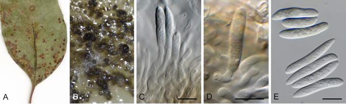


Eucalyptus saligna Australia CPC 35789 Corymbia maculata Australia CBS 143434T of Murramarangomyces corymbiae
Corymbia variegata Australia CPC 36084ET of Microthyrium amygdalinum
Eucalyptus leaf litter South Africa HPC 2269ET of Arnaudiella eucalyptorum
Eucalyptus dunnii Australia CPC 35847T of Phaeothyriolum dunnii
Melanconiella hyperopta NR_160258.1
20
100
100
64100
Phaeothyriolum amygdalinum, comb. nov.
Phaeothyriolum corymbiae, comb. nov.
Phaeothyriolum dunnii, sp. nov. Phaeothyriolum eucalyptorum, comb. nov.


Eucalyptus sp. Australia CPC 17051T of Pseudophloeospora eucalypti
Eucalyptus globulus Australia CPC 32661
Eucalyptus obliqua Australia CPC 32532T of Pp.eucalyptigena
Eucalyptus sp. Australia CPC 25600T of Pseudophloeospora eucalyptorum
Eucalyptus piperita Australia CPC 32132
Banksia vincentia Australia CPC 32164
Eucalyptus sp. Australia CPC 17137T of Pseudophloeospora jollyi
Discosia pseudoartocreas NR_132068.1
10
100
73
93
87
71
Pseudophloeospora jollyi, sp. nov.
Pseudophloeospora eucalyptigena, sp. nov.
Pseudophloeospora eucalyptorum
Pseudophloeospora eucalypti


10
Burgoa verzuoliana NR_145334.1
Mimosa tenuiflora Brazil - URM7756T of Quambalaria fabacearum (MG253664.1)
Vitis vinifera India - 5VVSTL3 (KT200228.1) Human breast implants China - 11PU348 (KF953496.1) Red kojic rice China - QY229 (HM013823.1) Human tissue China - 11PU348 (KU052084.1) Air of citrus plantation Egypt - AUMC 6293 (JQ425376.1) Man skin Finland - CBS 358.73 (MH860700.1) Punica granatum Iran - q10 (KX377510.1) Human skin scraping Malaysia - UM 1095 (KT186106.1) Man Netherlands - IHEM 5621 (KP132549.1) Man skin Netherlands - CBS 357.73T of Sporothrix cyanescens (DQ317622.1) Eucalyptus costata Australia NSW - CPC 34361 Eucalyptus populnea subsp. bimbil Australia - CPC 34353 Pistacia vera Iran? - 28 (KP641151.1)
Wasp Australia QLD - CMB W001 (KT454935.1) Eucalyptus pauciflora Australia NSW - CBS 876.73 (DQ317623.1) Native Corymbia citriodora Australia QLD - BRIP48396 (EF444874.1) Eucalyptus transcontinentalis Australia WA - IMI 298177 (AJ535500.1) Corymbia calophylla Australia WA - WAC12952 (DQ823419.1) Scolytus intricatus on Quercus sp. Bulgaria - MK 1710 (AM261922.2) Fruit Iran - SEM1 (KU663655.1) Persian oak Iran - A12 (KX611006.1) Air in sleeping room of allergic patient Netherlands - CBS 360.73 (MH860701.1) Phleotribus scarabeoides on Olea europaea Syria - MK 808 (AM261921.2) Indoor air Turkey - 2.2.217 (KX674666.1) Hypoborus ficus on Ficus carica Turkey - CF 3526 (DQ119134.1) Eucalyptus gracilis Australia NSW - CPC 34347 Musa sp. Thailand - CPC 19013 Pomegranate Thailand - CPC 19020 Musa sp. Thailand - CPC 19018B Eucalyptus pilularis Australia NSW - CPC 35399 Lophostemon confertus Australia NSW - CPC 33090
Unknown Australia - CPC 28209 Unknown Australia - CPC 28236
Corymbia foelscheana Australia NT - CPC 14464 Corymbia foelscheana Australia NT - CPC 14450
Citrus juice Egypt - AUMC 6294 (JQ425382.1) Staphylea bumalda Korea - CPC 14577 Corymbia sp. Australia NT - CPC 14470 Corymbia dunlopiana Australia NT - CPC 14496 Eucalyptus sp. Australia NT - CPC 14455
Withered grape Italy - UC19 (KU554586.1) Grapevine cane Turkey - CUZFVG39 (MK120284.1)
Eucalyptus camaldulensis Australia NSW - CPC 34337
Corymbia foelscheana Australia NT - CPC 14467
Corymbia ficifolia Australia WA - WAC12953 (DQ823422.1) Betula pendula Russia - CBS 127351 (MH864535.1) Lomatia silaifolia x myricoides hybrid Australia NSW - CPC 14453 Eucalyptus oblonga Australia NSW - CPC 14476
3x
Qua
mba
laria
cya
nesc
ens,
incl
udin
g Q
uam
bala
ria fa
bace
arum
59
76
100
63
58
76

10
Corymbia ficifolia Australia WA - WAC12950 (DQ823429.1) Corymbia calophylla Australia WA - WAC12947T of Quambalaria coyrecup (DQ823431.1) Corymbia polycarpa Australia NT - BRIP48339 (EF444878.1) Corymbia polycarpa Australia NT - BRIP48388 (EF444877.1)
Unknown Australia QLD - CPC 17299 Pueraria phaseolides Laos - CPC 17313 Callistemon sp. Australia QLD - CPC 17231 Eucalyptus sp. Brazil - S8 (AJ535493.1) Eucalyptus nitens South Africa - CBS 118615 (DQ317609.1) Eucalyptus grandis x camaldulensis South Africa - CMW14329 (DQ317614.1) Eucalyptus grandis South Africa - CBS 118844T of Sporothrix eucalypti (DQ317625.1) Eucalyptus grandis South Africa - CBS 119680ET of Quambalaria eucalypti (DQ317626.1) Corymbia torelliana x C. citriodora subsp. variegata Australia QLD - BRIP48367 (EF444823.1) Eucalyptus longirostrata Australia QLD - BRIP48519 (EF444831.1) Eucalyptus dunnii Australia NSW - BRIP48416 (EF444824.1) Eucalyptus longirostrata Australia NSW - BRIP48511 (EF444830.1) Myrceugenia glaucescens Uruguay - UY1718 (EU439923.1) Eucalyptus globulus Uruguay - UY1036 (EU439922.1) Eucalyptus globulus Portugal - PE154 (KR336807.1)
Eucalyptus grandis Australia QLD - BRIP48520 (EF444842.1) Eucalyptus grandis China - CERC 8476 (KY615009.1)
Eucalyptus grandis Australia NSW - BRIP48497 (EF444843.1)
Eucalyptus sp. Australia Tas - CPC 25462 Eucalyptus sp. Australia Tas - CPC 25464T of Quambalaria tasmaniae
Eucalyptus urophylla x E. grandis China - CERC 8505 (KY615036.1) Eucalyptus urophylla x E. grandis China - CERC 8496 (KY615034.1) Eucalyptus tintinnans Australia VIC - CBS 124772T of Quambalaria simpsonii (GQ303290.1) Eucalyptus sp. Thailand - CBS 124773 (GQ303291.1)
Corymbia citriodora subsp. variegata Australia NSW - BRIP48425 (EF444847.1)
Native Corymbia henryi Australia NSW - BRIP48512 (EF444856.1) Corymbia maculata Australia NSW - CMW6707 (DQ317627.1) Corymbia sp. Australia QLD - Q4 (AJ536606.1) Corymbia sp. Australia QLD - Q2 (AJ536604.1) Corymbia citriodora subsp. variegata Australia QLD - CBS 118828 (DQ317628.1) Corymbia henryi Australia QLD - BRIP48317 (EF444854.1)
Corymbia sp. Australia QLD - Q3 (AJ536605.1) Native Corymbia citriodora Australia QLD - BRIP48506 (EF444852.1) Corymbia eximia Australia NSW - DAR 19773T of Quambalaria pitereka (DQ823423.1)
Corymbia citriodora subsp. citriodora Australia QLD - BRIP48343 (EF444857.1)
Corymbia calophylla Australia WA - WAC12958 (DQ823427.1) Corymbia ficifolia Australia WA - WAC12957 (DQ823426.1) Eucalyptus sp. Australia WA - CPC 29478
Corymbia sp. Australia WA - E7353 (AJ536609.1)
Corymbia sp. Australia QLD - Q6 (AJ536608.1) Corymbia torelliana Australia QLD - BRIP48388 (EF444864.1) Corymbia torelliana x C. citriodora subsp. citriodora Australia QLD - BRIP48370 (EF427368.1) Corymbia torelliana x C. citriodora subsp. variegata Australia QLD - BRIP48350 (EF444861.1)
Native Corymbia citriodora subsp. variegata Australia NSW - BRIP48328 (EF444872.1)
Corymbia sp. Australia QLD - Q5 (AJ536607.1) Corymbia torelliana x C. henryi Australia QLD - BRIP48340 (EF444866.1) Corymbia citriodora China - CMW23610 (EF427372.1) Corymbia citriodora China - CERC 8486 (KY615017.1)
Corymbia variegata Australia NSW - CPC 35643
Leptospermum juniperinum Australia - AV17-3 (GQ258351.1) Eucalyptus rugosa Australia Kangaroo Island - CPC 20162T of Quambalaria rugosae
Quambalaria coyrecup
Quambalaria tasmaniae, sp. nov.
Quambalaria eucalypti
Quambalaria pusilla
Quambalaria pitereka
Q. rugosae, sp. nov.
99
54
96
84
100
95
84
91
63
64
79
95
50
65
65
66


Eucalyptus sp. Australia CPC 17677 Eucalyptus badjensis Australia CPC 32601
Eucalyptus sp. Australia CPC 17734 Eucalyptus coccifera Australia CBS 120220
Eucalyptus obliqua Australia CPC 32452
Eucalyptus radiata Australia CPC 18218T
Eucalyptus piperita Australia CPC 32134 Eucalyptus sp. Portugal CPC 31596 Eucalyptus sp. Portugal CBS 112502 Eucalyptus globulus Spain CPC 11251 Eucalyptus globulus Spain CPC 11252
Eucalyptus regans Australia CPC 17710
Eucalyptus costata Australia CPC 34395 Eucalyptus costata Australia CPC 34405T
Cladosporium allicinum CBS 177.71
10
100/100
100/100
100/100
55/-
57/-
59/60
63/99100/100 Neosonderhenia eucalypti, gen. et sp. nov.
Sonderhenia sp.
Sonderhenia eucalyptorum
Sonderhenia eucalypticola
Sonderhenia radiata, sp. nov.


Related Documents