COVID-19 Focal seizures with impaired awareness as long-term neurological complication of COVID-19: a case report Marco Bozzali 1,2 & Alberto Grassini 1 & Giovanni Morana 1 & Michela Zotta 3 & Sara Cabras 1 & Alberto Romagnolo 1 & Carlo Alberto Artusi 1 & Elisa Montalenti 4 & Mario Giorgio Rizzone 1 & Diego Garbossa 1 & Elisa Montanaro 4 & Mara Cercignani 2 & Leonardo Lopiano 1,4 Received: 17 January 2021 /Accepted: 6 April 2021 # Fondazione Società Italiana di Neurologia 2021 Abstract We report here the first case of a young individual otherwise healthy, who presented with frequent focal seizures with impaired awareness as a possible long-term complication of severe acute respiratory syndrome coronavirus-2 infection. Seizures were documented by electroencephalography and responded clinically and neuro-physiologically to antiseizure therapy. The patient underwent an extensive investigation including cerebrospinal fluid examination, conventional and quantitative brain magnetic resonance imaging, and 18-FDG positron emission tomography. Beyond the clinical interest, this case contributes to clarify the possible pathways by which SARS-CoV-2 may enter the central nervous system and cause long-term neurological complications. Keywords COVID-19 . Epilepsy . Focal seizures with impaired awareness . Encephalitis Introduction First-ever occurrence of seizures in non-epileptic individuals have been described in the acute phase of severe acute respi- ratory syndrome coronavirus-2 (SARS-CoV-2) infection [1]. In these cases, possible underlying mechanisms include met- abolic or fever induced brain dysfunction, cytokine-storm damage, brain vessel endothelial infarction, autoimmunity, or central nervous system (CNS) viral invasion [2]. We report here the case of a young individual with no remarkable med- ical history who presented with frequent focal seizures with impaired awareness newly arisen 2 months after resolution of their coronavirus disease 2019 (COVID-19) acute phase. Case report A 54-year-old lady working as a nurse in a COVID-19 ward received, at the end of March 2020, her first-ever SARS-CoV-2 nasopharyngeal swab after reporting a high-risk contact with a colleague suffering from COVID-19. Despite a negative out- come, 5 days later, she started complaining of fever, nasal con- gestion, throat pain, fatigue, shortness of breath, muscle and joint pain, diarrhoea, anosmia, and ageusia. These symptoms persisted for 4 weeks while she remained at home in precaution- ary isolation. At the end of April 2020, she recovered from all symptoms with the exception of fatigue, muscle and joint pain, anosmia, and ageusia. Concomitantly, she started complaining of continuous headaches and feeling of gait unbalance. She was admitted to the emergency ward of the University Hospital of Turin and received full blood and arterial blood oxygenation tests (all parameters in normal range), chest X-rays (normal), and real-time polymerase chain reaction (PCR) on nasopharyn- geal swab (positive for SARS-CoV-2). Due to her clinical sta- bility, she was discharged and remained at home in quarantine until she resulted negative on two consecutive SARS-CoV-2 swab tests. She was serologically tested for antibodies anti- SARS-CoV-2, resulting IgG positive and IgM negative. Clinically, she kept complaining of fatigue, gait unbalance, and headaches. In June 2020, she underwent a brain MRI scan, * Marco Bozzali [email protected] 1 Department of Neuroscience “Rita Levi Montalcini”, University of Torino, Via Cherasco 15, 10126 Turin, Italy 2 Department of Neuroscience, Brighton & Sussex Medical School, University of Sussex, Brighton, East Sussex, UK 3 Department of Diagnostic Imaging, Nuclear Medicine Unit, A.O.U. Città della Salute e della Scienza di Torino, Turin, Italy 4 Neurology 2 Unit, A.O.U. Città della Salute e della Scienza di Torino, 10124 Turin, Italy https://doi.org/10.1007/s10072-021-05233-y / Published online: 16 April 2021 Neurological Sciences (2021) 42:2619–2623

Welcome message from author
This document is posted to help you gain knowledge. Please leave a comment to let me know what you think about it! Share it to your friends and learn new things together.
Transcript
COVID-19
Focal seizures with impaired awareness as long-term neurologicalcomplication of COVID-19: a case report
Marco Bozzali1,2 & Alberto Grassini1 & Giovanni Morana1 & Michela Zotta3 & Sara Cabras1 & Alberto Romagnolo1&
Carlo Alberto Artusi1 & Elisa Montalenti4 & Mario Giorgio Rizzone1& Diego Garbossa1 & Elisa Montanaro4
&
Mara Cercignani2 & Leonardo Lopiano1,4
Received: 17 January 2021 /Accepted: 6 April 2021# Fondazione Società Italiana di Neurologia 2021
AbstractWe report here the first case of a young individual otherwise healthy, who presented with frequent focal seizures with impairedawareness as a possible long-term complication of severe acute respiratory syndrome coronavirus-2 infection. Seizures weredocumented by electroencephalography and responded clinically and neuro-physiologically to antiseizure therapy. The patientunderwent an extensive investigation including cerebrospinal fluid examination, conventional and quantitative brain magneticresonance imaging, and 18-FDG positron emission tomography. Beyond the clinical interest, this case contributes to clarify thepossible pathways by which SARS-CoV-2 may enter the central nervous system and cause long-term neurologicalcomplications.
Keywords COVID-19 . Epilepsy . Focal seizures with impaired awareness . Encephalitis
Introduction
First-ever occurrence of seizures in non-epileptic individualshave been described in the acute phase of severe acute respi-ratory syndrome coronavirus-2 (SARS-CoV-2) infection [1].In these cases, possible underlying mechanisms include met-abolic or fever induced brain dysfunction, cytokine-stormdamage, brain vessel endothelial infarction, autoimmunity,or central nervous system (CNS) viral invasion [2]. We reporthere the case of a young individual with no remarkable med-ical history who presented with frequent focal seizures withimpaired awareness newly arisen 2 months after resolution oftheir coronavirus disease 2019 (COVID-19) acute phase.
Case report
A 54-year-old lady working as a nurse in a COVID-19 wardreceived, at the end of March 2020, her first-ever SARS-CoV-2nasopharyngeal swab after reporting a high-risk contact with acolleague suffering from COVID-19. Despite a negative out-come, 5 days later, she started complaining of fever, nasal con-gestion, throat pain, fatigue, shortness of breath, muscle andjoint pain, diarrhoea, anosmia, and ageusia. These symptomspersisted for 4 weeks while she remained at home in precaution-ary isolation. At the end of April 2020, she recovered from allsymptoms with the exception of fatigue, muscle and joint pain,anosmia, and ageusia. Concomitantly, she started complainingof continuous headaches and feeling of gait unbalance. She wasadmitted to the emergency ward of the University Hospital ofTurin and received full blood and arterial blood oxygenationtests (all parameters in normal range), chest X-rays (normal),and real-time polymerase chain reaction (PCR) on nasopharyn-geal swab (positive for SARS-CoV-2). Due to her clinical sta-bility, she was discharged and remained at home in quarantineuntil she resulted negative on two consecutive SARS-CoV-2swab tests. She was serologically tested for antibodies anti-SARS-CoV-2, resulting IgG positive and IgM negative.Clinically, she kept complaining of fatigue, gait unbalance,and headaches. In June 2020, she underwent a brain MRI scan,
* Marco [email protected]
1 Department of Neuroscience “Rita Levi Montalcini”, University ofTorino, Via Cherasco 15, 10126 Turin, Italy
2 Department of Neuroscience, Brighton & Sussex Medical School,University of Sussex, Brighton, East Sussex, UK
3 Department of Diagnostic Imaging, Nuclear Medicine Unit, A.O.U.Città della Salute e della Scienza di Torino, Turin, Italy
4 Neurology 2 Unit, A.O.U. Città della Salute e della Scienza diTorino, 10124 Turin, Italy
https://doi.org/10.1007/s10072-021-05233-y
/ Published online: 16 April 2021
Neurological Sciences (2021) 42:2619–2623

showing minimal non-specific white matter hyperintensities onT2-weighted images. In July 2020, she started complaining of
frequent (6–8 times a day) short episodes (about 1-min durationeach) of olfactory hallucinations (described as “burning rubber
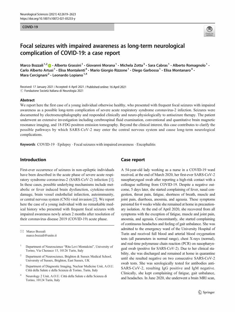
Fig. 1 The first EEG, recorded before therapy initiation, showed a patternof focal slow activity and spikes in the fronto-temporal area bilaterally(abnormalities magnified in red squares) (a). The second EEG, performed4 weeks after initiation of antiseizure medication, did no longer reveal anypathological alteration (b). EEG recordings were performed with scalpelectrodes placed according to the international 10–20 system withbipolar montage; brain MRI at the level of the centrum semiovale
demonstrated only few hyperintense foci on FLAIR (c), mildlyhypointense on T1-weighted images (d) with no contrast enhancement(CE) on corresponding CE T1-weighted images (e). These abnormalitieskeep with minimal non-specific changes (arrow, c–e). Co-registeredFLAIR (f, g), 18F-FDG PET (h, l), and fused PET/MRI FLAIR images(i) did not show any temporal lobe abnormalities, with physiologicaltracer uptake according to patient’s age
2620 Neurol Sci (2021) 42:2619–2623

smell”) followed by 10–15-min intervals of detachment fromreality (described as “mental confusion”). In mid-September2020, she was admitted to our Neurology Unit (Department ofNeuroscience, University of Turin, Italy) for investigation. Onexamination, there were no obvious impairments in her higher-level functions; cranial nerves were intact; there was no weak-ness at any limb; muscular tone was normal; reflexes were sym-metrical and plantars downgoing bilaterally; coordination testswere well performed; there were no sensory deficits; her gaitwas normal. A mild right deviation was observed on theUnterberger test. She underwent an electroencephalogram(EEG) demonstrating the presence of focal slow waves along-side sharp spikes in the fronto-temporal areas bilaterally (Fig.1a). Repetition of SARS-CoV-2 nasopharyngeal swab was neg-ative, while serum-specific SARS-CoV-2 IgG remained highlypositive (140 U/ml). Blood tests, including vitamin B12, folates,and thyroid function tests, were normal. Serological analysis toexclude autoimmune diseases and systemic infection resultednegative. A new brain MRI scan at 3T, including high-resolution conventional acquisitions (FLAIR, T1-, T2-weighted,susceptibility-weighted imaging, and post-contrast T1-weightedimages), confirmed prior non-specific white matter changes (noadditional abnormalities) (Fig. 1c–g). CSF examination revealeda slight increase of proteins (55mg/dl) with normal cell count.CSF PCR for detection of common neurotropic pathogens(i.e. Herpes simplex 1 and 2, Human herpes 6, Enterovirus,human parechovirus, Varicella zoster, Cytomegalovirus,
Streptococcus pneumoniae, Neisseria meningitidis,Haemophilus influenzae, Streptococcus agalactiae, Listeriamonocytogenes, Escherichia coli K1, Cryptococcusneoformans) and for SARS-CoV-2 returned negative results.Immunoelectrofocusing did not reveal any oligoclonal bands.No specific antibodies anti-SARS-CoV-2 neither brain autoim-mune (anti-NMDA glutamate receptor, AMPA 1 and 2glutamate receptor, GABA receptor, LG1, CASPR2, DPPX,thyroperoxidase, thyroglobulin, TSH receptor) norparaneoplastic antibodies (anti-YO, Sox1, Zic4, Titin, Hu,GAD65, CV2, Ri, Ma2, Retin, Amphiphysin, Tr) were identi-fied on the CSF. Considering the feeling of gait unbalance anddeviation on the Unterberger test, she underwent also brainstemauditory evoked potentials (BAEP) showing a mild bilateralincrease of latencies. An extensive neuropsychological assess-ment revealed an isolated impairment of frontal-executive func-tions (frontal assessment battery corrected score=12.3). For thisreason, the patient underwent a brain [18F]FDG positron emis-sion tomography that resulted normal (Fig. 1h–l). She wasstarted on levetiracetam therapy with only partial benefit andthen shifted to carbamazepine, 400 mg twice a day. After beingon carbamazepine for 12 weeks, the patient reported clear im-provement of symptoms with “burning sensation” [3] in hernose (instead of smell hallucinations) occurring with reducedfrequency (2 times aweek) alongside shorter intervals of detach-ment from reality.Moreover, EEG repetition did no longer showany spikes and revealed reduction of slow waves (Fig. 1b).
Fig. 2 The picture summarizes the clinical evolution of SARS-CoV-2 infection in the presented case alongside findings from the most relevantlaboratory and instrumental examinations. See text for further details
2621Neurol Sci (2021) 42:2619–2623

Discussion
Since COVID-19 became clinically overt, the patient present-ed with a clinical progression that fits with a subacute enceph-alitis (Fig. 2). Despite lack of evidence of SARS-CoV-2RNAs in the CSF, the clinical evolution indicates a plausibleanatomical pathway by which SARS-CoV-2 may have sub-acutely infected the central nervous system (CNS). Being pri-marily a respiratory virus, there are at least two possible waysfor SARS-CoV-2 to invade the CNS, through anhaematogenous or an axonal route [4]. In the latter case,SARS-CoV-2 is supposed to infect the axonal terminationsand actively translocate backwards into the CNS. The neuro-nal fibres that are most likely infected by SARS-CoV-2 arethose of the olfactory nerves, as suggested by anosmia astypical symptom of COVID-19 and supported by radiologicalevidence [5]. Another possible route is trough other cranialnerves with invasion of the brainstem, as partially supportedby our clinical (Unterberger test) and neurophysiological(BAEP) observations. Evidence of paracellular transmigrationof betacoronaviruses (e.g. SARS-CoV-1) has been providedin animal studies, with viral demonstration within the olfacto-ry bulb, piriform cortex, hippocampus and temporo-mesialcortex, thalamus, and brainstem nuclei [6, 7]. Importantly,persistence of coronavirus RNAs in the CNS was demonstrat-ed long time after acute encephalitis [8]. In our patient, wewere unable to detect viral RNAs in the CSF. This might bedue to the long interval elapsed between the acute COVID-19phase and CSF examination. Consistently, it was previouslyshown that SARS-CoV-2 RNAs are detectable in the CSF of asmall percentage of COVID-19 patients with neurologicalsymptoms [9], often requiring repeated CSF examination[10]. Nonetheless, our patient developed focal seizures withimpaired awareness (documented by EEG) responding to an-tiseizure medication. In contrast to previous reports in acutecases [10, 11], we could not demonstrate abnormalities in thetemporo-mesial cortex, which—we argue—might no longerbe detectable in post-acute COVID-19 stages. On neuropsy-chological assessment, our patient showed an isolated deficitof executive functions, whose relationship with SARS-CoV-2infection remains to be clarified despite a suggestive anatom-ical overlap with fronto-temporal seizures.
In conclusion, we report here a case of fronto-temporalepilepsy, possibly related to SARS-CoV-2 infection. This di-agnosis of causality, which remains speculative, is based onexclusion assessments and clinical elements supporting such ahypothesis (clinical evolution, EEG alterations; response toantiseizure medication).
We expect this sort of long-term complications requiringimmediate identification and treatment to become increasing-ly frequent in the future.
Author contribution M.B., A.G., and L.P. planned all clinical and instru-mental investigations, interpreted the results, and wrote the first draft ofthe manuscript. G.M. and M.C. supervised MRI acquisition and contrib-uted to data interpretation. M.Z. supervised PET imaging and contributedto data interpretation. A.R., M.G.R., C.A.A., D.G., and S.C. performedclinical assessments and managed all serological and CSF examinations.E. Montal. contributed to interpretation of neurophysiological data. E.Montan. did neuropsychological testing. All Authors were involved indrafting or revising the manuscript.
Data availability MB and AG have full access to all data presented in thestudy and take responsibility for their integrity and for the accuracy ofdata analysis.
Declarations
Ethical approval All procedures were performed in accordance with thelocal ethics committee and with the ethical standards laid down in the1964 Declaration of Helsinki and its later amendments.
Conflict of interest The authors declare no competing interests.
Informed consent The patient provided informed written consent.
References
1. Mao L, Wang M, Chen S et al (2020) Neurological manifestationsof hospitalized patients with COVID-19 in Wuhan, China: a retro-spective case series study. JAMA Neurol. https://doi.org/10.1001/jamaneurol.2020.1127
2. Asadi-Pooya AA (2020) Seizures associated with coronavirus in-fections. J Seizure. https://doi.org/10.1016/j.seizure.2020.05.005
3. Heo K, KimKM, Han SM, Cho KH, ChuMK (2020) Nasal pain asan aura: Amygdala origin? Seizure 2020:13–16. https://doi.org/10.1016/j.seizure.2020.09.028
4. Mishra R, Banerjea A (2020) Neurological damage bycoronaviruses. Front Immunol. https://doi.org/10.3389/fimmu.2020.565521
5. LaurendonTRT, Mugnier J et al (2020) Bilateral transient olfactorybulb edema during COVID-19-related anosmia. Neurology. 95:224–225. https://doi.org/10.1212/wnl.0000000000009850
6. Pearlman S, Jacobsen G, Afifi A (1989) Spread of a neurotropicmurine coronavirus into the CNS via the trigeminal and olfactorynerves. Virology. https://doi.org/10.1016/0042-6822(89)90446-7
7. McCray PB, Pewe L, Wohlford-Lenane C et al (2007) Lethal in-fection of K18-hACE2 mice infected with severe acute respiratorysyndrome coronavirus. J Virol 81:813–821. https://doi.org/10.1128/jvi.02012-06
8. Jacomy H, Fragoso G, Almazan G, Mushynski WE, Talbot PJ(2006) Human coronavirus OC43 infection induces chronic en-cephalitis leading to disabilities in BALB/C mice. Virology. 349:335–346. https://doi.org/10.1016/j.virol.2006.01.049
9. Neumann B, Schmidbauer ML, Dimitriadis K et al (2020)Cerebrospinal fluid findings in COVID-19 patients with neurolog-ical symptoms. J Neurol Sci. https://doi.org/10.1016/j.jns.2020.117090
10. Virhammar J, Kumlien E, Fällmar D, Frithiof R, Jackmann S, SköldMK, Kadir M, Frick J, Lindeberg J, Olivero-Reinius H, RyttleforsM, Cunningham JL, Wikström J, Grabowska A, Bondeson K,Bergquist J, Zetterberg H, Rostami E (2020) Acute necrotizing
2622 Neurol Sci (2021) 42:2619–2623

encephalopathy with SARS-CoV-2 RNA confirmed in cerebrospi-nal fluid. Neurology. 95:445–449. https://doi.org/10.1212/WNL.0000000000010250
11. Moriguchi T, Harii N, Goto J, Harada D, Sugawara H, Takamino J,UenoM, Sakata H, Kondo K,Myose N, Nakao A, TakedaM, HaroH, Inoue O, Suzuki-Inoue K, Kubokawa K, Ogihara S, Sasaki T,Kinouchi H, Kojin H, Ito M, Onishi H, Shimizu T, Sasaki Y,Enomoto N, Ishihara H, Furuya S, Yamamoto T, Shimada S
(2020) A first case of meningitis/encephalitis associated withSARS-Coronavirus-2. Int J Infect Dis. 94:55–58. https://doi.org/10.1016/j.ijid.2020.03.062
Publisher’s note Springer Nature remains neutral with regard to jurisdic-tional claims in published maps and institutional affiliations.
2623Neurol Sci (2021) 42:2619–2623
Related Documents