fMRI Evidence for Separable and Lateralized Prefrontal Memory Monitoring Processes (Article begins on next page) The Harvard community has made this article openly available. Please share how this access benefits you. Your story matters. Citation Dobbins, Ian G., Jon S. Simons, and Daniel L. Schacter. 2004. fMRI evidence for separable and lateralized prefrontal memory monitoring processes. Journal of Cognitive Neuroscience 16(6): 908-920. Published Version doi:10.1162/0898929041502751 Accessed May 1, 2016 1:34:30 PM EDT Citable Link http://nrs.harvard.edu/urn-3:HUL.InstRepos:3627116 Terms of Use This article was downloaded from Harvard University's DASH repository, and is made available under the terms and conditions applicable to Other Posted Material, as set forth at http://nrs.harvard.edu/urn-3:HUL.InstRepos:dash.current.terms-of- use#LAA

Welcome message from author
This document is posted to help you gain knowledge. Please leave a comment to let me know what you think about it! Share it to your friends and learn new things together.
Transcript

fMRI Evidence for Separable and Lateralized Prefrontal MemoryMonitoring Processes
(Article begins on next page)
The Harvard community has made this article openly available.Please share how this access benefits you. Your story matters.
Citation Dobbins, Ian G., Jon S. Simons, and Daniel L. Schacter. 2004.fMRI evidence for separable and lateralized prefrontal memorymonitoring processes. Journal of Cognitive Neuroscience 16(6):908-920.
Published Version doi:10.1162/0898929041502751
Accessed May 1, 2016 1:34:30 PM EDT
Citable Link http://nrs.harvard.edu/urn-3:HUL.InstRepos:3627116
Terms of Use This article was downloaded from Harvard University's DASHrepository, and is made available under the terms and conditionsapplicable to Other Posted Material, as set forth athttp://nrs.harvard.edu/urn-3:HUL.InstRepos:dash.current.terms-of-use#LAA

fMRI Evidence for Separable and Lateralized PrefrontalMemory Monitoring Processes
Ian G. Dobbins1, Jon S. Simons2, and Daniel L. Schacter3
Abstract
& Source memory research suggests that attempting toremember specific contextual aspects surrounding priorstimulus encounters results in greater left prefrontal cortex(PFC) activity than simple item-based old/new recognitionjudgments. Here, we tested a complementary hypothesis thatpredicts increases in the right PFC with tasks requiring closemonitoring of item familiarity. More specifically, we compareda judgment of frequency ( JOF) task to an item memory task, inwhich the former required estimating the number of previouspicture encounters and the latter required discriminating oldfrom new exemplars of previously seen items. In comparison
to standard old/new recognition, both source memory and theJOF task examined here require more precise mnemonicjudgments. However, in contrast to source memory, cognitivemodels suggest the JOF task relies heavily upon item familiar-ity, not specific contextual recollections. Event-related fMRIdemonstrated greater recruitment of right, not left, dorso-lateral and frontopolar PFC regions during the JOF comparedto item memory task. These data suggest a role for right PFC inthe close monitoring of the familiarity of objects, whichbecomes critical when contextual recollection is ineffective insatisfying a memory demand. &
INTRODUCTION
Memory demands vary considerably in the amount ofcontextual information required for success. At oneend of the spectrum, many laboratory tasks of recogni-tion can be accomplished via judgments of item familiar-ity (or novelty). During these tasks, subjects encounterwords or objects in a ‘‘study’’ list and then must laterdiscriminate between these and items that were notpreviously presented. Signal detection and computation-al models suggest that these tasks can potentially beaccomplished by simply relying upon a continuous itemstrength or familiarity signal, which builds monotonicallywith item exposures, without resorting to recoveringspecific contextual aspects regarding the previous en-counter with each item (Hintzman, Nozawa, & Irmscher,1982; Atkinson & Juola, 1974; Banks, 1970). In contrast,for some tasks, recovery of detailed contextual informa-tion is necessary for success. For example, in typicalsource memory tasks, items are shown in one of anumber of encoding contexts (e.g., male or femalevoicing, different locations, different orientating tasks,etc.) and subjects are represented the items at test andmust determine from which specific source each origi-nated. Because the items have been exposed to the samedegree, closely attending to differences in familiarity isunhelpful. Instead, subjects must recover or recollect
different contextual aspects of the previous encountersin sufficient detail to make a source attribution. Thus, therelative utility of recollection and familiarity processes isheavily influenced by the task design, with some designsrequiring more extreme reliance on one or the otherprocess.
Recent comparisons of source and item memory tasksusing functional imaging have led to a consistent patternof prefrontal cortex (PFC) response. In general, thesestudies have found increases in the left PFC when sourcememory is compared to item memory (Dobbins, Foley,Schacter, & Wagner, 2002; Raye, Johnson, Mitchell,Nolde, & D’Esposito, 2000; Rugg, Fletcher, Chua, &Dolan, 1999; Nolde, Johnson, & D’Esposito, 1998). Thistypically left-lateralized increase has been observed forpictures as well as words and can also be seen whenother contextually demanding retrieval tasks, such asitem cued recall, are compared with simple itemmemory(Cabeza, Locantore, & Anderson, 2003). Advocates of thesource monitoring framework have interpreted thesefindings as indicative of the need for more ‘‘reflective’’or ‘‘systematic’’ processing for source compared withitem memory, involving more subprocesses or moremonitoring of more specific information (Raye et al.,2000; Nolde et al., 1998; Johnson, Hashtroudi, & Lindsay,1993). Similarly, in a recent study by Ranganath, Johnson,and D’Esposito (2000), the authors interpreted increasedleft anterior PFC activity in terms of the demand for morecontextually or perceptually specific information duringretrieval attempt for source versus general item memorytasks. In general, these accounts lead to the prediction
1Duke University, 2University College London, 3HarvardUniversity
D 2004 Massachusetts Institute of Technology Journal of Cognitive Neuroscience 16:6, pp. 908–920

that increases in the specificity of contextual informationrequired during retrieval will also lead to increasedactivity in the left PFC. Additionally, the source monitor-ing framework has been used to suggest that regions inthe right PFC may instead support more ‘‘heuristic’’memory processes that have been characterized as rela-tively simple, quick processes sufficient for less reflec-tively demanding tasks such as memory judgments basedon familiarity (Nolde et al., 1998).
An alternate conceptualization of PFC responses dur-ing recognition has been developed to account forincreases in the right PFC during recognition. For exam-ple, Henson and colleagues used the remember/knowtechnique (Tulving, 1985) in an attempt to separatememory endorsements based upon contextual recollec-tion (remembered) from those merely relying upon asense of item familiarity (known) (Henson, Rugg, Shal-lice, Josephs, & Dolan, 1999). When successful ‘‘know’’reports were compared to successful ‘‘remember’’ re-ports, greater activity was observed in the right dorsolat-eral PFC (see also Eldridge, Knowlton, Furmanski,Bookheimer, & Engel, 2000). In addition, low confi-dence recognition reports, which are typically assumedto rely on familiarity in isolation, also lead to increasedactivity in the right dorsolateral PFC (Henson, Rugg,Shallice, & Dolan, 2000). These observations led to thesuggestion that the right dorsolateral PFC may be in-volved in the ‘‘retrieval monitoring’’ of items that areclose to the response criterion. A somewhat related ideais that the right PFC is critical for the ‘‘postretrievalmonitoring’’ of mnemonic content (Rugg & Wilding,2000; Rugg, Otten, & Henson, 2002; Rugg, Henson, &Robb, 2003; Schacter, Buckner, Koutstaal, Dale, & Rosen,1997). From this perspective, the right dorsolateral PFCis particularly involved in the monitoring of recoveredmemory content in order to verify whether it satisfies acurrent memory demand. Such postretrieval monitoringrequirements may be elevated when the recovered in-formation is of an impoverished nature such as duringlow-confidence reporting, when relying upon memoriesdevoid of much contextual content (i.e., ‘‘know’’ re-sponses), or under situations requiring the close scrutinyof potentially diagnostic information regarding priorsources (Rugg et al., 2002, 2003).
Overall, the ‘‘systematic/heuristic’’ and postretrievalmonitoring characterizations potentially conflict withrespect to explaining the laterality of PFC responseduring episodic retrieval. Whereas the systematic/heuris-tic characterization suggests that contextually demand-ing retrieval tasks will preferentially recruit the left PFC,these same tasks are presumed heavily dependent onthe right dorsolateral PFC under the postretrieval mon-itoring framework. Here, similar to the systematic/heu-ristic view, we suggest that the role of the right PFC maybe tied to monitoring a specific type of memory signal,namely, that based on the individual familiarity of theitem itself. This hypothesis would then explain why
activity is observed under conditions where memoryendorsements are made in the absence of recoveredcontextual information such as ‘‘know’’ and low confi-dence reports. Under these conditions, subjects wouldhave to closely attend to or monitor the familiaritysignal in order to make the memory decision. There islimited additional support for the role of the right PFC infamiliarity monitoring from neuropsychological researchon patient B.G. Patient B.G., who suffered stroke relateddamage to the posterior dorsal region of the right PFC,shows an aberrant response pattern in simple item-recognition paradigms, generating large numbers ofincorrect endorsements for novel lure items (falsealarms) (Curran, Schacter, Norman, & Galluccio, 1997;Schacter, Curran, Galluccio, & Milberg, 1996). Such apattern is consistent with the idea that this patient hasdifficulty effectively monitoring familiarity such that thelow familiarity of lures does not lead to rapid rejection.Additionally, repetitive transcranial magnetic stimulation(TMS) over the right dorsolateral PFC during picturerecognition has been shown to elevate false alarm ratesto new items (Rossi et al., 2001). Finally, Dobbins et al.(unpublished data) observed greater activity in the rightdorsolateral and frontopolar PFC in a comparison ofnovelty detection (which of three objects was new)relative to conceptual source memory (rememberingthe particular semantic task associated one of threeobjects) across identically constructed picture sets. Thus,there seems to be growing support for the notion thatright prefrontal regions may be recruited when subjectsclosely monitor the item novelty/familiarity dimensionduring retrieval tasks.
We directly tested the familiarity-monitoring hypothe-sis using a demanding retrieval task that requires a moreprecise judgment regarding previous exposure thanstandard old/new recognition, namely, judgment of fre-quency (JOF). We compared the JOF task with an itemmemory task in which the subjects simply decidedwhether the test probe was the same or a differentexemplar of the item studied. During the JOF task,subjects encounter particular items different numbersof times and must subsequently estimate the frequencyof prior encounter for each of the items during a latermemory test. It is important to note that although theJOF task makes reference to more precise episodicinformation than standard old/new recognition, thereare psychological and computational reasons to believethat subjects typically accomplish this task via reliance onitem familiarity without the retrieval of specific contex-tual information (Hintzman & Curran, 1994; Hintzman,1984; Hintzman et al., 1982). That is, although bothsource and JOF tasks are more demanding than old/new recognition, we assume that each requires theincreased monitoring of fundamentally different typesof mnemonic content. Psychologically, the familiarity-based characterization of the JOF task makes sense, giventhe stereotypy of each encounter. That is, there may be
Dobbins, Simons, and Schacter 909
little in the way of specific contextual content that couldbe recollected and would be useful for discriminating anitem seen, for example, three times, from one seen six(see also Wagner, Maril, & Schacter, 2000). Furthermore,the deliberate encoding of individual item frequenciesmay be virtually impossible given the constraints ofworking memory. For example, consider an experimentwhere items are seen two, four, or six times beforetesting. Typically subjects are told to ignore these repe-titions and perform some type of orienting task. Howev-er, it is important to consider whether they couldreasonably employ any strategy to explicitly encode thefrequency of separate items. Even a cursory task analysisindicates that the working memory load of such a taskfar exceeds the assumed capabilities of subjects. Forexample, subjects would have to not only track therunning frequency tally of each individual item, theywould also have to somehow actively note when an item‘‘failed’’ to recur beyond a particular frequency. That is,remembering seeing an item at a lower frequency, saytwice, is nondiagnostic because all items appearedtwice and because this recollection does not indicatethat the item did not also appear in later exposures.Instead, what would be necessary is remembering thatan item occurred twice and that it did not occur at agreater frequency. Such explicit encoding of nonoccur-rence for an earlier seen item is unlikely to occur whileengaged in the active encoding of different items; This,combined with the likelihood that maintaining a runningfrequency tally during encoding vastly exceeds workingmemory capacity, makes it likely that explicit encoding offrequency rarely occurs. This assumption is also consis-tent with work showing that levels of processing andattentional manipulations that increase or impede ex-plicit recollection have little impact in the encoding offrequency information, which appears largely automatic(Hasher, Zacks, Rose, & Sanft, 1987; Hasher & Zacks,1984; Zacks, Hasher, & Sanft, 1982; Hasher & Chromiak,1977). Thus, the JOF task potentially serves as thecomplement to standard source memory tasks. In sourcememory tasks the familiarity of the items is assumed tobe similar, but the encoding context of each differs insome critical fashion. In contrast, during JOF tasks, theencoding context is typically held relatively fixed but theitem familiarities systematically differ.
Overall, both the JOF and item memory (same ordifferent?) conditions are expected to involve an item-based feature judgment, namely, determining if the testpicture is the same or a different exemplar than what wasshown before. Additionally, however, from a familiarity-monitoring viewpoint, the JOF task requires monitoringof the familiarity signal, over and above any such moni-toring that may occur in the item memory task, in orderto estimate the prior presentation frequency. That is,under the item memory task, any monitoring can stoponce an item is determined to be old or new, furtherinspection of its familiarity in order to estimate frequency
is unnecessary. Given these considerations, we predictedthat the JOF task would yield increased activity in theright PFC compared to the item memory task.
RESULTS
Behavioral
Table 1 shows the accuracy data across the conditionsusing the signal detection measure d0. In general, sub-jects were less accurate on the frequency estimationthan the item memory tasks, t(15) = 10.47, p < .001.Furthermore, within the item memory task, increasingfrequency improved accuracy, t(15) = 6.88, p < .001,whereas within the JOF task, changing to a differentexemplar impaired performance, t(15) = 6.14, p < .001.
Table 2 shows the median reaction time data for thesubjects for correct responses. Overall, subjects wereslower on correct JOF decisions than on item memorydecisions, t(15) = 12.75, p < .001. Separate ItemFrequency � Picture Format ANOVAs were conductedseparately for each retrieval task. Within the item mem-ory task, there was a main effect of frequency, withquicker responding to more frequently seen items,F(1,15) = 18.70, p < .001, and an interaction betweenitem format and frequency. This interaction occurredbecause the correct identification of different pictureswas not altered by exposure frequency, whereas thecorrect identification of the same pictures was signifi-cantly faster with the higher exposure frequency,F(1,15) = 9.06, p < .01. Within the JOF task, therewas a main effect of item format with quicker JOFs toitems that were the same versus those that were differ-ent, F(1,15) = 112.43, p < .001, and a main effect offrequency with quicker responses to the high frequency
Table 1. Behavioral Accuracy
Accuracy
Twice Six Overall
IM(‘‘different’’)
Different .87 .88 .88
Same .38 .16 .27
d0 1.56 2.28 1.84
Same Different OverallJOF(‘‘six times’’)
Six .79 .63 .71
Two .32 .35 .34
d0 1.35 .77 1.02
IM = item memory; JOF = judgment of frequency.Note. d0 values based on individual subjects not aggregate proportions.In order to calculate accuracy, different exemplar items were arbitrarilyclassified as targets during item memory, and the items seen six timeswere arbitrarily classified as targets for JOFs.
910 Journal of Cognitive Neuroscience Volume 16, Number 6

items, F(1,15) = 30.34, p < .001. There was also aninteraction between item format and frequency, whichresulted because the slowing that occurred with differ-ent pictures was more severe for high frequency thanlow frequency items, F(1,15) = 14.16, p < .01.
fMRI
JOF Relative Increases
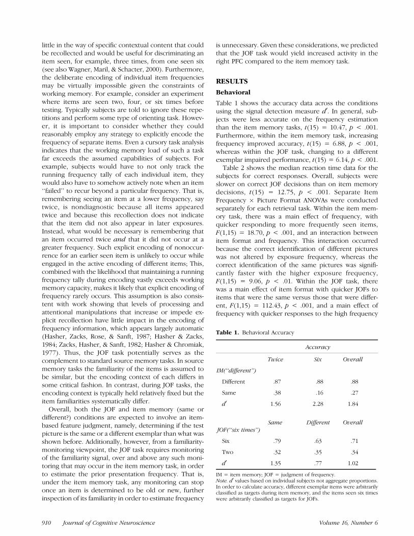
Bilateral prefrontal (dorsolateral and anterior), inferiorparietal, and midline regions demonstrated greater signalduring correct responses on the JOF task compared tothe itemmemory task, collapsed across potential changesin item format (same or different) (Figure 1, Table 3).However, inspection of the extracted time courses forROIs in the left and right PFC regions suggested that thehemispheres potentially differed in the nature of JOF taskresponse as a function of item format (same or different).More specifically, if one restricts attention to the taskresponse for items that remained the same across en-counters, the main evidence for an increased activity inJOF task appears predominantly in the right PFC; thehemodynamic responses to the retrieval tasks in the lefthemisphere look quite similar to one another (solid lines,Figure 1). In contrast, when attention is restricted toitems that changed between study and test, there appearsto be a bilaterally increased PFC response for the JOFcompared to item memory tasks.
To confirm this impression statistically, we analyzedthe peak response (6 sec post stimulus onset) from
Table 2. Behavioral Reaction Times
Reaction Time (msec)
Twice Six Overall
IM
Different 1755 1721
Same 1794 1595 1716
Same Different
JOF
six 1698 2054
two 2094 2245 2022
IM = item memory; JOF = judgment of frequency.Reaction times are for median correct responding only.
Figure 1. Cortical regions
demonstrating greater activity
for the JOF task in comparisonto the item memory (IM) task
(collapsed across differences in
item format). Activity maps are
thresholded at .001 5-voxelsone-tailed, overlaid on a
canonical high-resolution
structural image in MNI space(MRICRO software, www.
psychology.nottingham. ac.uk/
staff/cr1/mricro.html). Upper
insets show reconstructedhemodynamic responses across
conditions of interest for the
left PFC whereas lower insets
plot the same responses for theright PFC. y-axis is percent
signal change, x-axis is
poststimulus onset time in 2-secincrements out to 12 sec. Solid
lines indicate task performance
for items that remained same
between study and test; brokenlines indicate performance for
items that were different
exemplars than those studied.
Red lines indicate JOFresponse, blue lines IM
response. The green line shows
IM response at a lower level ofbehavioral performance
comparable to JOF
performance (IM for items seen
twice collapsed across same/different condition).
Dobbins, Simons, and Schacter 911

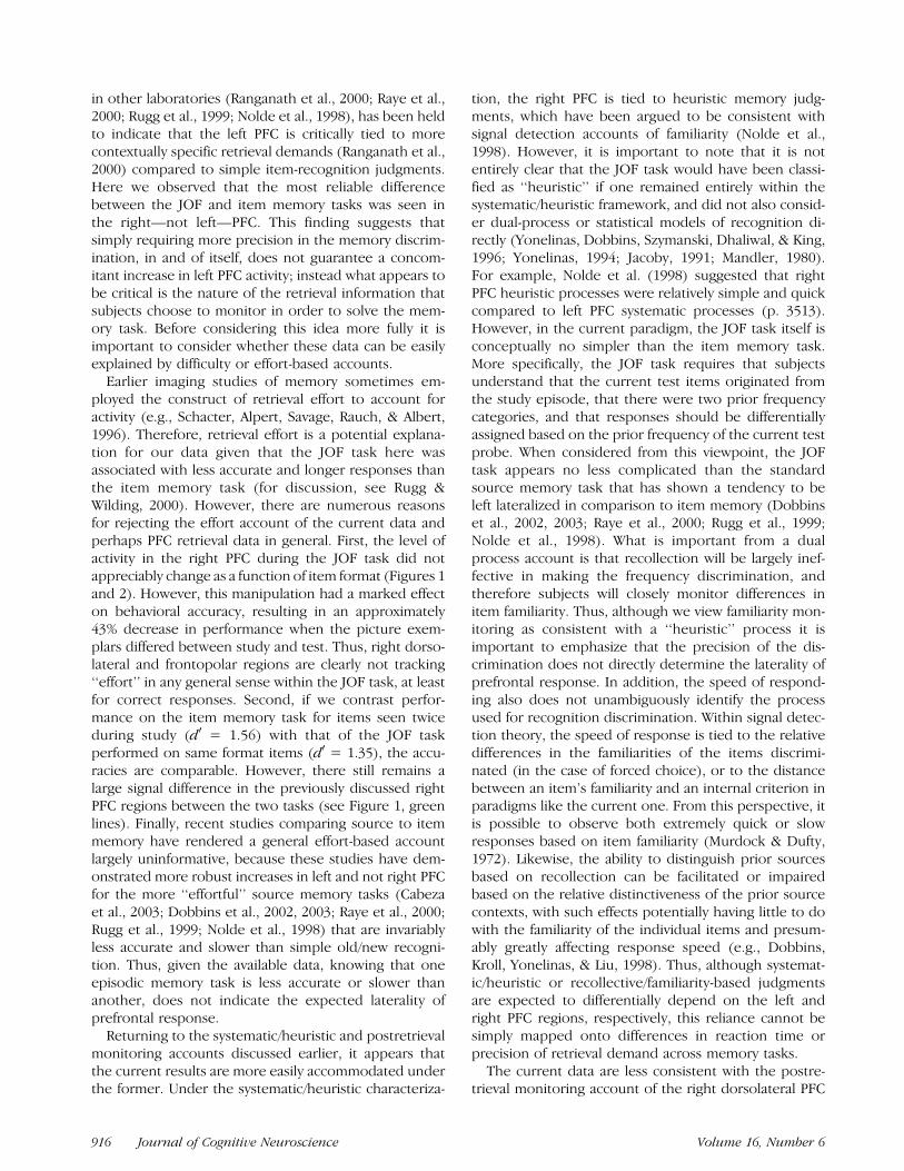
regions of interest (ROIs) in the left and right dorsolat-eral and frontopolar areas. These ROIs were drawn fromthe dorsolateral and frontopolar regions implicated inthe JOF greater than item memory contrast. Each ROI(left and right) contained significant voxels within three8-mm-diameter spheres centered on the three SPM-identified maxima in each hemisphere. The left-hemi-sphere ROI contained 130 voxels and the right contained138 voxels (Figure 2). These peak responses were thenentered into a Hemisphere (Left or Right) � RetrievalTask (Item Memory or JOF) � Item Format (Same orDifferent) three-way ANOVA (Figure 2). Because theseregions were selected without respect to item format,they are unbiased with respect to any potential ItemFormat � Hemisphere interactions.
Main Effects
The results yielded main effects of hemisphere, left >right; F(1,15) = 14.50, MSE = .051, p< .01, and retrievaltask, F(1,15) = 135.82, MSE = .005, p < .001. Addition-ally, the main effect of item format, different > same;F(1,15) = 4.41, MSE = .010, p = .053, approachedsignificance. The main effect of hemisphere must beinterpreted cautiously because it assumes the responsesof the two hemispheres are scaled similarly (i.e., ratioscaling with same range). However, given this assump-tion, the data suggest that on average the left hemi-sphere was engaged to a greater extent than the righthemisphere (.28 vs .13). The main effect of retrieval task
simply confirms that these ROIs were drawn from theoriginal frequency > item memory contrast. Finally, themain effect of item format demonstrates slightly greateractivity for exemplars that are different versus those thatremain the same across study and test (.23 vs .19).
Two-Way Interactions
There were significant Hemisphere � Item Format,F(1,15) = 25.04, MSE = .002, p < .001, and RetrievalTask� Item Format, F(1,15) = 7.28,MSE= .009, p< .05,interactions. Post hoc Tukey’s HSD comparisons indicat-ed that the Hemisphere � Item Format interactionoccurred because there was greater activity for dif-ferent versus same items in the left hemisphere (.33 vs..24, p < .001) but no item format differences in the right(.13 vs. .14, ns). Post hocs for the Retrieval Task � ItemFormat interaction indicated that it occurred becauseitem format did not affect the activity level during itemmemory judgments (.14 vs .13; same vs. different),whereas the different items yielded more signal thansame items during the JOF task (.32 vs. .24, p < .05).
Three-Way Interaction
There was a Hemisphere � Retrieval Task � ItemFormat interaction, F(1,15) = 5.86, MSE = .001, p <.05 (Figure 2). Post hoc interaction analyses were con-ducted separately for the left and right hemispheres. Inthe left hemisphere, there was a main effect of retrieval
Figure 2. Data for Hemisphere
(left or right) � Retrieval Task
(JOF or IM) � Item Format
(same or different) interactionobserved in anterior PFC
regions. Image overlays
demonstrate the left and right
regions that constituted eachROI. Box plots show the peak
response (6 sec post stimulus
onset) averaged across eachsubject for the conditions of
interest. Box is one standard
error of the mean and box plus
whiskers is two standard errors.Shaded boxes show task-related
response when the item format
remained the same across study
and test; open boxes show theresponse when the items were
different exemplars.
912 Journal of Cognitive Neuroscience Volume 16, Number 6

task, F(1,15) = 126.32, MSE = .002, p < .001, a maineffect of item format, F(1,15) = 14.13, MSE = .008,p < .01; different > same, and a significant RetrievalTask � Item Format Interaction, F(1,15) = 8.90, MSE =.006, p < .01. Follow-up Tukey’s HSD comparisonsdemonstrated that the interaction occurred becausethere was a greater signal for the JOF in comparison tothe item memory task for items that differed betweenstudy and test (p < .001); however, the retrieval tasksdid not significantly differ when items remained thesame between study and test. In the right hemisphere,there was the expected main effect of retrieval task,F(1,15) = 80.10, MSE = .005, p < .001, but no maineffect of item format, F < 1 or Retrieval Task � ItemFormat interaction, F(1,15) = 3.88, MSE = .004, p > .06.Because the interaction approached significance, posthoc Tukey’s comparisons were conducted to confirmthat the pattern was different from the left hemisphere.These demonstrated that unlike the left, there was asignificant increase for the JOF in comparison to theitem memory task, regardless of item format (ps < .001).
Overall, this pattern of results suggests that the rightPFC is engaged in a particular type of memory monitor-ing that is required regardless of whether the test itemserves as an exact replica (copy cue) or a more symboliccue regarding the frequency of prior encounters. That is,unlike the left hemisphere, this region is reliably engagedto a greater extent regardless of the item format duringJOFs. The fact that the level of response during the JOFtask appears similar regardless of item format also sug-gests that this region is not merely reflecting general
effort since this manipulation produced a large behav-ioral decline in performance in the task (Table 1). Wefurther examine ‘‘difficulty’’ or ‘‘effort’’ explanations inthe discussion.
A statistically different response pattern was observedin the left PFC. When activity was compared across thetasks for items that were the same between study andtest, no significant differences were observed. In con-trast, strong task differences were observed for itemsthat were different between study and test, with aselective increase observed for the JOF task. Similarleft dorsolateral and frontopolar responses have beenimplicated in contextual source and paired associate re-trieval paradigms (Maril, Simons, Mitchell, Schwartz, &Schacter, 2003; Dobbins, Rice, Wagner, & Schacter,2003; Dobbins et al., 2002) and for source judgmentsbased on perceptual characteristics of stimuli (Ranga-nath et al., 2000). Furthermore, the overall level of ac-tivity was higher in the left than the right dorsolateraland frontopolar regions, although this must be inter-preted cautiously.
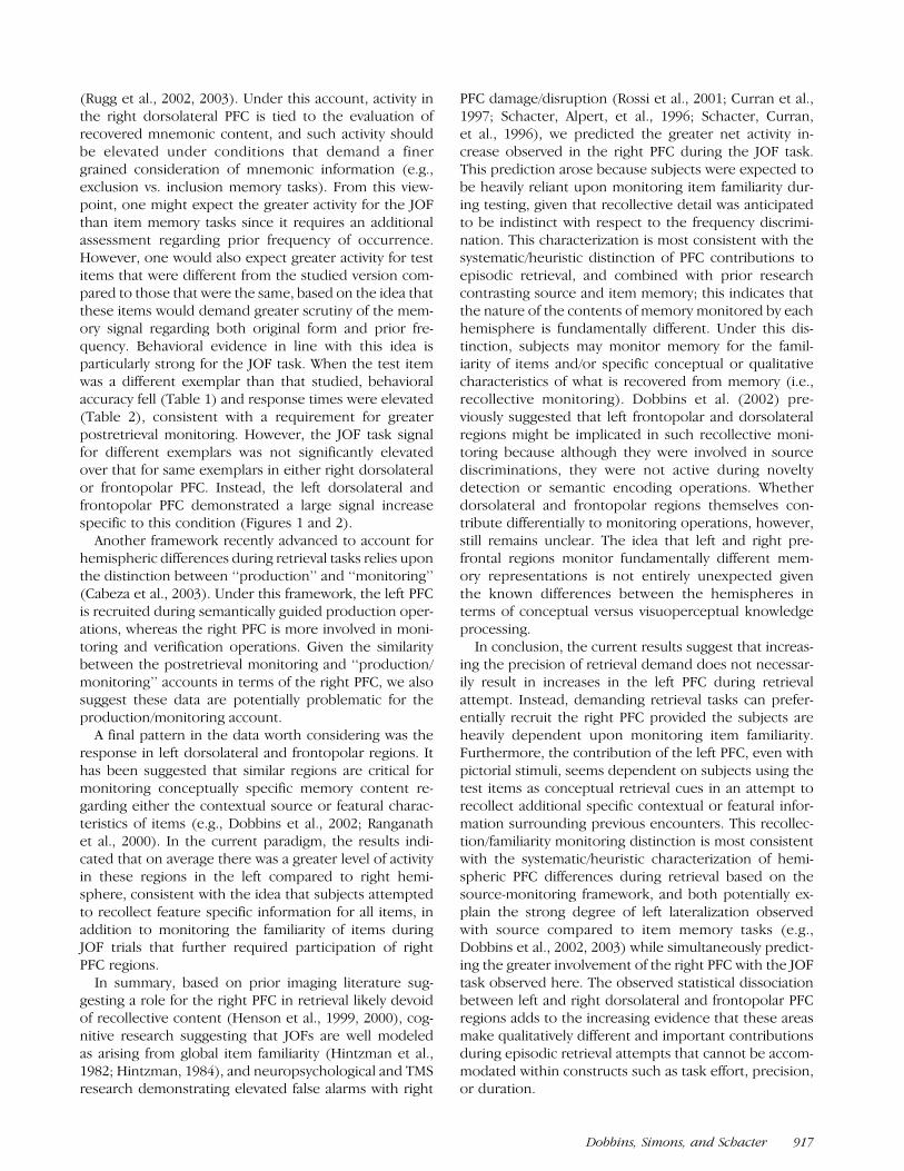
Item Memory Relative Increases
For the reverse comparisons, item memory versus JOF(collapsed across item format), no above baseline activ-ity differences were observed in the PFC (Table 4).However, there were extrastriate, IT, and MTL regionsthat demonstrated consistently greater signal duringitem memory compared to JOFs (Figure 3). In addition
Figure 3. Regions showing a
greater response during item
memory versus judgments of
frequency (collapsed acrossdifferences in item format).
Activity maps are thresholded at
.001 5-voxels one-tailed,
overlaid on a canonicalhigh-resolution structural
image in MNI space (MRICRO
software http://www.psychology.nottingham.ac.uk/
staff/cr1/mricro.html). See
Figure 1 caption for additional
information.
Dobbins, Simons, and Schacter 913

Table 3. SPM Results—JOF versus Item Memory Contrast
Talairach
Hemisphere Region BA x y z Voxels Z score
Left IFG 47 �33 23 �9 72 4.91
* IFG/MFG 10 �45 47 3 15 4.29
* MFG 45/46 �48 27 24 60 4.43
MFG 9 �42 13 27 19 3.58
* MFG 10 �39 52 �5 55 4.11
MFG 6 �42 14 46 19 3.93
IPL 7/39 �36 �59 42 58 4.33
IPL 40 �45 �47 49 24 3.95
Cingulate 32 �6 25 37 67 4.54
Posterior cingulate 23 0 �19 29 60 4.76
Posterior cingulate 23 �6 �25 26 38 4.37
Posterior cingulate 23 �6 �37 21 13 3.44
Precuneus 7 �3 �65 36 79 4.35
Precuneus 7 �12 �59 36 33 3.79
Thalamus �12 �17 9 32 3.96
Thalamus �9 �15 �2 22 3.90
Thalamus �18 �14 3 26 3.74
Brain stem �3 �30 �19 15 4.08
Right IFG 47 30 26 �9 61 4.67
* MFG 10 39 53 3 57 4.58
* MFG 9/46 45 33 29 57 4.46
* MFG 11 36 55 �13 24 4.39
MFG 9 45 25 29 50 4.24
MFG 10 45 47 11 29 3.91
MFG 8 39 20 43 40 4.2
SFG 9 9 36 26 52 4.29
IPL 7/40 42 �62 45 35 5.12
IPL 40 53 �44 46 31 4.22
IPL/Supramarginal 40 50 �39 38 21 4.01
Cingulate 32 9 25 37 43 4.14
Posterior cingulate 23 6 �37 21 32 4.24
Posterior cingulate 29 9 �43 10 7 3.59
Precuneus 31 15 �54 30 59 5.64
Thalamus 12 �11 12 30 4.04
Caudate 9 0 3 11 3.30
Caudate 15 6 11 5 3.34
Brain stem 6 �30 �19 8 3.66
Brain stem 0 �31 �29 5 3.39
Brain stem 12 14 �8 7 3.38
Note. Asterisks denote regions contributing to ROIs. IFG = inferior frontal gyrus; MFG = middle frontal gyrus; SFG = superior frontal gyrus;IPL = inferior parietal lobule; BA = approximate Brodmann’s location. Coordinates are in Talairach space transformed from the original MNI.
914 Journal of Cognitive Neuroscience Volume 16, Number 6

to the activity in visual association areas, there was abilateral relative decrease in signal in an anterior portionof MTL bordering perirhinal and anterior hippocampalareas. Since the data were not globally scaled, thisrelative decrease is likely not a processing artifact. Al-though there remains controversy regarding the inter-pretation of relative signal decreases, we report thesedeactivations because of the established importance ofthe MTL region in recognition memory, and becausealthough the signal differences were small, they werehighly reliable. Based on the repeated observation ofgreater activity for new than studied items across multi-ple experiments, Henson, Cansino, Herron, Robb, andRugg (2003) recently suggested that bilateral MTL re-gions proximal to those reported here may signal itemfamiliarity (Henson et al., 2003). In the current study wefound no evidence for familiarity modulation of theseregions (see also Rugg et al., 2003). In addition to failingto distinguish different and same items within each task,these regions also did not distinguish between itemsseen two and six times when in the same format even ata liberal threshold (.01). There are many possible rea-sons for this apparent discrepancy. For example, thefamiliarity signal differences observed here might beconsiderably smaller than those observed between pure-
ly novel and old items and this may have precludeddetection (see Brown & Aggleton, 2001). Regardless, inthe current data, bilateral anterior MTL regions arereliably modulated by the nature of the episodic retrievaltask and not the familiarity of the items. Under theassumption that MTL regions may be highly activeduring baseline tasks (Stark & Squire, 2001), it is possiblethat the ‘‘baseline’’ MTL activity seen here during theitem memory task actually corresponds to active featureencoding driven by object processing activity suggestedin the extrastriate and IT regions.
DISCUSSION
The current data demonstrate that judgments of fre-quency do not recruit the same neural substrates as tasksrequiring the recovery of specific contextual content(i.e., sourcememory). Prior research comparing source toitem memory in our laboratory has shown strikingly leftlateralized prefrontal and parietal responses for contex-tually demanding source memory tasks using both words(Dobbins et al., 2002, 2003) and pictures (Dobbins et al.,unpublished data). This tendency towards left lateraliza-tion for source memory, which has also been observed
Table 4. SPM Results—Item Memory versus JOF Contrast
Talairach
Hemisphere Region BA x y z Voxels Z score
Left PHG/Uncus �27 �7 �25 40 4.90
PHG/Fusiform 36 �33 �30 �11 13 4.18
PHG/Hippocampus �30 �15 �9 10 3.50
Fusiform 37 �33 �47 �13 19 3.85
SOG 19 �39 �83 24 19 4.06
MOG 19 �33 �75 18 11 3.34
MTG 37 �42 �61 1 10 3.94
MOG 19 �50 �61 �4 9 3.31
Right PHG/Uncus 24 �7 �20 60 4.92
Fusiform 37 33 �39 �13 29 3.95
MTG 19 48 �75 20 37 4.78
SOG 19 39 �77 29 44 4.71
MOG 37 42 �61 �4 30 3.76
MOG/ITG 19/37 50 �64 �2 38 3.70
ITG 20 39 �16 �27 13 4.15
PHG = parahippocampal gyrus; SOG = superior occipital gyrus; MOG = middle occipital gyrus; ITG = inferior temporal gyrus; MTG = middletemporal gyrus; BA = approximate Brodmann’s location. Coordinates are in Talairach space transformed from the original MNI. The tablelists relative activations only.
Dobbins, Simons, and Schacter 915
in other laboratories (Ranganath et al., 2000; Raye et al.,2000; Rugg et al., 1999; Nolde et al., 1998), has been heldto indicate that the left PFC is critically tied to morecontextually specific retrieval demands (Ranganath et al.,2000) compared to simple item-recognition judgments.Here we observed that the most reliable differencebetween the JOF and item memory tasks was seen inthe right—not left—PFC. This finding suggests thatsimply requiring more precision in the memory discrim-ination, in and of itself, does not guarantee a concom-itant increase in left PFC activity; instead what appears tobe critical is the nature of the retrieval information thatsubjects choose to monitor in order to solve the mem-ory task. Before considering this idea more fully it isimportant to consider whether these data can be easilyexplained by difficulty or effort-based accounts.
Earlier imaging studies of memory sometimes em-ployed the construct of retrieval effort to account foractivity (e.g., Schacter, Alpert, Savage, Rauch, & Albert,1996). Therefore, retrieval effort is a potential explana-tion for our data given that the JOF task here wasassociated with less accurate and longer responses thanthe item memory task (for discussion, see Rugg &Wilding, 2000). However, there are numerous reasonsfor rejecting the effort account of the current data andperhaps PFC retrieval data in general. First, the level ofactivity in the right PFC during the JOF task did notappreciably change as a function of item format (Figures 1and 2). However, this manipulation had a marked effecton behavioral accuracy, resulting in an approximately43% decrease in performance when the picture exem-plars differed between study and test. Thus, right dorso-lateral and frontopolar regions are clearly not tracking‘‘effort’’ in any general sense within the JOF task, at leastfor correct responses. Second, if we contrast perfor-mance on the item memory task for items seen twiceduring study (d0 = 1.56) with that of the JOF taskperformed on same format items (d0 = 1.35), the accu-racies are comparable. However, there still remains alarge signal difference in the previously discussed rightPFC regions between the two tasks (see Figure 1, greenlines). Finally, recent studies comparing source to itemmemory have rendered a general effort-based accountlargely uninformative, because these studies have dem-onstrated more robust increases in left and not right PFCfor the more ‘‘effortful’’ source memory tasks (Cabezaet al., 2003; Dobbins et al., 2002, 2003; Raye et al., 2000;Rugg et al., 1999; Nolde et al., 1998) that are invariablyless accurate and slower than simple old/new recogni-tion. Thus, given the available data, knowing that oneepisodic memory task is less accurate or slower thananother, does not indicate the expected laterality ofprefrontal response.
Returning to the systematic/heuristic and postretrievalmonitoring accounts discussed earlier, it appears thatthe current results are more easily accommodated underthe former. Under the systematic/heuristic characteriza-
tion, the right PFC is tied to heuristic memory judg-ments, which have been argued to be consistent withsignal detection accounts of familiarity (Nolde et al.,1998). However, it is important to note that it is notentirely clear that the JOF task would have been classi-fied as ‘‘heuristic’’ if one remained entirely within thesystematic/heuristic framework, and did not also consid-er dual-process or statistical models of recognition di-rectly (Yonelinas, Dobbins, Szymanski, Dhaliwal, & King,1996; Yonelinas, 1994; Jacoby, 1991; Mandler, 1980).For example, Nolde et al. (1998) suggested that rightPFC heuristic processes were relatively simple and quickcompared to left PFC systematic processes (p. 3513).However, in the current paradigm, the JOF task itself isconceptually no simpler than the item memory task.More specifically, the JOF task requires that subjectsunderstand that the current test items originated fromthe study episode, that there were two prior frequencycategories, and that responses should be differentiallyassigned based on the prior frequency of the current testprobe. When considered from this viewpoint, the JOFtask appears no less complicated than the standardsource memory task that has shown a tendency to beleft lateralized in comparison to item memory (Dobbinset al., 2002, 2003; Raye et al., 2000; Rugg et al., 1999;Nolde et al., 1998). What is important from a dualprocess account is that recollection will be largely inef-fective in making the frequency discrimination, andtherefore subjects will closely monitor differences initem familiarity. Thus, although we view familiarity mon-itoring as consistent with a ‘‘heuristic’’ process it isimportant to emphasize that the precision of the dis-crimination does not directly determine the laterality ofprefrontal response. In addition, the speed of respond-ing also does not unambiguously identify the processused for recognition discrimination. Within signal detec-tion theory, the speed of response is tied to the relativedifferences in the familiarities of the items discrimi-nated (in the case of forced choice), or to the distancebetween an item’s familiarity and an internal criterion inparadigms like the current one. From this perspective, itis possible to observe both extremely quick or slowresponses based on item familiarity (Murdock & Dufty,1972). Likewise, the ability to distinguish prior sourcesbased on recollection can be facilitated or impairedbased on the relative distinctiveness of the prior sourcecontexts, with such effects potentially having little to dowith the familiarity of the individual items and presum-ably greatly affecting response speed (e.g., Dobbins,Kroll, Yonelinas, & Liu, 1998). Thus, although systemat-ic/heuristic or recollective/familiarity-based judgmentsare expected to differentially depend on the left andright PFC regions, respectively, this reliance cannot besimply mapped onto differences in reaction time orprecision of retrieval demand across memory tasks.
The current data are less consistent with the postre-trieval monitoring account of the right dorsolateral PFC
916 Journal of Cognitive Neuroscience Volume 16, Number 6
(Rugg et al., 2002, 2003). Under this account, activity inthe right dorsolateral PFC is tied to the evaluation ofrecovered mnemonic content, and such activity shouldbe elevated under conditions that demand a finergrained consideration of mnemonic information (e.g.,exclusion vs. inclusion memory tasks). From this view-point, one might expect the greater activity for the JOFthan item memory tasks since it requires an additionalassessment regarding prior frequency of occurrence.However, one would also expect greater activity for testitems that were different from the studied version com-pared to those that were the same, based on the idea thatthese items would demand greater scrutiny of the mem-ory signal regarding both original form and prior fre-quency. Behavioral evidence in line with this idea isparticularly strong for the JOF task. When the test itemwas a different exemplar than that studied, behavioralaccuracy fell (Table 1) and response times were elevated(Table 2), consistent with a requirement for greaterpostretrieval monitoring. However, the JOF task signalfor different exemplars was not significantly elevatedover that for same exemplars in either right dorsolateralor frontopolar PFC. Instead, the left dorsolateral andfrontopolar PFC demonstrated a large signal increasespecific to this condition (Figures 1 and 2).
Another framework recently advanced to account forhemispheric differences during retrieval tasks relies uponthe distinction between ‘‘production’’ and ‘‘monitoring’’(Cabeza et al., 2003). Under this framework, the left PFCis recruited during semantically guided production oper-ations, whereas the right PFC is more involved in moni-toring and verification operations. Given the similaritybetween the postretrieval monitoring and ‘‘production/monitoring’’ accounts in terms of the right PFC, we alsosuggest these data are potentially problematic for theproduction/monitoring account.
A final pattern in the data worth considering was theresponse in left dorsolateral and frontopolar regions. Ithas been suggested that similar regions are critical formonitoring conceptually specific memory content re-garding either the contextual source or featural charac-teristics of items (e.g., Dobbins et al., 2002; Ranganathet al., 2000). In the current paradigm, the results indi-cated that on average there was a greater level of activityin these regions in the left compared to right hemi-sphere, consistent with the idea that subjects attemptedto recollect feature specific information for all items, inaddition to monitoring the familiarity of items duringJOF trials that further required participation of rightPFC regions.
In summary, based on prior imaging literature sug-gesting a role for the right PFC in retrieval likely devoidof recollective content (Henson et al., 1999, 2000), cog-nitive research suggesting that JOFs are well modeledas arising from global item familiarity (Hintzman et al.,1982; Hintzman, 1984), and neuropsychological and TMSresearch demonstrating elevated false alarms with right
PFC damage/disruption (Rossi et al., 2001; Curran et al.,1997; Schacter, Alpert, et al., 1996; Schacter, Curran,et al., 1996), we predicted the greater net activity in-crease observed in the right PFC during the JOF task.This prediction arose because subjects were expected tobe heavily reliant upon monitoring item familiarity dur-ing testing, given that recollective detail was anticipatedto be indistinct with respect to the frequency discrimi-nation. This characterization is most consistent with thesystematic/heuristic distinction of PFC contributions toepisodic retrieval, and combined with prior researchcontrasting source and item memory; this indicates thatthe nature of the contents of memory monitored by eachhemisphere is fundamentally different. Under this dis-tinction, subjects may monitor memory for the famil-iarity of items and/or specific conceptual or qualitativecharacteristics of what is recovered from memory (i.e.,recollective monitoring). Dobbins et al. (2002) pre-viously suggested that left frontopolar and dorsolateralregions might be implicated in such recollective moni-toring because although they were involved in sourcediscriminations, they were not active during noveltydetection or semantic encoding operations. Whetherdorsolateral and frontopolar regions themselves con-tribute differentially to monitoring operations, however,still remains unclear. The idea that left and right pre-frontal regions monitor fundamentally different mem-ory representations is not entirely unexpected giventhe known differences between the hemispheres interms of conceptual versus visuoperceptual knowledgeprocessing.
In conclusion, the current results suggest that increas-ing the precision of retrieval demand does not necessar-ily result in increases in the left PFC during retrievalattempt. Instead, demanding retrieval tasks can prefer-entially recruit the right PFC provided the subjects areheavily dependent upon monitoring item familiarity.Furthermore, the contribution of the left PFC, even withpictorial stimuli, seems dependent on subjects using thetest items as conceptual retrieval cues in an attempt torecollect additional specific contextual or featural infor-mation surrounding previous encounters. This recollec-tion/familiarity monitoring distinction is most consistentwith the systematic/heuristic characterization of hemi-spheric PFC differences during retrieval based on thesource-monitoring framework, and both potentially ex-plain the strong degree of left lateralization observedwith source compared to item memory tasks (e.g.,Dobbins et al., 2002, 2003) while simultaneously predict-ing the greater involvement of the right PFC with the JOFtask observed here. The observed statistical dissociationbetween left and right dorsolateral and frontopolar PFCregions adds to the increasing evidence that these areasmake qualitatively different and important contributionsduring episodic retrieval attempts that cannot be accom-modated within constructs such as task effort, precision,or duration.
Dobbins, Simons, and Schacter 917

METHODS
Subjects
Sixteen right-handed, 18- to 35-year-old, native English-speaking volunteers were paid $50 for participating inthe study. Informed consent was obtained in a mannerapproved by the Human Studies Committee at Massa-chusetts General Hospital, the Committee on the Useof Humans as Experimental Subjects at MIT, and theHarvard University Committee on the use of HumanSubjects in Research.
Study Materials
The stimuli consisted of 360 pairs of color pictures.The picture pairs depicted single man-made or livingobjects (e.g., umbrella, octopus), with the items in eachpair representing perceptually different exemplars ofobjects with the same name, and have demonstrated ahigh degree of naming agreement (mean 95%) (Simons,Koutstaal, Prince, Wagner, & Schacter, 2003). Eight dif-ferent versions of the task were created, which system-atically counterbalanced the stimuli between subjectsacross three factors: study frequency (two or six presen-tations), test picture format (same or different), and testcue (‘‘two<>six times?’’ or ‘‘same<>different?’’).
Task Procedures
Stimuli were back projected onto a screen at the rear ofthe magnet bore and were viewed with a mirror placedabove the eyes. The entire experiment consisted ofthree study/test cycles each using 120 items. During thenonscanned study phases, subjects performed rapidliving/nonliving judgments on randomized picturesshown either two or six times for 1200 msec each.Immediately following each study phase, they werescanned while making episodic memory judgments re-garding the previously viewed pictures. Subjects receivedeither JOF) (‘‘two<>six times ?’’) or item memory(‘‘same<>different ?’’) retrieval cues for each displayedpicture. For half of the retrieval trials, the item wasidentical to that which had been studied, for the remain-ing half it was the alternate exemplar. Subjects were toldthat in the case of frequency estimation, if they shoulddetect that the current item was different than that whichwas studied, that they should ignore this difference andattempt to remember how often they saw an item withthat name (e.g., a toaster). During retrieval trials, itemsremained visible for 3950 msec and subjects were in-structed to respond before the items disappeared fromthe screen. There was a 50-msec ITI between all trials.Interspersed among the retrieval trials were fixation trialsvarying from 2000 to 6000 msec. The order of retrievaland fixation trials was determined by an optimal sequenc-ing program designed to maximize recovery of the BOLDsignal (Dale, Greve, & Burock, 1999).
fMRI Data Acquisition
Scanning was performed on a 3T Siemens Allegra sys-tem using a standard whole-head coil. Functional datawere acquired using a gradient-echo, echo-planar pulsesequence (TR = 2 sec, TE = 30 msec, 21 axial slicesparallel to the AC–PC plane, 3.125 � 3.125 � 5 mm,1-mm interslice gap). Prior to functional data collection,four dummy volumes were collected and discarded toallow for T1-equilibration effects. High-resolution T1-weighted (MP-RAGE) anatomical images were collectedfor visualization. Head motion was restricted by using apillow and foam inserts.
fMRI Data Analysis
Data were preprocessed using SPM99 (Wellcome De-partment of Cognitive Neurology, London). Slice acqui-sition timing was corrected by resampling all slices intime relative to the first slice, followed by rigid bodymotion correction across all runs. Functional data werespatially normalized to an EPI template using a 12-parameter affine and nonlinear cosine basis functiontransformation. Volumes were resampled into 3-mmcubic voxels and spatially smoothed with an 8-mmFWHM isotropic Gaussian kernel. Each session was grandmean scaled such that the mean signal was 100. The datawere statistically analyzed treating subjects as a randomeffect. For the analyses, volumes were treated as atemporally correlated time series and modeled by con-volving a synthetic hemodynamic response function andits first-order time derivative using the onset times for theevents. The resulting functions were used as covariates ina general linear model, along with a basis set of cosinefunctions that were used to high-pass filter the data, anda block covariate representing session effects. The leastsquares parameter estimates of height of the best fittingsynthetic HRF for each condition of interest (averagedacross scans) were used in pairwise contrasts and storedas a separate image for each subject. These images werethen tested against the null hypothesis of no differencebetween contrast conditions using one-tailed t testsresulting in repeated measures t tests across subjects.Regions were considered significant and subjected tofurther analysis if they consisted of five or more conti-guous voxels and exceeded an alpha threshold of .001(Z > 3.09, uncorrected) for direct contrasts of retrievaltasks (frequency estimation vs. item memory for correctresponses). This threshold has been demonstrated toadequately control for familywise error in similar studiesand is comparable to that typically used in fMRI studies ofmemory enabling meaningful across study comparisons(Donaldson & Buckner, 1999; Buckner et al., 1998).Following this, functional ROIs were extracted usingperistimulus time averaging for the event-related fMRIdata surviving the retrieval task contrasts. Percent signalaverages were obtained for above threshold voxels with-
918 Journal of Cognitive Neuroscience Volume 16, Number 6

in an 8 mm radius of each of the SPM-identified maximafor the contrast. The reported ANOVAs on these dataused the third time point (6 sec post stimulus onset) andare unbiased with respect to interactions involving hemi-spheres or item format (same or different).
Acknowledgments
Supported by the NIH (NS26985, MH60941, DC04466, andAG08441) and Wellcome Trust (061171). We thank R. Poldrackfor development of analysis tools, W. Koutstaal for use of herstimuli, and Alexis Perkins for help with data collection.
Reprint requests should be sent to Ian G. Dobbins, [email protected].
The data reported in this experiment have been deposited inthe fMRI Data Center (http://www.fmridc.org). The accessionnumber is 2-2004-115MC.
REFERENCES
Atkinson, R. C., & Juola, J. G. (1974). Search and decisionprocesses in recognition memory. In R. C. Atkinson,R. D. Luce, D. H. Krantz, & P. Suppes (Eds.), Contemporarydevelopments in mathematical psychology: I. Learning,memory, and thinking (pp. 243–293). San Francisco:Freeman.
Banks, W. P. (1970). Signal detection theory and humanmemory. Psychological Bulletin, 74, 81–99.
Brown, M. W., & Aggleton, J. P. (2001). Recognitionmemory: What are the roles of the perirhinal cortex andhippocampus? Nature Reviews, Neuroscience, 2, 51–61.
Buckner, R. L., Koutstaal, W., Schacter, D. L., Dale, A. M.,Rotte, M., & Rosen, B. R. (1998). Functional-anatomic studyof episodic retrieval. II. Selective averaging of event-relatedfMRI trials to test the retrieval success hypothesis.Neuroimage, 7, 163–175.
Cabeza, R., Locantore, J. K., & Anderson, N. D. (2003).Lateralization of prefrontal activity during episodicmemory retrieval: Evidence for the production-monitoringhypothesis. Journal of Cognitive Neuroscience, 15, 249–259.
Curran, T., Schacter, D. L., Norman, K. A., & Galluccio, L.(1997). False recognition after a right frontal lobeinfarction: Memory for general and specific information.Neuropsychologia, 35, 1035–1049.
Dale, A. M., Greve, D. N., & Burock, M. A. (1999, June 22–26).Optimal stimulus sequences for event-related fMRI. Paperpresented at the 5th International Conference on FunctionalMapping of the Human Brain, Duesseldorf, Germany.
Dobbins, I. G., Foley, H., Schacter, D., & Wagner, A. (2002).Executive control during retrieval: Multiple prefrontalprocesses subserve source memory. Neuron, 35, 989–996.
Dobbins, I. G., Kroll, N. E. A., Yonelinas, A. P., & Liu, Q. (1998).Distinctiveness in recognition and free recall: The role ofrecollection in the rejection of the familiar. Journal ofMemory and Language, 38, 381–400.
Dobbins, I. G., Rice, H. J., Wagner, A. D., & Schacter, D. L.(2003). Memory orientation and success: Separableneurocognitive components underlying episodicrecognition. Neuropsychologia, 41, 318–333.
Donaldson, D. I., & Buckner, R. L. (1999). Trying versussucceeding: Event-related designs dissociate memoryprocesses. Neuron, 22, 412–414.
Eldridge, L. L., Knowlton, B. J., Furmanski, C. S., Bookheimer,
S. Y., & Engel, S. A. (2000). Remembering episodes: Aselective role for the hippocampus during retrieval.Nature Neuroscience, 3, 1149–1152.
Hasher, L., & Chromiak, W. (1977). The processing offrequency information: An automatic mechanism? Journalof Verbal Learning and Verbal Behavior, 16, 173–184.
Hasher, L., & Zacks, R. T. (1984). Automatic processingof fundamental information: The case of frequency ofoccurrence. American Psychologist, 39, 1372–1388.
Hasher, L., Zacks, R. T., Rose, K. C., & Sanft, H. (1987). Trulyincidental encoding of frequency information. AmericanJournal of Psychology, 100, 69–91.
Henson, R. N., Cansino, S., Herron, J. E., Robb, W. G., & Rugg,M. D. (2003). A familiarity signal in human anterior medialtemporal cortex? Hippocampus, 13, 301–304.
Henson, R. N., Rugg, M. D., Shallice, T., & Dolan, R. J. (2000).Confidence in recognition memory for words: Dissociatingright prefrontal roles in episodic retrieval. Journal ofCognitive Neuroscience, 12, 913–923.
Henson, R. N., Rugg, M. D., Shallice, T., Josephs, O., & Dolan,R. J. (1999). Recollection and familiarity in recognitionmemory: An event-related functional magnetic resonanceimaging study. Journal of Neuroscience, 19, 3962–3972.
Hintzman, D. L. (1984). MINERVA 2: A simulation model ofhuman memory. Behavior Research Methods, Instrumentsand Computers, 16, 96–101.
Hintzman, D. L., & Curran, T. (1994). Retrieval dynamics ofrecognition and frequency judgments: Evidence for separateprocesses of familiarity and recall. Journal of Memory andLanguage, 33, 1–18.
Hintzman, D. L., Nozawa, G., & Irmscher, M. (1982).Frequency as a nonpropositional attribute of memory.Journal of Verbal Learning and Verbal Behavior, 21,127–141.
Jacoby, L. L. (1991). A process dissociation framework:Separating automatic from intentional uses of memory.Journal of Memory and Language, 30, 513–541.
Johnson, M. K., Hashtroudi, S., & Lindsay, D. S. (1993). Sourcemonitoring. Psychological Bulletin, 114, 3–28.
Mandler, G. (1980). Recognizing: The judgment of previousoccurrence. Psychological Review, 87, 252–271.
Maril, A., Simons, J. S., Mitchell, J. P., Schwartz, B., & Schacter,D. L. (2003). Feeling-of-knowing in episodic memory: Anevent-related fMRI study. Neuroimage, 18, 827–836.
Murdock, B. B., & Dufty, P. O. (1972). Strength theory andrecognition memory. Journal of Experimental Psychology,94, 284–290.
Nolde, S. F., Johnson, M. K., & D’Esposito, M. (1998). Leftprefrontal activation during episodic remembering: Anevent-related fMRI study. NeuroReport, 9, 3509–3514.
Ranganath, C., Johnson, M. K., & D’Esposito, M. (2000).Left anterior prefrontal activation increases with demandsto recall specific perceptual information. Journal ofNeuroscience, 20, RC108.
Raye, C. L., Johnson, M. K., Mitchell, K. J., Nolde, S. F., &D’Esposito, M. (2000). fMRI investigations of left and rightPFC contributions to episodic remembering. Psychobiology,28, 197–206.
Rossi, S., Cappa, S. F., Babiloni, C., Pasqualetti, P., Miniussi, C.,Carducci, F., Babiloni, F., & Rossini, P. M. (2001). Prefrontal[correction of Prefontal] cortex in long-term memory: An‘‘interference’’ approach using magnetic stimulation. NatureNeuroscience, 4, 948–952.
Rugg, M. D., Fletcher, P. C., Chua, P. M., & Dolan, R. J. (1999).The role of the prefrontal cortex in recognition memoryand memory for source: An fMRI study. Neuroimage, 10,520–529.
Rugg, M. D., Henson, R. N., & Robb, W. G. (2003). Neural
Dobbins, Simons, and Schacter 919

correlates of retrieval processing in the prefrontal cortexduring recognition and exclusion tasks. Neuropsychologia,41, 40–52.
Rugg, M. D., Otten, L. J., & Henson, R. N. (2002). Theneural basis of episodic memory: Evidence from functionalneuroimaging. Philosophical Transactions of the RoyalSociety of London, Series B: Biological Sciences, 357,1097–1110.
Rugg, M. D., & Wilding, E. L. (2000). Retrieval processing andepisodic memory. Trends in Cognitive Sciences, 4, 108–115.
Schacter, D. L., Alpert, N. M., Savage, C. R., Rauch, S. L., &Albert, M. S. (1996). Conscious recollection and the humanhippocampal formation: Evidence from positron emissiontomography. Proceedings of the National Academy ofSciences, U.S.A., 93, 321–325.
Schacter, D. L., Buckner, R. L., Koutstaal, W., Dale, A. M., &Rosen, B. R. (1997). Late onset of anterior prefrontal activityduring true and false recognition: An event-related fMRIstudy. Neuroimage, 6, 259–269.
Schacter, D. L., Curran, T., Galluccio, L., & Milberg, W. P.(1996). False recognition and the right frontal lobe: A casestudy. Neuropsychologia, 34, 793–808.
Simons, J. S., Koutstaal, W., Prince, S., Wagner, A., &Schacter, D. (2003). Neural mechanisms of visualobject priming: Evidence for perceptual and semantic
distinctions in fusiform cortex. Neuroimage, 19,613–626.
Stark, C. E., & Squire, L. R. (2001). When zero is not zero:The problem of ambiguous baseline conditions in fMRI.Proceedings of the National Academy of Sciences, U.S.A.,98, 12760–12766.
Tulving, E. (1985). Memory and consciousness. CanadianPsychology, 26, 1–12.
Wagner, A. D., Maril, A., & Schacter, D. L. (2000). Interactionsbetween forms of memory: When priming hinders newepisodic learning. Journal of Cognitive Neuroscience, 12,52–60.
Yonelinas, A. P. (1994). Receiver-operating characteristics inrecognition memory: Evidence for a dual-process model.Journal of Experimental Psychology: Learning, Memory,and Cognition, 20, 1341–1354.
Yonelinas, A. P., Dobbins, I., Szymanski, M. D., Dhaliwal, H. S.,& King, L. (1996). Signal-detection, threshold, anddual-process models of recognition memory: ROCs andconscious recollection. Consciousness and Cognition, 5,418–441.
Zacks, R. T., Hasher, L., & Sanft, H. (1982). Automaticencoding of event frequency: Further findings. Journalof Experimental Psychology: Learning, Memory, andCognition, 8, 106–116.
920 Journal of Cognitive Neuroscience Volume 16, Number 6

This article has been cited by:
1. Daniela Czernochowski, Axel Mecklinger, Mikael Johansson. 2009. Age-related changes in the control of episodic retrieval: anERP study of recognition memory in children and adults. Developmental Science 12:6, 1026-1040. [CrossRef]
2. LAUREN L. DRAG, LINAS A. BIELIAUSKAS, ALFRED W. KASZNIAK, NICOLAAS I. BOHNEN, ELIZABETH L.GLISKY. 2009. Source memory and frontal functioning in Parkinson’s disease. Journal of the International NeuropsychologicalSociety 15:03, 399. [CrossRef]
3. Young Youn Kim, Ah Young Roh, Yoon Namgoong, Hang Joon Jo, Jong-Min Lee, Jun Soo Kwon. 2009. Cortical networkdynamics during source memory retrieval: Current density imaging with individual MRI. Human Brain Mapping 30:1, 78-91.[CrossRef]
4. Peggy St. Jacques, David C. Rubin, Kevin S. LaBar, Roberto Cabeza. 2008. The Short and Long of It: Neural Correlates ofTemporal-order Memory for Autobiographical EventsThe Short and Long of It: Neural Correlates of Temporal-order Memoryfor Autobiographical Events. Journal of Cognitive Neuroscience 20:7, 1327-1341. [Abstract] [PDF] [PDF Plus]
5. Chiang-shan Ray Li, Cong Huang, Peisi Yan, Prashni Paliwal, Robert Todd Constable, Rajita Sinha. 2008. Neural Correlates ofPost-error Slowing during a Stop Signal Task: A Functional Magnetic Resonance Imaging StudyNeural Correlates of Post-errorSlowing during a Stop Signal Task: A Functional Magnetic Resonance Imaging Study. Journal of Cognitive Neuroscience 20:6,1021-1029. [Abstract] [PDF] [PDF Plus]
6. Robert S. Ross, Scott D. Slotnick. 2008. The Hippocampus is Preferentially Associated with Memory for Spatial ContextTheHippocampus is Preferentially Associated with Memory for Spatial Context. Journal of Cognitive Neuroscience 20:3, 432-446.[Abstract] [PDF] [PDF Plus]
7. P. M. Paz-Alonso, S. Ghetti, S. E. Donohue, G. S. Goodman, S. A. Bunge. 2008. Neurodevelopmental Correlates of True andFalse Recognition. Cerebral Cortex 18:9, 2208-2216. [CrossRef]
8. Matthew W. Prull, Leslie L. Crandell Dawes, A. McLeish Martin, Heather F. Rosenberg, Leah L. Light. 2006. Recollectionand familiarity in recognition memory: Adult age differences and neuropsychological test correlates. Psychology and Aging 21:1,107-118. [CrossRef]
9. David A. Gallo , Elizabeth A. Kensinger , Daniel L. Schacter . 2006. Prefrontal Activity and Diagnostic Monitoring of MemoryRetrieval: fMRI of the Criterial Recollection TaskPrefrontal Activity and Diagnostic Monitoring of Memory Retrieval: fMRI ofthe Criterial Recollection Task. Journal of Cognitive Neuroscience 18:1, 135-148. [Abstract] [PDF] [PDF Plus]
10. 2005. Current awareness in NMR in biomedicine. NMR in Biomedicine 18:1, 56-63. [CrossRef]11. Charan Ranganath . 2004. The 3-D Prefrontal Cortex: Hemispheric Asymmetries in Prefrontal Activity and Their Relation to
Memory Retrieval ProcessesThe 3-D Prefrontal Cortex: Hemispheric Asymmetries in Prefrontal Activity and Their Relation toMemory Retrieval Processes. Journal of Cognitive Neuroscience 16:6, 903-907. [Abstract] [PDF] [PDF Plus]
12. Karen J. Mitchell , Marcia K. Johnson , Carol L. Raye , Erich J. Greene . 2004. Prefrontal Cortex Activity Associated with SourceMonitoring in a Working Memory TaskPrefrontal Cortex Activity Associated with Source Monitoring in a Working MemoryTask. Journal of Cognitive Neuroscience 16:6, 921-934. [Abstract] [PDF] [PDF Plus]
Related Documents