FLÁVIA ALVIM SANT´ANNA ADDOR Dermatite atópica: Correlação entre estado da barreira cutânea em pele não lesionada e atividade da doença Dissertação apresentada à Faculdade de Medicina da Universidade de São Paulo para obtenção do título de Mestre em Ciências Área de concentração: Dermatologia Orientadora: Prof. Dra. Valéria Aoki São Paulo 2008

Welcome message from author
This document is posted to help you gain knowledge. Please leave a comment to let me know what you think about it! Share it to your friends and learn new things together.
Transcript

FLÁVIA ALVIM SANT´ANNA ADDOR
Dermatite atópica: Correlação entre estado da barreira cutânea em pele não lesionada e atividade da doença
Dissertação apresentada à Faculdade de
Medicina da Universidade de São Paulo para
obtenção do título de Mestre em Ciências
Área de concentração: Dermatologia
Orientadora: Prof. Dra. Valéria Aoki
São Paulo 2008


Dedicatória

Aos meus pais, Creuza e Accacio, pelo amor e dedicação infinitos e
pelo apoio incondicional;
Ao meu querido marido Maurício, por proporcionar tudo que precisei
para buscar meus sonhos;
Aos meus filhos Maria Fernanda e Maurício, luzes da minha vida,
pela compreensão nas minhas ausências e pelo carinho infinito.

Agradecimentos

À minha orientadora Profa. Dra. Valéria Aoki, por todo apoio, orientação,
amizade e confiança em meu projeto e meu trabalho, e pelo acolhimento em
sua rotina de ambulatório.
Ao Prof. Dr. Evandro A. Rivitti, pela oportunidade e apoio ao meu ingresso
na pós-graduação.
Ao Prof. Dr. Luis Carlos Cucé, insubstituível em seu incentivo e orientações
fundamentais nos primórdios do projeto deste trabalho, e pelo apoio em todo
seu andamento.
Ao Dr. Roberto Takaoka, pelo seu carinho, interesse e motivação no
ambulatório de Dermatite Atópica.
À Prof. Dra. Mírian Nacagami Sotto, pelo apoio e constante atenção a todas
necessidades dos pós-graduandos.
Ao Dr. Sérgio Schalka, querido amigo e companheiro de tantos anos de
trabalho, pelo incentivo e pelo espírito colaborativo que partilhamos também
nos cursos da pós-graduação.
Ao Dr. Dilhermando Calil, pela amizade, confiança e incentivo, mesmo nas
horas mais difíceis e trabalhosas;
Ao Dr. Sérgio Barbosa, pela compreensão e paciência durante toda a pós-
graduação.

A todos os membros do Serviço de Dermatologia da UNISA, em especial ao
Prof. Dr. Jayme de Oliveira Filho, Prof. Dr. Reinaldo Tovo Filho, Prof. Dr.
Arthur Duarte, Profa. Dra. Márcia Ferraz, Prof. Dr. Sérgio Fava, Profa. Dra.
Marina Odo e Profa. Dra. Bertha Tamura, sempre incansáveis na tarefa de
educar, sempre disponíveis como colegas e amigos atenciosos.
Ao Prof. Dr. Joachim W. Fluhr, da Friedrich-Schiller University (Jena-
Alemanha), pelas grandes contribuições bibliográficas e metodológicas,
À Sra. Vanessa Melo, Srta. Christiane Agelune e ao Sr. Bruno Folino, pelas
importantes contribuições para compreensão no manuseio dos
equipamentos e pela grande paciência e disponibilidade.
À Sra. Eli Maria de Freitas Ferreira, pela atenção e total disponibilidade em
ajudar a resolver tudo, o tempo todo.
Ao Sr. Roberto Rinaldi e todos funcionários do Departamento de
Dermatologia, pela disponibilidade e atenção constantes.
À Sra. Valéria de Vilhena Lombardi, pela atenção na elaboração da ficha
catalográfica deste trabalho.
À Sra. Rute Tomida, que com incansável atenção, realizou a formatação
deste trabalho.
À Sra. Leyla Costa Ramos, pela valiosa colaboração na avaliação estatística
de todos os dados levantados.
À Sra. Neuma Balbioti de Almeida Silva e Sr. Davi Guimarães Pedro,
incansáveis no auxilio diário para a realização deste trabalho.

Aos médicos residentes da Divisão de Dermatologia da Faculdade de
Medicina da Universidade de São Paulo, pela amizade, convivência e
colaboração no ambulatório de dermatite atópica.
Aos prezados doentes de dermatite atópica, pela colaboração fundamental à
realização deste trabalho.

Normalização Esta dissertação está de acordo com:
Referências: adaptado de International Committee of Medical Journals
Editors (Vancouver)
Universidade de São Paulo. Faculdade de Medicina. Serviço de Biblioteca e
Documentação. Guia de apresentação de dissertações, teses e monografias.
Elaborado por Annelise Carneiro da Cunha, Maria Julia de A. L. Freddi, Maria
F. Crestana, Marinalva de Souza Aragão, Suely Campos Cardoso, Valéria
Vilhena. São Paulo: Serviço de Biblioteca e Documentação; 2005.
Abreviaturas dos títulos dos periódicos de acordo com List of Journals
Indexed in Index Medicus.

Sumário

Lista de figuras Lista de quadros Lista de tabelas Lista de gráficos Resumo Summary 1 INTRODUÇÃO.............................................................................................1 2 OBJETIVOS...............................................................................................14 3 REVISÃO DA LITERATURA......................................................................16
3.1 Barreira cutânea...............................................................................17 3.2 Barreira cutânea e dermatite atópica...............................................22 3.3 Parâmetros biofísicos para avaliação da dermatite atópica:
noções de bioengenharia ................................................................26 3.3.1 Avaliação da barreira cutânea por parâmetros biofísicos .....26 3.3.2 Avaliação funcional da barreira cutânea in vivo: Perda de
água transepidérmica ...........................................................28 3.3.3 Avaliação do conteúdo de água na camada córnea:
Capacitância .........................................................................32 4 MATERIAL E MÉTODO.............................................................................35
4.1 Seleção dos doentes .......................................................................36 5 RESULTADOS...........................................................................................45
5.1 Avaliação das características da amostra de doentes atópicos estudada: dados epidemiológicos ...................................................46 5.1.1 Distribuição: sexo, faixa etária e etnia ....................................46 5.1.2 Parâmetros instrumentais.......................................................48 5.1.3 Sexo, idade e etnia X medidas instrumentais.........................49 5.1.4 Presença de sinais menores em atópicos e não atópicos......52
5.2 Grupo atópico: Correlação entre parâmetros biofísicos score clínico de Rajka e Langeland e IgE .................................................57 5.2.1 Gravidade do quadro atópico X medidas instrumentais........57 5.2.2 Níveis de IgE sérico X medidas instrumentais......................64 5.2.3 Intensidade do prurido X medidas instrumentais ..................68 5.2.4 Presença de outras doenças atópicas X medidas
instrumentais ........................................................................70

6 DISCUSSÃO..............................................................................................80 6.1 Sexo .................................................................................................82 6.2 Idade ................................................................................................83 6.3 Etnia .................................................................................................85 6.4 Gravidade segundo Rajka e Langeland ...........................................88 6.5 IgE sérico .........................................................................................92 6.6 Prurido..............................................................................................94 6.7 Presença de outras doenças atópicas X medidas
instrumentais....................................................................................96 6.8 Sinais menores da dermatite atópica segundo Hanifin e Rajka .......99
7 CONCLUSÕES........................................................................................100 8 ANEXOS..................................................................................................103
Anexo 1 - Termo de Consentimento Livre e Esclarecido .......................104 Anexo 2 - Ficha Clínica ..........................................................................106 Anexo 3 - Artigo submetido à publicação...............................................108
9 REFERÊNCIAS .......................................................................................122

Listas

LISTA DE FIGURAS
Figura 1 - Esquema da fisiopatologia da dermatite atópica ......................4
Figura 2 - Representação do princípio do funcionamento do Corneometer® .........................................................................11
Figura 3 - Corneometer ® ........................................................................11
Figura 4 - Representação esquemática da sonda do Tewameter ® ........12
Figura 5 - Equipamento Tewameter ® .....................................................12
Figura 6 - Padrão “brick and mortar” do estrato córneo (barreira cutânea)...................................................................................18
Figura 7 - Mecanismos defensivos do estrato córneo e influência dos produtos da filagrina..........................................................19
Figura 8 - Influência do estado da barreira na regulação da diferenciação epidérmica e secreção de corpos lamelares .....20
Figura 9 - Agressão à barreira e desencadeamento da resposta inflamatória ..............................................................................22
Figura 10 - Esquema corporal com percentuais de área para cálculo.......41
Figura 11 - Coleta da medida: corneometria..............................................43
Figura 12 - Coleta da medida: TEWL ........................................................43
Figura 13 - Dermatite atópica leve.............................................................63
Figura 14 - Dermatite atópica moderada ...................................................63
Figura 15 - Dermatite atópica grave ..........................................................64
Figura 16 - Dermatite atópica crônica em adulto negróide, com prurido intenso e eritrodermia; observar a difícil visualização do eritema ...........................................................89
Figura 17 - Dermatite atópica em criança negróide: eritema de difícil visualização em lesões sub agudas ........................................89

LISTA DE QUADROS
Quadro 1 - Estudos sobre anormalidades da barreira cutânea e dermatite atópica ....................................................................25
Quadro 2- TEWL medida em crianças atópicas ativas e não atópicas ...................................................................................30
Quadro 3 - TEWL medida em crianças atópicas ativas, não ativas e não atópicas ............................................................................31
Quadro 4 - Definição do grau de xerose da superfície da pele com o corneometer ............................................................................33
Quadro 5 - Critérios de diagnóstico de dermatite atópica, segundo Hanifin e Rajka ........................................................................38
Quadro 6- Critérios menores de diagnostico de dermatite atópica, segundo Hanifin e Rajka..........................................................39
Quadro 7 - Gravidade da dermatite atópica, segundo Rajka e Langeland ................................................................................40
Quadro 8 - Classificação das etnias segundo os grupos populacionais...........................................................................85
Quadro 9 - Diferenças estruturais principais no grupo étnico preto em relação ao branco, para a função de barreira do estrato córneo..........................................................................86
Quadro 10 - Gravidade da doença antes e depois da exclusão do score de eritema ......................................................................90

LISTA DE TABELAS
Tabela 1 - Média das medidas dos parâmetros instrumentais nos grupos atópico e não atópico...................................................48
Tabela 2 - Média das medidas instrumentais e do score de gravidade clínica entre os sexos .............................................49
Tabela 3 - Média das medidas instrumentais e do score de gravidade clínica entre as faixas etárias..................................50
Tabela 4 - Média das medidas instrumentais e do score de gravidade clínica entre as etnias .............................................51
Tabela 5 - Análise de variância entre as médias das medidas instrumentais para sexo, faixa etária e etnias .........................51
Tabela 6 - Valor de p para a TEWL entre os grupos de etnias.................52
Tabela 7 - Presença de xerose entre os grupos atópico e não atópico.....................................................................................53
Tabela 8 - Presença de sinais menores da DA entre os grupos atópico e não atópico ..............................................................54
Tabela 9 - Valor de p para os sinais menores da DA entre os grupos atópico e não atópico ..............................................................55
Tabela 10 - Média das medidas instrumentais e graus clínicos da DA ......58
Tabela 11 - Valor de p para as medidas instrumentais e os graus clínicos de DA..........................................................................61
Tabela 12 - Níveis de igE sérica e medidas instrumentais e graus clínicos de DA..........................................................................65
Tabela 13 - Valor de p para IgE e os graus clínicos de DA ........................66
Tabela 14 - Correlação entre as variáveis IgE, corneometria e TEWL pelo coeficiente de correlação de Pearson..............................67
Tabela 15 - Intensidade do prurido e média das medidas instrumentais e graus clínicos de DA ......................................68
Tabela 16 - Intensidade de prurido e médias de TEWL: Teste de Tukey.......................................................................................69

Tabela 17 - Intensidade de prurido e médias de corneometria: Teste de Tukey..................................................................................69
Tabela 18 - Ocorrência de doenças atópicas, grau clínico de DA e medidas instrumentais.............................................................70
Tabela 19 - Valor de p para as médias de corneometria e ocorrência de doenças atópicas................................................................71
Tabela 20 - Valor de p para as médias de TEWL e ocorrência de doenças atópicas: Teste de Tukey ..........................................72
Tabela 21 - Percentuais de prevalência dos sinais menores de DA e grau clinico de DA ...................................................................73
Tabela 22 - Valor de p para prevalência dos sinais menores de DA e grau clínico de DA: Teste de Tukey.........................................74
Tabela 23 - Média das medidas de corneometria para cada grupo de DA, para cada sinal menor ......................................................75
Tabela 24 - Valor de p para as medidas de corneometria e sinais menores: Teste de Tukey........................................................76
Tabela 25 - Média das medidas de TEWL para cada grupo de DA, em cada sinal menor ...............................................................77
Tabela 26 – Valor de p para as medidas de TEWL e sinais menores: Teste de Tukey........................................................................78

LISTA DE GRÁFICOS
Gráfico 1 - Boxplot da média das medidas de corneometria e gravidade da DA......................................................................59
Gráfico 2 - Correlação das médias das medidas biofísicas de perda de água transepidérmica e graus clínicos de DA ....................60
Gráfico 3 - Grau de hidratação e intensidade do quadro segundo Rajka e Langeland...................................................................62
Gráfico 4 - Grau de TEWL e intensidade do quadro segundo Rajka e Langeland................................................................................62
Gráfico 5 - Boxplot das médias das medidas de IgE e graus clínicos de DA. .....................................................................................66

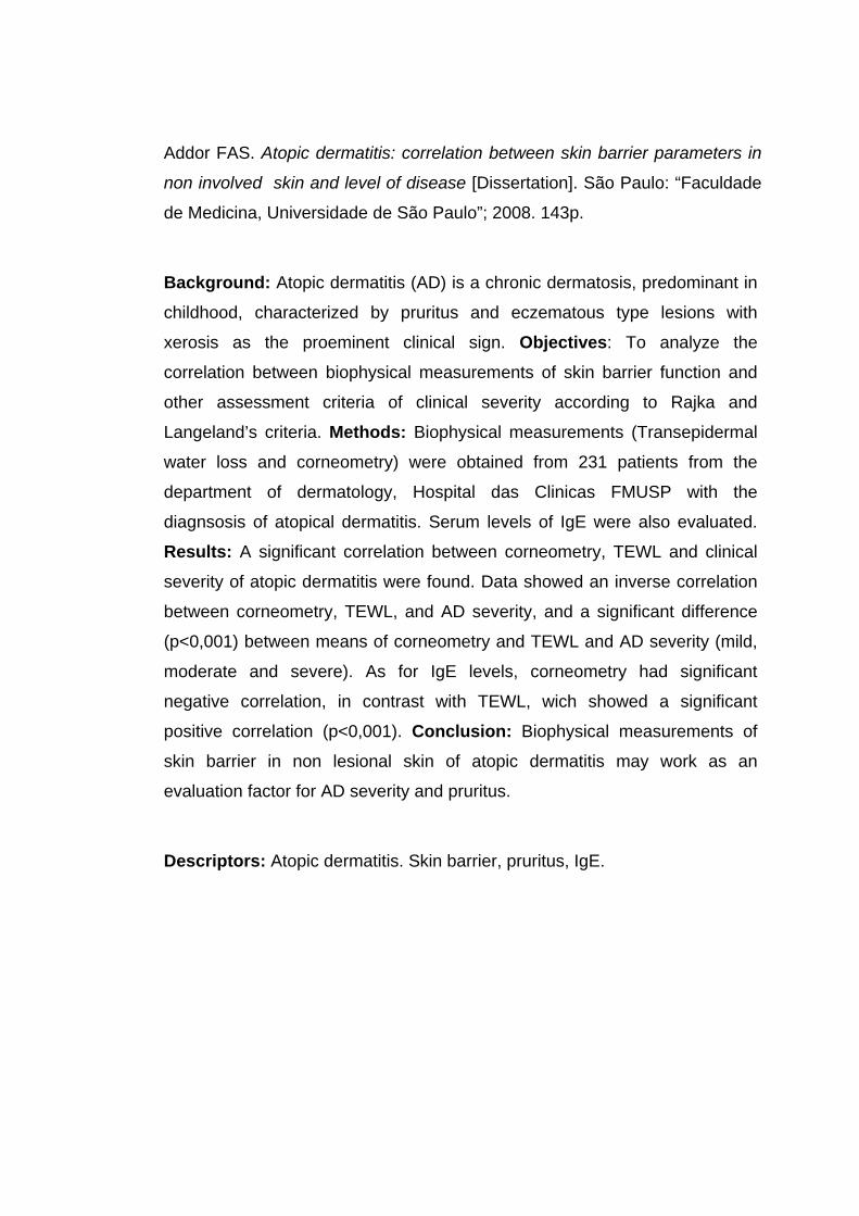
Resumo

Addor FAS. Dermatite atópica: Correlação entre estado da barreira cutânea
em pele não lesionada e atividade da doença [Dissertação]. São Paulo:
Faculdade de Medicina, Universidade de São Paulo; 2008. 143p.
Introdução: Dermatite atópica (DA) é uma doença cutânea crônica,
predominante na infância, cujo sintoma principal é o prurido de intensidade
variável, e os sinais são classicamente as lesões de padrão eczematoso. Há
anormalidades na formação e função da barreira cutânea, que estão
presentes não somente nas lesões cutâneas como na pele clinicamente não
afetada. Objetivo: Analisar a correlação entre as medidas biofísicas da
função de barreira cutânea e os critérios clínicos e intensidade da dermatite,
de acordo com os critérios de Rajka e Langeland. Métodos: 231 doentes do
Departamento de Dermatologia do Hospital das Clínicas da Faculdade de
Medicina da Universidade de São Paulo, com diagnóstico clínico de
dermatite atópica segundo os critérios diagnósticos de Rajka e Langeland
foram avaliados por exame físico, anamese, medidas biofísicas de grau de
hidratação de camada córnea pelo método de capacitância (corneometria) e
pelo método de perda de água transepidérmica (TEWL); a medida sérica de
IgE também foi solicitada no ato do exame. Resultados: Houve uma relação
significativa entre as medidas de corneometria, TEWL e gravidade clínica da
dermatite atópica. Os dados demonstraram uma correlação inversamente
proporcional entre a corneometria e o TEWL, e houve uma diferença
estatisticamente significativa (p<0,001) entre as médias de corneometria e
TEWL e grau de DA (leve, moderada ou intensa). Com relação aos níveis
séricos de IgE, as medidas de corneometria apresentaram uma correlação
negativa significativa; para TEWL, a correlação positiva foi estatisticamente
significativa (p<0,001). Conclusão: As medidas biofísicas de barreira
cutânea na DA, mesmo em pele aparentemente não lesada, podem
funcionar como fator de avaliação do grau clínico da DA e da intensidade do
prurido.
Descritores: Dermatite atópica, Barreira cutânea, prurido, IgE.

Summary
Addor FAS. Atopic dermatitis: correlation between skin barrier parameters in
non involved skin and level of disease [Dissertation]. São Paulo: “Faculdade
de Medicina, Universidade de São Paulo”; 2008. 143p.
Background: Atopic dermatitis (AD) is a chronic dermatosis, predominant in
childhood, characterized by pruritus and eczematous type lesions with
xerosis as the proeminent clinical sign. Objectives: To analyze the
correlation between biophysical measurements of skin barrier function and
other assessment criteria of clinical severity according to Rajka and
Langeland’s criteria. Methods: Biophysical measurements (Transepidermal
water loss and corneometry) were obtained from 231 patients from the
department of dermatology, Hospital das Clinicas FMUSP with the
diagnsosis of atopical dermatitis. Serum levels of IgE were also evaluated.
Results: A significant correlation between corneometry, TEWL and clinical
severity of atopic dermatitis were found. Data showed an inverse correlation
between corneometry, TEWL, and AD severity, and a significant difference
(p<0,001) between means of corneometry and TEWL and AD severity (mild,
moderate and severe). As for IgE levels, corneometry had significant
negative correlation, in contrast with TEWL, wich showed a significant
positive correlation (p<0,001). Conclusion: Biophysical measurements of
skin barrier in non lesional skin of atopic dermatitis may work as an
evaluation factor for AD severity and pruritus.
Descriptors: Atopic dermatitis. Skin barrier, pruritus, IgE.

1 Introdução

Introdução
2
A dermatite atópica (DA) é uma doença crônica, com evolução em
surtos, predominante na infância, tendo como principal sintoma o prurido de
intensidade variável e como sinais, a xerose cutânea e lesões de padrão
eczematoso (1). O termo atopia foi introduzido em 1923 por Coca e Cooke(2)
para descrever algumas das manifestações clínicas de hipersensibilidade
humana, como a asma. A palavra atopia vem do grego, significando “fora do
lugar”.
A fisiopatologia da DA é complexa, envolvendo vários fenômenos de
natureza imunológica e não imunológica(3-5).
Pastar e colaboradores(6), em uma recente revisão, detalharam os
fenômenos imunes e o impacto de outros fatores (genéticos e ambientais)
presentes na DA, sob os dois subtipos de DA observáveis: a chamada
intrínseca, não associada à IgE, e a extrínseca, que teria uma correlação
dos níveis séricos de IgE com o aparecimento de eczema, e que prevalece
em 70-80% dos doentes.
Os fatores genéticos determinam a expressão da atopia, que pode ser
completa (tríade atópica: dermatite, asma e rinite) ou incompleta; as
anormalidades da resposta imunológica são desencadeadas por “gatilhos”,

Introdução
3
como: aeroalérgenos, produtos microbianos, ou alérgenos de contato, dentre
outros.
O modelo aceito postula uma resposta do tipo Th2 na fase aguda,
com atração de macrófagos e eosinófilos, com conseqüente produção de
interleucina 12, que por sua vez ativa a resposta padrão Th1 e um misto de
padrões de resposta Th2 e Th1 nas lesões crônicas.
A pele com dermatite atópica contém um numero maior de células de
Langerhans com afinidade para IgE (high-affinity IgE receptor ou FcepsilonRI
–FcεRI) que se ligam aos alérgenos. Estas células desempenham um papel
importante na apresentação do alérgeno para as os linfócitos Th2. A redução
de citocinas decorrente deste fenômeno aumenta a produção de IgE e
eosinófilos.
A degranulação de eosinófilos ocorrerá na derme com a presença das
proteínas tóxicas externas, desencadeando paralelamente o aumento do
número de mastócitos e produção de citocinas pro inflamatórias e
mediadores como a histamina, que também aumentam a expressão do
interferon gama. Paralelamente, há uma elevada produção de
prostaglandina E2 pelos monócitos periféricos.
A PGE2 tem um possui papel importante no início da lesão de
dermatite atópica; uma vez que reduz a produção de IFN γ pelas células T
helper, favorecendo assim a resposta Th2; ainda aumenta a produção de
IgE pelos linfócitos B, e a secreção de interleucinas 4, 5 e 13. Por sua vez, a

Introdução
4
interleucina 4 também ocasiona um aumento de IgE, com conseqüente
produção e liberação elevada de histamina mastocitária (7,8).
Na DA extrínseca, há uma expressão elevada das interleucinas (IL)
citadas; na DA intrínseca, os níveis de IL 4 e 13 são menores (9).
EE E
E
mØ
mØ
mØ
TH2 TH2TH0 TH2 TH1 IFNγIL-12
IL 4, 5,13IL4
TH2 TH2IgE
IgEIgE
Mastócito
Lesão aguda Lesão crônica
alergenos Coçadura Toxinas microbianas
CL
circulação
Figura 1 - Esquema sumarizado da fisiopatologia da dermatite atópica Fonte: Leung, Soter, 2000(7).
CL=célula de Langerhans; E=eosinófilo; mØ= macrófago; TH2=linfócitos TH2; TH1=linfócitos TH1; IL= interleucinas; IFNγ= interferon gama; IgE= imunoglobulina E
O estímulo para a resposta anormal é, na maioria das vezes, externo,
devido à alteração da barreira cutânea: há xerose, com anormalidades do
estrato córneo e aumento da perda de água transepidérmica, que também
ocasionam um metabolismo anormal da IL-4(10).

Introdução
5
Freqüentemente, há uma história familiar de atopia; postula-se que
haja uma anormalidade genética relacionada com as bandas cromossômicas
11q13 e 5q31, conforme Blumenthal, em 1997(11).
Estudos posteriores de Lee e colaboradores (2000)(12) com 199
famílias com quadro clínico de atopia demonstraram a maior expressão do
cromossomo 3q21.
Entretanto, estudos de Morar e colaboradores (2006)(13), assim como
os estudos de Hoffjan e Epplen, em 2005(14), e do grupo de Cokson e
colaboradores (2001)(15) a partir de screenings do genoma de famílias com
dermatite atópica, demonstraram uma sobreposição das regiões
cromossômicas correspondentes à DA, com outras doenças inflamatórias e
auto-imunes. Estes achados foram segundo estes últimos autores,
sugestivos de uma predisposição genética a uma alteração de barreira
cutânea e uma falha de proteção, ou de respostas aberrantes a danos
microbianos e antígenos.
Existe uma correlação entre a ocorrência de dermatite atópica e duas
outras enfermidades: a asma brônquica e a rinite alérgica, que podem
coexistir em cerca de 30% dos pacientes (16,17).
Sob o ponto de vista epidemiológico, a dermatite atópica é uma
dermatose inflamatória das mais freqüentes na infância(18) manifestando-se
muitas vezes nos primeiros anos de vida. Das crianças que desenvolvem
DA, 50% manifestam o quadro até o primeiro ano de vida e 30%, do primeiro
até o quinto ano(19).

Introdução
6
Há uma ligeira prevalência no sexo feminino: 1:1, 4, afetando todas as
etnias (20).
A prevalência da DA vem crescendo nos últimos anos, e fatores
ambientais parecem ter um papel importante neste crescimento (21).
Um estudo com crianças escocesas demonstrou uma prevalência de
5,3% em 1964 e de 12% em 1986.(23). Alguns trabalhos de Williams e
colaboradores (24, 25) demonstram o fenômeno semelhante.
O aumento do número de casos não está bem compreendido;
entretanto, parece haver uma influência de fatores ambientais determinando
a expressão da doença.
Uma teoria, proposta inicialmente por Strachan é a Teoria da Higiene,
onde doenças alérgicas poderiam ser prevenidas por uma “imunização”
natural na infância, em ambientes de higienização não tão rigorosa, como o
ambiente rural(26).
Dados recentes chegam a demonstrar até o papel positivo de
infecções helmínticas na redução do aparecimento da dermatite atópica
(27,28).
As iniciativas do “International Study of Asthma and Allergies in
Childhood” (ISAAC) vem levantando dados sobre a prevalência de DA em
vários países, desde 1998; no Brasil, o estudo foi aplicado em 20 cidades
brasileiras de todo território nacional, através de questionários, a 23.422
crianças entre 6-7 anos e 58.144 adolescentes de 13 a 14 anos; os

Introdução
7
resultados demonstraram que, embora a prevalência da DA seja variável no
Brasil, houve uma prevalência significativamente maior no norte e nordeste.
Entre os escolares, as prevalências médias foram: eczema, 11,5%
(Nova Iguaçu, Natal e Aracaju); eczema flexural, 8,2% (Natal, Aracaju e
Nova Iguaçu); e eczema grave, 5,0% (Natal e Aracaju).
Entre os adolescentes, as prevalências médias foram: eczema, 8,9%
(Belém, Aracaju e Salvador); eczema flexural, 5,0% (Aracaju, Vitória da
Conquista e Natal); e eczema grave, 4,4% (Bahia e Aracaju).
O estudo da associação entre a latitude dos centros e a prevalência
de sintomas e de gravidade de DA mostrou significância estatística e
negativa para eczema flexural, ou seja, quanto menor a latitude (maior a
proximidade do Equador), maior a prevalência de respostas afirmativas a
essas questões.
Com relação à temperatura média anual, houve associação
significante e positiva com a prevalência de eczema entre os adolescentes.
Assim, quanto maior a temperatura média anual, maior foi a prevalência de
diagnóstico médico de eczema(29).
A DA pode ser difícil de ser definida pela variabilidade de sua
morfologia e distribuição, e sua natureza intermitente(30,31).
Vários critérios diagnósticos vêm sendo propostos, pois não há
nenhum marcador laboratorial para a doença.
Em 1980 Hanifin e Rajka(32) propuseram critérios para o diagnóstico
de DA, utilizando achados clínicos para estabelecer critérios maiores e

Introdução
8
menores. Segundo este trabalho, o paciente deve ter ao menos três dos
quatro critérios maiores e três dos critérios menores, para ser considerado
um portador de dermatite atópica. Os sintomas maiores segundo estes
autores podem variar de acordo com a faixa etária do paciente, obedecendo
a um certo padrão.
O estabelecimento de diretrizes para o diagnóstico são importantes
para identificar em populações os pacientes para estudos de investigação,
ou mesmo na prática diária.
Os critérios diagnósticos de Hanifin e Rajka são eventualmente
questionados por alguns autores, como Rudzki e colaboradores (1995)(33);
estes apontam diferenças significativas na incidência de alguns dos critérios
menores, de acordo com a população estudada, não chegando aos mesmos
resultados de H&R; concluem que o fato mais importante na DA é o
aparecimento precoce, antes dos 6 meses de vida.
Em 2000, Charman e Williams(34) publicaram um critério para
diagnóstico de DA, tentando uma simplificação; entretanto os critérios de
Hanifin e Rajka ainda permanecem como o conjunto de critérios mais
adotados quando se trata de identificar casos de dermatite atópica.
Atualmente, a história dos pacientes e a escala clínica são as
principais ferramentas para os médicos realizarem o diagnóstico e o
acompanhamento de pacientes com DA; contudo, devido à variabilidade das
avaliações médicas, pode haver um problema de reprodutibilidade das

Introdução
9
informações, agravada pela multiplicidade de critérios que são propostos
regularmente(35).
Na dermatite atópica, a xerose cutânea, terminologia para descrever a
pele seca, é um achado muito freqüente e significativo; por ser a expressão
clínica da anormalidade da barreira cutânea que estes pacientes
apresentam, é parâmetro diagnóstico e de acompanhamento.
O estudo de Bohme e colaboradores(36), realizado com 221 crianças
atópicas, demonstra claramente a importância deste critério menor,
observado em sua casuística em 100% dos pacientes
Na fisiopatologia da DA, o defeito da barreira cutânea está associada
com a redução dos níveis de ceramida e da produção de profilagrina, com
maior perda de água transepidérmica (TEWL) e maior predisposição a
agressões, que são gatilhos para a inflamação(37).
Com o objetivo de obter medidas objetivas e reprodutíveis das
condições da barreira cutânea, avaliações instrumentais de parâmetros
morfológicos vêm sendo investigadas. Técnicas não invasivas foram
desenvolvidas e validadas para alguns parâmetros específicos(38).
Dois métodos utilizando avaliação instrumental podem ser utilizados
para a avaliação da barreira cutânea na dermatite atópica:
1. A medida do conteúdo hídrico da camada córnea (corneometria)
2. A medida da perda de água transepidérmica (TEWL)
A medida do conteúdo hídrico da camada córnea pode ser obtida por
medidas elétricas da superfície cutânea, pelo seguinte mecanismo: a água

Introdução
10
possui uma maior constante dielétrica do que a pele, e um aumento do
conteúdo hídrico irão aumentar os valores de capacitância, ou seja, a
capacidade de guardar um gradiente de carga elétrica; o aparelho que
permite esta medida baseia-se nas mudanças destas constantes dielétricas,
que variam por conseguinte a capacitância.
Esta medida detecta mudanças leves, não perceptíveis clinicamente,
exibindo reprodutibilidade nas medidas tomadas em condições
padronizadas.
A medida da perda de água transepidérmica baseia-se na difusão
passiva de água através do estrato córneo, cujo gradiente é captado pela
sonda aberta do equipamento.
Em ambas as situações, a fidelidade das medidas e sua
reprodutibilidade dependerão fundamentalmente de um ambiente
padronizado em sua temperatura e umidade, eliminando variáveis como
sudorese ou produtos aplicados previamente à medida.
Os equipamentos que se destinam a realizar essas medidas devem
ser adequadamente validados. Os equipamentos Corneometer® CM 825
(para a medida de hidratação da camada córnea) e o Tewameter TM 300
(para a medida da perda de água transepidérmica), ambos do fabricante
Courage + Khazaka electronic GmbH (Alemanha) (Figuras 2, 3, 4 e 5), são
amplamente utilizados (39,40,41,42,43).

Introdução
11
- - - - -+++++
Circuito condutor Elétrico:
Capacitor
pele+ - + -
Funcionamento:
Figura 2 – Representação do princípio do funcionamento do
Corneometer®
Figura 3 - Corneometer ®

Introdução
12
Evaporação
Sensores de temperatura
Sensores deUmidaderelativa
Superfície da pele
Figura 4 - Representação esquemática da sonda do Tewameter ®
Figura 5 - Equipamento Tewameter ®
Seidenari e Giusti demonstraram redução dos valores de capacitância
na pele atópica comparada à pele não atópica, assim como uma perda de
água transepidérmica significativamente maior na pele nestes doentes, em
comparação com uma pele normal. Também observaram que a presença de

Introdução
13
atopia respiratória não levava a alterações significativas destas
medidas(44,45,46).
Não há dados na literatura médica de investigação sobre uma
possível correlação destes parâmetros biofísicos e gravidade do quadro
clínico de dermatite atópica em nosso meio; tampouco há uma pesquisa do
comportamento destas medidas de acordo com grupo etário, sexo ou etnia
na dermatite atópica, ou ainda correlação com níveis séricos de IgE.

2 Objetivos

Objetivos
15
O objetivo do presente estudo foi avaliar a correlação dos parâmetros
biofísicos não invasivos da barreira cutânea com sinais e sintomas na
dermatite atópica, verificando a correlação das medidas de conteúdo de
água na camada córnea e perda de água transepidérmica com:
1. a presença ou ausência da dermatite atópica;
2. a gravidade clínica da dermatite atópica;
3. a presença e intensidade do prurido;
4. a presença de outras doenças atópicas;
5. os níveis de IgE sérica.

3 Revisão da Literatura

Revisão da literatura
17
3.1 BARREIRA CUTÂNEA
O conceito de que a barreira cutânea seria uma mera “capa” para
separação do meio ambiente do meio interno sofreu mudanças radicais nos
últimos 50 anos. Até a década de 60, considerava-se que a barreira cutânea
estava de fato na porção superior da camada granulosa, e não formada pelo
estrato córneo. Os primeiros trabalhos que modificaram este pensamento
foram os de Cristopher e Kligman(47), analisando o estrato córneo de forma
isolada e demonstrando sua resistência; paralelamente, os trabalhos de
Blank(48) e Scheuplein e Blank(49) demonstraram as particularidades da
permeabilidade do estrato córneo (EC), cuja penetração é determinada por
características químicas da molécula e espessura do estrato córneo, além
do seu grau de umidade, conforme detalhado no trabalho de Sato e
colaboradores (2002)(50).
Neste mesmo período, Odland descreveu as organelas que levam seu
nome, também chamados corpos lamelares, cuja estrutura contém uma
mistura de ceramidas, colesterol e ácidos graxos livres e que tem papel
formador do componente lipídico da barreira cutânea (por exocitose) e de
manutenção da estabilidade da camada córnea (51).

Revisão da literatura
18
Em 1975, Michaels e colaboradores(52) propuseram um modelo
esquemático para explicar o estrato córneo do ponto de vista da
permeabilidade, chamado “brick & mortar”, onde os tijolos são os
corneócitos, e o cimento, os lipídeos. Este modelo foi revisado e confirmado
por Jonhson e colaboradores em 1997(53) sendo aceito até hoje como o mais
adequado para para a compreensão do arranjo celular e dos trajetos
tortuosos para a permeabilidade cutânea (Figura 06).
Lipídeos epidérmicos:“Mortar” (cimento)
Corneócitos:“Bricks” (tijolos)
Figura 6 – Padrão “brick and mortar” do estrato córneo (barreira cutânea) Fonte: Michaels et al., 1975 (52)
O estrato córneo é uma estrutura metabolicamente ativa e também
exerce funções adaptativas, com grande interação com as camadas
epidérmicas subjacentes (54,55).

Revisão da literatura
19
Fisiologicamente, a formação do estrato córneo se dá por uma
seqüência de eventos:
1. a membrana celular do queratinócito da camada granulosa torna-se mais
permeável a íons, especialmente o cálcio, que ativando as peptidases,
converte a pro-filagrina em filagrina: a filagrina é uma IFAP (intemediate
filament-associated protein) uma proteína contida nos grânulos de
querato-hialina, ativando as enzimas trigliceridadase, agregando os
filamentos de queratina em macrofibilas; depois, esta proteína é
degradada em aminoácidos livres que mais tarde serão utilizados na
constituição do NMF (natural moisturizing factor) ou convertidos em
ácido urocânico ou ácido pirrolidônico carboxílico (PCA) (56).
A filagrina é responsável por agregar a queratina e outras
proteínas nas camadas mais superficiais da epiderme para a formação
do estrato córneo; o processo de conversão da profilagrina em filagrina
mantém a integridade da epiderme (Figura 07).
filagrina
glutamina Ácido pirrolidônicocarboxílico
hidratação
histidina Ácido trans-urocânico acidificação
BARREIRA
Integridade/coesão
Proteçãosolar
NH2
histidase
UVB
Ácido cis-urocânicoimunossupressão
Figura 7 - Mecanismos defensivos do estrato córneo e influência dos
produtos da filagrina Adaptado de Elias e Feingold, 2006, p.7(57).

Revisão da literatura
20
2. com a degeneração do núcleo celular, as células se tornam achatadas e
as moléculas de queratina se alinham em paralelo, criando um envelope
corneificado, conectada com os lipídeos extracelulares. A força de
coesão desta camada depende da formação de ligações covalentes de
lisiane glutamina, onde proteínas precursoras são incorporadas à
queratina: involucrina, SPRR (pequenos peptídeos ricos em prolina ou
“small proline-rich peptides”), cornifina, loricrina, queratolinina e
proteínas desmossômicas como a envoplaquina e periplaquina (58).
3. Os corpos lamelares, provindos da camada granulosa, também
contribuem para formação da matriz lipídica onde estão os corneócitos
(59,60) (Figura 08).
Estado da barreira cutânea
Dano à barreira Estado mantidoGradiente doÍon Ca++
Redução do envelope proteico
Aumento da secreção de corpos lamelares
Aumento da síntese lipídica
Recuperação da barreira
-Ca++
Aumento da expressãodo envelope proteico
Inibição da secreção de corpos lamelares
Função basal de barreira
+Ca++
Figura 8 - Influência do estado da barreira na regulação da diferenciação epidérmica e secreção de corpos lamelares
Adaptado de Elias e Feingold, 2006, p.353(57).

Revisão da literatura
21
Os trabalhos de Elias e colaboradores (54,61,62,63) demonstraram que
qualquer perturbação da barreira cutânea desencadeia uma resposta de
reparação com duração de dias ou horas, conforme a intensidade do
estímulo. Há inicialmente a secreção de um pool de corpos lamelares pré
formados, seguido de um aumento da síntese de colesterol e ácidos graxos
livres e ceramidas; paralelamente, há um aumento de enzimas e aumento
dos níveis de RNAm para estas mesmas enzimas, com ativação primária por
fosforilação da HMGCoA redutase e das “sterol regulatory element binding
proteins” (SREBPs) como reguladoras da síntese do colesterol e ácidos
graxos epidérmicos. Há também o aumento da síntese de DNA epidérmico.
Do ponto de vista imunológico, há a liberação de “pools” de IL1alfa e
aumento da síntese de proteínas formadoras de citocinas, com a geração de
moléculas de adesão intracelular (ICAM), aumento da TNFα e da GM-CSF
(granulocyte-macrophage colony stimulating factor), com conseqüente
ativação das células de Langerhans. Estes fenômenos desencadeiam uma
resposta inflamatória, ativação de melanócitos, angiogênese e fibroplasia,
cuja intensidade dependerá basicamente da intensidade da agressão.
(Figura 9)

Revisão da literatura
22
Dano à barreira
Dano epidérmico
Apresentação da IL1
pré formada+
Geração e apresentação de IL1a, TNFa, GM-CSG, IL6
Citocinas quimiotáticase ativadoras (ex:IL 8)
Migração e ativação de células inflamatórias
Citocinas derivadas dos macrófagos
Figura 9 - Agressão à barreira e desencadeamento da resposta inflamatória Adaptado de: Elias e colaboradores, 1999(64)
3.2 BARREIRA CUTÂNEA E DERMATITE ATÓPICA
A dermatite atópica é uma dermatite eczematosa crônica,
caracterizada por um padrão de reação Th2 e Th1 contra alérgenos
ambientais.
Já é aceito que há também um defeito na função da barreira
epidérmica (65,66).

Revisão da literatura
23
As alterações da função barreira do estrato córneo estão presentes
não somente na pele lesada, mas também na pele aparentemente não
comprometida, durante a atividade da dermatite.
Di Nardo e colaboradores(67) demonstraram uma redução significativa
dos níveis de ceramidas 1 e 3 no estrato córneo; complementarmente, o
estudo de Pilgram e colaboradores(68) mostraram um desequilíbrio estrutural
da matriz lipídica extracelular; Murata e colaboradores(69) descreveram o
aumento dos níveis da enzima glicosilceramida/esfingomielina deacilase
como mecanismo da redução de ceramidas, ocorrendo na pele atópica
lesada ou não lesada. Mais tarde, Fartash e Diepgen (1992)(70) e Fartash
(1997)(71) demonstraram uma alteração na extrusão de corpos lamelares na
pele seca do atópico.
Em contrapartida, a organização e maturação dos corneócitos está
normal na pele seca do atópico (71,72).
Pastore e colaboradores postularam, em 2000(73), que os
queratinócitos são capazes de regular a resposta imune na DA, ativando,
através do aumento da expressão de GM-CSF (granulocyte-macrophage
colony stimulating factor) as células dendríticas epidérmicas e dérmicas.
As alterações metabólicas da esfingomielina, que levam a uma
redução das ceramidas (em particular a ceramida 1) também influenciam
decisivamente para o prejuízo da função barreira, conforme demonstrado
pelo trabalho de Hara e colaboradores, em 2000(74).

Revisão da literatura
24
Uma revisão recente de Simpson e Hanifin (2005)(75) destacou o
crescente interesse sobre o impacto da barreira cutânea na evolução da DA,
com as crescentes evidências de suas anormalidades.

Revisão da literatura
25
Quadro 1 - Estudos sobre anormalidades da barreira cutânea e dermatite atópica
Autores Metodologia Conclusão
Di Nardo et al. 1998(67) Cromatografia de fina camada em biopsias de camada córnea em cola de cianoacrilato
Redução significativa das ceramidas 1 e 3 no estrato córneo
Pilgram et al. 2001(68) Amostras de biópsia cutânea por microscopia eletronica
Alterações da organização lipídica intercelular
Murata et al. 1996(69) Estrato córneo de amostras de biópsias de doentes com DA, avaliado por cromatografia líquida
Redução do nível de ceramidas na pele atópica lesada ou não pelo aumento da expressão da esfingomielina acilase
Fartasch 1997(71) Estudo comparativo de biópsias de doentes atópicos, psoriáticos com ictiose lamelar com fixação por Rutênio tetróxido
alteração na extrusão de corpos lamelares na pele atópica com xerose.
Pastore et al. 2000(73) Cultura de queratinócitos sob análise de um reporter gene (chloramphenicol acetyl transferase)
Regulação pelos queratinócitos da resposta imune na DA, com maior atividade das células dendríticas epidérmicas e dérmicas
Hara et al. 2000(74) Medidas enzimáticas do estrato córneo de doentes com dermatite atópica comparados a doentes com dermatite de contato
Maior expressão da esfingomielina deacilase é responsável pela deficiência de ceramida na barreira cutânea do atópico
Choi e Maibach. 2005(76)
Medidas enzimáticas no estrato córneo de doentes atópicos
Atividades da ceramidase, esfingomielina deacilase, and glucosilceramida deacilase aumentadas nos doentes com dermatite atópica

Revisão da literatura
26
Um único estudo mais recente, de Farwanah e colaboradores,
2005(77), em doentes com psoriase e dermatite atópica comparados a
indivíduos normais, não encontrou deficiências significativas na quantidade
de ceramidas entre os grupos; portanto, os mesmos autores consideram que
o parâmetro ceramida não pode ser considerado como critério de
diagnóstico.
Os resultados de outro estudo do mesmo ano, de Lebwohl e
Herrmann (78) já contrariam estes achados, pois mesmo a pele sem lesão
evidente, mas com xerose, possuem alterações relacionadas a redução de
ceramidas.
3.3 PARÂMETROS BIOFÍSICOS PARA AVALIAÇÃO DA
DERMATITE ATÓPICA: NOÇÕES DE BIOENGENHARIA
3.3.1 AVALIAÇÃO DA BARREIRA CUTÂNEA POR PARÂMETROS
BIOFÍSICOS
O exame clínico é uma ferramenta fundamental ao dermatologista na
identificação da dermatite atópica. Entretanto, o componente subjetivo pode
interferir em avaliações para fins de pesquisa.

Revisão da literatura
27
Há ainda alterações cutâneas sub-clínicas, sutis à inspeção onde já
existe um prejuízo ou dano de barreira, imperceptível ao exame clínico de
rotina (79).
Na década de 60, estudos laboratoriais sobre a fisiologia cutânea
analisam algumas propriedades mecânicas da estrutura da pele,
particularmente por um grupo alemão, sendo uma das primeiras publicações
o trabalho de Ridge e Wright, de 1966(80).
A aplicabilidade destas tecnologias para medidas de características
cutâneas, como quantidade de água, microcirculação, pigmentação,
elasticidade, surgiu com a criação de equipamentos padronizados, que
possibilitaram a criação de métodos e obtenção de medidas reprodutíveis.
Estas metodologias, chamadas genericamente de técnicas de
bioengenharia cutânea, mostraram-se úteis na avaliação de doenças
cutâneas, assim como forneceram elementos para avaliação de tratamentos
destas mesmas doenças (81,82,83,84).
Na década de 80, o estudo de parâmetros biofísicos, pelos grupos de
Berardesca (Europa) e Maibach (EUA) favoreceram o desenvolvimento de
equipamentos de bioengenharia para medir alguns parâmetros cutâneos.
Estes equipamentos possibilitaram o desenvolvimento de métodos,
que têm como características:
1. não serem invasivos
2. oferecem medidas objetivas com quantificação de fenômenos
3. padronização de locais, técnicas e condições ambientais;

Revisão da literatura
28
4. avaliações sem interferência em tratamentos ou no curso
espontâneo das condições do paciente.(85)
É fundamental a padronização das condições ambientais para a
coleta de medidas, para que haja uma reprodutibilidade (86).
Condições de coleta das medidas:
Temperatura: T = 20 ± 2ºC
Umidade Relativa: UR = 40% a 60%
3.3.2 AVALIAÇÃO FUNCIONAL DA BARREIRA CUTÂNEA IN VIVO:
PERDA DE ÁGUA TRANSEPIDÉRMICA
A perda de água transepidérmica (TEWL: transepidemal water loss)
expressa medidas de difusão de água através da pele, sendo um parâmetro
importante da integridade da barreira cutânea.
O método descrito por Nilsson (1997)(87), utilizando uma câmara
aberta com dois sensores em diferentes níveis, obtém o grau de evaporação
a partir do gradiente obtido destas medidas. A partir de uma padronização
ambiental (umidade e temperatura) assim como do próprio paciente (em
repouso para aclimatação em média 15 minutos antes da medida, para
reduzir efeitos de sudorese e vasodilatação por atividade física) as medidas
podem ser obtidas com reprodutibilidade.

Revisão da literatura
29
Para obtenção destas medidas foram desenvolvidos equipamentos a
partir deste princípio.
Em 1995, Barel e Clarys(88) estudaram equipamentos para cálculo de
perda de água transepidérmica (Evaporimeter® e Tewamater®) tendo como
método de mensuração o gradiente de evaporação; coletaram medidas de
pele normal e com irritação induzida por stripping, em condições
laboratoriais padronizadas. Obtiveram desta validação uma boa correlação e
reprodutibilidade dos resultados.
A perda de água transepidérmica (TEWL) possui níveis normais de
acordo com a área corpórea; no tronco, por exemplo, há perda espontânea
de água através da camada córnea da ordem de 3-6g/h/m2; na face, os
valores variam entre 1 e 15 g/h/m2. Estas variações são devidas à espessura
do estrato córneo, além da microvasculatura dérmica.
Após um dano ao estrato córneo, esta perda pode alcançar até
70g/h/m2; esta medida tem se mostrado um meio conveniente de medir a
extensão da disfunção de barreira, e fornece um elemento importante no
conjunto de informações para sua avaliação.
O Tewameter® utilizado é do modelo TM 300, da Courage &
Khazaka, cujas medidas são consideradas fidedignas e reprodutíveis, desde
que realizadas em ambiente adequado por pessoal treinado. Segundo
Miteva e colaboradores (2006)(89), que avaliaram em condições
padronizadas os métodos de aferição e calibração disponíveis, concluíram

Revisão da literatura
30
que este equipamento pode realizar medidas confiáveis e com
reprodutibilidade em condições experimentais padrão.
Mesmo em fases de melhora, o paciente atópico mostra
ressecamento ou aspereza da pele com aumento do TEWL. Este aumento
está presente tanto na pele com lesão como na pele não envolvida de
pacientes atópicos, conforme demonstraram os trabalhos de Seidenari e
Giusti (1995)(44) em 66 crianças atópicas e 21 crianças saudáveis (sem
atopia ou xerose).
Quadro 2 - TEWL medida em crianças atópicas ativas e não atópicas
Atópicos ativos: pele eczematizada 30,48 ± 19,64*
Atópicos ativos: pele não eczematizada 8,01 ± 4,38*
Não atópicos (controle) 5,52 ± 3,10
Adaptado de Seidenari e Giusti, 1995 (44)
As médias obtidas em cada grupo eram significativamente maiores
para os pacientes atópicos, quando comparados ao controle. (p<0,05)
Outro estudo dos mesmos autores (Giusti e Seidenari, 1998)(90) em
crianças mostra que mesmo sem lesões ativas, a criança atópica tem um
aumento significativo de TEWL; 200 crianças atópicas participaram deste
estudo, além de 45 crianças não atópicas como grupo controle.

Revisão da literatura
31
Quadro 3 - TEWL medida em crianças atópicas ativas, não ativas e não atópicas
Pele não eczematizada de atópicos em atividade 9,02 ± 5,32
Pele não eczematizada de atópicos sem atividade 7,56 ± 4,54
Não atópicos (controle) 5,38 ± 2,96
Adaptado de Giusti e Seidenari, 1998 (90)
Da mesma forma, as médias obtidas em cada grupo eram
significativamente maiores para os pacientes atópicos, quando comparados
ao controle (p<0,05).
Com estes achados, os autores concluíram que a medida de TEWL é
um marcador funcional da dermatite atópica, e que pode variar de acordo
com a presença de lesões, mesmo na pele não acometida.
A TEWL tende a se normalizar na pele não lesada de indivíduos
atópicos que estão em remissão da dermatite atópica; as medidas de perda
de água, assim como de hidratação (capacitância) tendem a variar de
acordo com o curso da doença, o que sugere uma recuperação da barreira,
ou que estas alterações são reversíveis. Um estudo em indivíduos adultos
sem sinais clínicos de DA mostram medidas normais de TEWL no
antebraço, segundo Loffler e Effendy (1999)(91).
Da mesma forma, doentes com mais de dois anos sem lesões tiveram
valores de TEWL em antebraço semelhantes aos de indivíduos não
atópicos, em medidas estudadas por Agner (1991)(92).

Revisão da literatura
32
Conti e colaboradores (1996)(93) estudaram na mesma época uma
possível correlação dos parâmetros biofísicos com a presença de outras
atopias, mas não encontraram nenhuma alteração das medidas relacionadas
a alterações respiratórias.
3.3.3 AVALIAÇÃO DO CONTEÚDO DE ÁGUA NA CAMADA CÓRNEA:
CAPACITÂNCIA
A quantidade de água na camada córnea pode influenciar na função
barreira da pele; a maior hidratação aumenta a absorção percutânea.
A quantidade de água retida no estrato córneo depende da
capacidade de retenção de água, que mantém a pele macia e flexível,
mesmo em condições ambientais mais secas; esta água também favorece
reações enzimáticas para maturação e descamação dos corneócitos.
A redução da água leva a fissuras no estrato córneo, o que permite
uma maior permeação de substâncias de maior peso molecular, incluindo
alergenos e microorganismos.
A hidratação da superfície cutânea pode ser medida por métodos
elétricos; medidas da condutância, descritas por Watanabe, Tagami e
colaboradores (94) medem apenas a superfície, enquanto as medidas de
capacitância obtém medidas um pouco mais profundas. Resultados
semelhantes decorrentes de estudos comparativos de metodologias também

Revisão da literatura
33
foram obtidos por Agner e Serup (1998)(95) e por Fluhr e colaboradores(96),
em 1999.
Os valores de capacitância medidas pelo equipamento Corneometer
® foram correlacionados com graus de pele seca percebidos clinicamente,
em um estudo multicêntrico conduzido na Alemanha, com 349 pessoas em 6
localidades (97) conforme a quadro abaixo:
Quadro 4 - Definição do grau de xerose da superfície da pele com o corneometer ®
Grau de xerose clinica Medida em unidade corneométrica
Normal (sem xerose clinica) >40
Seca 40-30
Muito seca <30
Adaptado de Matta e colaboradores, 2000 (97)
Lóden e colaboradores(98) observaram valores de capacitância
significativamente menores em pacientes com dermatite atópica, sobretudo
naqueles com pele seca clinicamente e com valores de TEWL maiores.
Prosseguindo em seus estudos, a mesma autora(99) demonstrou uma
correlação entre achados clínicos na pele dos pacientes com DA e
intensidade do ressecamento, mensurável pela capacitância.

Revisão da literatura
34
Da mesma forma que o TEWL, a capacitância também se modifica de
acordo com a atividade da doença; para demonstrar este fato, Werner(100)
avaliou 40 pacientes atópicos: 20 com pele clinicamente seca e 20 com pele
clinicamente normal, onde encontrou uma média de medidas maior para a
pele normal, com uma diferença significativa de valores (p< 0,01).
A utilização dos métodos biofísicos tem tido aplicações variadas na
dermatite atópica: fornece elementos para medida da melhora da função
barreira e conteúdo de água no EC após tratamentos com medicamentos ou
hidratantes (101,102,103).
A correlação dos parâmetros biofísicos não invasivos da barreira
cutânea com sinais e sintomas na dermatite atópica vem sendo estudada.
Sugarman e colaboradores (2003)(104) desenvolveu um escala baseada em
medidas provenientes da corneometria e do TEWL, somadas as notas
clínicas da escala de SCORAD (SCORing Atopic Dermatitis) no sentido de
mensurar a gravidade do quadro. Entretanto, estudos para investigação da
correlação das medidas de conteúdo de água na camada córnea e perda de
água transepidérmica com os parâmetros clínicos, epidemiológicos e
bioquímicos são ainda escassos na literatura.

4 Material e Método

Material e Método
36
4.1 SELEÇÃO DOS DOENTES
Os 231 doentes de dermatite atópica deste trabalho foram avaliados
no Ambulatório especializado de Dermatite Atópica do Departamento de
Dermatologia do Hospital das Clínicas da Faculdade de Medicina da
Universidade de São Paulo, selecionados entre março de 2006 e março de
2007. Para todos os doentes, os objetivos da pesquisa foram esclarecidos e
sua concordância foi obtida de forma voluntária, documentada através da
assinatura do termo de consentimento livre e esclarecido (ANEXO 1).
Para a elaboração de um grupo controle, foram avaliados sob os
mesmos critérios clínicos, bioquímicos e biofísicos um grupo de 120 doentes
de outras dermatoses que procuraram o serviço no mesmo período. Foram
considerados não atópicos aqueles doentes que não preencheram
minimamente os critérios clínicos de Hanifin e Rajka (quadro 5)(32).

Material e Método
37
Critérios de inclusão:
• Doentes com quadro clínico de dermatite atópica compatíveis com os
critérios de Hanifin e Rajka, independentemente de idade ou sexo.
• Doentes sem lesão aparente na área de coleta de medida (face
flexora do antebraço esquerdo).
Critérios de exclusão:
• Doentes com lesão ativa na área de coleta da medida;
• Doentes que haviam usado medicação ou qualquer tópico na área de
coleta até 4 horas antes da avaliação.
• Doentes que haviam lavado a área de coleta até 4 horas antes da
avaliação;
• Doentes incapazes de assinar ou entender o TCLE;
• Doentes incapazes de informar sobre sua doença.
Avaliação dos doentes:
Todos os doentes responderam a um questionário padronizado sobre
sintomas e antecedentes pessoais e familiares e foram examinados
clinicamente, de acordo com os critérios de Hanifin e Rajka (1980)(32) para
comprovação do diagnóstico, segundo quadro a seguir:

Material e Método
38
Quadro 5 - Critérios maiores de diagnóstico de dermatite atópica, segundo Hanifin e Rajka
Prurido
Morfologia e distribuição típicas Adultos: Liquenificação ou linearidade flexural
Crianças: Envolvimento facial ou de face extensora de membros
Dermatite crônica e recidivante
Historia pessoal ou familiar de atopia (asma, rinite alérgica, dermatite)
Fonte: Hanifin e Rajka, 1980(101).
Os critérios menores também foram pesquisados (Quadro 6) e ao
menos três deles deveriam estar presentes para que o doente fosse
considerado com dermatite atópica.

Material e Método
39
Quadro 6 - Critérios menores de diagnóstico de dermatite atópica, segundo Hanifin e Rajka
Xerose
Dermografismo branco (DB)
Início precoce
Ictiose
Hiperlinearidade palmar (HP)
Pitiriase Alba (PA)
Queratose pilar (QP)
Palidez centrofacial com escurecimento orbitário (PCO)
Infecções cutâneas
Pregas de Dennie-Morgan (PDM)
Dermatite inesp. mãos e pés (Polpite atópica)
Sudorese com prurido
Intolerância a tecidos
Piora com stress embiental
Piora com stress emocional
Queilite
Para a avaliação da intensidade do quadro cutâneo, os doentes ou
seus responsáveis responderam ao inquérito de acordo com os critérios de
Rajka e Langeland (1989)(105), que permitiam classificar a dermatite em
graus leve, moderado e intenso, de acordo com o quadro a seguir:

Material e Método
40
Quadro 7 - Gravidade da dermatite atópica, segundo Rajka e Langeland(105)
Extensão Nota
Menos de 9% da área corporal envolvida (para crianças e adultos)
Menos de 18% da área corporal envolvida (para lactentes)
1
Entre 9 e 36% da área envolvida (para crianças e adultos)
Entre 18% e 54% da área corporal envolvida (para lactentes)
2
Acima de 36% da área corporal envolvida (para crianças e adultos)
Acima de 54% da área corporal envolvida (para lactentes)
3
Evolução
Mais de 3 meses de remissão durante um ano 1
Menos de 3 meses de remissão durante um ano 2
Sem remissões, evolução continuada 3
Intensidade
Prurido leve, excepcionalmente prejudicando o sono noturno
1
Prurido moderado, ocasionalmente prejudicando o sono noturno
2
Prurido intenso, freqüentemente 3
Soma das notas: extensão+ evolução + intensidade:
• 3-4: leve
• 4,5- 7,5: moderado
• 8-9: intenso

Material e Método
41
Para o cálculo da extensão, é recomendado pelos autores a utilização
da “regra dos nove”, usada para cálculo de área de queimaduras, onde cada
área representa um percentual de área corporal, conforme a figura a seguir:
Figura 10 - Esquema corporal com percentuais de área para cálculo
Todos os dados obtidos foram anotados em uma ficha clínica
(ANEXO 2); no momento do exame, foi solicitada a medida da IgE sérica,
que foi considerada apenas se coletada até 10 dias após o exame e coleta
de medidas instrumentais; posteriormente também a medida de IgE foi
anotada na ficha clínica.
No caso de doentes menores de 18 anos ou que não possuíam
condições de responder ao questionário, a mãe ou responsável legal pelo
doente respondeu às perguntas. Doentes que vinham acompanhados de
responsáveis que não conheciam a evolução da doença não foram incluídos
no estudo.
9
18 9
18

Material e Método
42
No ato da consulta médica, foi feita a aclimatação do doente em uma
das salas de atendimento, que dispunha de um termohigrômetro (marca
ICEL HT 208) para verificação das condições de umidade e temperatura;
para padronização, a temperatura admitida para a mensuração foi de 20 a
24º C, enquanto a umidade deveria estar entre 40-45%; medidas coletadas
fora destes parâmetros não foram incluídas no estudo.
Durante a aclimatação, a área flexora do antebraço esquerdo foi
exposta para a coleta das medidas biofísicas de capacitância e perda de
água transepidérmica; doentes que haviam aplicado qualquer produto tópico
ou lavado a região até 4 horas antes não puderam ser incluídos no estudo;
da mesma forma, doentes com lesões ativas no local padronizado de coleta
também não puderam ser incluídos.
Após a aclimatação, eram coletadas as medidas (figuras 11 e 12):
o Medida por Corneometria: 3 medidas obtidas de área íntegra,
padronizada (terço médio do antebraço, face flexora) em
equipamento corneometer MPA 580, ® Courage &Khazaka;
(do fabricante Courage + Khazaka electronic GmbH
(Alemanha)
o Perda de água transepidérmica: 3 medidas obtidas de área
íntegra, padronizada, (terço médio de antebraço, face flexora)
em equipamento tewameter TM300 ® Courage & Khazaka, do
fabricante Courage + Khazaka electronic GmbH (Alemanha)

Material e Método
43
Figura 11 – Coleta da medida: Corneometria
Figura 12 - Coleta da medida: TEWL
As médias de cada medida foram lançadas como medida única e
assim consideradas para efeito de tabulação e análise dos resultados.
Medidas muito discrepantes das duas outras em cada tomada (outliners)
foram desprezadas e repetidas.
Um grupo controle de indivíduos não atópicos (n=120) composto de
doentes que procuraram o serviço de dermatologia, mas que não
preencheram os critérios de Rajka e Langeland, foram avaliados nos

Material e Método
44
mesmos moldes para efeitos de comparação de dados com o grupo de
doentes atópicos.
Posteriormente, os dados foram organizados para efeito de análise
sob os seguintes parâmetros:
• Sexo
• Faixa etária:
o Infantil - até 10 anos
o Puberal - 11-17 anos
o Adulto -18 anos ou mais
• Etnia:
Para estudar se há alguma influência étnica, os doentes foram
agrupados de acordo com a classificação do CDC (Central of
Disease Control) de 1997 (106), baseada na origem geográfica e
fenótipo; Em nosso meio, identifica-se 3 dos grupos raciais
descritos nesta classificação: os brancos, aqui chamados
caucasóides; os afro-americanos, aqui denominados negróides
e os asiáticos, aqui também incluídos no grupo asiático.

5 Resultados

Resultados
46
A análise dos dados foi realizada em 2 etapas:
A. Comparação entre os dados dos parâmetros biofísicos obtidos no grupo
atópico e não atópico, no que diz respeito a sexo, faixa etária e etnia;
B. Grupo atópico: análise comparativa para investigar uma possível
correlação entre parâmetros biofísicos, parâmetros clínicos estudados
segundo score clínico de Rajka e Langeland e o parâmetro bioquímico:
IgE sérica.
5.1 AVALIAÇÃO DAS CARACTERÍSTICAS DA AMOSTRA DE DOENTES ATÓPICOS ESTUDADA: DADOS EPIDEMIOLÓGICOS
5.1.1 DISTRIBUIÇÃO: SEXO, FAIXA ETÁRIA E ETNIA
Dos 231 doentes atópicos avaliados, foram identificados os seguintes
grupos, dentro dos critérios adotados de sexo, faixa etária e etnia:

Resultados
47
Sexo:
o Feminino: 128 doentes
o Masculino: 103 doentes
Faixas etárias:
o 0-10 anos: 102 doentes
o 11-17 anos: 47 doentes
o 18 anos ou mais: 82 doentes
Etnias:
o Asiáticos: 52 doentes
o Caucasóides: 125 doentes
o Negróides: 54 doentes
A distribuição por sexo dos pacientes que participaram do estudo é
homogênea, o número de mulheres (55,4%) é discretamente superior ao de
homens (44,6%). A maioria dos pacientes é caucasóide (54,6%) e a
proporção de negróides (22,9%) e asiáticos (22,5%) é praticamente igual.
Idade:
Quanto à idade, os pacientes foram divididos em três faixas etárias:
crianças (0 a 10 anos) que representam a maioria dos pacientes (44,2%),
adolescentes (10 a 18 anos) representados por 20,4% da amostra e adultos
(maiores de 18 anos) que completam os 35,5% restantes.

Resultados
48
5.1.2 PARÂMETROS INSTRUMENTAIS
Tabela 1 - Média das medidas dos parâmetros instrumentais nos grupos atópico e não atópico
Grupo atópico
n=231
Grupo não atópico (controle)
n=120
TEWL Corneometria TEWL Corneometria
9,80* 42,40** 4,10* 61,20**
*p=0,030
**p=0,010
Houve diferença significativa entre as médias obtidas nos grupos para
ambos os parâmetros entre os grupos atópico e não atópico.

Resultados
49
5.1.3 SEXO, IDADE E ETNIA X MEDIDAS INSTRUMENTAIS
Sexo
De acordo com o sexo, a média das medidas corneométricas e de
perda de água transepidérmica obtidas em cada grupo foram as seguintes.
Tabela 2 - Média das medidas instrumentais e do score de gravidade clínica entre os sexos
Medidas biofísicas Masculino Feminino
Corneometria (unidades) 42,82 42,71
TEWL 09,63 08,93
Gravidade do quadro clínico (Rajka e Langeland)
6,23 5,43
Não foram encontradas diferenças significativas entre os sexos para a
gravidade do quadro clínico segundo os critérios de Rajka e Langeland
(todos grupos são considerados como moderados de acordo com o critério)
nem nos dados biofísicos obtidos (corneometria e TEWL).

Resultados
50
Faixa etária
De acordo com a faixa etária, a média das medidas corneométricas e
de perda de água transepidérmica obtidas em cada grupo foram as
seguintes:
Tabela 3 - Média das medidas instrumentais e do score de gravidade clínica entre as faixas etárias
Medidas biofísicas 0-10 anos 11-17 anos 18 anos ou mais
Corneometria 44,44 41,62 41,13
TEWL 08,72 09,49 9,89
Gravidade clínica (Rajka e Langeland)
5,57 5,93 5,97
Todos os grupos manifestam na média um quadro moderado, de
acordo com os critérios de Hajka e Langeland.
Etnia
De acordo com a etnia, a média das medidas corneométricas e de
perda de água transepidérmica obtidas em cada grupo foram as seguintes:

Resultados
51
Tabela 4 - Média das medidas instrumentais e do score de gravidade clínica entre as etnias
Medidas biofísicas Asiáticos Caucasóides Negróides
Corneometria 44,72 42,44 41,22
TEWL 07,79 09,67 10,08
Avaliação estatística
Para verificar a relação entre as medidas de Corneometria e TEWL e
as variáveis sexo, faixa etária e etnia foi aplicado o teste de análise de
variância para comparar as médias das medidas.
Tabela 5 - Análise de variância entre as médias das medidas insrumentais para sexo, faixa etária e etnias
Medidas Variáveis Valor de p Conclusão
Corneometria Sexo 0,927 Não rejeita a hipótese
Corneometria Etnia 0,202 Não rejeita a hipótese
Corneometria Faixa etária 0,063 Não rejeita a hipótese
TEWL Sexo 0,405 Não rejeita a hipótese
TEWL Etnia 0,001 Rejeita a hipótese
TEWL Faixa etária 0,116 Não rejeita a hipótese
Nível de Significância: 5%
** Hipótese: Não há diferença entre as médias.

Resultados
52
Tabela 6 - Valor de p para a TEWL entre os grupos de etnias
Diferença Valor de p Conclusão*
Caucasóide e Asiático 0,001 Rejeita a hipótese
Caucasóide e Negróide 0,977 Não rejeita a hipótese
Asiático e Negróide 0,002 Rejeita a hipótese
Nível de Significância: 5%
** Hipótese: Não há diferença entre as médias.
Houve, portanto, uma diferença estatisticamente significativa entre os
grupos étnicos asiático, caucasóide e negróide, com uma perda de água
transepidérmica maior entre o grupo considerado negróide. Esta diferença
não foi evidenciada no parâmetro biofísico de corneometria.
5.1.4 PRESENÇA DE SINAIS MENORES EM ATÓPICOS E NÃO ATÓPICOS
Algum grau de xerose cutânea foi identificada em 94,6% dos doentes;
para verificar se houve diferenças entre a proporção de doentes atópicos e
não atópicos, foi realizado o teste de diferença entre duas proporções.
Além disso, nos casos onde as diferenças forem significantes e não
existirem caselas com o valor zero, é apresentada as razões de chance e
seus intervalos de confiança Para verificar se essas diferenças são
significativas foi realizado o teste de hipótese para diferença entre duas
proporções.

Resultados
53
Tabela 7 - Presença de xerose entre os grupos atópico e não atópico
Achado Clínico
% Atópico LINF LSUP %
Não atópico LINF LSUP
Xerose* 94,6% 90,3% 100,0% 20,7% 10,6% 30,7%
*p<0,0001
LINF= limite percentual inferior; LSUP= limite percentual superior.
Em relação aos demais sinais menores da dermatite atópica, para
determinar a incidência dos achados clínicos em doentes atópicos e não
atópicos foi calculado a proporção de doentes com cada um dos achados
clínicos e seus respectivos intervalos de confiança.
Em seguida, para verificar se há diferenças entre a proporção de
doentes atópicos e a proporção de doentes não atópicos que possuem
determinado achado clínico, foram realizados testes para a diferença entre
duas proporções.
Nesta amostra estudada, os resultados foram os seguintes:

Resultados
54
Tabela 8 - Presença de sinais menores da DA entre os grupos atópico e não atópico
LINF= limite percentual inferor; LSUP= limite percentual superior
Achado Clínico % Atópico LINF LSUP
% Não
atópico LINF LSUP
Dermografismo branco (DB) 37,1% 25,1% 49,1% 1,1% 0,0% 3,7%
Ictiose 22,6% 12,2% 33,0% 1,1% 0,0% 3,7%
Hiperlinearidade palmar (HP) 22,6% 12,2% 33,0% 1,1% 0,0% 3,7%
Pitiriase Alba (PA) 51,6% 39,2% 64,1% 7,6% 0,0% 14,2%
Queratose pilar (QP) 45,2% 32,8% 57,5% 8,7% 1,7% 15,7%
Palidez centrofacial com escurecimento orbitário (PCO)
50,0% 37,6% 62,4% 0,0% 0,0% 0,0%
Pregas de Dennie-Morgan (PDM) 64,5% 52,6% 76,4% 6,5% 0,4% 12,7%
Sinal de Hertogue 1,6% 0,0% 4,7% 0,0% 0,0% 0,0%
Dermatite inesp. mãos e pés (Polpite atópica) 24,2% 13,5% 34,9% 2,2% 0,0% 5,8%
Eczema mamilar 1,6% 0,0% 4,7% 0,0% 0,0% 0,0%
Prega cervical anterior 9,7% 2,3% 17,0% 1,1% 0,0% 3,7%
Sudorese com prurido 17,7% 8,2% 27,3% 0,0% 0,0% 0,0%
Início precoce 37,1% 25,1% 49,1% 1,1% 0,0% 3,7%
Infecções cutâneas 45,2% 32,8% 57,5% 8,7% 1,7% 15,7%
Antecedentes oftalmicos (caratata, conjuntivite) 4,8% 0,0% 10,2% 10,9% 3,1% 18,6%
Intolerância a alimentos 4,8% 0,0% 10,2% 0,0% 0,0% 0,0%
Intolerância a lã/tecidos 21,0% 10,8% 31,1% 0,0% 0,0% 0,0%
Intolerância a produtos higiene pessoal 8,1% 1,3% 14,8% 1,1% 0,0% 3,7%
Pioraa poeira/mudanças ambientais (clima) 9,7% 2,3% 17,0% 1,1% 0,0% 3,7%
Piora com stress emocional 9,7% 2,3% 17,0% 1,1% 0,0% 3,7%
Acentuação folicular 4,8% 0,0% 10,2% 10,9% 3,1% 18,6%
Queilite 9,7% 2,3% 17,0% 1,1% 0,0% 3,7%

Resultados
55
Para verificar se essas diferenças são significativas foi realizado o
teste de hipótese para diferença entre duas proporções. Os resultados estão
apresentados na tabela a seguir:
Tabela 9 - Valor de p para os sinais menores da DA entre os grupos atópico e não atópico
Achado Clínico Valor de p Xerose <0,0001
Dermografismo branco (DB) <0,0001
Início precoce <0,0001
Ictiose <0,0001
Hiperlinearidade palmar (HP) <0,0001
Pitiriase Alba (PA) <0,0001
Queratose pilar (QP) <0,0001
Palidez centrofacial com escurecimento orbitário (PCO) <0,0001
Infecções cutâneas <0,0001
Pregas de Dennie-Morgan (PDM) <0,0001
Dermatite inesp. mãos e pés (Polpite atópica) <0,0001
Sudorese com prurido <0,0001
Intolerância a tecidos <0,0001
Piora com stress embiental 0,028
Piora com stress emocional 0,028
Queilite 0,028
Intolerancia a cosméticos 0,054
Antecedentes oftalmicos 0,076
Acentuação folicular 0,076
Intolerância a alimentos 0,076
Sinal de Hertogue 0,313
Eczema mamilar 0,313

Resultados
56
Para os itens intolerância a cosméticos, doenças oftálmicas,
acentuação folicular, intolerância ou piora com alimentos, sinal de Hertogue
e eczema mamilar, não há evidências suficientes para afirmar que existem
diferenças entre a incidência desses achados no grupo atópico e no grupo
não atópico.

Resultados
57
5.2 GRUPO ATÓPICO: CORRELAÇÃO ENTRE PARÂMETROS BIOFÍSICOS SCORE CLÍNICO DE RAJKA E LANGELAND E IgE
5.2.1 GRAVIDADE DO QUADRO ATÓPICO X MEDIDAS INSTRUMENTAIS
De acordo como os critérios de avaliação de gravidade de Rajka e
Langeland, os 231 doentes foram identificados como:
• Grau leve (até 4): 35 doentes (15,2%)
• Grau moderado (4,5 a 7,5): 100 doentes (43,3%)
• Grau intenso (8 a 9): 96 doentes (41,6%).
Foram avaliados todos doentes onde foi possível obter IgE para
formar uma amostra homogênea com relação a presença de todas as
medidas.
A amostra então ficou composta de 120 doentes, assim distribuídos:
• Grau leve (até 4): 35 doentes (28,9%)
• Grau moderado (4,5 a 7,5): 45 doentes (37,1%)
• Grau intenso (8 a 9): 41 doentes (33,8%)

Resultados
58
Com relação a gravidade, a média das medidas corneométricas e de
perda de água transepidérmica obtidas em cada grupo foram as seguintes:
Tabela 10 - Média das medidas instrumentais e graus clínicos da DA
Medidas biofísicas Grau leve Grau moderado Grau intenso
Corneometria 49,8 37,32 29,71
TEWL 05,88 10,26 15,18
Avaliação estatística
Avaliou-se se houve diferenças entre as médias das medidas de
corneometria, TEWL e IgE de acordo com a gravidade da dermatite atópica,
através de Boxplot. Para avaliar a significância da diferença, foi realizada a
análise de variância; para avaliar a diferença entre as médias para a
gravidade (variável com mais de duas categorias), aplicou-se o teste de
Tukey para verificar entre quais categorias houve essa diferença.

Resultados
59
Abaixo temos os Boxplots do índice de corneometria de acordo com a
gravidade da dermatite atópica (DA):
Gráfico 1 - Boxplot da média das medidas de corneometria e gravidade da DA
Nota-se no gráfico que os pacientes com gravidade do quadro leve
apresentam, em geral, índice de corneometria mais elevado que os
pacientes com gravidade moderada e os pacientes com gravidade moderada
apresentam índice de corneometria mais elevado que os pacientes com
gravidade intensa.

Resultados
60
Quanto ao TEWL, o comportamento das médias também exibiu uma
variação de acordo com a gravidade da DA:
Gráfico 2 - Boxplot da correlação das médias das medidas biofísicas de perda de água transepidérmica e graus clínicos de DA
Notamos nos gráficos que os pacientes com gravidade do quadro leve
apresentam, em geral, índice de TEWL menor que os pacientes com
gravidade moderada e os com gravidade moderada apresentam índice de
TEWL menor que os pacientes com gravidade intensa.

Resultados
61
Tabela 11 - Valor de p para as medidas instrumentais e os graus clínicos de DA
Variável Dependente Diferença Valor de
p Conclusão**
Corneometria Leve e Moderado <0,001 Rejeita a hipótese
Corneometria Leve e Intenso <0,001 Rejeita a hipótese
Corneometria Moderado e Intenso <0,001 Rejeita a hipótese
TEWL Leve e Moderado <0,001 Rejeita a hipótese
TEWL Leve e Intenso <0,001 Rejeita a hipótese
TEWL Moderado e Intenso <0,001 Rejeita a hipótese
Nível de Significância: 5%
** Hipótese: Não há diferença entre as médias.
Os gráficos de dispersão seguintes exibem o conjunto de dados e as
linhas de tendência das médias em cada grau de DA estudado.

Resultados
62
Grau de hidratação por corneometria e intensidade do quadro segundo Rajka e Langeland
05
101520253035404550556065707580
0 5 10 15 20 25 30 35 40 45 50
número de doentes
unid
ades
cor
neom
etri
cas
leve moderada intensa Linear (intensa) Linear (moderada) Linear (leve)
Gráfico 3 - Grau de hidratação e intensidade do quadro segundo Rajka e Langeland
TEWL e intensidade da dermatite atopica segundo Rajka e Langeland
3
8
13
18
23
28
0 5 10 15 20 25 30 35 40 45 50
número de doentes
TEW
L
leve moderado intenso
Linear (intenso) Linear (moderado) Linear (leve)
Gráfico 4 - Grau de TEWL e intensidade do quadro segundo Rajka e
Langeland

Resultados
63
Para o índice de corneometria e o TEWL há diferença entre os três
grupos de dermatite atópica (leve, moderada e intensa), conforme ilustradas
nas figuras 13,14 e 15:
Figura 13 - DA leve
Figura 14 - DA moderada

Resultados
64
Figura 15 - DA intensa
5.2.2 NÍVEIS DE IGE SÉRICO X MEDIDAS INSTRUMENTAIS
No grupo estudado, 19,4% dos doentes tinham níveis de IgE sérica
considerados normais, de acordo com a faixa etária (método nefelométrico).
Dentre os doentes atópicos com IgE normal, foi investigada a prevalência de
prurido, a escala de gravidade clínica de Rajka e Langeland e as medidas
biofísicas de corneometria e IgE:

Resultados
65
Tabela 12 - Níveis de igE sérica e medidas instrumentais e graus clínicos de DA
prurido score de gravidade Corneo* TEWL**
IgE normal 34% leve Leve 4,4 55,00 4,56
IgE aumentada 38% leve Moderado 6,1 41,83 9,70
Nível de significância:5% *p=0,002 **p=0,001
Não houve diferença estatisticamente significativa entre a prevalência
de prurido entre os grupos; entretanto, a média de notas para a gravidade
clínica foi considerada como leve para o grupo de IgE normal, e moderada
para o grupo de IgE elevada, demonstrando uma correlação entre IgE e a
intensidade do quadro segundo este critério.

Resultados
66
Abaixo temos os Boxplots do índice de IgE de acordo com a
gravidade da dermatite atópica (DA):
Gráfico 5 – Boxplot das médias das medidas de IgE e graus clínicos de DA.
Notamos no gráfico que os pacientes com gravidade do quadro leve
apresentam, em geral, índice de IgE menor que os pacientes com gravidade
moderada e os com gravidade moderada apresentam índice de IgE menor
que os pacientes com gravidade intensa.
Tabela 13 - Valor de p para IgE e os graus clínicos de DA
Intensidade Valor de p Conclusão
IgE Leve e Moderado 0,578 Não rejeita a hipótese
IgE Leve e Intenso <0,001 Rejeita a hipótese **
IgE Moderado e Intenso <0,001 Rejeita a hipótese
** Hipótese: não há diferença entre as médias

Resultados
67
A seguir, temos a tabela com os testes de análise de variância para
comparar as médias das medidas de corneometria, TEWL e IgE de acordo
com a gravidade da DA:
A avaliação da associação entre o índice corneometria, o TEWL e o
IgE foi realizada através de coeficiente de correlação de Pearson.
Tabela 14 - Correlação entre as variáveis IgE, corneometria e TEWL pelo coeficiente de correlação de Pearson
Variável Dependente Valor de p Conclusão*
Corneometria <0,001 Rejeita a hipótese**
TEWL <0,001 Rejeita a hipótese
IgE <0,001 Rejeita a hipótese
* Nível de Significância: 5%
** Hipótese: Não há diferença entre as médias
Verificamos que existe correlação significante e negativa entre o
índice de corneometria e o TEWL, ou seja, quanto maior o índice de
corneometria menor é o TEWL. Para o índice de corneometria e o IgE a
correlação também é significante e negativa. Enquanto para o TEWL e o IgE
a correlação é significante e positiva, isto é, quanto maior o TEWL maior o
IgE.

Resultados
68
Existe correlação linear entre as três variáveis e a correlação entre o
TEWL e o IgE é diretamente proporcional. Já a correlação entre a
corneometria e as outras duas variáveis (TEWL e IgE) é inversamente
proporcional.
Existe, na amostra estudada, dependência entre o índice de
corneometria, o TEWL, o IgE e a gravidade da dermatite atópica.
5.2.3 INTENSIDADE DO PRURIDO X MEDIDAS INSTRUMENTAIS
O prurido foi classificado como leve, moderado ou intenso de acordo
com a escala de Rajka e Langeland. Os dados obtidos em corneometria e
TEWL foram comparados conforme a tabela abaixo.
Tabela 15 - - Intensidade do prurido e média das medidas instrumentais e graus clínicos de DA
Prurido Escala de Rajka & Langeland
Média da corneometria*
Média da TEWL**
Leve 5,60 55,42 6,36
Moderado 6,25 41,45 10,25
Intenso 7,80 29,20 15,99
*p<0,05 **p<0,01
Para avaliar a significância entre as médias obtidas de corneometria e
TEWL entre os grupos, foi aplicado o Teste de Tukey, para TEWL, houve
uma diferença significativa entre os grupos de prurido leve e intenso.

Resultados
69
Abaixo temos a tabela com o teste de Tukey para as médias de TEWL
pelos grupos do prurido.
Tabela 16 - - Intensidade de prurido e médias de TEWL: Teste de Tukey
Prurido: diferença entre grupos Valor de p Conclusão*
Leve e Moderado 0,003 Rejeita a hipótese
Leve e Intenso 0,001 Rejeita a hipótese
Moderado e Intenso 0,008 Rejeita a hipótese
* nível de significância 5%
O teste de Tukey mostrou que existe diferença estatisticamente
significante entre as médias de TEWL dos pacientes com prurido leve
(média de 8,36) e intenso (média de 10,99).
Para corneometria, o Teste de Tukey para as médias pelos grupos de
prurido resultou em diferença significativa entre os graus leve e intenso.
Tabela 17 - Intensidade de prurido e médias de corneometria: Teste de Tukey
Prurido: diferença entre grupos Valor de p Conclusão*
Leve e Moderado 0,400 Rejeita a hipótese**
Leve e Intenso 0,044 Rejeita a hipótese
Moderado e Intenso 0,600 Rejeita a hipótese
* nível de significância 5% **hipótese: não há diferença entre as médias
Portanto, há diferença significativa entre o grupo de prurido leve
(média de 55,42) e intenso (média de 29,20).

Resultados
70
5.2.4 PRESENÇA DE OUTRAS DOENÇAS ATÓPICAS X MEDIDAS INSTRUMENTAIS
A referência da presença de outras doenças atópicas, que são a
asma brônquica e a rinite, foram relatadas pela amostra estudada em 60,6%
dos doentes, conforme a tabela abaixo.
Tabela 18 - Ocorrência de doenças atópicas, grau clínico de DA e medidas instrumentais
Ocorrência de doenças
atópicas
Percentual de
ocorrência na amostra
Escala de Rajka
&Langeland Média da
corneometria Média de
TEWL
Asma 15,15% 6,15 39,80 11,45
Asma + rinite 26,4% 5,70 40,45 9,02
Rinite 19,04% 4,80 45,20 8,20
Sem atopia respiratória 39,4% 6,18 44,27 8,84

Resultados
71
A ocorrência de uma ou duas doenças atópicas e as medidas de
TEWL e corneometria foram comparadas, assim como as notas da escala de
Rajka e Langeland; não houve diferença estatisticamente significativa para
as medidas de da escala clínica e a ocorrência de uma ou duas doenças
atópicas; houve, entretanto, diferença significativa para a medida de TEWL e
corneometria para o grupo de doentes com asma brônquica.
Tabela 19 - Valor de p para as médias de corneometria e ocorrência de doenças atópicas
Diferença entre doenças atópicas Valor de p Conclusão*
Ausente e asma 0,024 Rejeita a hipótese **
Ausente e rinite 0,330 Não rejeita a hipótese
Ausente e asma+rinite 0,080 Não rejeita a hipótese
Rinite e asma 0,001 Rejeita a hipótese
Rinite e asma + rinite 0,330 Não rejeita a hipótese
* nível de significância 5% **hipótese: não há diferença entre as médias
O teste de Tukey mostrou que existe diferença estatisticamente
significante entre as médias de corneometria dos pacientes com asma
(média de 39,80) comparada ao grupo de rinite (média de 45,20) e sem
doenças atópicas (média de 44,27). Na amostra estudada, houve uma
medida de corneometria maior no grupo de atópicos sem asma. O grupo de
doentes com associação das doenças também apresenta uma corneometria
inferior, mas sem diferença estatística do grupo sem doenças ou apenas
com rinite.

Resultados
72
Abaixo temos a tabela com o teste de Tukey para as médias de TEWL
pelos grupos das doenças atópicas.
Tabela 20 - Valor de p para as médias de TEWL e ocorrência de doenças atópicas: Teste de Tukey
Diferença entre doenças atópicas Valor de p Conclusão*
Ausente e Asma 0,015 Rejeita a hipótese **
Ausente e Rinite 0,607 Não rejeita a hipótese
Ausente e Asma + Rinite 0,976 Não rejeita a hipótese
Asma e Rinite 0,002 Rejeita a hipótese
Asma e Asma + Rinite 0,060 Não rejeita a hipótese
Rinite e Asma + Rinite 0,437 Não rejeita a hipótese
* nível de significância 5% **hipótese: não há diferença entre as médias
O teste de Tukey mostrou que existe diferença estatisticamente
significante entre as médias de TEWL dos pacientes com asma (média de
11,45) e dos com rinite (média de 8,20) e sem outras doenças atópicas
(média de 8,84). Na amostra estudada, houve uma medida de TEWL maior
no grupo de atópicos com asma.
A existência de rinite, assim como da associação das doenças rinite e
asma, não tem correlação com a intensidade clínica da dermatite, na
amostra de doentes estudada; entretanto, a ocorrência de asma parece
estar relacionada com medidas menores de corneometria, assim como maior
perda de água transepidérmica.

Resultados
73
Com a finalidade de correlacionar a prevalência de sinais menores
com os parâmetros biofísicos, foram comparadas as médias das medidas da
corneometria e TEWL entre os doentes com a presença ou ausência
daqueles sinais significativos no grupo atópico.
Na tabela abaixo, foi inicialmente pesquisada a prevalência de cada
sinal em cada intensidade clínica de DA.
Tabela 21 - Percentuais de prevalência dos sinais menores de DA e grau clinico de DA
Parâmetro clinico DA leve
DA moderada
DA intensa
Número de doentes 35 45 41
Xerose 82,85% 100% 92,68%
Dermografismo branco (DB) 2,85% 26,6% 34,14%
Início precoce 22,8% 31,11% 31,7%
Ictiose 40,0% 20,0% 9,75%
Hiperlinearidade palmar (HP) 20,0% 22,22% 31,7%
Pitiriase Alba (PA) 17,14% 22,22% 21,95%
Queratose pilar (QP) 17,14% 15,55% 12,19%
Palidez centrofacial com escurecimento orbitário (PCO) 17,14% 37,7% 26,82%
Infecções cutâneas 17,14% 31,11% 34,14%
Pregas de Dennie-Morgan (PDM) 31,42% 35,55% 29,62%
Dermatite inesp. mãos e pés (Polpite atópica) 14,28% 13,33% 19,51%
Sudorese com prurido 14,28% 22,22% 39,02%
Intolerância a tecidos 14,8% 44,44% 39,02%
Piora com stress embiental 37,14% 48,8% 39,02%
Piora com stress emocional 28,57% 44,44% 58,53%
Queilite 8,57% 13,33% 29,62%

Resultados
74
Abaixo temos a tabela com o teste de Tukey para as médias de cada
sinal em relação a gravidade clinica da DA.
Tabela 22 - Valor de p para prevalência dos sinais menores de DA e grau clínico de DA: Teste de Tukey
Sinal menor Valor de p *
Xerose: Leve e Moderada 0,005
Dermografismo branco: Leve e Intenso 0,001
Dermografismo branco: Leve e moderado 0,001
Ictiose: leve e moderada 0,002
Ictiose: leve e intensa 0,002
Ictiose: moderada e intensa 0,002
Palidez centro facial: leve e moderada 0,002
Infecções cutâneas: leve e intensa 0,002
Sudorese com prurido: leve e intensa 0,001
Intolerância a tecidos: leve e moderada 0,001
Intolerância a tecidos: leve e intensa 0,002
Piora com stress emocional: leve e moderada 0,004
Piora com stress emocional: leve e intensa >0,001
Queilite: Leve e intensa >0,001
Queilite: Moderada e inensa 0,02
* nível de significância 5%
A ocorrência da ictiose se mostrou inversamente proporcional à
gravidade da DA. Observa-se que não existe correlação da ocorrência de
um determinado sinal menor e a gravidade da DA.

Resultados
75
Foi então determinada a corneometria nestas amostras para cada
parâmetro.
Tabela 23 - Média das medidas de corneometria para cada grupo de DA, para cada sinal menor
Parâmetro clinico DA leve
DA moderada
DA intensa
Xerose 48,05 38,00 39,60
Dermografismo branco (DB) 38,90 31,20 31,30
Início precoce 56,30 31,20 30,22
Ictiose 50,23 31,50 18,80
Hiperlinearidade palmar (HP) 45,50 31,80 22,10
Pitiriase Alba (PA) 54,50 32,50 21,13
Queratose pilar (QP) 49,40 40,40 29,60
Palidez centrofacial com escurecimento orbitário (PCO) 38,76 45,00 28,70
Infecções cutâneas 47,02 28,90 26,70
Pregas de Dennie-Morgan (PDM) 48,30 28,90 29,30
Dermatite inesp. mãos e pés (Polpite atópica) 54,00 38,90 34,00
Sudorese com prurido 45,00 28,90 30,00
Intolerância a tecidos 41,70 31,20 29,80
Piora com stress embiental 45,25 32,00 29,80
Piora com stress emocional 37,20 37,30 36,00
Queilite 60,20 25,80 28,00

Resultados
76
Tabela 24 - Valor de p para as medidas de corneometria e sinais menores: Teste de Tukey
Sinal menor Valor de p *
Xerose: Leve e Moderada 0,007
Início precoce: Leve e Intenso 0,001
Início precoce: Leve e moderado 0,001
Ictiose: leve e moderada 0,001
Ictiose: leve e intensa >0,001
Ictiose: moderada e intensa 0,001
Pitiriase alba: leve e moderada 0,002
Pitiriase alba: leve e intensa 0,002
Queratose pilar: leve e intensa 0,002
Hiperlinearidade palmar: leve e moderada 0,003
Hiperlinearidade palmar: leve e intensa 0,001
Palidez centro facial: leve e moderada 0,002
Palidez centro facial: leve e intensa 0,002
Palidez centro facial: moderada e intensa 0,001
Infecções cutâneas: leve e intensa 0,002
Sudorese com prurido: leve e intensa 0,001
Pregas de Dennie Morgan: leve e moderada 0,001
Pregas de Dennie Morgan: leve e intensa 0,002
Piora com stress ambiental: leve e moderada 0,004
Piora com stress ambiental: leve e intensa >0,001
Queilite: Leve e moderada >0,001
Queilite: leve e intensa >0,001
* nível de significância 5%

Resultados
77
As medidas de corneometria demonstram uma redução
correlacionada com a intensidade da DA para alguns itens, mas sem uma
significância estatística entre todos os graus, com exceção da ictiose.
Se as medidas de corneometria não tiveram correlação, as medidas
de TEWL se comportaram de maneira diferente:
Tabela 25 - Média das medidas de TEWL para cada grupo de DA, em cada sinal menor
Parâmetro clinico DA leve
DA moderada
DA intensa
Xerose 4,52 7,20 15,20
Dermografismo branco (DB) 5,60 8,20 14,20
Início precoce 5,89 12,10 15,60
Ictiose 6,90 12,10 10,85
Hiperlinearidade palmar (HP) 7,30 12.9 16,90
Pitiriase Alba (PA) 5,60 14.1 21,00
Queratose pilar (QP) 5,63 14,70 26,70
Palidez centrofacial com escurecimento orbitário (PCO) 5,78 14,70 12,00
Infecções cutâneas 5,80 14,70 12,04
Pregas de Dennie-Morgan (PDM) 6,04 14,20 12,10
Dermatite inesp. mãos e pés (Polpite atópica) 6,40 11,10 12,60
Sudorese com prurido 4,45 17.7 14,42
Intolerância a tecidos 4,56 11,10 14,42
Piora com stress embiental 5,40 10,20 15,90
Piora com stress emocional 6,32 7,74 11,25
Queilite 8,20 8,1 14,7

Resultados
78
Tabela 26 - Valor de p para as medidas de TEWL e sinais menores: Teste de Tukey
Sinal menor Valor de p *
Xerose: Leve e Moderada 0,005
Xerose: Leve e intensa 0,001
Xerose: Moderada e intensa 0,003
Dermografismo branco: Leve e Moderada 0,005
Dermografismo branco: Leve e intensa 0,001
Dermografismo branco: Moderada e intensa 0,003
Início precoce: Leve e Intenso 0,001
Início precoce: Leve e moderado 0,001
Ictiose: leve e moderada 0,001
Ictiose: leve e intensa >0,001
Pitiriase alba: leve e moderada 0,002
Pitiriase alba: leve e intensa >0,001
Pitiriase alba: moderada e intensa 0,003
Queratose pilar: leve e moderada 0,002
Queratose pilar: leve e intensa >0,001
Queratose pilar: moderada e intensa 0,003
Hiperlinearidade palmar: leve e intensa 0,002
Palidez centro facial: leve e moderada 0,002
Palidez centro facial: leve e intensa 0,002
Infecções cutâneas: leve e intensa 0,002
Infecções cutâneas: leve e moderada 0,002
Sudorese com prurido: leve e intensa 0,001
Sudorese com prurido: leve e moderada 0,001
Pregas de Dennie Morgan: leve e moderada 0,001
Pregas de Dennie Morgan: leve e intensa 0,002
Piora com stress ambiental: leve e moderada 0,004
Piora com stress emocional: moderada e intensa 0,004
Piora com stress emocional: leve e intensa 0,001
Queilite: leve e intensa 0,003
Queilite: moderada e intensa 0,003
* nível de significância: 5%

Resultados
79
Investigando-se uma possível correlação entre estes achados e as
medidas de corneometria e TEWL, no grupo de atópicos, obteve-se
diferenças significativas nos seguintes itens: xerose, dermografismo branco,
pitiriase alba, queratose pilar e piora com stress emocional.

6 Discussão

Discussão
81
A dermatite atópica associa alterações da resposta imune celular Th2/
Th1 com alterações estruturais de barreira cutânea, ambas favorecendo o
desencadeamento do estado inflamatório cutâneo.
Este estado inflamatório parece estar presente no atópico mesmo em
áreas de pele clinicamente normal, pois as medidas biofísicas de TEWL e
corneometria se encontram modificadas nestas áreas. Vários estudos
(107,108,109) demonstraram uma TEWL significativamente aumentada em pele
não lesada do atópico, atribuída a um distúrbio de maturação dos corpos
lamelares.
Entretanto, as mesmas medidas realizadas em adultos normais, com
histórico de DA na infância, não apresentam anormalidades. Este achado é
confirmado por estudos de Matsumoto, em 2000(110), e Sakurai em 2002(111),
demonstrando que tanto o teor hídrico medido pela corneometria como a
TEWL estão normais em pacientes sem manifestações de atopia, apenas
alterados se há alguma área de atividade.
As medidas biofísicas de pele não lesada em doentes com dermatite
atópica em atividade parecem ter uma correlação com a gravidade clínica;
com a remissão do quadro cutâneo, a tendência das medidas parece ser a
normalização, de acordo com a literatura.

Discussão
82
Este achado pode significar que os parâmetros biofísicos de avaliação
de barreira podem ser um indicador fidedigno da atividade da dermatite
atópica, mesmo que não haja no momento do exame clínico, lesões
detectáveis.
Em nosso meio, não há estudos correlacionando os parâmetros
biofísicos citados e as demais variáveis epidemiológicas e clinicas da DA.
Os doentes avaliados clínica, bioquímica e instrumentalmente
forneceram uma série de dados que serão discutidos separadamente.
6.1 SEXO
A amostra estudada para a freqüência e intensidade da DA não
demonstrou diferenças em relação ao sexo, embora haja uma ligeira
superioridade no sexo feminino (55,4%); este dado coincide com os achados
de outros estudos, onde há uma ligeira predileção pelo sexo feminino.
(112,113). Na idade adulta, um estudo sobre qualidade de vida na DA em
pacientes ambulatoriais, realizado como 75 doentes em São Paulo por Coghi
e colaboradores, 2007(114), demonstrou uma maior freqüência no sexo
feminino em relação ao masculino (65,4% e 34,6%, respectivamente). Em
nosso meio, Fiore e colaboradores (2001)(115) não encontraram correlação
significativa entre atopia e sexo, em 855 crianças em idade escolar em Porto
Alegre.

Discussão
83
Com relação às medidas de parâmetros biofísicos de barreira, os
estudos de Wilhelm e colaboradores (116) também não observaram
diferenças significativas entre indivíduos adultos normais em relação ao
sexo.
O estudo mais recente, de Jacobi e colaboradores (2005)(117), em
condições padronizadas, mostrou valores similares para TEWL e
corneometria, embora tenha observado que existe uma tendência de
redução do TEWL durante o ciclo menstrual, mas que não influencia
significativamente nos valores médios, quando comparados ao sexo
masculino. Entretanto, este fator deve ser levado em conta na interpretação
dos valores obtidos (118).
A ligeira prevalência maior do sexo feminino sobre o masculino não se
deve a particularidades da barreira cutânea.
6.2 IDADE
A DA é uma dermatose tipicamente da infância, com uma tendência
de melhora ou regressão com a idade: aproximadamente 60% manifesta
melhora do quadro clínico na puberdade e apenas 15-25% chegam à idade
adulta com manifestações clinicas da DA (119).
Embora seja descrito que a maioria dos casos de DA na idade adulta
são moderados e graves, não houve na amostra avaliada uma diferença
significante entre os grupos etários (120).

Discussão
84
Possivelmente, estes dados sejam decorrentes de uma amostra
populacional de um hospital terciário, onde os casos mais leves não sejam
encaminhados, e os casos graves de crianças sejam drenados.
A amostra estudada não apresenta diferenças significativas com
relação à gravidade do quadro nem das medidas corneométricas ou de
TEWL; esta evidência favorece a hipótese de que a função barreira não
sofre modificações significativas decorrentes da idade, e que o aumento da
secreção sebácea não interfere na função barreira no que se relaciona a DA;
as medidas de TEWL são significativamente menores apenas em
prematuros e crianças no período neonatal; os valores obtidos após esta
fase não são significativamente diferentes (121).
Com relação à corneometria, Giusti comparou as medidas de adultos
e crianças de 8 a 24 meses, encontrando valores corneométricos
signficativamente maiores nas crianças; a TEWL não teve diferenças
significativas, o que sugere, ainda segundo o autor, que a melhor hidratação
não é obtida pela espessura da camada córnea, mas ainda não está bem
explicada (122).
Com relação à gravidade do quadro clínico, o presente estudo
evidenciou não haver diferenças relacionadas a faixa etária, ou seja, o
doente que evolui para a puberdade ou idade adulta com DA não terá
necessariamente um quadro de maior intensidade. A TEWL e a
corneometria estão mais relacionadas à intensidade do quadro e não à faixa
etária propriamente dita, pois estão relacionadas à integridade da barreira
cutânea e do estrato córneo.

Discussão
85
6.3 ETNIA
Em nosso meio, possivelmente possa haver um viés na avaliação
deste parâmetro, uma vez que o há um grau de miscigenação expressivo em
nosso meio; os estudos que correlacionam os grupos étnicos e parâmetros
biofísicos são todos estrangeiros e portanto, com um grau de miscigenação
diferente do brasileiro.
O quadro abaixo resume uma classificação para as diferentes etnias,
e ilustra como em nosso meio, é difícil definir uma determinada etnia para
estudá-la de forma isolada.
Quadro 8 - Classificação das etnias segundo os grupos populacionais
Raça Grupo representativo
caucasóide Europeus
Árabes, indianos, paquistaneses
mongolóide Asiáticos
australóide Aborigenes australianos
congóide ou negróide Africanos
Afro americanos
Afro caribenhos
capóide Tribo Kung San africana
Fonte: Coon, 1962 (123).

Discussão
86
Os estudos sobre as diferenças entre as características da barreira
cutânea entre as diferentes etnias obtêm resultados variados, pois também
os critérios variam.
Berardesca e Maibach sumarizaram em uma revisão de 2003(124) as
principais diferenças observadas entre a epiderme de negróides e
caucasóides.
Quadro 9 - Diferenças estruturais principais no grupo étnico preto em relação ao branco, para a função de barreira do estrato córneo
Espessura igual
Aumento de numero de células e resistência ao “stripping”
Maior quantidade lipídica
Maior resistência elétrica
Maior descamação
Igual tamanho dos corneócitos
Menor quantidade de ceramidas
Maior recuperação após stripping
Fonte: Berardesca e Maibach, 2003 (124).
Os resultados apontaram para medidas corneométricas sem
diferenças significativas entre os grupos; entretanto, houve diferenças para
TEWL consideradas significativas estatisticamente, com valores menores
para o grupo asiático quando comparados aos caucasóides e negróides. O

Discussão
87
grupo negróide obteve as maiores médias para TEWL, o que coincide com
alguns estudos da literatura, embora não haja um consenso entre os
autores. Em antebraço e dorso, estas medidas são supriores conforme os
resultados de Sugino e colaboradores(125), assim como o grupo de
Berardesca em 1998(126).
Entretanto, Warrier(127) obteve menores medidas em negróides nas
áreas malares e em membros inferiores.
Este fato possivelmente não está ligado à integridade da barreira
cutânea, e sim a uma maior reatividade capilar neste grupo étnico. Esta
reatividade maior à irritação foi evidenciada nos estudos de Berardesca e
Maibach(124) e também na avaliação de Andersen e Maibach(128), mas já não
foi reproduzida nos estudos em somalianos realizados por DeLuca e seu
grupo(129).
Em contrapartida, Kompaore e colaboradores(130) avaliaram TEWL
após a aplicação de uma substância vasoativa, obtendo uma maior TEWL
em negros e asiáticos do que em brancos.
Estes achados sugerem que as variáveis genéticas provenientes da
miscigenação podem influenciar os resultados obtidos, e mais estudos
devem ser desenvolvidos para uma melhor compreensão destas diferenças.
Não há também um perfil diferente do grau clínico quando se
comparam os grupos, corroborando mais uma vez não haver diferença entre
as etnias na gravidade da DA.

Discussão
88
6.4 GRAVIDADE SEGUNDO RAJKA E LANGELAND
Sugarman com um grupo de colaboradores (2003)(131) desenvolveram
uma equação para estabelecer uma correlação entre a gravidade da DA e
parâmetros de TEWL e medida de água da camada córnea (Objective
Severity Assesment of Atopic ermatitis (OSAAD) comparado ao SCORing
Atopic Dermatitis (SCORAD), obtendo uma boa correlação.
Embora o SCORAD seja mais utilizado na literatura do que os
critérios de Rajka e Langeland para fins de pesquisa, um ponto muito
importante, a etnia que foi variável no estudo, teria melhor investigação por
este critério.
Bem Gashir e colaboradores (2002)(132) demonstraram em seu estudo
sobre a gravidade de DA em crianças negras e brancas, que o SCORAD,
por levar em conta o eritema, gera resultados que subdimensionam os
quadros da DA em crianças negras, em uma amostra de 26 crianças negras
e 42 brancas.

Discussão
89
Figura 16 - Dermatite atópica crônica em adulto negróide, com prurido
intenso e eritrodermia; observar a difícil visualização do eritema
Figura 17 - Dermatite atópica em criança negróide: eritema de difícil visualização em lesões sub agudas

Discussão
90
SCORAD médio (SCORing Atopic Dermatitis) e distribuição pela
gravidade antes e depois de exluir o score de eritema para crianças pretas e
brancas.
Quadro 10 - Gravidade da doença antes e depois da exclusão do score de eritema
Antes Depois
Pretos Brancos Pretos Brancos Distribuição
pela gravidade 10,8 ± 10,9 12,6 ±13,3 10,1 ± 10,1 3,1 ± 4,6
Leve 20 (77%) 29 (69%) 20 (77%) 10 (100%)
Moderada 06 (23%) 11 (26%) 06 (23%) 0
Grave 0 2 (5%) 0 0
Adaptada de: Bem Gashir e colaboradores, 2002 (132).
Não há referências na literatura correlacionando o grau de intensidade
da DA pelos critérios de Rajka e Langeland e medidas biofísicas.
Embora Emerson e colaboradores (133) propusessem o Nottinghan
Severity Score (NESS) como alternativa aos critérios de Rajka e Langeland,
esta proposta parece não ter se consolidado; até o presente momento, os
critérios clínicos destes dois autores vêm sendo usados em pesquisa na DA,
pela sua praticidade e reprodutibilidade (134,135,136,137).
Na amostra avaliada, houve uma correlação proporcional entre as
medidas corneométricas e de TEWL, com redução progressiva do grau de

Discussão
91
hidratação medido pela corneometria e aumento proporcional da TEWL de
acordo com a maior intensidade da DA. Estas medidas foram
estatisticamente significativas. Estes dados sugerem haver uma correlação
positiva entre o grau de intensidade da DA e as medidas biofísicas de
TEWL, que traduzem a integridade de barreira e e grau de hidratação
epidérmica.
Esta correlação da intensidade da DA com medidas biofísicas de pele
lesada foi demosntrada em um estudo em crianças e adolescentes,
desenvolvido por Kim e colaboradores (2006)(138).
A correlação significativa destas medidas em pele não lesada foi
confirmada em outro estudo do mesmo ano, de Holm e colaboradores(139).
Este estudo se propôs a validar os parâmetros de hidratação, TEWL, além
de ultrassonografia e Doppler na avaliação da DA, em 101 pacientes
atópicos e 30 controles saudáveis. Embora os próprios autores não
considerem estas medidas um “gold standart” para a avaliação do
prognostico da DA, seus resultados demonstraram diferenças significativas
entre a pele atópica lesada, não lesada e controle, sugerindo uma correlação
com o grau de atividade da doença. Este trabalho também demonstra que
mesmo as medidas coletadas de pele aparentemente normal (não lesada)
se prestam à avaliação da intensidade do quadro.
Dois outros grupos de pesquisadores, em 2008: Hon e colaboradores
(140) e Gupta e colaboradores (141), demonstram a correlação entre TEWL e
SCORAD em crianças, corroborando que existe uma correlação entre o

Discussão
92
dano da barreira cutânea medido pelo TEWL e a intensidade clínica da
doença.
6.5 IgE SÉRICA
Spalding e colaboradores(142) encontraram níveis médios de IgE
significativamente maiores (média de 401UI/ml no grupo doente e 54UI/ml
no grupo controle) em atópicos em um estudo brasileiro com grupo controle,
em uma amostra de 92 doentes, concluindo que há importância na
investigação diagnóstica a solicitação do exame, independentemente da
associação ou não com outras doenças atópicas.
Os estudos apontam que 80% dos doentes com DA tem níveis
elevados de IgE(143,144).
No grupo avaliado, 80,4% dos doentes tinham IgE aumentado,
comapatíveis com os achados de literatura.
Ao dividir a amostra em grupos com IgE elevada e IgE normal, parece
haver uma correlação com a gravidade clínica e alterações funcionais da
barreira. As médias corneométricas menores e as maiores médias de TEWL
nos doentes com maiores níveis de IgE permitem estabelecer uma
correlação de alteração de barreira com níves de IgE; possivelmente, os
valores de corneometria e TEWL precedam o prurido ou sejam um sinal de
que a pele está mais predisposta ao prurido, mas não há evidências, nem

Discussão
93
referencias na literatura, se a IgE está aumentado pelo defeito de barreira
apontado pelas medidas, ou se é a IgE elevado que promove o defeito de
barreira, mesmo em pele atópica não lesada.
A expressão de receptores para IgE pode ser encontrada na epiderme
de lesões de DA. O motivo para uma capacidade de maior afinidade aos
receptores de IgE é que o receptor FcέRI é regulado de forma diferente nas
dermatites de doentes atopicos ou não atópicos, segundo Novak e
colaboradores(145).
Os doentes com DA e níveis séricos normais, que são minoria, são
considerados de DA intrínseca, que cursa com os mesmos achados;
entretanto, não desenvolvem atopia respiratória e possuem menor
predisposição a sensibilizações.
Na amostra de doentes estudada, não houve diferenças entre a
prevalência de outras doenças atópicas no grupo com IgE normal e
aumentada, sugerindo que este grupo seja, segundo esta classificação, uma
forma mista(146).
Seguindo o mesmo raciocínio, os níveis de IgE parecem ter uma
correlação com a gravidade do quadro de DA(147).
Um estudo de Weber e colaboradores (2005)(148), em 86 doentes
atópicos, demostrou também uma elevação de níveis de IgE em quadros
mais graves, mas sem uma significância estatística.
As medidas de corneometria também obtiveram médias
significativamente diferentes, com uma corneometria considerada normal no

Discussão
94
grupo de IgE sérica normal; as médias de perda de água transepidérmica
também foram significativamente maiores no grupo de IgE aumentada,
sugerindo uma correlação de alteração de barreira com níves de IgE;
possivelmente, os valores de corneometria e TEWL precedam o prurido ou
sejam um sinal de que a pele está mais predisposta ao prurido, mas não há
evidências, nem referencias na literatura, se o IgE está aumentado pelo
defeito de barreira apontado pelas medidas.
6.6 PRURIDO
A DA é uma das doenças que mais causa prurido, e seu surgimento
pode estar relacionado com o aparecimento das crises. O prurido é um sinal
maior nos critérios de Hanifin e Rajka e, embora haja vários estudos na
tentativa de medir sua intensidade não há ainda uma correlação linear entre
o grau de prurido e a gravidade da DA, devido a variação individual de
respostas (149,150).
O defeito primário de barreira da DA facilita a ação de irritantes e
reduz o limiar de prurido, facilitando o trauma da coçadura e conseqüente
prejuízo de barreira. Qualquer trauma a barreira ativa a cascata de citocinas
secretadas pelos queratinócitos, aumentando e perpetuando o processo
inflamatório(64).

Discussão
95
Muitos mediadores estão envolvidos na gênese do prurido na pele
inflamada; a histamina é fundamental, mas não é a única: as citocinas,
prostaglandinas, taquicininas, substância P e outras também possuem um
papel no desencadeamento do sintoma (151,152).
Outro fator é a modulação dos nervos sensoriais na apresentação dos
antígenos e inflamação na pele. Há evidências de que o prurido é uma
sensação complexa, influenciada não apenas a intensidade do estímulo ou
gravidade da doença atópica, mas por estímulos neurológicos centrais,
conforme achados de Heyer e colaboradores (1995)(153) e Elias e
colaboradores (1999)(64).
Nesta amostra de doentes, houve uma correlação entre a intensidade
dos sintomas segundo a escala de Rajka e Langeland, mas que não foi
estatisticamente significativa; na interpretação da escala, as médias
apontaram para um grau moderado de dermatite.
As avaliações instrumentais, medidas objetivas, foram efetuadas em
áreas não lesionadas, o que pode interferir na correlação do estado da
barreira cutânea nas áreas de lesão, sede do prurido.
Entretanto, observa-se uma redução progressiva dos valores de
hidratação da pele (corneometria) de acordo com a intensidade do prurido,
mesmo em área não lesionada, o que pode sugerir, embora não haja
significância estatística para estes valores, uma proporcionalidade da
redução dos valores corneométricos e da intensidade do prurido, mesmo em
áreas não lesadas.

Discussão
96
Com relação à perda de água transepidérmica, há uma piora
progressiva relacionada a graus mais intensos de prurido, de forma
estatisticamente significativa.
Esta correlação é descrita como fator de predisposição a irritações,
mesmo em áreas aparentemente sãs (44,154).
Um estudo de Lee e colaboradores(155) com doentes atópicos, com
dermatite de contato e grupo controle demosntrou uma correlação
significativa das maiores médias de IgE e intensidade de prurido, assim
como maiores níveis de TEWL em indivíduos atópicos. Os autores concluem
que TEWL é um bom marcador para intensidade de prurido, auxiliando
inclusive o acompanhamento destes doentes.
Portanto, há evidências de que alterações da integridade e hidratação
da camada córnea facilitam o aparecimento de prurido; os dados coletados
na amostra de doentes avaliada sugerem que as medidas biofísicas para
este parâmetro, mesmo em pele não lesionada, poderiam funcionar como
fator preditivo ou de mensuração desta predisposição ao prurido.
6.7 PRESENÇA DE OUTRAS DOENÇAS ATÓPICAS X MEDIDAS INSTRUMENTAIS
A prevalência de asma e rinite em doentes de dermatite atópica já
está bem estabelecida e os resultados obtidos na amostra de doentes
avaliada é compatível com os resultados obtidos em outros países. Salob e

Discussão
97
Atherton(156), que compararam 250 crianças atópicas com 250 não atópicas,
encontraram sintomas respiratórios em 85% no grupo atópico contra 26% no
grupo não atópico.
A relação entre um defeito da função de barreira cutânea na DA com
o aparecimento de asma vem sendo demonstrada.
A primeira investigação sobre esta correlação de reatividade cutânea
em doentes com atopia respiratória foi publicada em 1996, por Seidenari e
colaboradores(157), em em estudo comparativo entre 19 indivíduos apenas
com atopia respiratória e um grupo controle: os valores foram similares em
ambos os grupos, sugerindo que o defeito de barreira era restrito ao grupo
com dermatite.
Poucos estudos foram desenvolvidos com objetivo de pesquisar uma
possível correlação de um defeito de barreira com a predisposição de
aparecimento de atopia respiratória, ou que a atopia respiratória seja um
achado relacionado a um maior defeito de barreira, e os resultados são
contraditórios; Pastore e colaboradores (2000)(158) não encontraram
diferenças entre os queratinócitos de entre doentes de DA extrínseca ou
não. As alterações queratinocíticas funcionais pesquisadas (aumento de
citocinas pró-inflamatórias como IL1beta e TNFalfa) foram similares nas
duas formas.
Nassif e colaboradores(159) encontraram uma susceptibilidade
aumentada a irritações (através de um modelo experimental com estímulo
de SLS), em pacientes com atopia respiratória, e atribuiu seus resultados à

Discussão
98
influência de citocinas circulantes, também influenciando a pele;
contrariamente, Loffler e Effendy(91) não encontraram diferenças entre
doentes asmáticos e controles normais.
Entretanto, os estudos mais recentes demonstam que mutações dos
genes relacionados a filagrina estão presentes não somente nos indivíduos
atópicos, mas também estão relacionados a atopia respiratória.
O trabalho de Marenholz e colaboradores(160) identificou que
mutações nos genes da filagrina estão relacionados a maior ocorrência da
asma concomitantemente a dermatite, incluenciando na progressão da
marcha atópica.
O trabalho de Palmer e colaboradores(161) encontrou os mesmos
resultados, demonstrando a correlação entre 2 genes alelos e formas mais
intensas de asma associada à ocorrência da dermatite.
Mais recentemente, uma revisão de Vickery (2007)(162) demonstra
uma forte associação dos defeitos genéticos de barreira com o aparecimento
não somente da dermatite, mas também da asma.
Os grupos de doentes com DA associada a asma ou ambas doenças
(asma e rinite) obtiveram maiores médias de TEWL, com uma diferença
estatisticamente significativa entre o grupo com asma e o grupo controle e
com rinite; da mesma forma, a corneometria no grupo sem doenças teve as
maiores médias, que foram inferiores nos grupos com asma e asma + rinite.
A existência de rinite nãoapresenta correlação com a intensidade
clínica da dermatite, na amostra de doentes estudada; entretanto, a

Discussão
99
ocorrência de asma parece estar relacionada com medidas menores de
corneometria, assim como maior perda de água transepidérmica.
Estes dados sugerem que a associação da asma + DA pode indicar
um maior prejuízo de barreira do que nos doentes apenas com DA, ou com
DA + rinite.
6.8 SINAIS MENORES DA DERMATITE ATÓPICA SEGUNDO HANIFIN E RAJKA
Na amostra estudada, os sinais significativamente mais prevalentes
no grupo atópico foram estudados em relação à intensidade do quadro, as
médias de corneometria e TEWL.
As medidas de TEWL pareceram ter uma correlação com a gravidade
de sinais menores diretamente relacionados com a barreira cutânea, como
xerose, pitiríase alba, queratose pilar e com alterações de microvasculatura
dérmica, como dermografismo branco. Na literatura, não há correlação de
TEWL com estes achados.

7 Conclusões

Conclusões
101
A Dermatite atópica é uma doença de curso crônico, onde
anormalidades da resposta imune e da constituição da barreira cutânea
exercem um papel relevante nas crises da enfermidade. A barreira cutânea
possui alterações de ordem genética, mas sofre interferências de ordem
neurológica, imunológica e ambiental.
As alterações da barreira cutânea repercutem em sua função; quando
sua função barreira é prejudicada, o quadro tende a se exacerbar, pois o
limiar do prurido tende a cair, levando a uma piora concomitante do processo
inflamatório.
O trabalho apresentado nos permite concluir que:
• As medidas biofísicas de barreira cutânea (corneometria e TEWL)
representam um instrumento útil para acompanhamento da DA, por
medir o estado de sua barreira e as repercussões decorrentes de seu
dano, como gravidade das lesões e predisposição ou piora do prurido.

Conclusões
102
Esta conclusão se encontra fundametada nos seguintes achados:
1 - Há uma correlação entre medidas menores de corneometria e maiores
de TEWL para quadros mais graves de DA segundo a escala clínica de
Rajka e Langeland.
2 - As medidas biofísicas para este parâmetro, mesmo em pele não
lesionada, poderiam funcionar como fator preditivo ou de mensuração
do prurido.
3 - A existência de rinite não possui correlação com a intensidade clínica
da dermatite, na amostra de doentes estudada; entretanto, a ocorrência
de asma parece estar relacionada com medidas menores de
corneometria e maior perda de água transepidérmica, sugerindo que a
associação da asma + DA pode indicar um maior prejuízo de barreira
do que nos doentes apenas com DA, ou com DA + rinite.
4 - As médias corneométricas menores e as maiores médias de TEWL nos
doentes com maiores níveis de igE permitem estabelecer uma
correlação de alteração de barreira com níves de IgE; possivelmente,
os valores de corneometria e TEWL precedam o prurido ou sejam um
sinal de que a pele está mais predisposta ao prurido.

8 Anexos

Anexos
104
ANEXO 01: Termo de consentimento livre e esclarecido
HOSPITAL DAS CLÍNICAS
DA FACULDADE DE MEDICINA DA UNIVERSIDADE DE SÃO PAULO
CAIXA POSTAL, 8091 – SÃO PAULO - BRASIL
Anexo I TERMO DE CONSENTIMENTO LIVRE E ESCLARECIDO
(Instruções para preenchimento no verso)
_______________________________________________________________
I - DADOS DE IDENTIFICAÇÃO DO SUJEITO DA PESQUISA OU RESPONSÁVEL LEGAL
1. NOME DO PACIENTE .:.............................................................. ........................................................... DOCUMENTO DE IDENTIDADE Nº : ........................................ SEXO : .M � F � DATA NASCIMENTO: ......../......../...... ENDEREÇO ....................................................................... Nº .................... APTO: .................. BAIRRO: ................................................................ CIDADE .................................................... CEP:.................................. TELEFONE: DDD (............) ............................................................
2.RESPONSÁVEL LEGAL ............................................................................................................ NATUREZA (grau de parentesco, tutor, curador etc.) ............................................................ DOCUMENTO DE IDENTIDADE :....................................SEXO: M � F � DATA NASCIMENTO.: ....../......./...... ENDEREÇO: .............................................................................. Nº .................. APTO: ............................. BAIRRO: ...................................................................... CIDADE: ............................................................... CEP: ..................................... TELEFONE: DDD (............)...........................................................................
____________________________________________________________________________________ Você está sendo convidado a participar de uma pesquisa que tem como
objetivo coletar informações sobre sua doença de pele (dermatite
atópica), através de uma história clinica, um exame físico da pele e uma
medida do grau de hidratação da pele por um equipamento. Este
equipamento somente será encostado na sua pele por alguns segundos,
não causando nenhum tipo de desconforto, dor ou lesão. Não haverá
nenhuma modificação em seu tratamento, nem no estado que você se
encontra no momento do exame.
As informações que serão obtidas terão como objetivo um estudo mais
aprofundado deste tipo de doenças e poderá trazer informações
importantes para cuidar melhor de pacientes como você. Ao final do
estudo, estas informações serão publicadas e estarão à sua disposição.

Anexos
105
Concordando em participar deste estudo, este Termo de Consentimento
Livre e Esclarecido deverá ser lido e devidamente entendido. Só assim,
com a sua concordância, você irá assiná-lo e posteriormente, suas
informações serão coletadas pelo médico.
Você pode retirar sua participação a qualquer momento do estudo,
comunicando sua desistência ao médico responsável pelo seu
acompanhamento, sem que isso acarrete qualquer conseqüência
negativa a você.
Todas as informações obtidas e opiniões declaradas por você serão
tratadas de maneira confidencial, sendo que apenas as pessoas
diretamente ligadas a este estudo dentro da Pesquisa Clínica e o Comitê
de Ética em Pesquisa poderão ter acesso as suas informações, sem com
isso, violar o sigilo. Ao assinar este termo, você estará autorizando as
pessoas descritas acima a terem acesso as suas informações
relacionadas a este estudo.
Caso haja necessidade de atendimento médico ou esclarecimento, favor
entrar em contato com o ambulatório de Dermatologia da HCFMUSP; Se
você tiver qualquer dúvida quanto aos aspectos éticos deste estudo,
favor entrar em contato com o Comitê de Ética em Pesquisa da da
Faculdade de Medicina da Universidade de São Paulo, cujo endereço é:
Av. Dr. Enéas de Carvalho Aguiar, 255 Cerqueira César
São Paulo - Brasil
Tel.: (0xx11) 3069-6000
Metrô Estação Clínicas - Ramal Paulista
Declaro que, após convenientemente esclarecido pelo pesquisador e ter entendido o que me foi explicado, consinto em participar do presente Protocolo de Pesquisa
São Paulo, de de 200____ .
________________________________________________ ________________________________ assinatura do sujeito da pesquisa ou responsável legal assinatura do pesquisador (carimbo ou nome Legível)

Anexos
106
ANEXO 02: Ficha Clínica
FICHA CLÍNICA: DERMATITE ATÓPICA GRUPO: ___ATÓPICO
___NÃO ATÓPICO (CONTROLE)
Nome:________________ sexo: ____ raça: branca/mestiça/preta/amarela
Idade no momento do exame: ______
Fototipo: 1 / 2 / 3 / 4 / 5 / 6 (Fitzpatrick)
DOENÇAS ASSOCIADAS: ___ASMA BRONQUICA ___RINITE ___URTICÁRIA Critérios maiores (Hanifin & Rajka) ____Prurido leve/ moderdo/intenso ____Distribuição das lesões: intensidade (L/M/I) Superfícies extensoras Face Superfícies flexoras Outras ___Dermatite crônica e recidivante ___AF ou AP de atopia Escala de Gravidade (segundo Rajka & Langeland) Intensidade (L/M/I) Itens Score (1-3) Extensão Duração Intensidade TOTAL

Anexos
107
Estigmas (sinais clínicos menores de constatação ao exame físico):
estigmas presente Xerose cutânea Dermografismo branco Ictiose Hiperlinearidade palmar Pitiriase Alba Queratose pilar Palidez centro facial com escurecimento orbitário Pregas de Denie Morgan Sinal de Hertogue Dermatite inespecífica de mãos e pés Eczema mamilar Pregas anteriores cervicais Sudorese com prurido Início precoce Infecções cutâneas Piora com stress Queilite Piora com stress emocional Piora com stress ambiental medidas Tewl Corneometria Ambiente T: U: IgE sérica:____________ Data do exame:____________ Investigador responsável:_________________________

Anexos
108
Anexo 3 - Artigo submetido à publicação

Anexos
109
Atopic Dermatitis: Correlation between non-damaged skin barrier function and disease activity Flavia Alvim Sant’Anna Addor, Roberto Takaoka, Evandro A. Rivitti, Valéria Aoki.
Department of Dermatology, University of São Paulo Medical School, Brazil.
1. Introduction
Atopic dermatitis (AD) is a chronic dermatosis that progresses in
episodes, mainly affects children. Its main symptom is a variable pruritus and its
signs are represented by cutaneous xerosis and eczematous pattern lesion. The
prevalence of AD has been increasing in recent years, probably due to the role of
environmental. 1
Pathophysiology of AD is complex, involving many immune and non-
immune phenomena. Genetic factors determine the expression of atopy, which
may be complete (atopic triad: dermatitis, asthma and rhinitis) or incomplete;
abnormalities of immune responses mediated by TH2 lymphocytes are triggered
by airborne allergens, microbial products, or contact allergens. In addition to
immune disfunction, there are also abnormalities to skin barrier formation, which
are present not only in the damaged areas but also in clinically unaffected skin. 2-3
Abnormalities of barrier function favour contact with antigens and other
environmental stimuli; therefore activation of interleukins and cytokines starts,
culminating in enhanced inflammatory response. 4
In AD pathophysiology, skin barrier defect is associated with reduced
ceramide levels and profilaggrin production, leading to greater transepidermal
water loss (TEWL) and to an enhanced predisposition to aggression, triggering
inflammation.3
Determination of AD severity and monitoring of disease progression have
been attempted with various criteria. Currently, patient history and clinical
presentation are the main tools that physicians use to diagnose and follow up in
patients with AD; however, due to variability in medical examinations,
reproducing information may be problematic, which is aggravated by the
multiplicity of criteria regularly proposed.5
In AD, xerosis is a frequent and significant finding, which may be the
clinical expression of skin barrier abnormality, and it is a diagnostic and follow-
up parameter.6 Bohme et al., studying 221 atopic children, clearly show the
importance of xerosis in AD, observed in 100% of the subjects.7 In order to
obtain objective and reproducible measurements of skin barrier conditions, non-

Anexos
110
invasive instruments, assessment of hydration level, and integrity of skin barrier
function may be used. 8Such assessments can also detect underlying clinical skin
affections, which are imperceptible to clinical routine examination but may be
evidenced by careful inspection of damage to skin barrier function. 9
In 2003, Sugarman et al. developed an equation to define the correlation
between AD severity and TEWL parameters and measurement of water in
stratum corneum (Objective severity assessment of atopic dermatitis - OSAAD)
compared to Scoring atopic dermatitis - SCORAD), resulting in a good
correlation.10
Even though SCORAD is widely used criteria to follow up patients with AD,
its applicability in darker skin subjects has been discussed. Ben Gashir et al.
(2002), studying AD severity in Black and Caucasian children, demonstrated that
SCORAD results underestimate AD in black children because it considers
erythema. 11
This paper analyzes the correlation between biophysical measurements of
skin barrier function and other assessment criteria of clinical severity according
to Rajka and Langeland. 12
MATERIAL AND METHODS Subject Selection:
We studied 120 patients with atopic dermatitis that were followed up at
the Atopic Dermatitis Outpatient Clinic from the Department of Dermatology,
Hospital das Clinicas, University of Sao Paulo Medical School, Brazil. The clinical
diagnosis of AD was established according to the Hanifin and Rakja’s criteria13,
between March 2006 and March 2007 (Chart 1).

Anexos
111
All patients were informed of the study objectives and voluntarily agreed to
participate, signing an informed consent form.
To assess skin presentation severity, the patients or their guardians answered a
survey based on the criteria of Rajka and Langeland, which classifies dermatitis
into mild, moderate and severe, as provided below (Chart 2):
CHART 1: Inclusion and exclusion criteria
Inclusion criteria
• Patients with clinical atopic dermatitis according to Hanifin and Rajka’s
criteria, regardless of age and gender.
• Patients without apparent lesion on the collection area (flexural region of
left forearm)
Exclusion criteria:
• Patients with active lesion on the collection area;
• Patients using any drug or topical product on the collection area four
hours before the assessment;
• Patients who had washed the collection area four hours before the
assessment;
• Patients unable to understand and sign the Free Informed Consent Term;
• Patients unable to inform about the disease.

Anexos
112
Chart 2: Severity Score of atopic dermatitis according to Rajka and Langeland.
Extension Grade
Less than 9% of body area involved (children and adults)
Less than 18% of body area involved (infants)
1
Between 9 and 36% of body area involved (children and adults)
Between 18% and 54% of body area involved (infants)
2
Over 36% of body area involved (children and adults)
Over 54% of body area involved (infants)
3
Progression
Over 3-month remission within one year 1
Less than 3-month remission within one year 2
No remission, continuous progression 3
Intensity
Mild pruritus, rarely disturbing sleep at night 1
Moderate pruritus, occasionally disturbing sleep at night
2
Severe pruritus, frequently disturbing sleep at night 3
Sum of grades: extension+ progression + intensity:
• 3-4: mild
• 4.5- 7.5: moderate
• 8-9: severe
To calculate extension, authors recommended the use of the “rule of nine”,
used to calculate burnt areas, in which each area represents a percentage of
body area.
We have also measured serum IgE levels.
Upon the first medical visit, the following measurements were made:

Anexos
113
• Hydration level of stratum corneum, with equipment model Corneometer
MPA 580, ® Courage & Khazaka, by manufacturer Courage + Khazaka
Electronic GmbH (Germany)
• Transepidermal water loss, with equipment model Tewameter TM300 ®
Courage & Khazaka, by manufacturer Courage + Khazaka Electronic
GmbH (Germany)
All measurements were taken within standardized climate conditions, with
control of temperature and humidity monitored by thermohygrometer. For
standardization purposes, the temperature during measurement ranged from 18
to 22º C, whereas humidity was within 40 to 60º C; collected measures that fell
outside of these parameters were not included in the study. The collection area
chosen in all patients was the flexural region of mid-third forearm.
Patients that had applied topical products or had washed the region within four
hours were not included in the study. Similarly, patients with active lesions on
the standardized collection area were not included.
RESULTS
We assessed 120 patients, distributed according to clinical severity (Rajka
and Langeland scale) into:
o Mild grade (up to 4): 35 patients (28.9%)
o Moderate grade (4.5 to 7.5): 45 patients (37.1%)
o Severe grade (8 to 9): 41 patients (33.8%)
Concerning gender, 55% of the sample was formed by female patients (66)
and 45% by male patients (54). As to age, distribution ranges was as follows:
0-10 years: 55 patients (45.8%)
11-17 years: 22 patients (18.3%)
18 years or older: 43 patients (35.8%)
As to severity, means of corneometric measurements and
transepidermal water loss obtained for each group were the following (Table 1):

Anexos
114
Table 1: Mean biophysical measurements based on clinical severity of atopic
dermatitis, according to Rajka and Langeland.
Biophysical
measurements
Mild grade Moderate grade Severe grade
Corneometry 49.8 37.32 29.71
TEWL 5.88 10.26 15.18
P<0.001
We assessed the differences between the means of corneometric
measurements and TEWL according to atopic dermatitis severity, using a
Boxplot. To assess significance of difference, we performed variance analysis; to
assess the difference between means of severity (variable with more than two
categories), we applied a Tukey test. There was significant difference between
means of corneometry measurements, TEWL and clinical severity of the
presentation. (Figure 1 and 2) Data showed that there was significant and an
inversely proportional correlation between corneometry and TEWL, and the
means of corneometry and TEWL and level of AD differed significantly (mild,
moderate and severe).
Figure 1: Corneometry according to AD severity.
Mild Moderate Severe
AD severity

Anexos
115
Figure 2:TEWL according to AD severity.
Serum IgE levels X instrument measurements
Means of IgE serum levels obtained in each group according to the
severity ranged from 142.4 UI/ ml (mild), 310.48 (moderate) to 3644.22
(severe). (Figure 3)
Mild Moderate Severe
AD severity

Anexos
116
Figure 3: IgE levels according to severity of atopic dermatitis
(AD).
IgE Mild and Moderate 0.578 Consistent with hypothesis
IgE Mild and Severe <0.001 Contradicts hypothesis
IgE Moderate and Severe <0.001 Contradicts hypothesis
Correlation between IgE means and biophysical means
Variance analyses were used to compare means of measurements of
corneometry, TEWL and IgE according to severity of AD. Assessment of the
association between levels of corneometry, TEWL and IgE was performed using
Pearson correlation coefficient.
Dependent Variables p value Conclusion *
Corneometry <0.001 Contradicts hypothesis **
TEWL <0.001 Contradicts hypothesis IgE
<0.001 Consistent with hypothesis
* Level of Significance: 5% ** Hypothesis: There is no difference between means.
mild moderate severe AD severity

Anexos
117
There was a significant negative correlation between corneometry levels
and TEWL; that is, the higher the corneometry level, the lower TEWL.
Corneometry level and IgE had a significant negative correlation. However, TEWL
and IgE had a significant positive correlation; that is, the higher TEWL, the
higher IgE level. There was linear correlation between the three variables and
the correlation between TEWL and IgE was directly proportional. The correlation
between corneometry and the two other variables (TEWL and IgE) was inversely
proportional. There was dependence between the level of corneometry, TEWL,
IgE and severity of atopic dermatitis.
DISCUSSION
Atopic dermatitis includes abnormal Th2/Th1 cellular immune response
with structural abnormalities of the skin barrier, which trigger skin inflammation
status. 14This inflammatory status is present in atopic patients even in clinically
normal skin areas, where altered biophysical measurements of TEWL and
corneometry are detected. Some studies 15-16 have demonstrated significantly
increased TEWL on non-damaged skin of atopic subjects, which is attributed to a
maturation disorder of lamellar bodies, associated with a genetic predisposition
of high protease production, leading to an early break of the corneum
desmosomes17 with consequent disorders of the epidermal barrier. However, the
same measurements taken in normal adults with a history of AD in childhood,
showed no abnormalities. This finding is consistent with Matsumoto (2000), and
Sakurai (2002), who showed that the water content measured by corneometry,
such as in TEWL, is normal in patients without atopic manifestations. 18
Biophysical measurements of non-damaged skin in patients with atopic
dermatitis in activity seem to correlate with clinical severity, once measurements
tend to restore to the normal range as the AD skin lesions remit. This finding
may indicate that biophysical parameters of the barrier assessment are reliable
indicators of atopic dermatitis activity, even when there is no detectable damage
in the clinical examination.
The correlation between AD severity and biophysical measurements of
damaged skin has been shown in a study with children and adolescents. 19 The
significant correlation of these measurements of non-damaged skin was
confirmed by by Holm et al., 20 who intended to validate the parameters of

Anexos
118
hydration, TEWL, ultrasound and Doppler to assess AD. Even though these
authors did not consider these measurements as the “gold standard” to assess
AD severity and to define prognosis of AD, their results showed significant
differences between damaged atopic skin, non-damaged skin and the controls,
suggesting a correlation with levels of disease activity. The study has also
demonstrated that samples collected from apparently non-damaged can be used
to assess disease severity.
Even during remission periods, atopic patients show cutaneous xerosis or
rashes and TEWL increases. The increase is present both on the damaged skin
and on the non-damaged skin of atopic patients, as shown by Seidenari and
Giusti 21’ , whose study included 66 atopic and 21 healthy children. The means
obtained in each group were significantly higher for atopic patients than for the
controls. Another study by the same authors 22 showed that the atopic child has
significant increase in TEWL, even without active lesions. Based on these
findings, the authors concluded that TEWL measurement was a functional
marker of AD, which may vary based on the presence of skin damage, despite
absence of clinical lesions.
In our study, data showed that there was significant TEWL increase in
non damaged skin in atopic patients, in according the AD clinical level; the
corneometry levels are reduced proportionally the AD clinical intensity; there is
also an inversely proportional correlation between corneometry and TEWL in non
lesioned skin of atopic patients. These findings suggest that biophysical
measures could play a role in atopic dermatitis follow up, even in patients with
few apparent lesions.
IgE Measurements
IgE measurements showed significantly high mean serum IgE levels
(mean of 401UI/mL) in atopic patients in a Brazilian study comprising 92
patients, concluding that the diagnostic investigation should be performed
regardless of the association with other atopic diseases. 23
Although no significant linear relation between clinical severity grading
and IgE levels was detected, our data showed that corneometry measurements

Anexos
119
and IgE levels had a significant negative correlation, whereas TEWL and IgE had
a significant positive correlation.
Lower corneometry and higher TEWL means in atopic patients with high
levels of IgE lead to a correlation between barrier affection and IgE level;
possibly, affected corneometry and TEWL levels are manifested before pruritus
or function as a sign indicating that the skin is more predisposed to pruritus, but
there is no evidence to this effect. It is not known whether IgE increases
because of the barrier defect shown by the measurements, or whether increased
IgE promotes the barrier defect, even in non affected atopic skin. Likewise, IgE
levels seem to be correlated with AD severity.24-25
Conclusion:
AD is a chronic skin disease in which immune response abnormality and
skin barrier function play a relevant role, especially during severe flares. When
the skin barrier function is harmed, the clinical presentation tends to worsen,
leading to augmented inflammatory process.
TEWL and corneometry, two biophysical non-invasive measurements of
the skin barrier status, are related to severity of AD, once they represent skin
barrier and stratum corneum integrity.
Our study demonstrates a significant correlation between
corneometry,TEWL and AD severity. Moreover, augmented TEWL correlated with
high IgE levels, pruritus and asthma. Therefore, non-invasive biophysical
measurements of skin barrier such as corneometry and TEWL, even in non-
damaged AD skin, represent useful tools to evaluate disease severity and
pruritus.
References:
1.Williams HC. Clinical practice. Atopic dermatitis. N Engl J Med. 2005 Jun 2;352(22):2314-24. 2.Allam JP, Novak N. The pathophysiology of atopic eczema. Clin Exp Dermatol. 2006 Jan;31(1):89-93. Review.

Anexos
120
3. Cork MJ. The importance of skin barrier function. J Dermatol Treat 1997 8:S7-S13. 4.Morar N, Willis-Owen SA, Moffatt MF, Cookson WO. The genetics of atopic dermatitis. J Allergy Clin Immunol. 2006 Jul;118(1):24-34. 5.Sprikkelman AB, Tupker RA, Burgerhof H, Schouten JP, Brand PL, Heymans HS, vanAalderen WM. Severity scoring of atopic dermatitis: a comparison of three scoring systems. Allergy. 1997 Sep;52(9):944-9. 6. Tupker RA, Pinnagoda J, Coenraads PJ, Nater JP. Susceptibility to irritants: role of barrier function, skin dryness and history of atopic dermatitis. Br J Dermatol. 1990 Aug;123(2):199-205. 7. Bohme M, Svensson A, Kull I, Wahlgren CF. Hanifin's and Rajka's minor criteria for atopic dermatitis: which do 2-year-olds exhibit? J Am Acad Dermatol. 2000 Nov;43(5 Pt 1):785-92. 8. Serup J. Characterization of contact dermatitis and atopy using bioengineering techniques. A survey. Acta Derm Venereol Suppl (Stockh). 1992;177:14-25. 9.Lee, CH, Maibach HI. Study of cumulative irritant contact dermatitis in man utilizing open application on subclinically irritated skin. Contact Dermatitis. 1994 May;30(5):271-5. 10.Sugarman JL, Fluhr JW, Fowler AJ, Bruckner T, Diepgen TL, Williams ML. The objective severity assessment of atopic dermatitis score: an objective measure using permeability barrier function and stratum corneum hydration with computer-assisted estimates for extent of disease. Arch Dermatol. 2003 Nov;139(11):1417-22. 11.Bem-Gashir MA; Seed PT; Hay RJ. Reliance on erythema scores may mask severe atopic dermatitis in black children compared with their white counterparts. Br Journal Dermatol 2002 147:920-925. 12. Rajka G, Langeland T. Grading of the severity of atopic dermatitis. Acta Derm Venereol Suppl (Stockh). 1989;144:13-4. 13. Hanifin JM, Rajka G. Diagnostic features of atopic dermatitis. Acta Derm Venereol Suppl (stockh) 1980;92:44-7. 14. Fartasch M; Bassukas ID; Diepgen TL. Disturbed extruding mechanism of lamellar bodies in dry non eczematous skin of atopics. Br J Dermatol 1992.127, 221. 15. Seidenari S, Giusti G. (1995): Objective assessment of the skin of children affected by atopic dermatitis: a study on pH, capacitance and TEWL in eczematous and clinically uninvolved skin. Acta derm venereol (Stockh) 75:429-433. 16. Thune P. Evaluation of the hydration and water holding capacity in atopic skin and so called dry skin. Acta Derm Venereol (Stockholm) 1989. 144 (suppl)133.

Anexos
121
17. Cork MJ, Robinson DA, Vasilopoulos Y, Fergunson A, Moustafa N, MacGowan A, duff GW, Ward SJ, Tazi-Ahnini R. New perspectives on epidermal dysfunction in atopic dermatitis: gene environment interactions. J Allergy Clin Immunol 2006 Jul; 118 (1): 3-21. 18. Matsumoto, M. Difference in ceramide composition between dry and normal skin in patients with atopic dermatitis. Acta derm venereol. 2000.79, 246. 19. Kim DW, Park JY, Na GY, Lee SJ, Lee WJ. Correlation of clinical features and skin barrier function in adolescent and adult patients with atopic dermatitis. Int J Dermatol. 2006 Jun;45(6):698-701. 20. Holm EA, Wulf HC, Thomassen L, Jemec GB. Instrumental assessment of atopic eczema: validation of transepidermal water loss, stratum corneum hydration, erythema, scaling, and edema. J Am Acad Dermatol. 2006 Nov;55(5):772-80. Epub 2006 Jun 13. 21. Seidenari S, Giusti G.(1995): Objective assessment of the skin of children affected by atopic dermatitis: a study on pH, capacitance and TEWL in eczematous and clinically involved skin. Acta derm venereol (Stockh) 75:429-433. 22. Spalding SM; Wald V;Bernd LAG.IgE sérica total em atópicos e não atópicos na cidade de Porto Alegre. Ver Assoc Méd Brás 2000.v 46 n2: 93-97. 23. Tayeb, A. Hypothesis: from epidermal barrier dysfunction to atopic disorders (review). Contact Derm 1999:41, 177-180. 24.. Weber MB, et al. Avaliação da relação do prurido e níveis sangüíneos de IgE com a gravidade do quadro clínico em pacientes com dermatite atópica. Am. Bras. Dermatol., Jun 2005, vol.80, no.3, p.245-248. 25.. Hon KL, Lam MC, Leung TF, Wong KY, Chow CM, Fok TF, Ng PC. Are age-specific high serum IgE levels associated with worse symptomatology in children with atopic dermatitis? Int J Dermatol. 2007 Dec;46 (12):1258-62.

9 Referências

Referências
123
1 Williams HC. Clinical practice. Atopic dermatitis. N Engl J Med.
2005;352(22):2314-24.
2 Coca AF, Cooke RA. On the classification of the phenomena of
hypersensitiveness. J Immunol. 1923;8:163.
3 Rudikoff D, Lebwohl M. Atopic dermatitis. Lancet. 1998 Jun
6;351(9117):1715-21.
4 Hinz T, Staudacher A, Bieber T. [Advances in the pathogenesis of
atopic dermatitis]. Hautarzt. 2006;57(7):567-70, 572-5.
5 Allam JP, Novak N. The pathophysiology of atopic eczema. Clin Exp
Dermatol. 2006;31(1):89-93. Review.
6 Pastar Z, Lipozencic J, Ljubojevic S. Etiopathogenesis of atopic
dermatitis--an overview. Acta Dermatovenerol Croat. 2005;13(1):54-
62.
7 Leung DYM, Soter NA. Cellular and immunologic mechanisms in
atopic dermatitis. JAAD. 2001;44:S1-12.
8 Ogawa M, Berger PA, McIntyre OR, Clendenning WE, Ishizaka K. IgE
in atopic dermatitis. Arch Dermatol. 1971;103(6):575-80.
9 Miraglia del Giudice M, Decimo F, Leonardi S, Maioello N, Amelio R,
Capasso A, Capristo C, Capristo AF. Immune dysregulation in atopic
dermatitis. Allergy Asthma Proc. 2006;27(6):451-5.

Referências
124
10 Hanifin JM. Immunobiochemical aspects of atopic dermatitis. Acta
Derm Venereol Suppl (Stockh). 1989;144:45-7.
11 Blumenthal MN. The role of genetic factors in determining atopic
conditions. Can J Allergy Clin Immunol. 1997;2:69.
12 Lee YA, Wahn U, Kehrt R, Tarani L, Businco L, Gustafsson D,
Andersson F, Oranje AP, Wolkertstorfer A, v Berg A, Hoffmann U,
Kuster W, Wienker T, Ruschendorf F, Reis A. A major susceptibility
locus for atopic dermatitis maps to chromosome 3q21. Nat Genet.
2000;26(4):470-3.
13 Morar N, Willis-Owen SA, Moffatt MF, Cookson WO. The genetics of
atopic dermatitis. J Allergy Clin Immunol. 2006;118(1):24-34.
14 Hoffjan S, Epplen JT. The genetics of atopic dermatitis: recent findings
and future options. J Mol Med. 2005;83(9):682-92.
15 Cookson WO, Ubhi B, Lawrence R, Abecasis GR, Walley AJ, Cox HE,
Coleman R, Leaves NI, Trembath RC, Moffatt MF, Harper JI. Genetic
linkage of childhood atopic dermatitis to psoriasis susceptibility loci.
Nat Genet. 2001;27(4):372-3.
16 Chng SY, Goh DY, Wang XS, Tan TN, Ong NB. Snoring and atopic
disease: a strong association. Pediatr Pulmonol. 2004;38(3):210-6.
17 Gustafsson D, Sjoberg O, Foucard T. Development of allergies and
asthma in infants and young children with atopic dermatitis - a
prospective follow-up to 7 years of age. Allergy. 2000;55(3):240-5.
18 Sturgill S, Bernard LA. Atopic dermatitis update. Curr Opin Pediatr.
2004;16(4):396-401.

Referências
125
19 Freiberg IM, Eisen AZ, Wolff K, Austen KF, Goldsmith L, Katz SI,
Fitpatrick TB. Fitzpatrick’s Dermatology in general medicine. 6th ed.
Vol 1, 2003. p.1464.
20 Kuster W, Petersen M, Christophers E, Goos M, Sterry W. A family
study of atopic dermatitis. Clinical and genetic characteristics of 188
patients and 2,151 family members. Arch Dermatol Res.
1990;282(2):98-102.
21 Williams HC. On the definition and epidemiology of atopic dermatitis.
Dermatol Clin. 1995;13(3):649-57.
22 McNally NJ, Phillips DR, Williams HC. The problem of atopic eczema:
aetiological clues from the environment and lifestyles. Soc Sci Med.
1998;46(6):729-41.
23 Ninan TK, Russell G. Respiratory symptoms and atopy in Aberdeen
schoolchildren: evidence from two surveys 25 years apart. BMJ.
1992;304(6831):873-5. Erratum in: BMJ 1992;304(6835):1157.
24 Williams HC, Strachan DP, Hay RJ. Childhood eczema: disease of the
advantaged? BMJ. 1994;308(6937):1132-5.
25 - Emerson RM, Williams HC, Allen BR. Severity distribution of atopic
dermatitis in the community and its relationship to secondary referral.
Br J Dermatol. 1998;139(1):73-6.
26 Strachan DP. Hay fever, hygiene and household syze. BMJ
1989;299:1259.
27 Heratizadeh A, Werfel T, Kapp A. [Atopic dermatitis: The hygiene
hypothesis: Prevention through helminth infections?] Hautarzt.
2006;57(7):576, 578-85.

Referências
126
28 Yazdanbakhsh M, Wahyuni S. The role of helminth infections in
protection from atopic disorders. Curr Opin Allergy Clin Immunol.
2005;5(5):386-91.
29 Solé D, Wandalsen GF, Camelo-Nunes IC, Naspitz CK. Prevalência
de sintomas de asma, rinite e eczema atópico entre crianças e
adolescentes brasileiros identificados pelo International Study of
Asthma and Allergies (ISAAC): fase 3. Jornal de Pediatria. 2006;82(5).
ISSN 0021-7557.
30 Taïeb A. [Atopic dermatitis: definition, epidemiology, natural history,
severity and scores]. Ann Dermatol Venereol. 2005;132(Spec No
1):1S35-43.
31 Ricci G, Patrizi A, Baldi E, Menna G, Tabanelli M, Masi M. Long-term
follow-up of atopic dermatitis: retrospective analysis of related risk
factors and association with concomitant allergic diseases. J Am Acad
Dermatol. 2006;55(5):765-71.
32 Hanifin JM, Rajka G. Diagnostic features of atopic dermatitis. Acta
Derm Venereol Suppl (Stockh) 1980;92:44-7.
33 Rudzki E, Samochocki Z, Rebandel P, Saciuk E, Galecki W, Raczka
A, Szmurlo A. Frequency and significance of the major and minor
features of Hanifin and Rajka among patients with atopic dermatitis.
Dermatology. 1995;190(4):317.
34 Charman C, Williams H. Outcome measures of disease severity in
atopic eczema. Arch Dermatol. 2000 Jun;136(6):763-9. Review.
35 Sprikkelman AB, Tupker RA, Burgerhof H, Schouten JP, Brand PL,
Heymans HS, van Aalderen WM. Severity scoring of atopic dermatitis:
a comparison of three scoring systems. Allergy. 1997;52(9):944-9.

Referências
127
36 Bohme M, Svensson A, Kull I, Wahlgren CF. Hanifin's and Rajka's
minor criteria for atopic dermatitis: which do 2-year-olds exhibit? J Am
Acad Dermatol. 2000;43(5 Pt 1):785-92.
37 Leung DY, Boguneiwicz M, Howell MD, Nomura I, Hamid QA. New
insights into atopic dermatitis. J Clin Invest. 2004;113(5):651-7.
Review.
38 Serup J. Characterization of contact dermatitis and atopy using
bioengineering techniques. A survey. Acta Derm Venereol Suppl
(Stockh). 1992;177:14-25.
39 Vilaplana J, Coll J, Trullas C, Azon A, Pelejero C. Clinical and non-
invasive evaluation of 12% ammonium lactate emulsion for the
treatment of dry skin in atopic and non-atopic subjects. Acta Derm
Venereol. 1992;72(1):28-33.
40 Tupker RA, Pinnagoda J, Coenraads PJ, Nater JP. Susceptibility to
irritants: role of barrier function, skin dryness and history of atopic
dermatitis. Br J Dermatol. 1990;123(2):199-205.
41 Lee, CH, Maibach HI. Study of cumulative irritant contact dermatitis in
man utilizing open application on subclinically irritated skin. Contact
Dermatitis. 1994;30(5):271-5.
42 Loden M. Biophysical properties of dry atopic skin and normal skin
with special reference of skin care products. Acta Derm Venereol
Suppl (Stockh). 1995;192:1-48. Review.
43 EEMCO guidance for the assessment of stratum corneum hidration:
electrical methods. Berardesca,E; EEMCO (european group for
efficacy measurements on cosmetics and other topical products). Skin
Research and Technol. 1997;3:126-132.

Referências
128
44 Seidenari S, Giusti G. Objective assessment of the skin of children
affected by atopic dermatitis: a study of pH, capacitance and TEWL in
eczematous and clinically uninvolved skin. Acta Derm Venereol. 1995
Nov;75(6):429-33.
45 Giusti G, Seidenari S. La barriera cutânea nei bambini con dermatite
atopica; valutazione instrumentale in 200 pazienti e 45 controlli. Riv
Ital Pediatr. 1998; 24:954-959
46 Seidenari S, Belletti B, Schiavi ME. Skin reactivity to sodium lauryl
sulfate in patients with respiratory atopy. J Am Acad Dermatol. 1996
Jul;35(1):47-52.
47 Cristopher E, Kligman AM. Visualization of the cell layers of the
stratum corneum. J Invest Dermatol. 1964;42:406-7.
48 Blank IH. Transport across the stratum corneum. Toxicol apll
Pharmacol. 1969;3 (suppl):23-9.
49 Scheuplein RJ, Blank IH. Permeability of he skin. Physiol Rev.
1971;51:702-47.
50 Sato J, Denda M, Chang S, Elias PM, Feingold KR. Abrupt decreases
in environmental humidity induce abnormalities in permeability barrier
homeostasis. J Invest Dermatol. 2002;119:900-4.
51 Odland G, Reed T. Epidermis. In: Zelickson A, ed. Ultrastructure of
normal and abnormal skin. Philadelphia: Lea & Fabiger; 1967. p.54-
75.
52 Michaels AS, Chandrasekaran SK, Shaw JE. Drug permeation
through human skin: theory and in vitro experimental measurement.
Am Inst Chem Eng J. 1975;21:985-96.

Referências
129
53 Johnson ME, Blankschtein D, Langer R. Evaluation of solute
permeation through the stratum corneum: lateral bilayer diffusion as
the primary transport mechanism. J Pharm Sci. 1997;86:1162-72.
54 Elias PM, Menon GK. Structural and lipid biochemical correlates of the
epidermal permaeability barrier. Adv Lipid Res. 1991;24:1-26.
55 Loden M. Role of topical emollients and moisturizers in the treatment
of dry skin barrier disorders. Am J Clin Dermatol. 2003;4(11):771-88.
Review.
56 Tsai K, Valente NYS, Nico MMS. Inflammatory peeling skin syndrome
studied with electron microscopy. Pediatric Dermatology.
2006;23(5):488-92.
57 Elias PM, Feingold KR. Permeability barrier homeostasis. In: Elias
PM, Feingold KR. Skin barrier. New York: Taylor & Francis; 2006.
p.337-362.
58 Steinert PM, Cândi E, Karsatova T, Mrekov L. Small proline rich
proteins are cross-bridging proteins in the cornified cells envelopes of
stratifeid squamous epithelia. J Struc Biol. 1998;122:76-85.
59 Feingold KR. The regulation and role of epidermal lipid synthesis. Adv
Lipid Res. 1991;24:57-82.
60 Fartasch M, Williams ML, Elias PM. Altered lamellar body secretion
and stratum corneum membrane structure in Netherton syndrome:
differentiation from other infantile erythrodermas and pathogenic
implications. Arch Dermatol. 1999;135(7):823-32.

Referências
130
61 Proksch E, Holleran WM, Menon GK, Elias PM, Feingold KR. Barrier
function regulates epidermal lipid and DNA synthesis. Br J Dermatol.
1993 May;128(5):473-82. Review.
62 Elias PM. The stratum corneum revisited. J Dermatol. 1996
Nov;23(11):756-8. Review.
63 Wood LC, Elias PM, Calhoun C, Tsai JC, Grunfeld C, Feingold KR.
Barrier disruption stimulates interleukin-1 alpha expression and
release from a pre-formed pool in murine epidermis. J Invest
Dermatol. 1996 Mar;106(3):397-403.
64 Elias PM, Wood LC, Feingold KR. Epidermal pathogenesis of
inflammatory dermatoses. Am J Contact Derm. 1999;10(3):119-26.
65 Proksch E, Jensen JM, Elias PM. Skin lipids and epidermal
differentiation in atopic dermatitis. Clin Dermatol. 2003;21(2):134-44.
66 Linde YW. Dry skin in atopic dermatitis. Acta Derm Venereol Suppl.
(Stockh). 1992;177:9-13. Review.
67 Di Nardo A, Wertz P, Gianetti A, Seidenari S. Ceramide and
cholesterol composition of the skin patients with atopic dermatitis. Acta
Derm Venereol. 1998;78:27-30.
68 Pilgram GS, Vissers DC, Van der Meulen H, Pavel S Lavrijsen SP,
Bouwstra JA, Koerten HK. Aberant lipid organization in stratum
corneum of patients with atopic dermatitis and lamellar ichtiosis. J
Invest Dermatol. 2001;117:710-7.

Referências
131
69 Murata Y, Ogata,J, Higaki Y, Kawashima M, Yada Y, Higushi K,
Tsuchyia T, Kawainami S, Imogawa G. Abnormal expression of
sphingomielin acylase in atopic dermatitis: an etiologic factor for
ceramide deficienency? J Invest Dermatol. 1996;106:1242-9.
70 Diepgen TL, Fartasch M. Recent epidemiological and genetic studies
in atopic dermatitis. Acta Derm Venereol Suppl (Stockh). 1992;176:13-
8. Review.
71 Fartasch M. Epidermal barrier in disorders of the skin. Microsc Res
Tech. 1997;38:361-72.
72 Hirao T, Terui T, Takeushi I, Kobaiashi H, Okada M, Takahashi M,
Tagamai H. The ratio of immature cornified envelopes does not
correlate with parakeratosis in inflammatory skin disorders. Exp
Dermatol. 2003;12:591-601.
73 Pastore S, Giustizieri ML, Mascia F, Giannetti A, Kaushansky K,
Girolomoni G. Dysregulated activation of activator protein 1 in
keratinocytes of atopic dermatitis patients with enhanced expression
of granulocyte/macrophage-colony stimulating factor. J Invest
Dermatol. 2000;115:1134-43.
74 Hara J, Higuchi K, Okamata R, Kawashima M, Imokawa G. High
expression of sphingomyelin deaclase is an important determinant of
ceramide deficiency leading to barrier disruption in atopic dermatitis. J
Invest Dermatol. 2000;115:406-13.
75 Simpson EL, Hanifin JM. Atopic dermatitis. Periodic synopsis. J Am
Acad Dermatol. 2005;53:115-28.

Referências
132
76 Choi MJ, Maibach HI. Role of ceramides in barrier function of healthy
and diseased skin. Am J Clin Dermatol. 2005;6(4):215-23. Review.
77 Farwanah H, Raith K, Neubert RH, Wohlrab J. Links ceramide profiles
of the uninvolved skin in atopic dermatitis and psoriasis are
comparable to those of healthy skin. Arch Dermatol Res.
2005;296(11):514-21.
78 Lebwohl M, Herrmann LG. Impaired skin barrier function in
dermatologic disease and repair with moisturization. Cutis. 2005;76(6
Suppl):7-12.
79 Zink P. [Methods for the determination of mechanical properties of the
skin of the human corpse]. Dtsch Z Gesamte Gerichtl Med.
1965;56(6):349-70.
80 Ridge MD, Wright V. Mechanical properties of skin: a bioengineering
study of skin structure. J Appl Physiol. 1966;21(5):1602-6.
81 Tupker RA. Baseline transepidermal water loss (TEWL) as a
prediction of susceptibility to sodium lauryl sulphate. Contact
Dermatitis. 1989;20:265.
82 Rudolph R, Kownatzki E. Corneometric, sebumetric and TEWL
measurements following the cleaning of atopic skin with a urea
emulsion versus a detergent cleanser. Contact Dermatitis.
2004;50(6):354-8.
83 Kolbe L, Kligman AM, Schreiner V, Stoudemayer T. Corticosteroid-
induced atrophy and barrier impairment measured by non-invasive
methods in human skin. Skin Res Technol. 2001;7(2):73-7.

Referências
133
84 Yilmaz E, Borchert HH. Effect of lipid-containing, positively charged
nanoemulsions on skin hydration, elasticity and erythema - an in vivo
study. Int J Pharm. 2006;307(2):232-8.
85 Serup J. A three-hour test for rapid comparison of effects of
moisturizers and activeconstituents (urea). Measurement of hydration,
scaling and skin surface lipidization by noninvasive techniques. Acta
Derm Venereol Suppl (Stockh). 1992;177:29-33.
86 Agache P, Humbert P. Measuring the skin, non-invasive
investigations, physiology, normal constants. Berlin Heidelberg:
Springer-Verlag; 2004.
87 Nilsson GE. Measurement of water exchange through the skin. Med
Biol Eng Comput. 1997;15:209-18.
88 Barel AO, Clarys P. Study of the stratum corneum barrier function by
transepidermal water loss measurements: comparison between two
commercial instruments: Evaporimeter and Tewameter. Skin
Pharmacol. 1995;8(4):186-95.
89 Miteva M, Richter S, Elsner P, Fluhr JW. Approaches for optimizing
the calibration standard of Tewameter TM 300. Exp Dermat.
2006;15:904-12.
90 Giusti G, Seidenari S. La barriera cutânea nei bambini com dermatite
atopica: valutazione instrumentale in 200 pazienti e 45 controlli. Riv
Itali Pediatr. 1998;24:954-9.
91 Loffler H, Effendy I. Skin susceptibility of atopic individuals. Contact
Dermatitis. 1999;40:239-42.

Referências
134
92 Agner T. Skin susceptibility in unvolved skin of hand eczema patients
and healthy controls. Br J Dermatol. 1991;125:140-6.
93 Conti A, Di Nardo A, Seidenari S. No alteration of biophysical
parameters in the skin of subjects with respiratory atopy. Dermatology.
1996;192:317-320.
94 Watanabe M, Tagami H, Horii I, Takahashi M, Kligman AM. Functional
analyses of the superficial stratum corneum in atopic xerosis. Arch
Dermatol. 1991;127(11):1689-92.
95 Agner T, Serup J. Comparison of two electrical methods for
measurement of skin hydration. An experimental study on irritant
patch test reactions. Bioeng Skin. 1988;4:263-9.
96 Fluhr JW, Gloor M, Lazzerini S, Kleesz P, Grieshaber R, Berardesca
E. Comparative study of five instruments measuring stratum corneum
hydration (CORNEOMETER CM 820 and CM 825, Skicon 200, Nova
DPM 9003, DermaLab) Part II. In vivo. Skin Res Technol. 1999;5(3):
97 Matta A, Muret P, Humbert P. In vivo calibration of the corneometer
CM 820 and the evaporimeter EP1 Servomed for the assessment of
the water content of the upper part of the epidermis using a water
sorption Desorption test, 13th ISBS Congress, Jerusalém, March 2000.
98 Loden M, Olsson H, Axell T, Linde YW. Friction, capacitance and
transepidermal water loss (TEWL) in dry atopic and normal skin. Br J
Dermatol. 1992;126(2):137-41.
99 Loden M, Andersson AC, Lindberg M. The number of diagnostic
features in patients with atopic dermatitis correlates with dryness
severity. Acta Derm Venereol. 1998;78(5):387-8.

Referências
135
100 Werner Y. The water content of the stratum corneum in patients with
atopic dermatitis. Measurement with the Corneometer CM 420. Acta
Derm Venereol. 1986;66(4):281-4.
101 Loden M, Andersson AC, Andersson C, Frodin T, Oman H, Lindberg
M. Instrumental and dermatologist evaluation of the effect of glycerine
and urea on dry skin in atopic dermatitis. Skin Res Technol.
2001;7(4):209-13.
102 Fuchs M, Schliemann-Willers S, Heinemann C, Elsner P. Tacrolimus
enhances irritation in a 5-day human irritancy in vivo model. Contact
Dermatitis. 2002;46(5):290-4.
103 Aoki T. Serum antidiuretic hormone is elevated in relation to the
increase in average total body transepidermal water loss in severe
atopic dermatitis. Br J Dermatol. 2005;153(2):359-63.
104 Sugarman JL, Fluhr JW, Fowler AJ, Bruckner T, Diepgen TL, Williams
ML. The objective severity assessment of atopic dermatitis score: an
objectivemeasure using permeability barrier function and stratum
corneum hydration with computer-assisted estimates for extent of
disease. Arch Dermatol. 2003 Nov;139(11):1417-22.
105 Rajka G, Langeland T. Grading of the severity of atopic dermatitis.
Acta Derm Venereol Suppl (Stockh). 1989;144:13-4.
106. Recommendations from the Interagency Committee for the Review of
the Racial and Ethnic Standards to the Office of Management and
Budget Concerning Changes to the Standards for the Classification of
Federal Data on Race and Ethnicity.
www.whitehouse.gov/omb/fedreg/directive_15.html em 21/07/08

Referências
136
107 Thune P. Evaluation of the hydration and water holding capacity in
atopic skin and so called dry skin. Acta Derm Venereol (Stockholm).
1989;144 (suppl):133.
108 Fartasch M, Bassukas ID, Diepgen TL. Disturbed extruding
mechanism of lamellar bodies in dry non eczematous skin of atopics.
Br J Dermatol. 1992;127:221
109 Semper AE, Heron K, Woollard AC, Kochan JP, Friedmann PS,
Church MK, Reischl IG.Surface expression of Fc epsilon RI on
Langerhans' cells of clinically uninvolvedskin is associated with
disease activity in atopic dermatitis, allergic asthma, and rhinitis. J
Allergy Clin Immunol. 2003 Aug;112(2):411-9.
110 Matsumoto M. Difference in ceramide composition between dry and
normal skin in patients with atopic dermatitis. Acta Derm Venereol.
2000;79:246.
111 Sakurai K. Occurrence of patchyparakeratosis in normal appearing
skin in patients with atopic dermatitis and in patients with healed
atopic dermatitis: a cause of impaired barrier function of the atopic
skin. J Dermatol Sci. 2002;30(3):37.
112 Schaffer T, Vieluf D, Beherendt H, Kramer U, Ring J. Atopic eczema
and other manifestations of atopy. Results of a study in East ad West
Germany. Allergy. 1996;51:532-9.
113 Larsson P, Liden S. Prevalence os skin diseases among adolescents
12-16 years of age. Acta Derm Venereol (Stochk). 1980;60:415-423.

Referências
137
114 Coghi S, Bortoletto MC, Sampaio SAP, Andrade Jr HF, Aoki V. Quality
of life is severely compromised in adults patients with atopic dermatitis
in Brazil, specially due mental components. Clinics. 2007;62(3):245-
52.
115 Fiore RW, Comparsi AB, Reck CL. Variação na prevalência de asma
e atopia em um grupo de escolares em Porto Alegre, Rio Grande do
Sul. J Pneumol. 2001;27:237-42.
116 Wilhelm KP, Cua AB, Maibach HI. Skin aging. Effect on
transepidermal water loss, stratum corneum hydration, skin surface
pH and causl sebum content. Arch Dermatol. 1991;127:1806-9.
117 Jacobi U, Gautier J, Sterry W, Ladermann J. Gender related
differences in the physiology of the stratum corneum. Dermatology.
2005;211:312-7.
118 Agner T, Damm P, Skouby SO. Menstrual cycle and skin reactivity. J
Am Acad Dermatol. 1991;24:566-70.
119 Willians HC, Burney PGJ, Ray RJ, Strachan D, Hau RJ. The UK
working party´s diagnostic criteria for atopic dermatitis. II. Observer
variation and clinical diagnosis ans signs of atopic dermatitis. Br J
Dermatol. 1994;131:397-405.
120 Folster-Holst R, Weinchental M, Steinland K, Polzhofer G, Cristophers
E. Eczema infantum and its prognosis. Acta Derm Venereol.
2004;84:410-2.
121 Ewans NJ, Rutter N. Development of the epidermis in the newborn
Biol Neonate. 1986;49(2):74.

Referências
138
122 Giusti F. Skin barrier, hydration and pH of the skin of infants of 2 years
of age. Pediatr Dermatol. 2001;18(2):93.
123 Coon CS. The origin of races. New York (NY): Alfred A. Knopf; 1962.
124 Berardesca E, Maibach H. Ethnic skin. Overview of structure and
function. J Am Acad Dermatol. 2003:48:S139-42.
125 Sugino K, Imokawa G, Maibach H. Ethnic difference of stratum
corneum lipid in relation to stratum corneum function. J Invest
Dermatol. 1993;100:597.
127 Berardesca E, Pirot F, Singh M, Maibach H. Differences in stratum
corneum and pH gradient when comparing white Caucasian and black
African and American skin. Br J Dermatol. 1998;139(5):855-7.
128 Warrier AG. A comparison of black and White skin using non invasive
methods. J Soc Cosmet Chem. 1996;47:229.
129 Andersen KE, Maibach HI. Black and white human skin differences. J
Am Acad Dermatol. 1979;1:276-82.
128 DeLuca R, Balestrier A, Dinle Y. Measurement of cutaneous
evaporation. 6. Cutaneous water loss in the people of Somalia. Boll
Soc Ital Biol Sper. 1983:59;1499-501.
130 Kompaore F, Marty JP, Dupont C. In vivo evaluation of the stratum
corneum barrier function in blacks, caucasians and asians with two
non invasive methods. Skin Pharmacol. 1993;62:200-7.

Referências
139
131 Sugarman JL, Fluhr JW, Fowler AJ, Bruckner T, Diepgen TL, Williams
ML. The objective severity assessment of atopic dermatitis score: an
objective measure using permeability barrier function and stratum
corneum hydration with computer-assisted estimates for extent of
disease. Arch Dermatol. 2003;139(11):1417-22.
132 Bem-Gashir MA, Seed PT, Hay RJ. Reliance on erythema scores may
mask severe atopic dermatitis in black children compared with their
white counterparts. Br J Dermatol. 2002;147:920-25.
133 Emerson RM, Charman CR, Williams HC. The Nottinghan Eczema
Severity Score: preliminary refinement of the Rajka and Langeland
grading. Br J Dermatol. 2000 Feb; 142(2):288-297.
134 Kulthanan K, Samutrapong P, Jiamton S, Tuchinda P. Adult-onset
atopic dermatitis: a cross-sectional study of natural history and clinical
manifestation. Asian Pac J Allergy Immunol. 2007 Dec;25(4):207-14.
135. Neumann E, Amtage D, Bruckner-Tuderman L, Mockenhaupt M. A
single-center open-label long-term comparison of tacrolimus ointment
and topical corticosteroids for treatment of atopic dermatitis. J Dtsch
Dermatol Ges. 2008 Jan 30; [Epub ahead of print]
136. Ring J, Abraham A, de Cuyper C, Kim K, Langeland T, Parra V,
Pigatto P, Reunala T, Szczepanski R, Möhrenschlager M, Bräutigam
M, Rossi AB, Meents-Kopecky E, Schneider D. Control of atopic
eczema with pimecrolimus cream 1% under daily practice conditions:
results of a 2000 patient study. J Eur Acad Dermatol Venereol. 2008
Feb;22(2):195-203.

Referências
140
137. Zuberbier T, Heinzerling L, Bieber T, Schauer U, Klebs S, Bräutigam
M. Steroid-sparing effect of pimecrolimus cream 1% in children with
severe atopic dermatitis. Dermatology. 2007;215(4):325-30.
138. Kim DW, Park JY, Na GY, Lee SJ, Lee WJ. Correlation of clinical
features and skin barrier function in adolescent and adult patients with
atopic dermatitis. Int J Dermatol. 2006;45(6):698-701.
139. Holm EA, Wulf HC, Thomassen L, Jemec GB. Instrumental
assessment of atopic eczema: validation of transepidermal water loss,
stratum corneum hydration, erythema, scaling, and edema. J Am Acad
Dermatol. 2006;55(5):772-80.
140. Hon KL, Wong KY, Leung TF, Chow CM, Ng PC. Comparison of skin
hydration and evaluation sites and correlations among skin hydration,
transepidermal water loss, SCORAD index, Nottinghan Eczema
Severity Score and quality of life in patients with atopic dermatitis. Am
J Clin Dermatol. 2008;9(1):45-50.
141. Gupta J, Grube BA, Ericksen MB, Stevenson MD, Lucky AW, Sheth
AP, Assa’ad AH, Hershey GKK. Intrinsically defective skin barrier
function in children with atopic dermatitis correlates with disease
severity. J Allergy Dermatol. 2008; Mar 121 (3):725-730.
142. Spalding SM, Wald V, Bernd LAG. IgE sérica total em atópicos e não
atópicos na cidade de Porto Alegre. Rev Assoc Med Bras. 2000;46(2):
93-7.
143. Novak N, Bieber T. Allergic and non allergic forms of atopic diseases.
J Allergy Clin Immunol. 2003;112:252-62.

Referências
141
144. Park JH, Choi YL, Namkung JH, Kim WS, Lee JH, Park HJ, Lee ES,
Yang JM. Characteristics of extrinsic vs. intrinsic atopic dermatitis in
infancy: correlations with laboratory variables. Br J Dermatol. 2006
Oct;155(4):778-83.
145. Novak N, Tepel C, Koch S, Brix K, Bieber T, Kraft S. Evidence for a
differential expression of the Fcepsilon Rigamma chain in dentritic
cells of atopic and nonatopic donors. J Clin Invest. 2003;111:1047-56.
146. Schmidt-Grendelmeier P, Simon D, Simon HU, Adkis CA, Wüthrich B.
Epidemiology, clinical features, and immunology of the “intrinsic” (non-
IgG-mediated) type of atopic dermatitis (constitutional dermatitis).
Allergy. 2001:56:841-9.
147. Tayeb A. Hypothesis: from epidermal barrier dysfunction to atopic
disorders. Contact Derm. 1999:41,177-80. Review
148. Weber MB, Petry V, Weis L, Mazzotti NG, Cestari TF. Avaliação da
relação do prurido e níveis sangüíneos de IgE com a gravidade do
quadro clínico em pacientes com dermatite atópica. An Bras
Dermatol. 2005;80(3):245-8.
149 Bromm B, Schairen E, Darsow U, Ring J. Effects of menthol and cold
on histamine-induced itch and skin reactions in man 1995.
Neuroscience Lett. 1995;187:157-60.
150 Darsow U, Ring J, Schairen E, Bromm B. Correlations between
histamine-indiced wheal, flare and itch. Arch Dermatol Res.
1996;288:436-441.
151 Hägermark O. Peripheral and central mediators of itch. Skin
Pharmacol. 1992;5(1):1-8. Review.

Referências
142
152 Arai I, Takaoka A, Hashimoto Y, Honma Y, Koizumi C, Futaki N,
Sugimoto M, Takahashi N, Inoue T, Nakanishi Y, Sakurai T, Tanami T,
Yagi M, Ono N, Nakaike S. Effects of TS-022, a newly developed
prostanoid DP1 receptor agonist, on experimental pruritus, cutaneous
barrier disruptions and atopic dermatitis in mice. Eur J Pharmacol.
2007;556(1-3):207-14. Epub 2006 Nov 3.
153 Heyer G, Ulmer FJ, Schmitz J, Handwerker HO. Histamine induced
itch and alloknessis (icthy skin) in atopic eczema patients and
controls. Acta Derm Venereol. 1995;75:348-52.
154 Pinnagoda J, Tupker RA, Coenraads PJ, Nater JP. Prediction of
susceptibility to an irritant response by transepidermal water loss.
Contact Dermatitis. 1989;20:341-6.
155 Lee CH, Chuang HY, Shih CC, Jong SB, Chang CH, Yu HS.
Transepidermal water loss, serum IgE and beta-endorphin as
important and independent biological markers for development of itch
intensity in atopic dermatitis. Br J Dermatol. 2006;154(6):1100-7.
156 Salob SP, Atherton DJ. Prevalence of respiratory symptoms in
children with atopic dermatitis attending pediatric dermatology. Clinics
Pediatrics. 1993;91:8-12.
157 Seidenari S, Belletti B, Schiavi ME. Skin reactivity to sodium lauryl
sulfate in patients with respiratory atopy. J Am Acad Dermatol.
1996;35(1):47-52.
158 Pastore S, Mascia F, Giustizieri ML, Giananetti A, Giromoloni G.
Pathogenetic mechanisms of atopic dermatitis. Arch Immunol Ther
Exp. (Warz) 2000;48(6):497-504.

Referências
143
159 Nassif A, Chang SC, Storrs SJ, Hanifin JM. Abnormal skin irritance in
atopic dermatitis amd atopy without dermatitis. Arch Dermatol.
1994;130:1401-7.
160 Marenholz I, Nickel R, Rüschendorf F, Schulz F, Esparza-Gordillo J,
Kerscher T, Grüber C, Lau S, Worm M, Keil T, Kurek M, Zaluga E,
Wahn U, Lee YA. Filaggrin loss-of-function mutations predispose to
phenotypes involved in the atopic march. J Allergy Clin Immunol.
2006;118(4):866-71.
161 Palmer CN, Ismail T, Lee SP, Terron-Kwiatkowski A, Zhao Y, Liao H,
Smith FJ, McLean WH, Mukhopadhyay S. Filaggrin null mutations are
associated with increased asthma severity in children and young
adults. J Allergy Clin Immunol. 2007;120(1):64-8.
162 Vickery BP. Skin barrier function in atopic dermatitis. Curr Opin
Pediatr. 2007;19(1):89-93.
Related Documents