MARINE ECOLOGY PROGRESS SERIES Mar Ecol Prog Ser Vol. 521: 63–79, 2015 doi: 10.3354/meps11108 Published February 17 INTRODUCTION Mesophotic coral ecosystems (MCEs), which in- habit deep fore-reef zones, are similar to shallow reefs in that hermatypic corals build a complex struc- ture that becomes the foundation of the community. In contrast to shallow reefs, the physical environment of the mesophotic zone is characterized by low solar irradiance with blue spectral enrichment, low wave disturbance, reduced temperatures, and often a higher abundance of upwelled nutrients (Lesser et al. 2009, Kahng et al. 2010). Furthermore, mesophotic © The authors 2015. Open Access under Creative Commons by Attribution Licence. Use, distribution and reproduction are un- restricted. Authors and original publication must be credited. Publisher: Inter-Research · www.int-res.com *Corresponding author: [email protected] Fluorescent proteins in dominant mesophotic reef-building corals Melissa S. Roth 1, *, Jacqueline L. Padilla-Gamiño 2 , Xavier Pochon 3,4 , Robert R. Bidigare 5 , Ruth D. Gates 5 , Celia M. Smith 6 , Heather L. Spalding 6 1 Department of Plant and Microbial Biology, University of California Berkeley, 441 Koshland Hall, Berkeley, California 94720-3201, USA 2 Department of Biology, California State University Dominguez Hills, 1000 E. Victoria Street, Carson, California 90747, USA 3 Environmental Technologies, Coastal & Freshwater Group, Cawthron Institute, 98 Halifax Street East, Nelson 7010, New Zealand 4 Institute of Marine Science, University of Auckland, Private Bag 92019, Auckland 1142, New Zealand 5 Hawai‘i Institute of Marine Biology, PO Box 1346, Ka ¯ ne‘ohe, Hawai‘i 96744, USA 6 Department of Botany, University of Hawai‘i at Ma ¯ noa, 3190 Maile Way, Honolulu, Hawai‘i 96822, USA ABSTRACT: Reef-building corals inhabiting the mesophotic zone (30-150 m) not only survive but thrive in light-limiting environments. Similar to shallow corals, mesophotic corals also exhibit coral fluorescence. Because fluorescent proteins (FPs) absorb high-energy light and emit lower- energy light, FPs could play an important role in mesophotic coral physiology and ecology. For 4 species of the Hawaiian mesophotic reef-building coral Leptoseris (65-125 m), we investigated the abundance of fluorescent morphs, types of FPs, fluorescence emission phenotypes, and the physiological relationship between coral fluorescence and endosymbiotic Symbiodinium (dino- flagellate; Dinophyta). Cyan/green coral fluorescence emission was widespread in mesophotic Leptoseris spp.; more than 70% of corals fluoresced, yet fluorescent and nonfluorescent corals co- occurred at all depths investigated. Coral fluorescence was attributed to 2 proteins, a cyan fluores- cent protein (CFP, λ ex = 424 nm, λ em = 490 nm) and a green fluorescent protein (GFP, λ ex = 478 nm, λ em = 502 nm). The type of FP in Leptoseris colonies was correlated with depth; CFP was dominant in corals from shallower depths (65-85 m), GFP was dominant in corals from deeper depths (96-125 m), and CFP and GFP were present in corals from middle depths (86-95 m). Coral FP emission was primarily localized in the coenosarc and/or the oral disc. Symbiodinium from corals with and without fluorescence emission had similar genotypes, abundances, photosynthetic pigments, photosynthetic efficiencies, photosynthetic rates, and chlorophyll excitation spectra. As such, it is unlikely that these FPs play a significant role in enhancing symbiont photosynthesis. The high abundance of fluorescent morphs (> 70%) dominating this energetically limited environ- ment may suggest that FPs play an integral and conserved physiological role in corals. KEY WORDS: Photobiology · Ecophysiology · Fluorescent proteins · Green fluorescent proteins · GFP · Coral ecology · Leptoseris spp. · Deep water · Mesophotic coral ecosystems · MCE OPEN PEN ACCESS CCESS

Welcome message from author
This document is posted to help you gain knowledge. Please leave a comment to let me know what you think about it! Share it to your friends and learn new things together.
Transcript
MARINE ECOLOGY PROGRESS SERIESMar Ecol Prog Ser
Vol. 521: 63–79, 2015doi: 10.3354/meps11108
Published February 17
INTRODUCTION
Mesophotic coral ecosystems (MCEs), which in -habit deep fore-reef zones, are similar to shallowreefs in that hermatypic corals build a complex struc-ture that becomes the foundation of the community.
In contrast to shallow reefs, the physical environmentof the mesophotic zone is characterized by low solarirradiance with blue spectral enrichment, low wavedisturbance, reduced temperatures, and often ahigher abundance of upwelled nutrients (Lesser et al.2009, Kahng et al. 2010). Furthermore, mesophotic
© The authors 2015. Open Access under Creative Commons byAttribution Licence. Use, distribution and reproduction are un -restricted. Authors and original publication must be credited.
Publisher: Inter-Research · www.int-res.com
*Corresponding author: [email protected]
Fluorescent proteins in dominant mesophotic reef-building corals
Melissa S. Roth1,*, Jacqueline L. Padilla-Gamiño2, Xavier Pochon3,4, Robert R. Bidigare5, Ruth D. Gates5, Celia M. Smith6, Heather L. Spalding6
1Department of Plant and Microbial Biology, University of California Berkeley, 441 Koshland Hall, Berkeley, California 94720-3201, USA
2Department of Biology, California State University Dominguez Hills, 1000 E. Victoria Street, Carson, California 90747, USA3Environmental Technologies, Coastal & Freshwater Group, Cawthron Institute, 98 Halifax Street East, Nelson 7010,
New Zealand4Institute of Marine Science, University of Auckland, Private Bag 92019, Auckland 1142, New Zealand
5Hawai‘i Institute of Marine Biology, PO Box 1346, Kane‘ohe, Hawai‘i 96744, USA6Department of Botany, University of Hawai‘i at Manoa, 3190 Maile Way, Honolulu, Hawai‘i 96822, USA
ABSTRACT: Reef-building corals inhabiting the mesophotic zone (30−150 m) not only survive butthrive in light-limiting environments. Similar to shallow corals, mesophotic corals also exhibitcoral fluorescence. Because fluorescent proteins (FPs) absorb high-energy light and emit lower-energy light, FPs could play an important role in mesophotic coral physiology and ecology. For 4species of the Hawaiian mesophotic reef-building coral Leptoseris (65−125 m), we investigatedthe abundance of fluorescent morphs, types of FPs, fluorescence emission phenotypes, and thephysiological relationship between coral fluorescence and endosymbiotic Symbiodinium (dino -flagellate; Dinophyta). Cyan/green coral fluorescence emission was widespread in mesophoticLeptoseris spp.; more than 70% of corals fluoresced, yet fluorescent and nonfluorescent corals co-occurred at all depths investigated. Coral fluorescence was attributed to 2 proteins, a cyan fluores-cent protein (CFP, λex = 424 nm, λem = 490 nm) and a green fluorescent protein (GFP, λex = 478 nm,λem = 502 nm). The type of FP in Leptoseris colonies was correlated with depth; CFP was dominantin corals from shallower depths (65−85 m), GFP was dominant in corals from deeper depths(96−125 m), and CFP and GFP were present in corals from middle depths (86−95 m). Coral FPemission was primarily localized in the coenosarc and/or the oral disc. Symbiodinium from coralswith and without fluorescence emission had similar genotypes, abundances, photosynthetic pigments, photosynthetic efficiencies, photosynthetic rates, and chlorophyll excitation spectra. Assuch, it is unlikely that these FPs play a significant role in enhancing symbiont photosynthesis.The high abundance of fluorescent morphs (>70%) dominating this energetically limited environ-ment may suggest that FPs play an integral and conserved physiological role in corals.
KEY WORDS: Photobiology · Ecophysiology · Fluorescent proteins · Green fluorescent proteins ·GFP · Coral ecology · Leptoseris spp. · Deep water · Mesophotic coral ecosystems · MCE
OPENPEN ACCESSCCESS
Mar Ecol Prog Ser 521: 63–79, 2015
reefs are less vulnerable to stressors such as overfish-ing, climate change, and pollution than shallow reefs(Bak et al. 2005, Lesser et al. 2009). Because shallowand deep reefs are linked physically and biologically,there is the potential for mesophotic ecosystems toserve as refugia and a source/sink for shallow species(Lesser et al. 2009, Kahng et al. 2010). The difficultiesaccessing this zone limit knowledge of the productiv-ity, physiology, and ecology of mesophotic communi-ties (Bak et al. 2005, Lesser et al. 2009, Kahng et al.2010). While mesophotic coral reefs are found from30 m to over 150 m depths, this study focuses on dom-inant reef-building corals at depths below 60 m.Many mesophotic corals such as Leptoseris spp. arerestricted to these extreme low-irradiance environ-ments (Rooney et al. 2010). Understanding whatdrives the distribution, biological interactions, orphysiological traits of mesophotic corals may provideunique insights to clarify the paradox of reef-build-ing corals flourishing under ex treme low-irradianceconditions.
Reef-building scleractinian corals host endosymbi-otic dinoflagellates from the genus Symbiodinium. Inshallow corals, much of the energy needed for calci-fication of coral reefs is provided by photosyntheticproducts produced by Symbiodinium (Muscatine1990). While obligate symbiotic corals have beenobserved as deep as 165 m (Maragos & Jokiel 1986),the extent to which mesophotic corals rely on photo-synthetic products from Symbiodinium remainspoorly understood. The downwelling irradiance inthe mesophotic zone (~65–115 m) is about 3−10% ofsurface irradiance (Kahng et al. 2012). The dramaticdecline in the quantity of photosynthetically activeradiation (PAR) is compounded by a remarkablechange in quality (spectral composition) at depth.Blue wavelengths penetrate most deeply in clearoceanic waters and are the most abundant wave-length at depth, while red wavelengths and ultravio-let radiation are significantly reduced at depth (Kirk1994). The deep mesophotic zone in the presentstudy (65–125 m) is an extreme low-irradiance habi-tat akin to deeply shaded terrestrial systems, but withblue rather than red spectral enrichment to the bene-fit of deep water algae with chlorophyll a (chl a)-based light harvesting systems. The irradiance spec-tral composition and intensity are key regulatingcomponents of corals and Symbiodinium on multipletemporal and spatial scales (Roth 2014). In meso -photic photosynthetic communities, irradiance is adetermining factor that is likely to have profoundeffects on coral and symbiont physiology and ecology.
Mesophotic reef-building corals may use a varietyof strategies to live in an extreme low-irradianceenvironment, including reduced energetic require-ments, increased rates of heterotrophy, and flattenedskeletal morphology to optimize light collection(Kühlmann 1983, Lesser et al. 2009, Kahng et al.2010). While Symbiodinium in shallow-water coralscan vary concentrations of photosynthetic pigments,thereby affecting light-harvesting capabilities, pho-tosynthetic efficiency, and rates of photosynthesiswith depth (see Falkowski et al. 1990), Symbio-dinium spp. in mesophotic-restricted corals surpris-ingly appear to lack these conspicuous physiologicalacclimatizations (see Kahng et al. 2014). Investiga-tions into mesophotic-restricted corals and their sym-bionts, which are successful in these extreme low-irradiance environments, may provide novel insightsinto coral and symbiont biology.
Coral photobiology, fluorescent proteins (FPs),and mesophotic habitats pose intriguing researchopportunities. FPs have the potential to play im -portant roles in the physiology of corals and theirsymbionts because they absorb higher-energy irra-diance (primarily blue wavelengths) and emit lower-energy irradiance. Coral fluorescence emission isubiquitous in shallow corals; 97% of reef flat coralsat a site on the Great Barrier Reef exhibit mediumto high levels of fluorescence (Salih et al. 2000).While coral fluorescence has been observed frommesophotic-restricted corals (Schlichter & Fricke1990, Kahng et al. 2012), the distribution and abun-dance of fluorescent coral morphs in the deepmesophotic zone is unknown. Additionally, shallowcorals exhibit different patterns of fluorescenceemission including uniform (fluorescence over thewhole coral), highlighted (varying fluorescencewith concentrations in particular anatomical regionssuch as the oral disc), and complementary (differentFPs expressed in specific anatomical regions) (Gru-ber et al. 2008). Cnidarians produce a rainbow ofFPs, including cyan fluorescent proteins (CFPs),green fluorescent proteins (GFPs), yellow fluores-cent proteins (YFPs), red fluorescent proteins (RFPs),and purple-blue fluorescent proteins that do notfluoresce (also known as pocilloporins or chromo-proteins) (Alieva et al. 2008). Regardless of thecolor of emission or lack of emission, FPs all have asimilar protein sequence and structure (Alieva etal. 2008). The dominant fluorescence in shallowcorals is cyan/green emission (Vermeij et al. 2002,Gruber et al. 2008). In Leptoseris corals, cyan/greencoral fluorescence has been observed from both theshallow Caribbean coral L. cucullata (Mazel et al.
64
Roth et al.: Mesophotic coral fluorescence
2003) and the mesophotic-restricted corals L. fragilis(Red Sea; Schlichter & Fricke 1990) and Leptoserisspp. (Hawai‘i; Kahng et al. 2012). However, the fluorescent pigment of L. fragilis has not been iden-tified and it is unlikely to be a FP (Matz et al.2006a). Additionally, mesophotic corals that havebeen studied for coral FPs include Favia sp. fromthe Red Sea in which green and red fluorescencewas observed and 11 potential FP homologs wereidentified (Pooyaei Mehr et al. 2013) and the aher-matypic coral Carijoa riisei from Hawai‘i in whichgreen fluorescence was observed (Kahng & Salih2005). While reef-building corals have the capacityto produce significant quantities of FPs (Leuten -egger et al. 2007), the function of FPs is unknown.As a result of ambiguous data which may becaused by the diversity of FPs and corals, hypothe-sized roles for the function of FPs include photo -protection (Salih et al. 2000, Roth et al. 2010, Smithet al. 2013), antioxidant activity (Mazel et al. 2003,Bou-Abdallah et al. 2006, Palmer et al. 2009a), photosynthesis enhancement (Salih et al. 2000),Symbiodinium regulation (Field et al. 2006), part ofan immune response (Palmer et al. 2009b, D’Angeloet al. 2012), camouflage (Matz et al. 2006b), main-tenance of color diversity (Dove et al. 2001), andattraction of free-living Symbiodinium (Hollings -worth et al. 2004). It is possible that different FPscould have different roles, particularly in distinctenvironments such as shallow or mesophotic habi-tats. Thus research on coral fluorescence in meso -photic corals may provide unique insights into thefunction of FPs and coral−algal physiology.
The Indo-Pacific mesophotic zone offers a remark-able opportunity to study vibrant coral reef ecosys-tems that are dominated by several species of reef-building coral Leptoseris below 60 m (Kahng et al.2010, Luck et al. 2013). With the deepest in situobservation of L. hawaiiensis from 165 m at John-ston Atoll, an isolated reef in the Central Pacific(Maragos & Jokiel 1986), these reef-building coralsexemplify the paradox of photosynthetic orga -nisms thriving in low-irradiance environments. Thisstudy focuses on coral fluorescence in dominantmesophotic-restricted corals and is part of a ‘DeepReef’ research collective characterizing the bio -diversity, productivity, physiology, and ecology ofmesophotic Leptoseris reefs and their associatedcommunities in Hawai‘i. Because the extent of coralfluorescence is uncharacterized among species ofthe deep mesophotic zone, this study investigatedthe abundance of fluorescent morphs, the types ofFPs, and the FP emission phenotypes in 4 species of
mesophotic-restricted Leptoseris from 65 to 125 m inHawai‘i. Additionally, we ex plored the physiologicalrelationship between coral fluorescence and a vari-ety of Symbiodinium characteristics, including geno-type, abundance, photosynthetic parameters, andpigment quantities, to determine if mesophotic FPscould have a role in photosynthesis. This research isthe first to describe the ecology of coral fluorescencein dominant reef-building corals of the mesophoticzone and combines multiple data sets on corals andSymbiodinium from a distinctive, rarely accessibleenvironment. Overall, these findings have signifi-cant implications for the function of mesophotic FPsin corals.
MATERIALS AND METHODS
Irradiance measurements
Irradiance attenuation profiles were obtainedusing a spherical underwater irradiance sensor(LICOR LI-193) corrected for underwater use con-nected to a data logger (LICOR LI-1400) enclosedwithin a PVC housing integrated with a pres -sure transducer to calculate depth. Measurementswere taken directly over mesophotic coral reefs(20° 45.510’ N, 156° 34.503’ W) in close vicinity to thecorals used in this study. Measurements of PAR(400−700 nm) were taken at approximately 1 m inter-vals in the water column at midday (11:00−12:30 h) toa depth of ~90 m during 3 clear, calm, cloudless daysin the summers of 2008 and 2010. These data wereused to calculate the light attenuation according toBeer’s Law, which was then used to model the irradi-ance profile for each day. Irradiance profiles wereaveraged between all 6 days and the standard errorat each depth was calculated.
Sample collection
Leptoseris spp. (n = 48) were collected from reefsat 65 to 125 m depths in the ‘Au‘au Channel off-shore of Olowalu, west Maui (20° 46.851’ N,156° 40.391’ W), in February and March 2011 usingthe ‘Pisces V’ submersible (www.soest. hawaii.edu/HURL). The ‘Au‘au Channel separates theislands of Maui and Lana‘i, and has a bottomtopography consisting of a gently sloping, continu-ous limestone bridge.
At each site, representative corals ~20 to 30 cm indiameter were haphazardly selected from the middle
65
Mar Ecol Prog Ser 521: 63–79, 2015
of a Leptoseris reef, with each collected sampleseparated by at least 10 m in distance. The entirecoral or a small, triangular piece of coral (spanningfrom the middle to the outer edge of the coralhead) was gently removed using a Schilling Titan 4manipulator arm, and placed in individual samplecontainers in the sampling basket. Collected sam-ples were kept in a darkened container at ambientin situ seawater temperatures, and processed in adarkened air conditioned laboratory (using red lightheadlamps with an intensity of ~1 µmol quanta m−2
s−1 from a distance of 40 to 50 cm) onboard the R/V‘Ka‘imikai-O-Kanaloa’ (www.soest.hawaii.edu/ UMC/cms/ kaimikai - o- kanaloa/) within 3 to 9 h of ascent.Photosynthetic characteristics were assessed withchlorophyll fluorescence measurements taken witha pulse amplitude modulated (PAM) fluorometer(Diving-PAM, Walz). Subsequently, corals were as -sessed for FPs by visual examination of emissionphenotypes (see below) and fragmented for furthersubsampling. Fragments of Leptoseris spp. destinedfor analyses of spectral properties were maintainedalive in individual aquaria at ambient in situ seawater temperatures under low irradiance levels(~10 mol quanta m−2 s−1) and the water waschanged daily, for ≤5 d. At the end of the cruise,corals were packed in damp, light-proof containersand transported to the Hawai‘i Institute of MarineBiology, O‘ahu, for spectral measurements (see be -low). Fragments for host genetics, symbiont genet-ics, Symbiodinium abundances, and photosyntheticpigments analyses were frozen at −80°C on thecruise, transported on dry ice to the University ofHawai‘i at Manoa, O‘ahu, and maintained in a−80°C freezer until analyzed. Data analyses wereconducted with actual collection depth measure-ments, while in graphs, corals were grouped inroughly 10 m depth increments, based on sites ofcollection: 70 m (65−75 m, n = 11), 80 m (76−85 m,n = 10), 90 m (86−95 m, n = 9), 100 m (96−105 m,n = 6), and 120 m (120−125 m, n = 12).
Coral and symbiont genetics
The 48 Leptoseris spp. samples selected in thisstudy were a subset of a more detailed host/symbiontgenetic study (Pochon et al. 2015). To address thehigh cryptic diversity in the genus Leptoseris (Lucket al. 2013), we used the fast-evolving mitochondrialmarker cox1-1-rRNA intron to assign host species fol-lowing the protocol of Luck et al. (2013). Briefly,primers ZFCOXIF (forward; 5’-TCT GGT GAG CTC
TTT GGG CTC T-3’) and ZFtrnar (reverse; 5’-CGAACC CGC TTC TTC GGG GC-3’) and the thermo -cycling conditions described in Luck et al. (2013)were used to generate an approximately 800 bp fragment of the cox1-1-rRNA intron. For each coralsample, direct bi-directional sequences were gen -erated using the ABI Prism Big Dye™ TerminatorCycle Sequencing Ready Reaction Kit and an ABI3100 Genetic Analyzer (Perkin-Elmer Applied Bio-systems). All sequences were compared to the se -quence dataset of Luck et al. (2013) for species-levelidentification.
To determine symbiont genotype in each coralsample, the 1057 bp fragment of Symbiodinium spp.COI mtDNA was PCR-amplified using primersCOX1_FOR2 (forward; 5’-CCA CCA TTA TCC ACTTCT TTT A-3’) and COX1_REV1 (reverse; 5’-GGCATA ACA TTA AAT CCT AAG AA-3’), using thethermocycling conditions described in Pochon et al.(2012). To test if Symbiodinium diversity was corre-lated with corals with different fluorescence patterns,a subset of corals (n = 12) were sampled in the calyxand coenosarc for additional genotyping. PCR prod-ucts were purified and sequenced directly in bothdirections as described above.
Abundance of fluorescent morphs and characterization of fluorescence emission
phenotypes
Coral fluorescence was examined by using ablue light source (6 mW cm−2, ~450 nm, NightSea)and yellow barrier filter (≥500 nm longpass, Night-Sea) onboard the research vessel following PAMmeasurements. Presence and absence of green/cyan fluorescence was observed in the coenosarcand/or oral disc. Usual fluorescence phenotypeswere defined as coenosarc (includes coenosarc andoral disc fluorescence), oral disc only, or nonfluo-rescent.
Spectral properties of coral fluorescent pigmentsand Symbiodinium pigments
Live coral tissue was removed using 0.2 µm filteredseawater and a Waterpik®. A portion of the wholecoral homogenate was centrifuged to obtain coraland Symbiodinium enriched fractions. Excitationand emission spectra of the whole coral homogenate(coral and symbiont), the coral fraction, and theSymbiodinium fraction were measured using a
66
Roth et al.: Mesophotic coral fluorescence 67
fluorescence spectrofluorometer (SpectraMax M2,Molecular Devices). Samples (300 µl) were meas-ured in a black 96-well microtiter plate. To charac-terize FPs, emission scans were conducted with anexcitation beam of 450 ± 9 nm and emission was fol-lowed from 475 to 700 nm (3 nm increments) on thecoral fraction and coral homogenate. FP excitationwas scanned from 400 to 480 nm (3 nm increments)with an emission of 501 ± 9 nm. Excitation andemission spectra were normalized to the highestpeak in each scan. FPs were classified according toAlieva et al. (2008): fluorescence with emissionpeaks from 485 to 490 nm and excitation and emis-sion peaks with wide (~55 nm) full width at halfmaximum (FWHM) were classified as CFP, and fluo-rescence with emission peaks ≥500 nm and ex -citation and emission peaks with narrow FWHM(≤35 nm) were classified as GFP.
To characterize photosynthetic pigments, an exci-tation scan was conducted from 400 to 650 nm (3nm increments) at an emission of 680 ± 9 nm (chl a)on whole-cell Symbiodinium in the Symbiodiniumen riched fraction and in the coral homogenate.Spectra were normalized to the highest peak ofeach scan. One photosynthetic pigment excitationspectrum was excessively noisy and was smoothedwith a moving average prior to analyses (Fig. S1 inthe Supplement at www.int-res.com/articles/suppl/m521 p063_ supp. pdf).
PAM fluorometry
Upon collection, corals were maintained in seawa-ter in the dark and at ambient in situ temperaturesfor 3 to 9 h prior to measurements. Bio-optical meas-urements were collected using a Diving-PAM fluorometer (Walz). A 2 cm long piece of black tub-ing (1 cm diameter) was attached to the PAM fiberoptic sensor to standardize the area measured andto ease placement of the sensor onto the coral sur-face. Actinic PAR values from the PAM with thefiber optic sensor tubing were calibrated with acosine underwater quantum sensor (LICOR LI-192SA) and data logger (LICOR LI-1400). To ac -count for potential spatial variation in coral physiol-ogy, ~10 measurements of maximum quantum yieldof photosystem II (Fv/Fm; Fv = variable fluorescenceand Fm = maximum fluorescence; Ralph & Gade-mann 2005) were taken from haphazardly selected,spatially separated points on the coral tissue surface,and averaged for each sample. Rapid light responsecurves (RLCs) were used to measure photosynthetic
performance under different light levels accordingto Ralph & Gademann (2005). RLC irradiances wereset to match the local environment, and increasedfrom 0 to 75 µmol photons m−2 s−1 in 8 steps. Theaverage electron transport rates by actinic irradi-ance intensities (ETRs) for each algal sample werefit to a 3-parameter nonlinear model as described byFrenette et al. (1993). RLC data were used to esti-mate the relative maximum electron transport rate(rETRmax), α (initial slope of the RLC), and the mini-mum saturating irradiance for photosynthesis (Ek).We used the relative measure of rETR because theexact absorbance of the coral is unknown, andlikely varies by species and depth-related skeletalphenotypic differences. Curves were fitted usingthe Regression Wizard in Sigmaplot (v. 12.0, SPSS),and estimates of rETRmax, α, and Ek were used foranalyses. In all samples, the model fit the data wellwith an R2 of 0.97 ± 0.01 (mean ± SEM).
Symbiodinium abundances and pigments
Flow cytometry was used to determine symbiontcell counts according to Apprill et al. (2007). Briefly,the coral tissue was removed from the coral skele-ton using a Waterpik® and filtered seawater, thenblended, centrifuged, and washed repeatedly untilfree of host material, and resuspended in filtered seawater with 1% paraformaldehyde. Samples wereanalyzed using a Beckman-Coulter XL flow cytome-ter with a 15 mW argon ion laser providing excitationat 488 nm. The flow cytometer was interfaced with anOrion syringe pump for quantitative sample analysisusing a 3 ml syringe delivering 100 µl of suspendedcells at a flow rate of 50 µl min−1 for measurement offluorescence emission of chlorophyll (630 nm di -chroic filter, 680 nm bandpass filter), as well as for-ward and side scatter signals. Symbiodinium abun-dances were standardized to the coral surface areameasured with the aluminum foil method (Marsh1970) and to grams of ash-free dry weight (AFDW).
Photosynthetic pigments were analyzed usingHPLC. Sample preparation was carried out followingPadilla-Gamiño et al. (2013). Briefly, coral homogen -ate containing symbionts were filtered and extractedin 100% acetone. Pigments were analyzed by a Var-ian 9012 HPLC system equipped with a Varian 9300autosampler, a Timberline column heater (26°C), anda Waters Spherisorb® 5 µm ODS-2 analytical column(4.6 × 250 mm) and corresponding guard cartridge(7.5 × 4.6 mm) following Bidigare et al. (2005). Pig-ments were detected with a ThermoSeparation Prod-
Mar Ecol Prog Ser 521: 63–79, 2015
ucts UV2000 detector (λ1 = 436, λ2 = 450). Peak iden-tity was determined by comparing retention times ofpure standards with those of extracts prepared fromalgal cultures of known pigment composition. Pig-ments were standardized per cell and to coral surfacearea measured as above (Marsh 1970).
Data analyses
Statistical analyses were conducted using JMP ver-sion 8.0 (SAS Institute). The correlation betweendepth (actual depth) and FPs was assessed using thenonparametric Spearman’s rho (ρ). Two-tailed t-testswere used to compare the Symbiodinium physiologyand abundance data from nonfluorescent and fluo-rescent corals. Because Symbiodinium data sets werecollected primarily from symbionts in the coenosarc,fluorescence emission was classified as: fluorescentcorals (coenosarc and oral disc fluorescent) and non-fluorescent corals (oral disc only fluorescent and non-fluorescent). Two-way ANOVAs were conducted forthe 2 factors depth (actual depth) and coral fluores-cence for each parameter, and none had significantinteraction effects (p > 0.05); therefore, depths werecombined for t-tests of the effects of fluorescence onsymbiont physiology and abundance. Data weretested for assumptions of normality and transformedprior to analyses if appropriate. Data were also testedfor equal variances (Levene’s test) and when neces-sary, unequal t-tests were used. Statistical differ-ences were significant at the α < 0.05 level. Data arerepresented as mean ± SEM.
RESULTS
Genetic diversity of Leptoseris spp. and Symbiodinium
The detailed phylogenetic analysis of the 48 cox1-1-rRNA mtDNA coral sequences indicated unambig -uous correspondence for 4 species of Leptoseris: L.scabra (clade VII, n = 11), L. tubulifera (clade Ia, n =11), L. sp. 1 (clade Ia, n = 14), and L. hawaiiensis(clade Ib, n = 11) (clades as indicated in Luck et al.2013 and Pochon et al. 2015). Host genetics were notconducted on 1 sample (L76). GenBank accessionnumbers for cox1-1-rRNA mtDNA sequences can befound in Table S1 in the Supplement at www.int-res.com/articles/suppl/m521p063_supp.pdf.
Symbiodinium COI sequences grouped into 3unambiguous mitochondrial haplotypes (COI-1, n =
11; COI-2, n = 27; and COI-3, n = 10), all belonging toSymbiodinium clade C but different from the previ-ously published COI sequences produced in Pochonet al. (2012) for Symbiodinium C1 (4−6 bp differ-ences), C15 (3−7 bp), C90 (13−14 bp), and C91(14−17 bp) (data not shown). Haplotypes COI-1, COI-2, and COI-3 differed from each other by 3 to 7 bpchanges. No mixed Symbiodinium genotypes werefound within individual coral colonies, regardless oflocation on the coral (Table S2 in the Supplement).GenBank accession numbers for COI-1, COI-2, andCOI-3 are HG942426, HG942427, and HG942428,respectively.
Ecology of FPs in Leptoseris spp.
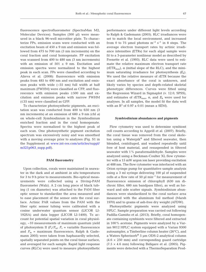
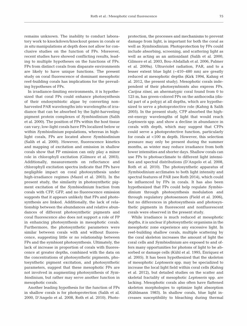
Over the habitat of the Leptoseris spp. mesophoticreef ecosystem, irradiance ranged from 68.7 ± 6.3µmol quanta m−2 s−1 at 65 m to 6 ± 0.9 µmol quantam−2 s−1 at 125 m (Fig. 1a), which represented 2.69%and 0.23% of surface PAR irradiance respectively.Most corals from the entire depth range (65−125 m)exhibited cyan/green fluorescence emission (64−89%; Fig. 1a); fluorescent and nonfluorescent coralswere found at every depth sampled. Two FPs wereresponsible for the cyan/green fluorescence: thehigher-energy CFP had peak excitation at 424 nm(FWHM > 54 nm) and peak emission at 490 nm(FWHM > 54nm) (n = 16; Fig. 1b), and the lower-energy GFP had peak excitation at 478 nm (FWHM >18 nm) and peak emission at 502 nm (FWHM =~30 nm) (n = 9; Fig. 1c). Two corals (collected from86−95 m) produced both CFP and GFP.
Technical limitations arose in the precise estima-tion of the excitation peak of GFP; the measurementsdid not extend above 480 nm. However, we considerthat it was likely that the measured GFP excitationpeak (478 nm) was very close to the true peakbecause the Stokes shift of GFP in corals is ~9−24 nm(Alieva et al. 2008). Additionally, some of the FWHMwere also not precisely measured because theFWHM was out of the range of the measurement.However, the data clearly showed that CFP hadmuch wider excitation and emission spectra com-pared to GFP, which is typical of these types of FPs.As a negative control, nonfluorescent corals lackedany FP peaks in their spectra (n = 13). For some coralswith low fluorescence, such as visual fluorescence inthe oral disc only (n = 8) and weak coenosarc fluores-cence (n = 1), it was not possible to obtain FP spectra,perhaps due to low instrument sensitivity. Addition-ally, one coral died prior to spectral analysis.
68
Roth et al.: Mesophotic coral fluorescence
FP type was significantly correlated with depthamong all the species (ρ = 0.71, p < 0.0001, n = 27;Fig. 1a) and within the species L. sp. 1 (ρ = 0.80, p <0.05, n = 9; Fig. 1d). CFP was prevalent in shallowercorals (65−85 m), the GFP was dominant in deepercorals (96−125 m), and the FPs overlapped at middledepths (86−95 m). The 4 species of Leptoseris all con-tained fluorescent and nonfluorescent individuals(Fig. 1d). L. scabra had the lowest proportion of fluo-rescent morphs (36%; n = 11), while 100% of L. tubu-lifera were fluorescent (n = 11). L. sp. 1 (n = 14) and L.hawaiiensis (n = 11) had 86% and 64% of corals withfluorescence, respectively (Fig. 1d). L. scabra onlyproduced the CFP; L. tubulifera mostly produced theCFP, with 2 individuals exhibiting both the CFP andthe GFP; L. sp. 1 produced both the CFP and theGFP; and L. hawaiiensis only produced the GFP(Fig. 1d). L. scabra was collected mostly from theshallower sites (65−85 m) and less with increasingdepth; L. tubulifera was collected primarily frommid-range depths (76−95 m); L. sp. 1 was collectedover the entire depth range (65−125 m); and L.hawaiiensis was only collected from the deepestrange (120−125 m) (Fig. 1d).
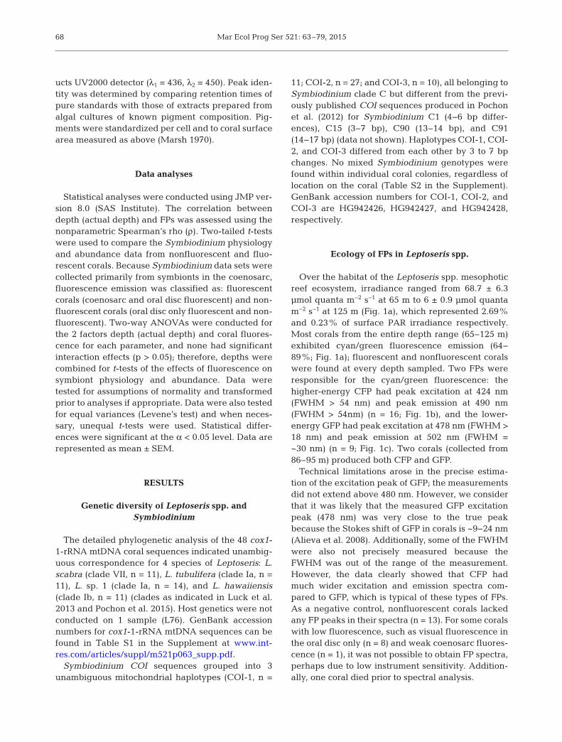
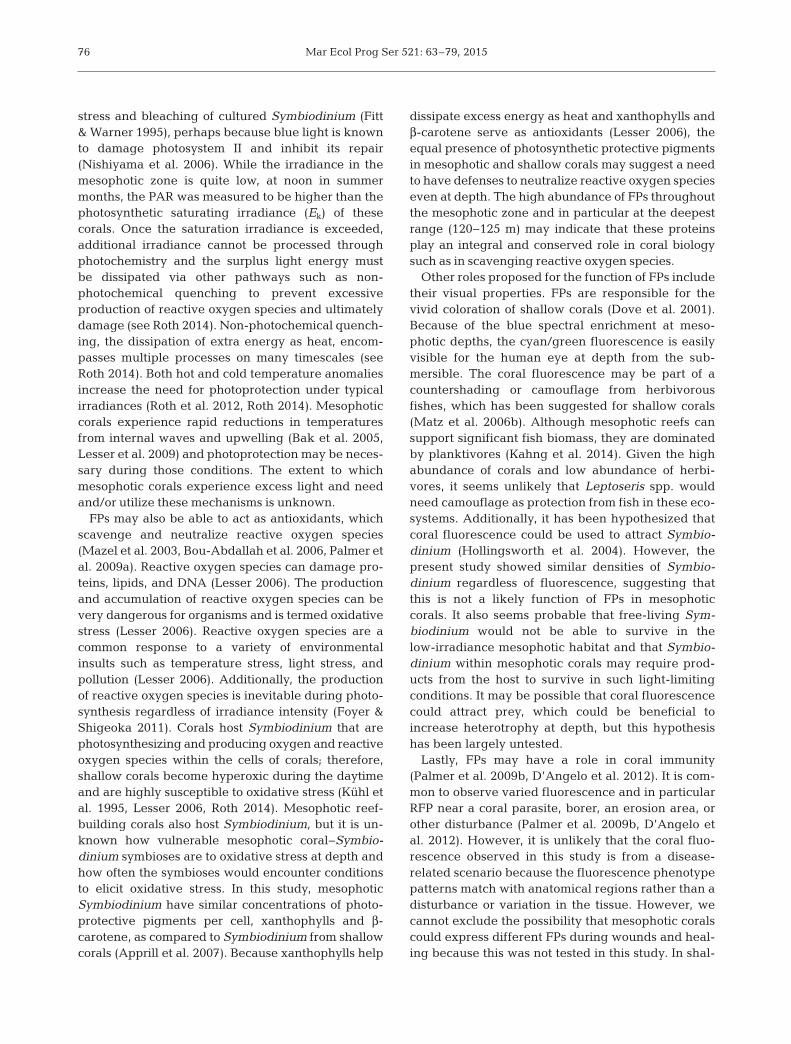
FP emission phenotypes in Leptoseris spp.
Mesophotic corals were observed with FP emissionpresent in the coenosarc (includes oral disc) (Fig. 2a),in the oral disc only (Fig. 2b), or without coral fluores-cence (Fig. 2c). In areas of the corals withoutcyan/green FP emission, the red fluorescence fromSymbiodinium photosystem II was visible (Fig. 2b,c).L. tubulifera was only observed with coenosarc fluo-rescence emission phenotype, while L. scabra, L. sp.1, and L. hawaiiensis contained both coenosarc andoral disc only fluorescence emission phenotypes(Fig. 2d). The coenosarc fluorescence emission phe-notype resulted from the CFP in L. scabra, GFP in L.hawaiiensis, and both CFP and GFP in L. tubuliferaand L. sp. 1. In contrast, the oral disc fluorescenceonly phenotype in L. scabra and L. sp. 1 resulted fromonly CFP. However, the number of samples with fluorescence emission only from the oral disc was low(n = 3), resulting in unclassified FP type(s) for mostoral disc only samples (n = 8). Rare coral fluorescenceemission phenotypes included a bright localization offluorescence spots (n = 7) in either random placement(Fig. S2a in the Supplement) or striations (Fig. S2b in
69
70 80 90 100 12002
468
10
1214
Num
ber
of c
oral
s
Depth bin (m)
Nonfluorescent corals
Fluorescent corals
CFP
CFP and GFP
GFP
Coral fluorescence
0
20
40
60
80
100
PA
R
PAR
0
2
4
6
8
10
12
Num
ber
of c
oral
s
(µm
ol q
uant
a m
–2 s
–1)
Emission
Excitation
CFP
0
0.2
0.4
0.6
0.8
1.2
300 400 500 600 700Rel
ativ
e in
tens
ity (a
.u.)
Wavelength (nm)
0
0.2
0.4
0.6
0.8
1.2
300 400 500 600 700Rel
ativ
e in
tens
ity (a
.u.)
Wavelength (nm)
GFP1.0 1.0
Depth bin (m)70 80 90 100 120 70 80 90 100 120 70 80 90 100 120 70 80 90 100 120
L. scabra L. hawaiiensisd
a b c
L. tubulifera L. sp. 1
Fig. 1. Ecology of coral fluorescent proteins (FPs) in Leptoseris spp. collected at 65 to 125 m. (a) Fluorescent and nonfluorescentcorals by depth plotted with photosynthetically active radiation (PAR) levels. A cyan fluorescent protein (CFP) and a greenfluo rescent protein (GFP) were identified from Leptoseris spp. (b,c) FP spectral characteristics showing excitation spectra withemission peak at 501 nm and emission spectra with 450 nm excitation. The excitation peaks were 424 and 478 nm for CFP andGFP, respectively, and the emission peaks were 490 and 502 nm, respectively. (d) Fluorescent corals and nonfluorescent corals
by depth and Leptoseris spp. Solid black bars represent fluorescent corals with unidentified FP type
Mar Ecol Prog Ser 521: 63–79, 201570
Nonfluorescent corals
Oral disc only
Coenosarc
0
2
4
6
8
10
12
Num
ber
of c
oral
s
d
a b c
L. scabra L. hawaiiensisL. tubulifera L. sp. 1
Depth bin (m)70 80 90 100 120 70 80 90 100 120 70 80 90 100 120 70 80 90 100 120
Fig. 2. Coral fluorescent protein localization in Leptoseris spp. Common patterns observed: (a) coral fluorescence in thecoenosarc and oral disc, (b) coral fluorescence in the oral disc only, and (c) no coral fluorescence. Leptoseris spp. were imagedusing a camera (Olympus C-5050) with a blue excitation light (Inon Z-240 strobe with NightSea blue filter, ~450 nm) and bar-rier filter (≥500 nm longpass, NightSea) on the lens of the camera for fluorescence (top row) and under white light (Inon Z-240
strobe) (bottom row). (d) Common fluorescence localization patterns by species and depth
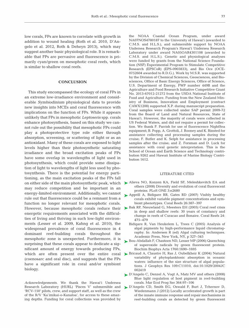
L. sc
abra
L. tu
bulife
ra
L. sp
. 102468
10121416
Num
ber
of c
oral
s
Non-fluorescent corals
Oral disc only
Coenosarc
COI-1COI-2COI-3
COI-10
5
10
15
20
25
30
02468
10121416
b Nonfluorescent corals
a
L. sc
abra
L. tu
bulife
ra
L. sp
. 1
L. h
awaii
ensis
Fluorescent corals
L. h
awaii
ensisCOI-2 COI-3
c
COI-1COI-2COI-3
Fig. 3. Fluorescence patterns and clade C Symbiodinium spp. in Leptoseris spp. (a) Corals with all 3 Symbiodinium haplotypesCOI-1, COI-2, and COI-3 and with either coenosarc fluorescence, oral disc only fluorescence, or no fluorescence. (b,c) Sym-
biodinium haplotypes COI-1, COI-2, and COI-3 in nonfluorescent and fluorescent corals
Roth et al.: Mesophotic coral fluorescence
the Supplement) in corals with coenosarc fluores-cence in L. tubulifera and L. sp. 1.
Leptoseris spp. fluorescence and Symbiodinium
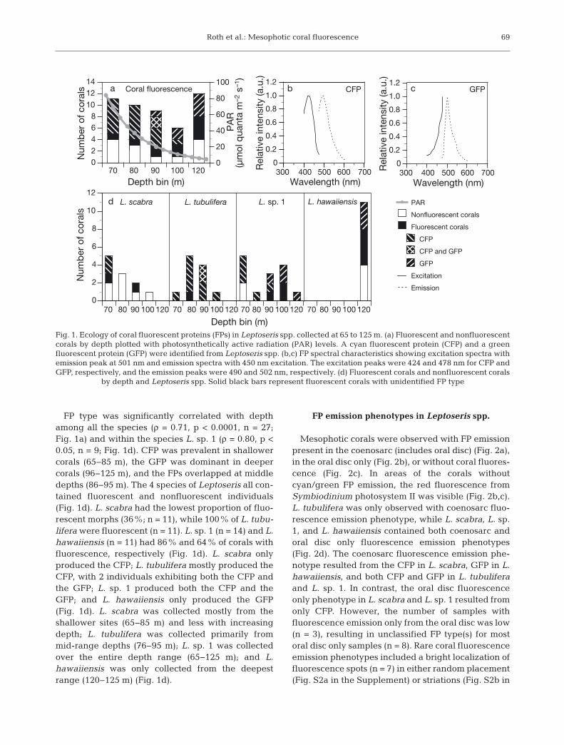
Three clade C haplotypes of Symbiodinium wereidentified from all corals both with and without fluo-rescence emission (Fig. 3a). The proportion of coralswith any type of visual fluorescence and haplotypeCOI-1 (n = 11), COI-2 (n = 27), and COI-3 (n = 10) was
64%, 78%, and 70% respectively. Corals that pro-duced the CFP hosted COI-2 and COI-3 haplotypes,while corals that produced the GFP hosted all 3 hap-lotypes. The corals that produced both the CFP andGFP hosted the COI-2 haplotype. L. scabra and L. sp.1 hosted 2 types of Symbiodinium (COI-2 and COI-3), while L. tubulifera only hosted COI-2, and L.hawaiiensis only hosted COI-1 (Fig. 3b,c). Nonfluo-rescent L. scabra hosted both COI-2 and COI-3,while fluorescent L. scabra only hosted COI-2. Incontrast, nonfluorescent L. sp. 1 hosted COI-3 and
71
Parameter Mean ± SEM t-testFluorescent Nonfluorescent t-ratio p df
rETRmax (µmol electrons m−2 s−1) 3.70 ± 0.33 3.67 ± 0.38 0.1 0.95 33α (µmol electrons) 0.25 ± 0.01 0.27 ± 0.02 −0.8 0.41 33Ek (µmol photons m−2 s−1) 14.19 ± 0.73 13.40 ± 0.82 0.7 0.47 33Maximum quantum yield (Fv/Fm) 0.66 ± 0.01 0.67 ± 0.01 −0.5 0.65 35Symbiodinium density (cells cm−2) 5.6 × 10⁵ ± 0.4 × 10⁵ 5.5 × 10⁵ ± 0.5 × 10⁵ 0.3 0.75 45Symbiodinium density (cells g−1 AFDW) 1.2 × 10¹⁰ ± 0.1 × 10¹⁰ 1.2 × 10¹⁰ ± 0.1 × 10¹⁰ −0.3 0.77 45Chl a (pg cell−1) 19.5 ± 1.5 16.9 ± 1.1 1.4 0.17 45Chl c2 (pg cell−1) 2.7 ± 0.2 2.2 ± 0.1 1.8 0.09 45Peridinin (pg cell−1) 8.7 ± 0.6 7.7 ± 0.5 1.3 0.20 45Diadinoxanthin + diatoxanthin (pg cell−1) 2.5 ± 0.2 2.1 ± 0.1 1.7 0.10 45β-carotene (pg cell−1) 0.46 ± 0.04 0.38 ± 0.02 1.8 0.07 45Chl a (ng cm−2) 355 ± 36 293 ± 24 −0.7 0.48 45Chl c2 (ng cm−2) 49 ± 5 39 ± 3 1.7 0.10 45Peridinin (ng cm−2) 160 ± 17 132 ± 11 1.4 0.17 45Diadinoxanthin + diatoxanthin (ng cm−2) 46 ± 4 37 ± 3 1.7 0.10 45β-carotene (ng cm−2) 8 ± 1 8 ± 1 1.9 0.06 45Chl c2:chl a 0.14 ± 0.01 0.13 ± 0.01 1.1 0.27 45Peridinin:chl a 0.45 ± 0.01 0.45 ± 0.01 −0.9 0.40 45
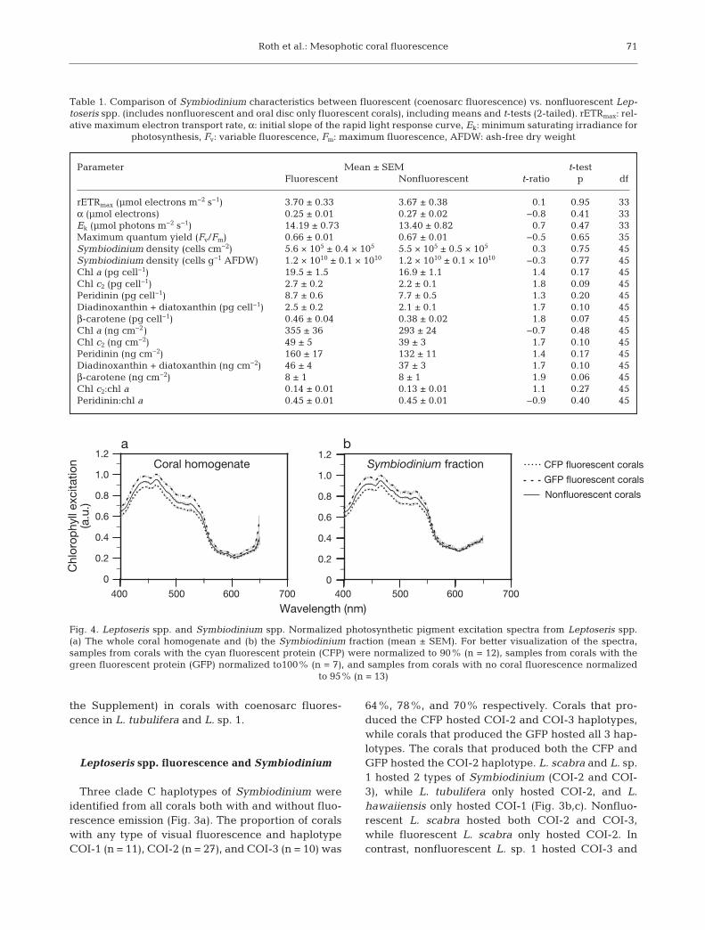
Table 1. Comparison of Symbiodinium characteristics between fluorescent (coenosarc fluorescence) vs. nonfluorescent Lep-toseris spp. (includes nonfluorescent and oral disc only fluorescent corals), including means and t-tests (2-tailed). rETRmax: rel-ative maximum electron transport rate, α: initial slope of the rapid light response curve, Ek: minimum saturating irradiance for
photosynthesis, Fv: variable fluorescence, Fm: maximum fluorescence, AFDW: ash-free dry weight
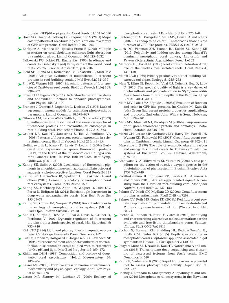
Wavelength (nm)
0
0.2
0.4
0.6
0.8
1.2
400 500 600 700 400 500 600 700
Chl
orop
hyll
exci
tatio
n(a
.u.)
Coral homogenate Symbiodinium fraction CFP fluorescent corals
GFP fluorescent corals
Nonfluorescent corals
1.0
0
0.2
0.4
0.6
0.8
1.2
1.0
a b
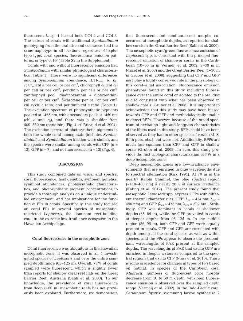
Fig. 4. Leptoseris spp. and Symbiodinium spp. Normalized photosynthetic pigment excitation spectra from Leptoseris spp.(a) The whole coral homogenate and (b) the Symbiodinium fraction (mean ± SEM). For better visualization of the spectra, samples from corals with the cyan fluorescent protein (CFP) were normalized to 90% (n = 12), samples from corals with thegreen fluorescent protein (GFP) normalized to100% (n = 7), and samples from corals with no coral fluorescence normalized
to 95% (n = 13)
Mar Ecol Prog Ser 521: 63–79, 201572
fluorescent L. sp. 1 hosted both COI-2 and COI-3.The subset of corals with additional Symbiodiniumgenotyping from the oral disc and coenosarc had thesame haplotype in all locations regardless of haplo-type type, coral species, fluorescence emission pat-terns, or type of FP (Table S2 in the Supplement).
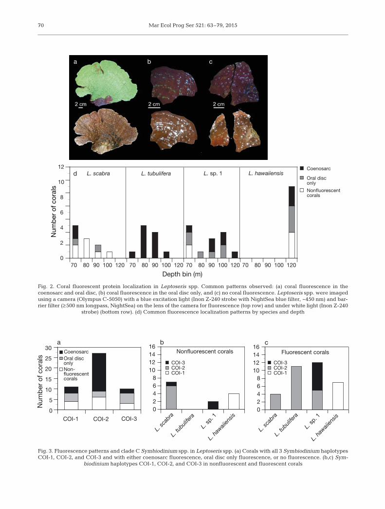
Corals with and without fluorescence emission hadSymbiodinium with similar physiological characteris-tics (Table 1). There were no significant differencesamong Symbiodinium abundance, rETRmax, α, Ek,Fv/Fm, chl a per cell or per cm2, chlorophyll c2 (chl c2)per cell or per cm2, peridinin per cell or per cm2, xanthophyll pool (diadinoxanthin + diatoxanthin)per cell or per cm2, β-carotene per cell or per cm2,chl c2:chl a ratio, and peridinin:chl a ratio (Table 1).The excitation spectrum of photosynthetic pigmentspeaked at ~465 nm, with a secondary peak at ~450 nm(chl a and c2), and there was a shoulder from500−550 nm (peridinin) (Fig. 4; cf. Bricaud et al. 2004).The excitation spectra of photosynthetic pigments inboth the whole coral homogenate (includes Symbio-dinium) and Symbiodinium fraction were similar, andthe spectra were similar among corals with CFP (n =12), GFP (n = 7), and no fluorescence (n = 13) (Fig. 4).
DISCUSSION
This study combined data on visual and spectralcoral fluorescence, host genetics, symbiont genetics,symbiont abundances, photosynthetic characteris-tics, and photosynthetic pigment concentrations toprovide a thorough analysis on a unique understud-ied environment, and has implications for the func-tion of FPs in corals. Specifically, this study focusedon coral FPs in several species of mesophotic-restricted Leptoseris, the dominant reef-buildingcoral in the extreme low-irradiance ecosystem in theHawaiian Archipelago.
Coral fluorescence in the mesophotic zone
Coral fluorescence was ubiquitous in the Hawaiianmesophotic zone; it was observed in all 4 investi-gated species of Leptoseris and over the entire sam-pled depth range (65−125 m). Overall, 73% of coralssampled were fluorescent, which is slightly lowerthan reports for shallow coral reef flats on the GreatBarrier Reef, Australia (Salih et al. 2000). To ourknowledge, the prevalence of coral fluorescencefrom deep (>60 m) mesophotic reefs has not previ-ously been explored. Furthermore, we demonstrate
that fluorescent and nonfluorescent morphs co-occurred at mesophotic depths, as reported for shal-low corals in the Great Barrier Reef (Salih et al. 2000).The mesophotic cyan/green fluorescence emission ofLeptoseris spp. is consistent with the principal fluo-rescence emission of shallower corals in the Carib-bean (10−60 m in Vermeij et al. 2002, 3−30 m inMazel et al. 2003) and the Great Barrier Reef (1−30 min Gruber et al. 2008), suggesting that CFP and GFPmay play a highly conserved role in the physiology ofthis coral−algal association. Fluorescence emissionphenotypes found in this study including fluores-cence over the entire coral or isolated to the oral discis also consistent with what has been observed inshallow corals (Gruber et al. 2008). It is important toacknowledge that this study may have been biasedtowards CFP and GFP and methodologically unableto detect RFPs. However, because of the broad spec-trum of excitation light and longpass characteristicsof the filters used in this study, RFPs could have beenobserved as they had in other species of corals (M. S.Roth pers. obs.), but were not. Additionally, RFPs aremuch less common than CFP and GFP in shallowcorals (Gruber et al. 2008). In sum, this study pro-vides the first ecological characterization of FPs in adeep mesophotic zone.
Deep mesophotic zones are low-irradiance envi-ronments that are enriched in blue wavelengths dueto spectral attenuation (Kirk 1994). At 70 m in thenearby Kalohi Channel, the blue spectral region(~410−480 nm) is nearly 20% of surface irradiance(Kahng et al. 2012). The present study found thatmesophotic Leptoseris spp. express 2 FPs with differ-ent spectral characteristics: CFP (λex = 424 nm, λem =490 nm) and GFP (λex ≈ 478 nm, λem = 502 nm). Strik-ingly, CFP was dominant in corals at shallowerdepths (65−85 m), while the GFP prevailed in coralsat deeper depths from 96−125 m. In the middleregion (86−95 m), both CFP and GFP were equallypresent in corals. CFP and GFP are correlated withdepth among all the coral species as well as withinspecies, and the FPs appear to absorb the predomi-nant wavelengths of PAR present at the sampleddepths. The wavelengths of PAR that excite GFP areenriched in deeper waters as compared to the spec-tral regions that excite CFP (Mass et al. 2010). Thereis some precedence for changes in types of FPs basedon habitat. In species of the Caribbean coralMadracis, numbers of fluorescent color morphsdecrease from 10 to 60 m depth, yet green fluores-cence emission is observed over the sampled depthrange (Vermeij et al. 2002). In the Indo-Pacific coralSeriatopora hystrix, swimming larvae synthesize 2
Roth et al.: Mesophotic coral fluorescence
GFPs, while the benthic adults produce a CFP, andthese 2 life history stages occupy different habitatswith distinct irradiance environments (Roth et al.2013). Overall, our finding of 2 types of FPs along thedepth gradient among congeners and conspecifics inthe mesophotic zone fits well with other studies inshallow systems, but has never been observed overthese depth zones of 65 to 125 m.
Previous studies have also shown that corals canalso vary the amount of FPs within their tissues inresponse to changes in environmental and physio-logical conditions (D’Angelo et al. 2008, Roth et al.2010, Roth & Deheyn 2013). However, in shallowCaribbean corals (3−30 m), there is no correlationbetween depth and GFP abundance in Montastreacavernosa and M. faveolata (Mazel et al. 2003). Geneexpression of M. cavernosa (8−24 m) suggests that 1or possibly 2 GFPs are inversely related with depth,but 5 other GFPs are not correlated with depth (Kaoet al. 2007). FP gene expression can change dynami-cally in response to environmental conditions in shal-low corals and FP transcription is most regulated byblue light rather than red or green light (D’Angelo etal. 2008). Additionally, the abundance of FPs withincorals rapidly decreases in response to both cold andheat stress (Roth & Deheyn 2013). Future studiesshould quantify the FP abundance and gene expres-sion in mesophotic corals from a variety of speciesand depths, as well as compare CFP and GFP genesfrom shallow and mesophotic corals to determinepossible differences or similarities.
The similarity in gross morphology of Leptoserisspp. and the high morphological plasticity withinspecies makes it difficult to separate species bothwhile viewing corals from the submersible as well aswhen handling the samples in the laboratory. In thisstudy, genetics revealed 4 species of Leptoseris (L.scabra, L. tubulifera, L. sp. 1, and L. hawaiiensis),each with distinct fluorescence patterns. The cyan/green fluorescence was localized in the coenosarc inall species, but only L. scabra, L. sp. 1, and L. hawai-iensis exhibited the oral disc only phenotype. Themost divergent species in our study, L. scabra (Lucket al. 2013), had the lowest abundance of fluorescentcorals (36%) and only produced CFP. It is intriguingto ask why L. scabra has a much lower abundance offluorescence, and it may suggest that their photo-physiology is different from the other species withless selective pressure for fluorescence. L. tubuliferaand L. sp. 1, which were the most closely related(Luck et al. 2013), had the highest abundances of flu-orescent corals (100% and 86% respectively) andproduced CFP and GFP. All colonies of L. tubulifera
were fluorescent over their entire surface, suggest-ing that fluorescence may have an adaptive valuein this species. Colonies of L. hawaiiensis, whichwere only located at the deepest sampled depths(120−125 m), were commonly fluorescent (64%) andonly produced GFP. Corals can quickly regulate FPtranscription and concentration on short time scalesin response to changes in their environment andstress (D’Angelo et al. 2008, Roth et al. 2010, Roth &Deheyn 2013). Thus, in species where fluorescentmorphs are highly abundant, weakly fluorescence ornonfluorescent morphs could indicate a differentphysiological state. Coral fluorescence has also beennoted in species of shallow Leptoseris and meso -photic GFP emission has similar emission to GFPfrom the Caribbean coral Leptoseris cucullata col-lected from 25 m and often shallower (Mazel et al.2003, C. H. Mazel pers. comm.).
Because FPs are part of the photophysiology of thecoral−algal symbiosis, this study also investigated alarge number of Symbiodinium characteristics. To ourknowledge, this is the first study to show that Symbio-dinium genotype does not have any relationship withcoral fluorescence. Additionally, if the coral is fluores-cent, the genotype of Symbiodinium does not affectthe fluorescence emission phenotype. Symbiodiniumdensity was highly variable, and no significant differ-ences were observed with corals that were fluorescentor nonfluorescent. Similarly, a study of shallow coralsshowed no overlap between the distribution of FPsand Symbiodinium (Gruber et al. 2008). Photosyn-thetic pigments absorb different wavelengths, withthe primary peak of chl a absorbing higher-energylight than chl c2, which is then absorbing higher-energy light than peridinin (Bri caud et al. 2004). Peri-dinin is an important light-harvesting pigment inSymbiodinium that extends the spectral range of lightharvested for photosynthesis into green wavelengths(Bricaud et al. 2004). The peak absorbance of peri-dinin most closely aligns with peak emissions of CFPand GFP. In this study, the concentrations of chl a,chl c2, and peridinin were similar between fluorescentand nonfluorescent corals, as were the ratios of chl c2:chl a and peridinin:chl a. The chlorophyll excitationspectra from corals with GFP, CFP, and no fluores-cence were similar in the coral homogenate, the Sym-biodinium fraction, and between the two fractions.The FP peak emissions (490 and 502 nm) do not over-lap with the absorbance peaks of chlorophylls (440−465 nm), but rather the shoulder of the photosyntheticpigment excitation and the peak absorbance of peri-dinin (490−510 nm). It may be possible that the rela-tionship between FPs and photo synthesis was dis-
73
Mar Ecol Prog Ser 521: 63–79, 201574
rupted when the coral tissue was removed from theskeleton; unfortunately, measurements with theskeleton provided unreliable excitation spectra andtherefore the coral tissue needed to be removed formeasurements.
It is interesting to note that the excitation peaks ofCFP and GFP fall on either side of the chlorophyllabsorbance peaks, which may reduce competition forparticular wavelengths of PAR and may be evidenceof energy partitioning. Having FPs and antennaecomplexes with distinct maximum absorbance peaksmay signal an evolutionary change to avoid competi-tion for irradiance. However, the excitation peak, andin particular that of CFP as well as intact light-har-vesting complexes, are broad and therefore theremay be some overlap between wavelengths that canbe absorbed by FPs or photosynthetic pigments. But,because of the distance between FPs and antennacomplexes, energy cannot be directly transferredfrom coral FPs to Symbiodinium in shallow corals(Gilmore et al. 2003). Nonetheless, it may be possiblefor antenna complexes to absorb re-emitted wave-lengths from FPs. However, the data in this studyshow that CFP and GFP absorption have limitedimpact on symbiont photosynthesis in mesophoticcorals, and this would be particularly true under theextremely low-irradiance regimes in winter months.This finding is similar to that from shallow Caribbeancorals, where a study also concluded that FP absorp-tion, emission, and reflection has an insignificanteffect on Symbiodinium photosynthesis (Mazel et al.2003).
Additional perspective is gained in examining thephotosynthetic parameters rETRmax, α, Ek, and Fv/Fm
in both fluorescent and nonfluorescent corals. Therewere no detectable differences between these corals,suggesting that Symbiodinium photophysiology isnot influenced by coral fluorescence. Because spec-tral features influence photosynthetic measurements(Mass et al. 2010), it is possible that using an irradi-ance source with a spectrum similar to the nativehabitat of these corals rather than the white light thatwas available would have produced a differentresult. For example, in Stylophora pistillata from40 m, the ratio of gross photosynthesis to respirationdoubled when measured under blue light as opposedto full spectrum light (Mass et al. 2010). In the RedSea, the mesophotic-restricted coral Leptoseris frag-ilis has an unidentified coral fluorescent pigment thatis hypothesized to be involved in improving the photosynthesis of Symbiodinium because higherphotosynthetic rates were obtained with a narrowspectrum of light matching the excitation of the fluo-
rescent pigment at some light intensities (Schlichter& Fricke 1990). The fluorescent pigment of L. fragilishas a peak emission (~440 nm) that overlaps with thepeak absorbance of chl a (Schlichter & Fricke 1990).It is not possible to measure the emission of a FP inchloroform because FPs degrade in chloroform, andtherefore it seems unlikely that the fluorescent pig-ment in L. fragilis is a FP (Matz et al. 2006a). Unfortu-nately, the fluorescent pigment for L. fragilis was notcharacterized further and remains unidentified. Incontrast, the FPs of Hawaiian meso photic Leptoserisdid not have any influence over the photophysiologyof Symbiodinium in meso photic corals, with themethods used here.
This study provides a first characterization of theprevalence and types of coral FPs and fluorescenceemission phenotypes in dominant mesophotic reef-building corals and suggests that FPs are importantfor coral physiology. Because of the difficulty ofobtaining samples from the mesophotic zone, thisstudy does have limitations of modest sample sizes,particularly when trying to compare among differentdepths and species. However, this study offers afoundation for coral fluorescence in the mesophoticzone and hopefully will stimulate more research inthis direction. Future research could investigate therelationship between FP gene expression, FP abun-dance within coral tissues, and localization of FPsover depth. Microscopy measurements on the spatialseparation between FPs and Symbiodinium will con-firm or refute the possibility of energy transfer. Whilethe focus of this study was broad in including 4closely related species over a large depth range(65−125 m), future studies targeting a specific Lep-toseris species, perhaps with only 2 depth ranges(e.g. a shallow versus deeper site) and includingreciprocal transplant experiments and additionalphysiological measurements, such as oxygen evolu-tion, under a variety of irradiance intensities andspectral qualities, may improve the understanding ofthe role of FPs in corals. While this study was focusedon the ecological characterization among dominantcorals of the mesophotic zone and does not deter-mine the function of FPs in mesophotic corals, thisstudy can provide evidence to support or negate var-ious hypotheses.
On the function of FPs
Despite early observations of coral fluorescenceand the widespread use of FPs as a tool in research,the function of FPs in shallow and deep corals
Roth et al.: Mesophotic coral fluorescence
remains unknown. The inability to conduct labora-tory work to knockdown/knockout genes in corals orin situ manipulations at depth does not allow for con-clusive studies on the function of FPs. Moreover,recent studies have showed conflicting results, lead-ing to multiple hypotheses on the functions of FPs.FPs from distinct corals from disparate environmentsare likely to have unique functions. The presentstudy on coral fluorescence of dominant mesophoticreef-building corals has implications for the prevail-ing hypotheses of FPs.
In irradiance-limiting environments, it is hypothe-sized that coral FPs could enhance photosynthesisof their endosymbiotic algae by converting non -harvested PAR wavelengths into wavelengths of irra-diance that can be absorbed by the light-harvestingpigment protein complexes of Symbiodinium (Salihet al. 2000). The position of FPs within the host tissuecan vary; low-light corals have FPs localized below orwithin Symbiodinium populations, whereas in high-light corals, FPs are located above Symbiodinium(Salih et al. 2000). However, fluorescence kineticsand mapping of excitation and emission in shallowcorals show that FP emission can only play a minorrole in chlorophyll excitation (Gilmore et al. 2003).Additionally, measurements on reflectance andchlorophyll excitation spectra indicate that FPs havenegligible impact on coral photosynthesis underhigh-irradiance regimes (Mazel et al. 2003). In thepresent study, the similarity in photosynthetic pig-ment excitation of the Symbiodinium fraction fromcorals with CFP, GFP, and no fluorescence emissionsuggests that it appears unlikely that FPs and photo-synthesis are linked. Additionally, the lack of rela-tionship between the abundances and relative abun-dances of different photosynthetic pigments andcoral fluorescence also does not support a role of FPin enhancing photosynthesis in mesophotic corals.Furthermore, the photosynthetic parameters weresimilar between corals with and without fluores-cence, suggesting little or no relationship betweenFPs and the symbiont photosynthesis. Ultimately, thelack of increase in proportion of corals with fluores-cence at greater depths, combined with the data onthe concentrations of photosynthetic pigments, pho-tosynthetic pigment excitation, and photosyntheticparameters, suggest that these mesophotic FPs arenot involved in augmenting photosynthesis of Sym-biodinium, but rather may serve another function inmesophotic corals.
Another leading hypothesis for the function of FPsin shallow corals is for photoprotection (Salih et al.2000, D’Angelo et al. 2008, Roth et al. 2010). Photo-
protection, the processes and mechanisms to preventdamage from light, is important for both the coral aswell as Symbiodinium. Photoprotection by FPs couldinclude absorbing, screening, and scattering light aswell as acting as an antioxidant (Salih et al. 2000,Gilmore et al. 2003, Bou-Abdallah et al. 2006, Palmeret al. 2009a). Ultraviolet radiation, PAR, and to alesser extent blue light (~410−480 nm) are greatlyreduced at mesophotic depths (Kirk 1994, Kahng etal. 2012, the present study). Mesophotic corals inde-pendent of their photosymbionts also express FPs.Carijoa riisei, an ahermatypic coral found from 0 to125 m, has green-colored FPs on the anthocodia (dis-tal part of a polyp) at all depths, which are hypothe-sized to serve a photoprotective role (Kahng & Salih2005). In the present study, CFP absorbed the high-est-energy wavelengths of light that would reachLeptoseris spp. and show a decline in abundance incorals with depth, which may suggest that CFPscould serve a photoprotective function, particularlyfor corals at <100 m depth. However, this selectionpressure may only be present during the summermonths, as winter may reduce irradiance from bothlower intensities and shorter days. Shallow corals canuse FPs to photoacclimate to different light intensi-ties and spectral distributions (D’Angelo et al. 2008,Roth et al. 2010). The photosynthetic apparatus ofSymbiodinium ac climates to both light intensity andspectral features of PAR (see Roth 2014), which couldbe influenced by FPs in corals. It has also beenhypothesized that FPs could help regulate Symbio-dinium through photosynthesis modulation andthrough regulatory photosensors (Field et al. 2006),but no differences in photosynthesis and photosyn-thetic pigments in fluorescent and nonfluorescentcorals were observed in the present study.
While irradiance is much reduced at mesophoticdepths, it is unclear if photosynthetic organisms in themesophotic zone experience any excessive light. Inreef-building shallow corals, multiple scattering bythe coral skeleton increases the amount of light thecoral cells and Symbiodinium are exposed to and of-fers many opportunities for photons of light to be ab-sorbed or damage cells (Kühl et al. 1995, Enríquez etal. 2005). It has been hypothesized that the skeletonof mesophotic Leptoseris spp. may be specialized toincrease the local light field within coral cells (Kahnget al. 2012), but detailed studies on the scatter andskeletal fractality of mesophotic Leptoseris spp. arelacking. Mesophotic corals also often have flattenedskeleton morphologies to optimize light absorption(Kühlmann 1983). In shallow corals, blue light in-creases susceptibility to bleaching during thermal
75
Mar Ecol Prog Ser 521: 63–79, 201576
stress and bleaching of cultured Symbiodinium (Fitt& Warner 1995), perhaps because blue light is knownto damage photosystem II and inhibit its repair(Nishiyama et al. 2006). While the irradiance in themesophotic zone is quite low, at noon in summermonths, the PAR was measured to be higher than thephotosynthetic saturating irradiance (Ek) of thesecorals. Once the saturation irradiance is ex ceeded,additional irradiance cannot be processed throughphotochemistry and the surplus light energy mustbe dissipated via other pathways such as non -photochemical quenching to prevent excessive production of reactive oxygen species and ultimatelydamage (see Roth 2014). Non-photochemical quench-ing, the dissipation of extra energy as heat, encom-passes multiple processes on many timescales (seeRoth 2014). Both hot and cold temperature anomaliesincrease the need for photoprotection under typicalirradiances (Roth et al. 2012, Roth 2014). Mesophoticcorals experience rapid reductions in temperaturesfrom internal waves and up welling (Bak et al. 2005,Lesser et al. 2009) and photoprotection may be neces-sary during those conditions. The extent to whichmesophotic corals experience excess light and needand/or utilize these mechanisms is unknown.
FPs may also be able to act as antioxidants, whichscavenge and neutralize reactive oxygen species(Mazel et al. 2003, Bou-Abdallah et al. 2006, Palmer etal. 2009a). Reactive oxygen species can damage pro-teins, lipids, and DNA (Lesser 2006). The productionand accumulation of reactive oxygen species can bevery dangerous for organisms and is termed oxidativestress (Lesser 2006). Reactive oxygen species are acommon response to a variety of environmentalinsults such as temperature stress, light stress, andpollution (Lesser 2006). Additionally, the productionof reactive oxygen species is inevitable during photo-synthesis regardless of irradiance intensity (Foyer &Shigeoka 2011). Corals host Symbiodinium that arephotosynthesizing and producing oxygen and reactiveoxygen species within the cells of corals; therefore,shallow corals become hyperoxic during the daytimeand are highly susceptible to oxidative stress (Kühl etal. 1995, Lesser 2006, Roth 2014). Mesophotic reef-building corals also host Symbiodinium, but it is un-known how vulnerable mesophotic coral−Symbio-dinium symbioses are to oxidative stress at depth andhow often the symbioses would encounter conditionsto elicit oxidative stress. In this study, mesophoticSymbiodinium have similar concentrations of photo-protective pigments per cell, xanthophylls and β-carotene, as compared to Symbiodinium from shallowcorals (Apprill et al. 2007). Because xanthophylls help
dissipate excess energy as heat and xanthophylls andβ-carotene serve as antioxidants (Lesser 2006), theequal presence of photosynthetic protective pigmentsin mesophotic and shallow corals may suggest a needto have defenses to neutralize reactive oxygen specieseven at depth. The high abundance of FPs throughoutthe mesophotic zone and in particular at the deepestrange (120−125 m) may indicate that these proteinsplay an integral and conserved role in coral biologysuch as in scavenging reactive oxygen species.
Other roles proposed for the function of FPs includetheir visual properties. FPs are responsible for thevivid coloration of shallow corals (Dove et al. 2001).Because of the blue spectral enrichment at meso -photic depths, the cyan/green fluorescence is easilyvisible for the human eye at depth from the sub-mersible. The coral fluorescence may be part of acountershading or camouflage from herbivorousfishes, which has been suggested for shallow corals(Matz et al. 2006b). Although mesophotic reefs cansupport significant fish biomass, they are dominatedby planktivores (Kahng et al. 2014). Given the highabundance of corals and low abundance of herbi-vores, it seems unlikely that Leptoseris spp. wouldneed camouflage as protection from fish in these eco-systems. Additionally, it has been hypothesized thatcoral fluorescence could be used to attract Symbio-dinium (Hollingsworth et al. 2004). However, thepresent study showed similar densities of Symbio-dinium regardless of fluorescence, suggesting thatthis is not a likely function of FPs in mesophoticcorals. It also seems probable that free-living Sym-biodinium would not be able to survive in the low-irradiance meso photic habitat and that Symbio-dinium within mesophotic corals may require prod-ucts from the host to survive in such light-limitingconditions. It may be possible that coral fluorescencecould attract prey, which could be beneficial toincrease heterotrophy at depth, but this hypothesishas been largely untested.
Lastly, FPs may have a role in coral immunity(Palmer et al. 2009b, D’Angelo et al. 2012). It is com-mon to observe varied fluorescence and in particularRFP near a coral parasite, borer, an erosion area, orother disturbance (Palmer et al. 2009b, D’Angelo etal. 2012). However, it is unlikely that the coral fluo-rescence observed in this study is from a disease-related scenario because the fluorescence phenotypepatterns match with anatomical regions rather than adisturbance or variation in the tissue. However, wecannot exclude the possibility that mesophotic coralscould express different FPs during wounds and heal-ing because this was not tested in this study. In shal-
Roth et al.: Mesophotic coral fluorescence
low corals, FPs are known to correlate with growth inaddition to wound healing (Roth et al. 2010, D’An-gelo et al. 2012, Roth & Deheyn 2013), which maysuggest another basic physiological role. It is remark-able that FPs are pervasive and fluorescence is pri-marily cyan/green on mesophotic coral reefs, whichis similar to shallow coral reefs.
CONCLUSION
This study encompassed the ecology of coral FPs inan extreme low-irradiance environment and consid-erable Symbiodinium physiological data to providenew insights into MCEs and coral fluorescence withimplications on the function of FPs. While it appearsunlikely that FPs in mesophotic Leptoseris spp. coralsenhance photosynthesis, based on this study we can-not rule out the possibility that mesophotic FPs couldplay a photoprotective type role either throughabsorption, screening, or scattering of light or as anantioxidant. Many of these corals are exposed to lightlevels higher than their photosynthetic saturatingirradiance and the broad excitation peaks of FPshave some overlap in wavelengths of light used inphotosynthesis, which could provide some dissipa-tion of light to wavelengths of light less used by pho-tosynthesis. There is the potential for energy parti-tioning, as the main excitation peaks of the FPs fallon either side of the main photosynthetic peak, whichmay reduce competition and be important in anenergy-limited environment. Additionally, we cannotrule out that fluorescence could be a remnant from afunction no longer relevant for mesophotic corals.However, because mesophotic corals reduce theirenergetic requirements associated with the difficul-ties of living and thriving in such low-light environ-ments (Lesser et al. 2009, Kahng et al. 2010), thewidespread prevalence of coral fluorescence in 4dominant reef-building corals throughout themesophotic zone is unexpected. Furthermore, it issurprising that these corals appear to dedicate a sig-nificant amount of energy towards producing FPs,which are often present over the entire coral(coenosarc and oral disc), and suggests that the FPshave a significant role in coral and/or symbiont biology.
Acknowledgements. We thank the Hawai‘i UnderseaResearch Laboratory (HURL) ‘Pisces V’ submersible and‘RCV-150’ pilots, crew, and support staff, as well as the crewof the R/V ‘Ka`imikai-o-Kanaloa’, for access to these amaz-ing depths. Funding for coral collections was provided by
the NOAA Coastal Ocean Program, under awardNA07NOS4780187 to the University of Hawai‘i (awarded toC.M.S. and H.L.S.), and submersible support by NOAAUndersea Research Program’s Hawai‘i Undersea ResearchLaboratory under award NA05OAR4301108 (awarded toC.M.S. and H.L.S.). Genetic and physiological analyseswere funded by grants from the National Science Founda-tion (NSF) Experimental Program to Stimulate CompetitiveResearch (EPSCoR) (EPS-0903833), and Bio Oce (OCE-0752604 awarded to R.D.G.). Work by M.S.R. was supportedby the Division of Chemical Sciences, Geosciences, and Bio-sciences, Office of Basic Energy Sciences, Office of Science,U.S. Department of Energy, FWP number 449B and theAgriculture and Food Research Initiative Competitive GrantNo. 2013-67012-21272 from the USDA National Institute ofFood and Agriculture. Funding from the New Zealand Min-istry of Business, Innovation and Employment (contractCAWX1208) supported X.P. during manuscript preparation.Coral samples were collected under SAP permit 2009-72from the Board of Land and Natural Resources, State ofHawai‘i. However, the majority of corals were collected inUS Federal Waters, and did not require a permit for collec-tion. We thank F. Parrish for use of fluorescence NightSeaequipment; B. Popp, A. Grottoli, J. Rooney and K. Binsted forassistance collecting and processing samples during thecruise; F. Butler and K. Selph for assistance in processingsamples after the cruise; and Z. Forsman and D. Luck forassistance with coral genetic interpretation. This is theSchool of Ocean and Earth Science and Technology contri-bution 9262 and Hawaii Institute of Marine Biology Contri-bution 1612.
LITERATURE CITED
Alieva NO, Konzen KA, Field SF, Meleshkevitch EA andothers (2008) Diversity and evolution of coral fluorescentproteins. PLoS ONE 3: e2680
Apprill A, Bidigare RR, Gates RD (2007) Visibly healthycorals exhibit variable pigment concentrations and sym-biont phenotypes. Coral Reefs 26: 387−397
Bak RP, Nieuwland G, Meesters EH (2005) Coral reef crisisin deep and shallow reefs: 30 years of constancy andchange in reefs of Curacao and Bonaire. Coral Reefs 24: 475−479
Bidigare R, Van Heukelem L, Trees C (2005) Analysis ofalgal pigments by high-performance liquid chromatog-raphy. In: Andersen R (ed) Algal culturing techniques.Academic Press, New York, NY, p 327−345
Bou-Abdallah F, Chasteen ND, Lesser MP (2006) Quenchingof superoxide radicals by green fluorescent protein.Biochim Biophys Acta 1760: 1690−1695
Bricaud A, Claustre H, Ras J, Oubelkheir K (2004) Naturalvariability of phytoplanktonic absorption in oceanicwaters: influence of the size structure of algal popula-tions. J Geophys Res 109: C11010, doi: 10.1029/ 2004JC002419
D’Angelo C, Denzel A, Vogt A, Matz MV and others (2008)Blue light regulation of host pigment in reef-buildingcorals. Mar Ecol Prog Ser 364: 97−106
D’Angelo CD, Smith EG, Oswald F, Burt J, Tchernov D,Wiedenmann J (2012) Locally accelerated growth is partof the innate immune response and repair mechanisms inreef-building corals as detected by green fluorescent
77
Mar Ecol Prog Ser 521: 63–79, 2015
protein (GFP)-like pigments. Coral Reefs 31: 1045−1056Dove SG, Hoegh-Guldberg O, Ranganathan S (2001) Major
colour patterns of reef-building corals are due to a familyof GFP-like proteins. Coral Reefs 19: 197−204
Enríquez S, Méndez ER, Iglesias-Prieto R (2005) Multiplescattering on coral skeletons enhances light absorptionby symbiotic algae. Limnol Oceanogr 50: 1025−1032
Falkowski PG, Jokiel PL, Kinzie RA (1990) Irradiance andcorals. In: Dubinsky Z (ed) Ecosystems of the world: coralreefs, Vol 25. Elsevier, Amsterdam, p 89−107
Field SF, Bulina MY, Kelmanson IV, Bielawski JP, Matz MV(2006) Adaptive evolution of multicolored fluorescentproteins in reef-building corals. J Mol Evol 62: 332−339
Fitt WK, Warner ME (1995) Bleaching patterns of four spe-cies of Caribbean reef corals. Biol Bull (Woods Hole) 189: 298−307
Foyer CH, Shigeoka S (2011) Understanding oxidative stressand antioxidant functions to enhance photosynthesis.Plant Physiol 155: 93−100
Frenette J, Demers S, Legendre L, Dodson JJ (1993) Lack ofagreement among models for estimating photosyntheticparameters. Limnol Oceanogr 38: 679−687
Gilmore AM, Larkum AWD, Sallh A, Itoh S and others (2003)Simultaneous time resolution of the emission spectra offluorescent proteins and zooxanthellar chlorophyll inreef-building coral. Photochem Photobiol 77: 515−523
Gruber DF, Kao HT, Janoschka S, Tsai J, Pieribone VA(2008) Patterns of fluorescent protein expression in scle-ractinian corals. Biol Bull (Woods Hole) 215: 143−154
Hollingsworth L, Krupp D, Lewis T, Leong J (2004) Earlyonset and expression of green fluorescent proteins(GFPs) in the larvae of the mushroom coral, Fungia scu-taria Lamarck 1801. In: Proc 10th Int Coral Reef Symp,Okinawa, p 99−105
Kahng SE, Salih A (2005) Localization of fluorescent pig-ments in a nonbioluminescent, azooxanthellate octocoralsuggests a photoprotective function. Coral Reefs 24: 435
Kahng SE, Garcia-Sais JR, Spalding HL, Brokovich E andothers (2010) Community ecology of mesophotic coralreef ecosystems. Coral Reefs 29: 255−275
Kahng SE, Hochberg EJ, Apprill A, Wagner D, Luck DG,Perez D, Bidigare RR (2012) Efficient light harvesting indeep-water zooxanthellate corals. Mar Ecol Prog Ser455: 65−77
Kahng SE, Copus JM, Wagner D (2014) Recent advances inthe ecology of mesophotic coral ecosystems (MCEs).Curr Opin Environ Sustain 7: 72−81
Kao HT, Sturgis S, DeSalle R, Tsai J, Davis D, Gruber D,Pieribone V (2007) Dynamic regulation of fluorescentproteins from a single species of coral. Mar Biotechnol 9: 733−746
Kirk JTO (1994) Light and photosynthesis in aquatic ecosys-tems. Cambridge University Press, New York, NY
Kühl M, Cohen Y, Dalsgaard T, Jorgensen BB, Revsbech NP(1995) Microenvironment and photosynthesis of zooxan-thellae in scleractinian corals studied with microsensorsfor O2, pH and light. Mar Ecol Prog Ser 117: 159−172
Kühlmann DHH (1983) Composition and ecology of deep-water coral associations. Helgol Meeresunters 36: 183−204
Lesser MP (2006) Oxidative stress in marine environments: biochemistry and physiological ecology. Annu Rev Phys-iol 68: 253−278
Lesser MP, Slattery M, Leichter JJ (2009) Ecology of
mesophotic coral reefs. J Exp Mar Biol Ecol 375: 1−8Leutenegger A, D’Angelo C, Matz MV, Denzel A and others
(2007) It’s cheap to be colorful. Anthozoans show a slowturnover of GFP-like proteins. FEBS J 274: 2496−2505
Luck DG, Forsman ZH, Toonen RJ, Leicht SJ, Kahng SE(2013) Polyphyly and hidden species among Hawai’i’sdominant mesophotic coral genera, Leptoseris andPavona (Scleractinia: Agariciidae). PeerJ 1: e132
Maragos JE, Jokiel PL (1986) Reef corals of Johnston Atoll: one of the world’s most isolated reefs. Coral Reefs 4: 141−150
Marsh JA Jr (1970) Primary productivity of reef-building cal-careous red algae. Ecology 51: 255−263
Mass T, Kline DI, Roopin M, Veal CJ, Cohen S, Iluz D, LevyO (2010) The spectral quality of light is a key driver ofphotosynthesis and photoadaptation in Stylophora pistil-lata colonies from different depths in the Red Sea. J ExpBiol 213: 4084−4091
Matz MV, Labas YA, Ugalde J (2006a) Evolution of functionand color in GFP-like proteins. In: Chalfie M, Kain SR(eds) Green fluorescent protein: properties, applications,and protocols, 2nd edn. John Wiley & Sons, Hoboken,NJ, p 139−161
Matz MV, Marshall NJ, Vorobyev M (2006b) Symposium-in-print: green fluorescent protein and homologs. Photo -chem Photobiol 82: 345−350
Mazel CH, Lesser MP, Gorbunov MY, Barry TM, Farrell JH,Wyman KD, Falkowski PG (2003) Green-fluorescent pro-teins in Caribbean corals. Limnol Oceanogr 48: 402−411
Muscatine L (1990) The role of symbiotic algae in carbonand energy flux in reef corals. In: Dubinsky Z (ed) Eco-systems of the world, Vol 25. Elsevier, Amsterdam,p 75−87
Nishiyama Y, Allakhverdiev SI, Murata N (2006) A new par-adigm for the action of reactive oxygen species in thephotoinhibition of photosystem II. Biochim Biophys Acta1757: 742−749
Padilla-Gamiño JL, Bridigare RR, Barshis DJ, Alamaru Aand others (2013) Are all eggs created equal? A casestudy from the Hawaiian reef-building coral Montiporacapitata. Coral Reefs 32: 137−152
Palmer CV, Modi CK, Mydlarz LD (2009a) Coral fluorescentproteins as antioxidants. PLoS ONE 4: e7298
Palmer CV, Roth MS, Gates RD (2009b) Red fluorescent pro-tein responsible for pigmentation in trematode-infectedPorites compressa tissues. Biol Bull (Woods Hole) 216: 68−74
Pochon X, Putnam H, Burki F, Gates R (2012) Identifyingand characterizing alternative molecular markers for thesymbiotic and free-living dinoflagellate genus Symbio-dinium. PLoS ONE 7: e29816
Pochon X, Forsman ZH, Spalding HL, Padilla-Gamiño JL,Smith CM, Gates RD (2015) Depth specialization inmesophotic corals (Leptoseris spp.) and associated algalsymbionts in Hawai‘i. R Soc Open Sci 2:140351
Pooyaei Mehr SF, DeSalle R, Kao HT, Narechania A and oth-ers (2013) Transcriptome deep-sequencing and cluster-ing of expressed isoforms from Favia corals. BMCGenomics 14: 546
Ralph P, Gademann R (2005) Rapid light curves: a powerfultool to assess photosynthetic activity. Aquat Bot 82: 222−237
Rooney J, Donham E, Montgomery A, Spalding H and oth-ers (2010) Mesophotic coral ecosystems in the Hawaiian
78
Roth et al.: Mesophotic coral fluorescence
Archipelago. Coral Reefs 29: 361−367Roth MS (2014) The engine of the reef: photobiology of the
coral-algal symbiosis. Front Microbiol 5: 422Roth MS, Deheyn DD (2013) Effects of cold stress and heat
stress on coral fluorescence in reef-building corals. SciRep 3: 1421
Roth MS, Latz MI, Goericke R, Deheyn DD (2010) Green flu-orescent protein regulation in the coral Acropora yongeiduring photoacclimation. J Exp Biol 213: 3644−3655
Roth MS, Goericke R, Deheyn DD (2012) Cold induces acutestress but heat is ultimately more deleterious for the reef-building coral Acropora yongei. Sci Rep 2: 240
Roth MS, Fan TY, Deheyn DD (2013) Life history changes incoral fluorescence and the effects of light intensity on lar-val physiology and settlement in Seriatopora hystrix.
PLoS ONE 8: e59476Salih A, Larkum A, Cox G, Kühl M, Hoegh-Guldberg O
(2000) Fluorescent pigments in corals are photoprotec-tive. Nature 408: 850−853
Schlichter D, Fricke HW (1990) Coral host improves photo-synthesis of endosymbiotic algae. Naturwissenschaften77: 447−450
Smith EG, D’Angelo C, Salih A, Wiedenmann J (2013)Screening by coral green fluorescent protein (GFP)-likechromoproteins supports a role in photoprotection ofzooxanthellae. Coral Reefs 32: 463−474
Vermeij MJA, Delvoye L, Nieuwland G, Bak RPM (2002)Patterns in fluorescence over a Caribbean reef slope: thecoral genus Madracis. Photosynthetica 40: 423−429
79
Editorial responsibility: Charles Birkeland,Honolulu, Hawaii, USA
Submitted: May 26, 2014; Accepted: October 31, 2014Proofs received from author(s): January 28, 2015
Related Documents