Fluorescent labelling of Beet necrotic yellow vein virus and Beet soil-borne mosaic virus for co- and superinfection experiments in Nicotiana benthamiana Marlene Laufer, 1 † Hamza Mohammad, 2 † Daniela S. Christ, 1 Dietmar Riedel, 3 Edgar Maiss, 2 Mark Varrelmann 1 and Sebastian Liebe 1, * Abstract Infectious full-length clones of Beet necrotic yellow vein virus (BNYVV) and Beet soil-borne mosaic virus (BSBMV), both genus Benyvirus, were used for fluorescent labelling with the objective to study their interaction in coinfection and superinfection experiments. Fluorescent labelling was achieved by replacing a part of the RNA2 encoded coat protein read-through domain with either GFP or mRFP fluorescent marker proteins. This resulted in a translational fusion comprising the coat and the fluorescent protein. The labelled viruses were infectious and moved systemically in Nicotiana benthamiana, producing wild-type- like symptoms. Virus particles could be observed by electron microscopy, demonstrating that the viral read-through domain is dispensable for particle formation. Coinfection experiments revealed a spatial separation of differentially labelled populations of both identical and different Benyvirus species after N. benthamiana agro-inoculation. Identical observations were obtained when Tobacco rattle virus (TRV) was differentially labelled and used for coinfection. In contrast, coinfections of BSBMV with Potato virus X (PVX) or TRV resulted in many co-infected cells lacking spatial separation. Micro-projectile co-bombardment of N. benthamiana leaves revealed that two differently labelled populations of the same virus co-infected only a few cells before starting to separate. In superinfection experiments with N. benthamiana, BSBMV and BNYVV were unable to establish a secondary infection in plants that were previously infected with BNYVV or BSBMV. Taken together, this is the first work to describe the interaction between two economically important Benyviruses using fluorescence-labelled full-length clones. INTRODUCTION Beet necrotic yellow vein virus (BNYVV) and Beet soil- borne mosaic virus (BSBMV) belong to the genus Beny- virus in the family Benyviridae, with BNYVV represent- ing the type species [1]. BNYVV is the causal agent of rhizomania, a disease with worldwide distribution in nearly all sugar beet-growing areas [2], whereas BSBMV is restricted to the USA [3, 4]. Both viruses are trans- mitted by the soil-borne plasmodiophoromycete Poly- myxa betae, which produces resting spores that allow the virus to persist in soil for decades [5, 6]. Although both virus species are closely related, symptoms found in the natural host, sugar beet, vary considerably. Roots infected with BSBMV appear asymptomatic, whereas light yellow vein banding, mottling, mosaic patterns and slight distortions can be observed on the leaves. In con- trast, BNYVV infections are mainly restricted to the root system with characteristic necrosis of vascular veins and massive root proliferation. Foliar symptoms comprise vein yellowing and yellow chlorotic spots. As BNYVV and BSBMV share the same host range and vector spe- cies, mixed infections in sugar beet plants have been identified in various commercial fields [7]. Moreover, reassortment experiments revealed that BSBMV small RNAs (RNA3 and 4) can even be trans-replicated and - encapsidated by BNYVV [8, 9]. Received 17 April 2018; Accepted 4 July 2018 Author affiliations: 1 Department of Phytopathology, Institute of Sugar Beet Research, 37079 Göttingen, Germany; 2 Department of Phytomedicine, Plant Virology, Institute of Horticultural Production Systems, Leibniz University, 30419 Hannover, Germany; 3 Laboratory of Electron Microscopy, Max- Planck Institute for Biophysical Chemistry, 37077 Göttingen, Germany. *Correspondence: Sebastian Liebe, [email protected] Keywords: BNYVV; BSBMV; read-through domain; fluorescent labelling; superinfection; coinfection. Abbreviations: 35S, Cauliflower mosaic virus 35S promoter; BNYVV, Beet necrotic yellow vein virus; BSBMV, Beet soil-borne mosaic virus; CLSM, confocal laser scanning microscopy; CP, coat protein; CP-RT, Discosoma sp. red fluorescent protein; GFP, green fluorescent protein; HDV, hepatitis delta virus ribozyme; LB, left border; mRFP, monomeric red fluorescent protein; OD, optical density; ORF, open reading frame; PVX, Potato virus X; RB, right border; RdRp, RNA-dependent RNA polymerase; T35S, CaMV terminator 35S; TEM, transmission electron microscopy; TGB, triple gene block; TRV, Tobacco rattle virus. †These authors contributed equally to this work. One supplementary table is available with the online version of this article. RESEARCH ARTICLE Laufer et al., Journal of General Virology 2018;99:1321–1330 DOI 10.1099/jgv.0.001122 001122 ã 2018 The Authors 1321

Welcome message from author
This document is posted to help you gain knowledge. Please leave a comment to let me know what you think about it! Share it to your friends and learn new things together.
Transcript
Fluorescent labelling of Beet necrotic yellow vein virus andBeet soil-borne mosaic virus for co- and superinfectionexperiments in Nicotiana benthamiana
Marlene Laufer,1† Hamza Mohammad,2† Daniela S. Christ,1 Dietmar Riedel,3 Edgar Maiss,2 Mark Varrelmann1 and
Sebastian Liebe1,*
Abstract
Infectious full-length clones of Beet necrotic yellow vein virus (BNYVV) and Beet soil-borne mosaic virus (BSBMV), both genus
Benyvirus, were used for fluorescent labelling with the objective to study their interaction in coinfection and superinfection
experiments. Fluorescent labelling was achieved by replacing a part of the RNA2 encoded coat protein read-through domain
with either GFP or mRFP fluorescent marker proteins. This resulted in a translational fusion comprising the coat and the
fluorescent protein. The labelled viruses were infectious and moved systemically in Nicotiana benthamiana, producing wild-type-
like symptoms. Virus particles could be observed by electron microscopy, demonstrating that the viral read-through domain is
dispensable for particle formation. Coinfection experiments revealed a spatial separation of differentially labelled populations of
both identical and different Benyvirus species after N. benthamiana agro-inoculation. Identical observations were obtained when
Tobacco rattle virus (TRV) was differentially labelled and used for coinfection. In contrast, coinfections of BSBMV with Potato
virus X (PVX) or TRV resulted in many co-infected cells lacking spatial separation. Micro-projectile co-bombardment of
N. benthamiana leaves revealed that two differently labelled populations of the same virus co-infected only a few cells before
starting to separate. In superinfection experiments with N. benthamiana, BSBMV and BNYVV were unable to establish a
secondary infection in plants that were previously infected with BNYVV or BSBMV. Taken together, this is the first work to
describe the interaction between two economically important Benyviruses using fluorescence-labelled full-length clones.
INTRODUCTION
Beet necrotic yellow vein virus (BNYVV) and Beet soil-borne mosaic virus (BSBMV) belong to the genus Beny-virus in the family Benyviridae, with BNYVV represent-ing the type species [1]. BNYVV is the causal agent ofrhizomania, a disease with worldwide distribution innearly all sugar beet-growing areas [2], whereas BSBMVis restricted to the USA [3, 4]. Both viruses are trans-mitted by the soil-borne plasmodiophoromycete Poly-myxa betae, which produces resting spores that allow thevirus to persist in soil for decades [5, 6]. Although bothvirus species are closely related, symptoms found in thenatural host, sugar beet, vary considerably. Roots
infected with BSBMV appear asymptomatic, whereas
light yellow vein banding, mottling, mosaic patterns and
slight distortions can be observed on the leaves. In con-
trast, BNYVV infections are mainly restricted to the root
system with characteristic necrosis of vascular veins and
massive root proliferation. Foliar symptoms comprise
vein yellowing and yellow chlorotic spots. As BNYVV
and BSBMV share the same host range and vector spe-
cies, mixed infections in sugar beet plants have been
identified in various commercial fields [7]. Moreover,
reassortment experiments revealed that BSBMV small
RNAs (RNA3 and 4) can even be trans-replicated and -
encapsidated by BNYVV [8, 9].
Received 17 April 2018; Accepted 4 July 2018Author affiliations: 1Department of Phytopathology, Institute of Sugar Beet Research, 37079 Göttingen, Germany; 2Department of Phytomedicine,Plant Virology, Institute of Horticultural Production Systems, Leibniz University, 30419 Hannover, Germany; 3Laboratory of Electron Microscopy, Max-Planck Institute for Biophysical Chemistry, 37077 Göttingen, Germany.*Correspondence: Sebastian Liebe, [email protected]: BNYVV; BSBMV; read-through domain; fluorescent labelling; superinfection; coinfection.Abbreviations: 35S, Cauliflower mosaic virus 35S promoter; BNYVV, Beet necrotic yellow vein virus; BSBMV, Beet soil-borne mosaic virus; CLSM,confocal laser scanning microscopy; CP, coat protein; CP-RT, Discosoma sp. red fluorescent protein; GFP, green fluorescent protein; HDV, hepatitisdelta virus ribozyme; LB, left border; mRFP, monomeric red fluorescent protein; OD, optical density; ORF, open reading frame; PVX, Potato virus X;RB, right border; RdRp, RNA-dependent RNA polymerase; T35S, CaMV terminator 35S; TEM, transmission electron microscopy; TGB, triplegene block; TRV, Tobacco rattle virus.†These authors contributed equally to this work.One supplementary table is available with the online version of this article.
RESEARCH ARTICLE
Laufer et al., Journal of General Virology 2018;99:1321–1330
DOI 10.1099/jgv.0.001122
001122 ã 2018 The Authors
1321
Mixed infections of related or unrelated viruses can occurafter co- or superinfection, depending on the intervalbetween two viruses infecting the same host plant [10, 11].The term coinfection is used when two viruses infect a hostplant simultaneously [12] leading to an antagonistic or syn-ergistic interaction. At the cellular level, two viral popula-tions remain spatially separated (coinfection exclusion) withonly a few mixed infected cells when they interact in anantagonistic manner. This exclusion mechanism has beendescribed for a broad range of plant viruses [12–16].However, coinfection can also lead to synergistic interactionwith many mixed infected cells without spatial separationwhen two distantly related viruses are co-infected [12]. Theterm superinfection is applied when a host plant, previouslysystemically infected by a primary virus, is subsequentlyinfected with a secondary virus. Synergistic interaction aftersuperinfection occurs between unrelated viruses, leading toenhanced symptom development and virus replication[13, 17, 18]. Moreover, two viruses interacting in a synergis-tic manner are able to replicate within the same cells [13].In contrast, related viruses tend to interact in an antagonis-tic manner, which is also referred to as superinfection exclu-sion. In this case, the infection with a primary virusprevents a subsequent infection with a secondary virus. Todate, relatively little is known about the interaction betweenBNYVV and BSBMV after co- and superinfection. Wisleret al. [19] showed at the whole-organism level a suppressionof BSBMV by BNYVV in mixed infections of greenhouse-grown sugar beet. However, whether both viruses alsoremain spatially separated at the cellular level is unknown.
Previous studies investigating the interaction betweenviruses used fluorescent-labelled full-length clones [12–16,20, 21]. With this approach, the distribution of differentiallylabelled populations from one or two viruses can readily bevisualized using confocal laser scanning microscopy(CLSM). This requires flexible viral genomes that allow theintegration of additional coding sequences and expressionof fluorescent proteins. BNYVV and BSBMV consist of foursingle-stranded, positive-sense RNAs with similar genomeorganization [2, 3]. Some isolates of BNYVV possess anadditional fifth RNA species. RNA1 of both viruses harbourone open reading frame (ORF) responsible for replication ofviral RNAs. The first 5¢-ORF on RNA2 encodes the 21 kDamajor viral coat protein (CP) and terminates with an amberstop codon (UAG) that can undergo suppression, leading toa 75 kDa coat protein readthrough (CP-RT) protein referredto as P75. The 54 kDa RT domain of CP-RT following theCP sequence is required for transmission by the fungal vec-tor P. betae [22]. The next three overlapping ORFs, namedtriple gene block (TGB1-3), are responsible for cell-to-cellmovement [23] while the last ORF encodes the viral sup-pressor of RNA silencing [24]. BNYVV RNA3 is involved inlong-distance movement [25, 26] and encodes the pathoge-nicity factor P25 [27, 28]. Similarly, BSBMV RNA3 is alsoinvolved in long-distance movement and encodes a P29protein that is probably responsible for viral pathogenicity
[8]. The RNA4-encoded BNYVV P31 and BSBMV P32 pro-teins are responsible for vector transmission [9, 29].
In previous studies, fluorescent labelling of BNYVV andBSBMV was achieved by coinfection with viral repliconsbased on RNA3 [8] or RNA5 [30]. Erhardt et al. [31] inte-grated the GFP gene into the RNA2 of BNYVV by replacingpart of the RT domain of the P75. Based on previous results,it was assumed that CP-RT is required for efficient virusassembly [32]. However, in this study, labelling of BNYVVand BSBMV full-length clones was achieved by replacementof the RT domain with different fluorescent proteins, leav-ing 249 nucleotides of the RT domain upstream of the TGBto act as a subgenomic promoter. The clones generated weretested for systemic infection and symptom expression. Theinfluence of fluorescent protein fusions on particle forma-tion was also investigated. Co- and superinfection experi-ments with BNYVV and BSBMV, as well as two unrelatedviruses, namely PVX and Tobacco rattle virus (TRV), wereconducted in N. benthamiana. Based on these results, theinteraction between two closely related Benyviruses wasrevealed at the cellular and whole-organism levels.
RESULTS
Effect of fluorescent labelling on infectivity of full-length clones
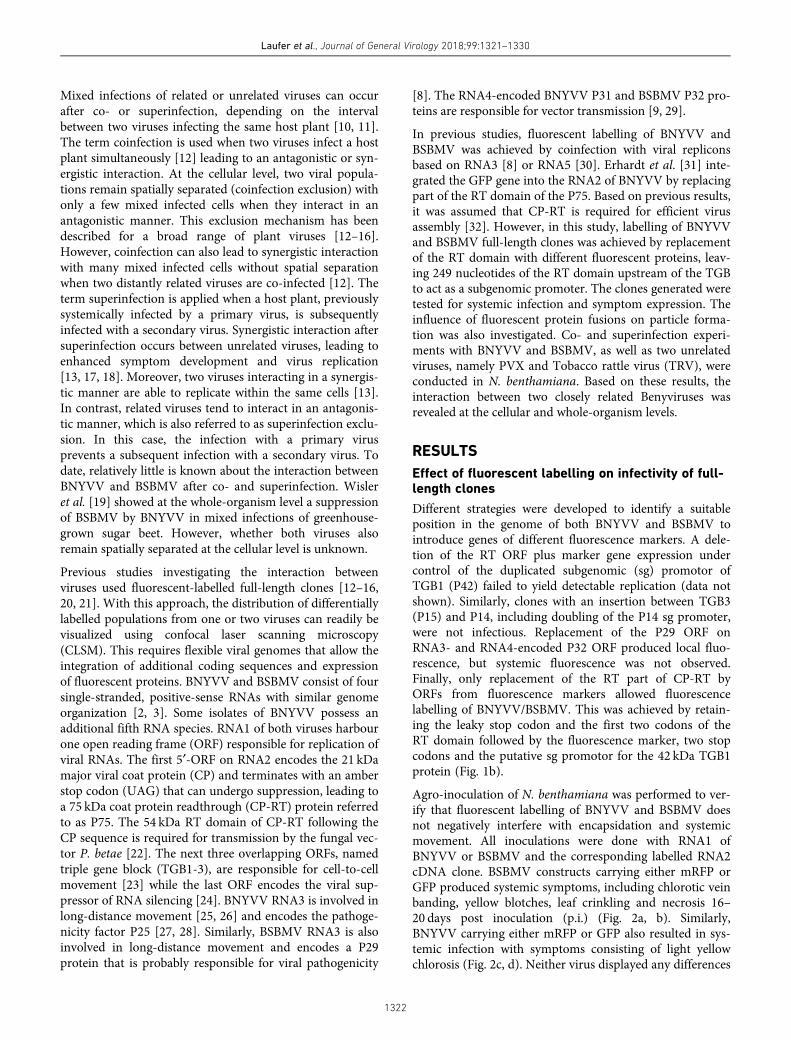
Different strategies were developed to identify a suitableposition in the genome of both BNYVV and BSBMV tointroduce genes of different fluorescence markers. A dele-tion of the RT ORF plus marker gene expression undercontrol of the duplicated subgenomic (sg) promotor ofTGB1 (P42) failed to yield detectable replication (data notshown). Similarly, clones with an insertion between TGB3(P15) and P14, including doubling of the P14 sg promoter,were not infectious. Replacement of the P29 ORF onRNA3- and RNA4-encoded P32 ORF produced local fluo-rescence, but systemic fluorescence was not observed.Finally, only replacement of the RT part of CP-RT byORFs from fluorescence markers allowed fluorescencelabelling of BNYVV/BSBMV. This was achieved by retain-ing the leaky stop codon and the first two codons of theRT domain followed by the fluorescence marker, two stopcodons and the putative sg promotor for the 42 kDa TGB1protein (Fig. 1b).
Agro-inoculation of N. benthamiana was performed to ver-ify that fluorescent labelling of BNYVV and BSBMV doesnot negatively interfere with encapsidation and systemicmovement. All inoculations were done with RNA1 ofBNYVV or BSBMV and the corresponding labelled RNA2cDNA clone. BSBMV constructs carrying either mRFP orGFP produced systemic symptoms, including chlorotic veinbanding, yellow blotches, leaf crinkling and necrosis 16–20 days post inoculation (p.i.) (Fig. 2a, b). Similarly,BNYVV carrying either mRFP or GFP also resulted in sys-temic infection with symptoms consisting of light yellowchlorosis (Fig. 2c, d). Neither virus displayed any differences
Laufer et al., Journal of General Virology 2018;99:1321–1330
1322
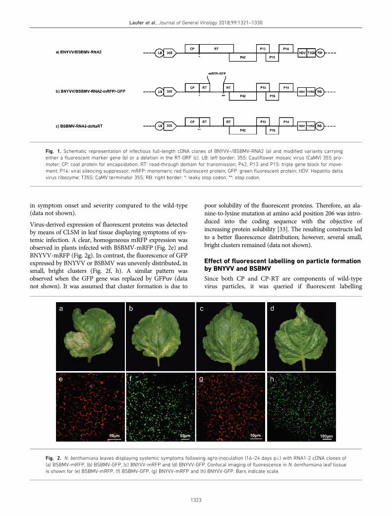
in symptom onset and severity compared to the wild-type(data not shown).
Virus-derived expression of fluorescent proteins was detectedby means of CLSM in leaf tissue displaying symptoms of sys-temic infection. A clear, homogeneous mRFP expression wasobserved in plants infected with BSBMV-mRFP (Fig. 2e) andBNYVV-mRFP (Fig. 2g). In contrast, the fluorescence of GFPexpressed by BNYVV or BSBMV was unevenly distributed, insmall, bright clusters (Fig. 2f, h). A similar pattern wasobserved when the GFP gene was replaced by GFPuv (datanot shown). It was assumed that cluster formation is due to
poor solubility of the fluorescent proteins. Therefore, an ala-nine-to-lysine mutation at amino acid position 206 was intro-duced into the coding sequence with the objective ofincreasing protein solubility [33]. The resulting constructs ledto a better fluorescence distribution; however, several small,bright clusters remained (data not shown).
Effect of fluorescent labelling on particle formationby BNYVV and BSBMV
Since both CP and CP-RT are components of wild-typevirus particles, it was queried if fluorescent labelling
Fig. 1. Schematic representation of infectious full-length cDNA clones of BNYVV-/BSBMV-RNA2 (a) and modified variants carrying
either a fluorescent marker gene (b) or a deletion in the RT-ORF (c). LB: left border; 35S: Cauliflower mosaic virus (CaMV) 35S pro-
moter; CP: coat protein for encapsidation; RT: read-through domain for transmission; P42, P13 and P15: triple gene block for move-
ment; P14: viral silencing suppressor; mRFP: monomeric red fluorescent protein; GFP: green fluorescent protein; HDV: Hepatitis delta
virus ribozyme; T35S: CaMV terminator 35S; RB: right border; *: leaky stop codon; **: stop codon.
Fig. 2. N. benthamiana leaves displaying systemic symptoms following agro-inoculation (16–24 days p.i.) with RNA1-2 cDNA clones of
(a) BSBMV-mRFP, (b) BSBMV-GFP, (c) BNYVV-mRFP and (d) BNYVV-GFP. Confocal imaging of fluorescence in N. benthamiana leaf tissue
is shown for (e) BSBMV-mRFP, (f) BSBMV-GFP, (g) BNYVV-mRFP and (h) BNYVV-GFP. Bars indicate scale.
Laufer et al., Journal of General Virology 2018;99:1321–1330
1323
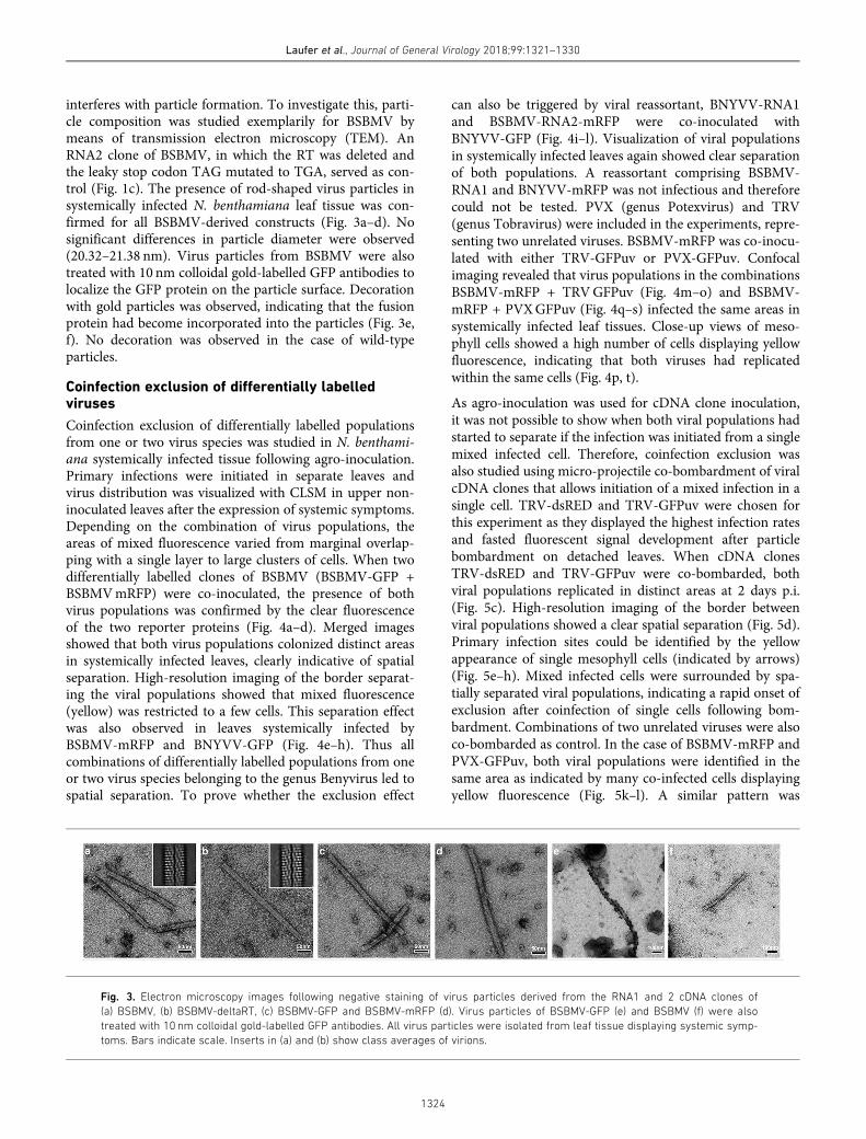
interferes with particle formation. To investigate this, parti-cle composition was studied exemplarily for BSBMV bymeans of transmission electron microscopy (TEM). AnRNA2 clone of BSBMV, in which the RT was deleted andthe leaky stop codon TAG mutated to TGA, served as con-trol (Fig. 1c). The presence of rod-shaped virus particles insystemically infected N. benthamiana leaf tissue was con-firmed for all BSBMV-derived constructs (Fig. 3a–d). Nosignificant differences in particle diameter were observed(20.32–21.38 nm). Virus particles from BSBMV were alsotreated with 10 nm colloidal gold-labelled GFP antibodies tolocalize the GFP protein on the particle surface. Decorationwith gold particles was observed, indicating that the fusionprotein had become incorporated into the particles (Fig. 3e,f). No decoration was observed in the case of wild-typeparticles.
Coinfection exclusion of differentially labelledviruses
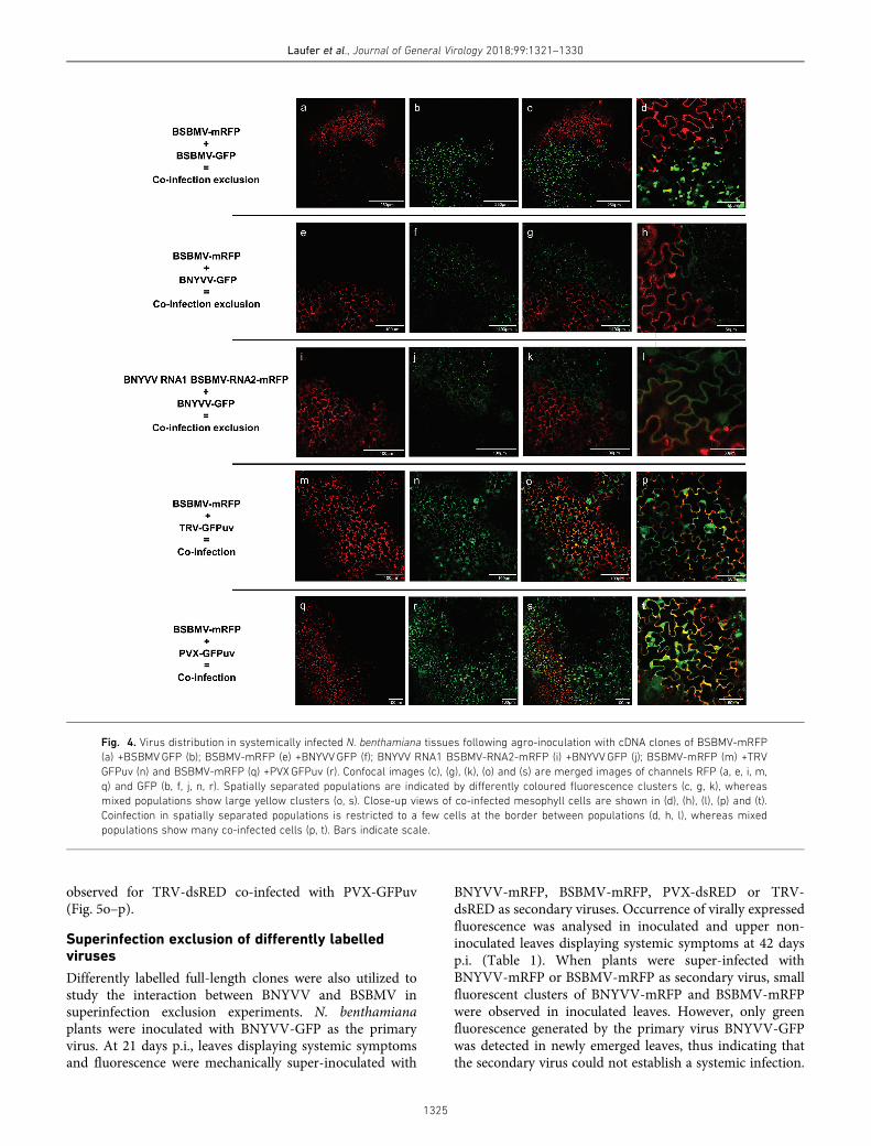
Coinfection exclusion of differentially labelled populationsfrom one or two virus species was studied in N. benthami-ana systemically infected tissue following agro-inoculation.Primary infections were initiated in separate leaves andvirus distribution was visualized with CLSM in upper non-inoculated leaves after the expression of systemic symptoms.Depending on the combination of virus populations, theareas of mixed fluorescence varied from marginal overlap-ping with a single layer to large clusters of cells. When twodifferentially labelled clones of BSBMV (BSBMV-GFP +BSBMVmRFP) were co-inoculated, the presence of bothvirus populations was confirmed by the clear fluorescenceof the two reporter proteins (Fig. 4a–d). Merged imagesshowed that both virus populations colonized distinct areasin systemically infected leaves, clearly indicative of spatialseparation. High-resolution imaging of the border separat-ing the viral populations showed that mixed fluorescence(yellow) was restricted to a few cells. This separation effectwas also observed in leaves systemically infected byBSBMV-mRFP and BNYVV-GFP (Fig. 4e–h). Thus allcombinations of differentially labelled populations from oneor two virus species belonging to the genus Benyvirus led tospatial separation. To prove whether the exclusion effect
can also be triggered by viral reassortant, BNYVV-RNA1and BSBMV-RNA2-mRFP were co-inoculated withBNYVV-GFP (Fig. 4i–l). Visualization of viral populationsin systemically infected leaves again showed clear separationof both populations. A reassortant comprising BSBMV-RNA1 and BNYVV-mRFP was not infectious and thereforecould not be tested. PVX (genus Potexvirus) and TRV(genus Tobravirus) were included in the experiments, repre-senting two unrelated viruses. BSBMV-mRFP was co-inocu-lated with either TRV-GFPuv or PVX-GFPuv. Confocalimaging revealed that virus populations in the combinationsBSBMV-mRFP + TRVGFPuv (Fig. 4m–o) and BSBMV-mRFP + PVXGFPuv (Fig. 4q–s) infected the same areas insystemically infected leaf tissues. Close-up views of meso-phyll cells showed a high number of cells displaying yellowfluorescence, indicating that both viruses had replicatedwithin the same cells (Fig. 4p, t).
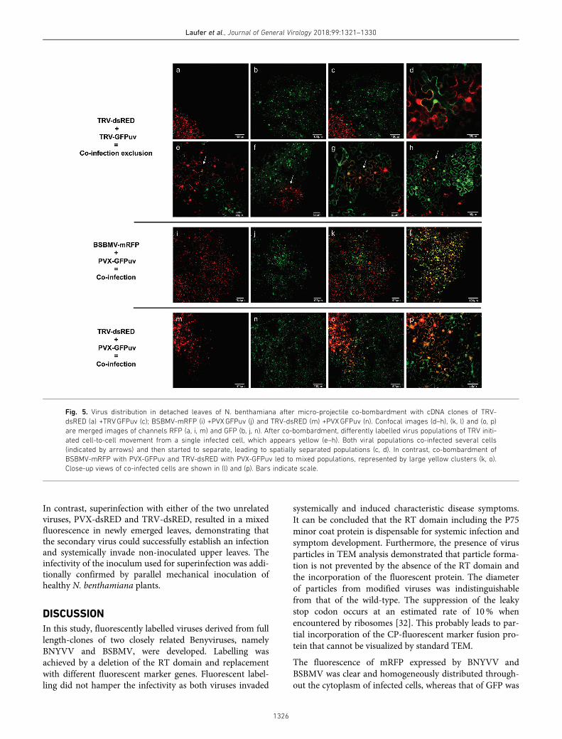
As agro-inoculation was used for cDNA clone inoculation,it was not possible to show when both viral populations hadstarted to separate if the infection was initiated from a singlemixed infected cell. Therefore, coinfection exclusion wasalso studied using micro-projectile co-bombardment of viralcDNA clones that allows initiation of a mixed infection in asingle cell. TRV-dsRED and TRV-GFPuv were chosen forthis experiment as they displayed the highest infection ratesand fasted fluorescent signal development after particlebombardment on detached leaves. When cDNA clonesTRV-dsRED and TRV-GFPuv were co-bombarded, bothviral populations replicated in distinct areas at 2 days p.i.(Fig. 5c). High-resolution imaging of the border betweenviral populations showed a clear spatial separation (Fig. 5d).Primary infection sites could be identified by the yellowappearance of single mesophyll cells (indicated by arrows)(Fig. 5e–h). Mixed infected cells were surrounded by spa-tially separated viral populations, indicating a rapid onset ofexclusion after coinfection of single cells following bom-bardment. Combinations of two unrelated viruses were alsoco-bombarded as control. In the case of BSBMV-mRFP andPVX-GFPuv, both viral populations were identified in thesame area as indicated by many co-infected cells displayingyellow fluorescence (Fig. 5k–l). A similar pattern was
Fig. 3. Electron microscopy images following negative staining of virus particles derived from the RNA1 and 2 cDNA clones of
(a) BSBMV, (b) BSBMV-deltaRT, (c) BSBMV-GFP and BSBMV-mRFP (d). Virus particles of BSBMV-GFP (e) and BSBMV (f) were also
treated with 10 nm colloidal gold-labelled GFP antibodies. All virus particles were isolated from leaf tissue displaying systemic symp-
toms. Bars indicate scale. Inserts in (a) and (b) show class averages of virions.
Laufer et al., Journal of General Virology 2018;99:1321–1330
1324
observed for TRV-dsRED co-infected with PVX-GFPuv(Fig. 5o–p).
Superinfection exclusion of differently labelledviruses
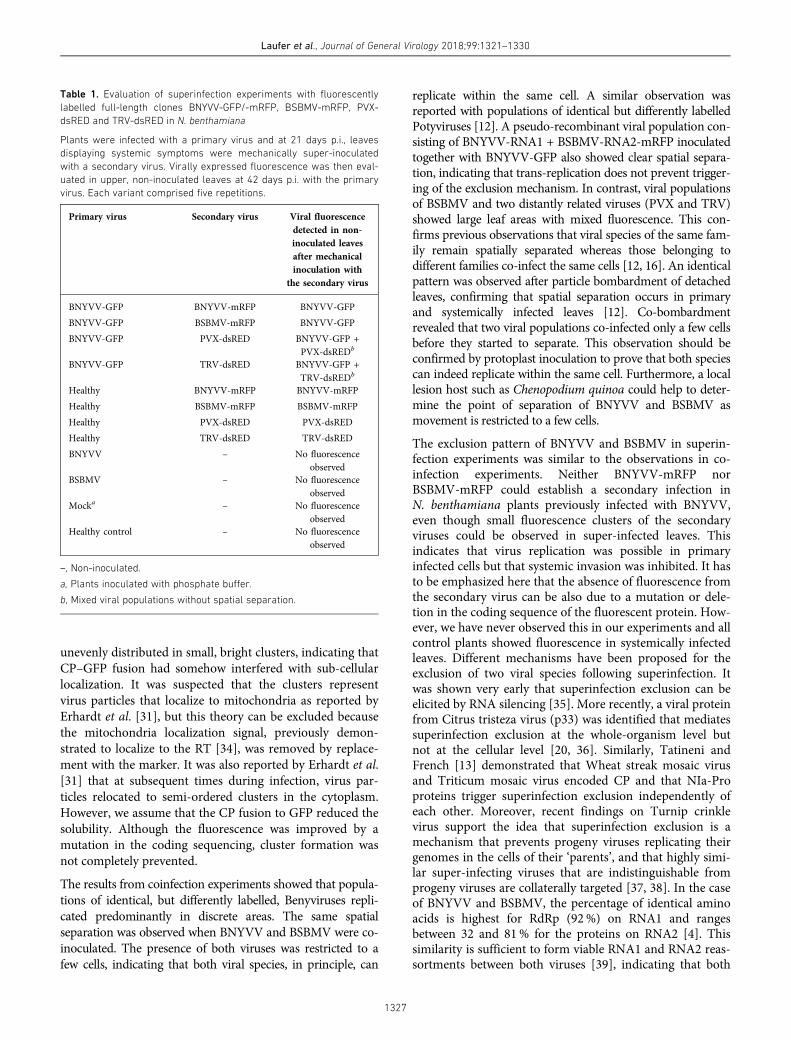
Differently labelled full-length clones were also utilized tostudy the interaction between BNYVV and BSBMV insuperinfection exclusion experiments. N. benthamianaplants were inoculated with BNYVV-GFP as the primaryvirus. At 21 days p.i., leaves displaying systemic symptomsand fluorescence were mechanically super-inoculated with
BNYVV-mRFP, BSBMV-mRFP, PVX-dsRED or TRV-dsRED as secondary viruses. Occurrence of virally expressedfluorescence was analysed in inoculated and upper non-inoculated leaves displaying systemic symptoms at 42 daysp.i. (Table 1). When plants were super-infected withBNYVV-mRFP or BSBMV-mRFP as secondary virus, smallfluorescent clusters of BNYVV-mRFP and BSBMV-mRFPwere observed in inoculated leaves. However, only greenfluorescence generated by the primary virus BNYVV-GFPwas detected in newly emerged leaves, thus indicating thatthe secondary virus could not establish a systemic infection.
Fig. 4. Virus distribution in systemically infected N. benthamiana tissues following agro-inoculation with cDNA clones of BSBMV-mRFP
(a) +BSBMVGFP (b); BSBMV-mRFP (e) +BNYVVGFP (f); BNYVV RNA1 BSBMV-RNA2-mRFP (i) +BNYVVGFP (j); BSBMV-mRFP (m) +TRV
GFPuv (n) and BSBMV-mRFP (q) +PVXGFPuv (r). Confocal images (c), (g), (k), (o) and (s) are merged images of channels RFP (a, e, i, m,
q) and GFP (b, f, j, n, r). Spatially separated populations are indicated by differently coloured fluorescence clusters (c, g, k), whereas
mixed populations show large yellow clusters (o, s). Close-up views of co-infected mesophyll cells are shown in (d), (h), (l), (p) and (t).
Coinfection in spatially separated populations is restricted to a few cells at the border between populations (d, h, l), whereas mixed
populations show many co-infected cells (p, t). Bars indicate scale.
Laufer et al., Journal of General Virology 2018;99:1321–1330
1325
In contrast, superinfection with either of the two unrelatedviruses, PVX-dsRED and TRV-dsRED, resulted in a mixedfluorescence in newly emerged leaves, demonstrating thatthe secondary virus could successfully establish an infectionand systemically invade non-inoculated upper leaves. Theinfectivity of the inoculum used for superinfection was addi-tionally confirmed by parallel mechanical inoculation ofhealthy N. benthamiana plants.
DISCUSSION
In this study, fluorescently labelled viruses derived from fulllength-clones of two closely related Benyviruses, namelyBNYVV and BSBMV, were developed. Labelling wasachieved by a deletion of the RT domain and replacementwith different fluorescent marker genes. Fluorescent label-ling did not hamper the infectivity as both viruses invaded
systemically and induced characteristic disease symptoms.
It can be concluded that the RT domain including the P75
minor coat protein is dispensable for systemic infection and
symptom development. Furthermore, the presence of virus
particles in TEM analysis demonstrated that particle forma-
tion is not prevented by the absence of the RT domain and
the incorporation of the fluorescent protein. The diameter
of particles from modified viruses was indistinguishable
from that of the wild-type. The suppression of the leaky
stop codon occurs at an estimated rate of 10% when
encountered by ribosomes [32]. This probably leads to par-
tial incorporation of the CP-fluorescent marker fusion pro-
tein that cannot be visualized by standard TEM.
The fluorescence of mRFP expressed by BNYVV and
BSBMV was clear and homogeneously distributed through-
out the cytoplasm of infected cells, whereas that of GFP was
Fig. 5. Virus distribution in detached leaves of N. benthamiana after micro-projectile co-bombardment with cDNA clones of TRV-
dsRED (a) +TRVGFPuv (c); BSBMV-mRFP (i) +PVXGFPuv (j) and TRV-dsRED (m) +PVXGFPuv (n). Confocal images (d–h), (k, l) and (o, p)
are merged images of channels RFP (a, i, m) and GFP (b, j, n). After co-bombardment, differently labelled virus populations of TRV initi-
ated cell-to-cell movement from a single infected cell, which appears yellow (e–h). Both viral populations co-infected several cells
(indicated by arrows) and then started to separate, leading to spatially separated populations (c, d). In contrast, co-bombardment of
BSBMV-mRFP with PVX-GFPuv and TRV-dsRED with PVX-GFPuv led to mixed populations, represented by large yellow clusters (k, o).
Close-up views of co-infected cells are shown in (l) and (p). Bars indicate scale.
Laufer et al., Journal of General Virology 2018;99:1321–1330
1326
unevenly distributed in small, bright clusters, indicating that
CP–GFP fusion had somehow interfered with sub-cellular
localization. It was suspected that the clusters represent
virus particles that localize to mitochondria as reported byErhardt et al. [31], but this theory can be excluded because
the mitochondria localization signal, previously demon-
strated to localize to the RT [34], was removed by replace-
ment with the marker. It was also reported by Erhardt et al.
[31] that at subsequent times during infection, virus par-
ticles relocated to semi-ordered clusters in the cytoplasm.
However, we assume that the CP fusion to GFP reduced the
solubility. Although the fluorescence was improved by amutation in the coding sequencing, cluster formation was
not completely prevented.
The results from coinfection experiments showed that popula-
tions of identical, but differently labelled, Benyviruses repli-
cated predominantly in discrete areas. The same spatialseparation was observed when BNYVV and BSBMV were co-
inoculated. The presence of both viruses was restricted to a
few cells, indicating that both viral species, in principle, can
replicate within the same cell. A similar observation wasreported with populations of identical but differently labelledPotyviruses [12]. A pseudo-recombinant viral population con-sisting of BNYVV-RNA1 + BSBMV-RNA2-mRFP inoculatedtogether with BNYVV-GFP also showed clear spatial separa-tion, indicating that trans-replication does not prevent trigger-ing of the exclusion mechanism. In contrast, viral populationsof BSBMV and two distantly related viruses (PVX and TRV)showed large leaf areas with mixed fluorescence. This con-firms previous observations that viral species of the same fam-ily remain spatially separated whereas those belonging todifferent families co-infect the same cells [12, 16]. An identicalpattern was observed after particle bombardment of detachedleaves, confirming that spatial separation occurs in primaryand systemically infected leaves [12]. Co-bombardmentrevealed that two viral populations co-infected only a few cellsbefore they started to separate. This observation should beconfirmed by protoplast inoculation to prove that both speciescan indeed replicate within the same cell. Furthermore, a locallesion host such as Chenopodium quinoa could help to deter-mine the point of separation of BNYVV and BSBMV asmovement is restricted to a few cells.
The exclusion pattern of BNYVV and BSBMV in superin-fection experiments was similar to the observations in co-infection experiments. Neither BNYVV-mRFP norBSBMV-mRFP could establish a secondary infection inN. benthamiana plants previously infected with BNYVV,even though small fluorescence clusters of the secondaryviruses could be observed in super-infected leaves. Thisindicates that virus replication was possible in primaryinfected cells but that systemic invasion was inhibited. It hasto be emphasized here that the absence of fluorescence fromthe secondary virus can be also due to a mutation or dele-tion in the coding sequence of the fluorescent protein. How-ever, we have never observed this in our experiments and allcontrol plants showed fluorescence in systemically infectedleaves. Different mechanisms have been proposed for theexclusion of two viral species following superinfection. Itwas shown very early that superinfection exclusion can beelicited by RNA silencing [35]. More recently, a viral proteinfrom Citrus tristeza virus (p33) was identified that mediatessuperinfection exclusion at the whole-organism level butnot at the cellular level [20, 36]. Similarly, Tatineni andFrench [13] demonstrated that Wheat streak mosaic virusand Triticum mosaic virus encoded CP and that NIa-Proproteins trigger superinfection exclusion independently ofeach other. Moreover, recent findings on Turnip crinklevirus support the idea that superinfection exclusion is amechanism that prevents progeny viruses replicating theirgenomes in the cells of their ‘parents’, and that highly simi-lar super-infecting viruses that are indistinguishable fromprogeny viruses are collaterally targeted [37, 38]. In the caseof BNYVV and BSBMV, the percentage of identical aminoacids is highest for RdRp (92%) on RNA1 and rangesbetween 32 and 81% for the proteins on RNA2 [4]. Thissimilarity is sufficient to form viable RNA1 and RNA2 reas-sortments between both viruses [39], indicating that both
Table 1. Evaluation of superinfection experiments with fluorescently
labelled full-length clones BNYVV-GFP/-mRFP, BSBMV-mRFP, PVX-
dsRED and TRV-dsRED in N. benthamiana
Plants were infected with a primary virus and at 21 days p.i., leaves
displaying systemic symptoms were mechanically super-inoculated
with a secondary virus. Virally expressed fluorescence was then eval-
uated in upper, non-inoculated leaves at 42 days p.i. with the primary
virus. Each variant comprised five repetitions.
Primary virus Secondary virus Viral fluorescence
detected in non-
inoculated leaves
after mechanical
inoculation with
the secondary virus
BNYVV-GFP BNYVV-mRFP BNYVV-GFP
BNYVV-GFP BSBMV-mRFP BNYVV-GFP
BNYVV-GFP PVX-dsRED BNYVV-GFP +
PVX-dsREDb
BNYVV-GFP TRV-dsRED BNYVV-GFP +
TRV-dsREDb
Healthy BNYVV-mRFP BNYVV-mRFP
Healthy BSBMV-mRFP BSBMV-mRFP
Healthy PVX-dsRED PVX-dsRED
Healthy TRV-dsRED TRV-dsRED
BNYVV – No fluorescence
observed
BSBMV – No fluorescence
observed
Mocka – No fluorescence
observed
Healthy control – No fluorescence
observed
–, Non-inoculated.
a, Plants inoculated with phosphate buffer.
b, Mixed viral populations without spatial separation.
Laufer et al., Journal of General Virology 2018;99:1321–1330
1327
viruses share a highly similar replication and movementstrategy. Further studies will be needed to identify the viralprotein responsible for the exclusion.
The data presented here provide the first evidence thatBNYVV and BSBMV remain spatially separated when colo-nizing the same host plant. Considering that BNYVV is thenearest known relative of BSBMV, co- and superinfectionexclusion of both viral species seems to be plausible in termsof virus evolution. The ability of viral variants to exclude eachother in mixed infections eliminates competition for the host’sresources. Furthermore, it has major implications for the sta-bility of viral sequences and the genetic structure of a viruspopulation [20]. Newly emerging viral variants gain benefit byfavouring uninfected cells rather than previously infected hostcells [11]. However, replication of two or more viral genomesin one cell increases the likelihood of recombination and reas-sortments. This is of particular relevance as it can increase thegenetic diversity within a viral population, leading to new viralvariants. Considering the results of the present study, the like-lihood of recombination and pseudo-recombinants betweenBNYVV and BSBMV in mixed infections seems to be rela-tively low due to the spatial separation at the cellular level.Moreover, both viral species seem to have developed differentcolonization strategies in their host, sugar beet, as BNYVVinfections are mainly restricted to the root while BSBMVresults in more systemic foliar symptoms [3]. The fluores-cence-labelled viruses derived from full-length clones devel-oped in this study represents versatile tools to address thisquestion in the natural host, sugar beet.
METHODS
Construction of fluorescence-labelled full-lengthclones
Clones BSBMV and BNYVV RNA2 cDNA for agro-inocu-lation [39] were modified to express mRFP [40] and GFP[41]. Labelling was achieved by replacing the read-throughpart of the CP-RT, thereby retaining the leaky stop codon ofthe CP and the first two codons of the RT domain. Replace-ment of the RT by different fluorescent proteins was fol-lowed by two stop codons (TGATAG) and the remaining249 nucleotides of the CP-RT, containing the putative sgpromotor for the 42 kDa TGB1 (Fig. 1b). This modificationresulted in a large read-through protein comprising the CPand the fluorescent protein. The DNA fragments of fluores-cent marker genes coding sequences were cloned into line-arized plasmids of BNYVV/BSBMV RNA2 clones by meansof Gibson assembly [42]. The marker gene coding sequenceswere amplified with Phusion Flash High-Fidelity PCR Mas-ter Mix (ThermoScientific) according to the manufacturer’sinstructions. Specific primers (Table S1, available in theonline version of this article) contained 5¢- and 3¢-exten-sions overlapping with RNA2 of BNYVV/BSBMV (25–28nt). The BNYVV/BSBMV RNA2 clones were linearised byPCR amplification using primers BNYVV2-s/BNYVV2-asand BSBMV2-s/BSBMV2-as (Table S1). All PCR productswere gel-purified with NucleoSpin Gel and PCR Clean-up
kit (Macherey-Nagel) according to the manufacturer’sinstructions. Following Gibson assembly, in vitro recombi-nation products were transformed into chemical competentEscherichia coli cells (strain DH5a) as described by Inoueet al. [43]. Plasmids carrying cDNA fragments of the fluo-rescent proteins were identified by means of appropriaterestriction enzyme digest, and all mutations were verified bycommercial capillary Sanger sequencing (Eurofins MWGOperon). The resulting clones were named BNYVV-RNA2-mRFP/-GFP and BSBMV-RNA2-mRFP/-GFP.
To study the effect of RT deletion on particle assembly, the RTcoding sequence was deleted from the BSBMV RNA2 cDNAclone. The leaky stop codon TAG was mutated to TGA. TheRT sequence downstream of the stop codon was deletedexcept for the last 249 nucleotides of the RT. This wasachieved by PCR amplification of BSBMV-RNA2 with the pri-mers BSBMV-deltaRT-fw and BSBMV-deltaRT-rv (Table S1).The resulting clone was named BSBMV-RNA2-deltaRT.
The infectivity of the above-mentioned constructs wastested in N. benthamiana using agro-inoculation. For thispurpose, viral cDNA clones of RNA1 and RNA2 were trans-formed into Rhizobium radiobacter (syn. Agrobacteriumtumefaciens) strain C58C1. Bacterial cultures were preparedaccording to Voinnet et al. [44] with an optical density at600 nm (OD600) of 0.1. The first two pairs of true leaveswere inoculated. Different cDNA components from multi-partite viruses were mixed in a 1 : 1 ratio prior to inocula-tion. All plants were grown under greenhouse conditions,24
�
C for 14 h and 18�
C for 10 h. CLSM (see below) wasapplied to visualize fluorescence-labelled full-length clonesin systemically infected leaf tissue.
Co- and superinfection exclusion of BNYVV, BSBMV,PVX and TRV
Coinfection exclusion was studied with BNYVV/BSBMVand two unrelated viruses (PVX and TRV). PVX-GFPuvwas constructed as described by Draghici and Varrelmann[45] using a PVX expression plasmid obtained from DavidBaulcombe (Sainsbury Laboratory, Norwich, UK). Con-struction of the PVX vector expressing dsRED has beendescribed previously (PVX201-optRed) [46]. TRV vectorscomprising pTRV1 and pTRV2 (pYL156) [47] were modi-fied as described by Ghazala and Varrelmann [48] toexpress either dsRED (TRV-dsRED) or GFPuv (TRV-GFPuv). Two differently labelled viruses were inoculatedsimultaneously but in separate leaves of 3-week-old N. ben-thamiana plants using agro-inoculation as described above.After symptom development, virus distribution was visual-ized in systemically infected leaf tissue by means of CLSM.Particle bombardment was also applied to study the spreadof differently labelled viruses starting from a single, doublyinfected mesophyll cell. Detached leaves from 4- to 5-week-old N. benthamiana plants were subjected to micro-projec-tile co-bombardment with a particle inflow gun [49] using10 µl purified plasmid DNA corresponding to each viralRNA component. Following bombardment, detached leaveswere placed in a Petri dish with watered filter paper and
Laufer et al., Journal of General Virology 2018;99:1321–1330
1328
incubated at room temperature in the dark. Virus distribu-tion was visualized with CLSM after 2–5 days.
Superinfection exclusion experiments were performed onN. benthamiana plants using BNYVV-GFP as the primaryvirus and BNYVV-mRFP, BSBMV-mRFP, TRV-dsRED aswell as PVX-dsRED, respectively, as the secondary virus.N. benthamiana plants were first infected with BNYVV-GFP using agro-inoculation as described above. After threeweeks, leaves displaying systemic symptoms were mechani-cally super-inoculated with the challenging virus. Prior tosecondary infection, establishment of the primary infectionwas confirmed by means of CLSM. The inoculum for thesecondary infection was produced in N. benthamiana usingagro-inoculation as described above. Leaves displaying sys-temic symptoms were ground in phosphate buffer (10mMNa2SO3, pH 7.0) and rub-inoculated on N. benthamianaleaves infected with the protecting virus. After a furtherthree weeks, establishment of secondary infection waschecked in inoculated and upper non-inoculated leavesusing CLSM. Each variant comprised five repetitions.
Confocal laser scanning microscopy
Systemically infected leaf tissue from B. macrocarpa andN. benthamiana was harvested and visualized with a TCS-SP5 confocal laser scanning microscope (Leica Microsys-tems). Excitation/emission wavelengths for the differentfluorescent proteins were as follows: mRFP 561 nm/520–540 nm, dsRED 561 nm/520–540 nm, GFP 488 nm/515–523 nm and GFPuv 405 nm/490–520 nm. All confocalimages were processed with LAS-AF software version2.6.3.8173 (Leica Microsystems).
Transmission electron microscopy
For negative staining of virus particles, sample material fromN. benthamiana was bound to a glow-discharged carbon foil-covered grid. After staining with 1% uranyl acetate, the sam-ples were evaluated at room temperature with a CM 120transmission electron microscope (FEI). Summed averagedimages of the virions were calculated using the softwareRELION (MRC Laboratory of Molecular Biology). Overall,7000 individual overlapping segments of the virions, respec-tively, were boxed using RELION. The images were sorted byMSA and summed to obtain a class average image of the viri-ons. For immuno-gold labelling, sample material from N. ben-thamiana was absorbed to formvar carbon-coated Ni-grids.These were fixed with 4% paraformaldehyde, quenched with20mM glycin and immunostained using the described sera,followed by the addition of Protein A-gold (10 nm). The prep-arations were then washed repeatedly with TPBS and high-salt TPBS (0.5 M NaCl) and post-fixed with 2% glutaralde-hyde. After counterstaining with 1% uranylacetate, sampleswere investigated using a CM120 Philips electron microscopeand a TemCam F416 CMOS camera (TVIPS).
Funding information
This research was supported in part by Deutsche Forschungsgemein-schaft (DFG) grant VA 202/7-1.
Acknowledgements
We thank Helmut Korf and Jutta Zimmermann for excellent technicalassistance. We also thank the reviewers for their comments, whichsignificantly improved the manuscript.
Conflicts of interest
The authors declare that there are no conflicts of interest.
References
1. Gilmer D, Ratti C. Benyvirus. In: King AMQ, Adams MJ, CarstensEB and Lefkowitz EJ (editors). Virus Taxonomy: Classification and
Nomenclature of Viruses. Ninth Report of the International Commit-tee on Taxonomy of Viruses. Elsevier Inc; 2012. pp. 1133–1138.
2. Peltier C, Hleibieh K, Thiel H, Klein E, Bragard C et al. Molecularbiology of the Beet necrotic yellow vein virus. Plant viruses 2008;2:14–24.
3. Heidel GB, Rush CM, Kendall TL, Lommel SA, French RC. Charac-teristics of Beet soilborne mosaic virus, a furo-like virus infectingsugar beet. Plant Disease 1997;81:1070–1076.
4. Lee L, Telford EB, Batten JS, Scholthof KB, Rush CM. Completenucleotide sequence and genome organization of Beet soilborne
mosaic virus, a proposed member of the genus Benyvirus. ArchVirol 2001;146:2443–2453.
5. Keskin B. Polymyxa betae n. sp., ein Parasit in den Wurzeln vonBeta vulgaris Tournefort, besonders w€ahrend der Jugendent-wicklung der Zuckerrübe. Arch Microbiol 1964;49:348–374.
6. Tamada T, Kondo H. Biological and genetic diversity of plasmodio-phorid-transmitted viruses and their vectors. J Gen Plant Pathol
2013;79:307–320.
7. Workneh F, Villanueva E, Steddom K, Rush CM. Spatial associa-tion and distribution of Beet necrotic yellow vein virus and Beet
soil-borne mosaic virus in sugar beet fields. Plant Dis 2003;87:707–711.
8. Ratti C, Hleibieh K, Bianchi L, Schirmer A, Autonell CR et al. Beet
soil-borne mosaic virus RNA-3 is replicated and encapsidated inthe presence of BNYVV RNA-1 and -2 and allows long distancemovement in Beta macrocarpa. Virology 2009;385:392–399.
9. D’Alonzo M, Delbianco A, Lanzoni C, Autonell CR, Gilmer D et al.
Beet soil-borne mosaic virus RNA-4 encodes a 32 kDa proteininvolved in symptom expression and in virus transmissionthrough Polymyxa betae. Virology 2012;423:187–194.
10. Syller J. Facilitative and antagonistic interactions between plantviruses in mixed infections. Mol Plant Pathol 2012;13:204–216.
11. Syller J, Grupa A. Antagonistic within-host interactions betweenplant viruses: molecular basis and impact on viral and host fit-ness. Mol Plant Pathol 2016;17:769–782.
12. Dietrich C, Maiss E. Fluorescent labelling reveals spatial separa-tion of potyvirus populations in mixed infected Nicotiana benthami-
ana plants. J Gen Virol 2003;84:2871–2876.
13. Tatineni S, French R. The coat protein and NIa protease of twoPotyviridae family members independently confer super-infectionexclusion. J Virol 2016;90:10886–10905.
14. Guti�errez S, Pirolles E, Yvon M, Baecker V, Michalakis Y et al. Themultiplicity of cellular infection changes depending on the route ofcell infection in a plant virus. J Virol 2015;89:9665–9675.
15. Julve JM, Gandía A, Fern�andez-del-Carmen A, Sarrion-
Perdigones A, Castelijns B et al. A coat-independent superinfec-tion exclusion rapidly imposed in Nicotiana benthamiana cells bytobacco mosaic virus is not prevented by depletion of the move-ment protein. Plant Mol Biol 2013;81:553–564.
16. Takahashi T, Sugawara T, Yamatsuta T, Isogai M, Natsuaki T
et al. Analysis of the spatial distribution of identical and two dis-tinct virus populations differently labeled with cyan and yellowfluorescent proteins in coinfected plants. Phytopathology 2007;97:1200–1206.
17. Gonz�alez-Jara P, Tenllado F, Martínez-García B, Atencio FA,
Barajas D et al. Host-dependent differences during synergistic
Laufer et al., Journal of General Virology 2018;99:1321–1330
1329
infection by Potyviruses with potato virus X. Mol Plant Pathol 2004;5:29–35.
18. Vance VB. Replication of potato virus X RNA is altered in coinfec-
tions with potato virus Y. Virology 1991;182:486–494.
19. Wisler GC, Lewellen RT, Sears JL, Wasson JW, Liu H-Y et al.
Interactions between Beet necrotic yellow vein virus and Beet soil-
borne mosaic virus in sugar beet. Plant Dis 2003;87:1170–1175.
20. Folimonova SY. Superinfection exclusion is an active virus-
controlled function that requires a specific viral protein. J Virol
2012;86:5554–5561.
21. Gonz�alez-Jara P, Fraile A, Canto T, García-Arenal F. The multi-
plicity of infection of a plant virus varies during colonization of itseukaryotic host. J Virol 2009;83:7487–7494.
22. Tamada T, Kusume T. Evidence that the 75K readthrough protein
of beet necrotic yellow vein virus RNA-2 is essential for transmis-sion by the fungus Polymyxa betae. J Gen Virol 1991;72:1497–1504.
23. Gilmer D, Bouzoubaa S, Hehn A, Guilley H, Richards K et al. Effi-
cient cell-to-cell movement of beet necrotic yellow vein virusrequires 3’ proximal genes located on RNA 2. Virology 1992;189:40–47.
24. Chiba S, Hleibieh K, Delbianco A, Klein E, Ratti C et al. The benyvi-
rus RNA silencing suppressor is essential for long-distance move-ment, requires both zinc-finger and NoLS basic residues but not anucleolar localization for its silencing-suppression activity. Mol
Plant Microbe Interact 2013;26:168–181.
25. Lauber E, Guilley H, Tamada T, Richards KE, Jonard G. Vascular
movement of beet necrotic yellow vein virus in Beta macrocarpa isprobably dependent on an RNA 3 sequence domain rather than agene product. J Gen Virol 1998;79:385–393.
26. Peltier C, Klein E, Hleibieh K, D’Alonzo M, Hammann P et al. Beet
necrotic yellow vein virus subgenomic RNA3 is a cleavage productleading to stable non-coding RNA required for long-distancemovement. J Gen Virol 2012;93:1093–1102.
27. Chiba S, Miyanishi M, Andika IB, Kondo H, Tamada T. Identification
of amino acids of the beet necrotic yellow vein virus p25 proteinrequired for induction of the resistance response in leaves of Betavulgaris plants. J Gen Virol 2008;89:1314–1323.
28. Koenig R, Jarausch W, Li Y, Commandeur U, Burgermeister W
et al. Effect of recombinant beet necrotic yellow vein virus with
different RNA compositions on mechanically inoculated sugar-beets leaves of Beta vulgaris plants. J Gen Virol 1991;89:1314–1323.
29. Tamada T, Abe H. Evidence that beet necrotic yellow vein virus
RNA-4 is essential for efficient transmission by the fungus Poly-
myxa betae. J Gen Virol 1989;70:3391–3398.
30. Schmidlin L, Link D, Mutterer J, Guilley H, Gilmer D. Use of a Beet
necrotic yellow vein virus RNA-5-derived replicon as a new tool forgene expression. J Gen Virol 2005;86:463–467.
31. Erhardt M, Dunoyer P, Guilley H, Richards K, Jonard G et al. Beet
necrotic yellow vein virus particles localize to mitochondria duringinfection. Virology 2001;286:256–262.
32. Schmitt C, Balmori E, Jonard G, Richards KE, Guilley H. In vitro
mutagenesis of biologically active transcripts of beet necrotic yel-low vein virus RNA 2: evidence that a domain of the 75-kDa read-through protein is important for efficient virus assembly. Proc NatlAcad Sci USA 1992;89:5715–5719.
33. von Stetten D, Noirclerc-Savoye M, Goedhart J, Gadella TW,
Royant A. Structure of a fluorescent protein from Aequorea victo-
ria bearing the obligate-monomer mutation A206K. Acta
Crystallogr Sect F Struct Biol Cryst Commun 2012;68:878–882.
34. Valentin C, Dunoyer P, Vetter G, Schalk C, Dietrich A et al. Molec-ular basis for mitochondrial localization of viral particles duringbeet necrotic yellow vein virus infection. J Virol 2005;79:9991–10002.
35. Ratcliff F, Harrison BD, Baulcombe DC. A similarity between viraldefense and gene silencing in plants. Science 1997;276:1558–1560.
36. Bergua M, Zwart MP, El-Mohtar C, Shilts T, Elena SF et al. A viralprotein mediates superinfection exclusion at the whole-organismlevel but is not required for exclusion at the cellular level. J Virol
2014;88:11327–11338.
37. Zhang XF, Sun R, Guo Q, Zhang S, Meulia T et al. A self-perpetuat-ing repressive state of a viral replication protein blocks superin-fection by the same virus. PLoS Pathog 2017;13:e1006253.
38. Zhang XF, Zhang S, Guo Q, Sun R, Wei T et al. A new mechanisticmodel for viral cross protection and superinfection exclusion.Front Plant Sci 2018;9:40.
39. Laufer M, Mohammad H, Maiss E, Richert-Pöggeler K, dall’ara M
et al. Biological properties of Beet soil-borne mosaic virus and Beet
necrotic yellow vein virus cDNA clones produced by isothermal invitro recombination: Insights for reassortant appearance. Virology2018;518:25–33.
40. Campbell RE, Tour O, Palmer AE, Steinbach PA, Baird GS et al. Amonomeric red fluorescent protein. Proc Natl Acad Sci USA 2002;99:7877–7882.
41. Davis SJ, Vierstra RD. Soluble, highly fluorescent variants ofgreen fluorescent protein (GFP) for use in higher plants. Plant MolBiol 1998;36:521–528.
42. Gibson DG, Young L, Chuang RY, Venter JC, Hutchison CA et al.
Enzymatic assembly of DNA molecules up to several hundred kil-obases. Nat Methods 2009;6:343–345.
43. Inoue H, Nojima H, Okayama H. High efficiency transformation ofEscherichia coli with plasmids. Gene 1990;96:23–28.
44. Voinnet O, Rivas S, Mestre P, Baulcombe D. An enhanced tran-sient expression system in plants based on suppression of genesilencing by the p19 protein of tomato bushy stunt virus. Plant J2003;33:949–956.
45. Draghici HK, Varrelmann M. Evidence that the linker between themethyltransferase and helicase domains of potato virus X repli-case is involved in homologous RNA recombination. J Virol 2009;83:7761–7769.
46. Dietrich C, Maiss E. Red fluorescent protein DsRed from Disco-
soma sp. as a reporter protein in higher plants. Biotechniques2002;32:286–291.
47. Liu Y, Schiff M, Marathe R, Dinesh-Kumar SP. Tobacco Rar1,EDS1 and NPR1/NIM1 like genes are required for N-mediatedresistance to tobacco mosaic virus. Plant J 2002;30:415–429.
48. Ghazala W, Varrelmann M. Tobacco rattle virus 29K movementprotein is the elicitor of extreme and hypersensitive-like resis-tance in two cultivars of Solanum tuberosum. Mol Plant Microbe
Interact 2007;20:1396–1405.
49. Gray DJ, Hiebert E, Lin CM, Compton ME, McColley DW et al. Sim-plified construction and performance of a device for particle bom-bardment. Plant Cell Tissue Organ Cult 1994;37:179–184.
Laufer et al., Journal of General Virology 2018;99:1321–1330
1330
Related Documents