Fluorescence quenching and photocatalytic degradation of textile dyeing waste water by silver nanoparticles S.R. Kavitha a , M. Umadevi a,⇑ , S.R. Janani b , T. Balakrishnan c , R. Ramanibai c a Department of Physics, Mother Teresa Women’s University, Kodaikanal, Tamil Nadu 624101, India b Department of Animal Nutrition, Veterinary College & Research Institute, TANUVAS, Namakkal, Tamil Nadu 637002, India c Department of Zoology, University of Madras, Guindy Campus, Chennai, Tamil Nadu 600025, India highlights Silver nanoparticles (Ag NPs) have been synthesized by chemical method. Spherical shaped Ag NPs were obtained. Low fluorescence quantum yield was obtained. Apparent rate constants were determined. Ag NPs acts as a good nanocatalyst for waste water. graphical abstract article info Article history: Received 24 December 2013 Accepted 12 February 2014 Available online 26 February 2014 Keywords: Silver nanoparticles Waste water Kinetic Quantum yield Fluorescence quenching Photocatalytic degradation abstract Silver nanoparticles (Ag NPs) of different sizes have been prepared by chemical reduction method and characterized using UV–vis spectroscopy and transmission electron microscopy (HRTEM). Fluorescence spectral analysis showed that the quenching of fluorescence of textile dyeing waste water (TDW) has been found to decrease with decrease in the size of the Ag NPs. Experimental results show that the silver nanoparticles can quench the fluorescence emission of adsorbed TDW effectively. The fluorescence interaction between Ag NPs (acceptor) and TDW (donor) confirms the Förster Resonance Energy Transfer (FRET) mechanism. Long range dipole–dipole interaction between the excited donor and ground state acceptor molecules is the dominant mechanism responsible for the energy transfer. Furthermore, photo- catalytic degradation of TDW was measured spectrophotometrically by using silver as nanocatalyst under UV light illumination. The kinetic study revealed that synthesized Ag NPs was found to be effective in degrading TDW. Ó 2014 Elsevier B.V. All rights reserved. Introduction Water is one of the important enablers of life on earth. It is one of the purest symbols of wealth, health, serenity, beauty and originality. Pure water, which is free of toxic chemicals and pathogenic bacteria, is necessary for human health [1]. Water and environment gets contaminated by the organic matters liberated from textile industries. Textile waste water includes a large variety of dyes and chemicals additions that make the environmental challenge for textile industry not only as liquid waste but also in its chemical composition [2]. Main pollution in TDW came from dyeing and finishing processes and these processes require the input of a wide range of chemicals and http://dx.doi.org/10.1016/j.saa.2014.02.076 1386-1425/Ó 2014 Elsevier B.V. All rights reserved. ⇑ Corresponding author. Tel.: +91 4542 293411; fax: +91 4542 241122. E-mail address: [email protected] (M. Umadevi). Spectrochimica Acta Part A: Molecular and Biomolecular Spectroscopy 127 (2014) 115–121 Contents lists available at ScienceDirect Spectrochimica Acta Part A: Molecular and Biomolecular Spectroscopy journal homepage: www.elsevier.com/locate/saa

Welcome message from author
This document is posted to help you gain knowledge. Please leave a comment to let me know what you think about it! Share it to your friends and learn new things together.
Transcript
Spectrochimica Acta Part A: Molecular and Biomolecular Spectroscopy 127 (2014) 115–121
Contents lists available at ScienceDirect
Spectrochimica Acta Part A: Molecular andBiomolecular Spectroscopy
journal homepage: www.elsevier .com/locate /saa
Fluorescence quenching and photocatalytic degradation of textile dyeingwaste water by silver nanoparticles
http://dx.doi.org/10.1016/j.saa.2014.02.0761386-1425/� 2014 Elsevier B.V. All rights reserved.
⇑ Corresponding author. Tel.: +91 4542 293411; fax: +91 4542 241122.E-mail address: [email protected] (M. Umadevi).
S.R. Kavitha a, M. Umadevi a,⇑, S.R. Janani b, T. Balakrishnan c, R. Ramanibai c
a Department of Physics, Mother Teresa Women’s University, Kodaikanal, Tamil Nadu 624101, Indiab Department of Animal Nutrition, Veterinary College & Research Institute, TANUVAS, Namakkal, Tamil Nadu 637002, Indiac Department of Zoology, University of Madras, Guindy Campus, Chennai, Tamil Nadu 600025, India
h i g h l i g h t s
� Silver nanoparticles (Ag NPs) havebeen synthesized by chemicalmethod.� Spherical shaped Ag NPs were
obtained.� Low fluorescence quantum yield was
obtained.� Apparent rate constants were
determined.� Ag NPs acts as a good nanocatalyst for
waste water.
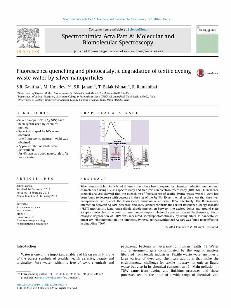
g r a p h i c a l a b s t r a c t
a r t i c l e i n f o
Article history:Received 24 December 2013Accepted 12 February 2014Available online 26 February 2014
Keywords:Silver nanoparticlesWaste waterKineticQuantum yieldFluorescence quenchingPhotocatalytic degradation
a b s t r a c t
Silver nanoparticles (Ag NPs) of different sizes have been prepared by chemical reduction method andcharacterized using UV–vis spectroscopy and transmission electron microscopy (HRTEM). Fluorescencespectral analysis showed that the quenching of fluorescence of textile dyeing waste water (TDW) hasbeen found to decrease with decrease in the size of the Ag NPs. Experimental results show that the silvernanoparticles can quench the fluorescence emission of adsorbed TDW effectively. The fluorescenceinteraction between Ag NPs (acceptor) and TDW (donor) confirms the Förster Resonance Energy Transfer(FRET) mechanism. Long range dipole–dipole interaction between the excited donor and ground stateacceptor molecules is the dominant mechanism responsible for the energy transfer. Furthermore, photo-catalytic degradation of TDW was measured spectrophotometrically by using silver as nanocatalystunder UV light illumination. The kinetic study revealed that synthesized Ag NPs was found to be effectivein degrading TDW.
� 2014 Elsevier B.V. All rights reserved.
Introduction
Water is one of the important enablers of life on earth. It is oneof the purest symbols of wealth, health, serenity, beauty andoriginality. Pure water, which is free of toxic chemicals and
pathogenic bacteria, is necessary for human health [1]. Waterand environment gets contaminated by the organic mattersliberated from textile industries. Textile waste water includes alarge variety of dyes and chemicals additions that make theenvironmental challenge for textile industry not only as liquidwaste but also in its chemical composition [2]. Main pollution inTDW came from dyeing and finishing processes and theseprocesses require the input of a wide range of chemicals and
116 S.R. Kavitha et al. / Spectrochimica Acta Part A: Molecular and Biomolecular Spectroscopy 127 (2014) 115–121
dyestuffs, which generally are organic compounds of complexstructure. Because all of them are not contained in the final prod-uct, became waste and caused disposal problems. Major pollutantsin TDWs are generally caustic soda, detergents, urea, ammonia,wax, starch, pigments, azo and vat dyes that increases its high sus-pended solids, chemical oxygen demand, heat, color, acidity, toxic-ity and other soluble substances [3]. These effluents cause a lot ofdamage to the environment. The removal of color from textileindustry and dyestuff manufacturing industry waste waters repre-sents a major environmental concern [4]. Besides high water con-sumption, this process generates colored waste waters that areparticularly difficult to treat [5].
It is well known that colloidal metal nanoparticles exhibitspecial catalytic, structural, chemical, electronic and novel opticalproperties from bulk materials [6] owing to quantum sizes and sur-face effects. NPs have been proven very good candidates for appli-cations in commercial products such as textiles will most likelyresult in these materials reaching waste water treatment plants.Technological applications of Ag NPs, generally associated withtheir antibacterial properties, have been continually increasingover the last decade. Ag NPs are currently used in a number ofconsumer products ranging from room fresheners to shampoos,biomedical products, laundry products and textiles. This latterapplication is thought to be one of the most important sources ofAg NPs to the environment [7].
The quenching of fluorescence provides useful information onthe nature of interaction between the fluorophore and the quench-er. The fluorescence of fluorophore might be enhanced orquenched due to the presence of nearby metallic NPs. The strengthof the enhancement/quenching is influenced by many factors suchas size and shape of the metal NPs, the orientation of the fluoro-phore dipole moments relative to the NPs, the radiative decay rateand quantum yield of the fluorophore. The quenching is usuallyobserved if the fluorescence is located at a very short distance(<5 nm) from the metal surface. When the fluorophore–metal dis-tance is increased, both fluorescence quenching and enhancementhave been observed [8,9].
Waste waters from textile industry contain various pollutantsincluding a high content of organic matter, surfactants, additivesand dyes. Dyes have obtained notoriety as hazardous substances,because most of them are toxic and considered to be resistant tobiodegradation. The discharge wastes containing organic mattersare toxic to micro organisms, aquatic organisms and human beings.These matters when discharged into rivers or lakes they cause non-aesthetic pollution, biological magnification eutrophication, toxic-ity and perturbation in aquatic life. These discharged pollutants arechemically stable, so traditional water treatment methods are inef-fective. Various methods have been proposed for the treatment ofcolored waste waters, namely, oxidation [10], electrolysis [11], bio-degradation [12], adsorption [13], chemical coagulation [14] andmembrane filtration [15] are currently used, which work by directprecipitation and separation of pollutants, or elimination byadsorption on activated carbon or similar materials. Among them,combination of UV radiation has received considerable attentionbecause it is possible to degrade organic compounds and colorfrom waste waters. UV process destroys the chromophore struc-ture of these matters, and leads to complete decolorization. Inthe degradation process, hydroxyl radicals (OH�) are generatedwhen the photo catalyst is illuminated in the presence of waterand air [16], these ultra reactive species associated with oxygenare able to achieve a complete mineralization of organic pollutantsinto carbon dioxide, water and other non-hazardous products [17].
Our group has studied the fluorescence quenching of quinonederivatives in Ag NPs environment [18–22]. Though there are num-ber of articles which illustrate the fluorescence quenching of fluo-rophore dyes by nanoparticles, no one has attempted to study the
fluorescence quenching of textile waste water by silver nanoparti-cles. Therefore, we got interested to investigate the effect of Agnanoparticles on the fluorescence quenching of TDW. Additionally,photocatalytic degradation of TDW was measured by using silveras nanocatalyst under UV irradiation. The prepared nanoparticleswill be used to quench and degrade the organic compoundspresent in waste water.
Experimental
Dyeing waste water sampling
Dyeing waste water sample was collected from Textile DyeingIndustry, Erode, Tamilnadu, India.
Materials
Silver nitrate (AgNO3, 99.5%) sodium borohydride (NaBH4, 95%)and Rhodamine 6G were purchased from MERCK. Spectral grademethanol (C2H5OH) was purchased from NICE. All the chemicalswere of Analytical Grade and used as purchased without furtherpurification. Doubly distilled water was used throughout theexperiment. All glasswares were properly washed with distilledwater and dried in hot air oven before use.
Preparation of colloidal silver
Silver colloid was prepared by boro-reduced method [23]. Inbrief, AgNO3 solution and NaBH4 solution were prepared by dis-solving 0.0294 mM of AgNO3 in 100 ml of distilled water and0.0096 mM of NaBH4 in 300 ml of distilled water. 100 ml of AgNO3
solution was added dropwise to 300 ml of ice-cold NaBH4 solutionand the mixture was stirred vigorously for 1 h until glassy yellowcolor was obtained (S1). It was repeated for different concentrationof AgNO3 solution (0.2649, 0.5003 and 0.7358 mM) (S2, S3 and S4)at constant concentration of sodium borohydride solution(0.0096 mM). The silver colloid was stored in a dark place, whichwas stable for several days or weeks. To investigate the influenceof Ag NPs on TDW, the above waste water and silver sol have beentaken in 1:1 volume ratio.
Methods of characterization
Shimadzu UV-1700 pharmaspec UV–vis spectrophotometerwas used to record the absorption spectra. Jobin Yvon Flurolog-3-11 Spectrofluorometer was used to record the emission spectra.The excitation wavelength of concentrated TDW is 430 nm. Themorphology and particle size of Ag NPs were obtained by Highresolution transmission electron microscopy (HRTEM), which wasperformed on a JEOL JEM-2100 high resolution electron micro-scope operating at 200 kV. The photocatalytic activity of TDW inpresence of Ag NPs was studied using UV irradiation (254 nm) atdifferent time intervals (0–120 min).
Fluorescence quantum yield
The relative fluorescence quantum yield ðxrelÞ [8] of the sample
in terms of reference sample (/o) is ð/relÞ ¼ FF0
� �OD0OD
� �n
n0
� �/0
Where F and F0 are the integrated fluorescence intensities, ODand OD0 are the optical densities and n and n0 are the refractive in-dexes for Rhodamine 6G in methanol. The relative fluorescencequantum yield (/rel) in which Rhodamine 6G was used as fluores-cence standard (/ = 0.94). In the present case, the fluorescencequantum yield of TDW was found to be 0.0173.

S.R. Kavitha et al. / Spectrochimica Acta Part A: Molecular and Biomolecular Spectroscopy 127 (2014) 115–121 117
Results and discussion
Optical characteristics of Ag NPs
The optical properties of prepared silver nanoparticles werecharacterized by UV–vis spectroscopy. Fig. 1 represents the opticalabsorption spectra of silver colloids at different AgNO3 concentra-tions. Silver nanoparticles exhibit an intense absorption peak in thevisible region due to the surface plasmon excitation. Surfaceplasmon resonance (SPR) absorption band is observed due to thecombined oscillation of free conduction electrons of metal nano-particles in resonance with light wave. The optical absorptionstrongly depends on the particle size, dielectric medium and chem-ical surroundings [24]. S1 was showing the formation of SPR bandat 399 nm with broad band and silver colloid S4 showed SPR bandat 395 nm with narrow peak indicating the formation of sphericalnanoparticles. Table 1 shows the observed SPR peak shift andFWHM values of the prepared silver nanoparticles. As the concen-tration of AgNO3 increased, the SPR bands of the prepared colloidsexhibited blue shift in the reaction medium. This result representsthat the diameter of the prepared silver nanoparticles decreasewith increasing concentration of the AgNO3, when electrons aredonated to the particles [25]. The spectrum can exhibit a shift to-wards the red end or the blue wavelength depending upon the par-ticle size, shape, state of aggregation and the surrounding dielectricmedium [26]. Absorption wavelength shifts towards the blue por-tion of the spectrum as the particle size decreases. The position ofthe plasmon absorption peak depends on the particle size andshape and the adsorption of nucleophile or electrophile to the par-ticle surface. In the case of aqueous silver nanoparticles, the Fermilevel can float upon chemisorption, depending on whether the sub-strate is nucleophilic and donates electron density into the parti-cles or is electrophilic and withdraws electron density. Usually, ablue shift is associated with a decrease in particle size or withthe donation of electron density from the surface. It is well-knownthat adsorption of the nucleophile to the particle surface bind thesilver particles and increases the Fermi level of the silver particledue to its donation of electron density to the particles [25]. This di-rectly corresponds to a shift towards the blue end or red end,where by small silver nanoparticle sizes would cause an absorptionpeak shift to smaller wavelengths, higher frequency and energies[26]. By adopting the concept of Brown et al. [27], the observed fullwidth at half maxima (FWHM) value of the colloidal silverdecreases from 29 nm to 10 nm with increasing concentration of
Fig. 1. Optical absorption spectra of Ag NPs.
AgNO3 indicates that the size of the particle decreases. The FWHMis reported to be helpful in understanding the particle size andtheir distribution within the medium. The plasmon peak andFWHM depends on the extent of colloid aggregation [28]. In thepresent case, as the particle size of silver nanoparticles decreases,the surface plasmon band shifts to shorter wavelength and itsFWHM also decreases. The observed decrease of FWHM with con-centration ratio indicates that the size of the particle decreases.Thus from the results it can be concluded that the concentrationof AgNO3 plays an important role in the formation of silver nano-particles. The observed SPR peak values are in consistent withTEM result.
Morphological analysis of Ag NPs
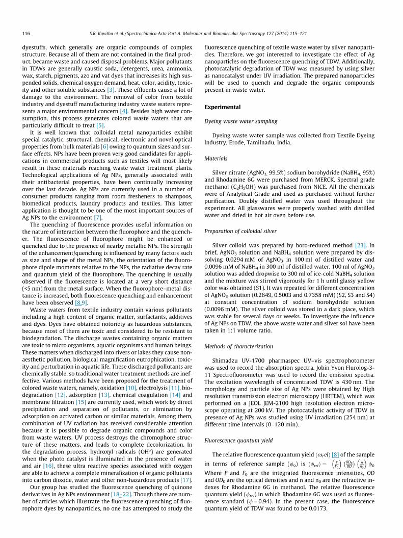
TEM has been used to characterize the size, shape and morphol-ogies of formed silver nanoparticles. The size dependent morphol-ogy of the silver nanoparticles prepared using different AgNO3
concentration was studied. The TEM micrographs of the silver col-loid for various concentration ratios S1 and S4 are shown inFig. 2(a) and (b) respectively. The average size of these nanoparti-cles is approximately ranged from 7 nm (S4) to 22 nm (S1) and thisresult was also confirmed by UV–vis spectra of Ag NPs. The pre-pared silver nanoparticle shows twinning structure. Twinning isone of the most commonly noticed planar defects in nanocrystals,and is frequently observed for metallic nanocrystals of face cen-tered cubic (fcc) structure. It occurs when two subgrains share acommon crystallographic plane. The structure of one subgrain isthen a mirror image of the other through the twinning plane.Additionally, TEM examination demonstrated that the sample iscomposed of several morphologies including multi-twinned nano-particles with fivefold symmetry, i.e. decahedra and icosahedraparticles. Because of this fivefold axis, strain was induced in theparticle to fill the gap. When the particles are smaller in size, themultiple twinning is the favorable structural configuration. Itmay be due to the smaller surface and volume energies [29].
Absorption characteristics of TDW in Ag NPs
UV–vis absorption spectrum of TDW and in the synthesized AgNPs is shown in Figs. 3 and 4 respectively. The absorption spectraof TDW in different sizes of Ag NPs show broad band in the visibleregion 350–550 nm. When TDW is added into silver sol, the plas-mon band slightly shifts towards shorter wavelength while the fea-ture of surface plasmon peak dominates and peak becomesbroaden. The major reason for surface plasmon resonance (SPR)broadening is electron surface scattering which may be enhancedfor very small clusters [24]. This change in spectral shift and bandbroadening of the surface plasmon band reveals that these TDWmay bind on the silver surface. The damping of the silver plasmonband indicates the attachment of the TDW molecule on the nano-particles surface. The interaction of silver nanoparticles with TDWalters the electron density of the Ag NPs, thereby directly affectingthe absorption of the surface bound TDW as well as surface plas-mon absorption band.
Fluorescence characteristics of TDW in Ag NPs
The fluorescence emission spectrum of TDW and in differentsizes of Ag NPs is shown in Figs. 3 and 5 respectively. The observedfluorescence quantum yield of TDW in different sizes of NPs hasbeen reported in Table 1. Fluorescence emission spectrum ofTDW shows broad band in the visible region 400 nm to 700 nm.It shows that one strong peak observed at 527 nm. In the presenceof quencher, the fluorescence intensity and quantum yield of TDWdecreases without any new broad band in the emission spectra
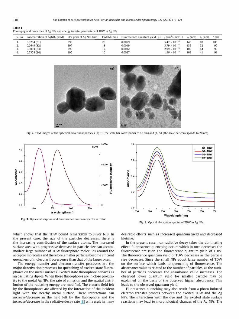
Table 1Photo-physical properties of Ag NPs and energy transfer parameters of TDW in Ag NPs.
S. No Concentration of AgNO3 (mM) SPR peak of Ag NPs (nm) FWHM (nm) Fluorescence quantum yield (/) J (cm3 L mol�1) R0 (nm) r0 (nm) E (%)
1. 0.0294 (S1) 399 29 0.0059 5.47 � 10�16 145 69 1002. 0.2649 (S2) 397 18 0.0049 3.79 � 10�16 135 52 973. 0.5003 (S3) 396 12 0.0032 2.99 � 10�15 109 44 934. 0.7358 (S4) 395 10 0.0027 1.96 � 10�15 103 41 91
Fig. 2. TEM images of the spherical silver nanoparticles (a) S1 (the scale bar corresponds to 10 nm) and (b) S4 (the scale bar corresponds to 20 nm).
Fig. 3. Optical absorption and fluorescence emission spectra of TDW.Fig. 4. Optical absorption spectra of TDW in Ag NPs.
118 S.R. Kavitha et al. / Spectrochimica Acta Part A: Molecular and Biomolecular Spectroscopy 127 (2014) 115–121
which shows that the TDW bound remarkably to silver NPs. Inthe present case, the size of the particles decreases, there isthe increasing contribution of the surface atoms. The increasedsurface area with progressive decrease in particle size can accom-modate large number of TDW fluorophore molecules around theacceptor molecules and therefore, smaller particles become efficientquenchers of molecular fluorescence than that of the larger ones.
The energy transfer and electron-transfer processes are themajor deactivation processes for quenching of excited state fluoro-phores on the metal surfaces. Excited state fluorophore behaves asan oscillating dipole. When these fluorophores are in close proxim-ity to the metal Ag NPs, the rate of emission and the spatial distri-bution of the radiating energy are modified. The electric field feltby the fluorophores are affected by the interaction of the incidentlight with the nearby metal surface. These interactions canincrease/decrease in the field felt by the fluorophore and theincrease/decrease in the radiative decay rate [8] will result in many
desirable effects such as increased quantum yield and decreasedlifetime.
In the present case, non-radiative decay takes the dominatingeffect, fluorescence quenching occurs which in turn decreases thefluorescence emission and fluorescence quantum yield of TDW.The fluorescence quantum yield of TDW decreases as the particlesize decreases. Since the small NPs adopt large number of TDWon the surface which leads to quenching of fluorescence. Theabsorbance value is related to the number of particles, as the num-ber of particles decreases the absorbance value increases. Theobserved lower quantum yield for smaller particle may beexplained on the basis of the observed higher absorbance. Thisleads to the observed quantum yield.
Fluorescence quenching may also result from a photo inducedelectron transfer process between the excited TDW and the AgNPs. The interaction with the dye and the excited state surfacereactions may lead to morphological changes of the Ag NPs. The

Fig. 5. Fluorescence emission spectra of TDW in Ag NPs.
S.R. Kavitha et al. / Spectrochimica Acta Part A: Molecular and Biomolecular Spectroscopy 127 (2014) 115–121 119
surface plasmon efficiently acts as energy acceptor even at a dis-tance of 1 nm between the probe molecules and the metal surface.Metal NPs also have a continuum of electronic states and exhibitenergy transfer behavior as excited state quenchers. The observedchanges in the absorbance of the absorption spectrum and fluores-cence intensity reflects the alteration of the electronic properties ofthe TDW chromophore as it binds to the Ag NPs which acts as aexcited state quencher. The TDW has a high emission quantumyield in the absence of NPs, the dominant effect for the quenchingof excited state of TDW in Ag NPs may be due to radiative energytransfer to the metal surface.
Fig. 6. Optical absorption spectra of Ag NPs and fluorescence emission spectrum ofTDW.
FRET between Ag NPs (acceptor) and TDW (donor)
There are few criteria that must be satisfied in order for FRET tooccur. These are: (i) the fluorescence emission spectrum of the do-nor molecule must overlap the absorption or excitation spectrumof the acceptor chromophore, (ii) the two fluorophore (donor andacceptor) must be in the close proximity to one another (typically1–10 nm), (iii) the transition dipole orientations of the donor andacceptor must be approximately parallel to each other, (iv) thefluorescence lifetime of the donor molecule must be of sufficientduration to allow the FRET to occur [8]. According to Förster’s the-ory, the rate of energy transfer is based on the spectral overlapbetween emission spectrum of the fluorophore and absorptionspectra of the Ag NPs, the relative orientation of the donor andacceptor transition dipoles, the distance between the donor andacceptor transition dipoles and the fluorescence quantum yieldof the donor. It results in long range dipole–dipole interactionsbetween the donor and acceptor [8,30].
Both energy-transfer and electron-transfer processes areconsidered to be the major deactivation pathways for excited fluo-rophore molecules on the Ag NPs surface. The electron transfermechanism is predominant for particle sizes of <5 nm as the parti-cles do not exhibit any surface plasmon band in the visible region.As the Ag NPs, in the present case, are larger than 5 nm, energytransfer dominates the quenching mechanism. The degree ofquenching depends on the structural details that control proximitybetween the fluorophore molecules and the Ag NPs core. When adonor molecules is placed in the vicinity of a conductive Ag NP sur-face, resonance energy transfer between the donor–acceptor takeplaces. The probability of this energy transfer depends on the over-lap of the emission band of the fluorophore molecules with theabsorption spectra of the Ag NPs [31].
In order to understand energy transfer, we have calculated thedistance between NPs and dye molecules using the Forstermechanism of non-radiative energy transfer. According to Forstertheory, the energy transfer effect is related not only to the distance(r0) between the acceptor and donor but also to the critical energytransfer distance (R0), i.e.
E ¼ 1� FF0¼ R6
0
R60 þ r6
0
ð1Þ
where F is the fluorescence intensity of the donor in the presence ofthe acceptor, F0 is the fluorescence intensity of the donor in the ab-sence of the acceptor and R0 is the critical distance at which the effi-ciency of energy transfer is 50%.
R60 ¼ 8:8� 10�25 k2n�4/ J ðkÞ ð2Þ
where k2 is the spatial orientation factor of the dipole, n is therefractive index of the medium, / is the fluorescence quantum yieldof the donor and J is the overlap integral of the fluorescence emis-sion spectrum of the donor and absorption spectra of the acceptorwas given by
J ðkÞ ¼R1
0 F ðkÞ e ðkÞ k4dkR10 F ðkÞ dk
ð3Þ
where F (k) is the fluorescence intensity of the fluorescence donor atwavelength k and e(k) is the molar absorptivity of the acceptor atwavelength k. In the present case, it has been reported that k2 ¼ 2
3,/ = 0.0173 and n = 1.336.
Based on these datas, the R0 and r0 values were calculated. Theoverlap of the optical absorption spectra of Ag NPs and the fluores-cence emission spectrum of TDW were shown in Fig. 6 The overlapintegral J(k), critical distance (R0), energy transfer efficiency (E) anddistance (r0) were shown in Table 1. From the overlapping spec-trum of the absorption spectra of acceptor and the fluorescencespectrum of TDW, J(k) can be evaluated by integrating the spectrafrom 440 to 640 nm. By adding the donor molecule into acceptormolecule with increasing concentration rate, the fluorescencespectra shifts toward shorter wavelength providing a smaller valueof spectral overlap integral. The increase in J(k) in turn causes anincrease in FRET efficiency. The value of J(k) changes from5.47 � 10�16 cm3 L mol�1 to 1.96 � 10�15 cm3 L mol�1 for changein AgNO3 concentration from S1 to S4.
Accordingly the energy transfer efficiency (E) between Ag NPsand TDW varies from 100% to 91%. It is evident that the efficiency
120 S.R. Kavitha et al. / Spectrochimica Acta Part A: Molecular and Biomolecular Spectroscopy 127 (2014) 115–121
of energy transfer is higher in S1 than that in S4. The maximumFRET efficiency was 91% for S4. Here energy transfer efficiencydecreases with increase in AgNO3 concentration ratio. The criticaldistance (R0) between Ag NPs and TDW for S1 is 145 nm and103 nm for S4 respectively. The distance (r0) between Ag NPs andTDW for S1 is 69 nm and 41 nm for S4 respectively. Fascinatinglyit was observed that the FRET efficiency decreases with increasein AgNO3 concentration. Therefore in the present system underinvestigation the FRET process between donor and acceptor mole-cule depends on AgNO3 concentration.
Fig. 6 shows the spectral overlap between absorption spectra ofAg NPs and emission spectrum of TDW. The calculated values ofthe energy transfer parameters are presented in Table 1. The smallspectral overlap between donor emission and acceptor absorptionin the TDW/Ag NPs blends indicates that energy transfer is ineffi-cient, and that a Forster-type energy transfer is unfeasible fromthe molecules of the TDW to the Ag NPs. Fig. 6 also shows thatacceptor weakly absorbed the excitation photon (430 nm), whichmay also contribute to the fluorescence intensity of Ag NPs. De-spite the possibility of electron energy transfer, this mechanism,however is considered significant because of weak overlap be-tween emission of donor and absorbance of acceptor. Moreover,the existence of fluorescence spectra as shown in Fig. 5 providesadditional evidence of negligible electron transfer mechanism.The radiative energy transfer between TDW and Ag NPs is alsoweak as supported by low fluorescence quantum yield of theTDW (Table 1), in addition to low concentration level of Ag [32].
From the overlap between the absorption and emission spectra,the critical transfer distance R0 was calculated using Eq. (2) andtabulated in Table 1. R0 is the average distance between the donorand acceptor molecules at which the probability of intermolecularenergy transfer is just equal to the sum of probabilities for all de-excitation processes of the donor excited state D*. It was found thatthe high value of donor quantum yield is responsible for high valueof R0. This high value of R0 is responsible for r0 and E values. Thelarge values of R0 indicate that the dominant mechanism responsi-ble for energy transfer is long range dipole–dipole energy transfer(Förster type) [33] and thus, confirming the appropriateness ofFörster theory to calculate the Förster energy transfer rates as re-ported by other researchers [34]. The high values of energy transferparameters obtained in this study are due to the use of donor withhigh molecular weight as acceptor [35,36].
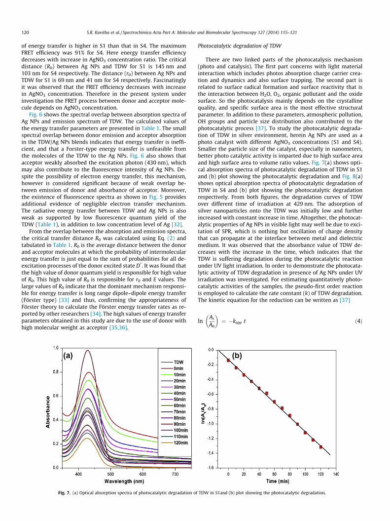
Fig. 7. (a) Optical absorption spectra of photocatalytic degradation of
Photocatalytic degradation of TDW
There are two linked parts of the photocatalysis mechanism(photo and catalysis). The first part concerns with light materialinteraction which includes photos absorption charge carrier crea-tion and dynamics and also surface trapping. The second part isrelated to surface radical formation and surface reactivity that isthe interaction between H2O, O2, organic pollutant and the oxidesurface. So the photocatalysis mainly depends on the crystallinequality, and specific surface area is the most effective structuralparameter. In addition to these parameters, atmospheric pollution,OH groups and particle size distribution also contributed to thephotocatalytic process [37]. To study the photocatalytic degrada-tion of TDW in silver environment, herein Ag NPs are used as aphoto catalyst with different AgNO3 concentrations (S1 and S4).Smaller the particle size of the catalyst, especially in nanometers,better photo catalytic activity is imparted due to high surface areaand high surface area to volume ratio values. Fig. 7(a) shows opti-cal absorption spectra of photocatalytic degradation of TDW in S1and (b) plot showing the photocatalytic degradation and Fig. 8(a)shows optical absorption spectra of photocatalytic degradation ofTDW in S4 and (b) plot showing the photocatalytic degradationrespectively. From both figures, the degradation curves of TDWover different time of irradiation at 429 nm. The adsorption ofsilver nanoparticles onto the TDW was initially low and furtherincreased with constant increase in time. Altogether, the photocat-alytic properties of Ag NPs in visible light may well be due to exci-tation of SPR, which is nothing but oscillation of charge densitythat can propagate at the interface between metal and dielectricmedium. It was observed that the absorbance value of TDW de-creases with the increase in the time, which indicates that theTDW is suffering degradation during the photocatalytic reactionunder UV light irradiation. In order to demonstrate the photocata-lytic activity of TDW degradation in presence of Ag NPs under UVirradiation was investigated. For estimating quantitatively photo-catalytic activities of the samples, the pseudo-first order reactionis employed to calculate the rate constant (k) of TDW degradation.The kinetic equation for the reduction can be written as [37]
lnAt
A0
� �¼ �kapp t ð4Þ
TDW in S1and (b) plot showing the photocatalytic degradation.

Fig. 8. (a) Optical absorption spectra of photocatalytic degradation of TDW in S4 and (b) plot showing the photocatalytic degradation.
S.R. Kavitha et al. / Spectrochimica Acta Part A: Molecular and Biomolecular Spectroscopy 127 (2014) 115–121 121
where A0 is the absorbance value of TDW at t = 0, At is the absor-bance of TDW at different irradiation time and kapp is the apparentrate constant, which can be obtained from the decrease of the peakintensity at 429 nm with time. A good linear correlation, ln (At/A0)versus time has been obtained. The apparent rate constant of thecatalytic reaction for TDW in presence of S1 is 0.009/min and S4is 0.011/min. By comparing both cases the apparent rate constantvalue was found to be the highest for S4 than in S1 because smallparticle size of Ag NPs acts as a good photocatalysts for textile wastewater treatments.
Conclusion
To conclude, Ag NPs have been successfully synthesized by asimple boro-reduced method. The photophysical properties of AgNPs in TDW have been extensively studied using optical absorptionand fluorescence emission techniques. The optical and emissionspectra show that fluorescence quantum yield of TDW decreases,this is due to size, shape, coupling between the Ag NPs withTDW and energy transfer between Ag NPs to TDW. The interactionsbetween Ag NPs and TDW molecule has been explained by FörsterResonance Energy Transfer mechanism. The higher value of R0
indicates that the dominant mechanism of fluorescence quenchingis resonance energy transfer due to long range dipole–dipole inter-action (Förster type). The Ag NPs were found to be active in degrad-ing textile dyeing waste water with UV light illumination. Thesefindings suggest that, synthesized Ag NPs are good, highly efficientand stable photocatalysts can able to degrade TDW under UV lightand paves way to control environmental pollution.
Acknowledgement
We gratefully acknowledge the financial assistance receivedfrom UGC-MRP and DST-CURIE, New Delhi, India for carrying outthis research work.
References
[1] T. Pradeep, Anshup, Thin Sol. Films 517 (2009) 6441–6478.[2] M.C. Venceslau, S. Tom, J.J. Simon, Environ. Technol. 163 (1994) 917–929.[3] A. Dae-Hee, C. Won-Seok, Y. Tai-Il, Process Biochem. 34 (1999) 429–439.[4] U. Pagga, D. Brown, Chemosphere 15 (1986) 479–491.
[5] M.M. Karim, A.K. Das, S.H. Lee, Anal. Chim. Acta 576 (2006) 37–42.[6] C.N.R. Rao, G.U. Kulkarni, P.J. Thomas, P.P. Edwards, Chem. Eur. J. 8 (2002) 28–
35.[7] N.C. Mueller, B. Nowack, Environ. Sci. Technol. 42 (2008) 4447–4453.[8] J.R. Lakowicz, Principles of Fluorescence Spectroscopy, third ed., Springer, New
York, 2006.[9] C.D. Geddes, H. Cao, I. Gryczynski, Z. Gryczynski, J.Y. Fang, J.R. Lakowicz, J. Phys.
Chem. A 107 (2003) 3443–3449.[10] I. Arslan, I.A. Balcioglu, Dyes Pigm. 43 (1999) 95–108.[11] B.K. Korbahti, A. Tanyolac, J. Hazard. Mater. 151 (2008) 422–431.[12] G. Sudarjanto, B. Keller-Lehmann, J. Keller, J. Hazard. Mater. 138 (2006) 160–
168.[13] R. Dolphen, N. Sakkayawong, P. Thiravetyan, W. Nakbanpote, J. Hazard. Mater.
145 (2007) 250–255.[14] B.H. Tan, T.T. Teng, A.K. Mohd Omar, Water Res. 34 (2000) 597–601.[15] N. Al-Bastaki, Chem. Eng. Process 43 (2004) 1561–1567.[16] H.Y. Chen, O. Zahraa, M. Bouchy, F. Thomas, J.Y. Bottero, J. Photochem.
Photobiol. A: Chem. 85 (1995) 179–186.[17] R.W. Matthews, S.R. McEvoy, J. Photochem. Photobiol. A: Chem. 66 (1992)
355–366.[18] M.Umadevi, N.A. Sridevi, A.S. Sharmila, B.J.M. Rajkumar, M. Briget Mary, P.
Vanelle, et al., J. Fluoresc. 20 (2010) 153–161.[19] M. Umadevi, P. Vanelle, T. Terme, B.J.M. Rajkumar, V. Ramakrishnan, J.
Fluoresc. 19 (2009) 3–10.[20] M. Umadevi, S.R. Kavitha, P. Vanelle, T. Terme, Plasmonics 8 (2013) 859–867.[21] M. Umadevi, S.R. Kavitha, P. Vanelle, T. Terme, O. Khoumeri, J. Lumin. 142
(2013) 1–7.[22] S.R. Kavitha, M. Umadevi, P. Vanelle, T. Terme, O. Khoumeri, Plasmonics
(2013), http://dx.doi.org/10.1007/s11468-013-9642-7.[23] X. Dong, H. Gu, J. Kang, X. Yuan, J. Wu, J. Mol. Struct. 984 (2010) 396–401.[24] S. Link, M.A. El-Sayed, Int. Rev. Phys. Chem. 19 (2000) 409–453.[25] A. Henglein, J. Phys. Chem. 97 (1993) 5457–5463.[26] S.L. Smitha, K.M. Nissamudeen, D. Philip, K.G. Gopchandran, J. Spectrochim.
Acta Part A 71 (2008) 186–190.[27] K.R. Brown, D.G. Walter, M. Natan, Chem. Mater. 12 (2000) 306–313.[28] C.F. Bohren, D.R. Huffman, Adsorption and Scattering of Light by Small
Particles, John Wiley & Sons, New York, 1983.[29] Z.L. Wang, J. Phys. Chem. B 104 (2000) 1153–1175.[30] T. Förster, Modern Quantum Chemistry, Academic Press, New York, 1965.[31] C. Fan, S. Wang, J.W. Hong, G.C. Bazan, K.W. Plaxco, A.J. Heeger, Proc. Nat. Acad.
Sci. 100 (2003) 6297–6301.[32] B.A. Al-Asbahi, M.S. Alsalhi, A.S. Al-Dwayyan, M.H. Haji Jumali, J. Lumin. 132
(2012) 386–390.[33] G.A. Kumar, V. Thomas, G. Jose, N.V. Unnikrishnan, V.P.N. Nampoori, J.
Photochem. Photobiol. A: Chem. 153 (2002) 145–151.[34] A. Mallick, B. Haldar, S. Sengupta, N. Chattopadhyay, J. Lumin. 118 (2006) 165–
172.[35] M.S. Alsalhi, Z.S. Abu Mustafa, V. Masilamani, Opt. Laser Technol. 43 (2011)
147–157.[36] J. Hill, S.Y. Heriot, O. Worsfold, T.H. Richardson, A.M. Fox, D.D.C. Bradley, Synth.
Metals 139 (2003) 787–790.[37] S. Boujday, F. Wunsch, P. Portes, J.F. Bocquet, C.C. Justin, Sol. Energy Mater. Sol.
Cells 83 (2004) 421–433.
Related Documents