The University of Manchester Research Fluorescence devices for the detection of dental caries DOI: 10.1002/14651858.CD013811. Document Version Final published version Link to publication record in Manchester Research Explorer Citation for published version (APA): Macey, R., Walsh, T., Riley, P., Worthington, H., Glenny, A-M., Clarkson, J., Ricketts, D., & Fee, P. A. (2020). Fluorescence devices for the detection of dental caries. Cochrane Database of Systematic Reviews, (12). https://doi.org/10.1002/14651858.CD013811. Published in: Cochrane Database of Systematic Reviews Citing this paper Please note that where the full-text provided on Manchester Research Explorer is the Author Accepted Manuscript or Proof version this may differ from the final Published version. If citing, it is advised that you check and use the publisher's definitive version. General rights Copyright and moral rights for the publications made accessible in the Research Explorer are retained by the authors and/or other copyright owners and it is a condition of accessing publications that users recognise and abide by the legal requirements associated with these rights. Takedown policy If you believe that this document breaches copyright please refer to the University of Manchester’s Takedown Procedures [http://man.ac.uk/04Y6Bo] or contact [email protected] providing relevant details, so we can investigate your claim. Download date:01. Jun. 2022

Welcome message from author
This document is posted to help you gain knowledge. Please leave a comment to let me know what you think about it! Share it to your friends and learn new things together.
Transcript
The University of Manchester Research
Fluorescence devices for the detection of dental caries
DOI:10.1002/14651858.CD013811.
Document VersionFinal published version
Link to publication record in Manchester Research Explorer
Citation for published version (APA):Macey, R., Walsh, T., Riley, P., Worthington, H., Glenny, A-M., Clarkson, J., Ricketts, D., & Fee, P. A. (2020).Fluorescence devices for the detection of dental caries. Cochrane Database of Systematic Reviews, (12).https://doi.org/10.1002/14651858.CD013811.
Published in:Cochrane Database of Systematic Reviews
Citing this paperPlease note that where the full-text provided on Manchester Research Explorer is the Author Accepted Manuscriptor Proof version this may differ from the final Published version. If citing, it is advised that you check and use thepublisher's definitive version.
General rightsCopyright and moral rights for the publications made accessible in the Research Explorer are retained by theauthors and/or other copyright owners and it is a condition of accessing publications that users recognise andabide by the legal requirements associated with these rights.
Takedown policyIf you believe that this document breaches copyright please refer to the University of Manchester’s TakedownProcedures [http://man.ac.uk/04Y6Bo] or contact [email protected] providingrelevant details, so we can investigate your claim.
Download date:01. Jun. 2022
CochraneLibrary
Cochrane Database of Systematic Reviews
Fluorescence devices for the detection of dental caries (Review)
Macey R, Walsh T, Riley P, Glenny AM, Worthington HV, Fee PA, Clarkson JE, Ricketts D
Macey R, Walsh T, Riley P, Glenny A-M, Worthington HV, Fee PA, Clarkson JE, Ricketts D. Fluorescence devices for the detection of dental caries. Cochrane Database of Systematic Reviews 2020, Issue 12. Art. No.: CD013811. DOI: 10.1002/14651858.CD013811.
www.cochranelibrary.com
Fluorescence devices for the detection of dental caries (Review)
Copyright © 2020 The Cochrane Collaboration. Published by John Wiley & Sons, Ltd.
CochraneLibrary
Trusted evidence.Informed decisions.Better health.
Cochrane Database of Systematic Reviews
T A B L E O F C O N T E N T S
HEADER......................................................................................................................................................................................................... 1
ABSTRACT..................................................................................................................................................................................................... 1
PLAIN LANGUAGE SUMMARY....................................................................................................................................................................... 2
SUMMARY OF FINDINGS.............................................................................................................................................................................. 4
BACKGROUND.............................................................................................................................................................................................. 6
Figure 1.................................................................................................................................................................................................. 8
OBJECTIVES.................................................................................................................................................................................................. 9
METHODS..................................................................................................................................................................................................... 9
RESULTS........................................................................................................................................................................................................ 13
Figure 2.................................................................................................................................................................................................. 14
Figure 3.................................................................................................................................................................................................. 16
Figure 4.................................................................................................................................................................................................. 17
Figure 5.................................................................................................................................................................................................. 22
Figure 6.................................................................................................................................................................................................. 24
Figure 7.................................................................................................................................................................................................. 26
Figure 8.................................................................................................................................................................................................. 28
Figure 9.................................................................................................................................................................................................. 30
Figure 10................................................................................................................................................................................................ 32
Figure 11................................................................................................................................................................................................ 34
Figure 12................................................................................................................................................................................................ 36
Figure 13................................................................................................................................................................................................ 38
Figure 14................................................................................................................................................................................................ 40
Figure 15................................................................................................................................................................................................ 42
Figure 16................................................................................................................................................................................................ 44
Figure 17................................................................................................................................................................................................ 46
Figure 18................................................................................................................................................................................................ 47
Figure 19................................................................................................................................................................................................ 49
Figure 20................................................................................................................................................................................................ 50
Figure 21................................................................................................................................................................................................ 51
DISCUSSION.................................................................................................................................................................................................. 51
AUTHORS' CONCLUSIONS........................................................................................................................................................................... 54
ACKNOWLEDGEMENTS................................................................................................................................................................................ 55
REFERENCES................................................................................................................................................................................................ 56
CHARACTERISTICS OF STUDIES.................................................................................................................................................................. 67
ADDITIONAL TABLES.................................................................................................................................................................................... 419
HISTORY........................................................................................................................................................................................................ 432
CONTRIBUTIONS OF AUTHORS................................................................................................................................................................... 432
DECLARATIONS OF INTEREST..................................................................................................................................................................... 432
SOURCES OF SUPPORT............................................................................................................................................................................... 432
DIFFERENCES BETWEEN PROTOCOL AND REVIEW.................................................................................................................................... 432
Fluorescence devices for the detection of dental caries (Review)
Copyright © 2020 The Cochrane Collaboration. Published by John Wiley & Sons, Ltd.
i
CochraneLibrary
Trusted evidence.Informed decisions.Better health.
Cochrane Database of Systematic Reviews
[Diagnostic Test Accuracy Review]
Fluorescence devices for the detection of dental caries
Richard Macey1, Tanya Walsh1, Philip Riley2, Anne-Marie Glenny1, Helen V Worthington2, Patrick A Fee3, Janet E Clarkson4, David
Ricketts5
1Division of Dentistry, School of Medical Sciences, Faculty of Biology, Medicine and Health, The University of Manchester, Manchester,
UK. 2Cochrane Oral Health, Division of Dentistry, School of Medical Sciences, Faculty of Biology, Medicine and Health, The University
of Manchester, Manchester, UK. 3Dundee Dental School, University of Dundee, Dundee, UK. 4Division of Oral Health Sciences, Dundee
Dental School, University of Dundee, Dundee, UK. 5Dundee Dental School, University of Dundee, Dundee, UK
Contact address: Tanya Walsh, [email protected].
Editorial group: Cochrane Oral Health Group.Publication status and date: New, published in Issue 12, 2020.
Citation: Macey R, Walsh T, Riley P, Glenny A-M, Worthington HV, Fee PA, Clarkson JE, Ricketts D. Fluorescence devices for the detectionof dental caries. Cochrane Database of Systematic Reviews 2020, Issue 12. Art. No.: CD013811. DOI: 10.1002/14651858.CD013811.
Copyright © 2020 The Cochrane Collaboration. Published by John Wiley & Sons, Ltd.
A B S T R A C T
Background
Caries is one of the most prevalent and preventable conditions worldwide. If identified early enough then non-invasive techniques can beapplied, and therefore this review focusses on early caries involving the enamel surface of the tooth. The cornerstone of caries detectionis a visual and tactile dental examination, however alternative methods of detection are available, and these include fluorescence-baseddevices. There are three categories of fluorescence-based device each primarily defined by the diEerent wavelengths they exploit; we havelabelled these groups as red, blue, and green fluorescence. These devices could support the visual examination for the detection anddiagnosis of caries at an early stage of decay.
Objectives
Our primary objectives were to estimate the diagnostic test accuracy of fluorescence-based devices for the detection and diagnosis ofenamel caries in children or adults. We planned to investigate the following potential sources of heterogeneity: tooth surface (occlusal,proximal, smooth surface or adjacent to a restoration); single point measurement devices versus imaging or surface assessment devices;and the prevalence of more severe disease in each study sample, at the level of caries into dentine.
Search methods
Cochrane Oral Health's Information Specialist undertook a search of the following databases: MEDLINE Ovid (1946 to 30 May 2019); EmbaseOvid (1980 to 30 May 2019); US National Institutes of Health Ongoing Trials Register (ClinicalTrials.gov, to 30 May 2019); and the World HealthOrganization International Clinical Trials Registry Platform (to 30 May 2019). We studied reference lists as well as published systematicreview articles.
Selection criteria
We included diagnostic accuracy study designs that compared a fluorescence-based device with a reference standard. This includedprospective studies that evaluated the diagnostic accuracy of single index tests and studies that directly compared two or more index tests.Studies that explicitly recruited participants with caries into dentine or frank cavitation were excluded.
Data collection and analysis
Two review authors extracted data independently using a piloted study data extraction form based on the Quality Assessment ofDiagnostic Accuracy Studies 2 (QUADAS-2). Sensitivity and specificity with 95% confidence intervals (CIs) were reported for each study.This information has been displayed as coupled forest plots and summary receiver operating characteristic (SROC) plots, displaying the
Fluorescence devices for the detection of dental caries (Review)
Copyright © 2020 The Cochrane Collaboration. Published by John Wiley & Sons, Ltd.
1
CochraneLibrary
Trusted evidence.Informed decisions.Better health.
Cochrane Database of Systematic Reviews
sensitivity-specificity points for each study. We estimated diagnostic accuracy using hierarchical summary receiver operating characteristic(HSROC) methods. We reported sensitivities at fixed values of specificity (median 0.78, upper quartile 0.90).
Main results
We included a total of 133 studies, 55 did not report data in the 2 x 2 format and could not be included in the meta-analysis. 79 studieswhich provided 114 datasets and evaluated 21,283 tooth surfaces were included in the meta-analysis. There was a high risk of bias for theparticipant selection domain. The index test, reference standard, and flow and timing domains all showed a high proportion of studies tobe at low risk of bias. Concerns regarding the applicability of the evidence were high or unclear for all domains, the highest proportionbeing seen in participant selection. Selective participant recruitment, poorly defined diagnostic thresholds, and in vitro studies being non-generalisable to the clinical scenario of a routine dental examination were the main reasons for these findings. The dominance of in vitrostudies also means that the information on how the results of these devices are used to support diagnosis, as opposed to pure detection,was extremely limited. There was substantial variability in the results which could not be explained by the diEerent devices or dentition orother sources of heterogeneity that we investigated. The diagnostic odds ratio (DOR) was 14.12 (95% CI 11.17 to 17.84).
The estimated sensitivity, at a fixed median specificity of 0.78, was 0.70 (95% CI 0.64 to 0.75). In a hypothetical cohort of 1000 tooth sites orsurfaces, with a prevalence of enamel caries of 57%, obtained from the included studies, the estimated sensitivity of 0.70 and specificityof 0.78 would result in 171 missed tooth sites or surfaces with enamel caries (false negatives) and 95 incorrectly classed as having earlycaries (false positives).
We used meta-regression to compare the accuracy of the diEerent devices for red fluorescence (84 datasets, 14,514 tooth sites), bluefluorescence (21 datasets, 3429 tooth sites), and green fluorescence (9 datasets, 3340 tooth sites) devices. Initially, we allowed threshold,shape, and accuracy to vary according to device type by including covariates in the model. Allowing consistency of shape, removal of the
covariates for accuracy had only a negligible eEect (Chi2 = 3.91, degrees of freedom (df) = 2, P = 0.14).
Despite the relatively large volume of evidence we rated the certainty of the evidence as low, downgraded two levels in total, for risk ofbias due to limitations in the design and conduct of the included studies, indirectness arising from the high number of in vitro studies, andinconsistency due to the substantial variability of results.
Authors' conclusions
There is considerable variation in the performance of these fluorescence-based devices that could not be explained by the diEerentwavelengths of the devices assessed, participant, or study characteristics. Blue and green fluorescence-based devices appeared tooutperform red fluorescence-based devices but this diEerence was not supported by the results of a formal statistical comparison. Theevidence base was considerable, but we were only able to include 79 studies out of 133 in the meta-analysis as estimates of sensitivity orspecificity values or both could not be extracted or derived. In terms of applicability, any future studies should be carried out in a clinicalsetting, where diEiculties of caries assessment within the oral cavity include plaque, staining, and restorations. Other considerationsinclude the potential of fluorescence devices to be used in combination with other technologies and comparative diagnostic accuracystudies.
P L A I N L A N G U A G E S U M M A R Y
Fluorescence devices for the detection of dental caries
Why is it important to improve dental caries (tooth decay) detection?
Dentists oNen aim to identify tooth decay that has already advanced to a level which needs a filling. If dentists were able to find tooth decaywhen it has only aEected the outer layer of the tooth then it is possible to stop the decay from spreading any further and prevent the needfor fillings. It is also important to avoid a false-positive result, when treatment may be provided when caries is absent.
What is the aim of this review?
This Cochrane Review aimed to find out how accurate fluorescence devices (non-invasive devices that shine a light on the surface of thetooth) are for detecting and diagnosing early tooth decay as part of the dental 'check-up' for children and adults who visit their generaldentist. Researchers included 133 studies to answer this question.
What was studied in the review?
There are three diEerent types of fluorescence device that use diEerent types of light which we grouped as red, blue, and greenfluorescence. Each device reflects more or less light depending on the amount of tooth decay, and this is measured by the device to give ascore which indicates whether there is tooth decay and how severe the decay is. We studied decay on the occlusal surfaces (biting surfacesof the back teeth), the proximal surfaces (tooth surfaces that are next to each other), and the smooth surfaces.
What are the main results of the review?
Fluorescence devices for the detection of dental caries (Review)
Copyright © 2020 The Cochrane Collaboration. Published by John Wiley & Sons, Ltd.
2
CochraneLibrary
Trusted evidence.Informed decisions.Better health.
Cochrane Database of Systematic Reviews
The review included 133 relevant studies but 55 of these did not provide data in a format that we could use for analysis, so 79 studies witha total of 21,283 teeth were included in the analysis. Some of these studies reported on more than one type of fluorescence device, thisgave us 114 sets of data. The results of these studies indicate that, in theory, if the fluorescence devices were to be used by a dentist for aroutine dental examination in a group of 1000 tooth sites or surfaces, of which 574 (57%) have early tooth decay:
• an estimated 494 will have a fluorescence device result indicating tooth decay, and of these, 95 (19%) will not have tooth decay (falsepositive - incorrect diagnosis);• of the 506 tooth sites or surfaces with a result indicating that tooth decay is not present, 171 (34%) will have early tooth decay (falsenegative - incorrect diagnosis).
Please see oralhealth.cochrane.org/fluorescence-devices-results.
We found no evidence that the devices that used diEerent types of light (red, blue, or green fluorescence) diEered in their accuracy.
How reliable are the results of the studies in this review?
We only included studies that assessed healthy teeth or those that were thought to have early tooth decay. This is because teeth with deeptooth decay would be easier to detect. However, there were some problems with how the studies were carried out. This may have resultedin the fluorescence-based devices appearing more accurate than they are. We judged the certainty of the evidence as low due to how thestudies selected their participants, the large number of studies that were carried out in a laboratory setting on extracted teeth, and thevariation in results reported.
Who do the results of this review apply to?
Studies included in the review were carried out in Brazil, Europe, the Middle East, Asia, North America, and Australia. A large number ofstudies used extracted teeth. Others were completed in dental hospitals, general dental practices, or schools. Studies were from the years1998 and 2019.
What are the implications of this review?
Because of the wide variation in performance that cannot be easily explained the interpretation of results is diEicult. The proportion ofcases missed or incorrectly diagnosed as evidence of caries is relatively high. Important information was missing from many of the includedstudies. Any future studies should be carried out in a clinical setting, and look at the potential of fluorescence devices to be used alongsideother devices.
How up-to-date is this review?
The review authors searched for and used studies published up to 30 May 2019.
Fluorescence devices for the detection of dental caries (Review)
Copyright © 2020 The Cochrane Collaboration. Published by John Wiley & Sons, Ltd.
3
Fluoresce
nce device
s for th
e detectio
n of dental ca
ries (R
eview)
Copyrig
ht © 2020 T
he Cochrane Collaboration. Publish
ed by Jo
hn Wiley & Sons, Ltd
.
4
S U M M A R Y O F F I N D I N G S
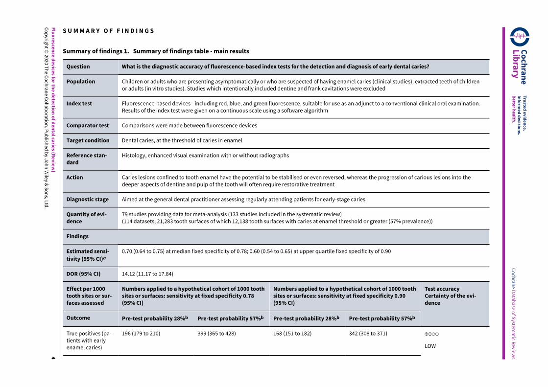
Summary of findings 1. Summary of findings table - main results
Question What is the diagnostic accuracy of fluorescence-based index tests for the detection and diagnosis of early dental caries?
Population Children or adults who are presenting asymptomatically or who are suspected of having enamel caries (clinical studies); extracted teeth of childrenor adults (in vitro studies). Studies which intentionally included dentine and frank cavitations were excluded
Index test Fluorescence-based devices - including red, blue, and green fluorescence, suitable for use as an adjunct to a conventional clinical oral examination.Results of the index test were given on a continuous scale using a software algorithm
Comparator test Comparisons were made between fluorescence devices
Target condition Dental caries, at the threshold of caries in enamel
Reference stan-dard
Histology, enhanced visual examination with or without radiographs
Action Caries lesions confined to tooth enamel have the potential to be stabilised or even reversed, whereas the progression of carious lesions into thedeeper aspects of dentine and pulp of the tooth will often require restorative treatment
Diagnostic stage Aimed at the general dental practitioner assessing regularly attending patients for early-stage caries
Quantity of evi-dence
79 studies providing data for meta-analysis (133 studies included in the systematic review)(114 datasets, 21,283 tooth surfaces of which 12,138 tooth surfaces with caries at enamel threshold or greater (57% prevalence))
Findings
Estimated sensi-
tivity (95% CI)a0.70 (0.64 to 0.75) at median fixed specificity of 0.78; 0.60 (0.54 to 0.65) at upper quartile fixed specificity of 0.90
DOR (95% CI) 14.12 (11.17 to 17.84)
Effect per 1000tooth sites or sur-faces assessed
Numbers applied to a hypothetical cohort of 1000 toothsites or surfaces: sensitivity at fixed specificity 0.78(95% CI)
Numbers applied to a hypothetical cohort of 1000 toothsites or surfaces: sensitivity at fixed specificity 0.90(95% CI)
Outcome Pre-test probability 28%b Pre-test probability 57%b Pre-test probability 28%b Pre-test probability 57%b
Test accuracyCertainty of the evi-dence
True positives (pa-tients with earlyenamel caries)
196 (179 to 210) 399 (365 to 428) 168 (151 to 182) 342 (308 to 371) ⊕⊕⊝⊝
LOW
Cochrane
Library
Truste
d evidence.
Inform
ed decisio
ns.
Bette
r health
.
Cochrane D
atabase o
f System
atic R
eviews
Fluoresce
nce device
s for th
e detectio
n of dental ca
ries (R
eview)
Copyrig
ht © 2020 T
he Cochrane Collaboration. Publish
ed by Jo
hn Wiley & Sons, Ltd
.
5
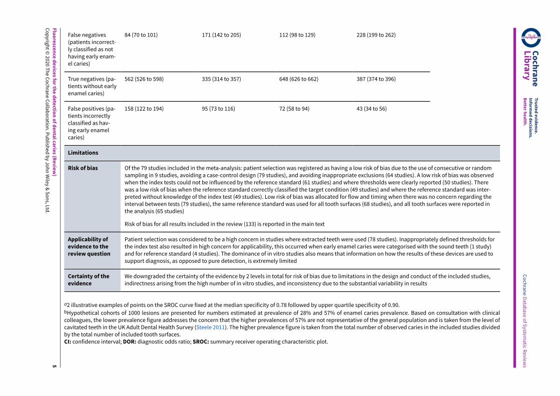
False negatives(patients incorrect-ly classified as nothaving early enam-el caries)
84 (70 to 101) 171 (142 to 205) 112 (98 to 129) 228 (199 to 262)
True negatives (pa-tients without earlyenamel caries)
562 (526 to 598) 335 (314 to 357) 648 (626 to 662) 387 (374 to 396)
False positives (pa-tients incorrectlyclassified as hav-ing early enamelcaries)
158 (122 to 194) 95 (73 to 116) 72 (58 to 94) 43 (34 to 56)
Limitations
Risk of bias Of the 79 studies included in the meta-analysis: patient selection was registered as having a low risk of bias due to the use of consecutive or randomsampling in 9 studies, avoiding a case-control design (79 studies), and avoiding inappropriate exclusions (64 studies). A low risk of bias was observedwhen the index tests could not be influenced by the reference standard (61 studies) and where thresholds were clearly reported (50 studies). Therewas a low risk of bias when the reference standard correctly classified the target condition (49 studies) and where the reference standard was inter-preted without knowledge of the index test (49 studies). Low risk of bias was allocated for flow and timing when there was no concern regarding theinterval between tests (79 studies), the same reference standard was used for all tooth surfaces (68 studies), and all tooth surfaces were reported inthe analysis (65 studies)
Risk of bias for all results included in the review (133) is reported in the main text
Applicability ofevidence to thereview question
Patient selection was considered to be a high concern in studies where extracted teeth were used (78 studies). Inappropriately defined thresholds forthe index test also resulted in high concern for applicability, this occurred when early enamel caries were categorised with the sound teeth (1 study)and for reference standard (4 studies). The dominance of in vitro studies also means that information on how the results of these devices are used tosupport diagnosis, as opposed to pure detection, is extremely limited
Certainty of theevidence
We downgraded the certainty of the evidence by 2 levels in total for risk of bias due to limitations in the design and conduct of the included studies,indirectness arising from the high number of in vitro studies, and inconsistency due to the substantial variability in results
a2 illustrative examples of points on the SROC curve fixed at the median specificity of 0.78 followed by upper quartile specificity of 0.90.bHypothetical cohorts of 1000 lesions are presented for numbers estimated at prevalence of 28% and 57% of enamel caries prevalence. Based on consultation with clinicalcolleagues, the lower prevalence figure addresses the concern that the higher prevalences of 57% are not representative of the general population and is taken from the level ofcavitated teeth in the UK Adult Dental Health Survey (Steele 2011). The higher prevalence figure is taken from the total number of observed caries in the included studies dividedby the total number of included tooth surfaces.CI: confidence interval; DOR: diagnostic odds ratio; SROC: summary receiver operating characteristic plot.
Cochrane
Library
Truste
d evidence.
Inform
ed decisio
ns.
Bette
r health
.
Cochrane D
atabase o
f System
atic R
eviews
CochraneLibrary
Trusted evidence.Informed decisions.Better health.
Cochrane Database of Systematic Reviews
B A C K G R O U N D
Cochrane Oral Health (COH) has undertaken several systematicreviews of diagnostic test accuracy (DTA) on the detection anddiagnosis of dental caries. The suite of systematic reviews formspart of a UK National Institute for Health Research (NIHR)Cochrane Programme Grant Scheme and involved collaborationwith the Complex Reviews Support Unit. The reviews followstandard Cochrane DTA methodology and have been diEerentiatedaccording to the index test under evaluation. A generic protocolserved as the basis for the suite of systematic reviews (Macey 2018).
Caries is an entire disease process, which can be stabilised andsometimes reversed if diagnosed and treated early on in the diseaseprocess (Fejerskov 2015; Pitts 2009). Most high-income countriesaround the world have evidenced a reduction in caries incidencein children and adolescents, and in some Scandinavian countriesprevention programmes have almost eradicated caries, but suchactivities have not been widely replicated in other locations (Pitts2017). Despite this reduction, the 2015 Global Burden of Diseasestudy identified dental caries as the most prevalent, preventablecondition worldwide (Feigin 2016; Kassebaum 2015), aEecting 60%to 90% of children and the majority of adults of the world'spopulation (Dye 2015; Petersen 2005). Furthermore, despite areduction in caries in many industrialised countries, the globalincidence of untreated caries was reported to be 2.4 billion in2010 (Feigin 2016; Kassebaum 2015; World Health Organization2017) and continues to increase year on year. In the UK, theprimary reason for childhood (aged 5 years to 9 years) hospitaladmissions is for the extraction of teeth (Public Health England2014). Longitudinal studies have shown that those who experiencecaries early in childhood will have an increased risk of severe cariesin later life, and that the disease trajectory will be steeper thanthose without early caries experience (Broadbent 2008; Hall-Scullin2017).
Untreated caries can lead to episodes of severe pain and infection,oNen requiring treatment with antibiotics. Dental anxiety resultingfrom untreated caries and the subsequent need for more invasivemanagement, can adversely aEect a person's future willingnessto visit their dentist, leading to a downward spiral of oral disease(Milsom 2003; Thomson 2000). If leN to progress, treatment optionsare limited to restoration or extraction, requiring repeated visits toa dental surgery or even to a hospital (Featherstone 2004; Fejerskov2015; Kidd 2004).
The cost of treating caries is high. In the UK alone, the NationalHealth Service (NHS) spends around GBP half a billion every yearin treating the disease. Hidden costs also exist, and the relatedproductivity losses are high, estimated at USD 27 billion globally in2010 (Listl 2015).
Caries detection and diagnosis will usually be undertaken ata routine dental examination, by a general dental practitioner,in patients who are presenting asymptomatically. However,caries detection can additionally be employed in secondarycare settings, school or community screening projects, andepidemiology or research studies (Braga 2009; Jones 2017). Thetraditional method of detecting dental caries in clinical practiceis a visual-tactile examination oNen with supporting radiographicinvestigations. This combination of methods is believed to besuccessful at detecting caries that has progressed into dentineand reached a threshold where a restoration may be necessary
(Kidd 2004). However, the detection of caries earlier in the diseasecontinuum could lead to stabilisation of disease or even possibleremineralisation of the tooth surface, thus preventing the patientfrom entering a lifelong cycle of restoration (Pitts 2017), but earlycaries is diEicult to detect visually, and the use of radiographsprovides only limited ability to detect small changes in dentalenamel (Ismail 2007).
Detection and diagnosis at the initial (non-cavitated) and moderatelevels of caries is fundamental in achieving the promotion of oralhealth and prevention of oral disease (Fejerskov 2015; Ismail 2013).The prevalence of this early caries state is not oNen reported indental epidemiology, most reports preferring to focus on cavitated/dentinal lesions which may be easier to detect, for example,the most recent UK Adult Dental Health survey reported 31% ofthe sample having untreated caries into dentine (Steele 2011;White 2012), and a US study reported levels of cavities at 15.3%in 12- to 19-year olds (Dye 2015). However, one UK survey ofchildren identified "clinical decay experience" which incorporatedany enamel breakdown and all other forms of caries and reported aprevalence of 63% in 15-year olds (Children’s Dental Health Survey2013).
A wide variety of management options are available under NHScare at these diEerent thresholds of disease, ranging from non-operative preventive strategies such as improved oral hygiene,reduced sugar diet and application of topical fluoride to minimallyinvasive treatments (e.g. sealing the aEected surface of the tooth,or 'infiltrating' the demineralised tissue with resins) for initialcaries, through to selective caries removal and restoration forextensive lesions. With advances in technology over the last twodecades, additional methods of detection have become available,such as advancements in radiography and the developmentof fluorescence, transillumination, and electrical conductancedevices. These could potentially aid the detection and diagnosisof caries at an early stage of decay. This would aEord the patientthe opportunity of a less invasive treatment with less destruction oftooth tissue and potentially result in a reduced cost of care to thepatient and healthcare services.
Target condition being diagnosed
The term dental caries is used to describe the mechanism whichcan ultimately lead to the breakdown of the tooth surface whichresults from an imbalance in the activity within the biofilm (ordental plaque) on the surface of the tooth within the oral cavity(Kidd 2016). This imbalance is due to bacterial breakdown ofsugars in the diet which leads to the production of acid andsubsequent demineralisation of the tooth. Disease progression canbe moderated by improved oral hygiene practices together withthe influx of fluoride from toothpaste and other available fluoridesources. However, the levels of sugar consumption observed inmany populations will oNen outweigh the benefits of fluoride (Hse2015). Ultimately, carious lesions may develop and destroy thestructure of the tooth.
The most common surfaces for caries to manifest are on theocclusal (biting) surfaces or the proximal surfaces (tooth surfacewhich face an adjacent tooth); although smooth surfaces on theflat exterior of teeth adjacent to the tongue, cheeks, and lips canbe aEected. The severity of the disease is defined by the depth ofdemineralisation of the tooth's structure and whether the lesion isactive or arrested. Caries presenting at levels into tooth enamel can
Fluorescence devices for the detection of dental caries (Review)
Copyright © 2020 The Cochrane Collaboration. Published by John Wiley & Sons, Ltd.
6
CochraneLibrary
Trusted evidence.Informed decisions.Better health.
Cochrane Database of Systematic Reviews
potentially be stabilised or even reversed, whereas the progressionof carious lesions into the dentine and pulp of the tooth will oNenrequire restoration (Bakhshandeh 2018; Kidd 2004).
Assessment of disease severity traditionally used inepidemiological and research studies has historically employedsome variant of the DMFT (decayed, missing, and filled teeth)scale. Within the D (decayed) component there are four clinicallydetectable thresholds applied as indicators for diagnosis andtreatment planning, oNen labelled as D1, D2, D3, and D4 (Anaise
1984) (Additional Table 1). Typically the D3 threshold, with only
lesions extending into dentine classed as carious, has been usedto determine the presence of caries (Pitts 1988; Shoaib 2009).These four categories have formed the basis for expanded cariesindices based on visual characteristics such as the InternationalCaries Detection and Assessment System (ICDAS) (Ekstrand 2007;Ismail 2007). Other available systems include: the Nyvad system(Nyvad 1999); Ekstrand-Ricketts-Kidd (ERK) system (Ekstrand1997); British Association for the Study of Community Dentistry(BASCD) (Pitts 1997); the Dundee Selectable Threshold Methodfor caries diagnosis (DSTM) (FyEe 2000); and the American DentalAssociation Caries Classification System for clinical practice (Young2015). The ICDAS and DSTM systems both provide the opportunityto investigate initial caries (into enamel) which may confer benefitsfor preventative or non-operative treatment.
Treatment of caries
There are many varied treatment options available to the dentalclinician, dependent on the thresholds of observed disease.Initial caries can be managed without surgical intervention usingapproaches such as plaque control, dietary advice, and applicationof fluoride to remineralise the tooth surface and prevent furtherprogression (Kidd 2016). Minimally invasive treatments for initialcaries are available, such as sealing the aEected surface of thetooth, or 'infiltrating' the demineralised tissue with resins. High-risk patients with severe caries may require selective caries removaland restoration of extensive lesions.
A caries management pathway, informed by diagnosticinformation, can be beneficial in guiding the clinician towardsprevention or a treatment plan. One recently developed carepathway is the International Caries Classification and ManagementSystem (ICCMS) (Ismail 2015). The system presents three forms ofmanagement in the care pathway:
• when dentition is sound the clinician proceeds withpreventative strategies to prevent sound surfaces fromdeveloping caries;
• non-invasive treatment of the lesion to arrest the decay processand encourage remineralisation, preventing initial lesions fromprogressing to cavitated decay; and
• management of more severe caries through excavation andrestoration or potentially extraction.
At the core of this care pathway is the ability to detect earlycaries accurately and optimise the preventative strategies throughtooth tissue-preserving excavation methods, and restoration orpotentially extraction in more severe cases. The detection anddiagnosis of early caries remain challenging, and the likelihoodof undiagnosed early disease is high (Ekstrand 1997). In suchinstances, the opportunity for preventing initial lesions fromprogressing to cavitated decay, or even reversing the disease
process, is missed, and disease progresses to cavitated decaywhere restoration is required (Ekstrand 1998).
Index test(s)
The cornerstone of caries detection is a visual and tactile dentalexamination, and the ability of clinicians to accurately detectdisease in this way has been researched for over half a century(Backer Dirks 1951). Many devices for the detection and diagnosisnow exist and may be suitable at diEerent stages of the carepathway (Bloemendal 2004; FyEe 2000). This review investigatesfluorescence-based devices that aim to measure the mineralcontent of the tooth according to changing fluorescence identifiedusing light with various wavelengths according to the device used(e.g. 405 nm for quantitative light-induced fluorescence (QLF) and655 nm for DIAGNOdent) (Kim 2019; Neuhaus 2019). Macey 2018provide details of the other index tests being investigated in thisseries of systematic reviews.
We included three categories of fluorescence index test eachprimarily defined by the diEerent wavelengths exploited by thedevices.
• Red fluorescence: these devices use a small laser with anexcitation wavelength greater than 655 nm. The tip of thedevice emits the excitation light and collects the resultantfluorescence and works on the principle that carious tissuecreates more emitted fluorescence than sound tissue throughthe fluorescence of bacterial by-products (porphyrins) (Pretty2006). These devices include: DIAGNOdent and DIAGNOdent pen(KaVo, Biberach, Germany) that feedback results via the device'sdisplay on a continuous scale (minimum 1 to maximum 99);MidWest (DENTSPLY Professional, New York, USA) emits soundand light (green/red) if caries is detected; and the Canary System(Quantum Dental Technologies Inc, Toronto, Ontario, Canada)which displays a number on a scale from 0 to 100 where 0 to 20is deemed to be healthy (Amaechi 2019; Lussi 1999; Lussi 2001;Neuhaus 2019; Rodrigues 2011).
• Blue fluorescence: these devices operate at wavelengthsbetween 400 nm and 450 nm at the blue/violet end of thevisible light spectrum and create luminescence in regionswhere there is bacterial activity which is oNen indicative ofdental caries; while the sound or healthy areas of the toothcontinue to fluoresce green (Rodrigues 2011). The devices inthis group rely on bespoke soNware to provide an image ofthe luminescence regions, examples are VistaProof (Durr Dental,Bietigheim-Bissingen, Germany), SoproLife (ACTEON Group, LaCiotat, France), and Spectra (Air Techniques, Melville, New York,USA) which use bespoke soNware packages to produce a digitalimage of the tooth which is interpreted by the operator. Thedevices use diEerent wavelengths of light (405 nm versus 450nm) however their mode of action is similar. VistaProof usessoNware to create a numeric score between 0 and 5 (Achilleos2013), SoproLife relies on the operator interpreting the findingsof the imaging program and allocating to one of six groups thatrange from sound to visible dentine (Rechmann 2012), Spectraprovides a numeric and colour category ranging from sound todentine lesions (Graye 2012).
• Green fluorescence: includes devices that use QLF, these relyon the characteristics of fluorescence at the green-yellow endof the spectrum (370 nm) (Angmar-Månsson 2001). This isemitted or refracted to the device and a measurement is taken,
Fluorescence devices for the detection of dental caries (Review)
Copyright © 2020 The Cochrane Collaboration. Published by John Wiley & Sons, Ltd.
7
CochraneLibrary
Trusted evidence.Informed decisions.Better health.
Cochrane Database of Systematic Reviews
which by definition is the tooth's "quantitative light-inducedfluorescence" and can be measured in terms of an average lossof fluorescence denoting lesion depth (oNen labelled ΔF andallocated to a point on a numeric scale) (Kim 2019; Neuhaus2016).
Clinical pathway
The process from a dental patient attending for a routineexamination and a caries assessment being undertaken potentiallyhas four intertwined phases: screening, detection, diagnosis,and treatment planning. If the presenting patient is at somerisk of disease but seemingly asymptomatic then this can beconsidered as a screening exercise (Wilson 1968) to detect initialcaries in individuals who do not yet have symptoms. Sincecaries is a dynamic process the pure detection of the disease
at a single time point is not suEicient to inform the futurecare of the patient, and additionally the depth and severity ofdemineralisation, allied to a decision on the caries activity levels,must be combined to reach a diagnosis (Ismail 2004; Nyvad 1997).This diagnosis then feeds into a caries management pathwayonce the patient's history, personal oral care, and risk factorshave been considered. A comprehensive methodology has beendeveloped, the International Caries Classification and ManagementSystem (ICCMS™), that "helps practitioners to intuitively andsystematically collect and analyze personal and clinical data todevelop comprehensive patient care plans" (Ismail 2015) that gobeyond restorative care.
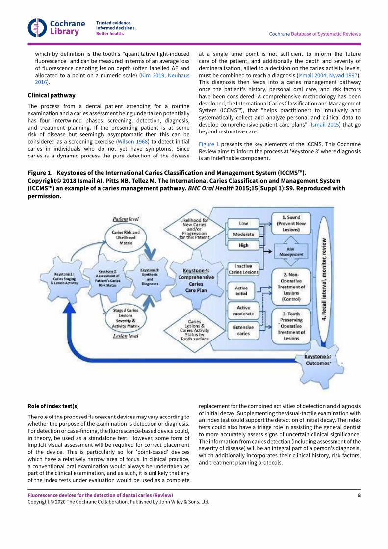
Figure 1 presents the key elements of the ICCMS. This CochraneReview aims to inform the process at 'Keystone 3' where diagnosisis an indefinable component.
Figure 1. Keystones of the International Caries Classification and Management System (ICCMS™).Copyright© 2018 Ismail AI, Pitts NB, Tellez M. The International Caries Classification and Management System(ICCMS™) an example of a caries management pathway. BMC Oral Health 2015;15(Suppl 1):S9. Reproduced withpermission.
Role of index test(s)
The role of the proposed fluorescent devices may vary according towhether the purpose of the examination is detection or diagnosis.For detection or case-finding, the fluorescence-based device could,in theory, be used as a standalone test. However, some form ofimplicit visual assessment will be required for correct placementof the device. This is particularly so for 'point-based' deviceswhich have a relatively narrow area of focus. In clinical practice,a conventional oral examination would always be undertaken aspart of the clinical examination, and as such, it is unlikely that anyof the index tests under evaluation would be used as a complete
replacement for the combined activities of detection and diagnosisof initial decay. Supplementing the visual-tactile examination withan index test could support the detection of initial decay. The indextests could also have a triage role in assisting the general dentistto more accurately assess signs of uncertain clinical significance.The information from caries detection (including assessment of theseverity of disease) will be an integral part of a person's diagnosis,which additionally incorporates their clinical history, risk factors,and treatment planning protocols.
Fluorescence devices for the detection of dental caries (Review)
Copyright © 2020 The Cochrane Collaboration. Published by John Wiley & Sons, Ltd.
8
CochraneLibrary
Trusted evidence.Informed decisions.Better health.
Cochrane Database of Systematic Reviews
Alternative test(s)
Alternative tests include.
• Comprehensive visual or visual-tactile examination with adetailed classification system: identifying caries according tovisual appearance, aided by a dental mirror and sometimes aprobe, on clean and dry teeth.
• Radiography: bitewing radiology is most commonly used. Othertechniques include: subtraction radiography which producesa semi-automated method for monitoring progression oflesions (Ellwood 1997; Wenzel 2006) and cone-beam computedtechnology (CBCT) which provides a three dimensional imagewhich appears to oEer great potential for diagnosis withincreased levels of radiation (Horner 2009).
• Fibre-optic transillumination (FOTI) which uses a light emittedfrom a handheld device that when placed directly onto the toothilluminates the tooth (Pretty 2006). Any demineralisation shouldappear as shadows in the tooth due to the disruption of thetooth's structure due to caries.
• Electrical conductance: the demineralisation of the tooth isreported to aEect the tooth's electrical conductance. This ismeasured by placing a probe on the tooth which measuresany potentially higher conductivity which occurs due to cariouslesions being filled with saliva (Tam 2001).
For more details please see the generic protocol for this review(Macey 2018).
Rationale
Despite technological advancement, caries detection is typicallybased upon information from a visual-tactile clinical examinationwith or without radiographs. Bader 2002 completed an extensiveliterature review of in vitro caries detection studies investigatingvisual, dental imaging, fibre-optic, electrical conductance, andfluorescence in primary and permanent dentition. The reviewwas restricted to studies that included a histological referencestandard and grouped studies according to index test, diseasethreshold (enamel or dentinal lesions), and tooth surface (occlusalor proximal); no meta-analysis was undertaken, and the authorsgraded the quality of the available evidence as low (Bader2002). Two years later the same authors published a reviewfocusing on fluorescence devices. Despite an increase in thenumber of eligible studies in the intervening years, the authorsdetermined that it was still not possible to carry out a meta-analysisand raised concerns over the propensity of the fluorescencedevices for decreasing specificity as sensitivity improved (Bader2004). These two reviews predate the development of meta-analysis methods for DTA reviews recommended in the CochraneHandbook for Systematic Reviews of Diagnostic Test Accuracy (Deeks2013). A subsequent systematic review investigated the accuracyof fluorescence devices, and included studies of the primaryand permanent dentition, occlusal and proximal surfaces, withreference standards of histology, operative, visual examination,and dental imaging (Gimenez 2013). We aimed to build uponexisting research in caries detection and diagnosis by expandingthe search strategy to capture all relevant evidence, applyingappropriate hierarchical meta-analytical models (Dinnes 2016),and assessing the body of evidence using GRADE (Schünemann2020; Schünemann 2020a) to facilitate the production of 'Summaryof findings' tables.
O B J E C T I V E S
Primary objectives
To determine the diagnostic accuracy of fluorescence-based indextests used alone or in combination with other tests for the detectionand diagnosis of coronal dental caries in children and adults. Weaimed to evaluate the comparative accuracy of red, blue, andgreen fluorescence-based devices; these included DIAGNOdent,DIAGNOdent pen, SoproLife, VistaProof, and quantitative light-induced fluorescence (QLF). The specific research questionsaddressed in this systematic review were.
• What is the diagnostic test accuracy of fluorescence-based testsfor detection or diagnosis in diEerent populations (children:primary/mixed dentition, adolescents: immature permanentdentition, or adults: mature permanent dentition), and whentested against diEerent reference standards.
• What is the diagnostic test accuracy of each of the threegroups of fluorescence-based index tests compared to anappropriate reference standard for detecting and diagnosinginitial stage decay on the occlusal, proximal, and smoothtooth surfaces?
• Do measures of sensitivity and specificity for single testsdiEer from the sensitivity and specificity of tests usedin combination (fluorescence test either individually orcombined with a visual examination)? Is there a benefit tousing more than one index test as opposed to a single test?
Secondary objectives
We aimed to investigate the following potential sources ofheterogeneity.
• Recruited population - children: primary/mixed dentition,adolescents: immature permanent dentition, or adults: maturepermanent dentition.
• Prevalence of caries into dentine in the study sample.
• Tooth surface being reported (occlusal, proximal, smoothsurface or adjacent to a restoration).
• Reference standards - in vitro studies commonly use histologyas the reference standard.
• Consideration of point measurement devices versus imaging orsurface assessment devices.
M E T H O D S
Criteria for considering studies for this review
Types of studies
We considered diagnostic accuracy study designs that were:
• studies with a single set of inclusion criteria that compareda fluorescence diagnostic test with a reference standard. Weincluded prospective studies that evaluated the diagnosticaccuracy of single index tests, and studies that directlycompared two or more index tests;
• randomised controlled trials (RCTs) of the diagnostic testaccuracy of one or more index tests in comparison, or versus ano test option;
Fluorescence devices for the detection of dental caries (Review)
Copyright © 2020 The Cochrane Collaboration. Published by John Wiley & Sons, Ltd.
9
CochraneLibrary
Trusted evidence.Informed decisions.Better health.
Cochrane Database of Systematic Reviews
• 'case-control' type accuracy studies where diEerent sets ofcriteria were used to recruit those with or without the targetcondition, although prone to bias some innovative tests may beidentifiable through this design only and this eligibility criterionmay provide an opportunity to report them, these studies wouldnot be included in the primary analysis;
• studies reporting at both the patient and tooth or tooth surfacelevel were included, however only those reporting at the toothsurface level would be included in the primary analysis.
In vitro and in vivo studies were eligible for inclusion. In vitrostudies use teeth that have been extracted prior to the start ofthe study. The index test is carried out on extracted teeth, albeitin a scenario which is not representative of the typical clinicalsetting, and will typically be followed by a reference standard ofhistology. In vivo studies recruit participants and conduct indextests with the teeth in the oral cavity. The reference standard isusually enhanced clinical examination or excavation. In some casesthe reference standard is histology, for example when a studyhas been conducted with participants who have teeth indicatedfor extraction due to orthodontic or third molar indications,periodontal diseases, or children with teeth that are due to exfoliatenaturally.
We excluded studies where:
• artificially created carious lesions were used in the testingprocedure;
• an index test was used during the excavation of dental caries toascertain the optimum depth of excavation.
Participants
Participants who are seemingly asymptomatic for dental caries,including those who may have carious lesions that are undetectedat the point of enrolment. Studies that explicitly recruitedparticipants with caries into dentine or frank cavitation wereexcluded. We also excluded studies where participants werereferred to secondary care for restorative treatment, as there is alikelihood that advanced caries (into dentine or pulp) would bepresent and readily detectable without the need for the index testsinvestigated in this review.
Studies recruiting children, adolescents, and adults were all eligiblefor inclusion. This allowed for the analysis of the diagnostictest accuracy of index tests for primary, mixed, and permanentdentition.
Index tests
Fluorescence-based devices: incorporating a variety of devicesthat included laser-based detection. Devices may have been usedas an adjunct to a conventional visual examination and requirean operator judgement or generate a conclusion via a soNwarealgorithm. There was considerable variation in the positivitythresholds used across the diEerent fluorescence-based devices.The devices that provided a numeric output on a continuous scalewere oNen interpreted at diEerent thresholds, but where multiplethresholds were reported within a study report we extracted dataat the pre-specified manufacturers' threshold wherever possible.
These index tests were completed on intact teeth and could beused as an adjunct or replacement for aspects of the currentexamination. The intention was to assess the index tests in isolation
wherever possible, otherwise the result of one index test may haveinfluenced another. Where multiple index tests were used as acombined index test these studies were reported separately.
Where studies used multiple examiners we extracted the resultsfor the most appropriate examiner to the research question.For example, if the study used dental students, general dentalpractitioners, and restorative consultants, then the results of thegeneral dental practitioners were extracted. In the scenario wheremultiple examiners showed similar skills and experience then themean sensitivity and specificity results were extracted. If this wasnot available then the reported results from the first examiner wereextracted.
Studies that investigated a standard clinical oral examinationwith an adjunct of fluorescence were included if the diagnosticinformation relating to fluorescence could be isolated from theother test. If the study reported a combined interpretation of bothmethods and if the review included suEicient numbers of combinedtests, then we planned to create a subgroup of these combinedtests.
Target conditions
Coronal caries: initial stage decay, defined as initial or incipientcaries or non-cavitated lesions. Specifically where there is adetectable change in enamel evident which is not thought to haveprogressed into dentine on occlusal, proximal surfaces, and smoothsurfaces.
Reference standards
Several diEerent reference standards have been used in primarydiagnostic test accuracy (DTA) studies for dental caries. The onlyway of achieving a true diagnosis of caries presence and severityis to extract and section the tooth and perform a histologicalassessment (Downer 1975; Kidd 2004). This would not be ethicallyreasonable to undertake on a healthy population in clinical (invivo) studies, but is acceptable and widely used in in vitro studiesconducted on previously extracted teeth. The only scenario wherehistology can be a viable scenario for clinical studies undertakenin a primary or secondary care setting would be where a toothhas been identified as requiring extraction (ideally for a non-cariesrelated reason such as orthodontic or third molar extraction), andthe index test could be applied before the extraction, followed bythe reference standard of histology. However, this would bring intoquestion the study's broader external validity as these types ofstudies are most likely to occur in adolescents or young adults andwho are therefore not representative of the wider population.
Alternatives to extraction and histological assessment areoperative exploration, where a clinician removes caries with adental burr (drill) in preparation for restoration and reports thedepth of decay. This technique would be acceptable as a referencestandard for patients with caries of severity where restorationis required, but would not be ethical for caries-free patients orthose with early caries since non-restorative treatment could beprovided. A diEerent reference standard would be required forthese early lesions, the possibilities available are limited to anenhanced visual examination or radiographic tests. Studies thatonly used an enhanced visual or radiographic examination wereincluded in the review as they have the benefit of allowing studiesto be conducted in a clinical setting, however, their limitationsin providing a true classification of disease would be identified
Fluorescence devices for the detection of dental caries (Review)
Copyright © 2020 The Cochrane Collaboration. Published by John Wiley & Sons, Ltd.
10
CochraneLibrary
Trusted evidence.Informed decisions.Better health.
Cochrane Database of Systematic Reviews
in the quality appraisal. Some primary studies have employed acomposite reference standard based on the results of informationfrom multiple sources.
A period of up to three months between the index test and thereference standard was deemed acceptable.
Search methods for identification of studies
Electronic searches
Cochrane Oral Health's Information Specialist conductedsystematic searches in the following databases without languageor publication status restrictions:
• MEDLINE Ovid (1946 to 30 May 2019) (Appendix 1);
• Embase Ovid (1980 to 30 May 2019) (Appendix 2).
Searching other resources
The following trial registries were searched for ongoing studies:
• US National Institutes of Health Ongoing Trials RegisterClinicalTrials.gov (clinicaltrials.gov; searched 30 May 2019)(Appendix 3);
• World Health Organization International Clinical Trials RegistryPlatform (apps.who.int/trialsearch; searched 30 May 2019)(Appendix 4).
We searched the reference lists of included papers and previouslypublished systematic reviews for additional publications notidentified in the electronic searches.
We checked that none of the included studies had been retracteddue to error or fraud.
Data collection and analysis
Selection of studies
Two review authors independently screened and assessed theresults of all searches for inclusion. Any disagreements wereresolved through discussion and, where necessary, consultationwith another clinical or methodological member of the authorteam. Studies that met the criteria but that did not report the datain the format of a 2 x 2 contingency table were still included. In suchinstances, the study authors were contacted and the required datarequested. An adapted PRISMA flowchart was used to report thestudy selection process (McInnes 2018).
Data extraction and management
Two review authors independently extracted data. A piloted studydata extraction form based on the review inclusion criteria wasdeveloped and applied to 10 eligible studies. Disagreements wereresolved through discussion with other members of the reviewteam. Where data were reported for both occlusal and proximalsurfaces the data were extracted separately for the diEerentsurfaces. Study authors were contacted to obtain missing data orcharacteristics which were not evident in the published paper.
We recorded the following data for each study:
• sample characteristics (age, sex, socioeconomic status, riskfactors where stated, number of patients/carious lesions,
lesion location, disease prevalence - at enamel and dentinethresholds);
• study setting (country, type of facility);
• the type of index test(s) used (category (i.e. red, blue, or greenfluorescence), the device used, mode of action, conditions (i.e.clean/dried teeth), positivity threshold);
• study information (design, reference standard, case definition,training and calibration of personnel);
• study results (true positive, true negative, false positive, falsenegative, any equivocal results).
Assessment of methodological quality
We used the Quality Assessment of Diagnostic Accuracy Studies 2(QUADAS-2) to assess the risk of bias and applicability of the eligibleprimary studies over the four domains of participant selection,index test, reference standard, and flow and timing (Whiting 2011),tailored for this review. 'Review specific' descriptions of howthe QUADAS-2 items were contextualised and implemented aredetailed in the accompanying checklist (Additional Table 2).
A 'Risk of bias' judgement ('high', 'low' or 'unclear') was made foreach domain for each study. Broadly, if the answers to all signallingquestions within a domain were judged as 'yes' (indicating lowrisk of bias for each question) then the domain was judged to beat low risk of bias. If any signalling question was judged as 'no',indicating a high risk of bias, the domain was scored as high risk ofbias. Concerns regarding applicability were then completed for theparticipant selection, index test, and reference standard domains.There was some flexibility within this assessment framework whichdeveloped during the data extraction process and is detailed below.
Participant selection domain (1)
The selection of patients has a fundamental eEect on the abilityof an index test to detect caries. The disease categories of soundand enamel caries needed be represented in the sample and theage range of patients needed to be reported to form a completeappraisal of the index test's potential to correctly classify disease indiEerent populations.
It was acceptable for studies to focus on a particular surface(occlusal/proximal) or age group (children/adults). Given that theprimary objective centred on early enamel lesions studies shouldbe reporting on this stage of the disease process. It was vital thatwithin the chosen population all participants or teeth meeting theeligibility criteria should be provided with an equal or randomopportunity to be included. Inappropriate exclusion may lead to anover or underestimation of the test's ability to detect disease, thusaEecting the internal validity of the study.
All studies should have fully reported the methods used to selectteeth. Ideally, a random or consecutive selection would be usedand the procedure explicitly reported. Additionally, the prevalenceof the diEerent levels of disease severity should be reported. Thisinformation was used to inform the applicability of this test to awider population.
Study results should be reported at the tooth or surface level, asapposed to the patient level, which has the potential for the indextest and reference standard to be report on diEerent sites within thesame mouth.
Fluorescence devices for the detection of dental caries (Review)
Copyright © 2020 The Cochrane Collaboration. Published by John Wiley & Sons, Ltd.
11
CochraneLibrary
Trusted evidence.Informed decisions.Better health.
Cochrane Database of Systematic Reviews
Index test domain (2)
The nature of the fluorescence index tests and the visualpresentation of the disease means that it should be feasible toensure that the index test is conducted prior to the referencestandard. Logically, the fluorescence tests had to be completedbefore the extraction of a tooth for any histological analysis, orbefore in situ excavation of a tooth is undertaken. This order ofpresentation (index test followed by reference standard) ensuredthat the index test was not influenced by the results of the referencestandard. The fluorescence-based index tests generally used adevice which reported a numerical value on a continuous scale.Where multiple index tests were used and where the fluorescence-based test was conducted aNer other index test(s) (e.g. radiograph),the objective reading and reporting of the fluorescence-baseddevice mean that the results would not be influenced by precedingtests.
The threshold of disease positive and negative should bepresented before any analysis, ideally by using the manufacturer'srecommended settings or thresholds recommended by previouslyvalidated studies. Studies may have been designed to calculatethe optimum threshold for a device but this will introduce bias.It is unlikely that studies will have utilised multiple index testexaminers for the assessment of diEerent disease severity or wherethey have it is probable that they each score all of the thresholdsand are included for validation of the test. However, the inclusionof a signalling question here allowed for the identification ofstudies that have achieved this and provided data to inform futurediscussions.
Reference standard domain (3)
If the reference standard was an enhanced visual examination orradiograph then it should be completed by an examiner diEerent tothe index test, as the subjectivity of this type of reference standardcould be compromised by knowledge of the index test results.An exception was built in for this signalling question becausewhere the tooth has been extracted, sectioned and prepared forhistological evaluation it is extremely unlikely that the examinerwould be able to recall the specific tooth or participant and theresults from the index test results. Time delays between index testand reference standard should be under three months for in vivostudies.
Ideally, each participant within a study would have received thesame reference test. This is possible in an in vitro setting as ahistological assessment can be applied to each selected, extractedtooth. In vivo studies may have applied the same referencestandard by using enhanced visual examination or radiograph toall participants. If a study allocated participants or specific teeth todiEerent reference standards then the reasons for this diEerentialallocation should have been explicitly reported. All referencestandards should have been completed without knowledge of theindex test results.
Flow and timing domain (4)
The index test should be conducted before the reference standard.If the reference standard used is enhanced visual, radiograph, orexcavation then there should be less than three months betweenindex test and reference standard. Caries is a slow-growing diseaseso minimal changes should be experienced within this time frame.All observations should receive both an index test and reference
standard. There are studies which report some teeth having anindex test but not a reference standard; if a reason is clearlyreported, such as teeth being broken during sectioning, then thiswould not influence the risk of bias decision.
Statistical analysis and data synthesis
The threshold of interest was between sound teeth and initial/early/enamel caries. This eEectively created two groups, a positivegroup with any caries from early to advanced and a negativegroup of sound or healthy teeth. Estimates of diagnostic accuracywere expressed as sensitivity and specificity with 95% confidenceintervals for each study and each available data point if thestudy reported multiple index tests, dentition (primary/permanent)or tooth surfaces (occlusal/proximal/smooth). We displayed thisinformation as coupled forest plots and summary receiveroperating characteristic (SROC) plots. When there were two ormore test results reported in the same study, we included them asseparate datasets, since the unit of analysis was the test result, notthe patient.
Hierarchical models were used for data synthesis. The data wereextracted for the target condition of early caries (caries intoenamel). This target condition has been consistently used acrossthe series of DTA caries reviews. A meta-analysis was conductedto combine the results of studies for each index test using thehierarchical summary ROC (HSROC) approach to estimate theexpected values of sensitivity and specificity (Macaskill 2010). Asummary curve using the HSROC model (Rutter 2001) was used tosummarise the results since the devices provided a numeric outputon a continuous scale and oNen interpreted these at diEerentcut-oEs. Consequently, it was not possible to apply a commonthreshold for analysis. An HSROC model was used to estimatea summary curve with parameter estimates for threshold, shapeand accuracy, for all available datasets with no restrictions ondentition, tooth surface, reference standard, or prevalence of cariesinto dentine (D3).
It was not possible to produce estimates of sensitivity andspecificity as summary operating points with confidence andprediction regions on SROC plots with 95% confidence regionssince the output of the HSROC model is the summary ROC curve.In the absence of clinical consensus of key values of specificity,we summarised the analysis using the median and upper quartilereported specificity and the corresponding estimate of sensitivity,along with the diagnostic odds ratio (DOR) with 95% confidenceintervals (Takwoingi 2015). To allow for the analysis of falsepositives and false negatives we computed the sensitivity at thepoint on the SROC curve with fixed values of specificity of 0.78 and0.90 (the median and upper quartile values from of all includeddatasets). These results are only included as examples of potentialsensitivity and specificity pairings and should not be reported orinterpreted formally as the summary points.
We made comparisons between the three device categories (blue,green, and red fluorescence) by comparing summary ROC curves(Takwoingi 2010). Initially, we allowed threshold, shape, andaccuracy to vary according to device type by including covariatesin the model (most complex model). DiEerences in the shapes ofthe summary curves were explored by removing the covariates forshape and comparing the results of this model to those of thecomplex model. Parameter estimates for the model assuming acommon or diEerent shape were used to generate HSROC curves
Fluorescence devices for the detection of dental caries (Review)
Copyright © 2020 The Cochrane Collaboration. Published by John Wiley & Sons, Ltd.
12
CochraneLibrary
Trusted evidence.Informed decisions.Better health.
Cochrane Database of Systematic Reviews
for the three categories as appropriate. If the diEerent deviceswere observed to have a common shape then the model wasfurther simplified by removing the covariates for accuracy, todetermine whether the accuracy of the diEerent devices diEeredin comparison with the previous model. The likelihood ratio testwas applied to formally assess the significance of any modelcomparisons (Macaskill 2010).
The numbers generated for a hypothetical cohort of 1000 toothsites or surfaces are reported in the 'Summary of findings' tablealong with the corresponding true positives, false negatives, falsepositives, and true negatives. The higher prevalence value wastaken from the total number of enamel lesions in the includedstudies divided by the total number of included tooth surfaces.The lower prevalence figure was taken from the UK Adult DentalHealth Survey (Steele 2011) and was used to address clinicalconsiderations that the higher prevalence value of enamel cariesreported in the primary studies, particularly in the in vitro studies,were not representative of that observed in the general population.
We used Review Manager 5 (Review Manager 2020), the NLMIXEDprocedure and the MetaDAS macro (Takwoingi 2010) in SAS 9.4 forWindows to carry out the analyses.
Investigations of heterogeneity
We initially inspected the clinical and methodologicalcharacteristics of the included studies, coupled forest plots, andsummary ROC plots to form the basis of the assessment ofheterogeneity. Where suEicient numbers of studies allowed, meta-regression analyses were undertaken to explore possible sourcesof heterogeneity. Formal model comparisons were compared usinga likelihood ratio test to determine the statistical significanceof adding each potential source of heterogeneity (covariate) tothe HSROC model. Model comparisons proceeded as for thecomparison of diEerent tests above i.e. fit a complex modelallowing shape, threshold, and accuracy to diEer according to thesource of heterogeneity, and assess the impact of the removal ofthe covariates for shape. If a common shape can be assumed thenexplore the impact of the removal of the covariates for accuracy.Each potential source of heterogeneity was analysed separately.
All investigations of heterogeneity were reported to aidinterpretation of the results.
The sources of heterogeneity included (specified a priori).
Population
• Children or adults; the detection of disease in the diEerentdentition of children or adolescents will aEect the stage at whichthe disease is identified and treatment options which would beconsidered.
• Tooth surface being evaluated (occlusal, proximal, smoothsurface or adjacent to a restoration).
• Prevalence of caries into dentine in each study sample.
Index test
• Consideration of point measurement devices versus imaging orsurface assessment devices.
Reference standard
• Reference standard used: histology, excavation, enhanced visualexamination, or radiograph.
Sensitivity analyses
Where a suEicient number of studies investigated the same indextest, we assessed the impact of study quality on the sensitivity andspecificity results.
Assessment of reporting bias
Methods currently available to assess reporting or publication biasfor diagnostic studies may lead to uncertainty and misleadingresults from funnel plots (Deeks 2005; Leeflang 2008), therefore wedid not carry out any tests of reporting bias.
Presentation of main results
We reported our results for fluorescence index tests and the maintarget conditions following GRADE methodology (Schünemann2020; Schünemann 2020a) and using the GRADEPro online tool(www.guidelinedevelopment.org). To enhance readability andunderstanding, we presented test accuracy results as naturalfrequencies to indicate numbers of false positives and falsenegatives. The certainty of the evidence was assessed for the overallrisk of bias of the included studies, the indirectness of the evidence,the inconsistency of the results, the imprecision of the estimates,and the subjective risk of publication bias. We conducted theassessment of the certainty of the evidence irrespective of whethera numerical, a range, or a narrative description of diagnostic testaccuracy was available. We categorised the certainty of the body ofevidence as high, moderate, low, or very low.
R E S U L T S
Results of the search
The search identified a total of 3259 records aNer duplicateswere removed. We excluded 3017 records based on the titles andabstracts, as per the eligibility criteria, the remaining 242 studieswere assessed based on the full published paper. 133 of thesestudies were eligible for inclusion, the PRISMA diagram in Figure2 shows the flow of studies through the review process (Moher2009). The included studies were mainly carried out in Brazil andEurope, followed by Turkey and the Middle East, Asia, 11 in NorthAmerica, and Australia. 62% (83/133) of studies performed the testson extracted teeth, 19% in a dental school university hospital,13% in a primary care or other clinical setting, and 6% in schools.Six of the studies (4%) reported the inclusion of fissure sealants.Studies were published between the years 1998 and 2019, 55%were published aNer 2010. All studies were cross-sectional andwere a single gate design.
Fluorescence devices for the detection of dental caries (Review)
Copyright © 2020 The Cochrane Collaboration. Published by John Wiley & Sons, Ltd.
13
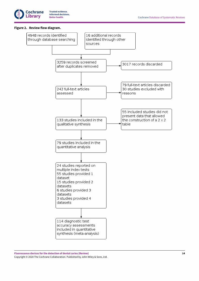
CochraneLibrary
Trusted evidence.Informed decisions.Better health.
Cochrane Database of Systematic Reviews
Figure 2. Review flow diagram.
Fluorescence devices for the detection of dental caries (Review)
Copyright © 2020 The Cochrane Collaboration. Published by John Wiley & Sons, Ltd.
14
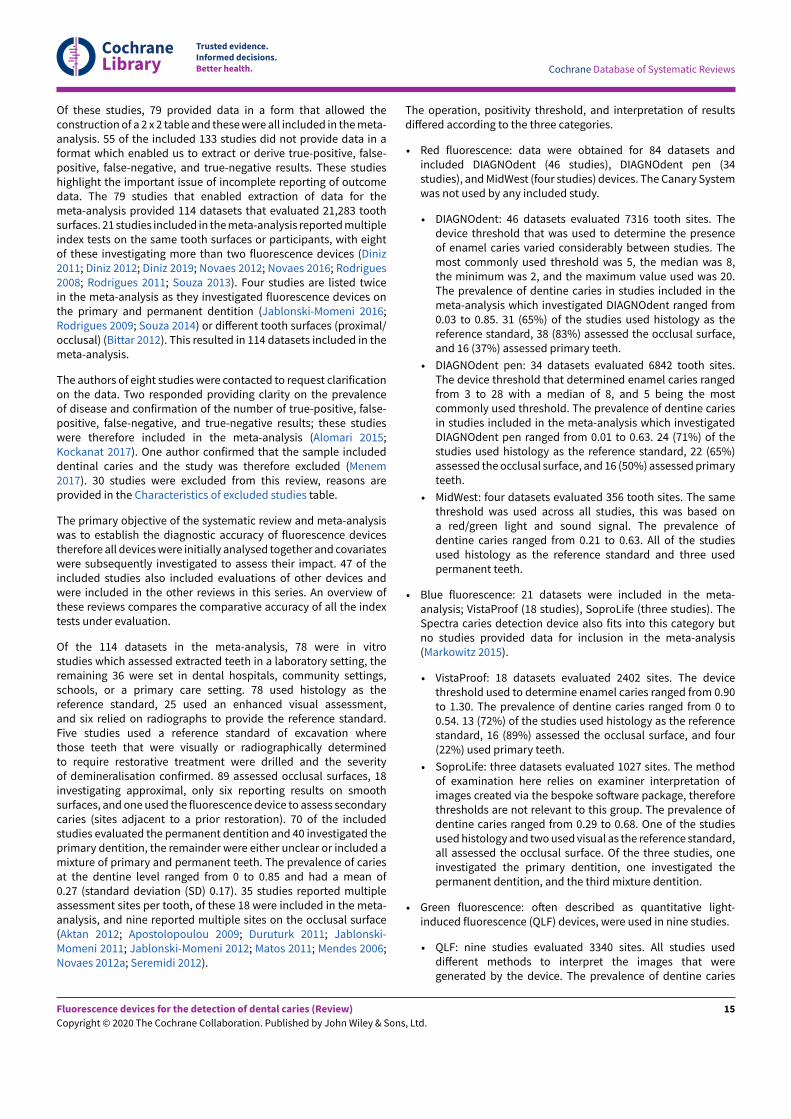
CochraneLibrary
Trusted evidence.Informed decisions.Better health.
Cochrane Database of Systematic Reviews
Of these studies, 79 provided data in a form that allowed theconstruction of a 2 x 2 table and these were all included in the meta-analysis. 55 of the included 133 studies did not provide data in aformat which enabled us to extract or derive true-positive, false-positive, false-negative, and true-negative results. These studieshighlight the important issue of incomplete reporting of outcomedata. The 79 studies that enabled extraction of data for themeta-analysis provided 114 datasets that evaluated 21,283 toothsurfaces. 21 studies included in the meta-analysis reported multipleindex tests on the same tooth surfaces or participants, with eightof these investigating more than two fluorescence devices (Diniz2011; Diniz 2012; Diniz 2019; Novaes 2012; Novaes 2016; Rodrigues2008; Rodrigues 2011; Souza 2013). Four studies are listed twicein the meta-analysis as they investigated fluorescence devices onthe primary and permanent dentition (Jablonski-Momeni 2016;Rodrigues 2009; Souza 2014) or diEerent tooth surfaces (proximal/occlusal) (Bittar 2012). This resulted in 114 datasets included in themeta-analysis.
The authors of eight studies were contacted to request clarificationon the data. Two responded providing clarity on the prevalenceof disease and confirmation of the number of true-positive, false-positive, false-negative, and true-negative results; these studieswere therefore included in the meta-analysis (Alomari 2015;Kockanat 2017). One author confirmed that the sample includeddentinal caries and the study was therefore excluded (Menem2017). 30 studies were excluded from this review, reasons areprovided in the Characteristics of excluded studies table.
The primary objective of the systematic review and meta-analysiswas to establish the diagnostic accuracy of fluorescence devicestherefore all devices were initially analysed together and covariateswere subsequently investigated to assess their impact. 47 of theincluded studies also included evaluations of other devices andwere included in the other reviews in this series. An overview ofthese reviews compares the comparative accuracy of all the indextests under evaluation.
Of the 114 datasets in the meta-analysis, 78 were in vitrostudies which assessed extracted teeth in a laboratory setting, theremaining 36 were set in dental hospitals, community settings,schools, or a primary care setting. 78 used histology as thereference standard, 25 used an enhanced visual assessment,and six relied on radiographs to provide the reference standard.Five studies used a reference standard of excavation wherethose teeth that were visually or radiographically determinedto require restorative treatment were drilled and the severityof demineralisation confirmed. 89 assessed occlusal surfaces, 18investigating approximal, only six reporting results on smoothsurfaces, and one used the fluorescence device to assess secondarycaries (sites adjacent to a prior restoration). 70 of the includedstudies evaluated the permanent dentition and 40 investigated theprimary dentition, the remainder were either unclear or included amixture of primary and permanent teeth. The prevalence of cariesat the dentine level ranged from 0 to 0.85 and had a mean of0.27 (standard deviation (SD) 0.17). 35 studies reported multipleassessment sites per tooth, of these 18 were included in the meta-analysis, and nine reported multiple sites on the occlusal surface(Aktan 2012; Apostolopoulou 2009; Duruturk 2011; Jablonski-Momeni 2011; Jablonski-Momeni 2012; Matos 2011; Mendes 2006;Novaes 2012a; Seremidi 2012).
The operation, positivity threshold, and interpretation of resultsdiEered according to the three categories.
• Red fluorescence: data were obtained for 84 datasets andincluded DIAGNOdent (46 studies), DIAGNOdent pen (34studies), and MidWest (four studies) devices. The Canary Systemwas not used by any included study.
• DIAGNOdent: 46 datasets evaluated 7316 tooth sites. Thedevice threshold that was used to determine the presenceof enamel caries varied considerably between studies. Themost commonly used threshold was 5, the median was 8,the minimum was 2, and the maximum value used was 20.The prevalence of dentine caries in studies included in themeta-analysis which investigated DIAGNOdent ranged from0.03 to 0.85. 31 (65%) of the studies used histology as thereference standard, 38 (83%) assessed the occlusal surface,and 16 (37%) assessed primary teeth.
• DIAGNOdent pen: 34 datasets evaluated 6842 tooth sites.The device threshold that determined enamel caries rangedfrom 3 to 28 with a median of 8, and 5 being the mostcommonly used threshold. The prevalence of dentine cariesin studies included in the meta-analysis which investigatedDIAGNOdent pen ranged from 0.01 to 0.63. 24 (71%) of thestudies used histology as the reference standard, 22 (65%)assessed the occlusal surface, and 16 (50%) assessed primaryteeth.
• MidWest: four datasets evaluated 356 tooth sites. The samethreshold was used across all studies, this was based ona red/green light and sound signal. The prevalence ofdentine caries ranged from 0.21 to 0.63. All of the studiesused histology as the reference standard and three usedpermanent teeth.
• Blue fluorescence: 21 datasets were included in the meta-analysis; VistaProof (18 studies), SoproLife (three studies). TheSpectra caries detection device also fits into this category butno studies provided data for inclusion in the meta-analysis(Markowitz 2015).
• VistaProof: 18 datasets evaluated 2402 sites. The devicethreshold used to determine enamel caries ranged from 0.90to 1.30. The prevalence of dentine caries ranged from 0 to0.54. 13 (72%) of the studies used histology as the referencestandard, 16 (89%) assessed the occlusal surface, and four(22%) used primary teeth.
• SoproLife: three datasets evaluated 1027 sites. The methodof examination here relies on examiner interpretation ofimages created via the bespoke soNware package, thereforethresholds are not relevant to this group. The prevalence ofdentine caries ranged from 0.29 to 0.68. One of the studiesused histology and two used visual as the reference standard,all assessed the occlusal surface. Of the three studies, oneinvestigated the primary dentition, one investigated thepermanent dentition, and the third mixture dentition.
• Green fluorescence: oNen described as quantitative light-induced fluorescence (QLF) devices, were used in nine studies.
• QLF: nine studies evaluated 3340 sites. All studies useddiEerent methods to interpret the images that weregenerated by the device. The prevalence of dentine caries
Fluorescence devices for the detection of dental caries (Review)
Copyright © 2020 The Cochrane Collaboration. Published by John Wiley & Sons, Ltd.
15
CochraneLibrary
Trusted evidence.Informed decisions.Better health.
Cochrane Database of Systematic Reviews
ranged from 0.11 to 0.63. Five datasets used histology asthe reference standard (56%), six (67%) investigated occlusalsurfaces, and eight (89%) used permanent teeth.
The most common reasons for exclusion from the review werestudies that explicitly included participants or teeth with dentinalor frankly cavitated surfaces and were therefore ineligible. Othercommonly excluded studies compared one index test with anotherbut with no reference standard, i.e. they were comparative ratherthan diagnostic test accuracy studies.
A combination of visual, radiograph, and DIAGNOdent was reportedin one study and this study has been reported separately (Alomari2015).
Additional Table 3 tabulates the study characteristics for eachdevice, the number of tooth sites, teeth, and participants evaluated,
in vivo or in vitro studies, the prevalence of enamel caries (D1), the
prevalence of dentine caries (D3), tooth surface, reference standard,
and dentition.
Methodological quality of included studies
This section reports on all 133 included studies, 79 that wereincluded in the meta-analysis, and 55 where insuEicient datawere provided to enable inclusion in the meta-analysis. Figure 3summarises the results of the quality assessment of the includedstudies. One study could be classified as being at low risk ofbias across all domains (Castilho 2016), although this studyinvestigated third molars which were due to be extracted, and sothe generalisability of the results of this study could be questioned.The results of the individual assessment of each study is providedin Figure 4.
Figure 3. Risk of bias and applicability concerns graph: review authors' judgements about each domain presentedas percentages across included studies.
Fluorescence devices for the detection of dental caries (Review)
Copyright © 2020 The Cochrane Collaboration. Published by John Wiley & Sons, Ltd.
16
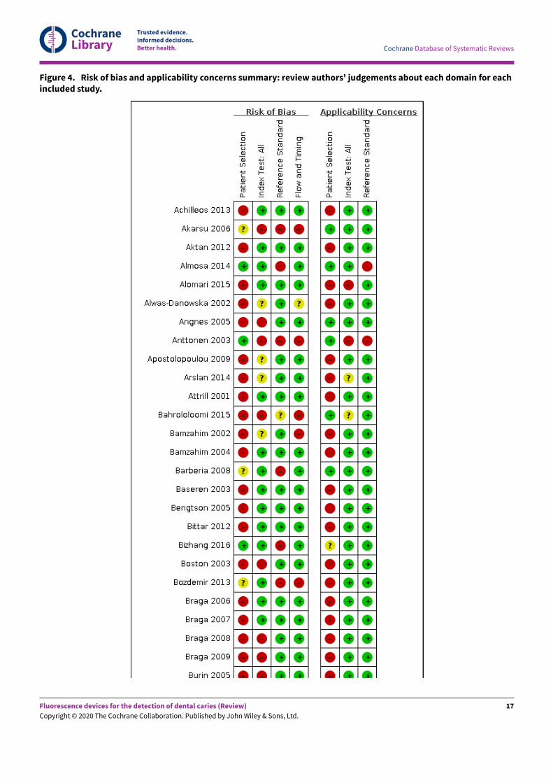
CochraneLibrary
Trusted evidence.Informed decisions.Better health.
Cochrane Database of Systematic Reviews
Figure 4. Risk of bias and applicability concerns summary: review authors' judgements about each domain for eachincluded study.
Fluorescence devices for the detection of dental caries (Review)
Copyright © 2020 The Cochrane Collaboration. Published by John Wiley & Sons, Ltd.
17
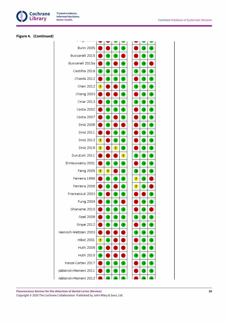
CochraneLibrary
Trusted evidence.Informed decisions.Better health.
Cochrane Database of Systematic Reviews
Figure 4. (Continued)
Fluorescence devices for the detection of dental caries (Review)
Copyright © 2020 The Cochrane Collaboration. Published by John Wiley & Sons, Ltd.
18
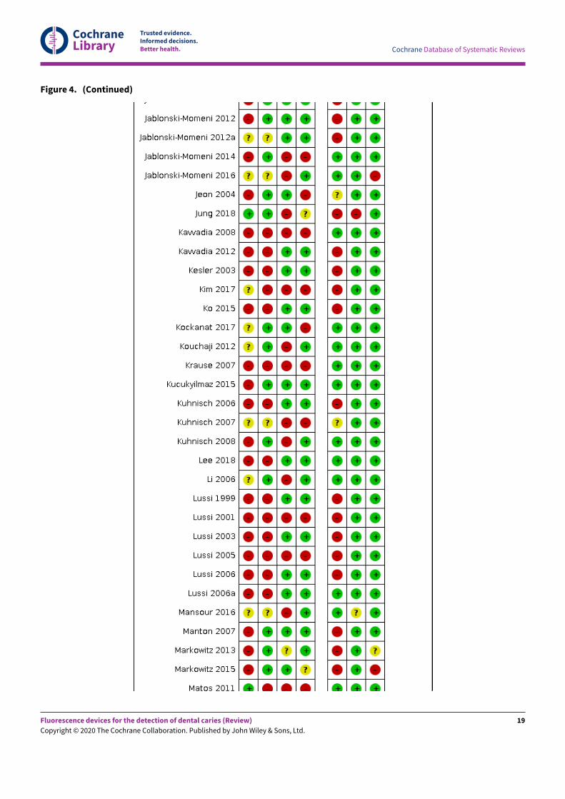
CochraneLibrary
Trusted evidence.Informed decisions.Better health.
Cochrane Database of Systematic Reviews
Figure 4. (Continued)
Fluorescence devices for the detection of dental caries (Review)
Copyright © 2020 The Cochrane Collaboration. Published by John Wiley & Sons, Ltd.
19
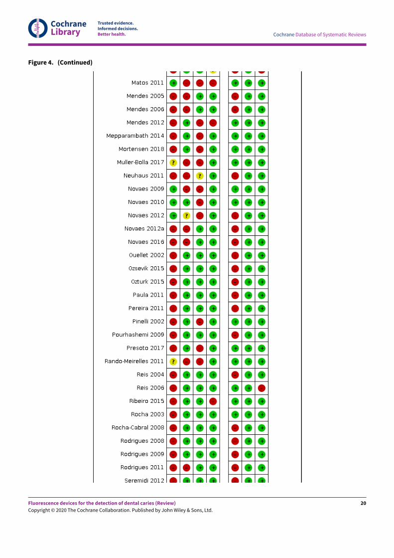
CochraneLibrary
Trusted evidence.Informed decisions.Better health.
Cochrane Database of Systematic Reviews
Figure 4. (Continued)
Fluorescence devices for the detection of dental caries (Review)
Copyright © 2020 The Cochrane Collaboration. Published by John Wiley & Sons, Ltd.
20
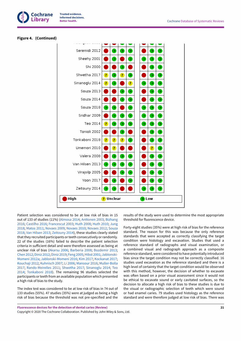
CochraneLibrary
Trusted evidence.Informed decisions.Better health.
Cochrane Database of Systematic Reviews
Figure 4. (Continued)
Patient selection was considered to be at low risk of bias in 15out of 133 of studies (11%) (Almosa 2014; Anttonen 2003; Bizhang2016; Castilho 2016; Francescut 2003; Huth 2008; Huth 2010; Jung2018; Matos 2011; Novaes 2009; Novaes 2010; Novaes 2012; Souza2018; Van Hilsen 2013; Zeitouny 2014), these studies clearly statedthat they recruited participants or teeth consecutively or randomly.22 of the studies (16%) failed to describe the patient selectioncriteria in suEicient detail and were therefore assessed as being atunclear risk of bias (Akarsu 2006; Barberia 2008; Bozdemir 2013;Chen 2012; Diniz 2012; Diniz 2019; Feng 2005; Hibst 2001; Jablonski-Momeni 2012a; Jablonski-Momeni 2016; Kim 2017; Kockanat 2017;Kouchaji 2012; Kuhnisch 2007; Li 2006; Mansour 2016; Muller-Bolla2017; Rando-Meirelles 2011; Shwetha 2017; Sinanoglu 2014; Teo2014; Tonkaboni 2018). The remaining 96 studies selected theparticipants or teeth from an available population which presenteda high risk of bias to the study.
The index test was considered to be at low risk of bias in 74 out of133 studies (55%). 47 studies (35%) were at judged as being a highrisk of bias because the threshold was not pre-specified and the
results of the study were used to determine the most appropriatethreshold for fluorescence device.
Forty-eight studies (35%) were at high risk of bias for the referencestandard. The reason for this was because the only referencestandards that were accepted as correctly classifying the targetcondition were histology and excavation. Studies that used areference standard of radiographs and visual examination, ora combined visual and radiograph approach as a compositereference standard, were considered to have potentially introducedbias since the target condition may not be correctly classified. 16studies used excavation as the reference standard and there is ahigh level of certainty that the target condition would be observedwith this method, however, the decision of whether to excavatewas oNen based on a prior visual assessment since it would notbe ethical to excavate sound or early cavitated surfaces, so thedecision to allocate a high risk of bias to these studies is due tothe visual or radiographic selection of teeth which were soundor had enamel caries. 79 studies used histology as the referencestandard and were therefore judged at low risk of bias. There was
Fluorescence devices for the detection of dental caries (Review)
Copyright © 2020 The Cochrane Collaboration. Published by John Wiley & Sons, Ltd.
21
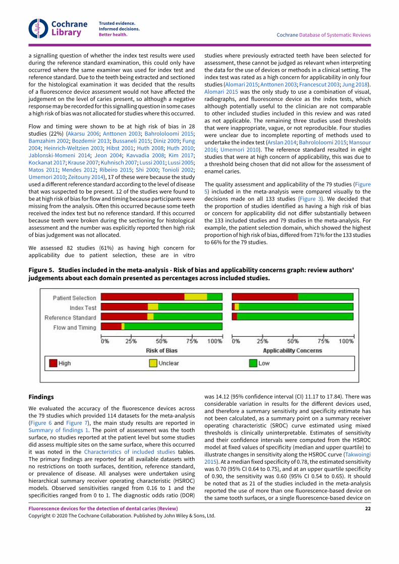
CochraneLibrary
Trusted evidence.Informed decisions.Better health.
Cochrane Database of Systematic Reviews
a signalling question of whether the index test results were usedduring the reference standard examination, this could only haveoccurred where the same examiner was used for index test andreference standard. Due to the teeth being extracted and sectionedfor the histological examination it was decided that the resultsof a fluorescence device assessment would not have aEected thejudgement on the level of caries present, so although a negativeresponse may be recorded for this signalling question in some casesa high risk of bias was not allocated for studies where this occurred.
Flow and timing were shown to be at high risk of bias in 28studies (22%) (Akarsu 2006; Anttonen 2003; Bahrololoomi 2015;Bamzahim 2002; Bozdemir 2013; Bussaneli 2015; Diniz 2009; Fung2004; Heinrich-Weltzien 2003; Hibst 2001; Huth 2008; Huth 2010;Jablonski-Momeni 2014; Jeon 2004; Kavvadia 2008; Kim 2017;Kockanat 2017; Krause 2007; Kuhnisch 2007; Lussi 2001; Lussi 2005;Matos 2011; Mendes 2012; Ribeiro 2015; Shi 2000; Tonioli 2002;Umemori 2010; Zeitouny 2014), 17 of these were because the studyused a diEerent reference standard according to the level of diseasethat was suspected to be present. 12 of the studies were found tobe at high risk of bias for flow and timing because participants weremissing from the analysis. ONen this occurred because some teethreceived the index test but no reference standard. If this occurredbecause teeth were broken during the sectioning for histologicalassessment and the number was explicitly reported then high riskof bias judgement was not allocated.
We assessed 82 studies (61%) as having high concern forapplicability due to patient selection, these are in vitro
studies where previously extracted teeth have been selected forassessment, these cannot be judged as relevant when interpretingthe data for the use of devices or methods in a clinical setting. Theindex test was rated as a high concern for applicability in only fourstudies (Alomari 2015; Anttonen 2003; Francescut 2003; Jung 2018).Alomari 2015 was the only study to use a combination of visual,radiographs, and fluorescence device as the index tests, whichalthough potentially useful to the clinician are not comparableto other included studies included in this review and was ratedas not applicable. The remaining three studies used thresholdsthat were inappropriate, vague, or not reproducible. Four studieswere unclear due to incomplete reporting of methods used toundertake the index test (Arslan 2014; Bahrololoomi 2015; Mansour2016; Umemori 2010). The reference standard resulted in eightstudies that were at high concern of applicability, this was due toa threshold being chosen that did not allow for the assessment ofenamel caries.
The quality assessment and applicability of the 79 studies (Figure5) included in the meta-analysis were compared visually to thedecisions made on all 133 studies (Figure 3). We decided thatthe proportion of studies identified as having a high risk of biasor concern for applicability did not diEer substantially betweenthe 133 included studies and 79 studies in the meta-analysis. Forexample, the patient selection domain, which showed the highestproportion of high risk of bias, diEered from 71% for the 133 studiesto 66% for the 79 studies.
Figure 5. Studies included in the meta-analysis - Risk of bias and applicability concerns graph: review authors'judgements about each domain presented as percentages across included studies.
Findings
We evaluated the accuracy of the fluorescence devices acrossthe 79 studies which provided 114 datasets for the meta-analysis(Figure 6 and Figure 7), the main study results are reported inSummary of findings 1. The point of assessment was the toothsurface, no studies reported at the patient level but some studiesdid assess multiple sites on the same surface, where this occurredit was noted in the Characteristics of included studies tables.The primary findings are reported for all available datasets withno restrictions on tooth surfaces, dentition, reference standard,or prevalence of disease. All analyses were undertaken usinghierarchical summary receiver operating characteristic (HSROC)models. Observed sensitivities ranged from 0.16 to 1 and thespecificities ranged from 0 to 1. The diagnostic odds ratio (DOR)
was 14.12 (95% confidence interval (CI) 11.17 to 17.84). There wasconsiderable variation in results for the diEerent devices used,and therefore a summary sensitivity and specificity estimate hasnot been calculated, as a summary point on a summary receiveroperating characteristic (SROC) curve estimated using mixedthresholds is clinically uninterpretable. Estimates of sensitivityand their confidence intervals were computed from the HSROCmodel at fixed values of specificity (median and upper quartile) toillustrate changes in sensitivity along the HSROC curve (Takwoingi2015). At a median fixed specificity of 0.78, the estimated sensitivitywas 0.70 (95% CI 0.64 to 0.75), and at an upper quartile specificityof 0.90, the sensitivity was 0.60 (95% CI 0.54 to 0.65). It shouldbe noted that as 21 of the studies included in the meta-analysisreported the use of more than one fluorescence-based device onthe same tooth surfaces, or a single fluorescence-based device on
Fluorescence devices for the detection of dental caries (Review)
Copyright © 2020 The Cochrane Collaboration. Published by John Wiley & Sons, Ltd.
22

CochraneLibrary
Trusted evidence.Informed decisions.Better health.
Cochrane Database of Systematic Reviews
diEerent dentition or diEerent tooth surfaces (proximal/occlusal),there is some non-independence of data in this analysis. No studiesthat directly compared tests reported the fully paired results in the
form of a 2 x 4 table of the results of the index tests cross-classifiedamongst cases and non-cases.
Fluorescence devices for the detection of dental caries (Review)
Copyright © 2020 The Cochrane Collaboration. Published by John Wiley & Sons, Ltd.
23
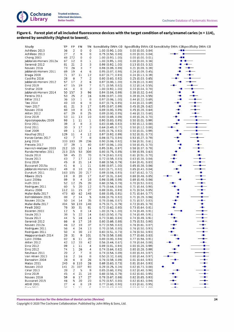
CochraneLibrary
Trusted evidence.Informed decisions.Better health.
Cochrane Database of Systematic Reviews
Figure 6. Forest plot of all included fluorescence devices with the target condition of early/enamel caries (n = 114),ordered by sensitivity (highest to lowest).
Fluorescence devices for the detection of dental caries (Review)
Copyright © 2020 The Cochrane Collaboration. Published by John Wiley & Sons, Ltd.
24
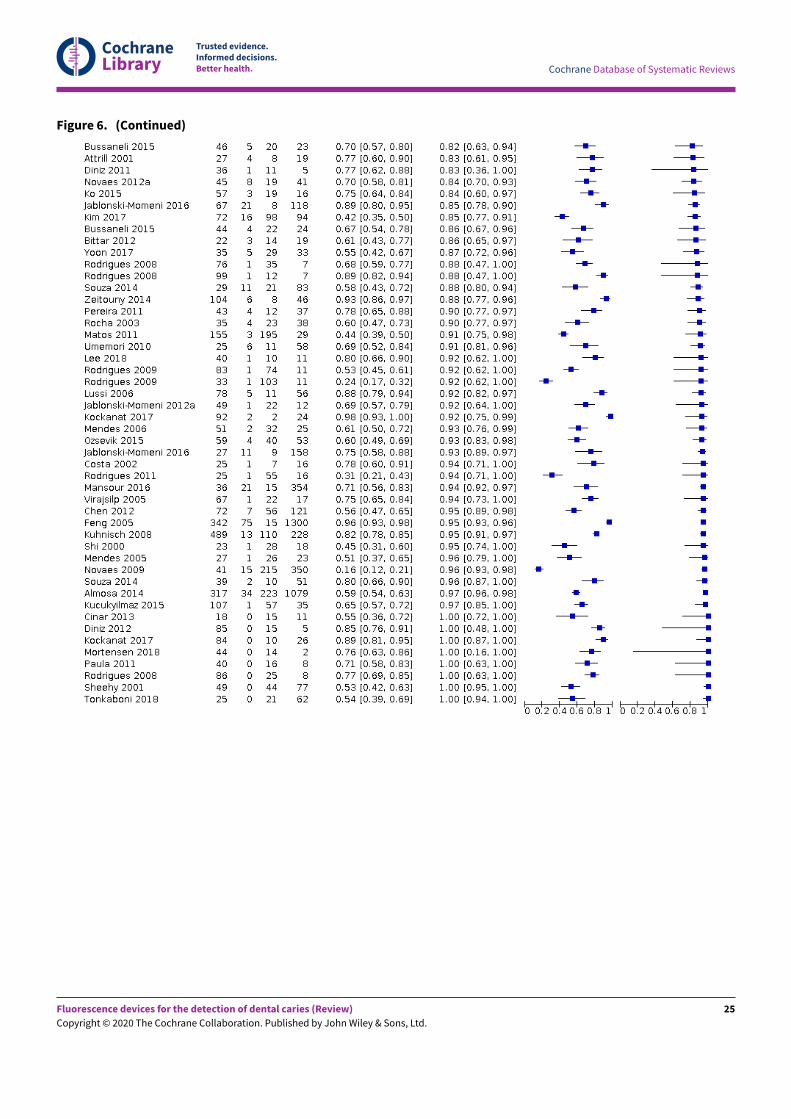
CochraneLibrary
Trusted evidence.Informed decisions.Better health.
Cochrane Database of Systematic Reviews
Figure 6. (Continued)
Fluorescence devices for the detection of dental caries (Review)
Copyright © 2020 The Cochrane Collaboration. Published by John Wiley & Sons, Ltd.
25
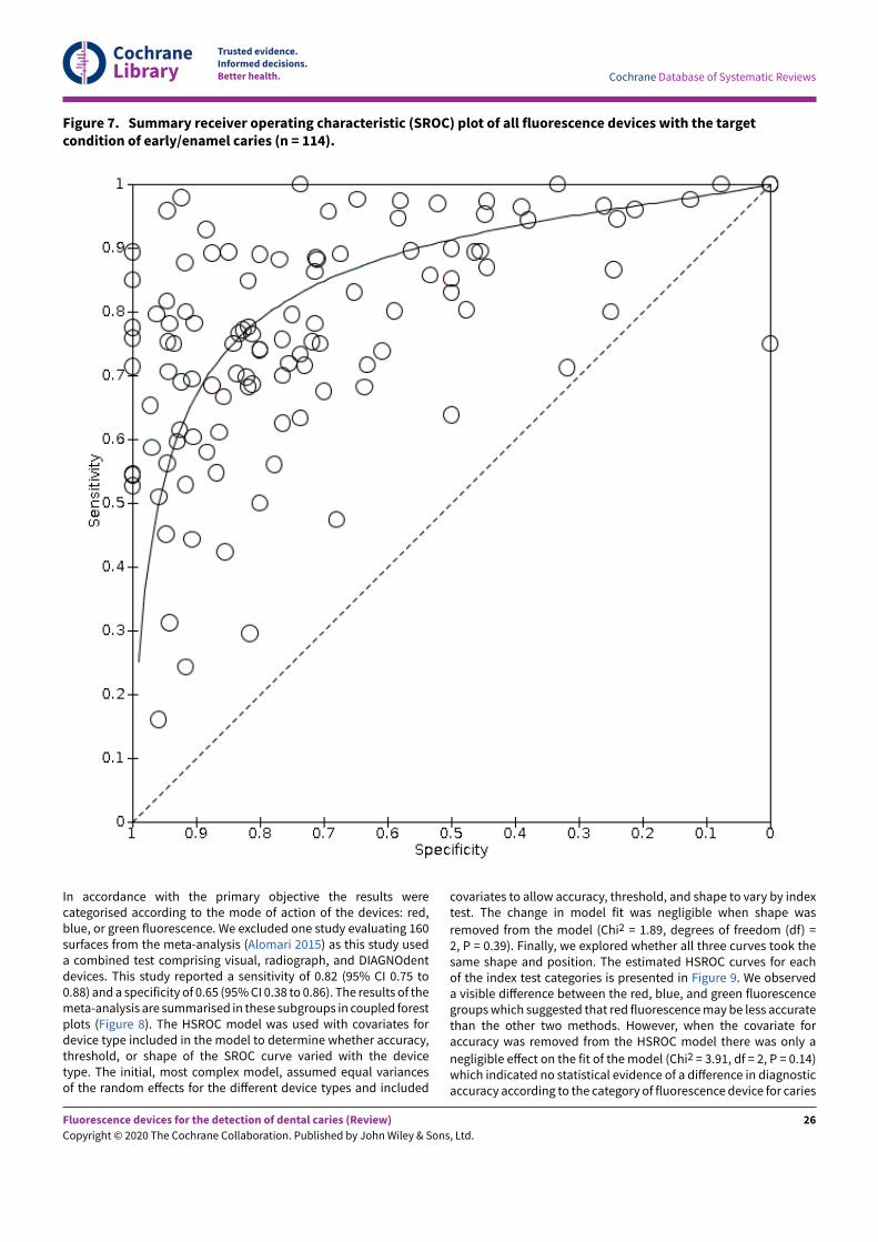
CochraneLibrary
Trusted evidence.Informed decisions.Better health.
Cochrane Database of Systematic Reviews
Figure 7. Summary receiver operating characteristic (SROC) plot of all fluorescence devices with the targetcondition of early/enamel caries (n = 114).
In accordance with the primary objective the results werecategorised according to the mode of action of the devices: red,blue, or green fluorescence. We excluded one study evaluating 160surfaces from the meta-analysis (Alomari 2015) as this study useda combined test comprising visual, radiograph, and DIAGNOdentdevices. This study reported a sensitivity of 0.82 (95% CI 0.75 to0.88) and a specificity of 0.65 (95% CI 0.38 to 0.86). The results of themeta-analysis are summarised in these subgroups in coupled forestplots (Figure 8). The HSROC model was used with covariates fordevice type included in the model to determine whether accuracy,threshold, or shape of the SROC curve varied with the devicetype. The initial, most complex model, assumed equal variancesof the random eEects for the diEerent device types and included
covariates to allow accuracy, threshold, and shape to vary by indextest. The change in model fit was negligible when shape was
removed from the model (Chi2 = 1.89, degrees of freedom (df) =2, P = 0.39). Finally, we explored whether all three curves took thesame shape and position. The estimated HSROC curves for eachof the index test categories is presented in Figure 9. We observeda visible diEerence between the red, blue, and green fluorescencegroups which suggested that red fluorescence may be less accuratethan the other two methods. However, when the covariate foraccuracy was removed from the HSROC model there was only a
negligible eEect on the fit of the model (Chi2 = 3.91, df = 2, P = 0.14)which indicated no statistical evidence of a diEerence in diagnosticaccuracy according to the category of fluorescence device for caries
Fluorescence devices for the detection of dental caries (Review)
Copyright © 2020 The Cochrane Collaboration. Published by John Wiley & Sons, Ltd.
26

CochraneLibrary
Trusted evidence.Informed decisions.Better health.
Cochrane Database of Systematic Reviews
detection. We therefore saw no need to investigate further analysesaccording to these subgroups.
Fluorescence devices for the detection of dental caries (Review)
Copyright © 2020 The Cochrane Collaboration. Published by John Wiley & Sons, Ltd.
27
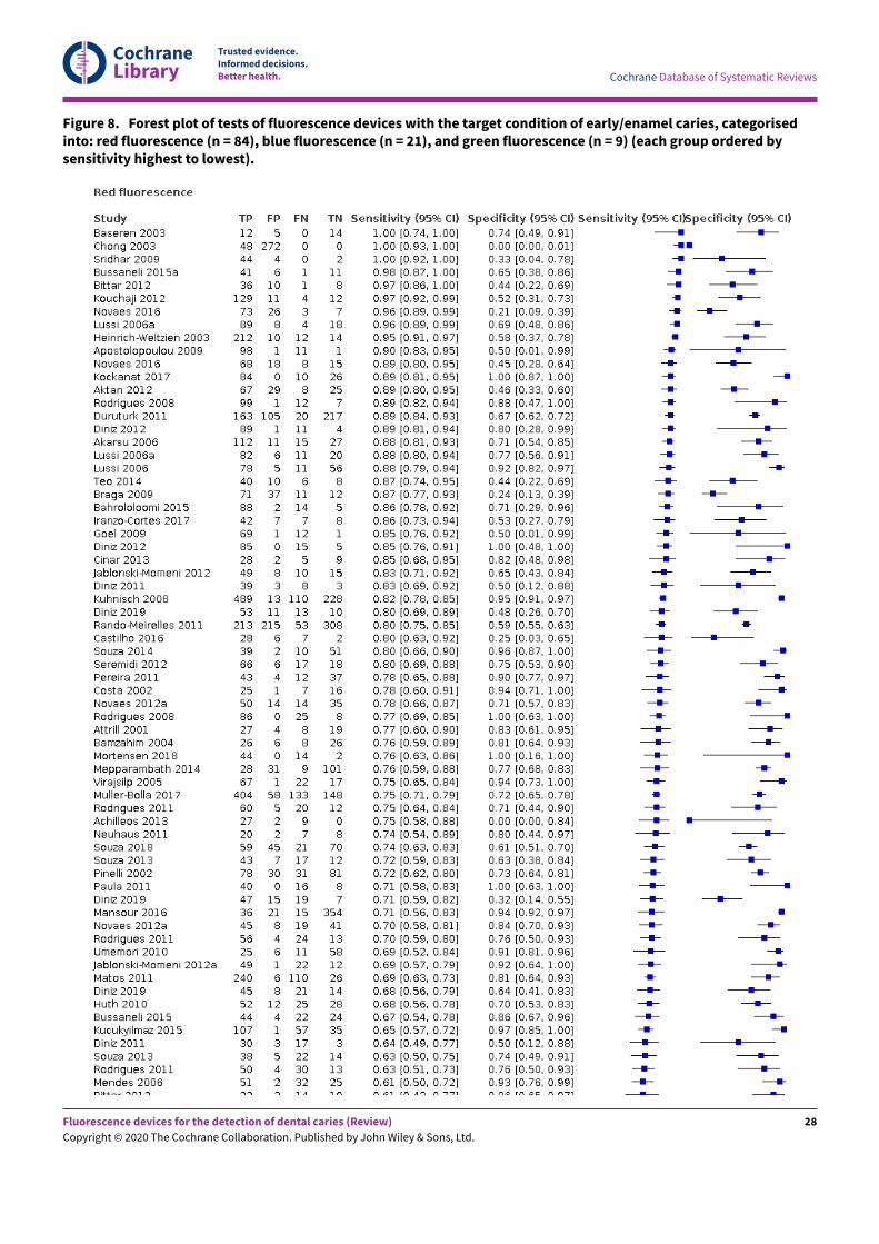
CochraneLibrary
Trusted evidence.Informed decisions.Better health.
Cochrane Database of Systematic Reviews
Figure 8. Forest plot of tests of fluorescence devices with the target condition of early/enamel caries, categorisedinto: red fluorescence (n = 84), blue fluorescence (n = 21), and green fluorescence (n = 9) (each group ordered bysensitivity highest to lowest).
Fluorescence devices for the detection of dental caries (Review)
Copyright © 2020 The Cochrane Collaboration. Published by John Wiley & Sons, Ltd.
28
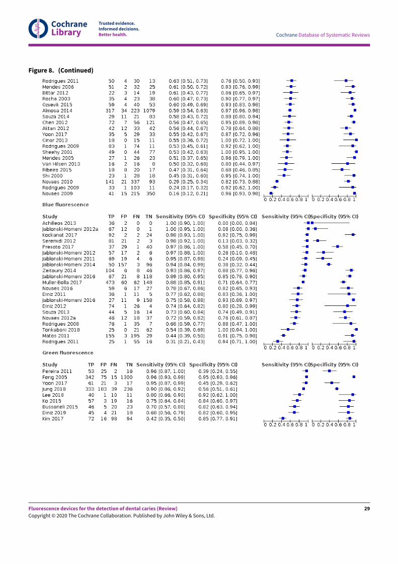
CochraneLibrary
Trusted evidence.Informed decisions.Better health.
Cochrane Database of Systematic Reviews
Figure 8. (Continued)
Fluorescence devices for the detection of dental caries (Review)
Copyright © 2020 The Cochrane Collaboration. Published by John Wiley & Sons, Ltd.
29
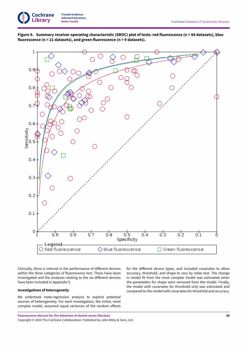
CochraneLibrary
Trusted evidence.Informed decisions.Better health.
Cochrane Database of Systematic Reviews
Figure 9. Summary receiver operating characteristic (SROC) plot of tests: red fluorescence (n = 84 datasets), bluefluorescence (n = 21 datasets), and green fluorescence (n = 9 datasets).
Clinically, there is interest in the performance of diEerent deviceswithin the three categories of fluorescence test. These have beeninvestigated and the analyses relating to the six diEerent deviceshave been included in Appendix 5.
Investigations of heterogeneity
We undertook meta-regression analysis to explore potentialsources of heterogeneity. For each investigation, the initial, mostcomplex model, assumed equal variances of the random eEects
for the diEerent device types, and included covariates to allowaccuracy, threshold, and shape to vary by index test. The changein model fit from the most complex model was estimated whenthe parameters for shape were removed from the model. Finally,the model with covariates for threshold only was estimated andcompared to the model with covariates for threshold and accuracy.
Fluorescence devices for the detection of dental caries (Review)
Copyright © 2020 The Cochrane Collaboration. Published by John Wiley & Sons, Ltd.
30
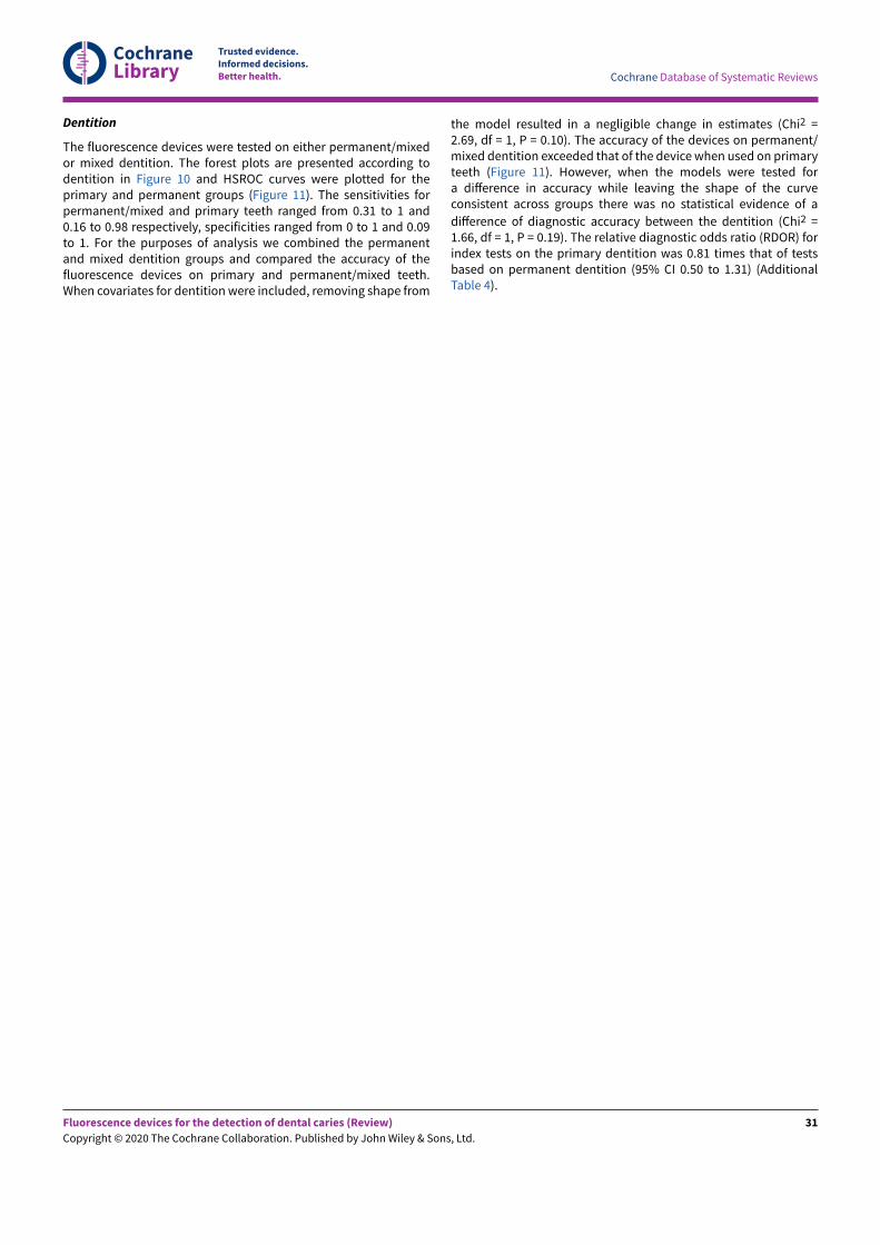
CochraneLibrary
Trusted evidence.Informed decisions.Better health.
Cochrane Database of Systematic Reviews
Dentition
The fluorescence devices were tested on either permanent/mixedor mixed dentition. The forest plots are presented according todentition in Figure 10 and HSROC curves were plotted for theprimary and permanent groups (Figure 11). The sensitivities forpermanent/mixed and primary teeth ranged from 0.31 to 1 and0.16 to 0.98 respectively, specificities ranged from 0 to 1 and 0.09to 1. For the purposes of analysis we combined the permanentand mixed dentition groups and compared the accuracy of thefluorescence devices on primary and permanent/mixed teeth.When covariates for dentition were included, removing shape from
the model resulted in a negligible change in estimates (Chi2 =2.69, df = 1, P = 0.10). The accuracy of the devices on permanent/mixed dentition exceeded that of the device when used on primaryteeth (Figure 11). However, when the models were tested fora diEerence in accuracy while leaving the shape of the curveconsistent across groups there was no statistical evidence of a
diEerence of diagnostic accuracy between the dentition (Chi2 =1.66, df = 1, P = 0.19). The relative diagnostic odds ratio (RDOR) forindex tests on the primary dentition was 0.81 times that of testsbased on permanent dentition (95% CI 0.50 to 1.31) (AdditionalTable 4).
Fluorescence devices for the detection of dental caries (Review)
Copyright © 2020 The Cochrane Collaboration. Published by John Wiley & Sons, Ltd.
31
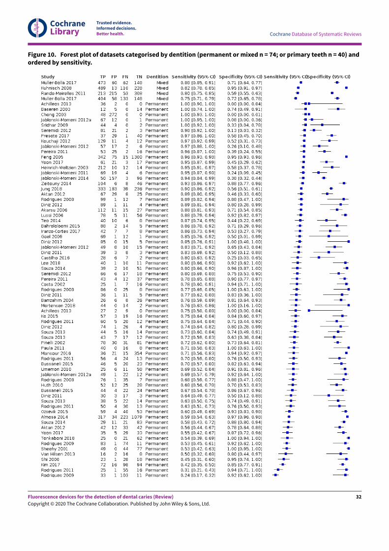
CochraneLibrary
Trusted evidence.Informed decisions.Better health.
Cochrane Database of Systematic Reviews
Figure 10. Forest plot of datasets categorised by dentition (permanent or mixed n = 74; or primary teeth n = 40) andordered by sensitivity.
Fluorescence devices for the detection of dental caries (Review)
Copyright © 2020 The Cochrane Collaboration. Published by John Wiley & Sons, Ltd.
32
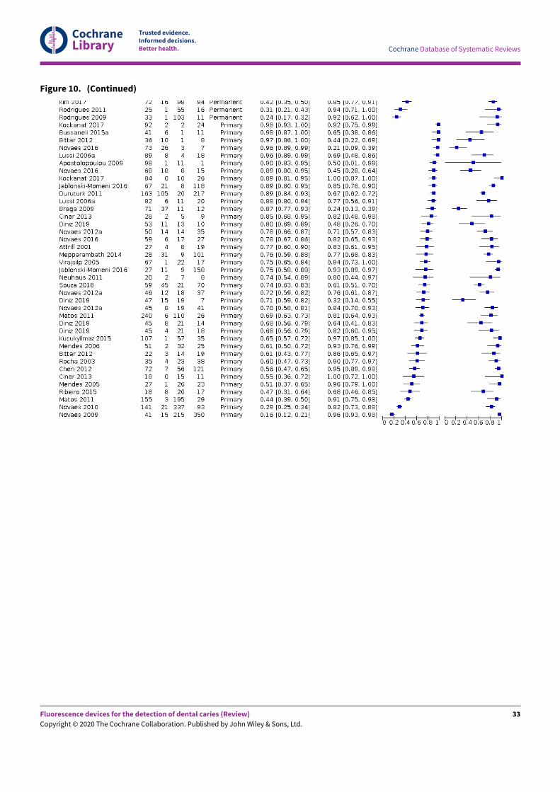
CochraneLibrary
Trusted evidence.Informed decisions.Better health.
Cochrane Database of Systematic Reviews
Figure 10. (Continued)
Fluorescence devices for the detection of dental caries (Review)
Copyright © 2020 The Cochrane Collaboration. Published by John Wiley & Sons, Ltd.
33
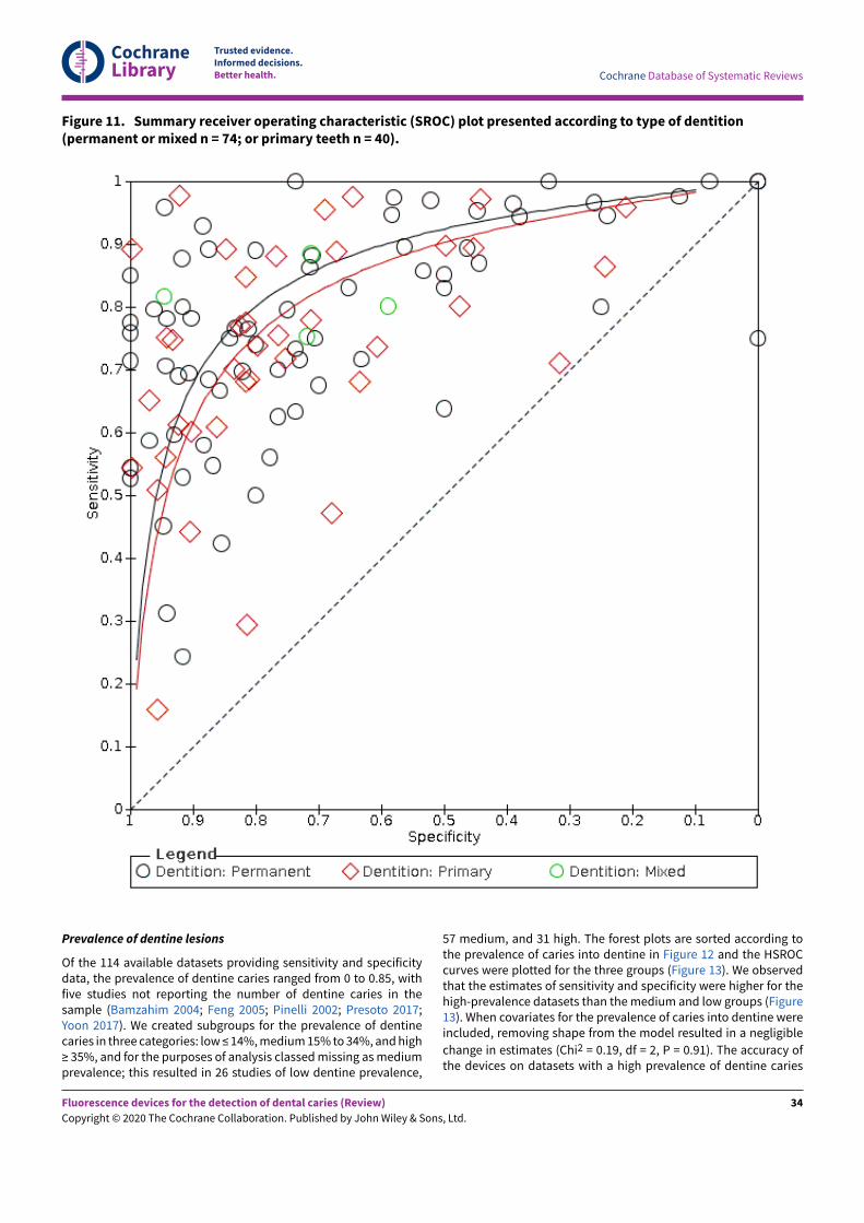
CochraneLibrary
Trusted evidence.Informed decisions.Better health.
Cochrane Database of Systematic Reviews
Figure 11. Summary receiver operating characteristic (SROC) plot presented according to type of dentition(permanent or mixed n = 74; or primary teeth n = 40).
Prevalence of dentine lesions
Of the 114 available datasets providing sensitivity and specificitydata, the prevalence of dentine caries ranged from 0 to 0.85, withfive studies not reporting the number of dentine caries in thesample (Bamzahim 2004; Feng 2005; Pinelli 2002; Presoto 2017;Yoon 2017). We created subgroups for the prevalence of dentinecaries in three categories: low ≤ 14%, medium 15% to 34%, and high≥ 35%, and for the purposes of analysis classed missing as mediumprevalence; this resulted in 26 studies of low dentine prevalence,
57 medium, and 31 high. The forest plots are sorted according tothe prevalence of caries into dentine in Figure 12 and the HSROCcurves were plotted for the three groups (Figure 13). We observedthat the estimates of sensitivity and specificity were higher for thehigh-prevalence datasets than the medium and low groups (Figure13). When covariates for the prevalence of caries into dentine wereincluded, removing shape from the model resulted in a negligible
change in estimates (Chi2 = 0.19, df = 2, P = 0.91). The accuracy ofthe devices on datasets with a high prevalence of dentine caries
Fluorescence devices for the detection of dental caries (Review)
Copyright © 2020 The Cochrane Collaboration. Published by John Wiley & Sons, Ltd.
34
CochraneLibrary
Trusted evidence.Informed decisions.Better health.
Cochrane Database of Systematic Reviews
exceeded that of low- or medium-prevalence datasets. However,when the models were tested for a diEerence in accuracy whileleaving the shape of the curve consistent across groups therewas no statistical evidence of a diEerence of diagnostic accuracy
between the two groups (Chi2 = 2.27, df = 2, P = 0.32). The RDORfor low prevalence was 0.76 (95% CI 0.39 to 1.48), and for mediumprevalence was 1.05 (95% CI 0.59 to 1.86) (Additional Table 4) whencompared with the reference category of high prevalence.
Fluorescence devices for the detection of dental caries (Review)
Copyright © 2020 The Cochrane Collaboration. Published by John Wiley & Sons, Ltd.
35
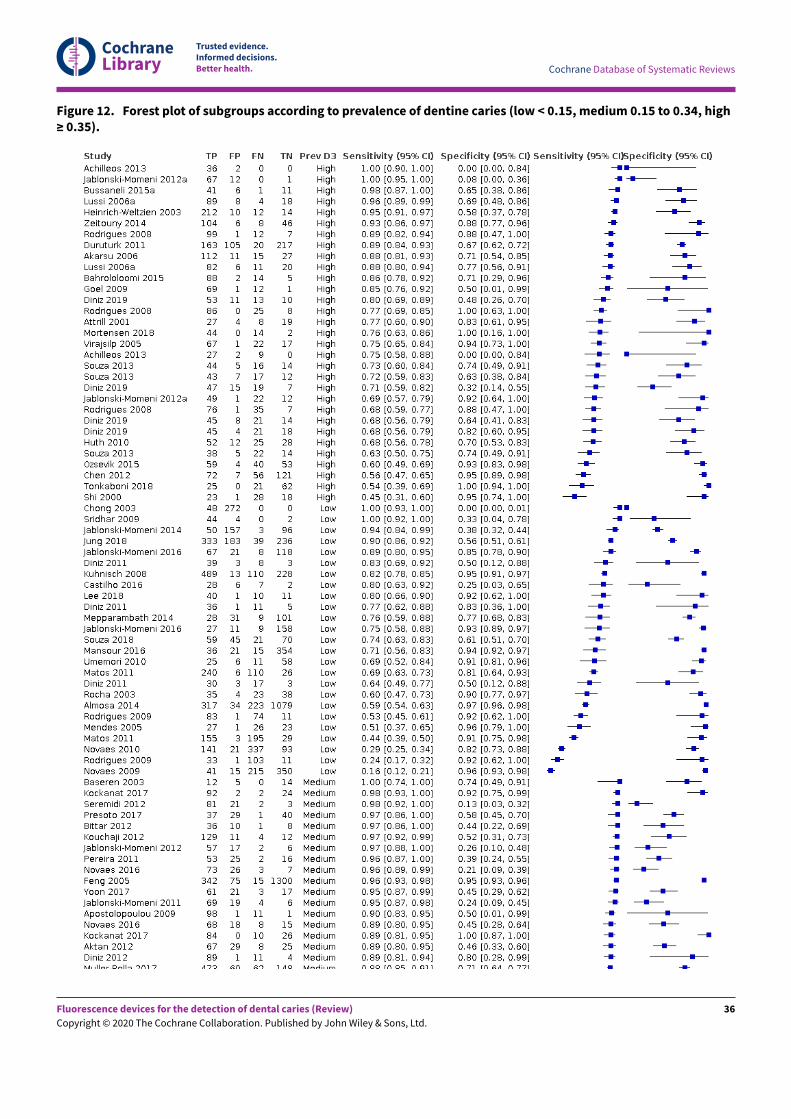
CochraneLibrary
Trusted evidence.Informed decisions.Better health.
Cochrane Database of Systematic Reviews
Figure 12. Forest plot of subgroups according to prevalence of dentine caries (low < 0.15, medium 0.15 to 0.34, high≥ 0.35).
Fluorescence devices for the detection of dental caries (Review)
Copyright © 2020 The Cochrane Collaboration. Published by John Wiley & Sons, Ltd.
36
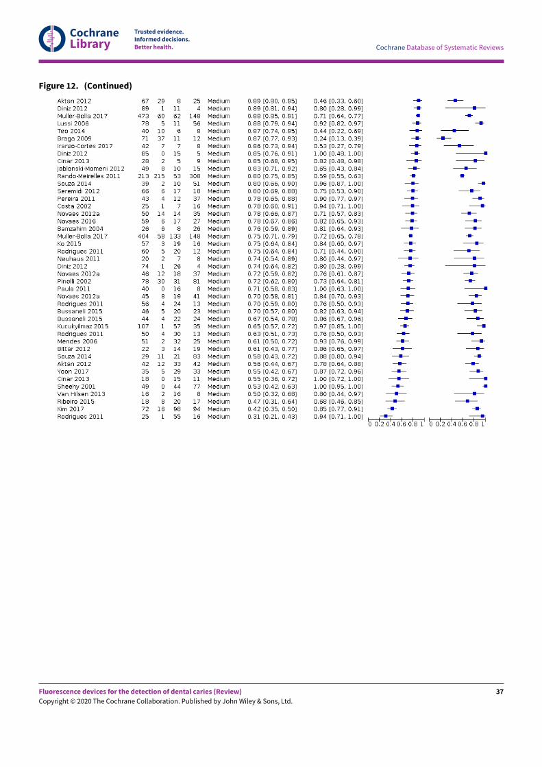
CochraneLibrary
Trusted evidence.Informed decisions.Better health.
Cochrane Database of Systematic Reviews
Figure 12. (Continued)
Fluorescence devices for the detection of dental caries (Review)
Copyright © 2020 The Cochrane Collaboration. Published by John Wiley & Sons, Ltd.
37
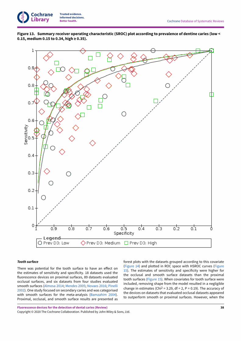
CochraneLibrary
Trusted evidence.Informed decisions.Better health.
Cochrane Database of Systematic Reviews
Figure 13. Summary receiver operating characteristic (SROC) plot according to prevalence of dentine caries (low <0.15, medium 0.15 to 0.34, high ≥ 0.35).
Tooth surface
There was potential for the tooth surface to have an eEect onthe estimates of sensitivity and specificity. 18 datasets used thefluorescence devices on proximal surfaces, 89 datasets evaluatedocclusal surfaces, and six datasets from four studies evaluatedsmooth surfaces (Almosa 2014; Mendes 2005; Novaes 2016; Pinelli2002). One study focused on secondary caries and was categorisedwith smooth surfaces for the meta-analysis (Bamzahim 2004).Proximal, occlusal, and smooth surface results are presented as
forest plots with the datasets grouped according to this covariate(Figure 14) and plotted in ROC space with HSROC curves (Figure15). The estimates of sensitivity and specificity were higher forthe occlusal and smooth surface datasets than the proximaltooth surfaces (Figure 15). When covariates for tooth surface wereincluded, removing shape from the model resulted in a negligible
change in estimates (Chi2 = 3.29, df = 2, P = 0.19). The accuracy ofthe devices on datasets that evaluated occlusal datasets appearedto outperform smooth or proximal surfaces. However, when the
Fluorescence devices for the detection of dental caries (Review)
Copyright © 2020 The Cochrane Collaboration. Published by John Wiley & Sons, Ltd.
38
CochraneLibrary
Trusted evidence.Informed decisions.Better health.
Cochrane Database of Systematic Reviews
models were tested for a diEerence in accuracy while leaving theshape of the curve consistent across groups there was no statisticalevidence of a diEerence of diagnostic accuracy between the groups
(Chi2 = 0.97, df = 2, P = 0.62). The RDOR of studies that evaluated
occlusal surfaces was 1.10 (95% CI 0.59 to 2.02), and smooth/secondary caries was 1.03 (95% CI 0.36 to 2.90) compared with thereference category of proximal surfaces (Additional Table 4).
Fluorescence devices for the detection of dental caries (Review)
Copyright © 2020 The Cochrane Collaboration. Published by John Wiley & Sons, Ltd.
39
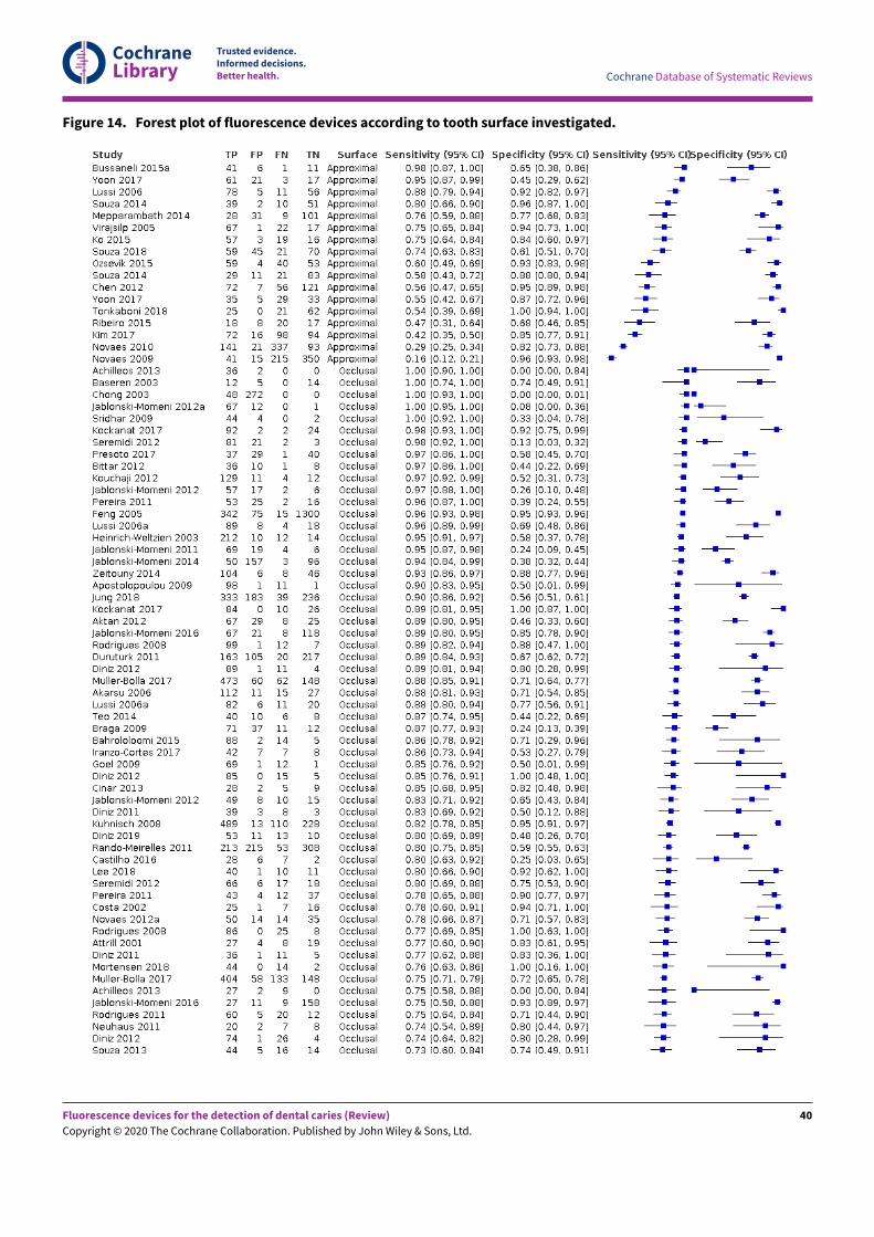
CochraneLibrary
Trusted evidence.Informed decisions.Better health.
Cochrane Database of Systematic Reviews
Figure 14. Forest plot of fluorescence devices according to tooth surface investigated.
Fluorescence devices for the detection of dental caries (Review)
Copyright © 2020 The Cochrane Collaboration. Published by John Wiley & Sons, Ltd.
40
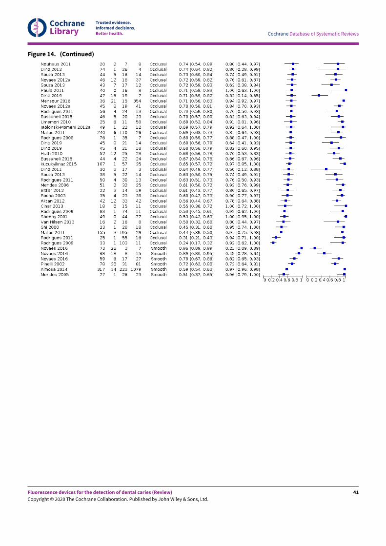
CochraneLibrary
Trusted evidence.Informed decisions.Better health.
Cochrane Database of Systematic Reviews
Figure 14. (Continued)
Fluorescence devices for the detection of dental caries (Review)
Copyright © 2020 The Cochrane Collaboration. Published by John Wiley & Sons, Ltd.
41
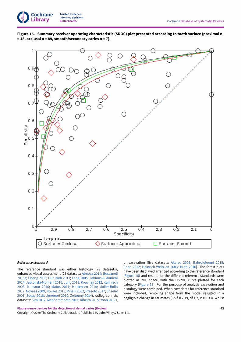
CochraneLibrary
Trusted evidence.Informed decisions.Better health.
Cochrane Database of Systematic Reviews
Figure 15. Summary receiver operating characteristic (SROC) plot presented according to tooth surface (proximal n= 18, occlusal n = 89, smooth/secondary caries n = 7).
Reference standard
The reference standard was either histology (78 datasets),enhanced visual assessment (25 datasets: Almosa 2014; Bussaneli2015a; Chong 2003; Duruturk 2011; Feng 2005; Jablonski-Momeni2014; Jablonski-Momeni 2016; Jung 2018; Kouchaji 2012; Kuhnisch2008; Mansour 2016; Matos 2011; Mortensen 2018; Muller-Bolla2017; Novaes 2009; Novaes 2010; Pinelli 2002; Presoto 2017; Sheehy2001; Souza 2018; Umemori 2010; Zeitouny 2014), radiograph (sixdatasets: Kim 2017; Mepparambath 2014; Ribeiro 2015; Yoon 2017),
or excavation (five datasets: Akarsu 2006; Bahrololoomi 2015;Chen 2012; Heinrich-Weltzien 2003; Huth 2010). The forest plotshave been displayed arranged according to the reference standard(Figure 16) and results for the diEerent reference standards wereplotted in ROC space, with the HSROC curve plotted for eachcategory (Figure 17). For the purpose of analysis excavation andhistology were combined. When covariates for reference standardwere included, removing shape from the model resulted in a
negligible change in estimates (Chi2 = 2.19, df = 2, P = 0.33). Whilst
Fluorescence devices for the detection of dental caries (Review)
Copyright © 2020 The Cochrane Collaboration. Published by John Wiley & Sons, Ltd.
42
CochraneLibrary
Trusted evidence.Informed decisions.Better health.
Cochrane Database of Systematic Reviews
there was some indication of a diEerence in curves according to thereference standard, when the models were tested for a diEerence inaccuracy while leaving the shape of the curve consistent there wasno statistical evidence of a diEerence in diagnostic accuracy across
the groups (Chi2 = 5.69, df = 2, P = 0.06). The RDOR for radiographswas 0.46 (95% CI 0.18 to 1.16), and for enhanced visual examinationwas 1.43 (95% CI 0.85 to 2.41) (Additional Table 4) when comparedwith the reference category of histology or excavation.
Fluorescence devices for the detection of dental caries (Review)
Copyright © 2020 The Cochrane Collaboration. Published by John Wiley & Sons, Ltd.
43
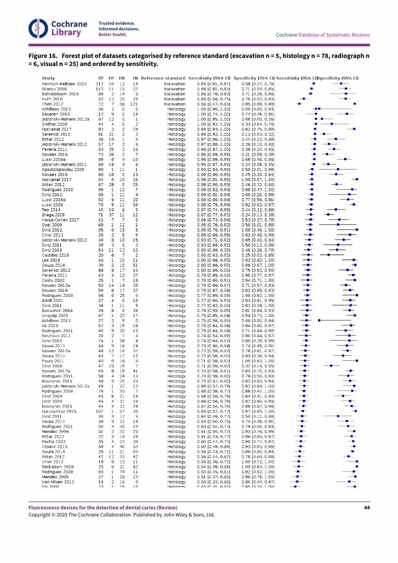
CochraneLibrary
Trusted evidence.Informed decisions.Better health.
Cochrane Database of Systematic Reviews
Figure 16. Forest plot of datasets categorised by reference standard (excavation n = 5, histology n = 78, radiograph n= 6, visual n = 25) and ordered by sensitivity.
Fluorescence devices for the detection of dental caries (Review)
Copyright © 2020 The Cochrane Collaboration. Published by John Wiley & Sons, Ltd.
44
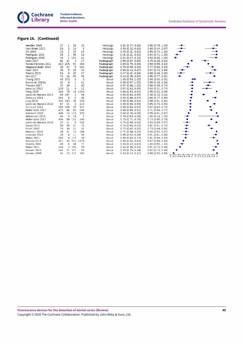
CochraneLibrary
Trusted evidence.Informed decisions.Better health.
Cochrane Database of Systematic Reviews
Figure 16. (Continued)
Fluorescence devices for the detection of dental caries (Review)
Copyright © 2020 The Cochrane Collaboration. Published by John Wiley & Sons, Ltd.
45
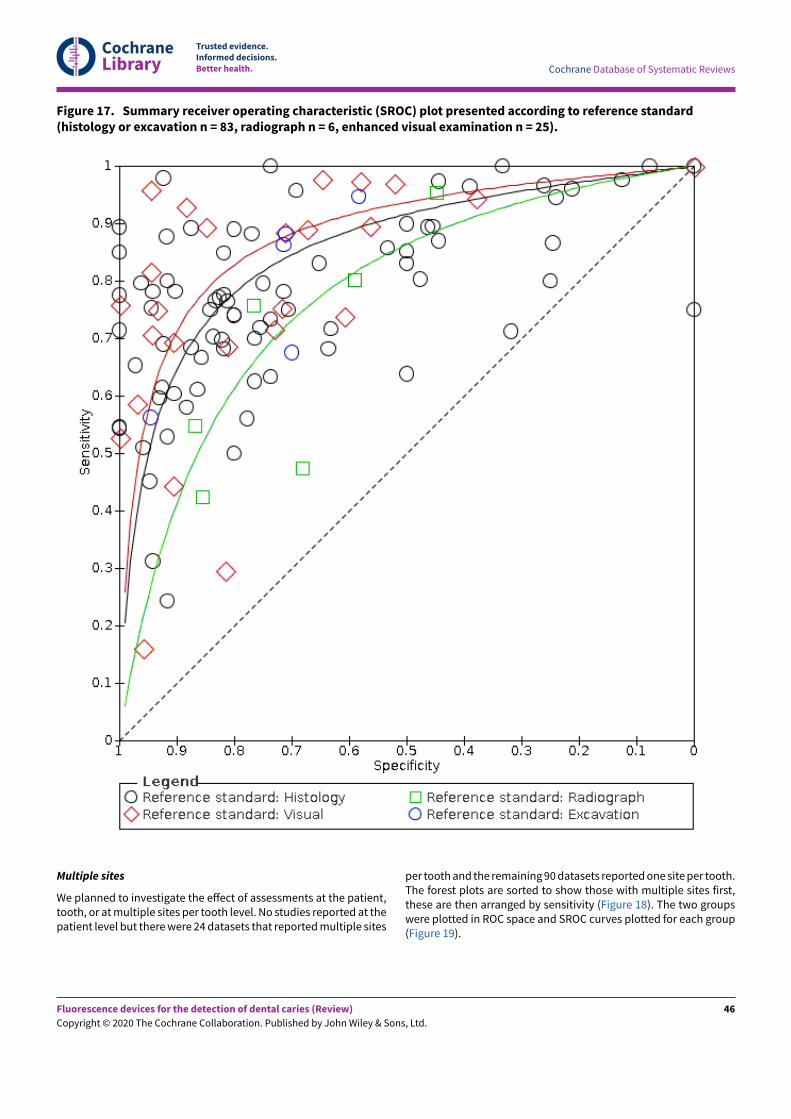
CochraneLibrary
Trusted evidence.Informed decisions.Better health.
Cochrane Database of Systematic Reviews
Figure 17. Summary receiver operating characteristic (SROC) plot presented according to reference standard(histology or excavation n = 83, radiograph n = 6, enhanced visual examination n = 25).
Multiple sites
We planned to investigate the eEect of assessments at the patient,tooth, or at multiple sites per tooth level. No studies reported at thepatient level but there were 24 datasets that reported multiple sites
per tooth and the remaining 90 datasets reported one site per tooth.The forest plots are sorted to show those with multiple sites first,these are then arranged by sensitivity (Figure 18). The two groupswere plotted in ROC space and SROC curves plotted for each group(Figure 19).
Fluorescence devices for the detection of dental caries (Review)
Copyright © 2020 The Cochrane Collaboration. Published by John Wiley & Sons, Ltd.
46
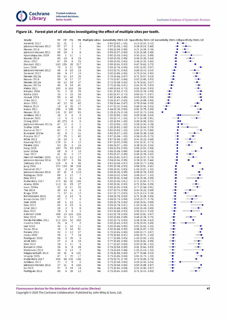
CochraneLibrary
Trusted evidence.Informed decisions.Better health.
Cochrane Database of Systematic Reviews
Figure 18. Forest plot of all studies investigating the e?ect of multiple sites per tooth.
Fluorescence devices for the detection of dental caries (Review)
Copyright © 2020 The Cochrane Collaboration. Published by John Wiley & Sons, Ltd.
47
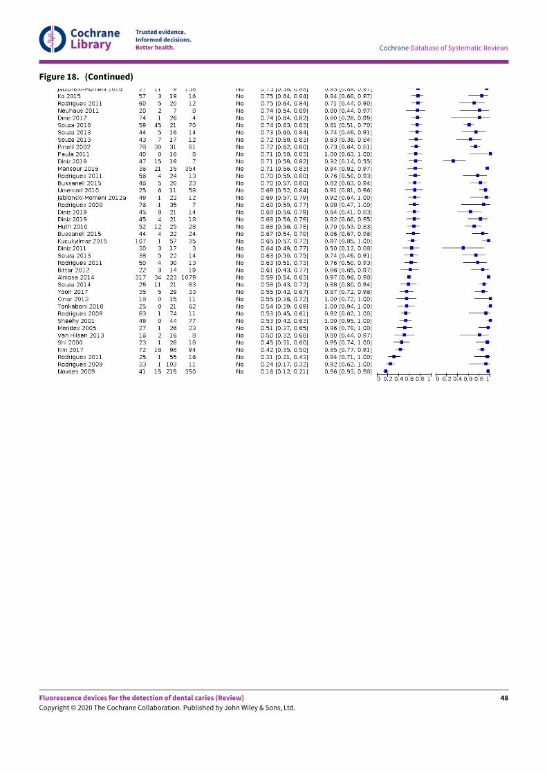
CochraneLibrary
Trusted evidence.Informed decisions.Better health.
Cochrane Database of Systematic Reviews
Figure 18. (Continued)
Fluorescence devices for the detection of dental caries (Review)
Copyright © 2020 The Cochrane Collaboration. Published by John Wiley & Sons, Ltd.
48
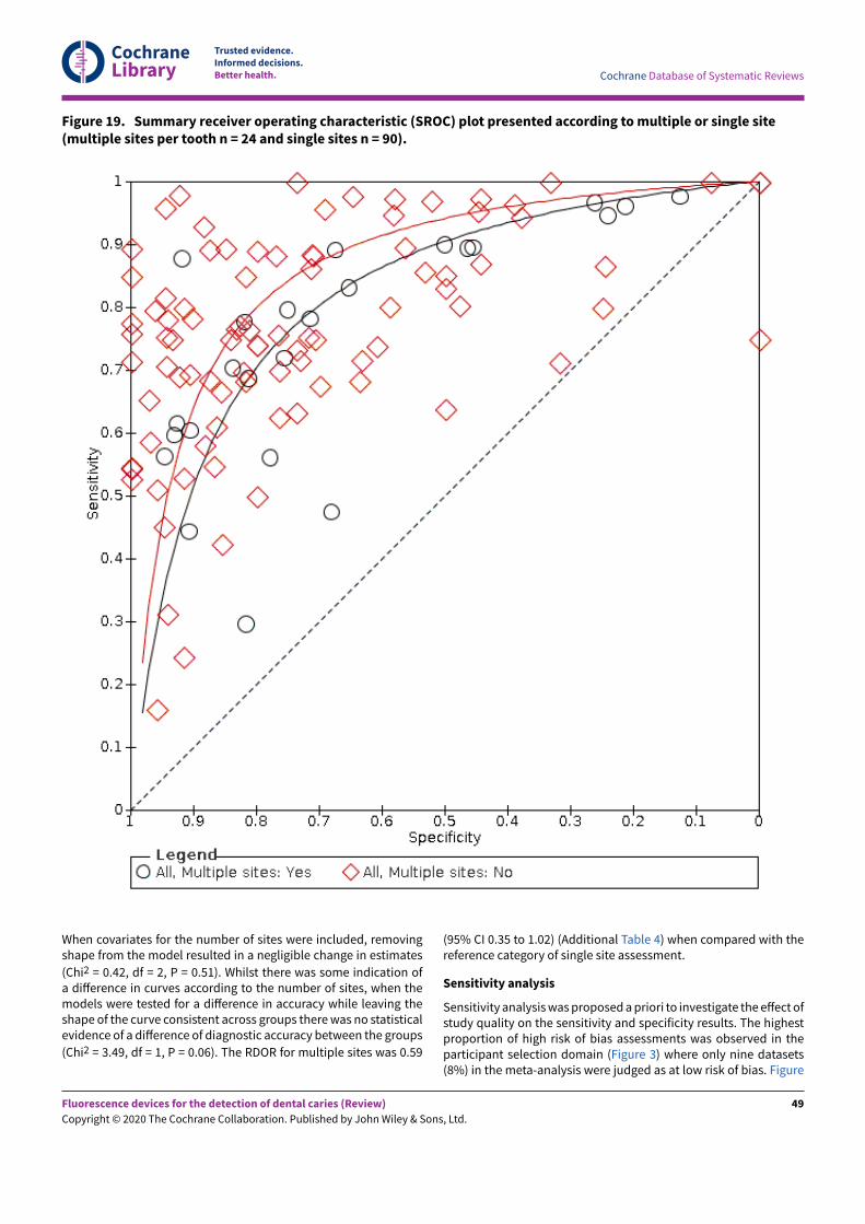
CochraneLibrary
Trusted evidence.Informed decisions.Better health.
Cochrane Database of Systematic Reviews
Figure 19. Summary receiver operating characteristic (SROC) plot presented according to multiple or single site(multiple sites per tooth n = 24 and single sites n = 90).
When covariates for the number of sites were included, removingshape from the model resulted in a negligible change in estimates
(Chi2 = 0.42, df = 2, P = 0.51). Whilst there was some indication ofa diEerence in curves according to the number of sites, when themodels were tested for a diEerence in accuracy while leaving theshape of the curve consistent across groups there was no statisticalevidence of a diEerence of diagnostic accuracy between the groups
(Chi2 = 3.49, df = 1, P = 0.06). The RDOR for multiple sites was 0.59
(95% CI 0.35 to 1.02) (Additional Table 4) when compared with thereference category of single site assessment.
Sensitivity analysis
Sensitivity analysis was proposed a priori to investigate the eEect ofstudy quality on the sensitivity and specificity results. The highestproportion of high risk of bias assessments was observed in theparticipant selection domain (Figure 3) where only nine datasets(8%) in the meta-analysis were judged as at low risk of bias. Figure
Fluorescence devices for the detection of dental caries (Review)
Copyright © 2020 The Cochrane Collaboration. Published by John Wiley & Sons, Ltd.
49
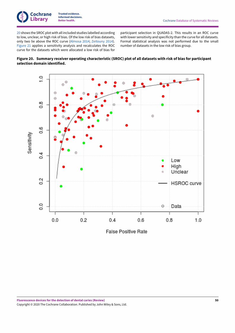
CochraneLibrary
Trusted evidence.Informed decisions.Better health.
Cochrane Database of Systematic Reviews
20 shows the SROC plot with all included studies labelled accordingto low, unclear, or high risk of bias. Of the low risk of bias datasets,only two lie above the ROC curve (Almosa 2014; Zeitouny 2014).Figure 21 applies a sensitivity analysis and recalculates the ROCcurve for the datasets which were allocated a low risk of bias for
participant selection in QUADAS-2. This results in an ROC curvewith lower sensitivity and specificity than the curve for all datasets.Formal statistical analysis was not performed due to the smallnumber of datasets in the low risk of bias group.
Figure 20. Summary receiver operating characteristic (SROC) plot of all datasets with risk of bias for participantselection domain identified.
Fluorescence devices for the detection of dental caries (Review)
Copyright © 2020 The Cochrane Collaboration. Published by John Wiley & Sons, Ltd.
50
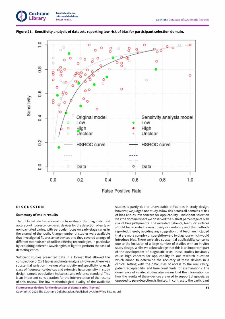
CochraneLibrary
Trusted evidence.Informed decisions.Better health.
Cochrane Database of Systematic Reviews
Figure 21. Sensitivity analysis of datasets reporting low risk of bias for participant selection domain.
D I S C U S S I O N
Summary of main results
The included studies allowed us to evaluate the diagnostic testaccuracy of fluorescence-based devices for the detection of early ornon-cavitated caries, with particular focus on early-stage caries inthe enamel of the tooth. A large number of studies were availablethat investigated fluorescence devices and they covered a range ofdiEerent methods which utilise diEering technologies, in particularby exploiting diEerent wavelengths of light to perform the task ofdetecting caries.
SuEicient studies presented data in a format that allowed theconstruction of 2 x 2 tables and meta-analyses. However, there wassubstantial variation in values of sensitivity and specificity for eachclass of fluorescence devices and extensive heterogeneity in studydesign, sample population, index test, and reference standard. Thisis an important consideration for the interpretation of the resultsof this review. The low methodological quality of the available
studies is partly due to unavoidable diEiculties in study design,however, we judged one study as low risk across all domains of riskof bias and as low concern for applicability. Participant selectionwas the domain where we observed the highest percentage of highrisk of bias judgements. The included patients, teeth, or surfacesshould be recruited consecutively or randomly and the methodsreported, thereby avoiding any suggestion that teeth are includedthat are more complex or straightforward to diagnose which wouldintroduce bias. There were also substantial applicability concernsdue to the inclusion of a large number of studies with an in vitrostudy design. Whilst we acknowledge that this is an important partof the development of diagnostic tests, these studies inevitablycause high concern for applicability to our research questionwhich aimed to determine the accuracy of these devices in aclinical setting with the diEiculties of access to the oral cavity,patient acceptability, and time constraints for examinations. Thedominance of in vitro studies also means that the information onhow the results of these devices are used to support diagnosis, asopposed to pure detection, is limited. In contrast to the participant
Fluorescence devices for the detection of dental caries (Review)
Copyright © 2020 The Cochrane Collaboration. Published by John Wiley & Sons, Ltd.
51
CochraneLibrary
Trusted evidence.Informed decisions.Better health.
Cochrane Database of Systematic Reviews
selection domain, the index test and reference standard domainsshowed a high number of studies with low risk of bias andapplicability concern. Similarly, flow and timing were of concernin only 25% of the included studies. Reasons for high risk of biasjudgements for the index test domain largely resulted from thelack of a pre-defined threshold. This was oNen because studieswere attempting to determine the most appropriate thresholdfor their sample population, resulting in inflating sensitivity andspecificity and therefore introducing bias. We awarded a decisionof high risk where an imperfect reference standard, such as avisual examination or radiograph, was used. This highlights themain diEiculty in studies of this type; to correctly classify thetarget condition, the preferred reference standard is histology.However, this automatically elicits concern for applicability inparticipant selection. The studies that circumvented this issue didso by targeting patients close to exfoliation of a primary toothor those who required a tooth extraction and applied the indextest in vivo with a subsequent reference standard in vitro. Suchstudies are challenging to organise and administer, and could stillbe considered to lack broader applicability since they oNen useteenage children requiring extractions for orthodontic purposes,and who would potentially have a lower prevalence of caries thanadults.
We estimated the accuracy of any fluorescence-based device forthe detection of early dental caries and compared the threegroups of red, blue, and green fluorescence. These devicesproduced an outcome on a continuous scale and applied diEerentthresholds to determine the result. Consequently we have usedsummary receiver operating characteristic (SROC) curves ratherthan summary sensitivity and specificity estimates. We tookillustrative sensitivity values from the hierarchical summaryreceiver operating characteristic (HSROC) curves (at a fixedsensitivity of 0.78 (median) and 0.90 (upper quartile)) to illustratechanges in sensitivity and specificity along the HSROC curve. Thesevalues are intended to be used only as a guide and should notbe used to indicate the actual performance of these fluorescencedevices. We used meta-regression to explore potential sources ofheterogeneity, but pre-specified patient or study characteristicswere unable to account for the substantial variation in results.
One of the primary objectives of the review was to investigate theeEect of using the fluorescence devices in combination with othertests, particularly as an adjunct to a visual examination. Only onestudy (Alomari 2015) formally reported this, and therefore it hasnot been possible to make an assessment. There were no case-control or randomised controlled trials included in this review, asthe searches retrieved no such eligible studies. There were also alimited number of included studies that investigated the eEect ofsealants or restorations on the diagnostic accuracy of fluorescencetests.
Despite the relatively large volume of evidence we rated thecertainty of the evidence as low, downgraded two levels in total,for risk of bias due to limitations in the design and conduct of theincluded studies, indirectness arising from the high number of invitro studies, and inconsistency due to the substantial variability ofresults.
The main findings of this review are that.
• The overall group results are presented as a HSROC curve.The diagnostic odds ratio (DOR) was 14.12 (95% confidence
interval (CI) 11.17 to 17.84). In the absence of clinical consensus,we elected to report sensitivities at fixed values of specificity(median, upper quartile) as a means of expressing numericalquantities from the curve. This is in preference to using theaverage values of sensitivity and specificity which do notcorrespond to any particular threshold. The estimated pointsfor sensitivity are 0.70 (95% CI 0.64 to 0.75) and 0.60 (95%CI 0.54 to 0.65), this is when applied at a fixed specificity of0.78 and 0.90 (Summary of findings 1). There is a degree ofnon-independence of data in this analysis, as some studiesprovided multiple datasets. For a cohort of 1000 tooth sites orsurfaces with a prevalence of enamel caries of 57% (the medianprevalence observed in studies included in the meta-analysis),the sensitivity of 0.70 at a fixed specificity of 0.78 would result in171 tooth sites not being identified as having early caries whencaries was present (false negatives) and 95 tooth sites beingidentified as having caries when they did not (false positives).The consequences of these misclassifications are concerning,and all interventions have a cost at a patient or systemlevel. A false positive classification for enamel caries wouldtypically result in the application of topical fluoride or otherminimally invasive treatments. A false negative classificationimplies that patients who require treatment would not receiveit. Given the recall period for routine dental examinations andthe slow-growing nature of the disease, the clinician may bereassured that the lesion could be identified at the patient'snext appointment. The prevalence of enamel caries applied tothis scenario is potentially inflated, due to many of the includedstudies being based on extracted teeth. In studies that employedan enhanced visual reference standard, based in either a school,primary care, or hospital setting, the median prevalence is lowerat 47%.
• There is no statistically significant di?erence in the accuracyof red, blue, or green fluorescence-based devices. 84 (74%)of the available datasets allowed us to assess red fluorescenceat the level of enamel caries, with 21 (18%) for blue, and 9 (8%)for green fluorescence, respectively. There was considerableheterogeneity of results within each of these subgroups that isreflective of the diEerent reference standards, the prevalenceof caries into dentine, tooth surface and dentition. A formalcomparison of the fluorescence-based devices indicated thatthere was no diEerence in accuracy according to the category ofthe device (P = 0.14).
• Studies with a higher proportion of observations with cariesinto dentine reported higher accuracy than studies with lowand medium prevalence. We considered the prevalence ofcaries into dentine to be important due to the potential forsensitivity and specificity to be inflated through the inclusionof large numbers of tooth surfaces with more advanced lesionsobviously into dentine or frankly cavitated. These could beconsiderably more straightforward to detect, and therefore theinflation of accuracy estimates would occur. The investigationof the covariate of high prevalence (≥ 35%) versus medium(15% to 34%) and low (15%) prevalence concurred that this wasoccurring in the data gathered from the included studies, formaltesting found that this diEerence was not statistically significanthowever (P = 0.32).
• There is no meaningful di?erence in the accuracy of studiesperformed in vitro and in vivo. The majority of studieswere conducted on extracted teeth (in vitro) using a referencestandard of histology, as opposed to teeth in situ conducted in
Fluorescence devices for the detection of dental caries (Review)
Copyright © 2020 The Cochrane Collaboration. Published by John Wiley & Sons, Ltd.
52
CochraneLibrary
Trusted evidence.Informed decisions.Better health.
Cochrane Database of Systematic Reviews
a clinical setting (in vivo). The results from the in vitro studiesare essential for determining the validity of devices but do nottruly inform us of the applicability of using these devices onpatients in a general dental practice setting. Detecting diseasein an in vitro setting can be assumed to be more straightforwardthan in a clinical setting as the challenges of accessing thetooth surfaces in the oral cavity, the complexity of soN tissues,or other teeth impeding the view, are largely eliminated. Theevaluation of extracted teeth also facilitates the use of histologyas a reliable and accurate reference standard; more recentlymicrocomputed tomography (microCT) has also been usedwith some confidence as a reference standard, although thiswas not the case for any of the included studies. Since it isnot feasible to extract and section healthy teeth and subjectthem to a histological reference standard, clinical studies havecircumvented this issue by using enhanced visual examinationor radiographs as eEectively imperfect reference standards. Thecomparative accuracy of in vitro and in vivo study designscan be assessed by investigating the two most frequentlyused reference standards of histology (78 datasets, 68%) andenhanced visual assessment (25 datasets, 22%). Whilst the DORwas highest for enhanced visual examination as a referencestandard formal comparison found no diEerence in accuracy (P= 0.06).
• Diagnostic accuracy was higher for occlusal surfaces. Themajority of studies evaluated either occlusal (89 datasets) orproximal surfaces (18 sets). Some concern has been expressedthat fluorescence-based devices are limited in their ability todetect proximal caries, as the excitation (laser) light needs tomake direct contact with the tooth surface. If another toothobstructs the excitation then the performance of the device willbe suboptimal. There is no evidence that fluorescence-baseddevices show greater accuracy in detecting caries on occlusalsurfaces than proximal surfaces (P = 0.62).
• Studies on permanent teeth suggest greater accuracyover primary teeth when using fluorescence devices. Thedistinction between the primary, mixed, and permanentdentition is of importance too. The detection of enamel cariesmay be of greater clinical importance in primary teeth as thedepth of enamel is less than that of permanent teeth, andearly caries could lead to more severe decay with greaterexpedience than would be witnessed in permanent teeth.However, the retention of permanent teeth throughout aperson's lifetime is also important. Despite caries being seen asa slow-growing disease, the need for prevention in permanentteeth is also important. The results of the meta-analysissuggest that fluorescence devices may have greater accuracy indetecting caries in primary teeth, although this is not statisticallysignificant (P = 0.19).
• Devices that evaluated multiple sites on a tooth's surfaceshowed a lower accuracy than those that evaluated asingle site per tooth. The assessment of multiple tooth sitesintroduces dependency, and a single underlying or hiddenlesion could influence multiple sites. 24 of the 114 datasets in themeta-analysis reported multiple sites per tooth, however, five ofthese reported proximal surfaces where it would be less likelythat this eEect would occur. The results of the meta-analysissuggest that single point assessments may be more accurate,however, this was not statistically significant (P = 0.06). Twocommon methods were used when collecting a single site resultper tooth, particularly when applied to the occlusal surface.
Firstly where the device was passed over the tooth surface andthe highest number from the device recorded, and secondlywhere the device was applied three times and the mean of thethree recordings was taken.
Strengths and weaknesses of the review
The strengths of this Cochrane Review are the completion ofa comprehensive literature search and rigorous application ofmethodology which ensured that all screening, inclusion decisions,and data extraction were performed in duplicate and with clinicalinput. Unlike many diagnostic test accuracy (DTA) systematicreviews, we did not restrict our inclusion criteria to studiespresenting data in a 2 x 2 format, and this has enabled us tohighlight the issue of incomplete reporting of outcome data and theinadequate reporting in primary DTA studies. We contacted studyauthors where necessary to ensure that we could obtain data for asmany studies as possible. Further, we used a clear and reproducibleprocess for methodological decision making.
The substantial number of included studies facilitated meta-analysis. The primary analysis was conducted using hierarchicalsummary receiver operating characteristic (HSROC) curves ratherthan the Moses-Littenberg method which has been used in othercaries DTA reviews, and which has been shown to perform poorlyin comparison to hierarchical approaches (Dinnes 2016). An HSROCapproach was undertaken as opposed to the bivariate method dueto the variation of thresholds employed between sound and carioustooth surfaces in the included studies. The quoted sensitivitiesand specificities used to calculate the natural frequencies shouldtherefore be interpreted cautiously.
This review comprises a substantial number of primary studies.Bader 2004 completed a review of fluorescence devices, and thisCochrane Review is a significant update that broadens the remitof the earlier review to include visual and radiographs referencestandards in addition to histology. This DTA systematic review hassubstantially increased the number of included studies from 25(Bader 2004) and 73 in a more recent review (Gimenez 2013) to133 (79 studies included in the meta-analysis) in this review. Theuse of HSROC methodology is an important component of thisDTA systematic review. Gimenez 2013 did not use the hierarchicalmodel, although our conclusion is similar - that fluorescencedevices show improved results in more severe caries, but thatthe accuracy of devices is similar across diEerent tooth surfaces.Our review also focuses on the target condition of early enamellesions which has the potential to inform clinicians on the decisionto intervene earlier in the disease process with preventive orminimally invasive treatments rather than operative.
The main weakness of the review is the substantial volume ofstudies with incomplete outcome data. 55 of the 133 includedstudies provided insuEicient information to enable us to constructor compute a 2 x 2 table. Many studies did not present thenumbers of true positives, true negatives, false positives, andfalse negatives at the enamel threshold. Rather, they reportedsensitivity, specificity, and area under the curve as their primaryresults. This did not allow us to include a study in the meta-analysis unless the prevalence of caries at the enamel thresholdwas reported, enabling the construction of the required 2 x 2 tableof outcomes.
Fluorescence devices for the detection of dental caries (Review)
Copyright © 2020 The Cochrane Collaboration. Published by John Wiley & Sons, Ltd.
53
CochraneLibrary
Trusted evidence.Informed decisions.Better health.
Cochrane Database of Systematic Reviews
A significant source of bias in many of the studies was that theparticipants or teeth were selected, with the risk that teeth wereselected teeth that made caries detection more straightforward,with resulting inflation of sensitivity and/or specificity values. Whenplanning the meta-analysis, it became apparent that an argumentcould be created to subgroup by in vitro and in vivo studies, or byindex test, or by the reference standard. We decided to allow theprimary meta-analysis to remain as a single complete dataset andto investigate the eEects of these factors through meta-regression,and to allow the results of this analysis to guide the remainder ofthe meta-analysis.
The inclusion criteria were selected to ensure that the focusof the review was on the detection of early caries or carieslimited to enamel. However, with the best of intentions studiescould easily attempt to recruit sound or non-cavitated teeth butwhen investigated with a thorough/complete reference standard itbecame apparent that when viewed during participant selection,surfaces harboured dentinal caries. The concern of the review teamwas that if studies intentionally recruited dentinal lesions, thenthere would be a simplification of the detection and diagnosticdecision as a lesion which was validated and reached dentine isgenerally easier to observe than an early lesion which is limitedto the enamel. A further complication arose where some studieswere poorly reported or lacked clarity on the selection criteriathat they imposed on their sample. We took the position thatunless the authors clearly stated that frank or dentinal cavities wereintentionally included, then we were unable to exclude the studyfrom the review. The result of this decision has been diEicult toapply consistently, and consequently, we may have excluded somewell-reported studies due to their clarity of reporting, whereasstudies which intentionally included dentinal lesions, but failedto report this inclusion, were included. We accept this may leavethe review open to some criticism, and we would reiterate thatthis review intended to synthesise the evidence on early lesions.The inclusion of more advanced lesions that are obviously intodentine or frankly cavitated does not fit the remit of this review.Analysis of the prevalence of caries at the dentinal level enabled usto investigate this assertion which results confirmed.
Some studies purposefully investigated the most accuratethreshold, using the study data and ROC curve to determine theoptimum threshold to maximise values of sensitivity or specificityor both. The focus of our review, however, was on the accuracyof these devices when used by general dentists, which requiresthe use of a pre-defined threshold. The reporting of resultsaccording to optimised data-driven thresholds is problematic asthe observed sensitivity and specificity values will be higher inthese studies than those applying pre-determined thresholds, thethresholds selected by these studies may not be generalisable toother patient populations. Although useful, such studies may havelimited relevance to our research question. Another area of concernarose when the reference standard was histology and studies didnot report whether the same examiners conducted the index testand reference standard assessments. This issue was logged in thecharacteristics of included studies tables, but our interpretationwas that this would not aEect the judgement of the referencestandard as it was hard to see how an examiner would rememberthe results of the fluorescence devices and recall it during theexamination of a sectioned tooth. A final area of concern wasthe eEect of the chosen threshold between the sound, enamelcaries, and dentinal caries. For example, the thresholds used for the
DIAGNOdent device to diEerentiate between sound and dentinalcaries ranged from 2 to 20 so the results of one study could bereassessed according to other thresholds and very diEerent resultsobtained. As the HSROC approach models threshold eEects nofurther assessment was required.
Applicability of findings to the review question
There are concerns regarding the clinical applicability of thefindings of this review resulting from the fact that 68% of thedatasets are based on in vitro studies and therefore not conductedin a setting which is representative of the general dental setting.Until a more perfect reference standard for safe use in vivo isdeveloped, this is likely to be the status quo. Developments in theuse of 3D technology in vitro (microCT) and in vivo (cone-beam CT)may go some way to improve upon these concerns.
A U T H O R S ' C O N C L U S I O N S
Implications for practice
We intended that the results of this review be directly applicableto the general dental practitioner. Ideally, clinicians would haveall diagnostic test or devices available to them and use themost appropriate according to the clinical scenario. This is notpossible for most dental practices who have finite resourcesand existing infrastructure which would almost always feature aradiographic device to support the conventional oral examination.The question remains to clinicians whether the utilisation ofa fluorescence device provides suEicient benefits to justify thecost. There is considerable variation in the performance of thefluorescence-based devices included in this Cochrane Review thatcould not be explained by the diEerent wavelengths of the devicesassessed, or by participant or study characteristics. Blue andgreen fluorescence-based devices appeared to outperform redfluorescence-based devices, but this diEerence was not supportedby the results of a formal statistical comparison. There are concernsthat these results may be confounded by stain, and that thelower number of studies included for some blue fluorescencedevices means that further research into the accuracy of thesedevices may be warranted. The reproducibility of the devices wasbeyond the scope of this review, but one important, clinically usefulapplication could be the use of these devices over multiple timepoints to monitor lesions or even to quantify lesion severity tojustify any intervention. Clinicians will always perform a visualexamination but may well look to an adjunct to provide validationor confirmation of their decision. Due to the low certainty ofthe evidence from studies included in this review, considerableuncertainty remains regarding the accuracy of fluorescence-baseddevices for early caries detection.
Bader 2004 recommended that fluorescent devices should notbe used in isolation and based on the certainty of the evidencethere is little to challenge this recommendation. Despite thereasonably high sensitivity and specificity estimates, we cannotenvisage a scenario where a clinician would carry out a clinicalexamination without performing a thorough visual diagnosis, andwith development future fluorescence-based devices may supportthe clinician in confirming the status of uncertain or diEicult todiagnose teeth.
Fluorescence devices for the detection of dental caries (Review)
Copyright © 2020 The Cochrane Collaboration. Published by John Wiley & Sons, Ltd.
54
CochraneLibrary
Trusted evidence.Informed decisions.Better health.
Cochrane Database of Systematic Reviews
Implications for research
As is highlighted by the number of studies included in this reviewwhich did not report data in a useable format, it is of vitalimportance that future research studies report the data in a clearconcise method and following the STARD checklist (Bossuyt 2003;Bossuyt 2015), ideally with a cross-tabulation of the index testand reference standard with a minimum requirement of threecategories of each which could be classified as sound/cariesfree, early/enamel caries, advanced/dentine caries. Many studiessubdivided these latter two categories into inner and outer enamel/dentine caries, and this allowed us to extract true-positive, false-positive, false-negative, and true-positive results.
Importantly, future studies should be aware of the importanceof sampling participants using consecutive or random sampling.This should serve to minimise the bias which originates from theselection of teeth in which early caries is either easier or morediEicult to detect. Sensitivity analysis suggested that sensitivityand specificity could be overestimated by failing to use randomor consecutive sampling. Studies should also specify the testpositivity thresholds a priori rather than selecting the thresholdwhich maximises estimates of sensitivity and specificity, ideallyusing manufacturer recommended thresholds or those validated inprevious research studies. Studies may be conducted to determinethe most accurate thresholds for a given population. We wouldrecommend that studies such as these report the manufacturerrecommended thresholds in addition to the maximised thresholdsto facilitate a comparison between the two and allow for analysisin future reviews.
When designing the ideal study for future research, it is importantto consider the single study that we judged to be at low risk ofbias and low concern across all domains for Quality Assessmentof Diagnostic Accuracy Studies 2 (QUADAS-2). This study identifiedchildren that required a tooth extraction, which enabled theindex test to be conducted in the clinical setting, and a histologyreference standard once the tooth had been extracted. Future
studies could look at the potential of fluorescence devices to beused in combination with other technologies and to make directcomparisons between their use at diEerent points of the diseasespectrum, i.e. general practice: seemingly asymptomatic, low/highneed, irregular attenders, previously diseased participants. Giventhe potential utility of the devices in aiding the clinician to confirmborderline cases where the clinician is uncertain of the true diseasestate, a study could be designed which investigates only those siteswhich have a degree of uncertainty.
Randomised controlled trials could be beneficial by investigatingthe longer-term eEects of using the fluorescence devices fordetection, diagnosis, and monitoring to identify whether theyaid the prevention of disease through active preventativeinterventions.
A C K N O W L E D G E M E N T S
This series of Cochrane Reviews was funded by the UK NationalInstitute for Health Research (NIHR) Cochrane Programme GrantScheme (Project: 16/114/23). We would like to thank AnneLittlewood (Information Specialist, Cochrane Oral Health) for heradvice on the search strategy and conducting the search of theliterature, and Luisa M Fernandez MauleEinch (Managing Editor andCopy Editor, Cochrane Oral Health) for her assistance in facilitatingthis review. We thank Richard Hogan and Iain Pretty for their adviceduring protocol development and the interpretation of findings;Associate Professor KR Ekstrand, and J Bader (Emeritus Professor,UNC School of Dentistry, Chapel Hill North Carolina, USA) fortheir feedback on the protocol; Jennifer Hilgart, J Bader, AlonsoCarrasco-Labra (Senior Director, Department of Evidence Synthesisand Translation Research, ADA Science & Research Institute, LLC),and the Cochrane Diagnostic Test Accuracy Editorial Team for theirfeedback on the review. Also Alex Sutton and Suzanne Freemanfrom the NIHR Complex Review Support Unit for their support onthis review. For translation of primary studies we wish to thankMargriet van Baar, Fang Hua, Szimonetta Lohner, Anette Bluemle,Karin BischoE, and Maddalena Manfredi.
Fluorescence devices for the detection of dental caries (Review)
Copyright © 2020 The Cochrane Collaboration. Published by John Wiley & Sons, Ltd.
55
CochraneLibrary
Trusted evidence.Informed decisions.Better health.
Cochrane Database of Systematic Reviews
R E F E R E N C E S
References to studies included in this review
Achilleos 2013 {published data only}
Achilleos EE, Rahiotis C, Kakaboura A, Vougiouklakis G.Evaluation of a new fluorescence-based device in the detectionof incipient occlusal caries lesions. Lasers in Medical Science2013;28:193-201.
Akarsu 2006 {published data only}
Akarsu S, Koprulu H. In vivo comparison of the eEicacy ofDIAGNOdent by visual inspection and radiographic diagnostictechniques in the diagnosis of occlusal caries. Journal of ClinicalDentistry 2006;17(3):53-8.
Aktan 2012 {published data only}
Aktan AM, Cebe MA, CiNci ME, Sirin Karaarslan E. A novel LED-based device for occlusal caries detection. Lasers in MedicalScience 2012;27:1157-63.
Almosa 2014 {published data only}
Almosa NA, Lundgren T, Aldrees AM, Birkhed D, Kjellberg H.Diagnosing the severity of buccal caries lesions in governmentaland private orthodontic patients at debonding, using theICDAS-II and the DIAGNOdent pen. Angle Orthodontist2014;84:430-6.
Alomari 2015 {published data only}
Alomari QD, Qudeimat MA, Khalaf ME, Al-Tarakemah Y. TheeEect of combining radiographs and DIAGNOdent withvisual examination on detection and treatment decisions ofnoncavitated occluso-dentinal caries. Operative Dentistry2015;40:313-21.
Alwas-Danowska 2002 {published data only}
Alwas-Danowska HM, Plasschaert AJ, Suliborski S,Verdonschot EH. Reliability and validity issues of laserfluorescence measurements in occlusal caries diagnosis.Journal of Dentistry 2002;30:129-34.
Angnes 2005 {published data only}
Angnes V, Angnes G, Batisttella M, Grande RH, Loguercio AD,Reis A. Clinical eEectiveness of laser fluorescence, visualinspection and radiography in the detection of occlusal caries.Caries Research 2005;39:490-5.
Anttonen 2003 {published data only}
Anttonen V, Seppa L, Hausen H. Clinical study of the use of thelaser fluorescence device DIAGNOdent for detection of occlusalcaries in children. Caries Research 2003;37:17-23.
Apostolopoulou 2009 {published data only}
Apostolopoulou D, Lagouvardos P, Kavvadia K,Papagiannoulis L. Histological validation of a laser fluorescencedevice for occlusal caries detection in primary molars. EuropeanArchives of Paediatric Dentistry 2009;10 Suppl 1:11-5.
Arslan 2014 {published data only}
Arslan U, Karaagaoglu E, Ozkan G, Kanli A. Evaluationof diagnostic tests using information theory for multi-
class diagnostic problems and its application for thedetection of occlusal caries lesions. Balkan Medical Journal2014;31(3):214-8.
Attrill 2001 {published data only}
Attrill DC, Ashley PF. Occlusal caries detection in primary teeth:a comparison of DIAGNOdent with conventional methods.British Dental Journal 2001;190:440-3.
Bahrololoomi 2015 {published data only}
Bahrololoomi Z, Ezoddini F, Halvani N. Comparison ofradiography, laser fluorescence and visual examination fordiagnosing incipient occlusal caries of permanent first molars.Journal of Dentistry (Tehran, Iran) 2015;12(5):324-32.
Bamzahim 2002 {published data only}
Bamzahim M, Shi XQ, Angmar-Mansson B. Occlusal cariesdetection and quantification by DIAGNOdent and ElectronicCaries Monitor: in vitro comparison. Acta OdontologicaScandinavica 2002;60:360-4.
Bamzahim 2004 {published data only}
Bamzahim M, Shi XQ, Angmar-Månsson B. Secondary cariesdetection by DIAGNOdent and radiography: a comparative invitro study. Acta Odontologica Scandinavica 2004;62(1):61-4.
Barberia 2008 {published data only}
Barberia E, Maroto M, Arenas M, Silva CC. A clinical study ofcaries diagnosis with a laser fluorescence system. Journal of theAmerican Dental Association 2008;139:572-9.
Baseren 2003 {published data only}
Baseren NM, Gokalp S. Validity of a laser fluorescence system(DIAGNOdent) for detection of occlusal caries in third molars: anin vitro study. Journal of Oral Rehabilitation 2003;30:1190-4.
Bengtson 2005 {published data only}
Bengtson AL, Gomes AC, Mendes FM, Cichello LR, Bengtson NG,Pinheiro SL. Influence of examiner's clinical experience indetecting occlusal caries lesions in primary teeth. PediatricDentistry 2005;27:238-43.
Bittar 2012 {published data only}
Bittar DG, Gimenez T, Morais CC, De Benedetto MS, Braga MM,Mendes FM. Influence of moisture and plaque on theperformance of a laser fluorescence device in detectingcaries lesions in primary teeth. Lasers in Medical Science2012;27:1169-74.
Bizhang 2016 {published data only}
Bizhang M, Wollenweber N, Singh-Husgen P, Danesh G,Zimmer S. Pen-type laser fluorescence device versus bitewingradiographs for caries detection on approximal surfaces. Head &Face Medicine 2016;12:30.
Boston 2003 {published data only}
Boston DW. Initial in vitro evaluation of DIAGNOdentfor detecting secondary carious lesions associated with
Fluorescence devices for the detection of dental caries (Review)
Copyright © 2020 The Cochrane Collaboration. Published by John Wiley & Sons, Ltd.
56
CochraneLibrary
Trusted evidence.Informed decisions.Better health.
Cochrane Database of Systematic Reviews
resin composite restorations. Quintessence International2003;34:109-16.
Bozdemir 2013 {published data only}
Bozdemir E, Karaarslan ES, Ozsevik AS, Ata Cebe M, Aktan AM. Invivo performance of two devices for occlusal caries detection.Photomedicine and Laser Surgery 2013;31:322-7.
Braga 2006 {published data only}
Braga MM, Mendes FM, Martins CR, Imparato JC. EEect ofthe calibration method of a laser fluorescence device fordetecting occlusal caries in primary molars. Pediatric Dentistry2006;28:451-4.
Braga 2007 {published data only}
Braga MM, Mendes FM, Imparato JC, Rodrigues CR. EEectof cut-oE points on performance of laser fluorescence fordetecting occlusal caries. Journal of Clinical Pediatric Dentistry2007;32:33-6.
Braga 2008 {published data only}
Braga M, Nicolau J, Rodrigues CR, Imparato JC, Mendes FM.Laser fluorescence device does not perform well in detectionof early caries lesions in primary teeth: an in vitro study. OralHealth & Preventive Dentistry 2008;6:165-9.
Braga 2009 {published data only}
Braga MM, Morais CC, Nakama RCS, Leamari VM, Siqueira WL,Mendes FM. In vitro performance of methods of approximalcaries detection in primary molars. Oral Surgery, OralMedicine, Oral Pathology, Oral Radiology and Endodontology2009;108:e35-41.
Burin 2005 {published data only}
Burin C, Burin C, Loguercio AD, Grande RH, Reis A. Occlusalcaries detection: a comparison of a laser fluorescence systemand conventional methods. Pediatric Dentistry 2005;27:307-12.
Bussaneli 2015 {published data only}
Bussaneli DG, Restrepo M, Boldieri T, Pretel H, Mancini MW,Santos-Pinto L, et al. Assessment of a new infrared lasertransillumination technology (808 nm) for the detection ofocclusal caries - an in vitro study. Lasers in Medical Science2015;30(7):1873-9.
Bussaneli 2015a {published data only}
Bussaneli DG, Restrepo M, Boldieri T, Albertoni TH, Santos-Pinto L, Cordeiro RCL. Proximal caries lesion detection inprimary teeth: does this justify the association of diagnosticmethods? Lasers in Medical Science 2015;30(9):2239-44.
Castilho 2016 {published data only}
Castilho LS, Cotta FV, Bueno AC, Moreira AN, Ferreira EF,Magalhaes CS. Validation of DIAGNOdent laser fluorescenceand the International Caries Detection and Assessment System(ICDAS) in diagnosis of occlusal caries in permanent teeth: an invivo study. European Journal of Oral Sciences 2016;124:188-94.
Chawla 2012 {published data only}
Chawla N, Messer LB, Adams GG, Manton DJ. An in vitrocomparison of detection methods for approximal cariouslesions in primary molars. Caries Research 2012;46(2):161-9.
Chen 2012 {published data only}
Chen J, Qin M, Ma W, Ge L. A clinical study of a laserfluorescence device for the detection of approximal caries inprimary molars. International Journal of Paediatric Dentistry2012;22:132-8.
Chong 2003 {published data only}
Chong MJ, Seow WK, Purdie DM, Cheng E, Wan V. Visual-tactileexamination compared with conventional radiography, digitalradiography, and Diagnodent in the diagnosis of occlusal occultcaries in extracted premolars. Pediatric Dentistry 2003;25:341-9.
Cinar 2013 {published data only}
Cinar C, Atabek D, Odabas ME, Olmez A. Comparison of laserfluorescence devices for detection of caries in primary teeth.International Dental Journal 2013;63:97-102.
Costa 2002 {published data only}
Costa AM, Yamaguti PM, De Paula LM, Bezerra AC. In vitro studyof laser diode 655 nm diagnosis of occlusal caries. Journal ofDentistry for Children 2002;69(3):249-53, 233.
Costa 2007 {published data only}
Costa AM, Bezzerra AC, Fuks AB. Assessment of the accuracy ofvisual examination, bite-wing radiographs and DIAGNOdent onthe diagnosis of occlusal caries. European Archives of PaediatricDentistry 2007;8:118-22.
Diniz 2009 {published data only}
Diniz MB, Rodrigues JA, de Paula AB, Cordeiro R de C. In vivoevaluation of laser fluorescence performance using diEerentcut-oE limits for occlusal caries detection. Lasers in MedicalScience 2009;24:295-300.
Diniz 2011 {published data only}
Diniz MB, Sciasci P, Rodrigues JA, Lussi A, Cordeiro RCL.Influence of diEerent professional prophylactic methods onfluorescence measurements for detection of occlusal caries.Caries Research 2011;45(3):264-8.
Diniz 2012 {published data only}
Diniz MB, Boldieri T, Rodrigues JA, Santos-Pinto L, Lussi A,Cordeiro RC. The performance of conventional andfluorescence-based methods for occlusal caries detection: anin vivo study with histologic validation. Journal of the AmericanDental Association 2012;143(4):339-50.
Diniz 2019 {published data only}
Diniz MB, Campos PH, Wilde S, Cordeiro RCL, Zandona AGF.Performance of light-emitting diode device in detectingocclusal caries in the primary molars. Lasers in Medical Science2019;34(9):1235-41.
Duruturk 2011 {published data only}
Duruturk L, CiNci A, Baharoglu S, Oztuna D. Clinical evaluationof DIAGNOdent in detection of occlusal caries in newly erupted
Fluorescence devices for the detection of dental caries (Review)
Copyright © 2020 The Cochrane Collaboration. Published by John Wiley & Sons, Ltd.
57
CochraneLibrary
Trusted evidence.Informed decisions.Better health.
Cochrane Database of Systematic Reviews
noncavitated first permanent molars in caries-active children.Operative Dentistry 2011;36:348-55.
El-Housseiny 2001 {published data only}
El-Housseiny AA, Jamjoum H. Evaluation of visual, explorer, anda laser device for detection of early occlusal caries. Journal ofClinical Pediatric Dentistry 2001;26:41-8.
Feng 2005 {published data only}
Feng Y, Yin W, Zhang YY, Zhang B, Hu DY. Comparison ofprimary caries detection on smooth surface in the maxillaryanterior teeth using QLF, digital photo and visual diagnosis.Shanghai Kou Qiang Yi Xue [Shanghai Journal of Stomatology]2005;14(6):565-8.
Ferreira 1998 {published data only}
Ferreira Zandona AG, Analoui M, Beiswanger BB, Isaacs RL,Kafrawy AH, Eckert GJ, et al. An in vitro comparison betweenlaser fluorescence and visual examination for detection ofdemineralization in occlusal pits and fissures. Caries Research1998;32:210-8.
Ferreira 2008 {published data only}
Ferreira JM, Silva MF, Oliveira AF, Sampaio FC. Evaluation ofdiEerent methods for monitoring incipient carious lesions insmooth surfaces under fluoride varnish therapy. InternationalJournal of Paediatric Dentistry 2008;18:300-5.
Francescut 2003 {published data only}
Francescut P, Lussi A. Correlation between fissure discoloration,Diagnodent measurements, and caries depth: an in vitro study.Pediatric Dentistry 2003;25:559-64.
Fung 2004 {published data only}
Fung L, Smales R, Ngo H, Mount G. Diagnostic comparison ofthree groups of examiners using visual and laser fluorescencemethods to detect occlusal caries in vitro. Australian DentalJournal 2004;49:67-71.
Ghaname 2010 {published data only}
Ghaname ES, Ritter AV, Heymann HO, Vann WF Jr, Shugars DA,Bader JD. Correlation between laser fluorescence readings andvolume of tooth preparation in incipient occlusal caries in vitro.Journal of Esthetic & Restorative Dentistry 2010;22:31-9.
Goel 2009 {published data only}
Goel A, Chawla HS, Gauba K, Goyal A. Comparison of validityof DIAGNOdent with conventional methods for detection ofocclusal caries in primary molars using the histological goldstandard: an in vivo study. Journal of the Indian Society ofPedodontics and Preventive Dentistry 2009;27(4):227-34.
Graye 2012 {published data only}
Graye M, Markowitz K, Strickland M, Guzy G, Burke M, Houpt M.In vitro evaluation of the Spectra early caries detection system.Journal of Clinical Dentistry 2012;23:1-6.
Heinrich-Weltzien 2003 {published data only}
Heinrich-Weltzien R, Kuhnisch J, Oehme T, Ziehe A, Stosser L,Garcia-Godoy F. Comparison of diEerent DIAGNOdent cut-oE
limits for in vivo detection of occlusal caries. Operative Dentistry2003;28(6):672-80.
Hibst 2001 {published data only}
Hibst R, Paulus R, Lussi A. Detection of occlusal caries by laserfluorescence: basic and clinical investigations. Medical LaserApplication 2001;16(3):205-13.
Huth 2008 {published data only}
Huth KC, Neuhaus KW, Gygax M, Bucher K, Crispin A, Paschos E,et al. Clinical performance of a new laser fluorescence devicefor detection of occlusal caries lesions in permanent molars.Journal of Dentistry 2008;36(12):1033-40.
Huth 2010 {published data only}
Huth KC, Lussi A, Gygax M, Thum M, Crispin A, Paschos E, etal. In vivo performance of a laser fluorescence device for theapproximal detection of caries in permanent molars. Journal ofDentistry 2010;38(12):1019-26.
Iranzo-Cortes 2017 {published data only}
Iranzo-Cortes JE, Terzic S, Montiel-Company JM, Almerich-Silla JM. Diagnostic validity of ICDAS and DIAGNOdentcombined: an in vitro study in pre-cavitated lesions. Lasers inMedical Science 2017;32(3):543-8.
Jablonski-Momeni 2011 {published data only}
Jablonski-Momeni A, Schipper HM, Rosen SM, Heinzel-Gutenbrunner M, Roggendorf MJ, Stoll R, et al. Performance ofa fluorescence camera for detection of occlusal caries in vitro.Odontology 2011;99(1):55-61.
Jablonski-Momeni 2012 {published data only}
Jablonski-Momeni A, Rosen SM, Schipper HM, Stoll R,Roggendorf MJ, Heinzel-Gutenbrunner M, et al. Impact ofmeasuring multiple or single occlusal lesions on estimatesof diagnostic accuracy using fluorescence methods. Lasers inMedical Science 2012;27(2):343-52.
Jablonski-Momeni 2012a {published data only}
Jablonski-Momeni A, Stucke J, Steinberg T, Heinzel-Gutenbrunner M. Use of ICDAS-II, fluorescence-based methods,and radiography in detection and treatment decision ofocclusal caries lesions: an in vitro study. International Journal ofDentistry 2012;2012:371595.
Jablonski-Momeni 2014 {published data only}
Jablonski-Momeni A, Heinzel-Gutenbrunner M, Klein SM. Invivo performance of the VistaProof fluorescence-based camerafor detection of occlusal lesions. Clinical Oral Investigations2014;18(7):1757-62.
Jablonski-Momeni 2016 {published data only}
Jablonski-Momeni A, Heinzel-Gutenbrunner M, Vill G. Use of afluorescence-based camera for monitoring occlusal surfacesof primary and permanent teeth. International Journal ofPaediatric Dentistry 2016;26(6):448-56.
Jeon 2004 {published data only}
Jeon RJ, Han C, Mandelis A, Sanchez V, Abrams SH. Diagnosisof pit and fissure caries using frequency-domain infrared
Fluorescence devices for the detection of dental caries (Review)
Copyright © 2020 The Cochrane Collaboration. Published by John Wiley & Sons, Ltd.
58
CochraneLibrary
Trusted evidence.Informed decisions.Better health.
Cochrane Database of Systematic Reviews
photothermal radiometry and modulated laser luminescence.Caries Research 2004;38(6):497-513.
Jung 2018 {published data only}
Jung EH, Lee ES, Jung HI, Kang SM, de Josselin de Jong E,Kim BI. Development of a fluorescence-image scoring systemfor assessing noncavitated occlusal caries. Photodiagnosis andPhotodynamic Therapy 2018;21:36-42.
Kavvadia 2008 {published data only}
Kavvadia K, Lagouvardos P. Clinical performance of a diodelaser fluorescence device for the detection of occlusal cariesin primary teeth. International Journal of Paediatric Dentistry2008;18(3):197-204.
Kavvadia 2012 {published data only}
Kavvadia K, Lagouvardos P, Apostolopoulou D. Combinedvalidity of DIAGNOdentTM and visual examination for in vitrodetection of occlusal caries in primary molars. Lasers in MedicalScience 2012;27(2):313-9.
Kesler 2003 {published data only}
Kesler G, Masychev V, Sokolovsky A, Alexandrov M, Kesler A,Koren R. Photon undulatory non-linear conversion diagnosticmethod for caries detection: a pilot study. Journal of ClinicalLaser Medicine & Surgery 2003;21(4):209-17.
Kim 2017 {published data only}
Kim ES, Lee ES, Kang SM, Jung EH, de Josselin de Jong E,Jung HI, et al. A new screening method to detect proximaldental caries using fluorescence imaging. Photodiagnosis andPhotodynamic Therapy 2017;20:257-62.
Ko 2015 {published data only}
Ko HY, Kang SM, Kim HE, Kwon HK, Kim BI. Validation ofquantitative light-induced fluorescence-digital (QLF-D) for thedetection of approximal caries in vitro. Journal of Dentistry2015;43(5):568-75.
Kockanat 2017 {published data only}
Kockanat A, Unal M. In vivo and in vitro comparison of ICDASII, DIAGNOdent pen, CarieScan PRO and SoproLife camera forocclusal caries detection in primary molar teeth. EuropeanJournal of Paediatric Dentistry 2017;18(2):99-104.
Kouchaji 2012 {published data only}
Kouchaji C. Comparison between a laser fluorescence deviceand visual examination in the detection of occlusal caries inchildren. Saudi Dental Journal 2012;24(3-4):169-74.
Krause 2007 {published data only}
Krause F, Jepsen S, Braun A. Comparison of two laserfluorescence devices for the detection of occlusal caries in vivo.European Journal of Oral Sciences 2007;115(4):252-6.
Kucukyilmaz 2015 {published data only}
Kucukyilmaz E, Sener Y, Botsali MS. In vivo and in vitroperformance of conventional methods, DIAGNOdent, andan electronic caries monitor for occlusal caries detection inprimary teeth. Pediatric Dentistry 2015;37(4):E14-22.
Kuhnisch 2006 {published data only}
Kuhnisch J, Ifland S, Tranaeus S, Angmar-Mansson B, Hickel R,Stosser L, et al. Establishing quantitative light-inducedfluorescence cut-oEs for the detection of occlusal dentinelesions. European Journal of Oral Sciences 2006;114(6):483-8.
Kuhnisch 2007 {published data only}
Kuhnisch J, Ifland S, Tranaeus S, Hickel R, Stosser L, Heinrich-Weltzien R. In vivo detection of non-cavitated caries lesionson occlusal surfaces by visual inspection and quantitativelight-induced fluorescence. Acta Odontologica Scandinavica2007;65(3):183-8.
Kuhnisch 2008 {published data only}
Kuhnisch J, Berger S, Goddon I, Senkel H, Pitts N, Heinrich-Weltzien R. Occlusal caries detection in permanent molarsaccording to WHO basic methods, ICDAS II and laserfluorescence measurements. Community Dentistry and OralEpidemiology 2008;36(6):475-84.
Lee 2018 {published data only}
Lee HS, Kim SK, Park SW, Jong E de Josselin de, Kwon HK,Jeong SH, et al. Caries detection and quantification aroundstained pits and fissures in occlusal tooth surfaces withfluorescence. Journal of Biomedical Optics 2018;23(9):1-7.
Li 2006 {published data only}
Li X, Fan X, Jia SH, Hu DY. Clinical study of use of laserfluorescence for detecting occlusal caries in deciduousteeth. Hua Xi Kou Qiang Yi Xue Za Zhi [West China Journal ofStomatology] 2006;24(1):36-8.
Lussi 1999 {published data only}
Lussi A, Imwinkelried S, Pitts N, Longbottom C, Reich E.Performance and reproducibility of a laser fluorescencesystem for detection of occlusal caries in vitro. Caries Research1999;33(4):261-6.
Lussi 2001 {published data only}
Lussi A, Megert B, Longbottom C, Reich E, Francescut P. Clinicalperformance of a laser fluorescence device for detection ofocclusal caries lesions. European Journal of Oral Sciences2001;109(1):14-9.
Lussi 2003 {published data only}
Lussi A, Francescut P. Performance of conventional and newmethods for the detection of occlusal caries in deciduous teeth.Caries Research 2003;37(1):2-7.
Lussi 2005 {published data only}
Lussi A, Longbottom C, Gygax M, Braig F. Influence ofprofessional cleaning and drying of occlusal surfaces on laserfluorescence in vivo. Caries Research 2005;39(4):284-6.
Lussi 2006 {published data only}
Lussi A, Hack A, Hug I, Heckenberger H, Megert B, Stich H.Detection of approximal caries with a new laser fluorescencedevice. Caries Research 2006;40(2):97-103.
Fluorescence devices for the detection of dental caries (Review)
Copyright © 2020 The Cochrane Collaboration. Published by John Wiley & Sons, Ltd.
59
CochraneLibrary
Trusted evidence.Informed decisions.Better health.
Cochrane Database of Systematic Reviews
Lussi 2006a {published data only}
Lussi A, Hellwig E. Performance of a new laser fluorescencedevice for the detection of occlusal caries in vitro. Journal ofDentistry 2006;34(7):467-71.
Mansour 2016 {published data only}
Mansour S, Ajdaharian J, Nabelsi T, Chan G, Wilder-Smith P.Comparison of caries diagnostic modalities: a clinical study in40 subjects. Lasers in Surgery and Medicine 2016;48(10):924-8.
Manton 2007 {published data only}
Manton DJ, Messer LB. The eEect of pit and fissure sealants onthe detection of occlusal caries in vitro. European Archives ofPaediatric Dentistry 2007;8(1):43-8.
Markowitz 2013 {published data only}
Markowitz K, Rosenfeld D, Peikes D, Guzy G, Rosivack G. EEectof pit and fissure sealants on caries detection by a fluorescentcamera system. Journal of Dentistry 2013;41(7):590-9.
Markowitz 2015 {published data only}
Markowitz K, Gutta A, Merdad H E, Guzy G, Rosivack G. In vitrostudy of the diagnostic performance of the Spectra CariesDetection Aid. Journal of Clinical Dentistry 2015;26(1):17-22.
Matos 2011 {published data only}
Matos R, Novaes TF, Braga MM, Siqueira WL, Duarte DA,Mendes FM. Clinical performance of two fluorescence-basedmethods in detecting occlusal caries lesions in primary teeth.Caries Research 2011;45(3):294-302.
Mendes 2005 {published data only}
Mendes FM, Siqueira WL, Mazzitelli JF, Pinheiro SL, Bengtson AL.Performance of DIAGNOdent for detection and quantificationof smooth-surface caries in primary teeth. Journal of Dentistry2005;33(1):79-84.
Mendes 2006 {published data only}
Mendes FM, Ganzerla E, Nunes AF, Puig AV, Imparato JC. Use ofhigh-powered magnification to detect occlusal caries in primaryteeth. American Journal of Dentistry 2006;19(1):19-22.
Mendes 2012 {published data only}
Mendes FM, Novaes TF, Matos R, Bittar DG, Piovesan C,Gimenez T, et al. Radiographic and laser fluorescence methodshave no benefits for detecting caries in primary teeth. CariesResearch 2012;46(6):536-43.
Mepparambath 2014 {published data only}
Mepparambath R, S Bhat S, K Hegde S, Anjana G, Sunil M,Mathew S. Comparison of proximal caries detection in primaryteeth between laser fluorescence and bitewing radiography: anin vivo study. Jaypees International Journal of Clinical PediatricDentistry 2014;7(3):163-7.
Mortensen 2018 {published data only}
Mortensen D, Hessing-Olsen I, Ekstrand KR, Twetman S. In-vivoperformance of impedance spectroscopy, laser fluorescence,and bitewing radiographs for occlusal caries detection.Quintessence International 2018;49(4):293-9.
Muller-Bolla 2017 {published data only}
Muller-Bolla M, Joseph C, Pisapia M, Tramini P, Velly AM,Tassery H. Performance of a recent light fluorescence devicefor detection of occlusal carious lesions in children andadolescents. European Archives of Paediatric Dentistry2017;18(3):187-95.
Neuhaus 2011 {published data only}
Neuhaus KW, Rodrigues JA, Hug I, Stich H, Lussi A. Performanceof laser fluorescence devices, visual and radiographicexamination for the detection of occlusal caries in primarymolars. Clinical Oral Investigations 2011;15(5):635-41.
Novaes 2009 {published data only}
Novaes TF, Matos R, Braga MM, Imparato JC, Raggio DP,Mendes FM. Performance of a pen-type laser fluorescencedevice and conventional methods in detecting approximalcaries lesions in primary teeth - in vivo study. Caries Research2009;43(1):36-42.
Novaes 2010 {published data only}
Novaes TF, Matos R, Raggio DP, Imparato JC, Braga MM,Mendes FM. Influence of the discomfort reported by childrenon the performance of approximal caries detection methods.Caries Research 2010;44(5):465-71.
Novaes 2012 {published data only}
Novaes TF, Matos R, Celiberti P, Braga MM, Mendes FM. Theinfluence of interdental spacing on the detection of proximalcaries lesions in primary teeth. Brazilian Oral Research2012;26(4):293-9.
Novaes 2012a {published data only}
Novaes TF, Matos R, Gimenez T, Braga MM, DE Benedetto MS,Mendes FM. Performance of fluorescence-based andconventional methods of occlusal caries detection in primarymolars - an in vitro study. International Journal of PaediatricDentistry 2012;22(6):459-66.
Novaes 2016 {published data only}
Novaes TF, Moriyama CM, De Benedetto MS, Kohara EK,Braga MM, Mendes FM. Performance of fluorescence-basedmethods for detecting and quantifying smooth-surface carieslesions in primary teeth: an in vitro study. International Journalof Paediatric Dentistry 2016;26(1):13-9.
Ouellet 2002 {published data only}
Ouellet A, Hondrum SO, Pietz DM. Detection of occlusal cariouslesions. General Dentistry 2002;50(4):346-50.
Ozsevik 2015 {published data only}
Ozsevik AS, Kararslan ES, Aktan AM, Bozdemir E, Cebe F, Sari F.EEect of diEerent contact materials on approximal cariesdetection by laser fluorescence and light-emitting diodedevices. Photomedicine and Laser Surgery 2015;33(10):492-7.
Ozturk 2015 {published data only}
Ozturk E, Sinanoglu A. Histological validation of cone-beam computed tomography versus laser fluorescence andconventional diagnostic methods for occlusal caries detection.Photomedicine and Laser Surgery 2015;33(2):61-8.
Fluorescence devices for the detection of dental caries (Review)
Copyright © 2020 The Cochrane Collaboration. Published by John Wiley & Sons, Ltd.
60
CochraneLibrary
Trusted evidence.Informed decisions.Better health.
Cochrane Database of Systematic Reviews
Paula 2011 {published data only}
De Paula AB, Campos JADB, Diniz MB, Hebling J, Rodrigues JA.In situ and in vitro comparison of laser fluorescence with visualinspection in detecting occlusal caries lesions. Lasers in MedicalScience 2011;26:1-5.
Pereira 2011 {published data only}
Pereira AC, Eggertsson H, González-Cabezas C, Zero DT,Eckert GJ, Mialhe FL. Quantitative light-induced fluorescence(QLF) in relation to other technologies and conventionalmethods for detecting occlusal caries in permanent teeth.Brazilian Journal of Oral Sciences 2011;10(1):27-32.
Pinelli 2002 {published data only}
Pinelli C, Campos Serra M, de Castro Monteiro LoEredo L.Validity and reproducibility of a laser fluorescence system fordetecting the activity of white-spot lesions on free smoothsurfaces in vivo. Caries Research 2002;36(1):19-24.
Pourhashemi 2009 {published data only}
Pourhashemi SJ, Jafari A, Motahhari P, Panjnoosh M, KharraziFard MJ, Sanati I, et al. An in-vitro comparison of visualinspection, bite-wing radiography, and laser fluorescencemethods for the diagnosis of occlusal caries. Journal ofthe Indian Society of Pedodontics and Preventive Dentistry2009;27(2):90-3.
Presoto 2017 {published data only}
Presoto CD, Trevisan TC, Andrade MC, Dantas AA, Campos JADB,Oliveira-Junior OB. Clinical eEectiveness of fluorescence, digitalimages and ICDAS for detecting occlusal caries. Revista deOdontologia da UNESP 2017;46(2):109-15.
Rando-Meirelles 2011 {published data only}
Rando-Meirelles MP, de Sousa Mda L. Using laser fluorescence(DIAGNOdent) in surveys for the detection of noncavitatedocclusal dentine caries. Community Dental Health2011;28(1):17-21.
Reis 2004 {published data only}
Reis A, Zach VL Jr, de Lima AC, de Lima Navarro MF, Grande RH.Occlusal caries detection: a comparison of DIAGNOdent and twoconventional diagnostic methods. Journal of Clinical Dentistry2004;15(3):76-82.
Reis 2006 {published data only}
Reis A, Mendes FM, Angnes V, Angnes G, Grande RH,Loguercio AD. Performance of methods of occlusal cariesdetection in permanent teeth under clinical and laboratoryconditions. Journal of Dentistry 2006;34(2):89-96.
Ribeiro 2015 {published data only}
Ribeiro AA, Purger F, Rodrigues JA, Oliveira PR, Lussi A,Monteiro AH, et al. Influence of contact points on theperformance of caries detection methods in approximalsurfaces of primary molars: an in vivo study. Caries Research2015;49(2):99-108.
Rocha 2003 {published data only}
Rocha RO, Ardenghi TM, Oliveira LB, Rodrigues CR, Ciamponi AL.In vivo eEectiveness of laser fluorescence compared to visual
inspection and radiography for the detection of occlusal cariesin primary teeth. Caries Research 2003;37(6):437-41.
Rocha-Cabral 2008 {published data only}
Rocha-Cabral RM, Mendes FM, Miura F, Ribeiro Ada C, Braga MM,Zezell DM. Autoclaving and battery capacity influence on laserfluorescence measurements. Acta Odontologica Scandinavica2008;66(2):122-7.
Rodrigues 2008 {published data only}
Rodrigues JA, Hug I, Diniz MB, Lussi A. Performance offluorescence methods, radiographic examination andICDAS II on occlusal surfaces in vitro. Caries Research2008;42(4):297-304.
Rodrigues 2009 {published data only}
Rodrigues JA, Diniz MB, Josgrilberg EB, Cordeiro RC. In vitrocomparison of laser fluorescence performance with visualexamination for detection of occlusal caries in permanent andprimary molars. Lasers in Medical Science 2009;24(4):501-6.
Rodrigues 2011 {published data only}
Rodrigues JA, Hug I, Neuhaus KW, Lussi A. Light-emitting diodeand laser fluorescence-based devices in detecting occlusalcaries. Journal of Biomedical Optics 2011;16(10):107003.
Seremidi 2012 {published data only}
Seremidi K, Lagouvardos P, Kavvadia K. Comparative in vitrovalidation of VistaProof and DIAGNOdent pen for occlusalcaries detection in permanent teeth. Operative Dentistry2012;37(3):234-45.
Sheehy 2001 {published data only}
Sheehy EC, Brailsford SR, Kidd EA, Beighton D, Zoitopoulos L.Comparison between visual examination and a laserfluorescence system for in vivo diagnosis of occlusal caries.Caries Research 2001;35:421-6.
Shi 2000 {published data only}
Shi XQ, Welander U, Angmar-Mansson B. Occlusal cariesdetection with KaVo DIAGNOdent and radiography: an in vitrocomparison. Caries Research 2000;34:151-8.
Shwetha 2017 {published data only}
Shwetha G, Chandra P, Anandakrishna L, Dhananjaya G,Shetty AK, Kamath PS. Validation of diEerent diagnostic aids indetection of occlusal caries in primary molars: an in vitro study.Journal of the Indian Society of Pedodontics and PreventiveDentistry 2017;35(4):301-6.
Sinanoglu 2014 {published data only}
Sinanoglu A, Ozturk E, Ozel E. Diagnosis of occlusal caries usinglaser fluorescence versus conventional methods in permanentposterior teeth: a clinical study. Photomedicine and LaserSurgery 2014;32(3):130-7.
Souza 2013 {published data only}
Souza JF, Boldieri T, Diniz MB, Rodrigues JA, Lussi A,Cordeiro RC. Traditional and novel methods for occlusal cariesdetection: performance on primary teeth. Lasers in MedicalScience 2013;28:287-95.
Fluorescence devices for the detection of dental caries (Review)
Copyright © 2020 The Cochrane Collaboration. Published by John Wiley & Sons, Ltd.
61
CochraneLibrary
Trusted evidence.Informed decisions.Better health.
Cochrane Database of Systematic Reviews
Souza 2014 {published data only}
de Souza JF, Diniz MB, Boldieri T, Rodrigues JA, Lussi A,de Cassia Loiola Cordeiro R. In vitro performance of a pen-type laser fluorescence device and bitewing radiographs forapproximal caries detection in permanent and primary teeth.Indian Journal of Dental Research 2014;25(6):702-10.
Souza 2018 {published data only}
de Souza LA, Cancio V, Tostes MA. Accuracy of pen-type laserfluorescence device and radiographic methods in detectingapproximal carious lesions in primary teeth - an in vivo study.International Journal of Paediatric Dentistry 2018;28(5):472-80.
Sridhar 2009 {published data only}
Sridhar N, Tandon S, Rao N. A comparative evaluation ofDIAGNOdent with visual and radiography for detection ofocclusal caries: an in vitro study. Indian Journal of DentalResearch 2009;20:326-31.
Teo 2014 {published data only}
Teo TK, Ashley PF, Louca C. An in vivo and in vitro investigationof the use of ICDAS, DIAGNOdent pen and CarieScan PRO for thedetection and assessment of occlusal caries in primary molarteeth. Clinical Oral Investigations 2014;18(3):737-44.
Tonioli 2002 {published data only}
Tonioli MB, Bouschlicher MR, Hillis SL. Laser fluorescencedetection of occlusal caries. American Journal of Dentistry2002;15:268-73.
Tonkaboni 2018 {published data only}
Tonkaboni A, SaEarpour A, Aghapourzangeneh F, Fard MJK.Comparison of diagnostic eEects of infrared imaging andbitewing radiography in proximal caries of permanent teeth.Lasers in Medical Science 2018;34(5):873-9.
Umemori 2010 {published data only}
Umemori S, Tonami K, Nitta H, Mataki S, Araki K. Thepossibility of digital imaging in the diagnosis of occlusal caries.International Journal of Dentistry 2010;2010:860515.
Valera 2008 {published data only}
Valera FB, Pessan JP, Valera R, Mondelli J, Percinoto C.Comparison of visual inspection, radiographic examination,laser fluorescence and their combinations on treatmentdecisions for occlusal surfaces. American Journal of Dentistry2008;21:25-9.
Van Hilsen 2013 {published data only}
Van Hilsen Z, Jones RS. Comparing potential early cariesassessment methods for teledentistry. BMC Oral Health2013;13:16.
Virajsilp 2005 {published data only}
Virajsilp V, Thearmontree A, Aryatawong S, Paiboonwarachat D.Comparison of proximal caries detection in primary teethbetween laser fluorescence and bitewing radiography. PediatricDentistry 2005;27:493-9.
Yoon 2017 {published data only}
Yoon HI, Yoo MJ, Park EJ. Detection of proximal caries usingquantitative light-induced fluorescence-digital and laserfluorescence: a comparative study. Journal of AdvancedProsthodontics 2017;9(6):432-8.
Zeitouny 2014 {published data only}
Zeitouny M, Feghali M, Nasr A, Abou-Samra P, Saleh N,Bourgeois D, et al. SOPROLIFE system: an accurate diagnosticenhancer. Scientific World Journal 2014;2014:924741.
References to studies excluded from this review
Abalos 2009 {published data only}
Abalos C, Herrera M, Jimenez-Planas A, Llamas R. Performanceof laser fluorescence for detection of occlusal dentinal carieslesions in permanent molars: an in vivo study with totalvalidation of the sample. Caries Research 2009;43:137-41.
Abalos 2012 {published data only}
Abalos C, Mendoza A, Jimenez-Planas A, Guerrero E, Chaparro A,Garcia-Godoy F. Performance of laser fluorescence for thedetection of enamel caries in non-cavitated occlusal surfaces:clinical study with total validation of the sample. AmericanJournal of Dentistry 2012;25:44-8.
Abou 2016 {published data only}
Abou Nader C, Pellen F, Loutfi H, Mansour R, Le Jeune B,Le Brun G, et al. Early diagnosis of teeth erosion usingpolarized laser speckle imaging. Journal of Biomedical Optics2016;21:71103.
Abrams 2017 {published data only}
Abrams TE, Abrams SH, Sivagurunathan KS, Silvertown JD,Hellen WMP, Elman GI, et al. In vitro detection of caries aroundamalgam restorations using four diEerent modalities. OpenDentistry Journal 2017;11:609.
Amaechi 2013 {published data only}
Amaechi BT, Chedjieu I, Lozano-Pineda J. Clinical evaluationof an enhanced white light and fluorescence device forearly detection of caries lesions. Journal of Clinical Dentistry2013;24:43-8.
Anttonen 2004 {published data only}
Anttonen V, Seppa L, Hausen H. A follow-up study of theuse of DIAGNOdent for monitoring fissure caries in children.Community Dentistry and Oral Epidemiology 2004;32:312-8.
Askaroglou 2011 {published data only}
Askaroglou E, Kavvadia K, Lagouvardos P, Papagiannoulis L.EEect of sealants on laser fluorescence caries detection inprimary teeth. Lasers in Medical Science 2011;26:29-34.
Betrisey 2014 {published data only}
Betrisey E, Rizcalla N, Krejci I, Ardu S. Caries diagnosis usinglight fluorescence devices: vistaProof and DIAGNOdent.Odontology 2014;102(2):330-5.
Fluorescence devices for the detection of dental caries (Review)
Copyright © 2020 The Cochrane Collaboration. Published by John Wiley & Sons, Ltd.
62
CochraneLibrary
Trusted evidence.Informed decisions.Better health.
Cochrane Database of Systematic Reviews
Blazejewska 2016 {published data only}
Blazejewska A, Dacyna N, Niesiobedzki P, Trzaska M,Gozdowski D, Turska-Szybka A, et al. Comparison of thedetection of proximal caries in children and youth usingDIAGNOcam and bitewing radiovisiography. Dental and MedicalProblems 2016;53:468-75.
Diniz 2016 {published data only}
Diniz MB, Cordeiro RC, Ferreira-Zandona AG. Detection ofcaries around amalgam restorations on approximal surfaces.Operative Dentistry 2016;41:34-43.
Gomez 2013 {published data only}
Gomez J, Zakian C, Salsone S, Pinto SC, Taylor A, Pretty IA, et al.In vitro performance of diEerent methods in detecting occlusalcaries lesions. Journal of Dentistry 2013;41(2):180-6.
Heinrich-Weltzien 2005 {published data only}
Heinrich-Weltzien R, Kuhnisch J, Ifland S, Tranaeus S, Angmar-Mansson B, Stosser L. Detection of initial caries lesions onsmooth surfaces by quantitative light-induced fluorescence andvisual examination: an in vivo comparison. European Journal ofOral Sciences 2005;113(6):494-8.
Holtzman 2014 {published data only}
Holtzman JS, Ballantine J, Fontana M, Wang A, Calantog A,Benavides E, et al. Assessment of early occlusal caries pre-and post-sealant application - an imaging approach. Lasers inSurgery and Medicine 2014;46(6):499-507.
Jablonski-Momeni 2011a {published data only}
Jablonski-Momeni A, Ricketts DN, Rolfsen S, Stoll R, Heinzel-Gutenbrunner M, Stachniss V, et al. Performance of laserfluorescence at tooth surface and histological section. Lasers inMedical Science 2011;26:171-8.
Jablonski-Momeni 2013 {published data only}
Jablonski-Momeni A, Liebegall F, Stoll R, Heinzel-Gutenbrunner M, Pieper K. Performance of a new fluorescencecamera for detection of occlusal caries in vitro. Lasers in MedicalScience 2013;28(1):101-9.
Jallad 2015 {published data only}
Jallad M, Zero D, Eckert G, Ferreira Zandona A. In vitro detectionof occlusal caries on permanent teeth by a visual, light-inducedfluorescence and photothermal radiometry and modulatedluminescence methods. Caries Research 2015;49(5):523-30.
Kordic 2003 {published data only}
Kordic A, Lussi A, Luder HU. Performance of visual inspection,electrical conductance and laser fluorescence in detectingocclusal caries in vitro. Schweizer Monatsschri; fur Zahnmedizin2003;113(8):852-9.
Marinova-Takorova 2014 {published data only}
Marinova-Takorova M, Anastasova R, Panov VE. Comparativeevaluation of the eEectiveness of five methods for earlydiagnosis of occlusal caries lesions - in vitro study. Journal ofIMAB 2014;20(3):533-6.
Melo 2015 {published data only}
Melo M, Pascual A, Camps I, Del Campo A. In vivo study ofdiEerent methods for diagnosing pit and fissure caries. Journalof Clinical and Experimental Dentistry 2015;7(3):e387-91.
Menem 2017 {published data only}
Menem R, Barngkgei I, Beiruti N, Al HaEar I, Joury E. Thediagnostic accuracy of a laser fluorescence device and digitalradiography in detecting approximal caries lesions in posteriorpermanent teeth: an in vivo study. Lasers in Medical Science2017;32(3):621-8.
Mujat 2003 {published data only}
Mujat C, van der Veen MH, Ruben JL, ten Bosch JJ, Dogariu A.Optical path-length spectroscopy of incipient caries lesions inrelation to quantitative light-induced fluorescence and lesioncharacteristics. Applied Optics 2003;42(16):2979-86.
Mujat 2004 {published data only}
Mujat C, Van Der Veen MH, Ruben JL, Dogariu A, Ten Bosch JJ.The influence of drying on quantitative laser fluorescence andoptical path lengths in incipient natural caries lesions. CariesResearch 2004;38(5):484-92.
Nemes 2001 {published data only}
Nemes J, Csillag M, Toth Z, Fazekas A. Reproducibility of thelaser fluorescence method for the diagnosis of occlusal caries.Clinical study. Fogorvosi Szemle 2001;94(1):33-6.
Parviainen 2013 {published data only}
Parviainen H, Vahanikkila H, Laitala ML, Tjaderhane L,Anttonen V. Evaluating performance of dental caries detectionmethods among third-year dental students. BMC Oral Health2013;13:70.
Patel 2014 {published data only}
Patel SA, Shepard WD, Barros JA, Streckfus CF, Quock RL.In vitro evaluation of Midwest Caries ID: a novel light-emitting diode for caries detection. Operative Dentistry2014;39(6):644-51.
Pereira 2009 {published data only}
Pereira AC, Eggertsson H, Martinez-Mier EA, Mialhe FL,Eckert GJ, Zero DT. Validity of caries detection on occlusalsurfaces and treatment decisions based on results frommultiple caries-detection methods. European Journal of OralSciences 2009;117(1):51-7.
Rechmann 2012 {published data only}
Rechmann P, Charland D, Rechmann BM, Featherstone JD.Performance of laser fluorescence devices and visualexamination for the detection of occlusal caries in permanentmolars. Journal of Biomedical Optics 2012;17(3):036006.
Subka 2019 {published data only}
Subka S, Rodd H, Nugent Z, Deery C. In vivo validity of proximalcaries detection in primary teeth, with histological validation.International Journal of Paediatric Dentistry 2019;29(4):429-38.
Fluorescence devices for the detection of dental caries (Review)
Copyright © 2020 The Cochrane Collaboration. Published by John Wiley & Sons, Ltd.
63
CochraneLibrary
Trusted evidence.Informed decisions.Better health.
Cochrane Database of Systematic Reviews
Theocharopoulou 2015 {published data only}
Theocharopoulou A, Lagerweij MD, van Strijp AJ. Use of theICDAS system and two fluorescence-based intraoral devicesfor examination of occlusal surfaces. European Journal ofPaediatric Dentistry 2015;16(1):51-5.
Zhang 2009 {published data only}
Zhang W, McGrath C, Lo ECM. A comparison of root cariesdiagnosis based on visual-tactile criteria and DIAGNOdent invivo. Journal of Dentistry 2009;37(7):509-13.
Additional references
Amaechi 2019
Amaechi Bennett T. Photothermal radiometry and modulatedluminescence: the Canary system. In: Detection and Assessmentof Dental Caries. Springer, 2019:177-86.
Anaise 1984
Anaise JZ. Measurement of dental caries experience -modification of the DMFT index. Community Dentistry and OralEpidemiology 1984;12(1):43-6.
Angmar-Månsson 2001
Angmar-Månsson B, Ten Bosch JJ. Quantitative light-inducedfluorescence (QLF): a method for assessment of incipient carieslesions. Dento Maxillo Facial Radiology 2001;30:298-307.
Backer Dirks 1951
Backer Dirks O, Van Amerongen J, Winkler KC. A reproduciblemethod for caries evaluation. Journal of Dental Research1951;30:346-59.
Bader 2002
Bader JD, Shugars DA, Bonito AJ. A systematic review of theperformance of methods for identifying carious lesions. Journalof Public Health Dentistry 2002;62(4):201-13.
Bader 2004
Bader JD, Shugars DA. A systematic review of the performanceof a laser fluorescence device for detecting caries. Journal of theAmerican Dental Association 2004;135(10):1413-26.
Bakhshandeh 2018
Bakhshandeh A, Floriano I, Braga MM, Thorlacius KA,Ekstrand KR. Relationship between depth of approximal carieslesions and presence of bacteria in the dentine in primary andpermanent posterior teeth: a radiographic examination withmicrobiological evaluation. Acta Odontologica Scandinavica2018;76(7):509-14.
Bloemendal 2004
Bloemendal E, De Vet HC, Bouter LM. The value of bitewingradiographs in epidemiological caries research: a systematicreview of the literature. Journal of Dentistry 2004;32:255-64.
Bossuyt 2003
Bossuyt PM, Reitsma JB, Bruns DE, Gatsonis CA, Glasziou PP,Irwig LM, et al. Towards complete and accurate reporting of
studies of diagnostic accuracy: the STARD initiative. Radiology2003;226(1):24-8.
Bossuyt 2015
Bossuyt PM, Reitsma JB, Bruns DE, Gatsonis CA, Glasziou PP,Irwig L, et al. STARD 2015: an updated list of essential itemsfor reporting diagnostic accuracy studies. Clinical Chemistry2015;61(12):1446-52.
Broadbent 2008
Broadbent JM, Thomson WM, Poulton R. Trajectory patternsof dental caries experience in the permanent dentition to thefourth decade of life. Journal of Dental Research 2008;87:69-72.
Children’s Dental Health Survey 2013
Pitts N, Chadwick B, Anderson T, Ramsay G. Children’sDental Health Survey 2013. Report 2: Dental Disease andDamage in Children England, Wales and Northern Ireland.files.digital.nhs.uk/publicationimport/pub17xxx/pub17137/cdhs2013-report2-dental-disease.pdf 2015.
Deeks 2005
Deeks JJ, Macaskill P, Irwig L. The performance of tests ofpublication bias and other sample size eEects in systematicreviews of diagnostic test accuracy was assessed. Journal ofClinical Epidemiology 2005;58(9):882-93.
Deeks 2013
Deeks JJ, Bossuyt PM, Gatsonis C, editor(s). CochraneHandbook for Systematic Reviews of Diagnostic Test AccuracyVersion 1.0. The Cochrane Collaboration, 2013. Available fromsrdta.cochrane.org.
de Vet 2008
de Vet HCW, Eisinga A, Riphagen II, Aertgeerts B, Pewsner D.Chapter 7: Searching for studies. In: Cochrane Handbook forSystematic Reviews of Diagnostic Test Accuracy Version 0.4(updated September 2008). The Cochrane Collaboration, 2008.Available from srdta.cochrane.org.
Dinnes 2016
Dinnes J, Mallett S, Hopewell S, Roderick PJ, Deeks JJ.The Moses–Littenberg meta-analytical method generatessystematic diEerences in test accuracy compared to hierarchicalmeta-analytical models. Journal of Clinical Epidemiology2016;80:77-87.
Downer 1975
Downer MC. Concurrent validity of an epidemiologicaldiagnostic system for caries with the histological appearanceof extracted teeth as validating criterion. Caries Research1975;9:231-46.
Dye 2015
Dye BA, Thornton-Evans G, Li X, Iafolla TJ. Dental caries andsealant prevalence in children and adolescents in the UnitedStates, 2011-2012. NCHS Data Brief 2015;191:1-8.
Ekstrand 1997
Ekstrand KR, Ricketts DNJ, Kidd EAM. Reproducibility andaccuracy of three methods for assessment of demineralization
Fluorescence devices for the detection of dental caries (Review)
Copyright © 2020 The Cochrane Collaboration. Published by John Wiley & Sons, Ltd.
64
CochraneLibrary
Trusted evidence.Informed decisions.Better health.
Cochrane Database of Systematic Reviews
depth on the occlusal surface: an in vitro examination. CariesResearch 1997;31:224-31.
Ekstrand 1998
Ekstrand KR, Ricketts DNJ, Kidd EAM, Qvist V, Schou S.Detection, diagnosing, monitoring and logical treatment ofocclusal caries in relation to lesion activity and severity: an invivo examination with histological validation. Caries Research1998;32:247-54.
Ekstrand 2007
Ekstrand KR, Martignon S, Ricketts DJN, Qvist V. Detectionand activity assessment of primary coronal caries lesions: amethodologic study. Operative Dentistry 2007;32(3):225-35.
Ellwood 1997
Ellwood RP, Davies RM, Worthington HV. Evaluation of adental subtraction radiography system. Journal of PeriodontalResearch 1997;32:241-8.
Featherstone 2004
Featherstone JDB. The continuum of dental caries - evidencefor a dynamic disease process. Journal of Dental Research2004;83:39-42.
Feigin 2016
Feigin V. Global, regional, and national incidence, prevalence,and years lived with disability for 310 acute and chronicdiseases and injuries, 1990-2015: a systematic analysis for theglobal burden of disease study 2015. Lancet 2016;388:1545-602.
Fejerskov 2015
Fejerskov O, Nyvad B, Kidd E, editor(s). Dental Caries: TheDisease and its Clinical Management. 3rd edition. Wiley-Blackwell, 2015.
Fy?e 2000
FyEe HE, Deery C, Nugent ZJ, Nuttall NM, Pitts NB. EEectof diagnostic threshold on the validity and reliability ofepidemiological caries diagnosis using the Dundee SelectableThreshold Method for caries diagnosis (DSTM). CommunityDentistry and Oral Epidemiology 2000;28:42-51.
Gimenez 2013
Gimenez T, Braga MM, Raggio DP, Deery C, Ricketts DN,Mendes FM. Fluorescence-based methods for detecting carieslesions: systematic review, meta-analysis and sources ofheterogeneity. PLOS One 2013;8(4):e60421.
Gopalakrishna 2014
Gopalakrishna G, Mustafa RA, Davenport C, Scholten RJPM,Hyde C, Brozek J, et al. Applying Grading of RecommendationsAssessment, Development and Evaluation (GRADE) todiagnostic tests was challenging but doable. Journal of ClinicalEpidemiology 2014;67:760-8.
Hall-Scullin 2017
Hall-Scullin E, Whitehead H, Milsom K, Tickle M, Su T-L, Walsh T.Longitudinal study of caries development from childhood toadolescence. Journal of Dental Research 2017;96:762-7.
Horner 2009
Horner K, Islam M, Flygare L, Tsiklakis K, Whaites E. Basicprinciples for use of dental cone beam computed tomography:consensus guidelines of the European Academy of Dentaland Maxillofacial Radiology. Dento Maxillo Facial Radiology2009;38:187-95.
Hse 2015
Hse K. The challenge of oral disease - a call for global action. In:The Oral Health Atlas. 2nd edition. Geneva: FDI World DentalFederation, 2015:8-11.
Hsu 2011
Hsu J, Brożek JL, Terracciano L, Kreis J, Compalati E,Stein AT, et al. Application of GRADE: making evidence-basedrecommendations about diagnostic tests in clinical practiceguidelines. Implementation Science 2011;6(1):62.
Ismail 2004
Ismail AI. Visual and visuo-tactile detection of dental caries.Journal of Dental Research 2004;83:56-66.
Ismail 2007
Ismail AI, Sohn W, Tellez M, Amaya A, Sen A, Hasson H, et al. TheInternational Caries Detection and Assessment System (ICDAS):an integrated system for measuring dental caries. CommunityDentistry and Oral Epidemiology 2007;35:170-8.
Ismail 2013
Ismail AI, Tellez M, Pitts NB, Ekstrand KR, Ricketts D,Longbottom C, et al. Caries management pathways preservedental tissues and promote oral health. Community Dentistryand Oral Epidemiology 2013;41(1):e12-40.
Ismail 2015
Ismail AI, Pitts NB, Tellez M. The International CariesClassification and Management System (ICCMS™) an example ofa caries management pathway. BMC Oral Health 2015;15 Suppl1:S9. [DOI: 10.1186/1472-6831-15-S1-S9]
Jones 2017
Jones CM, Davies GM, Monaghan N, Morgan MZ, Neville JS,Pitts NB. The caries experience of 5 year-old children inScotland in 2013-2014, and in England and Wales in 2014-2015.Reports of cross-sectional dental surveys using BASCD criteria.Community Dental Health 2017;34:157-62.
Kassebaum 2015
Kassebaum NJ, Bernabé E, Dahiya M, Bhandari B, Murray CJL,Marcenes W. Global burden of untreated caries: a systematicreview and metaregression. Journal of Dental Research2015;94:650-8.
Kidd 2004
Kidd EAM, Fejerskov O. What constitutes dental caries?Histopathology of carious enamel and dentin related to theaction of cariogenic biofilms. Journal of Dental Research2004;83:35-8.
Fluorescence devices for the detection of dental caries (Review)
Copyright © 2020 The Cochrane Collaboration. Published by John Wiley & Sons, Ltd.
65
CochraneLibrary
Trusted evidence.Informed decisions.Better health.
Cochrane Database of Systematic Reviews
Kidd 2016
Kidd EAM, Fejerskov O. Essentials of Dental Caries. 4th edition.Oxford University Press, 2016.
Kim 2019
Kim Baek-II. Quantitative light-induced fluorescence. In:Detection and Assessment of Dental Caries. Springer,2019:159-70.
Leeflang 2008
Leeflang MMG, Deeks JJ, Gatsonis C, Bossuyt PMM. Systematicreviews of diagnostic test accuracy. Annals of Internal Medicine2008;149:889-97.
Listl 2015
Listl S, Galloway J, Mossey PA, Marcenes W. Global economicimpact of dental diseases. Journal of Dental Research2015;94:1355-61.
Macaskill 2010
Macaskill P, Gatsonis C, Deeks JJ, Harbord RM, Takwoingi Y.Chapter 10: Analysing and presenting results. In: Deeks JJ,Bossuyt PM, Gatsonis C, editor(s). Cochrane Handbook forSystematic Reviews of Diagnostic Test Accuracy Version1.0. The Cochrane Collaboration, 2010. Available fromsrdta.cochrane.org.
Macey 2018
Macey R, Walsh T, Riley P, Glenny AM, Worthington HV,Clarkson JE, et al. Tests to detect and inform the diagnosis ofcaries. Cochrane Database of Systematic Reviews 2018, Issue 12.Art. No: CD013215. [DOI: 10.1002/14651858.CD013215]
McInnes 2018
McInnes MDF, Moher D, Thombs BD, McGrath TA, Bossuyt PM,CliEord T, et al. Preferred reporting items for a systematicreview and meta-analysis of diagnostic test accuracy studies:the PRISMA-DTA statement. JAMA 2018;319:388-96.
Milsom 2003
Milsom KM, Tickle M, Humphris GM, Blinkhorn AS. Therelationship between anxiety and dental treatment experiencein 5-year-old children. British Dental Journal 2003;194:503.
Moher 2009
Moher D, Liberati A, TetzlaE J, Altman DG. Preferred reportingitems for systematic reviews and meta-analyses: the PRISMAstatement. Annals of Internal Medicine 2009;151(4):264-9.
Neuhaus 2016
Neuhaus KW, Lussi A. New caries diagnostic methods. In:Understanding Dental Caries. Springer, 2016:53-61.
Neuhaus 2019
Neuhaus KW, Lussi A. DIAGNOdent. In: Detection andAssessment of Dental Caries. Springer, 2019:171-5.
Nyvad 1997
Nyvad B, Fejerskov O. Assessing the stage of caries lesionactivity on the basis of clinical and microbiological
examination. Community Dentistry and Oral Epidemiology1997;25:69-75.
Nyvad 1999
Nyvad B, Machiulskiene V, Bælum V. Reliability of a new cariesdiagnostic system diEerentiating between active and inactivecaries lesions. Caries Research 1999;33:252-60.
Petersen 2005
Petersen PE, Bourgeois D, Ogawa H, Estupinan-Day S, Ndiaye C.The global burden of oral diseases and risks to oral health.Bulletin of the World Health Organization 2005;83:661-9.
Pitts 1988
Pitts NB, FyEe HE. The eEect of varying diagnostic thresholdsupon clinical caries data for a low prevalence group. Journal ofDental Research 1988;67:592-6.
Pitts 1997
Pitts NB, Evans DJ, Pine CM. British Association for the Studyof Community Dentistry (BASCD) diagnostic criteria forcaries prevalence surveys-1996/97. Community Dental Health1997;14:6-9.
Pitts 2001
Pitts NB. Clinical diagnosis of dental caries: a Europeanperspective. Journal of Dental Education 2001;65:972-8.
Pitts 2009
Pitts N. Detection, Assessment, Diagnosis and Monitoring ofCaries. Switzerland: Karger Basel, 2009.
Pitts 2017
Pitts NB, Zero DT, Marsh PD, Ekstrand K, Weintraub JA, Ramos-Gomez F, et al. Dental caries. Nature Reviews Disease Primers2017;3:17030.
Pretty 2006
Pretty IA. Caries detection and diagnosis: novel technologies.Journal of Dentistry 2006;34:727-39.
Public Health England 2014
Public Health England. Oral health survey of five-year-old. Dental public health epidemiology programme.www.nwph.net/dentalhealth/14_15_5yearold/Protocol_2014_15_5%20yr%20olds%20v2.pdf (accessed 15 May2018).
Reitsma 2005
Reitsma JB, Glas AS, Rutjes AWS, Scholten RJPM, Bossuyt PM,Zwinderman AH. Bivariate analysis of sensitivity and specificityproduces informative summary measures in diagnostic reviews.Journal of Clinical Epidemiology 2005;58:982-90.
Review Manager 2020 [Computer program]
Nordic Cochrane Centre, The Cochrane Collaboration ReviewManager 5 (RevMan 5). Version 5.4. Copenhagen: NordicCochrane Centre, The Cochrane Collaboration, 2020.
Fluorescence devices for the detection of dental caries (Review)
Copyright © 2020 The Cochrane Collaboration. Published by John Wiley & Sons, Ltd.
66
CochraneLibrary
Trusted evidence.Informed decisions.Better health.
Cochrane Database of Systematic Reviews
Rutter 2001
Rutter CM, Gatsonis CA. A hierarchical regression approach tometa-analysis of diagnostic test accuracy evaluations. Statisticsin Medicine 2001;20(19):2865-84.
Schünemann 2020
Schünemann HJ, Mustafa RA, Brozek J, Steingart KR,Leeflang M, Murad MH, et al. GRADE guidelines: 21 part 2.Test accuracy: inconsistency, imprecision, publication bias,and other domains for rating the certainty of evidence andpresenting it in evidence profiles and summary of findingstables. Journal of Clinical Epidemiology 2020;122:142-52.
Schünemann 2020a
Schünemann HJ, Mustafa RA, Brozek J, Steingart KR,Leeflang M, Murad MH, et al. GRADE guidelines: 21 part 1. Studydesign, risk of bias, and indirectness in rating the certaintyacross a body of evidence for test accuracy. Journal of ClinicalEpidemiology 2020;122:129-41.
Shoaib 2009
Shoaib L, Deery CM, Ricketts DNJ, Nugent ZJ. Validity andreproducibility of ICDAS II in primary teeth. Caries Research2009;43:442-8.
Steele 2011
Steele J, O’ Sullivan I. Executive summary: Adult Dental HealthSurvey 2009. files.digital.nhs.uk/publicationimport/pub01xxx/pub01086/adul-dent-heal-surv-summ-them-exec-2009-rep2.pdf2011.
Takwoingi 2010
Takwoingi Y, Deeks JJ. MetaDAS: a SAS macro for meta-analysisof diagnostic accuracy studies. Quick reference and workedexample. srdta.cochrane.org/ 2010.
Takwoingi 2015
Takwoingi Y, Riley RD, Deeks JJ. Meta-analysis of diagnosticaccuracy studies in mental health. Evidence-Based MentalHealth 2015;18:103-9.
Tam 2001
Tam LE, McComb D. Diagnosis of occlusal caries: Part II. Recentdiagnostic technologies. Journal of the Canadian DentalAssociation 2001;67:459-64.
Thomson 2000
Thomson WM, Locker D, Poulton R. Incidence of dental anxietyin young adults in relation to dental treatment experience.Community Dentistry and Oral Epidemiology 2000;28:289-94.
Wenzel 2006
Wenzel A, Anthonisen PN, Juul MB. Reproducibility in theassessment of caries lesion behaviour: a comparison betweenconventional film and subtraction radiography. Caries Research2000;34:214-8.
White 2012
White DA, Tsakos G, Pitts NB, Fuller E, Douglas GVA, Murray JJ,et al. Adult Dental Health Survey 2009: common oral healthconditions and their impact on the population. British DentalJournal 2012;213(11):567.
Whiting 2011
Whiting PF, Rutjes AWS, Westwood ME, Mallett S, Deeks JJ,Reitsma JB, et al. QUADAS-2: a revised tool for the qualityassessment of diagnostic accuracy studies. Annals of InternalMedicine 2011;155:529-36.
Wilson 1968
Wilson JMG, Jungner G, World Health Organization. Principlesand Practice of Screening for Disease. Geneva: World HealthOrganization, 1968.
World Health Organization 2017
World Health Organization. Sugars and dental caries. WHOTechnical Information Note. apps.who.int/iris/bitstream/handle/10665/259413/WHO-NMH-NHD-17.12-eng.pdf?sequence=1 (accessed 15 May 2018).
Young 2015
Young DA, Nový BB, Zeller GG, Hale R, Hart TC, Truelove EL. TheAmerican Dental Association Caries Classification System forclinical practice: a report of the American Dental AssociationCouncil on Scientific AEairs. Journal of the American DentalAssociation 2015;146(2):79-86.
Zhang 2019
Zhang Y, Coello PA, Guyatt GH, Yepes-Nuñez JJ, Akl EA,Hazlewood G, et al. GRADE guidelines: 20. Assessing thecertainty of evidence in the importance of outcomes or valuesand preferences - inconsistency, imprecision, and otherdomains. Journal of Clinical Epidemiology 2019;111:83-93.
C H A R A C T E R I S T I C S O F S T U D I E S
Characteristics of included studies [ordered by study ID]
Study characteristics
Patient Sampling Method of sampling: selected
Included conditions: no cavitation
Teeth: permanent molars and premolars
Achilleos 2013
Fluorescence devices for the detection of dental caries (Review)
Copyright © 2020 The Cochrane Collaboration. Published by John Wiley & Sons, Ltd.
67
CochraneLibrary
Trusted evidence.Informed decisions.Better health.
Cochrane Database of Systematic Reviews
Sealants: unclear
Surface: occlusal
Patient characteristics and setting Age: not reported
Sex: not reported
Ethnicity: not reported
Country: Greece
Setting: extracted for orthodontic purposes
Number of participants/teeth/sites: 38 teeth
Prevalence: enamel 0.95, dentine 0.39
Index tests Category of test: DIAGNOdent pen and VistaProof
Sequence of test(s): visual, then index tests, then reference standard
Examiner training and calibration: experienced, trained, and calibrat-ed dentists
Teeth cleaning prior to examination: calculus and debris were re-moved by paste and brush burr
Tooth drying prior to examination: yes
Threshold applied:
DIAGNOdent pen: 0-13 sound, 14-20 enamel (outer), 21-29 enamel(deep), > 30 dentinal
VistaProof: "software shows the region of the teeth that emits fluo-rescence and an outcome value in different colors, ranging from 0 to5, which defines the caries lesions extension according to the manu-facturer’s recommendations. Numerical and color scales were:
1.0–1.5/blue shows beginning enamel caries,
1.5–2.0/red shows deep enamel caries,
2.0–2.5/orange shows dentin caries, and
2.5–5.0/yellow shows deep dentin caries"
Device specifics: sapphire fibre tip
Target condition and reference standard(s) Category: histology
Sequence of index test and reference standard: following index test
Training of examiner: experienced, same examiner as index test
Blinding to index test: no
Multiple tests: no
Site selection: 3 sections
Target condition: caries free, early enamel, deep enamel, outer den-tine, dentine, deep dentine
Flow and timing Participants with index test but no reference standard: 0
Achilleos 2013 (Continued)
Fluorescence devices for the detection of dental caries (Review)
Copyright © 2020 The Cochrane Collaboration. Published by John Wiley & Sons, Ltd.
68
CochraneLibrary
Trusted evidence.Informed decisions.Better health.
Cochrane Database of Systematic Reviews
Participants with reference standard but no index test: 0
Time interval between tests: minimal
Participants receiving both tests but excluded from results: 0
Comparative
Notes Multiple examiners reported so examiner one values reported
Methodological quality
Item Authors' judgement Risk of bias Applicability con-cerns
DOMAIN 1: Patient Selection
Was a consecutive or random sample of patients enrolled? No
Was a case-control design avoided? Yes
Did the study avoid inappropriate exclusions? Yes
Could the selection of patients have introduced bias? High risk
Are there concerns that the included patients and settingdo not match the review question?
High
DOMAIN 2: Index Test (All)
Were the index test results interpreted without knowledge ofthe results of the reference standard?
Yes
If a threshold was used, was it pre-specified? Yes
If multiple tests were applied were different examiners usedfor each (in vivo)?
No
Could the conduct or interpretation of the index test haveintroduced bias?
Low risk
Are there concerns that the index test, its conduct, or in-terpretation differ from the review question?
Low concern
DOMAIN 2: Index Test (Green fluorescence)
DOMAIN 2: Index Test (Blue fluorescence)
Were the index test results interpreted without knowledge ofthe results of the reference standard?
Yes
If a threshold was used, was it pre-specified? Yes
If multiple tests were applied were different examiners usedfor each (in vivo)?
No
Could the conduct or interpretation of the index test haveintroduced bias?
Low risk
Achilleos 2013 (Continued)
Fluorescence devices for the detection of dental caries (Review)
Copyright © 2020 The Cochrane Collaboration. Published by John Wiley & Sons, Ltd.
69
CochraneLibrary
Trusted evidence.Informed decisions.Better health.
Cochrane Database of Systematic Reviews
Are there concerns that the index test, its conduct, or in-terpretation differ from the review question?
Low concern
DOMAIN 2: Index Test (Red fluorescence)
DOMAIN 3: Reference Standard
Is the reference standards likely to correctly classify the tar-get condition?
Yes
Were the reference standard results interpreted withoutknowledge of the results of the index tests?
Yes
Could the reference standard, its conduct, or its interpre-tation have introduced bias?
Low risk
Are there concerns that the target condition as defined bythe reference standard does not match the question?
Low concern
DOMAIN 4: Flow and Timing
Was there an appropriate interval between index test andreference standard?
Yes
Did all patients receive the same reference standard? Yes
Were all patients included in the analysis? Yes
Could the patient flow have introduced bias? Low risk
Achilleos 2013 (Continued)
Study characteristics
Patient Sampling Method of sampling: unclear
Included conditions: unclear, "suspected to have occlusal caries"but unclear to what level
Teeth: permanent molars (third molars excluded)
Sealants: no
Surface: occlusal
Patient characteristics and setting Age: 18 to 25 years
Sex: 87 female, 74 male
Ethnicity: not reported
Country: Turkey
Setting: restorative clinic at dental hospital
Number of participants/teeth/sites: 161 participants, 187 teeth
Prevalence: enamel 0.77, dentine 0.52
Akarsu 2006
Fluorescence devices for the detection of dental caries (Review)
Copyright © 2020 The Cochrane Collaboration. Published by John Wiley & Sons, Ltd.
70
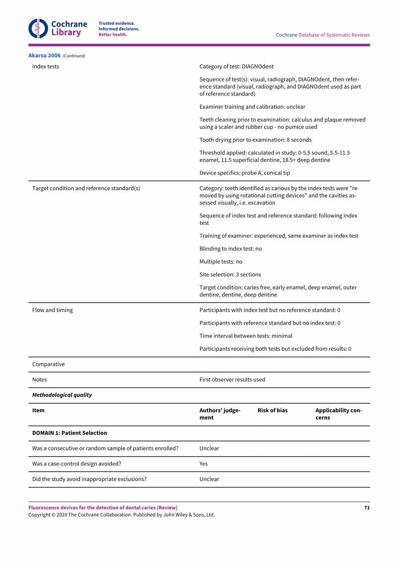
CochraneLibrary
Trusted evidence.Informed decisions.Better health.
Cochrane Database of Systematic Reviews
Index tests Category of test: DIAGNOdent
Sequence of test(s): visual, radiograph, DIAGNOdent, then refer-ence standard (visual, radiograph, and DIAGNOdent used as partof reference standard)
Examiner training and calibration: unclear
Teeth cleaning prior to examination: calculus and plaque removedusing a scaler and rubber cup - no pumice used
Tooth drying prior to examination: 8 seconds
Threshold applied: calculated in study: 0-5.5 sound, 5.5-11.5enamel, 11.5 superficial dentine, 18.5+ deep dentine
Device specifics: probe A, conical tip
Target condition and reference standard(s) Category: teeth identified as carious by the index tests were "re-moved by using rotational cutting devices" and the cavities as-sessed visually, i.e. excavation
Sequence of index test and reference standard: following indextest
Training of examiner: experienced, same examiner as index test
Blinding to index test: no
Multiple tests: no
Site selection: 3 sections
Target condition: caries free, early enamel, deep enamel, outerdentine, dentine, deep dentine
Flow and timing Participants with index test but no reference standard: 0
Participants with reference standard but no index test: 0
Time interval between tests: minimal
Participants receiving both tests but excluded from results: 0
Comparative
Notes First observer results used
Methodological quality
Item Authors' judge-ment
Risk of bias Applicability con-cerns
DOMAIN 1: Patient Selection
Was a consecutive or random sample of patients enrolled? Unclear
Was a case-control design avoided? Yes
Did the study avoid inappropriate exclusions? Unclear
Akarsu 2006 (Continued)
Fluorescence devices for the detection of dental caries (Review)
Copyright © 2020 The Cochrane Collaboration. Published by John Wiley & Sons, Ltd.
71
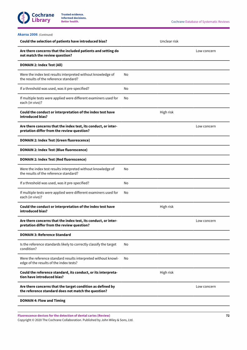
CochraneLibrary
Trusted evidence.Informed decisions.Better health.
Cochrane Database of Systematic Reviews
Could the selection of patients have introduced bias? Unclear risk
Are there concerns that the included patients and setting donot match the review question?
Low concern
DOMAIN 2: Index Test (All)
Were the index test results interpreted without knowledge ofthe results of the reference standard?
No
If a threshold was used, was it pre-specified? No
If multiple tests were applied were different examiners used foreach (in vivo)?
No
Could the conduct or interpretation of the index test haveintroduced bias?
High risk
Are there concerns that the index test, its conduct, or inter-pretation differ from the review question?
Low concern
DOMAIN 2: Index Test (Green fluorescence)
DOMAIN 2: Index Test (Blue fluorescence)
DOMAIN 2: Index Test (Red fluorescence)
Were the index test results interpreted without knowledge ofthe results of the reference standard?
No
If a threshold was used, was it pre-specified? No
If multiple tests were applied were different examiners used foreach (in vivo)?
No
Could the conduct or interpretation of the index test haveintroduced bias?
High risk
Are there concerns that the index test, its conduct, or inter-pretation differ from the review question?
Low concern
DOMAIN 3: Reference Standard
Is the reference standards likely to correctly classify the targetcondition?
No
Were the reference standard results interpreted without knowl-edge of the results of the index tests?
No
Could the reference standard, its conduct, or its interpreta-tion have introduced bias?
High risk
Are there concerns that the target condition as defined bythe reference standard does not match the question?
Low concern
DOMAIN 4: Flow and Timing
Akarsu 2006 (Continued)
Fluorescence devices for the detection of dental caries (Review)
Copyright © 2020 The Cochrane Collaboration. Published by John Wiley & Sons, Ltd.
72
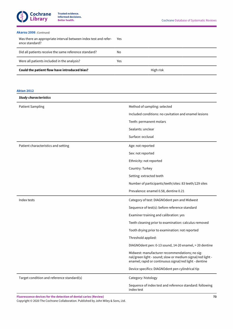
CochraneLibrary
Trusted evidence.Informed decisions.Better health.
Cochrane Database of Systematic Reviews
Was there an appropriate interval between index test and refer-ence standard?
Yes
Did all patients receive the same reference standard? No
Were all patients included in the analysis? Yes
Could the patient flow have introduced bias? High risk
Akarsu 2006 (Continued)
Study characteristics
Patient Sampling Method of sampling: selected
Included conditions: no cavitation and enamel lesions
Teeth: permanent molars
Sealants: unclear
Surface: occlusal
Patient characteristics and setting Age: not reported
Sex: not reported
Ethnicity: not reported
Country: Turkey
Setting: extracted teeth
Number of participants/teeth/sites: 83 teeth/129 sites
Prevalence: enamel 0.58, dentine 0.21
Index tests Category of test: DIAGNOdent pen and Midwest
Sequence of test(s): before reference standard
Examiner training and calibration: yes
Teeth cleaning prior to examination: calculus removed
Tooth drying prior to examination: not reported
Threshold applied:
DIAGNOdent pen: 0-13 sound, 14-20 enamel, > 20 dentine
Midwest: manufacturer recommendations; no sig-nal/green light - sound; slow or medium signal/red light -enamel; rapid or continuous signal/red light - dentine
Device specifics: DIAGNOdent pen cylindrical tip
Target condition and reference standard(s) Category: histology
Sequence of index test and reference standard: followingindex test
Aktan 2012
Fluorescence devices for the detection of dental caries (Review)
Copyright © 2020 The Cochrane Collaboration. Published by John Wiley & Sons, Ltd.
73
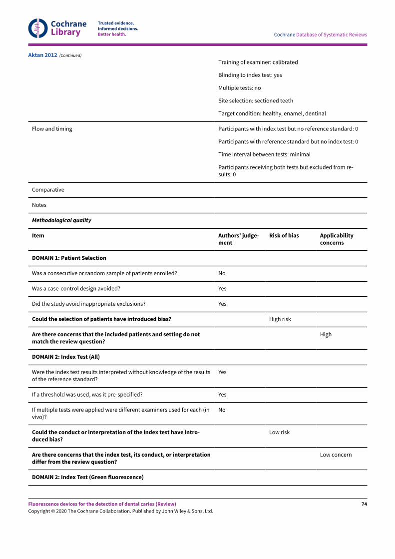
CochraneLibrary
Trusted evidence.Informed decisions.Better health.
Cochrane Database of Systematic Reviews
Training of examiner: calibrated
Blinding to index test: yes
Multiple tests: no
Site selection: sectioned teeth
Target condition: healthy, enamel, dentinal
Flow and timing Participants with index test but no reference standard: 0
Participants with reference standard but no index test: 0
Time interval between tests: minimal
Participants receiving both tests but excluded from re-sults: 0
Comparative
Notes
Methodological quality
Item Authors' judge-ment
Risk of bias Applicabilityconcerns
DOMAIN 1: Patient Selection
Was a consecutive or random sample of patients enrolled? No
Was a case-control design avoided? Yes
Did the study avoid inappropriate exclusions? Yes
Could the selection of patients have introduced bias? High risk
Are there concerns that the included patients and setting do notmatch the review question?
High
DOMAIN 2: Index Test (All)
Were the index test results interpreted without knowledge of the resultsof the reference standard?
Yes
If a threshold was used, was it pre-specified? Yes
If multiple tests were applied were different examiners used for each (invivo)?
No
Could the conduct or interpretation of the index test have intro-duced bias?
Low risk
Are there concerns that the index test, its conduct, or interpretationdiffer from the review question?
Low concern
DOMAIN 2: Index Test (Green fluorescence)
Aktan 2012 (Continued)
Fluorescence devices for the detection of dental caries (Review)
Copyright © 2020 The Cochrane Collaboration. Published by John Wiley & Sons, Ltd.
74
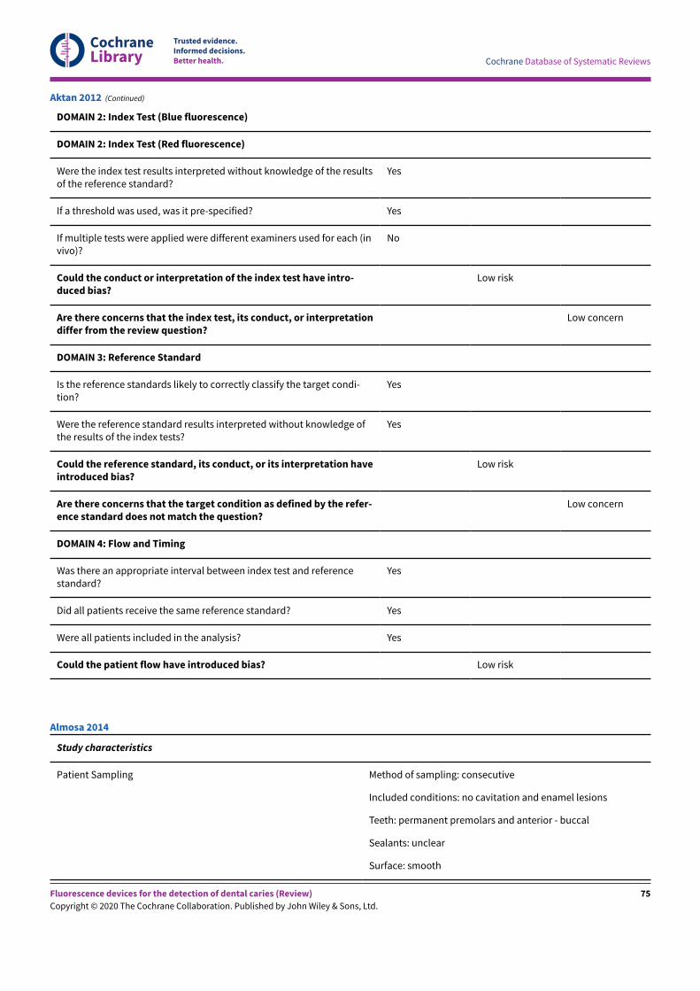
CochraneLibrary
Trusted evidence.Informed decisions.Better health.
Cochrane Database of Systematic Reviews
DOMAIN 2: Index Test (Blue fluorescence)
DOMAIN 2: Index Test (Red fluorescence)
Were the index test results interpreted without knowledge of the resultsof the reference standard?
Yes
If a threshold was used, was it pre-specified? Yes
If multiple tests were applied were different examiners used for each (invivo)?
No
Could the conduct or interpretation of the index test have intro-duced bias?
Low risk
Are there concerns that the index test, its conduct, or interpretationdiffer from the review question?
Low concern
DOMAIN 3: Reference Standard
Is the reference standards likely to correctly classify the target condi-tion?
Yes
Were the reference standard results interpreted without knowledge ofthe results of the index tests?
Yes
Could the reference standard, its conduct, or its interpretation haveintroduced bias?
Low risk
Are there concerns that the target condition as defined by the refer-ence standard does not match the question?
Low concern
DOMAIN 4: Flow and Timing
Was there an appropriate interval between index test and referencestandard?
Yes
Did all patients receive the same reference standard? Yes
Were all patients included in the analysis? Yes
Could the patient flow have introduced bias? Low risk
Aktan 2012 (Continued)
Study characteristics
Patient Sampling Method of sampling: consecutive
Included conditions: no cavitation and enamel lesions
Teeth: permanent premolars and anterior - buccal
Sealants: unclear
Surface: smooth
Almosa 2014
Fluorescence devices for the detection of dental caries (Review)
Copyright © 2020 The Cochrane Collaboration. Published by John Wiley & Sons, Ltd.
75
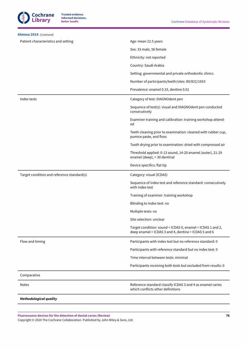
CochraneLibrary
Trusted evidence.Informed decisions.Better health.
Cochrane Database of Systematic Reviews
Patient characteristics and setting Age: mean 22.5 years
Sex: 33 male, 56 female
Ethnicity: not reported
Country: Saudi Arabia
Setting: governmental and private orthodontic clinics
Number of participants/teeth/sites: 89/822/1653
Prevalence: enamel 0.33, dentine 0.01
Index tests Category of test: DIAGNOdent pen
Sequence of test(s): visual and DIAGNOdent pen conductedconsecutively
Examiner training and calibration: training workshop attend-ed
Teeth cleaning prior to examination: cleaned with rubber cup,pumice paste, and floss
Tooth drying prior to examination: dried with compressed air
Threshold applied: 0-13 sound, 14-20 enamel (outer), 21-29enamel (deep), > 30 dentinal
Device specifics: flat tip
Target condition and reference standard(s) Category: visual (ICDAS)
Sequence of index test and reference standard: consecutivelywith index test
Training of examiner: training workshop
Blinding to index test: no
Multiple tests: no
Site selection: unclear
Target condition: sound = ICDAS 0, enamel = ICDAS 1 and 2,deep enamel = ICDAS 3 and 4, dentine = ICDAS 5 and 6
Flow and timing Participants with index test but no reference standard: 0
Participants with reference standard but no index test: 0
Time interval between tests: minimal
Participants receiving both tests but excluded from results: 0
Comparative
Notes Reference standard classify ICDAS 3 and 4 as enamel carieswhich conflicts other definitions
Methodological quality
Almosa 2014 (Continued)
Fluorescence devices for the detection of dental caries (Review)
Copyright © 2020 The Cochrane Collaboration. Published by John Wiley & Sons, Ltd.
76
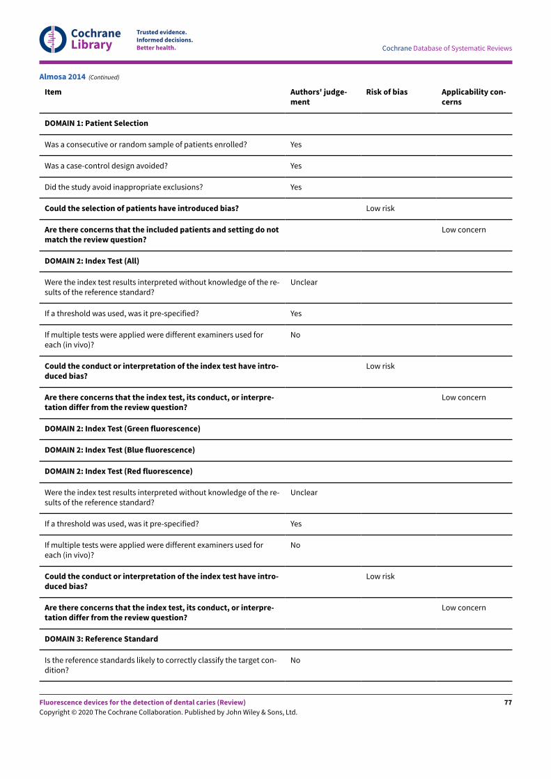
CochraneLibrary
Trusted evidence.Informed decisions.Better health.
Cochrane Database of Systematic Reviews
Item Authors' judge-ment
Risk of bias Applicability con-cerns
DOMAIN 1: Patient Selection
Was a consecutive or random sample of patients enrolled? Yes
Was a case-control design avoided? Yes
Did the study avoid inappropriate exclusions? Yes
Could the selection of patients have introduced bias? Low risk
Are there concerns that the included patients and setting do notmatch the review question?
Low concern
DOMAIN 2: Index Test (All)
Were the index test results interpreted without knowledge of the re-sults of the reference standard?
Unclear
If a threshold was used, was it pre-specified? Yes
If multiple tests were applied were different examiners used foreach (in vivo)?
No
Could the conduct or interpretation of the index test have intro-duced bias?
Low risk
Are there concerns that the index test, its conduct, or interpre-tation differ from the review question?
Low concern
DOMAIN 2: Index Test (Green fluorescence)
DOMAIN 2: Index Test (Blue fluorescence)
DOMAIN 2: Index Test (Red fluorescence)
Were the index test results interpreted without knowledge of the re-sults of the reference standard?
Unclear
If a threshold was used, was it pre-specified? Yes
If multiple tests were applied were different examiners used foreach (in vivo)?
No
Could the conduct or interpretation of the index test have intro-duced bias?
Low risk
Are there concerns that the index test, its conduct, or interpre-tation differ from the review question?
Low concern
DOMAIN 3: Reference Standard
Is the reference standards likely to correctly classify the target con-dition?
No
Almosa 2014 (Continued)
Fluorescence devices for the detection of dental caries (Review)
Copyright © 2020 The Cochrane Collaboration. Published by John Wiley & Sons, Ltd.
77
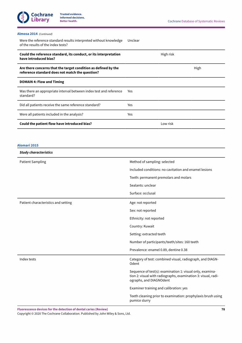
CochraneLibrary
Trusted evidence.Informed decisions.Better health.
Cochrane Database of Systematic Reviews
Were the reference standard results interpreted without knowledgeof the results of the index tests?
Unclear
Could the reference standard, its conduct, or its interpretationhave introduced bias?
High risk
Are there concerns that the target condition as defined by thereference standard does not match the question?
High
DOMAIN 4: Flow and Timing
Was there an appropriate interval between index test and referencestandard?
Yes
Did all patients receive the same reference standard? Yes
Were all patients included in the analysis? Yes
Could the patient flow have introduced bias? Low risk
Almosa 2014 (Continued)
Study characteristics
Patient Sampling Method of sampling: selected
Included conditions: no cavitation and enamel lesions
Teeth: permanent premolars and molars
Sealants: unclear
Surface: occlusal
Patient characteristics and setting Age: not reported
Sex: not reported
Ethnicity: not reported
Country: Kuwait
Setting: extracted teeth
Number of participants/teeth/sites: 160 teeth
Prevalence: enamel 0.89, dentine 0.38
Index tests Category of test: combined visual, radiograph, and DIAGN-Odent
Sequence of test(s): examination 1: visual only, examina-tion 2: visual with radiographs, examination 3: visual, radi-ographs, and DIAGNOdent
Examiner training and calibration: yes
Teeth cleaning prior to examination: prophylaxis brush usingpumice slurry
Alomari 2015
Fluorescence devices for the detection of dental caries (Review)
Copyright © 2020 The Cochrane Collaboration. Published by John Wiley & Sons, Ltd.
78
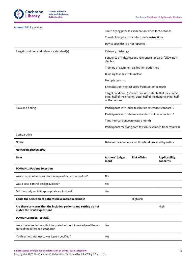
CochraneLibrary
Trusted evidence.Informed decisions.Better health.
Cochrane Database of Systematic Reviews
Tooth drying prior to examination: dried for 5 seconds
Threshold applied: manufacturer's instructions
Device specifics: tip not reported
Target condition and reference standard(s) Category: histology
Sequence of index test and reference standard: following in-dex test
Training of examiner: calibration performed
Blinding to index test: unclear
Multiple tests: no
Site selection: highest score from sectioned tooth
Target condition: (Downer): sound, outer half of the enamel,inner half of the enamel, outer half of the dentine, inner halfof the dentine
Flow and timing Participants with index test but no reference standard: 0
Participants with reference standard but no index test: 0
Time interval between tests: 1 month
Participants receiving both tests but excluded from results: 0
Comparative
Notes Data for the enamel caries threshold provided by author
Methodological quality
Item Authors' judge-ment
Risk of bias Applicabilityconcerns
DOMAIN 1: Patient Selection
Was a consecutive or random sample of patients enrolled? No
Was a case-control design avoided? Yes
Did the study avoid inappropriate exclusions? Yes
Could the selection of patients have introduced bias? High risk
Are there concerns that the included patients and setting do notmatch the review question?
High
DOMAIN 2: Index Test (All)
Were the index test results interpreted without knowledge of the re-sults of the reference standard?
Yes
If a threshold was used, was it pre-specified? Yes
Alomari 2015 (Continued)
Fluorescence devices for the detection of dental caries (Review)
Copyright © 2020 The Cochrane Collaboration. Published by John Wiley & Sons, Ltd.
79
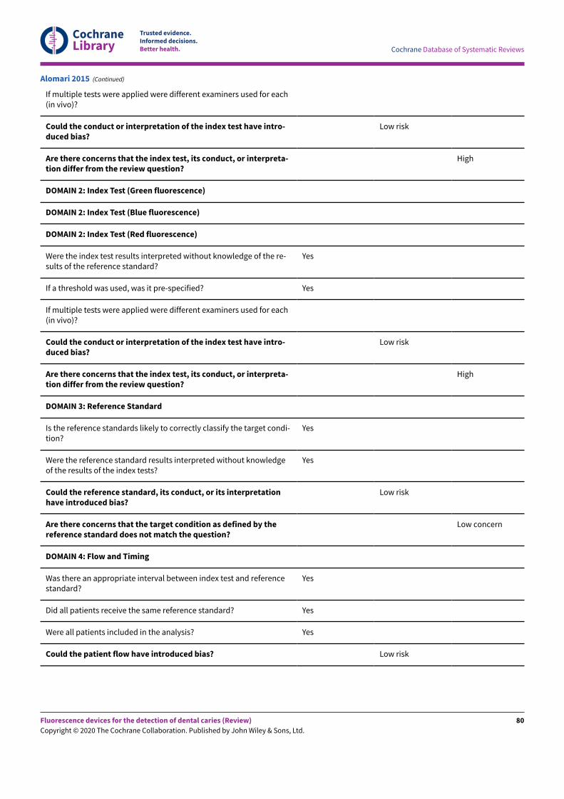
CochraneLibrary
Trusted evidence.Informed decisions.Better health.
Cochrane Database of Systematic Reviews
If multiple tests were applied were different examiners used for each(in vivo)?
Could the conduct or interpretation of the index test have intro-duced bias?
Low risk
Are there concerns that the index test, its conduct, or interpreta-tion differ from the review question?
High
DOMAIN 2: Index Test (Green fluorescence)
DOMAIN 2: Index Test (Blue fluorescence)
DOMAIN 2: Index Test (Red fluorescence)
Were the index test results interpreted without knowledge of the re-sults of the reference standard?
Yes
If a threshold was used, was it pre-specified? Yes
If multiple tests were applied were different examiners used for each(in vivo)?
Could the conduct or interpretation of the index test have intro-duced bias?
Low risk
Are there concerns that the index test, its conduct, or interpreta-tion differ from the review question?
High
DOMAIN 3: Reference Standard
Is the reference standards likely to correctly classify the target condi-tion?
Yes
Were the reference standard results interpreted without knowledgeof the results of the index tests?
Yes
Could the reference standard, its conduct, or its interpretationhave introduced bias?
Low risk
Are there concerns that the target condition as defined by thereference standard does not match the question?
Low concern
DOMAIN 4: Flow and Timing
Was there an appropriate interval between index test and referencestandard?
Yes
Did all patients receive the same reference standard? Yes
Were all patients included in the analysis? Yes
Could the patient flow have introduced bias? Low risk
Alomari 2015 (Continued)
Fluorescence devices for the detection of dental caries (Review)
Copyright © 2020 The Cochrane Collaboration. Published by John Wiley & Sons, Ltd.
80
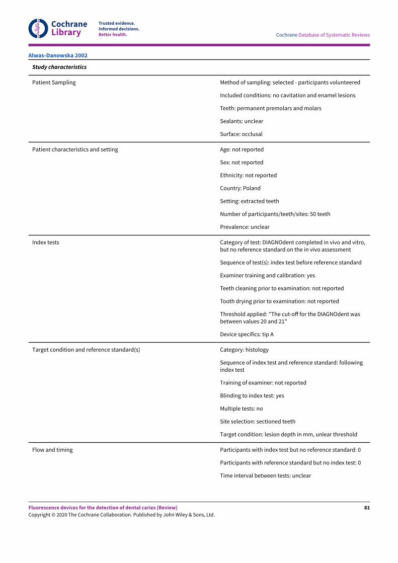
CochraneLibrary
Trusted evidence.Informed decisions.Better health.
Cochrane Database of Systematic Reviews
Study characteristics
Patient Sampling Method of sampling: selected - participants volunteered
Included conditions: no cavitation and enamel lesions
Teeth: permanent premolars and molars
Sealants: unclear
Surface: occlusal
Patient characteristics and setting Age: not reported
Sex: not reported
Ethnicity: not reported
Country: Poland
Setting: extracted teeth
Number of participants/teeth/sites: 50 teeth
Prevalence: unclear
Index tests Category of test: DIAGNOdent completed in vivo and vitro,but no reference standard on the in vivo assessment
Sequence of test(s): index test before reference standard
Examiner training and calibration: yes
Teeth cleaning prior to examination: not reported
Tooth drying prior to examination: not reported
Threshold applied: "The cut-oE for the DIAGNOdent wasbetween values 20 and 21"
Device specifics: tip A
Target condition and reference standard(s) Category: histology
Sequence of index test and reference standard: followingindex test
Training of examiner: not reported
Blinding to index test: yes
Multiple tests: no
Site selection: sectioned teeth
Target condition: lesion depth in mm, unlear threshold
Flow and timing Participants with index test but no reference standard: 0
Participants with reference standard but no index test: 0
Time interval between tests: unclear
Alwas-Danowska 2002
Fluorescence devices for the detection of dental caries (Review)
Copyright © 2020 The Cochrane Collaboration. Published by John Wiley & Sons, Ltd.
81
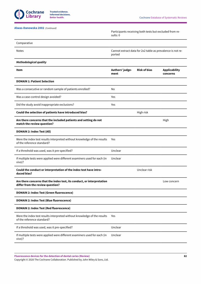
CochraneLibrary
Trusted evidence.Informed decisions.Better health.
Cochrane Database of Systematic Reviews
Participants receiving both tests but excluded from re-sults: 0
Comparative
Notes Cannot extract data for 2x2 table as prevalence is not re-ported
Methodological quality
Item Authors' judge-ment
Risk of bias Applicabilityconcerns
DOMAIN 1: Patient Selection
Was a consecutive or random sample of patients enrolled? No
Was a case-control design avoided? Yes
Did the study avoid inappropriate exclusions? Yes
Could the selection of patients have introduced bias? High risk
Are there concerns that the included patients and setting do notmatch the review question?
High
DOMAIN 2: Index Test (All)
Were the index test results interpreted without knowledge of the resultsof the reference standard?
Yes
If a threshold was used, was it pre-specified? Unclear
If multiple tests were applied were different examiners used for each (invivo)?
Unclear
Could the conduct or interpretation of the index test have intro-duced bias?
Unclear risk
Are there concerns that the index test, its conduct, or interpretationdiffer from the review question?
Low concern
DOMAIN 2: Index Test (Green fluorescence)
DOMAIN 2: Index Test (Blue fluorescence)
DOMAIN 2: Index Test (Red fluorescence)
Were the index test results interpreted without knowledge of the resultsof the reference standard?
Yes
If a threshold was used, was it pre-specified? Unclear
If multiple tests were applied were different examiners used for each (invivo)?
Unclear
Alwas-Danowska 2002 (Continued)
Fluorescence devices for the detection of dental caries (Review)
Copyright © 2020 The Cochrane Collaboration. Published by John Wiley & Sons, Ltd.
82
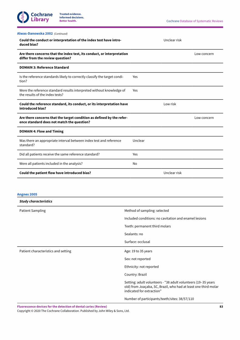
CochraneLibrary
Trusted evidence.Informed decisions.Better health.
Cochrane Database of Systematic Reviews
Could the conduct or interpretation of the index test have intro-duced bias?
Unclear risk
Are there concerns that the index test, its conduct, or interpretationdiffer from the review question?
Low concern
DOMAIN 3: Reference Standard
Is the reference standards likely to correctly classify the target condi-tion?
Yes
Were the reference standard results interpreted without knowledge ofthe results of the index tests?
Yes
Could the reference standard, its conduct, or its interpretation haveintroduced bias?
Low risk
Are there concerns that the target condition as defined by the refer-ence standard does not match the question?
Low concern
DOMAIN 4: Flow and Timing
Was there an appropriate interval between index test and referencestandard?
Unclear
Did all patients receive the same reference standard? Yes
Were all patients included in the analysis? No
Could the patient flow have introduced bias? Unclear risk
Alwas-Danowska 2002 (Continued)
Study characteristics
Patient Sampling Method of sampling: selected
Included conditions: no cavitation and enamel lesions
Teeth: permanent third molars
Sealants: no
Surface: occlusal
Patient characteristics and setting Age: 19 to 35 years
Sex: not reported
Ethnicity: not reported
Country: Brazil
Setting: adult volunteers - "38 adult volunteers (19–35 yearsold) from Joaçaba, SC, Brazil, who had at least one third molarindicated for extraction"
Number of participants/teeth/sites: 38/57/110
Angnes 2005
Fluorescence devices for the detection of dental caries (Review)
Copyright © 2020 The Cochrane Collaboration. Published by John Wiley & Sons, Ltd.
83
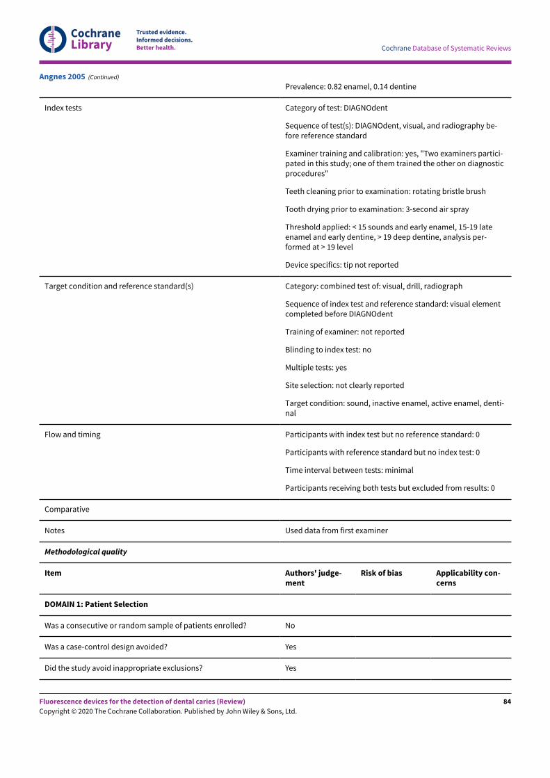
CochraneLibrary
Trusted evidence.Informed decisions.Better health.
Cochrane Database of Systematic Reviews
Prevalence: 0.82 enamel, 0.14 dentine
Index tests Category of test: DIAGNOdent
Sequence of test(s): DIAGNOdent, visual, and radiography be-fore reference standard
Examiner training and calibration: yes, "Two examiners partici-pated in this study; one of them trained the other on diagnosticprocedures"
Teeth cleaning prior to examination: rotating bristle brush
Tooth drying prior to examination: 3-second air spray
Threshold applied: < 15 sounds and early enamel, 15-19 lateenamel and early dentine, > 19 deep dentine, analysis per-formed at > 19 level
Device specifics: tip not reported
Target condition and reference standard(s) Category: combined test of: visual, drill, radiograph
Sequence of index test and reference standard: visual elementcompleted before DIAGNOdent
Training of examiner: not reported
Blinding to index test: no
Multiple tests: yes
Site selection: not clearly reported
Target condition: sound, inactive enamel, active enamel, denti-nal
Flow and timing Participants with index test but no reference standard: 0
Participants with reference standard but no index test: 0
Time interval between tests: minimal
Participants receiving both tests but excluded from results: 0
Comparative
Notes Used data from first examiner
Methodological quality
Item Authors' judge-ment
Risk of bias Applicability con-cerns
DOMAIN 1: Patient Selection
Was a consecutive or random sample of patients enrolled? No
Was a case-control design avoided? Yes
Did the study avoid inappropriate exclusions? Yes
Angnes 2005 (Continued)
Fluorescence devices for the detection of dental caries (Review)
Copyright © 2020 The Cochrane Collaboration. Published by John Wiley & Sons, Ltd.
84
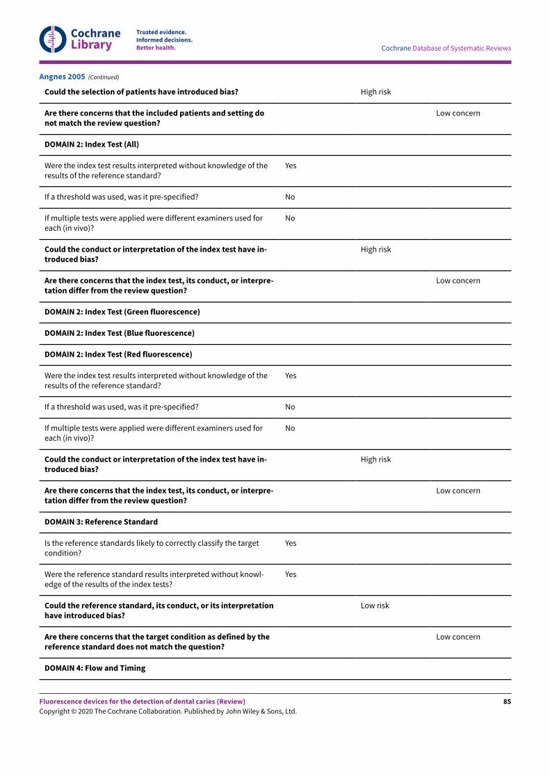
CochraneLibrary
Trusted evidence.Informed decisions.Better health.
Cochrane Database of Systematic Reviews
Could the selection of patients have introduced bias? High risk
Are there concerns that the included patients and setting donot match the review question?
Low concern
DOMAIN 2: Index Test (All)
Were the index test results interpreted without knowledge of theresults of the reference standard?
Yes
If a threshold was used, was it pre-specified? No
If multiple tests were applied were different examiners used foreach (in vivo)?
No
Could the conduct or interpretation of the index test have in-troduced bias?
High risk
Are there concerns that the index test, its conduct, or interpre-tation differ from the review question?
Low concern
DOMAIN 2: Index Test (Green fluorescence)
DOMAIN 2: Index Test (Blue fluorescence)
DOMAIN 2: Index Test (Red fluorescence)
Were the index test results interpreted without knowledge of theresults of the reference standard?
Yes
If a threshold was used, was it pre-specified? No
If multiple tests were applied were different examiners used foreach (in vivo)?
No
Could the conduct or interpretation of the index test have in-troduced bias?
High risk
Are there concerns that the index test, its conduct, or interpre-tation differ from the review question?
Low concern
DOMAIN 3: Reference Standard
Is the reference standards likely to correctly classify the targetcondition?
Yes
Were the reference standard results interpreted without knowl-edge of the results of the index tests?
Yes
Could the reference standard, its conduct, or its interpretationhave introduced bias?
Low risk
Are there concerns that the target condition as defined by thereference standard does not match the question?
Low concern
DOMAIN 4: Flow and Timing
Angnes 2005 (Continued)
Fluorescence devices for the detection of dental caries (Review)
Copyright © 2020 The Cochrane Collaboration. Published by John Wiley & Sons, Ltd.
85
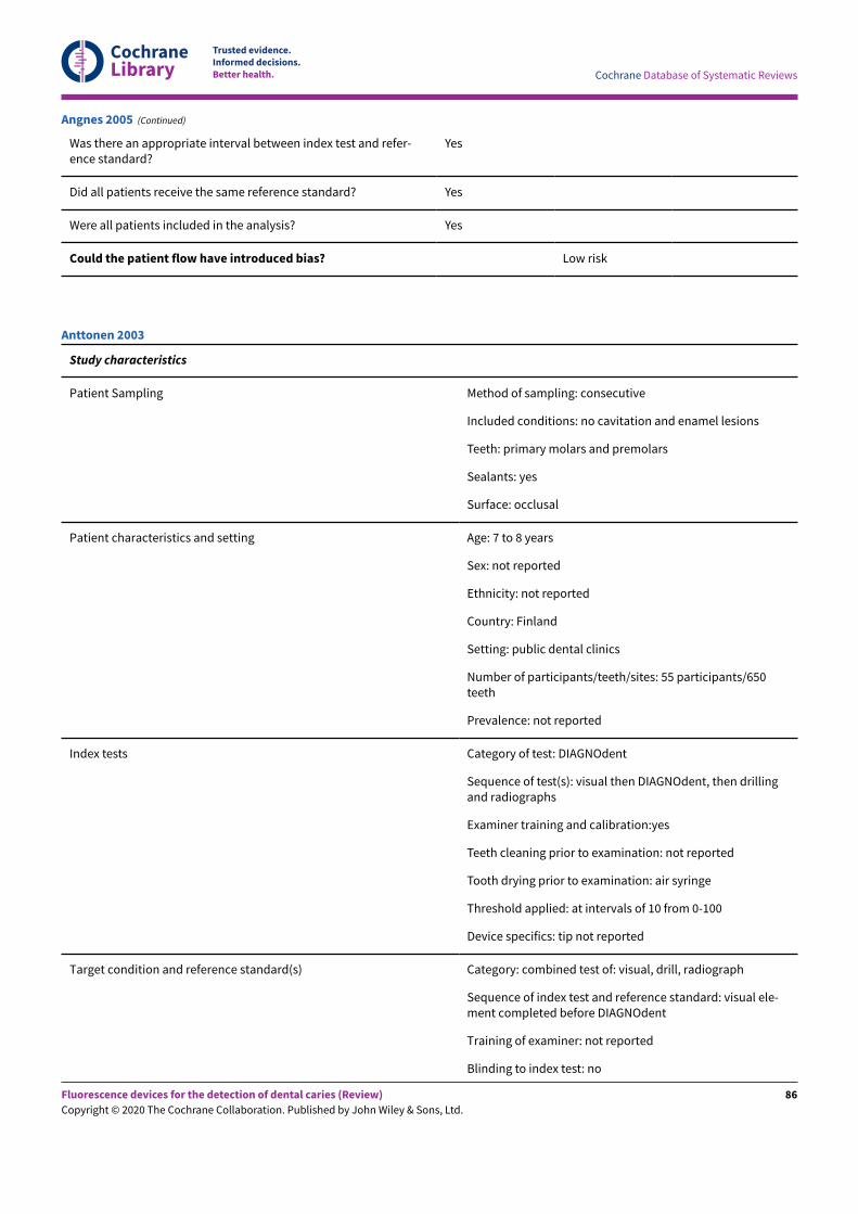
CochraneLibrary
Trusted evidence.Informed decisions.Better health.
Cochrane Database of Systematic Reviews
Was there an appropriate interval between index test and refer-ence standard?
Yes
Did all patients receive the same reference standard? Yes
Were all patients included in the analysis? Yes
Could the patient flow have introduced bias? Low risk
Angnes 2005 (Continued)
Study characteristics
Patient Sampling Method of sampling: consecutive
Included conditions: no cavitation and enamel lesions
Teeth: primary molars and premolars
Sealants: yes
Surface: occlusal
Patient characteristics and setting Age: 7 to 8 years
Sex: not reported
Ethnicity: not reported
Country: Finland
Setting: public dental clinics
Number of participants/teeth/sites: 55 participants/650teeth
Prevalence: not reported
Index tests Category of test: DIAGNOdent
Sequence of test(s): visual then DIAGNOdent, then drillingand radiographs
Examiner training and calibration:yes
Teeth cleaning prior to examination: not reported
Tooth drying prior to examination: air syringe
Threshold applied: at intervals of 10 from 0-100
Device specifics: tip not reported
Target condition and reference standard(s) Category: combined test of: visual, drill, radiograph
Sequence of index test and reference standard: visual ele-ment completed before DIAGNOdent
Training of examiner: not reported
Blinding to index test: no
Anttonen 2003
Fluorescence devices for the detection of dental caries (Review)
Copyright © 2020 The Cochrane Collaboration. Published by John Wiley & Sons, Ltd.
86
CochraneLibrary
Trusted evidence.Informed decisions.Better health.
Cochrane Database of Systematic Reviews
Multiple tests: yes
Site selection: not clearly reported
Target condition: sound, inactive enamel, active enamel,dentinal
Flow and timing Participants with index test but no reference standard: un-clear
Participants with reference standard but no index test: un-clear
Time interval between tests: minimal
Participants receiving both tests but excluded from results:unclear
Comparative
Notes Unclear reporting of data. Primary teeth had visual andDIAGNOdent only. Permanent had excavation and radi-ograph, but unclear on numbers of who receiving tests
Methodological quality
Item Authors' judge-ment
Risk of bias Applicabilityconcerns
DOMAIN 1: Patient Selection
Was a consecutive or random sample of patients enrolled? Yes
Was a case-control design avoided? Yes
Did the study avoid inappropriate exclusions? No
Could the selection of patients have introduced bias? Low risk
Are there concerns that the included patients and setting do notmatch the review question?
Low concern
DOMAIN 2: Index Test (All)
Were the index test results interpreted without knowledge of the re-sults of the reference standard?
No
If a threshold was used, was it pre-specified? No
If multiple tests were applied were different examiners used for each(in vivo)?
No
Could the conduct or interpretation of the index test have intro-duced bias?
High risk
Are there concerns that the index test, its conduct, or interpreta-tion differ from the review question?
High
DOMAIN 2: Index Test (Green fluorescence)
Anttonen 2003 (Continued)
Fluorescence devices for the detection of dental caries (Review)
Copyright © 2020 The Cochrane Collaboration. Published by John Wiley & Sons, Ltd.
87
CochraneLibrary
Trusted evidence.Informed decisions.Better health.
Cochrane Database of Systematic Reviews
DOMAIN 2: Index Test (Blue fluorescence)
DOMAIN 2: Index Test (Red fluorescence)
Were the index test results interpreted without knowledge of the re-sults of the reference standard?
No
If a threshold was used, was it pre-specified? No
If multiple tests were applied were different examiners used for each(in vivo)?
No
Could the conduct or interpretation of the index test have intro-duced bias?
High risk
Are there concerns that the index test, its conduct, or interpreta-tion differ from the review question?
High
DOMAIN 3: Reference Standard
Is the reference standards likely to correctly classify the target condi-tion?
No
Were the reference standard results interpreted without knowledge ofthe results of the index tests?
No
Could the reference standard, its conduct, or its interpretationhave introduced bias?
High risk
Are there concerns that the target condition as defined by the ref-erence standard does not match the question?
High
DOMAIN 4: Flow and Timing
Was there an appropriate interval between index test and referencestandard?
Yes
Did all patients receive the same reference standard? No
Were all patients included in the analysis? Unclear
Could the patient flow have introduced bias? High risk
Anttonen 2003 (Continued)
Study characteristics
Patient Sampling Method of sampling: selected
Included conditions: no cavitation and early lesions
Teeth: primary molars
Sealants: no
Surface: occlusal
Apostolopoulou 2009
Fluorescence devices for the detection of dental caries (Review)
Copyright © 2020 The Cochrane Collaboration. Published by John Wiley & Sons, Ltd.
88
CochraneLibrary
Trusted evidence.Informed decisions.Better health.
Cochrane Database of Systematic Reviews
Patient characteristics and setting Age: not reported
Sex: not reported
Ethnicity: not reported
Country: Greece
Setting: extracted teeth
Number of participants/teeth/sites: 24 teeth/111 sites
Prevalence: enamel 0.98, dentine 0.22
Index tests Category of test: DIAGNOdent
Sequence of test(s): index tests (visual, radiograph, andDIAGNOdent) performed prior to reference standard
Examiner training and calibration: not reported
Teeth cleaning prior to examination: toothbrush andpumice-free paste
Tooth drying prior to examination: air dried 5 seconds
Threshold applied: converted scale, unclear, "the originalDD readings on the 0-99 scale were converted, using Cron-bach’s A coefficient alpha, to the 0, 1 and 2 caries scoringscale used by all other methods"
Device specifics: tip A
Target condition and reference standard(s) Category: histology
Sequence of index test and reference standard: index testthen reference standard
Training of examiner: not reported
Blinding to index test: unclear
Multiple tests: no
Site selection: sectioned teeth
Target condition: sound, enamel, dentine
Flow and timing Participants with index test but no reference standard: 0
Participants with reference standard but no index test: 0
Time interval between tests: minimal
Participants receiving both tests but excluded from re-sults: 0
Comparative
Notes
Methodological quality
Apostolopoulou 2009 (Continued)
Fluorescence devices for the detection of dental caries (Review)
Copyright © 2020 The Cochrane Collaboration. Published by John Wiley & Sons, Ltd.
89
CochraneLibrary
Trusted evidence.Informed decisions.Better health.
Cochrane Database of Systematic Reviews
Item Authors' judge-ment
Risk of bias Applicabilityconcerns
DOMAIN 1: Patient Selection
Was a consecutive or random sample of patients enrolled? No
Was a case-control design avoided? Yes
Did the study avoid inappropriate exclusions? Yes
Could the selection of patients have introduced bias? High risk
Are there concerns that the included patients and setting do notmatch the review question?
High
DOMAIN 2: Index Test (All)
Were the index test results interpreted without knowledge of the resultsof the reference standard?
Yes
If a threshold was used, was it pre-specified? Unclear
If multiple tests were applied were different examiners used for each (invivo)?
No
Could the conduct or interpretation of the index test have intro-duced bias?
Unclear risk
Are there concerns that the index test, its conduct, or interpretationdiffer from the review question?
Low concern
DOMAIN 2: Index Test (Green fluorescence)
DOMAIN 2: Index Test (Blue fluorescence)
DOMAIN 2: Index Test (Red fluorescence)
Were the index test results interpreted without knowledge of the resultsof the reference standard?
Yes
If a threshold was used, was it pre-specified? Unclear
If multiple tests were applied were different examiners used for each (invivo)?
No
Could the conduct or interpretation of the index test have intro-duced bias?
Unclear risk
Are there concerns that the index test, its conduct, or interpretationdiffer from the review question?
Low concern
DOMAIN 3: Reference Standard
Is the reference standards likely to correctly classify the target condi-tion?
Yes
Apostolopoulou 2009 (Continued)
Fluorescence devices for the detection of dental caries (Review)
Copyright © 2020 The Cochrane Collaboration. Published by John Wiley & Sons, Ltd.
90
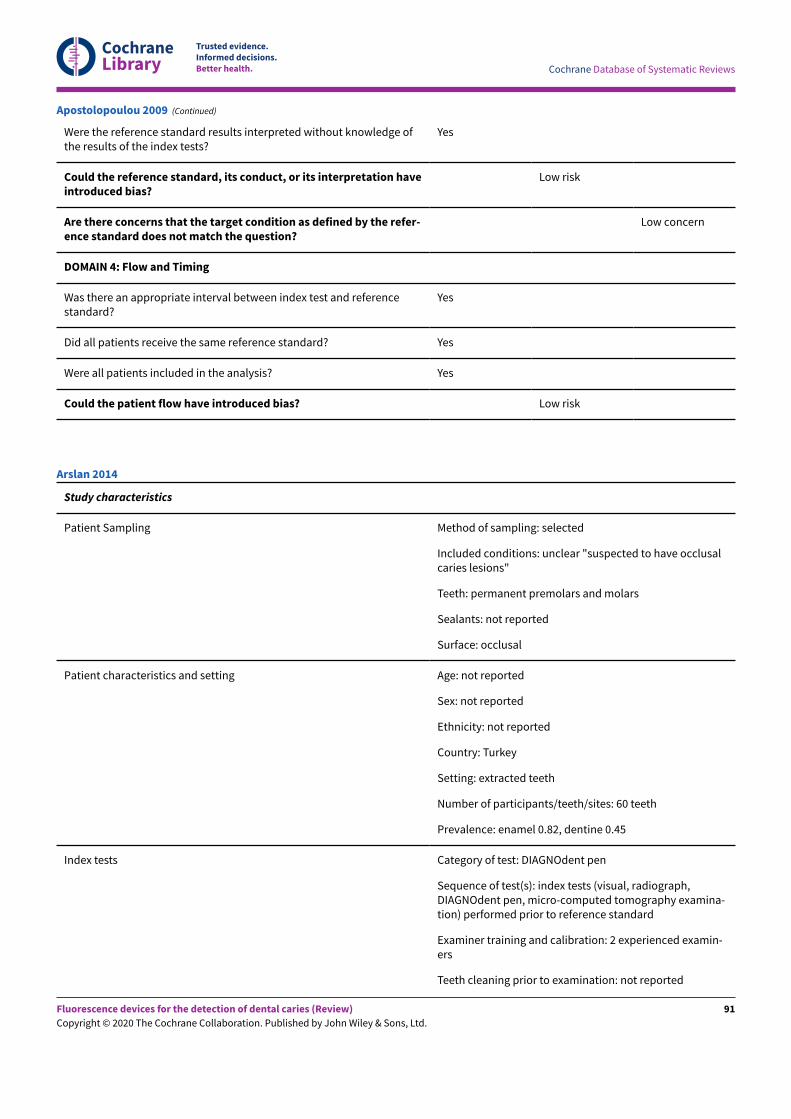
CochraneLibrary
Trusted evidence.Informed decisions.Better health.
Cochrane Database of Systematic Reviews
Were the reference standard results interpreted without knowledge ofthe results of the index tests?
Yes
Could the reference standard, its conduct, or its interpretation haveintroduced bias?
Low risk
Are there concerns that the target condition as defined by the refer-ence standard does not match the question?
Low concern
DOMAIN 4: Flow and Timing
Was there an appropriate interval between index test and referencestandard?
Yes
Did all patients receive the same reference standard? Yes
Were all patients included in the analysis? Yes
Could the patient flow have introduced bias? Low risk
Apostolopoulou 2009 (Continued)
Study characteristics
Patient Sampling Method of sampling: selected
Included conditions: unclear "suspected to have occlusalcaries lesions"
Teeth: permanent premolars and molars
Sealants: not reported
Surface: occlusal
Patient characteristics and setting Age: not reported
Sex: not reported
Ethnicity: not reported
Country: Turkey
Setting: extracted teeth
Number of participants/teeth/sites: 60 teeth
Prevalence: enamel 0.82, dentine 0.45
Index tests Category of test: DIAGNOdent pen
Sequence of test(s): index tests (visual, radiograph,DIAGNOdent pen, micro-computed tomography examina-tion) performed prior to reference standard
Examiner training and calibration: 2 experienced examin-ers
Teeth cleaning prior to examination: not reported
Arslan 2014
Fluorescence devices for the detection of dental caries (Review)
Copyright © 2020 The Cochrane Collaboration. Published by John Wiley & Sons, Ltd.
91
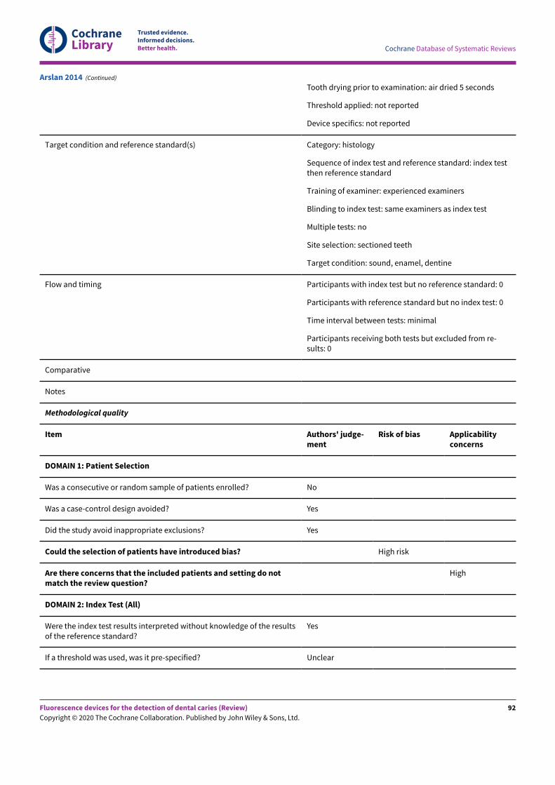
CochraneLibrary
Trusted evidence.Informed decisions.Better health.
Cochrane Database of Systematic Reviews
Tooth drying prior to examination: air dried 5 seconds
Threshold applied: not reported
Device specifics: not reported
Target condition and reference standard(s) Category: histology
Sequence of index test and reference standard: index testthen reference standard
Training of examiner: experienced examiners
Blinding to index test: same examiners as index test
Multiple tests: no
Site selection: sectioned teeth
Target condition: sound, enamel, dentine
Flow and timing Participants with index test but no reference standard: 0
Participants with reference standard but no index test: 0
Time interval between tests: minimal
Participants receiving both tests but excluded from re-sults: 0
Comparative
Notes
Methodological quality
Item Authors' judge-ment
Risk of bias Applicabilityconcerns
DOMAIN 1: Patient Selection
Was a consecutive or random sample of patients enrolled? No
Was a case-control design avoided? Yes
Did the study avoid inappropriate exclusions? Yes
Could the selection of patients have introduced bias? High risk
Are there concerns that the included patients and setting do notmatch the review question?
High
DOMAIN 2: Index Test (All)
Were the index test results interpreted without knowledge of the resultsof the reference standard?
Yes
If a threshold was used, was it pre-specified? Unclear
Arslan 2014 (Continued)
Fluorescence devices for the detection of dental caries (Review)
Copyright © 2020 The Cochrane Collaboration. Published by John Wiley & Sons, Ltd.
92
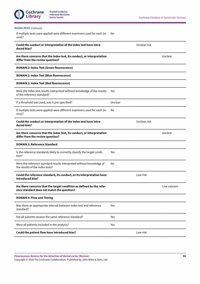
CochraneLibrary
Trusted evidence.Informed decisions.Better health.
Cochrane Database of Systematic Reviews
If multiple tests were applied were different examiners used for each (invivo)?
No
Could the conduct or interpretation of the index test have intro-duced bias?
Unclear risk
Are there concerns that the index test, its conduct, or interpretationdiffer from the review question?
Unclear
DOMAIN 2: Index Test (Green fluorescence)
DOMAIN 2: Index Test (Blue fluorescence)
DOMAIN 2: Index Test (Red fluorescence)
Were the index test results interpreted without knowledge of the resultsof the reference standard?
Yes
If a threshold was used, was it pre-specified? Unclear
If multiple tests were applied were different examiners used for each (invivo)?
No
Could the conduct or interpretation of the index test have intro-duced bias?
Unclear risk
Are there concerns that the index test, its conduct, or interpretationdiffer from the review question?
Unclear
DOMAIN 3: Reference Standard
Is the reference standards likely to correctly classify the target condi-tion?
Yes
Were the reference standard results interpreted without knowledge ofthe results of the index tests?
No
Could the reference standard, its conduct, or its interpretation haveintroduced bias?
Low risk
Are there concerns that the target condition as defined by the refer-ence standard does not match the question?
Low concern
DOMAIN 4: Flow and Timing
Was there an appropriate interval between index test and referencestandard?
Yes
Did all patients receive the same reference standard? Yes
Were all patients included in the analysis? Yes
Could the patient flow have introduced bias? Low risk
Arslan 2014 (Continued)
Fluorescence devices for the detection of dental caries (Review)
Copyright © 2020 The Cochrane Collaboration. Published by John Wiley & Sons, Ltd.
93
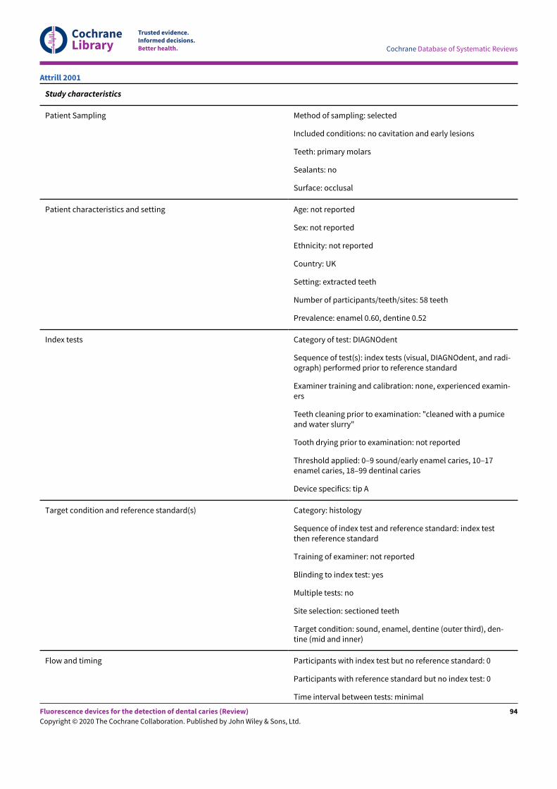
CochraneLibrary
Trusted evidence.Informed decisions.Better health.
Cochrane Database of Systematic Reviews
Study characteristics
Patient Sampling Method of sampling: selected
Included conditions: no cavitation and early lesions
Teeth: primary molars
Sealants: no
Surface: occlusal
Patient characteristics and setting Age: not reported
Sex: not reported
Ethnicity: not reported
Country: UK
Setting: extracted teeth
Number of participants/teeth/sites: 58 teeth
Prevalence: enamel 0.60, dentine 0.52
Index tests Category of test: DIAGNOdent
Sequence of test(s): index tests (visual, DIAGNOdent, and radi-ograph) performed prior to reference standard
Examiner training and calibration: none, experienced examin-ers
Teeth cleaning prior to examination: "cleaned with a pumiceand water slurry"
Tooth drying prior to examination: not reported
Threshold applied: 0–9 sound/early enamel caries, 10–17enamel caries, 18–99 dentinal caries
Device specifics: tip A
Target condition and reference standard(s) Category: histology
Sequence of index test and reference standard: index testthen reference standard
Training of examiner: not reported
Blinding to index test: yes
Multiple tests: no
Site selection: sectioned teeth
Target condition: sound, enamel, dentine (outer third), den-tine (mid and inner)
Flow and timing Participants with index test but no reference standard: 0
Participants with reference standard but no index test: 0
Time interval between tests: minimal
Attrill 2001
Fluorescence devices for the detection of dental caries (Review)
Copyright © 2020 The Cochrane Collaboration. Published by John Wiley & Sons, Ltd.
94
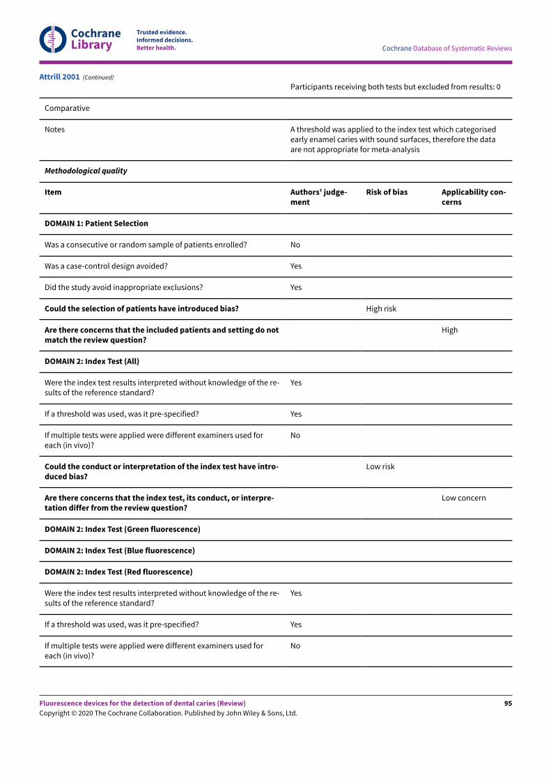
CochraneLibrary
Trusted evidence.Informed decisions.Better health.
Cochrane Database of Systematic Reviews
Participants receiving both tests but excluded from results: 0
Comparative
Notes A threshold was applied to the index test which categorisedearly enamel caries with sound surfaces, therefore the dataare not appropriate for meta-analysis
Methodological quality
Item Authors' judge-ment
Risk of bias Applicability con-cerns
DOMAIN 1: Patient Selection
Was a consecutive or random sample of patients enrolled? No
Was a case-control design avoided? Yes
Did the study avoid inappropriate exclusions? Yes
Could the selection of patients have introduced bias? High risk
Are there concerns that the included patients and setting do notmatch the review question?
High
DOMAIN 2: Index Test (All)
Were the index test results interpreted without knowledge of the re-sults of the reference standard?
Yes
If a threshold was used, was it pre-specified? Yes
If multiple tests were applied were different examiners used foreach (in vivo)?
No
Could the conduct or interpretation of the index test have intro-duced bias?
Low risk
Are there concerns that the index test, its conduct, or interpre-tation differ from the review question?
Low concern
DOMAIN 2: Index Test (Green fluorescence)
DOMAIN 2: Index Test (Blue fluorescence)
DOMAIN 2: Index Test (Red fluorescence)
Were the index test results interpreted without knowledge of the re-sults of the reference standard?
Yes
If a threshold was used, was it pre-specified? Yes
If multiple tests were applied were different examiners used foreach (in vivo)?
No
Attrill 2001 (Continued)
Fluorescence devices for the detection of dental caries (Review)
Copyright © 2020 The Cochrane Collaboration. Published by John Wiley & Sons, Ltd.
95
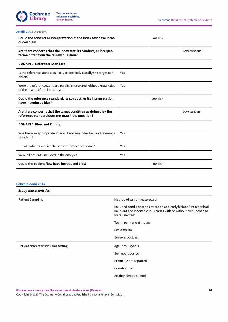
CochraneLibrary
Trusted evidence.Informed decisions.Better health.
Cochrane Database of Systematic Reviews
Could the conduct or interpretation of the index test have intro-duced bias?
Low risk
Are there concerns that the index test, its conduct, or interpre-tation differ from the review question?
Low concern
DOMAIN 3: Reference Standard
Is the reference standards likely to correctly classify the target con-dition?
Yes
Were the reference standard results interpreted without knowledgeof the results of the index tests?
Yes
Could the reference standard, its conduct, or its interpretationhave introduced bias?
Low risk
Are there concerns that the target condition as defined by thereference standard does not match the question?
Low concern
DOMAIN 4: Flow and Timing
Was there an appropriate interval between index test and referencestandard?
Yes
Did all patients receive the same reference standard? Yes
Were all patients included in the analysis? Yes
Could the patient flow have introduced bias? Low risk
Attrill 2001 (Continued)
Study characteristics
Patient Sampling Method of sampling: selected
Included conditions: no cavitation and early lesions "intact or hadincipient and inconspicuous caries with or without colour changewere selected"
Teeth: permanent molars
Sealants: no
Surface: occlusal
Patient characteristics and setting Age: 7 to 13 years
Sex: not reported
Ethnicity: not reported
Country: Iran
Setting: dental school
Bahrololoomi 2015
Fluorescence devices for the detection of dental caries (Review)
Copyright © 2020 The Cochrane Collaboration. Published by John Wiley & Sons, Ltd.
96
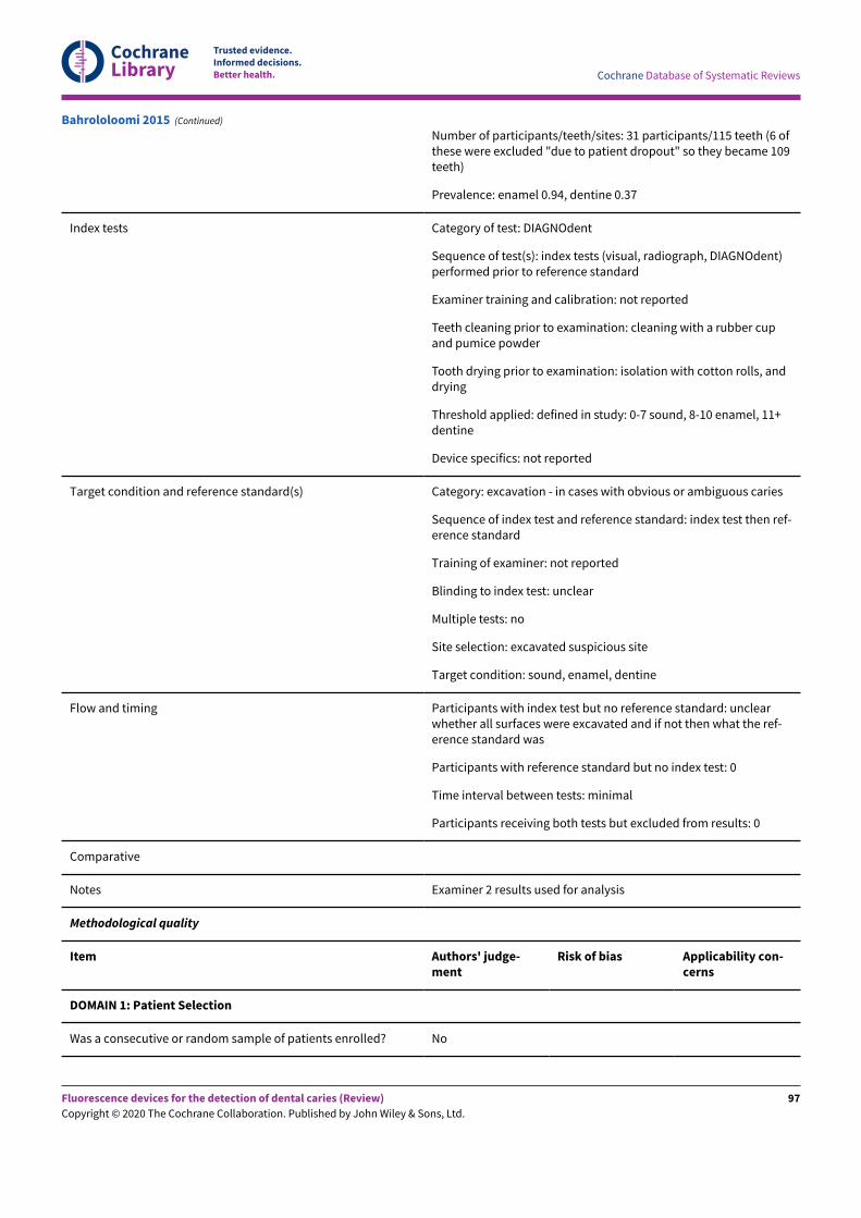
CochraneLibrary
Trusted evidence.Informed decisions.Better health.
Cochrane Database of Systematic Reviews
Number of participants/teeth/sites: 31 participants/115 teeth (6 ofthese were excluded "due to patient dropout" so they became 109teeth)
Prevalence: enamel 0.94, dentine 0.37
Index tests Category of test: DIAGNOdent
Sequence of test(s): index tests (visual, radiograph, DIAGNOdent)performed prior to reference standard
Examiner training and calibration: not reported
Teeth cleaning prior to examination: cleaning with a rubber cupand pumice powder
Tooth drying prior to examination: isolation with cotton rolls, anddrying
Threshold applied: defined in study: 0-7 sound, 8-10 enamel, 11+dentine
Device specifics: not reported
Target condition and reference standard(s) Category: excavation - in cases with obvious or ambiguous caries
Sequence of index test and reference standard: index test then ref-erence standard
Training of examiner: not reported
Blinding to index test: unclear
Multiple tests: no
Site selection: excavated suspicious site
Target condition: sound, enamel, dentine
Flow and timing Participants with index test but no reference standard: unclearwhether all surfaces were excavated and if not then what the ref-erence standard was
Participants with reference standard but no index test: 0
Time interval between tests: minimal
Participants receiving both tests but excluded from results: 0
Comparative
Notes Examiner 2 results used for analysis
Methodological quality
Item Authors' judge-ment
Risk of bias Applicability con-cerns
DOMAIN 1: Patient Selection
Was a consecutive or random sample of patients enrolled? No
Bahrololoomi 2015 (Continued)
Fluorescence devices for the detection of dental caries (Review)
Copyright © 2020 The Cochrane Collaboration. Published by John Wiley & Sons, Ltd.
97
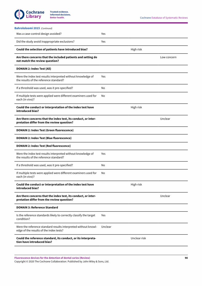
CochraneLibrary
Trusted evidence.Informed decisions.Better health.
Cochrane Database of Systematic Reviews
Was a case-control design avoided? Yes
Did the study avoid inappropriate exclusions? Yes
Could the selection of patients have introduced bias? High risk
Are there concerns that the included patients and setting donot match the review question?
Low concern
DOMAIN 2: Index Test (All)
Were the index test results interpreted without knowledge ofthe results of the reference standard?
Yes
If a threshold was used, was it pre-specified? No
If multiple tests were applied were different examiners used foreach (in vivo)?
No
Could the conduct or interpretation of the index test haveintroduced bias?
High risk
Are there concerns that the index test, its conduct, or inter-pretation differ from the review question?
Unclear
DOMAIN 2: Index Test (Green fluorescence)
DOMAIN 2: Index Test (Blue fluorescence)
DOMAIN 2: Index Test (Red fluorescence)
Were the index test results interpreted without knowledge ofthe results of the reference standard?
Yes
If a threshold was used, was it pre-specified? No
If multiple tests were applied were different examiners used foreach (in vivo)?
No
Could the conduct or interpretation of the index test haveintroduced bias?
High risk
Are there concerns that the index test, its conduct, or inter-pretation differ from the review question?
Unclear
DOMAIN 3: Reference Standard
Is the reference standards likely to correctly classify the targetcondition?
Yes
Were the reference standard results interpreted without knowl-edge of the results of the index tests?
Unclear
Could the reference standard, its conduct, or its interpreta-tion have introduced bias?
Unclear risk
Bahrololoomi 2015 (Continued)
Fluorescence devices for the detection of dental caries (Review)
Copyright © 2020 The Cochrane Collaboration. Published by John Wiley & Sons, Ltd.
98
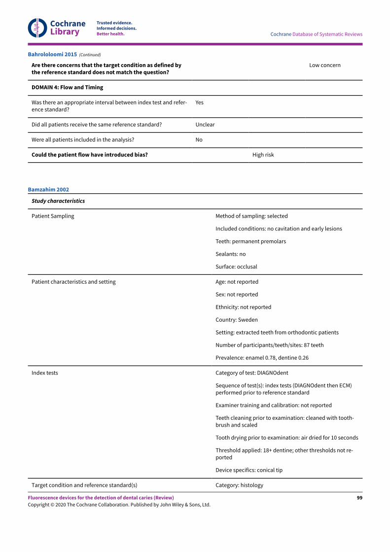
CochraneLibrary
Trusted evidence.Informed decisions.Better health.
Cochrane Database of Systematic Reviews
Are there concerns that the target condition as defined bythe reference standard does not match the question?
Low concern
DOMAIN 4: Flow and Timing
Was there an appropriate interval between index test and refer-ence standard?
Yes
Did all patients receive the same reference standard? Unclear
Were all patients included in the analysis? No
Could the patient flow have introduced bias? High risk
Bahrololoomi 2015 (Continued)
Study characteristics
Patient Sampling Method of sampling: selected
Included conditions: no cavitation and early lesions
Teeth: permanent premolars
Sealants: no
Surface: occlusal
Patient characteristics and setting Age: not reported
Sex: not reported
Ethnicity: not reported
Country: Sweden
Setting: extracted teeth from orthodontic patients
Number of participants/teeth/sites: 87 teeth
Prevalence: enamel 0.78, dentine 0.26
Index tests Category of test: DIAGNOdent
Sequence of test(s): index tests (DIAGNOdent then ECM)performed prior to reference standard
Examiner training and calibration: not reported
Teeth cleaning prior to examination: cleaned with tooth-brush and scaled
Tooth drying prior to examination: air dried for 10 seconds
Threshold applied: 18+ dentine; other thresholds not re-ported
Device specifics: conical tip
Target condition and reference standard(s) Category: histology
Bamzahim 2002
Fluorescence devices for the detection of dental caries (Review)
Copyright © 2020 The Cochrane Collaboration. Published by John Wiley & Sons, Ltd.
99
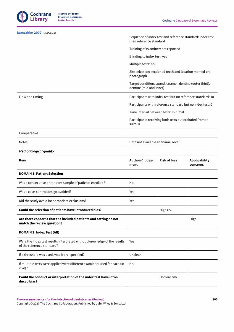
CochraneLibrary
Trusted evidence.Informed decisions.Better health.
Cochrane Database of Systematic Reviews
Sequence of index test and reference standard: index testthen reference standard
Training of examiner: not reported
Blinding to index test: yes
Multiple tests: no
Site selection: sectioned teeth and location marked onphotograph
Target condition: sound, enamel, dentine (outer third),dentine (mid and inner)
Flow and timing Participants with index test but no reference standard: 10
Participants with reference standard but no index test: 0
Time interval between tests: minimal
Participants receiving both tests but excluded from re-sults: 0
Comparative
Notes Data not available at enamel level
Methodological quality
Item Authors' judge-ment
Risk of bias Applicabilityconcerns
DOMAIN 1: Patient Selection
Was a consecutive or random sample of patients enrolled? No
Was a case-control design avoided? Yes
Did the study avoid inappropriate exclusions? Yes
Could the selection of patients have introduced bias? High risk
Are there concerns that the included patients and setting do notmatch the review question?
High
DOMAIN 2: Index Test (All)
Were the index test results interpreted without knowledge of the resultsof the reference standard?
Yes
If a threshold was used, was it pre-specified? Unclear
If multiple tests were applied were different examiners used for each (invivo)?
No
Could the conduct or interpretation of the index test have intro-duced bias?
Unclear risk
Bamzahim 2002 (Continued)
Fluorescence devices for the detection of dental caries (Review)
Copyright © 2020 The Cochrane Collaboration. Published by John Wiley & Sons, Ltd.
100
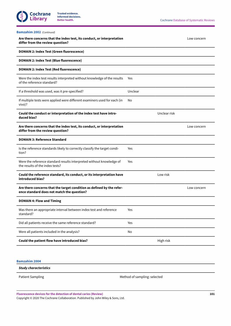
CochraneLibrary
Trusted evidence.Informed decisions.Better health.
Cochrane Database of Systematic Reviews
Are there concerns that the index test, its conduct, or interpretationdiffer from the review question?
Low concern
DOMAIN 2: Index Test (Green fluorescence)
DOMAIN 2: Index Test (Blue fluorescence)
DOMAIN 2: Index Test (Red fluorescence)
Were the index test results interpreted without knowledge of the resultsof the reference standard?
Yes
If a threshold was used, was it pre-specified? Unclear
If multiple tests were applied were different examiners used for each (invivo)?
No
Could the conduct or interpretation of the index test have intro-duced bias?
Unclear risk
Are there concerns that the index test, its conduct, or interpretationdiffer from the review question?
Low concern
DOMAIN 3: Reference Standard
Is the reference standards likely to correctly classify the target condi-tion?
Yes
Were the reference standard results interpreted without knowledge ofthe results of the index tests?
Yes
Could the reference standard, its conduct, or its interpretation haveintroduced bias?
Low risk
Are there concerns that the target condition as defined by the refer-ence standard does not match the question?
Low concern
DOMAIN 4: Flow and Timing
Was there an appropriate interval between index test and referencestandard?
Yes
Did all patients receive the same reference standard? Yes
Were all patients included in the analysis? No
Could the patient flow have introduced bias? High risk
Bamzahim 2002 (Continued)
Study characteristics
Patient Sampling Method of sampling: selected
Bamzahim 2004
Fluorescence devices for the detection of dental caries (Review)
Copyright © 2020 The Cochrane Collaboration. Published by John Wiley & Sons, Ltd.
101
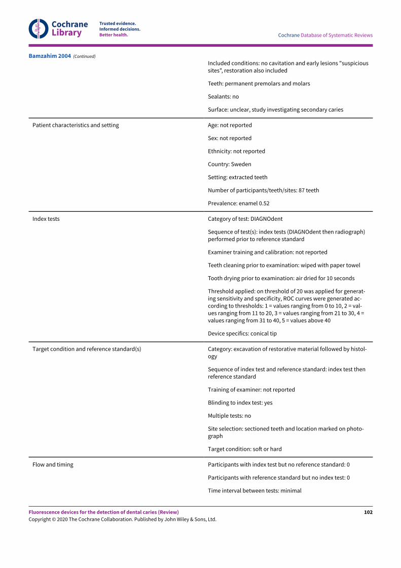
CochraneLibrary
Trusted evidence.Informed decisions.Better health.
Cochrane Database of Systematic Reviews
Included conditions: no cavitation and early lesions "suspicioussites", restoration also included
Teeth: permanent premolars and molars
Sealants: no
Surface: unclear, study investigating secondary caries
Patient characteristics and setting Age: not reported
Sex: not reported
Ethnicity: not reported
Country: Sweden
Setting: extracted teeth
Number of participants/teeth/sites: 87 teeth
Prevalence: enamel 0.52
Index tests Category of test: DIAGNOdent
Sequence of test(s): index tests (DIAGNOdent then radiograph)performed prior to reference standard
Examiner training and calibration: not reported
Teeth cleaning prior to examination: wiped with paper towel
Tooth drying prior to examination: air dried for 10 seconds
Threshold applied: on threshold of 20 was applied for generat-ing sensitivity and specificity, ROC curves were generated ac-cording to thresholds: 1 = values ranging from 0 to 10, 2 = val-ues ranging from 11 to 20, 3 = values ranging from 21 to 30, 4 =values ranging from 31 to 40, 5 = values above 40
Device specifics: conical tip
Target condition and reference standard(s) Category: excavation of restorative material followed by histol-ogy
Sequence of index test and reference standard: index test thenreference standard
Training of examiner: not reported
Blinding to index test: yes
Multiple tests: no
Site selection: sectioned teeth and location marked on photo-graph
Target condition: soN or hard
Flow and timing Participants with index test but no reference standard: 0
Participants with reference standard but no index test: 0
Time interval between tests: minimal
Bamzahim 2004 (Continued)
Fluorescence devices for the detection of dental caries (Review)
Copyright © 2020 The Cochrane Collaboration. Published by John Wiley & Sons, Ltd.
102
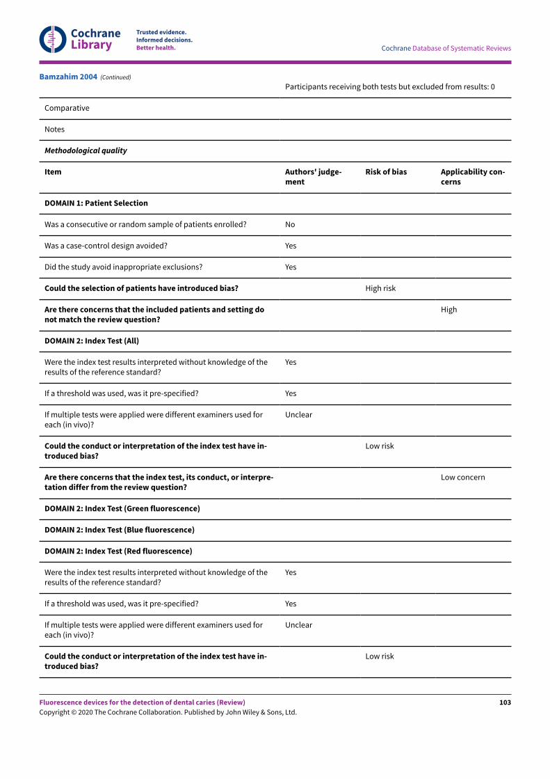
CochraneLibrary
Trusted evidence.Informed decisions.Better health.
Cochrane Database of Systematic Reviews
Participants receiving both tests but excluded from results: 0
Comparative
Notes
Methodological quality
Item Authors' judge-ment
Risk of bias Applicability con-cerns
DOMAIN 1: Patient Selection
Was a consecutive or random sample of patients enrolled? No
Was a case-control design avoided? Yes
Did the study avoid inappropriate exclusions? Yes
Could the selection of patients have introduced bias? High risk
Are there concerns that the included patients and setting donot match the review question?
High
DOMAIN 2: Index Test (All)
Were the index test results interpreted without knowledge of theresults of the reference standard?
Yes
If a threshold was used, was it pre-specified? Yes
If multiple tests were applied were different examiners used foreach (in vivo)?
Unclear
Could the conduct or interpretation of the index test have in-troduced bias?
Low risk
Are there concerns that the index test, its conduct, or interpre-tation differ from the review question?
Low concern
DOMAIN 2: Index Test (Green fluorescence)
DOMAIN 2: Index Test (Blue fluorescence)
DOMAIN 2: Index Test (Red fluorescence)
Were the index test results interpreted without knowledge of theresults of the reference standard?
Yes
If a threshold was used, was it pre-specified? Yes
If multiple tests were applied were different examiners used foreach (in vivo)?
Unclear
Could the conduct or interpretation of the index test have in-troduced bias?
Low risk
Bamzahim 2004 (Continued)
Fluorescence devices for the detection of dental caries (Review)
Copyright © 2020 The Cochrane Collaboration. Published by John Wiley & Sons, Ltd.
103
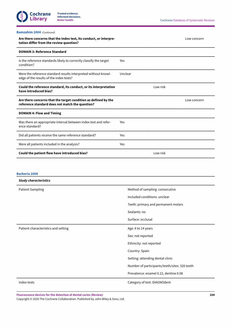
CochraneLibrary
Trusted evidence.Informed decisions.Better health.
Cochrane Database of Systematic Reviews
Are there concerns that the index test, its conduct, or interpre-tation differ from the review question?
Low concern
DOMAIN 3: Reference Standard
Is the reference standards likely to correctly classify the targetcondition?
Yes
Were the reference standard results interpreted without knowl-edge of the results of the index tests?
Unclear
Could the reference standard, its conduct, or its interpretationhave introduced bias?
Low risk
Are there concerns that the target condition as defined by thereference standard does not match the question?
Low concern
DOMAIN 4: Flow and Timing
Was there an appropriate interval between index test and refer-ence standard?
Yes
Did all patients receive the same reference standard? Yes
Were all patients included in the analysis? Yes
Could the patient flow have introduced bias? Low risk
Bamzahim 2004 (Continued)
Study characteristics
Patient Sampling Method of sampling: consecutive
Included conditions: unclear
Teeth: primary and permanent molars
Sealants: no
Surface: occlusal
Patient characteristics and setting Age: 6 to 14 years
Sex: not reported
Ethnicity: not reported
Country: Spain
Setting: attending dental clinic
Number of participants/teeth/sites: 320 teeth
Prevalence: enamel 0.22, dentine 0.08
Index tests Category of test: DIAGNOdent
Barberia 2008
Fluorescence devices for the detection of dental caries (Review)
Copyright © 2020 The Cochrane Collaboration. Published by John Wiley & Sons, Ltd.
104
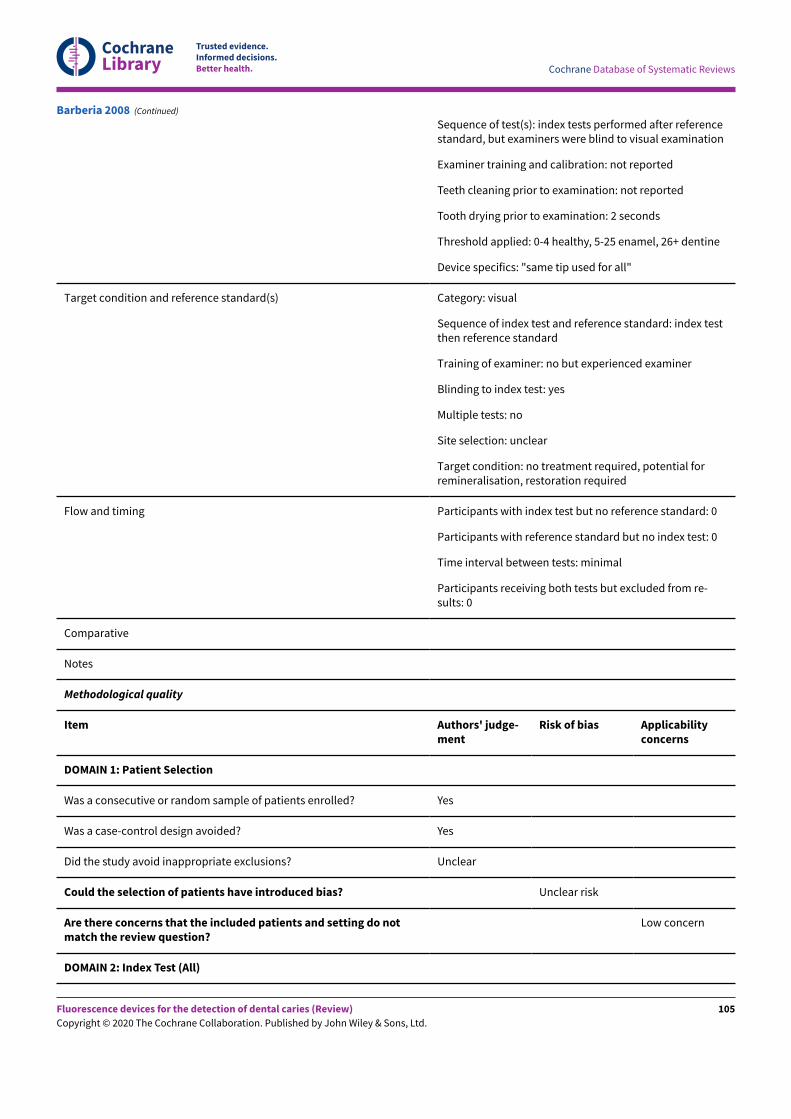
CochraneLibrary
Trusted evidence.Informed decisions.Better health.
Cochrane Database of Systematic Reviews
Sequence of test(s): index tests performed after referencestandard, but examiners were blind to visual examination
Examiner training and calibration: not reported
Teeth cleaning prior to examination: not reported
Tooth drying prior to examination: 2 seconds
Threshold applied: 0-4 healthy, 5-25 enamel, 26+ dentine
Device specifics: "same tip used for all"
Target condition and reference standard(s) Category: visual
Sequence of index test and reference standard: index testthen reference standard
Training of examiner: no but experienced examiner
Blinding to index test: yes
Multiple tests: no
Site selection: unclear
Target condition: no treatment required, potential forremineralisation, restoration required
Flow and timing Participants with index test but no reference standard: 0
Participants with reference standard but no index test: 0
Time interval between tests: minimal
Participants receiving both tests but excluded from re-sults: 0
Comparative
Notes
Methodological quality
Item Authors' judge-ment
Risk of bias Applicabilityconcerns
DOMAIN 1: Patient Selection
Was a consecutive or random sample of patients enrolled? Yes
Was a case-control design avoided? Yes
Did the study avoid inappropriate exclusions? Unclear
Could the selection of patients have introduced bias? Unclear risk
Are there concerns that the included patients and setting do notmatch the review question?
Low concern
DOMAIN 2: Index Test (All)
Barberia 2008 (Continued)
Fluorescence devices for the detection of dental caries (Review)
Copyright © 2020 The Cochrane Collaboration. Published by John Wiley & Sons, Ltd.
105
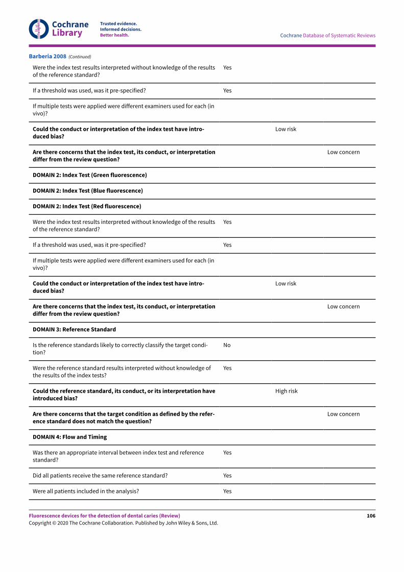
CochraneLibrary
Trusted evidence.Informed decisions.Better health.
Cochrane Database of Systematic Reviews
Were the index test results interpreted without knowledge of the resultsof the reference standard?
Yes
If a threshold was used, was it pre-specified? Yes
If multiple tests were applied were different examiners used for each (invivo)?
Could the conduct or interpretation of the index test have intro-duced bias?
Low risk
Are there concerns that the index test, its conduct, or interpretationdiffer from the review question?
Low concern
DOMAIN 2: Index Test (Green fluorescence)
DOMAIN 2: Index Test (Blue fluorescence)
DOMAIN 2: Index Test (Red fluorescence)
Were the index test results interpreted without knowledge of the resultsof the reference standard?
Yes
If a threshold was used, was it pre-specified? Yes
If multiple tests were applied were different examiners used for each (invivo)?
Could the conduct or interpretation of the index test have intro-duced bias?
Low risk
Are there concerns that the index test, its conduct, or interpretationdiffer from the review question?
Low concern
DOMAIN 3: Reference Standard
Is the reference standards likely to correctly classify the target condi-tion?
No
Were the reference standard results interpreted without knowledge ofthe results of the index tests?
Yes
Could the reference standard, its conduct, or its interpretation haveintroduced bias?
High risk
Are there concerns that the target condition as defined by the refer-ence standard does not match the question?
Low concern
DOMAIN 4: Flow and Timing
Was there an appropriate interval between index test and referencestandard?
Yes
Did all patients receive the same reference standard? Yes
Were all patients included in the analysis? Yes
Barberia 2008 (Continued)
Fluorescence devices for the detection of dental caries (Review)
Copyright © 2020 The Cochrane Collaboration. Published by John Wiley & Sons, Ltd.
106

CochraneLibrary
Trusted evidence.Informed decisions.Better health.
Cochrane Database of Systematic Reviews
Could the patient flow have introduced bias? Low risk
Barberia 2008 (Continued)
Study characteristics
Patient Sampling Method of sampling: selected
Included conditions: no cavitation and early lesions
Teeth: permanent molars
Sealants: not reported
Surface: occlusal
Patient characteristics and setting Age: not reported
Sex: not reported
Ethnicity: not reported
Country: Turkey
Setting: extracted teeth
Number of participants/teeth/sites: 35 teeth
Prevalence: enamel 0.39, dentine 0.19
Index tests Category of test: DIAGNOdent
Sequence of test(s): index tests (DIAGNOdent) per-formed prior to reference standard
Examiner training and calibration: calibrated examin-ers
Teeth cleaning prior to examination: water, brush,and pumice
Tooth drying prior to examination: paper tissues
Threshold applied: 0-13 sound, 14-19 enamel, > 20dentine
Device specifics: tip A
Target condition and reference standard(s) Category: histology
Sequence of index test and reference standard: indextest then reference standard
Training of examiner: not reported
Blinding to index test: unclear
Multiple tests: no
Site selection: location marked on drawing
Baseren 2003
Fluorescence devices for the detection of dental caries (Review)
Copyright © 2020 The Cochrane Collaboration. Published by John Wiley & Sons, Ltd.
107
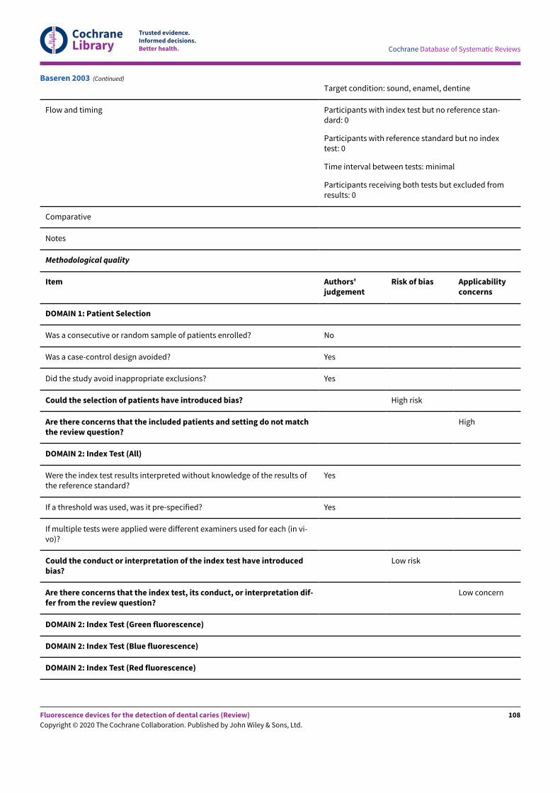
CochraneLibrary
Trusted evidence.Informed decisions.Better health.
Cochrane Database of Systematic Reviews
Target condition: sound, enamel, dentine
Flow and timing Participants with index test but no reference stan-dard: 0
Participants with reference standard but no indextest: 0
Time interval between tests: minimal
Participants receiving both tests but excluded fromresults: 0
Comparative
Notes
Methodological quality
Item Authors'judgement
Risk of bias Applicabilityconcerns
DOMAIN 1: Patient Selection
Was a consecutive or random sample of patients enrolled? No
Was a case-control design avoided? Yes
Did the study avoid inappropriate exclusions? Yes
Could the selection of patients have introduced bias? High risk
Are there concerns that the included patients and setting do not matchthe review question?
High
DOMAIN 2: Index Test (All)
Were the index test results interpreted without knowledge of the results ofthe reference standard?
Yes
If a threshold was used, was it pre-specified? Yes
If multiple tests were applied were different examiners used for each (in vi-vo)?
Could the conduct or interpretation of the index test have introducedbias?
Low risk
Are there concerns that the index test, its conduct, or interpretation dif-fer from the review question?
Low concern
DOMAIN 2: Index Test (Green fluorescence)
DOMAIN 2: Index Test (Blue fluorescence)
DOMAIN 2: Index Test (Red fluorescence)
Baseren 2003 (Continued)
Fluorescence devices for the detection of dental caries (Review)
Copyright © 2020 The Cochrane Collaboration. Published by John Wiley & Sons, Ltd.
108
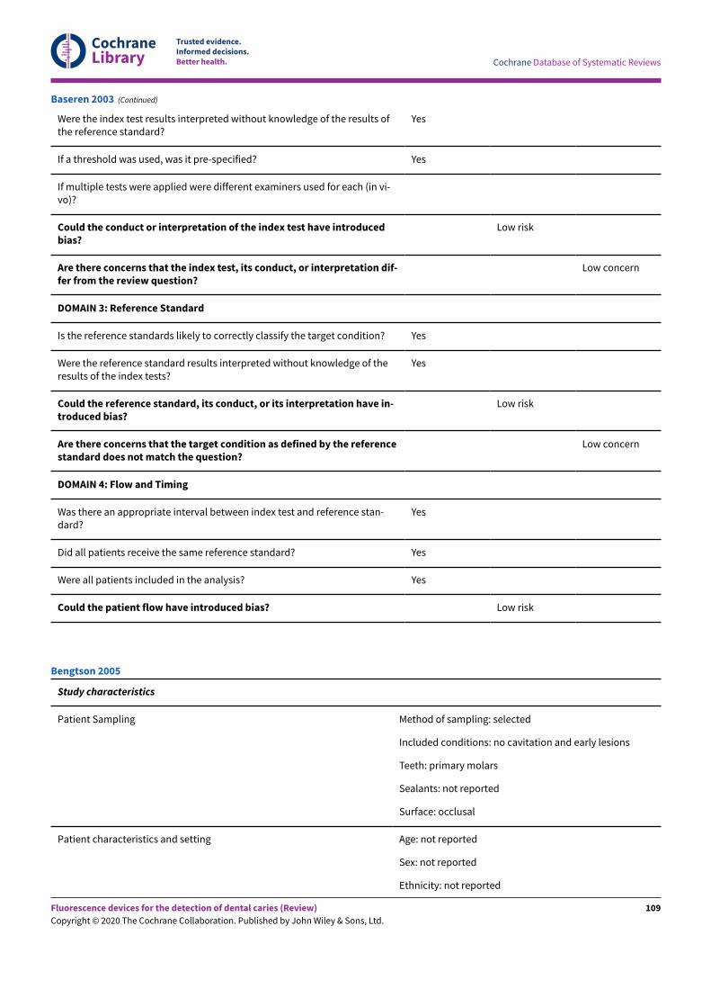
CochraneLibrary
Trusted evidence.Informed decisions.Better health.
Cochrane Database of Systematic Reviews
Were the index test results interpreted without knowledge of the results ofthe reference standard?
Yes
If a threshold was used, was it pre-specified? Yes
If multiple tests were applied were different examiners used for each (in vi-vo)?
Could the conduct or interpretation of the index test have introducedbias?
Low risk
Are there concerns that the index test, its conduct, or interpretation dif-fer from the review question?
Low concern
DOMAIN 3: Reference Standard
Is the reference standards likely to correctly classify the target condition? Yes
Were the reference standard results interpreted without knowledge of theresults of the index tests?
Yes
Could the reference standard, its conduct, or its interpretation have in-troduced bias?
Low risk
Are there concerns that the target condition as defined by the referencestandard does not match the question?
Low concern
DOMAIN 4: Flow and Timing
Was there an appropriate interval between index test and reference stan-dard?
Yes
Did all patients receive the same reference standard? Yes
Were all patients included in the analysis? Yes
Could the patient flow have introduced bias? Low risk
Baseren 2003 (Continued)
Study characteristics
Patient Sampling Method of sampling: selected
Included conditions: no cavitation and early lesions
Teeth: primary molars
Sealants: not reported
Surface: occlusal
Patient characteristics and setting Age: not reported
Sex: not reported
Ethnicity: not reported
Bengtson 2005
Fluorescence devices for the detection of dental caries (Review)
Copyright © 2020 The Cochrane Collaboration. Published by John Wiley & Sons, Ltd.
109
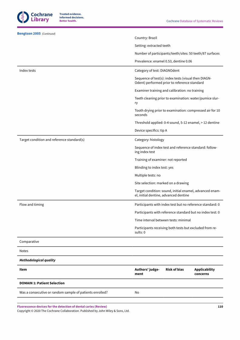
CochraneLibrary
Trusted evidence.Informed decisions.Better health.
Cochrane Database of Systematic Reviews
Country: Brazil
Setting: extracted teeth
Number of participants/teeth/sites: 50 teeth/87 surfaces
Prevalence: enamel 0.53, dentine 0.06
Index tests Category of test: DIAGNOdent
Sequence of test(s): index tests (visual then DIAGN-Odent) performed prior to reference standard
Examiner training and calibration: no training
Teeth cleaning prior to examination: water/pumice slur-ry
Tooth drying prior to examination: compressed air for 10seconds
Threshold applied: 0-4 sound, 5-12 enamel, > 12 dentine
Device specifics: tip A
Target condition and reference standard(s) Category: histology
Sequence of index test and reference standard: follow-ing index test
Training of examiner: not reported
Blinding to index test: yes
Multiple tests: no
Site selection: marked on a drawing
Target condition: sound, initial enamel, advanced enam-el, initial dentine, advanced dentine
Flow and timing Participants with index test but no reference standard: 0
Participants with reference standard but no index test: 0
Time interval between tests: minimal
Participants receiving both tests but excluded from re-sults: 0
Comparative
Notes
Methodological quality
Item Authors' judge-ment
Risk of bias Applicabilityconcerns
DOMAIN 1: Patient Selection
Was a consecutive or random sample of patients enrolled? No
Bengtson 2005 (Continued)
Fluorescence devices for the detection of dental caries (Review)
Copyright © 2020 The Cochrane Collaboration. Published by John Wiley & Sons, Ltd.
110
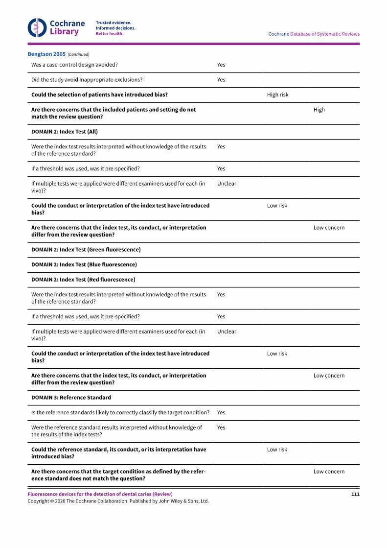
CochraneLibrary
Trusted evidence.Informed decisions.Better health.
Cochrane Database of Systematic Reviews
Was a case-control design avoided? Yes
Did the study avoid inappropriate exclusions? Yes
Could the selection of patients have introduced bias? High risk
Are there concerns that the included patients and setting do notmatch the review question?
High
DOMAIN 2: Index Test (All)
Were the index test results interpreted without knowledge of the resultsof the reference standard?
Yes
If a threshold was used, was it pre-specified? Yes
If multiple tests were applied were different examiners used for each (invivo)?
Unclear
Could the conduct or interpretation of the index test have introducedbias?
Low risk
Are there concerns that the index test, its conduct, or interpretationdiffer from the review question?
Low concern
DOMAIN 2: Index Test (Green fluorescence)
DOMAIN 2: Index Test (Blue fluorescence)
DOMAIN 2: Index Test (Red fluorescence)
Were the index test results interpreted without knowledge of the resultsof the reference standard?
Yes
If a threshold was used, was it pre-specified? Yes
If multiple tests were applied were different examiners used for each (invivo)?
Unclear
Could the conduct or interpretation of the index test have introducedbias?
Low risk
Are there concerns that the index test, its conduct, or interpretationdiffer from the review question?
Low concern
DOMAIN 3: Reference Standard
Is the reference standards likely to correctly classify the target condition? Yes
Were the reference standard results interpreted without knowledge ofthe results of the index tests?
Yes
Could the reference standard, its conduct, or its interpretation haveintroduced bias?
Low risk
Are there concerns that the target condition as defined by the refer-ence standard does not match the question?
Low concern
Bengtson 2005 (Continued)
Fluorescence devices for the detection of dental caries (Review)
Copyright © 2020 The Cochrane Collaboration. Published by John Wiley & Sons, Ltd.
111
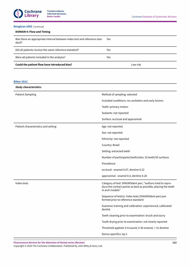
CochraneLibrary
Trusted evidence.Informed decisions.Better health.
Cochrane Database of Systematic Reviews
DOMAIN 4: Flow and Timing
Was there an appropriate interval between index test and reference stan-dard?
Yes
Did all patients receive the same reference standard? Yes
Were all patients included in the analysis? Yes
Could the patient flow have introduced bias? Low risk
Bengtson 2005 (Continued)
Study characteristics
Patient Sampling Method of sampling: selected
Included conditions: no cavitation and early lesions
Teeth: primary molars
Sealants: not reported
Surface: occlusal and approximal
Patient characteristics and setting Age: not reported
Sex: not reported
Ethnicity: not reported
Country: Brazil
Setting: extracted teeth
Number of participants/teeth/sites: 33 teeth/55 surfaces
Prevalence:
occlusal - enamel 0.67, dentine 0.22
approximal - enamel 0.4, dentine 0.28
Index tests Category of test: DIAGNOdent pen, "authors tried to repro-duce the contact points as best as possible, placing the teethin arch models"
Sequence of test(s): index tests (DIAGNOdent pen) per-formed prior to reference standard
Examiner training and calibration: experienced, calibrateddentist
Teeth cleaning prior to examination: brush and slurry
Tooth drying prior to examination: not clearly reported
Threshold applied: 0-8 sound, 9-30 enamel, > 31 dentine
Device specifics: tip 2
Bittar 2012
Fluorescence devices for the detection of dental caries (Review)
Copyright © 2020 The Cochrane Collaboration. Published by John Wiley & Sons, Ltd.
112
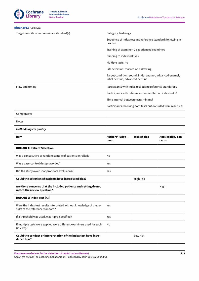
CochraneLibrary
Trusted evidence.Informed decisions.Better health.
Cochrane Database of Systematic Reviews
Target condition and reference standard(s) Category: histology
Sequence of index test and reference standard: following in-dex test
Training of examiner: 2 experienced examiners
Blinding to index test: yes
Multiple tests: no
Site selection: marked on a drawing
Target condition: sound, initial enamel, advanced enamel,intial dentine, advanced dentine
Flow and timing Participants with index test but no reference standard: 0
Participants with reference standard but no index test: 0
Time interval between tests: minimal
Participants receiving both tests but excluded from results: 0
Comparative
Notes
Methodological quality
Item Authors' judge-ment
Risk of bias Applicability con-cerns
DOMAIN 1: Patient Selection
Was a consecutive or random sample of patients enrolled? No
Was a case-control design avoided? Yes
Did the study avoid inappropriate exclusions? Yes
Could the selection of patients have introduced bias? High risk
Are there concerns that the included patients and setting do notmatch the review question?
High
DOMAIN 2: Index Test (All)
Were the index test results interpreted without knowledge of the re-sults of the reference standard?
Yes
If a threshold was used, was it pre-specified? Yes
If multiple tests were applied were different examiners used for each(in vivo)?
No
Could the conduct or interpretation of the index test have intro-duced bias?
Low risk
Bittar 2012 (Continued)
Fluorescence devices for the detection of dental caries (Review)
Copyright © 2020 The Cochrane Collaboration. Published by John Wiley & Sons, Ltd.
113
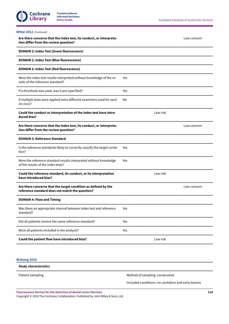
CochraneLibrary
Trusted evidence.Informed decisions.Better health.
Cochrane Database of Systematic Reviews
Are there concerns that the index test, its conduct, or interpreta-tion differ from the review question?
Low concern
DOMAIN 2: Index Test (Green fluorescence)
DOMAIN 2: Index Test (Blue fluorescence)
DOMAIN 2: Index Test (Red fluorescence)
Were the index test results interpreted without knowledge of the re-sults of the reference standard?
Yes
If a threshold was used, was it pre-specified? Yes
If multiple tests were applied were different examiners used for each(in vivo)?
No
Could the conduct or interpretation of the index test have intro-duced bias?
Low risk
Are there concerns that the index test, its conduct, or interpreta-tion differ from the review question?
Low concern
DOMAIN 3: Reference Standard
Is the reference standards likely to correctly classify the target condi-tion?
Yes
Were the reference standard results interpreted without knowledgeof the results of the index tests?
Yes
Could the reference standard, its conduct, or its interpretationhave introduced bias?
Low risk
Are there concerns that the target condition as defined by thereference standard does not match the question?
Low concern
DOMAIN 4: Flow and Timing
Was there an appropriate interval between index test and referencestandard?
Yes
Did all patients receive the same reference standard? Yes
Were all patients included in the analysis? Yes
Could the patient flow have introduced bias? Low risk
Bittar 2012 (Continued)
Study characteristics
Patient Sampling Method of sampling: consecutive
Included conditions: no cavitation and early lesions
Bizhang 2016
Fluorescence devices for the detection of dental caries (Review)
Copyright © 2020 The Cochrane Collaboration. Published by John Wiley & Sons, Ltd.
114
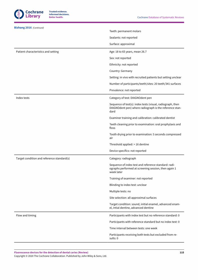
CochraneLibrary
Trusted evidence.Informed decisions.Better health.
Cochrane Database of Systematic Reviews
Teeth: permanent molars
Sealants: not reported
Surface: approximal
Patient characteristics and setting Age: 18 to 65 years, mean 26.7
Sex: not reported
Ethnicity: not reported
Country: Germany
Setting: in vivo with recruited patients but setting unclear
Number of participants/teeth/sites: 20 teeth/341 surfaces
Prevalence: not reported
Index tests Category of test: DIAGNOdent pen
Sequence of test(s): index tests (visual, radiograph, thenDIAGNOdent pen) where radiograph is the reference stan-dard
Examiner training and calibration: calibrated dentist
Teeth cleaning prior to examination: oral prophylaxis andfloss
Tooth drying prior to examination: 5 seconds compressedair
Threshold applied: > 16 dentine
Device specifics: not reported
Target condition and reference standard(s) Category: radiograph
Sequence of index test and reference standard: radi-ographs performed at screening session, then again 1week later
Training of examiner: not reported
Blinding to index test: unclear
Multiple tests: no
Site selection: all approximal surfaces
Target condition: sound, initial enamel, advanced enam-el, intial dentine, advanced dentine
Flow and timing Participants with index test but no reference standard: 0
Participants with reference standard but no index test: 0
Time interval between tests: one week
Participants receiving both tests but excluded from re-sults: 0
Bizhang 2016 (Continued)
Fluorescence devices for the detection of dental caries (Review)
Copyright © 2020 The Cochrane Collaboration. Published by John Wiley & Sons, Ltd.
115
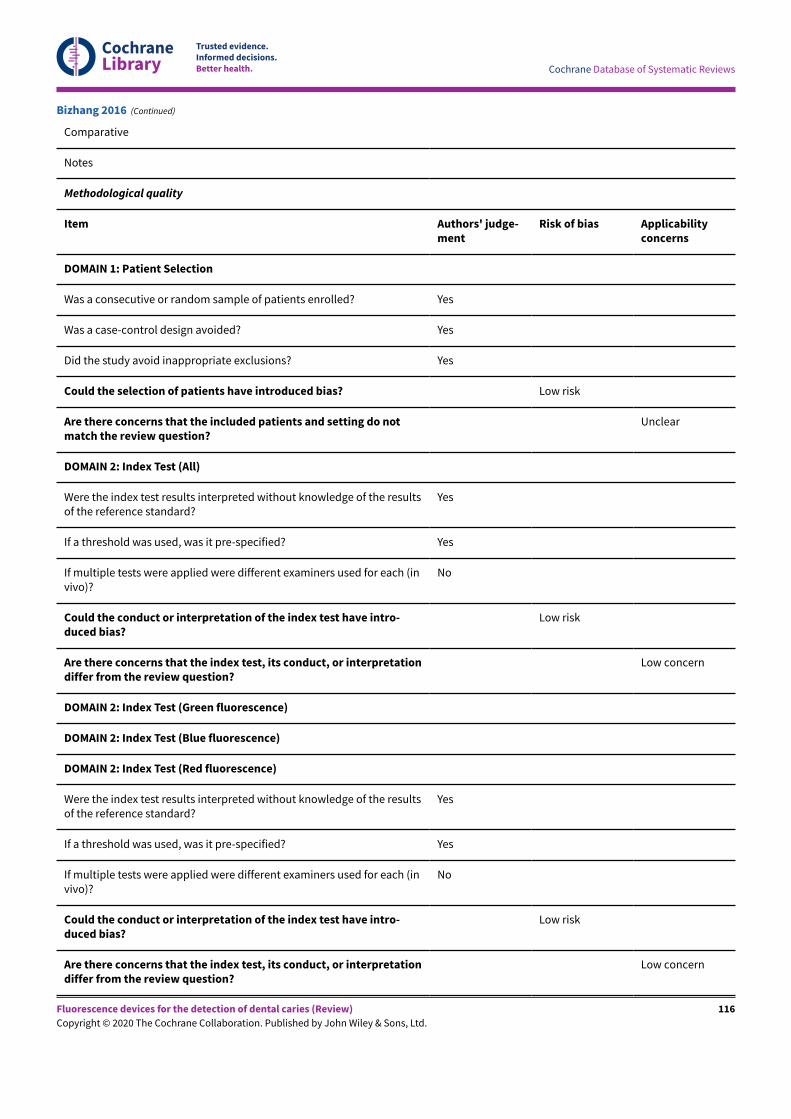
CochraneLibrary
Trusted evidence.Informed decisions.Better health.
Cochrane Database of Systematic Reviews
Comparative
Notes
Methodological quality
Item Authors' judge-ment
Risk of bias Applicabilityconcerns
DOMAIN 1: Patient Selection
Was a consecutive or random sample of patients enrolled? Yes
Was a case-control design avoided? Yes
Did the study avoid inappropriate exclusions? Yes
Could the selection of patients have introduced bias? Low risk
Are there concerns that the included patients and setting do notmatch the review question?
Unclear
DOMAIN 2: Index Test (All)
Were the index test results interpreted without knowledge of the resultsof the reference standard?
Yes
If a threshold was used, was it pre-specified? Yes
If multiple tests were applied were different examiners used for each (invivo)?
No
Could the conduct or interpretation of the index test have intro-duced bias?
Low risk
Are there concerns that the index test, its conduct, or interpretationdiffer from the review question?
Low concern
DOMAIN 2: Index Test (Green fluorescence)
DOMAIN 2: Index Test (Blue fluorescence)
DOMAIN 2: Index Test (Red fluorescence)
Were the index test results interpreted without knowledge of the resultsof the reference standard?
Yes
If a threshold was used, was it pre-specified? Yes
If multiple tests were applied were different examiners used for each (invivo)?
No
Could the conduct or interpretation of the index test have intro-duced bias?
Low risk
Are there concerns that the index test, its conduct, or interpretationdiffer from the review question?
Low concern
Bizhang 2016 (Continued)
Fluorescence devices for the detection of dental caries (Review)
Copyright © 2020 The Cochrane Collaboration. Published by John Wiley & Sons, Ltd.
116
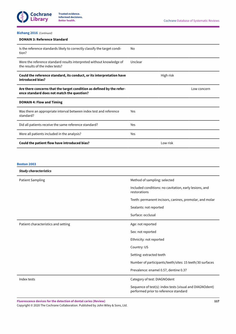
CochraneLibrary
Trusted evidence.Informed decisions.Better health.
Cochrane Database of Systematic Reviews
DOMAIN 3: Reference Standard
Is the reference standards likely to correctly classify the target condi-tion?
No
Were the reference standard results interpreted without knowledge ofthe results of the index tests?
Unclear
Could the reference standard, its conduct, or its interpretation haveintroduced bias?
High risk
Are there concerns that the target condition as defined by the refer-ence standard does not match the question?
Low concern
DOMAIN 4: Flow and Timing
Was there an appropriate interval between index test and referencestandard?
Yes
Did all patients receive the same reference standard? Yes
Were all patients included in the analysis? Yes
Could the patient flow have introduced bias? Low risk
Bizhang 2016 (Continued)
Study characteristics
Patient Sampling Method of sampling: selected
Included conditions: no cavitation, early lesions, andrestorations
Teeth: permanent incisors, canines, premolar, and molar
Sealants: not reported
Surface: occlusal
Patient characteristics and setting Age: not reported
Sex: not reported
Ethnicity: not reported
Country: US
Setting: extracted teeth
Number of participants/teeth/sites: 15 teeth/30 surfaces
Prevalence: enamel 0.57, dentine 0.37
Index tests Category of test: DIAGNOdent
Sequence of test(s): index tests (visual and DIAGNOdent)performed prior to reference standard
Boston 2003
Fluorescence devices for the detection of dental caries (Review)
Copyright © 2020 The Cochrane Collaboration. Published by John Wiley & Sons, Ltd.
117
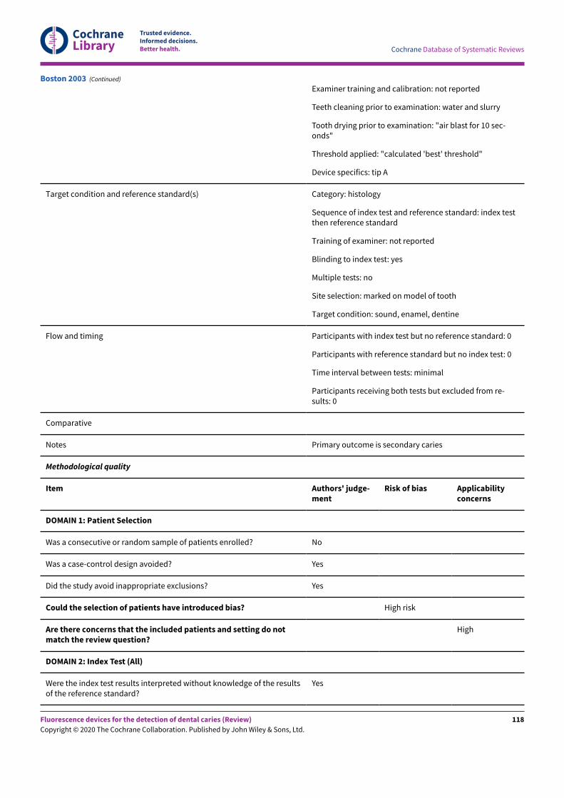
CochraneLibrary
Trusted evidence.Informed decisions.Better health.
Cochrane Database of Systematic Reviews
Examiner training and calibration: not reported
Teeth cleaning prior to examination: water and slurry
Tooth drying prior to examination: "air blast for 10 sec-onds"
Threshold applied: "calculated 'best' threshold"
Device specifics: tip A
Target condition and reference standard(s) Category: histology
Sequence of index test and reference standard: index testthen reference standard
Training of examiner: not reported
Blinding to index test: yes
Multiple tests: no
Site selection: marked on model of tooth
Target condition: sound, enamel, dentine
Flow and timing Participants with index test but no reference standard: 0
Participants with reference standard but no index test: 0
Time interval between tests: minimal
Participants receiving both tests but excluded from re-sults: 0
Comparative
Notes Primary outcome is secondary caries
Methodological quality
Item Authors' judge-ment
Risk of bias Applicabilityconcerns
DOMAIN 1: Patient Selection
Was a consecutive or random sample of patients enrolled? No
Was a case-control design avoided? Yes
Did the study avoid inappropriate exclusions? Yes
Could the selection of patients have introduced bias? High risk
Are there concerns that the included patients and setting do notmatch the review question?
High
DOMAIN 2: Index Test (All)
Were the index test results interpreted without knowledge of the resultsof the reference standard?
Yes
Boston 2003 (Continued)
Fluorescence devices for the detection of dental caries (Review)
Copyright © 2020 The Cochrane Collaboration. Published by John Wiley & Sons, Ltd.
118
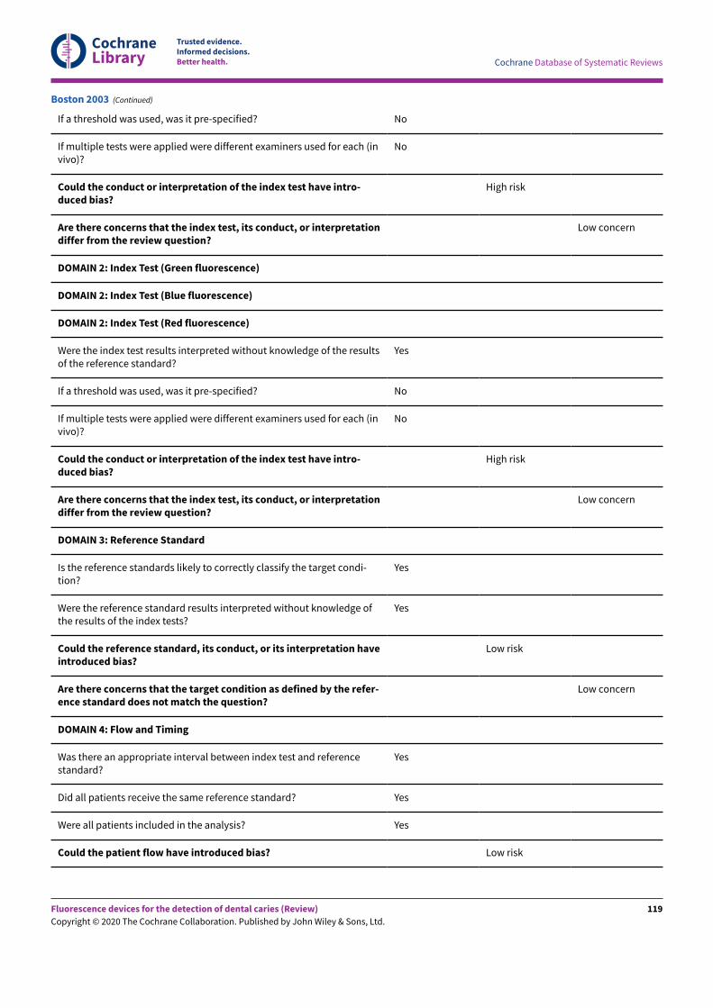
CochraneLibrary
Trusted evidence.Informed decisions.Better health.
Cochrane Database of Systematic Reviews
If a threshold was used, was it pre-specified? No
If multiple tests were applied were different examiners used for each (invivo)?
No
Could the conduct or interpretation of the index test have intro-duced bias?
High risk
Are there concerns that the index test, its conduct, or interpretationdiffer from the review question?
Low concern
DOMAIN 2: Index Test (Green fluorescence)
DOMAIN 2: Index Test (Blue fluorescence)
DOMAIN 2: Index Test (Red fluorescence)
Were the index test results interpreted without knowledge of the resultsof the reference standard?
Yes
If a threshold was used, was it pre-specified? No
If multiple tests were applied were different examiners used for each (invivo)?
No
Could the conduct or interpretation of the index test have intro-duced bias?
High risk
Are there concerns that the index test, its conduct, or interpretationdiffer from the review question?
Low concern
DOMAIN 3: Reference Standard
Is the reference standards likely to correctly classify the target condi-tion?
Yes
Were the reference standard results interpreted without knowledge ofthe results of the index tests?
Yes
Could the reference standard, its conduct, or its interpretation haveintroduced bias?
Low risk
Are there concerns that the target condition as defined by the refer-ence standard does not match the question?
Low concern
DOMAIN 4: Flow and Timing
Was there an appropriate interval between index test and referencestandard?
Yes
Did all patients receive the same reference standard? Yes
Were all patients included in the analysis? Yes
Could the patient flow have introduced bias? Low risk
Boston 2003 (Continued)
Fluorescence devices for the detection of dental caries (Review)
Copyright © 2020 The Cochrane Collaboration. Published by John Wiley & Sons, Ltd.
119
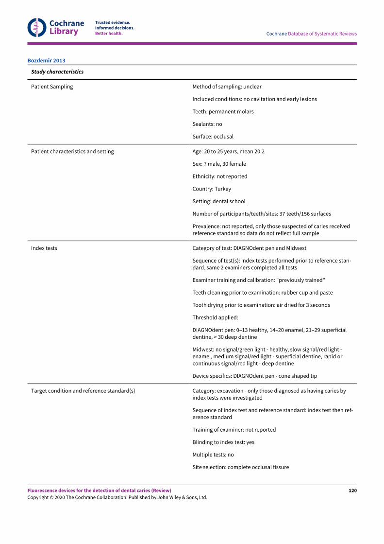
CochraneLibrary
Trusted evidence.Informed decisions.Better health.
Cochrane Database of Systematic Reviews
Study characteristics
Patient Sampling Method of sampling: unclear
Included conditions: no cavitation and early lesions
Teeth: permanent molars
Sealants: no
Surface: occlusal
Patient characteristics and setting Age: 20 to 25 years, mean 20.2
Sex: 7 male, 30 female
Ethnicity: not reported
Country: Turkey
Setting: dental school
Number of participants/teeth/sites: 37 teeth/156 surfaces
Prevalence: not reported, only those suspected of caries receivedreference standard so data do not reflect full sample
Index tests Category of test: DIAGNOdent pen and Midwest
Sequence of test(s): index tests performed prior to reference stan-dard, same 2 examiners completed all tests
Examiner training and calibration: "previously trained"
Teeth cleaning prior to examination: rubber cup and paste
Tooth drying prior to examination: air dried for 3 seconds
Threshold applied:
DIAGNOdent pen: 0–13 healthy, 14–20 enamel, 21–29 superficialdentine, > 30 deep dentine
Midwest: no signal/green light - healthy, slow signal/red light -enamel, medium signal/red light - superficial dentine, rapid orcontinuous signal/red light - deep dentine
Device specifics: DIAGNOdent pen - cone shaped tip
Target condition and reference standard(s) Category: excavation - only those diagnosed as having caries byindex tests were investigated
Sequence of index test and reference standard: index test then ref-erence standard
Training of examiner: not reported
Blinding to index test: yes
Multiple tests: no
Site selection: complete occlusal fissure
Bozdemir 2013
Fluorescence devices for the detection of dental caries (Review)
Copyright © 2020 The Cochrane Collaboration. Published by John Wiley & Sons, Ltd.
120
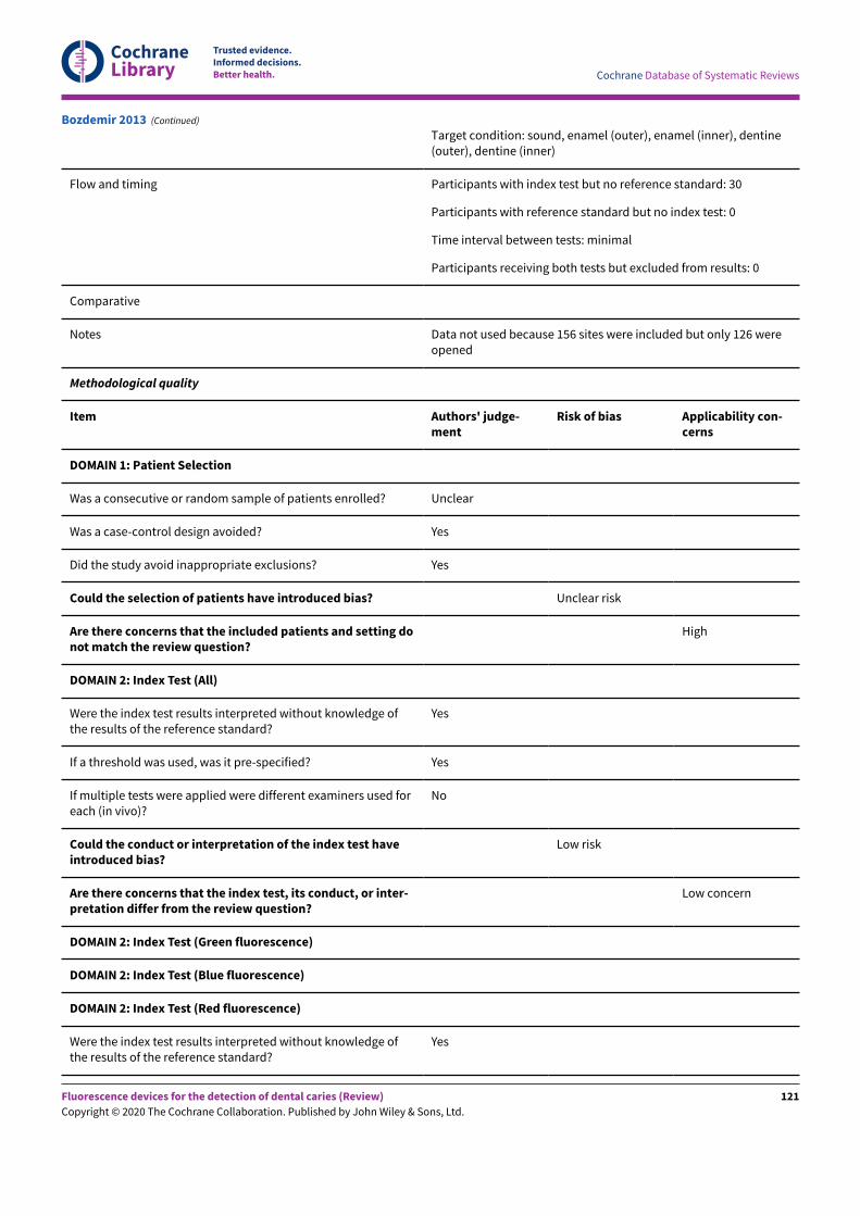
CochraneLibrary
Trusted evidence.Informed decisions.Better health.
Cochrane Database of Systematic Reviews
Target condition: sound, enamel (outer), enamel (inner), dentine(outer), dentine (inner)
Flow and timing Participants with index test but no reference standard: 30
Participants with reference standard but no index test: 0
Time interval between tests: minimal
Participants receiving both tests but excluded from results: 0
Comparative
Notes Data not used because 156 sites were included but only 126 wereopened
Methodological quality
Item Authors' judge-ment
Risk of bias Applicability con-cerns
DOMAIN 1: Patient Selection
Was a consecutive or random sample of patients enrolled? Unclear
Was a case-control design avoided? Yes
Did the study avoid inappropriate exclusions? Yes
Could the selection of patients have introduced bias? Unclear risk
Are there concerns that the included patients and setting donot match the review question?
High
DOMAIN 2: Index Test (All)
Were the index test results interpreted without knowledge ofthe results of the reference standard?
Yes
If a threshold was used, was it pre-specified? Yes
If multiple tests were applied were different examiners used foreach (in vivo)?
No
Could the conduct or interpretation of the index test haveintroduced bias?
Low risk
Are there concerns that the index test, its conduct, or inter-pretation differ from the review question?
Low concern
DOMAIN 2: Index Test (Green fluorescence)
DOMAIN 2: Index Test (Blue fluorescence)
DOMAIN 2: Index Test (Red fluorescence)
Were the index test results interpreted without knowledge ofthe results of the reference standard?
Yes
Bozdemir 2013 (Continued)
Fluorescence devices for the detection of dental caries (Review)
Copyright © 2020 The Cochrane Collaboration. Published by John Wiley & Sons, Ltd.
121
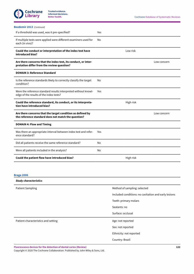
CochraneLibrary
Trusted evidence.Informed decisions.Better health.
Cochrane Database of Systematic Reviews
If a threshold was used, was it pre-specified? Yes
If multiple tests were applied were different examiners used foreach (in vivo)?
No
Could the conduct or interpretation of the index test haveintroduced bias?
Low risk
Are there concerns that the index test, its conduct, or inter-pretation differ from the review question?
Low concern
DOMAIN 3: Reference Standard
Is the reference standards likely to correctly classify the targetcondition?
No
Were the reference standard results interpreted without knowl-edge of the results of the index tests?
Yes
Could the reference standard, its conduct, or its interpreta-tion have introduced bias?
High risk
Are there concerns that the target condition as defined bythe reference standard does not match the question?
Low concern
DOMAIN 4: Flow and Timing
Was there an appropriate interval between index test and refer-ence standard?
Yes
Did all patients receive the same reference standard? No
Were all patients included in the analysis? No
Could the patient flow have introduced bias? High risk
Bozdemir 2013 (Continued)
Study characteristics
Patient Sampling Method of sampling: selected
Included conditions: no cavitation and early lesions
Teeth: primary molars
Sealants: no
Surface: occlusal
Patient characteristics and setting Age: not reported
Sex: not reported
Ethnicity: not reported
Country: Brazil
Braga 2006
Fluorescence devices for the detection of dental caries (Review)
Copyright © 2020 The Cochrane Collaboration. Published by John Wiley & Sons, Ltd.
122
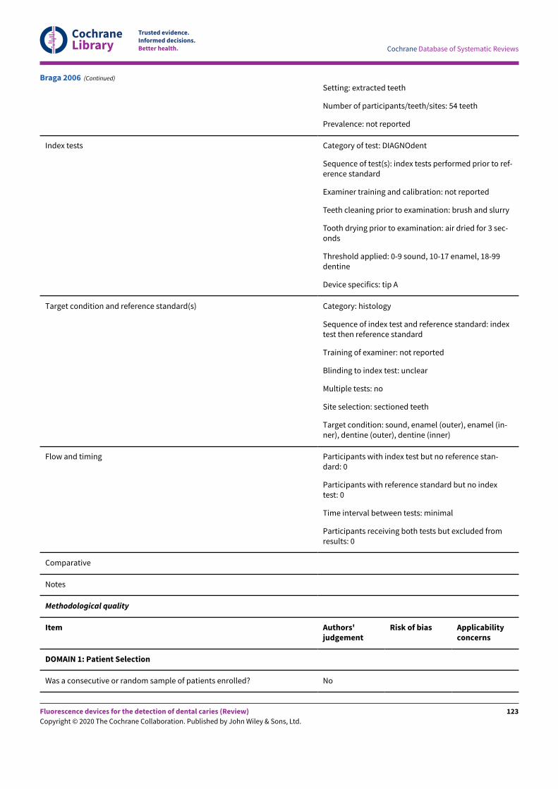
CochraneLibrary
Trusted evidence.Informed decisions.Better health.
Cochrane Database of Systematic Reviews
Setting: extracted teeth
Number of participants/teeth/sites: 54 teeth
Prevalence: not reported
Index tests Category of test: DIAGNOdent
Sequence of test(s): index tests performed prior to ref-erence standard
Examiner training and calibration: not reported
Teeth cleaning prior to examination: brush and slurry
Tooth drying prior to examination: air dried for 3 sec-onds
Threshold applied: 0-9 sound, 10-17 enamel, 18-99dentine
Device specifics: tip A
Target condition and reference standard(s) Category: histology
Sequence of index test and reference standard: indextest then reference standard
Training of examiner: not reported
Blinding to index test: unclear
Multiple tests: no
Site selection: sectioned teeth
Target condition: sound, enamel (outer), enamel (in-ner), dentine (outer), dentine (inner)
Flow and timing Participants with index test but no reference stan-dard: 0
Participants with reference standard but no indextest: 0
Time interval between tests: minimal
Participants receiving both tests but excluded fromresults: 0
Comparative
Notes
Methodological quality
Item Authors'judgement
Risk of bias Applicabilityconcerns
DOMAIN 1: Patient Selection
Was a consecutive or random sample of patients enrolled? No
Braga 2006 (Continued)
Fluorescence devices for the detection of dental caries (Review)
Copyright © 2020 The Cochrane Collaboration. Published by John Wiley & Sons, Ltd.
123
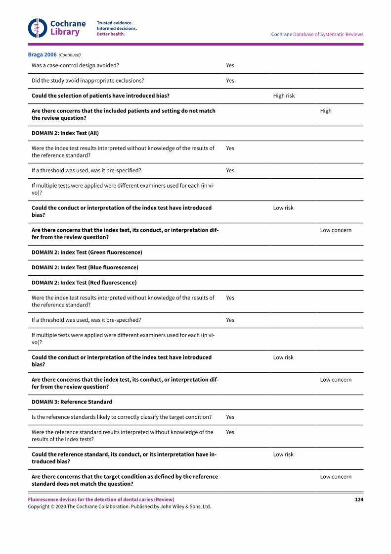
CochraneLibrary
Trusted evidence.Informed decisions.Better health.
Cochrane Database of Systematic Reviews
Was a case-control design avoided? Yes
Did the study avoid inappropriate exclusions? Yes
Could the selection of patients have introduced bias? High risk
Are there concerns that the included patients and setting do not matchthe review question?
High
DOMAIN 2: Index Test (All)
Were the index test results interpreted without knowledge of the results ofthe reference standard?
Yes
If a threshold was used, was it pre-specified? Yes
If multiple tests were applied were different examiners used for each (in vi-vo)?
Could the conduct or interpretation of the index test have introducedbias?
Low risk
Are there concerns that the index test, its conduct, or interpretation dif-fer from the review question?
Low concern
DOMAIN 2: Index Test (Green fluorescence)
DOMAIN 2: Index Test (Blue fluorescence)
DOMAIN 2: Index Test (Red fluorescence)
Were the index test results interpreted without knowledge of the results ofthe reference standard?
Yes
If a threshold was used, was it pre-specified? Yes
If multiple tests were applied were different examiners used for each (in vi-vo)?
Could the conduct or interpretation of the index test have introducedbias?
Low risk
Are there concerns that the index test, its conduct, or interpretation dif-fer from the review question?
Low concern
DOMAIN 3: Reference Standard
Is the reference standards likely to correctly classify the target condition? Yes
Were the reference standard results interpreted without knowledge of theresults of the index tests?
Yes
Could the reference standard, its conduct, or its interpretation have in-troduced bias?
Low risk
Are there concerns that the target condition as defined by the referencestandard does not match the question?
Low concern
Braga 2006 (Continued)
Fluorescence devices for the detection of dental caries (Review)
Copyright © 2020 The Cochrane Collaboration. Published by John Wiley & Sons, Ltd.
124
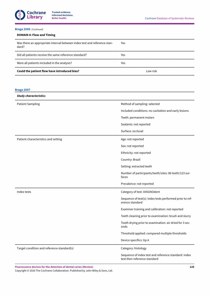
CochraneLibrary
Trusted evidence.Informed decisions.Better health.
Cochrane Database of Systematic Reviews
DOMAIN 4: Flow and Timing
Was there an appropriate interval between index test and reference stan-dard?
Yes
Did all patients receive the same reference standard? Yes
Were all patients included in the analysis? Yes
Could the patient flow have introduced bias? Low risk
Braga 2006 (Continued)
Study characteristics
Patient Sampling Method of sampling: selected
Included conditions: no cavitation and early lesions
Teeth: permanent molars
Sealants: not reported
Surface: occlusal
Patient characteristics and setting Age: not reported
Sex: not reported
Ethnicity: not reported
Country: Brazil
Setting: extracted teeth
Number of participants/teeth/sites: 86 teeth/123 sur-faces
Prevalence: not reported
Index tests Category of test: DIAGNOdent
Sequence of test(s): index tests performed prior to ref-erence standard
Examiner training and calibration: not reported
Teeth cleaning prior to examination: brush and slurry
Tooth drying prior to examination: air dried for 3 sec-onds
Threshold applied: compared multiple thresholds
Device specifics: tip A
Target condition and reference standard(s) Category: histology
Sequence of index test and reference standard: indextest then reference standard
Braga 2007
Fluorescence devices for the detection of dental caries (Review)
Copyright © 2020 The Cochrane Collaboration. Published by John Wiley & Sons, Ltd.
125
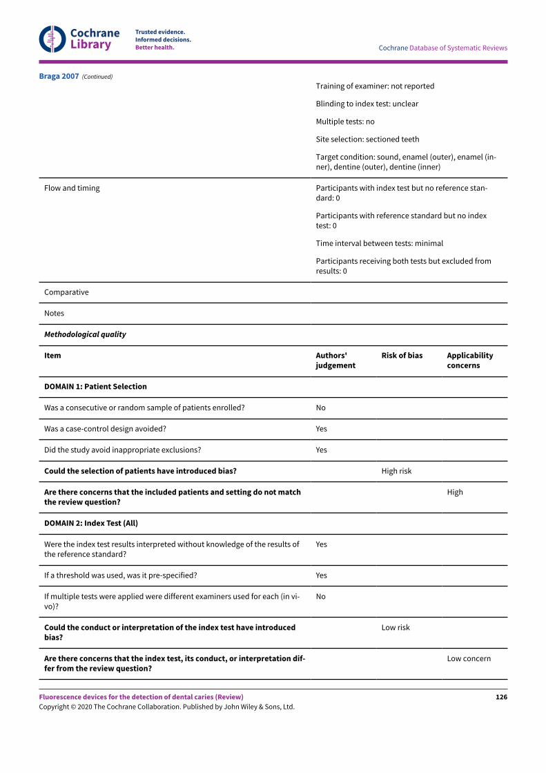
CochraneLibrary
Trusted evidence.Informed decisions.Better health.
Cochrane Database of Systematic Reviews
Training of examiner: not reported
Blinding to index test: unclear
Multiple tests: no
Site selection: sectioned teeth
Target condition: sound, enamel (outer), enamel (in-ner), dentine (outer), dentine (inner)
Flow and timing Participants with index test but no reference stan-dard: 0
Participants with reference standard but no indextest: 0
Time interval between tests: minimal
Participants receiving both tests but excluded fromresults: 0
Comparative
Notes
Methodological quality
Item Authors'judgement
Risk of bias Applicabilityconcerns
DOMAIN 1: Patient Selection
Was a consecutive or random sample of patients enrolled? No
Was a case-control design avoided? Yes
Did the study avoid inappropriate exclusions? Yes
Could the selection of patients have introduced bias? High risk
Are there concerns that the included patients and setting do not matchthe review question?
High
DOMAIN 2: Index Test (All)
Were the index test results interpreted without knowledge of the results ofthe reference standard?
Yes
If a threshold was used, was it pre-specified? Yes
If multiple tests were applied were different examiners used for each (in vi-vo)?
No
Could the conduct or interpretation of the index test have introducedbias?
Low risk
Are there concerns that the index test, its conduct, or interpretation dif-fer from the review question?
Low concern
Braga 2007 (Continued)
Fluorescence devices for the detection of dental caries (Review)
Copyright © 2020 The Cochrane Collaboration. Published by John Wiley & Sons, Ltd.
126
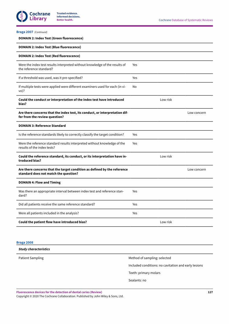
CochraneLibrary
Trusted evidence.Informed decisions.Better health.
Cochrane Database of Systematic Reviews
DOMAIN 2: Index Test (Green fluorescence)
DOMAIN 2: Index Test (Blue fluorescence)
DOMAIN 2: Index Test (Red fluorescence)
Were the index test results interpreted without knowledge of the results ofthe reference standard?
Yes
If a threshold was used, was it pre-specified? Yes
If multiple tests were applied were different examiners used for each (in vi-vo)?
No
Could the conduct or interpretation of the index test have introducedbias?
Low risk
Are there concerns that the index test, its conduct, or interpretation dif-fer from the review question?
Low concern
DOMAIN 3: Reference Standard
Is the reference standards likely to correctly classify the target condition? Yes
Were the reference standard results interpreted without knowledge of theresults of the index tests?
Yes
Could the reference standard, its conduct, or its interpretation have in-troduced bias?
Low risk
Are there concerns that the target condition as defined by the referencestandard does not match the question?
Low concern
DOMAIN 4: Flow and Timing
Was there an appropriate interval between index test and reference stan-dard?
Yes
Did all patients receive the same reference standard? Yes
Were all patients included in the analysis? Yes
Could the patient flow have introduced bias? Low risk
Braga 2007 (Continued)
Study characteristics
Patient Sampling Method of sampling: selected
Included conditions: no cavitation and early lesions
Teeth: primary molars
Sealants: no
Braga 2008
Fluorescence devices for the detection of dental caries (Review)
Copyright © 2020 The Cochrane Collaboration. Published by John Wiley & Sons, Ltd.
127
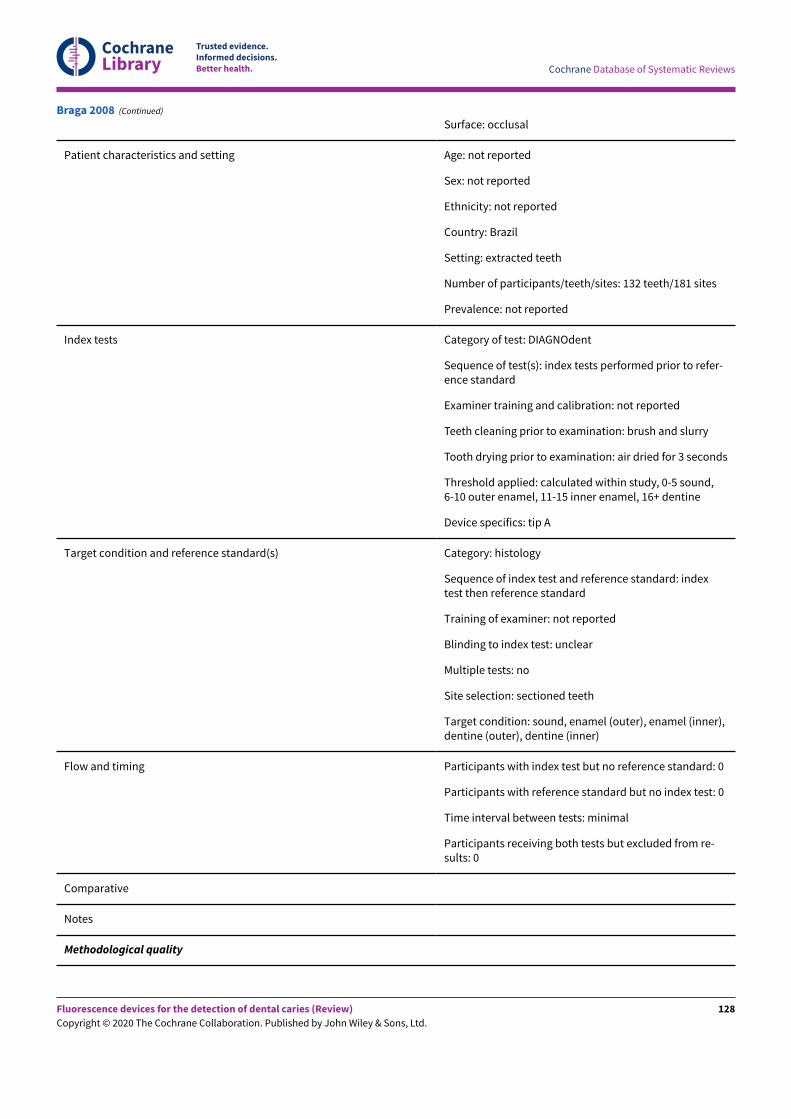
CochraneLibrary
Trusted evidence.Informed decisions.Better health.
Cochrane Database of Systematic Reviews
Surface: occlusal
Patient characteristics and setting Age: not reported
Sex: not reported
Ethnicity: not reported
Country: Brazil
Setting: extracted teeth
Number of participants/teeth/sites: 132 teeth/181 sites
Prevalence: not reported
Index tests Category of test: DIAGNOdent
Sequence of test(s): index tests performed prior to refer-ence standard
Examiner training and calibration: not reported
Teeth cleaning prior to examination: brush and slurry
Tooth drying prior to examination: air dried for 3 seconds
Threshold applied: calculated within study, 0-5 sound,6-10 outer enamel, 11-15 inner enamel, 16+ dentine
Device specifics: tip A
Target condition and reference standard(s) Category: histology
Sequence of index test and reference standard: indextest then reference standard
Training of examiner: not reported
Blinding to index test: unclear
Multiple tests: no
Site selection: sectioned teeth
Target condition: sound, enamel (outer), enamel (inner),dentine (outer), dentine (inner)
Flow and timing Participants with index test but no reference standard: 0
Participants with reference standard but no index test: 0
Time interval between tests: minimal
Participants receiving both tests but excluded from re-sults: 0
Comparative
Notes
Methodological quality
Braga 2008 (Continued)
Fluorescence devices for the detection of dental caries (Review)
Copyright © 2020 The Cochrane Collaboration. Published by John Wiley & Sons, Ltd.
128
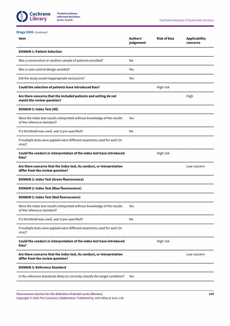
CochraneLibrary
Trusted evidence.Informed decisions.Better health.
Cochrane Database of Systematic Reviews
Item Authors'judgement
Risk of bias Applicabilityconcerns
DOMAIN 1: Patient Selection
Was a consecutive or random sample of patients enrolled? No
Was a case-control design avoided? Yes
Did the study avoid inappropriate exclusions? Yes
Could the selection of patients have introduced bias? High risk
Are there concerns that the included patients and setting do notmatch the review question?
High
DOMAIN 2: Index Test (All)
Were the index test results interpreted without knowledge of the resultsof the reference standard?
Yes
If a threshold was used, was it pre-specified? No
If multiple tests were applied were different examiners used for each (invivo)?
Could the conduct or interpretation of the index test have introducedbias?
High risk
Are there concerns that the index test, its conduct, or interpretationdiffer from the review question?
Low concern
DOMAIN 2: Index Test (Green fluorescence)
DOMAIN 2: Index Test (Blue fluorescence)
DOMAIN 2: Index Test (Red fluorescence)
Were the index test results interpreted without knowledge of the resultsof the reference standard?
Yes
If a threshold was used, was it pre-specified? No
If multiple tests were applied were different examiners used for each (invivo)?
Could the conduct or interpretation of the index test have introducedbias?
High risk
Are there concerns that the index test, its conduct, or interpretationdiffer from the review question?
Low concern
DOMAIN 3: Reference Standard
Is the reference standards likely to correctly classify the target condition? Yes
Braga 2008 (Continued)
Fluorescence devices for the detection of dental caries (Review)
Copyright © 2020 The Cochrane Collaboration. Published by John Wiley & Sons, Ltd.
129
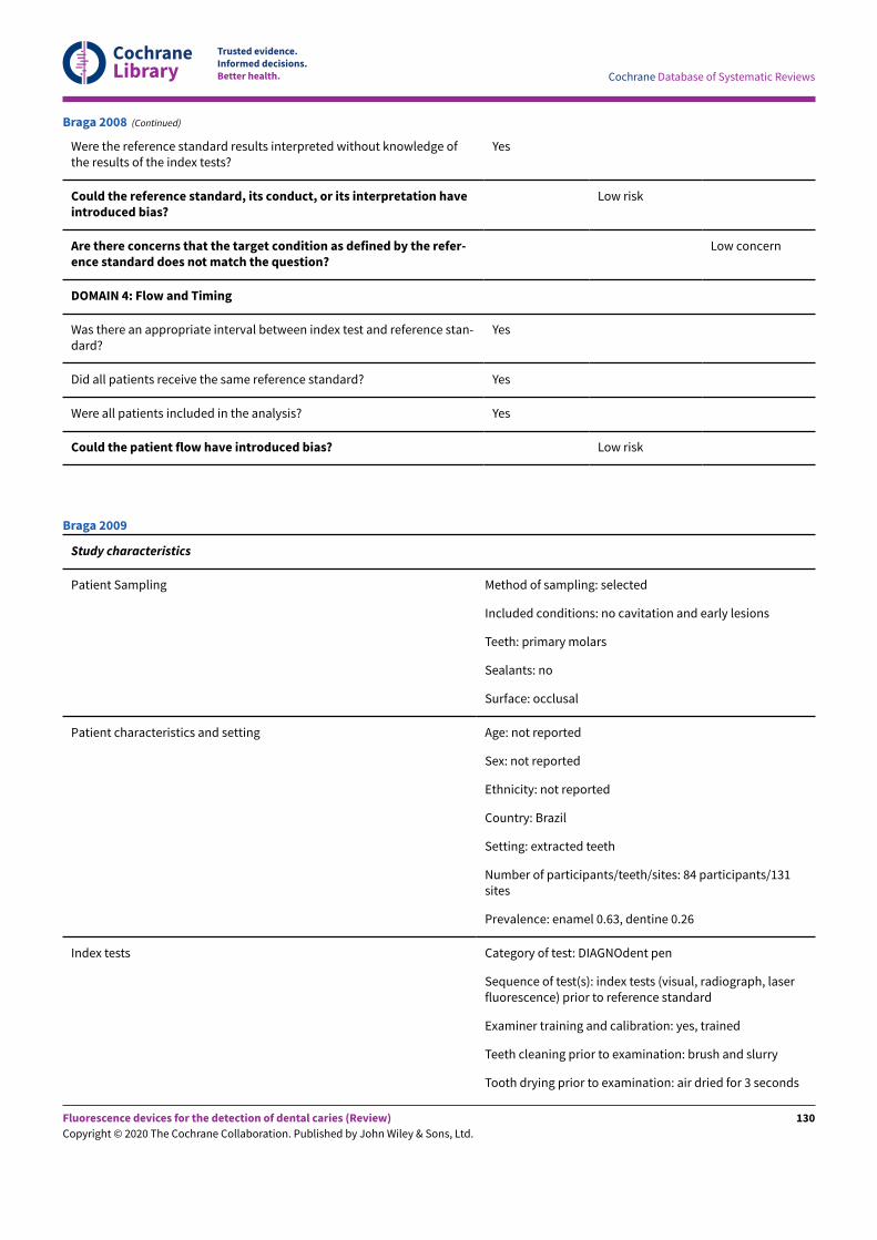
CochraneLibrary
Trusted evidence.Informed decisions.Better health.
Cochrane Database of Systematic Reviews
Were the reference standard results interpreted without knowledge ofthe results of the index tests?
Yes
Could the reference standard, its conduct, or its interpretation haveintroduced bias?
Low risk
Are there concerns that the target condition as defined by the refer-ence standard does not match the question?
Low concern
DOMAIN 4: Flow and Timing
Was there an appropriate interval between index test and reference stan-dard?
Yes
Did all patients receive the same reference standard? Yes
Were all patients included in the analysis? Yes
Could the patient flow have introduced bias? Low risk
Braga 2008 (Continued)
Study characteristics
Patient Sampling Method of sampling: selected
Included conditions: no cavitation and early lesions
Teeth: primary molars
Sealants: no
Surface: occlusal
Patient characteristics and setting Age: not reported
Sex: not reported
Ethnicity: not reported
Country: Brazil
Setting: extracted teeth
Number of participants/teeth/sites: 84 participants/131sites
Prevalence: enamel 0.63, dentine 0.26
Index tests Category of test: DIAGNOdent pen
Sequence of test(s): index tests (visual, radiograph, laserfluorescence) prior to reference standard
Examiner training and calibration: yes, trained
Teeth cleaning prior to examination: brush and slurry
Tooth drying prior to examination: air dried for 3 seconds
Braga 2009
Fluorescence devices for the detection of dental caries (Review)
Copyright © 2020 The Cochrane Collaboration. Published by John Wiley & Sons, Ltd.
130
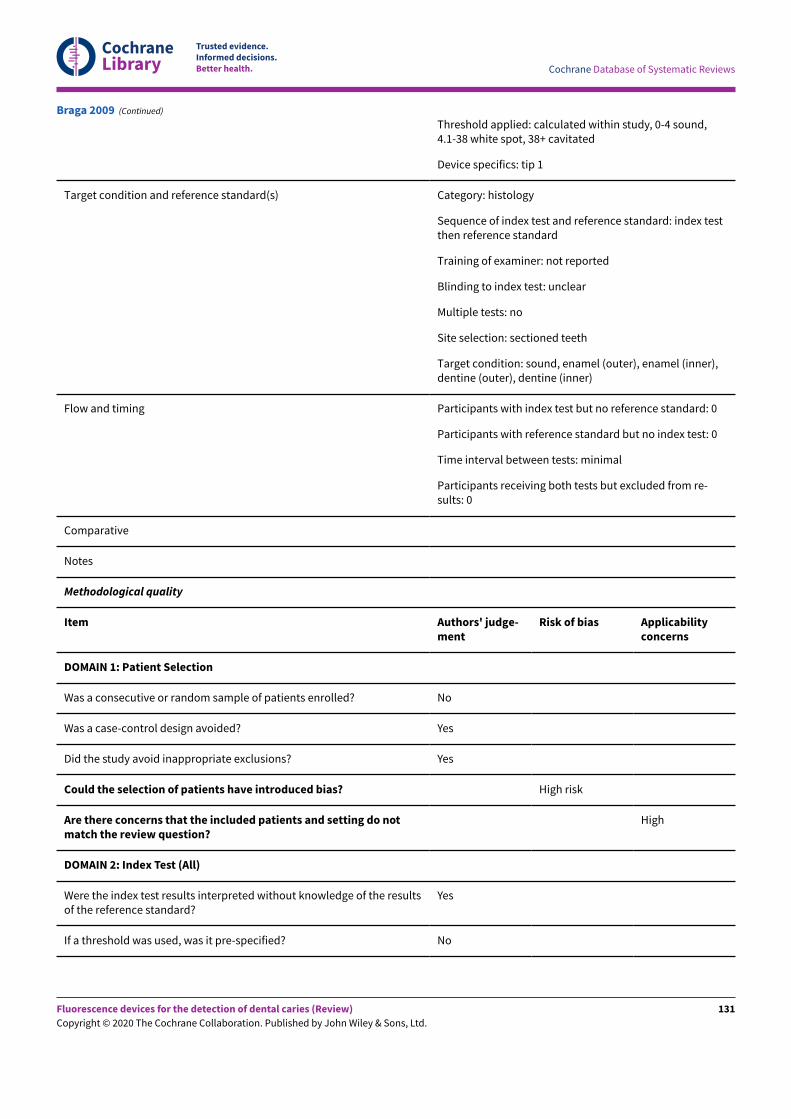
CochraneLibrary
Trusted evidence.Informed decisions.Better health.
Cochrane Database of Systematic Reviews
Threshold applied: calculated within study, 0-4 sound,4.1-38 white spot, 38+ cavitated
Device specifics: tip 1
Target condition and reference standard(s) Category: histology
Sequence of index test and reference standard: index testthen reference standard
Training of examiner: not reported
Blinding to index test: unclear
Multiple tests: no
Site selection: sectioned teeth
Target condition: sound, enamel (outer), enamel (inner),dentine (outer), dentine (inner)
Flow and timing Participants with index test but no reference standard: 0
Participants with reference standard but no index test: 0
Time interval between tests: minimal
Participants receiving both tests but excluded from re-sults: 0
Comparative
Notes
Methodological quality
Item Authors' judge-ment
Risk of bias Applicabilityconcerns
DOMAIN 1: Patient Selection
Was a consecutive or random sample of patients enrolled? No
Was a case-control design avoided? Yes
Did the study avoid inappropriate exclusions? Yes
Could the selection of patients have introduced bias? High risk
Are there concerns that the included patients and setting do notmatch the review question?
High
DOMAIN 2: Index Test (All)
Were the index test results interpreted without knowledge of the resultsof the reference standard?
Yes
If a threshold was used, was it pre-specified? No
Braga 2009 (Continued)
Fluorescence devices for the detection of dental caries (Review)
Copyright © 2020 The Cochrane Collaboration. Published by John Wiley & Sons, Ltd.
131

CochraneLibrary
Trusted evidence.Informed decisions.Better health.
Cochrane Database of Systematic Reviews
If multiple tests were applied were different examiners used for each (invivo)?
Unclear
Could the conduct or interpretation of the index test have intro-duced bias?
High risk
Are there concerns that the index test, its conduct, or interpretationdiffer from the review question?
Low concern
DOMAIN 2: Index Test (Green fluorescence)
DOMAIN 2: Index Test (Blue fluorescence)
DOMAIN 2: Index Test (Red fluorescence)
Were the index test results interpreted without knowledge of the resultsof the reference standard?
Yes
If a threshold was used, was it pre-specified? No
If multiple tests were applied were different examiners used for each (invivo)?
Unclear
Could the conduct or interpretation of the index test have intro-duced bias?
High risk
Are there concerns that the index test, its conduct, or interpretationdiffer from the review question?
Low concern
DOMAIN 3: Reference Standard
Is the reference standards likely to correctly classify the target condi-tion?
Yes
Were the reference standard results interpreted without knowledge ofthe results of the index tests?
Yes
Could the reference standard, its conduct, or its interpretation haveintroduced bias?
Low risk
Are there concerns that the target condition as defined by the refer-ence standard does not match the question?
Low concern
DOMAIN 4: Flow and Timing
Was there an appropriate interval between index test and referencestandard?
Yes
Did all patients receive the same reference standard? Yes
Were all patients included in the analysis? Yes
Could the patient flow have introduced bias? Low risk
Braga 2009 (Continued)
Fluorescence devices for the detection of dental caries (Review)
Copyright © 2020 The Cochrane Collaboration. Published by John Wiley & Sons, Ltd.
132
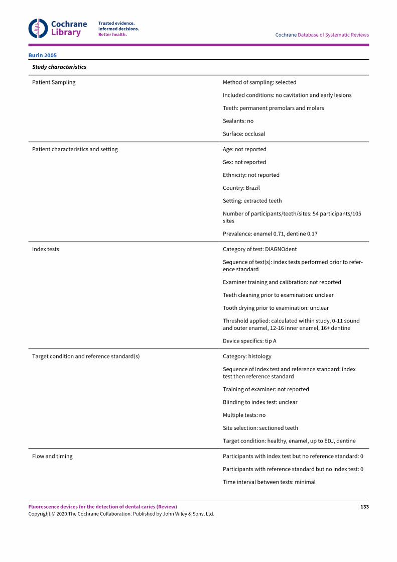
CochraneLibrary
Trusted evidence.Informed decisions.Better health.
Cochrane Database of Systematic Reviews
Study characteristics
Patient Sampling Method of sampling: selected
Included conditions: no cavitation and early lesions
Teeth: permanent premolars and molars
Sealants: no
Surface: occlusal
Patient characteristics and setting Age: not reported
Sex: not reported
Ethnicity: not reported
Country: Brazil
Setting: extracted teeth
Number of participants/teeth/sites: 54 participants/105sites
Prevalence: enamel 0.71, dentine 0.17
Index tests Category of test: DIAGNOdent
Sequence of test(s): index tests performed prior to refer-ence standard
Examiner training and calibration: not reported
Teeth cleaning prior to examination: unclear
Tooth drying prior to examination: unclear
Threshold applied: calculated within study, 0-11 soundand outer enamel, 12-16 inner enamel, 16+ dentine
Device specifics: tip A
Target condition and reference standard(s) Category: histology
Sequence of index test and reference standard: indextest then reference standard
Training of examiner: not reported
Blinding to index test: unclear
Multiple tests: no
Site selection: sectioned teeth
Target condition: healthy, enamel, up to EDJ, dentine
Flow and timing Participants with index test but no reference standard: 0
Participants with reference standard but no index test: 0
Time interval between tests: minimal
Burin 2005
Fluorescence devices for the detection of dental caries (Review)
Copyright © 2020 The Cochrane Collaboration. Published by John Wiley & Sons, Ltd.
133
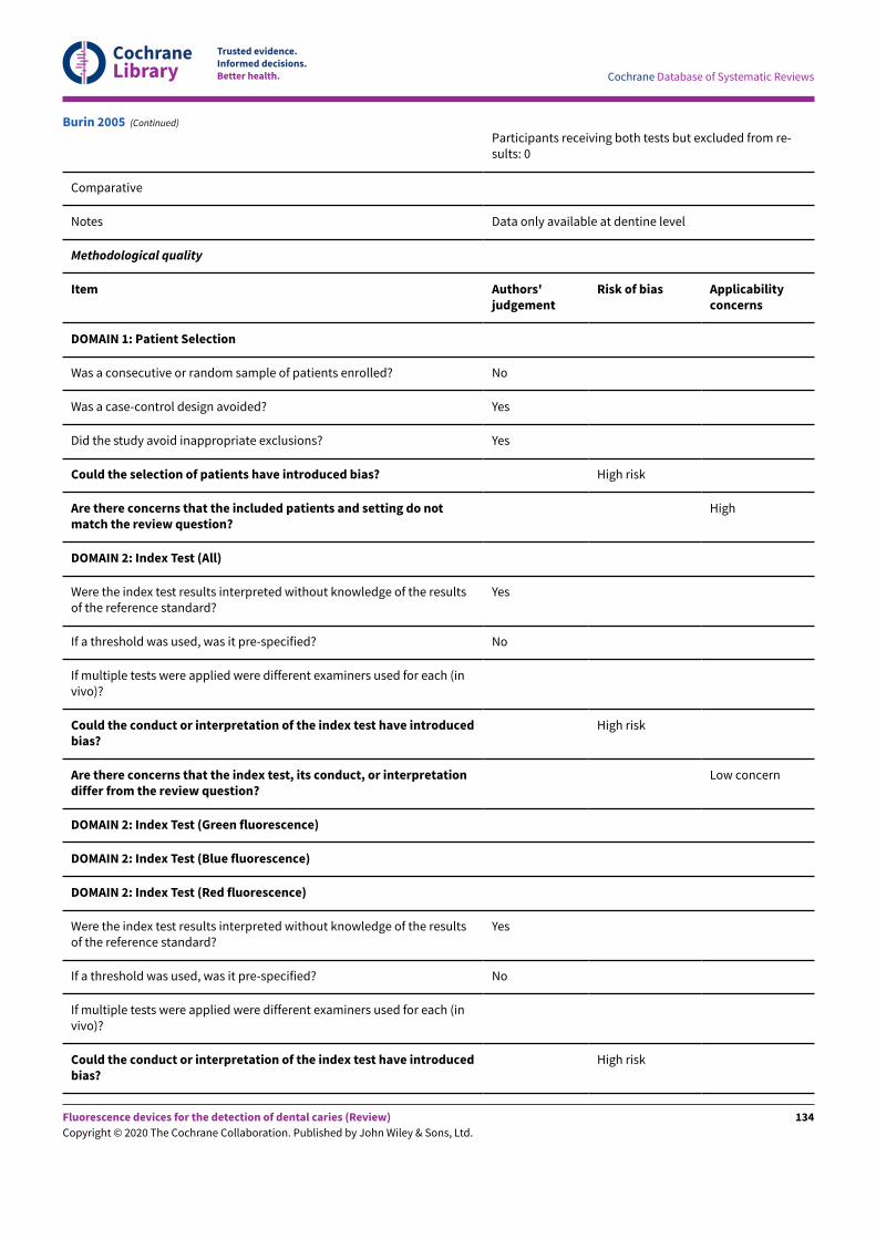
CochraneLibrary
Trusted evidence.Informed decisions.Better health.
Cochrane Database of Systematic Reviews
Participants receiving both tests but excluded from re-sults: 0
Comparative
Notes Data only available at dentine level
Methodological quality
Item Authors'judgement
Risk of bias Applicabilityconcerns
DOMAIN 1: Patient Selection
Was a consecutive or random sample of patients enrolled? No
Was a case-control design avoided? Yes
Did the study avoid inappropriate exclusions? Yes
Could the selection of patients have introduced bias? High risk
Are there concerns that the included patients and setting do notmatch the review question?
High
DOMAIN 2: Index Test (All)
Were the index test results interpreted without knowledge of the resultsof the reference standard?
Yes
If a threshold was used, was it pre-specified? No
If multiple tests were applied were different examiners used for each (invivo)?
Could the conduct or interpretation of the index test have introducedbias?
High risk
Are there concerns that the index test, its conduct, or interpretationdiffer from the review question?
Low concern
DOMAIN 2: Index Test (Green fluorescence)
DOMAIN 2: Index Test (Blue fluorescence)
DOMAIN 2: Index Test (Red fluorescence)
Were the index test results interpreted without knowledge of the resultsof the reference standard?
Yes
If a threshold was used, was it pre-specified? No
If multiple tests were applied were different examiners used for each (invivo)?
Could the conduct or interpretation of the index test have introducedbias?
High risk
Burin 2005 (Continued)
Fluorescence devices for the detection of dental caries (Review)
Copyright © 2020 The Cochrane Collaboration. Published by John Wiley & Sons, Ltd.
134
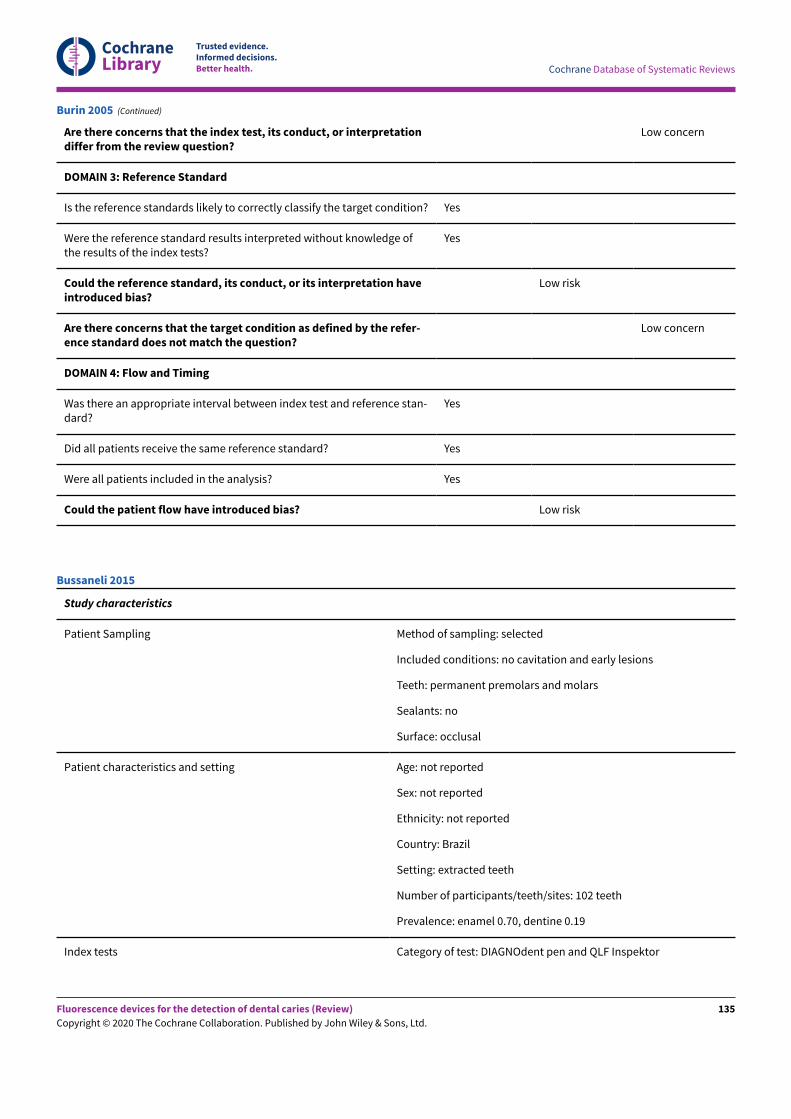
CochraneLibrary
Trusted evidence.Informed decisions.Better health.
Cochrane Database of Systematic Reviews
Are there concerns that the index test, its conduct, or interpretationdiffer from the review question?
Low concern
DOMAIN 3: Reference Standard
Is the reference standards likely to correctly classify the target condition? Yes
Were the reference standard results interpreted without knowledge ofthe results of the index tests?
Yes
Could the reference standard, its conduct, or its interpretation haveintroduced bias?
Low risk
Are there concerns that the target condition as defined by the refer-ence standard does not match the question?
Low concern
DOMAIN 4: Flow and Timing
Was there an appropriate interval between index test and reference stan-dard?
Yes
Did all patients receive the same reference standard? Yes
Were all patients included in the analysis? Yes
Could the patient flow have introduced bias? Low risk
Burin 2005 (Continued)
Study characteristics
Patient Sampling Method of sampling: selected
Included conditions: no cavitation and early lesions
Teeth: permanent premolars and molars
Sealants: no
Surface: occlusal
Patient characteristics and setting Age: not reported
Sex: not reported
Ethnicity: not reported
Country: Brazil
Setting: extracted teeth
Number of participants/teeth/sites: 102 teeth
Prevalence: enamel 0.70, dentine 0.19
Index tests Category of test: DIAGNOdent pen and QLF Inspektor
Bussaneli 2015
Fluorescence devices for the detection of dental caries (Review)
Copyright © 2020 The Cochrane Collaboration. Published by John Wiley & Sons, Ltd.
135
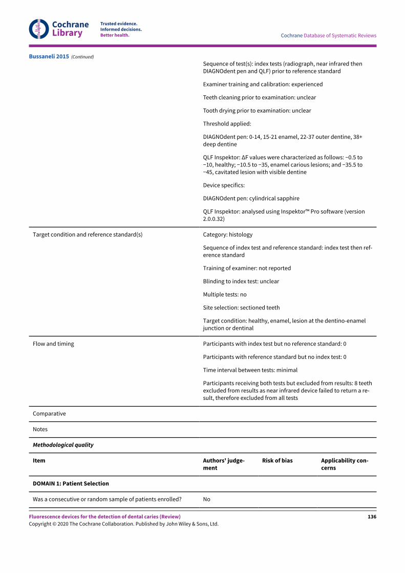
CochraneLibrary
Trusted evidence.Informed decisions.Better health.
Cochrane Database of Systematic Reviews
Sequence of test(s): index tests (radiograph, near infrared thenDIAGNOdent pen and QLF) prior to reference standard
Examiner training and calibration: experienced
Teeth cleaning prior to examination: unclear
Tooth drying prior to examination: unclear
Threshold applied:
DIAGNOdent pen: 0-14, 15-21 enamel, 22-37 outer dentine, 38+deep dentine
QLF Inspektor: ΔF values were characterized as follows: −0.5 to−10, healthy; −10.5 to −35, enamel carious lesions; and −35.5 to−45, cavitated lesion with visible dentine
Device specifics:
DIAGNOdent pen: cylindrical sapphire
QLF Inspektor: analysed using Inspektor™ Pro software (version2.0.0.32)
Target condition and reference standard(s) Category: histology
Sequence of index test and reference standard: index test then ref-erence standard
Training of examiner: not reported
Blinding to index test: unclear
Multiple tests: no
Site selection: sectioned teeth
Target condition: healthy, enamel, lesion at the dentino-enameljunction or dentinal
Flow and timing Participants with index test but no reference standard: 0
Participants with reference standard but no index test: 0
Time interval between tests: minimal
Participants receiving both tests but excluded from results: 8 teethexcluded from results as near infrared device failed to return a re-sult, therefore excluded from all tests
Comparative
Notes
Methodological quality
Item Authors' judge-ment
Risk of bias Applicability con-cerns
DOMAIN 1: Patient Selection
Was a consecutive or random sample of patients enrolled? No
Bussaneli 2015 (Continued)
Fluorescence devices for the detection of dental caries (Review)
Copyright © 2020 The Cochrane Collaboration. Published by John Wiley & Sons, Ltd.
136
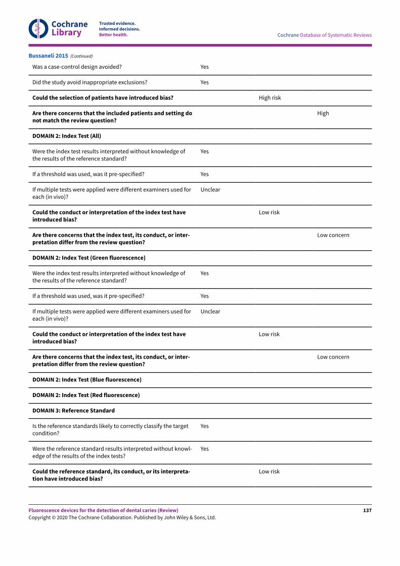
CochraneLibrary
Trusted evidence.Informed decisions.Better health.
Cochrane Database of Systematic Reviews
Was a case-control design avoided? Yes
Did the study avoid inappropriate exclusions? Yes
Could the selection of patients have introduced bias? High risk
Are there concerns that the included patients and setting donot match the review question?
High
DOMAIN 2: Index Test (All)
Were the index test results interpreted without knowledge ofthe results of the reference standard?
Yes
If a threshold was used, was it pre-specified? Yes
If multiple tests were applied were different examiners used foreach (in vivo)?
Unclear
Could the conduct or interpretation of the index test haveintroduced bias?
Low risk
Are there concerns that the index test, its conduct, or inter-pretation differ from the review question?
Low concern
DOMAIN 2: Index Test (Green fluorescence)
Were the index test results interpreted without knowledge ofthe results of the reference standard?
Yes
If a threshold was used, was it pre-specified? Yes
If multiple tests were applied were different examiners used foreach (in vivo)?
Unclear
Could the conduct or interpretation of the index test haveintroduced bias?
Low risk
Are there concerns that the index test, its conduct, or inter-pretation differ from the review question?
Low concern
DOMAIN 2: Index Test (Blue fluorescence)
DOMAIN 2: Index Test (Red fluorescence)
DOMAIN 3: Reference Standard
Is the reference standards likely to correctly classify the targetcondition?
Yes
Were the reference standard results interpreted without knowl-edge of the results of the index tests?
Yes
Could the reference standard, its conduct, or its interpreta-tion have introduced bias?
Low risk
Bussaneli 2015 (Continued)
Fluorescence devices for the detection of dental caries (Review)
Copyright © 2020 The Cochrane Collaboration. Published by John Wiley & Sons, Ltd.
137
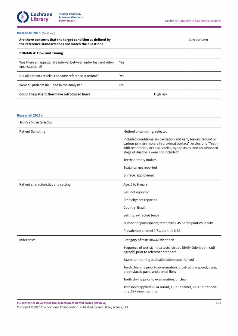
CochraneLibrary
Trusted evidence.Informed decisions.Better health.
Cochrane Database of Systematic Reviews
Are there concerns that the target condition as defined bythe reference standard does not match the question?
Low concern
DOMAIN 4: Flow and Timing
Was there an appropriate interval between index test and refer-ence standard?
Yes
Did all patients receive the same reference standard? Yes
Were all patients included in the analysis? No
Could the patient flow have introduced bias? High risk
Bussaneli 2015 (Continued)
Study characteristics
Patient Sampling Method of sampling: selected
Included conditions: no cavitation and early lesions "sound orcarious primary molars in proximal contact", exclusions "Teethwith restoration, occlusal caries, hypoplasias, and an advancedstage of rhizolysis were not included"
Teeth: primary molars
Sealants: not reported
Surface: approximal
Patient characteristics and setting Age: 5 to 9 years
Sex: not reported
Ethnicity: not reported
Country: Brazil
Setting: extracted teeth
Number of participants/teeth/sites: 45 participants/59 teeth
Prevalence: enamel 0.71, dentine 0.58
Index tests Category of test: DIAGNOdent pen
Sequence of test(s): index tests (visual, DIAGNOdent pen, radi-ograph) prior to reference standard
Examiner training and calibration: experienced
Teeth cleaning prior to examination: brush at low speed, usingprophylactic paste and dental floss
Tooth drying prior to examination: unclear
Threshold applied: 0-14 sound, 15-21 enamel, 22-37 outer den-tine, 38+ inner dentine
Bussaneli 2015a
Fluorescence devices for the detection of dental caries (Review)
Copyright © 2020 The Cochrane Collaboration. Published by John Wiley & Sons, Ltd.
138
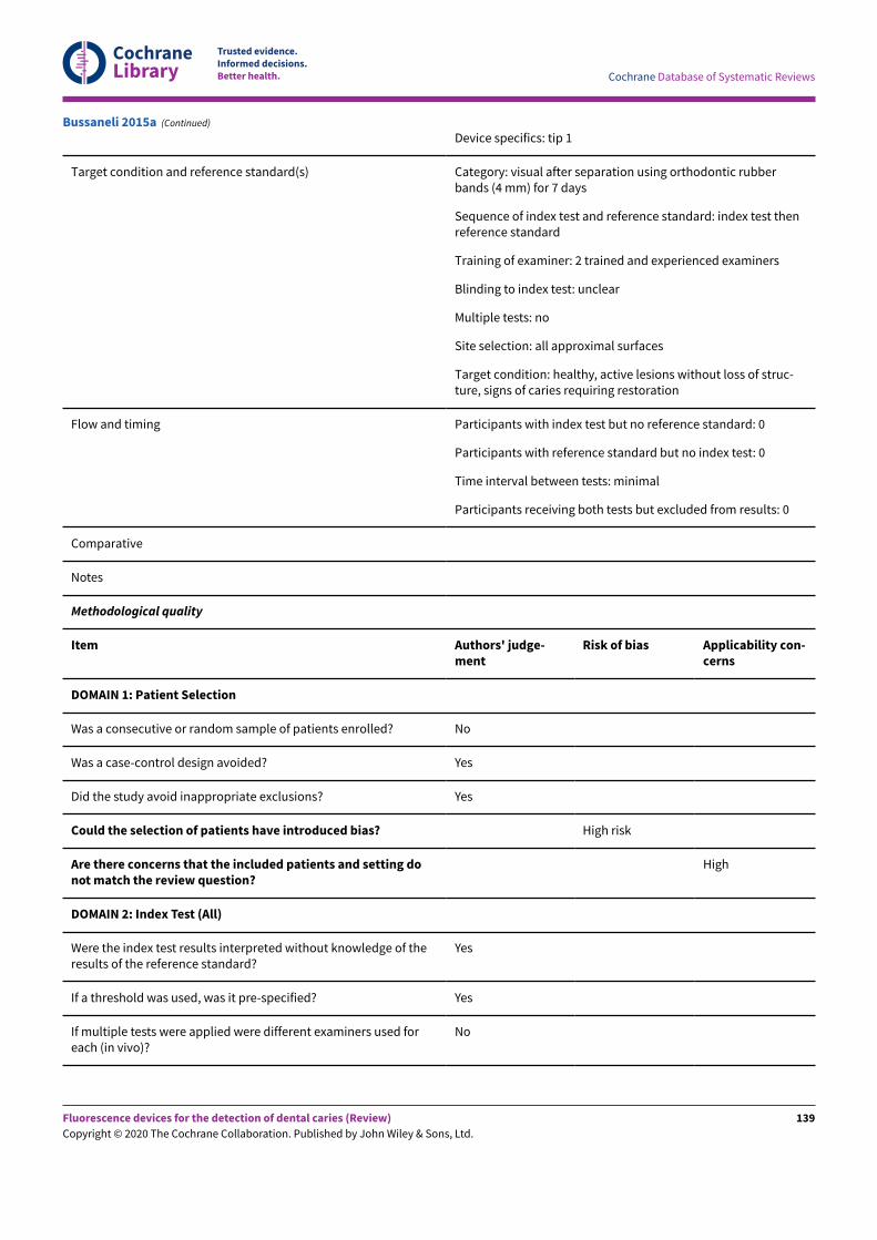
CochraneLibrary
Trusted evidence.Informed decisions.Better health.
Cochrane Database of Systematic Reviews
Device specifics: tip 1
Target condition and reference standard(s) Category: visual after separation using orthodontic rubberbands (4 mm) for 7 days
Sequence of index test and reference standard: index test thenreference standard
Training of examiner: 2 trained and experienced examiners
Blinding to index test: unclear
Multiple tests: no
Site selection: all approximal surfaces
Target condition: healthy, active lesions without loss of struc-ture, signs of caries requiring restoration
Flow and timing Participants with index test but no reference standard: 0
Participants with reference standard but no index test: 0
Time interval between tests: minimal
Participants receiving both tests but excluded from results: 0
Comparative
Notes
Methodological quality
Item Authors' judge-ment
Risk of bias Applicability con-cerns
DOMAIN 1: Patient Selection
Was a consecutive or random sample of patients enrolled? No
Was a case-control design avoided? Yes
Did the study avoid inappropriate exclusions? Yes
Could the selection of patients have introduced bias? High risk
Are there concerns that the included patients and setting donot match the review question?
High
DOMAIN 2: Index Test (All)
Were the index test results interpreted without knowledge of theresults of the reference standard?
Yes
If a threshold was used, was it pre-specified? Yes
If multiple tests were applied were different examiners used foreach (in vivo)?
No
Bussaneli 2015a (Continued)
Fluorescence devices for the detection of dental caries (Review)
Copyright © 2020 The Cochrane Collaboration. Published by John Wiley & Sons, Ltd.
139
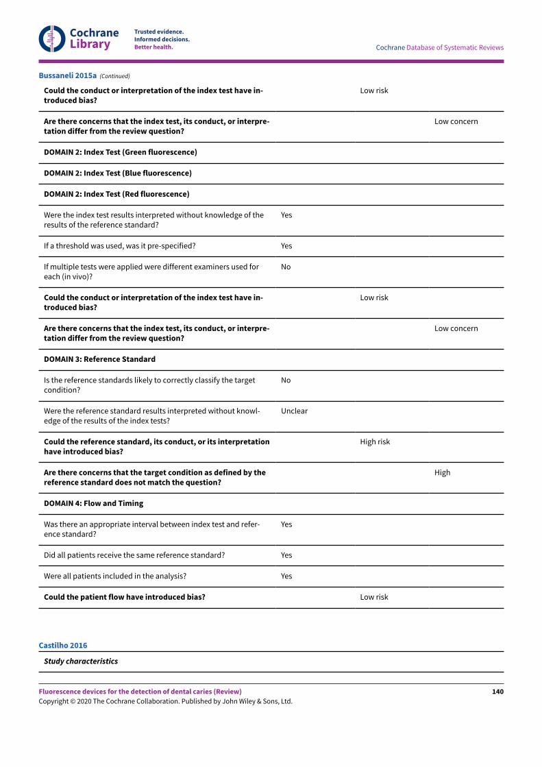
CochraneLibrary
Trusted evidence.Informed decisions.Better health.
Cochrane Database of Systematic Reviews
Could the conduct or interpretation of the index test have in-troduced bias?
Low risk
Are there concerns that the index test, its conduct, or interpre-tation differ from the review question?
Low concern
DOMAIN 2: Index Test (Green fluorescence)
DOMAIN 2: Index Test (Blue fluorescence)
DOMAIN 2: Index Test (Red fluorescence)
Were the index test results interpreted without knowledge of theresults of the reference standard?
Yes
If a threshold was used, was it pre-specified? Yes
If multiple tests were applied were different examiners used foreach (in vivo)?
No
Could the conduct or interpretation of the index test have in-troduced bias?
Low risk
Are there concerns that the index test, its conduct, or interpre-tation differ from the review question?
Low concern
DOMAIN 3: Reference Standard
Is the reference standards likely to correctly classify the targetcondition?
No
Were the reference standard results interpreted without knowl-edge of the results of the index tests?
Unclear
Could the reference standard, its conduct, or its interpretationhave introduced bias?
High risk
Are there concerns that the target condition as defined by thereference standard does not match the question?
High
DOMAIN 4: Flow and Timing
Was there an appropriate interval between index test and refer-ence standard?
Yes
Did all patients receive the same reference standard? Yes
Were all patients included in the analysis? Yes
Could the patient flow have introduced bias? Low risk
Bussaneli 2015a (Continued)
Study characteristics
Castilho 2016
Fluorescence devices for the detection of dental caries (Review)
Copyright © 2020 The Cochrane Collaboration. Published by John Wiley & Sons, Ltd.
140
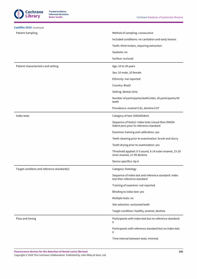
CochraneLibrary
Trusted evidence.Informed decisions.Better health.
Cochrane Database of Systematic Reviews
Patient Sampling Method of sampling: consecutive
Included conditions: no cavitation and early lesions
Teeth: third molars, requiring extraction
Sealants: no
Surface: occlusal
Patient characteristics and setting Age: 16 to 39 years
Sex: 10 male, 16 female
Ethnicity: not reported
Country: Brazil
Setting: dental clinic
Number of participants/teeth/sites: 26 participants/43teeth
Prevalence: enamel 0.81, dentine 0.07
Index tests Category of test: DIAGNOdent
Sequence of test(s): index tests (visual then DIAGN-Odent pen) prior to reference standard
Examiner training and calibration: yes
Teeth cleaning prior to examination: brush and slurry
Tooth drying prior to examination: yes
Threshold applied: 0-5 sound, 6-14 outer enamel, 15-20inner enamel, 21-99 dentine
Device specifics: tip A
Target condition and reference standard(s) Category: histology
Sequence of index test and reference standard: indextest then reference standard
Training of examiner: not reported
Blinding to index test: yes
Multiple tests: no
Site selection: sectioned teeth
Target condition: healthy, enamel, dentine
Flow and timing Participants with index test but no reference standard:0
Participants with reference standard but no index test:0
Time interval between tests: minimal
Castilho 2016 (Continued)
Fluorescence devices for the detection of dental caries (Review)
Copyright © 2020 The Cochrane Collaboration. Published by John Wiley & Sons, Ltd.
141
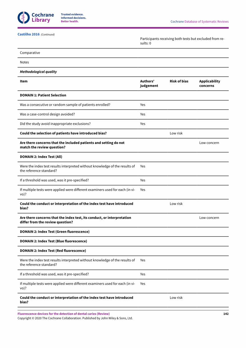
CochraneLibrary
Trusted evidence.Informed decisions.Better health.
Cochrane Database of Systematic Reviews
Participants receiving both tests but excluded from re-sults: 0
Comparative
Notes
Methodological quality
Item Authors'judgement
Risk of bias Applicabilityconcerns
DOMAIN 1: Patient Selection
Was a consecutive or random sample of patients enrolled? Yes
Was a case-control design avoided? Yes
Did the study avoid inappropriate exclusions? Yes
Could the selection of patients have introduced bias? Low risk
Are there concerns that the included patients and setting do notmatch the review question?
Low concern
DOMAIN 2: Index Test (All)
Were the index test results interpreted without knowledge of the results ofthe reference standard?
Yes
If a threshold was used, was it pre-specified? Yes
If multiple tests were applied were different examiners used for each (in vi-vo)?
Yes
Could the conduct or interpretation of the index test have introducedbias?
Low risk
Are there concerns that the index test, its conduct, or interpretationdiffer from the review question?
Low concern
DOMAIN 2: Index Test (Green fluorescence)
DOMAIN 2: Index Test (Blue fluorescence)
DOMAIN 2: Index Test (Red fluorescence)
Were the index test results interpreted without knowledge of the results ofthe reference standard?
Yes
If a threshold was used, was it pre-specified? Yes
If multiple tests were applied were different examiners used for each (in vi-vo)?
Yes
Could the conduct or interpretation of the index test have introducedbias?
Low risk
Castilho 2016 (Continued)
Fluorescence devices for the detection of dental caries (Review)
Copyright © 2020 The Cochrane Collaboration. Published by John Wiley & Sons, Ltd.
142
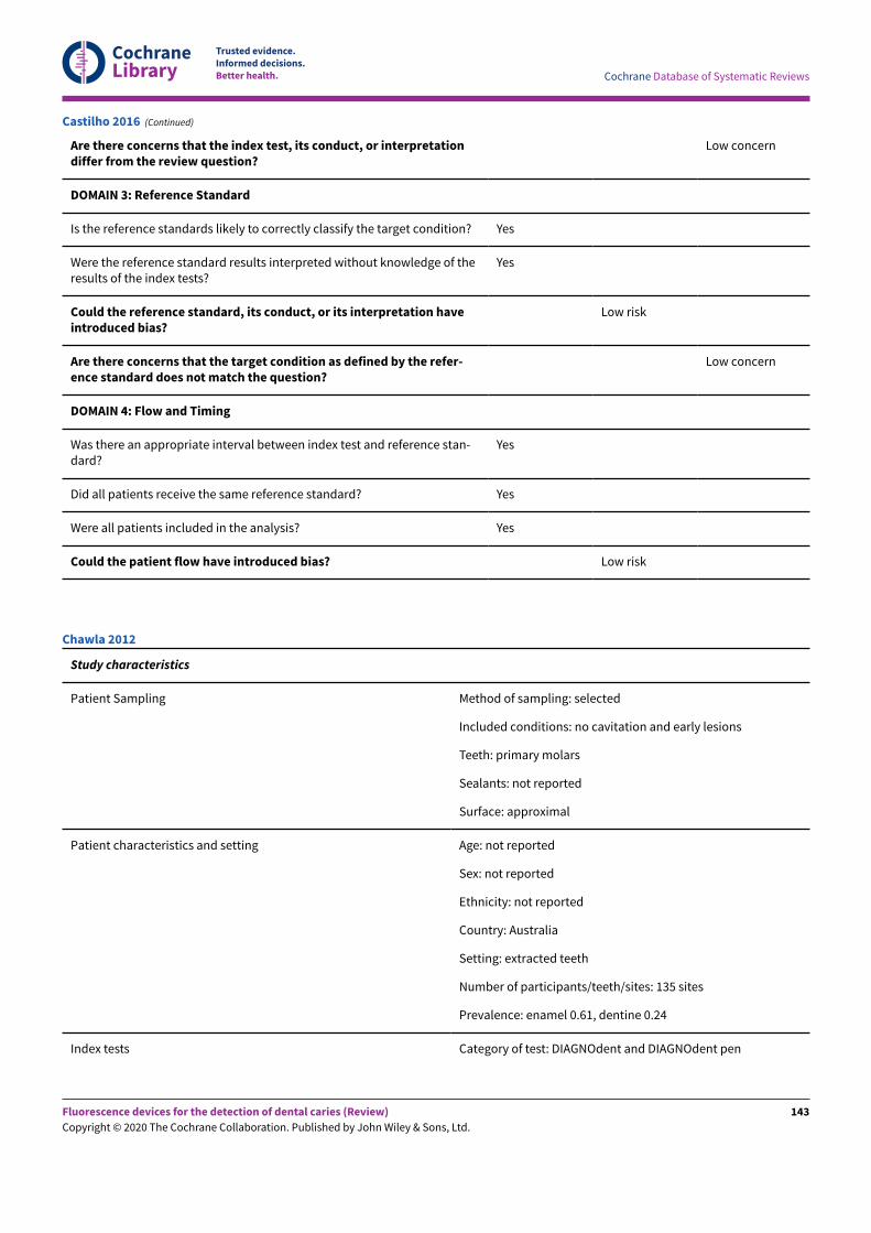
CochraneLibrary
Trusted evidence.Informed decisions.Better health.
Cochrane Database of Systematic Reviews
Are there concerns that the index test, its conduct, or interpretationdiffer from the review question?
Low concern
DOMAIN 3: Reference Standard
Is the reference standards likely to correctly classify the target condition? Yes
Were the reference standard results interpreted without knowledge of theresults of the index tests?
Yes
Could the reference standard, its conduct, or its interpretation haveintroduced bias?
Low risk
Are there concerns that the target condition as defined by the refer-ence standard does not match the question?
Low concern
DOMAIN 4: Flow and Timing
Was there an appropriate interval between index test and reference stan-dard?
Yes
Did all patients receive the same reference standard? Yes
Were all patients included in the analysis? Yes
Could the patient flow have introduced bias? Low risk
Castilho 2016 (Continued)
Study characteristics
Patient Sampling Method of sampling: selected
Included conditions: no cavitation and early lesions
Teeth: primary molars
Sealants: not reported
Surface: approximal
Patient characteristics and setting Age: not reported
Sex: not reported
Ethnicity: not reported
Country: Australia
Setting: extracted teeth
Number of participants/teeth/sites: 135 sites
Prevalence: enamel 0.61, dentine 0.24
Index tests Category of test: DIAGNOdent and DIAGNOdent pen
Chawla 2012
Fluorescence devices for the detection of dental caries (Review)
Copyright © 2020 The Cochrane Collaboration. Published by John Wiley & Sons, Ltd.
143
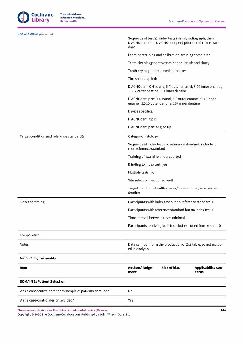
CochraneLibrary
Trusted evidence.Informed decisions.Better health.
Cochrane Database of Systematic Reviews
Sequence of test(s): index tests (visual, radiograph, thenDIAGNOdent then DIAGNOdent pen) prior to reference stan-dard
Examiner training and calibration: training completed
Teeth cleaning prior to examination: brush and slurry
Tooth drying prior to examination: yes
Threshold applied:
DIAGNOdent: 0-4 sound, 5-7 outer enamel, 8-10 inner enamel,11-12 outer dentine, 13+ inner dentine
DIAGNOdent pen: 0-4 sound, 5-8 outer enamel, 9-11 innerenamel, 12-15 outer dentine, 16+ inner dentine
Device specifics:
DIAGNOdent: tip B
DIAGNOdent pen: angled tip
Target condition and reference standard(s) Category: histology
Sequence of index test and reference standard: index testthen reference standard
Training of examiner: not reported
Blinding to index test: yes
Multiple tests: no
Site selection: sectioned teeth
Target condition: healthy, inner/outer enamel, inner/outerdentine
Flow and timing Participants with index test but no reference standard: 0
Participants with reference standard but no index test: 0
Time interval between tests: minimal
Participants receiving both tests but excluded from results: 0
Comparative
Notes Data cannot inform the production of 2x2 table, so not includ-ed in analysis
Methodological quality
Item Authors' judge-ment
Risk of bias Applicability con-cerns
DOMAIN 1: Patient Selection
Was a consecutive or random sample of patients enrolled? No
Was a case-control design avoided? Yes
Chawla 2012 (Continued)
Fluorescence devices for the detection of dental caries (Review)
Copyright © 2020 The Cochrane Collaboration. Published by John Wiley & Sons, Ltd.
144
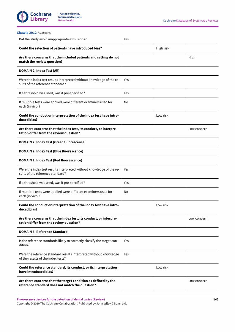
CochraneLibrary
Trusted evidence.Informed decisions.Better health.
Cochrane Database of Systematic Reviews
Did the study avoid inappropriate exclusions? Yes
Could the selection of patients have introduced bias? High risk
Are there concerns that the included patients and setting do notmatch the review question?
High
DOMAIN 2: Index Test (All)
Were the index test results interpreted without knowledge of the re-sults of the reference standard?
Yes
If a threshold was used, was it pre-specified? Yes
If multiple tests were applied were different examiners used foreach (in vivo)?
No
Could the conduct or interpretation of the index test have intro-duced bias?
Low risk
Are there concerns that the index test, its conduct, or interpre-tation differ from the review question?
Low concern
DOMAIN 2: Index Test (Green fluorescence)
DOMAIN 2: Index Test (Blue fluorescence)
DOMAIN 2: Index Test (Red fluorescence)
Were the index test results interpreted without knowledge of the re-sults of the reference standard?
Yes
If a threshold was used, was it pre-specified? Yes
If multiple tests were applied were different examiners used foreach (in vivo)?
No
Could the conduct or interpretation of the index test have intro-duced bias?
Low risk
Are there concerns that the index test, its conduct, or interpre-tation differ from the review question?
Low concern
DOMAIN 3: Reference Standard
Is the reference standards likely to correctly classify the target con-dition?
Yes
Were the reference standard results interpreted without knowledgeof the results of the index tests?
Yes
Could the reference standard, its conduct, or its interpretationhave introduced bias?
Low risk
Are there concerns that the target condition as defined by thereference standard does not match the question?
Low concern
Chawla 2012 (Continued)
Fluorescence devices for the detection of dental caries (Review)
Copyright © 2020 The Cochrane Collaboration. Published by John Wiley & Sons, Ltd.
145
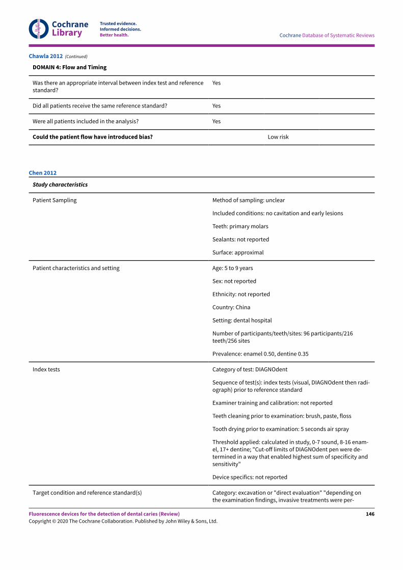
CochraneLibrary
Trusted evidence.Informed decisions.Better health.
Cochrane Database of Systematic Reviews
DOMAIN 4: Flow and Timing
Was there an appropriate interval between index test and referencestandard?
Yes
Did all patients receive the same reference standard? Yes
Were all patients included in the analysis? Yes
Could the patient flow have introduced bias? Low risk
Chawla 2012 (Continued)
Study characteristics
Patient Sampling Method of sampling: unclear
Included conditions: no cavitation and early lesions
Teeth: primary molars
Sealants: not reported
Surface: approximal
Patient characteristics and setting Age: 5 to 9 years
Sex: not reported
Ethnicity: not reported
Country: China
Setting: dental hospital
Number of participants/teeth/sites: 96 participants/216teeth/256 sites
Prevalence: enamel 0.50, dentine 0.35
Index tests Category of test: DIAGNOdent
Sequence of test(s): index tests (visual, DIAGNOdent then radi-ograph) prior to reference standard
Examiner training and calibration: not reported
Teeth cleaning prior to examination: brush, paste, floss
Tooth drying prior to examination: 5 seconds air spray
Threshold applied: calculated in study, 0-7 sound, 8-16 enam-el, 17+ dentine; "Cut-oE limits of DIAGNOdent pen were de-termined in a way that enabled highest sum of specificity andsensitivity"
Device specifics: not reported
Target condition and reference standard(s) Category: excavation or "direct evaluation" "depending onthe examination findings, invasive treatments were per-
Chen 2012
Fluorescence devices for the detection of dental caries (Review)
Copyright © 2020 The Cochrane Collaboration. Published by John Wiley & Sons, Ltd.
146
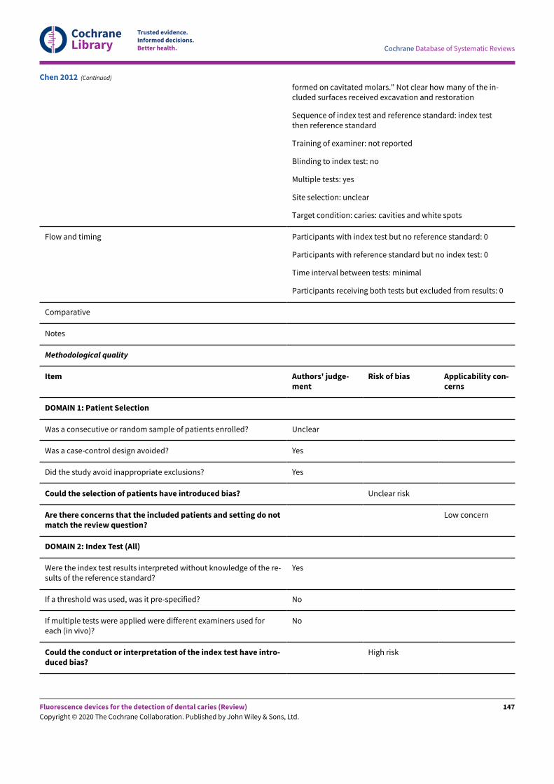
CochraneLibrary
Trusted evidence.Informed decisions.Better health.
Cochrane Database of Systematic Reviews
formed on cavitated molars." Not clear how many of the in-cluded surfaces received excavation and restoration
Sequence of index test and reference standard: index testthen reference standard
Training of examiner: not reported
Blinding to index test: no
Multiple tests: yes
Site selection: unclear
Target condition: caries: cavities and white spots
Flow and timing Participants with index test but no reference standard: 0
Participants with reference standard but no index test: 0
Time interval between tests: minimal
Participants receiving both tests but excluded from results: 0
Comparative
Notes
Methodological quality
Item Authors' judge-ment
Risk of bias Applicability con-cerns
DOMAIN 1: Patient Selection
Was a consecutive or random sample of patients enrolled? Unclear
Was a case-control design avoided? Yes
Did the study avoid inappropriate exclusions? Yes
Could the selection of patients have introduced bias? Unclear risk
Are there concerns that the included patients and setting do notmatch the review question?
Low concern
DOMAIN 2: Index Test (All)
Were the index test results interpreted without knowledge of the re-sults of the reference standard?
Yes
If a threshold was used, was it pre-specified? No
If multiple tests were applied were different examiners used foreach (in vivo)?
No
Could the conduct or interpretation of the index test have intro-duced bias?
High risk
Chen 2012 (Continued)
Fluorescence devices for the detection of dental caries (Review)
Copyright © 2020 The Cochrane Collaboration. Published by John Wiley & Sons, Ltd.
147
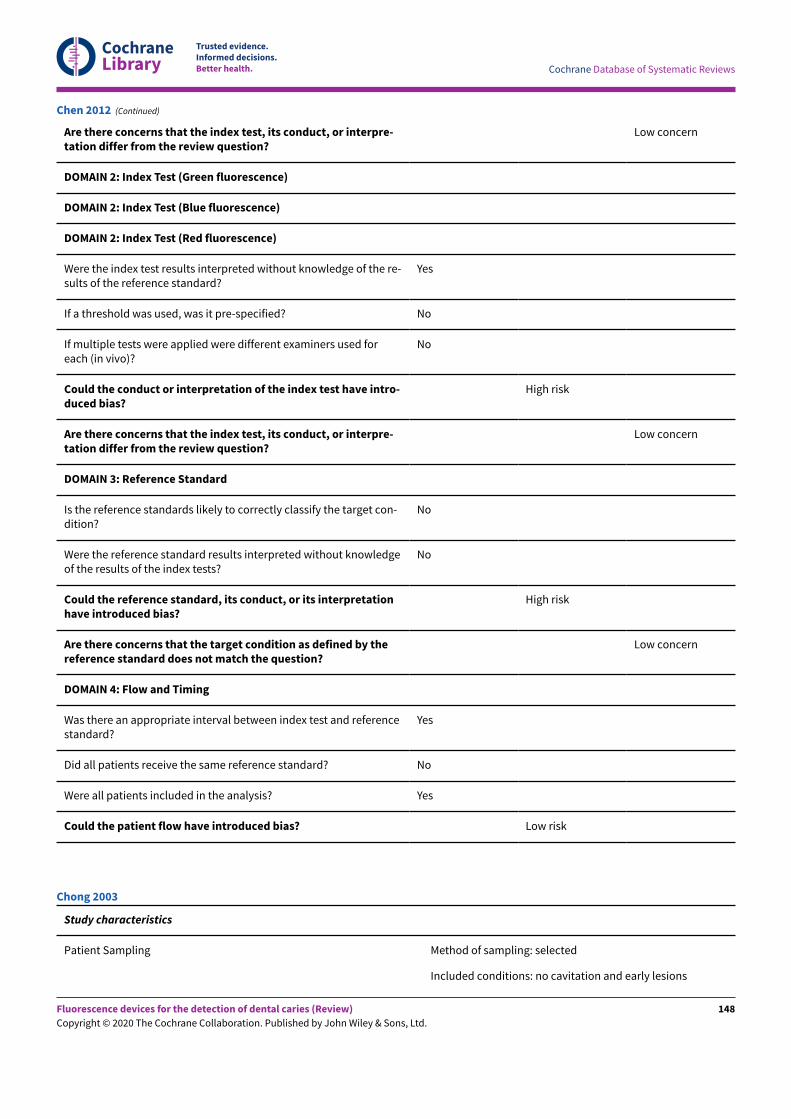
CochraneLibrary
Trusted evidence.Informed decisions.Better health.
Cochrane Database of Systematic Reviews
Are there concerns that the index test, its conduct, or interpre-tation differ from the review question?
Low concern
DOMAIN 2: Index Test (Green fluorescence)
DOMAIN 2: Index Test (Blue fluorescence)
DOMAIN 2: Index Test (Red fluorescence)
Were the index test results interpreted without knowledge of the re-sults of the reference standard?
Yes
If a threshold was used, was it pre-specified? No
If multiple tests were applied were different examiners used foreach (in vivo)?
No
Could the conduct or interpretation of the index test have intro-duced bias?
High risk
Are there concerns that the index test, its conduct, or interpre-tation differ from the review question?
Low concern
DOMAIN 3: Reference Standard
Is the reference standards likely to correctly classify the target con-dition?
No
Were the reference standard results interpreted without knowledgeof the results of the index tests?
No
Could the reference standard, its conduct, or its interpretationhave introduced bias?
High risk
Are there concerns that the target condition as defined by thereference standard does not match the question?
Low concern
DOMAIN 4: Flow and Timing
Was there an appropriate interval between index test and referencestandard?
Yes
Did all patients receive the same reference standard? No
Were all patients included in the analysis? Yes
Could the patient flow have introduced bias? Low risk
Chen 2012 (Continued)
Study characteristics
Patient Sampling Method of sampling: selected
Included conditions: no cavitation and early lesions
Chong 2003
Fluorescence devices for the detection of dental caries (Review)
Copyright © 2020 The Cochrane Collaboration. Published by John Wiley & Sons, Ltd.
148
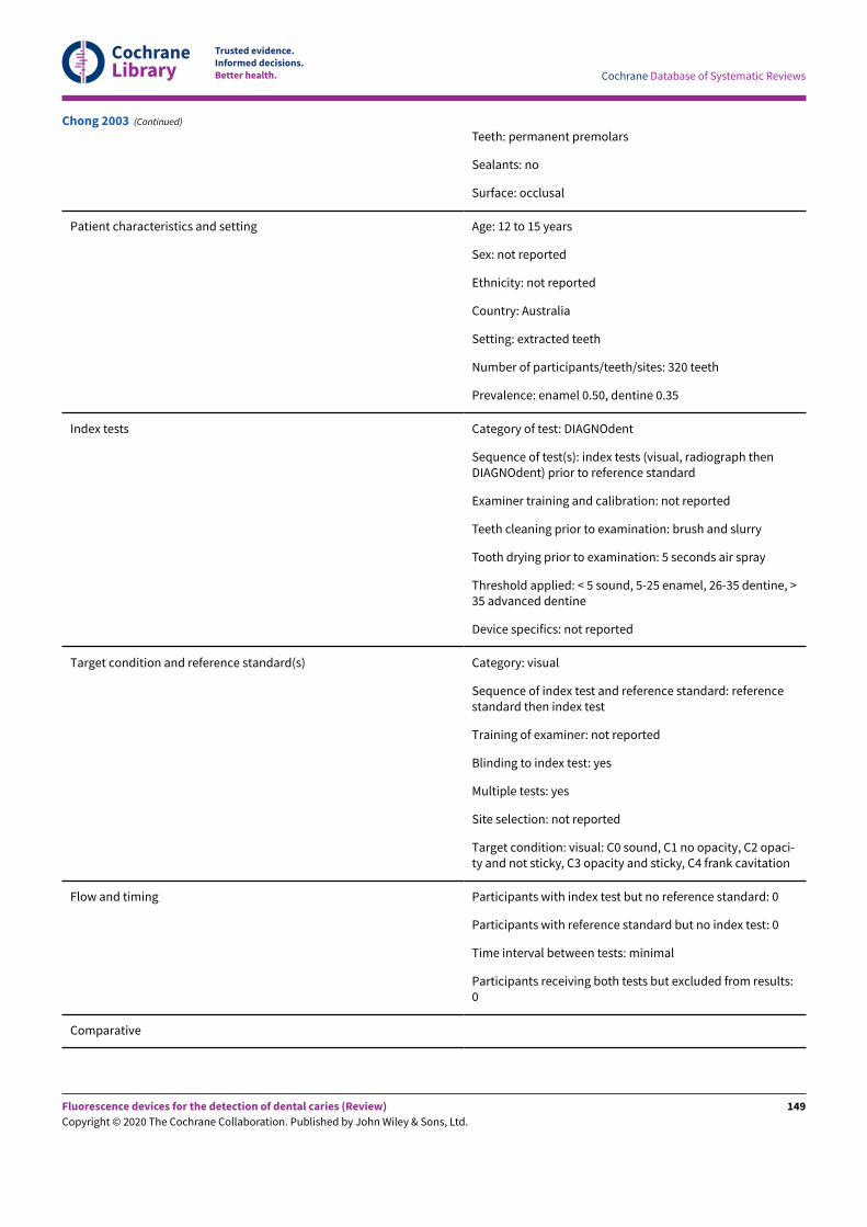
CochraneLibrary
Trusted evidence.Informed decisions.Better health.
Cochrane Database of Systematic Reviews
Teeth: permanent premolars
Sealants: no
Surface: occlusal
Patient characteristics and setting Age: 12 to 15 years
Sex: not reported
Ethnicity: not reported
Country: Australia
Setting: extracted teeth
Number of participants/teeth/sites: 320 teeth
Prevalence: enamel 0.50, dentine 0.35
Index tests Category of test: DIAGNOdent
Sequence of test(s): index tests (visual, radiograph thenDIAGNOdent) prior to reference standard
Examiner training and calibration: not reported
Teeth cleaning prior to examination: brush and slurry
Tooth drying prior to examination: 5 seconds air spray
Threshold applied: < 5 sound, 5-25 enamel, 26-35 dentine, >35 advanced dentine
Device specifics: not reported
Target condition and reference standard(s) Category: visual
Sequence of index test and reference standard: referencestandard then index test
Training of examiner: not reported
Blinding to index test: yes
Multiple tests: yes
Site selection: not reported
Target condition: visual: C0 sound, C1 no opacity, C2 opaci-ty and not sticky, C3 opacity and sticky, C4 frank cavitation
Flow and timing Participants with index test but no reference standard: 0
Participants with reference standard but no index test: 0
Time interval between tests: minimal
Participants receiving both tests but excluded from results:0
Comparative
Chong 2003 (Continued)
Fluorescence devices for the detection of dental caries (Review)
Copyright © 2020 The Cochrane Collaboration. Published by John Wiley & Sons, Ltd.
149
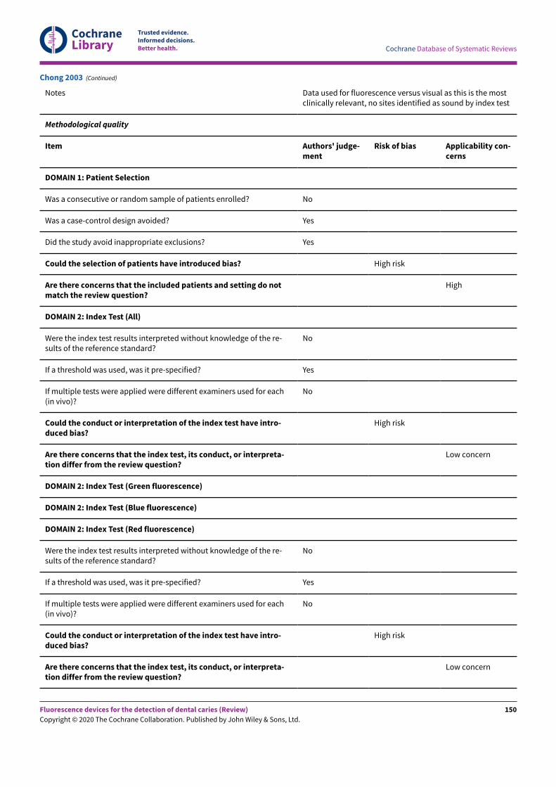
CochraneLibrary
Trusted evidence.Informed decisions.Better health.
Cochrane Database of Systematic Reviews
Notes Data used for fluorescence versus visual as this is the mostclinically relevant, no sites identified as sound by index test
Methodological quality
Item Authors' judge-ment
Risk of bias Applicability con-cerns
DOMAIN 1: Patient Selection
Was a consecutive or random sample of patients enrolled? No
Was a case-control design avoided? Yes
Did the study avoid inappropriate exclusions? Yes
Could the selection of patients have introduced bias? High risk
Are there concerns that the included patients and setting do notmatch the review question?
High
DOMAIN 2: Index Test (All)
Were the index test results interpreted without knowledge of the re-sults of the reference standard?
No
If a threshold was used, was it pre-specified? Yes
If multiple tests were applied were different examiners used for each(in vivo)?
No
Could the conduct or interpretation of the index test have intro-duced bias?
High risk
Are there concerns that the index test, its conduct, or interpreta-tion differ from the review question?
Low concern
DOMAIN 2: Index Test (Green fluorescence)
DOMAIN 2: Index Test (Blue fluorescence)
DOMAIN 2: Index Test (Red fluorescence)
Were the index test results interpreted without knowledge of the re-sults of the reference standard?
No
If a threshold was used, was it pre-specified? Yes
If multiple tests were applied were different examiners used for each(in vivo)?
No
Could the conduct or interpretation of the index test have intro-duced bias?
High risk
Are there concerns that the index test, its conduct, or interpreta-tion differ from the review question?
Low concern
Chong 2003 (Continued)
Fluorescence devices for the detection of dental caries (Review)
Copyright © 2020 The Cochrane Collaboration. Published by John Wiley & Sons, Ltd.
150
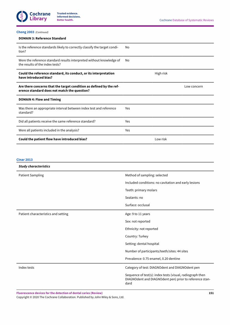
CochraneLibrary
Trusted evidence.Informed decisions.Better health.
Cochrane Database of Systematic Reviews
DOMAIN 3: Reference Standard
Is the reference standards likely to correctly classify the target condi-tion?
No
Were the reference standard results interpreted without knowledge ofthe results of the index tests?
No
Could the reference standard, its conduct, or its interpretationhave introduced bias?
High risk
Are there concerns that the target condition as defined by the ref-erence standard does not match the question?
Low concern
DOMAIN 4: Flow and Timing
Was there an appropriate interval between index test and referencestandard?
Yes
Did all patients receive the same reference standard? Yes
Were all patients included in the analysis? Yes
Could the patient flow have introduced bias? Low risk
Chong 2003 (Continued)
Study characteristics
Patient Sampling Method of sampling: selected
Included conditions: no cavitation and early lesions
Teeth: primary molars
Sealants: no
Surface: occlusal
Patient characteristics and setting Age: 9 to 11 years
Sex: not reported
Ethnicity: not reported
Country: Turkey
Setting: dental hospital
Number of participants/teeth/sites: 44 sites
Prevalence: 0.75 enamel, 0.20 dentine
Index tests Category of test: DIAGNOdent and DIAGNOdent pen
Sequence of test(s): index tests (visual, radiograph thenDIAGNOdent and DIAGNOdent pen) prior to reference stan-dard
Cinar 2013
Fluorescence devices for the detection of dental caries (Review)
Copyright © 2020 The Cochrane Collaboration. Published by John Wiley & Sons, Ltd.
151
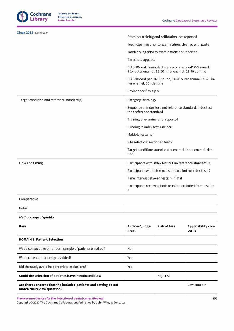
CochraneLibrary
Trusted evidence.Informed decisions.Better health.
Cochrane Database of Systematic Reviews
Examiner training and calibration: not reported
Teeth cleaning prior to examination: cleaned with paste
Tooth drying prior to examination: not reported
Threshold applied:
DIAGNOdent: "manufacturer recommended" 0-5 sound,6-14 outer enamel, 15-20 inner enamel, 21-99 dentine
DIAGNOdent pen: 0-13 sound, 14-20 outer enamel, 21-29 in-ner enamel, 30+ dentine
Device specifics: tip A
Target condition and reference standard(s) Category: histology
Sequence of index test and reference standard: index testthen reference standard
Training of examiner: not reported
Blinding to index test: unclear
Multiple tests: no
Site selection: sectioned teeth
Target condition: sound, outer enamel, inner enamel, den-tine
Flow and timing Participants with index test but no reference standard: 0
Participants with reference standard but no index test: 0
Time interval between tests: minimal
Participants receiving both tests but excluded from results:0
Comparative
Notes
Methodological quality
Item Authors' judge-ment
Risk of bias Applicability con-cerns
DOMAIN 1: Patient Selection
Was a consecutive or random sample of patients enrolled? No
Was a case-control design avoided? Yes
Did the study avoid inappropriate exclusions? Yes
Could the selection of patients have introduced bias? High risk
Are there concerns that the included patients and setting do notmatch the review question?
Low concern
Cinar 2013 (Continued)
Fluorescence devices for the detection of dental caries (Review)
Copyright © 2020 The Cochrane Collaboration. Published by John Wiley & Sons, Ltd.
152

CochraneLibrary
Trusted evidence.Informed decisions.Better health.
Cochrane Database of Systematic Reviews
DOMAIN 2: Index Test (All)
Were the index test results interpreted without knowledge of the re-sults of the reference standard?
Yes
If a threshold was used, was it pre-specified? Yes
If multiple tests were applied were different examiners used for each(in vivo)?
Yes
Could the conduct or interpretation of the index test have intro-duced bias?
Low risk
Are there concerns that the index test, its conduct, or interpreta-tion differ from the review question?
Low concern
DOMAIN 2: Index Test (Green fluorescence)
DOMAIN 2: Index Test (Blue fluorescence)
DOMAIN 2: Index Test (Red fluorescence)
Were the index test results interpreted without knowledge of the re-sults of the reference standard?
Yes
If a threshold was used, was it pre-specified? Yes
If multiple tests were applied were different examiners used for each(in vivo)?
Yes
Could the conduct or interpretation of the index test have intro-duced bias?
Low risk
Are there concerns that the index test, its conduct, or interpreta-tion differ from the review question?
Low concern
DOMAIN 3: Reference Standard
Is the reference standards likely to correctly classify the target condi-tion?
Yes
Were the reference standard results interpreted without knowledge ofthe results of the index tests?
Yes
Could the reference standard, its conduct, or its interpretationhave introduced bias?
Low risk
Are there concerns that the target condition as defined by the ref-erence standard does not match the question?
Low concern
DOMAIN 4: Flow and Timing
Was there an appropriate interval between index test and referencestandard?
Yes
Did all patients receive the same reference standard? Yes
Cinar 2013 (Continued)
Fluorescence devices for the detection of dental caries (Review)
Copyright © 2020 The Cochrane Collaboration. Published by John Wiley & Sons, Ltd.
153
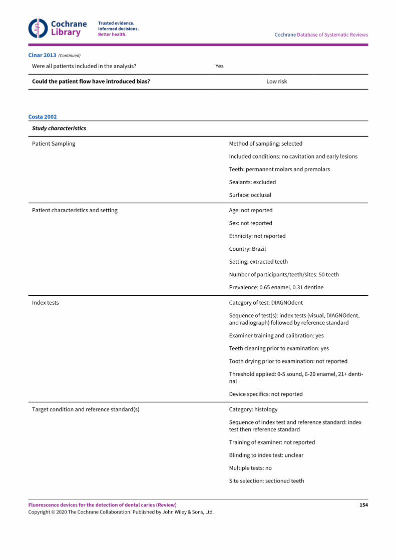
CochraneLibrary
Trusted evidence.Informed decisions.Better health.
Cochrane Database of Systematic Reviews
Were all patients included in the analysis? Yes
Could the patient flow have introduced bias? Low risk
Cinar 2013 (Continued)
Study characteristics
Patient Sampling Method of sampling: selected
Included conditions: no cavitation and early lesions
Teeth: permanent molars and premolars
Sealants: excluded
Surface: occlusal
Patient characteristics and setting Age: not reported
Sex: not reported
Ethnicity: not reported
Country: Brazil
Setting: extracted teeth
Number of participants/teeth/sites: 50 teeth
Prevalence: 0.65 enamel, 0.31 dentine
Index tests Category of test: DIAGNOdent
Sequence of test(s): index tests (visual, DIAGNOdent,and radiograph) followed by reference standard
Examiner training and calibration: yes
Teeth cleaning prior to examination: yes
Tooth drying prior to examination: not reported
Threshold applied: 0-5 sound, 6-20 enamel, 21+ denti-nal
Device specifics: not reported
Target condition and reference standard(s) Category: histology
Sequence of index test and reference standard: indextest then reference standard
Training of examiner: not reported
Blinding to index test: unclear
Multiple tests: no
Site selection: sectioned teeth
Costa 2002
Fluorescence devices for the detection of dental caries (Review)
Copyright © 2020 The Cochrane Collaboration. Published by John Wiley & Sons, Ltd.
154
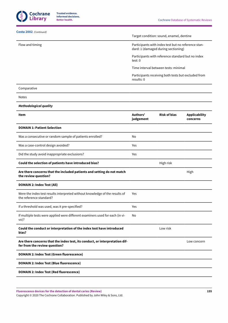
CochraneLibrary
Trusted evidence.Informed decisions.Better health.
Cochrane Database of Systematic Reviews
Target condition: sound, enamel, dentine
Flow and timing Participants with index test but no reference stan-dard: 1 (damaged during sectioning)
Participants with reference standard but no indextest: 0
Time interval between tests: minimal
Participants receiving both tests but excluded fromresults: 0
Comparative
Notes
Methodological quality
Item Authors'judgement
Risk of bias Applicabilityconcerns
DOMAIN 1: Patient Selection
Was a consecutive or random sample of patients enrolled? No
Was a case-control design avoided? Yes
Did the study avoid inappropriate exclusions? Yes
Could the selection of patients have introduced bias? High risk
Are there concerns that the included patients and setting do not matchthe review question?
High
DOMAIN 2: Index Test (All)
Were the index test results interpreted without knowledge of the results ofthe reference standard?
Yes
If a threshold was used, was it pre-specified? Yes
If multiple tests were applied were different examiners used for each (in vi-vo)?
No
Could the conduct or interpretation of the index test have introducedbias?
Low risk
Are there concerns that the index test, its conduct, or interpretation dif-fer from the review question?
Low concern
DOMAIN 2: Index Test (Green fluorescence)
DOMAIN 2: Index Test (Blue fluorescence)
DOMAIN 2: Index Test (Red fluorescence)
Costa 2002 (Continued)
Fluorescence devices for the detection of dental caries (Review)
Copyright © 2020 The Cochrane Collaboration. Published by John Wiley & Sons, Ltd.
155
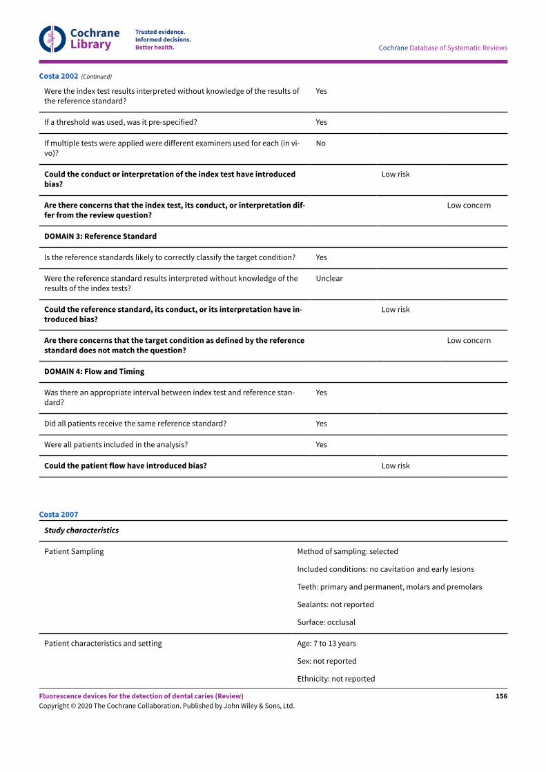
CochraneLibrary
Trusted evidence.Informed decisions.Better health.
Cochrane Database of Systematic Reviews
Were the index test results interpreted without knowledge of the results ofthe reference standard?
Yes
If a threshold was used, was it pre-specified? Yes
If multiple tests were applied were different examiners used for each (in vi-vo)?
No
Could the conduct or interpretation of the index test have introducedbias?
Low risk
Are there concerns that the index test, its conduct, or interpretation dif-fer from the review question?
Low concern
DOMAIN 3: Reference Standard
Is the reference standards likely to correctly classify the target condition? Yes
Were the reference standard results interpreted without knowledge of theresults of the index tests?
Unclear
Could the reference standard, its conduct, or its interpretation have in-troduced bias?
Low risk
Are there concerns that the target condition as defined by the referencestandard does not match the question?
Low concern
DOMAIN 4: Flow and Timing
Was there an appropriate interval between index test and reference stan-dard?
Yes
Did all patients receive the same reference standard? Yes
Were all patients included in the analysis? Yes
Could the patient flow have introduced bias? Low risk
Costa 2002 (Continued)
Study characteristics
Patient Sampling Method of sampling: selected
Included conditions: no cavitation and early lesions
Teeth: primary and permanent, molars and premolars
Sealants: not reported
Surface: occlusal
Patient characteristics and setting Age: 7 to 13 years
Sex: not reported
Ethnicity: not reported
Costa 2007
Fluorescence devices for the detection of dental caries (Review)
Copyright © 2020 The Cochrane Collaboration. Published by John Wiley & Sons, Ltd.
156
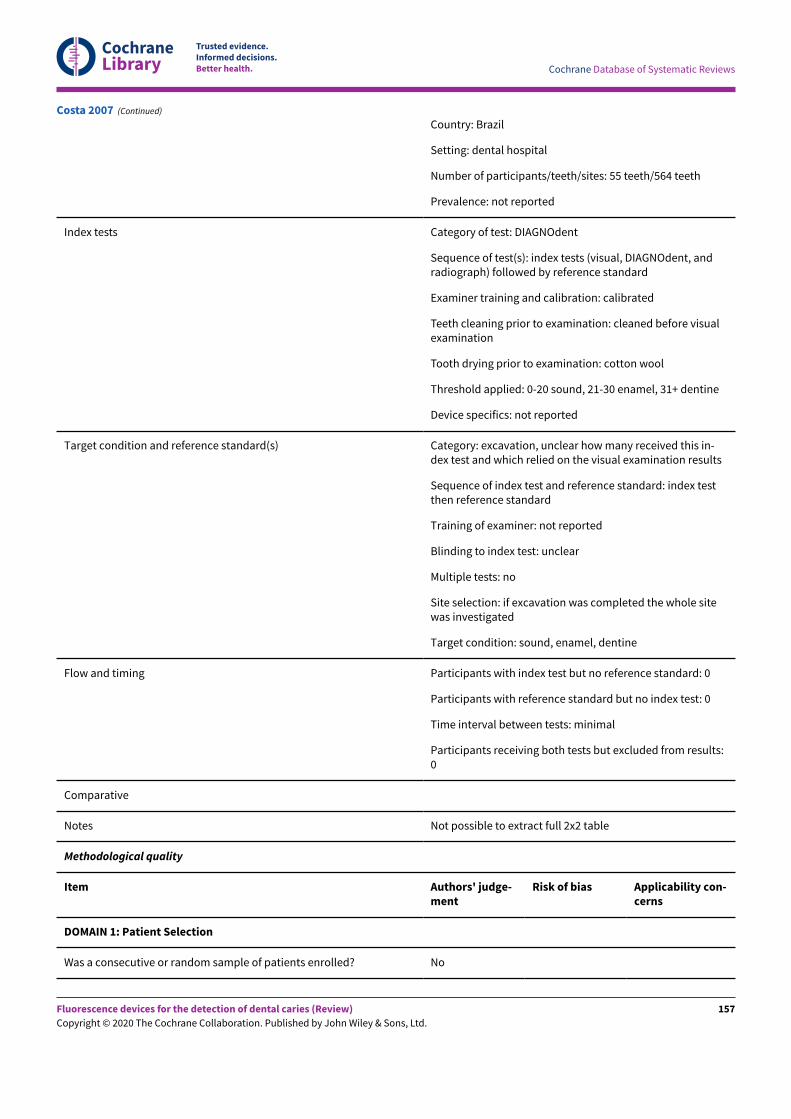
CochraneLibrary
Trusted evidence.Informed decisions.Better health.
Cochrane Database of Systematic Reviews
Country: Brazil
Setting: dental hospital
Number of participants/teeth/sites: 55 teeth/564 teeth
Prevalence: not reported
Index tests Category of test: DIAGNOdent
Sequence of test(s): index tests (visual, DIAGNOdent, andradiograph) followed by reference standard
Examiner training and calibration: calibrated
Teeth cleaning prior to examination: cleaned before visualexamination
Tooth drying prior to examination: cotton wool
Threshold applied: 0-20 sound, 21-30 enamel, 31+ dentine
Device specifics: not reported
Target condition and reference standard(s) Category: excavation, unclear how many received this in-dex test and which relied on the visual examination results
Sequence of index test and reference standard: index testthen reference standard
Training of examiner: not reported
Blinding to index test: unclear
Multiple tests: no
Site selection: if excavation was completed the whole sitewas investigated
Target condition: sound, enamel, dentine
Flow and timing Participants with index test but no reference standard: 0
Participants with reference standard but no index test: 0
Time interval between tests: minimal
Participants receiving both tests but excluded from results:0
Comparative
Notes Not possible to extract full 2x2 table
Methodological quality
Item Authors' judge-ment
Risk of bias Applicability con-cerns
DOMAIN 1: Patient Selection
Was a consecutive or random sample of patients enrolled? No
Costa 2007 (Continued)
Fluorescence devices for the detection of dental caries (Review)
Copyright © 2020 The Cochrane Collaboration. Published by John Wiley & Sons, Ltd.
157
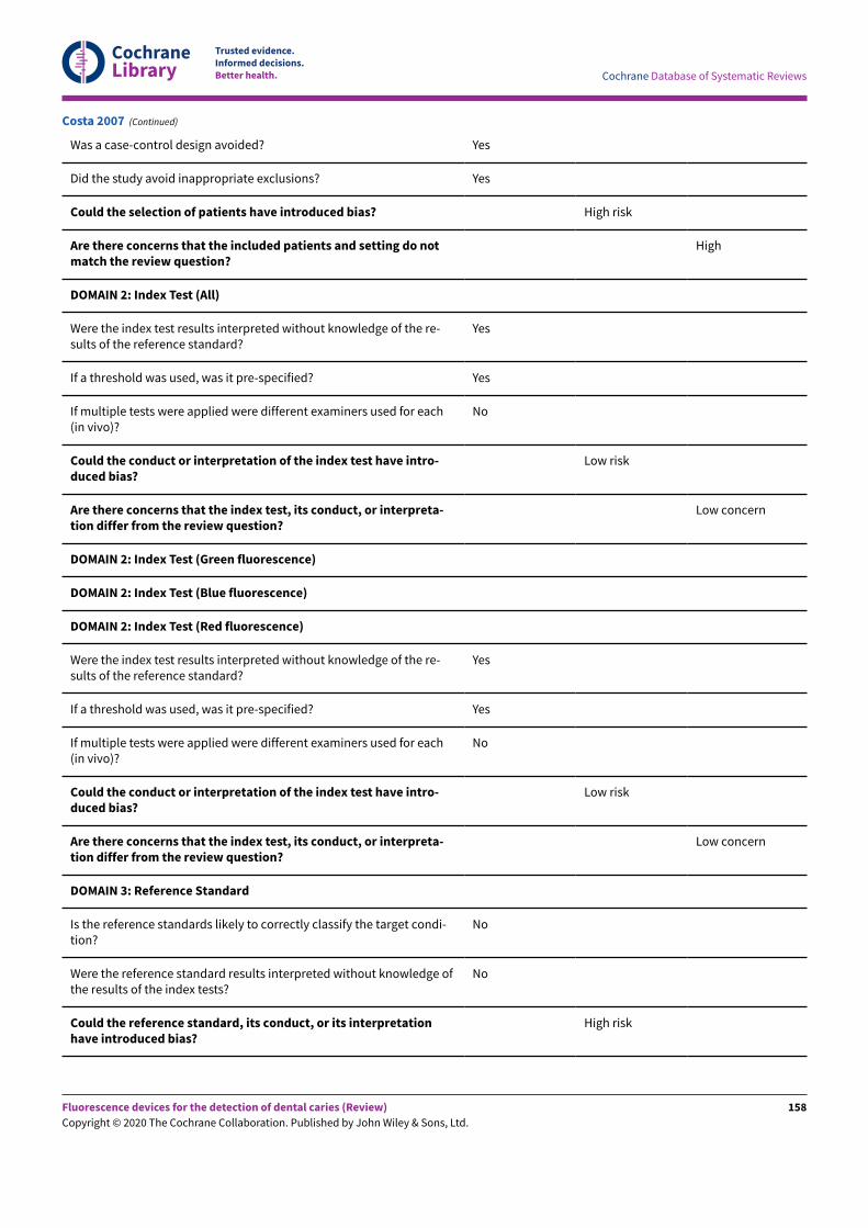
CochraneLibrary
Trusted evidence.Informed decisions.Better health.
Cochrane Database of Systematic Reviews
Was a case-control design avoided? Yes
Did the study avoid inappropriate exclusions? Yes
Could the selection of patients have introduced bias? High risk
Are there concerns that the included patients and setting do notmatch the review question?
High
DOMAIN 2: Index Test (All)
Were the index test results interpreted without knowledge of the re-sults of the reference standard?
Yes
If a threshold was used, was it pre-specified? Yes
If multiple tests were applied were different examiners used for each(in vivo)?
No
Could the conduct or interpretation of the index test have intro-duced bias?
Low risk
Are there concerns that the index test, its conduct, or interpreta-tion differ from the review question?
Low concern
DOMAIN 2: Index Test (Green fluorescence)
DOMAIN 2: Index Test (Blue fluorescence)
DOMAIN 2: Index Test (Red fluorescence)
Were the index test results interpreted without knowledge of the re-sults of the reference standard?
Yes
If a threshold was used, was it pre-specified? Yes
If multiple tests were applied were different examiners used for each(in vivo)?
No
Could the conduct or interpretation of the index test have intro-duced bias?
Low risk
Are there concerns that the index test, its conduct, or interpreta-tion differ from the review question?
Low concern
DOMAIN 3: Reference Standard
Is the reference standards likely to correctly classify the target condi-tion?
No
Were the reference standard results interpreted without knowledge ofthe results of the index tests?
No
Could the reference standard, its conduct, or its interpretationhave introduced bias?
High risk
Costa 2007 (Continued)
Fluorescence devices for the detection of dental caries (Review)
Copyright © 2020 The Cochrane Collaboration. Published by John Wiley & Sons, Ltd.
158
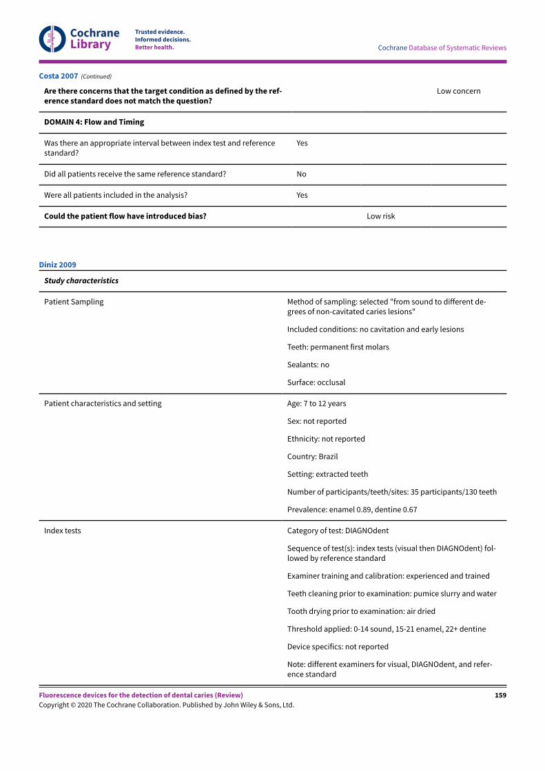
CochraneLibrary
Trusted evidence.Informed decisions.Better health.
Cochrane Database of Systematic Reviews
Are there concerns that the target condition as defined by the ref-erence standard does not match the question?
Low concern
DOMAIN 4: Flow and Timing
Was there an appropriate interval between index test and referencestandard?
Yes
Did all patients receive the same reference standard? No
Were all patients included in the analysis? Yes
Could the patient flow have introduced bias? Low risk
Costa 2007 (Continued)
Study characteristics
Patient Sampling Method of sampling: selected "from sound to different de-grees of non-cavitated caries lesions"
Included conditions: no cavitation and early lesions
Teeth: permanent first molars
Sealants: no
Surface: occlusal
Patient characteristics and setting Age: 7 to 12 years
Sex: not reported
Ethnicity: not reported
Country: Brazil
Setting: extracted teeth
Number of participants/teeth/sites: 35 participants/130 teeth
Prevalence: enamel 0.89, dentine 0.67
Index tests Category of test: DIAGNOdent
Sequence of test(s): index tests (visual then DIAGNOdent) fol-lowed by reference standard
Examiner training and calibration: experienced and trained
Teeth cleaning prior to examination: pumice slurry and water
Tooth drying prior to examination: air dried
Threshold applied: 0-14 sound, 15-21 enamel, 22+ dentine
Device specifics: not reported
Note: different examiners for visual, DIAGNOdent, and refer-ence standard
Diniz 2009
Fluorescence devices for the detection of dental caries (Review)
Copyright © 2020 The Cochrane Collaboration. Published by John Wiley & Sons, Ltd.
159
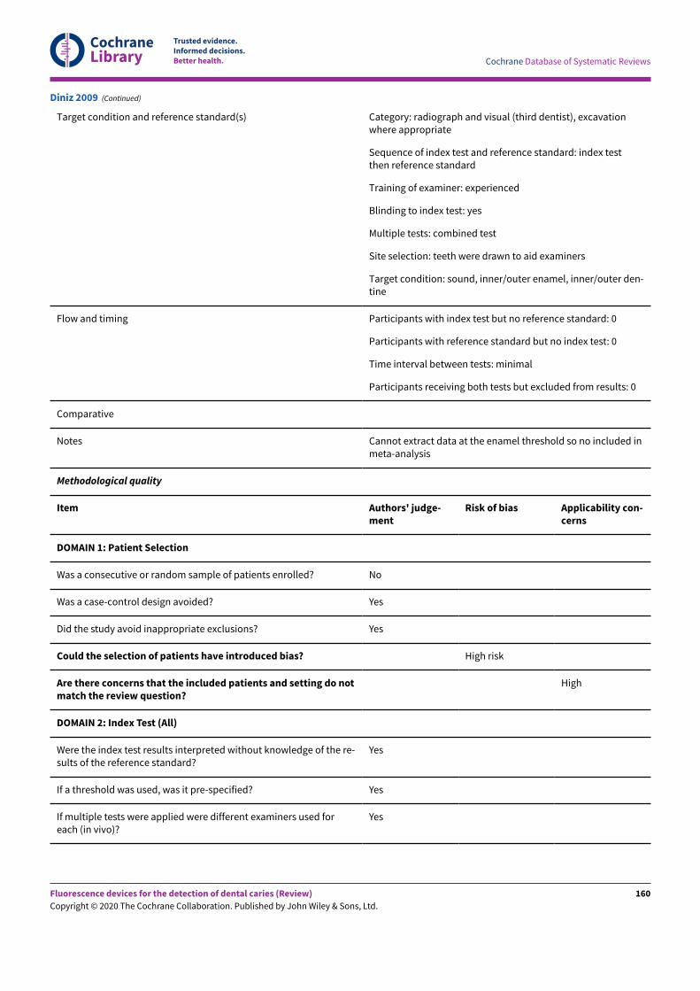
CochraneLibrary
Trusted evidence.Informed decisions.Better health.
Cochrane Database of Systematic Reviews
Target condition and reference standard(s) Category: radiograph and visual (third dentist), excavationwhere appropriate
Sequence of index test and reference standard: index testthen reference standard
Training of examiner: experienced
Blinding to index test: yes
Multiple tests: combined test
Site selection: teeth were drawn to aid examiners
Target condition: sound, inner/outer enamel, inner/outer den-tine
Flow and timing Participants with index test but no reference standard: 0
Participants with reference standard but no index test: 0
Time interval between tests: minimal
Participants receiving both tests but excluded from results: 0
Comparative
Notes Cannot extract data at the enamel threshold so no included inmeta-analysis
Methodological quality
Item Authors' judge-ment
Risk of bias Applicability con-cerns
DOMAIN 1: Patient Selection
Was a consecutive or random sample of patients enrolled? No
Was a case-control design avoided? Yes
Did the study avoid inappropriate exclusions? Yes
Could the selection of patients have introduced bias? High risk
Are there concerns that the included patients and setting do notmatch the review question?
High
DOMAIN 2: Index Test (All)
Were the index test results interpreted without knowledge of the re-sults of the reference standard?
Yes
If a threshold was used, was it pre-specified? Yes
If multiple tests were applied were different examiners used foreach (in vivo)?
Yes
Diniz 2009 (Continued)
Fluorescence devices for the detection of dental caries (Review)
Copyright © 2020 The Cochrane Collaboration. Published by John Wiley & Sons, Ltd.
160
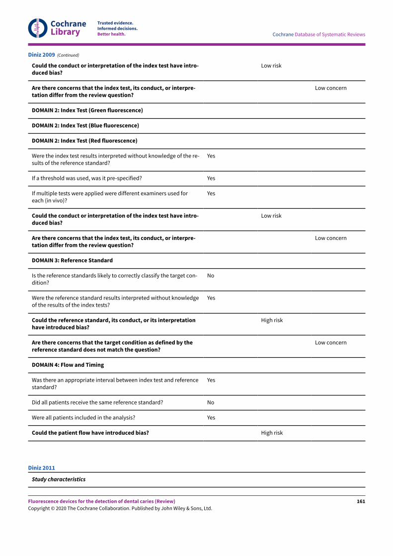
CochraneLibrary
Trusted evidence.Informed decisions.Better health.
Cochrane Database of Systematic Reviews
Could the conduct or interpretation of the index test have intro-duced bias?
Low risk
Are there concerns that the index test, its conduct, or interpre-tation differ from the review question?
Low concern
DOMAIN 2: Index Test (Green fluorescence)
DOMAIN 2: Index Test (Blue fluorescence)
DOMAIN 2: Index Test (Red fluorescence)
Were the index test results interpreted without knowledge of the re-sults of the reference standard?
Yes
If a threshold was used, was it pre-specified? Yes
If multiple tests were applied were different examiners used foreach (in vivo)?
Yes
Could the conduct or interpretation of the index test have intro-duced bias?
Low risk
Are there concerns that the index test, its conduct, or interpre-tation differ from the review question?
Low concern
DOMAIN 3: Reference Standard
Is the reference standards likely to correctly classify the target con-dition?
No
Were the reference standard results interpreted without knowledgeof the results of the index tests?
Yes
Could the reference standard, its conduct, or its interpretationhave introduced bias?
High risk
Are there concerns that the target condition as defined by thereference standard does not match the question?
Low concern
DOMAIN 4: Flow and Timing
Was there an appropriate interval between index test and referencestandard?
Yes
Did all patients receive the same reference standard? No
Were all patients included in the analysis? Yes
Could the patient flow have introduced bias? High risk
Diniz 2009 (Continued)
Study characteristics
Diniz 2011
Fluorescence devices for the detection of dental caries (Review)
Copyright © 2020 The Cochrane Collaboration. Published by John Wiley & Sons, Ltd.
161
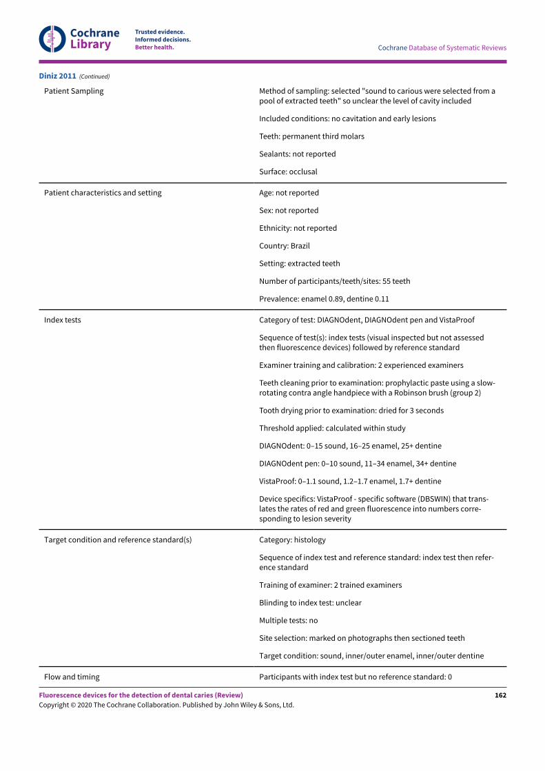
CochraneLibrary
Trusted evidence.Informed decisions.Better health.
Cochrane Database of Systematic Reviews
Patient Sampling Method of sampling: selected "sound to carious were selected from apool of extracted teeth" so unclear the level of cavity included
Included conditions: no cavitation and early lesions
Teeth: permanent third molars
Sealants: not reported
Surface: occlusal
Patient characteristics and setting Age: not reported
Sex: not reported
Ethnicity: not reported
Country: Brazil
Setting: extracted teeth
Number of participants/teeth/sites: 55 teeth
Prevalence: enamel 0.89, dentine 0.11
Index tests Category of test: DIAGNOdent, DIAGNOdent pen and VistaProof
Sequence of test(s): index tests (visual inspected but not assessedthen fluorescence devices) followed by reference standard
Examiner training and calibration: 2 experienced examiners
Teeth cleaning prior to examination: prophylactic paste using a slow-rotating contra angle handpiece with a Robinson brush (group 2)
Tooth drying prior to examination: dried for 3 seconds
Threshold applied: calculated within study
DIAGNOdent: 0–15 sound, 16–25 enamel, 25+ dentine
DIAGNOdent pen: 0–10 sound, 11–34 enamel, 34+ dentine
VistaProof: 0–1.1 sound, 1.2–1.7 enamel, 1.7+ dentine
Device specifics: VistaProof - specific software (DBSWIN) that trans-lates the rates of red and green fluorescence into numbers corre-sponding to lesion severity
Target condition and reference standard(s) Category: histology
Sequence of index test and reference standard: index test then refer-ence standard
Training of examiner: 2 trained examiners
Blinding to index test: unclear
Multiple tests: no
Site selection: marked on photographs then sectioned teeth
Target condition: sound, inner/outer enamel, inner/outer dentine
Flow and timing Participants with index test but no reference standard: 0
Diniz 2011 (Continued)
Fluorescence devices for the detection of dental caries (Review)
Copyright © 2020 The Cochrane Collaboration. Published by John Wiley & Sons, Ltd.
162
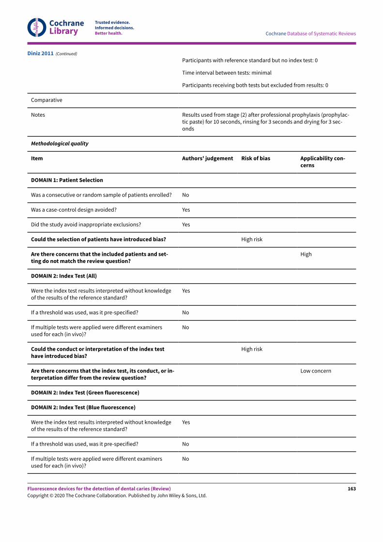
CochraneLibrary
Trusted evidence.Informed decisions.Better health.
Cochrane Database of Systematic Reviews
Participants with reference standard but no index test: 0
Time interval between tests: minimal
Participants receiving both tests but excluded from results: 0
Comparative
Notes Results used from stage (2) after professional prophylaxis (prophylac-tic paste) for 10 seconds, rinsing for 3 seconds and drying for 3 sec-onds
Methodological quality
Item Authors' judgement Risk of bias Applicability con-cerns
DOMAIN 1: Patient Selection
Was a consecutive or random sample of patients enrolled? No
Was a case-control design avoided? Yes
Did the study avoid inappropriate exclusions? Yes
Could the selection of patients have introduced bias? High risk
Are there concerns that the included patients and set-ting do not match the review question?
High
DOMAIN 2: Index Test (All)
Were the index test results interpreted without knowledgeof the results of the reference standard?
Yes
If a threshold was used, was it pre-specified? No
If multiple tests were applied were different examinersused for each (in vivo)?
No
Could the conduct or interpretation of the index testhave introduced bias?
High risk
Are there concerns that the index test, its conduct, or in-terpretation differ from the review question?
Low concern
DOMAIN 2: Index Test (Green fluorescence)
DOMAIN 2: Index Test (Blue fluorescence)
Were the index test results interpreted without knowledgeof the results of the reference standard?
Yes
If a threshold was used, was it pre-specified? No
If multiple tests were applied were different examinersused for each (in vivo)?
No
Diniz 2011 (Continued)
Fluorescence devices for the detection of dental caries (Review)
Copyright © 2020 The Cochrane Collaboration. Published by John Wiley & Sons, Ltd.
163
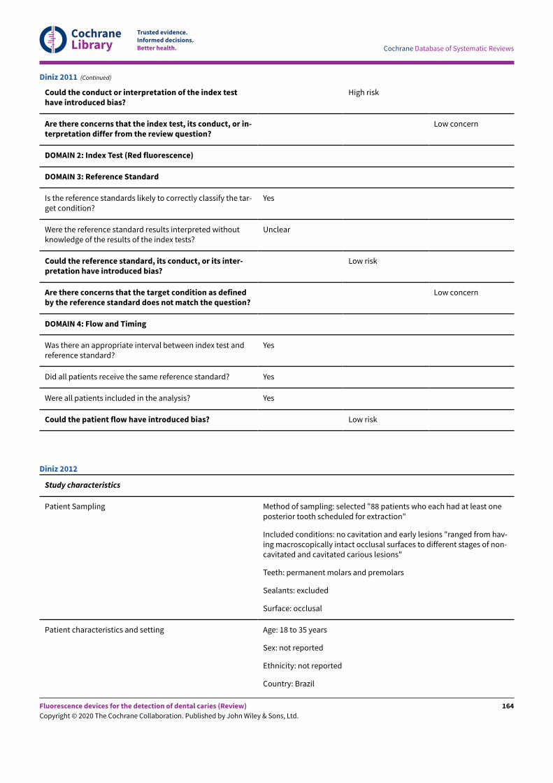
CochraneLibrary
Trusted evidence.Informed decisions.Better health.
Cochrane Database of Systematic Reviews
Could the conduct or interpretation of the index testhave introduced bias?
High risk
Are there concerns that the index test, its conduct, or in-terpretation differ from the review question?
Low concern
DOMAIN 2: Index Test (Red fluorescence)
DOMAIN 3: Reference Standard
Is the reference standards likely to correctly classify the tar-get condition?
Yes
Were the reference standard results interpreted withoutknowledge of the results of the index tests?
Unclear
Could the reference standard, its conduct, or its inter-pretation have introduced bias?
Low risk
Are there concerns that the target condition as definedby the reference standard does not match the question?
Low concern
DOMAIN 4: Flow and Timing
Was there an appropriate interval between index test andreference standard?
Yes
Did all patients receive the same reference standard? Yes
Were all patients included in the analysis? Yes
Could the patient flow have introduced bias? Low risk
Diniz 2011 (Continued)
Study characteristics
Patient Sampling Method of sampling: selected "88 patients who each had at least oneposterior tooth scheduled for extraction"
Included conditions: no cavitation and early lesions "ranged from hav-ing macroscopically intact occlusal surfaces to different stages of non-cavitated and cavitated carious lesions"
Teeth: permanent molars and premolars
Sealants: excluded
Surface: occlusal
Patient characteristics and setting Age: 18 to 35 years
Sex: not reported
Ethnicity: not reported
Country: Brazil
Diniz 2012
Fluorescence devices for the detection of dental caries (Review)
Copyright © 2020 The Cochrane Collaboration. Published by John Wiley & Sons, Ltd.
164
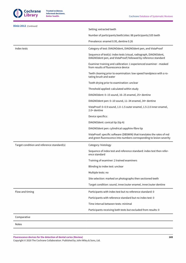
CochraneLibrary
Trusted evidence.Informed decisions.Better health.
Cochrane Database of Systematic Reviews
Setting: extracted teeth
Number of participants/teeth/sites: 88 participants/105 teeth
Prevalence: enamel 0.95, dentine 0.26
Index tests Category of test: DIAGNOdent, DIAGNOdent pen, and VistaProof
Sequence of test(s): index tests (visual, radiograph, DIAGNOdent,DIAGNOdent pen, and VistaProof) followed by reference standard
Examiner training and calibration: 1 experienced examiner - maskedfrom results of fluorescence device
Teeth cleaning prior to examination: low-speed handpiece with a ro-tating brush and water
Tooth drying prior to examination: unclear
Threshold applied: calculated within study
DIAGNOdent: 0–15 sound, 16–25 enamel, 25+ dentine
DIAGNOdent pen: 0–10 sound, 11–34 enamel, 34+ dentine
VistaProof: 0–0.9 sound, 1.0–1.5 outer enamel, 1.5-2.0 inner enamel,2.0+ dentine
Device specifics:
DIAGNOdent: conical tip (tip A)
DIAGNOdent pen: cylindrical sapphire-fibre tip
VistaProof: specific software (DBSWIN) that translates the rates of redand green fluorescence into numbers corresponding to lesion severity
Target condition and reference standard(s) Category: histology
Sequence of index test and reference standard: index test then refer-ence standard
Training of examiner: 2 trained examiners
Blinding to index test: unclear
Multiple tests: no
Site selection: marked on photographs then sectioned teeth
Target condition: sound, inner/outer enamel, inner/outer dentine
Flow and timing Participants with index test but no reference standard: 0
Participants with reference standard but no index test: 0
Time interval between tests: minimal
Participants receiving both tests but excluded from results: 0
Comparative
Notes
Diniz 2012 (Continued)
Fluorescence devices for the detection of dental caries (Review)
Copyright © 2020 The Cochrane Collaboration. Published by John Wiley & Sons, Ltd.
165
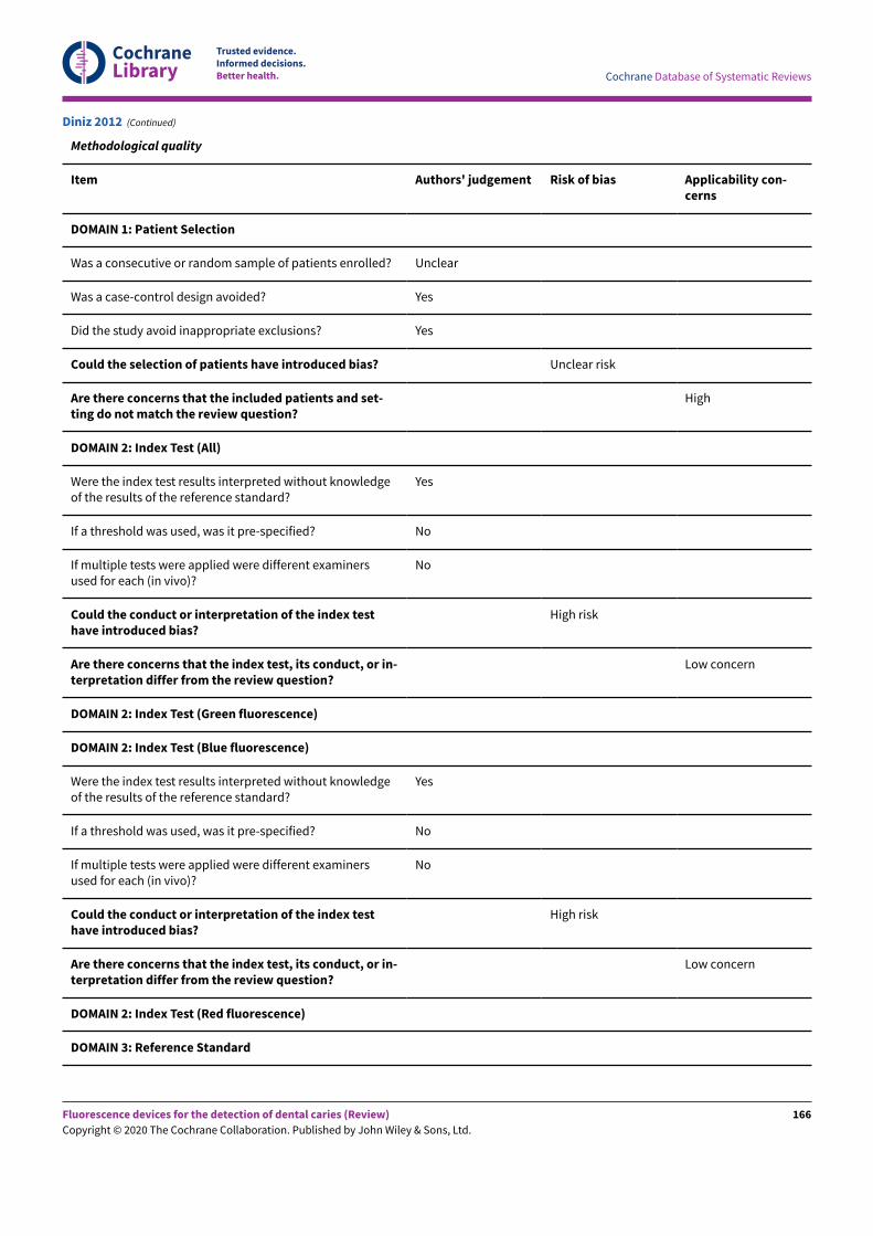
CochraneLibrary
Trusted evidence.Informed decisions.Better health.
Cochrane Database of Systematic Reviews
Methodological quality
Item Authors' judgement Risk of bias Applicability con-cerns
DOMAIN 1: Patient Selection
Was a consecutive or random sample of patients enrolled? Unclear
Was a case-control design avoided? Yes
Did the study avoid inappropriate exclusions? Yes
Could the selection of patients have introduced bias? Unclear risk
Are there concerns that the included patients and set-ting do not match the review question?
High
DOMAIN 2: Index Test (All)
Were the index test results interpreted without knowledgeof the results of the reference standard?
Yes
If a threshold was used, was it pre-specified? No
If multiple tests were applied were different examinersused for each (in vivo)?
No
Could the conduct or interpretation of the index testhave introduced bias?
High risk
Are there concerns that the index test, its conduct, or in-terpretation differ from the review question?
Low concern
DOMAIN 2: Index Test (Green fluorescence)
DOMAIN 2: Index Test (Blue fluorescence)
Were the index test results interpreted without knowledgeof the results of the reference standard?
Yes
If a threshold was used, was it pre-specified? No
If multiple tests were applied were different examinersused for each (in vivo)?
No
Could the conduct or interpretation of the index testhave introduced bias?
High risk
Are there concerns that the index test, its conduct, or in-terpretation differ from the review question?
Low concern
DOMAIN 2: Index Test (Red fluorescence)
DOMAIN 3: Reference Standard
Diniz 2012 (Continued)
Fluorescence devices for the detection of dental caries (Review)
Copyright © 2020 The Cochrane Collaboration. Published by John Wiley & Sons, Ltd.
166
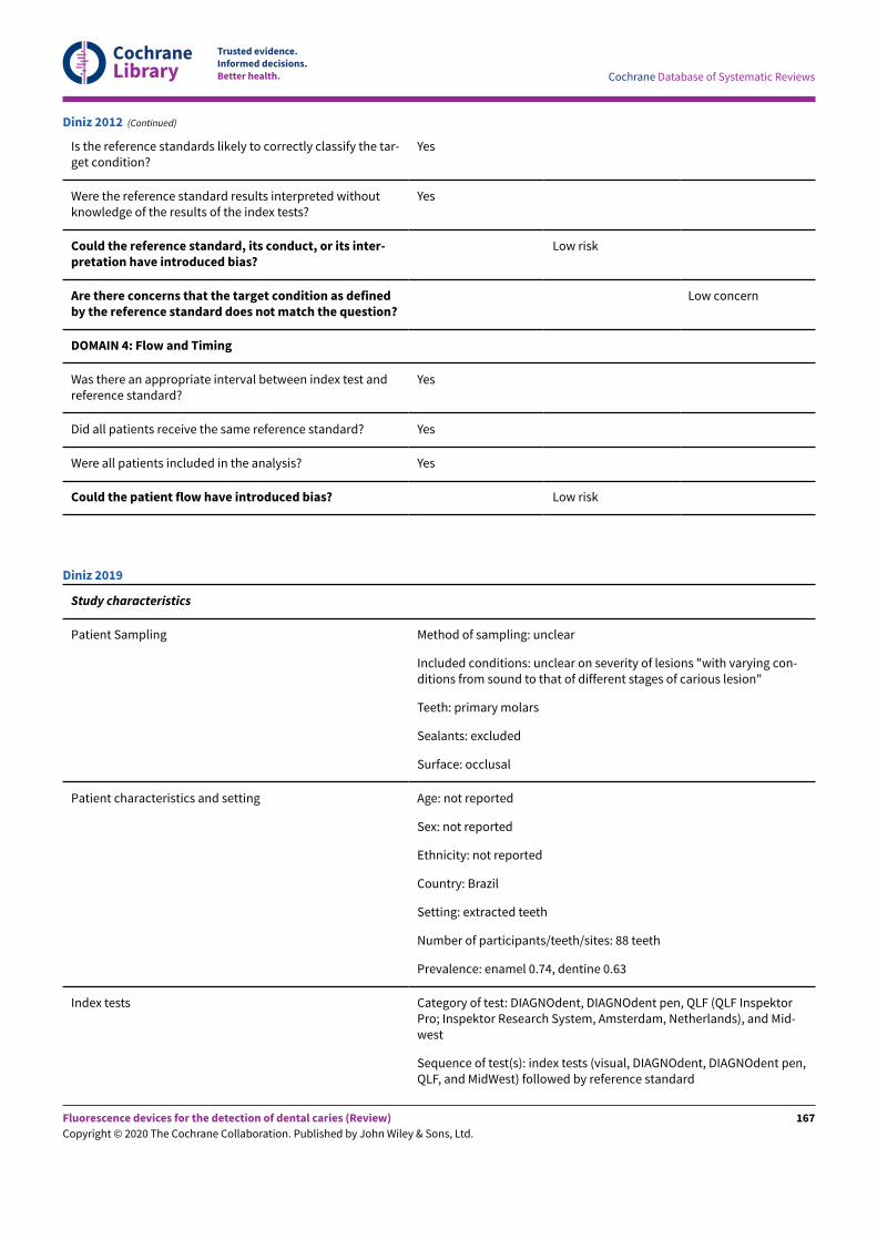
CochraneLibrary
Trusted evidence.Informed decisions.Better health.
Cochrane Database of Systematic Reviews
Is the reference standards likely to correctly classify the tar-get condition?
Yes
Were the reference standard results interpreted withoutknowledge of the results of the index tests?
Yes
Could the reference standard, its conduct, or its inter-pretation have introduced bias?
Low risk
Are there concerns that the target condition as definedby the reference standard does not match the question?
Low concern
DOMAIN 4: Flow and Timing
Was there an appropriate interval between index test andreference standard?
Yes
Did all patients receive the same reference standard? Yes
Were all patients included in the analysis? Yes
Could the patient flow have introduced bias? Low risk
Diniz 2012 (Continued)
Study characteristics
Patient Sampling Method of sampling: unclear
Included conditions: unclear on severity of lesions "with varying con-ditions from sound to that of different stages of carious lesion"
Teeth: primary molars
Sealants: excluded
Surface: occlusal
Patient characteristics and setting Age: not reported
Sex: not reported
Ethnicity: not reported
Country: Brazil
Setting: extracted teeth
Number of participants/teeth/sites: 88 teeth
Prevalence: enamel 0.74, dentine 0.63
Index tests Category of test: DIAGNOdent, DIAGNOdent pen, QLF (QLF InspektorPro; Inspektor Research System, Amsterdam, Netherlands), and Mid-west
Sequence of test(s): index tests (visual, DIAGNOdent, DIAGNOdent pen,QLF, and MidWest) followed by reference standard
Diniz 2019
Fluorescence devices for the detection of dental caries (Review)
Copyright © 2020 The Cochrane Collaboration. Published by John Wiley & Sons, Ltd.
167
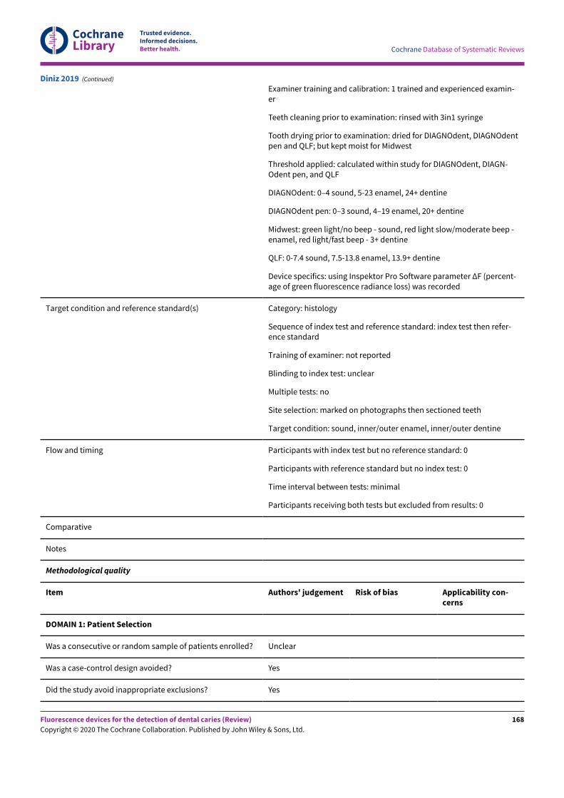
CochraneLibrary
Trusted evidence.Informed decisions.Better health.
Cochrane Database of Systematic Reviews
Examiner training and calibration: 1 trained and experienced examin-er
Teeth cleaning prior to examination: rinsed with 3in1 syringe
Tooth drying prior to examination: dried for DIAGNOdent, DIAGNOdentpen and QLF; but kept moist for Midwest
Threshold applied: calculated within study for DIAGNOdent, DIAGN-Odent pen, and QLF
DIAGNOdent: 0–4 sound, 5-23 enamel, 24+ dentine
DIAGNOdent pen: 0–3 sound, 4–19 enamel, 20+ dentine
Midwest: green light/no beep - sound, red light slow/moderate beep -enamel, red light/fast beep - 3+ dentine
QLF: 0-7.4 sound, 7.5-13.8 enamel, 13.9+ dentine
Device specifics: using Inspektor Pro Software parameter ΔF (percent-age of green fluorescence radiance loss) was recorded
Target condition and reference standard(s) Category: histology
Sequence of index test and reference standard: index test then refer-ence standard
Training of examiner: not reported
Blinding to index test: unclear
Multiple tests: no
Site selection: marked on photographs then sectioned teeth
Target condition: sound, inner/outer enamel, inner/outer dentine
Flow and timing Participants with index test but no reference standard: 0
Participants with reference standard but no index test: 0
Time interval between tests: minimal
Participants receiving both tests but excluded from results: 0
Comparative
Notes
Methodological quality
Item Authors' judgement Risk of bias Applicability con-cerns
DOMAIN 1: Patient Selection
Was a consecutive or random sample of patients enrolled? Unclear
Was a case-control design avoided? Yes
Did the study avoid inappropriate exclusions? Yes
Diniz 2019 (Continued)
Fluorescence devices for the detection of dental caries (Review)
Copyright © 2020 The Cochrane Collaboration. Published by John Wiley & Sons, Ltd.
168
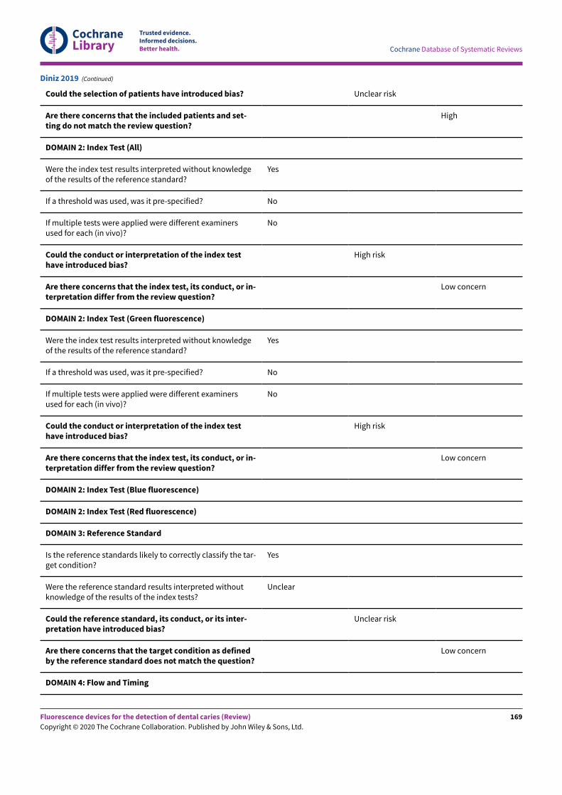
CochraneLibrary
Trusted evidence.Informed decisions.Better health.
Cochrane Database of Systematic Reviews
Could the selection of patients have introduced bias? Unclear risk
Are there concerns that the included patients and set-ting do not match the review question?
High
DOMAIN 2: Index Test (All)
Were the index test results interpreted without knowledgeof the results of the reference standard?
Yes
If a threshold was used, was it pre-specified? No
If multiple tests were applied were different examinersused for each (in vivo)?
No
Could the conduct or interpretation of the index testhave introduced bias?
High risk
Are there concerns that the index test, its conduct, or in-terpretation differ from the review question?
Low concern
DOMAIN 2: Index Test (Green fluorescence)
Were the index test results interpreted without knowledgeof the results of the reference standard?
Yes
If a threshold was used, was it pre-specified? No
If multiple tests were applied were different examinersused for each (in vivo)?
No
Could the conduct or interpretation of the index testhave introduced bias?
High risk
Are there concerns that the index test, its conduct, or in-terpretation differ from the review question?
Low concern
DOMAIN 2: Index Test (Blue fluorescence)
DOMAIN 2: Index Test (Red fluorescence)
DOMAIN 3: Reference Standard
Is the reference standards likely to correctly classify the tar-get condition?
Yes
Were the reference standard results interpreted withoutknowledge of the results of the index tests?
Unclear
Could the reference standard, its conduct, or its inter-pretation have introduced bias?
Unclear risk
Are there concerns that the target condition as definedby the reference standard does not match the question?
Low concern
DOMAIN 4: Flow and Timing
Diniz 2019 (Continued)
Fluorescence devices for the detection of dental caries (Review)
Copyright © 2020 The Cochrane Collaboration. Published by John Wiley & Sons, Ltd.
169
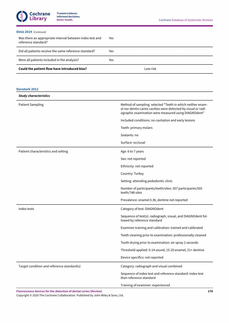
CochraneLibrary
Trusted evidence.Informed decisions.Better health.
Cochrane Database of Systematic Reviews
Was there an appropriate interval between index test andreference standard?
Yes
Did all patients receive the same reference standard? Yes
Were all patients included in the analysis? Yes
Could the patient flow have introduced bias? Low risk
Diniz 2019 (Continued)
Study characteristics
Patient Sampling Method of sampling: selected "Teeth in which neither enam-el nor dentin caries cavities were detected by visual or radi-ographic examination were measured using DIAGNOdent"
Included conditions: no cavitation and early lesions
Teeth: primary molars
Sealants: no
Surface: occlusal
Patient characteristics and setting Age: 6 to 7 years
Sex: not reported
Ethnicity: not reported
Country: Turkey
Setting: attending pedodontic clinic
Number of participants/teeth/sites: 307 participants/505teeth/748 sites
Prevalence: enamel 0.36, dentine not reported
Index tests Category of test: DIAGNOdent
Sequence of test(s): radiograph, visual, and DIAGNOdent fol-lowed by reference standard
Examiner training and calibration: trained and calibrated
Teeth cleaning prior to examination: professionally cleaned
Tooth drying prior to examination: air spray 2 seconds
Threshold applied: 0-14 sound, 15-20 enamel, 21+ dentine
Device specifics: not reported
Target condition and reference standard(s) Category: radiograph and visual combined
Sequence of index test and reference standard: index testthen reference standard
Training of examiner: experienced
Duruturk 2011
Fluorescence devices for the detection of dental caries (Review)
Copyright © 2020 The Cochrane Collaboration. Published by John Wiley & Sons, Ltd.
170
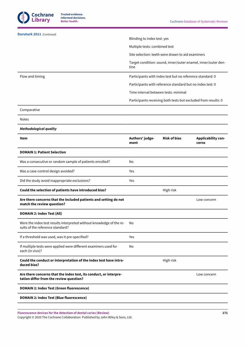
CochraneLibrary
Trusted evidence.Informed decisions.Better health.
Cochrane Database of Systematic Reviews
Blinding to index test: yes
Multiple tests: combined test
Site selection: teeth were drawn to aid examiners
Target condition: sound, inner/outer enamel, inner/outer den-tine
Flow and timing Participants with index test but no reference standard: 0
Participants with reference standard but no index test: 0
Time interval between tests: minimal
Participants receiving both tests but excluded from results: 0
Comparative
Notes
Methodological quality
Item Authors' judge-ment
Risk of bias Applicability con-cerns
DOMAIN 1: Patient Selection
Was a consecutive or random sample of patients enrolled? No
Was a case-control design avoided? Yes
Did the study avoid inappropriate exclusions? Yes
Could the selection of patients have introduced bias? High risk
Are there concerns that the included patients and setting do notmatch the review question?
Low concern
DOMAIN 2: Index Test (All)
Were the index test results interpreted without knowledge of the re-sults of the reference standard?
No
If a threshold was used, was it pre-specified? Yes
If multiple tests were applied were different examiners used foreach (in vivo)?
No
Could the conduct or interpretation of the index test have intro-duced bias?
High risk
Are there concerns that the index test, its conduct, or interpre-tation differ from the review question?
Low concern
DOMAIN 2: Index Test (Green fluorescence)
DOMAIN 2: Index Test (Blue fluorescence)
Duruturk 2011 (Continued)
Fluorescence devices for the detection of dental caries (Review)
Copyright © 2020 The Cochrane Collaboration. Published by John Wiley & Sons, Ltd.
171
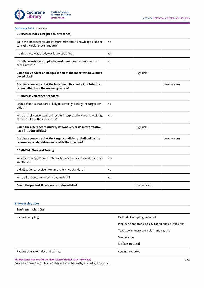
CochraneLibrary
Trusted evidence.Informed decisions.Better health.
Cochrane Database of Systematic Reviews
DOMAIN 2: Index Test (Red fluorescence)
Were the index test results interpreted without knowledge of the re-sults of the reference standard?
No
If a threshold was used, was it pre-specified? Yes
If multiple tests were applied were different examiners used foreach (in vivo)?
No
Could the conduct or interpretation of the index test have intro-duced bias?
High risk
Are there concerns that the index test, its conduct, or interpre-tation differ from the review question?
Low concern
DOMAIN 3: Reference Standard
Is the reference standards likely to correctly classify the target con-dition?
No
Were the reference standard results interpreted without knowledgeof the results of the index tests?
Yes
Could the reference standard, its conduct, or its interpretationhave introduced bias?
High risk
Are there concerns that the target condition as defined by thereference standard does not match the question?
Low concern
DOMAIN 4: Flow and Timing
Was there an appropriate interval between index test and referencestandard?
Yes
Did all patients receive the same reference standard? No
Were all patients included in the analysis? Yes
Could the patient flow have introduced bias? Unclear risk
Duruturk 2011 (Continued)
Study characteristics
Patient Sampling Method of sampling: selected
Included conditions: no cavitation and early lesions
Teeth: permanent premolars and molars
Sealants: no
Surface: occlusal
Patient characteristics and setting Age: not reported
El-Housseiny 2001
Fluorescence devices for the detection of dental caries (Review)
Copyright © 2020 The Cochrane Collaboration. Published by John Wiley & Sons, Ltd.
172
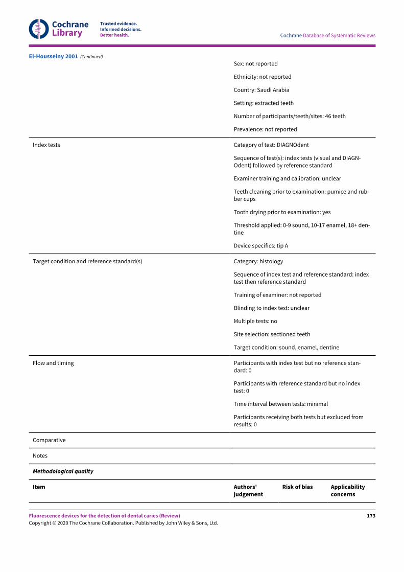
CochraneLibrary
Trusted evidence.Informed decisions.Better health.
Cochrane Database of Systematic Reviews
Sex: not reported
Ethnicity: not reported
Country: Saudi Arabia
Setting: extracted teeth
Number of participants/teeth/sites: 46 teeth
Prevalence: not reported
Index tests Category of test: DIAGNOdent
Sequence of test(s): index tests (visual and DIAGN-Odent) followed by reference standard
Examiner training and calibration: unclear
Teeth cleaning prior to examination: pumice and rub-ber cups
Tooth drying prior to examination: yes
Threshold applied: 0-9 sound, 10-17 enamel, 18+ den-tine
Device specifics: tip A
Target condition and reference standard(s) Category: histology
Sequence of index test and reference standard: indextest then reference standard
Training of examiner: not reported
Blinding to index test: unclear
Multiple tests: no
Site selection: sectioned teeth
Target condition: sound, enamel, dentine
Flow and timing Participants with index test but no reference stan-dard: 0
Participants with reference standard but no indextest: 0
Time interval between tests: minimal
Participants receiving both tests but excluded fromresults: 0
Comparative
Notes
Methodological quality
Item Authors'judgement
Risk of bias Applicabilityconcerns
El-Housseiny 2001 (Continued)
Fluorescence devices for the detection of dental caries (Review)
Copyright © 2020 The Cochrane Collaboration. Published by John Wiley & Sons, Ltd.
173
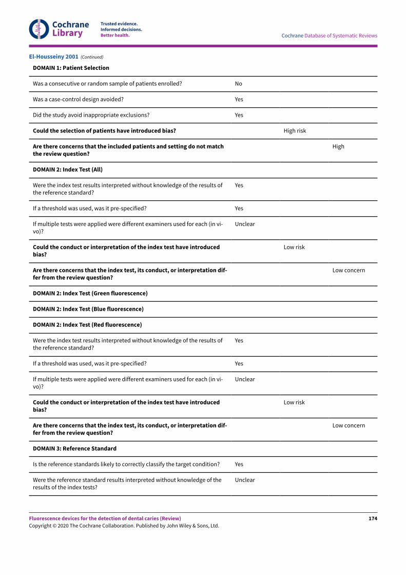
CochraneLibrary
Trusted evidence.Informed decisions.Better health.
Cochrane Database of Systematic Reviews
DOMAIN 1: Patient Selection
Was a consecutive or random sample of patients enrolled? No
Was a case-control design avoided? Yes
Did the study avoid inappropriate exclusions? Yes
Could the selection of patients have introduced bias? High risk
Are there concerns that the included patients and setting do not matchthe review question?
High
DOMAIN 2: Index Test (All)
Were the index test results interpreted without knowledge of the results ofthe reference standard?
Yes
If a threshold was used, was it pre-specified? Yes
If multiple tests were applied were different examiners used for each (in vi-vo)?
Unclear
Could the conduct or interpretation of the index test have introducedbias?
Low risk
Are there concerns that the index test, its conduct, or interpretation dif-fer from the review question?
Low concern
DOMAIN 2: Index Test (Green fluorescence)
DOMAIN 2: Index Test (Blue fluorescence)
DOMAIN 2: Index Test (Red fluorescence)
Were the index test results interpreted without knowledge of the results ofthe reference standard?
Yes
If a threshold was used, was it pre-specified? Yes
If multiple tests were applied were different examiners used for each (in vi-vo)?
Unclear
Could the conduct or interpretation of the index test have introducedbias?
Low risk
Are there concerns that the index test, its conduct, or interpretation dif-fer from the review question?
Low concern
DOMAIN 3: Reference Standard
Is the reference standards likely to correctly classify the target condition? Yes
Were the reference standard results interpreted without knowledge of theresults of the index tests?
Unclear
El-Housseiny 2001 (Continued)
Fluorescence devices for the detection of dental caries (Review)
Copyright © 2020 The Cochrane Collaboration. Published by John Wiley & Sons, Ltd.
174
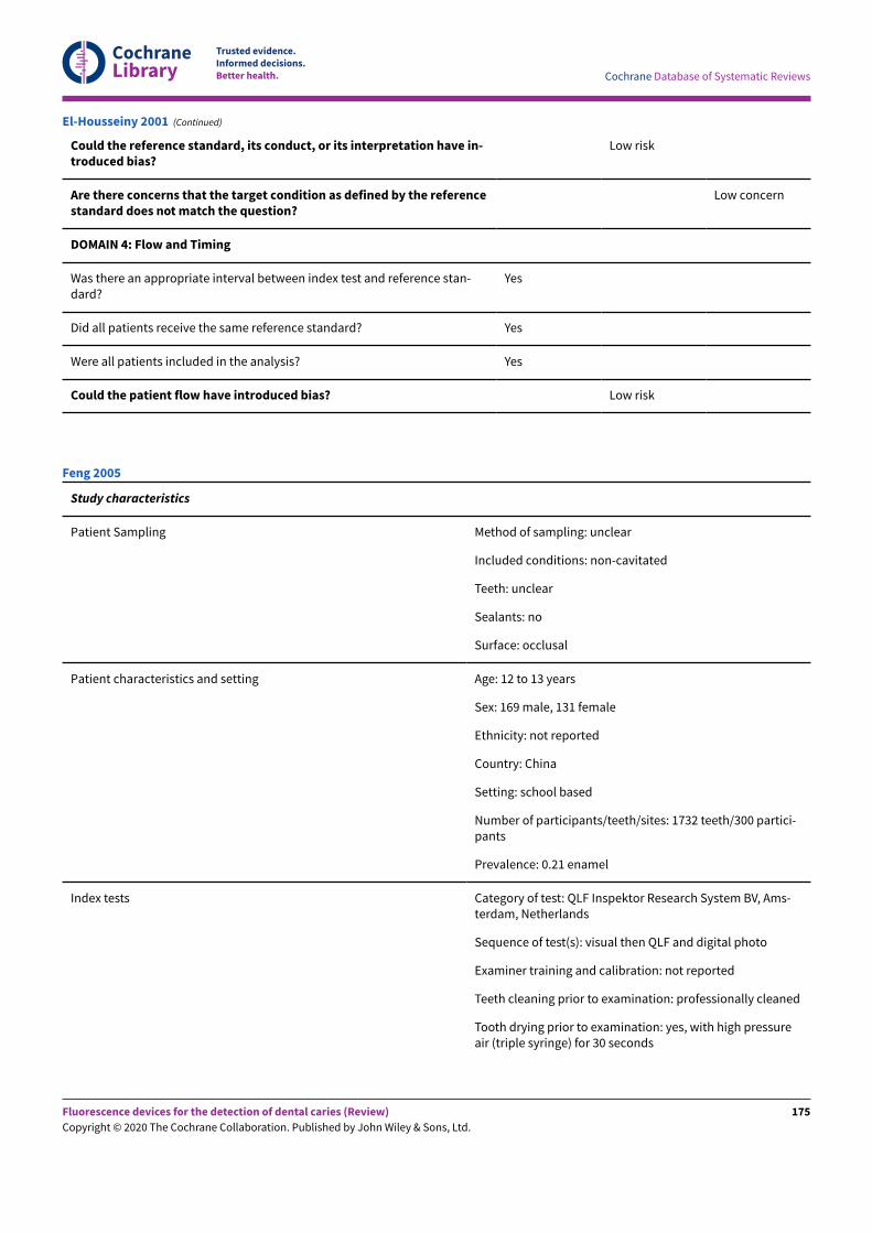
CochraneLibrary
Trusted evidence.Informed decisions.Better health.
Cochrane Database of Systematic Reviews
Could the reference standard, its conduct, or its interpretation have in-troduced bias?
Low risk
Are there concerns that the target condition as defined by the referencestandard does not match the question?
Low concern
DOMAIN 4: Flow and Timing
Was there an appropriate interval between index test and reference stan-dard?
Yes
Did all patients receive the same reference standard? Yes
Were all patients included in the analysis? Yes
Could the patient flow have introduced bias? Low risk
El-Housseiny 2001 (Continued)
Study characteristics
Patient Sampling Method of sampling: unclear
Included conditions: non-cavitated
Teeth: unclear
Sealants: no
Surface: occlusal
Patient characteristics and setting Age: 12 to 13 years
Sex: 169 male, 131 female
Ethnicity: not reported
Country: China
Setting: school based
Number of participants/teeth/sites: 1732 teeth/300 partici-pants
Prevalence: 0.21 enamel
Index tests Category of test: QLF Inspektor Research System BV, Ams-terdam, Netherlands
Sequence of test(s): visual then QLF and digital photo
Examiner training and calibration: not reported
Teeth cleaning prior to examination: professionally cleaned
Tooth drying prior to examination: yes, with high pressureair (triple syringe) for 30 seconds
Feng 2005
Fluorescence devices for the detection of dental caries (Review)
Copyright © 2020 The Cochrane Collaboration. Published by John Wiley & Sons, Ltd.
175
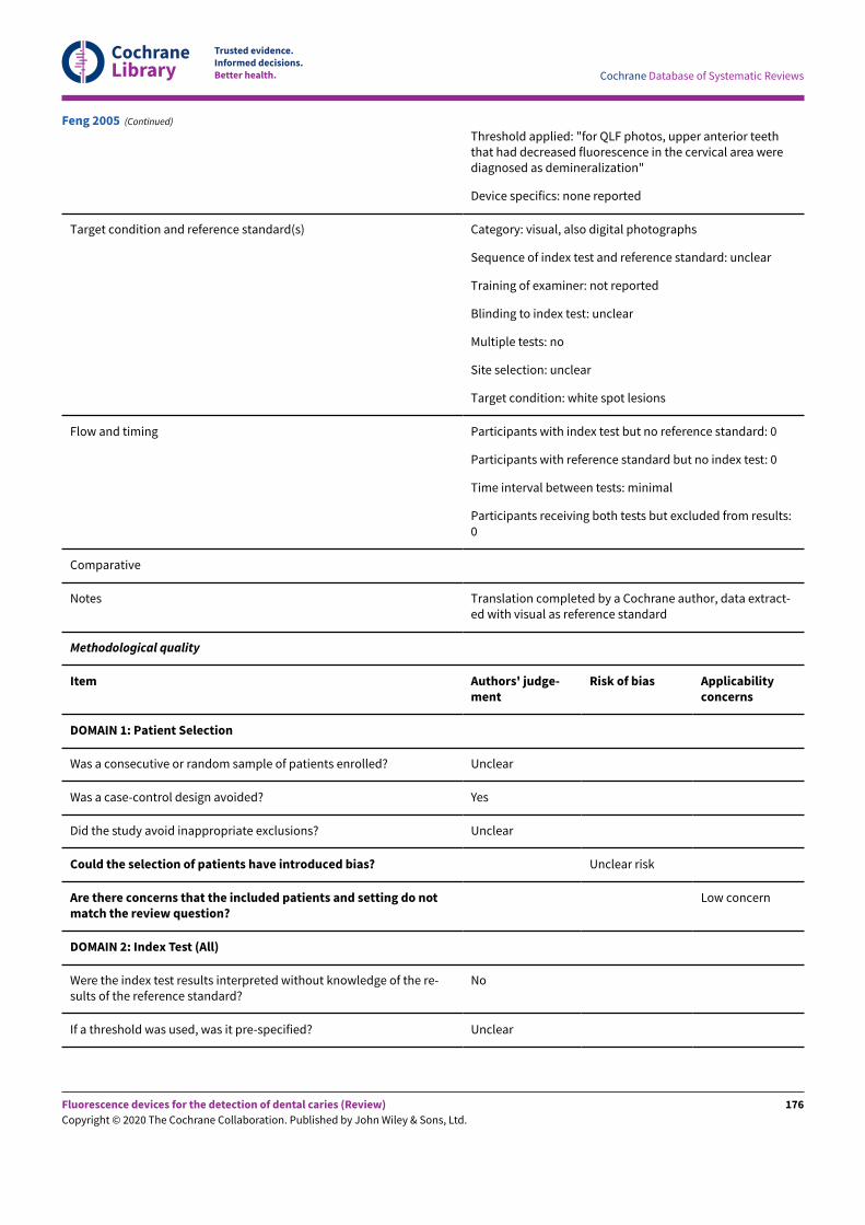
CochraneLibrary
Trusted evidence.Informed decisions.Better health.
Cochrane Database of Systematic Reviews
Threshold applied: "for QLF photos, upper anterior teeththat had decreased fluorescence in the cervical area werediagnosed as demineralization"
Device specifics: none reported
Target condition and reference standard(s) Category: visual, also digital photographs
Sequence of index test and reference standard: unclear
Training of examiner: not reported
Blinding to index test: unclear
Multiple tests: no
Site selection: unclear
Target condition: white spot lesions
Flow and timing Participants with index test but no reference standard: 0
Participants with reference standard but no index test: 0
Time interval between tests: minimal
Participants receiving both tests but excluded from results:0
Comparative
Notes Translation completed by a Cochrane author, data extract-ed with visual as reference standard
Methodological quality
Item Authors' judge-ment
Risk of bias Applicabilityconcerns
DOMAIN 1: Patient Selection
Was a consecutive or random sample of patients enrolled? Unclear
Was a case-control design avoided? Yes
Did the study avoid inappropriate exclusions? Unclear
Could the selection of patients have introduced bias? Unclear risk
Are there concerns that the included patients and setting do notmatch the review question?
Low concern
DOMAIN 2: Index Test (All)
Were the index test results interpreted without knowledge of the re-sults of the reference standard?
No
If a threshold was used, was it pre-specified? Unclear
Feng 2005 (Continued)
Fluorescence devices for the detection of dental caries (Review)
Copyright © 2020 The Cochrane Collaboration. Published by John Wiley & Sons, Ltd.
176
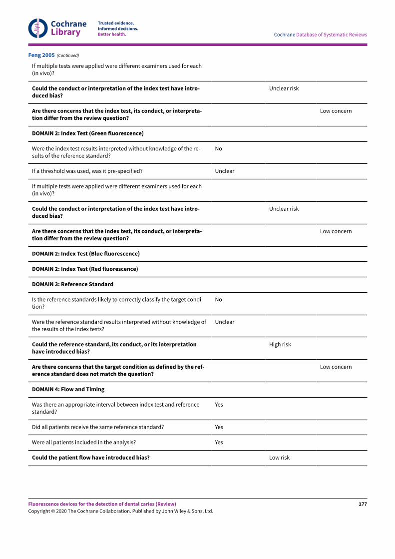
CochraneLibrary
Trusted evidence.Informed decisions.Better health.
Cochrane Database of Systematic Reviews
If multiple tests were applied were different examiners used for each(in vivo)?
Could the conduct or interpretation of the index test have intro-duced bias?
Unclear risk
Are there concerns that the index test, its conduct, or interpreta-tion differ from the review question?
Low concern
DOMAIN 2: Index Test (Green fluorescence)
Were the index test results interpreted without knowledge of the re-sults of the reference standard?
No
If a threshold was used, was it pre-specified? Unclear
If multiple tests were applied were different examiners used for each(in vivo)?
Could the conduct or interpretation of the index test have intro-duced bias?
Unclear risk
Are there concerns that the index test, its conduct, or interpreta-tion differ from the review question?
Low concern
DOMAIN 2: Index Test (Blue fluorescence)
DOMAIN 2: Index Test (Red fluorescence)
DOMAIN 3: Reference Standard
Is the reference standards likely to correctly classify the target condi-tion?
No
Were the reference standard results interpreted without knowledge ofthe results of the index tests?
Unclear
Could the reference standard, its conduct, or its interpretationhave introduced bias?
High risk
Are there concerns that the target condition as defined by the ref-erence standard does not match the question?
Low concern
DOMAIN 4: Flow and Timing
Was there an appropriate interval between index test and referencestandard?
Yes
Did all patients receive the same reference standard? Yes
Were all patients included in the analysis? Yes
Could the patient flow have introduced bias? Low risk
Feng 2005 (Continued)
Fluorescence devices for the detection of dental caries (Review)
Copyright © 2020 The Cochrane Collaboration. Published by John Wiley & Sons, Ltd.
177
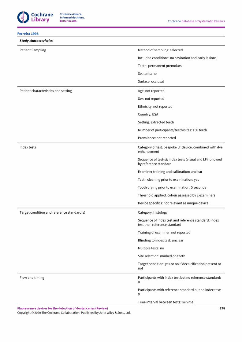
CochraneLibrary
Trusted evidence.Informed decisions.Better health.
Cochrane Database of Systematic Reviews
Study characteristics
Patient Sampling Method of sampling: selected
Included conditions: no cavitation and early lesions
Teeth: permanent premolars
Sealants: no
Surface: occlusal
Patient characteristics and setting Age: not reported
Sex: not reported
Ethnicity: not reported
Country: USA
Setting: extracted teeth
Number of participants/teeth/sites: 150 teeth
Prevalence: not reported
Index tests Category of test: bespoke LF device, combined with dyeenhancement
Sequence of test(s): index tests (visual and LF) followedby reference standard
Examiner training and calibration: unclear
Teeth cleaning prior to examination: yes
Tooth drying prior to examination: 5 seconds
Threshold applied: colour assessed by 2 examiners
Device specifics: not relevant as unique device
Target condition and reference standard(s) Category: histology
Sequence of index test and reference standard: indextest then reference standard
Training of examiner: not reported
Blinding to index test: unclear
Multiple tests: no
Site selection: marked on teeth
Target condition: yes or no if decalcification present ornot
Flow and timing Participants with index test but no reference standard:0
Participants with reference standard but no index test:0
Time interval between tests: minimal
Ferreira 1998
Fluorescence devices for the detection of dental caries (Review)
Copyright © 2020 The Cochrane Collaboration. Published by John Wiley & Sons, Ltd.
178
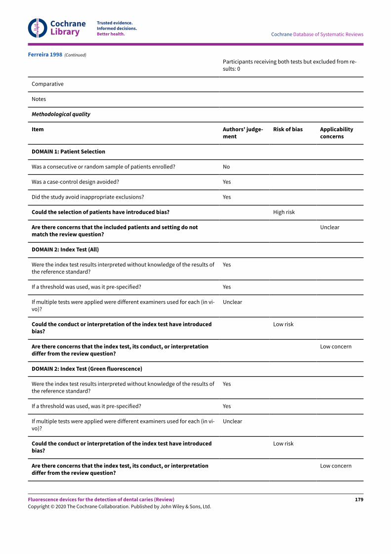
CochraneLibrary
Trusted evidence.Informed decisions.Better health.
Cochrane Database of Systematic Reviews
Participants receiving both tests but excluded from re-sults: 0
Comparative
Notes
Methodological quality
Item Authors' judge-ment
Risk of bias Applicabilityconcerns
DOMAIN 1: Patient Selection
Was a consecutive or random sample of patients enrolled? No
Was a case-control design avoided? Yes
Did the study avoid inappropriate exclusions? Yes
Could the selection of patients have introduced bias? High risk
Are there concerns that the included patients and setting do notmatch the review question?
Unclear
DOMAIN 2: Index Test (All)
Were the index test results interpreted without knowledge of the results ofthe reference standard?
Yes
If a threshold was used, was it pre-specified? Yes
If multiple tests were applied were different examiners used for each (in vi-vo)?
Unclear
Could the conduct or interpretation of the index test have introducedbias?
Low risk
Are there concerns that the index test, its conduct, or interpretationdiffer from the review question?
Low concern
DOMAIN 2: Index Test (Green fluorescence)
Were the index test results interpreted without knowledge of the results ofthe reference standard?
Yes
If a threshold was used, was it pre-specified? Yes
If multiple tests were applied were different examiners used for each (in vi-vo)?
Unclear
Could the conduct or interpretation of the index test have introducedbias?
Low risk
Are there concerns that the index test, its conduct, or interpretationdiffer from the review question?
Low concern
Ferreira 1998 (Continued)
Fluorescence devices for the detection of dental caries (Review)
Copyright © 2020 The Cochrane Collaboration. Published by John Wiley & Sons, Ltd.
179
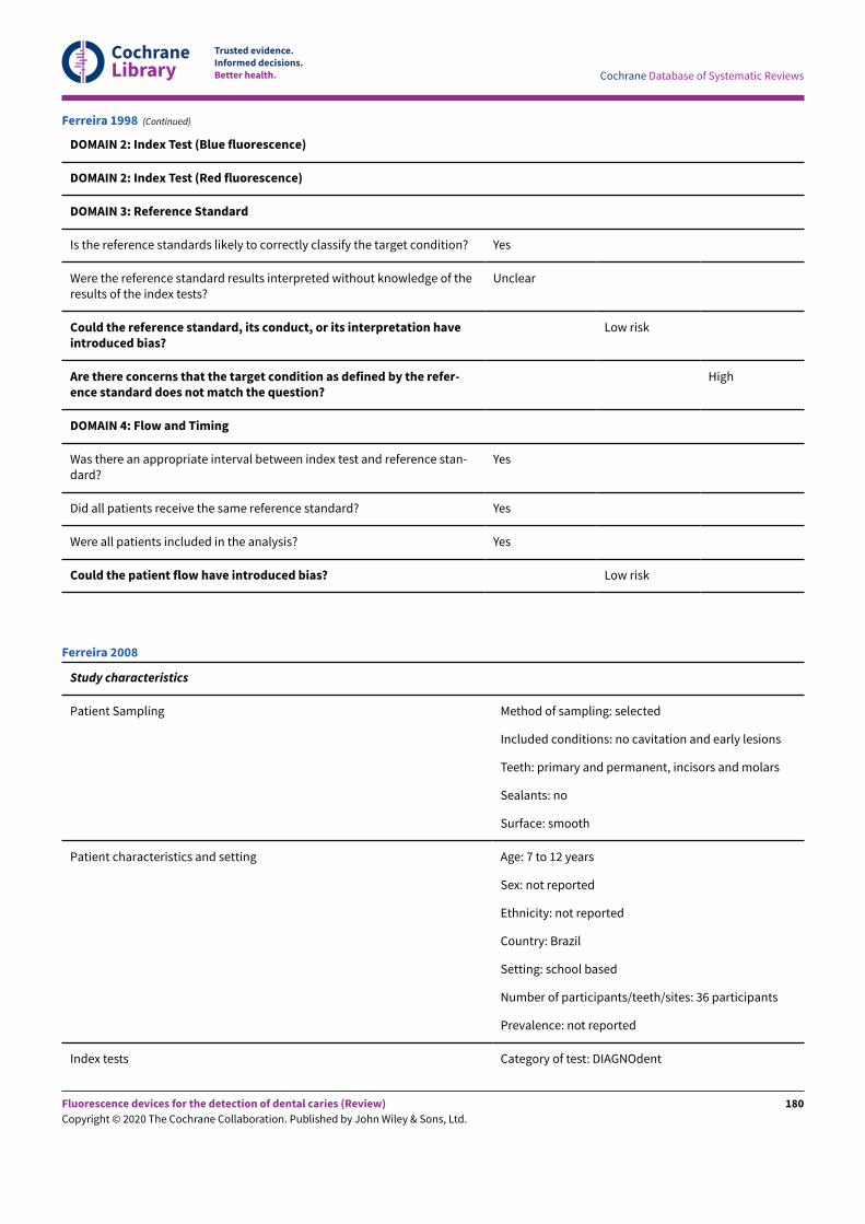
CochraneLibrary
Trusted evidence.Informed decisions.Better health.
Cochrane Database of Systematic Reviews
DOMAIN 2: Index Test (Blue fluorescence)
DOMAIN 2: Index Test (Red fluorescence)
DOMAIN 3: Reference Standard
Is the reference standards likely to correctly classify the target condition? Yes
Were the reference standard results interpreted without knowledge of theresults of the index tests?
Unclear
Could the reference standard, its conduct, or its interpretation haveintroduced bias?
Low risk
Are there concerns that the target condition as defined by the refer-ence standard does not match the question?
High
DOMAIN 4: Flow and Timing
Was there an appropriate interval between index test and reference stan-dard?
Yes
Did all patients receive the same reference standard? Yes
Were all patients included in the analysis? Yes
Could the patient flow have introduced bias? Low risk
Ferreira 1998 (Continued)
Study characteristics
Patient Sampling Method of sampling: selected
Included conditions: no cavitation and early lesions
Teeth: primary and permanent, incisors and molars
Sealants: no
Surface: smooth
Patient characteristics and setting Age: 7 to 12 years
Sex: not reported
Ethnicity: not reported
Country: Brazil
Setting: school based
Number of participants/teeth/sites: 36 participants
Prevalence: not reported
Index tests Category of test: DIAGNOdent
Ferreira 2008
Fluorescence devices for the detection of dental caries (Review)
Copyright © 2020 The Cochrane Collaboration. Published by John Wiley & Sons, Ltd.
180
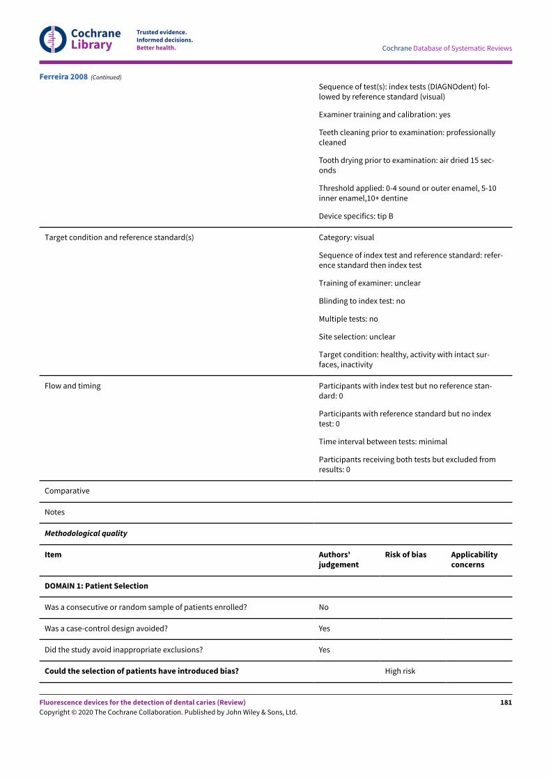
CochraneLibrary
Trusted evidence.Informed decisions.Better health.
Cochrane Database of Systematic Reviews
Sequence of test(s): index tests (DIAGNOdent) fol-lowed by reference standard (visual)
Examiner training and calibration: yes
Teeth cleaning prior to examination: professionallycleaned
Tooth drying prior to examination: air dried 15 sec-onds
Threshold applied: 0-4 sound or outer enamel, 5-10inner enamel,10+ dentine
Device specifics: tip B
Target condition and reference standard(s) Category: visual
Sequence of index test and reference standard: refer-ence standard then index test
Training of examiner: unclear
Blinding to index test: no
Multiple tests: no
Site selection: unclear
Target condition: healthy, activity with intact sur-faces, inactivity
Flow and timing Participants with index test but no reference stan-dard: 0
Participants with reference standard but no indextest: 0
Time interval between tests: minimal
Participants receiving both tests but excluded fromresults: 0
Comparative
Notes
Methodological quality
Item Authors'judgement
Risk of bias Applicabilityconcerns
DOMAIN 1: Patient Selection
Was a consecutive or random sample of patients enrolled? No
Was a case-control design avoided? Yes
Did the study avoid inappropriate exclusions? Yes
Could the selection of patients have introduced bias? High risk
Ferreira 2008 (Continued)
Fluorescence devices for the detection of dental caries (Review)
Copyright © 2020 The Cochrane Collaboration. Published by John Wiley & Sons, Ltd.
181
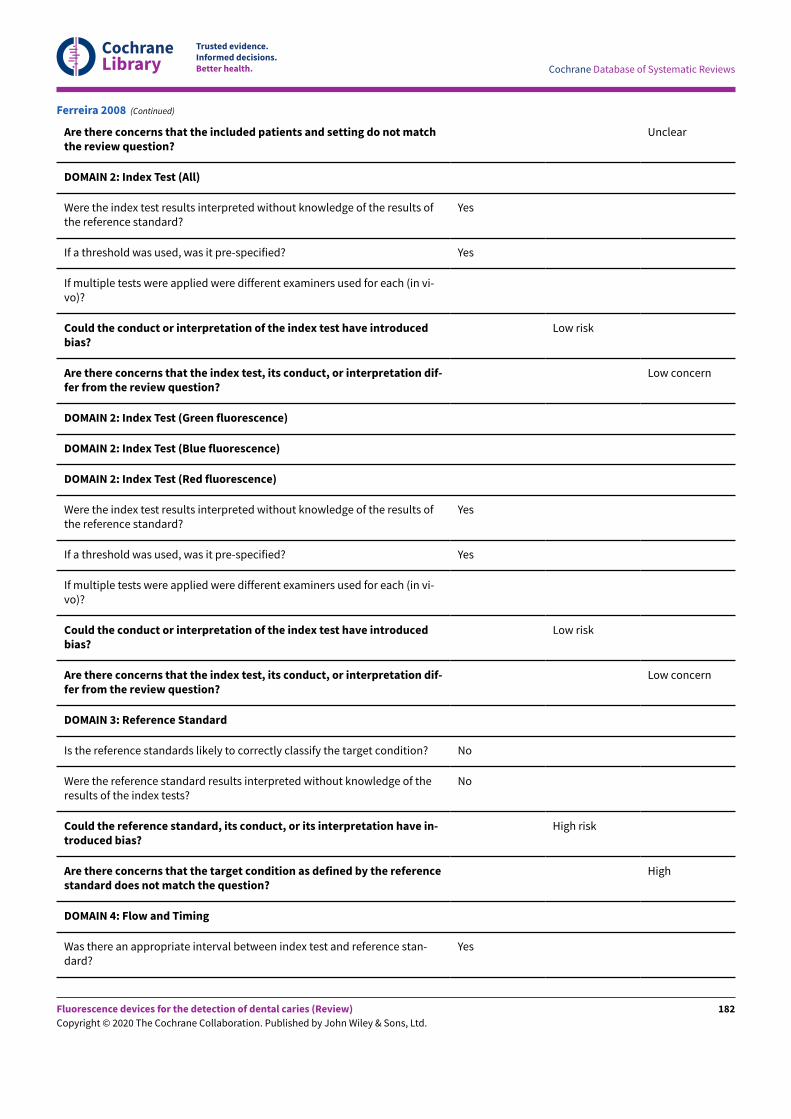
CochraneLibrary
Trusted evidence.Informed decisions.Better health.
Cochrane Database of Systematic Reviews
Are there concerns that the included patients and setting do not matchthe review question?
Unclear
DOMAIN 2: Index Test (All)
Were the index test results interpreted without knowledge of the results ofthe reference standard?
Yes
If a threshold was used, was it pre-specified? Yes
If multiple tests were applied were different examiners used for each (in vi-vo)?
Could the conduct or interpretation of the index test have introducedbias?
Low risk
Are there concerns that the index test, its conduct, or interpretation dif-fer from the review question?
Low concern
DOMAIN 2: Index Test (Green fluorescence)
DOMAIN 2: Index Test (Blue fluorescence)
DOMAIN 2: Index Test (Red fluorescence)
Were the index test results interpreted without knowledge of the results ofthe reference standard?
Yes
If a threshold was used, was it pre-specified? Yes
If multiple tests were applied were different examiners used for each (in vi-vo)?
Could the conduct or interpretation of the index test have introducedbias?
Low risk
Are there concerns that the index test, its conduct, or interpretation dif-fer from the review question?
Low concern
DOMAIN 3: Reference Standard
Is the reference standards likely to correctly classify the target condition? No
Were the reference standard results interpreted without knowledge of theresults of the index tests?
No
Could the reference standard, its conduct, or its interpretation have in-troduced bias?
High risk
Are there concerns that the target condition as defined by the referencestandard does not match the question?
High
DOMAIN 4: Flow and Timing
Was there an appropriate interval between index test and reference stan-dard?
Yes
Ferreira 2008 (Continued)
Fluorescence devices for the detection of dental caries (Review)
Copyright © 2020 The Cochrane Collaboration. Published by John Wiley & Sons, Ltd.
182
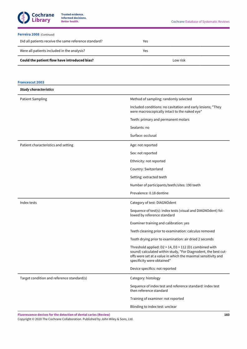
CochraneLibrary
Trusted evidence.Informed decisions.Better health.
Cochrane Database of Systematic Reviews
Did all patients receive the same reference standard? Yes
Were all patients included in the analysis? Yes
Could the patient flow have introduced bias? Low risk
Ferreira 2008 (Continued)
Study characteristics
Patient Sampling Method of sampling: randomly selected
Included conditions: no cavitation and early lesions; "Theywere macroscopically intact to the naked eye"
Teeth: primary and permanent molars
Sealants: no
Surface: occlusal
Patient characteristics and setting Age: not reported
Sex: not reported
Ethnicity: not reported
Country: Switzerland
Setting: extracted teeth
Number of participants/teeth/sites: 190 teeth
Prevalence: 0.18 dentine
Index tests Category of test: DIAGNOdent
Sequence of test(s): index tests (visual and DIAGNOdent) fol-lowed by reference standard
Examiner training and calibration: yes
Teeth cleaning prior to examination: calculus removed
Tooth drying prior to examination: air dried 2 seconds
Threshold applied: D2 > 14, D3 > 112 (D1 combined withsound) calculated within study, "For Diagnodent, the best cut-offs were set at a value in which the maximal sensitivity andspecificity were obtained"
Device specifics: not reported
Target condition and reference standard(s) Category: histology
Sequence of index test and reference standard: index testthen reference standard
Training of examiner: not reported
Blinding to index test: unclear
Francescut 2003
Fluorescence devices for the detection of dental caries (Review)
Copyright © 2020 The Cochrane Collaboration. Published by John Wiley & Sons, Ltd.
183
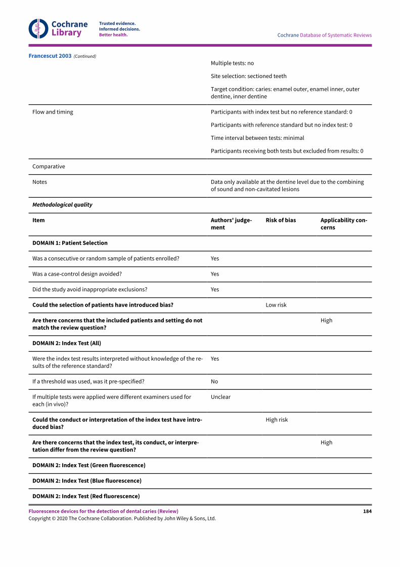
CochraneLibrary
Trusted evidence.Informed decisions.Better health.
Cochrane Database of Systematic Reviews
Multiple tests: no
Site selection: sectioned teeth
Target condition: caries: enamel outer, enamel inner, outerdentine, inner dentine
Flow and timing Participants with index test but no reference standard: 0
Participants with reference standard but no index test: 0
Time interval between tests: minimal
Participants receiving both tests but excluded from results: 0
Comparative
Notes Data only available at the dentine level due to the combiningof sound and non-cavitated lesions
Methodological quality
Item Authors' judge-ment
Risk of bias Applicability con-cerns
DOMAIN 1: Patient Selection
Was a consecutive or random sample of patients enrolled? Yes
Was a case-control design avoided? Yes
Did the study avoid inappropriate exclusions? Yes
Could the selection of patients have introduced bias? Low risk
Are there concerns that the included patients and setting do notmatch the review question?
High
DOMAIN 2: Index Test (All)
Were the index test results interpreted without knowledge of the re-sults of the reference standard?
Yes
If a threshold was used, was it pre-specified? No
If multiple tests were applied were different examiners used foreach (in vivo)?
Unclear
Could the conduct or interpretation of the index test have intro-duced bias?
High risk
Are there concerns that the index test, its conduct, or interpre-tation differ from the review question?
High
DOMAIN 2: Index Test (Green fluorescence)
DOMAIN 2: Index Test (Blue fluorescence)
DOMAIN 2: Index Test (Red fluorescence)
Francescut 2003 (Continued)
Fluorescence devices for the detection of dental caries (Review)
Copyright © 2020 The Cochrane Collaboration. Published by John Wiley & Sons, Ltd.
184
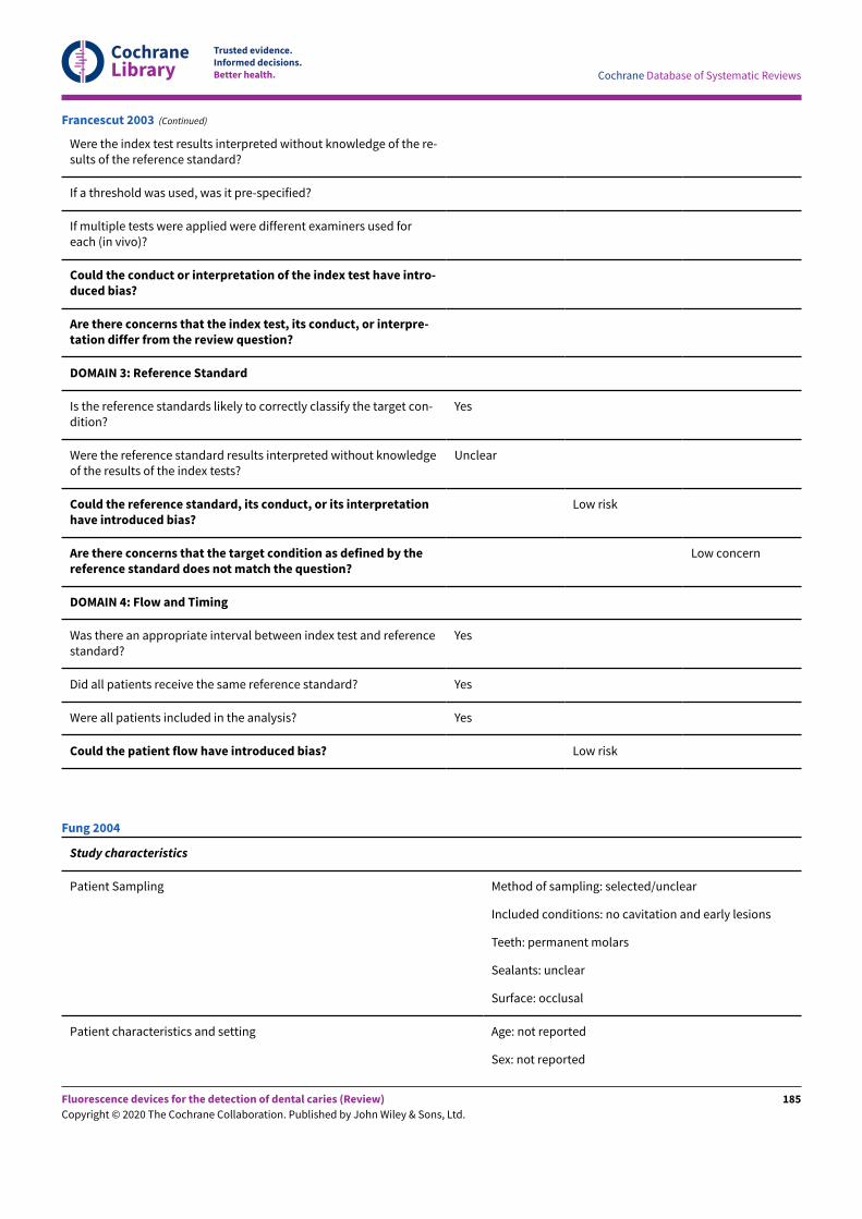
CochraneLibrary
Trusted evidence.Informed decisions.Better health.
Cochrane Database of Systematic Reviews
Were the index test results interpreted without knowledge of the re-sults of the reference standard?
If a threshold was used, was it pre-specified?
If multiple tests were applied were different examiners used foreach (in vivo)?
Could the conduct or interpretation of the index test have intro-duced bias?
Are there concerns that the index test, its conduct, or interpre-tation differ from the review question?
DOMAIN 3: Reference Standard
Is the reference standards likely to correctly classify the target con-dition?
Yes
Were the reference standard results interpreted without knowledgeof the results of the index tests?
Unclear
Could the reference standard, its conduct, or its interpretationhave introduced bias?
Low risk
Are there concerns that the target condition as defined by thereference standard does not match the question?
Low concern
DOMAIN 4: Flow and Timing
Was there an appropriate interval between index test and referencestandard?
Yes
Did all patients receive the same reference standard? Yes
Were all patients included in the analysis? Yes
Could the patient flow have introduced bias? Low risk
Francescut 2003 (Continued)
Study characteristics
Patient Sampling Method of sampling: selected/unclear
Included conditions: no cavitation and early lesions
Teeth: permanent molars
Sealants: unclear
Surface: occlusal
Patient characteristics and setting Age: not reported
Sex: not reported
Fung 2004
Fluorescence devices for the detection of dental caries (Review)
Copyright © 2020 The Cochrane Collaboration. Published by John Wiley & Sons, Ltd.
185
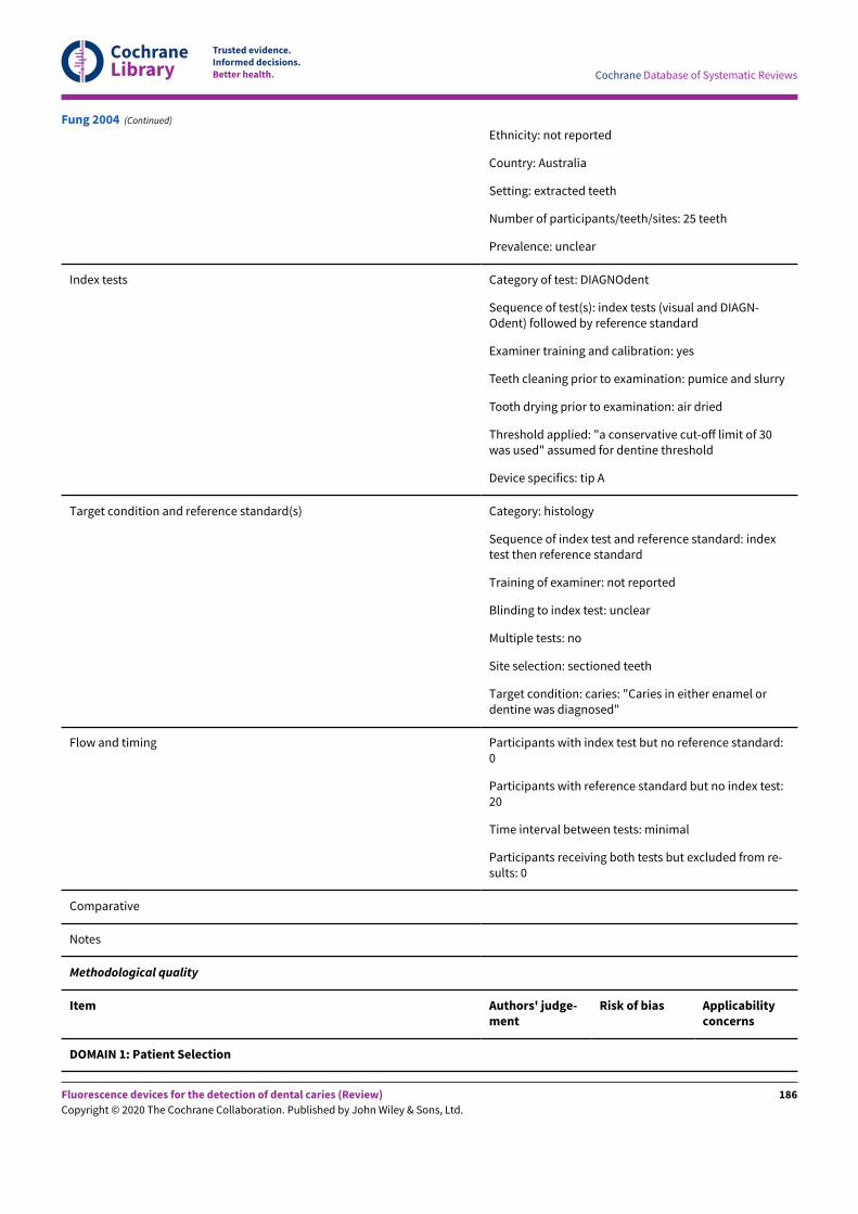
CochraneLibrary
Trusted evidence.Informed decisions.Better health.
Cochrane Database of Systematic Reviews
Ethnicity: not reported
Country: Australia
Setting: extracted teeth
Number of participants/teeth/sites: 25 teeth
Prevalence: unclear
Index tests Category of test: DIAGNOdent
Sequence of test(s): index tests (visual and DIAGN-Odent) followed by reference standard
Examiner training and calibration: yes
Teeth cleaning prior to examination: pumice and slurry
Tooth drying prior to examination: air dried
Threshold applied: "a conservative cut-oE limit of 30was used" assumed for dentine threshold
Device specifics: tip A
Target condition and reference standard(s) Category: histology
Sequence of index test and reference standard: indextest then reference standard
Training of examiner: not reported
Blinding to index test: unclear
Multiple tests: no
Site selection: sectioned teeth
Target condition: caries: "Caries in either enamel ordentine was diagnosed"
Flow and timing Participants with index test but no reference standard:0
Participants with reference standard but no index test:20
Time interval between tests: minimal
Participants receiving both tests but excluded from re-sults: 0
Comparative
Notes
Methodological quality
Item Authors' judge-ment
Risk of bias Applicabilityconcerns
DOMAIN 1: Patient Selection
Fung 2004 (Continued)
Fluorescence devices for the detection of dental caries (Review)
Copyright © 2020 The Cochrane Collaboration. Published by John Wiley & Sons, Ltd.
186
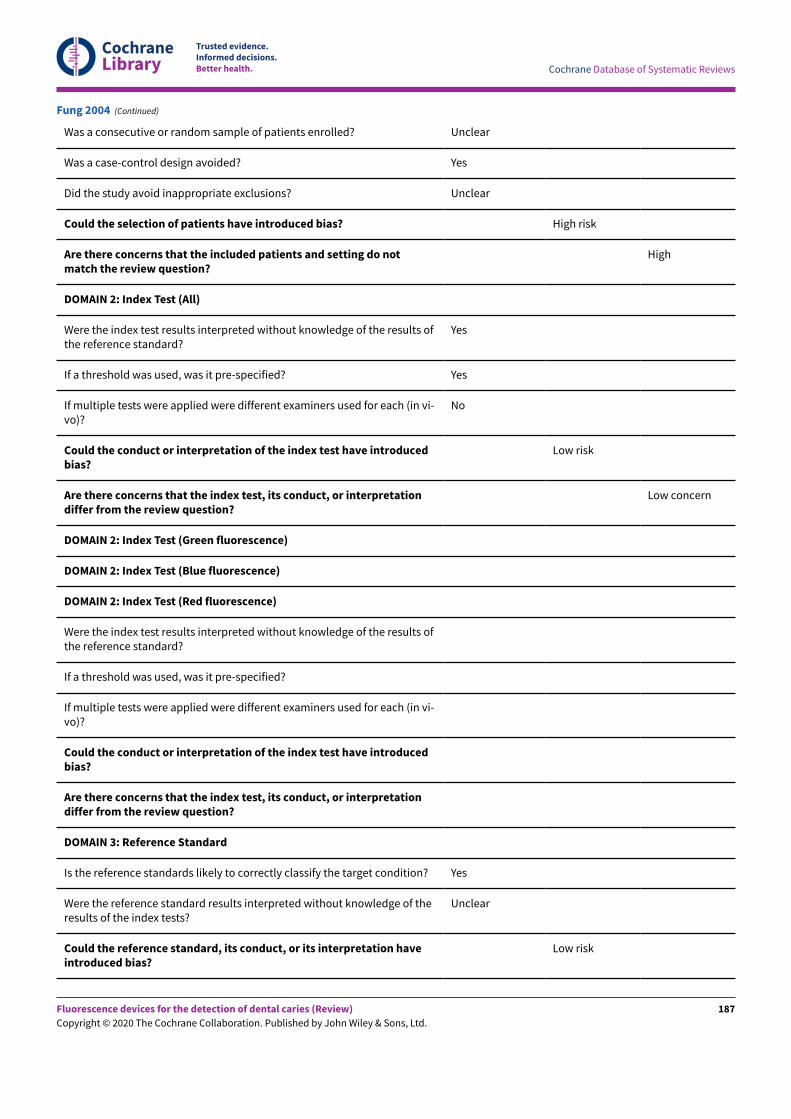
CochraneLibrary
Trusted evidence.Informed decisions.Better health.
Cochrane Database of Systematic Reviews
Was a consecutive or random sample of patients enrolled? Unclear
Was a case-control design avoided? Yes
Did the study avoid inappropriate exclusions? Unclear
Could the selection of patients have introduced bias? High risk
Are there concerns that the included patients and setting do notmatch the review question?
High
DOMAIN 2: Index Test (All)
Were the index test results interpreted without knowledge of the results ofthe reference standard?
Yes
If a threshold was used, was it pre-specified? Yes
If multiple tests were applied were different examiners used for each (in vi-vo)?
No
Could the conduct or interpretation of the index test have introducedbias?
Low risk
Are there concerns that the index test, its conduct, or interpretationdiffer from the review question?
Low concern
DOMAIN 2: Index Test (Green fluorescence)
DOMAIN 2: Index Test (Blue fluorescence)
DOMAIN 2: Index Test (Red fluorescence)
Were the index test results interpreted without knowledge of the results ofthe reference standard?
If a threshold was used, was it pre-specified?
If multiple tests were applied were different examiners used for each (in vi-vo)?
Could the conduct or interpretation of the index test have introducedbias?
Are there concerns that the index test, its conduct, or interpretationdiffer from the review question?
DOMAIN 3: Reference Standard
Is the reference standards likely to correctly classify the target condition? Yes
Were the reference standard results interpreted without knowledge of theresults of the index tests?
Unclear
Could the reference standard, its conduct, or its interpretation haveintroduced bias?
Low risk
Fung 2004 (Continued)
Fluorescence devices for the detection of dental caries (Review)
Copyright © 2020 The Cochrane Collaboration. Published by John Wiley & Sons, Ltd.
187
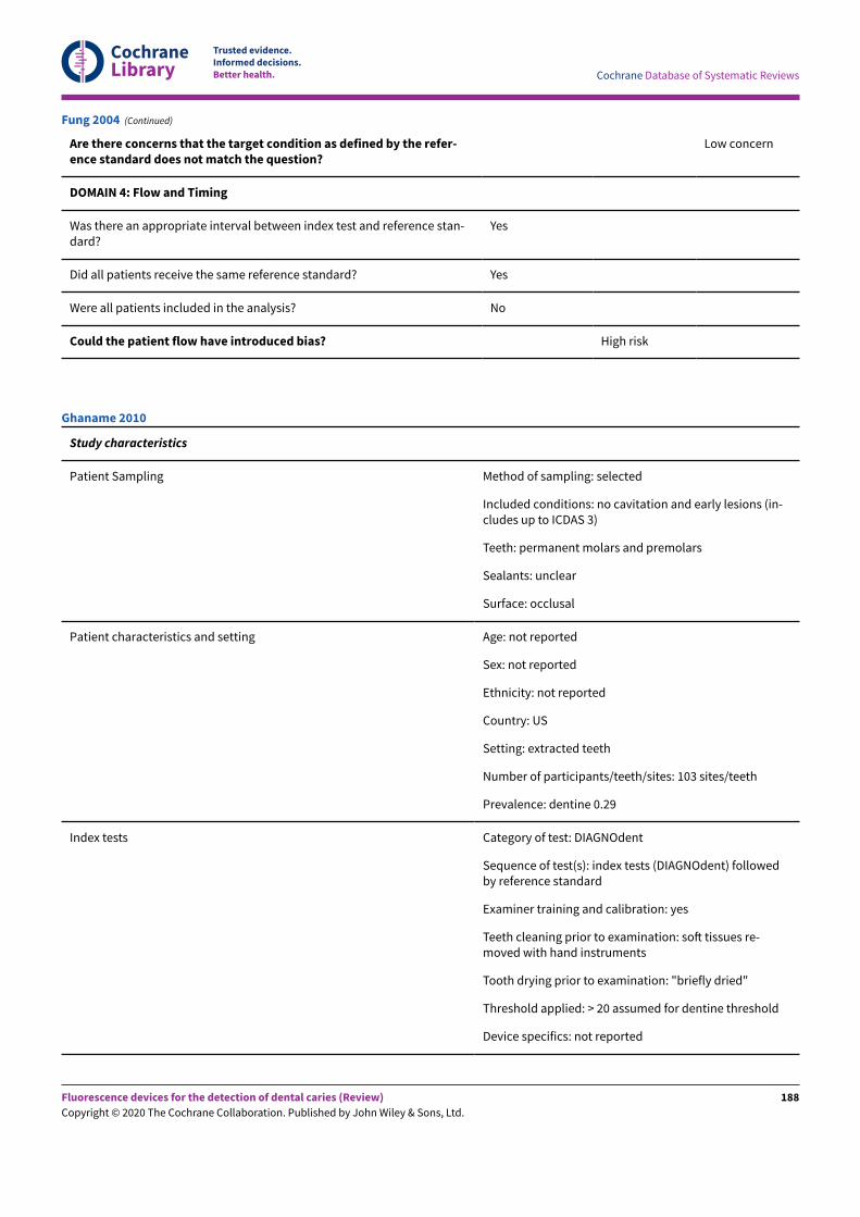
CochraneLibrary
Trusted evidence.Informed decisions.Better health.
Cochrane Database of Systematic Reviews
Are there concerns that the target condition as defined by the refer-ence standard does not match the question?
Low concern
DOMAIN 4: Flow and Timing
Was there an appropriate interval between index test and reference stan-dard?
Yes
Did all patients receive the same reference standard? Yes
Were all patients included in the analysis? No
Could the patient flow have introduced bias? High risk
Fung 2004 (Continued)
Study characteristics
Patient Sampling Method of sampling: selected
Included conditions: no cavitation and early lesions (in-cludes up to ICDAS 3)
Teeth: permanent molars and premolars
Sealants: unclear
Surface: occlusal
Patient characteristics and setting Age: not reported
Sex: not reported
Ethnicity: not reported
Country: US
Setting: extracted teeth
Number of participants/teeth/sites: 103 sites/teeth
Prevalence: dentine 0.29
Index tests Category of test: DIAGNOdent
Sequence of test(s): index tests (DIAGNOdent) followedby reference standard
Examiner training and calibration: yes
Teeth cleaning prior to examination: soN tissues re-moved with hand instruments
Tooth drying prior to examination: "briefly dried"
Threshold applied: > 20 assumed for dentine threshold
Device specifics: not reported
Ghaname 2010
Fluorescence devices for the detection of dental caries (Review)
Copyright © 2020 The Cochrane Collaboration. Published by John Wiley & Sons, Ltd.
188
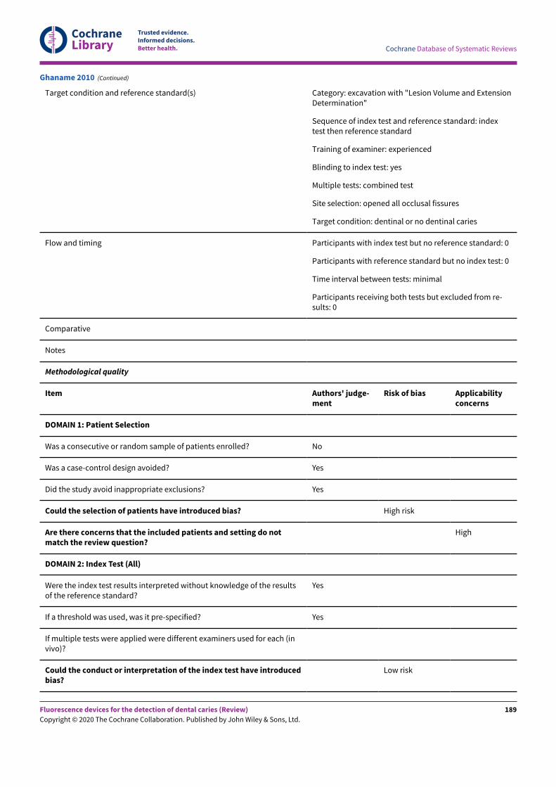
CochraneLibrary
Trusted evidence.Informed decisions.Better health.
Cochrane Database of Systematic Reviews
Target condition and reference standard(s) Category: excavation with "Lesion Volume and ExtensionDetermination"
Sequence of index test and reference standard: indextest then reference standard
Training of examiner: experienced
Blinding to index test: yes
Multiple tests: combined test
Site selection: opened all occlusal fissures
Target condition: dentinal or no dentinal caries
Flow and timing Participants with index test but no reference standard: 0
Participants with reference standard but no index test: 0
Time interval between tests: minimal
Participants receiving both tests but excluded from re-sults: 0
Comparative
Notes
Methodological quality
Item Authors' judge-ment
Risk of bias Applicabilityconcerns
DOMAIN 1: Patient Selection
Was a consecutive or random sample of patients enrolled? No
Was a case-control design avoided? Yes
Did the study avoid inappropriate exclusions? Yes
Could the selection of patients have introduced bias? High risk
Are there concerns that the included patients and setting do notmatch the review question?
High
DOMAIN 2: Index Test (All)
Were the index test results interpreted without knowledge of the resultsof the reference standard?
Yes
If a threshold was used, was it pre-specified? Yes
If multiple tests were applied were different examiners used for each (invivo)?
Could the conduct or interpretation of the index test have introducedbias?
Low risk
Ghaname 2010 (Continued)
Fluorescence devices for the detection of dental caries (Review)
Copyright © 2020 The Cochrane Collaboration. Published by John Wiley & Sons, Ltd.
189
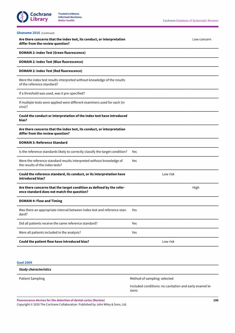
CochraneLibrary
Trusted evidence.Informed decisions.Better health.
Cochrane Database of Systematic Reviews
Are there concerns that the index test, its conduct, or interpretationdiffer from the review question?
Low concern
DOMAIN 2: Index Test (Green fluorescence)
DOMAIN 2: Index Test (Blue fluorescence)
DOMAIN 2: Index Test (Red fluorescence)
Were the index test results interpreted without knowledge of the resultsof the reference standard?
If a threshold was used, was it pre-specified?
If multiple tests were applied were different examiners used for each (invivo)?
Could the conduct or interpretation of the index test have introducedbias?
Are there concerns that the index test, its conduct, or interpretationdiffer from the review question?
DOMAIN 3: Reference Standard
Is the reference standards likely to correctly classify the target condition? Yes
Were the reference standard results interpreted without knowledge ofthe results of the index tests?
Yes
Could the reference standard, its conduct, or its interpretation haveintroduced bias?
Low risk
Are there concerns that the target condition as defined by the refer-ence standard does not match the question?
High
DOMAIN 4: Flow and Timing
Was there an appropriate interval between index test and reference stan-dard?
Yes
Did all patients receive the same reference standard? Yes
Were all patients included in the analysis? Yes
Could the patient flow have introduced bias? Low risk
Ghaname 2010 (Continued)
Study characteristics
Patient Sampling Method of sampling: selected
Included conditions: no cavitation and early enamel le-sions
Goel 2009
Fluorescence devices for the detection of dental caries (Review)
Copyright © 2020 The Cochrane Collaboration. Published by John Wiley & Sons, Ltd.
190
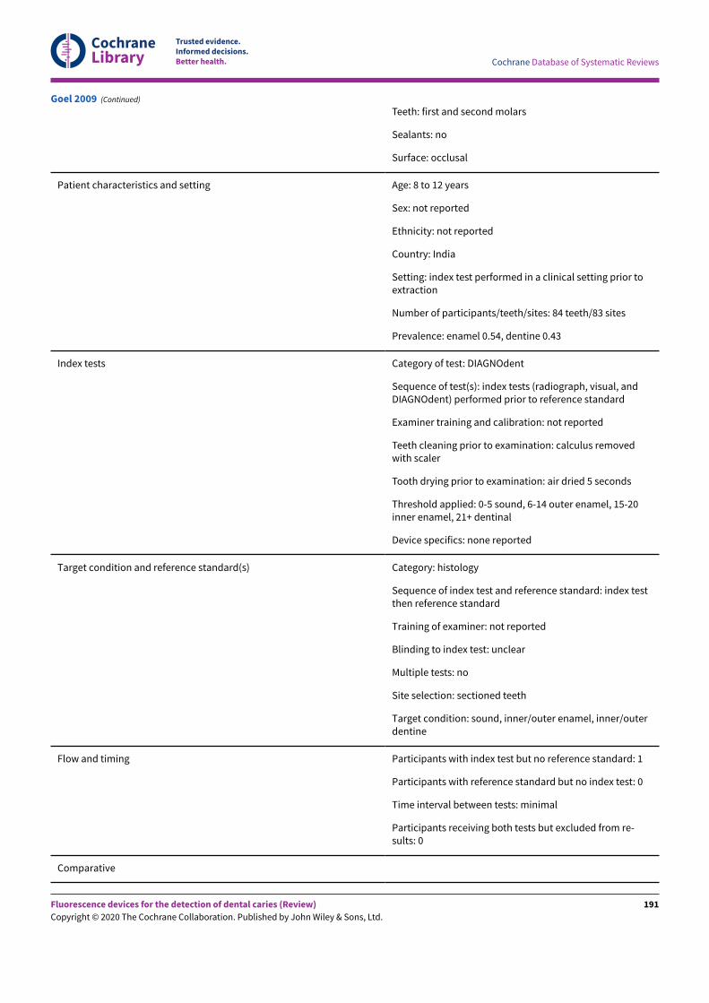
CochraneLibrary
Trusted evidence.Informed decisions.Better health.
Cochrane Database of Systematic Reviews
Teeth: first and second molars
Sealants: no
Surface: occlusal
Patient characteristics and setting Age: 8 to 12 years
Sex: not reported
Ethnicity: not reported
Country: India
Setting: index test performed in a clinical setting prior toextraction
Number of participants/teeth/sites: 84 teeth/83 sites
Prevalence: enamel 0.54, dentine 0.43
Index tests Category of test: DIAGNOdent
Sequence of test(s): index tests (radiograph, visual, andDIAGNOdent) performed prior to reference standard
Examiner training and calibration: not reported
Teeth cleaning prior to examination: calculus removedwith scaler
Tooth drying prior to examination: air dried 5 seconds
Threshold applied: 0-5 sound, 6-14 outer enamel, 15-20inner enamel, 21+ dentinal
Device specifics: none reported
Target condition and reference standard(s) Category: histology
Sequence of index test and reference standard: index testthen reference standard
Training of examiner: not reported
Blinding to index test: unclear
Multiple tests: no
Site selection: sectioned teeth
Target condition: sound, inner/outer enamel, inner/outerdentine
Flow and timing Participants with index test but no reference standard: 1
Participants with reference standard but no index test: 0
Time interval between tests: minimal
Participants receiving both tests but excluded from re-sults: 0
Comparative
Goel 2009 (Continued)
Fluorescence devices for the detection of dental caries (Review)
Copyright © 2020 The Cochrane Collaboration. Published by John Wiley & Sons, Ltd.
191
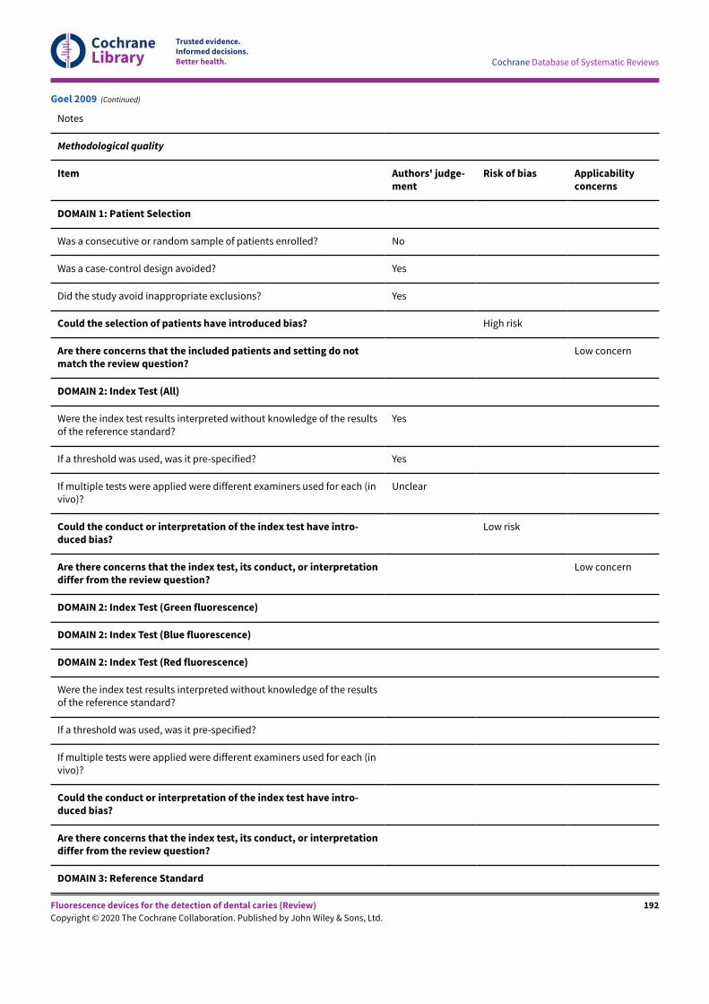
CochraneLibrary
Trusted evidence.Informed decisions.Better health.
Cochrane Database of Systematic Reviews
Notes
Methodological quality
Item Authors' judge-ment
Risk of bias Applicabilityconcerns
DOMAIN 1: Patient Selection
Was a consecutive or random sample of patients enrolled? No
Was a case-control design avoided? Yes
Did the study avoid inappropriate exclusions? Yes
Could the selection of patients have introduced bias? High risk
Are there concerns that the included patients and setting do notmatch the review question?
Low concern
DOMAIN 2: Index Test (All)
Were the index test results interpreted without knowledge of the resultsof the reference standard?
Yes
If a threshold was used, was it pre-specified? Yes
If multiple tests were applied were different examiners used for each (invivo)?
Unclear
Could the conduct or interpretation of the index test have intro-duced bias?
Low risk
Are there concerns that the index test, its conduct, or interpretationdiffer from the review question?
Low concern
DOMAIN 2: Index Test (Green fluorescence)
DOMAIN 2: Index Test (Blue fluorescence)
DOMAIN 2: Index Test (Red fluorescence)
Were the index test results interpreted without knowledge of the resultsof the reference standard?
If a threshold was used, was it pre-specified?
If multiple tests were applied were different examiners used for each (invivo)?
Could the conduct or interpretation of the index test have intro-duced bias?
Are there concerns that the index test, its conduct, or interpretationdiffer from the review question?
DOMAIN 3: Reference Standard
Goel 2009 (Continued)
Fluorescence devices for the detection of dental caries (Review)
Copyright © 2020 The Cochrane Collaboration. Published by John Wiley & Sons, Ltd.
192
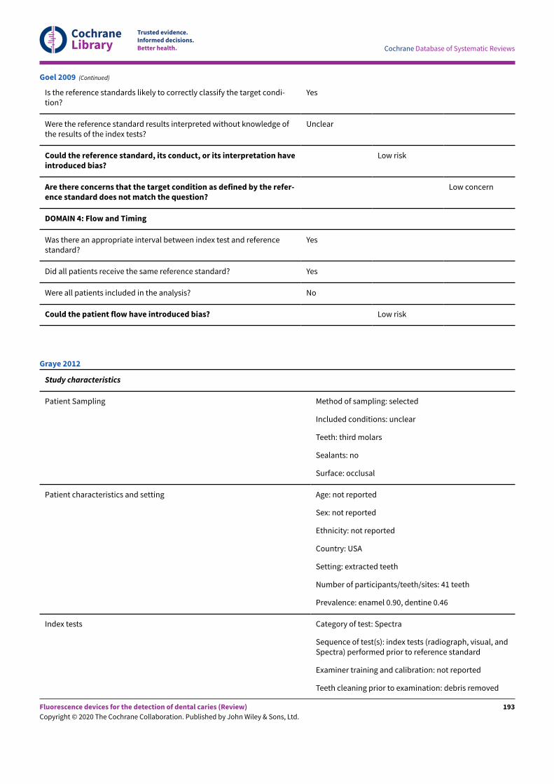
CochraneLibrary
Trusted evidence.Informed decisions.Better health.
Cochrane Database of Systematic Reviews
Is the reference standards likely to correctly classify the target condi-tion?
Yes
Were the reference standard results interpreted without knowledge ofthe results of the index tests?
Unclear
Could the reference standard, its conduct, or its interpretation haveintroduced bias?
Low risk
Are there concerns that the target condition as defined by the refer-ence standard does not match the question?
Low concern
DOMAIN 4: Flow and Timing
Was there an appropriate interval between index test and referencestandard?
Yes
Did all patients receive the same reference standard? Yes
Were all patients included in the analysis? No
Could the patient flow have introduced bias? Low risk
Goel 2009 (Continued)
Study characteristics
Patient Sampling Method of sampling: selected
Included conditions: unclear
Teeth: third molars
Sealants: no
Surface: occlusal
Patient characteristics and setting Age: not reported
Sex: not reported
Ethnicity: not reported
Country: USA
Setting: extracted teeth
Number of participants/teeth/sites: 41 teeth
Prevalence: enamel 0.90, dentine 0.46
Index tests Category of test: Spectra
Sequence of test(s): index tests (radiograph, visual, andSpectra) performed prior to reference standard
Examiner training and calibration: not reported
Teeth cleaning prior to examination: debris removed
Graye 2012
Fluorescence devices for the detection of dental caries (Review)
Copyright © 2020 The Cochrane Collaboration. Published by John Wiley & Sons, Ltd.
193
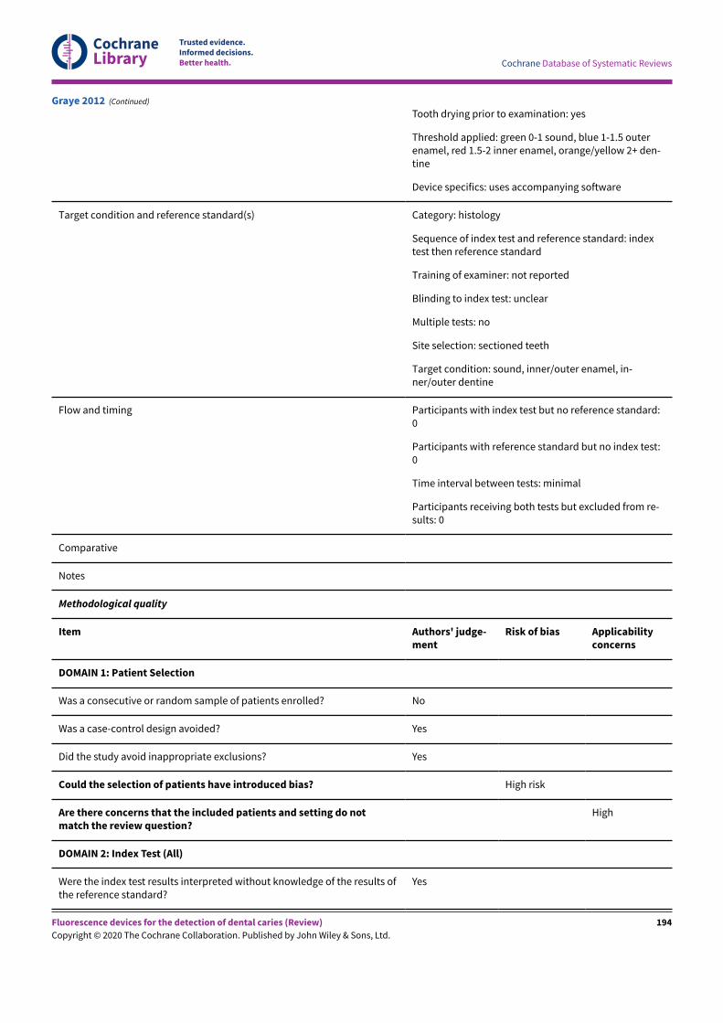
CochraneLibrary
Trusted evidence.Informed decisions.Better health.
Cochrane Database of Systematic Reviews
Tooth drying prior to examination: yes
Threshold applied: green 0-1 sound, blue 1-1.5 outerenamel, red 1.5-2 inner enamel, orange/yellow 2+ den-tine
Device specifics: uses accompanying software
Target condition and reference standard(s) Category: histology
Sequence of index test and reference standard: indextest then reference standard
Training of examiner: not reported
Blinding to index test: unclear
Multiple tests: no
Site selection: sectioned teeth
Target condition: sound, inner/outer enamel, in-ner/outer dentine
Flow and timing Participants with index test but no reference standard:0
Participants with reference standard but no index test:0
Time interval between tests: minimal
Participants receiving both tests but excluded from re-sults: 0
Comparative
Notes
Methodological quality
Item Authors' judge-ment
Risk of bias Applicabilityconcerns
DOMAIN 1: Patient Selection
Was a consecutive or random sample of patients enrolled? No
Was a case-control design avoided? Yes
Did the study avoid inappropriate exclusions? Yes
Could the selection of patients have introduced bias? High risk
Are there concerns that the included patients and setting do notmatch the review question?
High
DOMAIN 2: Index Test (All)
Were the index test results interpreted without knowledge of the results ofthe reference standard?
Yes
Graye 2012 (Continued)
Fluorescence devices for the detection of dental caries (Review)
Copyright © 2020 The Cochrane Collaboration. Published by John Wiley & Sons, Ltd.
194
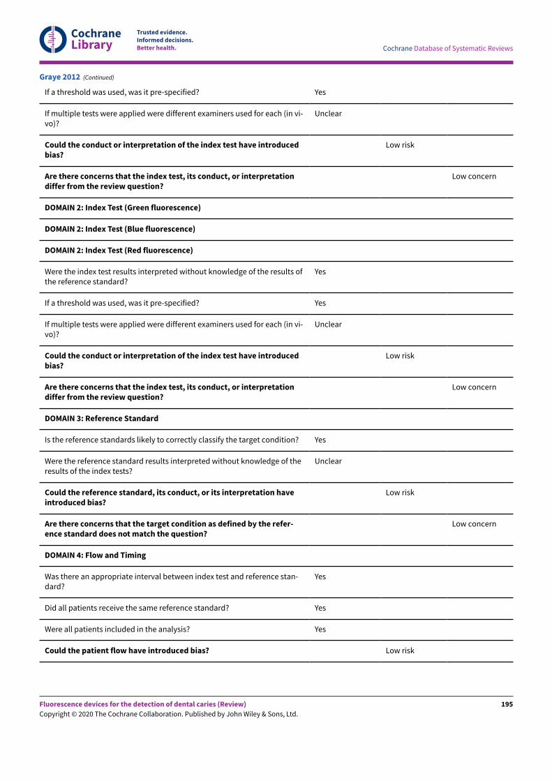
CochraneLibrary
Trusted evidence.Informed decisions.Better health.
Cochrane Database of Systematic Reviews
If a threshold was used, was it pre-specified? Yes
If multiple tests were applied were different examiners used for each (in vi-vo)?
Unclear
Could the conduct or interpretation of the index test have introducedbias?
Low risk
Are there concerns that the index test, its conduct, or interpretationdiffer from the review question?
Low concern
DOMAIN 2: Index Test (Green fluorescence)
DOMAIN 2: Index Test (Blue fluorescence)
DOMAIN 2: Index Test (Red fluorescence)
Were the index test results interpreted without knowledge of the results ofthe reference standard?
Yes
If a threshold was used, was it pre-specified? Yes
If multiple tests were applied were different examiners used for each (in vi-vo)?
Unclear
Could the conduct or interpretation of the index test have introducedbias?
Low risk
Are there concerns that the index test, its conduct, or interpretationdiffer from the review question?
Low concern
DOMAIN 3: Reference Standard
Is the reference standards likely to correctly classify the target condition? Yes
Were the reference standard results interpreted without knowledge of theresults of the index tests?
Unclear
Could the reference standard, its conduct, or its interpretation haveintroduced bias?
Low risk
Are there concerns that the target condition as defined by the refer-ence standard does not match the question?
Low concern
DOMAIN 4: Flow and Timing
Was there an appropriate interval between index test and reference stan-dard?
Yes
Did all patients receive the same reference standard? Yes
Were all patients included in the analysis? Yes
Could the patient flow have introduced bias? Low risk
Graye 2012 (Continued)
Fluorescence devices for the detection of dental caries (Review)
Copyright © 2020 The Cochrane Collaboration. Published by John Wiley & Sons, Ltd.
195
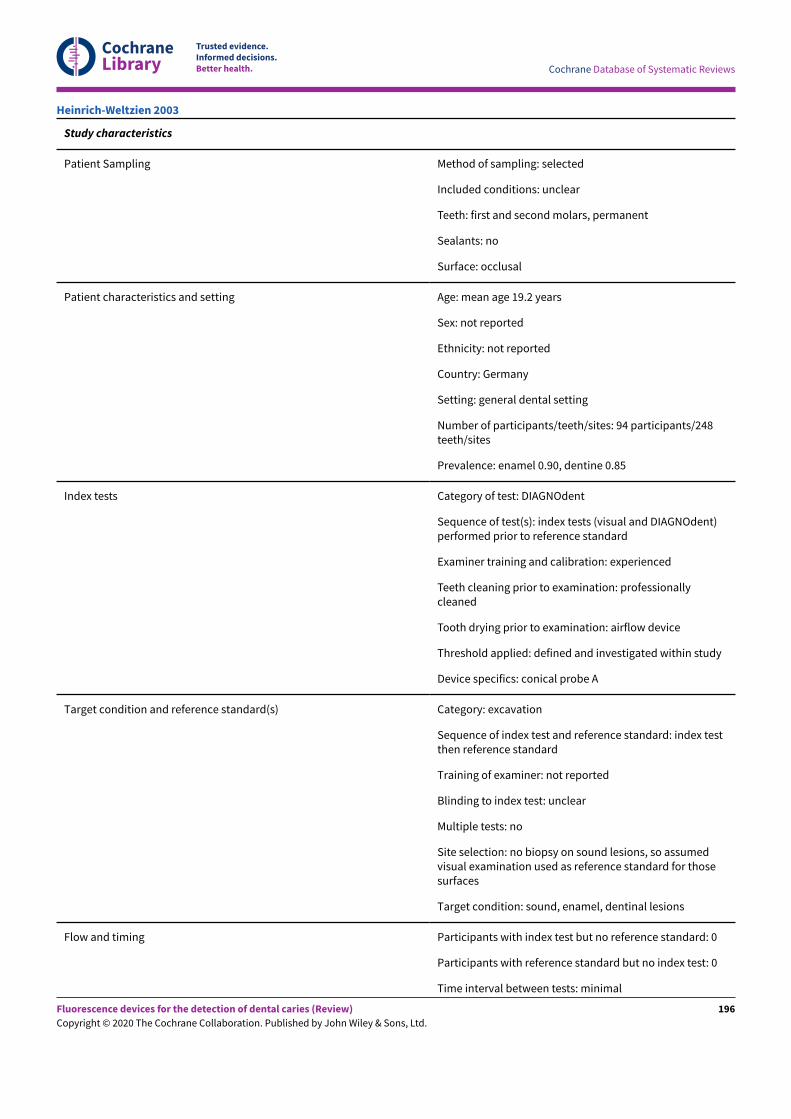
CochraneLibrary
Trusted evidence.Informed decisions.Better health.
Cochrane Database of Systematic Reviews
Study characteristics
Patient Sampling Method of sampling: selected
Included conditions: unclear
Teeth: first and second molars, permanent
Sealants: no
Surface: occlusal
Patient characteristics and setting Age: mean age 19.2 years
Sex: not reported
Ethnicity: not reported
Country: Germany
Setting: general dental setting
Number of participants/teeth/sites: 94 participants/248teeth/sites
Prevalence: enamel 0.90, dentine 0.85
Index tests Category of test: DIAGNOdent
Sequence of test(s): index tests (visual and DIAGNOdent)performed prior to reference standard
Examiner training and calibration: experienced
Teeth cleaning prior to examination: professionallycleaned
Tooth drying prior to examination: airflow device
Threshold applied: defined and investigated within study
Device specifics: conical probe A
Target condition and reference standard(s) Category: excavation
Sequence of index test and reference standard: index testthen reference standard
Training of examiner: not reported
Blinding to index test: unclear
Multiple tests: no
Site selection: no biopsy on sound lesions, so assumedvisual examination used as reference standard for thosesurfaces
Target condition: sound, enamel, dentinal lesions
Flow and timing Participants with index test but no reference standard: 0
Participants with reference standard but no index test: 0
Time interval between tests: minimal
Heinrich-Weltzien 2003
Fluorescence devices for the detection of dental caries (Review)
Copyright © 2020 The Cochrane Collaboration. Published by John Wiley & Sons, Ltd.
196
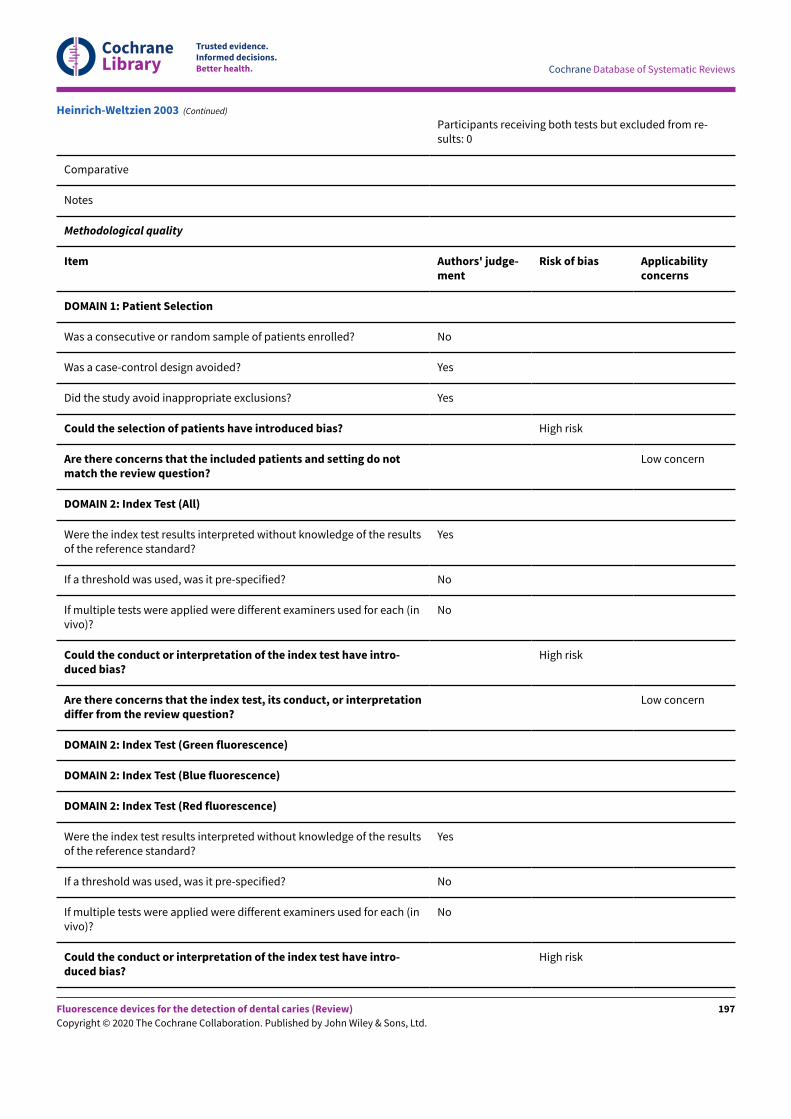
CochraneLibrary
Trusted evidence.Informed decisions.Better health.
Cochrane Database of Systematic Reviews
Participants receiving both tests but excluded from re-sults: 0
Comparative
Notes
Methodological quality
Item Authors' judge-ment
Risk of bias Applicabilityconcerns
DOMAIN 1: Patient Selection
Was a consecutive or random sample of patients enrolled? No
Was a case-control design avoided? Yes
Did the study avoid inappropriate exclusions? Yes
Could the selection of patients have introduced bias? High risk
Are there concerns that the included patients and setting do notmatch the review question?
Low concern
DOMAIN 2: Index Test (All)
Were the index test results interpreted without knowledge of the resultsof the reference standard?
Yes
If a threshold was used, was it pre-specified? No
If multiple tests were applied were different examiners used for each (invivo)?
No
Could the conduct or interpretation of the index test have intro-duced bias?
High risk
Are there concerns that the index test, its conduct, or interpretationdiffer from the review question?
Low concern
DOMAIN 2: Index Test (Green fluorescence)
DOMAIN 2: Index Test (Blue fluorescence)
DOMAIN 2: Index Test (Red fluorescence)
Were the index test results interpreted without knowledge of the resultsof the reference standard?
Yes
If a threshold was used, was it pre-specified? No
If multiple tests were applied were different examiners used for each (invivo)?
No
Could the conduct or interpretation of the index test have intro-duced bias?
High risk
Heinrich-Weltzien 2003 (Continued)
Fluorescence devices for the detection of dental caries (Review)
Copyright © 2020 The Cochrane Collaboration. Published by John Wiley & Sons, Ltd.
197
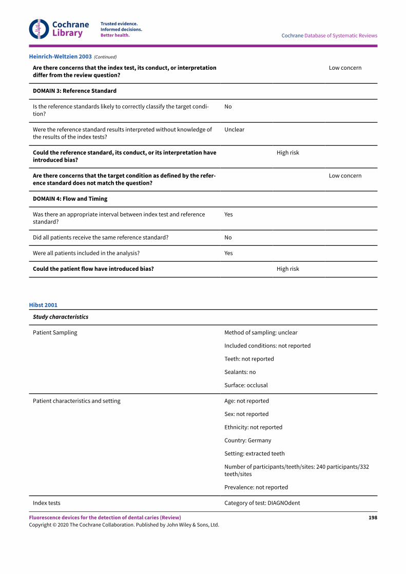
CochraneLibrary
Trusted evidence.Informed decisions.Better health.
Cochrane Database of Systematic Reviews
Are there concerns that the index test, its conduct, or interpretationdiffer from the review question?
Low concern
DOMAIN 3: Reference Standard
Is the reference standards likely to correctly classify the target condi-tion?
No
Were the reference standard results interpreted without knowledge ofthe results of the index tests?
Unclear
Could the reference standard, its conduct, or its interpretation haveintroduced bias?
High risk
Are there concerns that the target condition as defined by the refer-ence standard does not match the question?
Low concern
DOMAIN 4: Flow and Timing
Was there an appropriate interval between index test and referencestandard?
Yes
Did all patients receive the same reference standard? No
Were all patients included in the analysis? Yes
Could the patient flow have introduced bias? High risk
Heinrich-Weltzien 2003 (Continued)
Study characteristics
Patient Sampling Method of sampling: unclear
Included conditions: not reported
Teeth: not reported
Sealants: no
Surface: occlusal
Patient characteristics and setting Age: not reported
Sex: not reported
Ethnicity: not reported
Country: Germany
Setting: extracted teeth
Number of participants/teeth/sites: 240 participants/332teeth/sites
Prevalence: not reported
Index tests Category of test: DIAGNOdent
Hibst 2001
Fluorescence devices for the detection of dental caries (Review)
Copyright © 2020 The Cochrane Collaboration. Published by John Wiley & Sons, Ltd.
198
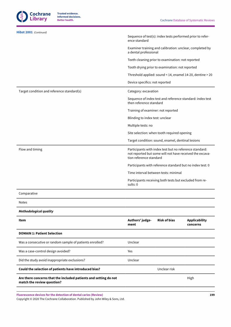
CochraneLibrary
Trusted evidence.Informed decisions.Better health.
Cochrane Database of Systematic Reviews
Sequence of test(s): index tests performed prior to refer-ence standard
Examiner training and calibration: unclear, completed bya dental professional
Teeth cleaning prior to examination: not reported
Tooth drying prior to examination: not reported
Threshold applied: sound < 14, enamel 14-20, dentine > 20
Device specifics: not reported
Target condition and reference standard(s) Category: excavation
Sequence of index test and reference standard: index testthen reference standard
Training of examiner: not reported
Blinding to index test: unclear
Multiple tests: no
Site selection: when tooth required opening
Target condition: sound, enamel, dentinal lesions
Flow and timing Participants with index test but no reference standard:not reported but some will not have received the excava-tion reference standard
Participants with reference standard but no index test: 0
Time interval between tests: minimal
Participants receiving both tests but excluded from re-sults: 0
Comparative
Notes
Methodological quality
Item Authors' judge-ment
Risk of bias Applicabilityconcerns
DOMAIN 1: Patient Selection
Was a consecutive or random sample of patients enrolled? Unclear
Was a case-control design avoided? Yes
Did the study avoid inappropriate exclusions? Unclear
Could the selection of patients have introduced bias? Unclear risk
Are there concerns that the included patients and setting do notmatch the review question?
High
Hibst 2001 (Continued)
Fluorescence devices for the detection of dental caries (Review)
Copyright © 2020 The Cochrane Collaboration. Published by John Wiley & Sons, Ltd.
199

CochraneLibrary
Trusted evidence.Informed decisions.Better health.
Cochrane Database of Systematic Reviews
DOMAIN 2: Index Test (All)
Were the index test results interpreted without knowledge of the resultsof the reference standard?
Yes
If a threshold was used, was it pre-specified? Yes
If multiple tests were applied were different examiners used for each (invivo)?
No
Could the conduct or interpretation of the index test have intro-duced bias?
Low risk
Are there concerns that the index test, its conduct, or interpretationdiffer from the review question?
Low concern
DOMAIN 2: Index Test (Green fluorescence)
DOMAIN 2: Index Test (Blue fluorescence)
DOMAIN 2: Index Test (Red fluorescence)
Were the index test results interpreted without knowledge of the resultsof the reference standard?
Yes
If a threshold was used, was it pre-specified? Yes
If multiple tests were applied were different examiners used for each (invivo)?
No
Could the conduct or interpretation of the index test have intro-duced bias?
Low risk
Are there concerns that the index test, its conduct, or interpretationdiffer from the review question?
Low concern
DOMAIN 3: Reference Standard
Is the reference standards likely to correctly classify the target condi-tion?
No
Were the reference standard results interpreted without knowledge ofthe results of the index tests?
No
Could the reference standard, its conduct, or its interpretation haveintroduced bias?
High risk
Are there concerns that the target condition as defined by the refer-ence standard does not match the question?
Low concern
DOMAIN 4: Flow and Timing
Was there an appropriate interval between index test and referencestandard?
Yes
Did all patients receive the same reference standard? No
Hibst 2001 (Continued)
Fluorescence devices for the detection of dental caries (Review)
Copyright © 2020 The Cochrane Collaboration. Published by John Wiley & Sons, Ltd.
200
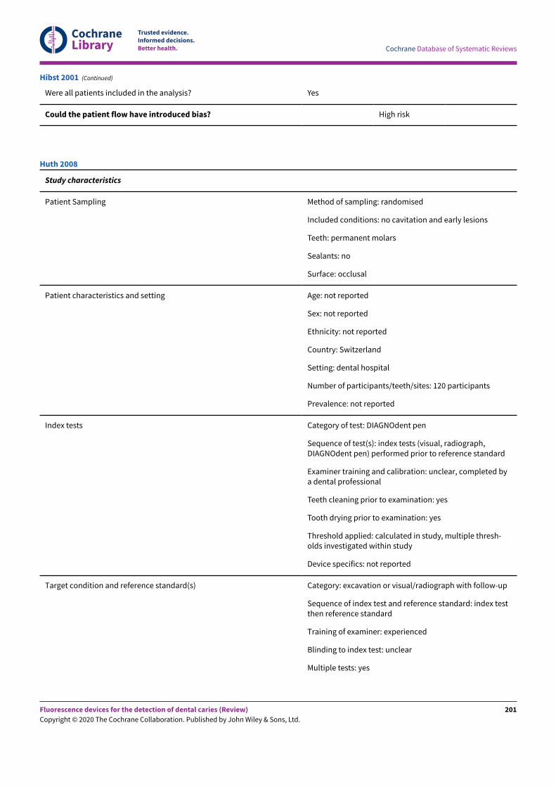
CochraneLibrary
Trusted evidence.Informed decisions.Better health.
Cochrane Database of Systematic Reviews
Were all patients included in the analysis? Yes
Could the patient flow have introduced bias? High risk
Hibst 2001 (Continued)
Study characteristics
Patient Sampling Method of sampling: randomised
Included conditions: no cavitation and early lesions
Teeth: permanent molars
Sealants: no
Surface: occlusal
Patient characteristics and setting Age: not reported
Sex: not reported
Ethnicity: not reported
Country: Switzerland
Setting: dental hospital
Number of participants/teeth/sites: 120 participants
Prevalence: not reported
Index tests Category of test: DIAGNOdent pen
Sequence of test(s): index tests (visual, radiograph,DIAGNOdent pen) performed prior to reference standard
Examiner training and calibration: unclear, completed bya dental professional
Teeth cleaning prior to examination: yes
Tooth drying prior to examination: yes
Threshold applied: calculated in study, multiple thresh-olds investigated within study
Device specifics: not reported
Target condition and reference standard(s) Category: excavation or visual/radiograph with follow-up
Sequence of index test and reference standard: index testthen reference standard
Training of examiner: experienced
Blinding to index test: unclear
Multiple tests: yes
Huth 2008
Fluorescence devices for the detection of dental caries (Review)
Copyright © 2020 The Cochrane Collaboration. Published by John Wiley & Sons, Ltd.
201

CochraneLibrary
Trusted evidence.Informed decisions.Better health.
Cochrane Database of Systematic Reviews
Site selection: unclear which site was investigated withwhich test
Target condition: sound, enamel, dentinal lesions
Flow and timing Participants with index test but no reference standard: 0
Participants with reference standard but no index test: 0
Time interval between tests: minimal
Participants receiving both tests but excluded from re-sults: 0
Comparative
Notes Data not useable as reported the mean for DIAGNOdentreadings
Methodological quality
Item Authors' judge-ment
Risk of bias Applicabilityconcerns
DOMAIN 1: Patient Selection
Was a consecutive or random sample of patients enrolled? Yes
Was a case-control design avoided? Yes
Did the study avoid inappropriate exclusions? Yes
Could the selection of patients have introduced bias? Low risk
Are there concerns that the included patients and setting do notmatch the review question?
Low concern
DOMAIN 2: Index Test (All)
Were the index test results interpreted without knowledge of the resultsof the reference standard?
Yes
If a threshold was used, was it pre-specified? No
If multiple tests were applied were different examiners used for each (invivo)?
Unclear
Could the conduct or interpretation of the index test have intro-duced bias?
High risk
Are there concerns that the index test, its conduct, or interpretationdiffer from the review question?
Low concern
DOMAIN 2: Index Test (Green fluorescence)
DOMAIN 2: Index Test (Blue fluorescence)
DOMAIN 2: Index Test (Red fluorescence)
Huth 2008 (Continued)
Fluorescence devices for the detection of dental caries (Review)
Copyright © 2020 The Cochrane Collaboration. Published by John Wiley & Sons, Ltd.
202
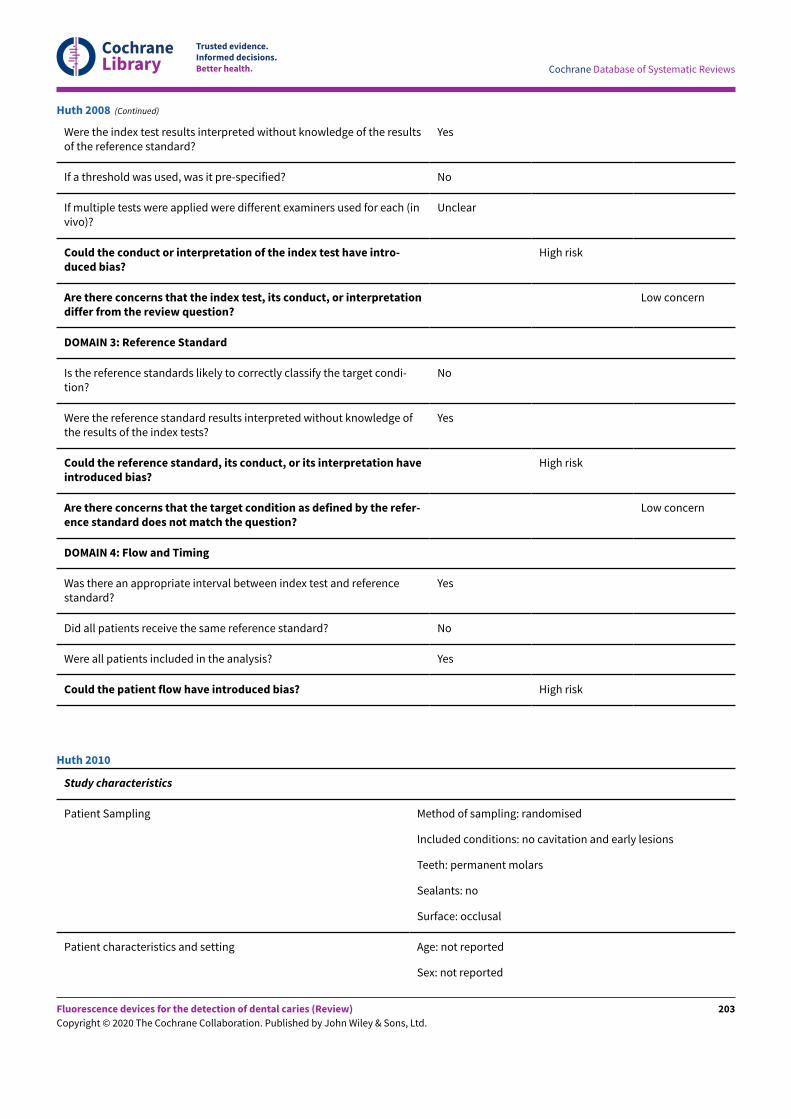
CochraneLibrary
Trusted evidence.Informed decisions.Better health.
Cochrane Database of Systematic Reviews
Were the index test results interpreted without knowledge of the resultsof the reference standard?
Yes
If a threshold was used, was it pre-specified? No
If multiple tests were applied were different examiners used for each (invivo)?
Unclear
Could the conduct or interpretation of the index test have intro-duced bias?
High risk
Are there concerns that the index test, its conduct, or interpretationdiffer from the review question?
Low concern
DOMAIN 3: Reference Standard
Is the reference standards likely to correctly classify the target condi-tion?
No
Were the reference standard results interpreted without knowledge ofthe results of the index tests?
Yes
Could the reference standard, its conduct, or its interpretation haveintroduced bias?
High risk
Are there concerns that the target condition as defined by the refer-ence standard does not match the question?
Low concern
DOMAIN 4: Flow and Timing
Was there an appropriate interval between index test and referencestandard?
Yes
Did all patients receive the same reference standard? No
Were all patients included in the analysis? Yes
Could the patient flow have introduced bias? High risk
Huth 2008 (Continued)
Study characteristics
Patient Sampling Method of sampling: randomised
Included conditions: no cavitation and early lesions
Teeth: permanent molars
Sealants: no
Surface: occlusal
Patient characteristics and setting Age: not reported
Sex: not reported
Huth 2010
Fluorescence devices for the detection of dental caries (Review)
Copyright © 2020 The Cochrane Collaboration. Published by John Wiley & Sons, Ltd.
203
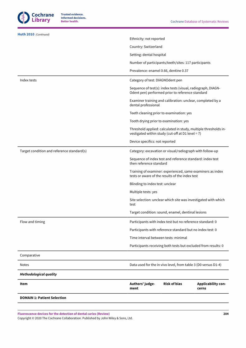
CochraneLibrary
Trusted evidence.Informed decisions.Better health.
Cochrane Database of Systematic Reviews
Ethnicity: not reported
Country: Switzerland
Setting: dental hospital
Number of participants/teeth/sites: 117 participants
Prevalence: enamel 0.66, dentine 0.37
Index tests Category of test: DIAGNOdent pen
Sequence of test(s): index tests (visual, radiograph, DIAGN-Odent pen) performed prior to reference standard
Examiner training and calibration: unclear, completed by adental professional
Teeth cleaning prior to examination: yes
Tooth drying prior to examination: yes
Threshold applied: calculated in study, multiple thresholds in-vestigated within study (cut-oE at D1 level = 7)
Device specifics: not reported
Target condition and reference standard(s) Category: excavation or visual/radiograph with follow-up
Sequence of index test and reference standard: index testthen reference standard
Training of examiner: experienced, same examiners as indextests or aware of the results of the index test
Blinding to index test: unclear
Multiple tests: yes
Site selection: unclear which site was investigated with whichtest
Target condition: sound, enamel, dentinal lesions
Flow and timing Participants with index test but no reference standard: 0
Participants with reference standard but no index test: 0
Time interval between tests: minimal
Participants receiving both tests but excluded from results: 0
Comparative
Notes Data used for the in vivo level, from table 3 (D0 versus D1-4)
Methodological quality
Item Authors' judge-ment
Risk of bias Applicability con-cerns
DOMAIN 1: Patient Selection
Huth 2010 (Continued)
Fluorescence devices for the detection of dental caries (Review)
Copyright © 2020 The Cochrane Collaboration. Published by John Wiley & Sons, Ltd.
204
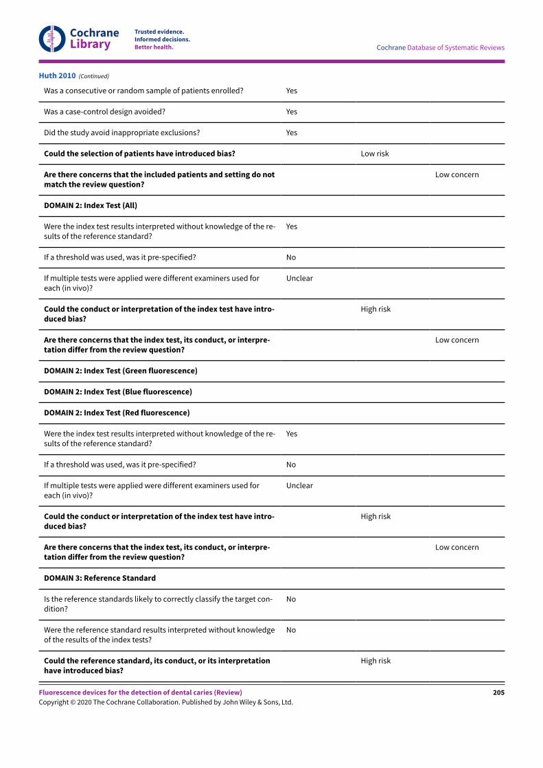
CochraneLibrary
Trusted evidence.Informed decisions.Better health.
Cochrane Database of Systematic Reviews
Was a consecutive or random sample of patients enrolled? Yes
Was a case-control design avoided? Yes
Did the study avoid inappropriate exclusions? Yes
Could the selection of patients have introduced bias? Low risk
Are there concerns that the included patients and setting do notmatch the review question?
Low concern
DOMAIN 2: Index Test (All)
Were the index test results interpreted without knowledge of the re-sults of the reference standard?
Yes
If a threshold was used, was it pre-specified? No
If multiple tests were applied were different examiners used foreach (in vivo)?
Unclear
Could the conduct or interpretation of the index test have intro-duced bias?
High risk
Are there concerns that the index test, its conduct, or interpre-tation differ from the review question?
Low concern
DOMAIN 2: Index Test (Green fluorescence)
DOMAIN 2: Index Test (Blue fluorescence)
DOMAIN 2: Index Test (Red fluorescence)
Were the index test results interpreted without knowledge of the re-sults of the reference standard?
Yes
If a threshold was used, was it pre-specified? No
If multiple tests were applied were different examiners used foreach (in vivo)?
Unclear
Could the conduct or interpretation of the index test have intro-duced bias?
High risk
Are there concerns that the index test, its conduct, or interpre-tation differ from the review question?
Low concern
DOMAIN 3: Reference Standard
Is the reference standards likely to correctly classify the target con-dition?
No
Were the reference standard results interpreted without knowledgeof the results of the index tests?
No
Could the reference standard, its conduct, or its interpretationhave introduced bias?
High risk
Huth 2010 (Continued)
Fluorescence devices for the detection of dental caries (Review)
Copyright © 2020 The Cochrane Collaboration. Published by John Wiley & Sons, Ltd.
205
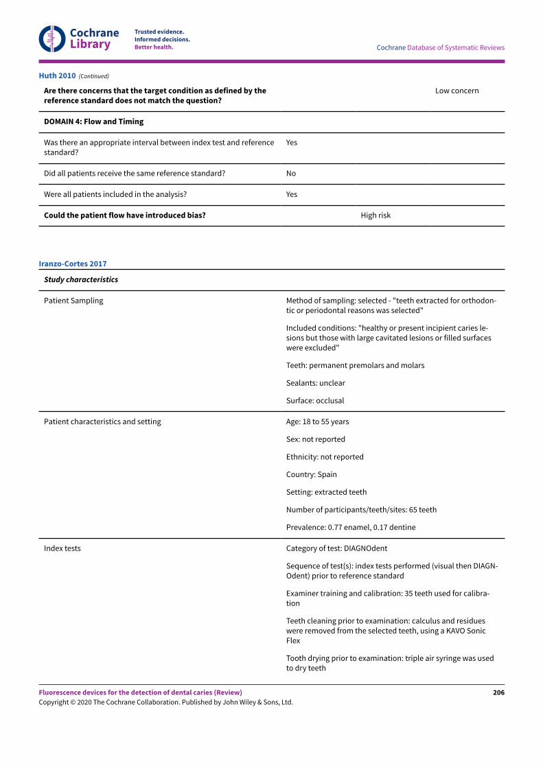
CochraneLibrary
Trusted evidence.Informed decisions.Better health.
Cochrane Database of Systematic Reviews
Are there concerns that the target condition as defined by thereference standard does not match the question?
Low concern
DOMAIN 4: Flow and Timing
Was there an appropriate interval between index test and referencestandard?
Yes
Did all patients receive the same reference standard? No
Were all patients included in the analysis? Yes
Could the patient flow have introduced bias? High risk
Huth 2010 (Continued)
Study characteristics
Patient Sampling Method of sampling: selected - "teeth extracted for orthodon-tic or periodontal reasons was selected"
Included conditions: "healthy or present incipient caries le-sions but those with large cavitated lesions or filled surfaceswere excluded"
Teeth: permanent premolars and molars
Sealants: unclear
Surface: occlusal
Patient characteristics and setting Age: 18 to 55 years
Sex: not reported
Ethnicity: not reported
Country: Spain
Setting: extracted teeth
Number of participants/teeth/sites: 65 teeth
Prevalence: 0.77 enamel, 0.17 dentine
Index tests Category of test: DIAGNOdent
Sequence of test(s): index tests performed (visual then DIAGN-Odent) prior to reference standard
Examiner training and calibration: 35 teeth used for calibra-tion
Teeth cleaning prior to examination: calculus and residueswere removed from the selected teeth, using a KAVO SonicFlex
Tooth drying prior to examination: triple air syringe was usedto dry teeth
Iranzo-Cortes 2017
Fluorescence devices for the detection of dental caries (Review)
Copyright © 2020 The Cochrane Collaboration. Published by John Wiley & Sons, Ltd.
206
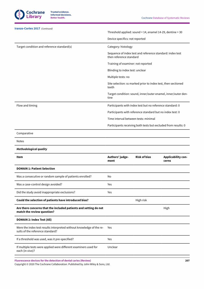
CochraneLibrary
Trusted evidence.Informed decisions.Better health.
Cochrane Database of Systematic Reviews
Threshold applied: sound < 14, enamel 14-29, dentine > 30
Device specifics: not reported
Target condition and reference standard(s) Category: histology
Sequence of index test and reference standard: index testthen reference standard
Training of examiner: not reported
Blinding to index test: unclear
Multiple tests: no
Site selection: ss marked prior to index test, then sectionedteeth
Target condition: sound, inner/outer enamel, inner/outer den-tine
Flow and timing Participants with index test but no reference standard: 0
Participants with reference standard but no index test: 0
Time interval between tests: minimal
Participants receiving both tests but excluded from results: 0
Comparative
Notes
Methodological quality
Item Authors' judge-ment
Risk of bias Applicability con-cerns
DOMAIN 1: Patient Selection
Was a consecutive or random sample of patients enrolled? No
Was a case-control design avoided? Yes
Did the study avoid inappropriate exclusions? Yes
Could the selection of patients have introduced bias? High risk
Are there concerns that the included patients and setting do notmatch the review question?
High
DOMAIN 2: Index Test (All)
Were the index test results interpreted without knowledge of the re-sults of the reference standard?
Yes
If a threshold was used, was it pre-specified? Yes
If multiple tests were applied were different examiners used foreach (in vivo)?
Unclear
Iranzo-Cortes 2017 (Continued)
Fluorescence devices for the detection of dental caries (Review)
Copyright © 2020 The Cochrane Collaboration. Published by John Wiley & Sons, Ltd.
207
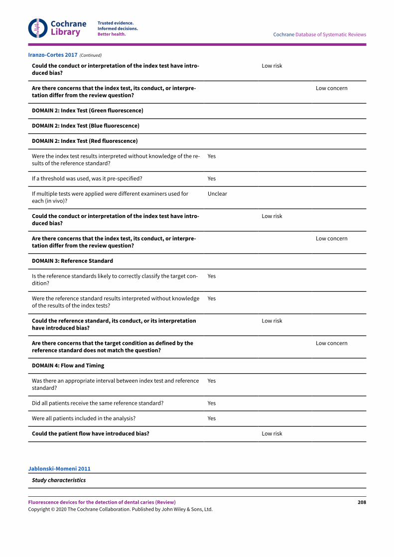
CochraneLibrary
Trusted evidence.Informed decisions.Better health.
Cochrane Database of Systematic Reviews
Could the conduct or interpretation of the index test have intro-duced bias?
Low risk
Are there concerns that the index test, its conduct, or interpre-tation differ from the review question?
Low concern
DOMAIN 2: Index Test (Green fluorescence)
DOMAIN 2: Index Test (Blue fluorescence)
DOMAIN 2: Index Test (Red fluorescence)
Were the index test results interpreted without knowledge of the re-sults of the reference standard?
Yes
If a threshold was used, was it pre-specified? Yes
If multiple tests were applied were different examiners used foreach (in vivo)?
Unclear
Could the conduct or interpretation of the index test have intro-duced bias?
Low risk
Are there concerns that the index test, its conduct, or interpre-tation differ from the review question?
Low concern
DOMAIN 3: Reference Standard
Is the reference standards likely to correctly classify the target con-dition?
Yes
Were the reference standard results interpreted without knowledgeof the results of the index tests?
Yes
Could the reference standard, its conduct, or its interpretationhave introduced bias?
Low risk
Are there concerns that the target condition as defined by thereference standard does not match the question?
Low concern
DOMAIN 4: Flow and Timing
Was there an appropriate interval between index test and referencestandard?
Yes
Did all patients receive the same reference standard? Yes
Were all patients included in the analysis? Yes
Could the patient flow have introduced bias? Low risk
Iranzo-Cortes 2017 (Continued)
Study characteristics
Jablonski-Momeni 2011
Fluorescence devices for the detection of dental caries (Review)
Copyright © 2020 The Cochrane Collaboration. Published by John Wiley & Sons, Ltd.
208
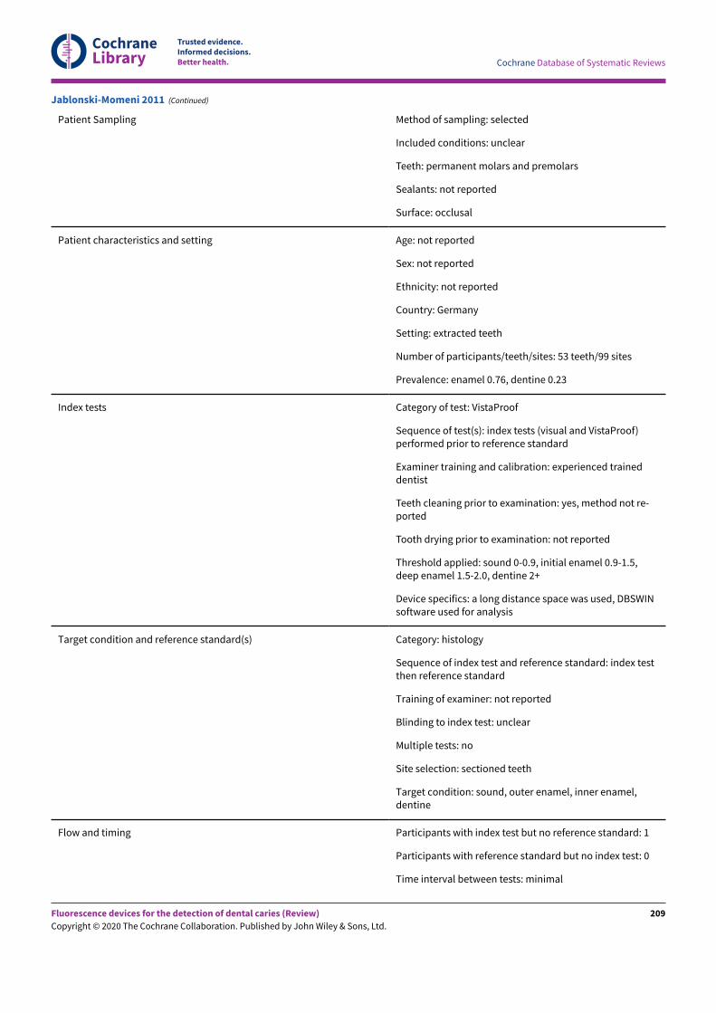
CochraneLibrary
Trusted evidence.Informed decisions.Better health.
Cochrane Database of Systematic Reviews
Patient Sampling Method of sampling: selected
Included conditions: unclear
Teeth: permanent molars and premolars
Sealants: not reported
Surface: occlusal
Patient characteristics and setting Age: not reported
Sex: not reported
Ethnicity: not reported
Country: Germany
Setting: extracted teeth
Number of participants/teeth/sites: 53 teeth/99 sites
Prevalence: enamel 0.76, dentine 0.23
Index tests Category of test: VistaProof
Sequence of test(s): index tests (visual and VistaProof)performed prior to reference standard
Examiner training and calibration: experienced traineddentist
Teeth cleaning prior to examination: yes, method not re-ported
Tooth drying prior to examination: not reported
Threshold applied: sound 0-0.9, initial enamel 0.9-1.5,deep enamel 1.5-2.0, dentine 2+
Device specifics: a long distance space was used, DBSWINsoftware used for analysis
Target condition and reference standard(s) Category: histology
Sequence of index test and reference standard: index testthen reference standard
Training of examiner: not reported
Blinding to index test: unclear
Multiple tests: no
Site selection: sectioned teeth
Target condition: sound, outer enamel, inner enamel,dentine
Flow and timing Participants with index test but no reference standard: 1
Participants with reference standard but no index test: 0
Time interval between tests: minimal
Jablonski-Momeni 2011 (Continued)
Fluorescence devices for the detection of dental caries (Review)
Copyright © 2020 The Cochrane Collaboration. Published by John Wiley & Sons, Ltd.
209
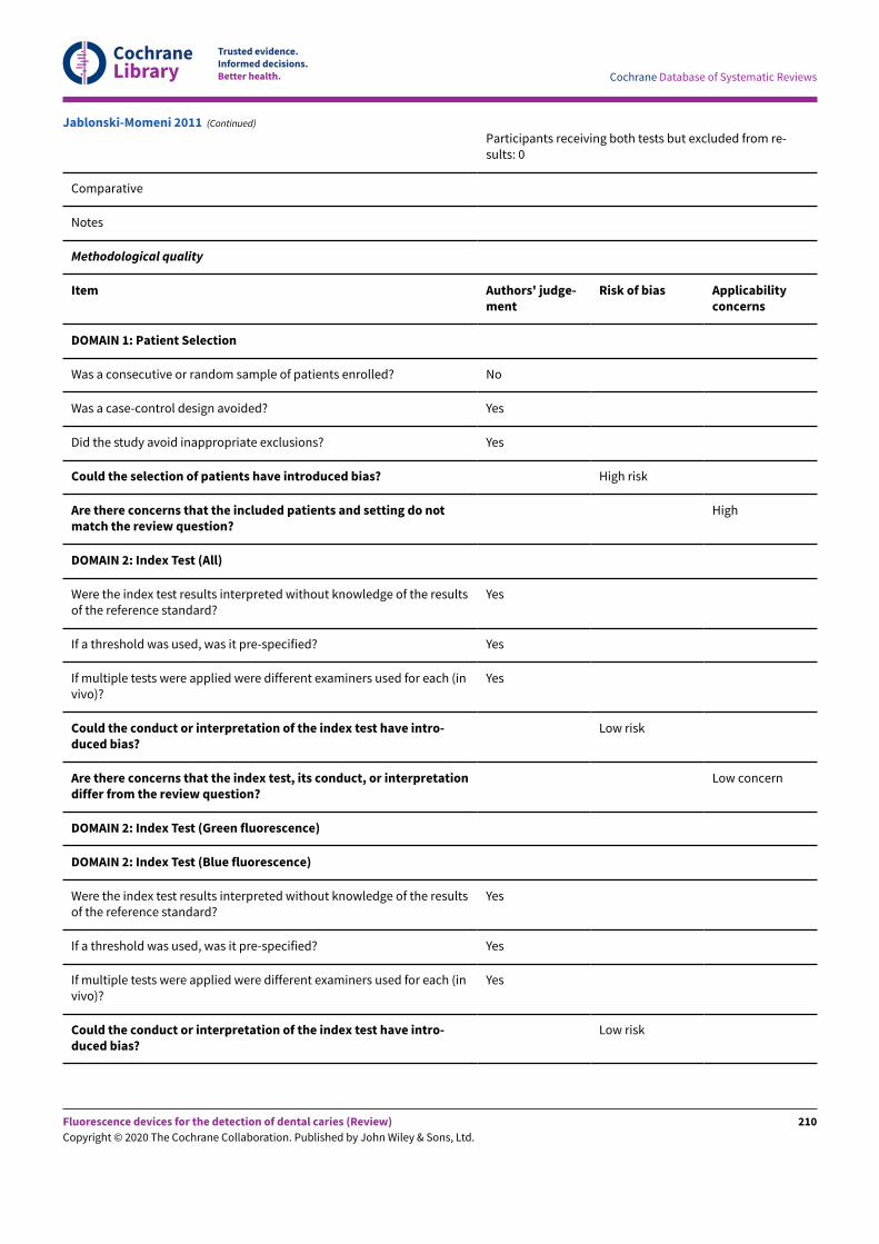
CochraneLibrary
Trusted evidence.Informed decisions.Better health.
Cochrane Database of Systematic Reviews
Participants receiving both tests but excluded from re-sults: 0
Comparative
Notes
Methodological quality
Item Authors' judge-ment
Risk of bias Applicabilityconcerns
DOMAIN 1: Patient Selection
Was a consecutive or random sample of patients enrolled? No
Was a case-control design avoided? Yes
Did the study avoid inappropriate exclusions? Yes
Could the selection of patients have introduced bias? High risk
Are there concerns that the included patients and setting do notmatch the review question?
High
DOMAIN 2: Index Test (All)
Were the index test results interpreted without knowledge of the resultsof the reference standard?
Yes
If a threshold was used, was it pre-specified? Yes
If multiple tests were applied were different examiners used for each (invivo)?
Yes
Could the conduct or interpretation of the index test have intro-duced bias?
Low risk
Are there concerns that the index test, its conduct, or interpretationdiffer from the review question?
Low concern
DOMAIN 2: Index Test (Green fluorescence)
DOMAIN 2: Index Test (Blue fluorescence)
Were the index test results interpreted without knowledge of the resultsof the reference standard?
Yes
If a threshold was used, was it pre-specified? Yes
If multiple tests were applied were different examiners used for each (invivo)?
Yes
Could the conduct or interpretation of the index test have intro-duced bias?
Low risk
Jablonski-Momeni 2011 (Continued)
Fluorescence devices for the detection of dental caries (Review)
Copyright © 2020 The Cochrane Collaboration. Published by John Wiley & Sons, Ltd.
210
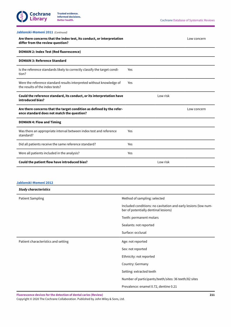
CochraneLibrary
Trusted evidence.Informed decisions.Better health.
Cochrane Database of Systematic Reviews
Are there concerns that the index test, its conduct, or interpretationdiffer from the review question?
Low concern
DOMAIN 2: Index Test (Red fluorescence)
DOMAIN 3: Reference Standard
Is the reference standards likely to correctly classify the target condi-tion?
Yes
Were the reference standard results interpreted without knowledge ofthe results of the index tests?
Yes
Could the reference standard, its conduct, or its interpretation haveintroduced bias?
Low risk
Are there concerns that the target condition as defined by the refer-ence standard does not match the question?
Low concern
DOMAIN 4: Flow and Timing
Was there an appropriate interval between index test and referencestandard?
Yes
Did all patients receive the same reference standard? Yes
Were all patients included in the analysis? Yes
Could the patient flow have introduced bias? Low risk
Jablonski-Momeni 2011 (Continued)
Study characteristics
Patient Sampling Method of sampling: selected
Included conditions: no cavitation and early lesions (low num-ber of potentially dentinal lesions)
Teeth: permanent molars
Sealants: not reported
Surface: occlusal
Patient characteristics and setting Age: not reported
Sex: not reported
Ethnicity: not reported
Country: Germany
Setting: extracted teeth
Number of participants/teeth/sites: 36 teeth/82 sites
Prevalence: enamel 0.72, dentine 0.21
Jablonski-Momeni 2012
Fluorescence devices for the detection of dental caries (Review)
Copyright © 2020 The Cochrane Collaboration. Published by John Wiley & Sons, Ltd.
211
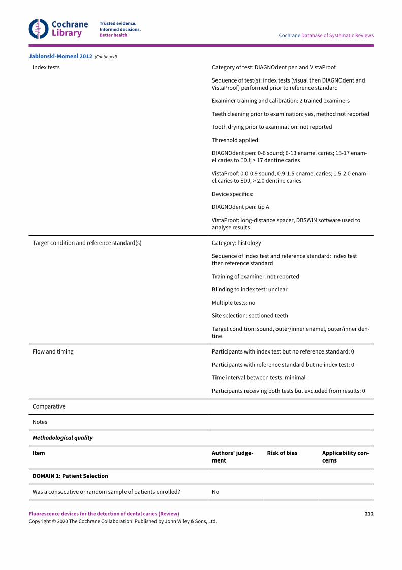
CochraneLibrary
Trusted evidence.Informed decisions.Better health.
Cochrane Database of Systematic Reviews
Index tests Category of test: DIAGNOdent pen and VistaProof
Sequence of test(s): index tests (visual then DIAGNOdent andVistaProof) performed prior to reference standard
Examiner training and calibration: 2 trained examiners
Teeth cleaning prior to examination: yes, method not reported
Tooth drying prior to examination: not reported
Threshold applied:
DIAGNOdent pen: 0-6 sound; 6-13 enamel caries; 13-17 enam-el caries to EDJ; > 17 dentine caries
VistaProof: 0.0-0.9 sound; 0.9-1.5 enamel caries; 1.5-2.0 enam-el caries to EDJ; > 2.0 dentine caries
Device specifics:
DIAGNOdent pen: tip A
VistaProof: long-distance spacer, DBSWIN software used toanalyse results
Target condition and reference standard(s) Category: histology
Sequence of index test and reference standard: index testthen reference standard
Training of examiner: not reported
Blinding to index test: unclear
Multiple tests: no
Site selection: sectioned teeth
Target condition: sound, outer/inner enamel, outer/inner den-tine
Flow and timing Participants with index test but no reference standard: 0
Participants with reference standard but no index test: 0
Time interval between tests: minimal
Participants receiving both tests but excluded from results: 0
Comparative
Notes
Methodological quality
Item Authors' judge-ment
Risk of bias Applicability con-cerns
DOMAIN 1: Patient Selection
Was a consecutive or random sample of patients enrolled? No
Jablonski-Momeni 2012 (Continued)
Fluorescence devices for the detection of dental caries (Review)
Copyright © 2020 The Cochrane Collaboration. Published by John Wiley & Sons, Ltd.
212
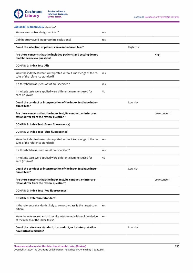
CochraneLibrary
Trusted evidence.Informed decisions.Better health.
Cochrane Database of Systematic Reviews
Was a case-control design avoided? Yes
Did the study avoid inappropriate exclusions? Yes
Could the selection of patients have introduced bias? High risk
Are there concerns that the included patients and setting do notmatch the review question?
High
DOMAIN 2: Index Test (All)
Were the index test results interpreted without knowledge of the re-sults of the reference standard?
Yes
If a threshold was used, was it pre-specified? Yes
If multiple tests were applied were different examiners used foreach (in vivo)?
No
Could the conduct or interpretation of the index test have intro-duced bias?
Low risk
Are there concerns that the index test, its conduct, or interpre-tation differ from the review question?
Low concern
DOMAIN 2: Index Test (Green fluorescence)
DOMAIN 2: Index Test (Blue fluorescence)
Were the index test results interpreted without knowledge of the re-sults of the reference standard?
Yes
If a threshold was used, was it pre-specified? Yes
If multiple tests were applied were different examiners used foreach (in vivo)?
No
Could the conduct or interpretation of the index test have intro-duced bias?
Low risk
Are there concerns that the index test, its conduct, or interpre-tation differ from the review question?
Low concern
DOMAIN 2: Index Test (Red fluorescence)
DOMAIN 3: Reference Standard
Is the reference standards likely to correctly classify the target con-dition?
Yes
Were the reference standard results interpreted without knowledgeof the results of the index tests?
Yes
Could the reference standard, its conduct, or its interpretationhave introduced bias?
Low risk
Jablonski-Momeni 2012 (Continued)
Fluorescence devices for the detection of dental caries (Review)
Copyright © 2020 The Cochrane Collaboration. Published by John Wiley & Sons, Ltd.
213
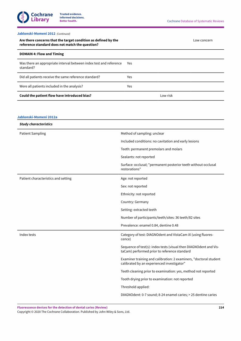
CochraneLibrary
Trusted evidence.Informed decisions.Better health.
Cochrane Database of Systematic Reviews
Are there concerns that the target condition as defined by thereference standard does not match the question?
Low concern
DOMAIN 4: Flow and Timing
Was there an appropriate interval between index test and referencestandard?
Yes
Did all patients receive the same reference standard? Yes
Were all patients included in the analysis? Yes
Could the patient flow have introduced bias? Low risk
Jablonski-Momeni 2012 (Continued)
Study characteristics
Patient Sampling Method of sampling: unclear
Included conditions: no cavitation and early lesions
Teeth: permanent premolars and molars
Sealants: not reported
Surface: occlusal; "permanent posterior teeth without occlusalrestorations"
Patient characteristics and setting Age: not reported
Sex: not reported
Ethnicity: not reported
Country: Germany
Setting: extracted teeth
Number of participants/teeth/sites: 36 teeth/82 sites
Prevalence: enamel 0.84, dentine 0.48
Index tests Category of test: DIAGNOdent and VistaCam iX (using fluores-cence)
Sequence of test(s): index tests (visual then DIAGNOdent and Vis-taCam) performed prior to reference standard
Examiner training and calibration: 2 examiners, "doctoral studentcalibrated by an experienced investigator"
Teeth cleaning prior to examination: yes, method not reported
Tooth drying prior to examination: not reported
Threshold applied:
DIAGNOdent: 0-7 sound; 8-24 enamel caries; > 25 dentine caries
Jablonski-Momeni 2012a
Fluorescence devices for the detection of dental caries (Review)
Copyright © 2020 The Cochrane Collaboration. Published by John Wiley & Sons, Ltd.
214
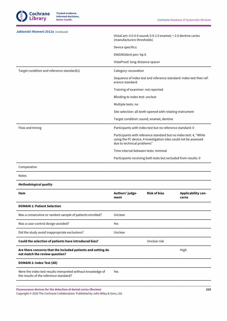
CochraneLibrary
Trusted evidence.Informed decisions.Better health.
Cochrane Database of Systematic Reviews
VistaCam: 0.0-0.9 sound; 0.9-2.0 enamel; > 2.0 dentine caries(manufacturers thresholds)
Device specifics:
DIAGNOdent pen: tip A
VistaProof: long-distance spacer
Target condition and reference standard(s) Category: excavation
Sequence of index test and reference standard: index test then ref-erence standard
Training of examiner: not reported
Blinding to index test: unclear
Multiple tests: no
Site selection: all teeth opened with rotating instrument
Target condition: sound, enamel, dentine
Flow and timing Participants with index test but no reference standard: 0
Participants with reference standard but no index test: 4, "Whileusing the FC device, 4 investigation sites could not be assesseddue to technical problems"
Time interval between tests: minimal
Participants receiving both tests but excluded from results: 0
Comparative
Notes
Methodological quality
Item Authors' judge-ment
Risk of bias Applicability con-cerns
DOMAIN 1: Patient Selection
Was a consecutive or random sample of patients enrolled? Unclear
Was a case-control design avoided? Yes
Did the study avoid inappropriate exclusions? Unclear
Could the selection of patients have introduced bias? Unclear risk
Are there concerns that the included patients and setting donot match the review question?
High
DOMAIN 2: Index Test (All)
Were the index test results interpreted without knowledge ofthe results of the reference standard?
Yes
Jablonski-Momeni 2012a (Continued)
Fluorescence devices for the detection of dental caries (Review)
Copyright © 2020 The Cochrane Collaboration. Published by John Wiley & Sons, Ltd.
215
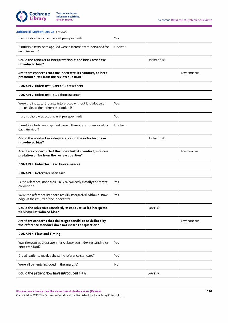
CochraneLibrary
Trusted evidence.Informed decisions.Better health.
Cochrane Database of Systematic Reviews
If a threshold was used, was it pre-specified? Yes
If multiple tests were applied were different examiners used foreach (in vivo)?
Unclear
Could the conduct or interpretation of the index test haveintroduced bias?
Unclear risk
Are there concerns that the index test, its conduct, or inter-pretation differ from the review question?
Low concern
DOMAIN 2: Index Test (Green fluorescence)
DOMAIN 2: Index Test (Blue fluorescence)
Were the index test results interpreted without knowledge ofthe results of the reference standard?
Yes
If a threshold was used, was it pre-specified? Yes
If multiple tests were applied were different examiners used foreach (in vivo)?
Unclear
Could the conduct or interpretation of the index test haveintroduced bias?
Unclear risk
Are there concerns that the index test, its conduct, or inter-pretation differ from the review question?
Low concern
DOMAIN 2: Index Test (Red fluorescence)
DOMAIN 3: Reference Standard
Is the reference standards likely to correctly classify the targetcondition?
Yes
Were the reference standard results interpreted without knowl-edge of the results of the index tests?
Yes
Could the reference standard, its conduct, or its interpreta-tion have introduced bias?
Low risk
Are there concerns that the target condition as defined bythe reference standard does not match the question?
Low concern
DOMAIN 4: Flow and Timing
Was there an appropriate interval between index test and refer-ence standard?
Yes
Did all patients receive the same reference standard? Yes
Were all patients included in the analysis? No
Could the patient flow have introduced bias? Low risk
Jablonski-Momeni 2012a (Continued)
Fluorescence devices for the detection of dental caries (Review)
Copyright © 2020 The Cochrane Collaboration. Published by John Wiley & Sons, Ltd.
216
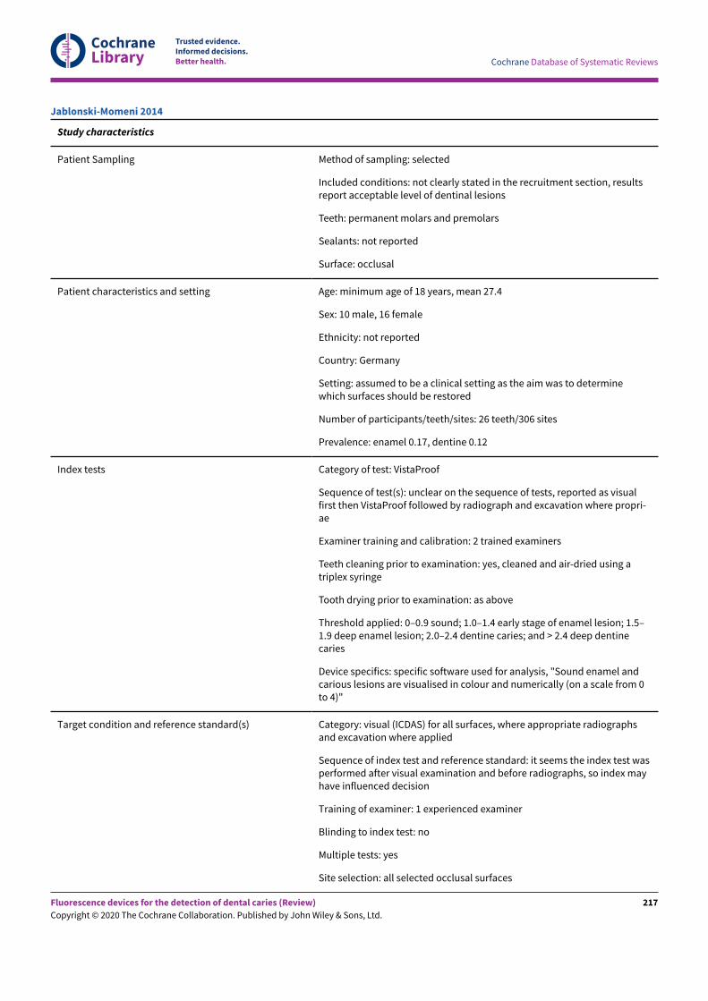
CochraneLibrary
Trusted evidence.Informed decisions.Better health.
Cochrane Database of Systematic Reviews
Study characteristics
Patient Sampling Method of sampling: selected
Included conditions: not clearly stated in the recruitment section, resultsreport acceptable level of dentinal lesions
Teeth: permanent molars and premolars
Sealants: not reported
Surface: occlusal
Patient characteristics and setting Age: minimum age of 18 years, mean 27.4
Sex: 10 male, 16 female
Ethnicity: not reported
Country: Germany
Setting: assumed to be a clinical setting as the aim was to determinewhich surfaces should be restored
Number of participants/teeth/sites: 26 teeth/306 sites
Prevalence: enamel 0.17, dentine 0.12
Index tests Category of test: VistaProof
Sequence of test(s): unclear on the sequence of tests, reported as visualfirst then VistaProof followed by radiograph and excavation where propri-ae
Examiner training and calibration: 2 trained examiners
Teeth cleaning prior to examination: yes, cleaned and air-dried using atriplex syringe
Tooth drying prior to examination: as above
Threshold applied: 0–0.9 sound; 1.0–1.4 early stage of enamel lesion; 1.5–1.9 deep enamel lesion; 2.0–2.4 dentine caries; and > 2.4 deep dentinecaries
Device specifics: specific software used for analysis, "Sound enamel andcarious lesions are visualised in colour and numerically (on a scale from 0to 4)"
Target condition and reference standard(s) Category: visual (ICDAS) for all surfaces, where appropriate radiographsand excavation where applied
Sequence of index test and reference standard: it seems the index test wasperformed after visual examination and before radiographs, so index mayhave influenced decision
Training of examiner: 1 experienced examiner
Blinding to index test: no
Multiple tests: yes
Site selection: all selected occlusal surfaces
Jablonski-Momeni 2014
Fluorescence devices for the detection of dental caries (Review)
Copyright © 2020 The Cochrane Collaboration. Published by John Wiley & Sons, Ltd.
217
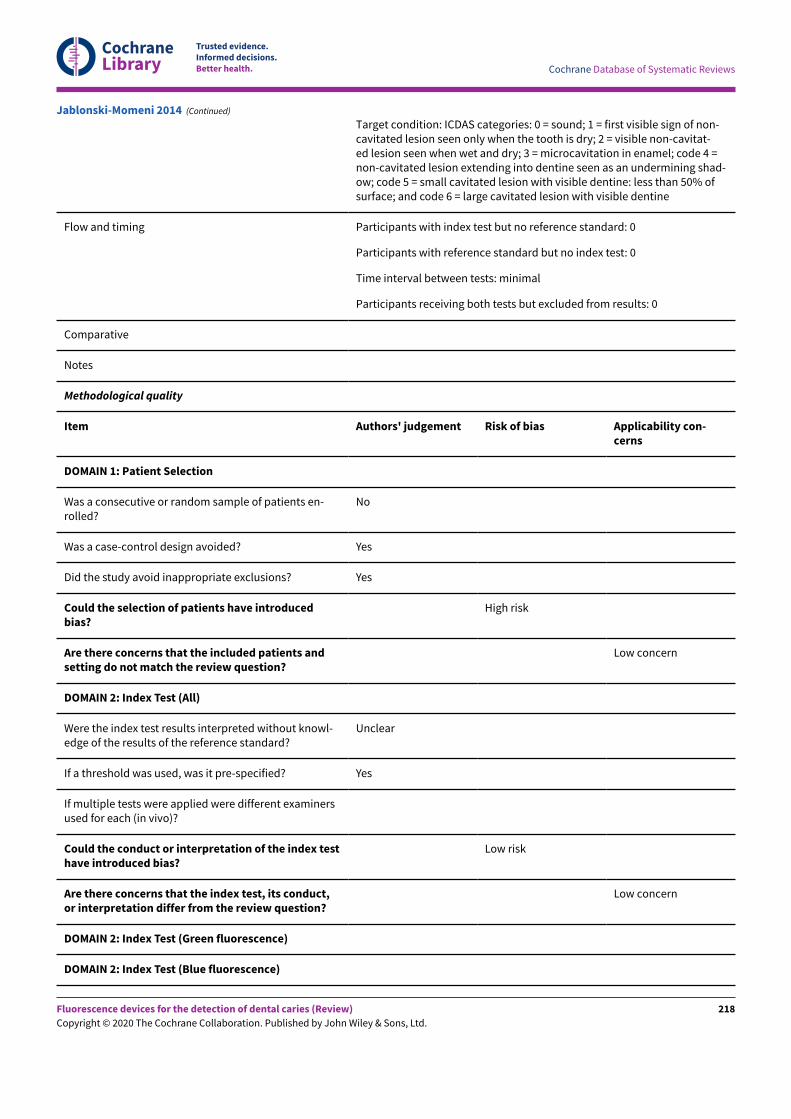
CochraneLibrary
Trusted evidence.Informed decisions.Better health.
Cochrane Database of Systematic Reviews
Target condition: ICDAS categories: 0 = sound; 1 = first visible sign of non-cavitated lesion seen only when the tooth is dry; 2 = visible non-cavitat-ed lesion seen when wet and dry; 3 = microcavitation in enamel; code 4 =non-cavitated lesion extending into dentine seen as an undermining shad-ow; code 5 = small cavitated lesion with visible dentine: less than 50% ofsurface; and code 6 = large cavitated lesion with visible dentine
Flow and timing Participants with index test but no reference standard: 0
Participants with reference standard but no index test: 0
Time interval between tests: minimal
Participants receiving both tests but excluded from results: 0
Comparative
Notes
Methodological quality
Item Authors' judgement Risk of bias Applicability con-cerns
DOMAIN 1: Patient Selection
Was a consecutive or random sample of patients en-rolled?
No
Was a case-control design avoided? Yes
Did the study avoid inappropriate exclusions? Yes
Could the selection of patients have introducedbias?
High risk
Are there concerns that the included patients andsetting do not match the review question?
Low concern
DOMAIN 2: Index Test (All)
Were the index test results interpreted without knowl-edge of the results of the reference standard?
Unclear
If a threshold was used, was it pre-specified? Yes
If multiple tests were applied were different examinersused for each (in vivo)?
Could the conduct or interpretation of the index testhave introduced bias?
Low risk
Are there concerns that the index test, its conduct,or interpretation differ from the review question?
Low concern
DOMAIN 2: Index Test (Green fluorescence)
DOMAIN 2: Index Test (Blue fluorescence)
Jablonski-Momeni 2014 (Continued)
Fluorescence devices for the detection of dental caries (Review)
Copyright © 2020 The Cochrane Collaboration. Published by John Wiley & Sons, Ltd.
218
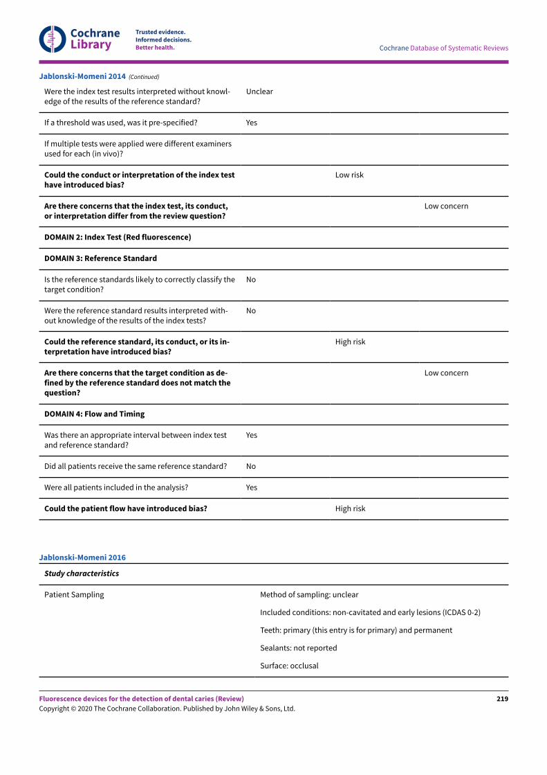
CochraneLibrary
Trusted evidence.Informed decisions.Better health.
Cochrane Database of Systematic Reviews
Were the index test results interpreted without knowl-edge of the results of the reference standard?
Unclear
If a threshold was used, was it pre-specified? Yes
If multiple tests were applied were different examinersused for each (in vivo)?
Could the conduct or interpretation of the index testhave introduced bias?
Low risk
Are there concerns that the index test, its conduct,or interpretation differ from the review question?
Low concern
DOMAIN 2: Index Test (Red fluorescence)
DOMAIN 3: Reference Standard
Is the reference standards likely to correctly classify thetarget condition?
No
Were the reference standard results interpreted with-out knowledge of the results of the index tests?
No
Could the reference standard, its conduct, or its in-terpretation have introduced bias?
High risk
Are there concerns that the target condition as de-fined by the reference standard does not match thequestion?
Low concern
DOMAIN 4: Flow and Timing
Was there an appropriate interval between index testand reference standard?
Yes
Did all patients receive the same reference standard? No
Were all patients included in the analysis? Yes
Could the patient flow have introduced bias? High risk
Jablonski-Momeni 2014 (Continued)
Study characteristics
Patient Sampling Method of sampling: unclear
Included conditions: non-cavitated and early lesions (ICDAS 0-2)
Teeth: primary (this entry is for primary) and permanent
Sealants: not reported
Surface: occlusal
Jablonski-Momeni 2016
Fluorescence devices for the detection of dental caries (Review)
Copyright © 2020 The Cochrane Collaboration. Published by John Wiley & Sons, Ltd.
219
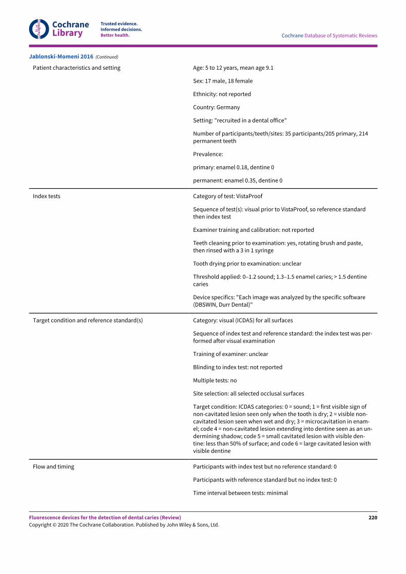
CochraneLibrary
Trusted evidence.Informed decisions.Better health.
Cochrane Database of Systematic Reviews
Patient characteristics and setting Age: 5 to 12 years, mean age 9.1
Sex: 17 male, 18 female
Ethnicity: not reported
Country: Germany
Setting: "recruited in a dental office"
Number of participants/teeth/sites: 35 participants/205 primary, 214permanent teeth
Prevalence:
primary: enamel 0.18, dentine 0
permanent: enamel 0.35, dentine 0
Index tests Category of test: VistaProof
Sequence of test(s): visual prior to VistaProof, so reference standardthen index test
Examiner training and calibration: not reported
Teeth cleaning prior to examination: yes, rotating brush and paste,then rinsed with a 3 in 1 syringe
Tooth drying prior to examination: unclear
Threshold applied: 0–1.2 sound; 1.3–1.5 enamel caries; > 1.5 dentinecaries
Device specifics: "Each image was analyzed by the specific software(DBSWIN, Durr Dental)"
Target condition and reference standard(s) Category: visual (ICDAS) for all surfaces
Sequence of index test and reference standard: the index test was per-formed after visual examination
Training of examiner: unclear
Blinding to index test: not reported
Multiple tests: no
Site selection: all selected occlusal surfaces
Target condition: ICDAS categories: 0 = sound; 1 = first visible sign ofnon-cavitated lesion seen only when the tooth is dry; 2 = visible non-cavitated lesion seen when wet and dry; 3 = microcavitation in enam-el; code 4 = non-cavitated lesion extending into dentine seen as an un-dermining shadow; code 5 = small cavitated lesion with visible den-tine: less than 50% of surface; and code 6 = large cavitated lesion withvisible dentine
Flow and timing Participants with index test but no reference standard: 0
Participants with reference standard but no index test: 0
Time interval between tests: minimal
Jablonski-Momeni 2016 (Continued)
Fluorescence devices for the detection of dental caries (Review)
Copyright © 2020 The Cochrane Collaboration. Published by John Wiley & Sons, Ltd.
220
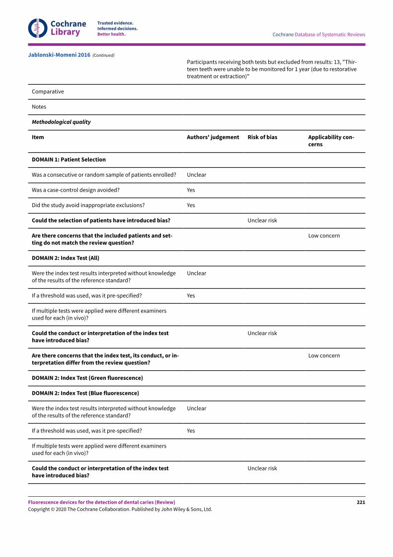
CochraneLibrary
Trusted evidence.Informed decisions.Better health.
Cochrane Database of Systematic Reviews
Participants receiving both tests but excluded from results: 13, "Thir-teen teeth were unable to be monitored for 1 year (due to restorativetreatment or extraction)"
Comparative
Notes
Methodological quality
Item Authors' judgement Risk of bias Applicability con-cerns
DOMAIN 1: Patient Selection
Was a consecutive or random sample of patients enrolled? Unclear
Was a case-control design avoided? Yes
Did the study avoid inappropriate exclusions? Yes
Could the selection of patients have introduced bias? Unclear risk
Are there concerns that the included patients and set-ting do not match the review question?
Low concern
DOMAIN 2: Index Test (All)
Were the index test results interpreted without knowledgeof the results of the reference standard?
Unclear
If a threshold was used, was it pre-specified? Yes
If multiple tests were applied were different examinersused for each (in vivo)?
Could the conduct or interpretation of the index testhave introduced bias?
Unclear risk
Are there concerns that the index test, its conduct, or in-terpretation differ from the review question?
Low concern
DOMAIN 2: Index Test (Green fluorescence)
DOMAIN 2: Index Test (Blue fluorescence)
Were the index test results interpreted without knowledgeof the results of the reference standard?
Unclear
If a threshold was used, was it pre-specified? Yes
If multiple tests were applied were different examinersused for each (in vivo)?
Could the conduct or interpretation of the index testhave introduced bias?
Unclear risk
Jablonski-Momeni 2016 (Continued)
Fluorescence devices for the detection of dental caries (Review)
Copyright © 2020 The Cochrane Collaboration. Published by John Wiley & Sons, Ltd.
221
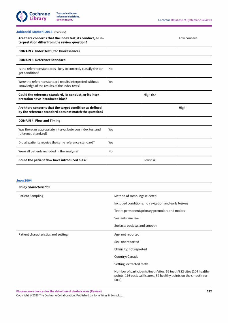
CochraneLibrary
Trusted evidence.Informed decisions.Better health.
Cochrane Database of Systematic Reviews
Are there concerns that the index test, its conduct, or in-terpretation differ from the review question?
Low concern
DOMAIN 2: Index Test (Red fluorescence)
DOMAIN 3: Reference Standard
Is the reference standards likely to correctly classify the tar-get condition?
No
Were the reference standard results interpreted withoutknowledge of the results of the index tests?
Yes
Could the reference standard, its conduct, or its inter-pretation have introduced bias?
High risk
Are there concerns that the target condition as definedby the reference standard does not match the question?
High
DOMAIN 4: Flow and Timing
Was there an appropriate interval between index test andreference standard?
Yes
Did all patients receive the same reference standard? Yes
Were all patients included in the analysis? No
Could the patient flow have introduced bias? Low risk
Jablonski-Momeni 2016 (Continued)
Study characteristics
Patient Sampling Method of sampling: selected
Included conditions: no cavitation and early lesions
Teeth: permanent/primary premolars and molars
Sealants: unclear
Surface: occlusal and smooth
Patient characteristics and setting Age: not reported
Sex: not reported
Ethnicity: not reported
Country: Canada
Setting: extracted teeth
Number of participants/teeth/sites: 52 teeth/332 sites (104 healthypoints, 176 occlusal fissures, 52 healthy points on the smooth sur-face)
Jeon 2004
Fluorescence devices for the detection of dental caries (Review)
Copyright © 2020 The Cochrane Collaboration. Published by John Wiley & Sons, Ltd.
222
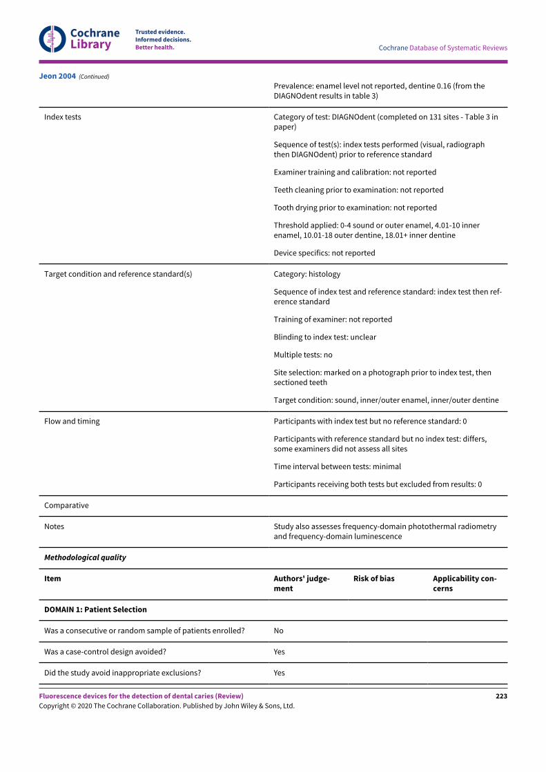
CochraneLibrary
Trusted evidence.Informed decisions.Better health.
Cochrane Database of Systematic Reviews
Prevalence: enamel level not reported, dentine 0.16 (from theDIAGNOdent results in table 3)
Index tests Category of test: DIAGNOdent (completed on 131 sites - Table 3 inpaper)
Sequence of test(s): index tests performed (visual, radiographthen DIAGNOdent) prior to reference standard
Examiner training and calibration: not reported
Teeth cleaning prior to examination: not reported
Tooth drying prior to examination: not reported
Threshold applied: 0-4 sound or outer enamel, 4.01-10 innerenamel, 10.01-18 outer dentine, 18.01+ inner dentine
Device specifics: not reported
Target condition and reference standard(s) Category: histology
Sequence of index test and reference standard: index test then ref-erence standard
Training of examiner: not reported
Blinding to index test: unclear
Multiple tests: no
Site selection: marked on a photograph prior to index test, thensectioned teeth
Target condition: sound, inner/outer enamel, inner/outer dentine
Flow and timing Participants with index test but no reference standard: 0
Participants with reference standard but no index test: differs,some examiners did not assess all sites
Time interval between tests: minimal
Participants receiving both tests but excluded from results: 0
Comparative
Notes Study also assesses frequency-domain photothermal radiometryand frequency-domain luminescence
Methodological quality
Item Authors' judge-ment
Risk of bias Applicability con-cerns
DOMAIN 1: Patient Selection
Was a consecutive or random sample of patients enrolled? No
Was a case-control design avoided? Yes
Did the study avoid inappropriate exclusions? Yes
Jeon 2004 (Continued)
Fluorescence devices for the detection of dental caries (Review)
Copyright © 2020 The Cochrane Collaboration. Published by John Wiley & Sons, Ltd.
223
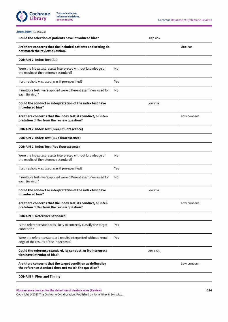
CochraneLibrary
Trusted evidence.Informed decisions.Better health.
Cochrane Database of Systematic Reviews
Could the selection of patients have introduced bias? High risk
Are there concerns that the included patients and setting donot match the review question?
Unclear
DOMAIN 2: Index Test (All)
Were the index test results interpreted without knowledge ofthe results of the reference standard?
No
If a threshold was used, was it pre-specified? Yes
If multiple tests were applied were different examiners used foreach (in vivo)?
No
Could the conduct or interpretation of the index test haveintroduced bias?
Low risk
Are there concerns that the index test, its conduct, or inter-pretation differ from the review question?
Low concern
DOMAIN 2: Index Test (Green fluorescence)
DOMAIN 2: Index Test (Blue fluorescence)
DOMAIN 2: Index Test (Red fluorescence)
Were the index test results interpreted without knowledge ofthe results of the reference standard?
No
If a threshold was used, was it pre-specified? Yes
If multiple tests were applied were different examiners used foreach (in vivo)?
No
Could the conduct or interpretation of the index test haveintroduced bias?
Low risk
Are there concerns that the index test, its conduct, or inter-pretation differ from the review question?
Low concern
DOMAIN 3: Reference Standard
Is the reference standards likely to correctly classify the targetcondition?
Yes
Were the reference standard results interpreted without knowl-edge of the results of the index tests?
Yes
Could the reference standard, its conduct, or its interpreta-tion have introduced bias?
Low risk
Are there concerns that the target condition as defined bythe reference standard does not match the question?
Low concern
DOMAIN 4: Flow and Timing
Jeon 2004 (Continued)
Fluorescence devices for the detection of dental caries (Review)
Copyright © 2020 The Cochrane Collaboration. Published by John Wiley & Sons, Ltd.
224
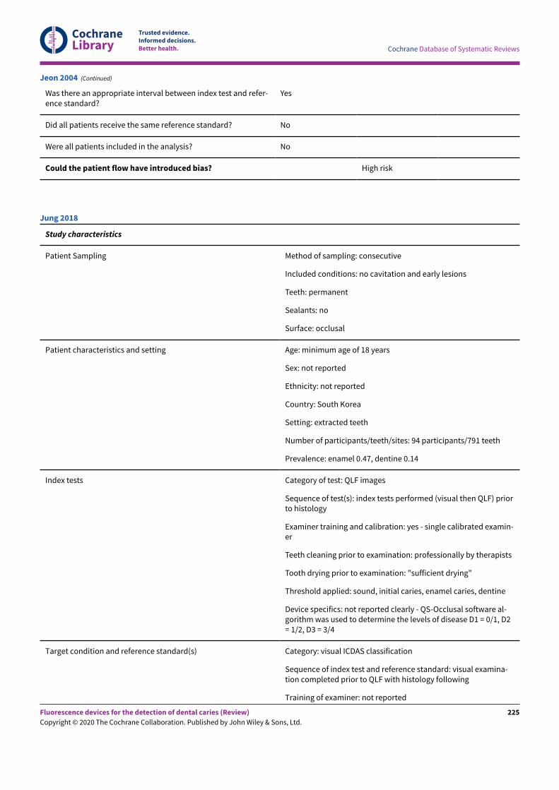
CochraneLibrary
Trusted evidence.Informed decisions.Better health.
Cochrane Database of Systematic Reviews
Was there an appropriate interval between index test and refer-ence standard?
Yes
Did all patients receive the same reference standard? No
Were all patients included in the analysis? No
Could the patient flow have introduced bias? High risk
Jeon 2004 (Continued)
Study characteristics
Patient Sampling Method of sampling: consecutive
Included conditions: no cavitation and early lesions
Teeth: permanent
Sealants: no
Surface: occlusal
Patient characteristics and setting Age: minimum age of 18 years
Sex: not reported
Ethnicity: not reported
Country: South Korea
Setting: extracted teeth
Number of participants/teeth/sites: 94 participants/791 teeth
Prevalence: enamel 0.47, dentine 0.14
Index tests Category of test: QLF images
Sequence of test(s): index tests performed (visual then QLF) priorto histology
Examiner training and calibration: yes - single calibrated examin-er
Teeth cleaning prior to examination: professionally by therapists
Tooth drying prior to examination: "sufficient drying"
Threshold applied: sound, initial caries, enamel caries, dentine
Device specifics: not reported clearly - QS-Occlusal software al-gorithm was used to determine the levels of disease D1 = 0/1, D2= 1/2, D3 = 3/4
Target condition and reference standard(s) Category: visual ICDAS classification
Sequence of index test and reference standard: visual examina-tion completed prior to QLF with histology following
Training of examiner: not reported
Jung 2018
Fluorescence devices for the detection of dental caries (Review)
Copyright © 2020 The Cochrane Collaboration. Published by John Wiley & Sons, Ltd.
225
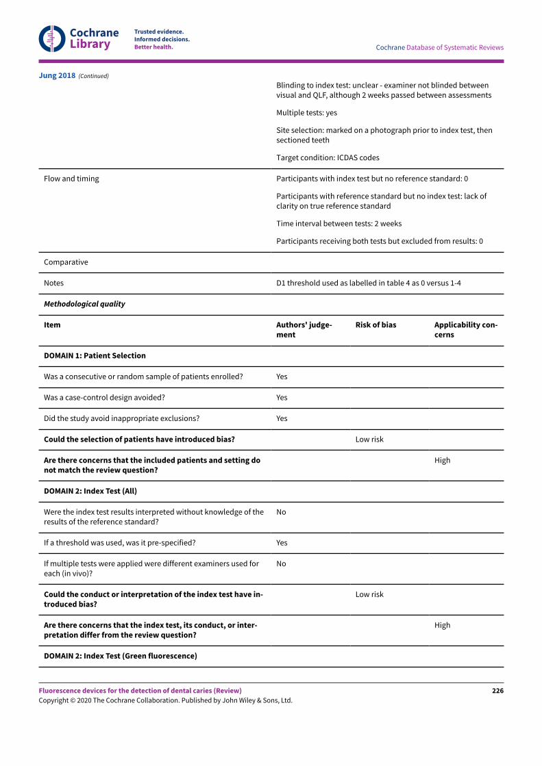
CochraneLibrary
Trusted evidence.Informed decisions.Better health.
Cochrane Database of Systematic Reviews
Blinding to index test: unclear - examiner not blinded betweenvisual and QLF, although 2 weeks passed between assessments
Multiple tests: yes
Site selection: marked on a photograph prior to index test, thensectioned teeth
Target condition: ICDAS codes
Flow and timing Participants with index test but no reference standard: 0
Participants with reference standard but no index test: lack ofclarity on true reference standard
Time interval between tests: 2 weeks
Participants receiving both tests but excluded from results: 0
Comparative
Notes D1 threshold used as labelled in table 4 as 0 versus 1-4
Methodological quality
Item Authors' judge-ment
Risk of bias Applicability con-cerns
DOMAIN 1: Patient Selection
Was a consecutive or random sample of patients enrolled? Yes
Was a case-control design avoided? Yes
Did the study avoid inappropriate exclusions? Yes
Could the selection of patients have introduced bias? Low risk
Are there concerns that the included patients and setting donot match the review question?
High
DOMAIN 2: Index Test (All)
Were the index test results interpreted without knowledge of theresults of the reference standard?
No
If a threshold was used, was it pre-specified? Yes
If multiple tests were applied were different examiners used foreach (in vivo)?
No
Could the conduct or interpretation of the index test have in-troduced bias?
Low risk
Are there concerns that the index test, its conduct, or inter-pretation differ from the review question?
High
DOMAIN 2: Index Test (Green fluorescence)
Jung 2018 (Continued)
Fluorescence devices for the detection of dental caries (Review)
Copyright © 2020 The Cochrane Collaboration. Published by John Wiley & Sons, Ltd.
226
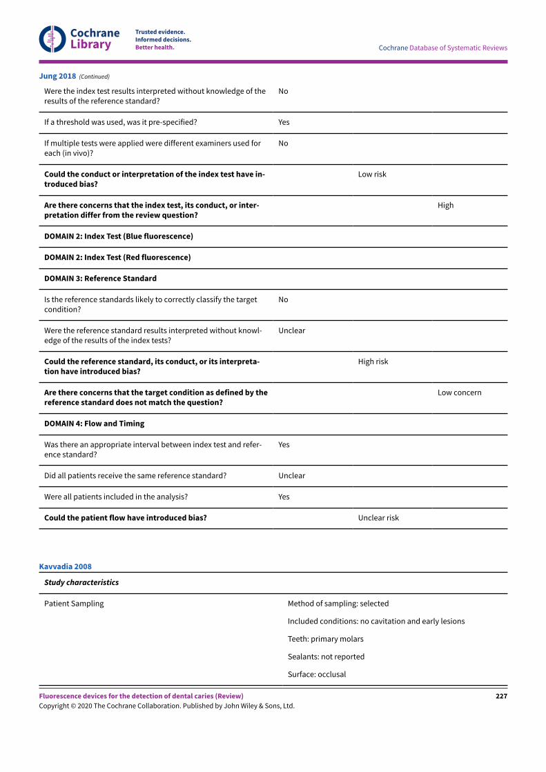
CochraneLibrary
Trusted evidence.Informed decisions.Better health.
Cochrane Database of Systematic Reviews
Were the index test results interpreted without knowledge of theresults of the reference standard?
No
If a threshold was used, was it pre-specified? Yes
If multiple tests were applied were different examiners used foreach (in vivo)?
No
Could the conduct or interpretation of the index test have in-troduced bias?
Low risk
Are there concerns that the index test, its conduct, or inter-pretation differ from the review question?
High
DOMAIN 2: Index Test (Blue fluorescence)
DOMAIN 2: Index Test (Red fluorescence)
DOMAIN 3: Reference Standard
Is the reference standards likely to correctly classify the targetcondition?
No
Were the reference standard results interpreted without knowl-edge of the results of the index tests?
Unclear
Could the reference standard, its conduct, or its interpreta-tion have introduced bias?
High risk
Are there concerns that the target condition as defined by thereference standard does not match the question?
Low concern
DOMAIN 4: Flow and Timing
Was there an appropriate interval between index test and refer-ence standard?
Yes
Did all patients receive the same reference standard? Unclear
Were all patients included in the analysis? Yes
Could the patient flow have introduced bias? Unclear risk
Jung 2018 (Continued)
Study characteristics
Patient Sampling Method of sampling: selected
Included conditions: no cavitation and early lesions
Teeth: primary molars
Sealants: not reported
Surface: occlusal
Kavvadia 2008
Fluorescence devices for the detection of dental caries (Review)
Copyright © 2020 The Cochrane Collaboration. Published by John Wiley & Sons, Ltd.
227
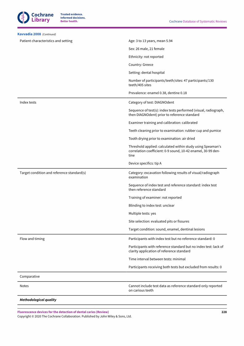
CochraneLibrary
Trusted evidence.Informed decisions.Better health.
Cochrane Database of Systematic Reviews
Patient characteristics and setting Age: 3 to 13 years, mean 5.94
Sex: 26 male, 21 female
Ethnicity: not reported
Country: Greece
Setting: dental hospital
Number of participants/teeth/sites: 47 participants/130teeth/405 sites
Prevalence: enamel 0.38, dentine 0.18
Index tests Category of test: DIAGNOdent
Sequence of test(s): index tests performed (visual, radiograph,then DIAGNOdent) prior to reference standard
Examiner training and calibration: calibrated
Teeth cleaning prior to examination: rubber cup and pumice
Tooth drying prior to examination: air dried
Threshold applied: calculated within study using Speaman'scorrelation coefficient: 0-9 sound, 10-42 enamel, 30-99 den-tine
Device specifics: tip A
Target condition and reference standard(s) Category: excavation following results of visual/radiographexamination
Sequence of index test and reference standard: index testthen reference standard
Training of examiner: not reported
Blinding to index test: unclear
Multiple tests: yes
Site selection: evaluated pits or fissures
Target condition: sound, enamel, dentinal lesions
Flow and timing Participants with index test but no reference standard: 0
Participants with reference standard but no index test: lack ofclarity application of reference standard
Time interval between tests: minimal
Participants receiving both tests but excluded from results: 0
Comparative
Notes Cannot include test data as reference standard only reportedon carious teeth
Methodological quality
Kavvadia 2008 (Continued)
Fluorescence devices for the detection of dental caries (Review)
Copyright © 2020 The Cochrane Collaboration. Published by John Wiley & Sons, Ltd.
228
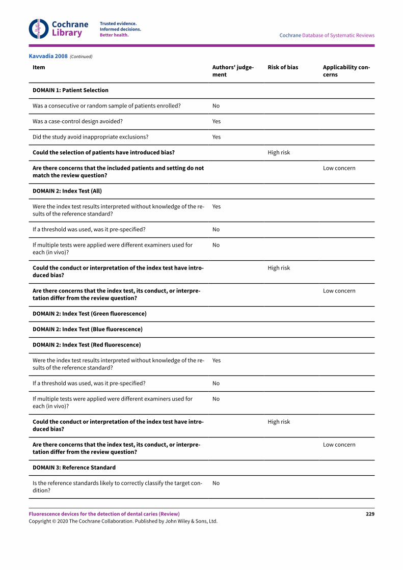
CochraneLibrary
Trusted evidence.Informed decisions.Better health.
Cochrane Database of Systematic Reviews
Item Authors' judge-ment
Risk of bias Applicability con-cerns
DOMAIN 1: Patient Selection
Was a consecutive or random sample of patients enrolled? No
Was a case-control design avoided? Yes
Did the study avoid inappropriate exclusions? Yes
Could the selection of patients have introduced bias? High risk
Are there concerns that the included patients and setting do notmatch the review question?
Low concern
DOMAIN 2: Index Test (All)
Were the index test results interpreted without knowledge of the re-sults of the reference standard?
Yes
If a threshold was used, was it pre-specified? No
If multiple tests were applied were different examiners used foreach (in vivo)?
No
Could the conduct or interpretation of the index test have intro-duced bias?
High risk
Are there concerns that the index test, its conduct, or interpre-tation differ from the review question?
Low concern
DOMAIN 2: Index Test (Green fluorescence)
DOMAIN 2: Index Test (Blue fluorescence)
DOMAIN 2: Index Test (Red fluorescence)
Were the index test results interpreted without knowledge of the re-sults of the reference standard?
Yes
If a threshold was used, was it pre-specified? No
If multiple tests were applied were different examiners used foreach (in vivo)?
No
Could the conduct or interpretation of the index test have intro-duced bias?
High risk
Are there concerns that the index test, its conduct, or interpre-tation differ from the review question?
Low concern
DOMAIN 3: Reference Standard
Is the reference standards likely to correctly classify the target con-dition?
No
Kavvadia 2008 (Continued)
Fluorescence devices for the detection of dental caries (Review)
Copyright © 2020 The Cochrane Collaboration. Published by John Wiley & Sons, Ltd.
229
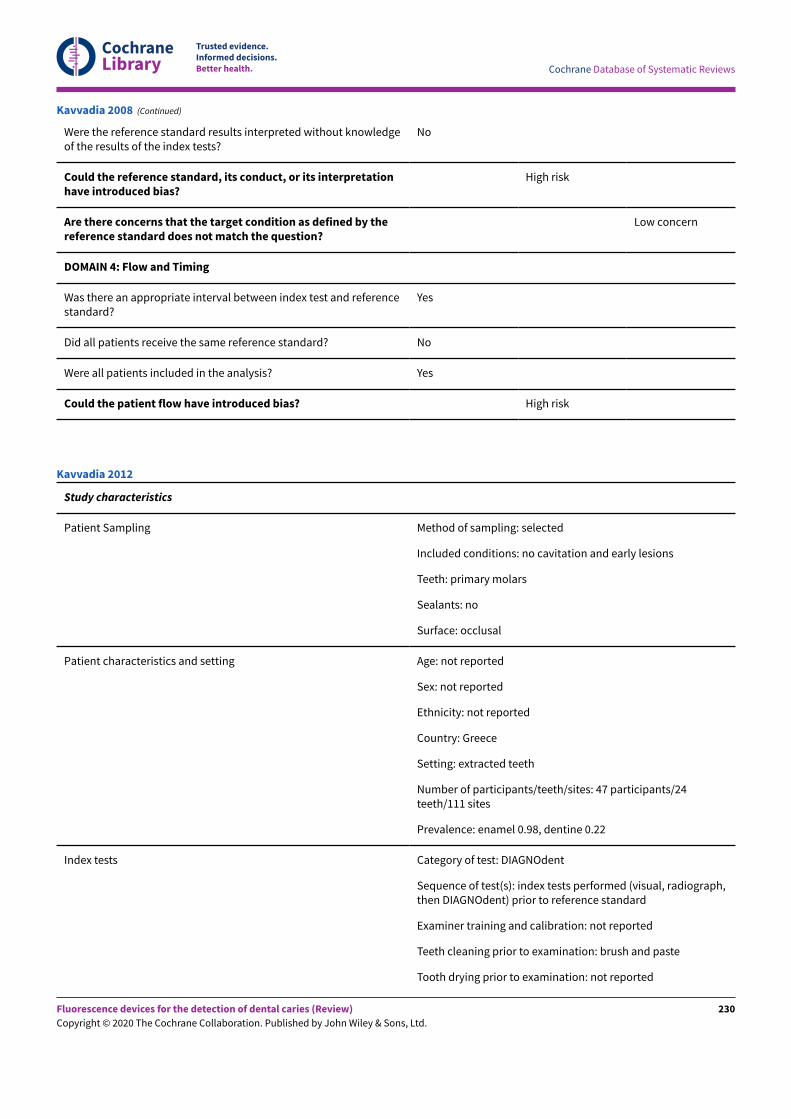
CochraneLibrary
Trusted evidence.Informed decisions.Better health.
Cochrane Database of Systematic Reviews
Were the reference standard results interpreted without knowledgeof the results of the index tests?
No
Could the reference standard, its conduct, or its interpretationhave introduced bias?
High risk
Are there concerns that the target condition as defined by thereference standard does not match the question?
Low concern
DOMAIN 4: Flow and Timing
Was there an appropriate interval between index test and referencestandard?
Yes
Did all patients receive the same reference standard? No
Were all patients included in the analysis? Yes
Could the patient flow have introduced bias? High risk
Kavvadia 2008 (Continued)
Study characteristics
Patient Sampling Method of sampling: selected
Included conditions: no cavitation and early lesions
Teeth: primary molars
Sealants: no
Surface: occlusal
Patient characteristics and setting Age: not reported
Sex: not reported
Ethnicity: not reported
Country: Greece
Setting: extracted teeth
Number of participants/teeth/sites: 47 participants/24teeth/111 sites
Prevalence: enamel 0.98, dentine 0.22
Index tests Category of test: DIAGNOdent
Sequence of test(s): index tests performed (visual, radiograph,then DIAGNOdent) prior to reference standard
Examiner training and calibration: not reported
Teeth cleaning prior to examination: brush and paste
Tooth drying prior to examination: not reported
Kavvadia 2012
Fluorescence devices for the detection of dental caries (Review)
Copyright © 2020 The Cochrane Collaboration. Published by John Wiley & Sons, Ltd.
230
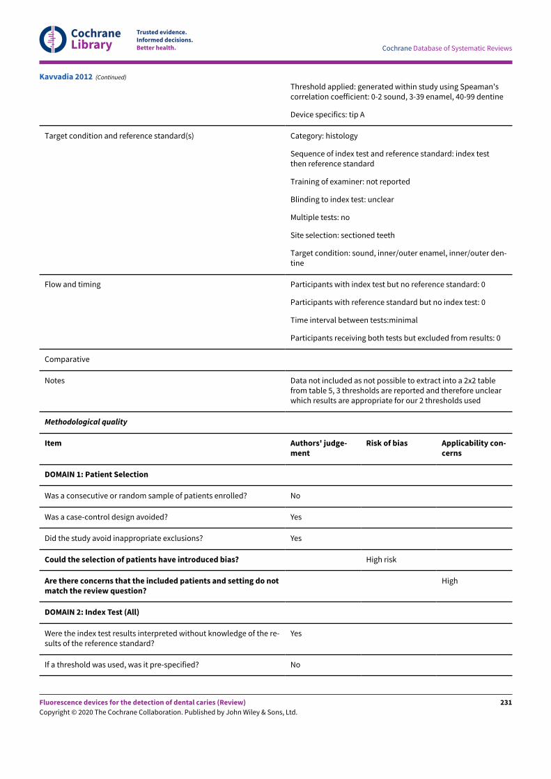
CochraneLibrary
Trusted evidence.Informed decisions.Better health.
Cochrane Database of Systematic Reviews
Threshold applied: generated within study using Speaman'scorrelation coefficient: 0-2 sound, 3-39 enamel, 40-99 dentine
Device specifics: tip A
Target condition and reference standard(s) Category: histology
Sequence of index test and reference standard: index testthen reference standard
Training of examiner: not reported
Blinding to index test: unclear
Multiple tests: no
Site selection: sectioned teeth
Target condition: sound, inner/outer enamel, inner/outer den-tine
Flow and timing Participants with index test but no reference standard: 0
Participants with reference standard but no index test: 0
Time interval between tests:minimal
Participants receiving both tests but excluded from results: 0
Comparative
Notes Data not included as not possible to extract into a 2x2 tablefrom table 5, 3 thresholds are reported and therefore unclearwhich results are appropriate for our 2 thresholds used
Methodological quality
Item Authors' judge-ment
Risk of bias Applicability con-cerns
DOMAIN 1: Patient Selection
Was a consecutive or random sample of patients enrolled? No
Was a case-control design avoided? Yes
Did the study avoid inappropriate exclusions? Yes
Could the selection of patients have introduced bias? High risk
Are there concerns that the included patients and setting do notmatch the review question?
High
DOMAIN 2: Index Test (All)
Were the index test results interpreted without knowledge of the re-sults of the reference standard?
Yes
If a threshold was used, was it pre-specified? No
Kavvadia 2012 (Continued)
Fluorescence devices for the detection of dental caries (Review)
Copyright © 2020 The Cochrane Collaboration. Published by John Wiley & Sons, Ltd.
231

CochraneLibrary
Trusted evidence.Informed decisions.Better health.
Cochrane Database of Systematic Reviews
If multiple tests were applied were different examiners used foreach (in vivo)?
No
Could the conduct or interpretation of the index test have intro-duced bias?
High risk
Are there concerns that the index test, its conduct, or interpre-tation differ from the review question?
Low concern
DOMAIN 2: Index Test (Green fluorescence)
DOMAIN 2: Index Test (Blue fluorescence)
DOMAIN 2: Index Test (Red fluorescence)
Were the index test results interpreted without knowledge of the re-sults of the reference standard?
Yes
If a threshold was used, was it pre-specified? No
If multiple tests were applied were different examiners used foreach (in vivo)?
No
Could the conduct or interpretation of the index test have intro-duced bias?
High risk
Are there concerns that the index test, its conduct, or interpre-tation differ from the review question?
Low concern
DOMAIN 3: Reference Standard
Is the reference standards likely to correctly classify the target con-dition?
Yes
Were the reference standard results interpreted without knowledgeof the results of the index tests?
Unclear
Could the reference standard, its conduct, or its interpretationhave introduced bias?
Low risk
Are there concerns that the target condition as defined by thereference standard does not match the question?
Low concern
DOMAIN 4: Flow and Timing
Was there an appropriate interval between index test and referencestandard?
Yes
Did all patients receive the same reference standard? Yes
Were all patients included in the analysis? Yes
Could the patient flow have introduced bias? Low risk
Kavvadia 2012 (Continued)
Fluorescence devices for the detection of dental caries (Review)
Copyright © 2020 The Cochrane Collaboration. Published by John Wiley & Sons, Ltd.
232
CochraneLibrary
Trusted evidence.Informed decisions.Better health.
Cochrane Database of Systematic Reviews
Study characteristics
Patient Sampling Method of sampling: selected
Included conditions: no cavitation and early lesions
Teeth: primary molars
Sealants: no
Surface: occlusal
Patient characteristics and setting Age: not reported
Sex: not reported
Ethnicity: not reported
Country: Israel
Setting: extracted teeth
Number of participants/teeth/sites: 901 teeth
Prevalence: enamel 0.67, dentine 0.19
Index tests Category of test: fluorescence - Oliver 101
Sequence of test(s): index tests performed (visual, ra-diograph, then fluorescence) prior to reference stan-dard
Examiner training and calibration: not reported
Teeth cleaning prior to examination: polished andcleaned
Tooth drying prior to examination: not reported
Threshold applied: calculated within study
Device specifics: unclear
Target condition and reference standard(s) Category: histology
Sequence of index test and reference standard: indextest then reference standard
Training of examiner: not reported
Blinding to index test: unclear
Multiple tests: no
Site selection: sectioned teeth
Target condition: enamel or dentine caries
Flow and timing Participants with index test but no reference stan-dard: 0
Participants with reference standard but no indextest: 0
Time interval between tests: minimal
Kesler 2003
Fluorescence devices for the detection of dental caries (Review)
Copyright © 2020 The Cochrane Collaboration. Published by John Wiley & Sons, Ltd.
233
CochraneLibrary
Trusted evidence.Informed decisions.Better health.
Cochrane Database of Systematic Reviews
Participants receiving both tests but excluded fromresults: 0
Comparative
Notes
Methodological quality
Item Authors'judgement
Risk of bias Applicabilityconcerns
DOMAIN 1: Patient Selection
Was a consecutive or random sample of patients enrolled? No
Was a case-control design avoided? Yes
Did the study avoid inappropriate exclusions? Yes
Could the selection of patients have introduced bias? High risk
Are there concerns that the included patients and setting do not matchthe review question?
High
DOMAIN 2: Index Test (All)
Were the index test results interpreted without knowledge of the results ofthe reference standard?
Yes
If a threshold was used, was it pre-specified? No
If multiple tests were applied were different examiners used for each (in vi-vo)?
Unclear
Could the conduct or interpretation of the index test have introducedbias?
High risk
Are there concerns that the index test, its conduct, or interpretation dif-fer from the review question?
Low concern
DOMAIN 2: Index Test (Green fluorescence)
Were the index test results interpreted without knowledge of the results ofthe reference standard?
Yes
If a threshold was used, was it pre-specified? No
If multiple tests were applied were different examiners used for each (in vi-vo)?
Unclear
Could the conduct or interpretation of the index test have introducedbias?
High risk
Are there concerns that the index test, its conduct, or interpretation dif-fer from the review question?
Low concern
Kesler 2003 (Continued)
Fluorescence devices for the detection of dental caries (Review)
Copyright © 2020 The Cochrane Collaboration. Published by John Wiley & Sons, Ltd.
234
CochraneLibrary
Trusted evidence.Informed decisions.Better health.
Cochrane Database of Systematic Reviews
DOMAIN 2: Index Test (Blue fluorescence)
DOMAIN 2: Index Test (Red fluorescence)
DOMAIN 3: Reference Standard
Is the reference standards likely to correctly classify the target condition? Yes
Were the reference standard results interpreted without knowledge of theresults of the index tests?
Yes
Could the reference standard, its conduct, or its interpretation have in-troduced bias?
Low risk
Are there concerns that the target condition as defined by the referencestandard does not match the question?
Low concern
DOMAIN 4: Flow and Timing
Was there an appropriate interval between index test and reference stan-dard?
Yes
Did all patients receive the same reference standard? Yes
Were all patients included in the analysis? Yes
Could the patient flow have introduced bias? Low risk
Kesler 2003 (Continued)
Study characteristics
Patient Sampling Method of sampling: not clearly reported
Included conditions: severity of condition unclear, "subjects with 1 ormore proximal caries surfaces detected visually or radiographically wereincluded in the study", restorations were included
Teeth: permanent molars and premolars
Sealants: not reported
Surface: approximal
Patient characteristics and setting Age: 19 to 60 years
Sex: 55% male
Ethnicity: not reported
Country: South Korea
Setting: extracted teeth
Number of participants/teeth/sites: 65 teeth/280 sites
Prevalence: enamel 0.61, dentine 0.20
Kim 2017
Fluorescence devices for the detection of dental caries (Review)
Copyright © 2020 The Cochrane Collaboration. Published by John Wiley & Sons, Ltd.
235
CochraneLibrary
Trusted evidence.Informed decisions.Better health.
Cochrane Database of Systematic Reviews
Index tests Category of test: QLF-Digital Biluminator 2+ (QLF-D, Inspektor ResearchSystems BV, Amsterdam, The Netherlands), 2 methods one using QA2 soft-ware, the second using fluorescence images interpreted by an examiner:"Normal white-light images and sequential fluorescence images were cap-tured with a “live view” enabled full-frame sensor digital SLR camera"
Sequence of test(s): visual then radiograph followed by QLF, radiographwas the reference standard
Examiner training and calibration: 1 trained examiner completed all indextests and reference standard
Teeth cleaning prior to examination: full-mouth scaling and polishing
Tooth drying prior to examination: not reported
Threshold applied: method used for fluorescence image method: shadowand no red fluorescence (Q0), an irregular dark shadow but no red fluores-cence (Q1), faint red fluorescence limited to 1/3 of the buccolingual width(Q2), and strong red fluorescence over 1/3 of the buccolingual width (Q3)
Target condition and reference standard(s) Category: radiograph
Sequence of index test and reference standard: reference standard prior toindex test
Training of examiner: not reported, but experienced
Blinding to index test: no
Multiple tests: no
Site selection: approximal surfaces
Target condition: sound, outer/inner enamel, outer/inner dentine
Flow and timing Participants with index test but no reference standard: 15
Participants with reference standard but no index test: 0
Time interval between tests: minimal
Participants receiving both tests but excluded from results: 0
Comparative
Notes Data used for the fluorescence images method as the 2x2 figures were notavailable for the software method
Methodological quality
Item Authors' judgement Risk of bias Applicability con-cerns
DOMAIN 1: Patient Selection
Was a consecutive or random sample of patients en-rolled?
Unclear
Was a case-control design avoided? Yes
Kim 2017 (Continued)
Fluorescence devices for the detection of dental caries (Review)
Copyright © 2020 The Cochrane Collaboration. Published by John Wiley & Sons, Ltd.
236
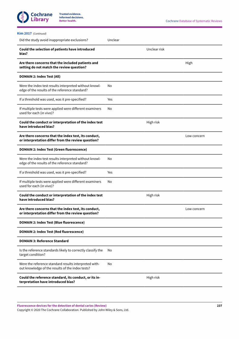
CochraneLibrary
Trusted evidence.Informed decisions.Better health.
Cochrane Database of Systematic Reviews
Did the study avoid inappropriate exclusions? Unclear
Could the selection of patients have introducedbias?
Unclear risk
Are there concerns that the included patients andsetting do not match the review question?
High
DOMAIN 2: Index Test (All)
Were the index test results interpreted without knowl-edge of the results of the reference standard?
No
If a threshold was used, was it pre-specified? Yes
If multiple tests were applied were different examinersused for each (in vivo)?
No
Could the conduct or interpretation of the index testhave introduced bias?
High risk
Are there concerns that the index test, its conduct,or interpretation differ from the review question?
Low concern
DOMAIN 2: Index Test (Green fluorescence)
Were the index test results interpreted without knowl-edge of the results of the reference standard?
No
If a threshold was used, was it pre-specified? Yes
If multiple tests were applied were different examinersused for each (in vivo)?
No
Could the conduct or interpretation of the index testhave introduced bias?
High risk
Are there concerns that the index test, its conduct,or interpretation differ from the review question?
Low concern
DOMAIN 2: Index Test (Blue fluorescence)
DOMAIN 2: Index Test (Red fluorescence)
DOMAIN 3: Reference Standard
Is the reference standards likely to correctly classify thetarget condition?
No
Were the reference standard results interpreted with-out knowledge of the results of the index tests?
No
Could the reference standard, its conduct, or its in-terpretation have introduced bias?
High risk
Kim 2017 (Continued)
Fluorescence devices for the detection of dental caries (Review)
Copyright © 2020 The Cochrane Collaboration. Published by John Wiley & Sons, Ltd.
237
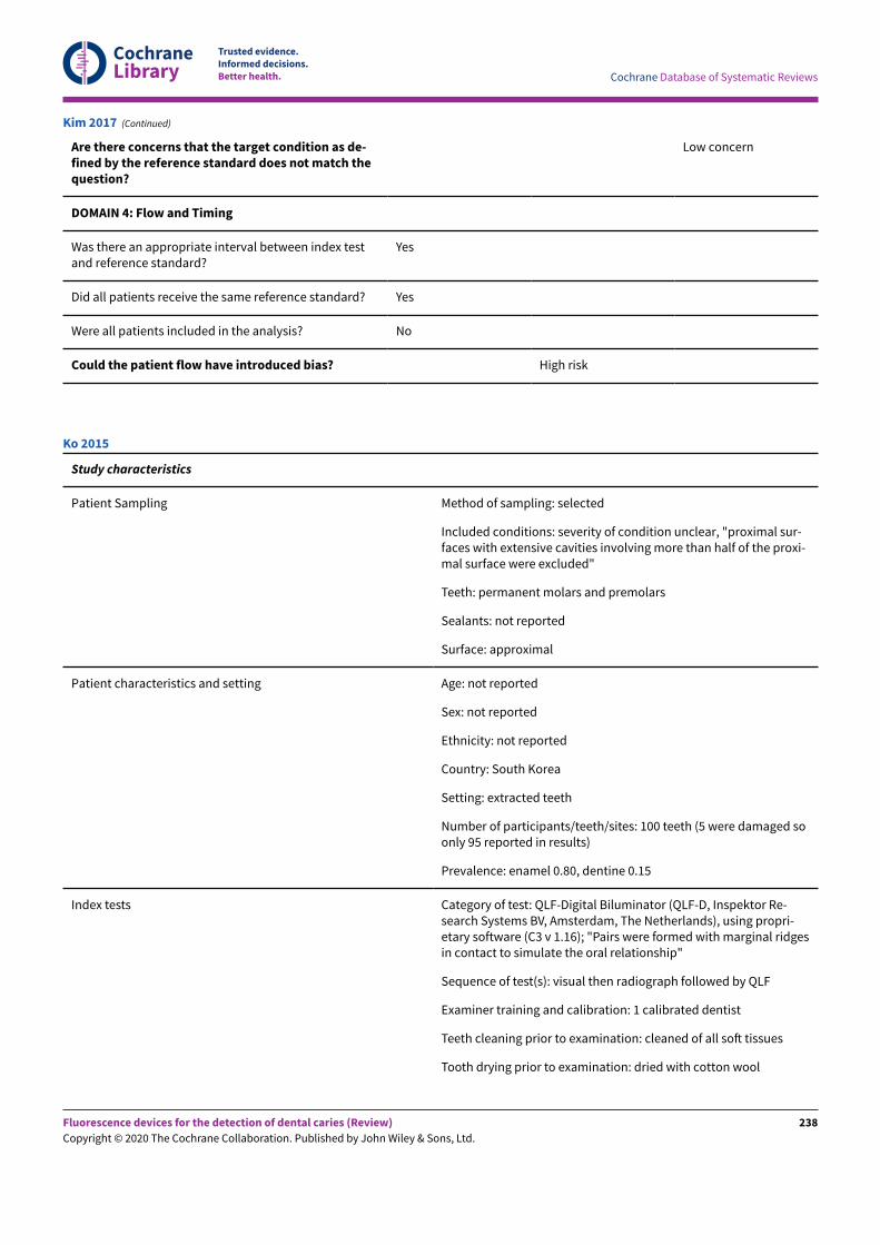
CochraneLibrary
Trusted evidence.Informed decisions.Better health.
Cochrane Database of Systematic Reviews
Are there concerns that the target condition as de-fined by the reference standard does not match thequestion?
Low concern
DOMAIN 4: Flow and Timing
Was there an appropriate interval between index testand reference standard?
Yes
Did all patients receive the same reference standard? Yes
Were all patients included in the analysis? No
Could the patient flow have introduced bias? High risk
Kim 2017 (Continued)
Study characteristics
Patient Sampling Method of sampling: selected
Included conditions: severity of condition unclear, "proximal sur-faces with extensive cavities involving more than half of the proxi-mal surface were excluded"
Teeth: permanent molars and premolars
Sealants: not reported
Surface: approximal
Patient characteristics and setting Age: not reported
Sex: not reported
Ethnicity: not reported
Country: South Korea
Setting: extracted teeth
Number of participants/teeth/sites: 100 teeth (5 were damaged soonly 95 reported in results)
Prevalence: enamel 0.80, dentine 0.15
Index tests Category of test: QLF-Digital Biluminator (QLF-D, Inspektor Re-search Systems BV, Amsterdam, The Netherlands), using propri-etary software (C3 v 1.16); "Pairs were formed with marginal ridgesin contact to simulate the oral relationship"
Sequence of test(s): visual then radiograph followed by QLF
Examiner training and calibration: 1 calibrated dentist
Teeth cleaning prior to examination: cleaned of all soN tissues
Tooth drying prior to examination: dried with cotton wool
Ko 2015
Fluorescence devices for the detection of dental caries (Review)
Copyright © 2020 The Cochrane Collaboration. Published by John Wiley & Sons, Ltd.
238
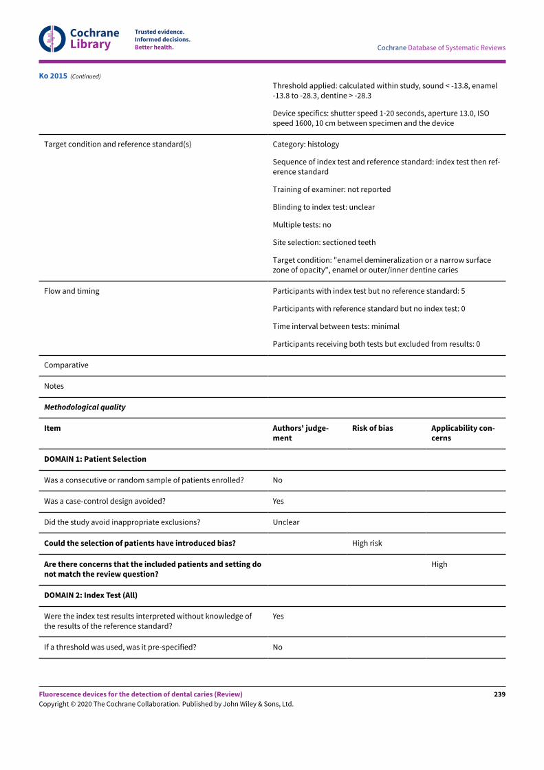
CochraneLibrary
Trusted evidence.Informed decisions.Better health.
Cochrane Database of Systematic Reviews
Threshold applied: calculated within study, sound < -13.8, enamel-13.8 to -28.3, dentine > -28.3
Device specifics: shutter speed 1-20 seconds, aperture 13.0, ISOspeed 1600, 10 cm between specimen and the device
Target condition and reference standard(s) Category: histology
Sequence of index test and reference standard: index test then ref-erence standard
Training of examiner: not reported
Blinding to index test: unclear
Multiple tests: no
Site selection: sectioned teeth
Target condition: "enamel demineralization or a narrow surfacezone of opacity", enamel or outer/inner dentine caries
Flow and timing Participants with index test but no reference standard: 5
Participants with reference standard but no index test: 0
Time interval between tests: minimal
Participants receiving both tests but excluded from results: 0
Comparative
Notes
Methodological quality
Item Authors' judge-ment
Risk of bias Applicability con-cerns
DOMAIN 1: Patient Selection
Was a consecutive or random sample of patients enrolled? No
Was a case-control design avoided? Yes
Did the study avoid inappropriate exclusions? Unclear
Could the selection of patients have introduced bias? High risk
Are there concerns that the included patients and setting donot match the review question?
High
DOMAIN 2: Index Test (All)
Were the index test results interpreted without knowledge ofthe results of the reference standard?
Yes
If a threshold was used, was it pre-specified? No
Ko 2015 (Continued)
Fluorescence devices for the detection of dental caries (Review)
Copyright © 2020 The Cochrane Collaboration. Published by John Wiley & Sons, Ltd.
239
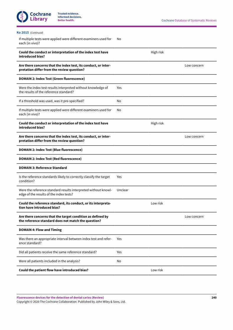
CochraneLibrary
Trusted evidence.Informed decisions.Better health.
Cochrane Database of Systematic Reviews
If multiple tests were applied were different examiners used foreach (in vivo)?
No
Could the conduct or interpretation of the index test haveintroduced bias?
High risk
Are there concerns that the index test, its conduct, or inter-pretation differ from the review question?
Low concern
DOMAIN 2: Index Test (Green fluorescence)
Were the index test results interpreted without knowledge ofthe results of the reference standard?
Yes
If a threshold was used, was it pre-specified? No
If multiple tests were applied were different examiners used foreach (in vivo)?
No
Could the conduct or interpretation of the index test haveintroduced bias?
High risk
Are there concerns that the index test, its conduct, or inter-pretation differ from the review question?
Low concern
DOMAIN 2: Index Test (Blue fluorescence)
DOMAIN 2: Index Test (Red fluorescence)
DOMAIN 3: Reference Standard
Is the reference standards likely to correctly classify the targetcondition?
Yes
Were the reference standard results interpreted without knowl-edge of the results of the index tests?
Unclear
Could the reference standard, its conduct, or its interpreta-tion have introduced bias?
Low risk
Are there concerns that the target condition as defined bythe reference standard does not match the question?
Low concern
DOMAIN 4: Flow and Timing
Was there an appropriate interval between index test and refer-ence standard?
Yes
Did all patients receive the same reference standard? Yes
Were all patients included in the analysis? No
Could the patient flow have introduced bias? Low risk
Ko 2015 (Continued)
Fluorescence devices for the detection of dental caries (Review)
Copyright © 2020 The Cochrane Collaboration. Published by John Wiley & Sons, Ltd.
240
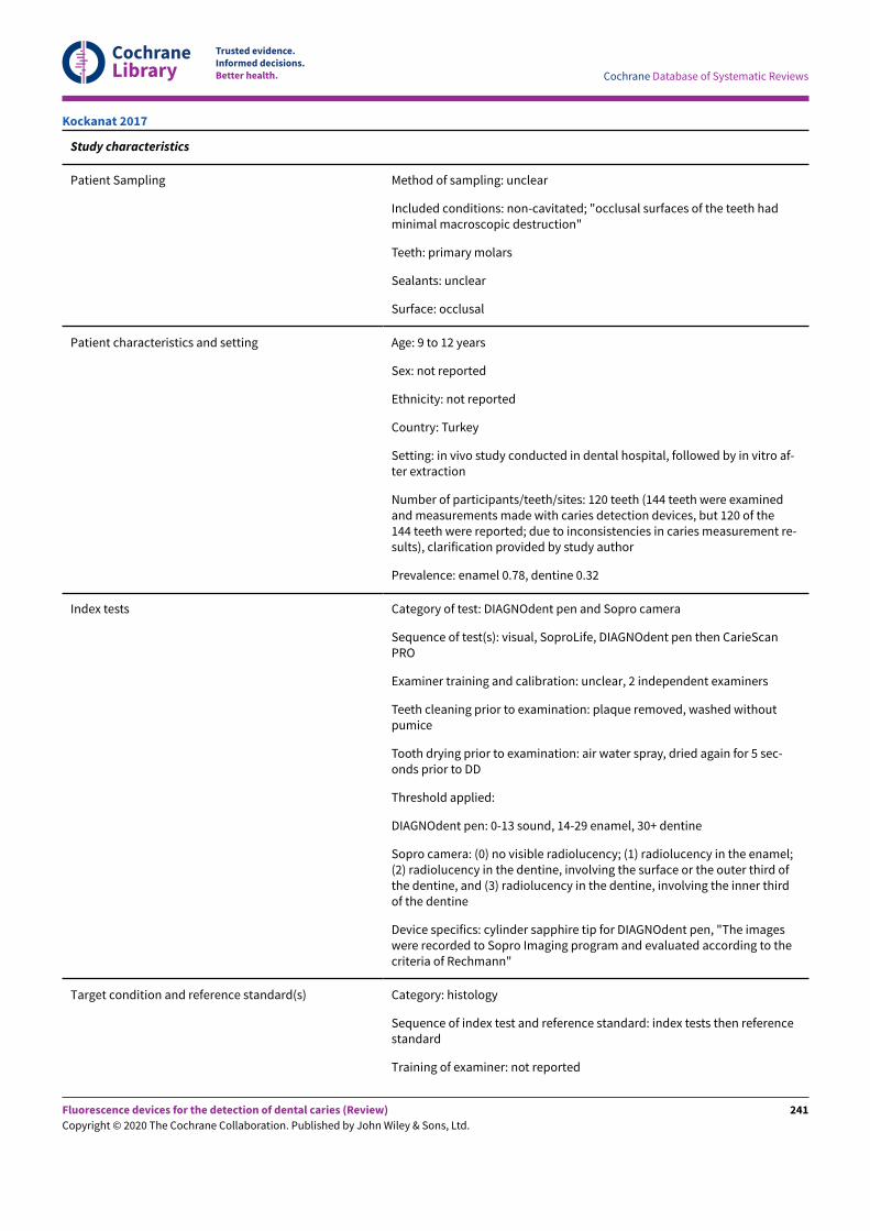
CochraneLibrary
Trusted evidence.Informed decisions.Better health.
Cochrane Database of Systematic Reviews
Study characteristics
Patient Sampling Method of sampling: unclear
Included conditions: non-cavitated; "occlusal surfaces of the teeth hadminimal macroscopic destruction"
Teeth: primary molars
Sealants: unclear
Surface: occlusal
Patient characteristics and setting Age: 9 to 12 years
Sex: not reported
Ethnicity: not reported
Country: Turkey
Setting: in vivo study conducted in dental hospital, followed by in vitro af-ter extraction
Number of participants/teeth/sites: 120 teeth (144 teeth were examinedand measurements made with caries detection devices, but 120 of the144 teeth were reported; due to inconsistencies in caries measurement re-sults), clarification provided by study author
Prevalence: enamel 0.78, dentine 0.32
Index tests Category of test: DIAGNOdent pen and Sopro camera
Sequence of test(s): visual, SoproLife, DIAGNOdent pen then CarieScanPRO
Examiner training and calibration: unclear, 2 independent examiners
Teeth cleaning prior to examination: plaque removed, washed withoutpumice
Tooth drying prior to examination: air water spray, dried again for 5 sec-onds prior to DD
Threshold applied:
DIAGNOdent pen: 0-13 sound, 14-29 enamel, 30+ dentine
Sopro camera: (0) no visible radiolucency; (1) radiolucency in the enamel;(2) radiolucency in the dentine, involving the surface or the outer third ofthe dentine, and (3) radiolucency in the dentine, involving the inner thirdof the dentine
Device specifics: cylinder sapphire tip for DIAGNOdent pen, "The imageswere recorded to Sopro Imaging program and evaluated according to thecriteria of Rechmann"
Target condition and reference standard(s) Category: histology
Sequence of index test and reference standard: index tests then referencestandard
Training of examiner: not reported
Kockanat 2017
Fluorescence devices for the detection of dental caries (Review)
Copyright © 2020 The Cochrane Collaboration. Published by John Wiley & Sons, Ltd.
241
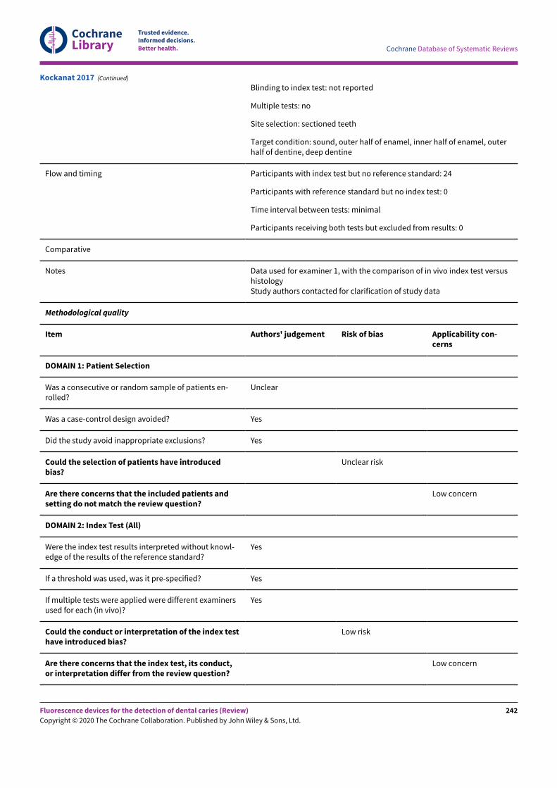
CochraneLibrary
Trusted evidence.Informed decisions.Better health.
Cochrane Database of Systematic Reviews
Blinding to index test: not reported
Multiple tests: no
Site selection: sectioned teeth
Target condition: sound, outer half of enamel, inner half of enamel, outerhalf of dentine, deep dentine
Flow and timing Participants with index test but no reference standard: 24
Participants with reference standard but no index test: 0
Time interval between tests: minimal
Participants receiving both tests but excluded from results: 0
Comparative
Notes Data used for examiner 1, with the comparison of in vivo index test versushistologyStudy authors contacted for clarification of study data
Methodological quality
Item Authors' judgement Risk of bias Applicability con-cerns
DOMAIN 1: Patient Selection
Was a consecutive or random sample of patients en-rolled?
Unclear
Was a case-control design avoided? Yes
Did the study avoid inappropriate exclusions? Yes
Could the selection of patients have introducedbias?
Unclear risk
Are there concerns that the included patients andsetting do not match the review question?
Low concern
DOMAIN 2: Index Test (All)
Were the index test results interpreted without knowl-edge of the results of the reference standard?
Yes
If a threshold was used, was it pre-specified? Yes
If multiple tests were applied were different examinersused for each (in vivo)?
Yes
Could the conduct or interpretation of the index testhave introduced bias?
Low risk
Are there concerns that the index test, its conduct,or interpretation differ from the review question?
Low concern
Kockanat 2017 (Continued)
Fluorescence devices for the detection of dental caries (Review)
Copyright © 2020 The Cochrane Collaboration. Published by John Wiley & Sons, Ltd.
242
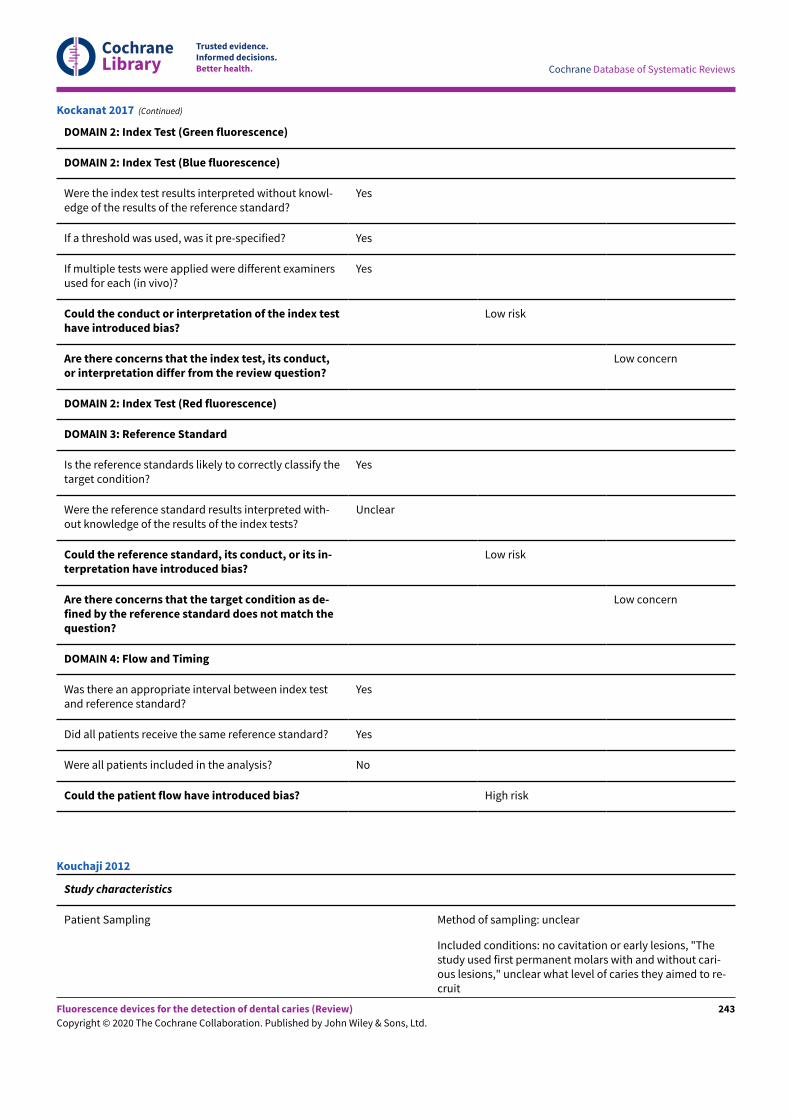
CochraneLibrary
Trusted evidence.Informed decisions.Better health.
Cochrane Database of Systematic Reviews
DOMAIN 2: Index Test (Green fluorescence)
DOMAIN 2: Index Test (Blue fluorescence)
Were the index test results interpreted without knowl-edge of the results of the reference standard?
Yes
If a threshold was used, was it pre-specified? Yes
If multiple tests were applied were different examinersused for each (in vivo)?
Yes
Could the conduct or interpretation of the index testhave introduced bias?
Low risk
Are there concerns that the index test, its conduct,or interpretation differ from the review question?
Low concern
DOMAIN 2: Index Test (Red fluorescence)
DOMAIN 3: Reference Standard
Is the reference standards likely to correctly classify thetarget condition?
Yes
Were the reference standard results interpreted with-out knowledge of the results of the index tests?
Unclear
Could the reference standard, its conduct, or its in-terpretation have introduced bias?
Low risk
Are there concerns that the target condition as de-fined by the reference standard does not match thequestion?
Low concern
DOMAIN 4: Flow and Timing
Was there an appropriate interval between index testand reference standard?
Yes
Did all patients receive the same reference standard? Yes
Were all patients included in the analysis? No
Could the patient flow have introduced bias? High risk
Kockanat 2017 (Continued)
Study characteristics
Patient Sampling Method of sampling: unclear
Included conditions: no cavitation or early lesions, "Thestudy used first permanent molars with and without cari-ous lesions," unclear what level of caries they aimed to re-cruit
Kouchaji 2012
Fluorescence devices for the detection of dental caries (Review)
Copyright © 2020 The Cochrane Collaboration. Published by John Wiley & Sons, Ltd.
243
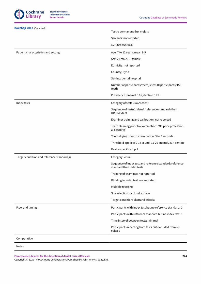
CochraneLibrary
Trusted evidence.Informed decisions.Better health.
Cochrane Database of Systematic Reviews
Teeth: permanent first molars
Sealants: not reported
Surface: occlusal
Patient characteristics and setting Age: 7 to 12 years, mean 9.5
Sex: 21 male, 19 female
Ethnicity: not reported
Country: Syria
Setting: dental hospital
Number of participants/teeth/sites: 40 participants/156teeth
Prevalence: enamel 0.85, dentine 0.29
Index tests Category of test: DIAGNOdent
Sequence of test(s): visual (reference standard) thenDIAGNOdent
Examiner training and calibration: not reported
Teeth cleaning prior to examination: "No prior profession-al cleaning"
Tooth drying prior to examination: 3 to 5 seconds
Threshold applied: 0-14 sound, 15-20 enamel, 21+ dentine
Device specifics: tip A
Target condition and reference standard(s) Category: visual
Sequence of index test and reference standard: referencestandard then index tests
Training of examiner: not reported
Blinding to index test: not reported
Multiple tests: no
Site selection: occlusal surface
Target condition: Ekstrand criteria
Flow and timing Participants with index test but no reference standard: 0
Participants with reference standard but no index test: 0
Time interval between tests: minimal
Participants receiving both tests but excluded from re-sults: 0
Comparative
Notes
Kouchaji 2012 (Continued)
Fluorescence devices for the detection of dental caries (Review)
Copyright © 2020 The Cochrane Collaboration. Published by John Wiley & Sons, Ltd.
244
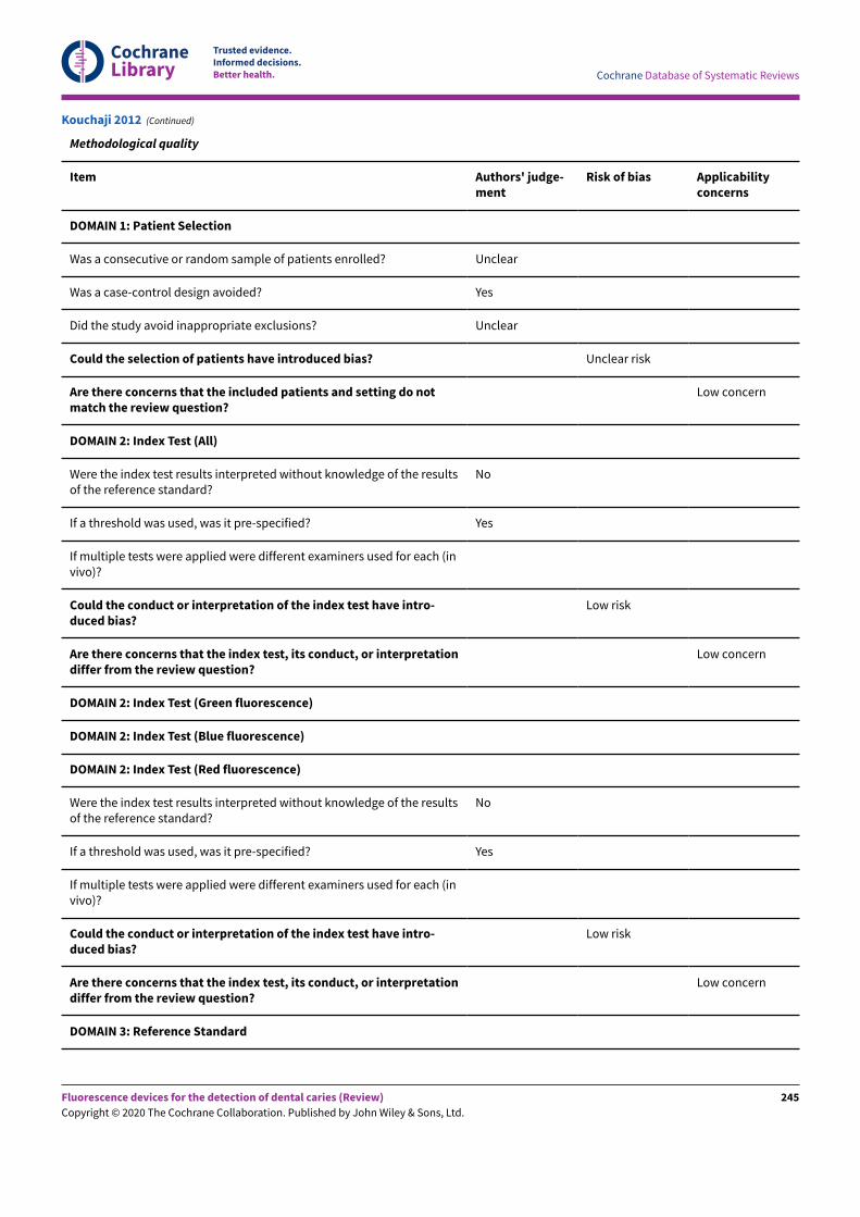
CochraneLibrary
Trusted evidence.Informed decisions.Better health.
Cochrane Database of Systematic Reviews
Methodological quality
Item Authors' judge-ment
Risk of bias Applicabilityconcerns
DOMAIN 1: Patient Selection
Was a consecutive or random sample of patients enrolled? Unclear
Was a case-control design avoided? Yes
Did the study avoid inappropriate exclusions? Unclear
Could the selection of patients have introduced bias? Unclear risk
Are there concerns that the included patients and setting do notmatch the review question?
Low concern
DOMAIN 2: Index Test (All)
Were the index test results interpreted without knowledge of the resultsof the reference standard?
No
If a threshold was used, was it pre-specified? Yes
If multiple tests were applied were different examiners used for each (invivo)?
Could the conduct or interpretation of the index test have intro-duced bias?
Low risk
Are there concerns that the index test, its conduct, or interpretationdiffer from the review question?
Low concern
DOMAIN 2: Index Test (Green fluorescence)
DOMAIN 2: Index Test (Blue fluorescence)
DOMAIN 2: Index Test (Red fluorescence)
Were the index test results interpreted without knowledge of the resultsof the reference standard?
No
If a threshold was used, was it pre-specified? Yes
If multiple tests were applied were different examiners used for each (invivo)?
Could the conduct or interpretation of the index test have intro-duced bias?
Low risk
Are there concerns that the index test, its conduct, or interpretationdiffer from the review question?
Low concern
DOMAIN 3: Reference Standard
Kouchaji 2012 (Continued)
Fluorescence devices for the detection of dental caries (Review)
Copyright © 2020 The Cochrane Collaboration. Published by John Wiley & Sons, Ltd.
245
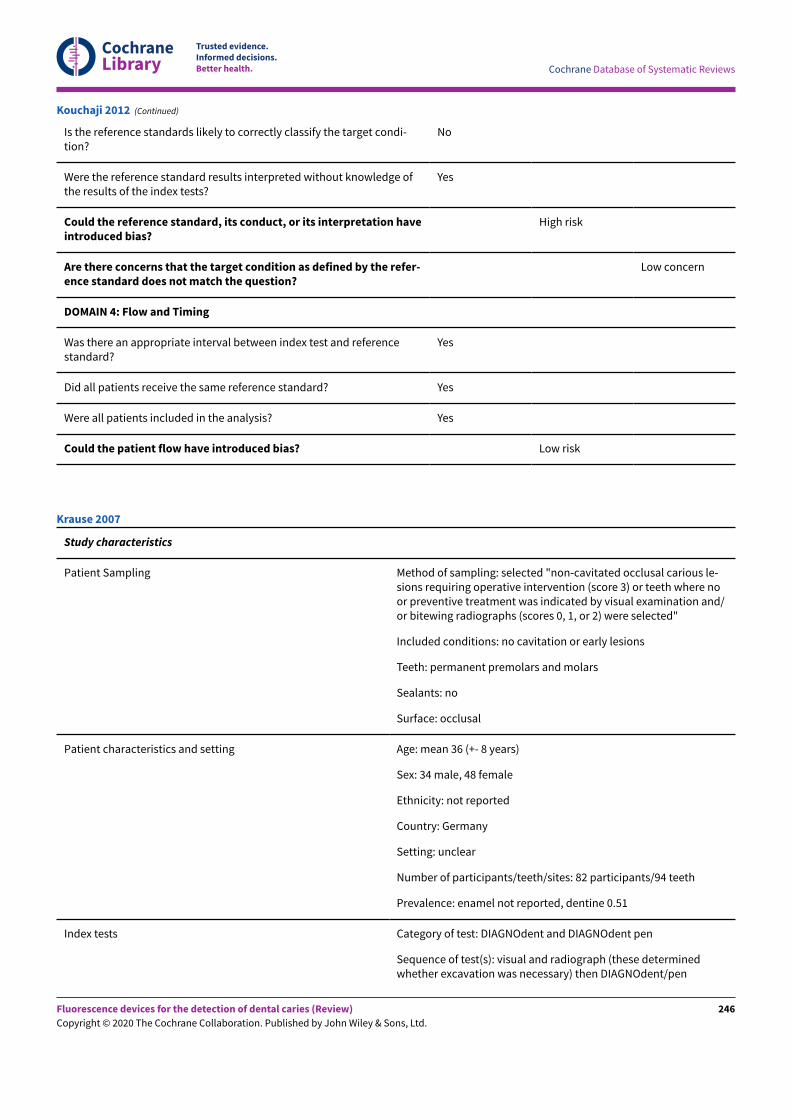
CochraneLibrary
Trusted evidence.Informed decisions.Better health.
Cochrane Database of Systematic Reviews
Is the reference standards likely to correctly classify the target condi-tion?
No
Were the reference standard results interpreted without knowledge ofthe results of the index tests?
Yes
Could the reference standard, its conduct, or its interpretation haveintroduced bias?
High risk
Are there concerns that the target condition as defined by the refer-ence standard does not match the question?
Low concern
DOMAIN 4: Flow and Timing
Was there an appropriate interval between index test and referencestandard?
Yes
Did all patients receive the same reference standard? Yes
Were all patients included in the analysis? Yes
Could the patient flow have introduced bias? Low risk
Kouchaji 2012 (Continued)
Study characteristics
Patient Sampling Method of sampling: selected "non-cavitated occlusal carious le-sions requiring operative intervention (score 3) or teeth where noor preventive treatment was indicated by visual examination and/or bitewing radiographs (scores 0, 1, or 2) were selected"
Included conditions: no cavitation or early lesions
Teeth: permanent premolars and molars
Sealants: no
Surface: occlusal
Patient characteristics and setting Age: mean 36 (+- 8 years)
Sex: 34 male, 48 female
Ethnicity: not reported
Country: Germany
Setting: unclear
Number of participants/teeth/sites: 82 participants/94 teeth
Prevalence: enamel not reported, dentine 0.51
Index tests Category of test: DIAGNOdent and DIAGNOdent pen
Sequence of test(s): visual and radiograph (these determinedwhether excavation was necessary) then DIAGNOdent/pen
Krause 2007
Fluorescence devices for the detection of dental caries (Review)
Copyright © 2020 The Cochrane Collaboration. Published by John Wiley & Sons, Ltd.
246
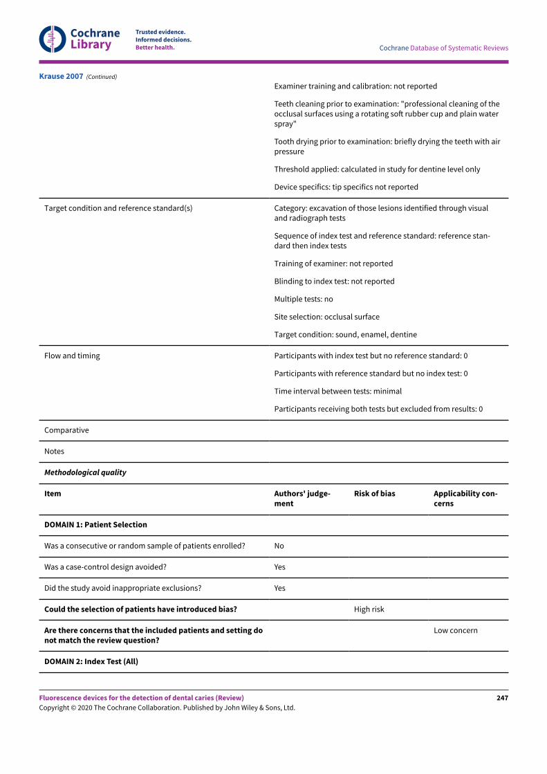
CochraneLibrary
Trusted evidence.Informed decisions.Better health.
Cochrane Database of Systematic Reviews
Examiner training and calibration: not reported
Teeth cleaning prior to examination: "professional cleaning of theocclusal surfaces using a rotating soN rubber cup and plain waterspray"
Tooth drying prior to examination: briefly drying the teeth with airpressure
Threshold applied: calculated in study for dentine level only
Device specifics: tip specifics not reported
Target condition and reference standard(s) Category: excavation of those lesions identified through visualand radiograph tests
Sequence of index test and reference standard: reference stan-dard then index tests
Training of examiner: not reported
Blinding to index test: not reported
Multiple tests: no
Site selection: occlusal surface
Target condition: sound, enamel, dentine
Flow and timing Participants with index test but no reference standard: 0
Participants with reference standard but no index test: 0
Time interval between tests: minimal
Participants receiving both tests but excluded from results: 0
Comparative
Notes
Methodological quality
Item Authors' judge-ment
Risk of bias Applicability con-cerns
DOMAIN 1: Patient Selection
Was a consecutive or random sample of patients enrolled? No
Was a case-control design avoided? Yes
Did the study avoid inappropriate exclusions? Yes
Could the selection of patients have introduced bias? High risk
Are there concerns that the included patients and setting donot match the review question?
Low concern
DOMAIN 2: Index Test (All)
Krause 2007 (Continued)
Fluorescence devices for the detection of dental caries (Review)
Copyright © 2020 The Cochrane Collaboration. Published by John Wiley & Sons, Ltd.
247
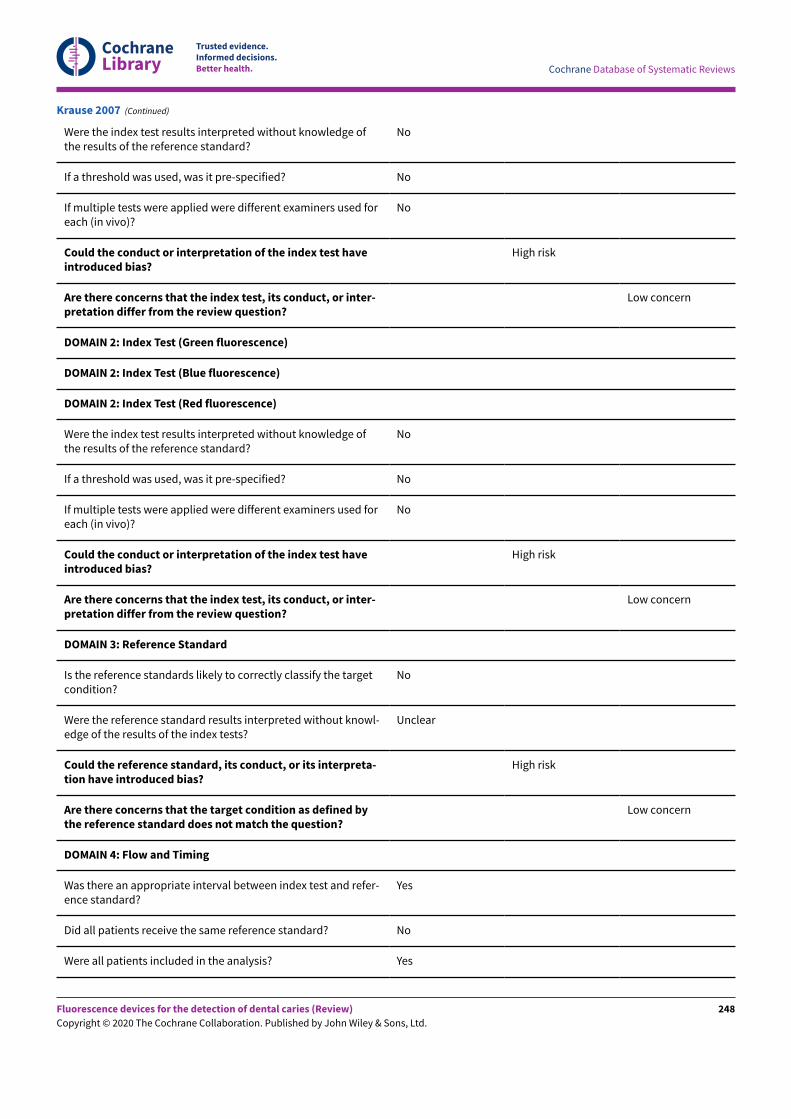
CochraneLibrary
Trusted evidence.Informed decisions.Better health.
Cochrane Database of Systematic Reviews
Were the index test results interpreted without knowledge ofthe results of the reference standard?
No
If a threshold was used, was it pre-specified? No
If multiple tests were applied were different examiners used foreach (in vivo)?
No
Could the conduct or interpretation of the index test haveintroduced bias?
High risk
Are there concerns that the index test, its conduct, or inter-pretation differ from the review question?
Low concern
DOMAIN 2: Index Test (Green fluorescence)
DOMAIN 2: Index Test (Blue fluorescence)
DOMAIN 2: Index Test (Red fluorescence)
Were the index test results interpreted without knowledge ofthe results of the reference standard?
No
If a threshold was used, was it pre-specified? No
If multiple tests were applied were different examiners used foreach (in vivo)?
No
Could the conduct or interpretation of the index test haveintroduced bias?
High risk
Are there concerns that the index test, its conduct, or inter-pretation differ from the review question?
Low concern
DOMAIN 3: Reference Standard
Is the reference standards likely to correctly classify the targetcondition?
No
Were the reference standard results interpreted without knowl-edge of the results of the index tests?
Unclear
Could the reference standard, its conduct, or its interpreta-tion have introduced bias?
High risk
Are there concerns that the target condition as defined bythe reference standard does not match the question?
Low concern
DOMAIN 4: Flow and Timing
Was there an appropriate interval between index test and refer-ence standard?
Yes
Did all patients receive the same reference standard? No
Were all patients included in the analysis? Yes
Krause 2007 (Continued)
Fluorescence devices for the detection of dental caries (Review)
Copyright © 2020 The Cochrane Collaboration. Published by John Wiley & Sons, Ltd.
248
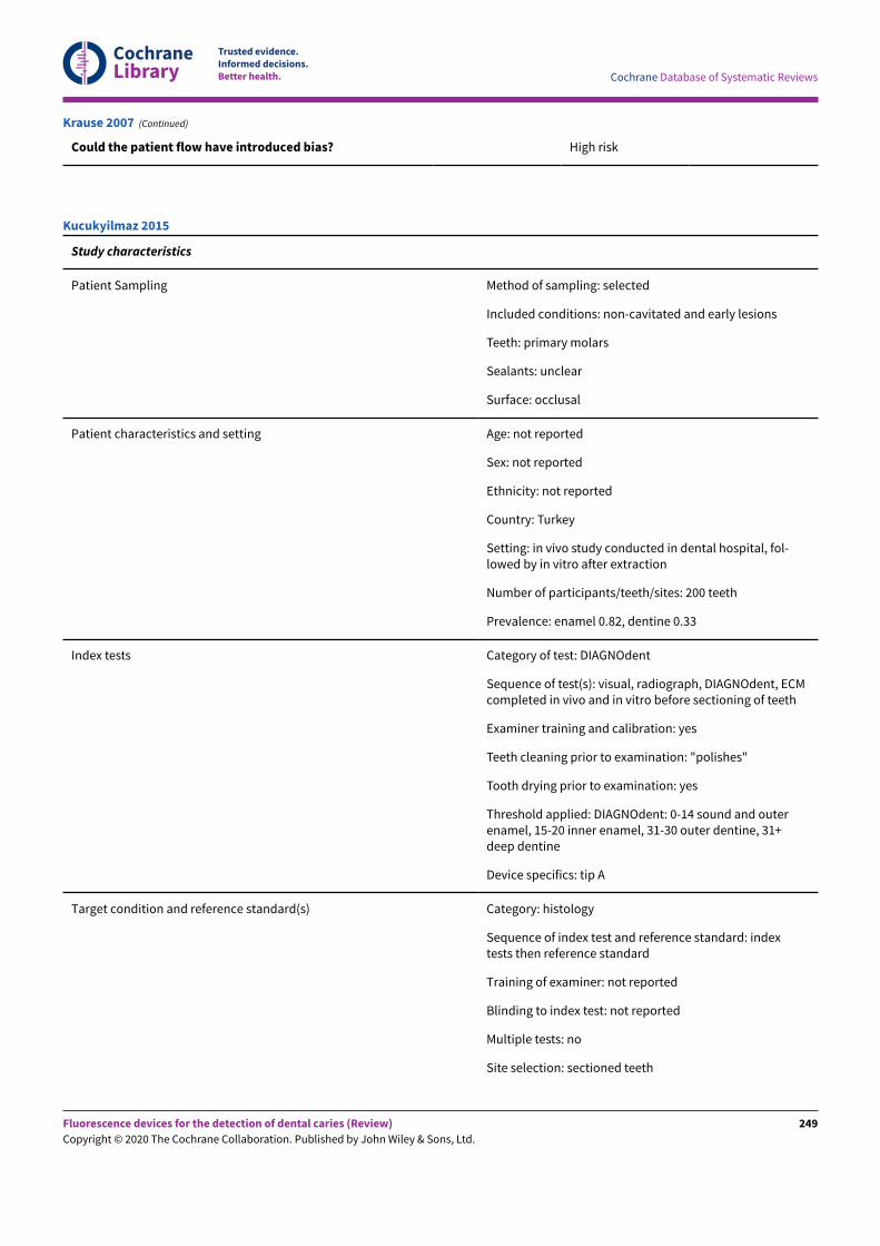
CochraneLibrary
Trusted evidence.Informed decisions.Better health.
Cochrane Database of Systematic Reviews
Could the patient flow have introduced bias? High risk
Krause 2007 (Continued)
Study characteristics
Patient Sampling Method of sampling: selected
Included conditions: non-cavitated and early lesions
Teeth: primary molars
Sealants: unclear
Surface: occlusal
Patient characteristics and setting Age: not reported
Sex: not reported
Ethnicity: not reported
Country: Turkey
Setting: in vivo study conducted in dental hospital, fol-lowed by in vitro after extraction
Number of participants/teeth/sites: 200 teeth
Prevalence: enamel 0.82, dentine 0.33
Index tests Category of test: DIAGNOdent
Sequence of test(s): visual, radiograph, DIAGNOdent, ECMcompleted in vivo and in vitro before sectioning of teeth
Examiner training and calibration: yes
Teeth cleaning prior to examination: "polishes"
Tooth drying prior to examination: yes
Threshold applied: DIAGNOdent: 0-14 sound and outerenamel, 15-20 inner enamel, 31-30 outer dentine, 31+deep dentine
Device specifics: tip A
Target condition and reference standard(s) Category: histology
Sequence of index test and reference standard: indextests then reference standard
Training of examiner: not reported
Blinding to index test: not reported
Multiple tests: no
Site selection: sectioned teeth
Kucukyilmaz 2015
Fluorescence devices for the detection of dental caries (Review)
Copyright © 2020 The Cochrane Collaboration. Published by John Wiley & Sons, Ltd.
249
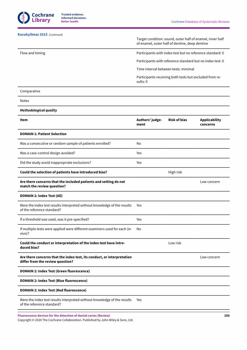
CochraneLibrary
Trusted evidence.Informed decisions.Better health.
Cochrane Database of Systematic Reviews
Target condition: sound, outer half of enamel, inner halfof enamel, outer half of dentine, deep dentine
Flow and timing Participants with index test but no reference standard: 0
Participants with reference standard but no index test: 0
Time interval between tests: minimal
Participants receiving both tests but excluded from re-sults: 0
Comparative
Notes
Methodological quality
Item Authors' judge-ment
Risk of bias Applicabilityconcerns
DOMAIN 1: Patient Selection
Was a consecutive or random sample of patients enrolled? No
Was a case-control design avoided? Yes
Did the study avoid inappropriate exclusions? Yes
Could the selection of patients have introduced bias? High risk
Are there concerns that the included patients and setting do notmatch the review question?
Low concern
DOMAIN 2: Index Test (All)
Were the index test results interpreted without knowledge of the resultsof the reference standard?
Yes
If a threshold was used, was it pre-specified? Yes
If multiple tests were applied were different examiners used for each (invivo)?
No
Could the conduct or interpretation of the index test have intro-duced bias?
Low risk
Are there concerns that the index test, its conduct, or interpretationdiffer from the review question?
Low concern
DOMAIN 2: Index Test (Green fluorescence)
DOMAIN 2: Index Test (Blue fluorescence)
DOMAIN 2: Index Test (Red fluorescence)
Were the index test results interpreted without knowledge of the resultsof the reference standard?
Yes
Kucukyilmaz 2015 (Continued)
Fluorescence devices for the detection of dental caries (Review)
Copyright © 2020 The Cochrane Collaboration. Published by John Wiley & Sons, Ltd.
250
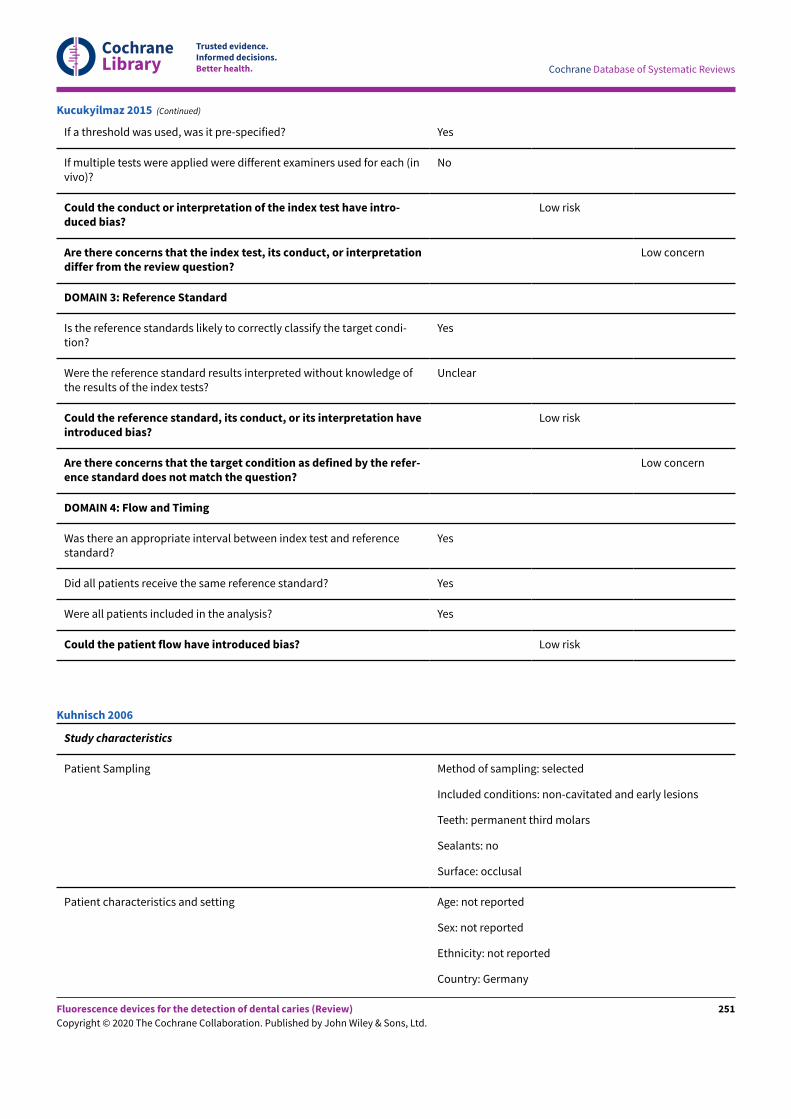
CochraneLibrary
Trusted evidence.Informed decisions.Better health.
Cochrane Database of Systematic Reviews
If a threshold was used, was it pre-specified? Yes
If multiple tests were applied were different examiners used for each (invivo)?
No
Could the conduct or interpretation of the index test have intro-duced bias?
Low risk
Are there concerns that the index test, its conduct, or interpretationdiffer from the review question?
Low concern
DOMAIN 3: Reference Standard
Is the reference standards likely to correctly classify the target condi-tion?
Yes
Were the reference standard results interpreted without knowledge ofthe results of the index tests?
Unclear
Could the reference standard, its conduct, or its interpretation haveintroduced bias?
Low risk
Are there concerns that the target condition as defined by the refer-ence standard does not match the question?
Low concern
DOMAIN 4: Flow and Timing
Was there an appropriate interval between index test and referencestandard?
Yes
Did all patients receive the same reference standard? Yes
Were all patients included in the analysis? Yes
Could the patient flow have introduced bias? Low risk
Kucukyilmaz 2015 (Continued)
Study characteristics
Patient Sampling Method of sampling: selected
Included conditions: non-cavitated and early lesions
Teeth: permanent third molars
Sealants: no
Surface: occlusal
Patient characteristics and setting Age: not reported
Sex: not reported
Ethnicity: not reported
Country: Germany
Kuhnisch 2006
Fluorescence devices for the detection of dental caries (Review)
Copyright © 2020 The Cochrane Collaboration. Published by John Wiley & Sons, Ltd.
251
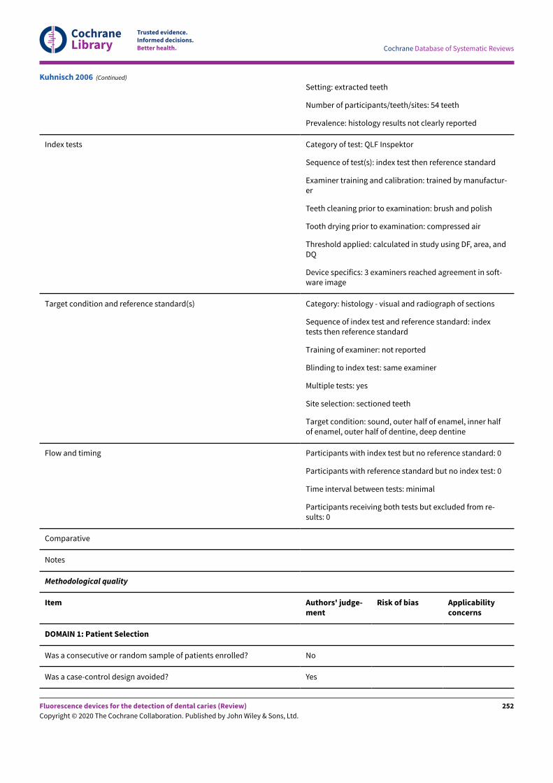
CochraneLibrary
Trusted evidence.Informed decisions.Better health.
Cochrane Database of Systematic Reviews
Setting: extracted teeth
Number of participants/teeth/sites: 54 teeth
Prevalence: histology results not clearly reported
Index tests Category of test: QLF Inspektor
Sequence of test(s): index test then reference standard
Examiner training and calibration: trained by manufactur-er
Teeth cleaning prior to examination: brush and polish
Tooth drying prior to examination: compressed air
Threshold applied: calculated in study using DF, area, andDQ
Device specifics: 3 examiners reached agreement in soft-ware image
Target condition and reference standard(s) Category: histology - visual and radiograph of sections
Sequence of index test and reference standard: indextests then reference standard
Training of examiner: not reported
Blinding to index test: same examiner
Multiple tests: yes
Site selection: sectioned teeth
Target condition: sound, outer half of enamel, inner halfof enamel, outer half of dentine, deep dentine
Flow and timing Participants with index test but no reference standard: 0
Participants with reference standard but no index test: 0
Time interval between tests: minimal
Participants receiving both tests but excluded from re-sults: 0
Comparative
Notes
Methodological quality
Item Authors' judge-ment
Risk of bias Applicabilityconcerns
DOMAIN 1: Patient Selection
Was a consecutive or random sample of patients enrolled? No
Was a case-control design avoided? Yes
Kuhnisch 2006 (Continued)
Fluorescence devices for the detection of dental caries (Review)
Copyright © 2020 The Cochrane Collaboration. Published by John Wiley & Sons, Ltd.
252
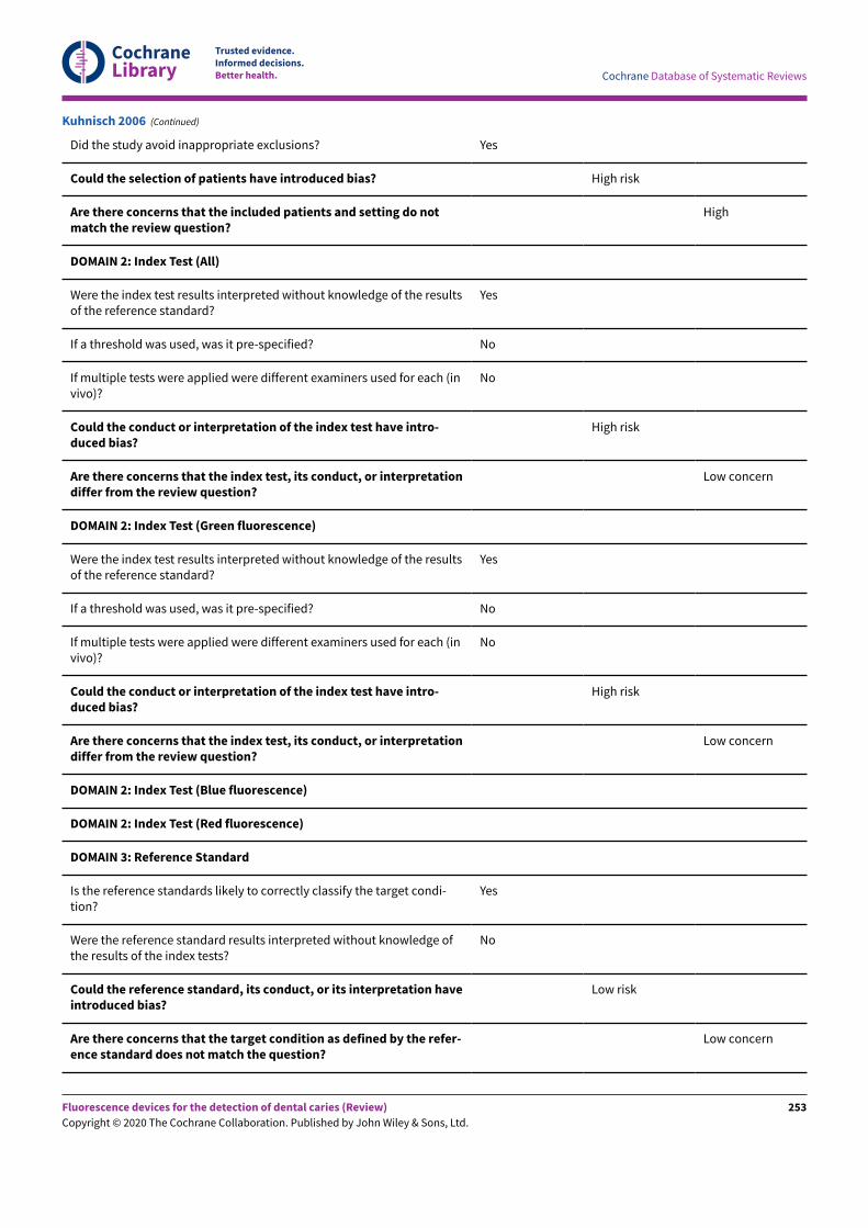
CochraneLibrary
Trusted evidence.Informed decisions.Better health.
Cochrane Database of Systematic Reviews
Did the study avoid inappropriate exclusions? Yes
Could the selection of patients have introduced bias? High risk
Are there concerns that the included patients and setting do notmatch the review question?
High
DOMAIN 2: Index Test (All)
Were the index test results interpreted without knowledge of the resultsof the reference standard?
Yes
If a threshold was used, was it pre-specified? No
If multiple tests were applied were different examiners used for each (invivo)?
No
Could the conduct or interpretation of the index test have intro-duced bias?
High risk
Are there concerns that the index test, its conduct, or interpretationdiffer from the review question?
Low concern
DOMAIN 2: Index Test (Green fluorescence)
Were the index test results interpreted without knowledge of the resultsof the reference standard?
Yes
If a threshold was used, was it pre-specified? No
If multiple tests were applied were different examiners used for each (invivo)?
No
Could the conduct or interpretation of the index test have intro-duced bias?
High risk
Are there concerns that the index test, its conduct, or interpretationdiffer from the review question?
Low concern
DOMAIN 2: Index Test (Blue fluorescence)
DOMAIN 2: Index Test (Red fluorescence)
DOMAIN 3: Reference Standard
Is the reference standards likely to correctly classify the target condi-tion?
Yes
Were the reference standard results interpreted without knowledge ofthe results of the index tests?
No
Could the reference standard, its conduct, or its interpretation haveintroduced bias?
Low risk
Are there concerns that the target condition as defined by the refer-ence standard does not match the question?
Low concern
Kuhnisch 2006 (Continued)
Fluorescence devices for the detection of dental caries (Review)
Copyright © 2020 The Cochrane Collaboration. Published by John Wiley & Sons, Ltd.
253
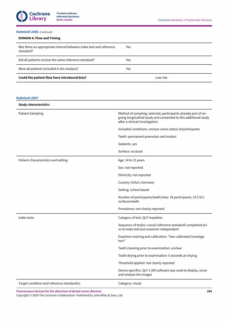
CochraneLibrary
Trusted evidence.Informed decisions.Better health.
Cochrane Database of Systematic Reviews
DOMAIN 4: Flow and Timing
Was there an appropriate interval between index test and referencestandard?
Yes
Did all patients receive the same reference standard? Yes
Were all patients included in the analysis? Yes
Could the patient flow have introduced bias? Low risk
Kuhnisch 2006 (Continued)
Study characteristics
Patient Sampling Method of sampling: selected, participants already part of on-going longitudinal study and consented to this additional studyafter a clinical investigation
Included conditions: unclear caries status of participants
Teeth: permanent premolars and molars
Sealants: yes
Surface: occlusal
Patient characteristics and setting Age: 14 to 15 years
Sex: not reported
Ethnicity: not reported
Country: Erfurt, Germany
Setting: school based
Number of participants/teeth/sites: 34 participants, 517/311surfaces/teeth
Prevalence: not clearly reported
Index tests Category of test: QLF Inspektor
Sequence of test(s): visual (reference standard) completed pri-or to index test but examiner independent
Examiner training and calibration: "two calibrated investiga-tors"
Teeth cleaning prior to examination: unclear
Tooth drying prior to examination: 5 seconds air drying
Threshold applied: not clearly reported
Device specifics: QLF 2.00f software was used to display, scoreand analyse the images
Target condition and reference standard(s) Category: visual
Kuhnisch 2007
Fluorescence devices for the detection of dental caries (Review)
Copyright © 2020 The Cochrane Collaboration. Published by John Wiley & Sons, Ltd.
254
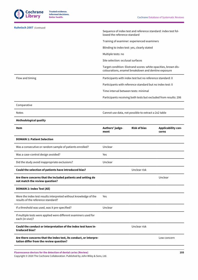
CochraneLibrary
Trusted evidence.Informed decisions.Better health.
Cochrane Database of Systematic Reviews
Sequence of index test and reference standard: index test fol-lowed the reference standard
Training of examiner: experienced examiners
Blinding to index test: yes, clearly stated
Multiple tests: no
Site selection: occlusal surfaces
Target condition: Ekstrand scores: white opacities, brown dis-colourations, enamel breakdown and dentine exposure
Flow and timing Participants with index test but no reference standard: 0
Participants with reference standard but no index test: 0
Time interval between tests: minimal
Participants receiving both tests but excluded from results: 206
Comparative
Notes Cannot use data, not possible to extract a 2x2 table
Methodological quality
Item Authors' judge-ment
Risk of bias Applicability con-cerns
DOMAIN 1: Patient Selection
Was a consecutive or random sample of patients enrolled? Unclear
Was a case-control design avoided? Yes
Did the study avoid inappropriate exclusions? Unclear
Could the selection of patients have introduced bias? Unclear risk
Are there concerns that the included patients and setting donot match the review question?
Unclear
DOMAIN 2: Index Test (All)
Were the index test results interpreted without knowledge of theresults of the reference standard?
Yes
If a threshold was used, was it pre-specified? Unclear
If multiple tests were applied were different examiners used foreach (in vivo)?
Could the conduct or interpretation of the index test have in-troduced bias?
Unclear risk
Are there concerns that the index test, its conduct, or interpre-tation differ from the review question?
Low concern
Kuhnisch 2007 (Continued)
Fluorescence devices for the detection of dental caries (Review)
Copyright © 2020 The Cochrane Collaboration. Published by John Wiley & Sons, Ltd.
255
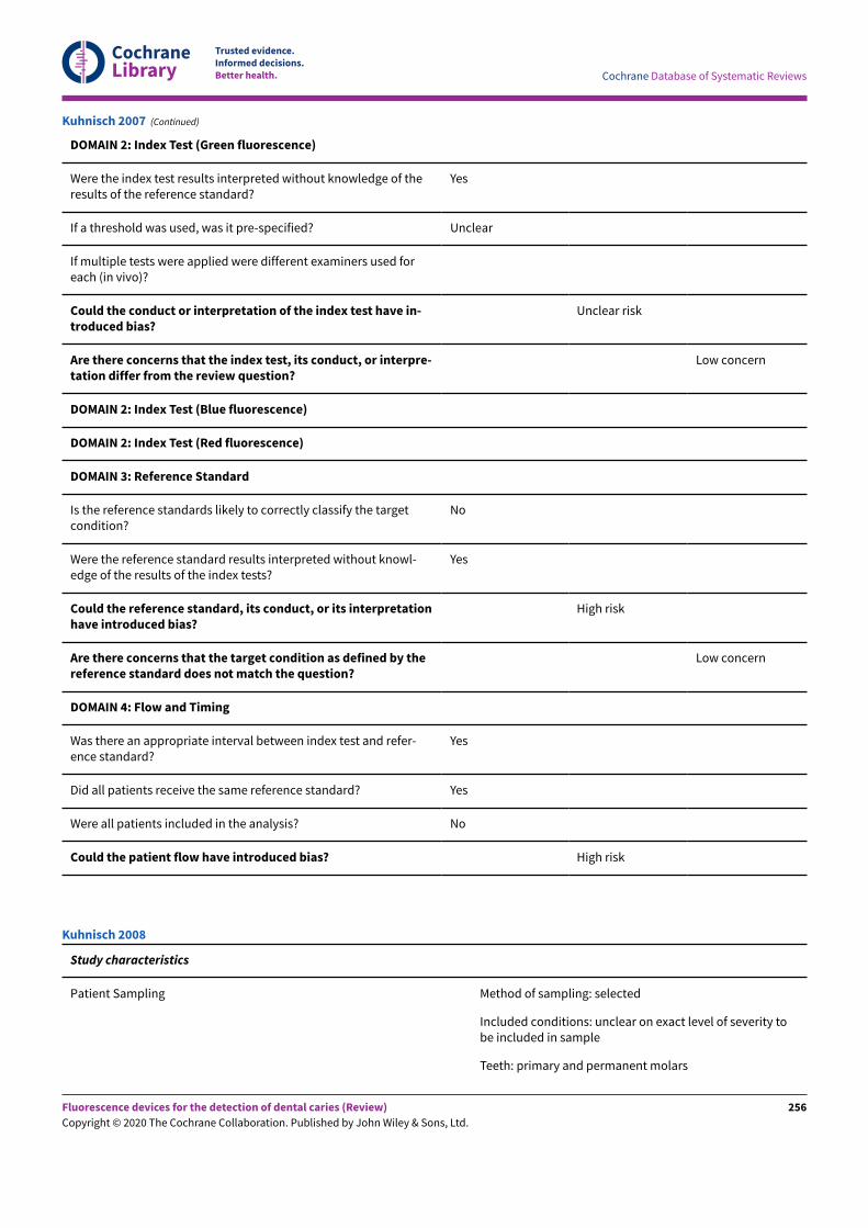
CochraneLibrary
Trusted evidence.Informed decisions.Better health.
Cochrane Database of Systematic Reviews
DOMAIN 2: Index Test (Green fluorescence)
Were the index test results interpreted without knowledge of theresults of the reference standard?
Yes
If a threshold was used, was it pre-specified? Unclear
If multiple tests were applied were different examiners used foreach (in vivo)?
Could the conduct or interpretation of the index test have in-troduced bias?
Unclear risk
Are there concerns that the index test, its conduct, or interpre-tation differ from the review question?
Low concern
DOMAIN 2: Index Test (Blue fluorescence)
DOMAIN 2: Index Test (Red fluorescence)
DOMAIN 3: Reference Standard
Is the reference standards likely to correctly classify the targetcondition?
No
Were the reference standard results interpreted without knowl-edge of the results of the index tests?
Yes
Could the reference standard, its conduct, or its interpretationhave introduced bias?
High risk
Are there concerns that the target condition as defined by thereference standard does not match the question?
Low concern
DOMAIN 4: Flow and Timing
Was there an appropriate interval between index test and refer-ence standard?
Yes
Did all patients receive the same reference standard? Yes
Were all patients included in the analysis? No
Could the patient flow have introduced bias? High risk
Kuhnisch 2007 (Continued)
Study characteristics
Patient Sampling Method of sampling: selected
Included conditions: unclear on exact level of severity tobe included in sample
Teeth: primary and permanent molars
Kuhnisch 2008
Fluorescence devices for the detection of dental caries (Review)
Copyright © 2020 The Cochrane Collaboration. Published by John Wiley & Sons, Ltd.
256
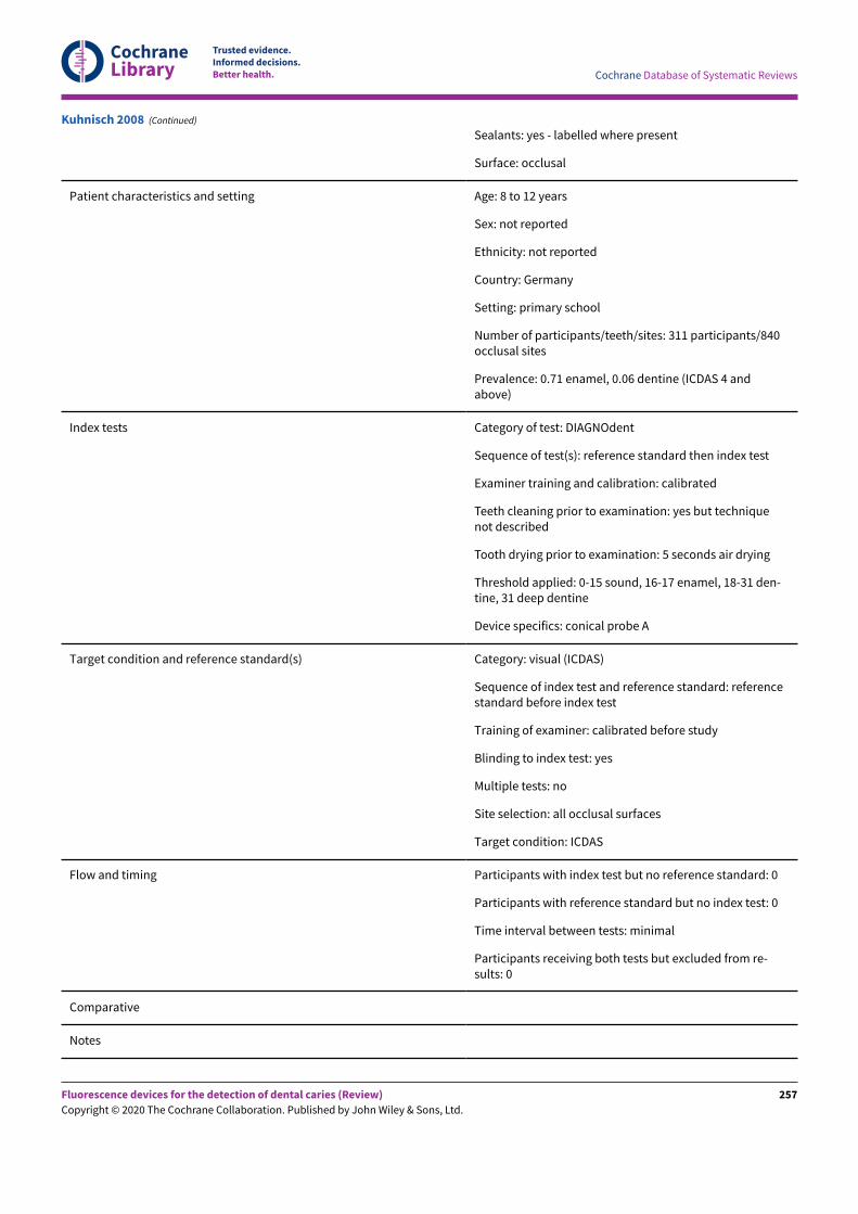
CochraneLibrary
Trusted evidence.Informed decisions.Better health.
Cochrane Database of Systematic Reviews
Sealants: yes - labelled where present
Surface: occlusal
Patient characteristics and setting Age: 8 to 12 years
Sex: not reported
Ethnicity: not reported
Country: Germany
Setting: primary school
Number of participants/teeth/sites: 311 participants/840occlusal sites
Prevalence: 0.71 enamel, 0.06 dentine (ICDAS 4 andabove)
Index tests Category of test: DIAGNOdent
Sequence of test(s): reference standard then index test
Examiner training and calibration: calibrated
Teeth cleaning prior to examination: yes but techniquenot described
Tooth drying prior to examination: 5 seconds air drying
Threshold applied: 0-15 sound, 16-17 enamel, 18-31 den-tine, 31 deep dentine
Device specifics: conical probe A
Target condition and reference standard(s) Category: visual (ICDAS)
Sequence of index test and reference standard: referencestandard before index test
Training of examiner: calibrated before study
Blinding to index test: yes
Multiple tests: no
Site selection: all occlusal surfaces
Target condition: ICDAS
Flow and timing Participants with index test but no reference standard: 0
Participants with reference standard but no index test: 0
Time interval between tests: minimal
Participants receiving both tests but excluded from re-sults: 0
Comparative
Notes
Kuhnisch 2008 (Continued)
Fluorescence devices for the detection of dental caries (Review)
Copyright © 2020 The Cochrane Collaboration. Published by John Wiley & Sons, Ltd.
257
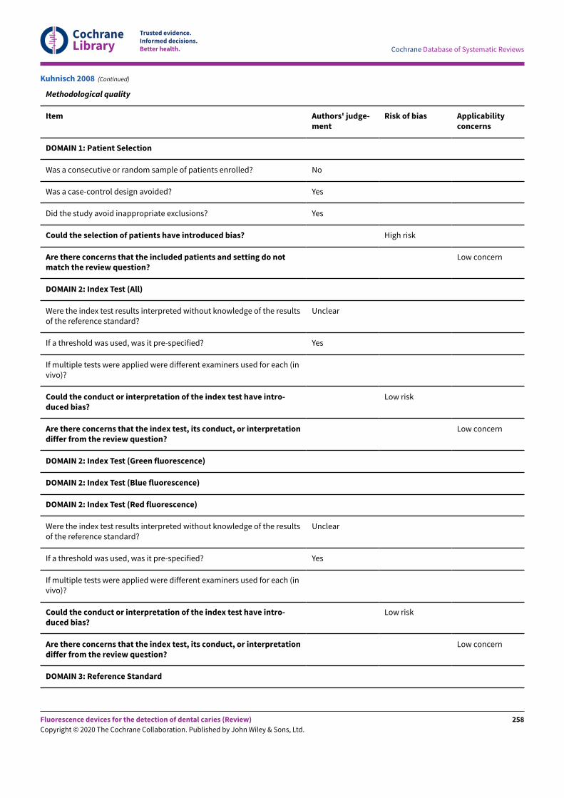
CochraneLibrary
Trusted evidence.Informed decisions.Better health.
Cochrane Database of Systematic Reviews
Methodological quality
Item Authors' judge-ment
Risk of bias Applicabilityconcerns
DOMAIN 1: Patient Selection
Was a consecutive or random sample of patients enrolled? No
Was a case-control design avoided? Yes
Did the study avoid inappropriate exclusions? Yes
Could the selection of patients have introduced bias? High risk
Are there concerns that the included patients and setting do notmatch the review question?
Low concern
DOMAIN 2: Index Test (All)
Were the index test results interpreted without knowledge of the resultsof the reference standard?
Unclear
If a threshold was used, was it pre-specified? Yes
If multiple tests were applied were different examiners used for each (invivo)?
Could the conduct or interpretation of the index test have intro-duced bias?
Low risk
Are there concerns that the index test, its conduct, or interpretationdiffer from the review question?
Low concern
DOMAIN 2: Index Test (Green fluorescence)
DOMAIN 2: Index Test (Blue fluorescence)
DOMAIN 2: Index Test (Red fluorescence)
Were the index test results interpreted without knowledge of the resultsof the reference standard?
Unclear
If a threshold was used, was it pre-specified? Yes
If multiple tests were applied were different examiners used for each (invivo)?
Could the conduct or interpretation of the index test have intro-duced bias?
Low risk
Are there concerns that the index test, its conduct, or interpretationdiffer from the review question?
Low concern
DOMAIN 3: Reference Standard
Kuhnisch 2008 (Continued)
Fluorescence devices for the detection of dental caries (Review)
Copyright © 2020 The Cochrane Collaboration. Published by John Wiley & Sons, Ltd.
258
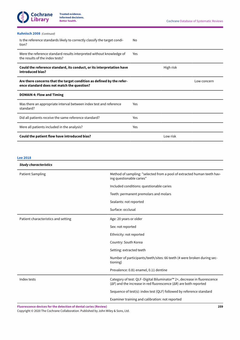
CochraneLibrary
Trusted evidence.Informed decisions.Better health.
Cochrane Database of Systematic Reviews
Is the reference standards likely to correctly classify the target condi-tion?
No
Were the reference standard results interpreted without knowledge ofthe results of the index tests?
Yes
Could the reference standard, its conduct, or its interpretation haveintroduced bias?
High risk
Are there concerns that the target condition as defined by the refer-ence standard does not match the question?
Low concern
DOMAIN 4: Flow and Timing
Was there an appropriate interval between index test and referencestandard?
Yes
Did all patients receive the same reference standard? Yes
Were all patients included in the analysis? Yes
Could the patient flow have introduced bias? Low risk
Kuhnisch 2008 (Continued)
Study characteristics
Patient Sampling Method of sampling: "selected from a pool of extracted human teeth hav-ing questionable caries"
Included conditions: questionable caries
Teeth: permanent premolars and molars
Sealants: not reported
Surface: occlusal
Patient characteristics and setting Age: 20 years or older
Sex: not reported
Ethnicity: not reported
Country: South Korea
Setting: extracted teeth
Number of participants/teeth/sites: 66 teeth (4 were broken during sec-tioning)
Prevalence: 0.81 enamel, 0.11 dentine
Index tests Category of test: QLF–Digital Biluminator™ 2+, decrease in fluorescence(ΔF) and the increase in red fluorescence (ΔR) are both reported
Sequence of test(s): index test (QLF) followed by reference standard
Examiner training and calibration: not reported
Lee 2018
Fluorescence devices for the detection of dental caries (Review)
Copyright © 2020 The Cochrane Collaboration. Published by John Wiley & Sons, Ltd.
259
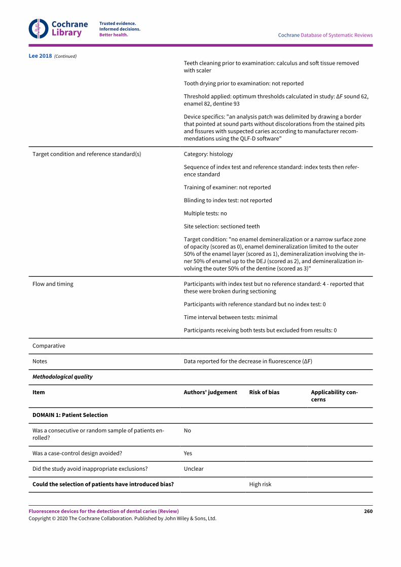
CochraneLibrary
Trusted evidence.Informed decisions.Better health.
Cochrane Database of Systematic Reviews
Teeth cleaning prior to examination: calculus and soN tissue removedwith scaler
Tooth drying prior to examination: not reported
Threshold applied: optimum thresholds calculated in study: ΔF sound 62,enamel 82, dentine 93
Device specifics: "an analysis patch was delimited by drawing a borderthat pointed at sound parts without discolorations from the stained pitsand fissures with suspected caries according to manufacturer recom-mendations using the QLF-D software"
Target condition and reference standard(s) Category: histology
Sequence of index test and reference standard: index tests then refer-ence standard
Training of examiner: not reported
Blinding to index test: not reported
Multiple tests: no
Site selection: sectioned teeth
Target condition: "no enamel demineralization or a narrow surface zoneof opacity (scored as 0), enamel demineralization limited to the outer50% of the enamel layer (scored as 1), demineralization involving the in-ner 50% of enamel up to the DEJ (scored as 2), and demineralization in-volving the outer 50% of the dentine (scored as 3)"
Flow and timing Participants with index test but no reference standard: 4 - reported thatthese were broken during sectioning
Participants with reference standard but no index test: 0
Time interval between tests: minimal
Participants receiving both tests but excluded from results: 0
Comparative
Notes Data reported for the decrease in fluorescence (ΔF)
Methodological quality
Item Authors' judgement Risk of bias Applicability con-cerns
DOMAIN 1: Patient Selection
Was a consecutive or random sample of patients en-rolled?
No
Was a case-control design avoided? Yes
Did the study avoid inappropriate exclusions? Unclear
Could the selection of patients have introduced bias? High risk
Lee 2018 (Continued)
Fluorescence devices for the detection of dental caries (Review)
Copyright © 2020 The Cochrane Collaboration. Published by John Wiley & Sons, Ltd.
260
CochraneLibrary
Trusted evidence.Informed decisions.Better health.
Cochrane Database of Systematic Reviews
Are there concerns that the included patients and set-ting do not match the review question?
Low concern
DOMAIN 2: Index Test (All)
Were the index test results interpreted without knowl-edge of the results of the reference standard?
Yes
If a threshold was used, was it pre-specified? No
If multiple tests were applied were different examinersused for each (in vivo)?
Unclear
Could the conduct or interpretation of the index testhave introduced bias?
High risk
Are there concerns that the index test, its conduct, orinterpretation differ from the review question?
Low concern
DOMAIN 2: Index Test (Green fluorescence)
Were the index test results interpreted without knowl-edge of the results of the reference standard?
Yes
If a threshold was used, was it pre-specified? No
If multiple tests were applied were different examinersused for each (in vivo)?
Unclear
Could the conduct or interpretation of the index testhave introduced bias?
High risk
Are there concerns that the index test, its conduct, orinterpretation differ from the review question?
Low concern
DOMAIN 2: Index Test (Blue fluorescence)
DOMAIN 2: Index Test (Red fluorescence)
DOMAIN 3: Reference Standard
Is the reference standards likely to correctly classify thetarget condition?
Yes
Were the reference standard results interpreted withoutknowledge of the results of the index tests?
Yes
Could the reference standard, its conduct, or its inter-pretation have introduced bias?
Low risk
Are there concerns that the target condition as de-fined by the reference standard does not match thequestion?
Low concern
DOMAIN 4: Flow and Timing
Lee 2018 (Continued)
Fluorescence devices for the detection of dental caries (Review)
Copyright © 2020 The Cochrane Collaboration. Published by John Wiley & Sons, Ltd.
261
CochraneLibrary
Trusted evidence.Informed decisions.Better health.
Cochrane Database of Systematic Reviews
Was there an appropriate interval between index testand reference standard?
Yes
Did all patients receive the same reference standard? Yes
Were all patients included in the analysis? No
Could the patient flow have introduced bias? Low risk
Lee 2018 (Continued)
Study characteristics
Patient Sampling Method of sampling: unclear
Included conditions: non-cavitated
Teeth: primary molars
Sealants: no
Surface: occlusal
Patient characteristics and setting Age: 5 to 6 years, mean 5.3
Sex: not reported
Ethnicity: not reported
Country: China
Setting: school based (kindergarten)
Number of participants/teeth/sites: 72 participants/541 teeth
Prevalence: not reported
Index tests Category of test: DIAGNOdent
Sequence of test(s): DIAGNOdent then visual
Examiner training and calibration: not reported
Teeth cleaning prior to examination: yes, with a portable low-speed hand-piece brush
Tooth drying prior to examination: yes, dried with high pres-sure air (triple syringe)
Threshold applied: < 10 intact, 10-14 early enamel caries,15-20 enamel caries, 21-30 early dentine caries, > = 31 deepdentine caries
Device specifics: tip A
Target condition and reference standard(s) Category: visual
Sequence of index test and reference standard: following butinfluenced by DIAGNOdent
Training of examiner: not reported
Li 2006
Fluorescence devices for the detection of dental caries (Review)
Copyright © 2020 The Cochrane Collaboration. Published by John Wiley & Sons, Ltd.
262
CochraneLibrary
Trusted evidence.Informed decisions.Better health.
Cochrane Database of Systematic Reviews
Blinding to index test: no "visual examination was performedon occlusal spots that had the highest laser fluorescencescores during DIAGNOdent examination"
Multiple tests: no
Site selection: sectioned teeth
Target condition: Caries (Ekstrand’s index)
Flow and timing Participants with index test but no reference standard: 0
Participants with reference standard but no index test: 0
Time interval between tests: minimal
Participants receiving both tests but excluded from results: 0
Comparative
Notes Paper translated by Cochrane author, data not useable as 2x2table not attainable, study investigates median DIAGNOdentvalues at each Ekstrand code
Methodological quality
Item Authors' judge-ment
Risk of bias Applicability con-cerns
DOMAIN 1: Patient Selection
Was a consecutive or random sample of patients enrolled? Unclear
Was a case-control design avoided? Yes
Did the study avoid inappropriate exclusions? Unclear
Could the selection of patients have introduced bias? Unclear risk
Are there concerns that the included patients and setting do notmatch the review question?
Low concern
DOMAIN 2: Index Test (All)
Were the index test results interpreted without knowledge of the re-sults of the reference standard?
Yes
If a threshold was used, was it pre-specified? Yes
If multiple tests were applied were different examiners used foreach (in vivo)?
Could the conduct or interpretation of the index test have intro-duced bias?
Low risk
Are there concerns that the index test, its conduct, or interpre-tation differ from the review question?
Low concern
DOMAIN 2: Index Test (Green fluorescence)
Li 2006 (Continued)
Fluorescence devices for the detection of dental caries (Review)
Copyright © 2020 The Cochrane Collaboration. Published by John Wiley & Sons, Ltd.
263
CochraneLibrary
Trusted evidence.Informed decisions.Better health.
Cochrane Database of Systematic Reviews
DOMAIN 2: Index Test (Blue fluorescence)
DOMAIN 2: Index Test (Red fluorescence)
Were the index test results interpreted without knowledge of the re-sults of the reference standard?
Yes
If a threshold was used, was it pre-specified? Yes
If multiple tests were applied were different examiners used foreach (in vivo)?
Could the conduct or interpretation of the index test have intro-duced bias?
Low risk
Are there concerns that the index test, its conduct, or interpre-tation differ from the review question?
Low concern
DOMAIN 3: Reference Standard
Is the reference standards likely to correctly classify the target con-dition?
No
Were the reference standard results interpreted without knowledgeof the results of the index tests?
No
Could the reference standard, its conduct, or its interpretationhave introduced bias?
High risk
Are there concerns that the target condition as defined by thereference standard does not match the question?
Low concern
DOMAIN 4: Flow and Timing
Was there an appropriate interval between index test and referencestandard?
Yes
Did all patients receive the same reference standard? Yes
Were all patients included in the analysis? Yes
Could the patient flow have introduced bias? Low risk
Li 2006 (Continued)
Study characteristics
Patient Sampling Method of sampling: selected
Included conditions: no cavitation, "All teeth had a macro-scopically intact occlusal surface"
Teeth: not reported
Sealants: not reported
Lussi 1999
Fluorescence devices for the detection of dental caries (Review)
Copyright © 2020 The Cochrane Collaboration. Published by John Wiley & Sons, Ltd.
264
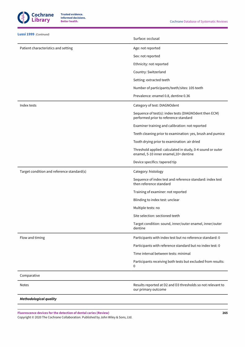
CochraneLibrary
Trusted evidence.Informed decisions.Better health.
Cochrane Database of Systematic Reviews
Surface: occlusal
Patient characteristics and setting Age: not reported
Sex: not reported
Ethnicity: not reported
Country: Switzerland
Setting: extracted teeth
Number of participants/teeth/sites: 105 teeth
Prevalence: enamel 0.8, dentine 0.36
Index tests Category of test: DIAGNOdent
Sequence of test(s): index tests (DIAGNOdent then ECM)performed prior to reference standard
Examiner training and calibration: not reported
Teeth cleaning prior to examination: yes, brush and pumice
Tooth drying prior to examination: air dried
Threshold applied: calculated in study, 0-4 sound or outerenamel, 5-10 inner enamel,10+ dentine
Device specifics: tapered tip
Target condition and reference standard(s) Category: histology
Sequence of index test and reference standard: index testthen reference standard
Training of examiner: not reported
Blinding to index test: unclear
Multiple tests: no
Site selection: sectioned teeth
Target condition: sound, inner/outer enamel, inner/outerdentine
Flow and timing Participants with index test but no reference standard: 0
Participants with reference standard but no index test: 0
Time interval between tests: minimal
Participants receiving both tests but excluded from results:0
Comparative
Notes Results reported at D2 and D3 thresholds so not relevant toour primary outcome
Methodological quality
Lussi 1999 (Continued)
Fluorescence devices for the detection of dental caries (Review)
Copyright © 2020 The Cochrane Collaboration. Published by John Wiley & Sons, Ltd.
265

CochraneLibrary
Trusted evidence.Informed decisions.Better health.
Cochrane Database of Systematic Reviews
Item Authors' judge-ment
Risk of bias Applicabilityconcerns
DOMAIN 1: Patient Selection
Was a consecutive or random sample of patients enrolled? No
Was a case-control design avoided? Yes
Did the study avoid inappropriate exclusions? Yes
Could the selection of patients have introduced bias? High risk
Are there concerns that the included patients and setting do notmatch the review question?
High
DOMAIN 2: Index Test (All)
Were the index test results interpreted without knowledge of the re-sults of the reference standard?
Yes
If a threshold was used, was it pre-specified? No
If multiple tests were applied were different examiners used for each(in vivo)?
Unclear
Could the conduct or interpretation of the index test have intro-duced bias?
High risk
Are there concerns that the index test, its conduct, or interpreta-tion differ from the review question?
Low concern
DOMAIN 2: Index Test (Green fluorescence)
DOMAIN 2: Index Test (Blue fluorescence)
DOMAIN 2: Index Test (Red fluorescence)
Were the index test results interpreted without knowledge of the re-sults of the reference standard?
Yes
If a threshold was used, was it pre-specified? No
If multiple tests were applied were different examiners used for each(in vivo)?
Unclear
Could the conduct or interpretation of the index test have intro-duced bias?
High risk
Are there concerns that the index test, its conduct, or interpreta-tion differ from the review question?
Low concern
DOMAIN 3: Reference Standard
Is the reference standards likely to correctly classify the target condi-tion?
Yes
Lussi 1999 (Continued)
Fluorescence devices for the detection of dental caries (Review)
Copyright © 2020 The Cochrane Collaboration. Published by John Wiley & Sons, Ltd.
266
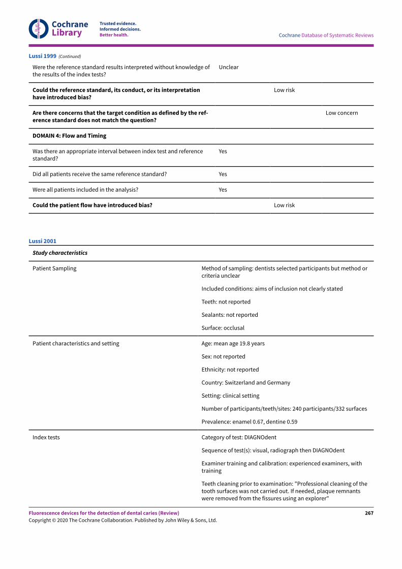
CochraneLibrary
Trusted evidence.Informed decisions.Better health.
Cochrane Database of Systematic Reviews
Were the reference standard results interpreted without knowledge ofthe results of the index tests?
Unclear
Could the reference standard, its conduct, or its interpretationhave introduced bias?
Low risk
Are there concerns that the target condition as defined by the ref-erence standard does not match the question?
Low concern
DOMAIN 4: Flow and Timing
Was there an appropriate interval between index test and referencestandard?
Yes
Did all patients receive the same reference standard? Yes
Were all patients included in the analysis? Yes
Could the patient flow have introduced bias? Low risk
Lussi 1999 (Continued)
Study characteristics
Patient Sampling Method of sampling: dentists selected participants but method orcriteria unclear
Included conditions: aims of inclusion not clearly stated
Teeth: not reported
Sealants: not reported
Surface: occlusal
Patient characteristics and setting Age: mean age 19.8 years
Sex: not reported
Ethnicity: not reported
Country: Switzerland and Germany
Setting: clinical setting
Number of participants/teeth/sites: 240 participants/332 surfaces
Prevalence: enamel 0.67, dentine 0.59
Index tests Category of test: DIAGNOdent
Sequence of test(s): visual, radiograph then DIAGNOdent
Examiner training and calibration: experienced examiners, withtraining
Teeth cleaning prior to examination: "Professional cleaning of thetooth surfaces was not carried out. If needed, plaque remnantswere removed from the fissures using an explorer"
Lussi 2001
Fluorescence devices for the detection of dental caries (Review)
Copyright © 2020 The Cochrane Collaboration. Published by John Wiley & Sons, Ltd.
267
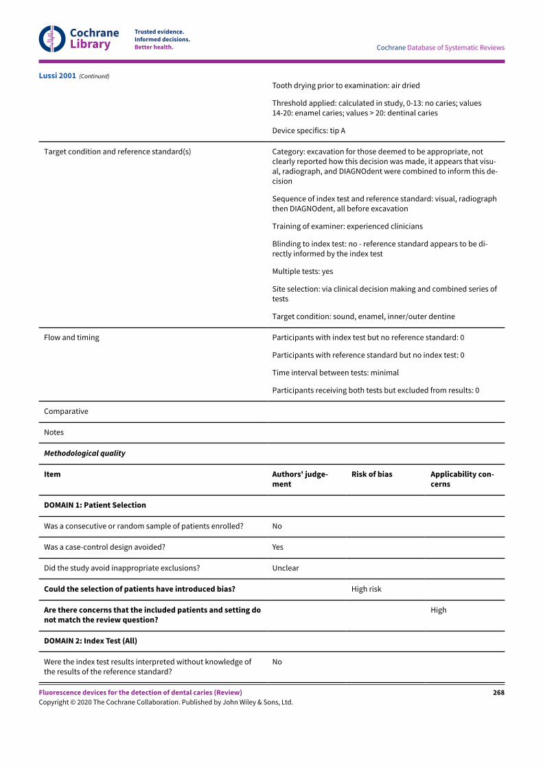
CochraneLibrary
Trusted evidence.Informed decisions.Better health.
Cochrane Database of Systematic Reviews
Tooth drying prior to examination: air dried
Threshold applied: calculated in study, 0-13: no caries; values14-20: enamel caries; values > 20: dentinal caries
Device specifics: tip A
Target condition and reference standard(s) Category: excavation for those deemed to be appropriate, notclearly reported how this decision was made, it appears that visu-al, radiograph, and DIAGNOdent were combined to inform this de-cision
Sequence of index test and reference standard: visual, radiographthen DIAGNOdent, all before excavation
Training of examiner: experienced clinicians
Blinding to index test: no - reference standard appears to be di-rectly informed by the index test
Multiple tests: yes
Site selection: via clinical decision making and combined series oftests
Target condition: sound, enamel, inner/outer dentine
Flow and timing Participants with index test but no reference standard: 0
Participants with reference standard but no index test: 0
Time interval between tests: minimal
Participants receiving both tests but excluded from results: 0
Comparative
Notes
Methodological quality
Item Authors' judge-ment
Risk of bias Applicability con-cerns
DOMAIN 1: Patient Selection
Was a consecutive or random sample of patients enrolled? No
Was a case-control design avoided? Yes
Did the study avoid inappropriate exclusions? Unclear
Could the selection of patients have introduced bias? High risk
Are there concerns that the included patients and setting donot match the review question?
High
DOMAIN 2: Index Test (All)
Were the index test results interpreted without knowledge ofthe results of the reference standard?
No
Lussi 2001 (Continued)
Fluorescence devices for the detection of dental caries (Review)
Copyright © 2020 The Cochrane Collaboration. Published by John Wiley & Sons, Ltd.
268
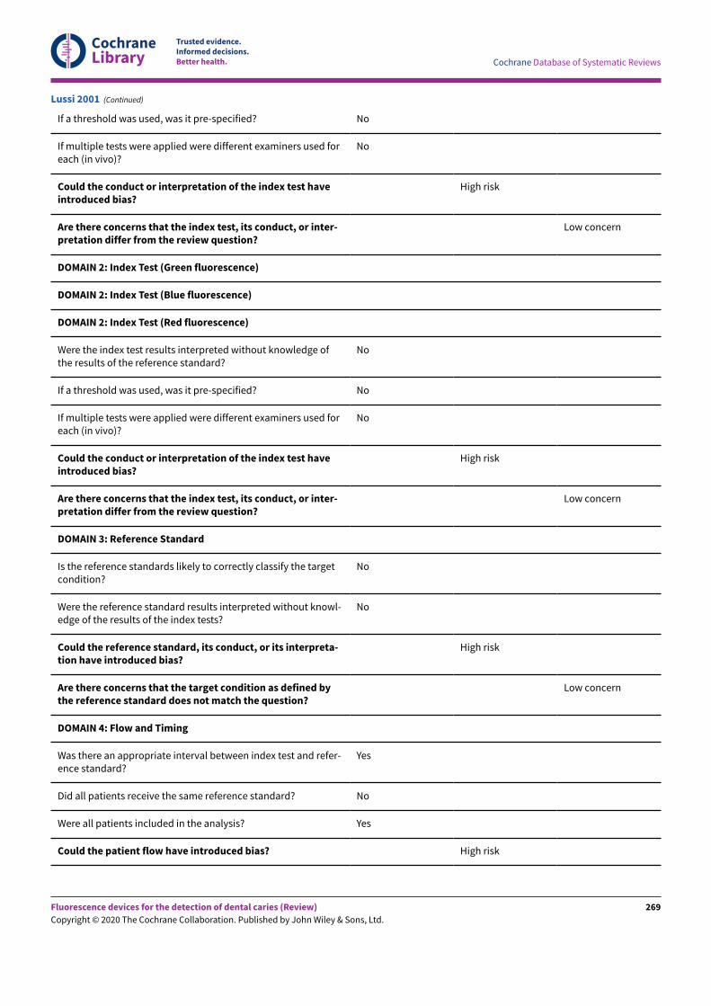
CochraneLibrary
Trusted evidence.Informed decisions.Better health.
Cochrane Database of Systematic Reviews
If a threshold was used, was it pre-specified? No
If multiple tests were applied were different examiners used foreach (in vivo)?
No
Could the conduct or interpretation of the index test haveintroduced bias?
High risk
Are there concerns that the index test, its conduct, or inter-pretation differ from the review question?
Low concern
DOMAIN 2: Index Test (Green fluorescence)
DOMAIN 2: Index Test (Blue fluorescence)
DOMAIN 2: Index Test (Red fluorescence)
Were the index test results interpreted without knowledge ofthe results of the reference standard?
No
If a threshold was used, was it pre-specified? No
If multiple tests were applied were different examiners used foreach (in vivo)?
No
Could the conduct or interpretation of the index test haveintroduced bias?
High risk
Are there concerns that the index test, its conduct, or inter-pretation differ from the review question?
Low concern
DOMAIN 3: Reference Standard
Is the reference standards likely to correctly classify the targetcondition?
No
Were the reference standard results interpreted without knowl-edge of the results of the index tests?
No
Could the reference standard, its conduct, or its interpreta-tion have introduced bias?
High risk
Are there concerns that the target condition as defined bythe reference standard does not match the question?
Low concern
DOMAIN 4: Flow and Timing
Was there an appropriate interval between index test and refer-ence standard?
Yes
Did all patients receive the same reference standard? No
Were all patients included in the analysis? Yes
Could the patient flow have introduced bias? High risk
Lussi 2001 (Continued)
Fluorescence devices for the detection of dental caries (Review)
Copyright © 2020 The Cochrane Collaboration. Published by John Wiley & Sons, Ltd.
269
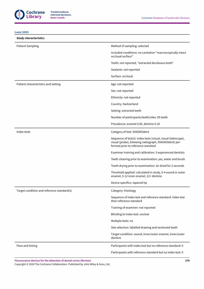
CochraneLibrary
Trusted evidence.Informed decisions.Better health.
Cochrane Database of Systematic Reviews
Study characteristics
Patient Sampling Method of sampling: selected
Included conditions: no cavitation "macroscopically intactocclusal surface"
Teeth: not reported, "extracted deciduous teeth"
Sealants: not reported
Surface: occlusal
Patient characteristics and setting Age: not reported
Sex: not reported
Ethnicity: not reported
Country: Switzerland
Setting: extracted teeth
Number of participants/teeth/sites: 95 teeth
Prevalence: enamel 0.85, dentine 0.18
Index tests Category of test: DIAGNOdent
Sequence of test(s): index tests (visual, visual (telescope),visual (probe), bitewing radiograph, DIAGNOdent) per-formed prior to reference standard
Examiner training and calibration: 3 experienced dentists
Teeth cleaning prior to examination: yes, water and brush
Tooth drying prior to examination: air dried for 2 seconds
Threshold applied: calculated in study, 0-4 sound or outerenamel, 5-12 inner enamel, 12+ dentine
Device specifics: tapered tip
Target condition and reference standard(s) Category: histology
Sequence of index test and reference standard: index testthen reference standard
Training of examiner: not reported
Blinding to index test: unclear
Multiple tests: no
Site selection: labelled drawing and sectioned teeth
Target condition: sound, inner/outer enamel, inner/outerdentine
Flow and timing Participants with index test but no reference standard: 0
Participants with reference standard but no index test: 0
Lussi 2003
Fluorescence devices for the detection of dental caries (Review)
Copyright © 2020 The Cochrane Collaboration. Published by John Wiley & Sons, Ltd.
270
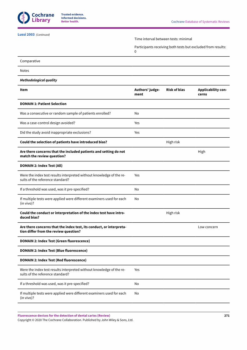
CochraneLibrary
Trusted evidence.Informed decisions.Better health.
Cochrane Database of Systematic Reviews
Time interval between tests: minimal
Participants receiving both tests but excluded from results:0
Comparative
Notes
Methodological quality
Item Authors' judge-ment
Risk of bias Applicability con-cerns
DOMAIN 1: Patient Selection
Was a consecutive or random sample of patients enrolled? No
Was a case-control design avoided? Yes
Did the study avoid inappropriate exclusions? Yes
Could the selection of patients have introduced bias? High risk
Are there concerns that the included patients and setting do notmatch the review question?
High
DOMAIN 2: Index Test (All)
Were the index test results interpreted without knowledge of the re-sults of the reference standard?
Yes
If a threshold was used, was it pre-specified? No
If multiple tests were applied were different examiners used for each(in vivo)?
No
Could the conduct or interpretation of the index test have intro-duced bias?
High risk
Are there concerns that the index test, its conduct, or interpreta-tion differ from the review question?
Low concern
DOMAIN 2: Index Test (Green fluorescence)
DOMAIN 2: Index Test (Blue fluorescence)
DOMAIN 2: Index Test (Red fluorescence)
Were the index test results interpreted without knowledge of the re-sults of the reference standard?
Yes
If a threshold was used, was it pre-specified? No
If multiple tests were applied were different examiners used for each(in vivo)?
No
Lussi 2003 (Continued)
Fluorescence devices for the detection of dental caries (Review)
Copyright © 2020 The Cochrane Collaboration. Published by John Wiley & Sons, Ltd.
271
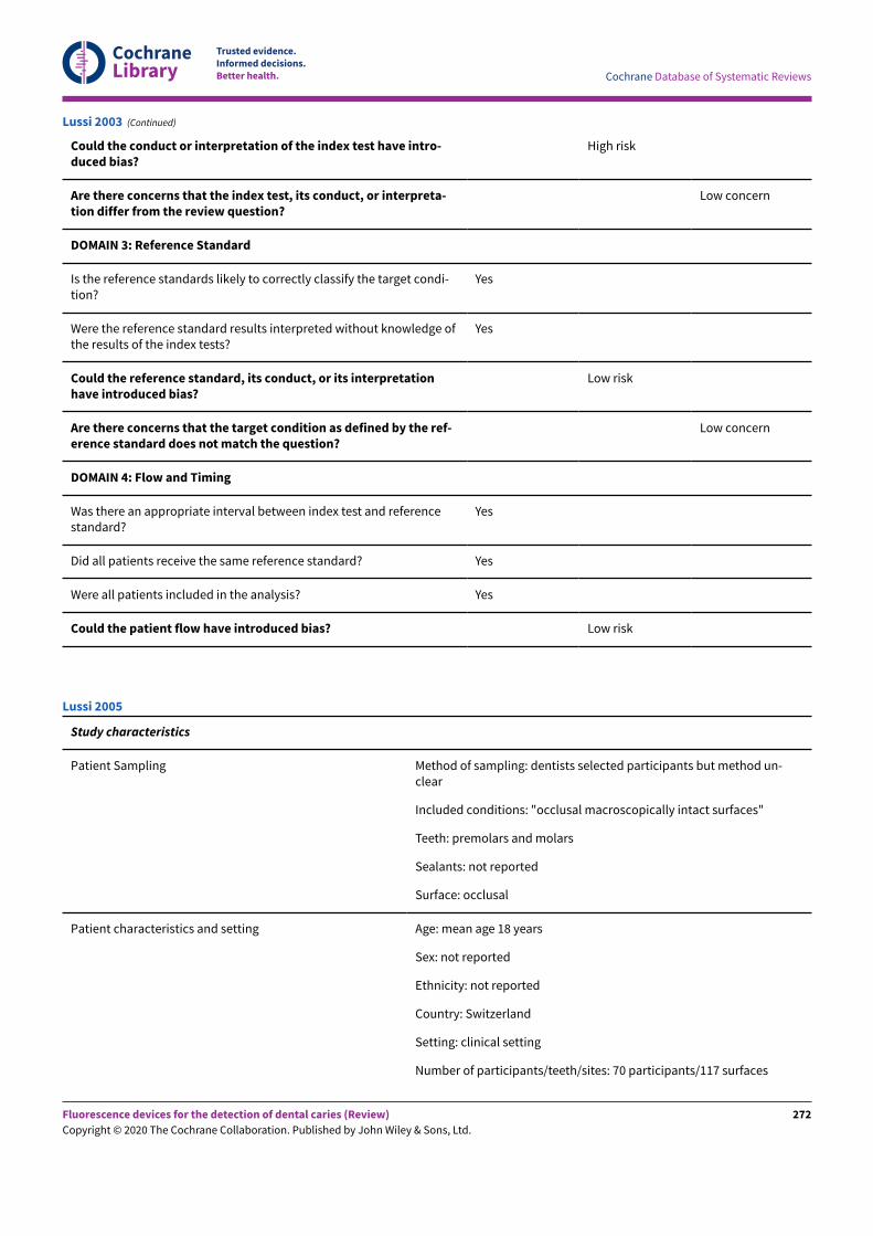
CochraneLibrary
Trusted evidence.Informed decisions.Better health.
Cochrane Database of Systematic Reviews
Could the conduct or interpretation of the index test have intro-duced bias?
High risk
Are there concerns that the index test, its conduct, or interpreta-tion differ from the review question?
Low concern
DOMAIN 3: Reference Standard
Is the reference standards likely to correctly classify the target condi-tion?
Yes
Were the reference standard results interpreted without knowledge ofthe results of the index tests?
Yes
Could the reference standard, its conduct, or its interpretationhave introduced bias?
Low risk
Are there concerns that the target condition as defined by the ref-erence standard does not match the question?
Low concern
DOMAIN 4: Flow and Timing
Was there an appropriate interval between index test and referencestandard?
Yes
Did all patients receive the same reference standard? Yes
Were all patients included in the analysis? Yes
Could the patient flow have introduced bias? Low risk
Lussi 2003 (Continued)
Study characteristics
Patient Sampling Method of sampling: dentists selected participants but method un-clear
Included conditions: "occlusal macroscopically intact surfaces"
Teeth: premolars and molars
Sealants: not reported
Surface: occlusal
Patient characteristics and setting Age: mean age 18 years
Sex: not reported
Ethnicity: not reported
Country: Switzerland
Setting: clinical setting
Number of participants/teeth/sites: 70 participants/117 surfaces
Lussi 2005
Fluorescence devices for the detection of dental caries (Review)
Copyright © 2020 The Cochrane Collaboration. Published by John Wiley & Sons, Ltd.
272
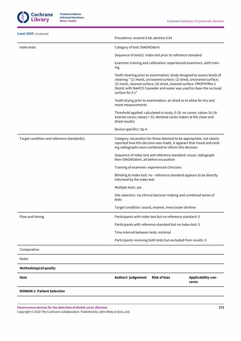
CochraneLibrary
Trusted evidence.Informed decisions.Better health.
Cochrane Database of Systematic Reviews
Prevalence: enamel 0.68, dentine 0.64
Index tests Category of test: DIAGNOdent
Sequence of test(s): index test prior to reference standard
Examiner training and calibration: experienced examiners, with train-ing
Teeth cleaning prior to examination: study designed to assess levels ofcleaning: "(1) moist, uncleaned surface; (2) dried, uncleaned surface;(3) moist, cleaned surface; (4) dried, cleaned surface. PROPHYflex 2(KaVo) with NaHCO 3 powder and water was used to clean the occlusalsurface for 5 s"
Tooth drying prior to examination: air dried as to allow for dry andmoist measurements
Threshold applied: calculated in study, 0-16: no caries; values 16-18:enamel caries; values > 32: dentinal caries (taken at the clean anddried results)
Device specifics: tip A
Target condition and reference standard(s) Category: excavation for those deemed to be appropriate, not clearlyreported how this decision was made, it appears that visual and exist-ing radiographs were combined to inform this decision
Sequence of index test and reference standard: visual, radiographthen DIAGNOdent, all before excavation
Training of examiner: experienced clinicians
Blinding to index test: no - reference standard appears to be directlyinformed by the index test
Multiple tests: yes
Site selection: via clinical decision making and combined series oftests
Target condition: sound, enamel, inner/outer dentine
Flow and timing Participants with index test but no reference standard: 0
Participants with reference standard but no index test: 0
Time interval between tests: minimal
Participants receiving both tests but excluded from results: 0
Comparative
Notes
Methodological quality
Item Authors' judgement Risk of bias Applicability con-cerns
DOMAIN 1: Patient Selection
Lussi 2005 (Continued)
Fluorescence devices for the detection of dental caries (Review)
Copyright © 2020 The Cochrane Collaboration. Published by John Wiley & Sons, Ltd.
273
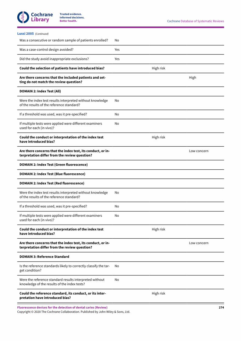
CochraneLibrary
Trusted evidence.Informed decisions.Better health.
Cochrane Database of Systematic Reviews
Was a consecutive or random sample of patients enrolled? No
Was a case-control design avoided? Yes
Did the study avoid inappropriate exclusions? Yes
Could the selection of patients have introduced bias? High risk
Are there concerns that the included patients and set-ting do not match the review question?
High
DOMAIN 2: Index Test (All)
Were the index test results interpreted without knowledgeof the results of the reference standard?
No
If a threshold was used, was it pre-specified? No
If multiple tests were applied were different examinersused for each (in vivo)?
No
Could the conduct or interpretation of the index testhave introduced bias?
High risk
Are there concerns that the index test, its conduct, or in-terpretation differ from the review question?
Low concern
DOMAIN 2: Index Test (Green fluorescence)
DOMAIN 2: Index Test (Blue fluorescence)
DOMAIN 2: Index Test (Red fluorescence)
Were the index test results interpreted without knowledgeof the results of the reference standard?
No
If a threshold was used, was it pre-specified? No
If multiple tests were applied were different examinersused for each (in vivo)?
No
Could the conduct or interpretation of the index testhave introduced bias?
High risk
Are there concerns that the index test, its conduct, or in-terpretation differ from the review question?
Low concern
DOMAIN 3: Reference Standard
Is the reference standards likely to correctly classify the tar-get condition?
No
Were the reference standard results interpreted withoutknowledge of the results of the index tests?
No
Could the reference standard, its conduct, or its inter-pretation have introduced bias?
High risk
Lussi 2005 (Continued)
Fluorescence devices for the detection of dental caries (Review)
Copyright © 2020 The Cochrane Collaboration. Published by John Wiley & Sons, Ltd.
274
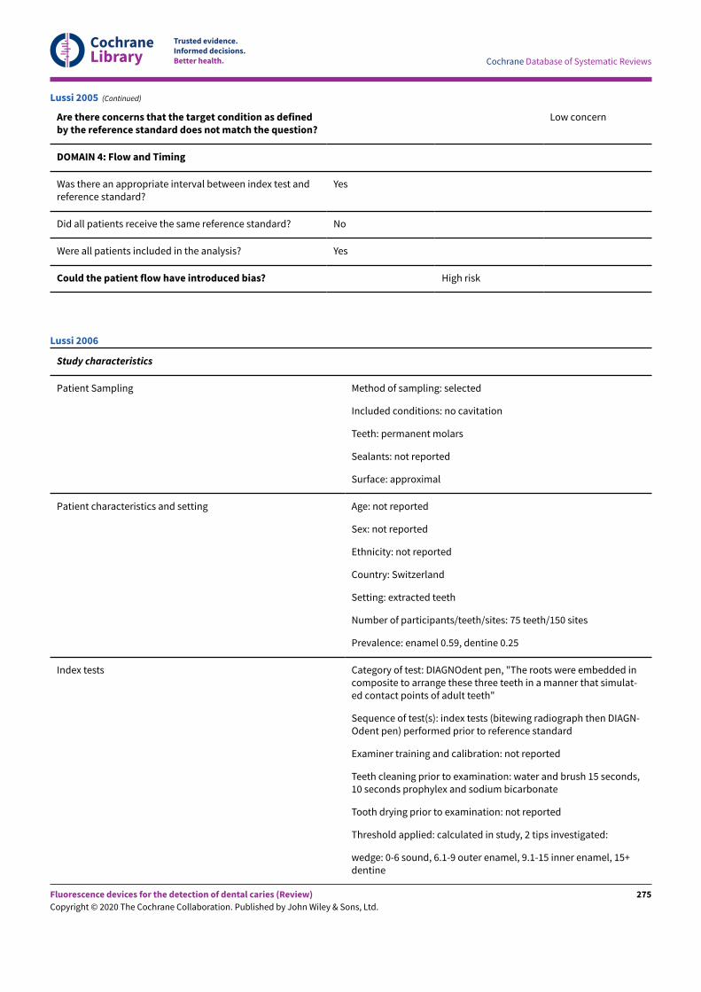
CochraneLibrary
Trusted evidence.Informed decisions.Better health.
Cochrane Database of Systematic Reviews
Are there concerns that the target condition as definedby the reference standard does not match the question?
Low concern
DOMAIN 4: Flow and Timing
Was there an appropriate interval between index test andreference standard?
Yes
Did all patients receive the same reference standard? No
Were all patients included in the analysis? Yes
Could the patient flow have introduced bias? High risk
Lussi 2005 (Continued)
Study characteristics
Patient Sampling Method of sampling: selected
Included conditions: no cavitation
Teeth: permanent molars
Sealants: not reported
Surface: approximal
Patient characteristics and setting Age: not reported
Sex: not reported
Ethnicity: not reported
Country: Switzerland
Setting: extracted teeth
Number of participants/teeth/sites: 75 teeth/150 sites
Prevalence: enamel 0.59, dentine 0.25
Index tests Category of test: DIAGNOdent pen, "The roots were embedded incomposite to arrange these three teeth in a manner that simulat-ed contact points of adult teeth"
Sequence of test(s): index tests (bitewing radiograph then DIAGN-Odent pen) performed prior to reference standard
Examiner training and calibration: not reported
Teeth cleaning prior to examination: water and brush 15 seconds,10 seconds prophylex and sodium bicarbonate
Tooth drying prior to examination: not reported
Threshold applied: calculated in study, 2 tips investigated:
wedge: 0-6 sound, 6.1-9 outer enamel, 9.1-15 inner enamel, 15+dentine
Lussi 2006
Fluorescence devices for the detection of dental caries (Review)
Copyright © 2020 The Cochrane Collaboration. Published by John Wiley & Sons, Ltd.
275
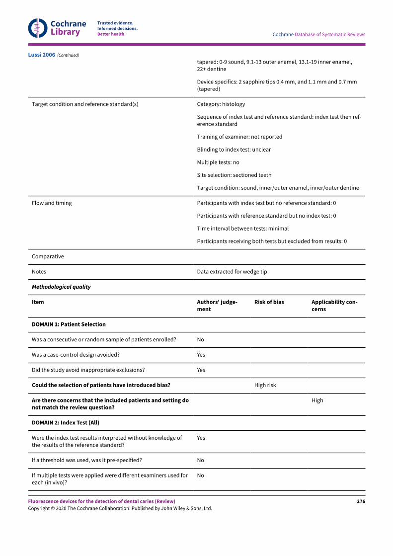
CochraneLibrary
Trusted evidence.Informed decisions.Better health.
Cochrane Database of Systematic Reviews
tapered: 0-9 sound, 9.1-13 outer enamel, 13.1-19 inner enamel,22+ dentine
Device specifics: 2 sapphire tips 0.4 mm, and 1.1 mm and 0.7 mm(tapered)
Target condition and reference standard(s) Category: histology
Sequence of index test and reference standard: index test then ref-erence standard
Training of examiner: not reported
Blinding to index test: unclear
Multiple tests: no
Site selection: sectioned teeth
Target condition: sound, inner/outer enamel, inner/outer dentine
Flow and timing Participants with index test but no reference standard: 0
Participants with reference standard but no index test: 0
Time interval between tests: minimal
Participants receiving both tests but excluded from results: 0
Comparative
Notes Data extracted for wedge tip
Methodological quality
Item Authors' judge-ment
Risk of bias Applicability con-cerns
DOMAIN 1: Patient Selection
Was a consecutive or random sample of patients enrolled? No
Was a case-control design avoided? Yes
Did the study avoid inappropriate exclusions? Yes
Could the selection of patients have introduced bias? High risk
Are there concerns that the included patients and setting donot match the review question?
High
DOMAIN 2: Index Test (All)
Were the index test results interpreted without knowledge ofthe results of the reference standard?
Yes
If a threshold was used, was it pre-specified? No
If multiple tests were applied were different examiners used foreach (in vivo)?
No
Lussi 2006 (Continued)
Fluorescence devices for the detection of dental caries (Review)
Copyright © 2020 The Cochrane Collaboration. Published by John Wiley & Sons, Ltd.
276
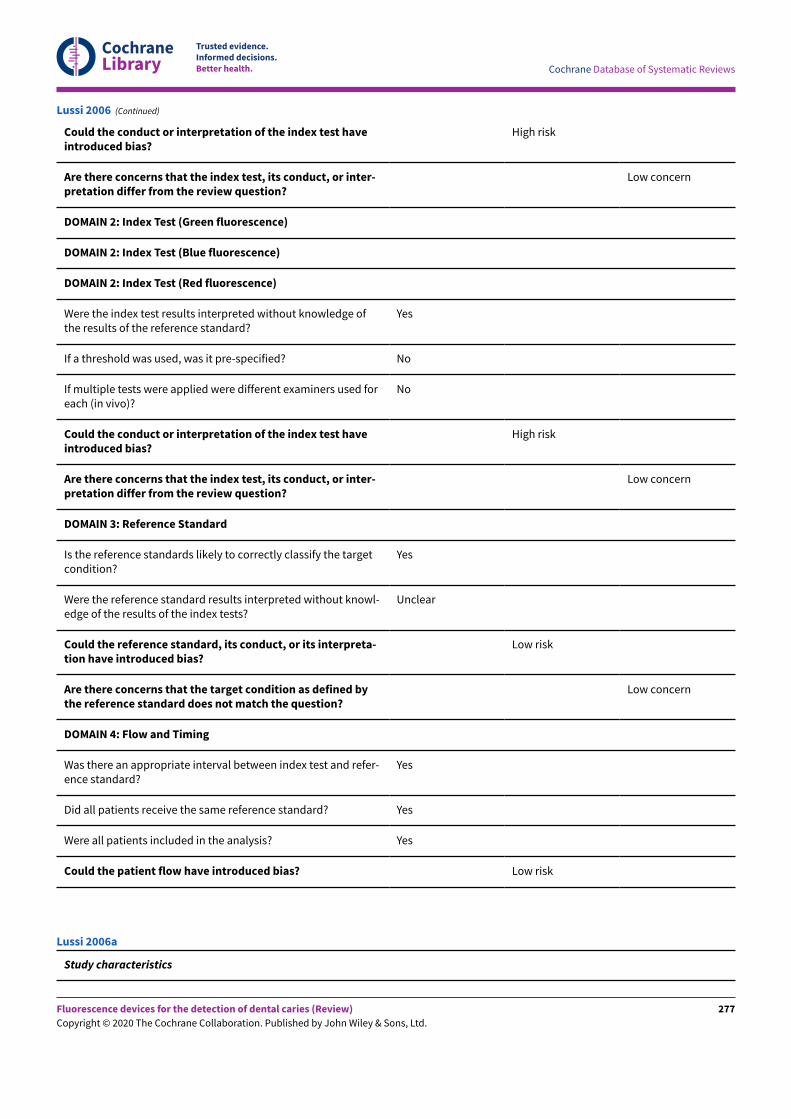
CochraneLibrary
Trusted evidence.Informed decisions.Better health.
Cochrane Database of Systematic Reviews
Could the conduct or interpretation of the index test haveintroduced bias?
High risk
Are there concerns that the index test, its conduct, or inter-pretation differ from the review question?
Low concern
DOMAIN 2: Index Test (Green fluorescence)
DOMAIN 2: Index Test (Blue fluorescence)
DOMAIN 2: Index Test (Red fluorescence)
Were the index test results interpreted without knowledge ofthe results of the reference standard?
Yes
If a threshold was used, was it pre-specified? No
If multiple tests were applied were different examiners used foreach (in vivo)?
No
Could the conduct or interpretation of the index test haveintroduced bias?
High risk
Are there concerns that the index test, its conduct, or inter-pretation differ from the review question?
Low concern
DOMAIN 3: Reference Standard
Is the reference standards likely to correctly classify the targetcondition?
Yes
Were the reference standard results interpreted without knowl-edge of the results of the index tests?
Unclear
Could the reference standard, its conduct, or its interpreta-tion have introduced bias?
Low risk
Are there concerns that the target condition as defined bythe reference standard does not match the question?
Low concern
DOMAIN 4: Flow and Timing
Was there an appropriate interval between index test and refer-ence standard?
Yes
Did all patients receive the same reference standard? Yes
Were all patients included in the analysis? Yes
Could the patient flow have introduced bias? Low risk
Lussi 2006 (Continued)
Study characteristics
Lussi 2006a
Fluorescence devices for the detection of dental caries (Review)
Copyright © 2020 The Cochrane Collaboration. Published by John Wiley & Sons, Ltd.
277
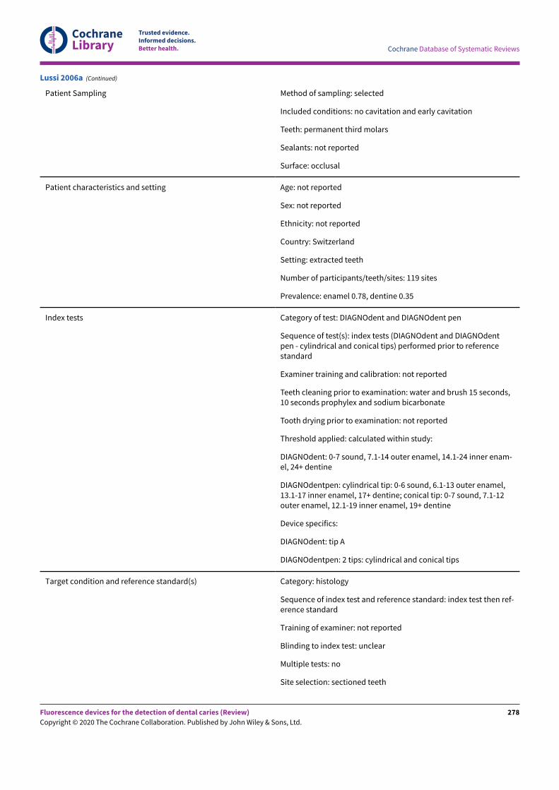
CochraneLibrary
Trusted evidence.Informed decisions.Better health.
Cochrane Database of Systematic Reviews
Patient Sampling Method of sampling: selected
Included conditions: no cavitation and early cavitation
Teeth: permanent third molars
Sealants: not reported
Surface: occlusal
Patient characteristics and setting Age: not reported
Sex: not reported
Ethnicity: not reported
Country: Switzerland
Setting: extracted teeth
Number of participants/teeth/sites: 119 sites
Prevalence: enamel 0.78, dentine 0.35
Index tests Category of test: DIAGNOdent and DIAGNOdent pen
Sequence of test(s): index tests (DIAGNOdent and DIAGNOdentpen - cylindrical and conical tips) performed prior to referencestandard
Examiner training and calibration: not reported
Teeth cleaning prior to examination: water and brush 15 seconds,10 seconds prophylex and sodium bicarbonate
Tooth drying prior to examination: not reported
Threshold applied: calculated within study:
DIAGNOdent: 0-7 sound, 7.1-14 outer enamel, 14.1-24 inner enam-el, 24+ dentine
DIAGNOdentpen: cylindrical tip: 0-6 sound, 6.1-13 outer enamel,13.1-17 inner enamel, 17+ dentine; conical tip: 0-7 sound, 7.1-12outer enamel, 12.1-19 inner enamel, 19+ dentine
Device specifics:
DIAGNOdent: tip A
DIAGNOdentpen: 2 tips: cylindrical and conical tips
Target condition and reference standard(s) Category: histology
Sequence of index test and reference standard: index test then ref-erence standard
Training of examiner: not reported
Blinding to index test: unclear
Multiple tests: no
Site selection: sectioned teeth
Lussi 2006a (Continued)
Fluorescence devices for the detection of dental caries (Review)
Copyright © 2020 The Cochrane Collaboration. Published by John Wiley & Sons, Ltd.
278
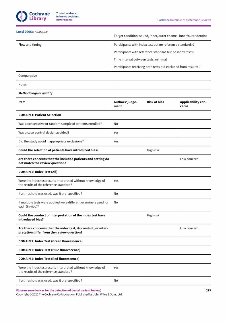
CochraneLibrary
Trusted evidence.Informed decisions.Better health.
Cochrane Database of Systematic Reviews
Target condition: sound, inner/outer enamel, inner/outer dentine
Flow and timing Participants with index test but no reference standard: 0
Participants with reference standard but no index test: 0
Time interval between tests: minimal
Participants receiving both tests but excluded from results: 0
Comparative
Notes
Methodological quality
Item Authors' judge-ment
Risk of bias Applicability con-cerns
DOMAIN 1: Patient Selection
Was a consecutive or random sample of patients enrolled? No
Was a case-control design avoided? Yes
Did the study avoid inappropriate exclusions? Yes
Could the selection of patients have introduced bias? High risk
Are there concerns that the included patients and setting donot match the review question?
Low concern
DOMAIN 2: Index Test (All)
Were the index test results interpreted without knowledge ofthe results of the reference standard?
Yes
If a threshold was used, was it pre-specified? No
If multiple tests were applied were different examiners used foreach (in vivo)?
No
Could the conduct or interpretation of the index test haveintroduced bias?
High risk
Are there concerns that the index test, its conduct, or inter-pretation differ from the review question?
Low concern
DOMAIN 2: Index Test (Green fluorescence)
DOMAIN 2: Index Test (Blue fluorescence)
DOMAIN 2: Index Test (Red fluorescence)
Were the index test results interpreted without knowledge ofthe results of the reference standard?
Yes
If a threshold was used, was it pre-specified? No
Lussi 2006a (Continued)
Fluorescence devices for the detection of dental caries (Review)
Copyright © 2020 The Cochrane Collaboration. Published by John Wiley & Sons, Ltd.
279
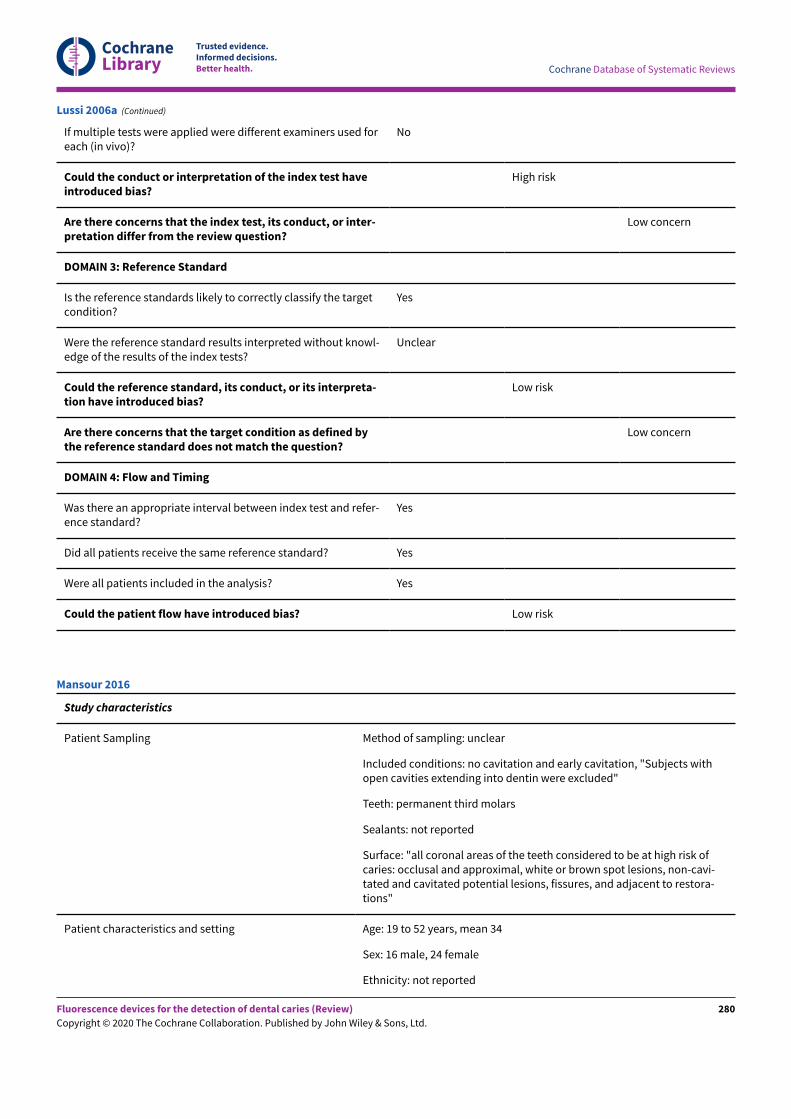
CochraneLibrary
Trusted evidence.Informed decisions.Better health.
Cochrane Database of Systematic Reviews
If multiple tests were applied were different examiners used foreach (in vivo)?
No
Could the conduct or interpretation of the index test haveintroduced bias?
High risk
Are there concerns that the index test, its conduct, or inter-pretation differ from the review question?
Low concern
DOMAIN 3: Reference Standard
Is the reference standards likely to correctly classify the targetcondition?
Yes
Were the reference standard results interpreted without knowl-edge of the results of the index tests?
Unclear
Could the reference standard, its conduct, or its interpreta-tion have introduced bias?
Low risk
Are there concerns that the target condition as defined bythe reference standard does not match the question?
Low concern
DOMAIN 4: Flow and Timing
Was there an appropriate interval between index test and refer-ence standard?
Yes
Did all patients receive the same reference standard? Yes
Were all patients included in the analysis? Yes
Could the patient flow have introduced bias? Low risk
Lussi 2006a (Continued)
Study characteristics
Patient Sampling Method of sampling: unclear
Included conditions: no cavitation and early cavitation, "Subjects withopen cavities extending into dentin were excluded"
Teeth: permanent third molars
Sealants: not reported
Surface: "all coronal areas of the teeth considered to be at high risk ofcaries: occlusal and approximal, white or brown spot lesions, non-cavi-tated and cavitated potential lesions, fissures, and adjacent to restora-tions"
Patient characteristics and setting Age: 19 to 52 years, mean 34
Sex: 16 male, 24 female
Ethnicity: not reported
Mansour 2016
Fluorescence devices for the detection of dental caries (Review)
Copyright © 2020 The Cochrane Collaboration. Published by John Wiley & Sons, Ltd.
280
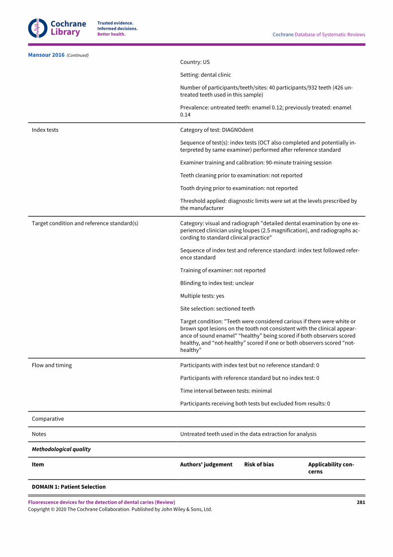
CochraneLibrary
Trusted evidence.Informed decisions.Better health.
Cochrane Database of Systematic Reviews
Country: US
Setting: dental clinic
Number of participants/teeth/sites: 40 participants/932 teeth (426 un-treated teeth used in this sample)
Prevalence: untreated teeth: enamel 0.12; previously treated: enamel0.14
Index tests Category of test: DIAGNOdent
Sequence of test(s): index tests (OCT also completed and potentially in-terpreted by same examiner) performed after reference standard
Examiner training and calibration: 90-minute training session
Teeth cleaning prior to examination: not reported
Tooth drying prior to examination: not reported
Threshold applied: diagnostic limits were set at the levels prescribed bythe manufacturer
Target condition and reference standard(s) Category: visual and radiograph "detailed dental examination by one ex-perienced clinician using loupes (2.5 magnification), and radiographs ac-cording to standard clinical practice"
Sequence of index test and reference standard: index test followed refer-ence standard
Training of examiner: not reported
Blinding to index test: unclear
Multiple tests: yes
Site selection: sectioned teeth
Target condition: "Teeth were considered carious if there were white orbrown spot lesions on the tooth not consistent with the clinical appear-ance of sound enamel" “healthy” being scored if both observers scoredhealthy, and “not-healthy” scored if one or both observers scored “not-healthy”
Flow and timing Participants with index test but no reference standard: 0
Participants with reference standard but no index test: 0
Time interval between tests: minimal
Participants receiving both tests but excluded from results: 0
Comparative
Notes Untreated teeth used in the data extraction for analysis
Methodological quality
Item Authors' judgement Risk of bias Applicability con-cerns
DOMAIN 1: Patient Selection
Mansour 2016 (Continued)
Fluorescence devices for the detection of dental caries (Review)
Copyright © 2020 The Cochrane Collaboration. Published by John Wiley & Sons, Ltd.
281
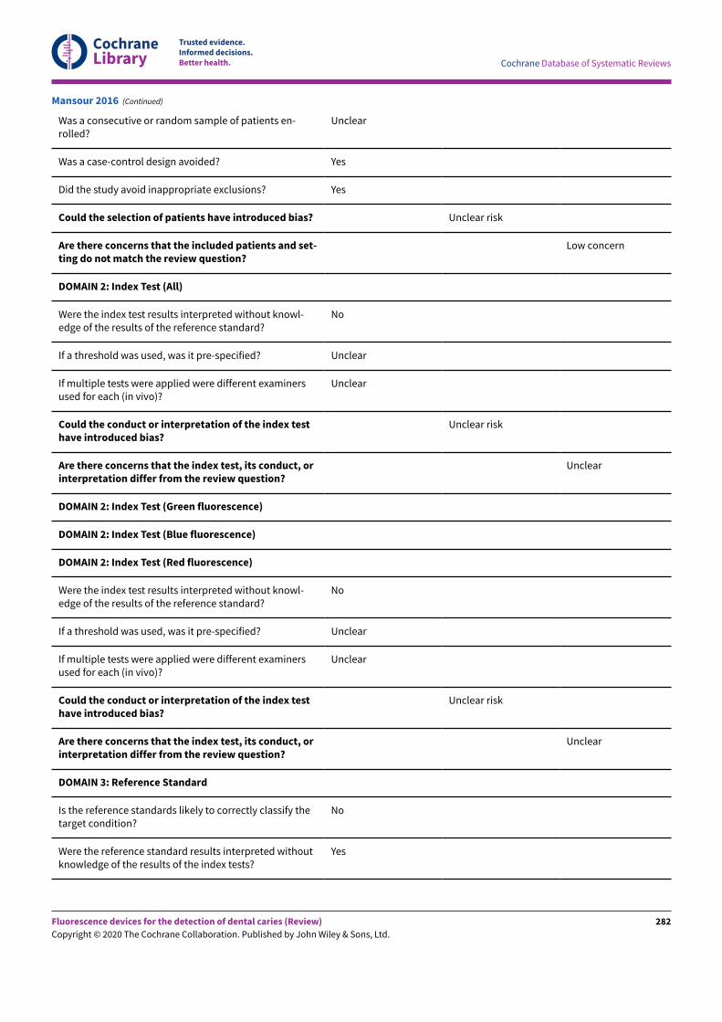
CochraneLibrary
Trusted evidence.Informed decisions.Better health.
Cochrane Database of Systematic Reviews
Was a consecutive or random sample of patients en-rolled?
Unclear
Was a case-control design avoided? Yes
Did the study avoid inappropriate exclusions? Yes
Could the selection of patients have introduced bias? Unclear risk
Are there concerns that the included patients and set-ting do not match the review question?
Low concern
DOMAIN 2: Index Test (All)
Were the index test results interpreted without knowl-edge of the results of the reference standard?
No
If a threshold was used, was it pre-specified? Unclear
If multiple tests were applied were different examinersused for each (in vivo)?
Unclear
Could the conduct or interpretation of the index testhave introduced bias?
Unclear risk
Are there concerns that the index test, its conduct, orinterpretation differ from the review question?
Unclear
DOMAIN 2: Index Test (Green fluorescence)
DOMAIN 2: Index Test (Blue fluorescence)
DOMAIN 2: Index Test (Red fluorescence)
Were the index test results interpreted without knowl-edge of the results of the reference standard?
No
If a threshold was used, was it pre-specified? Unclear
If multiple tests were applied were different examinersused for each (in vivo)?
Unclear
Could the conduct or interpretation of the index testhave introduced bias?
Unclear risk
Are there concerns that the index test, its conduct, orinterpretation differ from the review question?
Unclear
DOMAIN 3: Reference Standard
Is the reference standards likely to correctly classify thetarget condition?
No
Were the reference standard results interpreted withoutknowledge of the results of the index tests?
Yes
Mansour 2016 (Continued)
Fluorescence devices for the detection of dental caries (Review)
Copyright © 2020 The Cochrane Collaboration. Published by John Wiley & Sons, Ltd.
282
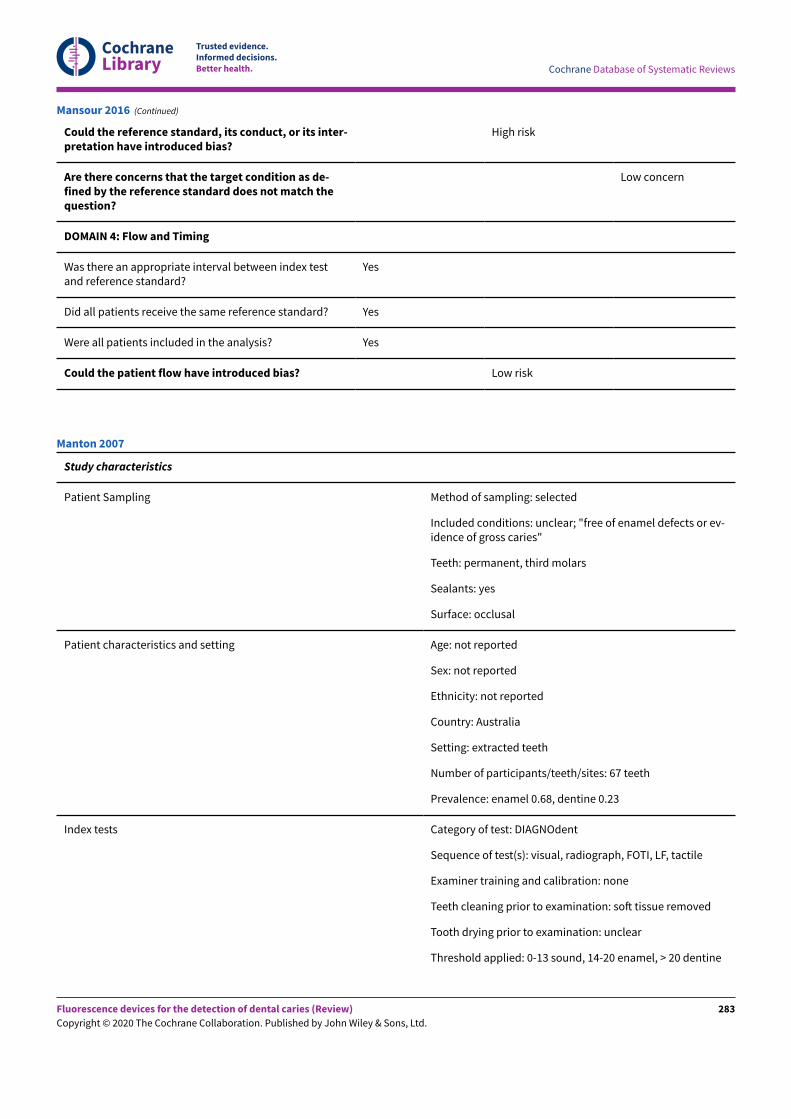
CochraneLibrary
Trusted evidence.Informed decisions.Better health.
Cochrane Database of Systematic Reviews
Could the reference standard, its conduct, or its inter-pretation have introduced bias?
High risk
Are there concerns that the target condition as de-fined by the reference standard does not match thequestion?
Low concern
DOMAIN 4: Flow and Timing
Was there an appropriate interval between index testand reference standard?
Yes
Did all patients receive the same reference standard? Yes
Were all patients included in the analysis? Yes
Could the patient flow have introduced bias? Low risk
Mansour 2016 (Continued)
Study characteristics
Patient Sampling Method of sampling: selected
Included conditions: unclear; "free of enamel defects or ev-idence of gross caries"
Teeth: permanent, third molars
Sealants: yes
Surface: occlusal
Patient characteristics and setting Age: not reported
Sex: not reported
Ethnicity: not reported
Country: Australia
Setting: extracted teeth
Number of participants/teeth/sites: 67 teeth
Prevalence: enamel 0.68, dentine 0.23
Index tests Category of test: DIAGNOdent
Sequence of test(s): visual, radiograph, FOTI, LF, tactile
Examiner training and calibration: none
Teeth cleaning prior to examination: soN tissue removed
Tooth drying prior to examination: unclear
Threshold applied: 0-13 sound, 14-20 enamel, > 20 dentine
Manton 2007
Fluorescence devices for the detection of dental caries (Review)
Copyright © 2020 The Cochrane Collaboration. Published by John Wiley & Sons, Ltd.
283
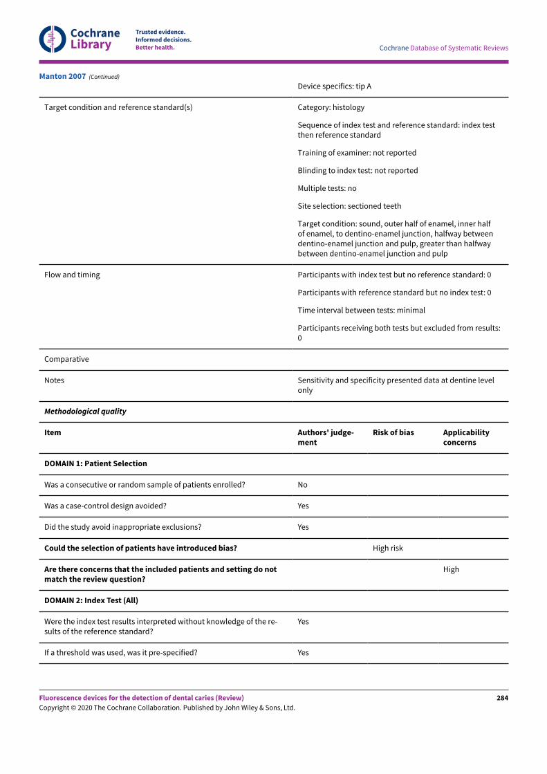
CochraneLibrary
Trusted evidence.Informed decisions.Better health.
Cochrane Database of Systematic Reviews
Device specifics: tip A
Target condition and reference standard(s) Category: histology
Sequence of index test and reference standard: index testthen reference standard
Training of examiner: not reported
Blinding to index test: not reported
Multiple tests: no
Site selection: sectioned teeth
Target condition: sound, outer half of enamel, inner halfof enamel, to dentino-enamel junction, halfway betweendentino-enamel junction and pulp, greater than halfwaybetween dentino-enamel junction and pulp
Flow and timing Participants with index test but no reference standard: 0
Participants with reference standard but no index test: 0
Time interval between tests: minimal
Participants receiving both tests but excluded from results:0
Comparative
Notes Sensitivity and specificity presented data at dentine levelonly
Methodological quality
Item Authors' judge-ment
Risk of bias Applicabilityconcerns
DOMAIN 1: Patient Selection
Was a consecutive or random sample of patients enrolled? No
Was a case-control design avoided? Yes
Did the study avoid inappropriate exclusions? Yes
Could the selection of patients have introduced bias? High risk
Are there concerns that the included patients and setting do notmatch the review question?
High
DOMAIN 2: Index Test (All)
Were the index test results interpreted without knowledge of the re-sults of the reference standard?
Yes
If a threshold was used, was it pre-specified? Yes
Manton 2007 (Continued)
Fluorescence devices for the detection of dental caries (Review)
Copyright © 2020 The Cochrane Collaboration. Published by John Wiley & Sons, Ltd.
284
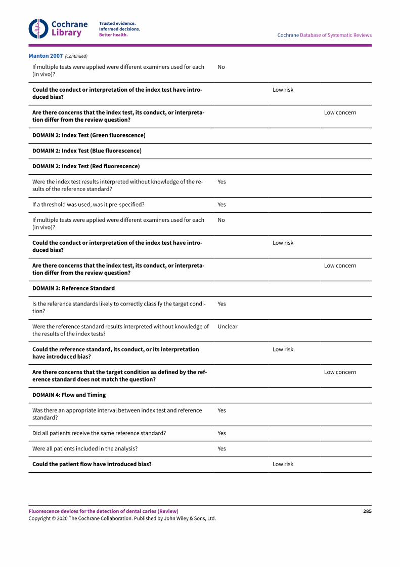
CochraneLibrary
Trusted evidence.Informed decisions.Better health.
Cochrane Database of Systematic Reviews
If multiple tests were applied were different examiners used for each(in vivo)?
No
Could the conduct or interpretation of the index test have intro-duced bias?
Low risk
Are there concerns that the index test, its conduct, or interpreta-tion differ from the review question?
Low concern
DOMAIN 2: Index Test (Green fluorescence)
DOMAIN 2: Index Test (Blue fluorescence)
DOMAIN 2: Index Test (Red fluorescence)
Were the index test results interpreted without knowledge of the re-sults of the reference standard?
Yes
If a threshold was used, was it pre-specified? Yes
If multiple tests were applied were different examiners used for each(in vivo)?
No
Could the conduct or interpretation of the index test have intro-duced bias?
Low risk
Are there concerns that the index test, its conduct, or interpreta-tion differ from the review question?
Low concern
DOMAIN 3: Reference Standard
Is the reference standards likely to correctly classify the target condi-tion?
Yes
Were the reference standard results interpreted without knowledge ofthe results of the index tests?
Unclear
Could the reference standard, its conduct, or its interpretationhave introduced bias?
Low risk
Are there concerns that the target condition as defined by the ref-erence standard does not match the question?
Low concern
DOMAIN 4: Flow and Timing
Was there an appropriate interval between index test and referencestandard?
Yes
Did all patients receive the same reference standard? Yes
Were all patients included in the analysis? Yes
Could the patient flow have introduced bias? Low risk
Manton 2007 (Continued)
Fluorescence devices for the detection of dental caries (Review)
Copyright © 2020 The Cochrane Collaboration. Published by John Wiley & Sons, Ltd.
285
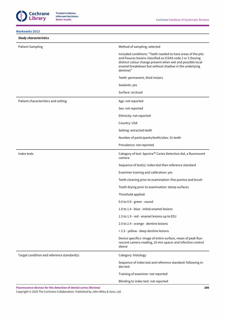
CochraneLibrary
Trusted evidence.Informed decisions.Better health.
Cochrane Database of Systematic Reviews
Study characteristics
Patient Sampling Method of sampling: selected
Included conditions: "Teeth needed to have areas of the pitsand fissures lesions classified as ICDAS code 2 or 3 (havingdistinct colour change present when wet and possible localenamel breakdown but without shadow in the underlyingdentine)"
Teeth: permanent, third molars
Sealants: yes
Surface: occlusal
Patient characteristics and setting Age: not reported
Sex: not reported
Ethnicity: not reported
Country: USA
Setting: extracted teeth
Number of participants/teeth/sites: 31 teeth
Prevalence: not reported
Index tests Category of test: Spectra™ Caries Detection Aid, a fluorescentcamera
Sequence of test(s): index test then reference standard
Examiner training and calibration: yes
Teeth cleaning prior to examination: fine pumice and brush
Tooth drying prior to examination: damp surfaces
Threshold applied:
0.0 to 0.9 - green - sound
1.0 to 1.4 - blue - initial enamel lesions
1.5 to 1.9 - red - enamel lesions up to EDJ
2.0 to 2.4 - orange - dentine lesions
> 2.5 - yellow - deep dentine lesions
Device specifics: image of entire surface, mean of peak fluo-rescent camera reading, 10 mm spacer and infection controlsleeve
Target condition and reference standard(s) Category: histology
Sequence of index test and reference standard: following in-dex test
Training of examiner: not reported
Blinding to index test: not reported
Markowitz 2013
Fluorescence devices for the detection of dental caries (Review)
Copyright © 2020 The Cochrane Collaboration. Published by John Wiley & Sons, Ltd.
286
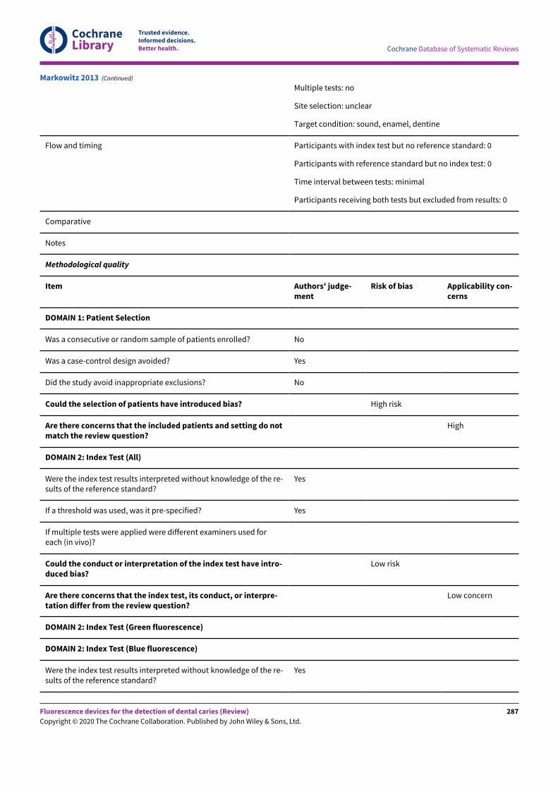
CochraneLibrary
Trusted evidence.Informed decisions.Better health.
Cochrane Database of Systematic Reviews
Multiple tests: no
Site selection: unclear
Target condition: sound, enamel, dentine
Flow and timing Participants with index test but no reference standard: 0
Participants with reference standard but no index test: 0
Time interval between tests: minimal
Participants receiving both tests but excluded from results: 0
Comparative
Notes
Methodological quality
Item Authors' judge-ment
Risk of bias Applicability con-cerns
DOMAIN 1: Patient Selection
Was a consecutive or random sample of patients enrolled? No
Was a case-control design avoided? Yes
Did the study avoid inappropriate exclusions? No
Could the selection of patients have introduced bias? High risk
Are there concerns that the included patients and setting do notmatch the review question?
High
DOMAIN 2: Index Test (All)
Were the index test results interpreted without knowledge of the re-sults of the reference standard?
Yes
If a threshold was used, was it pre-specified? Yes
If multiple tests were applied were different examiners used foreach (in vivo)?
Could the conduct or interpretation of the index test have intro-duced bias?
Low risk
Are there concerns that the index test, its conduct, or interpre-tation differ from the review question?
Low concern
DOMAIN 2: Index Test (Green fluorescence)
DOMAIN 2: Index Test (Blue fluorescence)
Were the index test results interpreted without knowledge of the re-sults of the reference standard?
Yes
Markowitz 2013 (Continued)
Fluorescence devices for the detection of dental caries (Review)
Copyright © 2020 The Cochrane Collaboration. Published by John Wiley & Sons, Ltd.
287
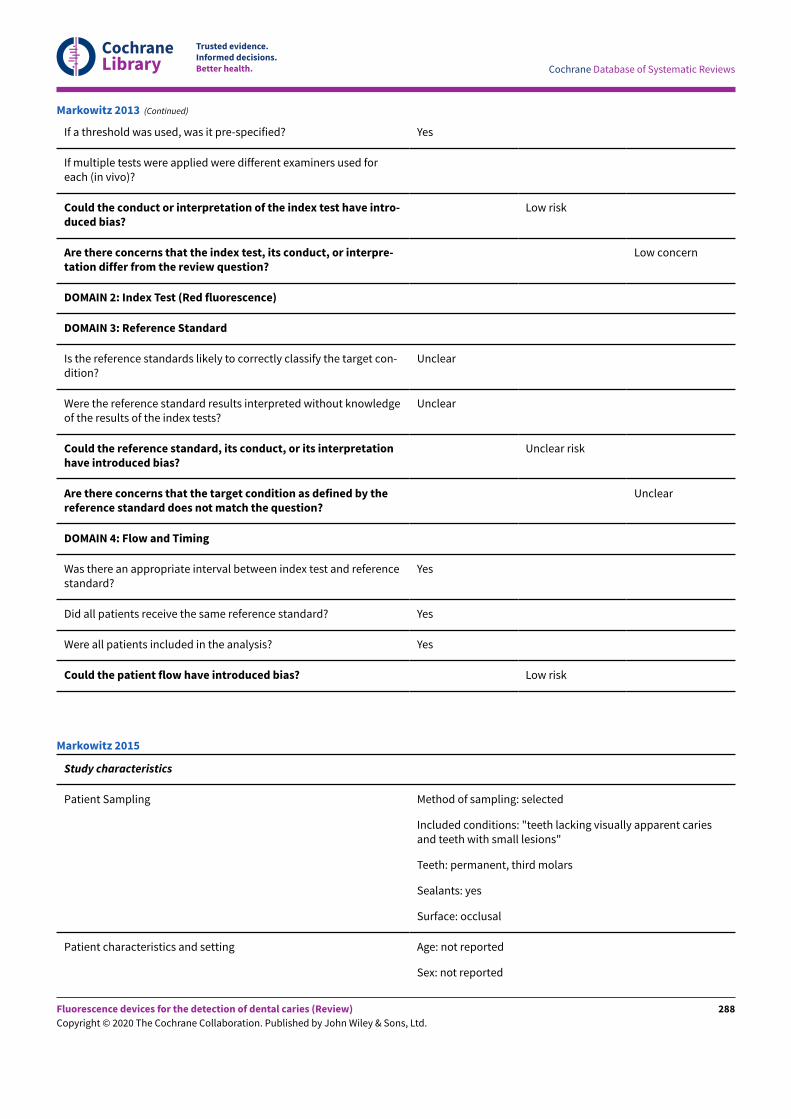
CochraneLibrary
Trusted evidence.Informed decisions.Better health.
Cochrane Database of Systematic Reviews
If a threshold was used, was it pre-specified? Yes
If multiple tests were applied were different examiners used foreach (in vivo)?
Could the conduct or interpretation of the index test have intro-duced bias?
Low risk
Are there concerns that the index test, its conduct, or interpre-tation differ from the review question?
Low concern
DOMAIN 2: Index Test (Red fluorescence)
DOMAIN 3: Reference Standard
Is the reference standards likely to correctly classify the target con-dition?
Unclear
Were the reference standard results interpreted without knowledgeof the results of the index tests?
Unclear
Could the reference standard, its conduct, or its interpretationhave introduced bias?
Unclear risk
Are there concerns that the target condition as defined by thereference standard does not match the question?
Unclear
DOMAIN 4: Flow and Timing
Was there an appropriate interval between index test and referencestandard?
Yes
Did all patients receive the same reference standard? Yes
Were all patients included in the analysis? Yes
Could the patient flow have introduced bias? Low risk
Markowitz 2013 (Continued)
Study characteristics
Patient Sampling Method of sampling: selected
Included conditions: "teeth lacking visually apparent cariesand teeth with small lesions"
Teeth: permanent, third molars
Sealants: yes
Surface: occlusal
Patient characteristics and setting Age: not reported
Sex: not reported
Markowitz 2015
Fluorescence devices for the detection of dental caries (Review)
Copyright © 2020 The Cochrane Collaboration. Published by John Wiley & Sons, Ltd.
288
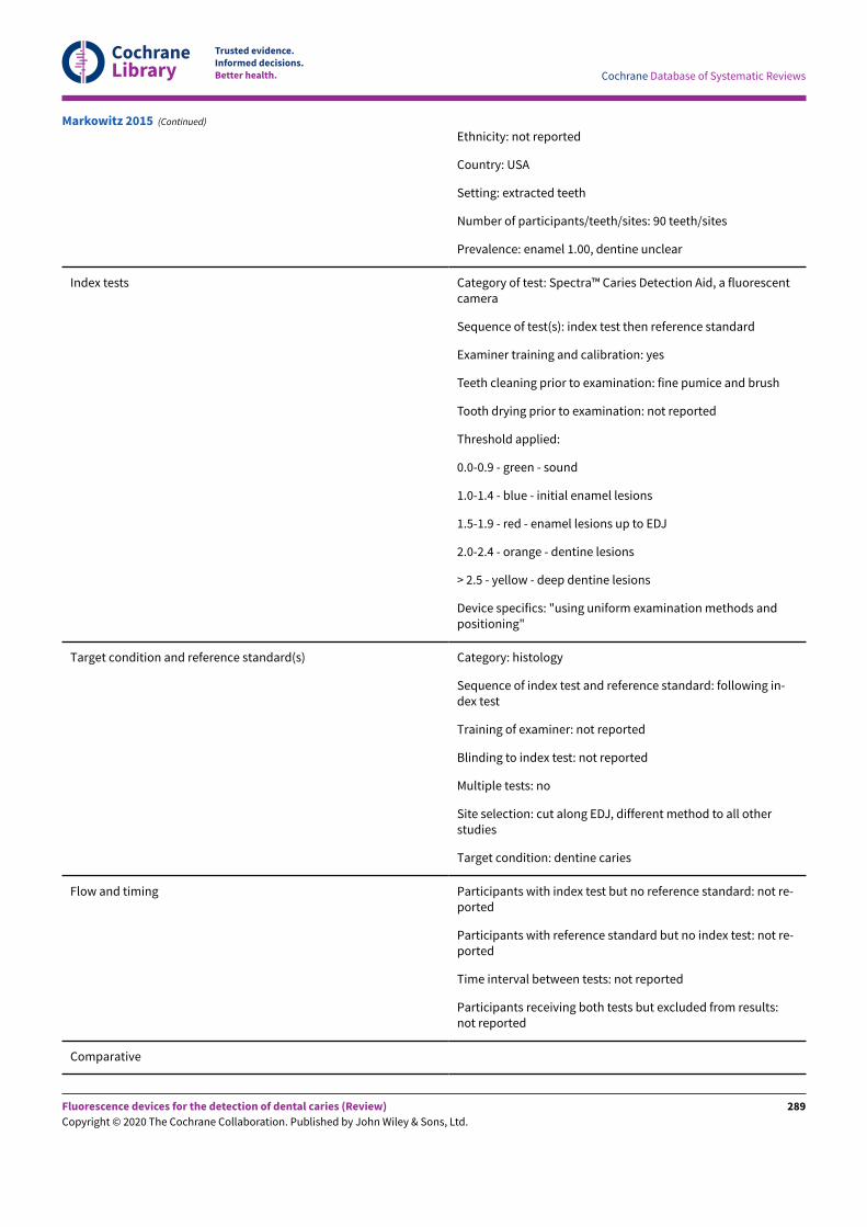
CochraneLibrary
Trusted evidence.Informed decisions.Better health.
Cochrane Database of Systematic Reviews
Ethnicity: not reported
Country: USA
Setting: extracted teeth
Number of participants/teeth/sites: 90 teeth/sites
Prevalence: enamel 1.00, dentine unclear
Index tests Category of test: Spectra™ Caries Detection Aid, a fluorescentcamera
Sequence of test(s): index test then reference standard
Examiner training and calibration: yes
Teeth cleaning prior to examination: fine pumice and brush
Tooth drying prior to examination: not reported
Threshold applied:
0.0-0.9 - green - sound
1.0-1.4 - blue - initial enamel lesions
1.5-1.9 - red - enamel lesions up to EDJ
2.0-2.4 - orange - dentine lesions
> 2.5 - yellow - deep dentine lesions
Device specifics: "using uniform examination methods andpositioning"
Target condition and reference standard(s) Category: histology
Sequence of index test and reference standard: following in-dex test
Training of examiner: not reported
Blinding to index test: not reported
Multiple tests: no
Site selection: cut along EDJ, different method to all otherstudies
Target condition: dentine caries
Flow and timing Participants with index test but no reference standard: not re-ported
Participants with reference standard but no index test: not re-ported
Time interval between tests: not reported
Participants receiving both tests but excluded from results:not reported
Comparative
Markowitz 2015 (Continued)
Fluorescence devices for the detection of dental caries (Review)
Copyright © 2020 The Cochrane Collaboration. Published by John Wiley & Sons, Ltd.
289
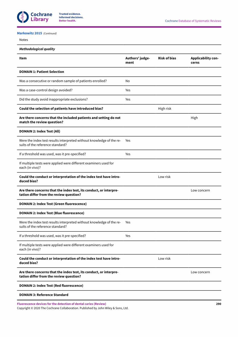
CochraneLibrary
Trusted evidence.Informed decisions.Better health.
Cochrane Database of Systematic Reviews
Notes
Methodological quality
Item Authors' judge-ment
Risk of bias Applicability con-cerns
DOMAIN 1: Patient Selection
Was a consecutive or random sample of patients enrolled? No
Was a case-control design avoided? Yes
Did the study avoid inappropriate exclusions? Yes
Could the selection of patients have introduced bias? High risk
Are there concerns that the included patients and setting do notmatch the review question?
High
DOMAIN 2: Index Test (All)
Were the index test results interpreted without knowledge of the re-sults of the reference standard?
Yes
If a threshold was used, was it pre-specified? Yes
If multiple tests were applied were different examiners used foreach (in vivo)?
Could the conduct or interpretation of the index test have intro-duced bias?
Low risk
Are there concerns that the index test, its conduct, or interpre-tation differ from the review question?
Low concern
DOMAIN 2: Index Test (Green fluorescence)
DOMAIN 2: Index Test (Blue fluorescence)
Were the index test results interpreted without knowledge of the re-sults of the reference standard?
Yes
If a threshold was used, was it pre-specified? Yes
If multiple tests were applied were different examiners used foreach (in vivo)?
Could the conduct or interpretation of the index test have intro-duced bias?
Low risk
Are there concerns that the index test, its conduct, or interpre-tation differ from the review question?
Low concern
DOMAIN 2: Index Test (Red fluorescence)
DOMAIN 3: Reference Standard
Markowitz 2015 (Continued)
Fluorescence devices for the detection of dental caries (Review)
Copyright © 2020 The Cochrane Collaboration. Published by John Wiley & Sons, Ltd.
290
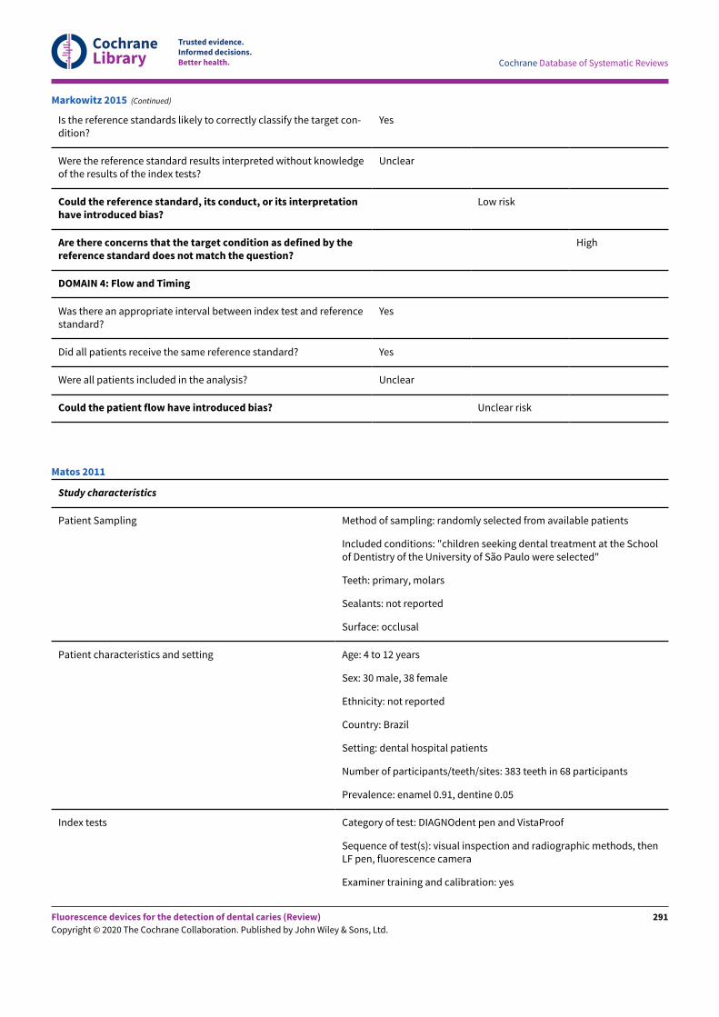
CochraneLibrary
Trusted evidence.Informed decisions.Better health.
Cochrane Database of Systematic Reviews
Is the reference standards likely to correctly classify the target con-dition?
Yes
Were the reference standard results interpreted without knowledgeof the results of the index tests?
Unclear
Could the reference standard, its conduct, or its interpretationhave introduced bias?
Low risk
Are there concerns that the target condition as defined by thereference standard does not match the question?
High
DOMAIN 4: Flow and Timing
Was there an appropriate interval between index test and referencestandard?
Yes
Did all patients receive the same reference standard? Yes
Were all patients included in the analysis? Unclear
Could the patient flow have introduced bias? Unclear risk
Markowitz 2015 (Continued)
Study characteristics
Patient Sampling Method of sampling: randomly selected from available patients
Included conditions: "children seeking dental treatment at the Schoolof Dentistry of the University of São Paulo were selected"
Teeth: primary, molars
Sealants: not reported
Surface: occlusal
Patient characteristics and setting Age: 4 to 12 years
Sex: 30 male, 38 female
Ethnicity: not reported
Country: Brazil
Setting: dental hospital patients
Number of participants/teeth/sites: 383 teeth in 68 participants
Prevalence: enamel 0.91, dentine 0.05
Index tests Category of test: DIAGNOdent pen and VistaProof
Sequence of test(s): visual inspection and radiographic methods, thenLF pen, fluorescence camera
Examiner training and calibration: yes
Matos 2011
Fluorescence devices for the detection of dental caries (Review)
Copyright © 2020 The Cochrane Collaboration. Published by John Wiley & Sons, Ltd.
291
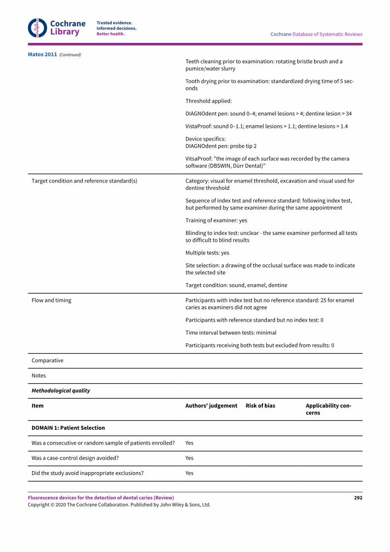
CochraneLibrary
Trusted evidence.Informed decisions.Better health.
Cochrane Database of Systematic Reviews
Teeth cleaning prior to examination: rotating bristle brush and apumice/water slurry
Tooth drying prior to examination: standardized drying time of 5 sec-onds
Threshold applied:
DIAGNOdent pen: sound 0–4; enamel lesions > 4; dentine lesion > 34
VistaProof: sound 0–1.1; enamel lesions > 1.1; dentine lesions > 1.4
Device specifics: DIAGNOdent pen: probe tip 2
VitsaProof: "the image of each surface was recorded by the camerasoftware (DBSWIN, Dürr Dental)"
Target condition and reference standard(s) Category: visual for enamel threshold, excavation and visual used fordentine threshold
Sequence of index test and reference standard: following index test,but performed by same examiner during the same appointment
Training of examiner: yes
Blinding to index test: unclear - the same examiner performed all testsso difficult to blind results
Multiple tests: yes
Site selection: a drawing of the occlusal surface was made to indicatethe selected site
Target condition: sound, enamel, dentine
Flow and timing Participants with index test but no reference standard: 25 for enamelcaries as examiners did not agree
Participants with reference standard but no index test: 0
Time interval between tests: minimal
Participants receiving both tests but excluded from results: 0
Comparative
Notes
Methodological quality
Item Authors' judgement Risk of bias Applicability con-cerns
DOMAIN 1: Patient Selection
Was a consecutive or random sample of patients enrolled? Yes
Was a case-control design avoided? Yes
Did the study avoid inappropriate exclusions? Yes
Matos 2011 (Continued)
Fluorescence devices for the detection of dental caries (Review)
Copyright © 2020 The Cochrane Collaboration. Published by John Wiley & Sons, Ltd.
292
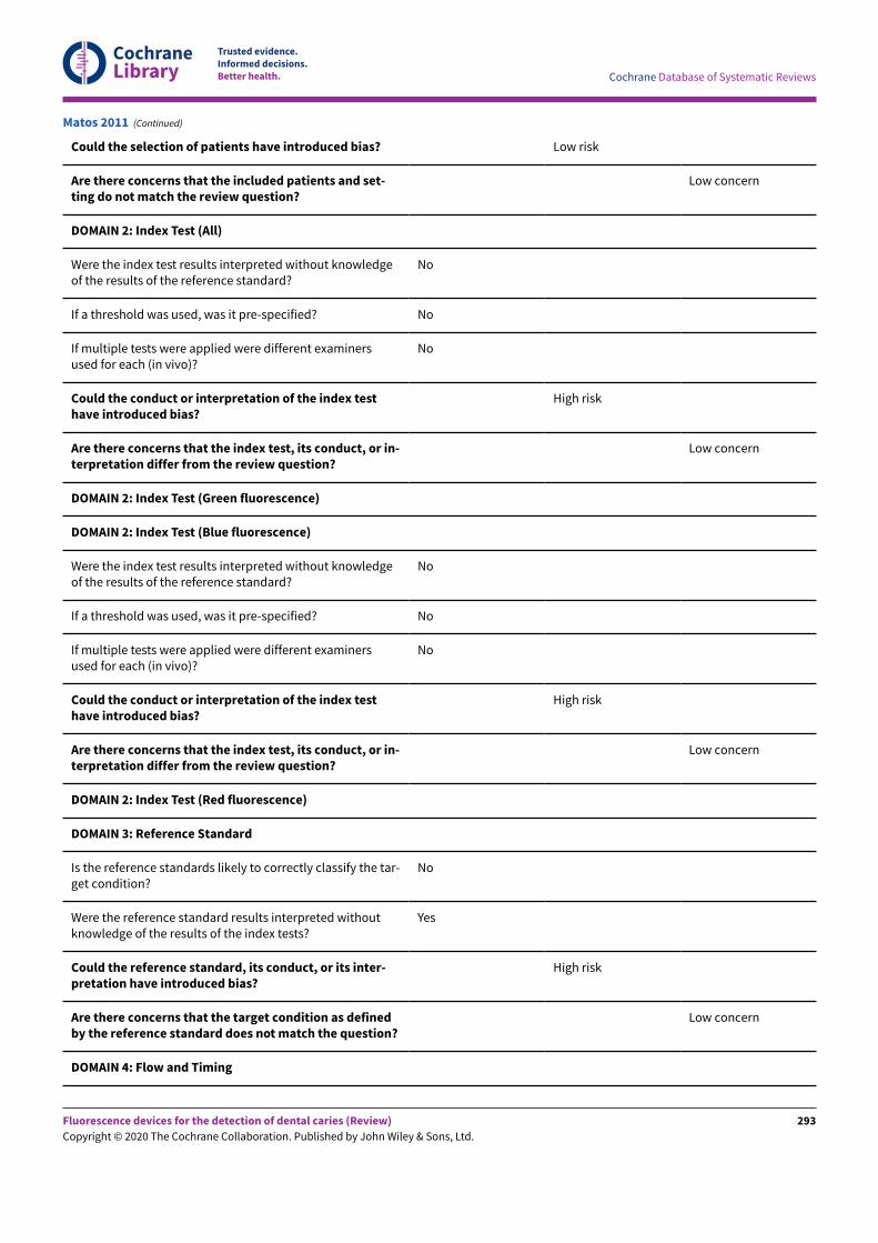
CochraneLibrary
Trusted evidence.Informed decisions.Better health.
Cochrane Database of Systematic Reviews
Could the selection of patients have introduced bias? Low risk
Are there concerns that the included patients and set-ting do not match the review question?
Low concern
DOMAIN 2: Index Test (All)
Were the index test results interpreted without knowledgeof the results of the reference standard?
No
If a threshold was used, was it pre-specified? No
If multiple tests were applied were different examinersused for each (in vivo)?
No
Could the conduct or interpretation of the index testhave introduced bias?
High risk
Are there concerns that the index test, its conduct, or in-terpretation differ from the review question?
Low concern
DOMAIN 2: Index Test (Green fluorescence)
DOMAIN 2: Index Test (Blue fluorescence)
Were the index test results interpreted without knowledgeof the results of the reference standard?
No
If a threshold was used, was it pre-specified? No
If multiple tests were applied were different examinersused for each (in vivo)?
No
Could the conduct or interpretation of the index testhave introduced bias?
High risk
Are there concerns that the index test, its conduct, or in-terpretation differ from the review question?
Low concern
DOMAIN 2: Index Test (Red fluorescence)
DOMAIN 3: Reference Standard
Is the reference standards likely to correctly classify the tar-get condition?
No
Were the reference standard results interpreted withoutknowledge of the results of the index tests?
Yes
Could the reference standard, its conduct, or its inter-pretation have introduced bias?
High risk
Are there concerns that the target condition as definedby the reference standard does not match the question?
Low concern
DOMAIN 4: Flow and Timing
Matos 2011 (Continued)
Fluorescence devices for the detection of dental caries (Review)
Copyright © 2020 The Cochrane Collaboration. Published by John Wiley & Sons, Ltd.
293
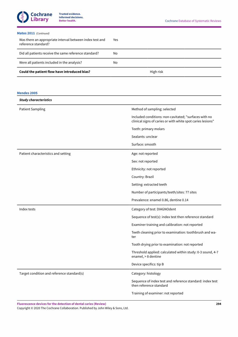
CochraneLibrary
Trusted evidence.Informed decisions.Better health.
Cochrane Database of Systematic Reviews
Was there an appropriate interval between index test andreference standard?
Yes
Did all patients receive the same reference standard? No
Were all patients included in the analysis? No
Could the patient flow have introduced bias? High risk
Matos 2011 (Continued)
Study characteristics
Patient Sampling Method of sampling: selected
Included conditions: non-cavitated; "surfaces with noclinical signs of caries or with white spot caries lesions"
Teeth: primary molars
Sealants: unclear
Surface: smooth
Patient characteristics and setting Age: not reported
Sex: not reported
Ethnicity: not reported
Country: Brazil
Setting: extracted teeth
Number of participants/teeth/sites: 77 sites
Prevalence: enamel 0.86, dentine 0.14
Index tests Category of test: DIAGNOdent
Sequence of test(s): index test then reference standard
Examiner training and calibration: not reported
Teeth cleaning prior to examination: toothbrush and wa-ter
Tooth drying prior to examination: not reported
Threshold applied: calculated within study: 0-3 sound, 4-7enamel, > 8 dentine
Device specifics: tip B
Target condition and reference standard(s) Category: histology
Sequence of index test and reference standard: index testthen reference standard
Training of examiner: not reported
Mendes 2005
Fluorescence devices for the detection of dental caries (Review)
Copyright © 2020 The Cochrane Collaboration. Published by John Wiley & Sons, Ltd.
294
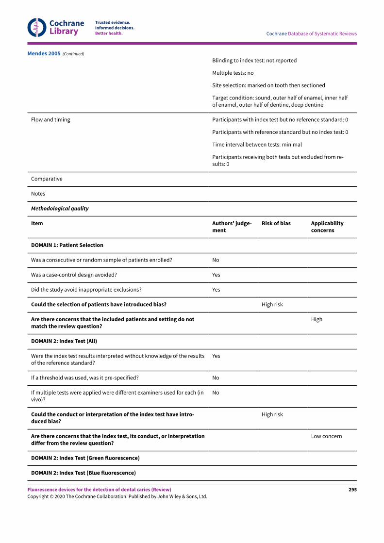
CochraneLibrary
Trusted evidence.Informed decisions.Better health.
Cochrane Database of Systematic Reviews
Blinding to index test: not reported
Multiple tests: no
Site selection: marked on tooth then sectioned
Target condition: sound, outer half of enamel, inner halfof enamel, outer half of dentine, deep dentine
Flow and timing Participants with index test but no reference standard: 0
Participants with reference standard but no index test: 0
Time interval between tests: minimal
Participants receiving both tests but excluded from re-sults: 0
Comparative
Notes
Methodological quality
Item Authors' judge-ment
Risk of bias Applicabilityconcerns
DOMAIN 1: Patient Selection
Was a consecutive or random sample of patients enrolled? No
Was a case-control design avoided? Yes
Did the study avoid inappropriate exclusions? Yes
Could the selection of patients have introduced bias? High risk
Are there concerns that the included patients and setting do notmatch the review question?
High
DOMAIN 2: Index Test (All)
Were the index test results interpreted without knowledge of the resultsof the reference standard?
Yes
If a threshold was used, was it pre-specified? No
If multiple tests were applied were different examiners used for each (invivo)?
No
Could the conduct or interpretation of the index test have intro-duced bias?
High risk
Are there concerns that the index test, its conduct, or interpretationdiffer from the review question?
Low concern
DOMAIN 2: Index Test (Green fluorescence)
DOMAIN 2: Index Test (Blue fluorescence)
Mendes 2005 (Continued)
Fluorescence devices for the detection of dental caries (Review)
Copyright © 2020 The Cochrane Collaboration. Published by John Wiley & Sons, Ltd.
295
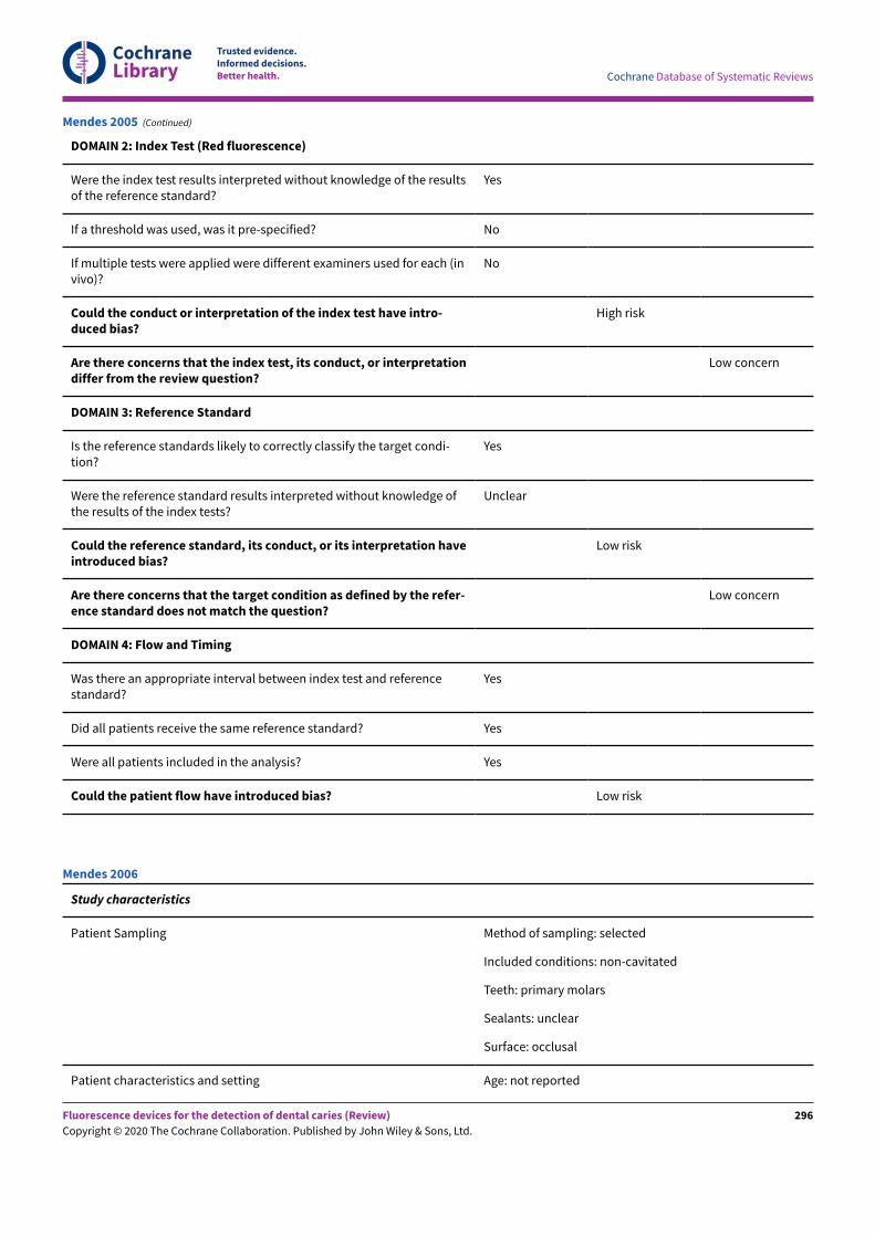
CochraneLibrary
Trusted evidence.Informed decisions.Better health.
Cochrane Database of Systematic Reviews
DOMAIN 2: Index Test (Red fluorescence)
Were the index test results interpreted without knowledge of the resultsof the reference standard?
Yes
If a threshold was used, was it pre-specified? No
If multiple tests were applied were different examiners used for each (invivo)?
No
Could the conduct or interpretation of the index test have intro-duced bias?
High risk
Are there concerns that the index test, its conduct, or interpretationdiffer from the review question?
Low concern
DOMAIN 3: Reference Standard
Is the reference standards likely to correctly classify the target condi-tion?
Yes
Were the reference standard results interpreted without knowledge ofthe results of the index tests?
Unclear
Could the reference standard, its conduct, or its interpretation haveintroduced bias?
Low risk
Are there concerns that the target condition as defined by the refer-ence standard does not match the question?
Low concern
DOMAIN 4: Flow and Timing
Was there an appropriate interval between index test and referencestandard?
Yes
Did all patients receive the same reference standard? Yes
Were all patients included in the analysis? Yes
Could the patient flow have introduced bias? Low risk
Mendes 2005 (Continued)
Study characteristics
Patient Sampling Method of sampling: selected
Included conditions: non-cavitated
Teeth: primary molars
Sealants: unclear
Surface: occlusal
Patient characteristics and setting Age: not reported
Mendes 2006
Fluorescence devices for the detection of dental caries (Review)
Copyright © 2020 The Cochrane Collaboration. Published by John Wiley & Sons, Ltd.
296
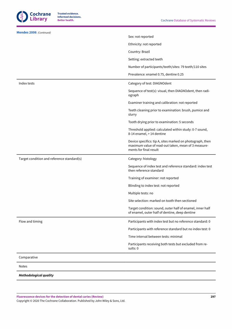
CochraneLibrary
Trusted evidence.Informed decisions.Better health.
Cochrane Database of Systematic Reviews
Sex: not reported
Ethnicity: not reported
Country: Brazil
Setting: extracted teeth
Number of participants/teeth/sites: 79 teeth/110 sites
Prevalence: enamel 0.75, dentine 0.25
Index tests Category of test: DIAGNOdent
Sequence of test(s): visual, then DIAGNOdent, then radi-ograph
Examiner training and calibration: not reported
Teeth cleaning prior to examination: brush, pumice andslurry
Tooth drying prior to examination: 5 seconds
Threshold applied: calculated within study: 0-7 sound,8-14 enamel, > 14 dentine
Device specifics: tip A, sites marked on photograph, thenmaximum value of read-out taken, mean of 3 measure-ments for final result
Target condition and reference standard(s) Category: histology
Sequence of index test and reference standard: index testthen reference standard
Training of examiner: not reported
Blinding to index test: not reported
Multiple tests: no
Site selection: marked on tooth then sectioned
Target condition: sound, outer half of enamel, inner halfof enamel, outer half of dentine, deep dentine
Flow and timing Participants with index test but no reference standard: 0
Participants with reference standard but no index test: 0
Time interval between tests: minimal
Participants receiving both tests but excluded from re-sults: 0
Comparative
Notes
Methodological quality
Mendes 2006 (Continued)
Fluorescence devices for the detection of dental caries (Review)
Copyright © 2020 The Cochrane Collaboration. Published by John Wiley & Sons, Ltd.
297
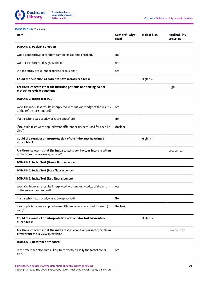
CochraneLibrary
Trusted evidence.Informed decisions.Better health.
Cochrane Database of Systematic Reviews
Item Authors' judge-ment
Risk of bias Applicabilityconcerns
DOMAIN 1: Patient Selection
Was a consecutive or random sample of patients enrolled? No
Was a case-control design avoided? Yes
Did the study avoid inappropriate exclusions? Yes
Could the selection of patients have introduced bias? High risk
Are there concerns that the included patients and setting do notmatch the review question?
High
DOMAIN 2: Index Test (All)
Were the index test results interpreted without knowledge of the resultsof the reference standard?
Yes
If a threshold was used, was it pre-specified? No
If multiple tests were applied were different examiners used for each (invivo)?
Unclear
Could the conduct or interpretation of the index test have intro-duced bias?
High risk
Are there concerns that the index test, its conduct, or interpretationdiffer from the review question?
Low concern
DOMAIN 2: Index Test (Green fluorescence)
DOMAIN 2: Index Test (Blue fluorescence)
DOMAIN 2: Index Test (Red fluorescence)
Were the index test results interpreted without knowledge of the resultsof the reference standard?
Yes
If a threshold was used, was it pre-specified? No
If multiple tests were applied were different examiners used for each (invivo)?
Unclear
Could the conduct or interpretation of the index test have intro-duced bias?
High risk
Are there concerns that the index test, its conduct, or interpretationdiffer from the review question?
Low concern
DOMAIN 3: Reference Standard
Is the reference standards likely to correctly classify the target condi-tion?
Yes
Mendes 2006 (Continued)
Fluorescence devices for the detection of dental caries (Review)
Copyright © 2020 The Cochrane Collaboration. Published by John Wiley & Sons, Ltd.
298
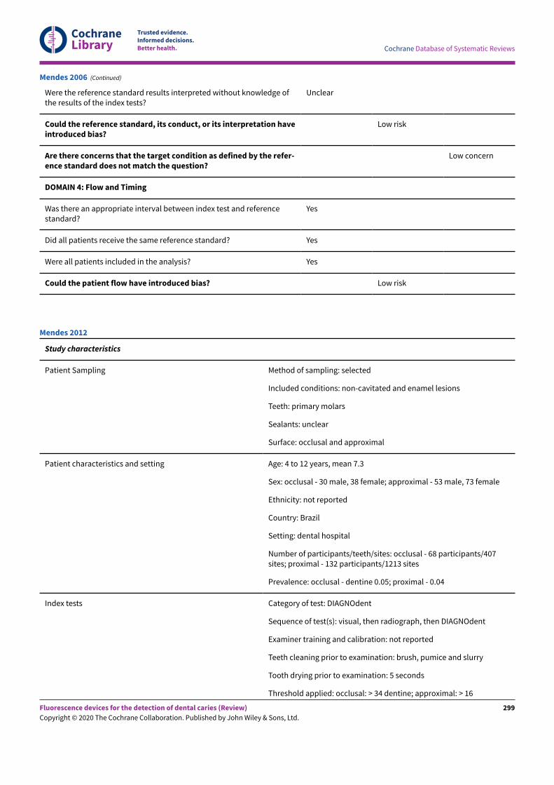
CochraneLibrary
Trusted evidence.Informed decisions.Better health.
Cochrane Database of Systematic Reviews
Were the reference standard results interpreted without knowledge ofthe results of the index tests?
Unclear
Could the reference standard, its conduct, or its interpretation haveintroduced bias?
Low risk
Are there concerns that the target condition as defined by the refer-ence standard does not match the question?
Low concern
DOMAIN 4: Flow and Timing
Was there an appropriate interval between index test and referencestandard?
Yes
Did all patients receive the same reference standard? Yes
Were all patients included in the analysis? Yes
Could the patient flow have introduced bias? Low risk
Mendes 2006 (Continued)
Study characteristics
Patient Sampling Method of sampling: selected
Included conditions: non-cavitated and enamel lesions
Teeth: primary molars
Sealants: unclear
Surface: occlusal and approximal
Patient characteristics and setting Age: 4 to 12 years, mean 7.3
Sex: occlusal - 30 male, 38 female; approximal - 53 male, 73 female
Ethnicity: not reported
Country: Brazil
Setting: dental hospital
Number of participants/teeth/sites: occlusal - 68 participants/407sites; proximal - 132 participants/1213 sites
Prevalence: occlusal - dentine 0.05; proximal - 0.04
Index tests Category of test: DIAGNOdent
Sequence of test(s): visual, then radiograph, then DIAGNOdent
Examiner training and calibration: not reported
Teeth cleaning prior to examination: brush, pumice and slurry
Tooth drying prior to examination: 5 seconds
Threshold applied: occlusal: > 34 dentine; approximal: > 16
Mendes 2012
Fluorescence devices for the detection of dental caries (Review)
Copyright © 2020 The Cochrane Collaboration. Published by John Wiley & Sons, Ltd.
299
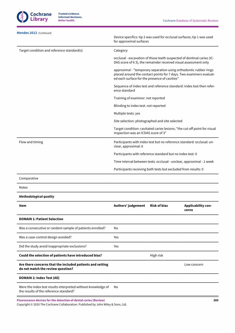
CochraneLibrary
Trusted evidence.Informed decisions.Better health.
Cochrane Database of Systematic Reviews
Device specifics: tip 2 was used for occlusal surfaces; tip 1 was usedfor approximal surfaces
Target condition and reference standard(s) Category:
occlusal - excavation of those teeth suspected of dentinal caries (IC-DAS score of 6 3), the remainder received visual assessment only
approximal - "temporary separation using orthodontic rubber ringsplaced around the contact points for 7 days. Two examiners evaluat-ed each surface for the presence of cavities"
Sequence of index test and reference standard: index test then refer-ence standard
Training of examiner: not reported
Blinding to index test: not reported
Multiple tests: yes
Site selection: photographed and site selected
Target condition: cavitated caries lesions; "the cut-oE point for visualinspection was an ICDAS score of 3"
Flow and timing Participants with index test but no reference standard: occlusal: un-clear, approximal: 6
Participants with reference standard but no index test: 0
Time interval between tests: occlusal - unclear, approximal - 1 week
Participants receiving both tests but excluded from results: 0
Comparative
Notes
Methodological quality
Item Authors' judgement Risk of bias Applicability con-cerns
DOMAIN 1: Patient Selection
Was a consecutive or random sample of patients enrolled? No
Was a case-control design avoided? Yes
Did the study avoid inappropriate exclusions? Yes
Could the selection of patients have introduced bias? High risk
Are there concerns that the included patients and settingdo not match the review question?
Low concern
DOMAIN 2: Index Test (All)
Were the index test results interpreted without knowledge ofthe results of the reference standard?
No
Mendes 2012 (Continued)
Fluorescence devices for the detection of dental caries (Review)
Copyright © 2020 The Cochrane Collaboration. Published by John Wiley & Sons, Ltd.
300
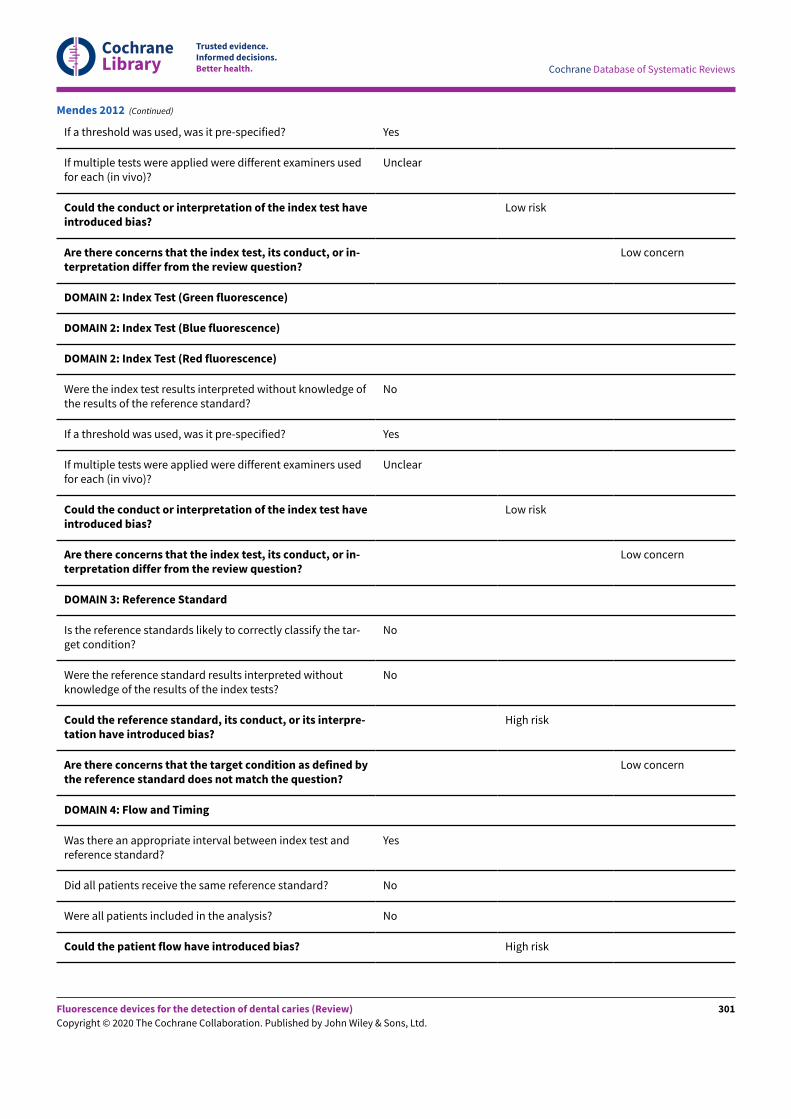
CochraneLibrary
Trusted evidence.Informed decisions.Better health.
Cochrane Database of Systematic Reviews
If a threshold was used, was it pre-specified? Yes
If multiple tests were applied were different examiners usedfor each (in vivo)?
Unclear
Could the conduct or interpretation of the index test haveintroduced bias?
Low risk
Are there concerns that the index test, its conduct, or in-terpretation differ from the review question?
Low concern
DOMAIN 2: Index Test (Green fluorescence)
DOMAIN 2: Index Test (Blue fluorescence)
DOMAIN 2: Index Test (Red fluorescence)
Were the index test results interpreted without knowledge ofthe results of the reference standard?
No
If a threshold was used, was it pre-specified? Yes
If multiple tests were applied were different examiners usedfor each (in vivo)?
Unclear
Could the conduct or interpretation of the index test haveintroduced bias?
Low risk
Are there concerns that the index test, its conduct, or in-terpretation differ from the review question?
Low concern
DOMAIN 3: Reference Standard
Is the reference standards likely to correctly classify the tar-get condition?
No
Were the reference standard results interpreted withoutknowledge of the results of the index tests?
No
Could the reference standard, its conduct, or its interpre-tation have introduced bias?
High risk
Are there concerns that the target condition as defined bythe reference standard does not match the question?
Low concern
DOMAIN 4: Flow and Timing
Was there an appropriate interval between index test andreference standard?
Yes
Did all patients receive the same reference standard? No
Were all patients included in the analysis? No
Could the patient flow have introduced bias? High risk
Mendes 2012 (Continued)
Fluorescence devices for the detection of dental caries (Review)
Copyright © 2020 The Cochrane Collaboration. Published by John Wiley & Sons, Ltd.
301
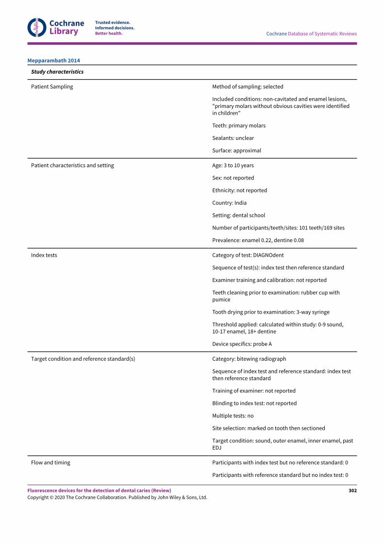
CochraneLibrary
Trusted evidence.Informed decisions.Better health.
Cochrane Database of Systematic Reviews
Study characteristics
Patient Sampling Method of sampling: selected
Included conditions: non-cavitated and enamel lesions,"primary molars without obvious cavities were identifiedin children"
Teeth: primary molars
Sealants: unclear
Surface: approximal
Patient characteristics and setting Age: 3 to 10 years
Sex: not reported
Ethnicity: not reported
Country: India
Setting: dental school
Number of participants/teeth/sites: 101 teeth/169 sites
Prevalence: enamel 0.22, dentine 0.08
Index tests Category of test: DIAGNOdent
Sequence of test(s): index test then reference standard
Examiner training and calibration: not reported
Teeth cleaning prior to examination: rubber cup withpumice
Tooth drying prior to examination: 3-way syringe
Threshold applied: calculated within study: 0-9 sound,10-17 enamel, 18+ dentine
Device specifics: probe A
Target condition and reference standard(s) Category: bitewing radiograph
Sequence of index test and reference standard: index testthen reference standard
Training of examiner: not reported
Blinding to index test: not reported
Multiple tests: no
Site selection: marked on tooth then sectioned
Target condition: sound, outer enamel, inner enamel, pastEDJ
Flow and timing Participants with index test but no reference standard: 0
Participants with reference standard but no index test: 0
Mepparambath 2014
Fluorescence devices for the detection of dental caries (Review)
Copyright © 2020 The Cochrane Collaboration. Published by John Wiley & Sons, Ltd.
302
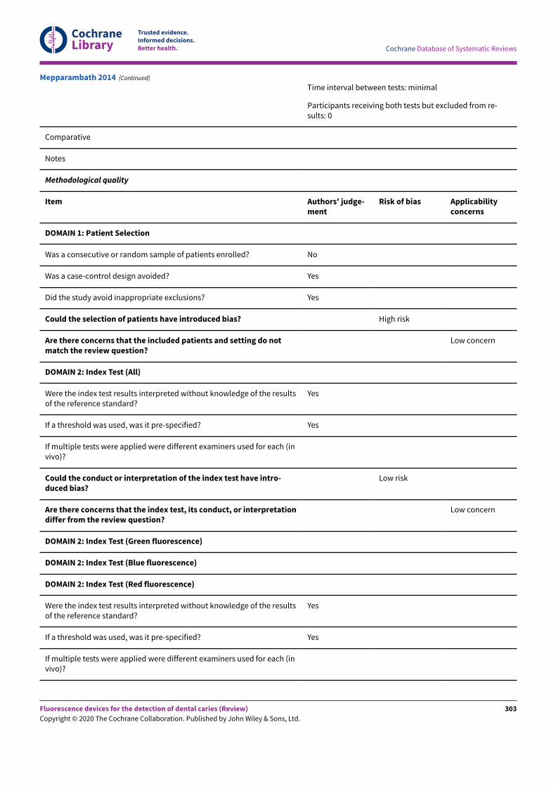
CochraneLibrary
Trusted evidence.Informed decisions.Better health.
Cochrane Database of Systematic Reviews
Time interval between tests: minimal
Participants receiving both tests but excluded from re-sults: 0
Comparative
Notes
Methodological quality
Item Authors' judge-ment
Risk of bias Applicabilityconcerns
DOMAIN 1: Patient Selection
Was a consecutive or random sample of patients enrolled? No
Was a case-control design avoided? Yes
Did the study avoid inappropriate exclusions? Yes
Could the selection of patients have introduced bias? High risk
Are there concerns that the included patients and setting do notmatch the review question?
Low concern
DOMAIN 2: Index Test (All)
Were the index test results interpreted without knowledge of the resultsof the reference standard?
Yes
If a threshold was used, was it pre-specified? Yes
If multiple tests were applied were different examiners used for each (invivo)?
Could the conduct or interpretation of the index test have intro-duced bias?
Low risk
Are there concerns that the index test, its conduct, or interpretationdiffer from the review question?
Low concern
DOMAIN 2: Index Test (Green fluorescence)
DOMAIN 2: Index Test (Blue fluorescence)
DOMAIN 2: Index Test (Red fluorescence)
Were the index test results interpreted without knowledge of the resultsof the reference standard?
Yes
If a threshold was used, was it pre-specified? Yes
If multiple tests were applied were different examiners used for each (invivo)?
Mepparambath 2014 (Continued)
Fluorescence devices for the detection of dental caries (Review)
Copyright © 2020 The Cochrane Collaboration. Published by John Wiley & Sons, Ltd.
303
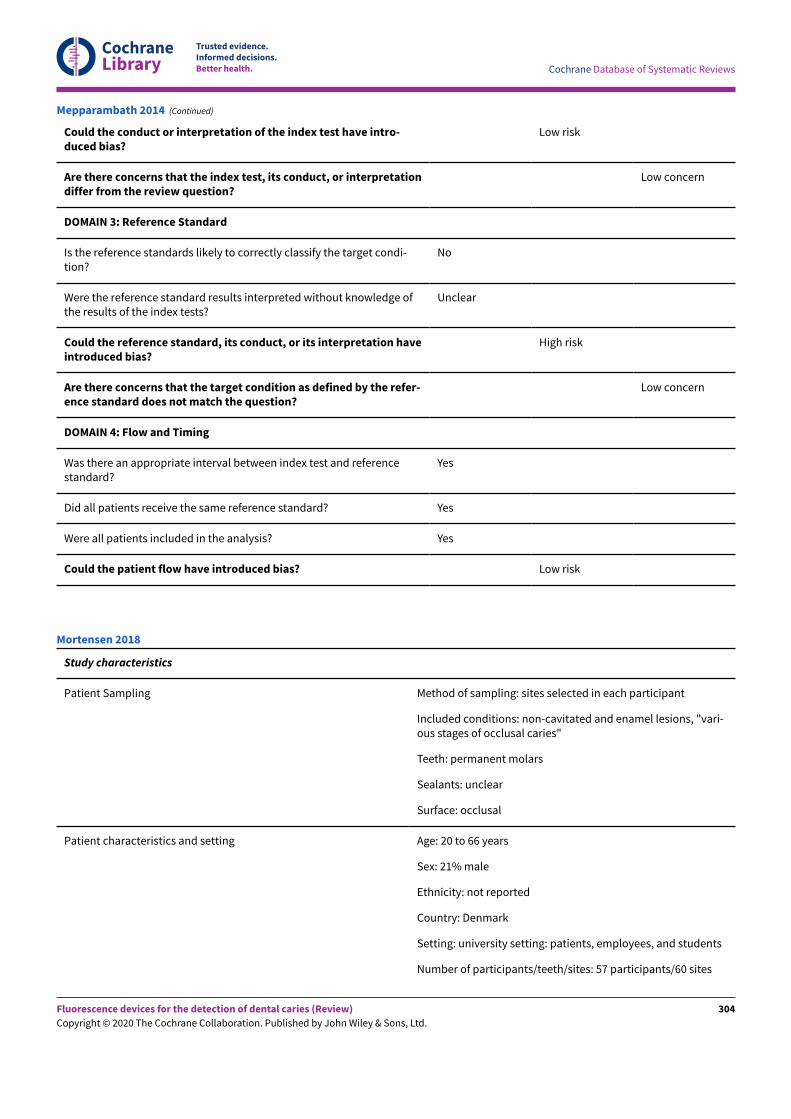
CochraneLibrary
Trusted evidence.Informed decisions.Better health.
Cochrane Database of Systematic Reviews
Could the conduct or interpretation of the index test have intro-duced bias?
Low risk
Are there concerns that the index test, its conduct, or interpretationdiffer from the review question?
Low concern
DOMAIN 3: Reference Standard
Is the reference standards likely to correctly classify the target condi-tion?
No
Were the reference standard results interpreted without knowledge ofthe results of the index tests?
Unclear
Could the reference standard, its conduct, or its interpretation haveintroduced bias?
High risk
Are there concerns that the target condition as defined by the refer-ence standard does not match the question?
Low concern
DOMAIN 4: Flow and Timing
Was there an appropriate interval between index test and referencestandard?
Yes
Did all patients receive the same reference standard? Yes
Were all patients included in the analysis? Yes
Could the patient flow have introduced bias? Low risk
Mepparambath 2014 (Continued)
Study characteristics
Patient Sampling Method of sampling: sites selected in each participant
Included conditions: non-cavitated and enamel lesions, "vari-ous stages of occlusal caries"
Teeth: permanent molars
Sealants: unclear
Surface: occlusal
Patient characteristics and setting Age: 20 to 66 years
Sex: 21% male
Ethnicity: not reported
Country: Denmark
Setting: university setting: patients, employees, and students
Number of participants/teeth/sites: 57 participants/60 sites
Mortensen 2018
Fluorescence devices for the detection of dental caries (Review)
Copyright © 2020 The Cochrane Collaboration. Published by John Wiley & Sons, Ltd.
304
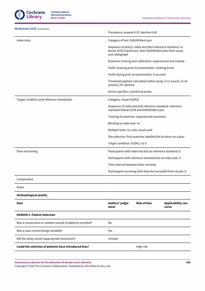
CochraneLibrary
Trusted evidence.Informed decisions.Better health.
Cochrane Database of Systematic Reviews
Prevalence: enamel 0.97, dentine 0.45
Index tests Category of test: DIAGNOdent pen
Sequence of test(s): index test then reference standard, or-dered: ECM (CarieScan), then DIAGNOdent pen then visualand radiograph
Examiner training and calibration: experienced and trained
Teeth cleaning prior to examination: rotating brush
Tooth drying prior to examination: 5 seconds
Threshold applied: calculated within study: 0-12 sound, 13-24enamel, 25+ dentine
Device specifics: cylindrical probe
Target condition and reference standard(s) Category: visual (ICDAS)
Sequence of index test and reference standard: referencestandard follows ECM and DIAGNOdent pen
Training of examiner: experienced examiners
Blinding to index test: no
Multiple tests: no, only visual used
Site selection: first examiner labelled the location on a plan
Target condition: ICDAS 1 to 5
Flow and timing Participants with index test but no reference standard: 0
Participants with reference standard but no index test: 0
Time interval between tests: minimal
Participants receiving both tests but excluded from results: 0
Comparative
Notes
Methodological quality
Item Authors' judge-ment
Risk of bias Applicability con-cerns
DOMAIN 1: Patient Selection
Was a consecutive or random sample of patients enrolled? No
Was a case-control design avoided? Yes
Did the study avoid inappropriate exclusions? Unclear
Could the selection of patients have introduced bias? High risk
Mortensen 2018 (Continued)
Fluorescence devices for the detection of dental caries (Review)
Copyright © 2020 The Cochrane Collaboration. Published by John Wiley & Sons, Ltd.
305
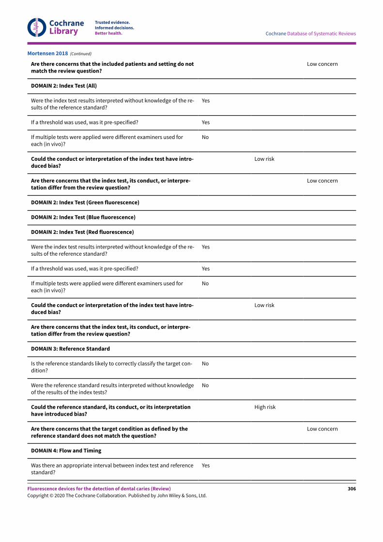
CochraneLibrary
Trusted evidence.Informed decisions.Better health.
Cochrane Database of Systematic Reviews
Are there concerns that the included patients and setting do notmatch the review question?
Low concern
DOMAIN 2: Index Test (All)
Were the index test results interpreted without knowledge of the re-sults of the reference standard?
Yes
If a threshold was used, was it pre-specified? Yes
If multiple tests were applied were different examiners used foreach (in vivo)?
No
Could the conduct or interpretation of the index test have intro-duced bias?
Low risk
Are there concerns that the index test, its conduct, or interpre-tation differ from the review question?
Low concern
DOMAIN 2: Index Test (Green fluorescence)
DOMAIN 2: Index Test (Blue fluorescence)
DOMAIN 2: Index Test (Red fluorescence)
Were the index test results interpreted without knowledge of the re-sults of the reference standard?
Yes
If a threshold was used, was it pre-specified? Yes
If multiple tests were applied were different examiners used foreach (in vivo)?
No
Could the conduct or interpretation of the index test have intro-duced bias?
Low risk
Are there concerns that the index test, its conduct, or interpre-tation differ from the review question?
DOMAIN 3: Reference Standard
Is the reference standards likely to correctly classify the target con-dition?
No
Were the reference standard results interpreted without knowledgeof the results of the index tests?
No
Could the reference standard, its conduct, or its interpretationhave introduced bias?
High risk
Are there concerns that the target condition as defined by thereference standard does not match the question?
Low concern
DOMAIN 4: Flow and Timing
Was there an appropriate interval between index test and referencestandard?
Yes
Mortensen 2018 (Continued)
Fluorescence devices for the detection of dental caries (Review)
Copyright © 2020 The Cochrane Collaboration. Published by John Wiley & Sons, Ltd.
306
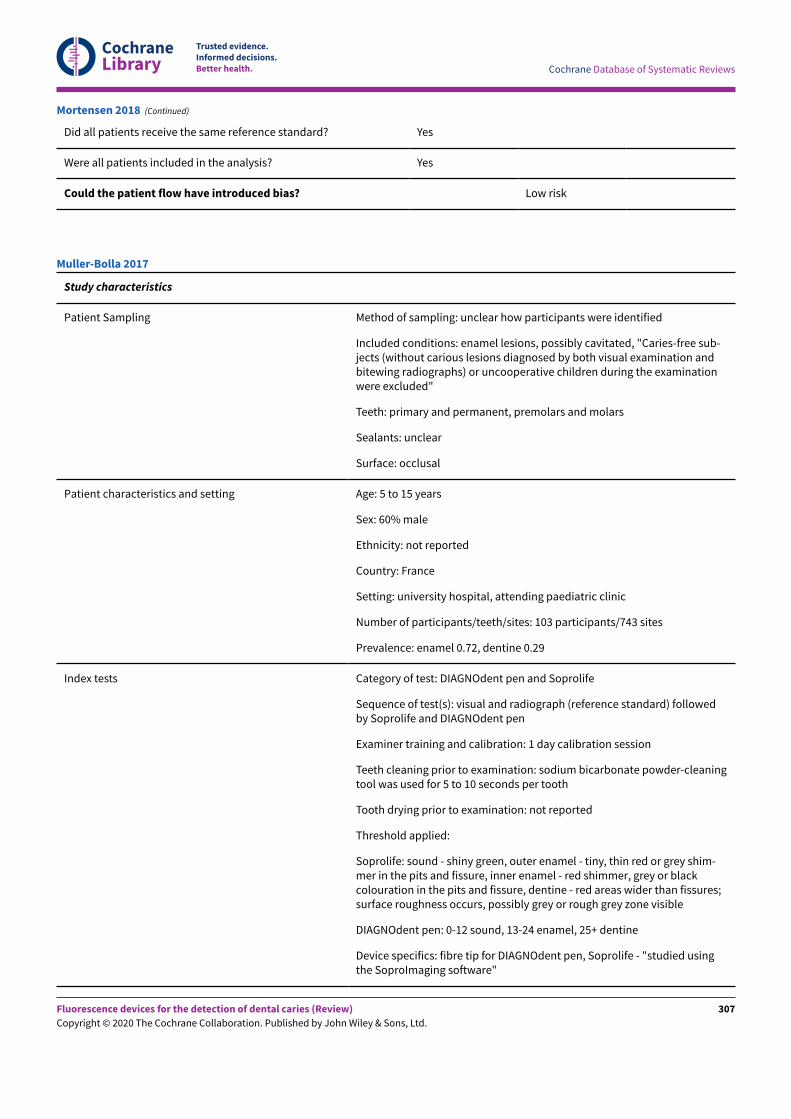
CochraneLibrary
Trusted evidence.Informed decisions.Better health.
Cochrane Database of Systematic Reviews
Did all patients receive the same reference standard? Yes
Were all patients included in the analysis? Yes
Could the patient flow have introduced bias? Low risk
Mortensen 2018 (Continued)
Study characteristics
Patient Sampling Method of sampling: unclear how participants were identified
Included conditions: enamel lesions, possibly cavitated, "Caries-free sub-jects (without carious lesions diagnosed by both visual examination andbitewing radiographs) or uncooperative children during the examinationwere excluded"
Teeth: primary and permanent, premolars and molars
Sealants: unclear
Surface: occlusal
Patient characteristics and setting Age: 5 to 15 years
Sex: 60% male
Ethnicity: not reported
Country: France
Setting: university hospital, attending paediatric clinic
Number of participants/teeth/sites: 103 participants/743 sites
Prevalence: enamel 0.72, dentine 0.29
Index tests Category of test: DIAGNOdent pen and Soprolife
Sequence of test(s): visual and radiograph (reference standard) followedby Soprolife and DIAGNOdent pen
Examiner training and calibration: 1 day calibration session
Teeth cleaning prior to examination: sodium bicarbonate powder-cleaningtool was used for 5 to 10 seconds per tooth
Tooth drying prior to examination: not reported
Threshold applied:
Soprolife: sound - shiny green, outer enamel - tiny, thin red or grey shim-mer in the pits and fissure, inner enamel - red shimmer, grey or blackcolouration in the pits and fissure, dentine - red areas wider than fissures;surface roughness occurs, possibly grey or rough grey zone visible
DIAGNOdent pen: 0-12 sound, 13-24 enamel, 25+ dentine
Device specifics: fibre tip for DIAGNOdent pen, Soprolife - "studied usingthe SoproImaging software"
Muller-Bolla 2017
Fluorescence devices for the detection of dental caries (Review)
Copyright © 2020 The Cochrane Collaboration. Published by John Wiley & Sons, Ltd.
307
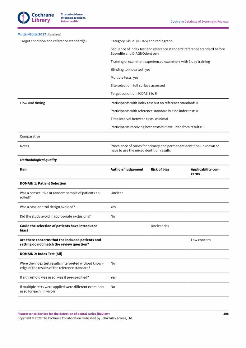
CochraneLibrary
Trusted evidence.Informed decisions.Better health.
Cochrane Database of Systematic Reviews
Target condition and reference standard(s) Category: visual (ICDAS) and radiograph
Sequence of index test and reference standard: reference standard beforeSoprolife and DIAGNOdent pen
Training of examiner: experienced examiners with 1 day training
Blinding to index test: yes
Multiple tests: yes
Site selection: full surface assessed
Target condition: ICDAS 1 to 6
Flow and timing Participants with index test but no reference standard: 0
Participants with reference standard but no index test: 0
Time interval between tests: minimal
Participants receiving both tests but excluded from results: 0
Comparative
Notes Prevalence of caries for primary and permanent dentition unknown sohave to use the mixed dentition results
Methodological quality
Item Authors' judgement Risk of bias Applicability con-cerns
DOMAIN 1: Patient Selection
Was a consecutive or random sample of patients en-rolled?
Unclear
Was a case-control design avoided? Yes
Did the study avoid inappropriate exclusions? No
Could the selection of patients have introducedbias?
Unclear risk
Are there concerns that the included patients andsetting do not match the review question?
Low concern
DOMAIN 2: Index Test (All)
Were the index test results interpreted without knowl-edge of the results of the reference standard?
No
If a threshold was used, was it pre-specified? Yes
If multiple tests were applied were different examinersused for each (in vivo)?
No
Muller-Bolla 2017 (Continued)
Fluorescence devices for the detection of dental caries (Review)
Copyright © 2020 The Cochrane Collaboration. Published by John Wiley & Sons, Ltd.
308
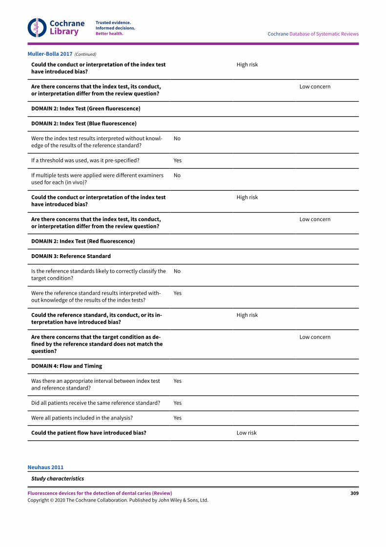
CochraneLibrary
Trusted evidence.Informed decisions.Better health.
Cochrane Database of Systematic Reviews
Could the conduct or interpretation of the index testhave introduced bias?
High risk
Are there concerns that the index test, its conduct,or interpretation differ from the review question?
Low concern
DOMAIN 2: Index Test (Green fluorescence)
DOMAIN 2: Index Test (Blue fluorescence)
Were the index test results interpreted without knowl-edge of the results of the reference standard?
No
If a threshold was used, was it pre-specified? Yes
If multiple tests were applied were different examinersused for each (in vivo)?
No
Could the conduct or interpretation of the index testhave introduced bias?
High risk
Are there concerns that the index test, its conduct,or interpretation differ from the review question?
Low concern
DOMAIN 2: Index Test (Red fluorescence)
DOMAIN 3: Reference Standard
Is the reference standards likely to correctly classify thetarget condition?
No
Were the reference standard results interpreted with-out knowledge of the results of the index tests?
Yes
Could the reference standard, its conduct, or its in-terpretation have introduced bias?
High risk
Are there concerns that the target condition as de-fined by the reference standard does not match thequestion?
Low concern
DOMAIN 4: Flow and Timing
Was there an appropriate interval between index testand reference standard?
Yes
Did all patients receive the same reference standard? Yes
Were all patients included in the analysis? Yes
Could the patient flow have introduced bias? Low risk
Muller-Bolla 2017 (Continued)
Study characteristics
Neuhaus 2011
Fluorescence devices for the detection of dental caries (Review)
Copyright © 2020 The Cochrane Collaboration. Published by John Wiley & Sons, Ltd.
309
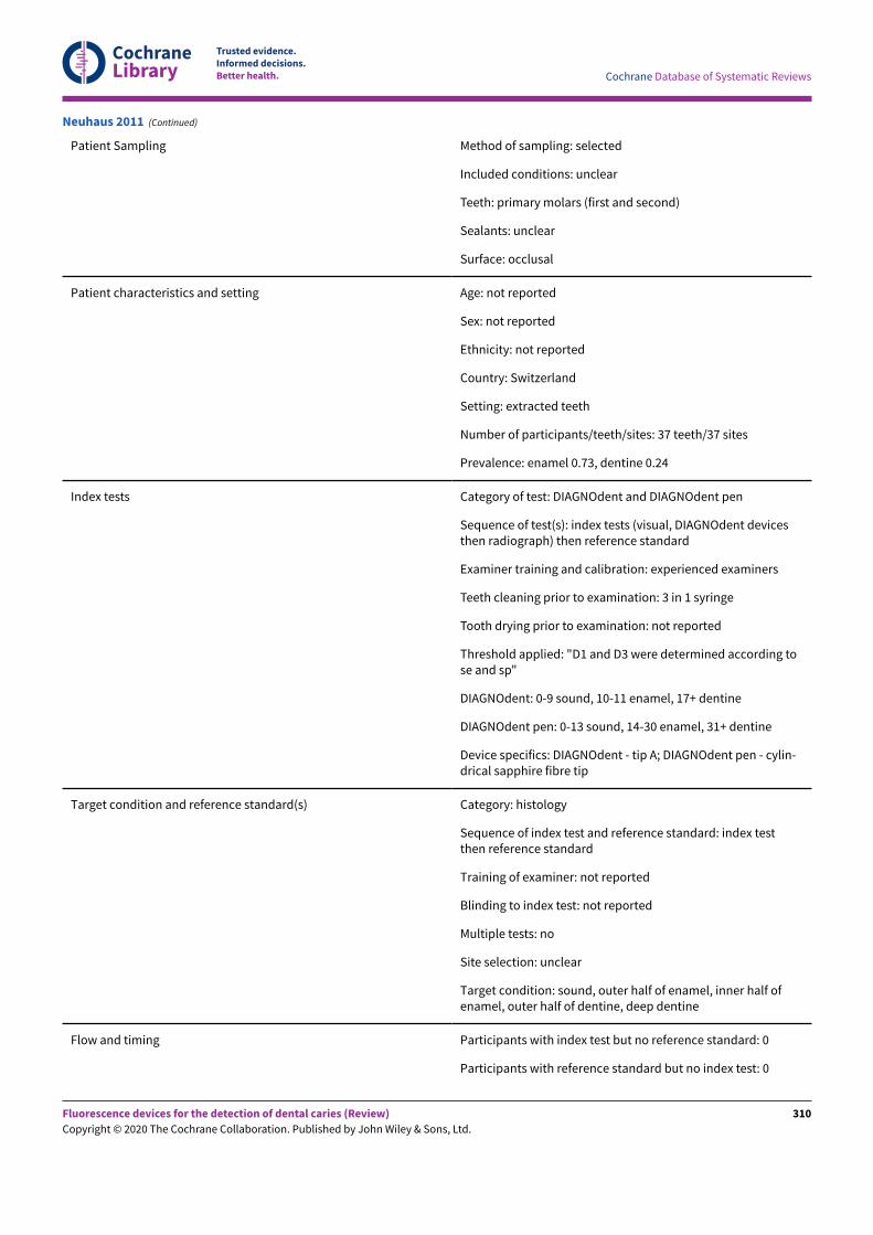
CochraneLibrary
Trusted evidence.Informed decisions.Better health.
Cochrane Database of Systematic Reviews
Patient Sampling Method of sampling: selected
Included conditions: unclear
Teeth: primary molars (first and second)
Sealants: unclear
Surface: occlusal
Patient characteristics and setting Age: not reported
Sex: not reported
Ethnicity: not reported
Country: Switzerland
Setting: extracted teeth
Number of participants/teeth/sites: 37 teeth/37 sites
Prevalence: enamel 0.73, dentine 0.24
Index tests Category of test: DIAGNOdent and DIAGNOdent pen
Sequence of test(s): index tests (visual, DIAGNOdent devicesthen radiograph) then reference standard
Examiner training and calibration: experienced examiners
Teeth cleaning prior to examination: 3 in 1 syringe
Tooth drying prior to examination: not reported
Threshold applied: "D1 and D3 were determined according tose and sp"
DIAGNOdent: 0-9 sound, 10-11 enamel, 17+ dentine
DIAGNOdent pen: 0-13 sound, 14-30 enamel, 31+ dentine
Device specifics: DIAGNOdent - tip A; DIAGNOdent pen - cylin-drical sapphire fibre tip
Target condition and reference standard(s) Category: histology
Sequence of index test and reference standard: index testthen reference standard
Training of examiner: not reported
Blinding to index test: not reported
Multiple tests: no
Site selection: unclear
Target condition: sound, outer half of enamel, inner half ofenamel, outer half of dentine, deep dentine
Flow and timing Participants with index test but no reference standard: 0
Participants with reference standard but no index test: 0
Neuhaus 2011 (Continued)
Fluorescence devices for the detection of dental caries (Review)
Copyright © 2020 The Cochrane Collaboration. Published by John Wiley & Sons, Ltd.
310
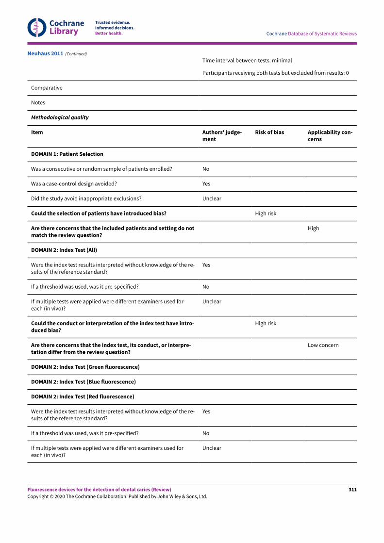
CochraneLibrary
Trusted evidence.Informed decisions.Better health.
Cochrane Database of Systematic Reviews
Time interval between tests: minimal
Participants receiving both tests but excluded from results: 0
Comparative
Notes
Methodological quality
Item Authors' judge-ment
Risk of bias Applicability con-cerns
DOMAIN 1: Patient Selection
Was a consecutive or random sample of patients enrolled? No
Was a case-control design avoided? Yes
Did the study avoid inappropriate exclusions? Unclear
Could the selection of patients have introduced bias? High risk
Are there concerns that the included patients and setting do notmatch the review question?
High
DOMAIN 2: Index Test (All)
Were the index test results interpreted without knowledge of the re-sults of the reference standard?
Yes
If a threshold was used, was it pre-specified? No
If multiple tests were applied were different examiners used foreach (in vivo)?
Unclear
Could the conduct or interpretation of the index test have intro-duced bias?
High risk
Are there concerns that the index test, its conduct, or interpre-tation differ from the review question?
Low concern
DOMAIN 2: Index Test (Green fluorescence)
DOMAIN 2: Index Test (Blue fluorescence)
DOMAIN 2: Index Test (Red fluorescence)
Were the index test results interpreted without knowledge of the re-sults of the reference standard?
Yes
If a threshold was used, was it pre-specified? No
If multiple tests were applied were different examiners used foreach (in vivo)?
Unclear
Neuhaus 2011 (Continued)
Fluorescence devices for the detection of dental caries (Review)
Copyright © 2020 The Cochrane Collaboration. Published by John Wiley & Sons, Ltd.
311
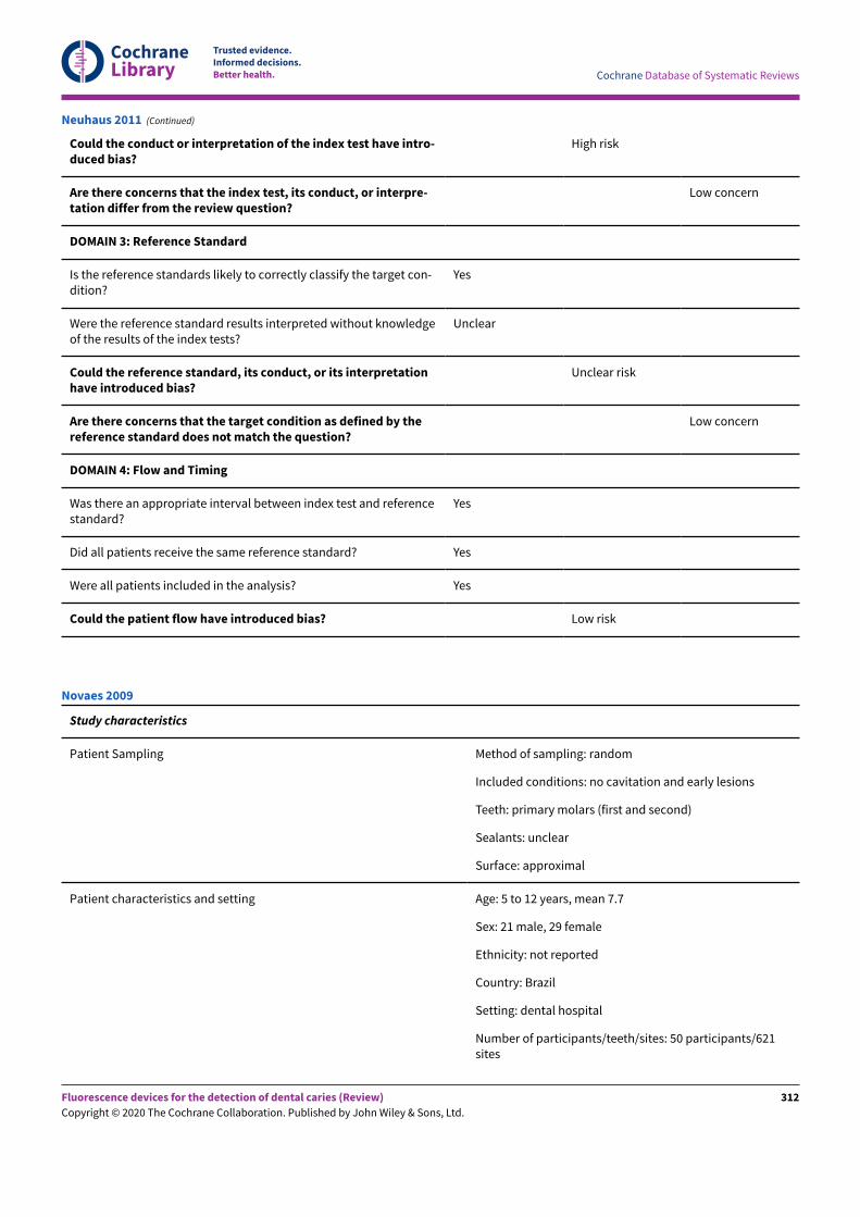
CochraneLibrary
Trusted evidence.Informed decisions.Better health.
Cochrane Database of Systematic Reviews
Could the conduct or interpretation of the index test have intro-duced bias?
High risk
Are there concerns that the index test, its conduct, or interpre-tation differ from the review question?
Low concern
DOMAIN 3: Reference Standard
Is the reference standards likely to correctly classify the target con-dition?
Yes
Were the reference standard results interpreted without knowledgeof the results of the index tests?
Unclear
Could the reference standard, its conduct, or its interpretationhave introduced bias?
Unclear risk
Are there concerns that the target condition as defined by thereference standard does not match the question?
Low concern
DOMAIN 4: Flow and Timing
Was there an appropriate interval between index test and referencestandard?
Yes
Did all patients receive the same reference standard? Yes
Were all patients included in the analysis? Yes
Could the patient flow have introduced bias? Low risk
Neuhaus 2011 (Continued)
Study characteristics
Patient Sampling Method of sampling: random
Included conditions: no cavitation and early lesions
Teeth: primary molars (first and second)
Sealants: unclear
Surface: approximal
Patient characteristics and setting Age: 5 to 12 years, mean 7.7
Sex: 21 male, 29 female
Ethnicity: not reported
Country: Brazil
Setting: dental hospital
Number of participants/teeth/sites: 50 participants/621sites
Novaes 2009
Fluorescence devices for the detection of dental caries (Review)
Copyright © 2020 The Cochrane Collaboration. Published by John Wiley & Sons, Ltd.
312
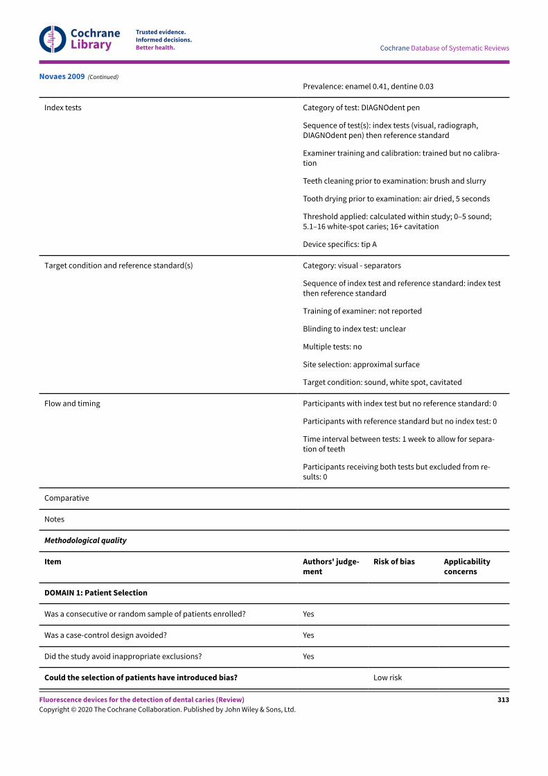
CochraneLibrary
Trusted evidence.Informed decisions.Better health.
Cochrane Database of Systematic Reviews
Prevalence: enamel 0.41, dentine 0.03
Index tests Category of test: DIAGNOdent pen
Sequence of test(s): index tests (visual, radiograph,DIAGNOdent pen) then reference standard
Examiner training and calibration: trained but no calibra-tion
Teeth cleaning prior to examination: brush and slurry
Tooth drying prior to examination: air dried, 5 seconds
Threshold applied: calculated within study; 0–5 sound;5.1–16 white-spot caries; 16+ cavitation
Device specifics: tip A
Target condition and reference standard(s) Category: visual - separators
Sequence of index test and reference standard: index testthen reference standard
Training of examiner: not reported
Blinding to index test: unclear
Multiple tests: no
Site selection: approximal surface
Target condition: sound, white spot, cavitated
Flow and timing Participants with index test but no reference standard: 0
Participants with reference standard but no index test: 0
Time interval between tests: 1 week to allow for separa-tion of teeth
Participants receiving both tests but excluded from re-sults: 0
Comparative
Notes
Methodological quality
Item Authors' judge-ment
Risk of bias Applicabilityconcerns
DOMAIN 1: Patient Selection
Was a consecutive or random sample of patients enrolled? Yes
Was a case-control design avoided? Yes
Did the study avoid inappropriate exclusions? Yes
Could the selection of patients have introduced bias? Low risk
Novaes 2009 (Continued)
Fluorescence devices for the detection of dental caries (Review)
Copyright © 2020 The Cochrane Collaboration. Published by John Wiley & Sons, Ltd.
313
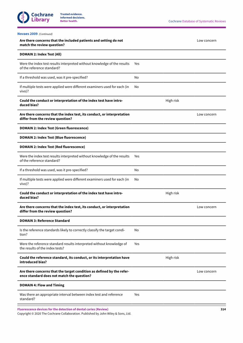
CochraneLibrary
Trusted evidence.Informed decisions.Better health.
Cochrane Database of Systematic Reviews
Are there concerns that the included patients and setting do notmatch the review question?
Low concern
DOMAIN 2: Index Test (All)
Were the index test results interpreted without knowledge of the resultsof the reference standard?
Yes
If a threshold was used, was it pre-specified? No
If multiple tests were applied were different examiners used for each (invivo)?
No
Could the conduct or interpretation of the index test have intro-duced bias?
High risk
Are there concerns that the index test, its conduct, or interpretationdiffer from the review question?
Low concern
DOMAIN 2: Index Test (Green fluorescence)
DOMAIN 2: Index Test (Blue fluorescence)
DOMAIN 2: Index Test (Red fluorescence)
Were the index test results interpreted without knowledge of the resultsof the reference standard?
Yes
If a threshold was used, was it pre-specified? No
If multiple tests were applied were different examiners used for each (invivo)?
No
Could the conduct or interpretation of the index test have intro-duced bias?
High risk
Are there concerns that the index test, its conduct, or interpretationdiffer from the review question?
Low concern
DOMAIN 3: Reference Standard
Is the reference standards likely to correctly classify the target condi-tion?
No
Were the reference standard results interpreted without knowledge ofthe results of the index tests?
Yes
Could the reference standard, its conduct, or its interpretation haveintroduced bias?
High risk
Are there concerns that the target condition as defined by the refer-ence standard does not match the question?
Low concern
DOMAIN 4: Flow and Timing
Was there an appropriate interval between index test and referencestandard?
Yes
Novaes 2009 (Continued)
Fluorescence devices for the detection of dental caries (Review)
Copyright © 2020 The Cochrane Collaboration. Published by John Wiley & Sons, Ltd.
314
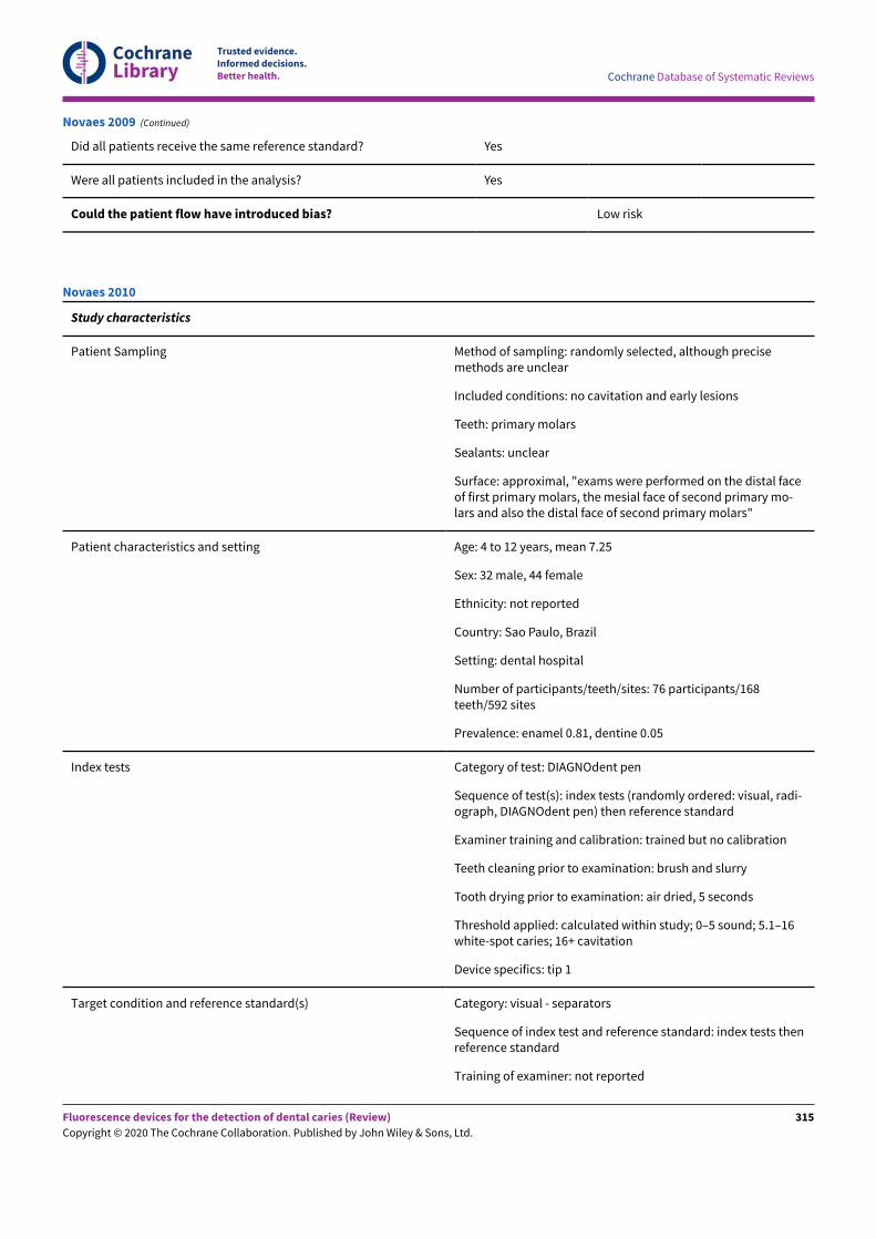
CochraneLibrary
Trusted evidence.Informed decisions.Better health.
Cochrane Database of Systematic Reviews
Did all patients receive the same reference standard? Yes
Were all patients included in the analysis? Yes
Could the patient flow have introduced bias? Low risk
Novaes 2009 (Continued)
Study characteristics
Patient Sampling Method of sampling: randomly selected, although precisemethods are unclear
Included conditions: no cavitation and early lesions
Teeth: primary molars
Sealants: unclear
Surface: approximal, "exams were performed on the distal faceof first primary molars, the mesial face of second primary mo-lars and also the distal face of second primary molars"
Patient characteristics and setting Age: 4 to 12 years, mean 7.25
Sex: 32 male, 44 female
Ethnicity: not reported
Country: Sao Paulo, Brazil
Setting: dental hospital
Number of participants/teeth/sites: 76 participants/168teeth/592 sites
Prevalence: enamel 0.81, dentine 0.05
Index tests Category of test: DIAGNOdent pen
Sequence of test(s): index tests (randomly ordered: visual, radi-ograph, DIAGNOdent pen) then reference standard
Examiner training and calibration: trained but no calibration
Teeth cleaning prior to examination: brush and slurry
Tooth drying prior to examination: air dried, 5 seconds
Threshold applied: calculated within study; 0–5 sound; 5.1–16white-spot caries; 16+ cavitation
Device specifics: tip 1
Target condition and reference standard(s) Category: visual - separators
Sequence of index test and reference standard: index tests thenreference standard
Training of examiner: not reported
Novaes 2010
Fluorescence devices for the detection of dental caries (Review)
Copyright © 2020 The Cochrane Collaboration. Published by John Wiley & Sons, Ltd.
315
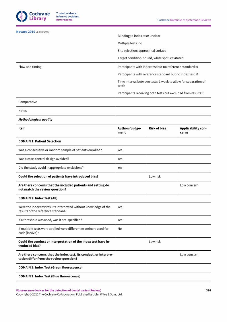
CochraneLibrary
Trusted evidence.Informed decisions.Better health.
Cochrane Database of Systematic Reviews
Blinding to index test: unclear
Multiple tests: no
Site selection: approximal surface
Target condition: sound, white spot, cavitated
Flow and timing Participants with index test but no reference standard: 0
Participants with reference standard but no index test: 0
Time interval between tests: 1 week to allow for separation ofteeth
Participants receiving both tests but excluded from results: 0
Comparative
Notes
Methodological quality
Item Authors' judge-ment
Risk of bias Applicability con-cerns
DOMAIN 1: Patient Selection
Was a consecutive or random sample of patients enrolled? Yes
Was a case-control design avoided? Yes
Did the study avoid inappropriate exclusions? Yes
Could the selection of patients have introduced bias? Low risk
Are there concerns that the included patients and setting donot match the review question?
Low concern
DOMAIN 2: Index Test (All)
Were the index test results interpreted without knowledge of theresults of the reference standard?
Yes
If a threshold was used, was it pre-specified? Yes
If multiple tests were applied were different examiners used foreach (in vivo)?
No
Could the conduct or interpretation of the index test have in-troduced bias?
Low risk
Are there concerns that the index test, its conduct, or interpre-tation differ from the review question?
Low concern
DOMAIN 2: Index Test (Green fluorescence)
DOMAIN 2: Index Test (Blue fluorescence)
Novaes 2010 (Continued)
Fluorescence devices for the detection of dental caries (Review)
Copyright © 2020 The Cochrane Collaboration. Published by John Wiley & Sons, Ltd.
316
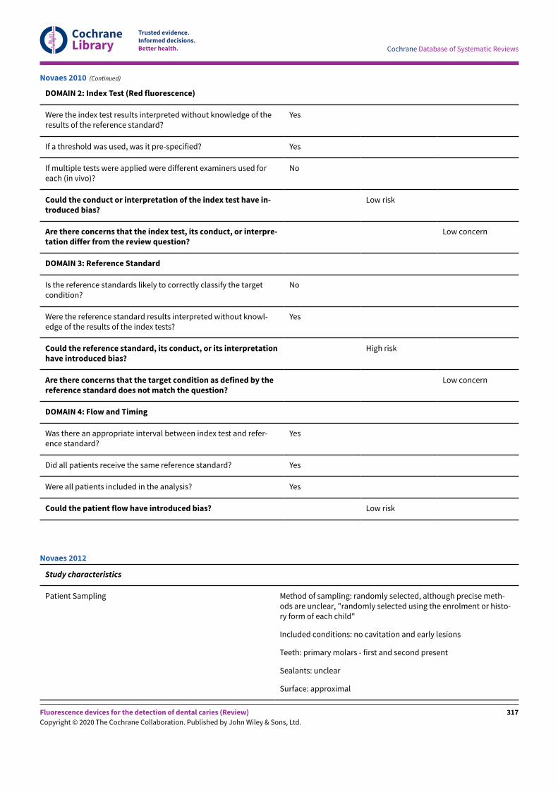
CochraneLibrary
Trusted evidence.Informed decisions.Better health.
Cochrane Database of Systematic Reviews
DOMAIN 2: Index Test (Red fluorescence)
Were the index test results interpreted without knowledge of theresults of the reference standard?
Yes
If a threshold was used, was it pre-specified? Yes
If multiple tests were applied were different examiners used foreach (in vivo)?
No
Could the conduct or interpretation of the index test have in-troduced bias?
Low risk
Are there concerns that the index test, its conduct, or interpre-tation differ from the review question?
Low concern
DOMAIN 3: Reference Standard
Is the reference standards likely to correctly classify the targetcondition?
No
Were the reference standard results interpreted without knowl-edge of the results of the index tests?
Yes
Could the reference standard, its conduct, or its interpretationhave introduced bias?
High risk
Are there concerns that the target condition as defined by thereference standard does not match the question?
Low concern
DOMAIN 4: Flow and Timing
Was there an appropriate interval between index test and refer-ence standard?
Yes
Did all patients receive the same reference standard? Yes
Were all patients included in the analysis? Yes
Could the patient flow have introduced bias? Low risk
Novaes 2010 (Continued)
Study characteristics
Patient Sampling Method of sampling: randomly selected, although precise meth-ods are unclear, "randomly selected using the enrolment or histo-ry form of each child"
Included conditions: no cavitation and early lesions
Teeth: primary molars - first and second present
Sealants: unclear
Surface: approximal
Novaes 2012
Fluorescence devices for the detection of dental caries (Review)
Copyright © 2020 The Cochrane Collaboration. Published by John Wiley & Sons, Ltd.
317
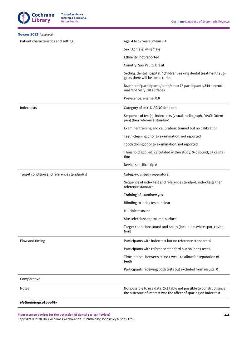
CochraneLibrary
Trusted evidence.Informed decisions.Better health.
Cochrane Database of Systematic Reviews
Patient characteristics and setting Age: 4 to 12 years, mean 7.4
Sex: 32 male, 44 female
Ethnicity: not reported
Country: Sao Paulo, Brazil
Setting: dental hospital, "children seeking dental treatment" sug-gests there will be some caries
Number of participants/teeth/sites: 76 participants/344 approxi-mal "spaces"/520 surfaces
Prevalence: enamel 0.8
Index tests Category of test: DIAGNOdent pen
Sequence of test(s): index tests (visual, radiograph, DIAGNOdentpen) then reference standard
Examiner training and calibration: trained but no calibration
Teeth cleaning prior to examination: not reported
Tooth drying prior to examination: not reported
Threshold applied: calculated within study; 0–5 sound; 6+ cavita-tion
Device specifics: tip A
Target condition and reference standard(s) Category: visual - separators
Sequence of index test and reference standard: index tests thenreference standard
Training of examiner: yes
Blinding to index test: unclear
Multiple tests: no
Site selection: approximal surface
Target condition: sound and caries (including: white spot, cavita-tion)
Flow and timing Participants with index test but no reference standard: 0
Participants with reference standard but no index test: 0
Time interval between tests: 1 week to allow for separation ofteeth
Participants receiving both tests but excluded from results: 0
Comparative
Notes Not possible to use data, 2x2 table not possible to construct sincethe outcome of interest was the affect of spacing on index test
Methodological quality
Novaes 2012 (Continued)
Fluorescence devices for the detection of dental caries (Review)
Copyright © 2020 The Cochrane Collaboration. Published by John Wiley & Sons, Ltd.
318
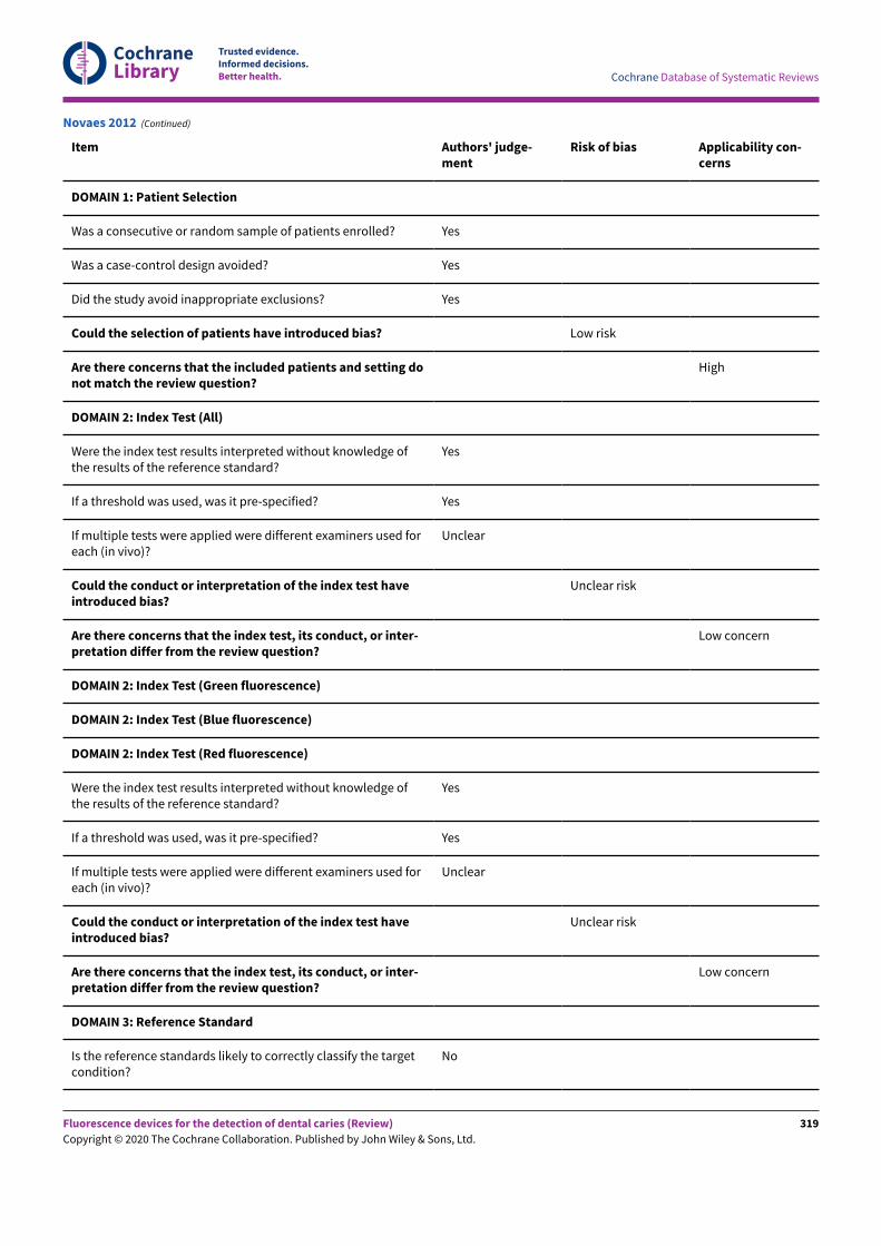
CochraneLibrary
Trusted evidence.Informed decisions.Better health.
Cochrane Database of Systematic Reviews
Item Authors' judge-ment
Risk of bias Applicability con-cerns
DOMAIN 1: Patient Selection
Was a consecutive or random sample of patients enrolled? Yes
Was a case-control design avoided? Yes
Did the study avoid inappropriate exclusions? Yes
Could the selection of patients have introduced bias? Low risk
Are there concerns that the included patients and setting donot match the review question?
High
DOMAIN 2: Index Test (All)
Were the index test results interpreted without knowledge ofthe results of the reference standard?
Yes
If a threshold was used, was it pre-specified? Yes
If multiple tests were applied were different examiners used foreach (in vivo)?
Unclear
Could the conduct or interpretation of the index test haveintroduced bias?
Unclear risk
Are there concerns that the index test, its conduct, or inter-pretation differ from the review question?
Low concern
DOMAIN 2: Index Test (Green fluorescence)
DOMAIN 2: Index Test (Blue fluorescence)
DOMAIN 2: Index Test (Red fluorescence)
Were the index test results interpreted without knowledge ofthe results of the reference standard?
Yes
If a threshold was used, was it pre-specified? Yes
If multiple tests were applied were different examiners used foreach (in vivo)?
Unclear
Could the conduct or interpretation of the index test haveintroduced bias?
Unclear risk
Are there concerns that the index test, its conduct, or inter-pretation differ from the review question?
Low concern
DOMAIN 3: Reference Standard
Is the reference standards likely to correctly classify the targetcondition?
No
Novaes 2012 (Continued)
Fluorescence devices for the detection of dental caries (Review)
Copyright © 2020 The Cochrane Collaboration. Published by John Wiley & Sons, Ltd.
319
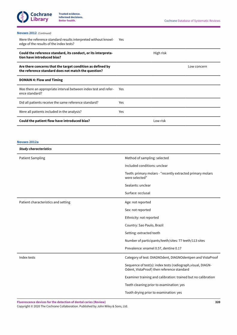
CochraneLibrary
Trusted evidence.Informed decisions.Better health.
Cochrane Database of Systematic Reviews
Were the reference standard results interpreted without knowl-edge of the results of the index tests?
Yes
Could the reference standard, its conduct, or its interpreta-tion have introduced bias?
High risk
Are there concerns that the target condition as defined bythe reference standard does not match the question?
Low concern
DOMAIN 4: Flow and Timing
Was there an appropriate interval between index test and refer-ence standard?
Yes
Did all patients receive the same reference standard? Yes
Were all patients included in the analysis? Yes
Could the patient flow have introduced bias? Low risk
Novaes 2012 (Continued)
Study characteristics
Patient Sampling Method of sampling: selected
Included conditions: unclear
Teeth: primary molars - "recently extracted primary molarswere selected"
Sealants: unclear
Surface: occlusal
Patient characteristics and setting Age: not reported
Sex: not reported
Ethnicity: not reported
Country: Sao Paulo, Brazil
Setting: extracted teeth
Number of participants/teeth/sites: 77 teeth/113 sites
Prevalence: enamel 0.57, dentine 0.17
Index tests Category of test: DIAGNOdent, DIAGNOdentpen and VistaProof
Sequence of test(s): index tests (radiograph,visual, DIAGN-Odent, VistaProof) then reference standard
Examiner training and calibration: trained but no calibration
Teeth cleaning prior to examination: yes
Tooth drying prior to examination: yes
Novaes 2012a
Fluorescence devices for the detection of dental caries (Review)
Copyright © 2020 The Cochrane Collaboration. Published by John Wiley & Sons, Ltd.
320
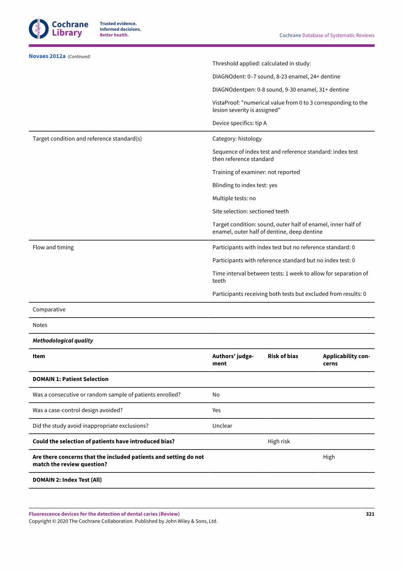
CochraneLibrary
Trusted evidence.Informed decisions.Better health.
Cochrane Database of Systematic Reviews
Threshold applied: calculated in study:
DIAGNOdent: 0–7 sound, 8-23 enamel, 24+ dentine
DIAGNOdentpen: 0-8 sound, 9-30 enamel, 31+ dentine
VistaProof: "numerical value from 0 to 3 corresponding to thelesion severity is assigned"
Device specifics: tip A
Target condition and reference standard(s) Category: histology
Sequence of index test and reference standard: index testthen reference standard
Training of examiner: not reported
Blinding to index test: yes
Multiple tests: no
Site selection: sectioned teeth
Target condition: sound, outer half of enamel, inner half ofenamel, outer half of dentine, deep dentine
Flow and timing Participants with index test but no reference standard: 0
Participants with reference standard but no index test: 0
Time interval between tests: 1 week to allow for separation ofteeth
Participants receiving both tests but excluded from results: 0
Comparative
Notes
Methodological quality
Item Authors' judge-ment
Risk of bias Applicability con-cerns
DOMAIN 1: Patient Selection
Was a consecutive or random sample of patients enrolled? No
Was a case-control design avoided? Yes
Did the study avoid inappropriate exclusions? Unclear
Could the selection of patients have introduced bias? High risk
Are there concerns that the included patients and setting do notmatch the review question?
High
DOMAIN 2: Index Test (All)
Novaes 2012a (Continued)
Fluorescence devices for the detection of dental caries (Review)
Copyright © 2020 The Cochrane Collaboration. Published by John Wiley & Sons, Ltd.
321

CochraneLibrary
Trusted evidence.Informed decisions.Better health.
Cochrane Database of Systematic Reviews
Were the index test results interpreted without knowledge of the re-sults of the reference standard?
Yes
If a threshold was used, was it pre-specified? No
If multiple tests were applied were different examiners used foreach (in vivo)?
Unclear
Could the conduct or interpretation of the index test have intro-duced bias?
High risk
Are there concerns that the index test, its conduct, or interpre-tation differ from the review question?
Low concern
DOMAIN 2: Index Test (Green fluorescence)
DOMAIN 2: Index Test (Blue fluorescence)
Were the index test results interpreted without knowledge of the re-sults of the reference standard?
Yes
If a threshold was used, was it pre-specified? No
If multiple tests were applied were different examiners used foreach (in vivo)?
Unclear
Could the conduct or interpretation of the index test have intro-duced bias?
High risk
Are there concerns that the index test, its conduct, or interpre-tation differ from the review question?
Low concern
DOMAIN 2: Index Test (Red fluorescence)
DOMAIN 3: Reference Standard
Is the reference standards likely to correctly classify the target con-dition?
Yes
Were the reference standard results interpreted without knowledgeof the results of the index tests?
Yes
Could the reference standard, its conduct, or its interpretationhave introduced bias?
Low risk
Are there concerns that the target condition as defined by thereference standard does not match the question?
Low concern
DOMAIN 4: Flow and Timing
Was there an appropriate interval between index test and referencestandard?
Yes
Did all patients receive the same reference standard? Yes
Were all patients included in the analysis? Yes
Novaes 2012a (Continued)
Fluorescence devices for the detection of dental caries (Review)
Copyright © 2020 The Cochrane Collaboration. Published by John Wiley & Sons, Ltd.
322
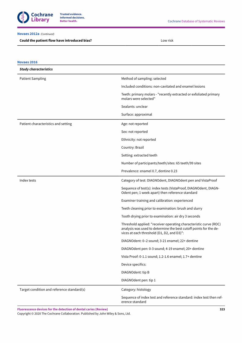
CochraneLibrary
Trusted evidence.Informed decisions.Better health.
Cochrane Database of Systematic Reviews
Could the patient flow have introduced bias? Low risk
Novaes 2012a (Continued)
Study characteristics
Patient Sampling Method of sampling: selected
Included conditions: non-cavitated and enamel lesions
Teeth: primary molars - "recently extracted or exfoliated primarymolars were selected"
Sealants: unclear
Surface: approximal
Patient characteristics and setting Age: not reported
Sex: not reported
Ethnicity: not reported
Country: Brazil
Setting: extracted teeth
Number of participants/teeth/sites: 65 teeth/99 sites
Prevalence: enamel 0.7, dentine 0.23
Index tests Category of test: DIAGNOdent, DIAGNOdent pen and VistaProof
Sequence of test(s): index tests (VistaProof, DIAGNOdent, DIAGN-Odent pen; 1 week apart) then reference standard
Examiner training and calibration: experienced
Teeth cleaning prior to examination: brush and slurry
Tooth drying prior to examination: air dry 3 seconds
Threshold applied: "receiver operating characteristic curve (ROC)analysis was used to determine the best cutoff points for the de-vices at each threshold (D1, D2, and D3)":
DIAGNOdent: 0–2 sound; 3-21 enamel; 22+ dentine
DIAGNOdent pen: 0-3 sound; 4-19 enamel; 20+ dentine
Vista Proof: 0-1.1 sound; 1.2-1.6 enamel; 1.7+ dentine
Device specifics:
DIAGNOdent: tip B
DIAGNOdent pen: tip 1
Target condition and reference standard(s) Category: histology
Sequence of index test and reference standard: index test then ref-erence standard
Novaes 2016
Fluorescence devices for the detection of dental caries (Review)
Copyright © 2020 The Cochrane Collaboration. Published by John Wiley & Sons, Ltd.
323
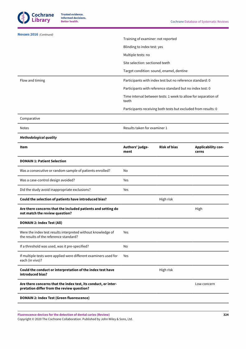
CochraneLibrary
Trusted evidence.Informed decisions.Better health.
Cochrane Database of Systematic Reviews
Training of examiner: not reported
Blinding to index test: yes
Multiple tests: no
Site selection: sectioned teeth
Target condition: sound, enamel, dentine
Flow and timing Participants with index test but no reference standard: 0
Participants with reference standard but no index test: 0
Time interval between tests: 1 week to allow for separation ofteeth
Participants receiving both tests but excluded from results: 0
Comparative
Notes Results taken for examiner 1
Methodological quality
Item Authors' judge-ment
Risk of bias Applicability con-cerns
DOMAIN 1: Patient Selection
Was a consecutive or random sample of patients enrolled? No
Was a case-control design avoided? Yes
Did the study avoid inappropriate exclusions? Yes
Could the selection of patients have introduced bias? High risk
Are there concerns that the included patients and setting donot match the review question?
High
DOMAIN 2: Index Test (All)
Were the index test results interpreted without knowledge ofthe results of the reference standard?
Yes
If a threshold was used, was it pre-specified? No
If multiple tests were applied were different examiners used foreach (in vivo)?
Yes
Could the conduct or interpretation of the index test haveintroduced bias?
High risk
Are there concerns that the index test, its conduct, or inter-pretation differ from the review question?
Low concern
DOMAIN 2: Index Test (Green fluorescence)
Novaes 2016 (Continued)
Fluorescence devices for the detection of dental caries (Review)
Copyright © 2020 The Cochrane Collaboration. Published by John Wiley & Sons, Ltd.
324
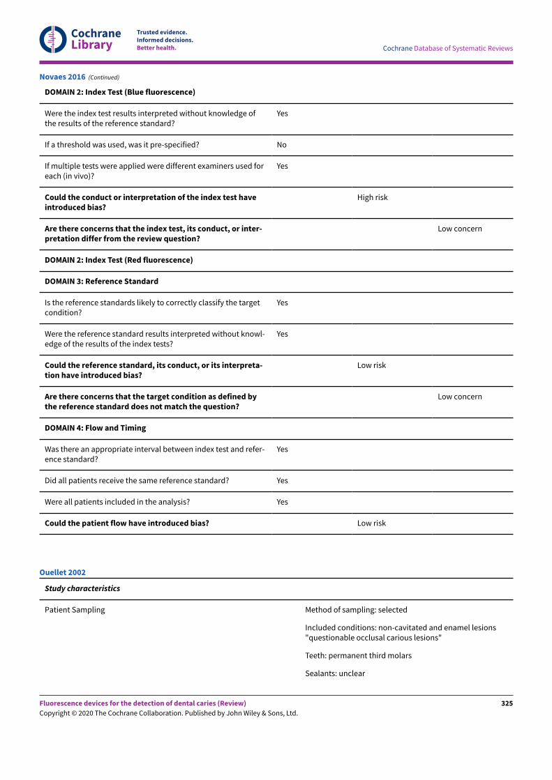
CochraneLibrary
Trusted evidence.Informed decisions.Better health.
Cochrane Database of Systematic Reviews
DOMAIN 2: Index Test (Blue fluorescence)
Were the index test results interpreted without knowledge ofthe results of the reference standard?
Yes
If a threshold was used, was it pre-specified? No
If multiple tests were applied were different examiners used foreach (in vivo)?
Yes
Could the conduct or interpretation of the index test haveintroduced bias?
High risk
Are there concerns that the index test, its conduct, or inter-pretation differ from the review question?
Low concern
DOMAIN 2: Index Test (Red fluorescence)
DOMAIN 3: Reference Standard
Is the reference standards likely to correctly classify the targetcondition?
Yes
Were the reference standard results interpreted without knowl-edge of the results of the index tests?
Yes
Could the reference standard, its conduct, or its interpreta-tion have introduced bias?
Low risk
Are there concerns that the target condition as defined bythe reference standard does not match the question?
Low concern
DOMAIN 4: Flow and Timing
Was there an appropriate interval between index test and refer-ence standard?
Yes
Did all patients receive the same reference standard? Yes
Were all patients included in the analysis? Yes
Could the patient flow have introduced bias? Low risk
Novaes 2016 (Continued)
Study characteristics
Patient Sampling Method of sampling: selected
Included conditions: non-cavitated and enamel lesions"questionable occlusal carious lesions"
Teeth: permanent third molars
Sealants: unclear
Ouellet 2002
Fluorescence devices for the detection of dental caries (Review)
Copyright © 2020 The Cochrane Collaboration. Published by John Wiley & Sons, Ltd.
325
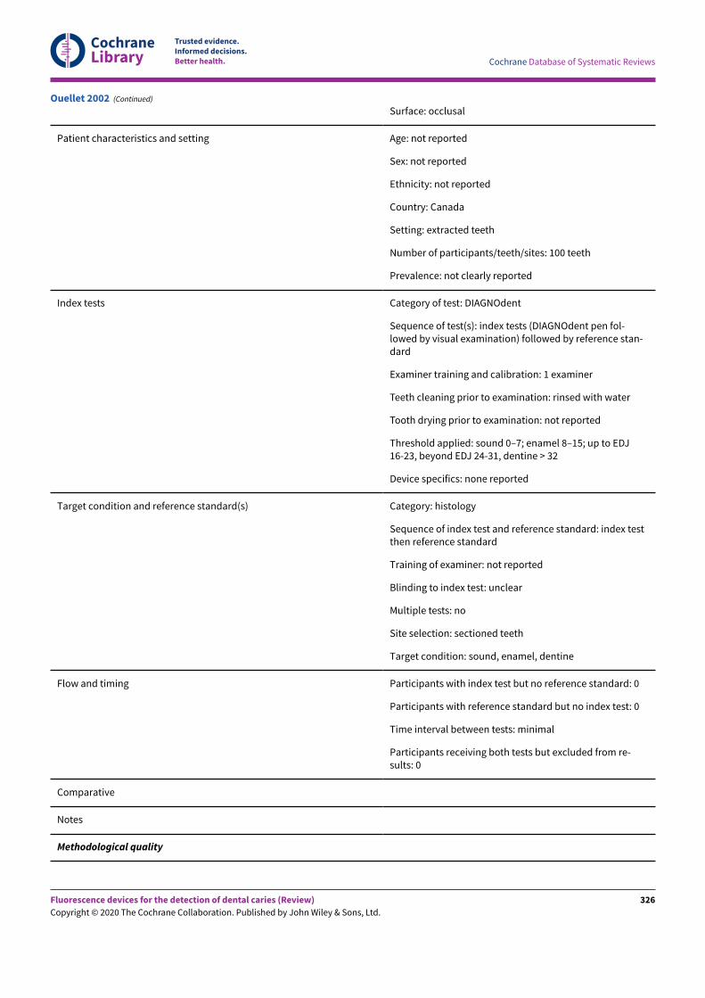
CochraneLibrary
Trusted evidence.Informed decisions.Better health.
Cochrane Database of Systematic Reviews
Surface: occlusal
Patient characteristics and setting Age: not reported
Sex: not reported
Ethnicity: not reported
Country: Canada
Setting: extracted teeth
Number of participants/teeth/sites: 100 teeth
Prevalence: not clearly reported
Index tests Category of test: DIAGNOdent
Sequence of test(s): index tests (DIAGNOdent pen fol-lowed by visual examination) followed by reference stan-dard
Examiner training and calibration: 1 examiner
Teeth cleaning prior to examination: rinsed with water
Tooth drying prior to examination: not reported
Threshold applied: sound 0–7; enamel 8–15; up to EDJ16-23, beyond EDJ 24-31, dentine > 32
Device specifics: none reported
Target condition and reference standard(s) Category: histology
Sequence of index test and reference standard: index testthen reference standard
Training of examiner: not reported
Blinding to index test: unclear
Multiple tests: no
Site selection: sectioned teeth
Target condition: sound, enamel, dentine
Flow and timing Participants with index test but no reference standard: 0
Participants with reference standard but no index test: 0
Time interval between tests: minimal
Participants receiving both tests but excluded from re-sults: 0
Comparative
Notes
Methodological quality
Ouellet 2002 (Continued)
Fluorescence devices for the detection of dental caries (Review)
Copyright © 2020 The Cochrane Collaboration. Published by John Wiley & Sons, Ltd.
326

CochraneLibrary
Trusted evidence.Informed decisions.Better health.
Cochrane Database of Systematic Reviews
Item Authors' judge-ment
Risk of bias Applicabilityconcerns
DOMAIN 1: Patient Selection
Was a consecutive or random sample of patients enrolled? No
Was a case-control design avoided? Yes
Did the study avoid inappropriate exclusions? Unclear
Could the selection of patients have introduced bias? High risk
Are there concerns that the included patients and setting do notmatch the review question?
High
DOMAIN 2: Index Test (All)
Were the index test results interpreted without knowledge of the resultsof the reference standard?
Yes
If a threshold was used, was it pre-specified? Yes
If multiple tests were applied were different examiners used for each (invivo)?
Unclear
Could the conduct or interpretation of the index test have intro-duced bias?
Low risk
Are there concerns that the index test, its conduct, or interpretationdiffer from the review question?
Low concern
DOMAIN 2: Index Test (Green fluorescence)
DOMAIN 2: Index Test (Blue fluorescence)
DOMAIN 2: Index Test (Red fluorescence)
Were the index test results interpreted without knowledge of the resultsof the reference standard?
Yes
If a threshold was used, was it pre-specified? Yes
If multiple tests were applied were different examiners used for each (invivo)?
Unclear
Could the conduct or interpretation of the index test have intro-duced bias?
Low risk
Are there concerns that the index test, its conduct, or interpretationdiffer from the review question?
Low concern
DOMAIN 3: Reference Standard
Is the reference standards likely to correctly classify the target condi-tion?
Yes
Ouellet 2002 (Continued)
Fluorescence devices for the detection of dental caries (Review)
Copyright © 2020 The Cochrane Collaboration. Published by John Wiley & Sons, Ltd.
327
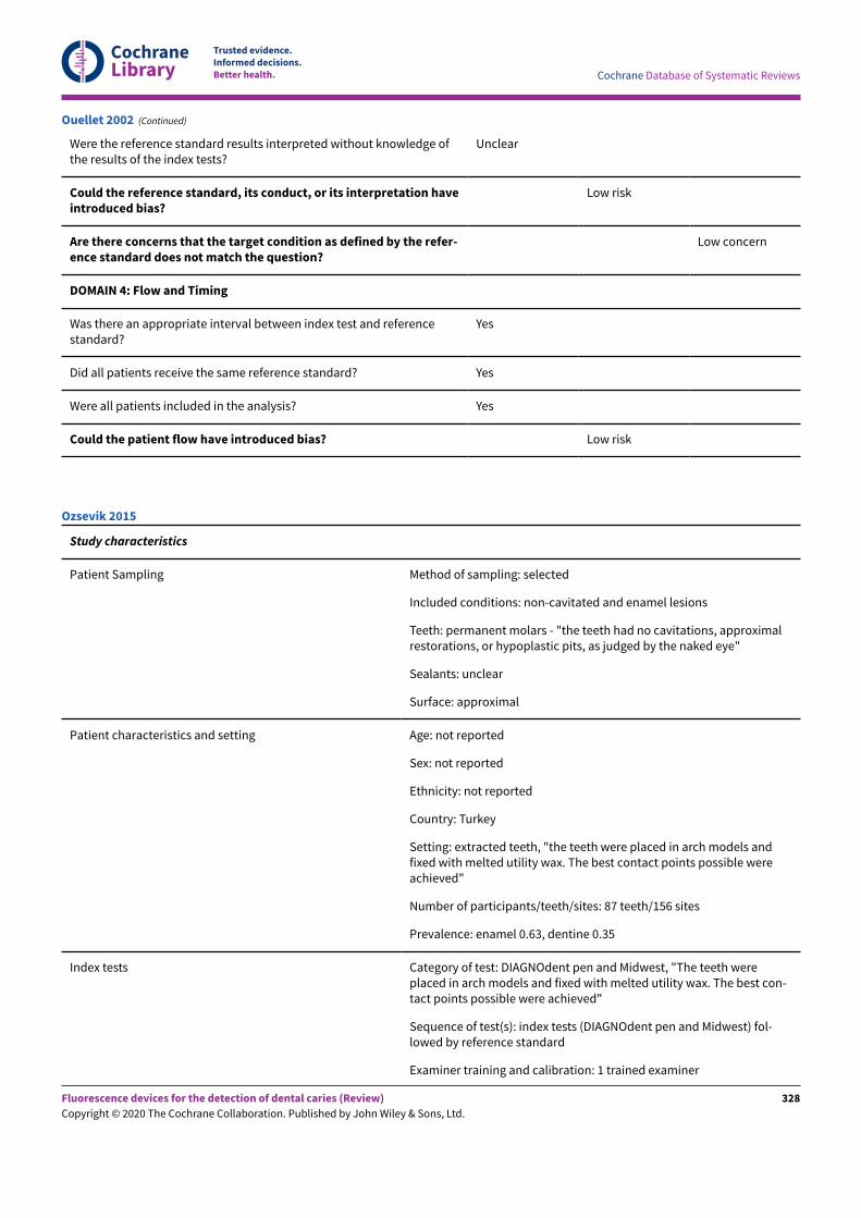
CochraneLibrary
Trusted evidence.Informed decisions.Better health.
Cochrane Database of Systematic Reviews
Were the reference standard results interpreted without knowledge ofthe results of the index tests?
Unclear
Could the reference standard, its conduct, or its interpretation haveintroduced bias?
Low risk
Are there concerns that the target condition as defined by the refer-ence standard does not match the question?
Low concern
DOMAIN 4: Flow and Timing
Was there an appropriate interval between index test and referencestandard?
Yes
Did all patients receive the same reference standard? Yes
Were all patients included in the analysis? Yes
Could the patient flow have introduced bias? Low risk
Ouellet 2002 (Continued)
Study characteristics
Patient Sampling Method of sampling: selected
Included conditions: non-cavitated and enamel lesions
Teeth: permanent molars - "the teeth had no cavitations, approximalrestorations, or hypoplastic pits, as judged by the naked eye"
Sealants: unclear
Surface: approximal
Patient characteristics and setting Age: not reported
Sex: not reported
Ethnicity: not reported
Country: Turkey
Setting: extracted teeth, "the teeth were placed in arch models andfixed with melted utility wax. The best contact points possible wereachieved"
Number of participants/teeth/sites: 87 teeth/156 sites
Prevalence: enamel 0.63, dentine 0.35
Index tests Category of test: DIAGNOdent pen and Midwest, "The teeth wereplaced in arch models and fixed with melted utility wax. The best con-tact points possible were achieved"
Sequence of test(s): index tests (DIAGNOdent pen and Midwest) fol-lowed by reference standard
Examiner training and calibration: 1 trained examiner
Ozsevik 2015
Fluorescence devices for the detection of dental caries (Review)
Copyright © 2020 The Cochrane Collaboration. Published by John Wiley & Sons, Ltd.
328
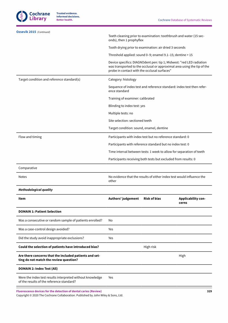
CochraneLibrary
Trusted evidence.Informed decisions.Better health.
Cochrane Database of Systematic Reviews
Teeth cleaning prior to examination: toothbrush and water (15 sec-onds), then 1 prophyflex
Tooth drying prior to examination: air dried 3 seconds
Threshold applied: sound 0–9; enamel 9.1–15; dentine > 15
Device specifics: DIAGNOdent pen: tip 1; Midwest: "red LED radiationwas transported to the occlusal or approximal area using the tip of theprobe in contact with the occlusal surfaces"
Target condition and reference standard(s) Category: histology
Sequence of index test and reference standard: index test then refer-ence standard
Training of examiner: calibrated
Blinding to index test: yes
Multiple tests: no
Site selection: sectioned teeth
Target condition: sound, enamel, dentine
Flow and timing Participants with index test but no reference standard: 0
Participants with reference standard but no index test: 0
Time interval between tests: 1 week to allow for separation of teeth
Participants receiving both tests but excluded from results: 0
Comparative
Notes No evidence that the results of either index test would influence theother
Methodological quality
Item Authors' judgement Risk of bias Applicability con-cerns
DOMAIN 1: Patient Selection
Was a consecutive or random sample of patients enrolled? No
Was a case-control design avoided? Yes
Did the study avoid inappropriate exclusions? Yes
Could the selection of patients have introduced bias? High risk
Are there concerns that the included patients and set-ting do not match the review question?
High
DOMAIN 2: Index Test (All)
Were the index test results interpreted without knowledgeof the results of the reference standard?
Yes
Ozsevik 2015 (Continued)
Fluorescence devices for the detection of dental caries (Review)
Copyright © 2020 The Cochrane Collaboration. Published by John Wiley & Sons, Ltd.
329
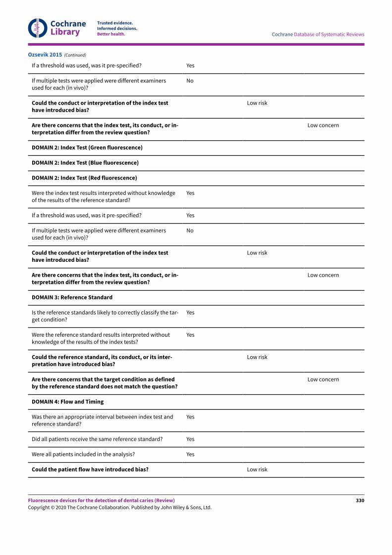
CochraneLibrary
Trusted evidence.Informed decisions.Better health.
Cochrane Database of Systematic Reviews
If a threshold was used, was it pre-specified? Yes
If multiple tests were applied were different examinersused for each (in vivo)?
No
Could the conduct or interpretation of the index testhave introduced bias?
Low risk
Are there concerns that the index test, its conduct, or in-terpretation differ from the review question?
Low concern
DOMAIN 2: Index Test (Green fluorescence)
DOMAIN 2: Index Test (Blue fluorescence)
DOMAIN 2: Index Test (Red fluorescence)
Were the index test results interpreted without knowledgeof the results of the reference standard?
Yes
If a threshold was used, was it pre-specified? Yes
If multiple tests were applied were different examinersused for each (in vivo)?
No
Could the conduct or interpretation of the index testhave introduced bias?
Low risk
Are there concerns that the index test, its conduct, or in-terpretation differ from the review question?
Low concern
DOMAIN 3: Reference Standard
Is the reference standards likely to correctly classify the tar-get condition?
Yes
Were the reference standard results interpreted withoutknowledge of the results of the index tests?
Yes
Could the reference standard, its conduct, or its inter-pretation have introduced bias?
Low risk
Are there concerns that the target condition as definedby the reference standard does not match the question?
Low concern
DOMAIN 4: Flow and Timing
Was there an appropriate interval between index test andreference standard?
Yes
Did all patients receive the same reference standard? Yes
Were all patients included in the analysis? Yes
Could the patient flow have introduced bias? Low risk
Ozsevik 2015 (Continued)
Fluorescence devices for the detection of dental caries (Review)
Copyright © 2020 The Cochrane Collaboration. Published by John Wiley & Sons, Ltd.
330
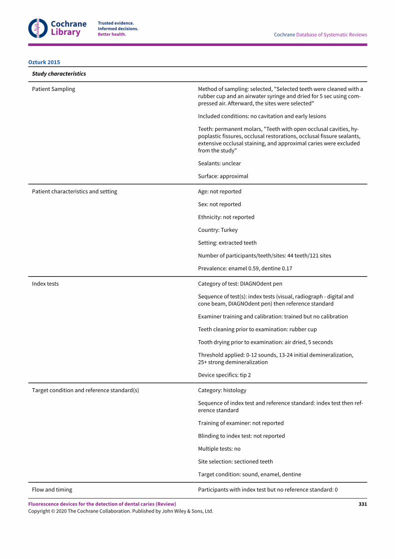
CochraneLibrary
Trusted evidence.Informed decisions.Better health.
Cochrane Database of Systematic Reviews
Study characteristics
Patient Sampling Method of sampling: selected, "Selected teeth were cleaned with arubber cup and an airwater syringe and dried for 5 sec using com-pressed air. Afterward, the sites were selected"
Included conditions: no cavitation and early lesions
Teeth: permanent molars, "Teeth with open occlusal cavities, hy-poplastic fissures, occlusal restorations, occlusal fissure sealants,extensive occlusal staining, and approximal caries were excludedfrom the study"
Sealants: unclear
Surface: approximal
Patient characteristics and setting Age: not reported
Sex: not reported
Ethnicity: not reported
Country: Turkey
Setting: extracted teeth
Number of participants/teeth/sites: 44 teeth/121 sites
Prevalence: enamel 0.59, dentine 0.17
Index tests Category of test: DIAGNOdent pen
Sequence of test(s): index tests (visual, radiograph - digital andcone beam, DIAGNOdent pen) then reference standard
Examiner training and calibration: trained but no calibration
Teeth cleaning prior to examination: rubber cup
Tooth drying prior to examination: air dried, 5 seconds
Threshold applied: 0-12 sounds, 13-24 initial demineralization,25+ strong demineralization
Device specifics: tip 2
Target condition and reference standard(s) Category: histology
Sequence of index test and reference standard: index test then ref-erence standard
Training of examiner: not reported
Blinding to index test: not reported
Multiple tests: no
Site selection: sectioned teeth
Target condition: sound, enamel, dentine
Flow and timing Participants with index test but no reference standard: 0
Ozturk 2015
Fluorescence devices for the detection of dental caries (Review)
Copyright © 2020 The Cochrane Collaboration. Published by John Wiley & Sons, Ltd.
331
CochraneLibrary
Trusted evidence.Informed decisions.Better health.
Cochrane Database of Systematic Reviews
Participants with reference standard but no index test: 0
Time interval between tests: 1 week to allow for separation ofteeth
Participants receiving both tests but excluded from results: 0
Comparative
Notes
Methodological quality
Item Authors' judge-ment
Risk of bias Applicability con-cerns
DOMAIN 1: Patient Selection
Was a consecutive or random sample of patients enrolled? No
Was a case-control design avoided? Yes
Did the study avoid inappropriate exclusions? Yes
Could the selection of patients have introduced bias? High risk
Are there concerns that the included patients and setting donot match the review question?
High
DOMAIN 2: Index Test (All)
Were the index test results interpreted without knowledge ofthe results of the reference standard?
Yes
If a threshold was used, was it pre-specified? Yes
If multiple tests were applied were different examiners used foreach (in vivo)?
No
Could the conduct or interpretation of the index test haveintroduced bias?
Low risk
Are there concerns that the index test, its conduct, or inter-pretation differ from the review question?
Low concern
DOMAIN 2: Index Test (Green fluorescence)
DOMAIN 2: Index Test (Blue fluorescence)
DOMAIN 2: Index Test (Red fluorescence)
Were the index test results interpreted without knowledge ofthe results of the reference standard?
Yes
If a threshold was used, was it pre-specified? Yes
If multiple tests were applied were different examiners used foreach (in vivo)?
No
Ozturk 2015 (Continued)
Fluorescence devices for the detection of dental caries (Review)
Copyright © 2020 The Cochrane Collaboration. Published by John Wiley & Sons, Ltd.
332
CochraneLibrary
Trusted evidence.Informed decisions.Better health.
Cochrane Database of Systematic Reviews
Could the conduct or interpretation of the index test haveintroduced bias?
Low risk
Are there concerns that the index test, its conduct, or inter-pretation differ from the review question?
Low concern
DOMAIN 3: Reference Standard
Is the reference standards likely to correctly classify the targetcondition?
Yes
Were the reference standard results interpreted without knowl-edge of the results of the index tests?
Yes
Could the reference standard, its conduct, or its interpreta-tion have introduced bias?
Low risk
Are there concerns that the target condition as defined bythe reference standard does not match the question?
Low concern
DOMAIN 4: Flow and Timing
Was there an appropriate interval between index test and refer-ence standard?
Yes
Did all patients receive the same reference standard? Yes
Were all patients included in the analysis? Yes
Could the patient flow have introduced bias? Low risk
Ozturk 2015 (Continued)
Study characteristics
Patient Sampling Method of sampling: selected
Included conditions: no cavitation and early lesions
Teeth: permanent third molars
Sealants: not reported
Surface: occlusal
Patient characteristics and setting Age: not reported
Sex: not reported
Ethnicity: not reported
Country: Brazil
Setting: extracted teeth - tooth bank
Number of participants/teeth/sites: 26 teeth/64 teeth
Prevalence: enamel 0.88, dentine 0.28
Paula 2011
Fluorescence devices for the detection of dental caries (Review)
Copyright © 2020 The Cochrane Collaboration. Published by John Wiley & Sons, Ltd.
333
CochraneLibrary
Trusted evidence.Informed decisions.Better health.
Cochrane Database of Systematic Reviews
Index tests Category of test: DIAGNOdent
Sequence of test(s): index tests followed by referencestandard
Examiner training and calibration: experienced
Teeth cleaning prior to examination: pumice slurryand water
Tooth drying prior to examination: air dried
Threshold applied: 0-10 sound, 11-20 enamel, 21+dentine
Device specifics: tip A
Target condition and reference standard(s) Category: histology
Sequence of index test and reference standard: indextest then reference standard
Training of examiner: not reported
Blinding to index test: unclear
Multiple tests: no
Site selection: sectioned teeth
Target condition: sound, enamel, dentine
Flow and timing Participants with index test but no reference stan-dard: 0
Participants with reference standard but no indextest: 0
Time interval between tests: 1-2 days
Participants receiving both tests but excluded fromresults: 0
Comparative
Notes
Methodological quality
Item Authors'judgement
Risk of bias Applicabilityconcerns
DOMAIN 1: Patient Selection
Was a consecutive or random sample of patients enrolled? No
Was a case-control design avoided? Yes
Did the study avoid inappropriate exclusions? Yes
Could the selection of patients have introduced bias? High risk
Paula 2011 (Continued)
Fluorescence devices for the detection of dental caries (Review)
Copyright © 2020 The Cochrane Collaboration. Published by John Wiley & Sons, Ltd.
334
CochraneLibrary
Trusted evidence.Informed decisions.Better health.
Cochrane Database of Systematic Reviews
Are there concerns that the included patients and setting do not matchthe review question?
High
DOMAIN 2: Index Test (All)
Were the index test results interpreted without knowledge of the results ofthe reference standard?
Yes
If a threshold was used, was it pre-specified? Yes
If multiple tests were applied were different examiners used for each (in vi-vo)?
Could the conduct or interpretation of the index test have introducedbias?
Low risk
Are there concerns that the index test, its conduct, or interpretation dif-fer from the review question?
Low concern
DOMAIN 2: Index Test (Green fluorescence)
DOMAIN 2: Index Test (Blue fluorescence)
DOMAIN 2: Index Test (Red fluorescence)
Were the index test results interpreted without knowledge of the results ofthe reference standard?
Yes
If a threshold was used, was it pre-specified? Yes
If multiple tests were applied were different examiners used for each (in vi-vo)?
Could the conduct or interpretation of the index test have introducedbias?
Low risk
Are there concerns that the index test, its conduct, or interpretation dif-fer from the review question?
Low concern
DOMAIN 3: Reference Standard
Is the reference standards likely to correctly classify the target condition? Yes
Were the reference standard results interpreted without knowledge of theresults of the index tests?
Unclear
Could the reference standard, its conduct, or its interpretation have in-troduced bias?
Low risk
Are there concerns that the target condition as defined by the referencestandard does not match the question?
Low concern
DOMAIN 4: Flow and Timing
Was there an appropriate interval between index test and reference stan-dard?
Yes
Paula 2011 (Continued)
Fluorescence devices for the detection of dental caries (Review)
Copyright © 2020 The Cochrane Collaboration. Published by John Wiley & Sons, Ltd.
335
CochraneLibrary
Trusted evidence.Informed decisions.Better health.
Cochrane Database of Systematic Reviews
Did all patients receive the same reference standard? Yes
Were all patients included in the analysis? Yes
Could the patient flow have introduced bias? Low risk
Paula 2011 (Continued)
Study characteristics
Patient Sampling Method of sampling: selected, "None of the teeth showed macroscop-ic signs of cavity formation"
Included conditions: no cavitation and early lesions
Teeth: permanent molars
Sealants: unclear
Surface: occlusal
Patient characteristics and setting Age: not reported
Sex: not reported
Ethnicity: not reported
Country: Brazil
Setting: extracted teeth
Number of participants/teeth/sites: 96 teeth
Prevalence: enamel 0.57, dentine 0.25
Index tests Category of test: DIAGNOdent pen and QLF (Inspektor Research)
Sequence of test(s): index tests (visual, radiograph, ECM, DIAGNOdent,QLF) then reference standard
Examiner training and calibration: training event
Teeth cleaning prior to examination: paste and rotating brush
Tooth drying prior to examination: yes
Threshold applied:
DIAGNOdent: > 5 indicated caries
QLF Inspektor: "The images were scored subjectively from the storedimages displayed on a CRT monitor"
Categories: no change in enamel fluorescence, slight change in enam-el fluorescence, fluorescence loss distinctly visible without enamelbroken, fluorescence loss distinctly visible with enamel broken, fluo-rescence loss distinctly visible with cavitation
Device specifics: DIAGNOdent - tip A
Target condition and reference standard(s) Category: histology
Pereira 2011
Fluorescence devices for the detection of dental caries (Review)
Copyright © 2020 The Cochrane Collaboration. Published by John Wiley & Sons, Ltd.
336
CochraneLibrary
Trusted evidence.Informed decisions.Better health.
Cochrane Database of Systematic Reviews
Sequence of index test and reference standard: index test then refer-ence standard
Training of examiner: "Three examiners underwent a training session,which consisted of 2 h of theoretical training and 4 h of practice on ex-tracted teeth"
Blinding to index test: not reported
Multiple tests: no
Site selection: sectioned teeth
Target condition:
no cariesdemineralization extending to the outer ½ of the enameldemineralization extending to the inner ½ of the enameldemineralization extending to the outer ½ of the dentinedemineralization extending to the outer ½ of the dentine
Flow and timing Participants with index test but no reference standard: 0
Participants with reference standard but no index test: 0
Time interval between tests: 1 week to allow for separation of teeth
Participants receiving both tests but excluded from results: 0
Comparative
Notes
Methodological quality
Item Authors' judgement Risk of bias Applicability con-cerns
DOMAIN 1: Patient Selection
Was a consecutive or random sample of patients enrolled? No
Was a case-control design avoided? Yes
Did the study avoid inappropriate exclusions? Yes
Could the selection of patients have introduced bias? High risk
Are there concerns that the included patients and set-ting do not match the review question?
High
DOMAIN 2: Index Test (All)
Were the index test results interpreted without knowledgeof the results of the reference standard?
Yes
If a threshold was used, was it pre-specified? Yes
If multiple tests were applied were different examinersused for each (in vivo)?
Unclear
Pereira 2011 (Continued)
Fluorescence devices for the detection of dental caries (Review)
Copyright © 2020 The Cochrane Collaboration. Published by John Wiley & Sons, Ltd.
337
CochraneLibrary
Trusted evidence.Informed decisions.Better health.
Cochrane Database of Systematic Reviews
Could the conduct or interpretation of the index testhave introduced bias?
Low risk
Are there concerns that the index test, its conduct, or in-terpretation differ from the review question?
Low concern
DOMAIN 2: Index Test (Green fluorescence)
Were the index test results interpreted without knowledgeof the results of the reference standard?
Yes
If a threshold was used, was it pre-specified? Yes
If multiple tests were applied were different examinersused for each (in vivo)?
Unclear
Could the conduct or interpretation of the index testhave introduced bias?
Low risk
Are there concerns that the index test, its conduct, or in-terpretation differ from the review question?
Low concern
DOMAIN 2: Index Test (Blue fluorescence)
DOMAIN 2: Index Test (Red fluorescence)
DOMAIN 3: Reference Standard
Is the reference standards likely to correctly classify the tar-get condition?
Yes
Were the reference standard results interpreted withoutknowledge of the results of the index tests?
Unclear
Could the reference standard, its conduct, or its inter-pretation have introduced bias?
Low risk
Are there concerns that the target condition as definedby the reference standard does not match the question?
Low concern
DOMAIN 4: Flow and Timing
Was there an appropriate interval between index test andreference standard?
Yes
Did all patients receive the same reference standard? Yes
Were all patients included in the analysis? Yes
Could the patient flow have introduced bias? Low risk
Pereira 2011 (Continued)
Study characteristics
Pinelli 2002
Fluorescence devices for the detection of dental caries (Review)
Copyright © 2020 The Cochrane Collaboration. Published by John Wiley & Sons, Ltd.
338
CochraneLibrary
Trusted evidence.Informed decisions.Better health.
Cochrane Database of Systematic Reviews
Patient Sampling Method of sampling: selected
Included conditions: no cavitation and early lesions, "none of theteeth showed macroscopic signs of cavity formation with expo-sure into dentin"
Teeth: permanent molars, "the inclusion criterion was the pres-ence of at least one white-spot caries lesion on a free smooth sur-face"
Sealants: not reported
Surface: "free smooth surfaces"
Patient characteristics and setting Age: 11 to 17 years
Sex: not reported
Ethnicity: not reported
Country: Brazil
Setting: school, "The examinations were carried out in classroomsunder good light conditions"
Number of participants/teeth/sites: 50 participants/220 surfaces
Prevalence: enamel 0.50
Index tests Category of test: DIAGNOdent
Sequence of test(s): visual then DIAGNOdent
Examiner training and calibration: yes
Teeth cleaning prior to examination: floss and brush
Tooth drying prior to examination: air dried, 10 seconds
Threshold applied: 0-4 arrested, 5+ active
Device specifics: "DIAGNOdent was used to examine only the le-sions detected by visual inspection"
Target condition and reference standard(s) Category: visual
Sequence of index test and reference standard: reference stan-dard performed before index test
Training of examiner: calibration completed
Blinding to index test: yes
Multiple tests: no
Site selection: those identified visually
Target condition: active or inactive lesions
Flow and timing Participants with index test but no reference standard: 0
Participants with reference standard but no index test: 0
Time interval between tests: 1 week to allow for separation ofteeth
Pinelli 2002 (Continued)
Fluorescence devices for the detection of dental caries (Review)
Copyright © 2020 The Cochrane Collaboration. Published by John Wiley & Sons, Ltd.
339

CochraneLibrary
Trusted evidence.Informed decisions.Better health.
Cochrane Database of Systematic Reviews
Participants receiving both tests but excluded from results: 0
Comparative
Notes Inclusion criteria suggests sound teeth were excluded, but the re-sults confirm that sound teeth were present in the sample
Methodological quality
Item Authors' judge-ment
Risk of bias Applicability con-cerns
DOMAIN 1: Patient Selection
Was a consecutive or random sample of patients enrolled? No
Was a case-control design avoided? Yes
Did the study avoid inappropriate exclusions? Yes
Could the selection of patients have introduced bias? High risk
Are there concerns that the included patients and setting donot match the review question?
Low concern
DOMAIN 2: Index Test (All)
Were the index test results interpreted without knowledge ofthe results of the reference standard?
No
If a threshold was used, was it pre-specified? Yes
If multiple tests were applied were different examiners used foreach (in vivo)?
Could the conduct or interpretation of the index test haveintroduced bias?
Low risk
Are there concerns that the index test, its conduct, or inter-pretation differ from the review question?
Low concern
DOMAIN 2: Index Test (Green fluorescence)
DOMAIN 2: Index Test (Blue fluorescence)
DOMAIN 2: Index Test (Red fluorescence)
Were the index test results interpreted without knowledge ofthe results of the reference standard?
No
If a threshold was used, was it pre-specified? Yes
If multiple tests were applied were different examiners used foreach (in vivo)?
Unclear
Could the conduct or interpretation of the index test haveintroduced bias?
Low risk
Pinelli 2002 (Continued)
Fluorescence devices for the detection of dental caries (Review)
Copyright © 2020 The Cochrane Collaboration. Published by John Wiley & Sons, Ltd.
340
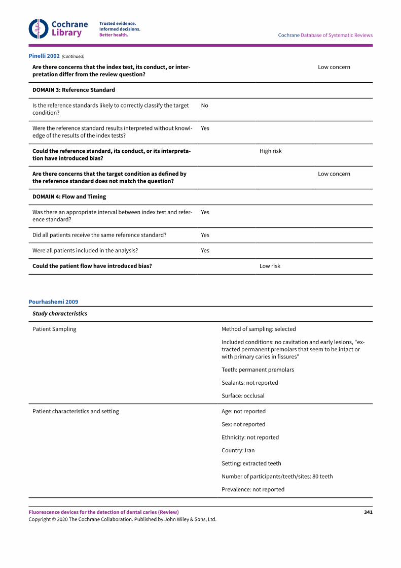
CochraneLibrary
Trusted evidence.Informed decisions.Better health.
Cochrane Database of Systematic Reviews
Are there concerns that the index test, its conduct, or inter-pretation differ from the review question?
Low concern
DOMAIN 3: Reference Standard
Is the reference standards likely to correctly classify the targetcondition?
No
Were the reference standard results interpreted without knowl-edge of the results of the index tests?
Yes
Could the reference standard, its conduct, or its interpreta-tion have introduced bias?
High risk
Are there concerns that the target condition as defined bythe reference standard does not match the question?
Low concern
DOMAIN 4: Flow and Timing
Was there an appropriate interval between index test and refer-ence standard?
Yes
Did all patients receive the same reference standard? Yes
Were all patients included in the analysis? Yes
Could the patient flow have introduced bias? Low risk
Pinelli 2002 (Continued)
Study characteristics
Patient Sampling Method of sampling: selected
Included conditions: no cavitation and early lesions, "ex-tracted permanent premolars that seem to be intact orwith primary caries in fissures"
Teeth: permanent premolars
Sealants: not reported
Surface: occlusal
Patient characteristics and setting Age: not reported
Sex: not reported
Ethnicity: not reported
Country: Iran
Setting: extracted teeth
Number of participants/teeth/sites: 80 teeth
Prevalence: not reported
Pourhashemi 2009
Fluorescence devices for the detection of dental caries (Review)
Copyright © 2020 The Cochrane Collaboration. Published by John Wiley & Sons, Ltd.
341
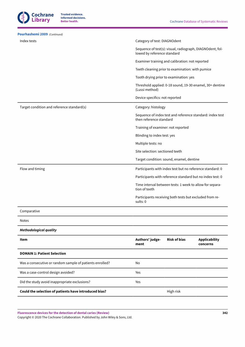
CochraneLibrary
Trusted evidence.Informed decisions.Better health.
Cochrane Database of Systematic Reviews
Index tests Category of test: DIAGNOdent
Sequence of test(s): visual, radiograph, DIAGNOdent, fol-lowed by reference standard
Examiner training and calibration: not reported
Teeth cleaning prior to examination: with pumice
Tooth drying prior to examination: yes
Threshold applied: 0-18 sound, 19-30 enamel, 30+ dentine(Lussi method)
Device specifics: not reported
Target condition and reference standard(s) Category: histology
Sequence of index test and reference standard: index testthen reference standard
Training of examiner: not reported
Blinding to index test: yes
Multiple tests: no
Site selection: sectioned teeth
Target condition: sound, enamel, dentine
Flow and timing Participants with index test but no reference standard: 0
Participants with reference standard but no index test: 0
Time interval between tests: 1 week to allow for separa-tion of teeth
Participants receiving both tests but excluded from re-sults: 0
Comparative
Notes
Methodological quality
Item Authors' judge-ment
Risk of bias Applicabilityconcerns
DOMAIN 1: Patient Selection
Was a consecutive or random sample of patients enrolled? No
Was a case-control design avoided? Yes
Did the study avoid inappropriate exclusions? Yes
Could the selection of patients have introduced bias? High risk
Pourhashemi 2009 (Continued)
Fluorescence devices for the detection of dental caries (Review)
Copyright © 2020 The Cochrane Collaboration. Published by John Wiley & Sons, Ltd.
342
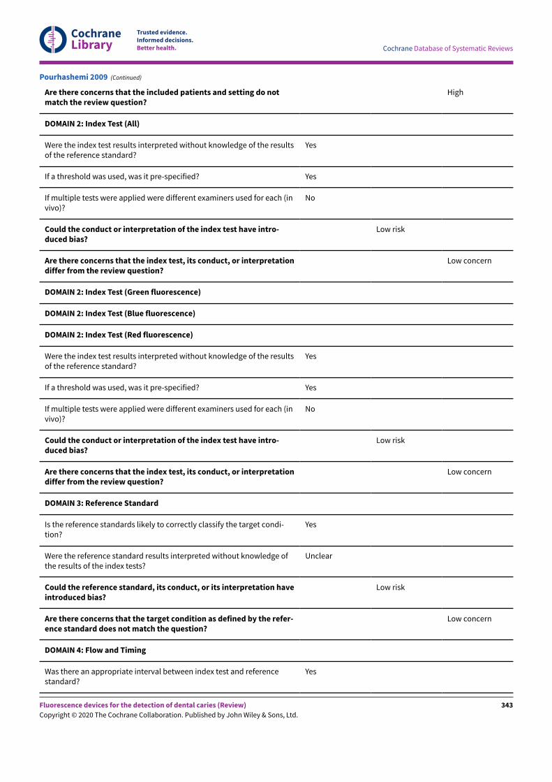
CochraneLibrary
Trusted evidence.Informed decisions.Better health.
Cochrane Database of Systematic Reviews
Are there concerns that the included patients and setting do notmatch the review question?
High
DOMAIN 2: Index Test (All)
Were the index test results interpreted without knowledge of the resultsof the reference standard?
Yes
If a threshold was used, was it pre-specified? Yes
If multiple tests were applied were different examiners used for each (invivo)?
No
Could the conduct or interpretation of the index test have intro-duced bias?
Low risk
Are there concerns that the index test, its conduct, or interpretationdiffer from the review question?
Low concern
DOMAIN 2: Index Test (Green fluorescence)
DOMAIN 2: Index Test (Blue fluorescence)
DOMAIN 2: Index Test (Red fluorescence)
Were the index test results interpreted without knowledge of the resultsof the reference standard?
Yes
If a threshold was used, was it pre-specified? Yes
If multiple tests were applied were different examiners used for each (invivo)?
No
Could the conduct or interpretation of the index test have intro-duced bias?
Low risk
Are there concerns that the index test, its conduct, or interpretationdiffer from the review question?
Low concern
DOMAIN 3: Reference Standard
Is the reference standards likely to correctly classify the target condi-tion?
Yes
Were the reference standard results interpreted without knowledge ofthe results of the index tests?
Unclear
Could the reference standard, its conduct, or its interpretation haveintroduced bias?
Low risk
Are there concerns that the target condition as defined by the refer-ence standard does not match the question?
Low concern
DOMAIN 4: Flow and Timing
Was there an appropriate interval between index test and referencestandard?
Yes
Pourhashemi 2009 (Continued)
Fluorescence devices for the detection of dental caries (Review)
Copyright © 2020 The Cochrane Collaboration. Published by John Wiley & Sons, Ltd.
343
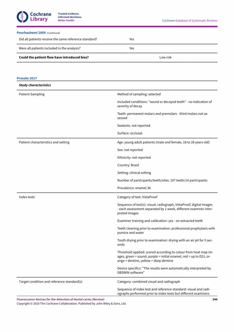
CochraneLibrary
Trusted evidence.Informed decisions.Better health.
Cochrane Database of Systematic Reviews
Did all patients receive the same reference standard? Yes
Were all patients included in the analysis? Yes
Could the patient flow have introduced bias? Low risk
Pourhashemi 2009 (Continued)
Study characteristics
Patient Sampling Method of sampling: selected
Included conditions: "sound or decayed teeth" - no indication ofseverity of decay
Teeth: permanent molars and premolars - third molars not as-sessed
Sealants: not reported
Surface: occlusal
Patient characteristics and setting Age: young adult patients (male and female, 18 to 28 years old)
Sex: not reported
Ethnicity: not reported
Country: Brazil
Setting: clinical setting
Number of participants/teeth/sites: 107 teeth/14 participants
Prevalence: enamel.36
Index tests Category of test: VistaProof
Sequence of test(s): visual, radiograph, VistaProof, digital images- each assessment separated by 1 week, different examiner inter-preted images
Examiner training and calibration: yes - on extracted teeth
Teeth cleaning prior to examination: professional prophylaxis withpumice and water
Tooth drying prior to examination: drying with an air jet for 5 sec-onds
Threshold applied: scored according to colour from heat map im-ages, green = sound, purple = initial enamel, red = up to EDJ, or-ange = dentine, yellow = deep dentine
Device specifics: "The results were automatically interpreted byDBSWIN software"
Target condition and reference standard(s) Category: combined visual and radiograph
Sequence of index test and reference standard: visual and radi-ographs performed prior to index tests but different examiners
Presoto 2017
Fluorescence devices for the detection of dental caries (Review)
Copyright © 2020 The Cochrane Collaboration. Published by John Wiley & Sons, Ltd.
344
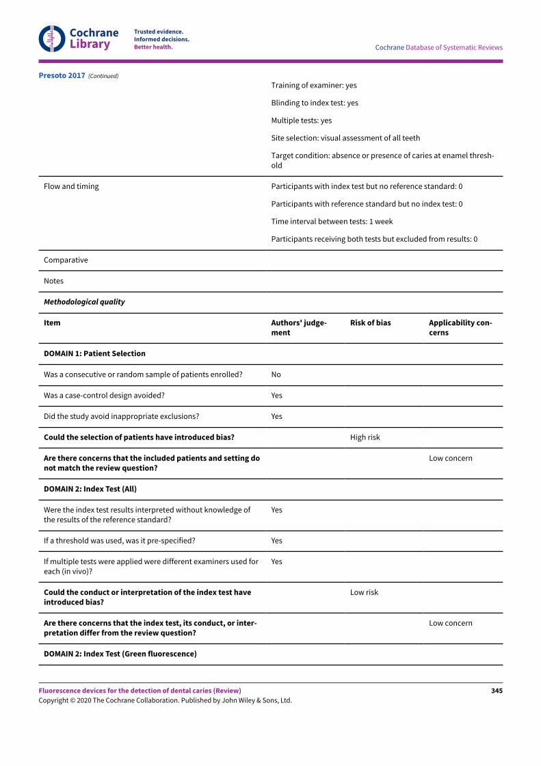
CochraneLibrary
Trusted evidence.Informed decisions.Better health.
Cochrane Database of Systematic Reviews
Training of examiner: yes
Blinding to index test: yes
Multiple tests: yes
Site selection: visual assessment of all teeth
Target condition: absence or presence of caries at enamel thresh-old
Flow and timing Participants with index test but no reference standard: 0
Participants with reference standard but no index test: 0
Time interval between tests: 1 week
Participants receiving both tests but excluded from results: 0
Comparative
Notes
Methodological quality
Item Authors' judge-ment
Risk of bias Applicability con-cerns
DOMAIN 1: Patient Selection
Was a consecutive or random sample of patients enrolled? No
Was a case-control design avoided? Yes
Did the study avoid inappropriate exclusions? Yes
Could the selection of patients have introduced bias? High risk
Are there concerns that the included patients and setting donot match the review question?
Low concern
DOMAIN 2: Index Test (All)
Were the index test results interpreted without knowledge ofthe results of the reference standard?
Yes
If a threshold was used, was it pre-specified? Yes
If multiple tests were applied were different examiners used foreach (in vivo)?
Yes
Could the conduct or interpretation of the index test haveintroduced bias?
Low risk
Are there concerns that the index test, its conduct, or inter-pretation differ from the review question?
Low concern
DOMAIN 2: Index Test (Green fluorescence)
Presoto 2017 (Continued)
Fluorescence devices for the detection of dental caries (Review)
Copyright © 2020 The Cochrane Collaboration. Published by John Wiley & Sons, Ltd.
345
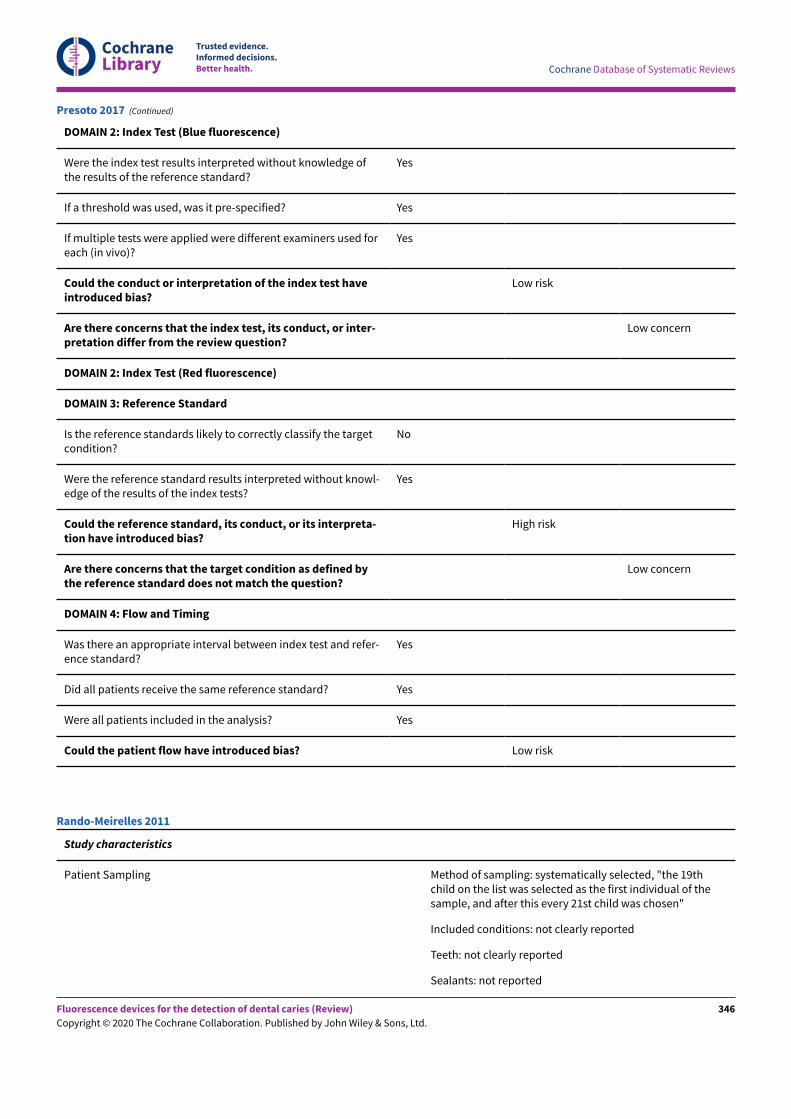
CochraneLibrary
Trusted evidence.Informed decisions.Better health.
Cochrane Database of Systematic Reviews
DOMAIN 2: Index Test (Blue fluorescence)
Were the index test results interpreted without knowledge ofthe results of the reference standard?
Yes
If a threshold was used, was it pre-specified? Yes
If multiple tests were applied were different examiners used foreach (in vivo)?
Yes
Could the conduct or interpretation of the index test haveintroduced bias?
Low risk
Are there concerns that the index test, its conduct, or inter-pretation differ from the review question?
Low concern
DOMAIN 2: Index Test (Red fluorescence)
DOMAIN 3: Reference Standard
Is the reference standards likely to correctly classify the targetcondition?
No
Were the reference standard results interpreted without knowl-edge of the results of the index tests?
Yes
Could the reference standard, its conduct, or its interpreta-tion have introduced bias?
High risk
Are there concerns that the target condition as defined bythe reference standard does not match the question?
Low concern
DOMAIN 4: Flow and Timing
Was there an appropriate interval between index test and refer-ence standard?
Yes
Did all patients receive the same reference standard? Yes
Were all patients included in the analysis? Yes
Could the patient flow have introduced bias? Low risk
Presoto 2017 (Continued)
Study characteristics
Patient Sampling Method of sampling: systematically selected, "the 19thchild on the list was selected as the first individual of thesample, and after this every 21st child was chosen"
Included conditions: not clearly reported
Teeth: not clearly reported
Sealants: not reported
Rando-Meirelles 2011
Fluorescence devices for the detection of dental caries (Review)
Copyright © 2020 The Cochrane Collaboration. Published by John Wiley & Sons, Ltd.
346
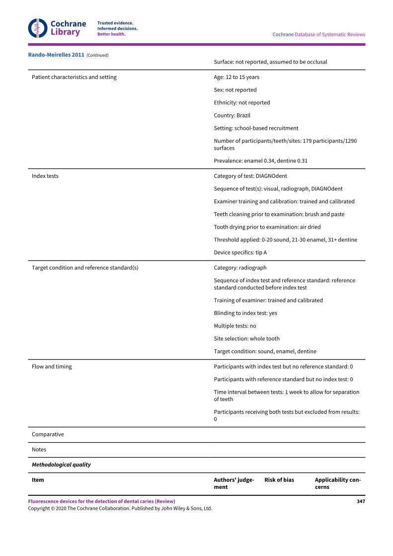
CochraneLibrary
Trusted evidence.Informed decisions.Better health.
Cochrane Database of Systematic Reviews
Surface: not reported, assumed to be occlusal
Patient characteristics and setting Age: 12 to 15 years
Sex: not reported
Ethnicity: not reported
Country: Brazil
Setting: school-based recruitment
Number of participants/teeth/sites: 179 participants/1290surfaces
Prevalence: enamel 0.34, dentine 0.31
Index tests Category of test: DIAGNOdent
Sequence of test(s): visual, radiograph, DIAGNOdent
Examiner training and calibration: trained and calibrated
Teeth cleaning prior to examination: brush and paste
Tooth drying prior to examination: air dried
Threshold applied: 0-20 sound, 21-30 enamel, 31+ dentine
Device specifics: tip A
Target condition and reference standard(s) Category: radiograph
Sequence of index test and reference standard: referencestandard conducted before index test
Training of examiner: trained and calibrated
Blinding to index test: yes
Multiple tests: no
Site selection: whole tooth
Target condition: sound, enamel, dentine
Flow and timing Participants with index test but no reference standard: 0
Participants with reference standard but no index test: 0
Time interval between tests: 1 week to allow for separationof teeth
Participants receiving both tests but excluded from results:0
Comparative
Notes
Methodological quality
Item Authors' judge-ment
Risk of bias Applicability con-cerns
Rando-Meirelles 2011 (Continued)
Fluorescence devices for the detection of dental caries (Review)
Copyright © 2020 The Cochrane Collaboration. Published by John Wiley & Sons, Ltd.
347
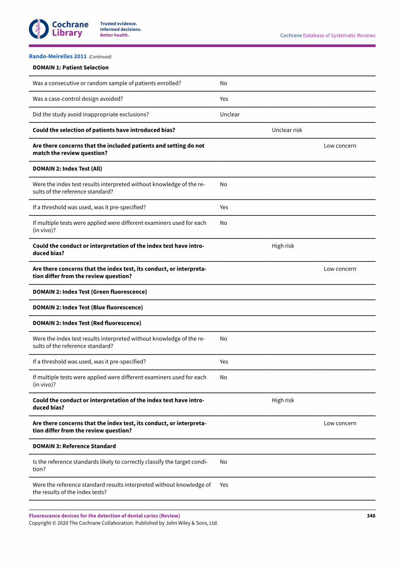
CochraneLibrary
Trusted evidence.Informed decisions.Better health.
Cochrane Database of Systematic Reviews
DOMAIN 1: Patient Selection
Was a consecutive or random sample of patients enrolled? No
Was a case-control design avoided? Yes
Did the study avoid inappropriate exclusions? Unclear
Could the selection of patients have introduced bias? Unclear risk
Are there concerns that the included patients and setting do notmatch the review question?
Low concern
DOMAIN 2: Index Test (All)
Were the index test results interpreted without knowledge of the re-sults of the reference standard?
No
If a threshold was used, was it pre-specified? Yes
If multiple tests were applied were different examiners used for each(in vivo)?
No
Could the conduct or interpretation of the index test have intro-duced bias?
High risk
Are there concerns that the index test, its conduct, or interpreta-tion differ from the review question?
Low concern
DOMAIN 2: Index Test (Green fluorescence)
DOMAIN 2: Index Test (Blue fluorescence)
DOMAIN 2: Index Test (Red fluorescence)
Were the index test results interpreted without knowledge of the re-sults of the reference standard?
No
If a threshold was used, was it pre-specified? Yes
If multiple tests were applied were different examiners used for each(in vivo)?
No
Could the conduct or interpretation of the index test have intro-duced bias?
High risk
Are there concerns that the index test, its conduct, or interpreta-tion differ from the review question?
Low concern
DOMAIN 3: Reference Standard
Is the reference standards likely to correctly classify the target condi-tion?
No
Were the reference standard results interpreted without knowledge ofthe results of the index tests?
Yes
Rando-Meirelles 2011 (Continued)
Fluorescence devices for the detection of dental caries (Review)
Copyright © 2020 The Cochrane Collaboration. Published by John Wiley & Sons, Ltd.
348
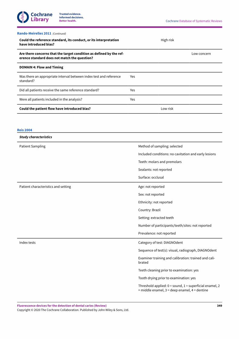
CochraneLibrary
Trusted evidence.Informed decisions.Better health.
Cochrane Database of Systematic Reviews
Could the reference standard, its conduct, or its interpretationhave introduced bias?
High risk
Are there concerns that the target condition as defined by the ref-erence standard does not match the question?
Low concern
DOMAIN 4: Flow and Timing
Was there an appropriate interval between index test and referencestandard?
Yes
Did all patients receive the same reference standard? Yes
Were all patients included in the analysis? Yes
Could the patient flow have introduced bias? Low risk
Rando-Meirelles 2011 (Continued)
Study characteristics
Patient Sampling Method of sampling: selected
Included conditions: no cavitation and early lesions
Teeth: molars and premolars
Sealants: not reported
Surface: occlusal
Patient characteristics and setting Age: not reported
Sex: not reported
Ethnicity: not reported
Country: Brazil
Setting: extracted teeth
Number of participants/teeth/sites: not reported
Prevalence: not reported
Index tests Category of test: DIAGNOdent
Sequence of test(s): visual, radiograph, DIAGNOdent
Examiner training and calibration: trained and cali-brated
Teeth cleaning prior to examination: yes
Tooth drying prior to examination: yes
Threshold applied: 0 = sound, 1 = superficial enamel, 2= middle enamel, 3 = deep enamel, 4 = dentine
Reis 2004
Fluorescence devices for the detection of dental caries (Review)
Copyright © 2020 The Cochrane Collaboration. Published by John Wiley & Sons, Ltd.
349
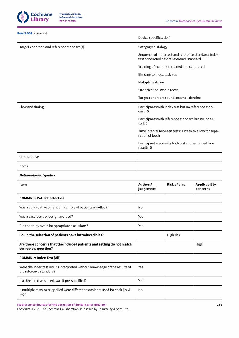
CochraneLibrary
Trusted evidence.Informed decisions.Better health.
Cochrane Database of Systematic Reviews
Device specifics: tip A
Target condition and reference standard(s) Category: histology
Sequence of index test and reference standard: indextest conducted before reference standard
Training of examiner: trained and calibrated
Blinding to index test: yes
Multiple tests: no
Site selection: whole tooth
Target condition: sound, enamel, dentine
Flow and timing Participants with index test but no reference stan-dard: 0
Participants with reference standard but no indextest: 0
Time interval between tests: 1 week to allow for sepa-ration of teeth
Participants receiving both tests but excluded fromresults: 0
Comparative
Notes
Methodological quality
Item Authors'judgement
Risk of bias Applicabilityconcerns
DOMAIN 1: Patient Selection
Was a consecutive or random sample of patients enrolled? No
Was a case-control design avoided? Yes
Did the study avoid inappropriate exclusions? Yes
Could the selection of patients have introduced bias? High risk
Are there concerns that the included patients and setting do not matchthe review question?
High
DOMAIN 2: Index Test (All)
Were the index test results interpreted without knowledge of the results ofthe reference standard?
Yes
If a threshold was used, was it pre-specified? Yes
If multiple tests were applied were different examiners used for each (in vi-vo)?
No
Reis 2004 (Continued)
Fluorescence devices for the detection of dental caries (Review)
Copyright © 2020 The Cochrane Collaboration. Published by John Wiley & Sons, Ltd.
350
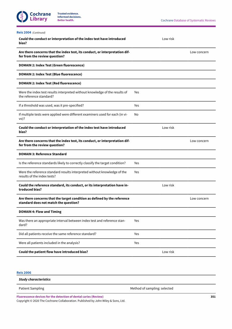
CochraneLibrary
Trusted evidence.Informed decisions.Better health.
Cochrane Database of Systematic Reviews
Could the conduct or interpretation of the index test have introducedbias?
Low risk
Are there concerns that the index test, its conduct, or interpretation dif-fer from the review question?
Low concern
DOMAIN 2: Index Test (Green fluorescence)
DOMAIN 2: Index Test (Blue fluorescence)
DOMAIN 2: Index Test (Red fluorescence)
Were the index test results interpreted without knowledge of the results ofthe reference standard?
Yes
If a threshold was used, was it pre-specified? Yes
If multiple tests were applied were different examiners used for each (in vi-vo)?
No
Could the conduct or interpretation of the index test have introducedbias?
Low risk
Are there concerns that the index test, its conduct, or interpretation dif-fer from the review question?
Low concern
DOMAIN 3: Reference Standard
Is the reference standards likely to correctly classify the target condition? Yes
Were the reference standard results interpreted without knowledge of theresults of the index tests?
Yes
Could the reference standard, its conduct, or its interpretation have in-troduced bias?
Low risk
Are there concerns that the target condition as defined by the referencestandard does not match the question?
Low concern
DOMAIN 4: Flow and Timing
Was there an appropriate interval between index test and reference stan-dard?
Yes
Did all patients receive the same reference standard? Yes
Were all patients included in the analysis? Yes
Could the patient flow have introduced bias? Low risk
Reis 2004 (Continued)
Study characteristics
Patient Sampling Method of sampling: selected
Reis 2006
Fluorescence devices for the detection of dental caries (Review)
Copyright © 2020 The Cochrane Collaboration. Published by John Wiley & Sons, Ltd.
351
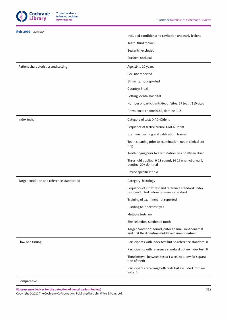
CochraneLibrary
Trusted evidence.Informed decisions.Better health.
Cochrane Database of Systematic Reviews
Included conditions: no cavitation and early lesions
Teeth: third molars
Sealants: excluded
Surface: occlusal
Patient characteristics and setting Age: 19 to 30 years
Sex: not reported
Ethnicity: not reported
Country: Brazil
Setting: dental hospital
Number of participants/teeth/sites: 57 teeth/110 sites
Prevalence: enamel 0.82, dentine 0.15
Index tests Category of test: DIAGNOdent
Sequence of test(s): visual, DIAGNOdent
Examiner training and calibration: trained
Teeth cleaning prior to examination: not in clinical set-ting
Tooth drying prior to examination: yes briefly air dried
Threshold applied: 0-13 sound, 14-19 enamel or earlydentine, 20+ dentinal
Device specifics: tip A
Target condition and reference standard(s) Category: histology
Sequence of index test and reference standard: indextest conducted before reference standard
Training of examiner: not reported
Blinding to index test: yes
Multiple tests: no
Site selection: sectioned tooth
Target condition: sound, outer enamel, inner enameland first third dentine middle and inner dentine
Flow and timing Participants with index test but no reference standard: 0
Participants with reference standard but no index test: 0
Time interval between tests: 1 week to allow for separa-tion of teeth
Participants receiving both tests but excluded from re-sults: 0
Comparative
Reis 2006 (Continued)
Fluorescence devices for the detection of dental caries (Review)
Copyright © 2020 The Cochrane Collaboration. Published by John Wiley & Sons, Ltd.
352

CochraneLibrary
Trusted evidence.Informed decisions.Better health.
Cochrane Database of Systematic Reviews
Notes
Methodological quality
Item Authors' judge-ment
Risk of bias Applicabilityconcerns
DOMAIN 1: Patient Selection
Was a consecutive or random sample of patients enrolled? No
Was a case-control design avoided? Yes
Did the study avoid inappropriate exclusions? Yes
Could the selection of patients have introduced bias? High risk
Are there concerns that the included patients and setting do notmatch the review question?
Low concern
DOMAIN 2: Index Test (All)
Were the index test results interpreted without knowledge of the resultsof the reference standard?
Yes
If a threshold was used, was it pre-specified? Yes
If multiple tests were applied were different examiners used for each (invivo)?
No
Could the conduct or interpretation of the index test have introducedbias?
Low risk
Are there concerns that the index test, its conduct, or interpretationdiffer from the review question?
Low concern
DOMAIN 2: Index Test (Green fluorescence)
DOMAIN 2: Index Test (Blue fluorescence)
DOMAIN 2: Index Test (Red fluorescence)
Were the index test results interpreted without knowledge of the resultsof the reference standard?
Yes
If a threshold was used, was it pre-specified? Yes
If multiple tests were applied were different examiners used for each (invivo)?
No
Could the conduct or interpretation of the index test have introducedbias?
Low risk
Are there concerns that the index test, its conduct, or interpretationdiffer from the review question?
Low concern
DOMAIN 3: Reference Standard
Reis 2006 (Continued)
Fluorescence devices for the detection of dental caries (Review)
Copyright © 2020 The Cochrane Collaboration. Published by John Wiley & Sons, Ltd.
353
CochraneLibrary
Trusted evidence.Informed decisions.Better health.
Cochrane Database of Systematic Reviews
Is the reference standards likely to correctly classify the target condition? Yes
Were the reference standard results interpreted without knowledge ofthe results of the index tests?
Yes
Could the reference standard, its conduct, or its interpretation haveintroduced bias?
Low risk
Are there concerns that the target condition as defined by the refer-ence standard does not match the question?
High
DOMAIN 4: Flow and Timing
Was there an appropriate interval between index test and reference stan-dard?
Yes
Did all patients receive the same reference standard? Yes
Were all patients included in the analysis? Yes
Could the patient flow have introduced bias? Low risk
Reis 2006 (Continued)
Study characteristics
Patient Sampling Method of sampling: selected, "The selected children presented a mini-mum of one tooth in an advanced stage of exfoliation"
Included conditions: no cavitation and early lesions
Teeth: primary molars
Sealants: not reported
Surface: approximal
Patient characteristics and setting Age: 8 to 12 years
Sex: not reported
Ethnicity: not reported
Country: Brazil
Setting: school based
Number of participants/teeth/sites: 112 participants/137 teeth/209 sites
Prevalence: enamel 0.60, dentine 0.29
Index tests Category of test: DIAGNOdent pen
Sequence of test(s): visual, DIAGNOdent, bitewing radiograph, separatorsat 3 time points: baseline, 7 days later and 2 months later
Examiner training and calibration: previously calibrated
Teeth cleaning prior to examination: using dental floss
Ribeiro 2015
Fluorescence devices for the detection of dental caries (Review)
Copyright © 2020 The Cochrane Collaboration. Published by John Wiley & Sons, Ltd.
354
CochraneLibrary
Trusted evidence.Informed decisions.Better health.
Cochrane Database of Systematic Reviews
Tooth drying prior to examination: 5 seconds with dried air
Threshold applied: 0-6 sound, 6.1-9 outer enamel, 9.1-15 inner enamel,15+ dentinal
Device specifics: tip A
Target condition and reference standard(s) Category: computed microtomography, SkyScan device (SkyScan 1174,Kontich, Belgium); description as follows:
"The specimens were rotated through 360°, at a rotation speed of 1.0, witha frame average of 2 and randomized movements. A 0.25-mm aluminumfilter was used. The teeth were scanned at a power of 50 kV and 800 μA";"The teeth were three-dimensionally reconstructed using the programNRecon, version 1.6.0.3, applying maximum reduction of ring artifacts andmaximum beam hardening correction (100%)"
Sequence of index test and reference standard: index tests conducted be-fore reference standard
Training of examiner: not reported
Blinding to index test: yes
Multiple tests: no
Site selection: whole tooth scanned
Target condition: sound, outer enamel, inner enamel, outer dentine, innerdentine
Flow and timing Participants with index test but no reference standard: 146 "63 surfacesout of the total sample were used for the study validation by μCT"
Participants with reference standard but no index test: 0
Time interval between tests: 2 months
Participants receiving both tests but excluded from results: 0
Comparative
Notes Primary data extracted is from the first time point prior to tooth separationas this presents the scenario closest to clinical use and is comparable toother included studies on approximal surfaces
Methodological quality
Item Authors' judgement Risk of bias Applicability con-cerns
DOMAIN 1: Patient Selection
Was a consecutive or random sample of patients en-rolled?
No
Was a case-control design avoided? Yes
Did the study avoid inappropriate exclusions? Yes
Ribeiro 2015 (Continued)
Fluorescence devices for the detection of dental caries (Review)
Copyright © 2020 The Cochrane Collaboration. Published by John Wiley & Sons, Ltd.
355
CochraneLibrary
Trusted evidence.Informed decisions.Better health.
Cochrane Database of Systematic Reviews
Could the selection of patients have introducedbias?
High risk
Are there concerns that the included patients andsetting do not match the review question?
Low concern
DOMAIN 2: Index Test (All)
Were the index test results interpreted without knowl-edge of the results of the reference standard?
Yes
If a threshold was used, was it pre-specified? Yes
If multiple tests were applied were different examinersused for each (in vivo)?
Yes
Could the conduct or interpretation of the index testhave introduced bias?
Low risk
Are there concerns that the index test, its conduct,or interpretation differ from the review question?
Low concern
DOMAIN 2: Index Test (Green fluorescence)
DOMAIN 2: Index Test (Blue fluorescence)
DOMAIN 2: Index Test (Red fluorescence)
Were the index test results interpreted without knowl-edge of the results of the reference standard?
Yes
If a threshold was used, was it pre-specified? Yes
If multiple tests were applied were different examinersused for each (in vivo)?
Yes
Could the conduct or interpretation of the index testhave introduced bias?
Low risk
Are there concerns that the index test, its conduct,or interpretation differ from the review question?
Low concern
DOMAIN 3: Reference Standard
Is the reference standards likely to correctly classify thetarget condition?
Yes
Were the reference standard results interpreted with-out knowledge of the results of the index tests?
Yes
Could the reference standard, its conduct, or its in-terpretation have introduced bias?
Low risk
Are there concerns that the target condition as de-fined by the reference standard does not match thequestion?
Low concern
Ribeiro 2015 (Continued)
Fluorescence devices for the detection of dental caries (Review)
Copyright © 2020 The Cochrane Collaboration. Published by John Wiley & Sons, Ltd.
356
CochraneLibrary
Trusted evidence.Informed decisions.Better health.
Cochrane Database of Systematic Reviews
DOMAIN 4: Flow and Timing
Was there an appropriate interval between index testand reference standard?
Yes
Did all patients receive the same reference standard? No
Were all patients included in the analysis? No
Could the patient flow have introduced bias? High risk
Ribeiro 2015 (Continued)
Study characteristics
Patient Sampling Method of sampling: selected, "FiNy occlusal sites were select-ed for this study"
Included conditions: no cavitation and early lesions
Teeth: primary molars
Sealants: excluded
Surface: occlusal
Patient characteristics and setting Age: 10 to 11 years
Sex: not reported
Ethnicity: not reported
Country: Brazil
Setting: children with teeth close to exfoliation
Number of participants/teeth/sites: 29 participants/50 sites
Prevalence: enamel 0.58, dentine 0.14
Index tests Category of test: DIAGNOdent
Sequence of test(s): DIAGNOdent, visual, radiograph followedby reference standard
Examiner training and calibration: previously trained
Teeth cleaning prior to examination: "professionally cleaned"
Tooth drying prior to examination: 5 seconds with dried air
Threshold applied: "The cutoff limits for all and dentin lesionswere values of 6 and 21"
Device specifics: tip A, marked on photograph, then maximumvalue, mean of 3 measurements
Target condition and reference standard(s) Category: histology
Rocha 2003
Fluorescence devices for the detection of dental caries (Review)
Copyright © 2020 The Cochrane Collaboration. Published by John Wiley & Sons, Ltd.
357
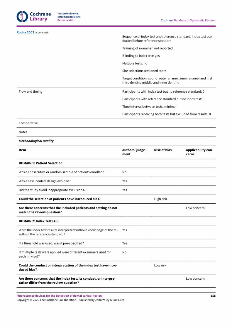
CochraneLibrary
Trusted evidence.Informed decisions.Better health.
Cochrane Database of Systematic Reviews
Sequence of index test and reference standard: index test con-ducted before reference standard
Training of examiner: not reported
Blinding to index test: yes
Multiple tests: no
Site selection: sectioned tooth
Target condition: sound, outer enamel, inner enamel and firstthird dentine middle and inner dentine
Flow and timing Participants with index test but no reference standard: 0
Participants with reference standard but no index test: 0
Time interval between tests: minimal
Participants receiving both tests but excluded from results: 0
Comparative
Notes
Methodological quality
Item Authors' judge-ment
Risk of bias Applicability con-cerns
DOMAIN 1: Patient Selection
Was a consecutive or random sample of patients enrolled? No
Was a case-control design avoided? Yes
Did the study avoid inappropriate exclusions? Yes
Could the selection of patients have introduced bias? High risk
Are there concerns that the included patients and setting do notmatch the review question?
Low concern
DOMAIN 2: Index Test (All)
Were the index test results interpreted without knowledge of the re-sults of the reference standard?
Yes
If a threshold was used, was it pre-specified? Yes
If multiple tests were applied were different examiners used foreach (in vivo)?
No
Could the conduct or interpretation of the index test have intro-duced bias?
Low risk
Are there concerns that the index test, its conduct, or interpre-tation differ from the review question?
Low concern
Rocha 2003 (Continued)
Fluorescence devices for the detection of dental caries (Review)
Copyright © 2020 The Cochrane Collaboration. Published by John Wiley & Sons, Ltd.
358
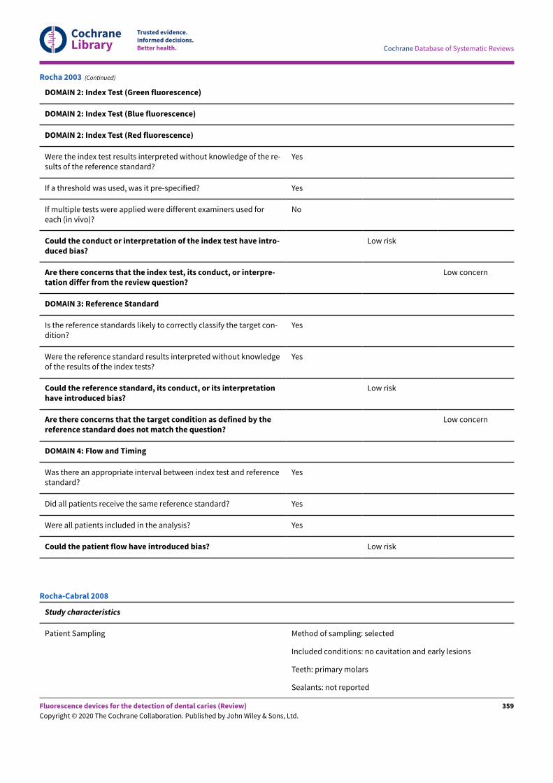
CochraneLibrary
Trusted evidence.Informed decisions.Better health.
Cochrane Database of Systematic Reviews
DOMAIN 2: Index Test (Green fluorescence)
DOMAIN 2: Index Test (Blue fluorescence)
DOMAIN 2: Index Test (Red fluorescence)
Were the index test results interpreted without knowledge of the re-sults of the reference standard?
Yes
If a threshold was used, was it pre-specified? Yes
If multiple tests were applied were different examiners used foreach (in vivo)?
No
Could the conduct or interpretation of the index test have intro-duced bias?
Low risk
Are there concerns that the index test, its conduct, or interpre-tation differ from the review question?
Low concern
DOMAIN 3: Reference Standard
Is the reference standards likely to correctly classify the target con-dition?
Yes
Were the reference standard results interpreted without knowledgeof the results of the index tests?
Yes
Could the reference standard, its conduct, or its interpretationhave introduced bias?
Low risk
Are there concerns that the target condition as defined by thereference standard does not match the question?
Low concern
DOMAIN 4: Flow and Timing
Was there an appropriate interval between index test and referencestandard?
Yes
Did all patients receive the same reference standard? Yes
Were all patients included in the analysis? Yes
Could the patient flow have introduced bias? Low risk
Rocha 2003 (Continued)
Study characteristics
Patient Sampling Method of sampling: selected
Included conditions: no cavitation and early lesions
Teeth: primary molars
Sealants: not reported
Rocha-Cabral 2008
Fluorescence devices for the detection of dental caries (Review)
Copyright © 2020 The Cochrane Collaboration. Published by John Wiley & Sons, Ltd.
359
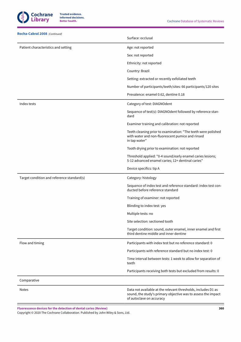
CochraneLibrary
Trusted evidence.Informed decisions.Better health.
Cochrane Database of Systematic Reviews
Surface: occlusal
Patient characteristics and setting Age: not reported
Sex: not reported
Ethnicity: not reported
Country: Brazil
Setting: extracted or recently exfoliated teeth
Number of participants/teeth/sites: 66 participants/120 sites
Prevalence: enamel 0.62, dentine 0.18
Index tests Category of test: DIAGNOdent
Sequence of test(s): DIAGNOdent followed by reference stan-dard
Examiner training and calibration: not reported
Teeth cleaning prior to examination: "The teeth were polishedwith water and non-fluorescent pumice and rinsedin tap water"
Tooth drying prior to examination: not reported
Threshold applied: "0-4 sound/early enamel caries lesions;5-12 advanced enamel caries; 12+ dentinal caries"
Device specifics: tip A
Target condition and reference standard(s) Category: histology
Sequence of index test and reference standard: index test con-ducted before reference standard
Training of examiner: not reported
Blinding to index test: yes
Multiple tests: no
Site selection: sectioned tooth
Target condition: sound, outer enamel, inner enamel and firstthird dentine middle and inner dentine
Flow and timing Participants with index test but no reference standard: 0
Participants with reference standard but no index test: 0
Time interval between tests: 1 week to allow for separation ofteeth
Participants receiving both tests but excluded from results: 0
Comparative
Notes Data not available at the relevant thresholds, includes D1 assound, the study's primary objective was to assess the impactof autoclave on accuracy
Rocha-Cabral 2008 (Continued)
Fluorescence devices for the detection of dental caries (Review)
Copyright © 2020 The Cochrane Collaboration. Published by John Wiley & Sons, Ltd.
360
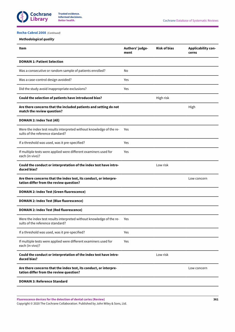
CochraneLibrary
Trusted evidence.Informed decisions.Better health.
Cochrane Database of Systematic Reviews
Methodological quality
Item Authors' judge-ment
Risk of bias Applicability con-cerns
DOMAIN 1: Patient Selection
Was a consecutive or random sample of patients enrolled? No
Was a case-control design avoided? Yes
Did the study avoid inappropriate exclusions? Yes
Could the selection of patients have introduced bias? High risk
Are there concerns that the included patients and setting do notmatch the review question?
High
DOMAIN 2: Index Test (All)
Were the index test results interpreted without knowledge of the re-sults of the reference standard?
Yes
If a threshold was used, was it pre-specified? Yes
If multiple tests were applied were different examiners used foreach (in vivo)?
Yes
Could the conduct or interpretation of the index test have intro-duced bias?
Low risk
Are there concerns that the index test, its conduct, or interpre-tation differ from the review question?
Low concern
DOMAIN 2: Index Test (Green fluorescence)
DOMAIN 2: Index Test (Blue fluorescence)
DOMAIN 2: Index Test (Red fluorescence)
Were the index test results interpreted without knowledge of the re-sults of the reference standard?
Yes
If a threshold was used, was it pre-specified? Yes
If multiple tests were applied were different examiners used foreach (in vivo)?
Yes
Could the conduct or interpretation of the index test have intro-duced bias?
Low risk
Are there concerns that the index test, its conduct, or interpre-tation differ from the review question?
Low concern
DOMAIN 3: Reference Standard
Rocha-Cabral 2008 (Continued)
Fluorescence devices for the detection of dental caries (Review)
Copyright © 2020 The Cochrane Collaboration. Published by John Wiley & Sons, Ltd.
361

CochraneLibrary
Trusted evidence.Informed decisions.Better health.
Cochrane Database of Systematic Reviews
Is the reference standards likely to correctly classify the target con-dition?
Yes
Were the reference standard results interpreted without knowledgeof the results of the index tests?
Yes
Could the reference standard, its conduct, or its interpretationhave introduced bias?
Low risk
Are there concerns that the target condition as defined by thereference standard does not match the question?
Low concern
DOMAIN 4: Flow and Timing
Was there an appropriate interval between index test and referencestandard?
Yes
Did all patients receive the same reference standard? Yes
Were all patients included in the analysis? Yes
Could the patient flow have introduced bias? Low risk
Rocha-Cabral 2008 (Continued)
Study characteristics
Patient Sampling Method of sampling: selected
Included conditions: no cavitation and early lesions
Teeth: permanent molars
Sealants: not reported
Surface: occlusal
Patient characteristics and setting Age: not reported
Sex: not reported
Ethnicity: not reported
Country: Switzerland
Setting: extracted teeth
Number of participants/teeth/sites: 119 teeth
Prevalence: enamel 0.93, dentine 0.54
Index tests Category of test: DIAGNOdent, DIAGNOdent pen andVistaProof
Sequence of test(s): DIAGNOdent, DIAGNOdent pen,VistaProof, visual, radiograph
Examiner training and calibration: experienced
Rodrigues 2008
Fluorescence devices for the detection of dental caries (Review)
Copyright © 2020 The Cochrane Collaboration. Published by John Wiley & Sons, Ltd.
362
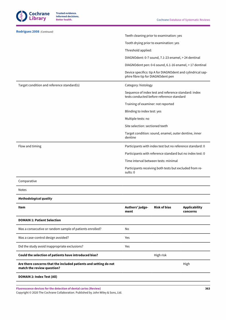
CochraneLibrary
Trusted evidence.Informed decisions.Better health.
Cochrane Database of Systematic Reviews
Teeth cleaning prior to examination: yes
Tooth drying prior to examination: yes
Threshold applied:
DIAGNOdent: 0-7 sound, 7.1-23 enamel, > 24 dentinal
DIAGNOdent pen: 0-6 sound, 6.1-16 enamel, > 17 dentinal
Device specifics: tip A for DIAGNOdent and cylindrical sap-phire fibre tip for DIAGNOdent pen
Target condition and reference standard(s) Category: histology
Sequence of index test and reference standard: indextests conducted before reference standard
Training of examiner: not reported
Blinding to index test: yes
Multiple tests: no
Site selection: sectioned teeth
Target condition: sound, enamel, outer dentine, innerdentine
Flow and timing Participants with index test but no reference standard: 0
Participants with reference standard but no index test: 0
Time interval between tests: minimal
Participants receiving both tests but excluded from re-sults: 0
Comparative
Notes
Methodological quality
Item Authors' judge-ment
Risk of bias Applicabilityconcerns
DOMAIN 1: Patient Selection
Was a consecutive or random sample of patients enrolled? No
Was a case-control design avoided? Yes
Did the study avoid inappropriate exclusions? Yes
Could the selection of patients have introduced bias? High risk
Are there concerns that the included patients and setting do notmatch the review question?
High
DOMAIN 2: Index Test (All)
Rodrigues 2008 (Continued)
Fluorescence devices for the detection of dental caries (Review)
Copyright © 2020 The Cochrane Collaboration. Published by John Wiley & Sons, Ltd.
363
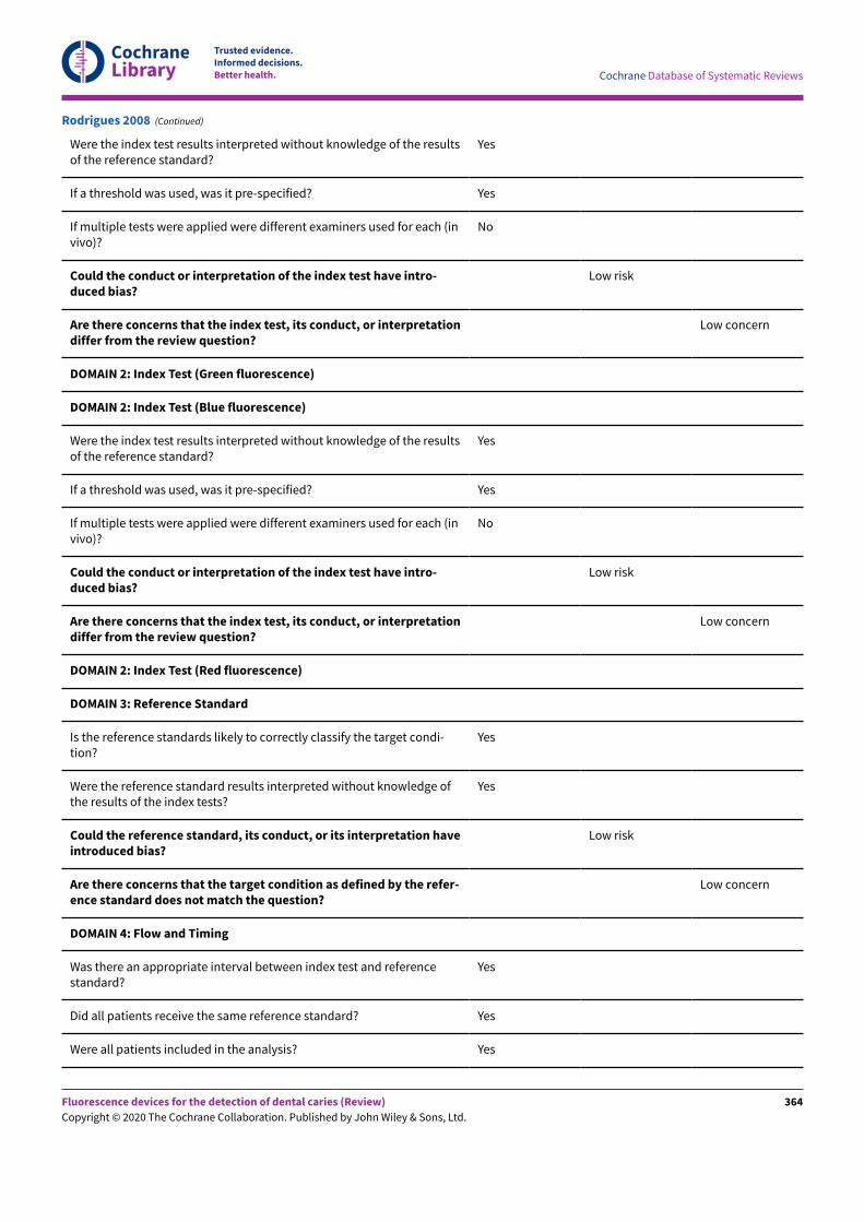
CochraneLibrary
Trusted evidence.Informed decisions.Better health.
Cochrane Database of Systematic Reviews
Were the index test results interpreted without knowledge of the resultsof the reference standard?
Yes
If a threshold was used, was it pre-specified? Yes
If multiple tests were applied were different examiners used for each (invivo)?
No
Could the conduct or interpretation of the index test have intro-duced bias?
Low risk
Are there concerns that the index test, its conduct, or interpretationdiffer from the review question?
Low concern
DOMAIN 2: Index Test (Green fluorescence)
DOMAIN 2: Index Test (Blue fluorescence)
Were the index test results interpreted without knowledge of the resultsof the reference standard?
Yes
If a threshold was used, was it pre-specified? Yes
If multiple tests were applied were different examiners used for each (invivo)?
No
Could the conduct or interpretation of the index test have intro-duced bias?
Low risk
Are there concerns that the index test, its conduct, or interpretationdiffer from the review question?
Low concern
DOMAIN 2: Index Test (Red fluorescence)
DOMAIN 3: Reference Standard
Is the reference standards likely to correctly classify the target condi-tion?
Yes
Were the reference standard results interpreted without knowledge ofthe results of the index tests?
Yes
Could the reference standard, its conduct, or its interpretation haveintroduced bias?
Low risk
Are there concerns that the target condition as defined by the refer-ence standard does not match the question?
Low concern
DOMAIN 4: Flow and Timing
Was there an appropriate interval between index test and referencestandard?
Yes
Did all patients receive the same reference standard? Yes
Were all patients included in the analysis? Yes
Rodrigues 2008 (Continued)
Fluorescence devices for the detection of dental caries (Review)
Copyright © 2020 The Cochrane Collaboration. Published by John Wiley & Sons, Ltd.
364
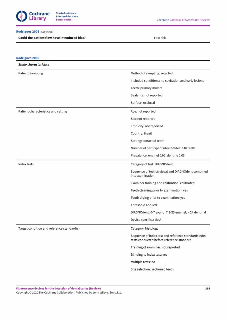
CochraneLibrary
Trusted evidence.Informed decisions.Better health.
Cochrane Database of Systematic Reviews
Could the patient flow have introduced bias? Low risk
Rodrigues 2008 (Continued)
Study characteristics
Patient Sampling Method of sampling: selected
Included conditions: no cavitation and early lesions
Teeth: primary molars
Sealants: not reported
Surface: occlusal
Patient characteristics and setting Age: not reported
Sex: not reported
Ethnicity: not reported
Country: Brazil
Setting: extracted teeth
Number of participants/teeth/sites: 148 teeth
Prevalence: enamel 0.92, dentine 0.03
Index tests Category of test: DIAGNOdent
Sequence of test(s): visual and DIAGNOdent combinedin 1 examination
Examiner training and calibration: calibrated
Teeth cleaning prior to examination: yes
Tooth drying prior to examination: yes
Threshold applied:
DIAGNOdent: 0-7 sound, 7.1-23 enamel, > 24 dentinal
Device specifics: tip A
Target condition and reference standard(s) Category: histology
Sequence of index test and reference standard: indextests conducted before reference standard
Training of examiner: not reported
Blinding to index test: yes
Multiple tests: no
Site selection: sectioned teeth
Rodrigues 2009
Fluorescence devices for the detection of dental caries (Review)
Copyright © 2020 The Cochrane Collaboration. Published by John Wiley & Sons, Ltd.
365
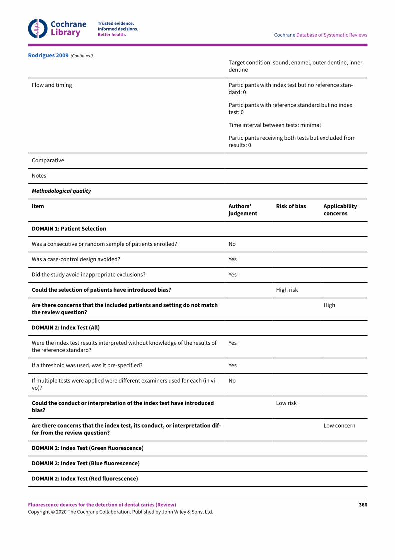
CochraneLibrary
Trusted evidence.Informed decisions.Better health.
Cochrane Database of Systematic Reviews
Target condition: sound, enamel, outer dentine, innerdentine
Flow and timing Participants with index test but no reference stan-dard: 0
Participants with reference standard but no indextest: 0
Time interval between tests: minimal
Participants receiving both tests but excluded fromresults: 0
Comparative
Notes
Methodological quality
Item Authors'judgement
Risk of bias Applicabilityconcerns
DOMAIN 1: Patient Selection
Was a consecutive or random sample of patients enrolled? No
Was a case-control design avoided? Yes
Did the study avoid inappropriate exclusions? Yes
Could the selection of patients have introduced bias? High risk
Are there concerns that the included patients and setting do not matchthe review question?
High
DOMAIN 2: Index Test (All)
Were the index test results interpreted without knowledge of the results ofthe reference standard?
Yes
If a threshold was used, was it pre-specified? Yes
If multiple tests were applied were different examiners used for each (in vi-vo)?
No
Could the conduct or interpretation of the index test have introducedbias?
Low risk
Are there concerns that the index test, its conduct, or interpretation dif-fer from the review question?
Low concern
DOMAIN 2: Index Test (Green fluorescence)
DOMAIN 2: Index Test (Blue fluorescence)
DOMAIN 2: Index Test (Red fluorescence)
Rodrigues 2009 (Continued)
Fluorescence devices for the detection of dental caries (Review)
Copyright © 2020 The Cochrane Collaboration. Published by John Wiley & Sons, Ltd.
366
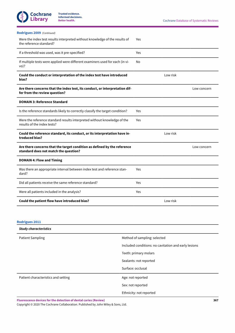
CochraneLibrary
Trusted evidence.Informed decisions.Better health.
Cochrane Database of Systematic Reviews
Were the index test results interpreted without knowledge of the results ofthe reference standard?
Yes
If a threshold was used, was it pre-specified? Yes
If multiple tests were applied were different examiners used for each (in vi-vo)?
No
Could the conduct or interpretation of the index test have introducedbias?
Low risk
Are there concerns that the index test, its conduct, or interpretation dif-fer from the review question?
Low concern
DOMAIN 3: Reference Standard
Is the reference standards likely to correctly classify the target condition? Yes
Were the reference standard results interpreted without knowledge of theresults of the index tests?
Yes
Could the reference standard, its conduct, or its interpretation have in-troduced bias?
Low risk
Are there concerns that the target condition as defined by the referencestandard does not match the question?
Low concern
DOMAIN 4: Flow and Timing
Was there an appropriate interval between index test and reference stan-dard?
Yes
Did all patients receive the same reference standard? Yes
Were all patients included in the analysis? Yes
Could the patient flow have introduced bias? Low risk
Rodrigues 2009 (Continued)
Study characteristics
Patient Sampling Method of sampling: selected
Included conditions: no cavitation and early lesions
Teeth: primary molars
Sealants: not reported
Surface: occlusal
Patient characteristics and setting Age: not reported
Sex: not reported
Ethnicity: not reported
Rodrigues 2011
Fluorescence devices for the detection of dental caries (Review)
Copyright © 2020 The Cochrane Collaboration. Published by John Wiley & Sons, Ltd.
367
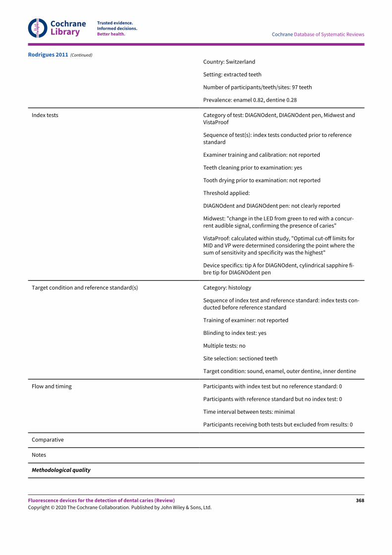
CochraneLibrary
Trusted evidence.Informed decisions.Better health.
Cochrane Database of Systematic Reviews
Country: Switzerland
Setting: extracted teeth
Number of participants/teeth/sites: 97 teeth
Prevalence: enamel 0.82, dentine 0.28
Index tests Category of test: DIAGNOdent, DIAGNOdent pen, Midwest andVistaProof
Sequence of test(s): index tests conducted prior to referencestandard
Examiner training and calibration: not reported
Teeth cleaning prior to examination: yes
Tooth drying prior to examination: not reported
Threshold applied:
DIAGNOdent and DIAGNOdent pen: not clearly reported
Midwest: "change in the LED from green to red with a concur-rent audible signal, confirming the presence of caries"
VistaProof: calculated within study, "Optimal cut-oE limits forMID and VP were determined considering the point where thesum of sensitivity and specificity was the highest"
Device specifics: tip A for DIAGNOdent, cylindrical sapphire fi-bre tip for DIAGNOdent pen
Target condition and reference standard(s) Category: histology
Sequence of index test and reference standard: index tests con-ducted before reference standard
Training of examiner: not reported
Blinding to index test: yes
Multiple tests: no
Site selection: sectioned teeth
Target condition: sound, enamel, outer dentine, inner dentine
Flow and timing Participants with index test but no reference standard: 0
Participants with reference standard but no index test: 0
Time interval between tests: minimal
Participants receiving both tests but excluded from results: 0
Comparative
Notes
Methodological quality
Rodrigues 2011 (Continued)
Fluorescence devices for the detection of dental caries (Review)
Copyright © 2020 The Cochrane Collaboration. Published by John Wiley & Sons, Ltd.
368
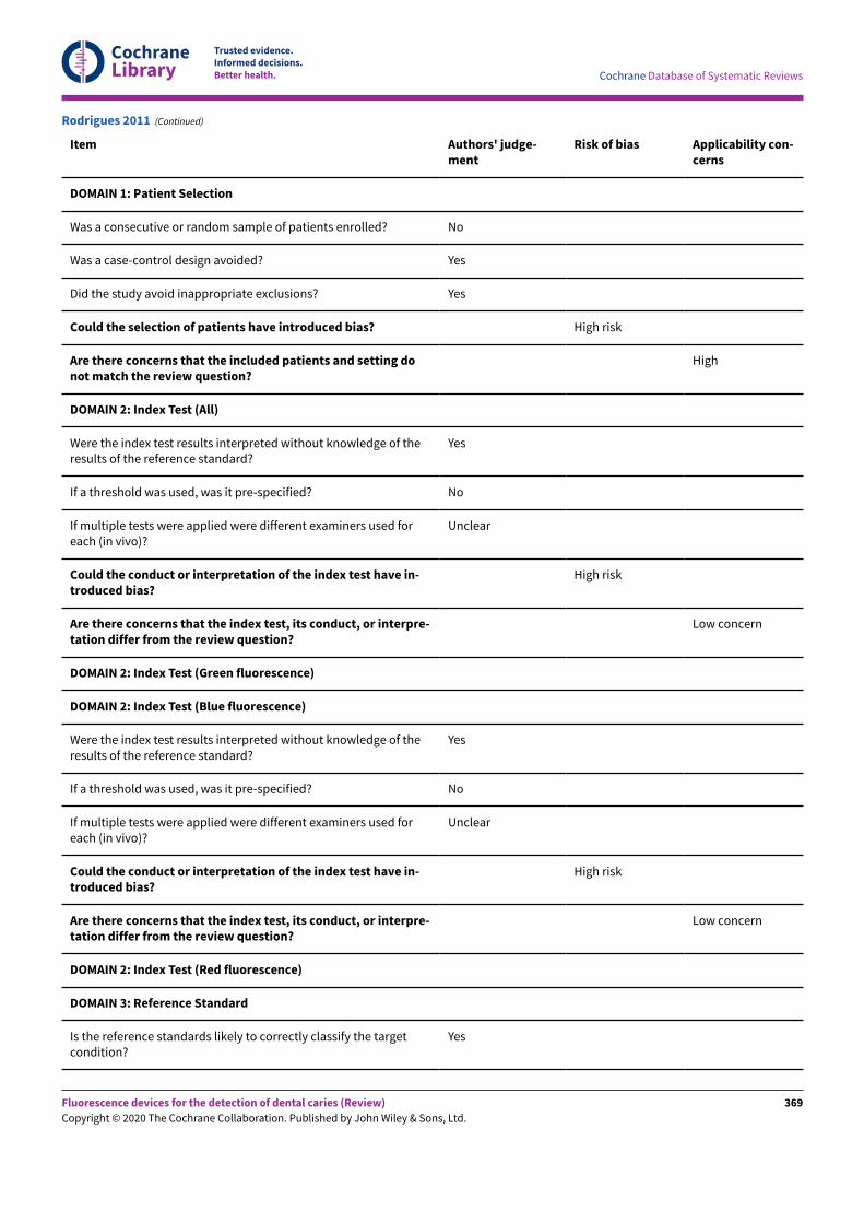
CochraneLibrary
Trusted evidence.Informed decisions.Better health.
Cochrane Database of Systematic Reviews
Item Authors' judge-ment
Risk of bias Applicability con-cerns
DOMAIN 1: Patient Selection
Was a consecutive or random sample of patients enrolled? No
Was a case-control design avoided? Yes
Did the study avoid inappropriate exclusions? Yes
Could the selection of patients have introduced bias? High risk
Are there concerns that the included patients and setting donot match the review question?
High
DOMAIN 2: Index Test (All)
Were the index test results interpreted without knowledge of theresults of the reference standard?
Yes
If a threshold was used, was it pre-specified? No
If multiple tests were applied were different examiners used foreach (in vivo)?
Unclear
Could the conduct or interpretation of the index test have in-troduced bias?
High risk
Are there concerns that the index test, its conduct, or interpre-tation differ from the review question?
Low concern
DOMAIN 2: Index Test (Green fluorescence)
DOMAIN 2: Index Test (Blue fluorescence)
Were the index test results interpreted without knowledge of theresults of the reference standard?
Yes
If a threshold was used, was it pre-specified? No
If multiple tests were applied were different examiners used foreach (in vivo)?
Unclear
Could the conduct or interpretation of the index test have in-troduced bias?
High risk
Are there concerns that the index test, its conduct, or interpre-tation differ from the review question?
Low concern
DOMAIN 2: Index Test (Red fluorescence)
DOMAIN 3: Reference Standard
Is the reference standards likely to correctly classify the targetcondition?
Yes
Rodrigues 2011 (Continued)
Fluorescence devices for the detection of dental caries (Review)
Copyright © 2020 The Cochrane Collaboration. Published by John Wiley & Sons, Ltd.
369
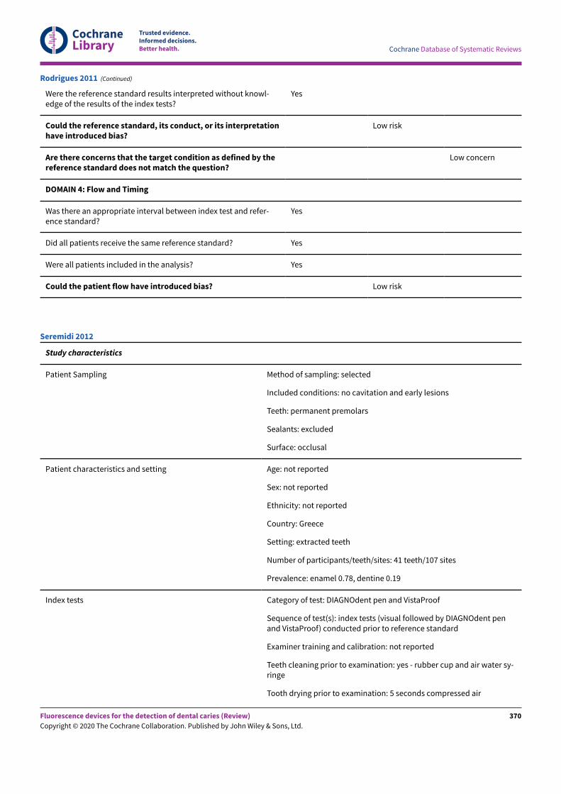
CochraneLibrary
Trusted evidence.Informed decisions.Better health.
Cochrane Database of Systematic Reviews
Were the reference standard results interpreted without knowl-edge of the results of the index tests?
Yes
Could the reference standard, its conduct, or its interpretationhave introduced bias?
Low risk
Are there concerns that the target condition as defined by thereference standard does not match the question?
Low concern
DOMAIN 4: Flow and Timing
Was there an appropriate interval between index test and refer-ence standard?
Yes
Did all patients receive the same reference standard? Yes
Were all patients included in the analysis? Yes
Could the patient flow have introduced bias? Low risk
Rodrigues 2011 (Continued)
Study characteristics
Patient Sampling Method of sampling: selected
Included conditions: no cavitation and early lesions
Teeth: permanent premolars
Sealants: excluded
Surface: occlusal
Patient characteristics and setting Age: not reported
Sex: not reported
Ethnicity: not reported
Country: Greece
Setting: extracted teeth
Number of participants/teeth/sites: 41 teeth/107 sites
Prevalence: enamel 0.78, dentine 0.19
Index tests Category of test: DIAGNOdent pen and VistaProof
Sequence of test(s): index tests (visual followed by DIAGNOdent penand VistaProof) conducted prior to reference standard
Examiner training and calibration: not reported
Teeth cleaning prior to examination: yes - rubber cup and air water sy-ringe
Tooth drying prior to examination: 5 seconds compressed air
Seremidi 2012
Fluorescence devices for the detection of dental caries (Review)
Copyright © 2020 The Cochrane Collaboration. Published by John Wiley & Sons, Ltd.
370
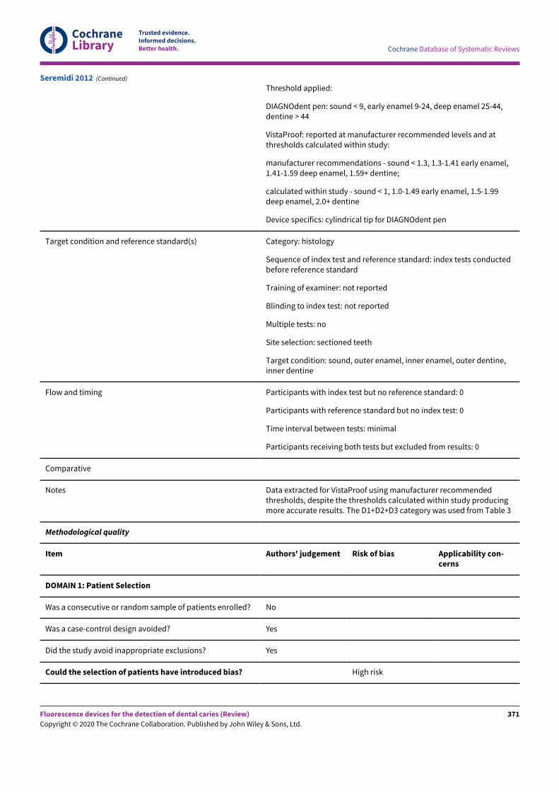
CochraneLibrary
Trusted evidence.Informed decisions.Better health.
Cochrane Database of Systematic Reviews
Threshold applied:
DIAGNOdent pen: sound < 9, early enamel 9-24, deep enamel 25-44,dentine > 44
VistaProof: reported at manufacturer recommended levels and atthresholds calculated within study:
manufacturer recommendations - sound < 1.3, 1.3-1.41 early enamel,1.41-1.59 deep enamel, 1.59+ dentine;
calculated within study - sound < 1, 1.0-1.49 early enamel, 1.5-1.99deep enamel, 2.0+ dentine
Device specifics: cylindrical tip for DIAGNOdent pen
Target condition and reference standard(s) Category: histology
Sequence of index test and reference standard: index tests conductedbefore reference standard
Training of examiner: not reported
Blinding to index test: not reported
Multiple tests: no
Site selection: sectioned teeth
Target condition: sound, outer enamel, inner enamel, outer dentine,inner dentine
Flow and timing Participants with index test but no reference standard: 0
Participants with reference standard but no index test: 0
Time interval between tests: minimal
Participants receiving both tests but excluded from results: 0
Comparative
Notes Data extracted for VistaProof using manufacturer recommendedthresholds, despite the thresholds calculated within study producingmore accurate results. The D1+D2+D3 category was used from Table 3
Methodological quality
Item Authors' judgement Risk of bias Applicability con-cerns
DOMAIN 1: Patient Selection
Was a consecutive or random sample of patients enrolled? No
Was a case-control design avoided? Yes
Did the study avoid inappropriate exclusions? Yes
Could the selection of patients have introduced bias? High risk
Seremidi 2012 (Continued)
Fluorescence devices for the detection of dental caries (Review)
Copyright © 2020 The Cochrane Collaboration. Published by John Wiley & Sons, Ltd.
371
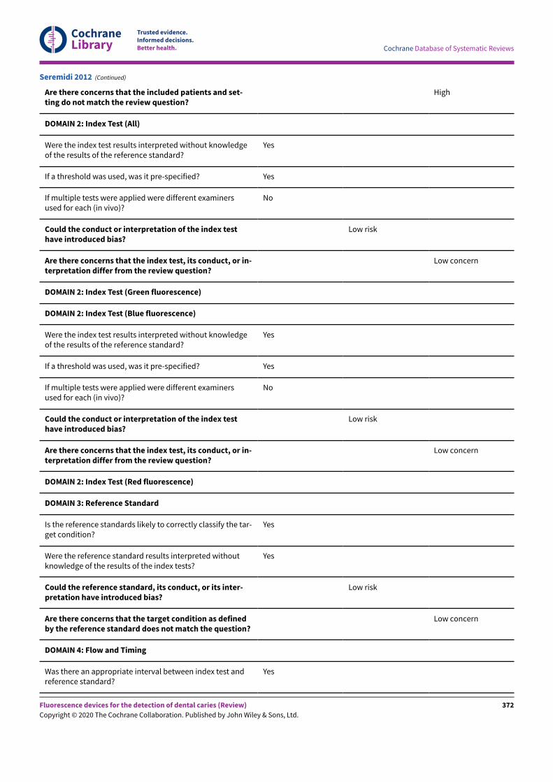
CochraneLibrary
Trusted evidence.Informed decisions.Better health.
Cochrane Database of Systematic Reviews
Are there concerns that the included patients and set-ting do not match the review question?
High
DOMAIN 2: Index Test (All)
Were the index test results interpreted without knowledgeof the results of the reference standard?
Yes
If a threshold was used, was it pre-specified? Yes
If multiple tests were applied were different examinersused for each (in vivo)?
No
Could the conduct or interpretation of the index testhave introduced bias?
Low risk
Are there concerns that the index test, its conduct, or in-terpretation differ from the review question?
Low concern
DOMAIN 2: Index Test (Green fluorescence)
DOMAIN 2: Index Test (Blue fluorescence)
Were the index test results interpreted without knowledgeof the results of the reference standard?
Yes
If a threshold was used, was it pre-specified? Yes
If multiple tests were applied were different examinersused for each (in vivo)?
No
Could the conduct or interpretation of the index testhave introduced bias?
Low risk
Are there concerns that the index test, its conduct, or in-terpretation differ from the review question?
Low concern
DOMAIN 2: Index Test (Red fluorescence)
DOMAIN 3: Reference Standard
Is the reference standards likely to correctly classify the tar-get condition?
Yes
Were the reference standard results interpreted withoutknowledge of the results of the index tests?
Yes
Could the reference standard, its conduct, or its inter-pretation have introduced bias?
Low risk
Are there concerns that the target condition as definedby the reference standard does not match the question?
Low concern
DOMAIN 4: Flow and Timing
Was there an appropriate interval between index test andreference standard?
Yes
Seremidi 2012 (Continued)
Fluorescence devices for the detection of dental caries (Review)
Copyright © 2020 The Cochrane Collaboration. Published by John Wiley & Sons, Ltd.
372
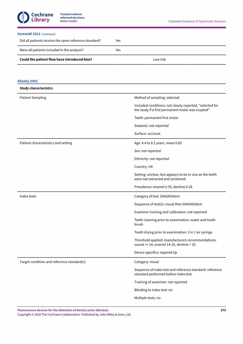
CochraneLibrary
Trusted evidence.Informed decisions.Better health.
Cochrane Database of Systematic Reviews
Did all patients receive the same reference standard? Yes
Were all patients included in the analysis? Yes
Could the patient flow have introduced bias? Low risk
Seremidi 2012 (Continued)
Study characteristics
Patient Sampling Method of sampling: selected
Included conditions: not clearly reported, "selected forthe study if a first permanent molar was erupted"
Teeth: permanent first molar
Sealants: not reported
Surface: occlusal
Patient characteristics and setting Age: 4.4 to 8.2 years, mean 6.85
Sex: not reported
Ethnicity: not reported
Country: UK
Setting: unclear, but appears to be in vivo as the teethwere not extracted and sectioned
Prevalence: enamel 0.55, dentine 0.28
Index tests Category of test: DIAGNOdent
Sequence of test(s): visual then DIAGNOdent
Examiner training and calibration: not reported
Teeth cleaning prior to examination: water and tooth-brush
Tooth drying prior to examination: 3 in 1 air syringe
Threshold applied: manufacturers recommendations:sound =< 14, enamel 14-20, dentine > 20
Device specifics: tapered tip
Target condition and reference standard(s) Category: visual
Sequence of index test and reference standard: referencestandard performed before index test
Training of examiner: not reported
Blinding to index test: no
Multiple tests: no
Sheehy 2001
Fluorescence devices for the detection of dental caries (Review)
Copyright © 2020 The Cochrane Collaboration. Published by John Wiley & Sons, Ltd.
373
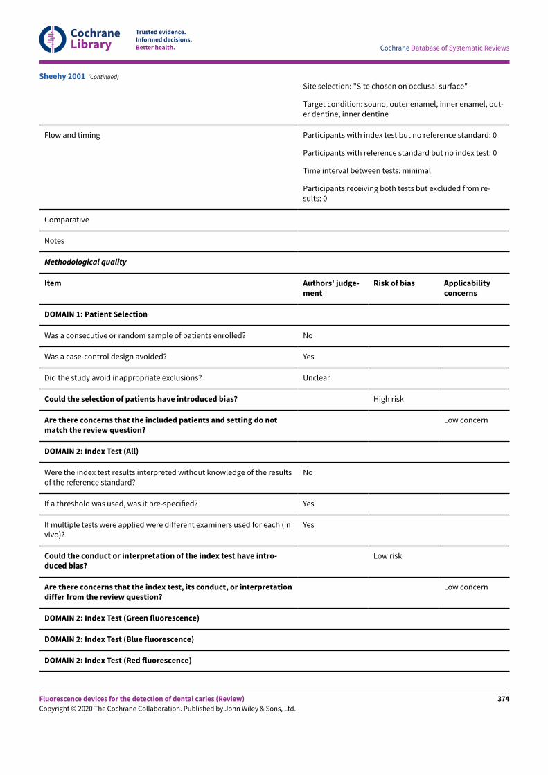
CochraneLibrary
Trusted evidence.Informed decisions.Better health.
Cochrane Database of Systematic Reviews
Site selection: "Site chosen on occlusal surface"
Target condition: sound, outer enamel, inner enamel, out-er dentine, inner dentine
Flow and timing Participants with index test but no reference standard: 0
Participants with reference standard but no index test: 0
Time interval between tests: minimal
Participants receiving both tests but excluded from re-sults: 0
Comparative
Notes
Methodological quality
Item Authors' judge-ment
Risk of bias Applicabilityconcerns
DOMAIN 1: Patient Selection
Was a consecutive or random sample of patients enrolled? No
Was a case-control design avoided? Yes
Did the study avoid inappropriate exclusions? Unclear
Could the selection of patients have introduced bias? High risk
Are there concerns that the included patients and setting do notmatch the review question?
Low concern
DOMAIN 2: Index Test (All)
Were the index test results interpreted without knowledge of the resultsof the reference standard?
No
If a threshold was used, was it pre-specified? Yes
If multiple tests were applied were different examiners used for each (invivo)?
Yes
Could the conduct or interpretation of the index test have intro-duced bias?
Low risk
Are there concerns that the index test, its conduct, or interpretationdiffer from the review question?
Low concern
DOMAIN 2: Index Test (Green fluorescence)
DOMAIN 2: Index Test (Blue fluorescence)
DOMAIN 2: Index Test (Red fluorescence)
Sheehy 2001 (Continued)
Fluorescence devices for the detection of dental caries (Review)
Copyright © 2020 The Cochrane Collaboration. Published by John Wiley & Sons, Ltd.
374
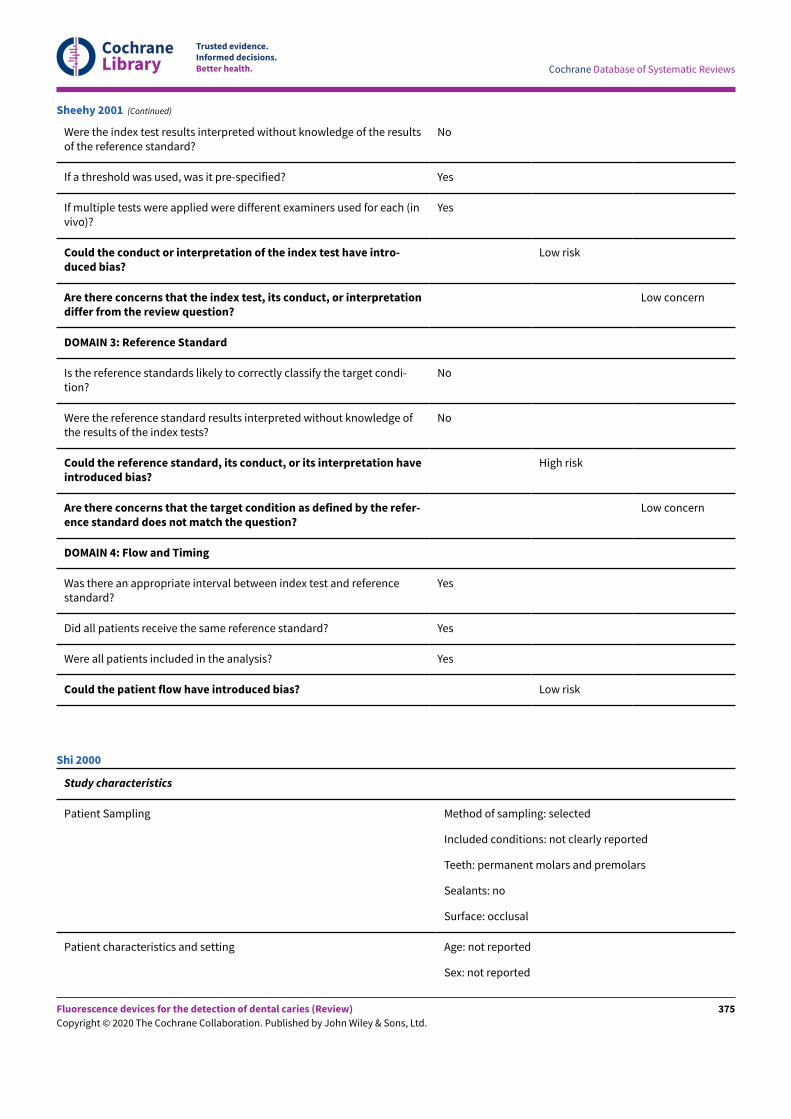
CochraneLibrary
Trusted evidence.Informed decisions.Better health.
Cochrane Database of Systematic Reviews
Were the index test results interpreted without knowledge of the resultsof the reference standard?
No
If a threshold was used, was it pre-specified? Yes
If multiple tests were applied were different examiners used for each (invivo)?
Yes
Could the conduct or interpretation of the index test have intro-duced bias?
Low risk
Are there concerns that the index test, its conduct, or interpretationdiffer from the review question?
Low concern
DOMAIN 3: Reference Standard
Is the reference standards likely to correctly classify the target condi-tion?
No
Were the reference standard results interpreted without knowledge ofthe results of the index tests?
No
Could the reference standard, its conduct, or its interpretation haveintroduced bias?
High risk
Are there concerns that the target condition as defined by the refer-ence standard does not match the question?
Low concern
DOMAIN 4: Flow and Timing
Was there an appropriate interval between index test and referencestandard?
Yes
Did all patients receive the same reference standard? Yes
Were all patients included in the analysis? Yes
Could the patient flow have introduced bias? Low risk
Sheehy 2001 (Continued)
Study characteristics
Patient Sampling Method of sampling: selected
Included conditions: not clearly reported
Teeth: permanent molars and premolars
Sealants: no
Surface: occlusal
Patient characteristics and setting Age: not reported
Sex: not reported
Shi 2000
Fluorescence devices for the detection of dental caries (Review)
Copyright © 2020 The Cochrane Collaboration. Published by John Wiley & Sons, Ltd.
375
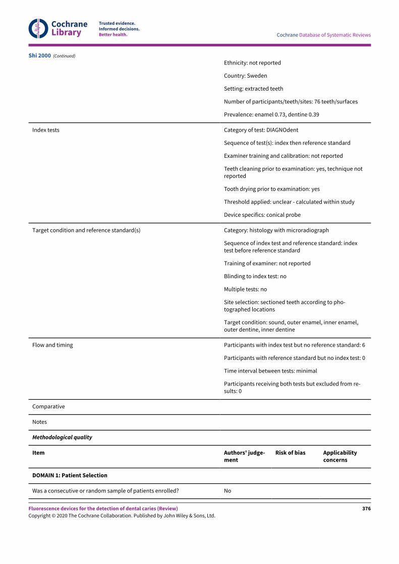
CochraneLibrary
Trusted evidence.Informed decisions.Better health.
Cochrane Database of Systematic Reviews
Ethnicity: not reported
Country: Sweden
Setting: extracted teeth
Number of participants/teeth/sites: 76 teeth/surfaces
Prevalence: enamel 0.73, dentine 0.39
Index tests Category of test: DIAGNOdent
Sequence of test(s): index then reference standard
Examiner training and calibration: not reported
Teeth cleaning prior to examination: yes, technique notreported
Tooth drying prior to examination: yes
Threshold applied: unclear - calculated within study
Device specifics: conical probe
Target condition and reference standard(s) Category: histology with microradiograph
Sequence of index test and reference standard: indextest before reference standard
Training of examiner: not reported
Blinding to index test: no
Multiple tests: no
Site selection: sectioned teeth according to pho-tographed locations
Target condition: sound, outer enamel, inner enamel,outer dentine, inner dentine
Flow and timing Participants with index test but no reference standard: 6
Participants with reference standard but no index test: 0
Time interval between tests: minimal
Participants receiving both tests but excluded from re-sults: 0
Comparative
Notes
Methodological quality
Item Authors' judge-ment
Risk of bias Applicabilityconcerns
DOMAIN 1: Patient Selection
Was a consecutive or random sample of patients enrolled? No
Shi 2000 (Continued)
Fluorescence devices for the detection of dental caries (Review)
Copyright © 2020 The Cochrane Collaboration. Published by John Wiley & Sons, Ltd.
376
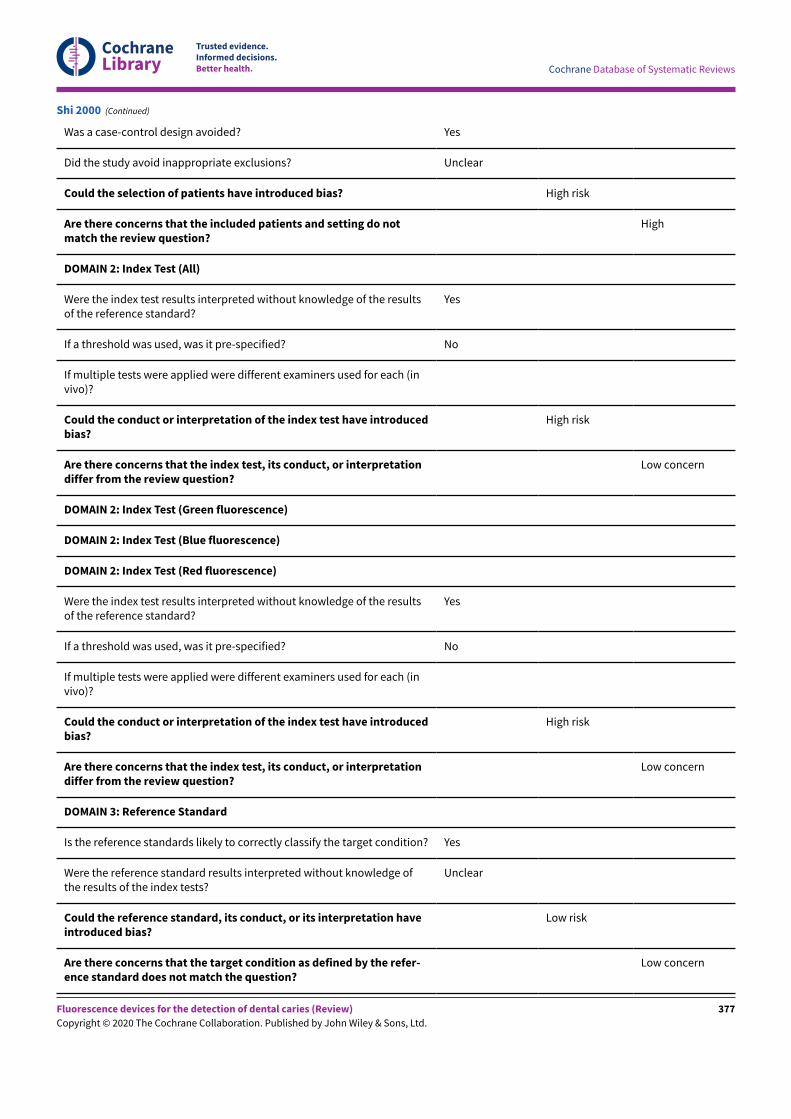
CochraneLibrary
Trusted evidence.Informed decisions.Better health.
Cochrane Database of Systematic Reviews
Was a case-control design avoided? Yes
Did the study avoid inappropriate exclusions? Unclear
Could the selection of patients have introduced bias? High risk
Are there concerns that the included patients and setting do notmatch the review question?
High
DOMAIN 2: Index Test (All)
Were the index test results interpreted without knowledge of the resultsof the reference standard?
Yes
If a threshold was used, was it pre-specified? No
If multiple tests were applied were different examiners used for each (invivo)?
Could the conduct or interpretation of the index test have introducedbias?
High risk
Are there concerns that the index test, its conduct, or interpretationdiffer from the review question?
Low concern
DOMAIN 2: Index Test (Green fluorescence)
DOMAIN 2: Index Test (Blue fluorescence)
DOMAIN 2: Index Test (Red fluorescence)
Were the index test results interpreted without knowledge of the resultsof the reference standard?
Yes
If a threshold was used, was it pre-specified? No
If multiple tests were applied were different examiners used for each (invivo)?
Could the conduct or interpretation of the index test have introducedbias?
High risk
Are there concerns that the index test, its conduct, or interpretationdiffer from the review question?
Low concern
DOMAIN 3: Reference Standard
Is the reference standards likely to correctly classify the target condition? Yes
Were the reference standard results interpreted without knowledge ofthe results of the index tests?
Unclear
Could the reference standard, its conduct, or its interpretation haveintroduced bias?
Low risk
Are there concerns that the target condition as defined by the refer-ence standard does not match the question?
Low concern
Shi 2000 (Continued)
Fluorescence devices for the detection of dental caries (Review)
Copyright © 2020 The Cochrane Collaboration. Published by John Wiley & Sons, Ltd.
377
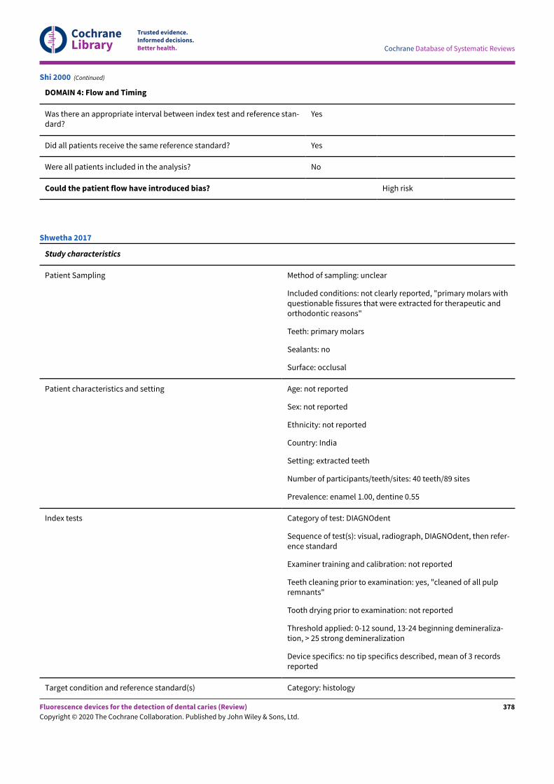
CochraneLibrary
Trusted evidence.Informed decisions.Better health.
Cochrane Database of Systematic Reviews
DOMAIN 4: Flow and Timing
Was there an appropriate interval between index test and reference stan-dard?
Yes
Did all patients receive the same reference standard? Yes
Were all patients included in the analysis? No
Could the patient flow have introduced bias? High risk
Shi 2000 (Continued)
Study characteristics
Patient Sampling Method of sampling: unclear
Included conditions: not clearly reported, "primary molars withquestionable fissures that were extracted for therapeutic andorthodontic reasons"
Teeth: primary molars
Sealants: no
Surface: occlusal
Patient characteristics and setting Age: not reported
Sex: not reported
Ethnicity: not reported
Country: India
Setting: extracted teeth
Number of participants/teeth/sites: 40 teeth/89 sites
Prevalence: enamel 1.00, dentine 0.55
Index tests Category of test: DIAGNOdent
Sequence of test(s): visual, radiograph, DIAGNOdent, then refer-ence standard
Examiner training and calibration: not reported
Teeth cleaning prior to examination: yes, "cleaned of all pulpremnants"
Tooth drying prior to examination: not reported
Threshold applied: 0-12 sound, 13-24 beginning demineraliza-tion, > 25 strong demineralization
Device specifics: no tip specifics described, mean of 3 recordsreported
Target condition and reference standard(s) Category: histology
Shwetha 2017
Fluorescence devices for the detection of dental caries (Review)
Copyright © 2020 The Cochrane Collaboration. Published by John Wiley & Sons, Ltd.
378
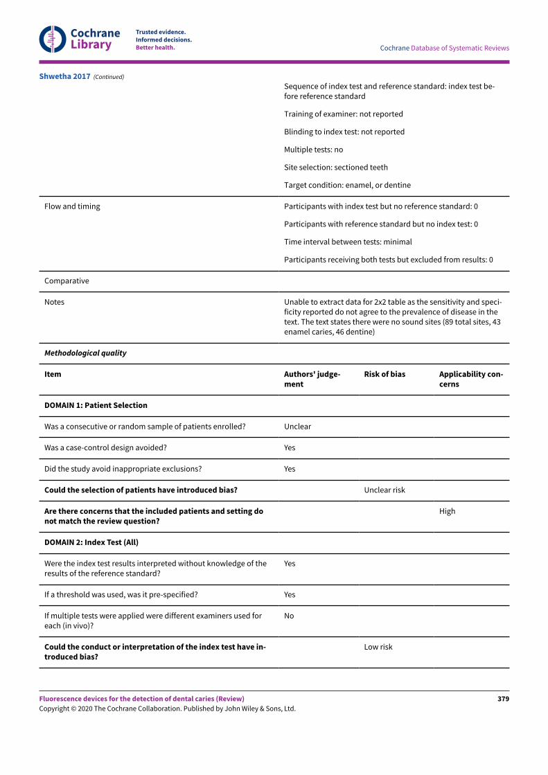
CochraneLibrary
Trusted evidence.Informed decisions.Better health.
Cochrane Database of Systematic Reviews
Sequence of index test and reference standard: index test be-fore reference standard
Training of examiner: not reported
Blinding to index test: not reported
Multiple tests: no
Site selection: sectioned teeth
Target condition: enamel, or dentine
Flow and timing Participants with index test but no reference standard: 0
Participants with reference standard but no index test: 0
Time interval between tests: minimal
Participants receiving both tests but excluded from results: 0
Comparative
Notes Unable to extract data for 2x2 table as the sensitivity and speci-ficity reported do not agree to the prevalence of disease in thetext. The text states there were no sound sites (89 total sites, 43enamel caries, 46 dentine)
Methodological quality
Item Authors' judge-ment
Risk of bias Applicability con-cerns
DOMAIN 1: Patient Selection
Was a consecutive or random sample of patients enrolled? Unclear
Was a case-control design avoided? Yes
Did the study avoid inappropriate exclusions? Yes
Could the selection of patients have introduced bias? Unclear risk
Are there concerns that the included patients and setting donot match the review question?
High
DOMAIN 2: Index Test (All)
Were the index test results interpreted without knowledge of theresults of the reference standard?
Yes
If a threshold was used, was it pre-specified? Yes
If multiple tests were applied were different examiners used foreach (in vivo)?
No
Could the conduct or interpretation of the index test have in-troduced bias?
Low risk
Shwetha 2017 (Continued)
Fluorescence devices for the detection of dental caries (Review)
Copyright © 2020 The Cochrane Collaboration. Published by John Wiley & Sons, Ltd.
379
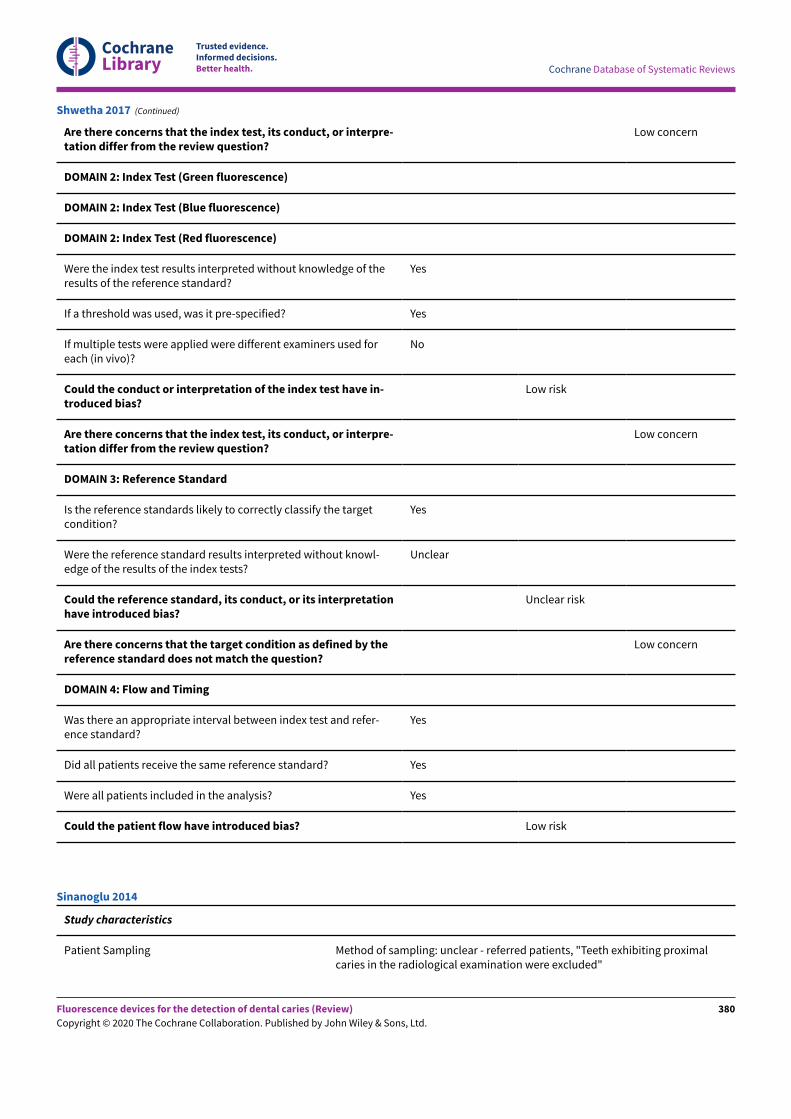
CochraneLibrary
Trusted evidence.Informed decisions.Better health.
Cochrane Database of Systematic Reviews
Are there concerns that the index test, its conduct, or interpre-tation differ from the review question?
Low concern
DOMAIN 2: Index Test (Green fluorescence)
DOMAIN 2: Index Test (Blue fluorescence)
DOMAIN 2: Index Test (Red fluorescence)
Were the index test results interpreted without knowledge of theresults of the reference standard?
Yes
If a threshold was used, was it pre-specified? Yes
If multiple tests were applied were different examiners used foreach (in vivo)?
No
Could the conduct or interpretation of the index test have in-troduced bias?
Low risk
Are there concerns that the index test, its conduct, or interpre-tation differ from the review question?
Low concern
DOMAIN 3: Reference Standard
Is the reference standards likely to correctly classify the targetcondition?
Yes
Were the reference standard results interpreted without knowl-edge of the results of the index tests?
Unclear
Could the reference standard, its conduct, or its interpretationhave introduced bias?
Unclear risk
Are there concerns that the target condition as defined by thereference standard does not match the question?
Low concern
DOMAIN 4: Flow and Timing
Was there an appropriate interval between index test and refer-ence standard?
Yes
Did all patients receive the same reference standard? Yes
Were all patients included in the analysis? Yes
Could the patient flow have introduced bias? Low risk
Shwetha 2017 (Continued)
Study characteristics
Patient Sampling Method of sampling: unclear - referred patients, "Teeth exhibiting proximalcaries in the radiological examination were excluded"
Sinanoglu 2014
Fluorescence devices for the detection of dental caries (Review)
Copyright © 2020 The Cochrane Collaboration. Published by John Wiley & Sons, Ltd.
380
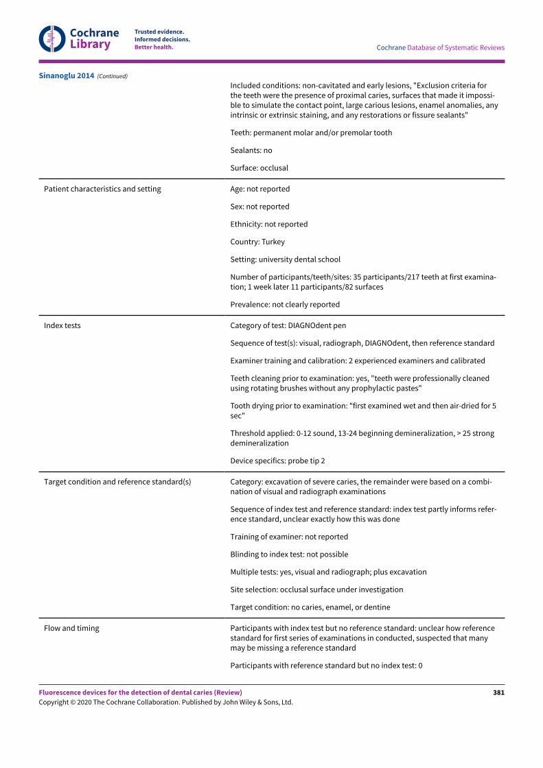
CochraneLibrary
Trusted evidence.Informed decisions.Better health.
Cochrane Database of Systematic Reviews
Included conditions: non-cavitated and early lesions, "Exclusion criteria forthe teeth were the presence of proximal caries, surfaces that made it impossi-ble to simulate the contact point, large carious lesions, enamel anomalies, anyintrinsic or extrinsic staining, and any restorations or fissure sealants"
Teeth: permanent molar and/or premolar tooth
Sealants: no
Surface: occlusal
Patient characteristics and setting Age: not reported
Sex: not reported
Ethnicity: not reported
Country: Turkey
Setting: university dental school
Number of participants/teeth/sites: 35 participants/217 teeth at first examina-tion; 1 week later 11 participants/82 surfaces
Prevalence: not clearly reported
Index tests Category of test: DIAGNOdent pen
Sequence of test(s): visual, radiograph, DIAGNOdent, then reference standard
Examiner training and calibration: 2 experienced examiners and calibrated
Teeth cleaning prior to examination: yes, "teeth were professionally cleanedusing rotating brushes without any prophylactic pastes"
Tooth drying prior to examination: "first examined wet and then air-dried for 5sec"
Threshold applied: 0-12 sound, 13-24 beginning demineralization, > 25 strongdemineralization
Device specifics: probe tip 2
Target condition and reference standard(s) Category: excavation of severe caries, the remainder were based on a combi-nation of visual and radiograph examinations
Sequence of index test and reference standard: index test partly informs refer-ence standard, unclear exactly how this was done
Training of examiner: not reported
Blinding to index test: not possible
Multiple tests: yes, visual and radiograph; plus excavation
Site selection: occlusal surface under investigation
Target condition: no caries, enamel, or dentine
Flow and timing Participants with index test but no reference standard: unclear how referencestandard for first series of examinations in conducted, suspected that manymay be missing a reference standard
Participants with reference standard but no index test: 0
Sinanoglu 2014 (Continued)
Fluorescence devices for the detection of dental caries (Review)
Copyright © 2020 The Cochrane Collaboration. Published by John Wiley & Sons, Ltd.
381
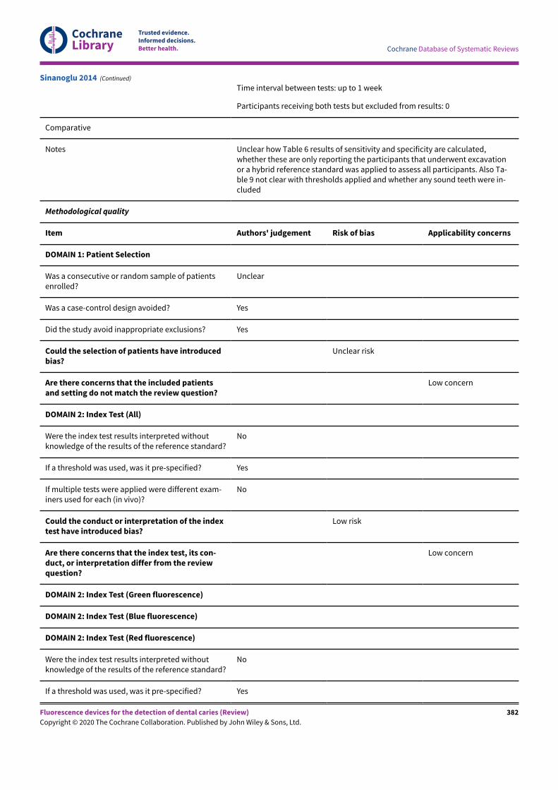
CochraneLibrary
Trusted evidence.Informed decisions.Better health.
Cochrane Database of Systematic Reviews
Time interval between tests: up to 1 week
Participants receiving both tests but excluded from results: 0
Comparative
Notes Unclear how Table 6 results of sensitivity and specificity are calculated,whether these are only reporting the participants that underwent excavationor a hybrid reference standard was applied to assess all participants. Also Ta-ble 9 not clear with thresholds applied and whether any sound teeth were in-cluded
Methodological quality
Item Authors' judgement Risk of bias Applicability concerns
DOMAIN 1: Patient Selection
Was a consecutive or random sample of patientsenrolled?
Unclear
Was a case-control design avoided? Yes
Did the study avoid inappropriate exclusions? Yes
Could the selection of patients have introducedbias?
Unclear risk
Are there concerns that the included patientsand setting do not match the review question?
Low concern
DOMAIN 2: Index Test (All)
Were the index test results interpreted withoutknowledge of the results of the reference standard?
No
If a threshold was used, was it pre-specified? Yes
If multiple tests were applied were different exam-iners used for each (in vivo)?
No
Could the conduct or interpretation of the indextest have introduced bias?
Low risk
Are there concerns that the index test, its con-duct, or interpretation differ from the reviewquestion?
Low concern
DOMAIN 2: Index Test (Green fluorescence)
DOMAIN 2: Index Test (Blue fluorescence)
DOMAIN 2: Index Test (Red fluorescence)
Were the index test results interpreted withoutknowledge of the results of the reference standard?
No
If a threshold was used, was it pre-specified? Yes
Sinanoglu 2014 (Continued)
Fluorescence devices for the detection of dental caries (Review)
Copyright © 2020 The Cochrane Collaboration. Published by John Wiley & Sons, Ltd.
382
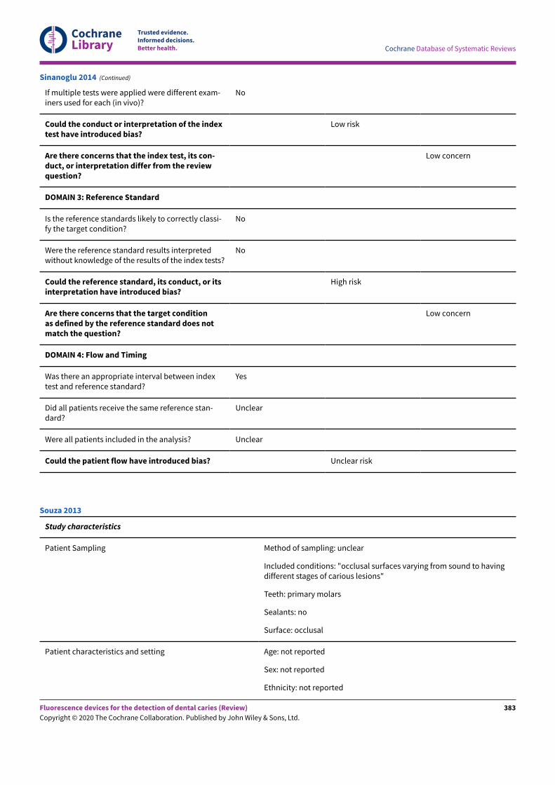
CochraneLibrary
Trusted evidence.Informed decisions.Better health.
Cochrane Database of Systematic Reviews
If multiple tests were applied were different exam-iners used for each (in vivo)?
No
Could the conduct or interpretation of the indextest have introduced bias?
Low risk
Are there concerns that the index test, its con-duct, or interpretation differ from the reviewquestion?
Low concern
DOMAIN 3: Reference Standard
Is the reference standards likely to correctly classi-fy the target condition?
No
Were the reference standard results interpretedwithout knowledge of the results of the index tests?
No
Could the reference standard, its conduct, or itsinterpretation have introduced bias?
High risk
Are there concerns that the target conditionas defined by the reference standard does notmatch the question?
Low concern
DOMAIN 4: Flow and Timing
Was there an appropriate interval between indextest and reference standard?
Yes
Did all patients receive the same reference stan-dard?
Unclear
Were all patients included in the analysis? Unclear
Could the patient flow have introduced bias? Unclear risk
Sinanoglu 2014 (Continued)
Study characteristics
Patient Sampling Method of sampling: unclear
Included conditions: "occlusal surfaces varying from sound to havingdifferent stages of carious lesions"
Teeth: primary molars
Sealants: no
Surface: occlusal
Patient characteristics and setting Age: not reported
Sex: not reported
Ethnicity: not reported
Souza 2013
Fluorescence devices for the detection of dental caries (Review)
Copyright © 2020 The Cochrane Collaboration. Published by John Wiley & Sons, Ltd.
383
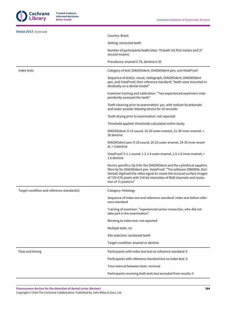
CochraneLibrary
Trusted evidence.Informed decisions.Better health.
Cochrane Database of Systematic Reviews
Country: Brazil
Setting: extracted teeth
Number of participants/teeth/sites: 79 teeth (42 first molars and 37second molars)
Prevalence: enamel 0.76, dentine 0.35
Index tests Category of test: DIAGNOdent, DIAGNOdent pen, and VistaProof
Sequence of test(s): visual, radiograph, DIAGNOdent, DIAGNOdentpen, and VistaProof, then reference standard; "teeth were mounted in-dividually on a dental model"
Examiner training and calibration: "Two experienced examiners inde-pendently assessed the teeth"
Teeth cleaning prior to examination: yes, with sodium bicarbonateand water-powder blasting device for 10 seconds
Tooth drying prior to examination: not reported
Threshold applied: thresholds calculated within study:
DIAGNOdent: 0-15 sound, 16-20 outer enamel, 21-30 inner enamel, >30 dentine
DIAGNOdent pen: 0-19 sound, 20-23 outer enamel, 24-35 inner enam-el, > 3 dentine
VistaProof: 0-1.1 sound, 1.2-1.4 outer enamel, 1.5-1.6 inner enamel, >1.6 dentine
Device specifics: tip A for the DIAGNOdent and the cylindrical sapphirefibre tip for DIAGNOdent pen. VistaProof: "The software (DBSWIN, DürrDental) digitised the video signal to create the occlusal surface imagesof 720×576 pixels with 3×8 bit intensities of RGB channels and resolu-tion of 72 pixels/in"
Target condition and reference standard(s) Category: histology
Sequence of index test and reference standard: index test before refer-ence standard
Training of examiner: "experienced senior researcher, who did nottake part in the examination"
Blinding to index test: not reported
Multiple tests: no
Site selection: sectioned teeth
Target condition: enamel or dentine
Flow and timing Participants with index test but no reference standard: 0
Participants with reference standard but no index test: 0
Time interval between tests: minimal
Participants receiving both tests but excluded from results: 0
Souza 2013 (Continued)
Fluorescence devices for the detection of dental caries (Review)
Copyright © 2020 The Cochrane Collaboration. Published by John Wiley & Sons, Ltd.
384
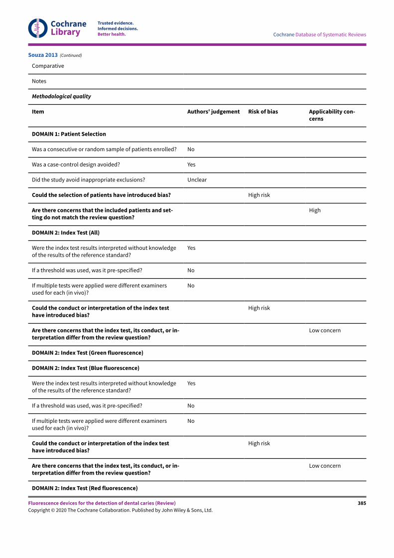
CochraneLibrary
Trusted evidence.Informed decisions.Better health.
Cochrane Database of Systematic Reviews
Comparative
Notes
Methodological quality
Item Authors' judgement Risk of bias Applicability con-cerns
DOMAIN 1: Patient Selection
Was a consecutive or random sample of patients enrolled? No
Was a case-control design avoided? Yes
Did the study avoid inappropriate exclusions? Unclear
Could the selection of patients have introduced bias? High risk
Are there concerns that the included patients and set-ting do not match the review question?
High
DOMAIN 2: Index Test (All)
Were the index test results interpreted without knowledgeof the results of the reference standard?
Yes
If a threshold was used, was it pre-specified? No
If multiple tests were applied were different examinersused for each (in vivo)?
No
Could the conduct or interpretation of the index testhave introduced bias?
High risk
Are there concerns that the index test, its conduct, or in-terpretation differ from the review question?
Low concern
DOMAIN 2: Index Test (Green fluorescence)
DOMAIN 2: Index Test (Blue fluorescence)
Were the index test results interpreted without knowledgeof the results of the reference standard?
Yes
If a threshold was used, was it pre-specified? No
If multiple tests were applied were different examinersused for each (in vivo)?
No
Could the conduct or interpretation of the index testhave introduced bias?
High risk
Are there concerns that the index test, its conduct, or in-terpretation differ from the review question?
Low concern
DOMAIN 2: Index Test (Red fluorescence)
Souza 2013 (Continued)
Fluorescence devices for the detection of dental caries (Review)
Copyright © 2020 The Cochrane Collaboration. Published by John Wiley & Sons, Ltd.
385
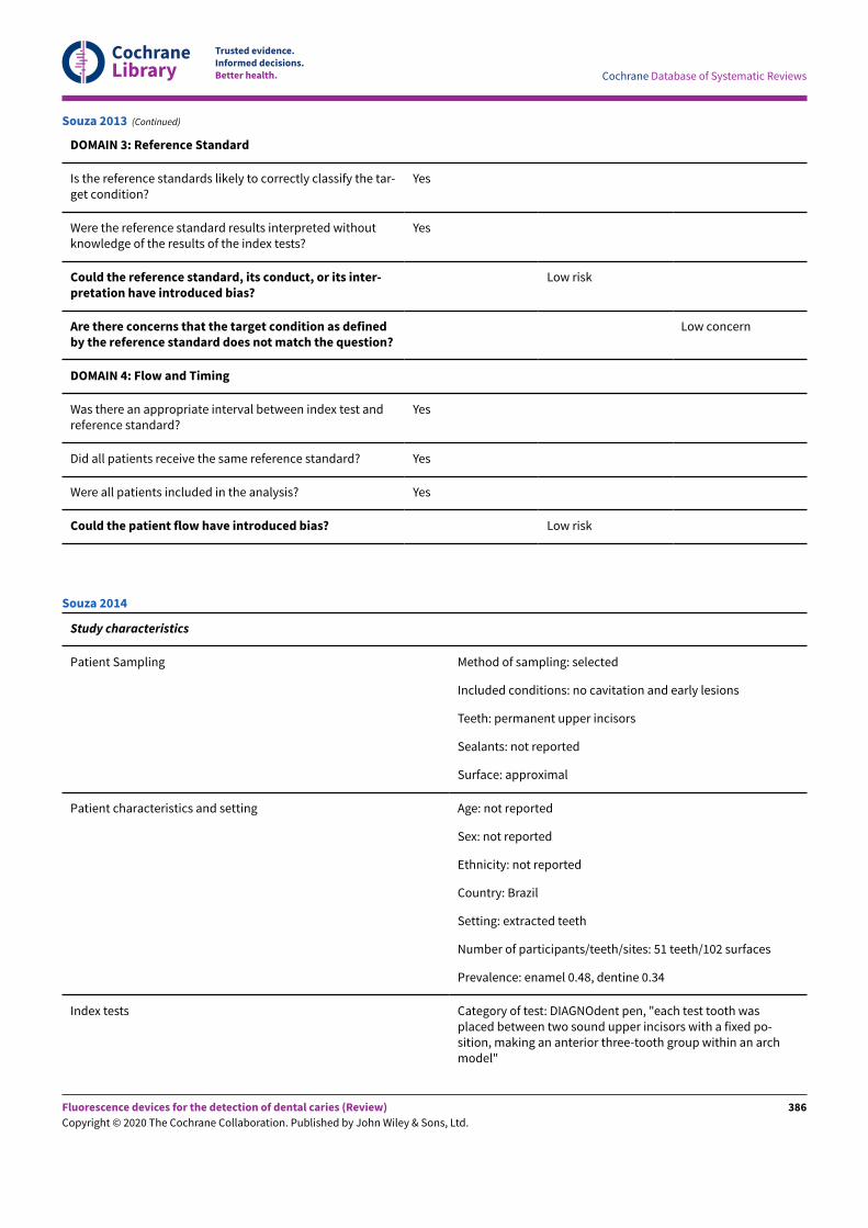
CochraneLibrary
Trusted evidence.Informed decisions.Better health.
Cochrane Database of Systematic Reviews
DOMAIN 3: Reference Standard
Is the reference standards likely to correctly classify the tar-get condition?
Yes
Were the reference standard results interpreted withoutknowledge of the results of the index tests?
Yes
Could the reference standard, its conduct, or its inter-pretation have introduced bias?
Low risk
Are there concerns that the target condition as definedby the reference standard does not match the question?
Low concern
DOMAIN 4: Flow and Timing
Was there an appropriate interval between index test andreference standard?
Yes
Did all patients receive the same reference standard? Yes
Were all patients included in the analysis? Yes
Could the patient flow have introduced bias? Low risk
Souza 2013 (Continued)
Study characteristics
Patient Sampling Method of sampling: selected
Included conditions: no cavitation and early lesions
Teeth: permanent upper incisors
Sealants: not reported
Surface: approximal
Patient characteristics and setting Age: not reported
Sex: not reported
Ethnicity: not reported
Country: Brazil
Setting: extracted teeth
Number of participants/teeth/sites: 51 teeth/102 surfaces
Prevalence: enamel 0.48, dentine 0.34
Index tests Category of test: DIAGNOdent pen, "each test tooth wasplaced between two sound upper incisors with a fixed po-sition, making an anterior three-tooth group within an archmodel"
Souza 2014
Fluorescence devices for the detection of dental caries (Review)
Copyright © 2020 The Cochrane Collaboration. Published by John Wiley & Sons, Ltd.
386
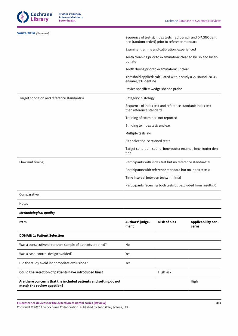
CochraneLibrary
Trusted evidence.Informed decisions.Better health.
Cochrane Database of Systematic Reviews
Sequence of test(s): index tests (radiograph and DIAGNOdentpen (random order)) prior to reference standard
Examiner training and calibration: experienced
Teeth cleaning prior to examination: cleaned brush and bicar-bonate
Tooth drying prior to examination: unclear
Threshold applied: calculated within study 0-27 sound, 28-33enamel, 33+ dentine
Device specifics: wedge shaped probe
Target condition and reference standard(s) Category: histology
Sequence of index test and reference standard: index testthen reference standard
Training of examiner: not reported
Blinding to index test: unclear
Multiple tests: no
Site selection: sectioned teeth
Target condition: sound, inner/outer enamel, inner/outer den-tine
Flow and timing Participants with index test but no reference standard: 0
Participants with reference standard but no index test: 0
Time interval between tests: minimal
Participants receiving both tests but excluded from results: 0
Comparative
Notes
Methodological quality
Item Authors' judge-ment
Risk of bias Applicability con-cerns
DOMAIN 1: Patient Selection
Was a consecutive or random sample of patients enrolled? No
Was a case-control design avoided? Yes
Did the study avoid inappropriate exclusions? Yes
Could the selection of patients have introduced bias? High risk
Are there concerns that the included patients and setting do notmatch the review question?
High
Souza 2014 (Continued)
Fluorescence devices for the detection of dental caries (Review)
Copyright © 2020 The Cochrane Collaboration. Published by John Wiley & Sons, Ltd.
387
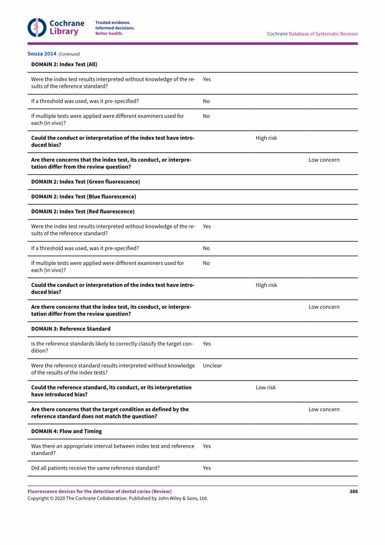
CochraneLibrary
Trusted evidence.Informed decisions.Better health.
Cochrane Database of Systematic Reviews
DOMAIN 2: Index Test (All)
Were the index test results interpreted without knowledge of the re-sults of the reference standard?
Yes
If a threshold was used, was it pre-specified? No
If multiple tests were applied were different examiners used foreach (in vivo)?
No
Could the conduct or interpretation of the index test have intro-duced bias?
High risk
Are there concerns that the index test, its conduct, or interpre-tation differ from the review question?
Low concern
DOMAIN 2: Index Test (Green fluorescence)
DOMAIN 2: Index Test (Blue fluorescence)
DOMAIN 2: Index Test (Red fluorescence)
Were the index test results interpreted without knowledge of the re-sults of the reference standard?
Yes
If a threshold was used, was it pre-specified? No
If multiple tests were applied were different examiners used foreach (in vivo)?
No
Could the conduct or interpretation of the index test have intro-duced bias?
High risk
Are there concerns that the index test, its conduct, or interpre-tation differ from the review question?
Low concern
DOMAIN 3: Reference Standard
Is the reference standards likely to correctly classify the target con-dition?
Yes
Were the reference standard results interpreted without knowledgeof the results of the index tests?
Unclear
Could the reference standard, its conduct, or its interpretationhave introduced bias?
Low risk
Are there concerns that the target condition as defined by thereference standard does not match the question?
Low concern
DOMAIN 4: Flow and Timing
Was there an appropriate interval between index test and referencestandard?
Yes
Did all patients receive the same reference standard? Yes
Souza 2014 (Continued)
Fluorescence devices for the detection of dental caries (Review)
Copyright © 2020 The Cochrane Collaboration. Published by John Wiley & Sons, Ltd.
388
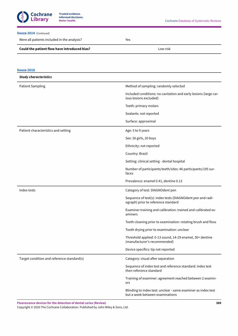
CochraneLibrary
Trusted evidence.Informed decisions.Better health.
Cochrane Database of Systematic Reviews
Were all patients included in the analysis? Yes
Could the patient flow have introduced bias? Low risk
Souza 2014 (Continued)
Study characteristics
Patient Sampling Method of sampling: randomly selected
Included conditions: no cavitation and early lesions (large car-ious lesions excluded)
Teeth: primary molars
Sealants: not reported
Surface: approximal
Patient characteristics and setting Age: 5 to 9 years
Sex: 26 girls, 20 boys
Ethnicity: not reported
Country: Brazil
Setting: clinical setting - dental hospital
Number of participants/teeth/sites: 46 participants/195 sur-faces
Prevalence: enamel 0.41, dentine 0.13
Index tests Category of test: DIAGNOdent pen
Sequence of test(s): index tests (DIAGNOdent pen and radi-ograph) prior to reference standard
Examiner training and calibration: trained and calibrated ex-aminers
Teeth cleaning prior to examination: rotating brush and floss
Tooth drying prior to examination: unclear
Threshold applied: 0-13 sound, 14-29 enamel, 30+ dentine(manufacturer's recommended)
Device specifics: tip not reported
Target condition and reference standard(s) Category: visual after separation
Sequence of index test and reference standard: index testthen reference standard
Training of examiner: agreement reached between 2 examin-ers
Blinding to index test: unclear - same examiner as index testbut a week between examinations
Souza 2018
Fluorescence devices for the detection of dental caries (Review)
Copyright © 2020 The Cochrane Collaboration. Published by John Wiley & Sons, Ltd.
389
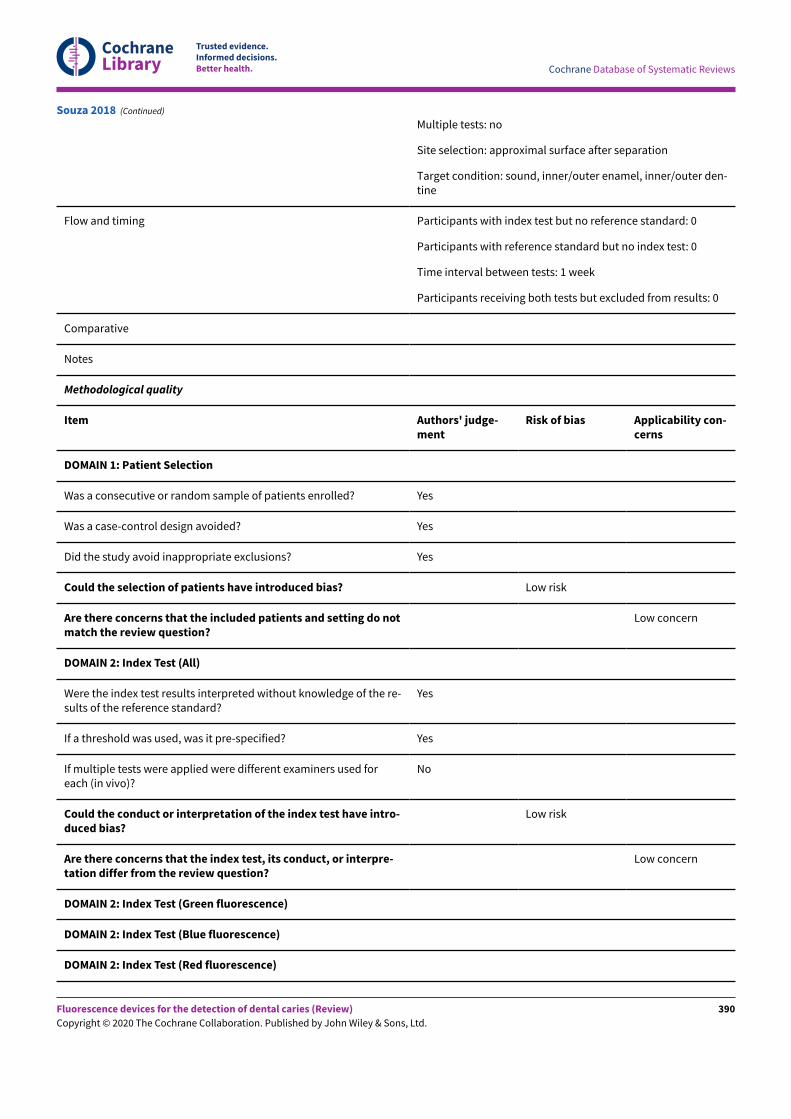
CochraneLibrary
Trusted evidence.Informed decisions.Better health.
Cochrane Database of Systematic Reviews
Multiple tests: no
Site selection: approximal surface after separation
Target condition: sound, inner/outer enamel, inner/outer den-tine
Flow and timing Participants with index test but no reference standard: 0
Participants with reference standard but no index test: 0
Time interval between tests: 1 week
Participants receiving both tests but excluded from results: 0
Comparative
Notes
Methodological quality
Item Authors' judge-ment
Risk of bias Applicability con-cerns
DOMAIN 1: Patient Selection
Was a consecutive or random sample of patients enrolled? Yes
Was a case-control design avoided? Yes
Did the study avoid inappropriate exclusions? Yes
Could the selection of patients have introduced bias? Low risk
Are there concerns that the included patients and setting do notmatch the review question?
Low concern
DOMAIN 2: Index Test (All)
Were the index test results interpreted without knowledge of the re-sults of the reference standard?
Yes
If a threshold was used, was it pre-specified? Yes
If multiple tests were applied were different examiners used foreach (in vivo)?
No
Could the conduct or interpretation of the index test have intro-duced bias?
Low risk
Are there concerns that the index test, its conduct, or interpre-tation differ from the review question?
Low concern
DOMAIN 2: Index Test (Green fluorescence)
DOMAIN 2: Index Test (Blue fluorescence)
DOMAIN 2: Index Test (Red fluorescence)
Souza 2018 (Continued)
Fluorescence devices for the detection of dental caries (Review)
Copyright © 2020 The Cochrane Collaboration. Published by John Wiley & Sons, Ltd.
390
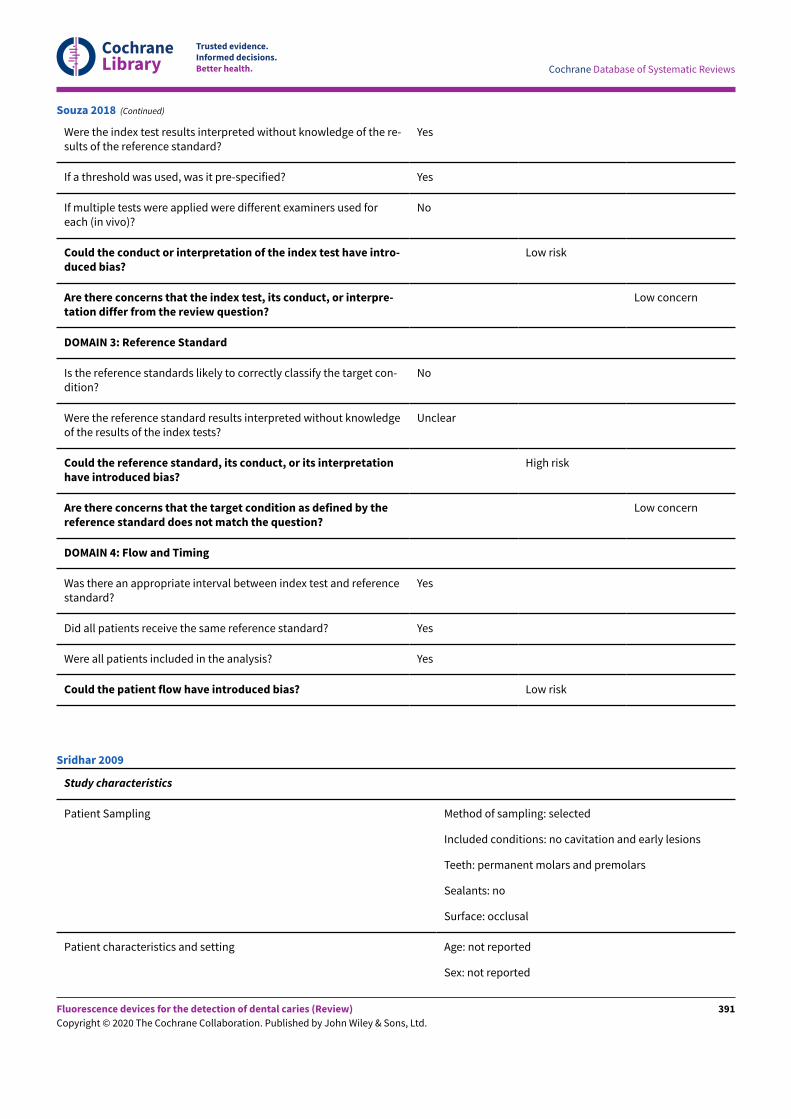
CochraneLibrary
Trusted evidence.Informed decisions.Better health.
Cochrane Database of Systematic Reviews
Were the index test results interpreted without knowledge of the re-sults of the reference standard?
Yes
If a threshold was used, was it pre-specified? Yes
If multiple tests were applied were different examiners used foreach (in vivo)?
No
Could the conduct or interpretation of the index test have intro-duced bias?
Low risk
Are there concerns that the index test, its conduct, or interpre-tation differ from the review question?
Low concern
DOMAIN 3: Reference Standard
Is the reference standards likely to correctly classify the target con-dition?
No
Were the reference standard results interpreted without knowledgeof the results of the index tests?
Unclear
Could the reference standard, its conduct, or its interpretationhave introduced bias?
High risk
Are there concerns that the target condition as defined by thereference standard does not match the question?
Low concern
DOMAIN 4: Flow and Timing
Was there an appropriate interval between index test and referencestandard?
Yes
Did all patients receive the same reference standard? Yes
Were all patients included in the analysis? Yes
Could the patient flow have introduced bias? Low risk
Souza 2018 (Continued)
Study characteristics
Patient Sampling Method of sampling: selected
Included conditions: no cavitation and early lesions
Teeth: permanent molars and premolars
Sealants: no
Surface: occlusal
Patient characteristics and setting Age: not reported
Sex: not reported
Sridhar 2009
Fluorescence devices for the detection of dental caries (Review)
Copyright © 2020 The Cochrane Collaboration. Published by John Wiley & Sons, Ltd.
391
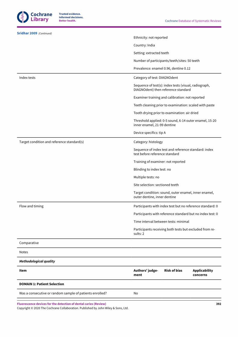
CochraneLibrary
Trusted evidence.Informed decisions.Better health.
Cochrane Database of Systematic Reviews
Ethnicity: not reported
Country: India
Setting: extracted teeth
Number of participants/teeth/sites: 50 teeth
Prevalence: enamel 0.96, dentine 0.12
Index tests Category of test: DIAGNOdent
Sequence of test(s): index tests (visual, radiograph,DIAGNOdent) then reference standard
Examiner training and calibration: not reported
Teeth cleaning prior to examination: scaled with paste
Tooth drying prior to examination: air dried
Threshold applied: 0-5 sound, 6-14 outer enamel, 15-20inner enamel, 21-99 dentine
Device specifics: tip A
Target condition and reference standard(s) Category: histology
Sequence of index test and reference standard: indextest before reference standard
Training of examiner: not reported
Blinding to index test: no
Multiple tests: no
Site selection: sectioned teeth
Target condition: sound, outer enamel, inner enamel,outer dentine, inner dentine
Flow and timing Participants with index test but no reference standard: 0
Participants with reference standard but no index test: 0
Time interval between tests: minimal
Participants receiving both tests but excluded from re-sults: 2
Comparative
Notes
Methodological quality
Item Authors' judge-ment
Risk of bias Applicabilityconcerns
DOMAIN 1: Patient Selection
Was a consecutive or random sample of patients enrolled? No
Sridhar 2009 (Continued)
Fluorescence devices for the detection of dental caries (Review)
Copyright © 2020 The Cochrane Collaboration. Published by John Wiley & Sons, Ltd.
392
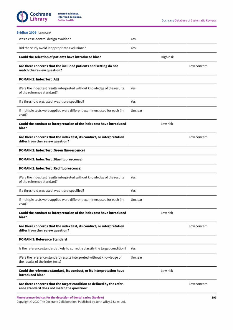
CochraneLibrary
Trusted evidence.Informed decisions.Better health.
Cochrane Database of Systematic Reviews
Was a case-control design avoided? Yes
Did the study avoid inappropriate exclusions? Yes
Could the selection of patients have introduced bias? High risk
Are there concerns that the included patients and setting do notmatch the review question?
Low concern
DOMAIN 2: Index Test (All)
Were the index test results interpreted without knowledge of the resultsof the reference standard?
Yes
If a threshold was used, was it pre-specified? Yes
If multiple tests were applied were different examiners used for each (invivo)?
Unclear
Could the conduct or interpretation of the index test have introducedbias?
Low risk
Are there concerns that the index test, its conduct, or interpretationdiffer from the review question?
Low concern
DOMAIN 2: Index Test (Green fluorescence)
DOMAIN 2: Index Test (Blue fluorescence)
DOMAIN 2: Index Test (Red fluorescence)
Were the index test results interpreted without knowledge of the resultsof the reference standard?
Yes
If a threshold was used, was it pre-specified? Yes
If multiple tests were applied were different examiners used for each (invivo)?
Unclear
Could the conduct or interpretation of the index test have introducedbias?
Low risk
Are there concerns that the index test, its conduct, or interpretationdiffer from the review question?
Low concern
DOMAIN 3: Reference Standard
Is the reference standards likely to correctly classify the target condition? Yes
Were the reference standard results interpreted without knowledge ofthe results of the index tests?
Unclear
Could the reference standard, its conduct, or its interpretation haveintroduced bias?
Low risk
Are there concerns that the target condition as defined by the refer-ence standard does not match the question?
Low concern
Sridhar 2009 (Continued)
Fluorescence devices for the detection of dental caries (Review)
Copyright © 2020 The Cochrane Collaboration. Published by John Wiley & Sons, Ltd.
393
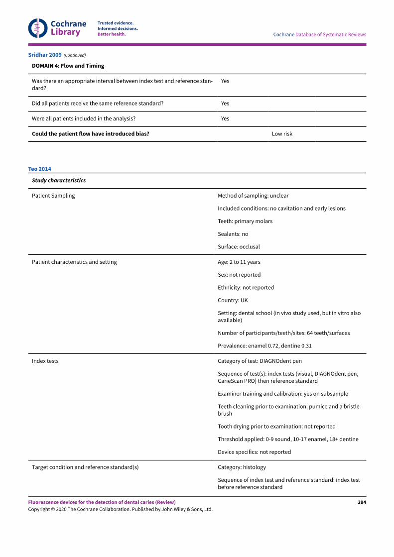
CochraneLibrary
Trusted evidence.Informed decisions.Better health.
Cochrane Database of Systematic Reviews
DOMAIN 4: Flow and Timing
Was there an appropriate interval between index test and reference stan-dard?
Yes
Did all patients receive the same reference standard? Yes
Were all patients included in the analysis? Yes
Could the patient flow have introduced bias? Low risk
Sridhar 2009 (Continued)
Study characteristics
Patient Sampling Method of sampling: unclear
Included conditions: no cavitation and early lesions
Teeth: primary molars
Sealants: no
Surface: occlusal
Patient characteristics and setting Age: 2 to 11 years
Sex: not reported
Ethnicity: not reported
Country: UK
Setting: dental school (in vivo study used, but in vitro alsoavailable)
Number of participants/teeth/sites: 64 teeth/surfaces
Prevalence: enamel 0.72, dentine 0.31
Index tests Category of test: DIAGNOdent pen
Sequence of test(s): index tests (visual, DIAGNOdent pen,CarieScan PRO) then reference standard
Examiner training and calibration: yes on subsample
Teeth cleaning prior to examination: pumice and a bristlebrush
Tooth drying prior to examination: not reported
Threshold applied: 0-9 sound, 10-17 enamel, 18+ dentine
Device specifics: not reported
Target condition and reference standard(s) Category: histology
Sequence of index test and reference standard: index testbefore reference standard
Teo 2014
Fluorescence devices for the detection of dental caries (Review)
Copyright © 2020 The Cochrane Collaboration. Published by John Wiley & Sons, Ltd.
394
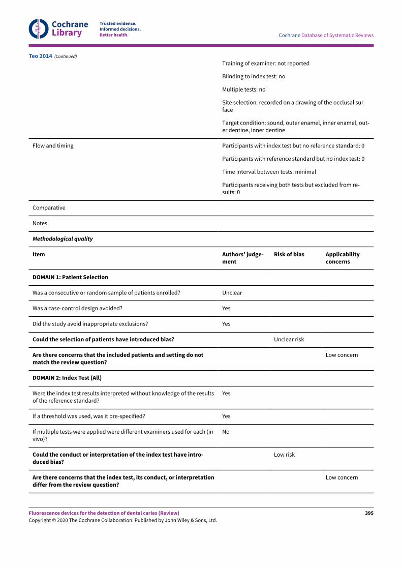
CochraneLibrary
Trusted evidence.Informed decisions.Better health.
Cochrane Database of Systematic Reviews
Training of examiner: not reported
Blinding to index test: no
Multiple tests: no
Site selection: recorded on a drawing of the occlusal sur-face
Target condition: sound, outer enamel, inner enamel, out-er dentine, inner dentine
Flow and timing Participants with index test but no reference standard: 0
Participants with reference standard but no index test: 0
Time interval between tests: minimal
Participants receiving both tests but excluded from re-sults: 0
Comparative
Notes
Methodological quality
Item Authors' judge-ment
Risk of bias Applicabilityconcerns
DOMAIN 1: Patient Selection
Was a consecutive or random sample of patients enrolled? Unclear
Was a case-control design avoided? Yes
Did the study avoid inappropriate exclusions? Yes
Could the selection of patients have introduced bias? Unclear risk
Are there concerns that the included patients and setting do notmatch the review question?
Low concern
DOMAIN 2: Index Test (All)
Were the index test results interpreted without knowledge of the resultsof the reference standard?
Yes
If a threshold was used, was it pre-specified? Yes
If multiple tests were applied were different examiners used for each (invivo)?
No
Could the conduct or interpretation of the index test have intro-duced bias?
Low risk
Are there concerns that the index test, its conduct, or interpretationdiffer from the review question?
Low concern
Teo 2014 (Continued)
Fluorescence devices for the detection of dental caries (Review)
Copyright © 2020 The Cochrane Collaboration. Published by John Wiley & Sons, Ltd.
395
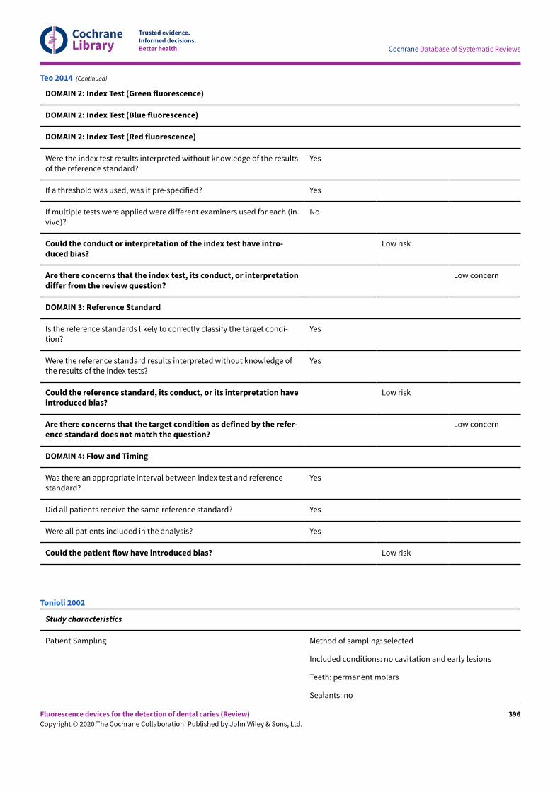
CochraneLibrary
Trusted evidence.Informed decisions.Better health.
Cochrane Database of Systematic Reviews
DOMAIN 2: Index Test (Green fluorescence)
DOMAIN 2: Index Test (Blue fluorescence)
DOMAIN 2: Index Test (Red fluorescence)
Were the index test results interpreted without knowledge of the resultsof the reference standard?
Yes
If a threshold was used, was it pre-specified? Yes
If multiple tests were applied were different examiners used for each (invivo)?
No
Could the conduct or interpretation of the index test have intro-duced bias?
Low risk
Are there concerns that the index test, its conduct, or interpretationdiffer from the review question?
Low concern
DOMAIN 3: Reference Standard
Is the reference standards likely to correctly classify the target condi-tion?
Yes
Were the reference standard results interpreted without knowledge ofthe results of the index tests?
Yes
Could the reference standard, its conduct, or its interpretation haveintroduced bias?
Low risk
Are there concerns that the target condition as defined by the refer-ence standard does not match the question?
Low concern
DOMAIN 4: Flow and Timing
Was there an appropriate interval between index test and referencestandard?
Yes
Did all patients receive the same reference standard? Yes
Were all patients included in the analysis? Yes
Could the patient flow have introduced bias? Low risk
Teo 2014 (Continued)
Study characteristics
Patient Sampling Method of sampling: selected
Included conditions: no cavitation and early lesions
Teeth: permanent molars
Sealants: no
Tonioli 2002
Fluorescence devices for the detection of dental caries (Review)
Copyright © 2020 The Cochrane Collaboration. Published by John Wiley & Sons, Ltd.
396
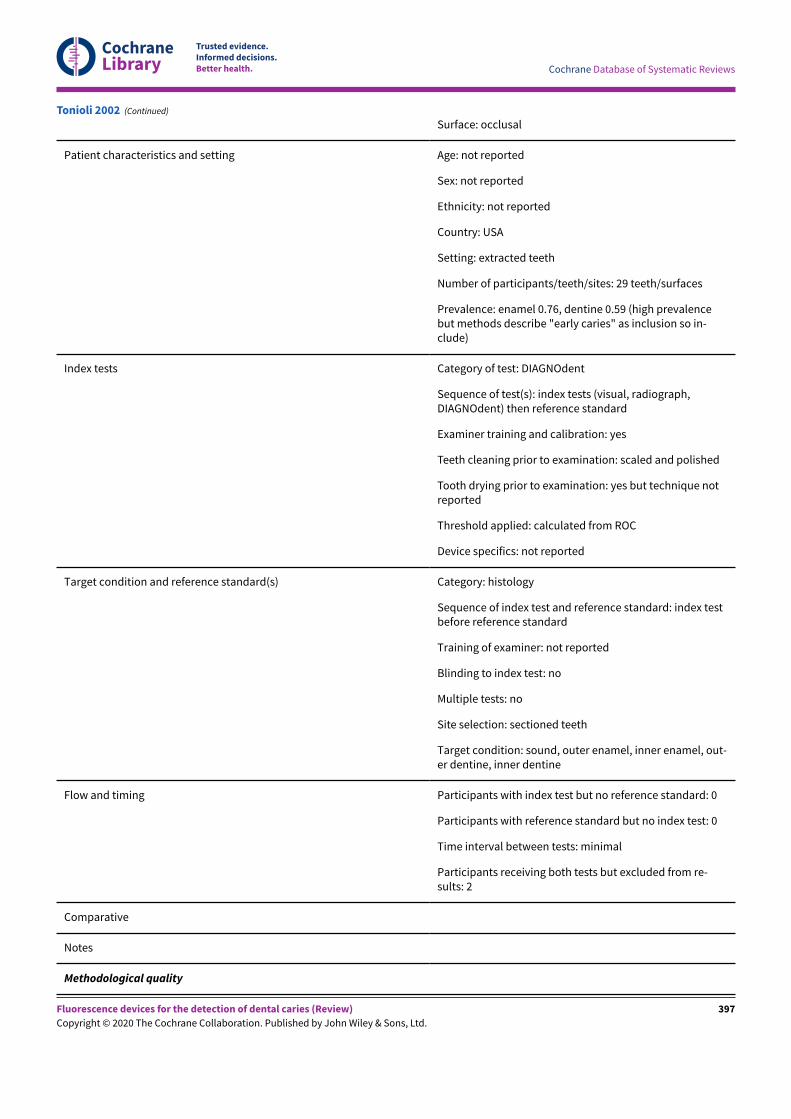
CochraneLibrary
Trusted evidence.Informed decisions.Better health.
Cochrane Database of Systematic Reviews
Surface: occlusal
Patient characteristics and setting Age: not reported
Sex: not reported
Ethnicity: not reported
Country: USA
Setting: extracted teeth
Number of participants/teeth/sites: 29 teeth/surfaces
Prevalence: enamel 0.76, dentine 0.59 (high prevalencebut methods describe "early caries" as inclusion so in-clude)
Index tests Category of test: DIAGNOdent
Sequence of test(s): index tests (visual, radiograph,DIAGNOdent) then reference standard
Examiner training and calibration: yes
Teeth cleaning prior to examination: scaled and polished
Tooth drying prior to examination: yes but technique notreported
Threshold applied: calculated from ROC
Device specifics: not reported
Target condition and reference standard(s) Category: histology
Sequence of index test and reference standard: index testbefore reference standard
Training of examiner: not reported
Blinding to index test: no
Multiple tests: no
Site selection: sectioned teeth
Target condition: sound, outer enamel, inner enamel, out-er dentine, inner dentine
Flow and timing Participants with index test but no reference standard: 0
Participants with reference standard but no index test: 0
Time interval between tests: minimal
Participants receiving both tests but excluded from re-sults: 2
Comparative
Notes
Methodological quality
Tonioli 2002 (Continued)
Fluorescence devices for the detection of dental caries (Review)
Copyright © 2020 The Cochrane Collaboration. Published by John Wiley & Sons, Ltd.
397

CochraneLibrary
Trusted evidence.Informed decisions.Better health.
Cochrane Database of Systematic Reviews
Item Authors' judge-ment
Risk of bias Applicabilityconcerns
DOMAIN 1: Patient Selection
Was a consecutive or random sample of patients enrolled? No
Was a case-control design avoided? Yes
Did the study avoid inappropriate exclusions? Yes
Could the selection of patients have introduced bias? High risk
Are there concerns that the included patients and setting do notmatch the review question?
High
DOMAIN 2: Index Test (All)
Were the index test results interpreted without knowledge of the resultsof the reference standard?
Yes
If a threshold was used, was it pre-specified? No
If multiple tests were applied were different examiners used for each (invivo)?
Yes
Could the conduct or interpretation of the index test have intro-duced bias?
High risk
Are there concerns that the index test, its conduct, or interpretationdiffer from the review question?
Low concern
DOMAIN 2: Index Test (Green fluorescence)
DOMAIN 2: Index Test (Blue fluorescence)
DOMAIN 2: Index Test (Red fluorescence)
Were the index test results interpreted without knowledge of the resultsof the reference standard?
Yes
If a threshold was used, was it pre-specified? No
If multiple tests were applied were different examiners used for each (invivo)?
Yes
Could the conduct or interpretation of the index test have intro-duced bias?
High risk
Are there concerns that the index test, its conduct, or interpretationdiffer from the review question?
Low concern
DOMAIN 3: Reference Standard
Is the reference standards likely to correctly classify the target condi-tion?
Yes
Tonioli 2002 (Continued)
Fluorescence devices for the detection of dental caries (Review)
Copyright © 2020 The Cochrane Collaboration. Published by John Wiley & Sons, Ltd.
398
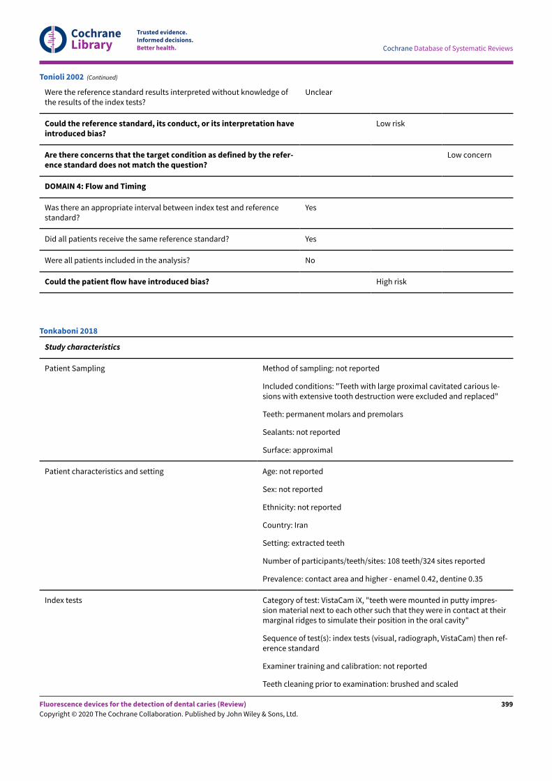
CochraneLibrary
Trusted evidence.Informed decisions.Better health.
Cochrane Database of Systematic Reviews
Were the reference standard results interpreted without knowledge ofthe results of the index tests?
Unclear
Could the reference standard, its conduct, or its interpretation haveintroduced bias?
Low risk
Are there concerns that the target condition as defined by the refer-ence standard does not match the question?
Low concern
DOMAIN 4: Flow and Timing
Was there an appropriate interval between index test and referencestandard?
Yes
Did all patients receive the same reference standard? Yes
Were all patients included in the analysis? No
Could the patient flow have introduced bias? High risk
Tonioli 2002 (Continued)
Study characteristics
Patient Sampling Method of sampling: not reported
Included conditions: "Teeth with large proximal cavitated carious le-sions with extensive tooth destruction were excluded and replaced"
Teeth: permanent molars and premolars
Sealants: not reported
Surface: approximal
Patient characteristics and setting Age: not reported
Sex: not reported
Ethnicity: not reported
Country: Iran
Setting: extracted teeth
Number of participants/teeth/sites: 108 teeth/324 sites reported
Prevalence: contact area and higher - enamel 0.42, dentine 0.35
Index tests Category of test: VistaCam iX, "teeth were mounted in putty impres-sion material next to each other such that they were in contact at theirmarginal ridges to simulate their position in the oral cavity"
Sequence of test(s): index tests (visual, radiograph, VistaCam) then ref-erence standard
Examiner training and calibration: not reported
Teeth cleaning prior to examination: brushed and scaled
Tonkaboni 2018
Fluorescence devices for the detection of dental caries (Review)
Copyright © 2020 The Cochrane Collaboration. Published by John Wiley & Sons, Ltd.
399
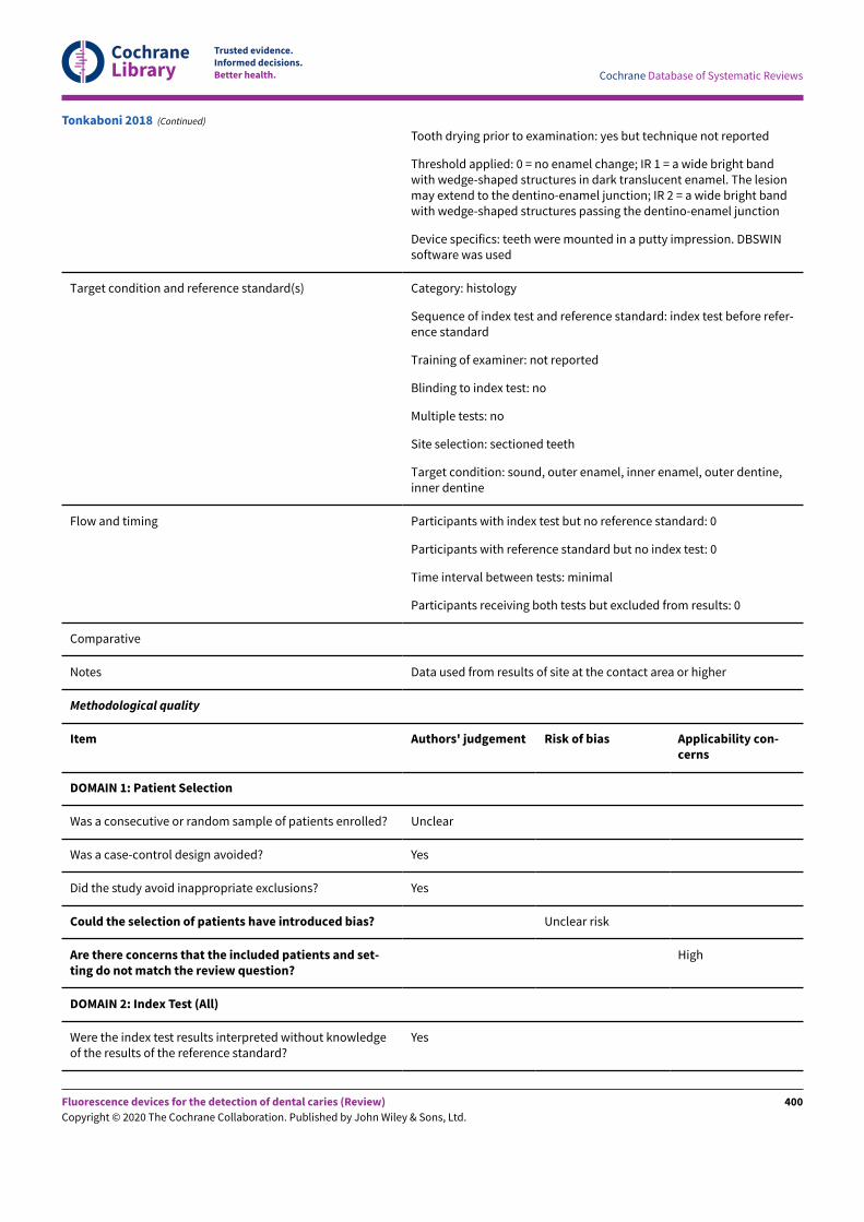
CochraneLibrary
Trusted evidence.Informed decisions.Better health.
Cochrane Database of Systematic Reviews
Tooth drying prior to examination: yes but technique not reported
Threshold applied: 0 = no enamel change; IR 1 = a wide bright bandwith wedge-shaped structures in dark translucent enamel. The lesionmay extend to the dentino-enamel junction; IR 2 = a wide bright bandwith wedge-shaped structures passing the dentino-enamel junction
Device specifics: teeth were mounted in a putty impression. DBSWINsoftware was used
Target condition and reference standard(s) Category: histology
Sequence of index test and reference standard: index test before refer-ence standard
Training of examiner: not reported
Blinding to index test: no
Multiple tests: no
Site selection: sectioned teeth
Target condition: sound, outer enamel, inner enamel, outer dentine,inner dentine
Flow and timing Participants with index test but no reference standard: 0
Participants with reference standard but no index test: 0
Time interval between tests: minimal
Participants receiving both tests but excluded from results: 0
Comparative
Notes Data used from results of site at the contact area or higher
Methodological quality
Item Authors' judgement Risk of bias Applicability con-cerns
DOMAIN 1: Patient Selection
Was a consecutive or random sample of patients enrolled? Unclear
Was a case-control design avoided? Yes
Did the study avoid inappropriate exclusions? Yes
Could the selection of patients have introduced bias? Unclear risk
Are there concerns that the included patients and set-ting do not match the review question?
High
DOMAIN 2: Index Test (All)
Were the index test results interpreted without knowledgeof the results of the reference standard?
Yes
Tonkaboni 2018 (Continued)
Fluorescence devices for the detection of dental caries (Review)
Copyright © 2020 The Cochrane Collaboration. Published by John Wiley & Sons, Ltd.
400
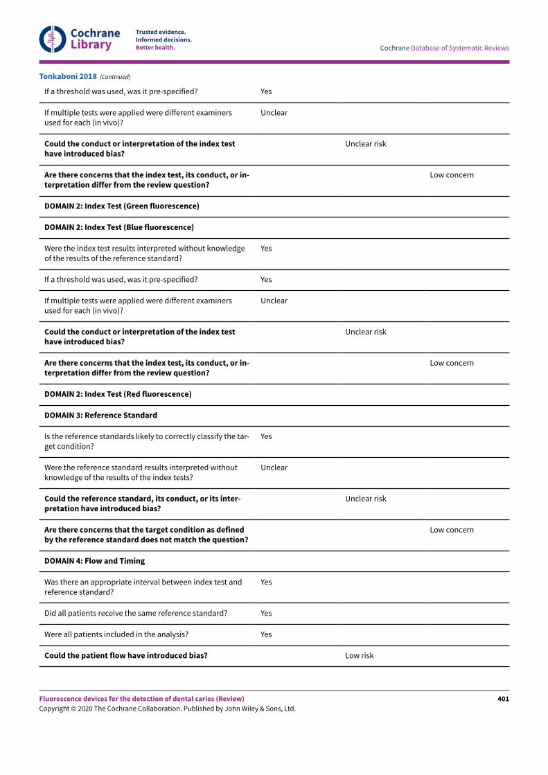
CochraneLibrary
Trusted evidence.Informed decisions.Better health.
Cochrane Database of Systematic Reviews
If a threshold was used, was it pre-specified? Yes
If multiple tests were applied were different examinersused for each (in vivo)?
Unclear
Could the conduct or interpretation of the index testhave introduced bias?
Unclear risk
Are there concerns that the index test, its conduct, or in-terpretation differ from the review question?
Low concern
DOMAIN 2: Index Test (Green fluorescence)
DOMAIN 2: Index Test (Blue fluorescence)
Were the index test results interpreted without knowledgeof the results of the reference standard?
Yes
If a threshold was used, was it pre-specified? Yes
If multiple tests were applied were different examinersused for each (in vivo)?
Unclear
Could the conduct or interpretation of the index testhave introduced bias?
Unclear risk
Are there concerns that the index test, its conduct, or in-terpretation differ from the review question?
Low concern
DOMAIN 2: Index Test (Red fluorescence)
DOMAIN 3: Reference Standard
Is the reference standards likely to correctly classify the tar-get condition?
Yes
Were the reference standard results interpreted withoutknowledge of the results of the index tests?
Unclear
Could the reference standard, its conduct, or its inter-pretation have introduced bias?
Unclear risk
Are there concerns that the target condition as definedby the reference standard does not match the question?
Low concern
DOMAIN 4: Flow and Timing
Was there an appropriate interval between index test andreference standard?
Yes
Did all patients receive the same reference standard? Yes
Were all patients included in the analysis? Yes
Could the patient flow have introduced bias? Low risk
Tonkaboni 2018 (Continued)
Fluorescence devices for the detection of dental caries (Review)
Copyright © 2020 The Cochrane Collaboration. Published by John Wiley & Sons, Ltd.
401
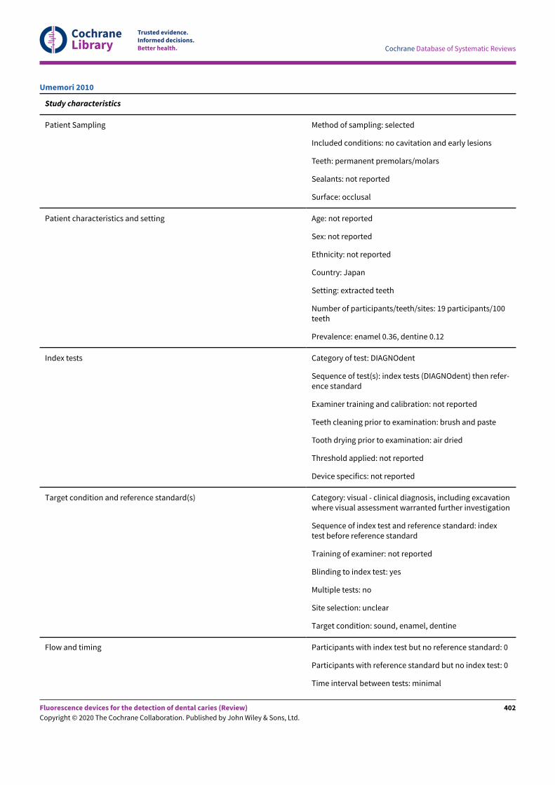
CochraneLibrary
Trusted evidence.Informed decisions.Better health.
Cochrane Database of Systematic Reviews
Study characteristics
Patient Sampling Method of sampling: selected
Included conditions: no cavitation and early lesions
Teeth: permanent premolars/molars
Sealants: not reported
Surface: occlusal
Patient characteristics and setting Age: not reported
Sex: not reported
Ethnicity: not reported
Country: Japan
Setting: extracted teeth
Number of participants/teeth/sites: 19 participants/100teeth
Prevalence: enamel 0.36, dentine 0.12
Index tests Category of test: DIAGNOdent
Sequence of test(s): index tests (DIAGNOdent) then refer-ence standard
Examiner training and calibration: not reported
Teeth cleaning prior to examination: brush and paste
Tooth drying prior to examination: air dried
Threshold applied: not reported
Device specifics: not reported
Target condition and reference standard(s) Category: visual - clinical diagnosis, including excavationwhere visual assessment warranted further investigation
Sequence of index test and reference standard: indextest before reference standard
Training of examiner: not reported
Blinding to index test: yes
Multiple tests: no
Site selection: unclear
Target condition: sound, enamel, dentine
Flow and timing Participants with index test but no reference standard: 0
Participants with reference standard but no index test: 0
Time interval between tests: minimal
Umemori 2010
Fluorescence devices for the detection of dental caries (Review)
Copyright © 2020 The Cochrane Collaboration. Published by John Wiley & Sons, Ltd.
402
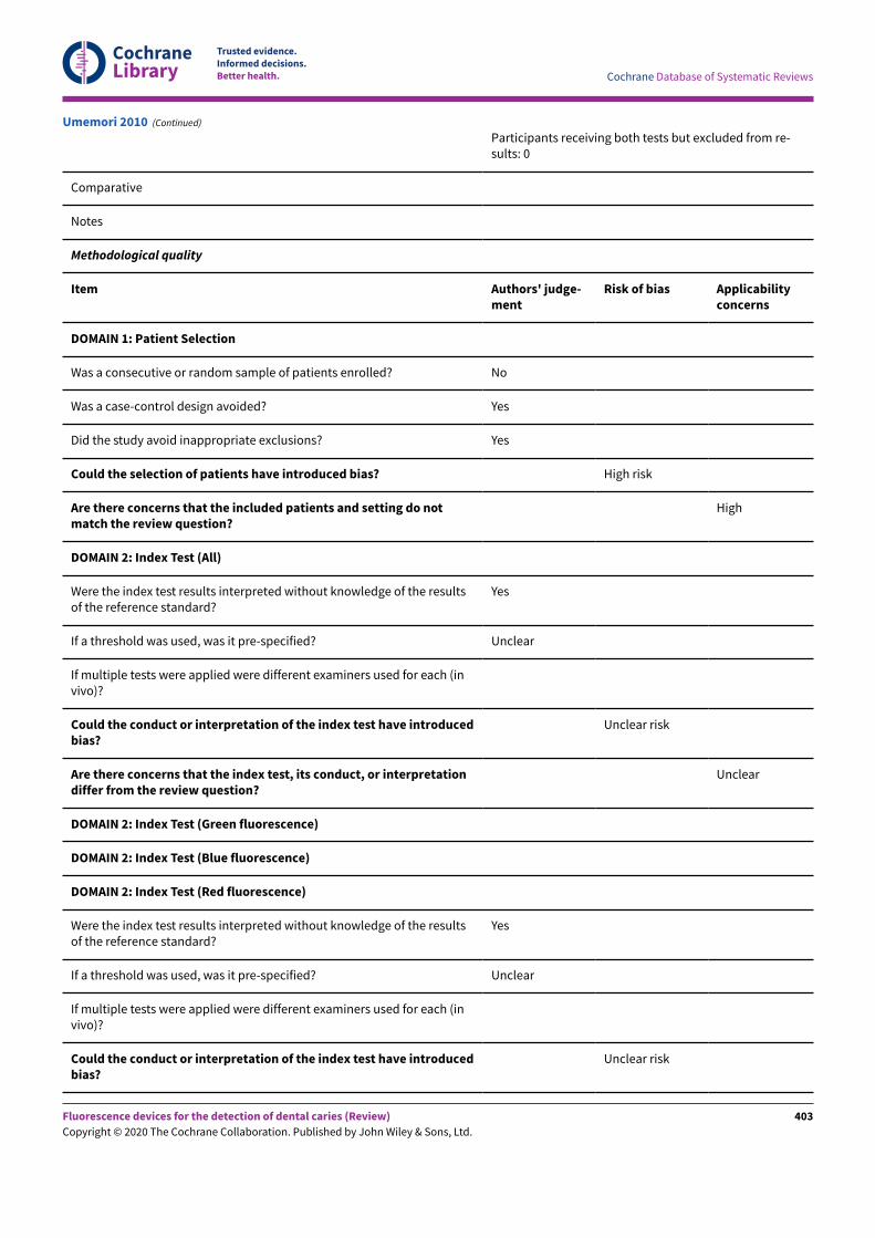
CochraneLibrary
Trusted evidence.Informed decisions.Better health.
Cochrane Database of Systematic Reviews
Participants receiving both tests but excluded from re-sults: 0
Comparative
Notes
Methodological quality
Item Authors' judge-ment
Risk of bias Applicabilityconcerns
DOMAIN 1: Patient Selection
Was a consecutive or random sample of patients enrolled? No
Was a case-control design avoided? Yes
Did the study avoid inappropriate exclusions? Yes
Could the selection of patients have introduced bias? High risk
Are there concerns that the included patients and setting do notmatch the review question?
High
DOMAIN 2: Index Test (All)
Were the index test results interpreted without knowledge of the resultsof the reference standard?
Yes
If a threshold was used, was it pre-specified? Unclear
If multiple tests were applied were different examiners used for each (invivo)?
Could the conduct or interpretation of the index test have introducedbias?
Unclear risk
Are there concerns that the index test, its conduct, or interpretationdiffer from the review question?
Unclear
DOMAIN 2: Index Test (Green fluorescence)
DOMAIN 2: Index Test (Blue fluorescence)
DOMAIN 2: Index Test (Red fluorescence)
Were the index test results interpreted without knowledge of the resultsof the reference standard?
Yes
If a threshold was used, was it pre-specified? Unclear
If multiple tests were applied were different examiners used for each (invivo)?
Could the conduct or interpretation of the index test have introducedbias?
Unclear risk
Umemori 2010 (Continued)
Fluorescence devices for the detection of dental caries (Review)
Copyright © 2020 The Cochrane Collaboration. Published by John Wiley & Sons, Ltd.
403
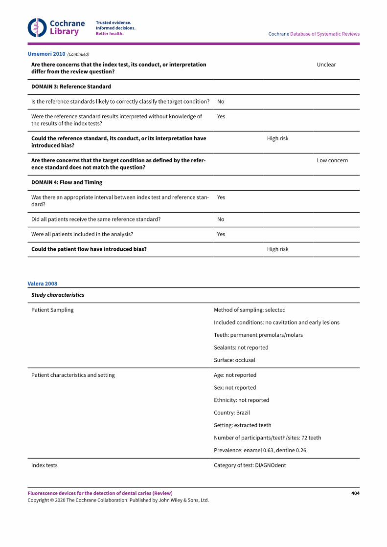
CochraneLibrary
Trusted evidence.Informed decisions.Better health.
Cochrane Database of Systematic Reviews
Are there concerns that the index test, its conduct, or interpretationdiffer from the review question?
Unclear
DOMAIN 3: Reference Standard
Is the reference standards likely to correctly classify the target condition? No
Were the reference standard results interpreted without knowledge ofthe results of the index tests?
Yes
Could the reference standard, its conduct, or its interpretation haveintroduced bias?
High risk
Are there concerns that the target condition as defined by the refer-ence standard does not match the question?
Low concern
DOMAIN 4: Flow and Timing
Was there an appropriate interval between index test and reference stan-dard?
Yes
Did all patients receive the same reference standard? No
Were all patients included in the analysis? Yes
Could the patient flow have introduced bias? High risk
Umemori 2010 (Continued)
Study characteristics
Patient Sampling Method of sampling: selected
Included conditions: no cavitation and early lesions
Teeth: permanent premolars/molars
Sealants: not reported
Surface: occlusal
Patient characteristics and setting Age: not reported
Sex: not reported
Ethnicity: not reported
Country: Brazil
Setting: extracted teeth
Number of participants/teeth/sites: 72 teeth
Prevalence: enamel 0.63, dentine 0.26
Index tests Category of test: DIAGNOdent
Valera 2008
Fluorescence devices for the detection of dental caries (Review)
Copyright © 2020 The Cochrane Collaboration. Published by John Wiley & Sons, Ltd.
404
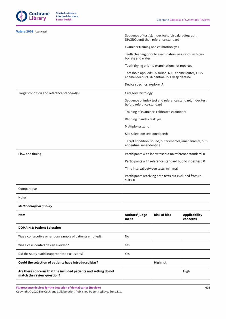
CochraneLibrary
Trusted evidence.Informed decisions.Better health.
Cochrane Database of Systematic Reviews
Sequence of test(s): index tests (visual, radiograph,DIAGNOdent) then reference standard
Examiner training and calibration: yes
Teeth cleaning prior to examination: yes - sodium bicar-bonate and water
Tooth drying prior to examination: not reported
Threshold applied: 0-5 sound, 6-10 enamel outer, 11-22enamel deep, 21-26 dentine, 27+ deep dentine
Device specifics: explorer A
Target condition and reference standard(s) Category: histology
Sequence of index test and reference standard: index testbefore reference standard
Training of examiner: calibrated examiners
Blinding to index test: yes
Multiple tests: no
Site selection: sectioned teeth
Target condition: sound, outer enamel, inner enamel, out-er dentine, inner dentine
Flow and timing Participants with index test but no reference standard: 0
Participants with reference standard but no index test: 0
Time interval between tests: minimal
Participants receiving both tests but excluded from re-sults: 0
Comparative
Notes
Methodological quality
Item Authors' judge-ment
Risk of bias Applicabilityconcerns
DOMAIN 1: Patient Selection
Was a consecutive or random sample of patients enrolled? No
Was a case-control design avoided? Yes
Did the study avoid inappropriate exclusions? Yes
Could the selection of patients have introduced bias? High risk
Are there concerns that the included patients and setting do notmatch the review question?
High
Valera 2008 (Continued)
Fluorescence devices for the detection of dental caries (Review)
Copyright © 2020 The Cochrane Collaboration. Published by John Wiley & Sons, Ltd.
405

CochraneLibrary
Trusted evidence.Informed decisions.Better health.
Cochrane Database of Systematic Reviews
DOMAIN 2: Index Test (All)
Were the index test results interpreted without knowledge of the resultsof the reference standard?
Yes
If a threshold was used, was it pre-specified? Yes
If multiple tests were applied were different examiners used for each (invivo)?
Unclear
Could the conduct or interpretation of the index test have intro-duced bias?
Low risk
Are there concerns that the index test, its conduct, or interpretationdiffer from the review question?
Low concern
DOMAIN 2: Index Test (Green fluorescence)
DOMAIN 2: Index Test (Blue fluorescence)
DOMAIN 2: Index Test (Red fluorescence)
Were the index test results interpreted without knowledge of the resultsof the reference standard?
Yes
If a threshold was used, was it pre-specified? Yes
If multiple tests were applied were different examiners used for each (invivo)?
Unclear
Could the conduct or interpretation of the index test have intro-duced bias?
Low risk
Are there concerns that the index test, its conduct, or interpretationdiffer from the review question?
Low concern
DOMAIN 3: Reference Standard
Is the reference standards likely to correctly classify the target condi-tion?
Yes
Were the reference standard results interpreted without knowledge ofthe results of the index tests?
Yes
Could the reference standard, its conduct, or its interpretation haveintroduced bias?
Low risk
Are there concerns that the target condition as defined by the refer-ence standard does not match the question?
Low concern
DOMAIN 4: Flow and Timing
Was there an appropriate interval between index test and referencestandard?
Yes
Did all patients receive the same reference standard? Yes
Valera 2008 (Continued)
Fluorescence devices for the detection of dental caries (Review)
Copyright © 2020 The Cochrane Collaboration. Published by John Wiley & Sons, Ltd.
406
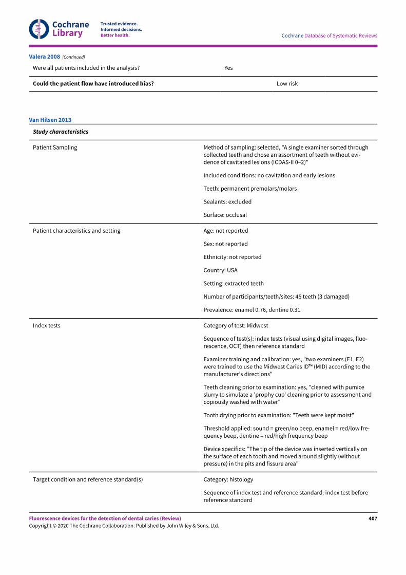
CochraneLibrary
Trusted evidence.Informed decisions.Better health.
Cochrane Database of Systematic Reviews
Were all patients included in the analysis? Yes
Could the patient flow have introduced bias? Low risk
Valera 2008 (Continued)
Study characteristics
Patient Sampling Method of sampling: selected, "A single examiner sorted throughcollected teeth and chose an assortment of teeth without evi-dence of cavitated lesions (ICDAS-II 0–2)"
Included conditions: no cavitation and early lesions
Teeth: permanent premolars/molars
Sealants: excluded
Surface: occlusal
Patient characteristics and setting Age: not reported
Sex: not reported
Ethnicity: not reported
Country: USA
Setting: extracted teeth
Number of participants/teeth/sites: 45 teeth (3 damaged)
Prevalence: enamel 0.76, dentine 0.31
Index tests Category of test: Midwest
Sequence of test(s): index tests (visual using digital images, fluo-rescence, OCT) then reference standard
Examiner training and calibration: yes, "two examiners (E1, E2)were trained to use the Midwest Caries ID™ (MID) according to themanufacturer’s directions"
Teeth cleaning prior to examination: yes, "cleaned with pumiceslurry to simulate a 'prophy cup' cleaning prior to assessment andcopiously washed with water"
Tooth drying prior to examination: "Teeth were kept moist"
Threshold applied: sound = green/no beep, enamel = red/low fre-quency beep, dentine = red/high frequency beep
Device specifics: "The tip of the device was inserted vertically onthe surface of each tooth and moved around slightly (withoutpressure) in the pits and fissure area"
Target condition and reference standard(s) Category: histology
Sequence of index test and reference standard: index test beforereference standard
Van Hilsen 2013
Fluorescence devices for the detection of dental caries (Review)
Copyright © 2020 The Cochrane Collaboration. Published by John Wiley & Sons, Ltd.
407
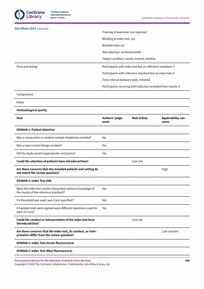
CochraneLibrary
Trusted evidence.Informed decisions.Better health.
Cochrane Database of Systematic Reviews
Training of examiner: not reported
Blinding to index test: yes
Multiple tests: no
Site selection: sectioned teeth
Target condition: sound, enamel, dentine
Flow and timing Participants with index test but no reference standard: 3
Participants with reference standard but no index test: 0
Time interval between tests: minimal
Participants receiving both tests but excluded from results: 0
Comparative
Notes
Methodological quality
Item Authors' judge-ment
Risk of bias Applicability con-cerns
DOMAIN 1: Patient Selection
Was a consecutive or random sample of patients enrolled? No
Was a case-control design avoided? Yes
Did the study avoid inappropriate exclusions? Yes
Could the selection of patients have introduced bias? Low risk
Are there concerns that the included patients and setting donot match the review question?
High
DOMAIN 2: Index Test (All)
Were the index test results interpreted without knowledge ofthe results of the reference standard?
Yes
If a threshold was used, was it pre-specified? Yes
If multiple tests were applied were different examiners used foreach (in vivo)?
Yes
Could the conduct or interpretation of the index test haveintroduced bias?
Low risk
Are there concerns that the index test, its conduct, or inter-pretation differ from the review question?
Low concern
DOMAIN 2: Index Test (Green fluorescence)
DOMAIN 2: Index Test (Blue fluorescence)
Van Hilsen 2013 (Continued)
Fluorescence devices for the detection of dental caries (Review)
Copyright © 2020 The Cochrane Collaboration. Published by John Wiley & Sons, Ltd.
408
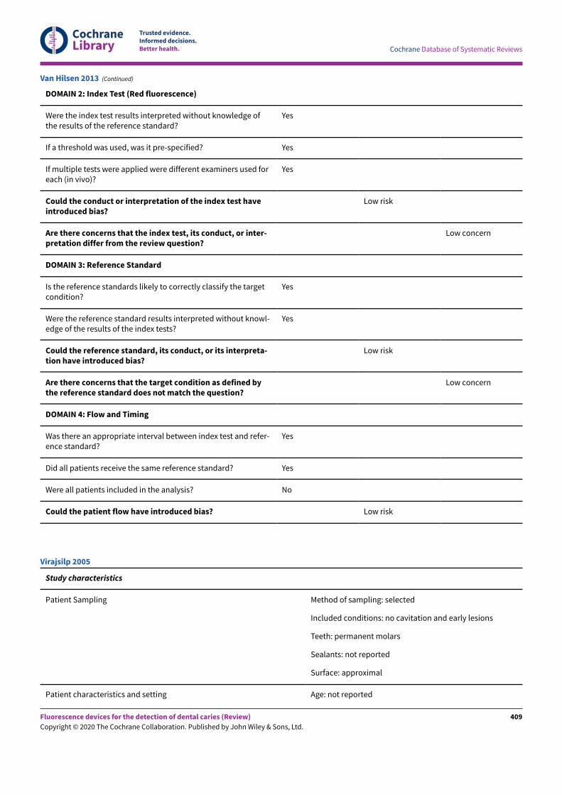
CochraneLibrary
Trusted evidence.Informed decisions.Better health.
Cochrane Database of Systematic Reviews
DOMAIN 2: Index Test (Red fluorescence)
Were the index test results interpreted without knowledge ofthe results of the reference standard?
Yes
If a threshold was used, was it pre-specified? Yes
If multiple tests were applied were different examiners used foreach (in vivo)?
Yes
Could the conduct or interpretation of the index test haveintroduced bias?
Low risk
Are there concerns that the index test, its conduct, or inter-pretation differ from the review question?
Low concern
DOMAIN 3: Reference Standard
Is the reference standards likely to correctly classify the targetcondition?
Yes
Were the reference standard results interpreted without knowl-edge of the results of the index tests?
Yes
Could the reference standard, its conduct, or its interpreta-tion have introduced bias?
Low risk
Are there concerns that the target condition as defined bythe reference standard does not match the question?
Low concern
DOMAIN 4: Flow and Timing
Was there an appropriate interval between index test and refer-ence standard?
Yes
Did all patients receive the same reference standard? Yes
Were all patients included in the analysis? No
Could the patient flow have introduced bias? Low risk
Van Hilsen 2013 (Continued)
Study characteristics
Patient Sampling Method of sampling: selected
Included conditions: no cavitation and early lesions
Teeth: permanent molars
Sealants: not reported
Surface: approximal
Patient characteristics and setting Age: not reported
Virajsilp 2005
Fluorescence devices for the detection of dental caries (Review)
Copyright © 2020 The Cochrane Collaboration. Published by John Wiley & Sons, Ltd.
409
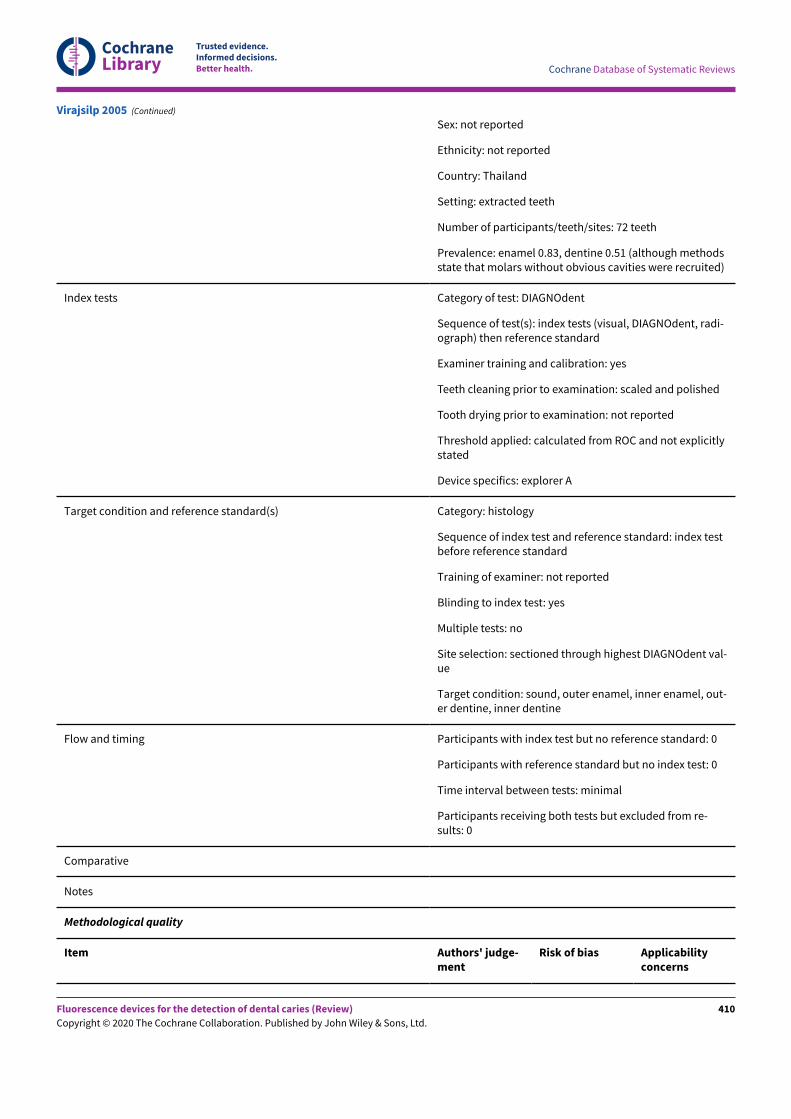
CochraneLibrary
Trusted evidence.Informed decisions.Better health.
Cochrane Database of Systematic Reviews
Sex: not reported
Ethnicity: not reported
Country: Thailand
Setting: extracted teeth
Number of participants/teeth/sites: 72 teeth
Prevalence: enamel 0.83, dentine 0.51 (although methodsstate that molars without obvious cavities were recruited)
Index tests Category of test: DIAGNOdent
Sequence of test(s): index tests (visual, DIAGNOdent, radi-ograph) then reference standard
Examiner training and calibration: yes
Teeth cleaning prior to examination: scaled and polished
Tooth drying prior to examination: not reported
Threshold applied: calculated from ROC and not explicitlystated
Device specifics: explorer A
Target condition and reference standard(s) Category: histology
Sequence of index test and reference standard: index testbefore reference standard
Training of examiner: not reported
Blinding to index test: yes
Multiple tests: no
Site selection: sectioned through highest DIAGNOdent val-ue
Target condition: sound, outer enamel, inner enamel, out-er dentine, inner dentine
Flow and timing Participants with index test but no reference standard: 0
Participants with reference standard but no index test: 0
Time interval between tests: minimal
Participants receiving both tests but excluded from re-sults: 0
Comparative
Notes
Methodological quality
Item Authors' judge-ment
Risk of bias Applicabilityconcerns
Virajsilp 2005 (Continued)
Fluorescence devices for the detection of dental caries (Review)
Copyright © 2020 The Cochrane Collaboration. Published by John Wiley & Sons, Ltd.
410
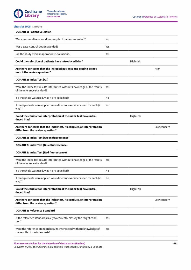
CochraneLibrary
Trusted evidence.Informed decisions.Better health.
Cochrane Database of Systematic Reviews
DOMAIN 1: Patient Selection
Was a consecutive or random sample of patients enrolled? No
Was a case-control design avoided? Yes
Did the study avoid inappropriate exclusions? Yes
Could the selection of patients have introduced bias? High risk
Are there concerns that the included patients and setting do notmatch the review question?
High
DOMAIN 2: Index Test (All)
Were the index test results interpreted without knowledge of the resultsof the reference standard?
Yes
If a threshold was used, was it pre-specified? No
If multiple tests were applied were different examiners used for each (invivo)?
No
Could the conduct or interpretation of the index test have intro-duced bias?
High risk
Are there concerns that the index test, its conduct, or interpretationdiffer from the review question?
Low concern
DOMAIN 2: Index Test (Green fluorescence)
DOMAIN 2: Index Test (Blue fluorescence)
DOMAIN 2: Index Test (Red fluorescence)
Were the index test results interpreted without knowledge of the resultsof the reference standard?
Yes
If a threshold was used, was it pre-specified? No
If multiple tests were applied were different examiners used for each (invivo)?
No
Could the conduct or interpretation of the index test have intro-duced bias?
High risk
Are there concerns that the index test, its conduct, or interpretationdiffer from the review question?
Low concern
DOMAIN 3: Reference Standard
Is the reference standards likely to correctly classify the target condi-tion?
Yes
Were the reference standard results interpreted without knowledge ofthe results of the index tests?
Yes
Virajsilp 2005 (Continued)
Fluorescence devices for the detection of dental caries (Review)
Copyright © 2020 The Cochrane Collaboration. Published by John Wiley & Sons, Ltd.
411
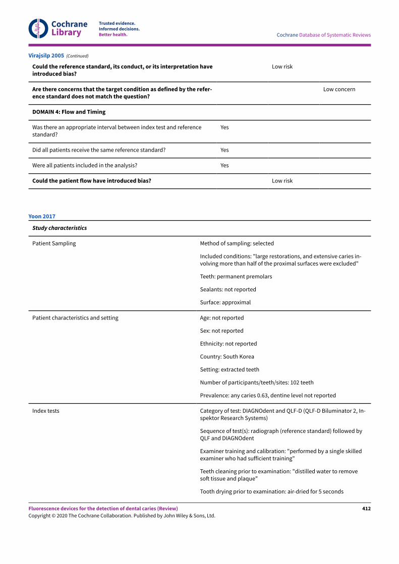
CochraneLibrary
Trusted evidence.Informed decisions.Better health.
Cochrane Database of Systematic Reviews
Could the reference standard, its conduct, or its interpretation haveintroduced bias?
Low risk
Are there concerns that the target condition as defined by the refer-ence standard does not match the question?
Low concern
DOMAIN 4: Flow and Timing
Was there an appropriate interval between index test and referencestandard?
Yes
Did all patients receive the same reference standard? Yes
Were all patients included in the analysis? Yes
Could the patient flow have introduced bias? Low risk
Virajsilp 2005 (Continued)
Study characteristics
Patient Sampling Method of sampling: selected
Included conditions: "large restorations, and extensive caries in-volving more than half of the proximal surfaces were excluded"
Teeth: permanent premolars
Sealants: not reported
Surface: approximal
Patient characteristics and setting Age: not reported
Sex: not reported
Ethnicity: not reported
Country: South Korea
Setting: extracted teeth
Number of participants/teeth/sites: 102 teeth
Prevalence: any caries 0.63, dentine level not reported
Index tests Category of test: DIAGNOdent and QLF-D (QLF-D Biluminator 2, In-spektor Research Systems)
Sequence of test(s): radiograph (reference standard) followed byQLF and DIAGNOdent
Examiner training and calibration: "performed by a single skilledexaminer who had sufficient training"
Teeth cleaning prior to examination: "distilled water to removesoN tissue and plaque"
Tooth drying prior to examination: air-dried for 5 seconds
Yoon 2017
Fluorescence devices for the detection of dental caries (Review)
Copyright © 2020 The Cochrane Collaboration. Published by John Wiley & Sons, Ltd.
412
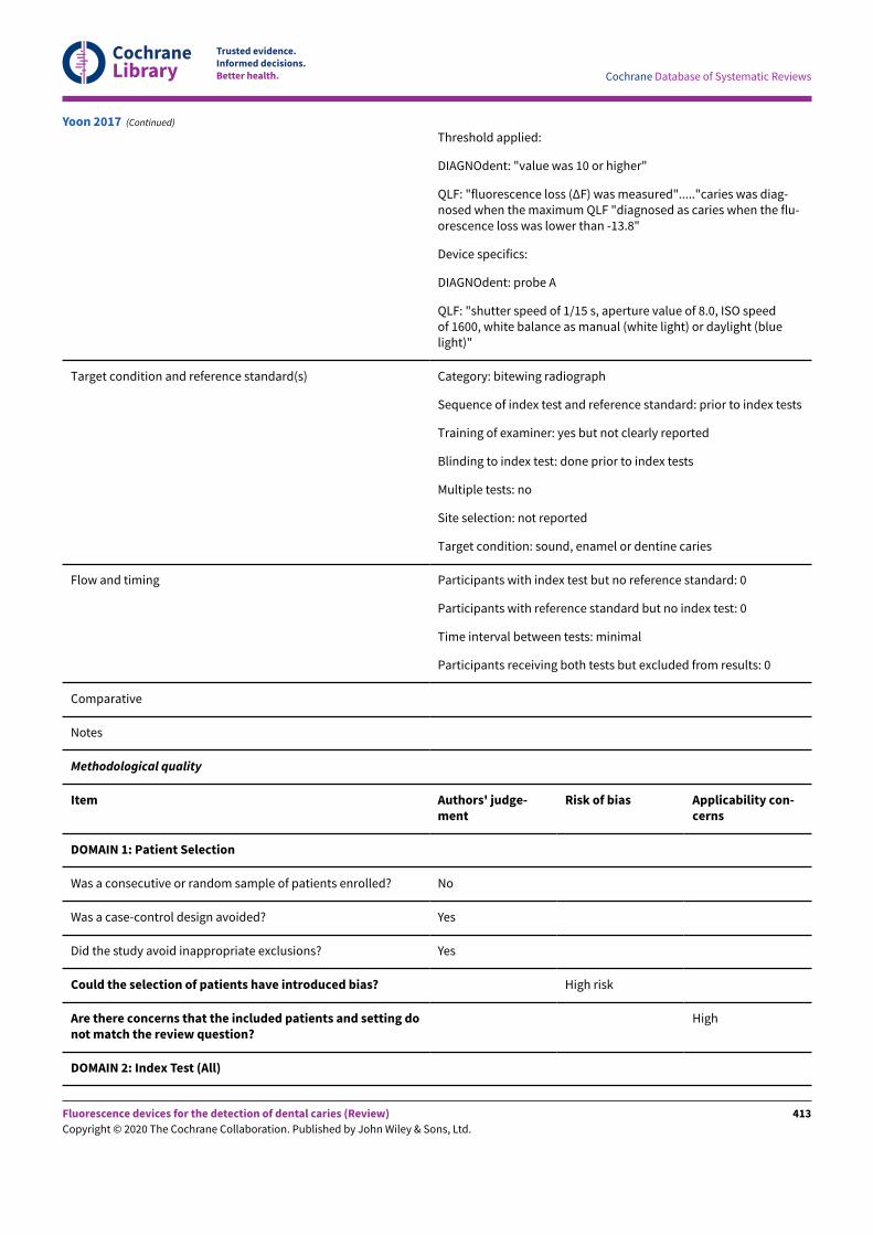
CochraneLibrary
Trusted evidence.Informed decisions.Better health.
Cochrane Database of Systematic Reviews
Threshold applied:
DIAGNOdent: "value was 10 or higher"
QLF: "fluorescence loss (ΔF) was measured"....."caries was diag-nosed when the maximum QLF "diagnosed as caries when the flu-orescence loss was lower than -13.8"
Device specifics:
DIAGNOdent: probe A
QLF: "shutter speed of 1/15 s, aperture value of 8.0, ISO speedof 1600, white balance as manual (white light) or daylight (bluelight)"
Target condition and reference standard(s) Category: bitewing radiograph
Sequence of index test and reference standard: prior to index tests
Training of examiner: yes but not clearly reported
Blinding to index test: done prior to index tests
Multiple tests: no
Site selection: not reported
Target condition: sound, enamel or dentine caries
Flow and timing Participants with index test but no reference standard: 0
Participants with reference standard but no index test: 0
Time interval between tests: minimal
Participants receiving both tests but excluded from results: 0
Comparative
Notes
Methodological quality
Item Authors' judge-ment
Risk of bias Applicability con-cerns
DOMAIN 1: Patient Selection
Was a consecutive or random sample of patients enrolled? No
Was a case-control design avoided? Yes
Did the study avoid inappropriate exclusions? Yes
Could the selection of patients have introduced bias? High risk
Are there concerns that the included patients and setting donot match the review question?
High
DOMAIN 2: Index Test (All)
Yoon 2017 (Continued)
Fluorescence devices for the detection of dental caries (Review)
Copyright © 2020 The Cochrane Collaboration. Published by John Wiley & Sons, Ltd.
413
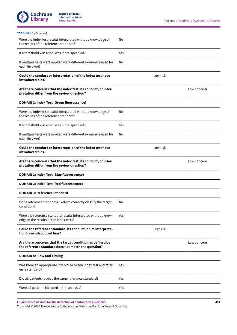
CochraneLibrary
Trusted evidence.Informed decisions.Better health.
Cochrane Database of Systematic Reviews
Were the index test results interpreted without knowledge ofthe results of the reference standard?
No
If a threshold was used, was it pre-specified? Yes
If multiple tests were applied were different examiners used foreach (in vivo)?
No
Could the conduct or interpretation of the index test haveintroduced bias?
Low risk
Are there concerns that the index test, its conduct, or inter-pretation differ from the review question?
Low concern
DOMAIN 2: Index Test (Green fluorescence)
Were the index test results interpreted without knowledge ofthe results of the reference standard?
No
If a threshold was used, was it pre-specified? Yes
If multiple tests were applied were different examiners used foreach (in vivo)?
No
Could the conduct or interpretation of the index test haveintroduced bias?
Low risk
Are there concerns that the index test, its conduct, or inter-pretation differ from the review question?
Low concern
DOMAIN 2: Index Test (Blue fluorescence)
DOMAIN 2: Index Test (Red fluorescence)
DOMAIN 3: Reference Standard
Is the reference standards likely to correctly classify the targetcondition?
No
Were the reference standard results interpreted without knowl-edge of the results of the index tests?
Yes
Could the reference standard, its conduct, or its interpreta-tion have introduced bias?
High risk
Are there concerns that the target condition as defined bythe reference standard does not match the question?
Low concern
DOMAIN 4: Flow and Timing
Was there an appropriate interval between index test and refer-ence standard?
Yes
Did all patients receive the same reference standard? Yes
Were all patients included in the analysis? Yes
Yoon 2017 (Continued)
Fluorescence devices for the detection of dental caries (Review)
Copyright © 2020 The Cochrane Collaboration. Published by John Wiley & Sons, Ltd.
414
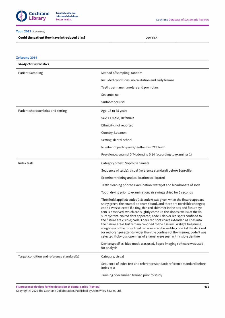
CochraneLibrary
Trusted evidence.Informed decisions.Better health.
Cochrane Database of Systematic Reviews
Could the patient flow have introduced bias? Low risk
Yoon 2017 (Continued)
Study characteristics
Patient Sampling Method of sampling: random
Included conditions: no cavitation and early lesions
Teeth: permanent molars and premolars
Sealants: no
Surface: occlusal
Patient characteristics and setting Age: 15 to 65 years
Sex: 11 male, 10 female
Ethnicity: not reported
Country: Lebanon
Setting: dental school
Number of participants/teeth/sites: 219 teeth
Prevalence: enamel 0.74, dentine 0.14 (according to examiner 1)
Index tests Category of test: Soprolife camera
Sequence of test(s): visual (reference standard) before Soprolife
Examiner training and calibration: calibrated
Teeth cleaning prior to examination: waterjet and bicarbonate of soda
Tooth drying prior to examination: air syringe dried for 5 seconds
Threshold applied: codes 0-5: code 0 was given when the fissure appearsshiny green, the enamel appears sound, and there are no visible changes;code 1 was selected if a tiny, thin red shimmer in the pits and fissure sys-tem is observed, which can slightly come up the slopes (walls) of the fis-sure system. No red dots appeared; code 2 darker red spots confined tothe fissure are visible; code 3 dark red spots have extended as lines intothe fissure areas but remain confined to the fissures. A slight beginningroughness of the more lined red areas can be visible; code 4 if the dark red(or red-orange) extends wider than the confines of the fissures; code 5 wasselected if obvious openings of enamel were seen with visible dentine
Device specifics: blue mode was used, Sopro imaging software was usedfor analysis
Target condition and reference standard(s) Category: visual
Sequence of index test and reference standard: reference standard beforeindex test
Training of examiner: trained prior to study
Zeitouny 2014
Fluorescence devices for the detection of dental caries (Review)
Copyright © 2020 The Cochrane Collaboration. Published by John Wiley & Sons, Ltd.
415
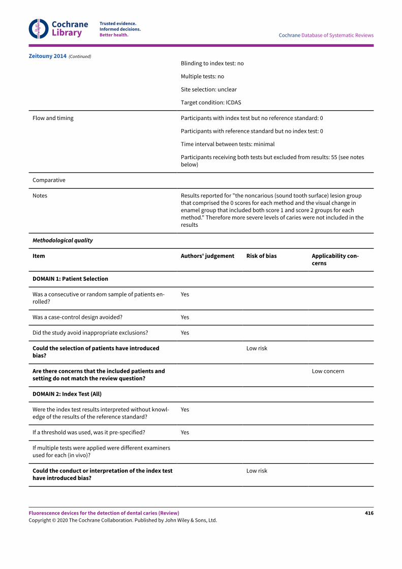
CochraneLibrary
Trusted evidence.Informed decisions.Better health.
Cochrane Database of Systematic Reviews
Blinding to index test: no
Multiple tests: no
Site selection: unclear
Target condition: ICDAS
Flow and timing Participants with index test but no reference standard: 0
Participants with reference standard but no index test: 0
Time interval between tests: minimal
Participants receiving both tests but excluded from results: 55 (see notesbelow)
Comparative
Notes Results reported for "the noncarious (sound tooth surface) lesion groupthat comprised the 0 scores for each method and the visual change inenamel group that included both score 1 and score 2 groups for eachmethod." Therefore more severe levels of caries were not included in theresults
Methodological quality
Item Authors' judgement Risk of bias Applicability con-cerns
DOMAIN 1: Patient Selection
Was a consecutive or random sample of patients en-rolled?
Yes
Was a case-control design avoided? Yes
Did the study avoid inappropriate exclusions? Yes
Could the selection of patients have introducedbias?
Low risk
Are there concerns that the included patients andsetting do not match the review question?
Low concern
DOMAIN 2: Index Test (All)
Were the index test results interpreted without knowl-edge of the results of the reference standard?
Yes
If a threshold was used, was it pre-specified? Yes
If multiple tests were applied were different examinersused for each (in vivo)?
Could the conduct or interpretation of the index testhave introduced bias?
Low risk
Zeitouny 2014 (Continued)
Fluorescence devices for the detection of dental caries (Review)
Copyright © 2020 The Cochrane Collaboration. Published by John Wiley & Sons, Ltd.
416
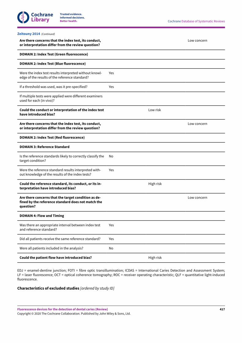
CochraneLibrary
Trusted evidence.Informed decisions.Better health.
Cochrane Database of Systematic Reviews
Are there concerns that the index test, its conduct,or interpretation differ from the review question?
Low concern
DOMAIN 2: Index Test (Green fluorescence)
DOMAIN 2: Index Test (Blue fluorescence)
Were the index test results interpreted without knowl-edge of the results of the reference standard?
Yes
If a threshold was used, was it pre-specified? Yes
If multiple tests were applied were different examinersused for each (in vivo)?
Could the conduct or interpretation of the index testhave introduced bias?
Low risk
Are there concerns that the index test, its conduct,or interpretation differ from the review question?
Low concern
DOMAIN 2: Index Test (Red fluorescence)
DOMAIN 3: Reference Standard
Is the reference standards likely to correctly classify thetarget condition?
No
Were the reference standard results interpreted with-out knowledge of the results of the index tests?
Yes
Could the reference standard, its conduct, or its in-terpretation have introduced bias?
High risk
Are there concerns that the target condition as de-fined by the reference standard does not match thequestion?
Low concern
DOMAIN 4: Flow and Timing
Was there an appropriate interval between index testand reference standard?
Yes
Did all patients receive the same reference standard? Yes
Were all patients included in the analysis? No
Could the patient flow have introduced bias? High risk
Zeitouny 2014 (Continued)
EDJ = enamel-dentine junction; FOTI = fibre optic transillumination; ICDAS = International Caries Detection and Assessment System;LF = laser fluorescence; OCT = optical coherence tomography; ROC = receiver operating characteristic; QLF = quantitative light-inducedfluorescence.
Characteristics of excluded studies [ordered by study ID]
Fluorescence devices for the detection of dental caries (Review)
Copyright © 2020 The Cochrane Collaboration. Published by John Wiley & Sons, Ltd.
417
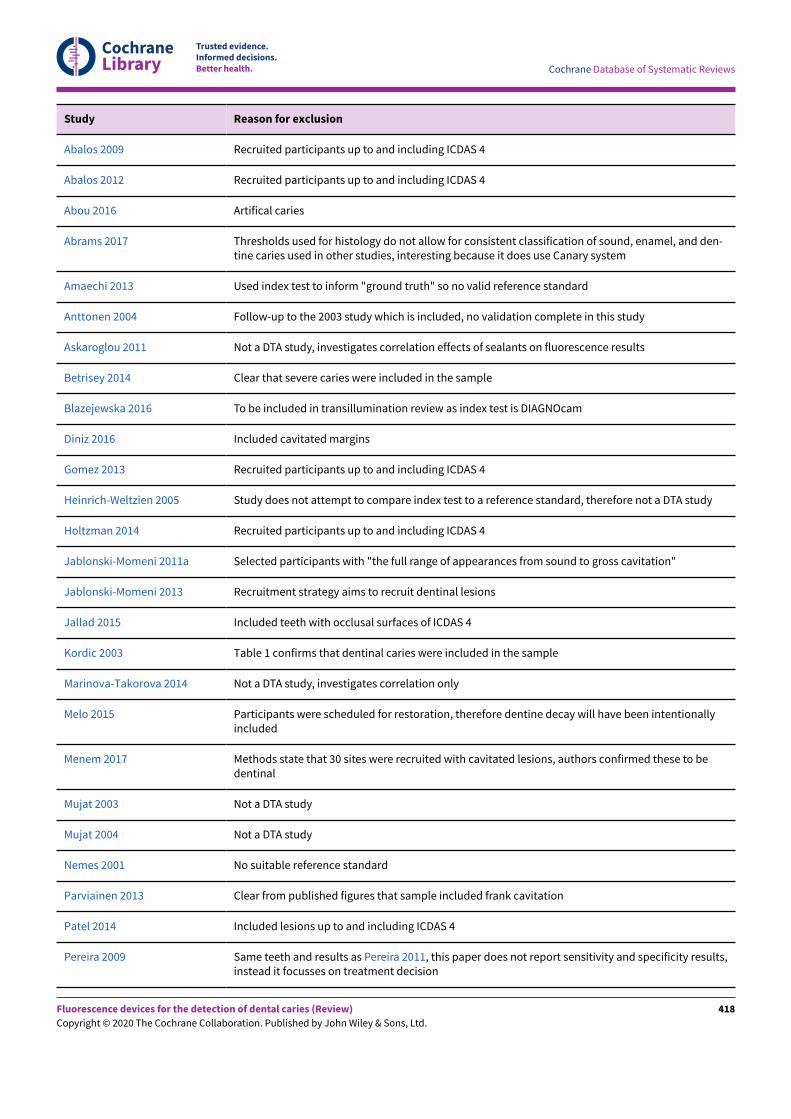
CochraneLibrary
Trusted evidence.Informed decisions.Better health.
Cochrane Database of Systematic Reviews
Study Reason for exclusion
Abalos 2009 Recruited participants up to and including ICDAS 4
Abalos 2012 Recruited participants up to and including ICDAS 4
Abou 2016 Artifical caries
Abrams 2017 Thresholds used for histology do not allow for consistent classification of sound, enamel, and den-tine caries used in other studies, interesting because it does use Canary system
Amaechi 2013 Used index test to inform "ground truth" so no valid reference standard
Anttonen 2004 Follow-up to the 2003 study which is included, no validation complete in this study
Askaroglou 2011 Not a DTA study, investigates correlation effects of sealants on fluorescence results
Betrisey 2014 Clear that severe caries were included in the sample
Blazejewska 2016 To be included in transillumination review as index test is DIAGNOcam
Diniz 2016 Included cavitated margins
Gomez 2013 Recruited participants up to and including ICDAS 4
Heinrich-Weltzien 2005 Study does not attempt to compare index test to a reference standard, therefore not a DTA study
Holtzman 2014 Recruited participants up to and including ICDAS 4
Jablonski-Momeni 2011a Selected participants with "the full range of appearances from sound to gross cavitation"
Jablonski-Momeni 2013 Recruitment strategy aims to recruit dentinal lesions
Jallad 2015 Included teeth with occlusal surfaces of ICDAS 4
Kordic 2003 Table 1 confirms that dentinal caries were included in the sample
Marinova-Takorova 2014 Not a DTA study, investigates correlation only
Melo 2015 Participants were scheduled for restoration, therefore dentine decay will have been intentionallyincluded
Menem 2017 Methods state that 30 sites were recruited with cavitated lesions, authors confirmed these to bedentinal
Mujat 2003 Not a DTA study
Mujat 2004 Not a DTA study
Nemes 2001 No suitable reference standard
Parviainen 2013 Clear from published figures that sample included frank cavitation
Patel 2014 Included lesions up to and including ICDAS 4
Pereira 2009 Same teeth and results as Pereira 2011, this paper does not report sensitivity and specificity results,instead it focusses on treatment decision
Fluorescence devices for the detection of dental caries (Review)
Copyright © 2020 The Cochrane Collaboration. Published by John Wiley & Sons, Ltd.
418
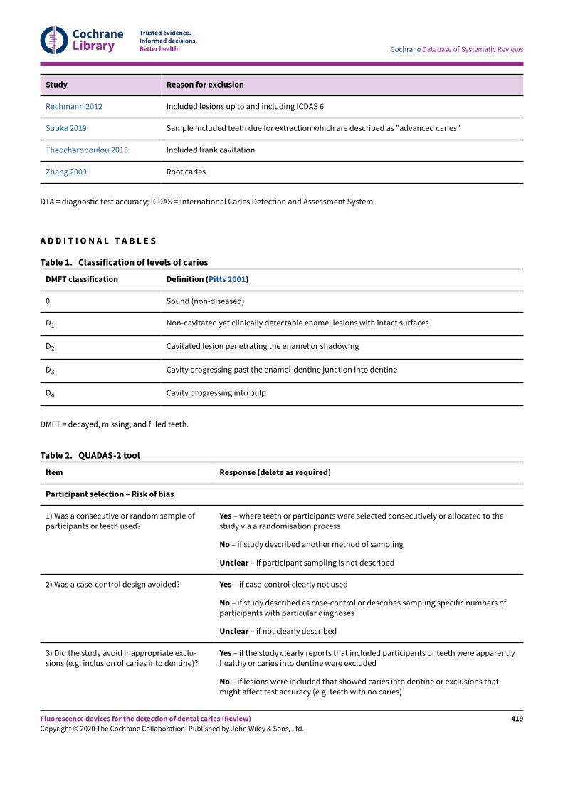
CochraneLibrary
Trusted evidence.Informed decisions.Better health.
Cochrane Database of Systematic Reviews
Study Reason for exclusion
Rechmann 2012 Included lesions up to and including ICDAS 6
Subka 2019 Sample included teeth due for extraction which are described as "advanced caries"
Theocharopoulou 2015 Included frank cavitation
Zhang 2009 Root caries
DTA = diagnostic test accuracy; ICDAS = International Caries Detection and Assessment System.
A D D I T I O N A L T A B L E S
DMFT classification Definition (Pitts 2001)
0 Sound (non-diseased)
D1 Non-cavitated yet clinically detectable enamel lesions with intact surfaces
D2 Cavitated lesion penetrating the enamel or shadowing
D3 Cavity progressing past the enamel-dentine junction into dentine
D4 Cavity progressing into pulp
Table 1. Classification of levels of caries
DMFT = decayed, missing, and filled teeth.
Item Response (delete as required)
Participant selection – Risk of bias
1) Was a consecutive or random sample ofparticipants or teeth used?
Yes – where teeth or participants were selected consecutively or allocated to thestudy via a randomisation process
No – if study described another method of sampling
Unclear – if participant sampling is not described
2) Was a case-control design avoided? Yes – if case-control clearly not used
No – if study described as case-control or describes sampling specific numbers ofparticipants with particular diagnoses
Unclear – if not clearly described
3) Did the study avoid inappropriate exclu-sions (e.g. inclusion of caries into dentine)?
Yes – if the study clearly reports that included participants or teeth were apparentlyhealthy or caries into dentine were excluded
No – if lesions were included that showed caries into dentine or exclusions thatmight affect test accuracy (e.g. teeth with no caries)
Table 2. QUADAS-2 tool
Fluorescence devices for the detection of dental caries (Review)
Copyright © 2020 The Cochrane Collaboration. Published by John Wiley & Sons, Ltd.
419
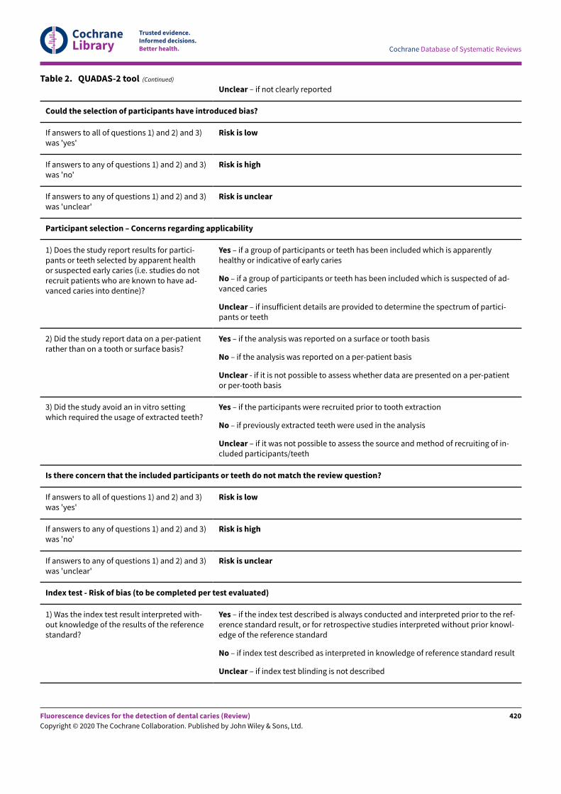
CochraneLibrary
Trusted evidence.Informed decisions.Better health.
Cochrane Database of Systematic Reviews
Unclear – if not clearly reported
Could the selection of participants have introduced bias?
If answers to all of questions 1) and 2) and 3)was 'yes'
Risk is low
If answers to any of questions 1) and 2) and 3)was 'no'
Risk is high
If answers to any of questions 1) and 2) and 3)was 'unclear'
Risk is unclear
Participant selection – Concerns regarding applicability
1) Does the study report results for partici-pants or teeth selected by apparent healthor suspected early caries (i.e. studies do notrecruit patients who are known to have ad-vanced caries into dentine)?
Yes – if a group of participants or teeth has been included which is apparentlyhealthy or indicative of early caries
No – if a group of participants or teeth has been included which is suspected of ad-vanced caries
Unclear – if insufficient details are provided to determine the spectrum of partici-pants or teeth
2) Did the study report data on a per-patientrather than on a tooth or surface basis?
Yes – if the analysis was reported on a surface or tooth basis
No – if the analysis was reported on a per-patient basis
Unclear - if it is not possible to assess whether data are presented on a per-patientor per-tooth basis
3) Did the study avoid an in vitro settingwhich required the usage of extracted teeth?
Yes – if the participants were recruited prior to tooth extraction
No – if previously extracted teeth were used in the analysis
Unclear – if it was not possible to assess the source and method of recruiting of in-cluded participants/teeth
Is there concern that the included participants or teeth do not match the review question?
If answers to all of questions 1) and 2) and 3)was 'yes'
Risk is low
If answers to any of questions 1) and 2) and 3)was 'no'
Risk is high
If answers to any of questions 1) and 2) and 3)was 'unclear'
Risk is unclear
Index test - Risk of bias (to be completed per test evaluated)
1) Was the index test result interpreted with-out knowledge of the results of the referencestandard?
Yes – if the index test described is always conducted and interpreted prior to the ref-erence standard result, or for retrospective studies interpreted without prior knowl-edge of the reference standard
No – if index test described as interpreted in knowledge of reference standard result
Unclear – if index test blinding is not described
Table 2. QUADAS-2 tool (Continued)
Fluorescence devices for the detection of dental caries (Review)
Copyright © 2020 The Cochrane Collaboration. Published by John Wiley & Sons, Ltd.
420
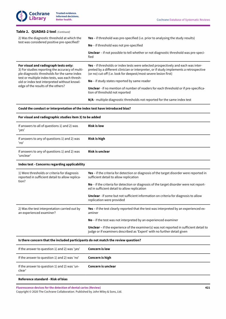
CochraneLibrary
Trusted evidence.Informed decisions.Better health.
Cochrane Database of Systematic Reviews
2) Was the diagnostic threshold at which thetest was considered positive pre-specified?
Yes – if threshold was pre-specified (i.e. prior to analysing the study results)
No – if threshold was not pre-specified
Unclear – if not possible to tell whether or not diagnostic threshold was pre-speci-fied
For visual and radiograph tests only:3) For studies reporting the accuracy of multi-ple diagnostic thresholds for the same indextest or multiple index tests, was each thresh-old or index test interpreted without knowl-edge of the results of the others?
Yes – if thresholds or index tests were selected prospectively and each was inter-preted by a different clinician or interpreter, or if study implements a retrospective(or no) cut-oE (i.e. look for deepest/most severe lesion first)
No – if study states reported by same reader
Unclear - if no mention of number of readers for each threshold or if pre-specifica-tion of threshold not reported
N/A - multiple diagnostic thresholds not reported for the same index test
Could the conduct or interpretation of the index test have introduced bias?
For visual and radiographic studies item 3) to be added
If answers to all of questions 1) and 2) was'yes'
Risk is low
If answers to any of questions 1) and 2) was'no'
Risk is high
If answers to any of questions 1) and 2) was'unclear'
Risk is unclear
Index test - Concerns regarding applicability
1) Were thresholds or criteria for diagnosisreported in sufficient detail to allow replica-tion?
Yes – if the criteria for detection or diagnosis of the target disorder were reported insufficient detail to allow replication
No – if the criteria for detection or diagnosis of the target disorder were not report-ed in sufficient detail to allow replication
Unclear - if some but not sufficient information on criteria for diagnosis to allowreplication were provided
2) Was the test interpretation carried out byan experienced examiner?
Yes – if the test clearly reported that the test was interpreted by an experienced ex-aminer
No – if the test was not interpreted by an experienced examiner
Unclear – if the experience of the examiner(s) was not reported in sufficient detail tojudge or if examiners described as 'Expert' with no further detail given
Is there concern that the included participants do not match the review question?
If the answer to question 1) and 2) was 'yes' Concern is low
If the answer to question 1) and 2) was 'no' Concern is high
If the answer to question 1) and 2) was 'un-clear'
Concern is unclear
Reference standard - Risk of bias
Table 2. QUADAS-2 tool (Continued)
Fluorescence devices for the detection of dental caries (Review)
Copyright © 2020 The Cochrane Collaboration. Published by John Wiley & Sons, Ltd.
421
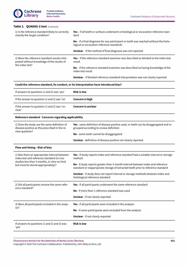
CochraneLibrary
Trusted evidence.Informed decisions.Better health.
Cochrane Database of Systematic Reviews
1) Is the reference standard likely to correctlyclassify the target condition?
Yes – if all teeth or surfaces underwent a histological or excavation reference stan-dard
No – if a final diagnosis for any participant or tooth was reached without the histo-logical or excavation reference standards
Unclear – if the method of final diagnosis was not reported
2) Were the reference standard results inter-preted without knowledge of the results ofthe index test?
Yes – if the reference standard examiner was described as blinded to the index testresult
No – if the reference standard examiner was described as having knowledge of theindex test result
Unclear – if blinded reference standard interpretation was not clearly reported
Could the reference standard, its conduct, or its interpretation have introduced bias?
If answers to questions 1) and 2) was 'yes' Risk is low
If the answer to question 1) and 2) was 'no' Concern is high
If the answer to question 1) and 2) was 'un-clear'
Concern is unclear
Reference standard - Concerns regarding applicability
1) Does the study use the same definition ofdisease positive as the prescribed in the re-view question?
Yes - same definition of disease positive used, or teeth can be disaggregated and re-grouped according to review definition
No - some teeth cannot be disaggregated
Unclear - definition of disease positive not clearly reported
Flow and timing - Risk of bias
1) Was there an appropriate interval betweenindex test and reference standard (in vivostudies less than 3 months, in vitro no limitbut must be stored appropriately)?
Yes - if study reports index and reference standard had a suitable interval or storagemethod
No - if study reports greater than 3-month interval between index and referencestandard or inappropriate storage of extracted teeth prior to reference standard
Unclear - if study does not report interval or storage methods between index andhistological reference standard
2) Did all participants receive the same refer-ence standard?
Yes - if all participants underwent the same reference standard
No - if more than 1 reference standard was used
Unclear - if not clearly reported
3) Were all participants included in the analy-sis?
Yes - if all participants were included in the analysis
No - if some participants were excluded from the analysis
Unclear - if not clearly reported
If answers to questions 1) and 2) and 3) was'yes'
Risk is low
Table 2. QUADAS-2 tool (Continued)
Fluorescence devices for the detection of dental caries (Review)
Copyright © 2020 The Cochrane Collaboration. Published by John Wiley & Sons, Ltd.
422
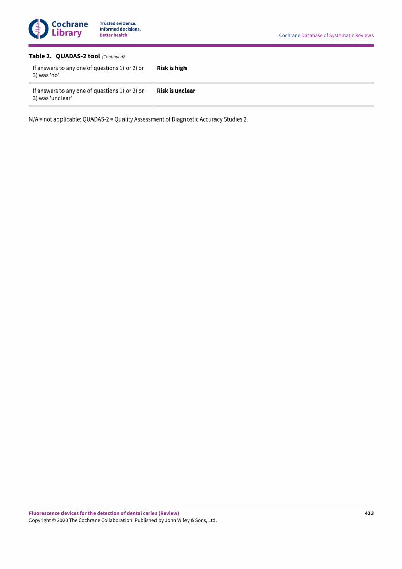
CochraneLibrary
Trusted evidence.Informed decisions.Better health.
Cochrane Database of Systematic Reviews
If answers to any one of questions 1) or 2) or3) was 'no'
Risk is high
If answers to any one of questions 1) or 2) or3) was 'unclear'
Risk is unclear
Table 2. QUADAS-2 tool (Continued)
N/A = not applicable; QUADAS-2 = Quality Assessment of Diagnostic Accuracy Studies 2.
Fluorescence devices for the detection of dental caries (Review)
Copyright © 2020 The Cochrane Collaboration. Published by John Wiley & Sons, Ltd.
423
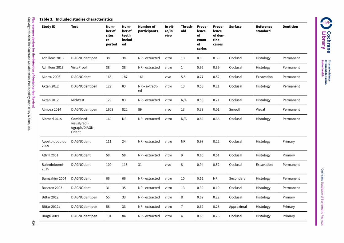
Fluoresce
nce device
s for th
e detectio
n of dental ca
ries (R
eview)
Copyrig
ht © 2020 T
he Cochrane Collaboration. Publish
ed by Jo
hn Wiley & Sons, Ltd
.
424
Study ID Test Num-ber ofsitesre-ported
Num-ber ofteethinclud-ed
Number ofparticipants
In vit-ro/invivo
Thresh-old
Preva-lenceofenam-elcaries
Preva-lenceof den-tinecaries
Surface Referencestandard
Dentition
Achilleos 2013 DIAGNOdent pen 38 38 NR - extracted vitro 13 0.95 0.39 Occlusal Histology Permanent
Achilleos 2013 VistaProof 38 38 NR - extracted vitro 1 0.95 0.39 Occlusal Histology Permanent
Akarsu 2006 DIAGNOdent 165 187 161 vivo 5.5 0.77 0.52 Occlusal Excavation Permanent
Aktan 2012 DIAGNOdent pen 129 83 NR – extract-ed
vitro 13 0.58 0.21 Occlusal Histology Permanent
Aktan 2012 MidWest 129 83 NR - extracted vitro N/A 0.58 0.21 Occlusal Histology Permanent
Almosa 2014 DIAGNOdent pen 1653 822 89 vivo 13 0.33 0.01 Smooth Visual Permanent
Alomari 2015 Combinedvisual/radi-ograph/DIAGN-Odent
160 NR NR - extracted vitro N/A 0.89 0.38 Occlusal Histology Permanent
Apostolopoulou2009
DIAGNOdent 111 24 NR - extracted vitro NR 0.98 0.22 Occlusal Histology Primary
Attrill 2001 DIAGNOdent 58 58 NR - extracted vitro 9 0.60 0.51 Occlusal Histology Primary
Bahrololoomi2015
DIAGNOdent 109 115 31 vivo 8 0.94 0.52 Occlusal Excavation Permanent
Bamzahim 2004 DIAGNOdent 66 66 NR - extracted vitro 10 0.52 NR Secondary Histology Permanent
Baseren 2003 DIAGNOdent 31 35 NR - extracted vitro 13 0.39 0.19 Occlusal Histology Permanent
Bittar 2012 DIAGNOdent pen 55 33 NR - extracted vitro 8 0.67 0.22 Occlusal Histology Primary
Bittar 2012a DIAGNOdent pen 58 33 NR - extracted vitro 7 0.62 0.28 Approximal Histology Primary
Braga 2009 DIAGNOdent pen 131 84 NR - extracted vitro 4 0.63 0.26 Occlusal Histology Primary
Table 3. Included studies characteristics Cochrane
Library
Truste
d evidence.
Inform
ed decisio
ns.
Bette
r health
.
Cochrane D
atabase o
f System
atic R
eviews
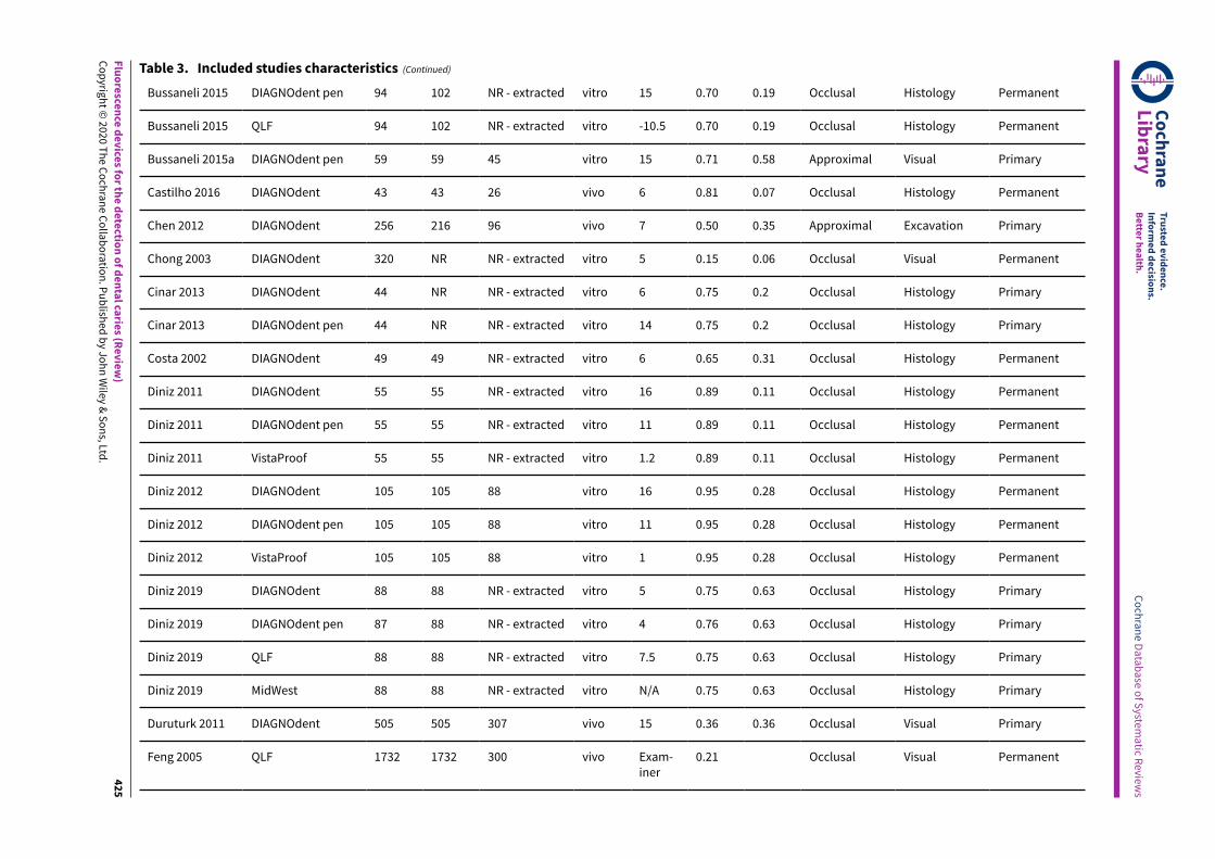
Fluoresce
nce device
s for th
e detectio
n of dental ca
ries (R
eview)
Copyrig
ht © 2020 T
he Cochrane Collaboration. Publish
ed by Jo
hn Wiley & Sons, Ltd
.
425
Bussaneli 2015 DIAGNOdent pen 94 102 NR - extracted vitro 15 0.70 0.19 Occlusal Histology Permanent
Bussaneli 2015 QLF 94 102 NR - extracted vitro -10.5 0.70 0.19 Occlusal Histology Permanent
Bussaneli 2015a DIAGNOdent pen 59 59 45 vitro 15 0.71 0.58 Approximal Visual Primary
Castilho 2016 DIAGNOdent 43 43 26 vivo 6 0.81 0.07 Occlusal Histology Permanent
Chen 2012 DIAGNOdent 256 216 96 vivo 7 0.50 0.35 Approximal Excavation Primary
Chong 2003 DIAGNOdent 320 NR NR - extracted vitro 5 0.15 0.06 Occlusal Visual Permanent
Cinar 2013 DIAGNOdent 44 NR NR - extracted vitro 6 0.75 0.2 Occlusal Histology Primary
Cinar 2013 DIAGNOdent pen 44 NR NR - extracted vitro 14 0.75 0.2 Occlusal Histology Primary
Costa 2002 DIAGNOdent 49 49 NR - extracted vitro 6 0.65 0.31 Occlusal Histology Permanent
Diniz 2011 DIAGNOdent 55 55 NR - extracted vitro 16 0.89 0.11 Occlusal Histology Permanent
Diniz 2011 DIAGNOdent pen 55 55 NR - extracted vitro 11 0.89 0.11 Occlusal Histology Permanent
Diniz 2011 VistaProof 55 55 NR - extracted vitro 1.2 0.89 0.11 Occlusal Histology Permanent
Diniz 2012 DIAGNOdent 105 105 88 vitro 16 0.95 0.28 Occlusal Histology Permanent
Diniz 2012 DIAGNOdent pen 105 105 88 vitro 11 0.95 0.28 Occlusal Histology Permanent
Diniz 2012 VistaProof 105 105 88 vitro 1 0.95 0.28 Occlusal Histology Permanent
Diniz 2019 DIAGNOdent 88 88 NR - extracted vitro 5 0.75 0.63 Occlusal Histology Primary
Diniz 2019 DIAGNOdent pen 87 88 NR - extracted vitro 4 0.76 0.63 Occlusal Histology Primary
Diniz 2019 QLF 88 88 NR - extracted vitro 7.5 0.75 0.63 Occlusal Histology Primary
Diniz 2019 MidWest 88 88 NR - extracted vitro N/A 0.75 0.63 Occlusal Histology Primary
Duruturk 2011 DIAGNOdent 505 505 307 vivo 15 0.36 0.36 Occlusal Visual Primary
Feng 2005 QLF 1732 1732 300 vivo Exam-iner
0.21 Occlusal Visual Permanent
Table 3. Included studies characteristics (Continued)
Cochrane
Library
Truste
d evidence.
Inform
ed decisio
ns.
Bette
r health
.
Cochrane D
atabase o
f System
atic R
eviews
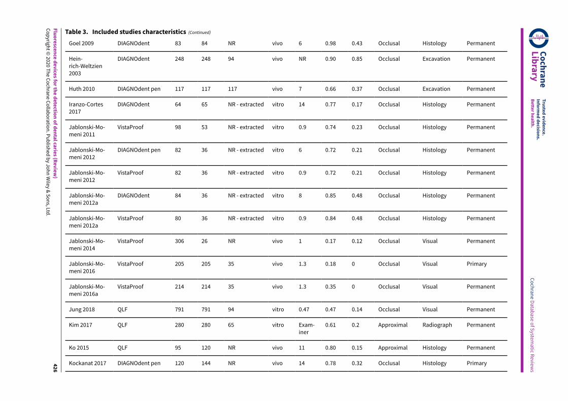
Fluoresce
nce device
s for th
e detectio
n of dental ca
ries (R
eview)
Copyrig
ht © 2020 T
he Cochrane Collaboration. Publish
ed by Jo
hn Wiley & Sons, Ltd
.
426
Goel 2009 DIAGNOdent 83 84 NR vivo 6 0.98 0.43 Occlusal Histology Permanent
Hein-rich-Weltzien2003
DIAGNOdent 248 248 94 vivo NR 0.90 0.85 Occlusal Excavation Permanent
Huth 2010 DIAGNOdent pen 117 117 117 vivo 7 0.66 0.37 Occlusal Excavation Permanent
Iranzo-Cortes2017
DIAGNOdent 64 65 NR - extracted vitro 14 0.77 0.17 Occlusal Histology Permanent
Jablonski-Mo-meni 2011
VistaProof 98 53 NR - extracted vitro 0.9 0.74 0.23 Occlusal Histology Permanent
Jablonski-Mo-meni 2012
DIAGNOdent pen 82 36 NR - extracted vitro 6 0.72 0.21 Occlusal Histology Permanent
Jablonski-Mo-meni 2012
VistaProof 82 36 NR - extracted vitro 0.9 0.72 0.21 Occlusal Histology Permanent
Jablonski-Mo-meni 2012a
DIAGNOdent 84 36 NR - extracted vitro 8 0.85 0.48 Occlusal Histology Permanent
Jablonski-Mo-meni 2012a
VistaProof 80 36 NR - extracted vitro 0.9 0.84 0.48 Occlusal Histology Permanent
Jablonski-Mo-meni 2014
VistaProof 306 26 NR vivo 1 0.17 0.12 Occlusal Visual Permanent
Jablonski-Mo-meni 2016
VistaProof 205 205 35 vivo 1.3 0.18 0 Occlusal Visual Primary
Jablonski-Mo-meni 2016a
VistaProof 214 214 35 vivo 1.3 0.35 0 Occlusal Visual Permanent
Jung 2018 QLF 791 791 94 vitro 0.47 0.47 0.14 Occlusal Visual Permanent
Kim 2017 QLF 280 280 65 vitro Exam-iner
0.61 0.2 Approximal Radiograph Permanent
Ko 2015 QLF 95 120 NR vivo 11 0.80 0.15 Approximal Histology Permanent
Kockanat 2017 DIAGNOdent pen 120 144 NR vivo 14 0.78 0.32 Occlusal Histology Primary
Table 3. Included studies characteristics (Continued)
Cochrane
Library
Truste
d evidence.
Inform
ed decisio
ns.
Bette
r health
.
Cochrane D
atabase o
f System
atic R
eviews
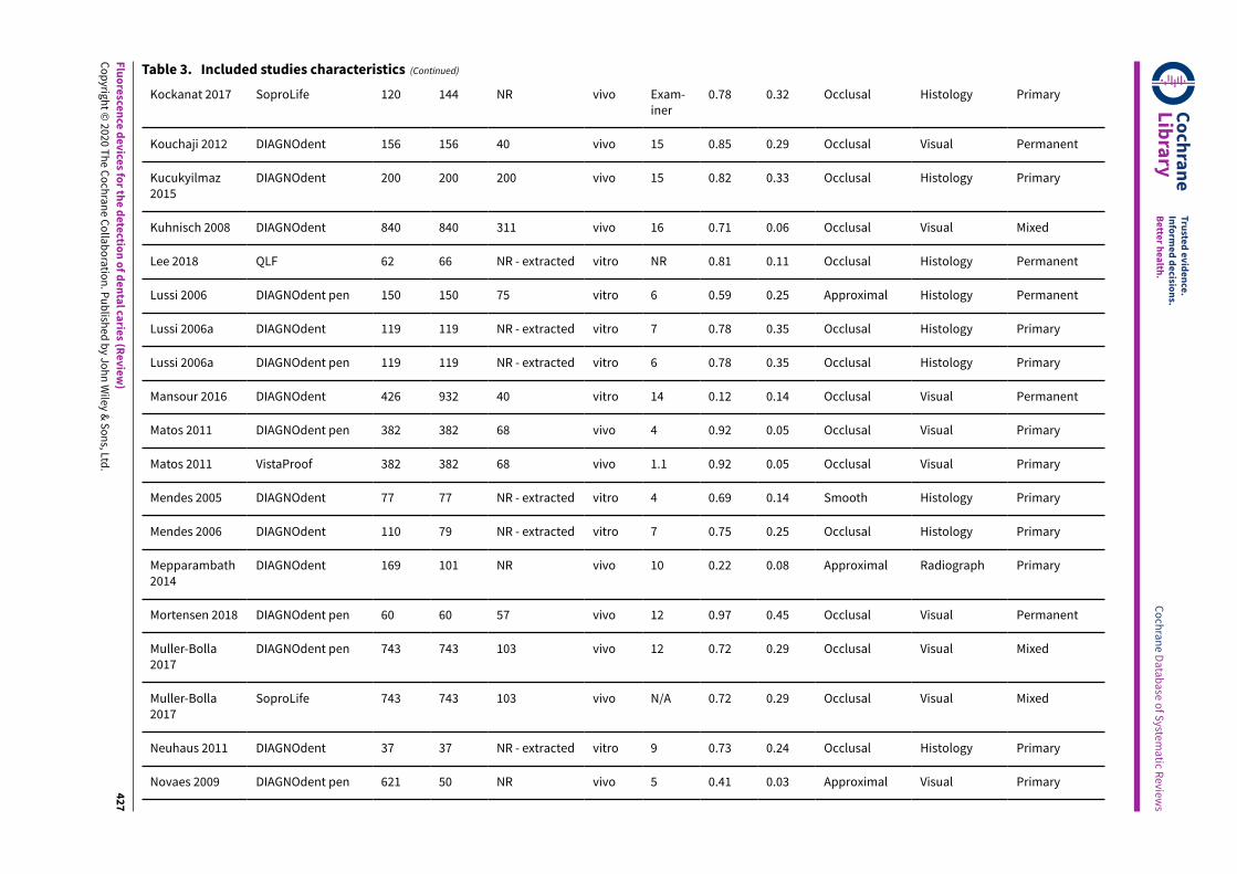
Fluoresce
nce device
s for th
e detectio
n of dental ca
ries (R
eview)
Copyrig
ht © 2020 T
he Cochrane Collaboration. Publish
ed by Jo
hn Wiley & Sons, Ltd
.
427
Kockanat 2017 SoproLife 120 144 NR vivo Exam-iner
0.78 0.32 Occlusal Histology Primary
Kouchaji 2012 DIAGNOdent 156 156 40 vivo 15 0.85 0.29 Occlusal Visual Permanent
Kucukyilmaz2015
DIAGNOdent 200 200 200 vivo 15 0.82 0.33 Occlusal Histology Primary
Kuhnisch 2008 DIAGNOdent 840 840 311 vivo 16 0.71 0.06 Occlusal Visual Mixed
Lee 2018 QLF 62 66 NR - extracted vitro NR 0.81 0.11 Occlusal Histology Permanent
Lussi 2006 DIAGNOdent pen 150 150 75 vitro 6 0.59 0.25 Approximal Histology Permanent
Lussi 2006a DIAGNOdent 119 119 NR - extracted vitro 7 0.78 0.35 Occlusal Histology Primary
Lussi 2006a DIAGNOdent pen 119 119 NR - extracted vitro 6 0.78 0.35 Occlusal Histology Primary
Mansour 2016 DIAGNOdent 426 932 40 vitro 14 0.12 0.14 Occlusal Visual Permanent
Matos 2011 DIAGNOdent pen 382 382 68 vivo 4 0.92 0.05 Occlusal Visual Primary
Matos 2011 VistaProof 382 382 68 vivo 1.1 0.92 0.05 Occlusal Visual Primary
Mendes 2005 DIAGNOdent 77 77 NR - extracted vitro 4 0.69 0.14 Smooth Histology Primary
Mendes 2006 DIAGNOdent 110 79 NR - extracted vitro 7 0.75 0.25 Occlusal Histology Primary
Mepparambath2014
DIAGNOdent 169 101 NR vivo 10 0.22 0.08 Approximal Radiograph Primary
Mortensen 2018 DIAGNOdent pen 60 60 57 vivo 12 0.97 0.45 Occlusal Visual Permanent
Muller-Bolla2017
DIAGNOdent pen 743 743 103 vivo 12 0.72 0.29 Occlusal Visual Mixed
Muller-Bolla2017
SoproLife 743 743 103 vivo N/A 0.72 0.29 Occlusal Visual Mixed
Neuhaus 2011 DIAGNOdent 37 37 NR - extracted vitro 9 0.73 0.24 Occlusal Histology Primary
Novaes 2009 DIAGNOdent pen 621 50 NR vivo 5 0.41 0.03 Approximal Visual Primary
Table 3. Included studies characteristics (Continued)
Cochrane
Library
Truste
d evidence.
Inform
ed decisio
ns.
Bette
r health
.
Cochrane D
atabase o
f System
atic R
eviews
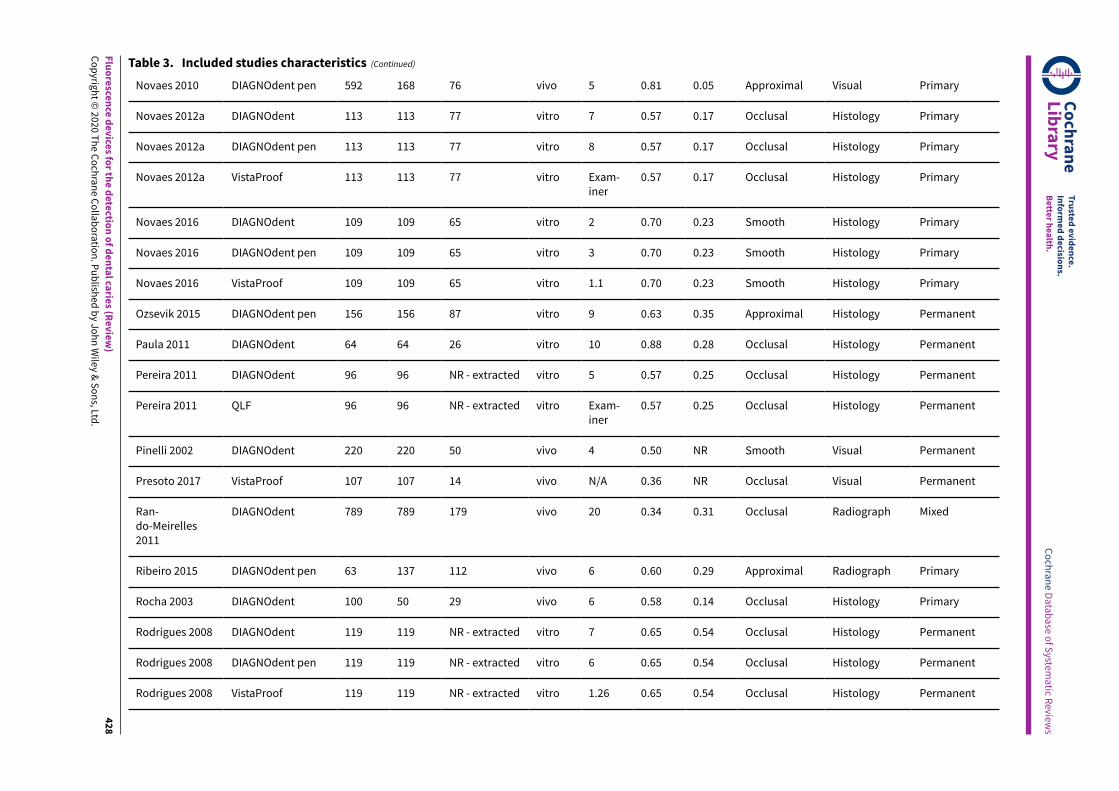
Fluoresce
nce device
s for th
e detectio
n of dental ca
ries (R
eview)
Copyrig
ht © 2020 T
he Cochrane Collaboration. Publish
ed by Jo
hn Wiley & Sons, Ltd
.
428
Novaes 2010 DIAGNOdent pen 592 168 76 vivo 5 0.81 0.05 Approximal Visual Primary
Novaes 2012a DIAGNOdent 113 113 77 vitro 7 0.57 0.17 Occlusal Histology Primary
Novaes 2012a DIAGNOdent pen 113 113 77 vitro 8 0.57 0.17 Occlusal Histology Primary
Novaes 2012a VistaProof 113 113 77 vitro Exam-iner
0.57 0.17 Occlusal Histology Primary
Novaes 2016 DIAGNOdent 109 109 65 vitro 2 0.70 0.23 Smooth Histology Primary
Novaes 2016 DIAGNOdent pen 109 109 65 vitro 3 0.70 0.23 Smooth Histology Primary
Novaes 2016 VistaProof 109 109 65 vitro 1.1 0.70 0.23 Smooth Histology Primary
Ozsevik 2015 DIAGNOdent pen 156 156 87 vitro 9 0.63 0.35 Approximal Histology Permanent
Paula 2011 DIAGNOdent 64 64 26 vitro 10 0.88 0.28 Occlusal Histology Permanent
Pereira 2011 DIAGNOdent 96 96 NR - extracted vitro 5 0.57 0.25 Occlusal Histology Permanent
Pereira 2011 QLF 96 96 NR - extracted vitro Exam-iner
0.57 0.25 Occlusal Histology Permanent
Pinelli 2002 DIAGNOdent 220 220 50 vivo 4 0.50 NR Smooth Visual Permanent
Presoto 2017 VistaProof 107 107 14 vivo N/A 0.36 NR Occlusal Visual Permanent
Ran-do-Meirelles2011
DIAGNOdent 789 789 179 vivo 20 0.34 0.31 Occlusal Radiograph Mixed
Ribeiro 2015 DIAGNOdent pen 63 137 112 vivo 6 0.60 0.29 Approximal Radiograph Primary
Rocha 2003 DIAGNOdent 100 50 29 vivo 6 0.58 0.14 Occlusal Histology Primary
Rodrigues 2008 DIAGNOdent 119 119 NR - extracted vitro 7 0.65 0.54 Occlusal Histology Permanent
Rodrigues 2008 DIAGNOdent pen 119 119 NR - extracted vitro 6 0.65 0.54 Occlusal Histology Permanent
Rodrigues 2008 VistaProof 119 119 NR - extracted vitro 1.26 0.65 0.54 Occlusal Histology Permanent
Table 3. Included studies characteristics (Continued)
Cochrane
Library
Truste
d evidence.
Inform
ed decisio
ns.
Bette
r health
.
Cochrane D
atabase o
f System
atic R
eviews
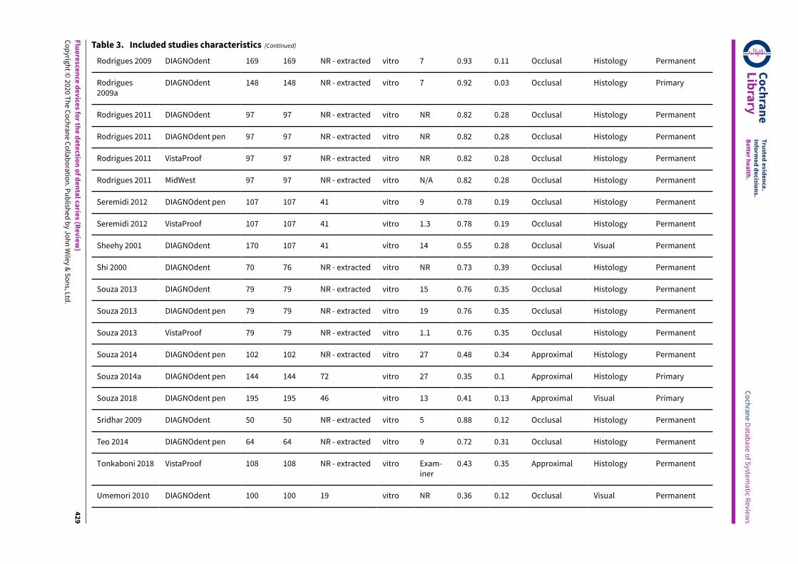
Fluoresce
nce device
s for th
e detectio
n of dental ca
ries (R
eview)
Copyrig
ht © 2020 T
he Cochrane Collaboration. Publish
ed by Jo
hn Wiley & Sons, Ltd
.
429
Rodrigues 2009 DIAGNOdent 169 169 NR - extracted vitro 7 0.93 0.11 Occlusal Histology Permanent
Rodrigues2009a
DIAGNOdent 148 148 NR - extracted vitro 7 0.92 0.03 Occlusal Histology Primary
Rodrigues 2011 DIAGNOdent 97 97 NR - extracted vitro NR 0.82 0.28 Occlusal Histology Permanent
Rodrigues 2011 DIAGNOdent pen 97 97 NR - extracted vitro NR 0.82 0.28 Occlusal Histology Permanent
Rodrigues 2011 VistaProof 97 97 NR - extracted vitro NR 0.82 0.28 Occlusal Histology Permanent
Rodrigues 2011 MidWest 97 97 NR - extracted vitro N/A 0.82 0.28 Occlusal Histology Permanent
Seremidi 2012 DIAGNOdent pen 107 107 41 vitro 9 0.78 0.19 Occlusal Histology Permanent
Seremidi 2012 VistaProof 107 107 41 vitro 1.3 0.78 0.19 Occlusal Histology Permanent
Sheehy 2001 DIAGNOdent 170 107 41 vitro 14 0.55 0.28 Occlusal Visual Permanent
Shi 2000 DIAGNOdent 70 76 NR - extracted vitro NR 0.73 0.39 Occlusal Histology Permanent
Souza 2013 DIAGNOdent 79 79 NR - extracted vitro 15 0.76 0.35 Occlusal Histology Permanent
Souza 2013 DIAGNOdent pen 79 79 NR - extracted vitro 19 0.76 0.35 Occlusal Histology Permanent
Souza 2013 VistaProof 79 79 NR - extracted vitro 1.1 0.76 0.35 Occlusal Histology Permanent
Souza 2014 DIAGNOdent pen 102 102 NR - extracted vitro 27 0.48 0.34 Approximal Histology Permanent
Souza 2014a DIAGNOdent pen 144 144 72 vitro 27 0.35 0.1 Approximal Histology Primary
Souza 2018 DIAGNOdent pen 195 195 46 vitro 13 0.41 0.13 Approximal Visual Primary
Sridhar 2009 DIAGNOdent 50 50 NR - extracted vitro 5 0.88 0.12 Occlusal Histology Permanent
Teo 2014 DIAGNOdent pen 64 64 NR - extracted vitro 9 0.72 0.31 Occlusal Histology Permanent
Tonkaboni 2018 VistaProof 108 108 NR - extracted vitro Exam-iner
0.43 0.35 Approximal Histology Permanent
Umemori 2010 DIAGNOdent 100 100 19 vitro NR 0.36 0.12 Occlusal Visual Permanent
Table 3. Included studies characteristics (Continued)
Cochrane
Library
Truste
d evidence.
Inform
ed decisio
ns.
Bette
r health
.
Cochrane D
atabase o
f System
atic R
eviews
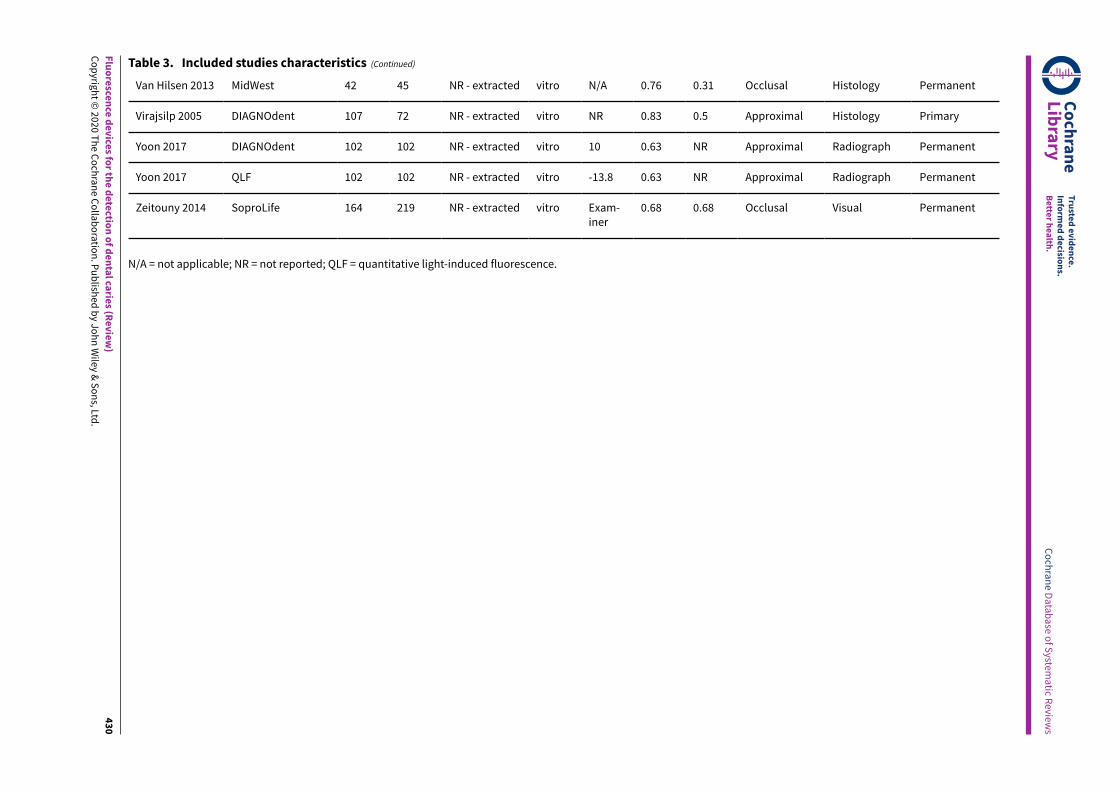
Fluoresce
nce device
s for th
e detectio
n of dental ca
ries (R
eview)
Copyrig
ht © 2020 T
he Cochrane Collaboration. Publish
ed by Jo
hn Wiley & Sons, Ltd
.
430
Van Hilsen 2013 MidWest 42 45 NR - extracted vitro N/A 0.76 0.31 Occlusal Histology Permanent
Virajsilp 2005 DIAGNOdent 107 72 NR - extracted vitro NR 0.83 0.5 Approximal Histology Primary
Yoon 2017 DIAGNOdent 102 102 NR - extracted vitro 10 0.63 NR Approximal Radiograph Permanent
Yoon 2017 QLF 102 102 NR - extracted vitro -13.8 0.63 NR Approximal Radiograph Permanent
Zeitouny 2014 SoproLife 164 219 NR - extracted vitro Exam-iner
0.68 0.68 Occlusal Visual Permanent
Table 3. Included studies characteristics (Continued)
N/A = not applicable; NR = not reported; QLF = quantitative light-induced fluorescence.
Cochrane
Library
Truste
d evidence.
Inform
ed decisio
ns.
Bette
r health
.
Cochrane D
atabase o
f System
atic R
eviews
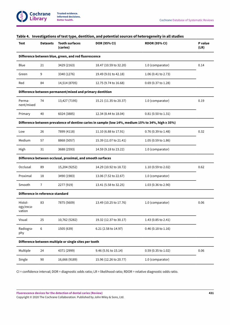
CochraneLibrary
Trusted evidence.Informed decisions.Better health.
Cochrane Database of Systematic Reviews
Test Datasets Tooth surfaces(caries)
DOR (95% CI) RDOR (95% CI) P value(LR)
Difference between blue, green, and red fluorescence
Blue 21 3429 (2163) 18.47 (10.59 to 32.20) 1.0 (comparator)
Green 9 3340 (1276) 19.49 (9.01 to 42.18) 1.06 (0.41 to 2.73)
Red 84 14,514 (8705) 12.75 (9.74 to 16.68) 0.69 (0.37 to 1.28)
0.14
Difference between permanent/mixed and primary dentition
Perma-nent/mixed
74 13,427 (7195) 15.21 (11.35 to 20.37) 1.0 (comparator)
Primary 40 6024 (3885) 12.34 (8.44 to 18.04) 0.81 (0.50 to 1.31)
0.19
Difference between prevalence of dentine caries in sample (low 14%, medium 15% to 34%, high ≥ 35%)
Low 26 7899 (4118) 11.10 (6.88 to 17.91) 0.76 (0.39 to 1.48)
Medium 57 8868 (5057) 15.39 (11.07 to 21.41) 1.05 (0.59 to 1.86)
High 31 3688 (2593) 14.59 (9.18 to 23.22) 1.0 (comparator)
0.32
Difference between occlusal, proximal, and smooth surfaces
Occlusal 89 15,204 (9252) 14.29 (10.92 to 18.72) 1.10 (0.59 to 2.02)
Proximal 18 3490 (1983) 13.06 (7.52 to 22.67) 1.0 (comparator)
Smooth 7 2277 (919) 13.41 (5.58 to 32.25) 1.03 (0.36 to 2.90)
0.62
Difference in reference standard
Histol-ogy/exca-vation
83 7875 (5609) 13.49 (10.25 to 17.76) 1.0 (comparator)
Visual 25 10,762 (5282) 19.32 (12.37 to 30.17) 1.43 (0.85 to 2.41)
Radiogra-phy
6 1505 (639) 6.21 (2.58 to 14.97) 0.46 (0.18 to 1.16)
0.06
Difference between multiple or single sites per tooth
Multiple 24 4371 (2999) 9.46 (5.91 to 15.14) 0.59 (0.35 to 1.02)
Single 90 16,666 (9189) 15.96 (12.26 to 20.77) 1.0 (comparator)
0.06
Table 4. Investigations of test type, dentition, and potential sources of heterogeneity in all studies
CI = confidence interval; DOR = diagnostic odds ratio; LR = likelihood ratio; RDOR = relative diagnostic odds ratio.
Fluorescence devices for the detection of dental caries (Review)
Copyright © 2020 The Cochrane Collaboration. Published by John Wiley & Sons, Ltd.
431
CochraneLibrary
Trusted evidence.Informed decisions.Better health.
Cochrane Database of Systematic Reviews
H I S T O R Y
Review first published: Issue 12, 2020
C O N T R I B U T I O N S O F A U T H O R S
All review authors collaborated in the conception of the review purpose, design, and interpretation of results.DraNing the protocol and final draN of the review: Tanya Walsh (TW), Richard Macey (RM).Developing the search strategy: TW, RM.Co-ordination of contributions from the co-authors: RM.Screening of papers against eligibility criteria: RM, TW, Philip Riley (PR), Helen Worthington (HW), and Anne-Marie Glenny (AMG).Obtained data on published, ongoing, and unpublished studies: RM.Appraising the quality of papers: RM, TW, PR, HW, and AMG.Extracting data for the review: RM, TW, PR, HW, Patrick Fee (PF), and AMG.Entering data into Review Manager 5: RM.Analysis of data: RM and TW.Provided clinical guidance during all phases of review: Janet Clarkson (JC) and David Ricketts (DR).
D E C L A R A T I O N S O F I N T E R E S T
Richard Macey: none known.Tanya Walsh: none known. I am Statistical Editor with Cochrane Oral Health.Philip Riley: none known. I am Deputy Co-ordinating Editor of Cochrane Oral Health.Anne-Marie Glenny: none known. I am Co-ordinating Editor of Cochrane Oral Health.Helen V Worthington: none known. I am an Editor with Cochrane Oral Health.Patrick A Fee: none known.Janet E Clarkson: none known. I am Co-ordinating Editor of Cochrane Oral Health.David Ricketts: none known.
S O U R C E S O F S U P P O R T
Internal sources
• Division of Dentistry, School of Medical Sciences, Faculty of Biology, Medicine and Health, The University of Manchester, UK
• Manchester Academic Health Sciences Centre (MAHSC) and the NIHR Manchester Biomedical Research Centre, UK
External sources
• National Institute for Health Research (NIHR), UK
NIHR Cochrane Programme Grant: 16/114/23 Detection and diagnosis of common oral diseases: diagnostic test accuracy of tests of oralcancer and caries
• Cochrane Oral Health Global Alliance, Other
The production of Cochrane Oral Health reviews has been supported financially by our Global Alliance since 2011(oralhealth.cochrane.org/partnerships-alliances). Contributors in the last 2 years have been the American Association of Public HealthDentistry, USA; AS-Akademie, Germany; the British Association for the Study of Community Dentistry, UK; the British Society ofPaediatric Dentistry, UK; the Canadian Dental Hygienists Association, Canada; the Centre for Dental Education and Research at All IndiaInstitute of Medical Sciences, India; the National Center for Dental Hygiene Research & Practice, USA; New York University College ofDentistry, USA; and Swiss Society of Endodontology, Switzerland
• NIHR, UK
This project was supported by the NIHR, via Cochrane Infrastructure funding to Cochrane Oral Health. The views and opinions expressedherein are those of the review authors and do not necessarily reflect those of the Evidence Synthesis Programme, the NIHR, the NHS,or the Department of Health and Social Care
D I F F E R E N C E S B E T W E E N P R O T O C O L A N D R E V I E W
• Three categories of fluorescence index test were defined in the index test section of the 'Background'. It was important to categorise thedevices as they each utilise diEerent wavelengths to reach a diagnostic decision.
• One of the objectives was removed because the search produced a large body of evidence for the primary time point in clinical processso we decided it would add unnecessary complexity to investigate the additional objective of the value of each index test at diEerentpositions in the clinical pathway.
Fluorescence devices for the detection of dental caries (Review)
Copyright © 2020 The Cochrane Collaboration. Published by John Wiley & Sons, Ltd.
432
CochraneLibrary
Trusted evidence.Informed decisions.Better health.
Cochrane Database of Systematic Reviews
• We removed the secondary objective which stipulated that we would investigate the impact of previously applied restorations andfissure sealants as there were insuEicient studies that included previously restored or sealed teeth. This also allowed us to amend thelisted target conditions which stated caries adjacent to existing restorations.
• The protocol specified that we would investigate the diEerence between in vitro and in vivo studies, this has not been reported explicitlybecause the reference standard investigations cover the same issue. All in vitro studies employed a histological reference standard sothis can be used as a proxy for the in vitro/in vivo comparison.
Fluorescence devices for the detection of dental caries (Review)
Copyright © 2020 The Cochrane Collaboration. Published by John Wiley & Sons, Ltd.
433
Related Documents