ORIGINAL ARTICLE Open Access Fluorescence-based in situ assay to probe the viability and growth kinetics of surface-adhering and suspended recombinant bacteria Ima Avalos Vizcarra 1 , Philippe Emge 1 , Philipp Miermeister 2 , Mamta Chabria 1 , Rupert Konradi 3 , Viola Vogel 1 and Jens Möller 1* Abstract Bacterial adhesion and biofilm growth can cause severe biomaterial-related infections and failure of medical implants. To assess the antifouling properties of engineered coatings, advanced approaches are needed for in situ monitoring of bacterial viability and growth kinetics as the bacteria colonize a surface. Here, we present an optimized protocol for optical real-time quantification of bacterial viability. To stain living bacteria, we replaced the commonly used fluorescent dye SYTO ® 9 with endogenously expressed eGFP, as SYTO ® 9 inhibited bacterial growth. With the addition of nontoxic concentrations of propidium iodide (PI) to the culture medium, the fraction of live and dead bacteria could be continuously monitored by fluorescence microscopy as demonstrated here using GFP expressing Escherichia coli as model organism. The viability of bacteria was thereby monitored on untreated and bioactive dimethyloctadecyl[3-(trimethoxysilyl)propyl]ammonium chloride (DMOAC)-coated glass substrates over several hours. Pre-adsorption of the antimicrobial surfaces with serum proteins, which mimics typical protein adsorption to biomaterial surfaces upon contact with host body fluids, completely blocked the antimicrobial activity of the DMOAC surfaces as we observed the recovery of bacterial growth. Hence, this optimized eGFP/PI viability assay provides a protocol for unperturbed in situ monitoring of bacterial viability and colonization on engineered biomaterial surfaces with single-bacteria sensitivity under physiologically relevant conditions. Keywords: Antimicrobial surfaces; Optical viability monitoring; Green fluorescent protein (GFP); SYTO ® 9; Propidium iodide (PI) Background Clinically relevant nosocomial infections are frequently caused by adherent bacteria and the subsequent biofilm formation within tissues or on biomaterial surfaces [1]. Surface biofouling commonly starts with the adhesion of individual bacteria that subsequently grow into mature biofilms. To prevent bacterial adhesion and growth already during the pre-biofilm phase, two main surface engineering strategies have been developed so far: non-fouling “stealth” surface coatings that inhibit adhesion of proteins and bac- teria [2-4] and bioactive materials, which upon bacterial contact or release of the active molecules interfere with bacterial viability [5-10]. To compare the antimicrobial properties of surface coatings and to study the kinetics of bacterial surface colonization, assays are needed that allow for in situ monitoring of bacterial adhesion and viability. The gold standard for bacterial viability tests has long been quantification of colony forming units (CFU) by plating bacterial suspensions that were incubated with the test surface on nutrient agar [11]. Counting bacter- ial colonies, which result from plating suspended viable and cultivatable bacteria, however, does not account for the in- herent phenotypic heterogeneity and the ability of the bac- teria to persist in dormant states [12,13]. Furthermore, plating assays lack the ability to measure the colonization and viability kinetics directly on the test surface and might not be representative for the surface-attached bacterial population. An alternative to determine bacterial viability is to probe for the bacterial membrane integrity that is maintained by * Correspondence: [email protected] 1 Department of Health Sciences and Technology, Laboratory of Applied Mechanobiology, ETH Zurich, CH-8093 Zurich, Switzerland Full list of author information is available at the end of the article © 2013 Avalos Vizcarra et al.; licensee Springer. This is an Open Access article distributed under the terms of the Creative Commons Attribution License (http://creativecommons.org/licenses/by/2.0), which permits unrestricted use, distribution, and reproduction in any medium, provided the original work is properly cited. Avalos Vizcarra et al. Biointerphases 2013, 8:22 http://www.biointerphases.com/content/8/1/22

Welcome message from author
This document is posted to help you gain knowledge. Please leave a comment to let me know what you think about it! Share it to your friends and learn new things together.
Transcript

Avalos Vizcarra et al. Biointerphases 2013, 8:22http://www.biointerphases.com/content/8/1/22
ORIGINAL ARTICLE Open Access
Fluorescence-based in situ assay to probe theviability and growth kinetics of surface-adheringand suspended recombinant bacteriaIma Avalos Vizcarra1, Philippe Emge1, Philipp Miermeister2, Mamta Chabria1, Rupert Konradi3, Viola Vogel1
and Jens Möller1*
Abstract
Bacterial adhesion and biofilm growth can cause severe biomaterial-related infections and failure of medicalimplants. To assess the antifouling properties of engineered coatings, advanced approaches are needed for in situmonitoring of bacterial viability and growth kinetics as the bacteria colonize a surface. Here, we present anoptimized protocol for optical real-time quantification of bacterial viability. To stain living bacteria, we replaced thecommonly used fluorescent dye SYTO® 9 with endogenously expressed eGFP, as SYTO® 9 inhibited bacterial growth.With the addition of nontoxic concentrations of propidium iodide (PI) to the culture medium, the fraction of liveand dead bacteria could be continuously monitored by fluorescence microscopy as demonstrated here using GFPexpressing Escherichia coli as model organism. The viability of bacteria was thereby monitored on untreated andbioactive dimethyloctadecyl[3-(trimethoxysilyl)propyl]ammonium chloride (DMOAC)-coated glass substrates overseveral hours. Pre-adsorption of the antimicrobial surfaces with serum proteins, which mimics typical proteinadsorption to biomaterial surfaces upon contact with host body fluids, completely blocked the antimicrobial activityof the DMOAC surfaces as we observed the recovery of bacterial growth. Hence, this optimized eGFP/PI viabilityassay provides a protocol for unperturbed in situ monitoring of bacterial viability and colonization on engineeredbiomaterial surfaces with single-bacteria sensitivity under physiologically relevant conditions.
Keywords: Antimicrobial surfaces; Optical viability monitoring; Green fluorescent protein (GFP); SYTO® 9;Propidium iodide (PI)
BackgroundClinically relevant nosocomial infections are frequentlycaused by adherent bacteria and the subsequent biofilmformation within tissues or on biomaterial surfaces [1].Surface biofouling commonly starts with the adhesion ofindividual bacteria that subsequently grow into maturebiofilms. To prevent bacterial adhesion and growth alreadyduring the pre-biofilm phase, two main surface engineeringstrategies have been developed so far: non-fouling “stealth”surface coatings that inhibit adhesion of proteins and bac-teria [2-4] and bioactive materials, which upon bacterialcontact or release of the active molecules interfere withbacterial viability [5-10]. To compare the antimicrobial
* Correspondence: [email protected] of Health Sciences and Technology, Laboratory of AppliedMechanobiology, ETH Zurich, CH-8093 Zurich, SwitzerlandFull list of author information is available at the end of the article
© 2013 Avalos Vizcarra et al.; licensee Springer.Commons Attribution License (http://creativecoreproduction in any medium, provided the orig
properties of surface coatings and to study the kinetics ofbacterial surface colonization, assays are needed thatallow for in situ monitoring of bacterial adhesion andviability. The gold standard for bacterial viability testshas long been quantification of colony forming units(CFU) by plating bacterial suspensions that were incubatedwith the test surface on nutrient agar [11]. Counting bacter-ial colonies, which result from plating suspended viable andcultivatable bacteria, however, does not account for the in-herent phenotypic heterogeneity and the ability of the bac-teria to persist in dormant states [12,13]. Furthermore,plating assays lack the ability to measure the colonizationand viability kinetics directly on the test surface and mightnot be representative for the surface-attached bacterialpopulation.An alternative to determine bacterial viability is to probe
for the bacterial membrane integrity that is maintained by
This is an Open Access article distributed under the terms of the Creativemmons.org/licenses/by/2.0), which permits unrestricted use, distribution, andinal work is properly cited.

Avalos Vizcarra et al. Biointerphases 2013, 8:22 Page 2 of 9http://www.biointerphases.com/content/8/1/22
energy-dependent processes in living bacteria and is lostupon bacterial death [14]. Membrane integrity can be testedoptically by using a combination of membrane permeableand impermeable fluorescent dyes that selectively enterlive and dead bacteria (Figure 1a) [8,14-17]. While beingbroadly employed as endpoint staining assays to determinethe viability of single bacteria and bacterial colonies directlyon the test surface, these assays are not optimized for real-time in situ bacterial viability monitoring. Particularlywhen DNA intercalating dyes like SYTO® 9 and propidiumiodide (PI) are used [18], the impact of the potentiallytoxic stains on bacterial physiology has to be consideredto avoid false negative results [19]. Furthermore, sinceboth stains target DNA, the competitive displacement ofthe SYTO® 9 (live stain) by the high affinity PI (dead stain)upon membrane breakdown can affect the staining reliabil-ity [20]. To eliminate the competitive displacement of thetwo DNA stains and the demand for prolonged incubationtimes caused by the passive diffusion of the SYTO® 9 livestain through the bacterial membrane, it was suggestedto replace SYTO® 9 with green fluorescent protein (GFP)expressed by the bacteria as demonstrated previously forflow cytometry applications (Figure 1b) [21]. Althoughflow cytometry has been used to measure the viabilityof GFP expressing bacteria adsorbed to polystyrene
Figure 1 Bacterial viability assays in comparison. (a) Conventionalendpoint dual staining bacterial viability assay and (b) eGFP/PI assayoptimized for in situ bacterial viability monitoring. The dual stainingassay commonly employs two DNA stains, SYTO® 9 (green triangle)and propidium iodide (PI, red rectangle) that both intercalate intothe bacterial DNA. SYTO® 9 diffuses passively into living bacteriawhereas PI cannot pass intact bacterial membranes and only enterspermeabilized dead bacteria. The use of constitutive eGFP expressionrather than SYTO® 9 provides direct detection of viable bacteria withoutthe addition of a fluorescent dye and circumvents the competitivedisplacement of SYTO® 9 by PI that can result in a dual staining ofdead bacteria.
beads functionalized with antimicrobial coatings [22],it cannot be applied for continuous in situ bacterialviability monitoring on planar surfaces.Building upon those observations, we present an opti-
mized protocol to probe the viability and growth kinetics ofsurface-adhering and suspended bacteria using non-toxicconcentrations of propidium iodide and Escherichia colithat express the fluorescent protein GFP. Beyond calibrat-ing the assay and monitoring E. coli surface colonizationkinetics on bare glass substrates, we demonstrate that thisassay is applicable to monitor the inactivation kinetics ofE. coli in contact with antimicrobial surface coatings,using dimethyloctadecyl[3-(trimethoxysilyl)propyl]ammo-nium chloride (DMOAC) coated glass surfaces as modelsubstrate. To kill bacteria, the quaternary ammoniumchloride complexes of surface-bound DMOAC have todirectly interact with the bacterial membrane [23]. Wepreviously showed that bacterial fimbriae strongly influ-ence the unspecific adhesion of E. coli to engineered sur-faces [4]. Type 1 fimbriae (7 nm diameter, several 100 μmlength) protrude from the bacterial membrane therebypreventing the bulk bacterial body from direct inter-action with the underlying material surface. To ensurea physical contact of the bacterial membrane with thematerial surface, we used here the non-fimbriated K-12derivative AAEC191A E. coli strain. In addition, we high-light the effect of serum protein adsorption on the bacteri-cidal properties of antimicrobial surfaces. We incubatedthe DMOAC surfaces with fetal bovine serum (FBS) tomimic the physiological situation where serum proteinsadsorb to engineered biomaterials upon contact with hostbody fluids.
MethodsBacteriaNon-fimbriated E. coli AAEC191A bacteria, a derivativeof E. coli K-12 MG1655 containing a deletion in theentire fim cluster [24] was provided by Prof. E. Sokurenko,University of Washington, Seattle, USA. For GFP expres-sion, chemocompetent AAEC191A E. coli were transformedwith eGFP pHis plasmid under the control of the tacpromoter (AAEC191A pHis-GFP). To obtain E. coli thatexpress eGFP under the control of the constitutive rpsmpromoter (AAEC191A rpsm-GFP), E. coli AAEC191Awere transformed with the rpsm-GFP plasmid that wasextracted from the original fusion library strain MG1655rpsm-GFP [25] by Qiaprep Spin Miniprep kit (Qiagen27106). Transformed bacteria were selected by cultivationon LB agar plates supplemented with either 100 μg/mlampicillin (pHis-GFP) or 50 μg/ml kanamycin (rpsm-GFP).Bacterial precultures were inoculated from glycerol stocksinto LB medium (5 g/l yeast extract, 10 g/l tryptone, 10 g/lNaCl) containing appropriate antibiotics. To induce GFPexpression in AAEC191A pHis-GFP E. coli, 0.1 mM

Avalos Vizcarra et al. Biointerphases 2013, 8:22 Page 3 of 9http://www.biointerphases.com/content/8/1/22
Isopropyl-β-D-thiogalactopyranoside (IPTG, ApplichemA1008) was added. LB precultures were grown overnightat 37°C under continuous shaking at 180 rpm (InforsUnitron HT). To ensure defined culture conditions for thebacterial growth and viability assays, bacteria from theovernight culture were centrifuged at 1700 g, washedthree times and subcultured in 20 ml minimal M9 medium(1x M9 salts (Sigma-Aldrich M6030), 10 mM Mg2SO4
(Sigma-Aldrich 63126), 10 g/l Glucose (Sigma-AldrichG8270), 0.5 mM CaCl2 (Sigma-Aldrich C5080), 1x MEMvitamins (Gibco 11120), 1x MEM amino acids (Gibco11130)) supplemented with the appropriate antibioticsand 0.1 mM IPTG for AAEC191A pHis-GFP E. coli. Bac-teria were subcultured at 37°C, 180 rpm until exponentialgrowth phase (OD600 = 0.3-0.8). Bacteria were harvestedby centrifugation at 1700 g followed by three washingsteps. Immediately before the experiment, bacteria wereresuspended in M9 medium that contained the strain-specific antibiotics, IPTG as well as 0, 3, 30 μM PI(Sigma-Aldrich, 81845) and 6 μM SYTO® 9 (InvitrogenL13152), respectively.
Growth curve measurementsGrowth curves of suspended bacteria were recorded byturbidity measurements at 600 nm in 96 well plates(Tecan Infinity 200 Pro plate reader). Kinetic measure-ments were performed every 15 minutes at 37°C andcontinuous shaking. Bacteria were inoculated to an initialturbidity of 0.01 at 600 nm in M9 medium containingappropriate antibiotics, PI and SYTO® 9.
Viability assayFor kinetic viability measurements under physiologicalconditions, bacteria were cultivated in an ibidi® glassbottom flow chamber (ibidi, 80168) within a temperature-controlled microscope incubator to guarantee constantnutrient supply and optimal growth conditions at 37°C.Bioactive surfaces were prepared according to publishedprotocols [23]. Briefly, glass cover slides, that later resem-ble the bottom slide of the flow chamber, were exposedto air plasma for 15 seconds (Harrick Plasma, PDC-32G)followed by dipping into a 5% (v/v) aqueous DMOAC(Sigma-Aldrich) solution for 1 second, and drying at 105°Covernight. To test the effect of protein pre-incubationon the antimicrobial activity of the DMOAC coatings,slides were incubated in undiluted fetal bovine serum(FBS, Thermo Scientific SH30071.02) for 1 h prior to theassembly of the flow chamber. Bare glass cover slides wereattached to the ibidi® chambers as control surfaces. Thebacterial suspension (OD600 0.05) in M9 mediumcontaining different concentrations of PI and SYTO® 9,was directly added to the flow chamber and immediatelytransferred to an epifluorescence microscope (NikonTE2000-E) for in situ viability monitoring. Adhesion of
bacteria to the glass bottom slide was allowed for 5 mi-nutes before the flow chamber was gently washed with5 ml M9 medium (flow rate 0.01 ml/min) to remove non-adherent bacteria. As control staining at defined timepoints, the BacLight™ viability kit (Invitrogen, L13152)was used according to the supplier instructions.
EllipsometryThe adsorbed dry film thickness of DMOAC andDMOAC + FBS layers on silicone wafers was measuredby variable-angle spectroscopic ellipsometry (VASE) usingthe M2000F variable-angle spectroscopic ellipsometer (J.A.Woollam Co., Inc.). The measurement was performedat 70° relative to the surface normal under ambientconditions. Ellipsometry data were fitted using a cauchymodel with parameters for organic layers (n(λ) = Aλ + Bn/λ^2 +Cn/λ^4, with An = 1.45, Bn = 0.01, Cn = 0.0) to obtaindry thickness of adlayers.
Image segmentation and quantificationTo limit the viability analysis to fluorescent E. coli and toeliminate bias in the data analysis based on GFP fluores-cence intensity, fluorescence images were thresholdedand segmented using the morphological strel algorithmof the image processing toolbox of MATLAB® software(MATLAB, MathWorks; version R2010b) that combinesimage erosion and dilation operations. The algorithm wasincluded into a semiautomatic image processing workflowthat allows for manual adjustment of the thresholdinglevels of the entire time series as well as individual timeframes. Binary masks were generated from the thresholdedimages and surface-adherent bacteria were counted auto-matically. The summing of binary masks from consecutivetime points allowed for correction of fluorescence signalloss caused by GFP bleaching, washout and degradation ofthe stained DNA. To prevent false-positive results, binarymasks that were not positive for the GFP channel before,were excluded from the PI positive counts to limit theanalysis to bacteria that were viable initially. Eliminationof x,y drift of time series data was achieved by the registervirtual stack slices and transform virtual stack slicesplugins of Fiji that were incorporated into a MATLAB®
routine using the MIJ java package for bi-directional com-munication between MATLAB® and ImageJ by D. Sage.The MATLAB® file for the analysis workflow is available inthe Additional file 1.
ResultsImpact of SYTO® 9 and propidium iodide concentrationson the growth of suspended E. coliTo determine toxicity levels of the fluorescent DNAstains, we probed the effect of SYTO® 9 and propidiumiodide (PI) on E. coli growth. We supplemented bacterialbatch cultures in M9 growth medium with varying dye

Avalos Vizcarra et al. Biointerphases 2013, 8:22 Page 4 of 9http://www.biointerphases.com/content/8/1/22
concentrations (0, 3, 30 μM PI, 6 μM SYTO® 9) andmonitored bacterial growth at 37°C by quantifying theincrease in the turbidity of the solution at 600 nm(Figure 2a). 6 μM SYTO® 9 in combination with 30 μM PI,as recommended in the conventional and commercialdual staining assay [26], completely inhibited E. coligrowth. Supplementing the E. coli suspensions with 6 μMSYTO® 9 alone showed the same growth inhibition, while30 μM PI itself did not inhibit growth completely but didreduce the growth rate compared to the pure M9medium. This indicates that 6 μM SYTO® 9 causes majorchanges in bacterial physiology. The impaired growth rateupon addition of PI was eliminated when we decreasedthe PI concentration in the bacterial growth medium ten-fold, i.e. from 30 μM to 3 μM (Figure 2a).To confirm that the viable E. coli in the medium
supplemented with 3 μM PI were able to replicate, wecompared the turbidity increase of a 50% live / 50%isopropanol killed bacterial mixture to cultures containing100% live and 100% isopropanol treated E. coli (Figure 2a).Within 4 hours, the turbidity increase for the mixed 50%live / 50% dead starting culture did not reach the samelevel as for the 100% live culture. Those results are con-sistent with the expected exponential growth rate of viablebacterial batch cultures and thus show that the bacteriareplicated normally in 3 μM PI containing medium. Todetermine if the reduced PI concentration was sufficientto detect dead bacteria in solution, we incubated bacterialbatch cultures starting from either 100% live or 50% live /50% killed bacteria in M9 medium containing 3 and30 μM PI respectively. The PI fluorescence at 630 nm wassubsequently measured by fluorescence spectroscopy over3 hours (Figure 2b). Supplementing the growth medium
Figure 2 Impact of SYTO® 9 and propidium iodide (PI) concentrationbacteria in M9 bacterial medium. (a) Inhibition of bacterial growth in thdependent growth inhibition. At a concentration of 30 μM E. coli growth wreduced tenfold from 30 to 3 μM. Replication of E. coli in 3 μM PI containinstarting culture of 50% live / 50% dead E. coli (inset) (b) PI fluorescence of3 μM PI sufficiently stained dead bacteria in a 50% live / 50% dead E. coli mfor 3 μM PI compared to the background for a 100% live bacterial solutionsignificant increase of PI stained bacteria from a 100% live starting culturebacterial viability. Background fluorescence of PI supplemented M9 mediumError bars represent the standard error of the mean.
with 3 μM PI adequately stained the isopropanol treatedbacteria in the 1:1 mixture of live and dead E. coli but didnot result in a significant increase of the backgroundfluorescence of the 100% live starting culture. In contrast,supplementing the medium with 30 μM PI significantlyincreased the PI fluorescence from the 100% live startingculture (Figure 2b), which was consistent with the im-paired growth rate under those conditions (Figure 2a),indicating that 30 μM but not 3 μM PI is toxic to E. colibacteria.
Viability and growth rate of surface-adhering E. coli isstrongly reduced upon long-term incubation in culturemedium supplemented with SYTO® 9For viability and growth kinetic studies of surface-adherentbacteria, E. coli that unspecifically adhered to bare glasssurfaces were incubated in M9 growth medium thatcontained either 3 μM PI or a mixture of 6 μM SYTO® 9and 30 μM PI (Figure 3a). For SYTO® 9 containingmedium, E. coli replication and surface colonization wascompletely blocked, as determined by time lapse videomicroscopy (Figure 3a, Additional file 1: Figure S1). Inaddition to inhibiting bacterial growth, the viability ofsurface-adhering E. coli (AAEC191A), decreased forincubation times longer than 1.5 hours, as detected bytwo-channel fluorescence microscopy. In contrast, nodecrease of viability or impaired growth was observedfor endogenously eGFP expressing E. coli (AAEC191ApHis-GFP) counterstained with 3 μM PI (Figure 3a), whichis in agreement with the results from the batch culture ex-periments (Figure 2). In controls we confirmed that GFPexpression itself did not perturb E. coli adhesion andgrowth (Figure 4a,b). Furthermore, the fraction of GFP
on E. coli growth rate and the detection efficiency of deade presence of 6 μM SYTO® 9 and 30 μM PI. PI alone showed a dose-as inhibited, which was not detected when the PI concentration wasg M9 medium was confirmed by growth rate measurements from aE. coli cultivated in M9 medium containing different PI concentrations.ixture, while no significant background signal increase was detected. In contrast, supplementing the medium with 30 μM PI resulted in aindicating that this high concentration of the DNA stain interferes withwas subtracted for each of the three PI concentrations, respectively.

Figure 3 In situ monitoring of the growth and viability ofsurface-adhering E. coli using the eGFP/3 μM PI assay. (a) Timeseries of surface-adhering E. coli on bare glass substrates. The dualstaining assay (6 μM SYTO® 9 / 30 μM PI) decreases bacterial viabilityon untreated glass substrates after incubation times longer than1 hour as E. coli (AAEC191A) incubated with 6 μM SYTO® 9containing medium failed to replicate (inset). In contrast, eGFP-expressing E. coli (AAEC191A pHis-GFP) that were incubated with3 μM PI were able to replicate and grow on the glass surface.(b) Viability of E.coli (AAEC191A pHis-GFP) on antimicrobial DMOAC-coated glass surfaces as monitored by eGFP/PI fluorescencemicroscopy. Pre-exposure of the DMOAC surfaces to fetal bovineserum (FBS) completely blocked the antimicrobial activity.Microscopy images show the overlay of the SYTO® 9 / eGFP and PIfluorescence channels, i.e. differentiating live (green) from deadbacteria (red). 3 independent fields of view from differentexperiments were analyzed containing a total of 125–250 surfaceattached bacteria for each condition. Error bars represent thestandard deviation. Scale bar 20 μm.
Avalos Vizcarra et al. Biointerphases 2013, 8:22 Page 5 of 9http://www.biointerphases.com/content/8/1/22
fluorescent E. coli (87%) was not significantly different(α = 0.05) for eGFP expression from the IPTG induciblepHis plasmid under the control of lac promoter (AAE-C191A pHis-GFP) and under the control of a constitutiverpsm promoter (AAEC191A rpsm-GFP) (Figure 4c,d).To evaluate whether the eGFP/3 μM PI assay is suited
for in situ monitoring of bacterial viability and growthon a bioactive model substrate, eGFP-expressing E. coli(AAEC191A pHis-GFP) were incubated on antimicrobialdimethyloctadecyl[3-(trimethoxysilyl)propyl]ammoniumchloride (DMOAC) coated glass surfaces [23,27] (Figure 3b).Homogeneous DMOAC coating with a dry adlayer thick-ness of 2.2 nm was confirmed by variable-angle spectro-scopic ellipsometry. To allow for a direct contact ofthe bacterial membrane with the surface-immobilizedmembrane-active DMOAC molecules, non-fimbriatedeGFP expressing E. coli (AAEC191A pHis-GFP) wereused [4]. As detected by 3 μM PI staining, all adherentbacteria on the DMOAC surface were killed within30 minutes of surface incubation and no measurablebacterial growth occurred (Figure 3b, Additional file 1:Figure S2, Additional file 2: Movie S1).
Pre-incubation of bioactive DMOAC surfaces with serumproteins completely blocked the antimicrobial activityand restored bacterial growth on the surfaceTo investigate if unspecific protein adsorption wouldinterfere with the bactericidal activity of the DMOACsurfaces, the DMOAC surfaces were pre-incubated withfetal bovine serum (FBS) prior to bacterial incubation.Preconditioning of the antimicrobial surface with serumprovides a model system for the rapid protein adsorptionon biomaterial surfaces upon contact with host bodyfluids, notably blood, that can significantly impact the spe-cific and unspecific binding of bacteria to the engineeredmaterial [10,28]. Serum protein adsorption increased the
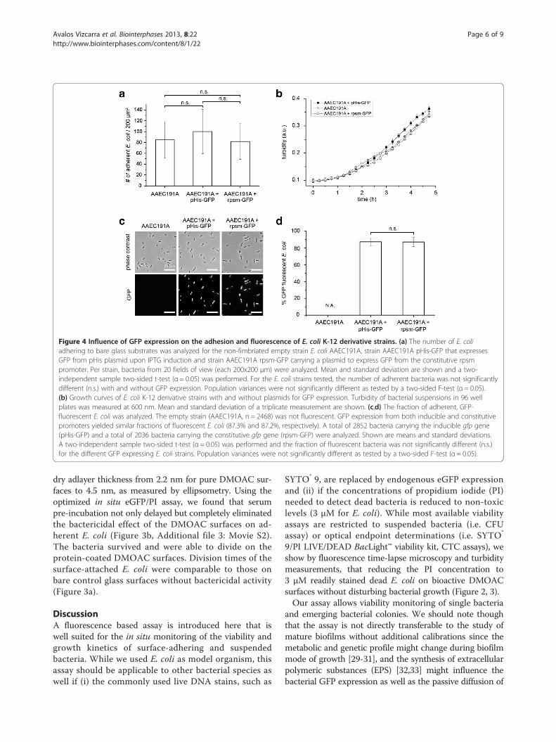
Figure 4 Influence of GFP expression on the adhesion and fluorescence of E. coli K-12 derivative strains. (a) The number of E. coliadhering to bare glass substrates was analyzed for the non-fimbriated empty strain E. coli AAEC191A, strain AAEC191A pHis-GFP that expressesGFP from pHis plasmid upon IPTG induction and strain AAEC191A rpsm-GFP carrying a plasmid to express GFP from the constitutive rpsmpromoter. Per strain, bacteria from 20 fields of view (each 200x200 μm) were analyzed. Mean and standard deviation are shown and a two-independent sample two-sided t-test (α = 0.05) was performed. For the E. coli strains tested, the number of adherent bacteria was not significantlydifferent (n.s.) with and without GFP expression. Population variances were not significantly different as tested by a two-sided F-test (α = 0.05).(b) Growth curves of E. coli K-12 derivative strains with and without plasmids for GFP expression. Turbidity of bacterial suspensions in 96 wellplates was measured at 600 nm. Mean and standard deviation of a triplicate measurement are shown. (c,d) The fraction of adherent, GFP-fluorescent E. coli was analyzed. The empty strain (AAEC191A, n = 2468) was not fluorescent. GFP expression from both inducible and constitutivepromoters yielded similar fractions of fluorescent E. coli (87.3% and 87.2%, respectively). A total of 2852 bacteria carrying the inducible gfp gene(pHis-GFP) and a total of 2036 bacteria carrying the constitutive gfp gene (rpsm-GFP) were analyzed. Shown are means and standard deviations.A two-independent sample two-sided t-test (α = 0.05) was performed and the fraction of fluorescent bacteria was not significantly different (n.s.)for the different GFP expressing E. coli strains. Population variances were not significantly different as tested by a two-sided F-test (α = 0.05).
Avalos Vizcarra et al. Biointerphases 2013, 8:22 Page 6 of 9http://www.biointerphases.com/content/8/1/22
dry adlayer thickness from 2.2 nm for pure DMOAC sur-faces to 4.5 nm, as measured by ellipsometry. Using theoptimized in situ eGFP/PI assay, we found that serumpre-incubation not only delayed but completely eliminatedthe bactericidal effect of the DMOAC surfaces on ad-herent E. coli (Figure 3b, Additional file 3: Movie S2).The bacteria survived and were able to divide on theprotein-coated DMOAC surfaces. Division times of thesurface-attached E. coli were comparable to those onbare control glass surfaces without bactericidal activity(Figure 3a).
DiscussionA fluorescence based assay is introduced here that iswell suited for the in situ monitoring of the viability andgrowth kinetics of surface-adhering and suspendedbacteria. While we used E. coli as model organism, thisassay should be applicable to other bacterial species aswell if (i) the commonly used live DNA stains, such as
SYTO® 9, are replaced by endogenous eGFP expressionand (ii) if the concentrations of propidium iodide (PI)needed to detect dead bacteria is reduced to non-toxiclevels (3 μM for E. coli). While most available viabilityassays are restricted to suspended bacteria (i.e. CFUassay) or optical endpoint determinations (i.e. SYTO®
9/PI LIVE/DEAD BacLight™ viability kit, CTC assays), weshow by fluorescence time-lapse microscopy and turbiditymeasurements, that reducing the PI concentration to3 μM readily stained dead E. coli on bioactive DMOACsurfaces without disturbing bacterial growth (Figure 2, 3).Our assay allows viability monitoring of single bacteria
and emerging bacterial colonies. We should note thoughthat the assay is not directly transferable to the study ofmature biofilms without additional calibrations since themetabolic and genetic profile might change during biofilmmode of growth [29-31], and the synthesis of extracellularpolymeric substances (EPS) [32,33] might influence thebacterial GFP expression as well as the passive diffusion of

Avalos Vizcarra et al. Biointerphases 2013, 8:22 Page 7 of 9http://www.biointerphases.com/content/8/1/22
PI through the biofilm matrix. Furthermore, detection ofsingle bacteria within a dense three-dimensional biofilmmatrix by epifluorescence microscopy might be challenging.However, since bacterial surface colonization startswith the adhesion of individual bacteria, the presentedassay provides a versatile new tool for high spatial andtemporal evaluation of bacterial viability on engineeredsurface coatings. The assay thus adds to the previouslyreported eGFP/PI flow cytometry assay that was limited toviability determination of suspended bacteria [21] and tothe eGFP/PI endpoint viability study of groundwater E.coli [34].Evaluating bacterial viability on the test surface omits
the extraction of the adherent bacteria as required forsolution based assays, e.g. CFU counts. Therefore, test-ing the bacterial viability on the substrate might increasethe reliability of the assay since extraction is commonlyachieved by ultrasonication or harsh washing procedures,both of which can harm bacteria. Furthermore, all CFUassays require prolonged incubation times for colonygrowth and are thus not applicable for real-time viabilitymonitoring. As an alternative to extraction, an agar sand-wich assay has been suggested to determine the viabilityof surface-adherent bacteria [35]. This method, however,is prone to errors, since each transferred bacteriumwill grow into a colony that in turn might overlap withcolonies nearby. As an alternative direct optical viabilityassay of surface-adherent bacteria, the respiratory potentialof bacteria can be monitored using 5-cyano-2,3-ditolyltetrazolium chloride (CTC) [17]. The drawback however isthat the CTC stain cannot be used for real-time monitoringsince CTC disrupts the respiratory chain and is toxic tobacteria. This makes the CTC assay only suitable for endpoint determinations. We compared the performance ofour eGFP/PI assay to the well-established SYTO® 9/PIendpoint dual staining assay and found identical detectionefficiencies of dead E. coli (Additional file 1: Figure S2).The SYTO® 9/PI assay itself has been extensively comparedto the above-mentioned viability tests [17,19,21,26,36] andshowed comparative results to the solution based CFUassay as well as to other microscopy based endpoint viabil-ity protocols including the CTC assay. The added advan-tage of our assay is the ability to monitor the viability ofadherent bacteria in real-time.For each bacterial strain and species, one needs to
optimize the PI concentrations to keep the bacteria viable,as done here for Escherichia coli (E. coli) K-12 MG1655. E.coli K-12 derivatives have been widely used as model strainsin surface adhesion and biofilm studies [4,31,37-39]. Weexpressed eGFP from the pHis plasmid under the controlof the IPTG inducible tac promoter to replace the growthinhibiting DNA stain SYTO® 9 as live bacterial marker. Toexclude negative effects of GFP expression as well as IPTGand antibiotic addition on bacterial viability and adhesion,
we compared the adhesion properties and growth kineticsof the K-12 AAEC191A background strain [24] to aconstitutive eGFP expressing strain (rpsm-GFP) and theIPTG inducible pHis-GFP strain (Figure 4a,b). No signifi-cant difference in adhesion and growth was observed.Furthermore, no significant difference in the fraction ofGFP-expressing bacteria was found for the eGFP expressionunder the constitutive and inducible promoter used in thisstudy (Figure 4c,d). However, even non-toxic gene productslike GFP can have detrimental effects on bacteria whenoverexpressed [40], since overexpression of an introducedgene requires a lot of resources and thus might disorganizethe bacterial metabolism. Therefore, the inducible IPTG-based expression system, as compared to constitutiveexpression systems, allows for a control of GFP expressionand guarantees a balance between fluorescent and healthybacteria (Figure 4c,d). Since eGFP is a very stable protein[41,42] new GFP variants with reduced half-lives, e.g. GFP(LVA), have been suggested to study transient gene ex-pression in bacteria [43]. The enzymatic degradation ofunstable GFP(LVA) however, requires a metabolicallyactive viable bacterium (Additional file 1: Figure S3). Thus,the use of GFP(LVA) variants does not improve our assayto limit false-positive detection of dead bacteria.For live cell imaging, GFP expression in bacteria is
used extensively and expression systems are availablefor many different bacterial species, including clinicallyrelevant strains of Salmonella, Streptococcus, Listeriamonocytogenes, Pseudomonas aeruginosa, Staphylococcusaureus and Escherichia coli O157:H7 [41,44-47]. Plasmidbased gene expression is a well-established and long-usedmethod in microbiology. Handling of plasmids is usuallyeasy and versatile and genes, promoters or selectionmarkers can quickly be exchanged and adapted to needs.Compared to plasmids, chromosomal insertions are morecomplicated and cannot be adapted as easily. Dependingon the insertion method used, the gene of interest isinserted in the chromosome at a random location andmight disrupt an important chromosomal gene. As forplasmids, selection markers like antibiotic resistances arecommonly used for chromosomal insertions, too [48].Furthermore, chromosomal insertions usually result in theinsertion of a single copy of the gene into the chromosome.The pHis plasmid used in this study carries a ColE1-likereplicator and occurs at nearly 20 copies per bacterium[49], each providing the gene of interest. This results in verystable and usually higher expression levels of the gene ofinterest (here eGFP) than with chromosomal insertions.Plasmids with ColE1-like promoters are stably inheritedeven without the presence of the corresponding selectionagent [50,51], long-term experiments with GFP expressionfrom a plasmid rather than from a chromosomal insertionare feasible for more than a few hours. For other bacterialspecies and expression systems, the IPTG level and the

Avalos Vizcarra et al. Biointerphases 2013, 8:22 Page 8 of 9http://www.biointerphases.com/content/8/1/22
concentration of the antibiotic selection marker should bere-evaluated to assure a stable GFP expression withoutdisturbance of bacterial growth.Finally, we applied our viability assay to highlight the
impact of protein adsorption on the antimicrobial activityof engineered DMOAC surfaces. Upon incubation of theDMOAC surfaces with protein-rich fetal bovine serum,bacterial growth on the otherwise bactericidal surfacewas possible, indicating that the protein layer on topblocked the bioactive quaternary ammonium groups ofthe DMOAC coating (Figure 3b). The growth rate of thesurface-attached E. coli on the control glass and serum-coated DMOAC substrates were identical, illustratingthat the design rules for antimicrobial coatings primarilyhave to be tuned to prevent both, bacterial and proteinadsorption since additional bioactive modifications can belost when the biomaterial gets in contact with protein-rich(host) fluids.
ConclusionsIn conclusion, we show that the eGFP/PI assay is suitedto study the antimicrobial properties of (bio-) materialsurface coatings under physiological conditions in realtime and with single-bacterium sensitivity. This was sofar not possible with the widely used solution based assays(i.e. CFU) or endpoint dual staining protocols (i.e. LIVE/DEAD BacLight™ viability kit, CTC). Possible applicationsfor the assay include studies of bacterial fitness and patho-genicity on biomaterial surfaces using live cell imaging ofbacteria as additional readout. While we calibrated andillustrated the advantages of the assay for E. coli, other PIconcentrations might have to be employed to optimizethe kinetic viability monitoring of other bacterial species.While conventional bacteria viability assays allow for fastendpoint checks without requiring genetic modifications,the eGFP/PI assay presented here constitutes a viabilitytest procedure that requires only one sample and its repli-cates per time series and is particularly suited for kineticstudies.
Additional files
Additional file 1: Figure S1. Protein coating reconstitutes bacterialgrowth on bioactive DMOAC surfaces as measured by an increase inbacterial surface coverage. The growth kinetics of 5–15 surface attachedbacteria were analyzed and averaged for each condition. Error barsrepresent the standard deviation. Figure S2: The eGFP/PI and the SYTO®
9/PI dual staining assays yield identical detection efficiencies of E. coliviability on bioactive DMOAC surfaces with fast bacteria deactivationkinetics (complete bacterial killing within 1 h incubation). Figure S3:Enzymatic degradation of GFP variants with different stability. All E. colistrains express GFP from plasmid pHis under control of the inducible tacpromoter. At time point 0 h GFP expression was stopped by removingthe IPTG inducer. E. coli expressing the stable eGFP variant showed thehighest fluorescence intensity and nearly no degradation within 4.5 h.Strains that expressed the unstable GFP(LVA) variant exhibited aninherent lower fluorescence intensity from the start, as the unstable GFP
(LVA) was constantly being degraded by innate E. coli proteases. WhenGFP(LVA) expression was stopped by IPTG removal and the culture ismaintained at 37°C, the GFP fluorescence decreased rapidly, indicatingthat the GFP(LVA) is degraded enzymatically. If the culture was kept at 0°C after IPTG removal, no degradation of the GFP(LVA) was observed. Allmeasurements were performed in M9 minimal medium. 1 ml sampleswere drawn at each time point and measured with a Perkin Elmerspectrophotometer. OD600 of all cultures at 0 h was set to 1.
Additional file 2: Movie S1. DMOAC-coated bioactive surfaces showfast killing kinetics of surface-attached E. coli bacteria. Time-lapse data offluorescent images of surface-bound E. coli bacteria (green: eGFP signal,red: PI signal) is shown.
Additional file 3: Movie S2. Incubation of serum with DMOAC-coatedbioactive surfaces eliminates bioactive effect of DMOAC surfaces andrescues E. coli viability and growth. Time-lapse data of fluorescent imagesof surface-bound E. coli bacteria (green: eGFP signal, red: PI signal) isshown.
Competing interestsThe authors declare that they have no competing interests.
Authors’ contributionsIAV, JM, MC, RK and VV designed research, PE provided eGFP expressingbacteria, PM developed semiautomatic image analysis software, IAV and JMperformed research, IAV, PE, PM and JM analyzed data, IAV, PE, RK, W and JMwrote the paper. All authors read and approved the final manuscript.
AcknowledgementsThe authors thank Prof. Evgeni Sokurenko (University of Washington, Seattle,USA) for providing the E.coli AAEC191A strain and Dr. Peter Kaiser for helpon Metamorph journals. We thank Mohammad Divandari and Dr. EdmondoM. Benetti for assistance with ellipsometry measurements and MarkusArnoldini and Prof. Martin Ackermann for providing the MG1655 rpsm-GFPstrain. We thank Prof. Marcus Textor for valuable discussions during thedesign of the assay. Funding from BASF SE, Ludwigshafen, Germany and ETHZurich (V.V.) is gratefully acknowledged.
Author details1Department of Health Sciences and Technology, Laboratory of AppliedMechanobiology, ETH Zurich, CH-8093 Zurich, Switzerland. 2FraunhoferInstitute for Manufacturing Engineering and Automation IPA, Nobelstraße 12,70569 Stuttgart, Germany. 3BASF SE, Advanced Materials and SystemsResearch, D-67056 Ludwigshafen, Germany.
Received: 17 June 2013 Accepted: 16 August 2013Published: 21 August 2013
References1. Costerton JW, Stewart PS, Greenberg EP (1999) Bacterial biofilms: a common
cause of persistent infections. Science 284:1318–13222. Neoh KG, Kang ET (2011) Combating bacterial colonization on metals via
polymer coatings: relevance to marine and medical applications. ACS ApplMater Interfaces 3:2808–2819
3. Charnley M, Textor M, Acikgoz C (2011) Designed polymer structures withantifouling-antimicrobial properties. React Funct Polym 71:329–334
4. Pidhatika B, Möller J, Benetti EM, Konradi R, Rakhmatullina E, Mühlebach A,Zimmermann R, Werner C, Vogel V, Textor M (2010) The role of theinterplay between polymer architecture and bacterial surface properties onthe microbial adhesion to polyoxazoline-based ultrathin films. Biomaterials31:9462–9472
5. Li P, Poon YF, Li W, Zhu H-Y, Yeap SH, Cao Y, Qi X, Zhou C, Lamrani M,Beuerman RW, Kang E-T, Mu Y, Li CM, Chang MW, Leong SSJ, Chan-Park MB(2011) A polycationic antimicrobial and biocompatible hydrogel withmicrobe membrane suctioning ability. Nature Mater 10:149–156
6. Kang S, Pinault M, Pfefferle LD, Elimelech M (2007) Single-walled carbonnanotubes exhibit strong antimicrobial activity. Langmuir 23:8670–8673
7. Parreira P, Magalhães A, Gonçalves IC, Gomes J, Vidal R, Reis CA, LeckbandDE, Martins MCL (2011) Effect of surface chemistry on bacterial adhesion,viability, and morphology. J Biomed Mater Res, Part A 99:344–353

Avalos Vizcarra et al. Biointerphases 2013, 8:22 Page 9 of 9http://www.biointerphases.com/content/8/1/22
8. Schiffman JD, Elimelech M (2011) Antibacterial activity of electrospunpolymer mats with incorporated narrow diameter single-walled carbonnanotubes. ACS Appl Mater Interfaces 3:462–468
9. Klibanov AM (2007) Permanently microbicidal materials coatings. J MaterChem 17:2479–2482
10. Kenawy E-R, Worley SD, Broughton R (2007) The chemistry and applicationsof antimicrobial polymers: A state-of-the-art review. Biomacromolecules8:1359–1384
11. Postgate JR (1969) Viable counts and Viability. Method Microbiol 1:611–62812. Davey HM (2011) Life, death, and in-between: Meanings and methods in
microbiology. Appl Environ Microbiol 77:5571–557613. Kaprelyants AS, Gottschal JC, Kell DB (1993) Dormancy in non-sporulating
bacteria. FEMS Microbiol Rev 10:271–28514. Sträuber H, Müller S (2010) Viability states of bacteria–specific mechanisms
of selected probes. Cytom Part A 77:623–63415. Kennedy D, Cronin UP, Wilkinson MG (2011) Responses of Escherichia coli,
Listeria monocytogenes, and Staphylococcus aureus to simulated foodprocessing treatments, determined using fluorescence-activated cell sortingand plate counting. Appl Environ Microbiol 77:4657–4668
16. Choi O, Deng KK, Kim N-J, Ross L, Surampalli RY, Hu Z (2008) The inhibitoryeffects of silver nanoparticles, silver ions, and silver chloride colloids onmicrobial growth. Water Res 42:3066–3074
17. Asadishad B, Ghoshal S, Tufenkji N (2011) Method for the direct observationand quantification of survival of bacteria attached to negatively or positivelycharged surfaces in an aqueous medium. Environ Sci Technol 45:8345–8351
18. Deligeorgiev TG, Kaloyanova S (2009) Intercalating cyanine dyes for nucleicacid detection. Recent Pat Mater Sci 2:1–26
19. Berney M, Hammes F, Bosshard F, Weilenmann H-U, Egli T (2007)Assessment and interpretation of bacterial viability by using the LIVE/DEADBacLight Kit in combination with flow cytometry. Appl Environ Microbiol73:3283–3290
20. Stocks SM (2004) Mechanism and use of the commercially available viabilitystain, BacLight. Cytom Part A 61:189–195
21. Lehtinen J, Nuutila J, Lilius E-M (2004) Green fluorescent protein-propidiumiodide (GFP-PI) based assay for flow cytometric measurement of bacterialviability. Cytom Part A 60:165–172
22. Xie X, Möller J, Konradi R, Kisielow M, Franco-Obregón A, Nyfeler E,Mühlebach A, Chabria M, Textor M, Lu Z, Reimhult E (2011) Automatedtime-resolved analysis of bacteria-substrate interactions using functionalizedmicroparticles and flow cytometry. Biomaterials 32:4347–4357
23. Green J-BD, Bickner S, Carter PW, Fulghum T, Luebke M, Nordhaus MA,Strathmann S (2010) Antimicrobial testing for surface-immobilized agents witha surface-separated live-dead staining method. Biotechnol Bioeng 108:231–236
24. Blomfield IC, McClain MS, Eisenstein BI (1991) Type 1 fimbriae mutants ofEscherichia coli K12: characterization of recognized afimbriate strains andconstruction of new fim deletion mutants. Mol Microbiol 5:1439–1445
25. Zaslaver A, Bren A, Ronen M, Itzkovitz S, Kikoin I, Shavit S, Liebermeister W,Surette MG, Alon U (2006) A comprehensive library of fluorescenttranscriptional reporters for Escherichia coli. Nat Methods 3:623–628
26. Boulos L, Prévost M, Barbeau B, Coallier J, Desjardins R (1999) LIVE/DEADBacLight : application of a new rapid staining method for directenumeration of viable and total bacteria in drinking water. J MicrobiolMethods 37:77–86
27. Isquith AJ, Abbott EA, Walters PA (1972) Surface-bonded antimicrobialactivity of an organosilicon quaternary ammonium chloride. Appl Microbiol24:859–863
28. Pei J, Hall H, Spencer ND (2011) The role of plasma proteins in cell adhesionto PEG surface-density-gradient-modified titanium oxide. Biomaterials32:8968–8978
29. Vlamakis H, Aguilar C, Losick R, Kolter R (2008) Control of cell fate by theformation of an architecturally complex bacterial community. Genes Dev22:945–953
30. Resch A, Rosenstein R, Nerz C, Götz F (2005) Differential gene expressionprofiling of Staphylococcus aureus cultivated under biofilm and planktonicconditions. Appl Environ Microbiol 71:2663–2676
31. Beloin C, Valle J, Latour-Lambert P, Faure P, Kzreminski M, Balestrino D,Haagensen JAJ, Molin S, Prensier G, Arbeille B, Ghigo J-M (2004) Globalimpact of mature biofilm lifestyle on Escherichia coli K-12 gene expression.Mol Microbiol 51:659–674
32. Branda SS, Vik S, Friedman L, Kolter R (2005) Biofilms: the matrix revisited.Trends Microbiol 13:20–26
33. Flemming H-C, Wingender J (2010) The biofilm matrix. Nat Rev Microbiol8:623–633
34. Banning N, Toze S, Mee BJ (2002) Escherichia coli survival in groundwaterand effluent measured using a combination of propidium iodide and thegreen fluorescent protein. J Appl Microbiol 93:69–76
35. Tiller JC, Liao CJ, Lewis K, Klibanov AM (2001) Designing surfaces that killbacteria on contact. PNAS 98:5981–5985
36. Benoit MR, Conant CG, Ionescu-Zanetti C, Schwartz M, Matin A (2010) Newdevice for high-throughput viability screening of flow biofilms. Appl EnvironMicrobiol 76:4136–4142
37. Reisner A, Krogfelt KA, Klein BM, Zechner EL, Molin S (2006) In vitro biofilmformation of commensal and pathogenic Escherichia coli strains: impact ofenvironmental and genetic factors. J Bacteriol 188:3572–3581
38. Anderson BN, Ding AM, Nilsson LM, Kusuma K, Tchesnokova V, Vogel V,Sokurenko EV, Thomas WE (2007) Weak rolling adhesion enhances bacterialsurface colonization. J Bacteriol 189:1794–1802
39. Rodrigues DF, Elimelech M (2009) Role of type 1 fimbriae and mannose inthe development of Escherichia coli K12 biofilm: from initial cell adhesion tobiofilm formation. Biofouling 25:401–411
40. Miroux B, Walker JE (1996) Over-production of proteins in Escherichia coli:Mutant hosts that allow synthesis of some membrane proteins and globularproteins at high levels. J Mol Biol 260:289–298
41. Tombolini R, Unge A, Davey ME, Bruijn FJ, Jansson JK (1997) Flowcytometric and microscopic analysis of GFP‐tagged Pseudomonasfluorescens bacteria. FEMS Microbiol Ecol 22:17–28
42. Tsien RY (1998) The green fluorescent protein. Annu Rev Biochem 67:509–54443. Andersen JB, Sternberg C, Poulsen LK, Bjorn SP, Givskov M, Molin S (1998)
New unstable variants of green fluorescent protein for studies of transientgene expression in bacteria. Appl Environ Microbiol 64:2240–2246
44. Aspiras MB, Kazmerzak KM, Kolenbrander PE, McNab R, Hardegen N,Jenkinson HF (2000) Expression of green fluorescent protein inStreptococcus gordonii DL1 and its use as a species-specific marker incoadhesion with Streptococcus oralis 34 in saliva-conditioned biofilmsin vitro. Appl Environ Microbiol 66:4074–4083
45. Lenz AP, Williamson KS, Pitts B, Stewart PS, Franklin MJ (2008) Localizedgene expression in Pseudomonas aeruginosa biofilms. Appl EnvironMicrobiol 74:4463–4471
46. Malone CL, Boles BR, Lauderdale KJ, Thoendel M, Kavanaugh JS, Horswill AR(2009) Fluorescent reporters for Staphylococcus aureus. J Microbiol Methods77:251–260
47. Ma L, Zhang G, Doyle MP (2011) Green Fluorescent Protein labeling ofListeria, Salmonella, and Escherichia coli O157:H7 for safety-related studies.PLoS One 6:e18083
48. Berg CM, Berg DE (1996) Transposable element tools for microbial genetics.In: A B, Curtiss I, R, Kasper JB, Karp PD, Neidhardt FC, Nyström T, Slauch JM,Squires CL, Ussery D (ed) EcoSal - Escherichia coli and Salmonella: Cellularand Molecular Biology. ASM Press, Washington, DC, http://www.ecosal.org
49. Kües U, Stahl U (1989) Replication of plasmids in gram-negative bacteria.Microbiol Rev 53:491–516
50. Summers DK, Sherratt DJ (1985) Bacterial plasmid stability. Bioessays2:209–211
51. Ayala-Sanmartín J, Gómez-Eichelmann MC (1989) Stability of ColE1-like andpBR322-like plasmids in Escherichia coli. Mol Microbiol 3:1745–1752
doi:10.1186/1559-4106-8-22Cite this article as: Avalos Vizcarra et al.: Fluorescence-based in situ assayto probe the viability and growth kinetics of surface-adhering andsuspended recombinant bacteria. Biointerphases 2013 8:22.
Related Documents