Research Article Fluorescein Leakage within Recent Subretinal Hemorrhage in Pathologic Myopia: Suggestive of CNV? LanMi , 1 Chengguo Zuo , 1 Xiongze Zhang , 1 Bing Liu , 1 Yuting Peng , 2 andFengWen 1 1 State Key Laboratory of Ophthalmology, Zhongshan Ophthalmic Center, Sun Yat-sen University, Guangzhou 510060, China 2 Department of Ophthalmology, Guangdong Provincial Hospital of Traditional Chinese Medicine, Guangzhou 510060, China Correspondence should be addressed to Feng Wen; [email protected] Received 3 April 2018; Accepted 23 May 2018; Published 13 August 2018 Academic Editor: Siamak Ansari-Shahrezaei Copyright © 2018 Lan Mi et al. is is an open access article distributed under the Creative Commons Attribution License, which permits unrestricted use, distribution, and reproduction in any medium, provided the original work is properly cited. Purpose. To determine whether fluorescein leakage within subretinal hemorrhage is definitely suggestive of choroidal neo- vascularization (CNV) by multimodal imaging including optical coherence tomography angiography (OCTA). Methods. Twenty- five consecutive highly myopic patients (25 eyes) with fluorescein leakage within subretinal hemorrhage detected within 1 month were prospectively included. All patients underwent OCTA and spectral-domain optical coherence tomography (SD-OCT). e OCTA and SD-OCT findings at the site of fluorescein leakage were analyzed. In cases of a doubtful diagnosis, indocyanine green angiography (ICGA) was also performed to differentiate myopic CNV from lacquer crack if necessary; all patients were followed up by SD-OCTand/or OCTA for at least 2 weeks. Results. In terms of the site of fluorescein leakage, OCTA revealed an abnormal vascular network in the outer retina and a choriocapillaris slab in 22 out of 25 eyes (88%), which were confirmed to be CNV. However, no high-flow signal was observed in 3 of 25 eyes (12%). In these 3 cases, SD-OCTshowed a focal rupture of the retinal pigment epithelium-Bruch’s membrane-choriocapillaris (RPE-BM-CC) complex and a columnar hyperreflective signal of blood originating from defects with a volcanic geyser-like appearance, and no exudative signs were detected. Notably, all ruptures of the RPE-BM-CC complex were located exactly at lacquer crack sites. Moreover, with the absorption of subretinal hemorrhage, ruptures of the RPE-BM-CC complex spontaneously resolved without any intervention. Considering the multimodal imaging appearance and follow-up outcomes, these 3 eyes were eventually diagnosed as simple bleeding associated with lacquer cracks. Conclusions. Dye leakage within recent subretinal hemorrhage on FA could be caused by new-onset lacquer cracks in pathologic myopia. Multimodal imaging including OCTA is helpful to differentiate lacquer cracks from myopic CNV. 1.Introduction Pathologic myopia is defined by an axial length of the eye greater than 26 mm or a refractive error of at least −6diopters (D) and is associated with complications of the posterior segment secondary to excessive elongation of axial length. Myopic maculopathy, such as diffuse and patchy chorior- etinal atrophy, lacquer cracks, myopic choroidal neo- vascularization (CNV), myopic subretinal hemorrhage (mSH), and posterior staphyloma, has been reported to be a significant cause of visual impairment and legal blindness worldwide, especially in Asian countries [1–4]. Subretinal hemorrhage is frequently seen with recent myopic CNV but is also a common finding in cases of new-onset lacquer cracks [5–7]. Myopic CNV is the most common central vision-threatening complication in patients with high myopia, affecting 5% to 11% of cases [8]. Once confirmed as active CNV, timely treatment such as anti-VEGF therapy is recommended. However, mSH associated with lacquer cracks, which is also called simple bleeding, generally has a better prognosis. erefore, the differentiation and diagnosis of these two types of subretinal hemorrhage are crucial. Fluorescein angiography (FA) is an essential method for the diagnosis of myopic CNV. Recent myopic CNV usually displays well-defined hyperfluorescence in the early phase with dye leakage in the late phase [5]. However, simple bleeding associated with lacquer cracks is generally thought to display blocked fluorescence. In our clinical work, an Hindawi Journal of Ophthalmology Volume 2018, Article ID 4707832, 7 pages https://doi.org/10.1155/2018/4707832

Welcome message from author
This document is posted to help you gain knowledge. Please leave a comment to let me know what you think about it! Share it to your friends and learn new things together.
Transcript

Research ArticleFluorescein Leakage within Recent Subretinal Hemorrhage inPathologic Myopia: Suggestive of CNV?
Lan Mi ,1 Chengguo Zuo ,1 Xiongze Zhang ,1 Bing Liu ,1 Yuting Peng ,2
and Feng Wen 1
1State Key Laboratory of Ophthalmology, Zhongshan Ophthalmic Center, Sun Yat-sen University, Guangzhou 510060, China2Department of Ophthalmology, Guangdong Provincial Hospital of Traditional Chinese Medicine, Guangzhou 510060, China
Correspondence should be addressed to Feng Wen; [email protected]
Received 3 April 2018; Accepted 23 May 2018; Published 13 August 2018
Academic Editor: Siamak Ansari-Shahrezaei
Copyright © 2018 LanMi et al.-is is an open access article distributed under the Creative Commons Attribution License, whichpermits unrestricted use, distribution, and reproduction in any medium, provided the original work is properly cited.
Purpose. To determine whether fluorescein leakage within subretinal hemorrhage is definitely suggestive of choroidal neo-vascularization (CNV) by multimodal imaging including optical coherence tomography angiography (OCTA).Methods. Twenty-five consecutive highly myopic patients (25 eyes) with fluorescein leakage within subretinal hemorrhage detected within 1 monthwere prospectively included. All patients underwent OCTA and spectral-domain optical coherence tomography (SD-OCT). -eOCTA and SD-OCT findings at the site of fluorescein leakage were analyzed. In cases of a doubtful diagnosis, indocyanine greenangiography (ICGA) was also performed to differentiate myopic CNV from lacquer crack if necessary; all patients were followedup by SD-OCTand/or OCTA for at least 2 weeks. Results. In terms of the site of fluorescein leakage, OCTA revealed an abnormalvascular network in the outer retina and a choriocapillaris slab in 22 out of 25 eyes (88%), which were confirmed to be CNV.However, no high-flow signal was observed in 3 of 25 eyes (12%). In these 3 cases, SD-OCT showed a focal rupture of the retinalpigment epithelium-Bruch’s membrane-choriocapillaris (RPE-BM-CC) complex and a columnar hyperreflective signal of bloodoriginating from defects with a volcanic geyser-like appearance, and no exudative signs were detected. Notably, all ruptures of theRPE-BM-CC complex were located exactly at lacquer crack sites. Moreover, with the absorption of subretinal hemorrhage,ruptures of the RPE-BM-CC complex spontaneously resolved without any intervention. Considering the multimodal imagingappearance and follow-up outcomes, these 3 eyes were eventually diagnosed as simple bleeding associated with lacquer cracks.Conclusions. Dye leakage within recent subretinal hemorrhage on FA could be caused by new-onset lacquer cracks in pathologicmyopia. Multimodal imaging including OCTA is helpful to differentiate lacquer cracks from myopic CNV.
1. Introduction
Pathologic myopia is defined by an axial length of the eyegreater than 26mm or a refractive error of at least −6diopters(D) and is associated with complications of the posteriorsegment secondary to excessive elongation of axial length.Myopic maculopathy, such as diffuse and patchy chorior-etinal atrophy, lacquer cracks, myopic choroidal neo-vascularization (CNV), myopic subretinal hemorrhage(mSH), and posterior staphyloma, has been reported to bea significant cause of visual impairment and legal blindnessworldwide, especially in Asian countries [1–4]. Subretinalhemorrhage is frequently seen with recent myopic CNV butis also a common finding in cases of new-onset lacquer
cracks [5–7]. Myopic CNV is the most common centralvision-threatening complication in patients with highmyopia, affecting 5% to 11% of cases [8]. Once confirmed asactive CNV, timely treatment such as anti-VEGF therapy isrecommended. However, mSH associated with lacquer cracks,which is also called simple bleeding, generally has a betterprognosis. -erefore, the differentiation and diagnosis ofthese two types of subretinal hemorrhage are crucial.
Fluorescein angiography (FA) is an essential method forthe diagnosis of myopic CNV. Recent myopic CNV usuallydisplays well-defined hyperfluorescence in the early phasewith dye leakage in the late phase [5]. However, simplebleeding associated with lacquer cracks is generally thoughtto display blocked fluorescence. In our clinical work, an
HindawiJournal of OphthalmologyVolume 2018, Article ID 4707832, 7 pageshttps://doi.org/10.1155/2018/4707832

incidental case of pathologic myopia with new-onset extra-foveal hemorrhage attracted our attention (SupplementaryMaterials (available here), which illustrates this case; https://figshare.com/s/7dcf53004450e72f209b). SD-OCT revealeda focal rupture of the retinal pigment epithelium-Bruch’smembrane-choriocapillaris (RPE-BM-CC) complex un-derlying the hemorrhage without an exudative sign, whichcorresponded to the site of lacquer crack confirmed byindocyanine green angiography (ICGA), while FA presentedmild fluorescein leakage within the hemorrhage. Notably, withthe absorption of subretinal hemorrhage, the ruptured RPE-BM-CC complex completely resolved spontaneously. In viewof multimodal imaging features and outcome, this case waspresumed to be simple hemorrhage associated with lacquercrack. We were curious about fluorescein leakage caused bylacquer cracks. As proposed by Klein and Curtin [9], if lacquercracks are indeed defects in the RPE-BM-CC complex, leakageof fluorescein from the choriocapillaris may be expected. In-deed, previous studies have reported discontinuities in theRPE-BM-CC complex at the site of lacquer cracks on spectraldomain-optical coherence tomography (SD-OCT) [10, 11];however, until very recently, no previous studies have reporteddye leakage associated with lacquer cracks on FA.
-e primary purpose of this study was to investigatewhether fluorescein leakage within recent subretinal hem-orrhage in pathologic myopia is suggestive of CNV.
2. Methods
-is is a prospective observational case series of patientsfrom the fundus disease clinic of the Zhongshan Oph-thalmic Center who presented between November 2016 andDecember 2017. -e protocol of this study was approved bythe Ethics Committee of the Zhongshan Ophthalmic Centerof Sun Yat-sen University, and the study was performed inaccordance with the Declaration of Helsinki.
-e inclusion criteria were as follows: (1) high myopia(refractive error≤−6D or axial length ≥26mm); (2) within 1month after the onset of ophthalmic symptoms includingacute visual loss and fixed scotoma with or without meta-morphopsia; and (3) dye leakage within subretinal hem-orrhage on FA. Fluorescein leakage was characterized byincreased size and intensity.
-e exclusion criteria were as follows: (1) mild ormoderate myopia (refractive error≥−6D); (2) clinical fea-tures of age-related maculopathy such as soft or hard drusen;(3) a history of other ocular disorders or intraocular surgery,such as multifocal choroiditis or punctate inner choroid-opathy, ocular trauma, severe cataract, retinal artery orretinal vein occlusion, diabetic retinopathy, or other retinalvascular diseases; and (4) previous ocular therapy, such aslaser photocoagulation, photodynamic therapy, or intra-vitreal injection of anti-VEGF.
Each patient underwent a comprehensive ophthalmologicexamination, including assessment of the best-corrected vi-sual acuity (BCVA) using Snellen charts, dilated slit-lampanterior segment and fundus biomicroscopy, fundus pho-tography, OCTA, and SD-OCT.-e diagnosis of CNV basedon OCTA was defined as a vascular network pattern in
outer retina and choriocapillaris slab corresponding to thesite of fluorescein leakage [5]. -e diagnosis of CNV basedon SD-OCT was defined as hyperreflective lesion contig-uous above the RPE, with or without an associated exu-dative sign, such as subretinal fluid, intraretinal fluid, andsubretinal hyperreflective exudation [5]. Corresponding tothe leakage site on FA, patients with neither abnormalhigh-flow vascular network on OCTA nor CNV appear-ance on SD-OCT were rated as a doubtful case, for which,ICGA was also performed. Furthermore, SD-OCT andOCTA were performed again within 2 weeks after theinitial clinical examination.
Photographic images of the fundus were obtained usingconfocal laser scan systems (Carl Zeiss, Inc., Jena, Germany).FA and ICGA images were obtained using confocal laserscanning systems (Spectralis, Heidelberg Engineering,Dossenheim, Germany). SD-OCT was performed with anHRA+OCT Spectralis (Spectralis, Heidelberg Engineering,Dossenheim, Germany). Horizontal and vertical SD-OCTscans of 6mm or 3mmwere centered on the lesions with dyeleakage on FA. OCTA images were obtained using theAngioVue imaging system (Optovue Inc., CA, US) with themacular cube (3× 3mm and/or HD 6× 6mm) protocol.Each OCTA volume involved in the bleeding area contained304× 304 A-scans with 2 consecutive B-scans. Automaticretinal segmentation was performed by embedded softwarein the machine. Outer retina slab is autosegmented from9 μm beneath the OPL to 9 μm above the BRM and cho-riocapillaris slab is autosegmented from 9 μm above theBRM to 31 μmbeneath the BRM. Two ophthalmologists (LMand CGZ) independently assessed the multimodal images.
3. Results
Twenty-five highly myopic eyes (25 patients) were included inthis study. -e subjects included 10 men and 15 women withamean age of 37.8± 12.1 years (range: 21–56 years).-emeanrefractive error was −12.2± 3.2D (range: −7D to −19.0D).-emean baseline BCVAwas 0.34± 0.22 (range: 0.1–1.0).-emean duration of visual symptom was 16.9± 6.8 days (range:5–29 days). Demographic and morphologic characteristicsof the study population are listed in Table 1. Relevant datahave been uploaded to figshare, the link is https://figshare.com/s/dc2237097539fcdeff4e.
Table 1: Demographic and morphologic characteristics of thestudy population.
Patient characteristics (n � 25)Age, years (range) 37.8± 12.1 (21–56)Sex (male/female) 10/15Mean refractive error, diopters(range) −12.2± 3.2D (−7.0 to −19.0D)
Right eye (n) 15Left eye (n) 10Best-corrected visual acuity(range) 0.34± 0.22 (0.1–1.0)
Duration of visual symptom,days (range) 16.9± 6.8 (5–29)
2 Journal of Ophthalmology

3.1. FA and SD-OCT Findings. Blocked fluorescein fromsubretinal hemorrhage was observed in all cases. Of these, 21hemorrhages covered the central fovea. Well-definedhyperfluorescence during the early phase with fluoresceinleakage during the late phase was detected in the bleedingareas. All patients were preliminarily diagnosed with myopicCNV based on the FA appearance. Exudative signs corre-sponding to the site of fluorescein leakage were observed onSD-OCT in 22 out of 25 eyes (88%). Of these, SD-OCTrevealed intraretinal cysts in 4 of 25 eyes (16%), subretinalfluid in 10 of 25 eyes (40%), and subretinal hyperreflectiveexudation in 17 of 25 eyes (68%).
3.2. OCTA Findings. OCTA revealed an abnormal vascularnetwork in the outer retina and the choriocapillaris slab in88% of eyes (22 of 25), whereas no high-flow signal wasobserved in 12% of eyes (3 of 25). -ose 22 eyes with classic
vascular network on OCTA were confirmed to be CNV.-elesion was visible on the outer retinal slab and less visible onthe choriocapillaris slab, and the best visualization of theCNV network corresponded to a manual segmentationmostly on the RPE (a 40 μm manual segmentation wasimplemented, and the lower boundary of this manual seg-mentation was localized on BM). A representative case isdescribed in Figure 1.
In the remaining 3 eyes with no high-flow signal onOCTA, SD-OCT revealed subretinal hemorrhage with focalrupture of the RPE-BM-CC complex. A columnar hyper-reflective signal of blood originating from ruptures witha volcanic geyser-like appearance was observed. Neitherhyperreflective lesion contiguous with the RPE nor exu-dative signs, such as subretinal fluid, intraretinal cysts, orsubretinal hyperreflective fuzzy exudation, were detected.ICGA was also performed and revealed that ruptures of theRPE-BM-CC complex captured via SD-OCT were located
(a) (b) (c)
(d) (e) (f)
Figure 1: Myopic CNV with recent subretinal hemorrhage imaged by FA, SD-OCT, and OCTA. Left eye imaging of a 44-year-old manpreliminarily diagnosed with myopic CNV by FA (refractive error: −9.0D). Fluorescein angiography showing early hyperfluorescence(a) with late leakage (b). Spectral-domain optical coherence tomography scan passing through the site of leakage revealing CNV, asevidenced by a hyperreflective lesion with the RPE with subretinal hemorrhage and slight subretinal fluid (c). Optical coherence tomographyangiography 3× 3mm images including the choriocapillaris slab (d), the outer retinal slab (e), and the RPE slab (f ). Abnormal vascularnetwork is less visible on the choriocapillaris slab (d) and is best visible on the RPE slab (f ).
Journal of Ophthalmology 3
(a)
0:27.50
(d)(b)
2:43.30
(c)
14:30.15
(e) (f) (g) (h) (i)
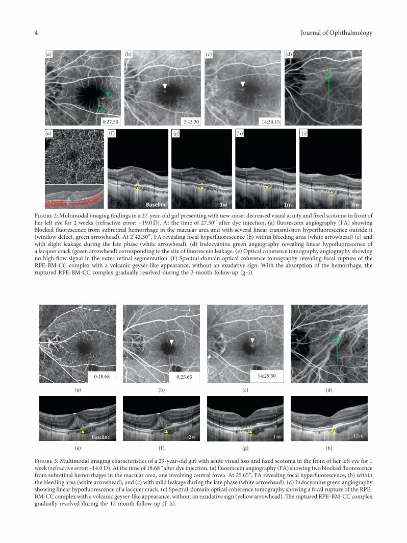
Figure 2: Multimodal imaging findings in a 27-year-old girl presenting with new-onset decreased visual acuity and fixed scotoma in front ofher left eye for 2 weeks (refractive error: −19.0D). At the time of 27.50″ after dye injection, (a) fluorescein angiography (FA) showingblocked fluorescence from subretinal hemorrhage in the macular area and with several linear transmission hyperfluorescence outside it(window defect, green arrowhead). At 2′43.30″, FA revealing focal hyperfluorescence (b) within bleeding area (white arrowhead) (c) andwith slight leakage during the late phase (white arrowhead). (d) Indocyanine green angiography revealing linear hypofluorescence ofa lacquer crack (green arrowhead) corresponding to the site of fluorescein leakage. (e) Optical coherence tomography angiography showingno high-flow signal in the outer retinal segmentation. (f ) Spectral-domain optical coherence tomography revealing focal rupture of theRPE-BM-CC complex with a volcanic geyser-like appearance, without an exudative sign. With the absorption of the hemorrhage, theruptured RPE-BM-CC complex gradually resolved during the 3-month follow-up (g–i).
0:18.68
(a)
0:25.65
(b)
14:29.50
(c) (d)
Baseline
(e)
2w
(f )
1m
(g)
12m
(h)
Figure 3: Multimodal imaging characteristics of a 29-year-old girl with acute visual loss and fixed scotoma in the front of her left eye for 1week (refractive error: –14.0 D). At the time of 18.68 “after dye injection, (a) fluorescein angiography (FA) showing two blocked fluorescencefrom subretinal hemorrhages in the macular area, one involving central fovea. At 25.65”, FA revealing focal hyperfluorescence, (b) withinthe bleeding area (white arrowhead), and (c) with mild leakage during the late phase (white arrowhead). (d) Indocyanine green angiographyshowing linear hypofluorescence of a lacquer crack. (e) Spectral-domain optical coherence tomography showing a focal rupture of the RPE-BM-CC complex with a volcanic geyser-like appearance, without an exudative sign (yellow arrowhead).-e ruptured RPE-BM-CC complexgradually resolved during the 12-month follow-up (f–h).
4 Journal of Ophthalmology

exactly at the site of the lacquer crack. -e preliminarydiagnosis of myopic CNV was disregarded; thus, nointerventional therapy was administered. Notably, withthe absorption of subretinal hemorrhage, all the rupturedRPE-BM-CC complexes appeared to spontaneously resolveduring the follow-up period of 2 to 3 months without anyinterventions. -ese 3 leakage lesions were eventuallyconsidered to be a result of the rupture of the RPE-BM-CCcomplex caused by lacquer cracks, and these cases werefinally diagnosed as simple hemorrhage. Representativecases are listed in Figures 2–4.
4. Discussion
Fluorescein angiography (FA) is an important tool foridentifying myopic CNV and for evaluating its activity.Active myopic CNV usually involves well-defined hyper-fluorescence during the early phase with dye leakage duringthe late phase, even in cases with subretinal hemorrhage,because the hemorrhage usually does not completely maskor cover the underlying CNV [5]. -erefore, patients withearly hyperfluorescence and late fluorescein leakage withinsubretinal hemorrhage are usually diagnosed with active
CNV and receive intravitreal injection of anti-VEGF inclinical practice. However, this study found that dye leakagewithin simple bleeding could also be caused by lacquercracks on FA.
In this study, 25 eyes preliminarily diagnosed as myopicCNV with subretinal hemorrhage by FA were analyzed.Twenty-two of 25 patients (88%) presented with classic CNVnetwork on OCTA. However, the preliminary diagnosis ofCNV in the remaining 3 of 25 cases (12%) was finallydisregarded. By combining the OCTA, SD-OCT, and ICGAappearance, these 3 cases were eventually diagnosed assimple bleeding associated with lacquer cracks. Moreover,the self-limiting outcomes further support the final di-agnosis. -is result demonstrated that lacquer cracks withinrecent myopic subretinal hemorrhage can also cause fluo-rescein leakage.
To date, no prior study has reported dye leakage asso-ciated with lacquer cracks on FA; previous researchers as-cribed it to a closed defect caused by an avascular barrier, suchas scar tissue. In this study, three patients with fluoresceinleakage associated with lacquer cracks were initially examinedwithin 2 weeks after the onset of visual symptoms. Apparentdefects of the RPE-BM-CC complex corresponding to lacquer
0:35.67
(a)
14:50.26
(b) (c)
Baseline
(d)
2m
(e)
Figure 4: Multimodal imaging findings in a 49-year-old highly myopic man with left eye metamorphopsia for 5 days (refractive error:−13.5D). At the time of 35.67″ after dye injection, (a) focal hyperfluorescence within extrafoveal bleeding site appearing on FA and (b) withmild fluorescein leakage during the late phase (arrowhead). (c) OCTA showing no abnormal vascular network. (e) SD-OCTshowing a focalrupture of the RPE-BM-CC complex corresponding to the site of fluorescein leakage (yellow arrowhead). (e) Continuity of the rupturedRPE-BM-CC complex resolved during a 2-month follow-up (yellow arrowhead).
Journal of Ophthalmology 5

cracks were also detected with SD-OCT. We speculate thatfluorescein leakage within simple myopic hemorrhage de-pends on the extent of the damage to the RPE-BM-CCcomplex and when the hemorrhage occurred. Perhaps,early-onset hemorrhage associated with lacquer crack com-bined with a secondary relatively large rupture of the RPE-BM-CC complex possibly results in dye leakage on FA, whichmay easily be misdiagnosed as myopic CNV and should benoted in clinical practice.
Lacquer cracks, which are a hallmark of pathologicmyopia, typically appears as yellowish linear lesions in theposterior pole of the eye and are widely considered to bemechanical breaks in the RPE-BM-CC complex secondaryto excessive axial elongation [9]. Lacquer cracks usuallypresent as window defect on FA. Newly developed lacquercracks may be seen with subretinal hemorrhage [6, 7, 12].Subretinal bleeding without CNV in pathologic myopia isalso considered to be a sign of new lacquer crack formation[7]. As proposed by Klein and Curtin [9], if lacquer cracksare indeed defects in the RPE-BM-CC complex, leakage offluorescein from the choriocapillaris might be expected.Similarly, it is also speculated that if the defect in theRPE-BM-CC complex caused by a lacquer crack is largeenough, detectable rupture of the RPE-BM-CC complex onSD-OCTmay be detected. Indeed, Hung et al. and Shinoharaet al. reported discontinuities of the RPE-BM-CC complex atthe site of lacquer cracks on SD-OCT in some high myopiceyes [10, 11].
-e clinical diagnosis of myopic CNV is usually con-firmed by FA. Diagnosis features on FA include well-definedhyperfluorescence during the early phase and a classic CNVpattern of leakage (usually mild leakage) [5, 13–15]. Levezielet al. reported that exudative features of myopic CNV aremore obvious on FA than on SD-OCTand suggested that FAshould be performed in any case of suspected new-onsetmyopic CNV [16]. Garcia-Layana et al. reported that thesensitivity of OCT performed after a diagnosis of myopicCNV was confirmed to be 97% for detecting CNV activityduring treatment [17]. Indeed, to increase the diagnosticaccuracy, it is generally recommended to perform both FAand SD-OCT in doubtful cases [18]. OCTA has proven to beparticularly valuable for the diagnosis of myopic CNV withhigh sensitivity and specificity (>90%) [19–21]. In theirstudy, Miyata et al. reported that there was no false-positivedetection of CNV by OCTA in treatment-naive patientsand even suggested that, if myopic CNV is detected byOCTA, FFA becomes unnecessary [21]. In this study, mul-timodal imaging including OCTA, SD-OCT, and ICGAexcluded the preliminary diagnosis of myopic CNV in 3 eyes,demonstrating that FA alone might not be the optimal methodfor initial CNV detection in high myopia with subretinalhemorrhage.
Our study has several limitations that need to be consid-ered. Firstly, the number of patients participated in this study isrelatively small. Secondly, all patients were from a single in-stitution; thus, a referral bias may exist. -irdly, as a clinic-based observational study, selection bias may exist. However,this study is the first to report fluorescein leakage of lacquercracks, which is easy to be misdiagnosed as active CNV.
In conclusion, we observed a series of highly myopicpatients with fluorescein leakage within recent subretinalhemorrhage and found that the ruptured RPE-BM-CC com-plex caused by lacquer cracks can also present as fluoresceinleakage, which needs to be noted in clinical practice, especiallyin cases with new-onset subretinal hemorrhage.
Data Availability
-e data used to support the findings of this study are in-cluded within the article.
Conflicts of Interest
-e authors declare that they have no conflicts of interest.
Acknowledgments
-is research was supported by the National Natural ScienceFoundation of China (NSFC) (81570831) and the FundamentalResearch Funds of State Key Laboratory of Ophthalmology.
Supplementary Materials
-e supplementary material describes multimodal images ofa highly myopic patient with fluorescein leakage caused bylacquer crack within recent subretinal hemorrhage. (Sup-plementary Materials)
References
[1] L. Chang, C. W. Pan, K. Ohno-Matsui et al., “Myopia-relatedfundus changes in Singapore adults with high myopia,”American Journal of Ophthalmology, vol. 155, no. 6, pp. 991.e1–999.e1, 2013.
[2] H. Li, P. Mitchell, E. Rochtchina, G. Burlutsky, T. Y. Wong,and J. J. Wang, “Retinal vessel caliber andmyopic retinopathy:the blue mountains eye study,” Ophthalmic Epidemiology,vol. 18, no. 6, pp. 275–280, 2011.
[3] A. Iwase, M. Araie, A. Tomidokoro, T. Yamamoto, H. Shimizu,and Y. Kitazawa, “Prevalence and causes of low vision andblindness in a Japanese adult population: the Tajimi study,”Ophthalmology, vol. 113, no. 8, pp. 1354.e1–1362.e1, 2006.
[4] L. Xu, Y. Wang, Y. Li et al., “Causes of blindness and visualimpairment in urban and rural areas in Beijing: the BeijingEye Study,” Ophthalmology, vol. 113, no. 7, pp. 1134.e1–1134.e11, 2006.
[5] K. Ohno-Matsui, Y. Ikuno, T. Y. Y. Lai, and C. M. GemmyCheung, “Diagnosis and treatment guideline for myopicchoroidal neovascularization due to pathologic myopia,”Progress in Retinal and Eye Research, vol. 63, pp. 92–106, 2017.
[6] K. Ohno-Matsui, R. Kawasaki, J. B. Jonas et al., “Internationalphotographic classification and grading system for myopicmaculopathy,” American Journal of Ophthalmology, vol. 159,no. 5, pp. 877.e7–883.e7, 2015.
[7] K. Ohno-Matsui, M. Ito, and T. Tokoro, “Subretinal bleedingwithout choroidal neovascularization in pathologic myopia. Asign of new lacquer crack formation,” Retina, vol. 16, no. 3,pp. 196–202, 1996.
[8] T. Y.Wong, A. Ferreira, R. Hughes, G. Carter, and P.Mitchell,“Epidemiology and disease burden of pathologic myopia andmyopic choroidal neovascularization: an evidence-based
6 Journal of Ophthalmology

systematic review,” American Journal of Ophthalmology,vol. 157, no. 1, pp. 9.e12–25.e12, 2014.
[9] R. M. Klein and B. J. Curtin, “Lacquer crack lesions inpathologic myopia,” American Journal of Ophthalmology,vol. 79, no. 3, pp. 386–392, 1975.
[10] K. C. Hung, M. S. Chen, C. M. Yang, S.-W. Wang, andT.-C. Ho, “Multimodal imaging of linear lesions in the fundusof pathologic myopic eyes with macular lesions,” Graefe’sArchive for Clinical and Experimental Ophthalmology,vol. 256, no. 1, pp. 71–81, 2018.
[11] K. Shinohara, M. Moriyama, N. Shimada, Y. Tanaka, andK. Ohno-Matsui, “Myopic stretch lines: linear lesions infundus of eyes with pathologic myopia that differ from lacquercracks,” Retina, vol. 34, no. 3, pp. 461–469, 2014.
[12] I. D. Ladas, M. M. Moschos, A. A. Rouvas, D. A. Karagiannis,and S. N. Kokolakis, “Lacquer crack formation after photo-dynamic therapy,” European Journal of Ophthalmology,vol. 13, no. 8, pp. 729–733, 2003.
[13] K. Neelam, C. M. Cheung, K. Ohno-Matsui, T. Y. Y. Lai, andT. Y. Wong, “Choroidal neovascularization in pathologicalmyopia,” Progress in Retinal and Eye Research, vol. 31, no. 5,pp. 495–525, 2012.
[14] G. Soubrane, “Choroidal neovascularization in pathologicmyopia: recent developments in diagnosis and treatment,”Survey of Ophthalmology, vol. 53, no. 2, pp. 121–138, 2008.
[15] W. M. Chan, M. Ohji, T. Y. Lai, D. T. L. Liu, Y. Tano, andD. S. C. Lam, “Choroidal neovascularisation in pathologicalmyopia: an update in management,” British Journal ofOphthalmology, vol. 89, no. 11, pp. 1522–1528, 2005.
[16] N. Leveziel, V. Caillaux, S. Bastuji-Garin, M. Zmuda, andE. H. Souied, “Angiographic and optical coherence tomog-raphy characteristics of recent myopic choroidal neo-vascularization,” American Journal of Ophthalmology,vol. 155, no. 5, pp. 913–919, 2013.
[17] A. Garcia-Layana, A. Salinas-Alaman, M. J. Maldonado,C. Sainz-Gomez, and A. Fernandez-Hortelano, “Optical co-herence tomography to monitor photodynamic therapy inpathological myopia,” British Journal of Ophthalmology,vol. 90, no. 5, pp. 555–558, 2006.
[18] P. Milani, A. Massacesi, S. Moschini et al., “Multimodalimaging and diagnosis of myopic choroidal neo-vascularization in Caucasians,” Clinical Ophthalmology,vol. 10, pp. 1749–1757, 2016.
[19] E. Bruyere, A. Miere, S. Y. Cohen et al., “Neovascularizationsecondary to high myopia imaged by optical coherence to-mography angiography,” Retina, vol. 37, no. 11, pp. 2095–2101, 2017.
[20] L. Querques, C. Giuffre, F. Corvi et al., “Optical coherencetomography angiography of myopic choroidal neo-vascularisation,” British Journal of Ophthalmology, vol. 101,no. 5, pp. 609–615, 2017.
[21] M. Miyata, S. Ooto, M. Hata et al., “Detection of myopicchoroidal neovascularization using optical coherence to-mography angiography,”American Journal of Ophthalmology,vol. 165, pp. 108–114, 2016.
Journal of Ophthalmology 7

Stem Cells International
Hindawiwww.hindawi.com Volume 2018
Hindawiwww.hindawi.com Volume 2018
MEDIATORSINFLAMMATION
of
EndocrinologyInternational Journal of
Hindawiwww.hindawi.com Volume 2018
Hindawiwww.hindawi.com Volume 2018
Disease Markers
Hindawiwww.hindawi.com Volume 2018
BioMed Research International
OncologyJournal of
Hindawiwww.hindawi.com Volume 2013
Hindawiwww.hindawi.com Volume 2018
Oxidative Medicine and Cellular Longevity
Hindawiwww.hindawi.com Volume 2018
PPAR Research
Hindawi Publishing Corporation http://www.hindawi.com Volume 2013Hindawiwww.hindawi.com
The Scientific World Journal
Volume 2018
Immunology ResearchHindawiwww.hindawi.com Volume 2018
Journal of
ObesityJournal of
Hindawiwww.hindawi.com Volume 2018
Hindawiwww.hindawi.com Volume 2018
Computational and Mathematical Methods in Medicine
Hindawiwww.hindawi.com Volume 2018
Behavioural Neurology
OphthalmologyJournal of
Hindawiwww.hindawi.com Volume 2018
Diabetes ResearchJournal of
Hindawiwww.hindawi.com Volume 2018
Hindawiwww.hindawi.com Volume 2018
Research and TreatmentAIDS
Hindawiwww.hindawi.com Volume 2018
Gastroenterology Research and Practice
Hindawiwww.hindawi.com Volume 2018
Parkinson’s Disease
Evidence-Based Complementary andAlternative Medicine
Volume 2018Hindawiwww.hindawi.com
Submit your manuscripts atwww.hindawi.com
Related Documents