International Journal of Clinical and Experimental Medical Sciences 2018; 4(4): 57-62 http://www.sciencepublishinggroup.com/j/ijcems doi: 10.11648/j.ijcems.20180404.11 ISSN: 2469-8024 (Print); ISSN: 2469-8032 (Online) Fixed Time and Fixed Angle External Fixation in the Treatment of Gartland Type III Supracondylar Humerous Fractures in Children Wei Cai * , Yang Wenbin, Liao Hailang, Zeng Lingyuan Department of Orthopedics, People's Hospital of Laibin, Laibin, China Email address: * Corresponding author To cite this article: Wei Cai, Yang Wenbin, Liao Hailang, Zeng Lingyuan. Open reduction Internal Fixation Combined with Fixed Time and Fixed Angle External Fixation in the Treatment of Gartland Type III Supracondylar Humerous Fractures in Children. International Journal of Clinical and Experimental Medical Sciences. Vol. 4, No. 4, 2018, pp. 57-62. doi: 10.11648/j.ijcems.20180404.11 Received: August 15, 2018; Accepted: September 14, 2018; Published: October 12, 2018 Abstract: Objective: To assess the efficacy of open reduction internal fixation (ORIF) combined with fixed-time and fixed- angle external fixation (FTFAEF) in the treatment of Gartland type III supracondylar humerous fracture (SHF) in children. Methods: Clinical data of 172 children with Gartland type III SHF from March 2012 to December 2017 were prospectively analyzed. All 172 patients were initially treated with ORIF, then 86 underwent FTFAEF for 3 weeks post-surgery (intervention group) while the remaining 86 received conventional plaster external fixation (70°-90°) for 3 weeks post-surgery (control group). The plaster casts were removed from both groups after the 3-week fixation period and functional exercise was initiated. Regular clinical and radiologic follow-ups were conducted on all patients. Range of motion (ROM) measurements and modified Hospital for Special Surgery (HSS) elbow function assessments were performed at 1 and 3 months post-surgery. Results: At 1 month post-surgery, both ROM and modified HSS scores were significantly higher in the intervention group (85.8±6.1° and 65.2±3.6°, respectively) than in the control group (62.3±5.2° and 56.6±2.1°, respectively)(both P<0.05). After 3 months, both ROM and modified HSS scores were still significantly higher in the intervention group (132.0±4.7° and 98.5±1.3°, respectively) than in the control group (107.5±24.4° and 85.0±10.3°, respectively) (both P<0.05). Furthermore, the percentage of patients scoring excellent on the HSS scale was significantly higher in the intervention group (100%) than in the control group (74.42%) (P<0.05). Conclusions: ORIF combined with FTFAEF is an efficacious approach that should be widely promoted for the treatment of Gartland type III SHF in children. Keywords: Supracondylar Humerous Fracture, Children, Open Reduction Internal Fixationn, Dysfunction, External Fixation 1. Introduction Supracondylar humerus fractures (SHF) are common elbow fractures in children, accounting for 30% - 40% of elbow fractures. In some of these severe fractures, such as Gartland type III supracondylar humerus fractures, the clinical symptoms are large displacement of the fracture end, unstable fracture end, and severe soft tissue injury around a joint, which can cause elbow-joint dysfunction if not handled properly. Therefore, such fractures require internal fixation for treatment. After the operation of the supracondylar humerus fracture, the elbow is usually immobilized by plaster cast at a flexion angle of 70° to 90° for three to four weeks. Cast immobilization at a fixed angle for a long time can cause elbow-joint dysfunction. The longer the plastercast remains, the more pronounced the elbow-joint dysfunction. Elbow organized hematoma and soft tissue contracture are the major causes of elbow-joint dysfunction following an operation of the supracondylar humerus fracture. The characteristics of soft tissue are elasticity and plasticity. In the process of repairing the elbow injury, the stress generated during the extending and bending of the elbow joint can

Welcome message from author
This document is posted to help you gain knowledge. Please leave a comment to let me know what you think about it! Share it to your friends and learn new things together.
Transcript
International Journal of Clinical and Experimental Medical Sciences 2018; 4(4): 57-62
http://www.sciencepublishinggroup.com/j/ijcems
doi: 10.11648/j.ijcems.20180404.11
ISSN: 2469-8024 (Print); ISSN: 2469-8032 (Online)
Fixed Time and Fixed Angle External Fixation in the Treatment of Gartland Type III Supracondylar Humerous Fractures in Children
Wei Cai*, Yang Wenbin, Liao Hailang, Zeng Lingyuan
Department of Orthopedics, People's Hospital of Laibin, Laibin, China
Email address:
*Corresponding author
To cite this article: Wei Cai, Yang Wenbin, Liao Hailang, Zeng Lingyuan. Open reduction Internal Fixation Combined with Fixed Time and Fixed Angle
External Fixation in the Treatment of Gartland Type III Supracondylar Humerous Fractures in Children. International Journal of Clinical
and Experimental Medical Sciences. Vol. 4, No. 4, 2018, pp. 57-62. doi: 10.11648/j.ijcems.20180404.11
Received: August 15, 2018; Accepted: September 14, 2018; Published: October 12, 2018
Abstract: Objective: To assess the efficacy of open reduction internal fixation (ORIF) combined with fixed-time and fixed-
angle external fixation (FTFAEF) in the treatment of Gartland type III supracondylar humerous fracture (SHF) in children.
Methods: Clinical data of 172 children with Gartland type III SHF from March 2012 to December 2017 were prospectively
analyzed. All 172 patients were initially treated with ORIF, then 86 underwent FTFAEF for 3 weeks post-surgery (intervention
group) while the remaining 86 received conventional plaster external fixation (70°-90°) for 3 weeks post-surgery (control
group). The plaster casts were removed from both groups after the 3-week fixation period and functional exercise was initiated.
Regular clinical and radiologic follow-ups were conducted on all patients. Range of motion (ROM) measurements and
modified Hospital for Special Surgery (HSS) elbow function assessments were performed at 1 and 3 months post-surgery.
Results: At 1 month post-surgery, both ROM and modified HSS scores were significantly higher in the intervention group
(85.8±6.1° and 65.2±3.6°, respectively) than in the control group (62.3±5.2° and 56.6±2.1°, respectively)(both P<0.05). After 3
months, both ROM and modified HSS scores were still significantly higher in the intervention group (132.0±4.7° and
98.5±1.3°, respectively) than in the control group (107.5±24.4° and 85.0±10.3°, respectively) (both P<0.05). Furthermore, the
percentage of patients scoring excellent on the HSS scale was significantly higher in the intervention group (100%) than in the
control group (74.42%) (P<0.05). Conclusions: ORIF combined with FTFAEF is an efficacious approach that should be widely
promoted for the treatment of Gartland type III SHF in children.
Keywords: Supracondylar Humerous Fracture, Children, Open Reduction Internal Fixationn, Dysfunction,
External Fixation
1. Introduction
Supracondylar humerus fractures (SHF) are common
elbow fractures in children, accounting for 30% - 40% of
elbow fractures. In some of these severe fractures, such as
Gartland type III supracondylar humerus fractures, the
clinical symptoms are large displacement of the fracture end,
unstable fracture end, and severe soft tissue injury around a
joint, which can cause elbow-joint dysfunction if not handled
properly. Therefore, such fractures require internal fixation
for treatment. After the operation of the supracondylar
humerus fracture, the elbow is usually immobilized by
plaster cast at a flexion angle of 70° to 90° for three to four
weeks. Cast immobilization at a fixed angle for a long time
can cause elbow-joint dysfunction. The longer the plastercast
remains, the more pronounced the elbow-joint dysfunction.
Elbow organized hematoma and soft tissue contracture are
the major causes of elbow-joint dysfunction following an
operation of the supracondylar humerus fracture. The
characteristics of soft tissue are elasticity and plasticity. In
the process of repairing the elbow injury, the stress generated
during the extending and bending of the elbow joint can
58 Wei Cai et al.: Open reduction Internal Fixation Combined with Fixed Time and Fixed Angle External Fixation in the
Treatment of Gartland Type III Supracondylar Humerous Fractures in Children
lengthen the soft tissue around the joint. When the stress is
removed, the lengthened soft tissue can return to the original
length, due to the elastic deformation of the soft tissue.
Sometimes, when the stress is removed, the lengthened soft
tissue can maintain a lengthened condition, known as the
plastic deformation of the soft tissue. Whether the soft tissue
presents elastic deformation or plastic deformation depends
on the magnitude and duration of the stress. In order to
ensure the stability of the fracture end, improper extending
and bending of the elbow joint is prohibited after the
operation. However, the single angle of immobilization
increases the risk of elbow-joint dysfunction. To address this
problem, our hospital applied open reduction internal fixation
(ORIF)surgery combined with fixed-time and fixed-angle
external fixation(FTFAEF) to treat Gartland type III
supracondylar humerus fractures in children from March
2012 to December 2017 and obtained promising results.
2. Materials and Methods
2.1. General Information
Inclusion criteria: 1) Aged 3.5-12 years, normal
intelligence with a certain level of linguistic competence; 2)
Closed Gartland type III SFH; 3) Absence of significant
vascular nerve injury in the affected arm; 4) Absence of head
injury or brain disease; 5) Surgery performed 3d-7d after
injury; 6) Patients available for follow-up after at least 3
months.
Patients who met the inclusion criteria were randomly
assigned into an intervention (n=86) or control group (n=86).
Intervention group: 56 males, 30 females, 8.4±2.1 years of
age, with 4.8±1.5d between injury and surgery. Control
group: 50 males, 36 females, 7.9±2.6 years of age, with
5.1±1.7d between injury and surgery. There were no
significant differences in patient age and time between injury
and surgery between the two groups (P>0.05). This study
was approved by the Ethics Committee of the hospital. All
patients and their families provided informed consent.
2.2. Materials
Functional plaster cast: An orthopedic splint was cut into
two segments based on the lengths of the patient’s upper arm
and forearm. Metal hinge plates were inserted into one end of
both splints and connected to form a hinge joint. The splints
were tied onto both the patient’s upper arm and forearm,
molded to the shape of the arm and allowed to harden.
2.3. Treatment
2.3.1. Surgery
Patients were placed in a supine position under general
anesthesia or a brachial plexus block. After the entire arm
was disinfected and covered with a surgical drape, closed
reduction of the fracture was firstly performed[1, 2]. If this
procedure failed, a lateral approach was used. Briefly, a 2-
4cm incision was created lateral to the distal humerous, and
the tissues between the biceps and triceps were incised and
separated until the fractured ends of the bone were exposed.
After limited peeling of the periosteum and removal of the
soft tissues and blood clots at the ends of the fracture, the
bones were realigned and fixed by inserting two 1.5mm- or
2.0mm-diameter Kirschner needles (K-needles) through the
lateral and medial epicondyles until 2.0mm of the needle has
pierced through the cortical bone of the proximal humerous.
The patient’s elbow was moved to confirm joint movement,
and if satisfactory, the distal ends of the K-needles were bent,
cut then embedded subcutaneously [3-5]. After the surgical
site had been rinsed with hydrogen peroxide and normal
saline, a silica gel drainage strip was inserted and the tissues
closed layer by layer. The surgery was completed by
wrapping the elbow with sterile gauze.
2.3.2. Plaster External Fixation
Patients in the intervention group received plaster external
fixation with the elbow at maximum extension immediately
following surgery. Starting on day 1 post-surgery, the
functional cast was adjusted daily to the maximum tolerable
elbow flexion at 10am and then to the maximum tolerable
elbow extension at 10pm for a duration of 3 weeks.
Adjustment of the cast was performed by medical staff
during hospitalization, and the procedure explained to the
patients’ families in order that the adjustments were
performed at the appropriate time and angle after patient
discharge. Patients in the control group were provided plaster
fixation at an elbow flexion of 70-90° post-surgery. Casts
were removed from both groups at 3 weeks post-surgery and
functional exercises initiated.
2.4. Follow-up and Assessment
Patients were followed up regularly by X-ray examination
to assess bone healing and potential complications. Range of
motion (ROM) measurements and modified Hospital for
Special Surgery (HSS) elbow functional assessments were
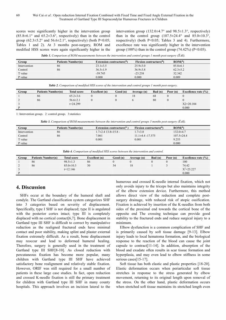
conducted at 1 and 3 months post-surgery. Images of a
patient in each group are shown in Figures 1 and 2.
International Journal of Clinical and Experimental Medical Sciences 2018; 4(4): 57-62 59
Figure 1. A patient in the intervention group diagnosed with left Gartland type III SHF. X-ray images of patient’s elbow before (A and B) and after surgery (C
and D). Patient was given post-surgery FTFAEF (E and F). Elbow function was assessed at 2 months post-surgery (G and H).
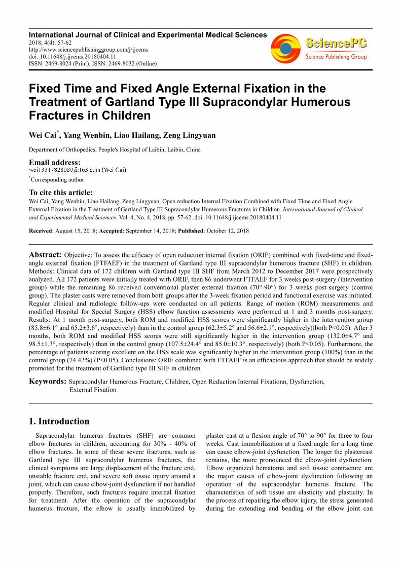
Figure 2. A patient in the control group diagnosed with left Gartland type III SHF. X-ray images of patient’s elbow before (a and b) and after surgery (c and
d). The patient was given conventional post-surgery plaster external fixation (e). Elbow function was assessed at 4 months post-surgery (f and g).
2.5. Statistical Analysis
Data were analyzed using SPSS version 16.0 software.
Measured data, such as age, duration of bone healing or total
HSS score were expressed as mean±SD (X�±S)
and compared between the two groups using a two-sample
t-test. Count data, such as complication and excellent HSS
score rates, were compared between the two groups using a
chi-square (X2) test. P<0.05 was considered statistically
significant.
3. Results
Both intervention and control groups were followed up for
a mean of 3.6 months (3-4 months). There was no significant
difference in the duration of bone healing between the groups
(7.6±1.5 versus 7.5±1.7 weeks for the intervention and
control groups, respectively, P>0.05). No incidence of K-
needle loosening or atopic ossification was observed in any
patient. At 1 month post-surgery, ROM and modified HSS
60 Wei Cai et al.: Open reduction Internal Fixation Combined with Fixed Time and Fixed Angle External Fixation in the
Treatment of Gartland Type III Supracondylar Humerous Fractures in Children
scores were significantly higher in the intervention group
(85.8±6.1° and 65.2±3.6°, respectively) than in the control
group (62.3±5.2° and 56.6±2.1°, respectively) (both P<0.05;
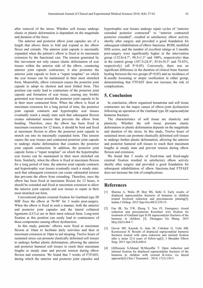
Tables 1 and 2). At 3 months post-surgery, ROM and
modified HSS scores were again significantly higher in the
intervention group (132.0±4.7° and 98.5±1.3°, respectively)
than in the control group (107.5±24.4° and 85.0±10.3°,
respectively) (both P<0.05; Tables 3 and 4). Furthermore,
excellence rate was significantly higher in the intervention
group (100%) than in the control group (74.42%) (P<0.05).
Table 1. Comparison of ROM measurements between the intervention and control groups 1 month post-surgery (��±S).
Group Patients Number(n) Extension contracture(°°°°) Flexion contracture(°°°°) ROM(°°°°)
Intervention 86 23.1±3.5 25.9±3.0 85.8±6.1
Control 86 36.5±1.9 36.9±3.8 62.3±5.2
T value -39.745 -23.238 32.342
P value 0.000 0.000 0.009
Table 2. Comparison of modified HSS scores of the intervention and control groups 1 month post-surgery.
Group Patients Number(n) Total score Excellent (n) Good (n) Average (n) Bad (n) Poor (n) Excellence rate (%)
1 86 65.2±3.6 0 0 18 68 0 0
2 86 56.6±2.1 0 0 6 80 0 0
3 t=24.299 X2=20.104
P 0.000
1: Intervention group;2: control group;3:statistics
Table 3. Comparison of ROM measurements between the intervention and control groups 3 months post-surgery (��±S).
Group Patients Number(n) Extension contracture(°°°° ) Flexion contracture(°°°°) ROM(°°°°)
Intervention 86 1.7±2.4 13.8±15.8 - 1.7±3.0 132.0±4.7
Control 86 7.061 11.1±4.4 -17.575 107.5±24.4
T value 0.001 0.001 9.253
P value 0.000
Table 4. Comparison of modified HSS scores between the intervention and control.
Group Patients Number(n) Total score Excellent (n) Good (n) Average (n) Bad (n) Poor (n) Excellence rate (%)
1 86 98.5±1.3 86 0 0 0 0 100
2 86 85.0±10.3 30 34 18 1 3 74.42
3 t=12.146 X2=25.227
P 0.000
4. Discussion
SHFs occur at the boundary of the humeral shaft and
condyle. The Gartland classification system categorizes SHF
into 3 categories based on severity of displacement.
Specifically, type I SHF is not displaced; type II is angulated
with the posterior cortex intact; type III is completely
displaced with no cortical contact[6,7]. Bone displacement in
Gartland type III SHF is difficult to correct by manipulative
reduction as the realigned fractured ends have minimal
contact and poor stability, making splint and plaster external
fixation extremely difficult. As a result, bone displacement
may reoccur and lead to deformed humeral healing.
Therefore, surgery is generally used in the treatment of
Gartland type III SHF[8-10]. As closed reduction with
percutaneous fixation has become more popular, many
children with Gartland type III SHF have achieved
satisfactory bone realignment and relatively stable fixation.
However, ORIF was still required for a small number of
patients in these large case studies. In fact, open reduction
and crossed K-needle fixation is still the primary treatment
for children with Gartland type III SHF in many county
hospitals. This approach involves an incision lateral to the
humerous and crossed K-needle internal fixation, which not
only avoids injury to the triceps but also maintains integrity
of the elbow extension device. Furthermore, this method
allows direct view of the reduction and complete post-
surgery drainage, with reduced risk of atopic ossification.
Fixation is achieved by insertion of the K-needles from both
sides of the proximal end towards the cortical bone of the
opposite end The crossing technique can provide good
stability to the fractured ends and reduce surgical injury to a
minimum.
Elbow dysfunction is a common complication of SHF and
is primarily caused by soft tissue damage [9-13]. Elbow
injury leads to local hematoma formation, and the biological
response to the reaction of the blood can cause the joint
capsule to contract[11-14]. In addition, absorption of the
blood and exudate often results in scar tissue formation and
hyperplasia, and may even lead to elbow stiffness in some
serious cases[15-17].
Soft tissue has both elastic and plastic properties [18-20].
Elastic deformation occurs when periarticular soft tissue
stretches in response to the stress generated by elbow
movement, returning to its original length upon removal of
the stress. On the other hand, plastic deformation occurs
when stretched soft tissue maintains its stretched length even
International Journal of Clinical and Experimental Medical Sciences 2018; 4(4): 57-62 61
after removal of the stress. Whether soft tissues undergo
elastic or plastic deformation is dependent on the magnitude
and duration of the force.
The anterior and posterior elbow joint capsules are of a
length that allows them to fold and expand as the elbow
flexes and extends. The anterior joint capsule is maximally
expanded when the patient’s elbow is fixed at its maximum
extension by the functional cast. The tension generated by
this movement not only causes elastic deformation of scar
tissues within the anterior side of the elbow, countering
anterior joint capsule contracture, it also promotes the
anterior joint capsule to form a “repair template” on which
the scar tissues can be maintained in their most stretched
form. Meanwhile, elbow extension causes the posterior joint
capsule to adopt its shortest and most folded form. This
position can easily lead to contracture of the posterior joint
capsule and formation of scar tissue, causing any freshly
generated scar tissue around the posterior joint capsule to be
in their most contracted form. When the elbow is fixed at
maximum extension for a long period of time, the posterior
joint capsule contracts and hypertrophic scar tissues
eventually reach a steady state such that subsequent flexion
creates substantial tension that prevents the elbow from
bending. Therefore, once the elbow has been fixed at
maximum extension for 12 hours, it should be bent and fixed
at maximum flexion to allow the posterior joint capsule to
stretch out into its maximally expanded form. This tension
causes the scar tissues and contracted posterior joint capsule
to undergo elastic deformation that counters the posterior
joint capsule contraction. In addition, the posterior joint
capsule forms a “repair template” on which the hypertrophic
scar tissues can be maintained in their most stretched out
form. Similarly, when the elbow is fixed at maximum flexion
for a long period of time, the anterior joint capsule contracts
and hypertrophic scar tissues eventually reach a steady state,
such that subsequent extension can create substantial tension
that prevents the elbow from extending. Therefore, once the
elbow has been fixed at maximum flexion for 12 hours, it
should be extended and fixed at maximum extension to allow
the anterior joint capsule and scar tissues to repair in their
most stretched out form.
Conventional plaster external fixation for Gartland type III
SHF fixes the elbow at 70-90° for 3 weeks post-surgery.
When the elbow is fixed in such a manner, both the anterior
and posterior joint capsules and the lateral collateral
ligaments (LCLs) are in their most relaxed form. Long-term
fixation at this position can easily lead to contractures of
these components causing elbow dysfunction.
In this study, patients’ elbows were fixed at maximum
flexion at 10am to facilitate daily activities and then at
maximum extension at 10pm to aid sleeping. Twelve hours of
sustained stress can promote elastically deformed soft tissues
to undergo further plastic deformation, allowing the anterior
and posterior humeral soft tissues to reach their maximum
lengths at steady state and prevent tension during elbow
flexion and extension. We found that 3 weeks of FTFAEF,
during which the anterior and posterior joint capsules and
hypertrophic scar tissues undergo repair cycles of “anterior
extended posterior contracted” to “anterior contracted
posterior extended”, resulted in satisfactory elbow activity
shortly after surgery and provided a good foundation for
subsequent rehabilitation of elbow functions. ROM, modified
HSS scores, and the number of excellent ratings at 3 months
post-surgery were significantly higher in the intervention
group (132.0±4.7°, 98.5±1.3° and 100%, respectively) than
in the control group (107.5±24.4°, 85.0±10.3° and 74.42%,
respectively) (all P<0.05). Conversely, there was no
significant difference in the duration of clinical bone fracture
healing between the two groups (P>0.05) and no incidence of
K-needle loosening or atopic ossification in either group,
demonstrating that FTFAEF does not increase the risk of
complications.
5. Conclusion
In conclusion, elbow organized hematoma and soft tissue
contracture are the major causes of elbow-joint dysfunction
following an operation of the Gartland type III supracondylar
humerus fractures.
The characteristics of soft tissue are elasticity and
plasticity. Whether the soft tissue presents elastic
deformation or plastic deformation depends on the magnitude
and duration of the stress. In this study, Twelve hours of
sustained stress can promote elastically deformed soft tissues
to undergo further plastic deformation, allowing the anterior
and posterior humeral soft tissues to reach their maximum
lengths at steady state and prevent tension during elbow
flexion and extension.
We found that 3 weeks of fixed-time and fixed-angle
external fixation resulted in satisfactory elbow activity
shortly after surgery and provided a good foundation for
subsequent rehabilitation of elbow functions.And FTFAEF
does not increase the risk of complications.
References
[1] Sharma A, Walia JP, Brar BS, Sethi S. Early results of displaced supracondylar fractures of humerus in children treated byclosed reduction and percutaneous pinning[J]. Indian J Orthop. 2015 Sep-Oct;49(5):529-35.
[2] Fan JR, Xu YW, Zheng Y, You JY. Emergency closed reduction and percutaneous Kirschner wire fixation for treatment of Gartland type II-III supracondylar fractures of the humerus in children. [J]. Zhongguo Gu Shang. 2015 May;28(5):464-7.
[3] Guven MF, Kaynak G, Inan M, Caliskan G, Unlu HB, Kesmezacar H. Results of displaced supracondylar humerus fractures treated with open reduction and internal fixation after a mean 22.4 years of follow-up[J]. J Shoulder Elbow Surg. 2015 Apr;24(4):640-6.
[4] [4]Hussain S,Ahmad M,Muzaffar T. Open reduction and internal fixation for displaced supracondylar fractures of the humerus in children with crossed K-wires via lateral approach[J].Chin J Traumatol。2014; 17(3):130-5.
62 Wei Cai et al.: Open reduction Internal Fixation Combined with Fixed Time and Fixed Angle External Fixation in the
Treatment of Gartland Type III Supracondylar Humerous Fractures in Children
[5] Ahmad Khan RDO, Yousaf MN, Zain-Ur-Rehman M, Yasin A. Utcome of open reduction internal fixation with cross K-wires for supracondylar fracture of humerus in terms of Flynn's criteria in children[J]. J Pak Med Assoc.2015 Nov ;65(11 Suppl 3): S186-9.
[6] [6]Gartland JJ.Management of supracondylar fractures of the humerus in children[J].Surg Gynecol Obstet,1959,109:145-154.
[7] Rocha IT,Faria Ade S,Filho CF,Rocha, MA.Reproducibility of the AO/ASIF and Gartland classifications for supracondylar fractures of the humerus in children[J].Rev Bras Ortop.2015 ;50(3) :266-9.
[8] Sarrafan N, Nasab SA, Ghalami T. Treatment of displaced supracondylar fracture of the humerus in children by open pining from lateral approach: an investigation of clinical and radiographical results[J]. Pak J Med Sci. 2015 Jul-Aug;31(4):930-935.
[9] Ahmad Khan, RD; Yousaf, MN; Zain - Ur Rehman, M; Yasin, A; Etc. Outcome of open reduction internal fixation with cross K-wires for supracondylar fracture of humerus in terms of Flynn's criteria in children[J].J Pak Med Assoc.2015 Nov;65(11 Suppl 3): S186-189.
[10] Dhoju D, Shrestha D, Parajuli N, Dhakal G, Shrestha R. Displaced supracondylar humeral fractures in children: Comparison of three treatment approaches[J]. Kathmandu Univ Med J (KUMJ). 2011 Apr-Jun;9(34):11-16.
[11] Kopka M, Monument MJ, Befus AD, Hildebrand KA.Serum Mast Cell Tryptase as a Marker of Posttraumatic Joint Contracture in a Rabbit Model[J]. J Orthop Trauma.2017 Mar ;31(3): e86-e89.
[12] Sun Yangbai, Li Fengfeng, Fan Cunyi.Effect of pERK2 on extracellular matrix turnover of the fibrotic joint capsule in a
post-traumatic joint contracture model[J]. Exp Ther Med.2016 Feb ;11(2) 547-552.
[13] Hildebrand KA, Zhang M, Hart DA. Myofibroblast upregulators are elevated in joint capsules in posttraumatic contractures[J]. Clin Orthop Relat Res. 2007 Mar; 456:85-91.
[14] Hildebrand KA, Zhang M, Germscheid NM, Wang C, Hart DA. Cellular, matrix, and growth factor components of the joint capsule are modified early in the process of posttraumatic contracture formation in a rabbit model[J]. Acta Orthop. 2008 Feb;79(1):116-25.
[15] Michelsson JE, Rauschning W. Pathogenesis of experimental heterotopic bone formation following temporary forcible exercising of immobilized limbs[J]. Clin Orthop,1983,17(6):265-272.
[16] Hotchkiss RN. Elbow contracture/Green DP, Hotchkiss RN, Pederson WC, Wolfe SW(eds). Green’s Operative Hand Surgery[M]. 5thed.New York, NY: Church ill-Livingstone,2005:667-682.
[17] Wu, Xinghuo; Wang, Hong; Meng, Chunqing; Etc.Outcomes of arthroscopic arthrolysis for the post-traumatic elbow stiffness[J]. Knee Surg Sports Traumatol Arthrosc.2015;23(9):2715-20.
[18] Pradas MM, Calleja RD.Nonlinear viscoilastec behaviour of the flexor tendon of the human hand [J].J Biomechan,1990,23:773.
[19] Spirt AA,Mak AF,Wassell RP.Nonlinear viscoelastic properties of articular cartilage in shear[J].J Orthop Res,1989,7:3.
[20] Taylor DC,Dalton JD.Viscoelastic properties of muscle tendon units:The biomechanical effects of stretching[J].Am J Sport Med,1990,l8:300.
Related Documents