Fish muscle: the exceptional case of notothenioids Daniel A. Ferna ´ndez Jorge Calvo Received: 23 February 2008 / Accepted: 13 October 2008 Ó Springer Science+Business Media B.V. 2008 Abstract Fish skeletal muscle is an excellent model for studying muscle structure and function, since it has a very well-structured arrangement with different fiber types segregated in the axial and pectoral fin muscles. The morphological and physiological characteristics of the different muscle fiber types have been studied in several teleost species. In fish muscle, fiber number and size varies with the species considered, limiting fish maximum final length due to constraints in metabolites and oxygen diffusion. In this work, we analyze some special characteristics of the skeletal muscle of the suborder Notothenioidei. They experi- enced an impressive radiation inside Antarctic waters, a stable and cold environment that could account for some of their special characteristics. The number of muscle fibers is very low, 12,700–164,000, in com- parison to 550,000–1,200,000 in Salmo salar of similar sizes. The size of the fibers is very large, reaching 600 lm in diameter, while for example Salmo salar of similar sizes have fibers of 220 lm maximum diameter. Evolutionary adjustment in cell cycle length for working at low temperature has been shown in Harpagifer antarcticus (111 h at 0°C), when compared to the closely related sub-Antarctic species Harpagifer bispinis (150 h at 5°C). Maximum muscle fiber number decreases towards the more derived notothenioids, a trend that is more related to phylog- eny than to geographical distribution (and hence water temperature), with values as low as 3,600 in Harpag- ifer bispinis. Mitochondria volume density in slow muscles of notothenioids is very high (reaching 0.56) and since maximal rates of substrate oxidation by mitochondria is not enhanced, at least in demersal notothenioids, volume density is the only means of overcoming thermal constraints on oxidative capacity. In brief, some characteristics of the muscles of notothenioids have an apparent phylogenetic compo- nent while others seem to be adaptations to low temperature. Keywords Fish muscle Á Muscle growth Á Fiber size Á Notothenioids Á Temperature Introduction Notothenioids (Perciforms) are indigenous fish from the southern hemisphere. They are thought to have evolved from a temperate small benthic ancestor and have radiated very rapidly in the subzero waters of the Antarctic (Eastman 1993), sometime between 24 Ma (million years ago) (Near 2004) and 7 Ma (Bargelloni et al. 2000), according to dissimilar molecular clock estimations. The absence of an undisputed fossil makes the molecular clock calibration very inexact. This suborder is the best example of extensive D. A. Ferna ´ndez (&) Á J. Calvo Austral Center for Scientific Research (CADIC- CONICET), 200 B. Houssay, CC 92 CADIC, 9410 Ushuaia, Tierra del Fuego, Argentina e-mail: [email protected] 123 Fish Physiol Biochem DOI 10.1007/s10695-008-9282-6

Welcome message from author
This document is posted to help you gain knowledge. Please leave a comment to let me know what you think about it! Share it to your friends and learn new things together.
Transcript
Fish muscle: the exceptional case of notothenioids
Daniel A. Fernandez Æ Jorge Calvo
Received: 23 February 2008 / Accepted: 13 October 2008
� Springer Science+Business Media B.V. 2008
Abstract Fish skeletal muscle is an excellent model
for studying muscle structure and function, since it has
a very well-structured arrangement with different fiber
types segregated in the axial and pectoral fin muscles.
The morphological and physiological characteristics
of the different muscle fiber types have been studied in
several teleost species. In fish muscle, fiber number
and size varies with the species considered, limiting
fish maximum final length due to constraints in
metabolites and oxygen diffusion. In this work, we
analyze some special characteristics of the skeletal
muscle of the suborder Notothenioidei. They experi-
enced an impressive radiation inside Antarctic waters,
a stable and cold environment that could account for
some of their special characteristics. The number of
muscle fibers is very low, 12,700–164,000, in com-
parison to 550,000–1,200,000 in Salmo salar of
similar sizes. The size of the fibers is very large,
reaching 600 lm in diameter, while for example
Salmo salar of similar sizes have fibers of 220 lm
maximum diameter. Evolutionary adjustment in cell
cycle length for working at low temperature has been
shown in Harpagifer antarcticus (111 h at 0�C), when
compared to the closely related sub-Antarctic species
Harpagifer bispinis (150 h at 5�C). Maximum muscle
fiber number decreases towards the more derived
notothenioids, a trend that is more related to phylog-
eny than to geographical distribution (and hence water
temperature), with values as low as 3,600 in Harpag-
ifer bispinis. Mitochondria volume density in slow
muscles of notothenioids is very high (reaching 0.56)
and since maximal rates of substrate oxidation by
mitochondria is not enhanced, at least in demersal
notothenioids, volume density is the only means of
overcoming thermal constraints on oxidative capacity.
In brief, some characteristics of the muscles of
notothenioids have an apparent phylogenetic compo-
nent while others seem to be adaptations to low
temperature.
Keywords Fish muscle � Muscle growth �Fiber size � Notothenioids � Temperature
Introduction
Notothenioids (Perciforms) are indigenous fish from
the southern hemisphere. They are thought to have
evolved from a temperate small benthic ancestor and
have radiated very rapidly in the subzero waters of the
Antarctic (Eastman 1993), sometime between 24 Ma
(million years ago) (Near 2004) and 7 Ma (Bargelloni
et al. 2000), according to dissimilar molecular clock
estimations. The absence of an undisputed fossil
makes the molecular clock calibration very inexact.
This suborder is the best example of extensive
D. A. Fernandez (&) � J. Calvo
Austral Center for Scientific Research (CADIC-
CONICET), 200 B. Houssay, CC 92 CADIC,
9410 Ushuaia, Tierra del Fuego, Argentina
e-mail: [email protected]
123
Fish Physiol Biochem
DOI 10.1007/s10695-008-9282-6
radiation in a marine fish group (Eastman and Eakin
2000) and can be considered a species flock similar to
the Cottoid species flock from Lake Baikal (Eastman
and McCune 2000). The key physiological feature that
allowed the notothenioids to diversify and become
dominant in the fish fauna of the Southern Ocean was,
almost certainly, the development of antifreeze gly-
coproteins (AFGPs) (Cheng and DeVries 1991;
Eastman 1993). The key ecological feature that
permitted the diversification of the suborder was
probably the weak competition notothenioids experi-
enced in Antarctic shallow waters due to the extinction
events associated with the cooling conditions and the
isolation of Antarctica (Eastman 1993). Although
buoyancy modification that allowed notothenioids to
occupy distinct positions in the water column was also
appreciated as an important factor for the radiation,
this trait seems to have evolved only once in notothe-
nioids, and it is also not very common in the suborder,
with only three species so far proven to be neutrally
buoyant (Eastman and De Vries 1982; Balushkin
2000; Eastman and Sidell 2002; Near et al. 2004;
Eastman 2005; Near et al. 2007). Today, nototheni-
oids encompass a total of 44 genera and 129 species,
101 Antarctic and 28 non-Antarctic, the latter living in
the Beagle Channel, Patagonian Shelf, along the
Pacific Coast of South America, and in the sub-
Antarctic waters of New Zealand (Eastman 2005).
Fish axial muscle formation
In fish, the skeletal muscles of the trunk and limb are
derived from the somites. These repetitive structures
are formed in a rostral-to-caudal sequence from the
paraxial mesoderm, and their number varies greatly
in fish, from 26 in the platy fish to more than 200 in
some eels (Richardson et al. 1998). Recent works on
zebrafish have proved the existence of a dermamyo-
tome in fish (similar to the one described for
amniotes) that is formed from the ABC cells (anterior
border cells) of the somite through a ‘‘somite
rotation’’ movement. These ABC cells will give rise
to muscle fibers of the primary myotome and the
dermamyotome, that will subsequently give rise to
muscle precursors for the axial and fin muscles, and
probably other cell types of the somite (Hollway and
Currie 2005; Stellabotte and Devoto 2007; Stellabotte
et al. 2007). The first functional fibers of the somites
(primary myotome) elongate before the formation of
the dermamyotome. Further growth in the myotome
could occur by the ingression of these ‘‘external
cells’’ into the myotome through the external layer of
slow muscle cells (Stellabotte and Devoto 2007).
‘‘External cells’’ have been previously described in
other teleost species (Johnston 1993; Stoiber and
Sanger 1996), and therefore the mechanism described
for zebrafish could be a common one for teleosts.
More data on different teleost species are needed in
order to asses the importance of this mechanism.
Muscle fiber types: axial
More than 60% of the body weight in fish can be
muscle. This is only possible due to the minimal
requirement of skeletal structure to support the weight
of the body in water, and because of the need for a
larger amount of muscle to power swimming in a
denser medium like water (Bone 1978; Johnston
1981). The spatial arrangement of adult fish muscle,
with different muscle fiber types located in discrete
areas, allows a detailed analysis of its structure and
function. Axial musculature is formed mainly by two
different muscle fiber types, the white fibers and the
red fibers, but there are also other fiber types (inter-
mediate, tonic, red muscle rim fibers, etc.). The largest
part of the axial musculature is white muscle, about
90% versus 10% of the rest of the fibers (mainly red) in
several species of sub-Antarctic notothenioids ana-
lyzed (Fernandez, unpublished results), falling within
the range typically observed in other teleost species
(Bone 1978). White and red muscle fiber types differ
not only in their color, but also in many other important
structural and physiological characteristics: innerva-
tion, blood supply, abundance of mitochondria,
myoglobin content, energy storage, speed of contrac-
tion, fiber size, myosin isoforms, etc. Because of the
different speed of their contractions, white fibers are
also called ‘‘fast’’ and red fibers are called ‘‘slow’’
(Johnston 1981). From now on, we shall call them fast
and slow, respectively.
Four different fiber types have been described for
the axial muscles of nototheniods: slow, tonic, inter-
mediate, and fast, using histochemical techniques for
myosin ATPase, succinic dehydrogenase (SDHase),
glycogen, and lipids (Walesby and Johnston 1980;
Smialowska and Kilarski 1981; Dunn et al. 1989;
Fernandez 2000; Fernandez et al. 2000). The different
Fish Physiol Biochem
123
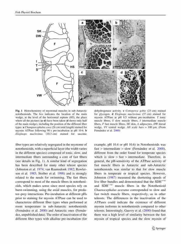
fiber types are relatively segregated in the myotome of
notothenioids, with a superficial layer (the width varies
in the different species) composed of tonic, slow, and
intermediate fibers surrounding a core of fast fibers
(see details in Fig. 1). A similar kind of segregation
has been described for many other teleost species
(Johnston et al. 1974; van Raamsdonk 1982; Rowler-
son et al. 1985; Stoiber et al. 1998) and is strongly
related to the needs for swimming. The fast fibers
correspond to most of the muscle fibers in nototheni-
oids, which makes sense since most species rely on
burst-swimming, using the axial muscles, for preda-
tor–prey interactions. Pre-incubation at different pHs
prior to staining for myosin ATPase can be used to
characterize different fiber types when performed at
room temperature in sub-Antarctic notothenioids
(Fernandez et al. 2000) and Antarctic ones (Fernan-
dez, unpublished data). The order of inactivation of the
different fiber types with alkaline pre-incubation (for
example, pH 10.4 or pH 10.6) in Notothenioids was
fast [ intermediate [ slow (Fernandez et al. 2000),
different from the order found for temperate species
which is slow [ fast [ intermediate. Therefore, in
general, the pH-sensitivity of the ATPase activity of
fast muscle fibers in Antarctic and sub-Antarctic
notothenioids was similar to that for slow muscle
fibers in temperate or tropical species. However,
Johnston (1987) measured the shortening speeds of
live fiber bundles and demonstrated that the SDH?ve
and SDH-ve muscle fibers in the Notothenioid
Chaenocephalus aceratus corresponded to slow and
fast twitch muscle fibers, respectively, as in other
teleosts. The differences in the inactivation of the
ATPases could indicate the existence of different
myosin isoforms in notothenioids compared to other
teleosts. Interestingly, Gauvry et al. (2000) found that
there was a high level of similarity between the fast
myosin of tropical species and the slow myosin of
Fig. 1 Histochemistry of myotomal muscles in sub-Antarctic
notothenioids. The box indicates the location of the main
wedge, at the level of the horizontal septum (HS), the place
where all the pictures (a–d) have been taken (d shows only half
of the main wedge), including the position of the different fiber
types. a Champsocephalus esox (26 cm total length) stained for
myosin ATPase following 90 s pre-incubation at pH 10�6. bEleginops maclovinus (10.3 cm) stained for succinic
dehydrogenase activity. c Cottoperca gobio (23 cm) stained
for glycogen. d Eleginops maclovinus (37 cm) stained for
myosin ATPase at pH 9.5 without pre-incubation. T tonic
muscle fibres, S slow muscle fibres, I intermediate muscle
fibers, F fast muscle fibres, SK skin, A adipocytes, DW dorsal
wedge, VV ventral wedge. All scale bars = 100 lm. (From
Fernandez et al. 2000)
Fish Physiol Biochem
123

Antarctic species, comparing the amino acid sequence
and the structure of ATPase binding sites.
Pectoral fin muscles
Fin muscles in teleosts are derived from the paraxial
mesoderm, more specifically from the dermomyotome.
Three of the four fiber types have been described for the
pectoral fin muscles in sub-Antarctic notothenioids:
tonic, slow, and fast (Fernandez et al. 2000). The fiber
type distribution in the abductor profundis muscle of
all the species described was similar, comprising four
different zones: (1) tonic: tonic fibers were found close
to the pectoral girdle bones; (2) central region: a core of
slow muscle fibers; (3) mosaic region: an area of slow
muscle fibers intermingled with fast; and (4) peripheral
region: an area of fast fibers occupying the more
external part of the muscle (Fig. 2). Even though the
distribution of the fiber types was very conserved in all
the species, there was a consistent variation of the
proportion of the different zones from the proximal to
the distal ends of the muscle (Fernandez et al. 2000). A
similar zonation has also been found in Antarctic
nototheniods (Walesby and Johnston 1980; Davison
and MacDonald 1985; Harrison et al. 1987) and in
other teleosts (Patterson et al. 2007; Devincenti et al.
2008). While slow muscle fibers are abundant in
notothenioids, leaving an external marginal location to
the fast fibers, the opposite situation is observed in the
other teleosts studied. This fact is very likely related to
the importance of labriform swimming (using pectoral
fin muscles) in notothenioids in comparison with
carangiform or subcarangiform swimming in the other
species studied.
Fig. 2 Histochemical staining characteristics of the pectoral
fin adductor muscle of P. tessellata (a–c) and E. maclovinus(d–f). The drawings indicate the location of the different
regions of the adductor profundis muscle and the other muscles
that form the pectoral musculature at the proximal, interme-
diate and distal ends of the muscles. a Tonic fibers (T) are
adjacent to the girdle bones (top), the slow muscle fibers
compose the central region (CR) at all levels of the pectoral
muscles and the connective tissue (CT) separates adductorprofundis from adductor superficialis muscles (distal); (b) the
CR and the mosaic regions (MR) are clearly distinguished
because the latest has slow and fast muscle fibers intermingled
(proximal); (c) detail of the CR and MR stained for mATPase
following alkaline (pH 10.6; 90 s) preincubation; (d) narrow
band of T adjacent to the bone; (e) CR and MR in
E. maclovinus (proximal) (f) spectacular mosaic in large
E. maclovinus adductor muscle in a section stained for
mATPase following alkaline (pH 10.6; 90 s) preincubation;
the peripheral region (PR) can also be observed in the right topof the picture (intermediate). S Slow muscle fiber, B bone.
Scale bar represents 100 lm (a), (c), and (d) and 400 lm (b),
(e), and (f). (From Fernandez et al. 1999, 2000)
Fish Physiol Biochem
123
Muscle growth
Muscle growth occurs by two main processes, the
addition of new fibers (hyperplasia) and the increase
in size of existing fibers (hypertrophy). Muscle
growth in fish differs from other vertebrates because
it occurs indeterminately, due to continuous growth
through life (Mommsen 2001). Four main phases of
muscle growth occur in teleosts: one embryonic and
three post-embryonic, but not all phases are present in
each fish species (reviewed in Johnston 2003; Rowl-
erson and Veggetti 2001). Postembryonic growth
phases are called stratified hyperplasia, mosaic
hyperplasia, and hypertrophy, depending on the main
process that is taking place at a given time. The final
size of a given species is strongly regulated by the
duration of the hyperplastic phases, since those
phases determine the final number of muscle fibers.
The final number of fibers restrains the final size of
the species, given that the maximum size of a given
fiber has physiological constraints, probably due to
limitations in diffusion rates.
Axial muscle fiber diameter
The axial muscle of the notothenioids is unusual in
containing very large diameter muscle fibers in
comparison to other teleosts (Smialowska and Kilar-
ski 1981; Dunn et al. 1989; Battram and Johnston
1991; Fernandez et al. 2000; Johnston et al. 2003b).
The maximum fiber diameters increase linearly with
standard length, reaching more than 500 lm in many
of the species studied (Johnston et al. 2003b). Similar
fiber diameter values were found in the light fibers of
the blue crab Callinectes sapidus (Boyle et al. 2003).
In general terms, muscle fibers are limited in size to
promote short maximal intracellular diffusion dis-
tances in order to facilitate both rapid O2 flux to
mitochondria and ATP flux from mitochondria to
sites of ATP demand. Therefore, excessive cell size
reduces the capacity for critical oxidative metabolic
processes (Egginton and Sidell 1989; Boyle et al.
2003; Johnston et al. 2003b; Kinsey et al. 2005). One
way of overcoming this problem, found in fish and
crustaceans, is the redistribution of intracellular
metabolic machinery over the course of development
in order to reduce the diffusion distances between
small blood vessels and mitochondria, something that
has been documented in the burst swimming muscle
of blue crab Callinectes sapidus (Boyle et al. 2003)
and black sea bass Centropristis striata (Nyack et al.
2007). Small white muscle fibers have mitochondria
evenly distributed throughout the fiber (intermyofibr-
illar mitochondria), while in large white muscle fibers
the mitochondria become increasingly clustered at the
periphery of the cell (subsarcolemmal mitochondria)
and are much less densely distributed in the fiber
interior. However, the redistribution increases the
intracellular diffusive distances between mitochon-
drial clusters, which may greatly slow ATP diffusive
flux (Boyle et al. 2003; Nyack et al. 2007). The burst
fibers of fish and crustaceans rely on endogenous fuels
to power a series of rapid contractions, being
independent of the transport of either O2 to the
mitochondria or ATP/phosphagen from mitochondria
to cellular ATPases. Therefore, the metabolic recov-
ery after a series of burst contractions. but not the
contractile process (anaerobic), is influenced by an
increase in fiber size or changing mitochondrial
distribution. This may have serious implications for
the animal’s survival if multiple events of burst
contractions are needed, such as during repeated
predator–prey interactions (Kinsey et al. 2007).
Johnston et al. (2004) proposed for fish that there
is an optimal maximum fiber diameter, which reflects
a trade-off between avoiding diffusional constraints
and the need to minimize the costs of ion pumping,
and called this idea the ‘‘optimal fiber size hypoth-
esis’’. The surface/volume ratio of muscle fibers, and
therefore the maintenance of ionic homeostasis, that
constitutes 20–40% of the resting metabolic rate in
teleosts (Jobling 1995), decreases with increasing
fiber diameter. It would therefore be beneficial that
muscle fibers reach a size that is just below that
which would be diffusion limited.
Axial muscle fiber number
Notothenioids, in general, have low muscle fiber
numbers (Battram and Johnston 1991). Eleginops
maclovinus, for example, a notothenioid with an
unusually large number of fibers, has only 164,000
fibers in contrast to 1,200,000 fibers in an Atlantic
salmon Salmo salar of similar size (Johnston et al.
2003b). Phylogenetic independent contrast analysis in
notothenioids showed that fiber number differs sig-
nificantly between species that belong to the more
basal and the most derived families, suggesting a
Fish Physiol Biochem
123
decreasing trend in fiber numbers during the evolution
of the suborder. Moreover, the decrease in the number
of fibers correlates with an increase in the diameter of
the fibers (Johnston et al. 2003b). On the other hand,
there is no evident relationship between the geo-
graphical zone of origin (Antarctic vs sub-Antarctic)
and the maximum fiber diameter of the species.
Therefore, the special traits of Notothenioid muscle
(low fiber number and giant fiber size) have an
important phylogenetic component, apart from the
well-established relationship between low tempera-
ture and large fiber diameter.
The main phases in post-embryonic muscle growth
are stratified and mosaic hyperplasia. Some species lack
mosaic hyperplasia, and thus the final number of fibers is
greatly reduced. Mosaic hyperplasia is absent in all the
species of the more derived families already studied
(Harpagiferidae and Channichthyidae), giving a clue
about how muscle fiber number has been adjusted during
the evolution of this suborder (Johnston et al. 2003b).
Muscle growth and temperature
The main process involved in muscle growth of
notothenioids, hypertrophy, has been studied in adult
Harpagifer antarcticus (Antarctic) and Harpagifer
bispinis (sub-Antarctic) acclimated at summer and
winter temperatures (Brodeur et al. 2003a, b). These
species are very good models for studying hypertro-
phy, since hyperplasia has completely stopped in adult
fish (Johnston et al. 2003a). Cell cycle times were
estimated for H. bispinis at 10�C (81.3 h) and 5�C
(150 h), with Q10 = 3.4, and H. antarcticus at 0�C
(111 h). The longer duration of the cell cycle at 5�C in
H. bispinis than at 0�C in H. antarcticus indicates the
existence of a cold compensation in the Antarctic
species, allowing a substantial reduction in the time of
the cell cycle progression rate at low temperatures.
The predicted cell cycle time for H. bispinis at 0�C
(based on the Q10 relationship) would be 227 h, more
than double the value found for H. antarcticus.
Brodeur et al. (2003b) found evidence of a direct
stimulation of myogenic cell activation by feeding at
two different temperatures (about a two-fold increase
in the cells that express the surface protein c-met that
seems to be involved in their activation; Cornelison
and Wold 1997) in H. bispinis. The number of
myogenic cells generated in response to feeding did
not appear to be directly related to temperature. The
main difference between the responses to feeding of
fish acclimatized to simulated winter and summer
conditions resided in the expression of myogenin,
which was much less pronounced in summer. Inter-
estingly, the delay between the ingestion of the meal
and the activation of the myogenic progenitors (cells
that express the surface protein c-met that seems to be
involved in their activation; Cornelison and Wold
1997) in H. bispinis was shorter than the cell cycle
duration estimated for both summer and winter
temperatures (150 and 81 h, respectively). This result
could indicate either that the cell cycle progression
rate is accelerated by feeding, or that a proportion of
the activated cells were stopped at a checkpoint in the
cell cycle, and therefore could divide faster after
activation since they had already progressed through
part of the cell cycle (Walworth 2000). The latter is in
agreement with previous results on Notothenia corii-
ceps suggesting that myogenic cells activated by
feeding were cells stopped at the G1/S checkpoint of
the cell cycle (Brodeur et al. 2002).
Antarctic and sub-Antarctic notothenioids, adapted
to living at very low temperatures, have abundant
mitochondria in the slow muscle fibers (Johnston 1987;
Londraville and Sidell 1990). For example, reported
mitochondrial volume density values of slow muscle
fibers were 0.56 for Pleuragramma antarcticum
(Johnston et al. 1988) and 0.51 for Champsocephalus
esox (Johnston et al. 1998), amongst the highest
recorded for vertebrates. Nevertheless, different life-
styles of the species may confound the relationship
between temperature and mitochondria volume densi-
ties. For example, Sanger et al. (2005) found a
significant difference in the mitochondrial content in
all the skeletal muscles sampled from Pagothenia
borchgrevinki (cryopelagic) and Trematomus bernac-
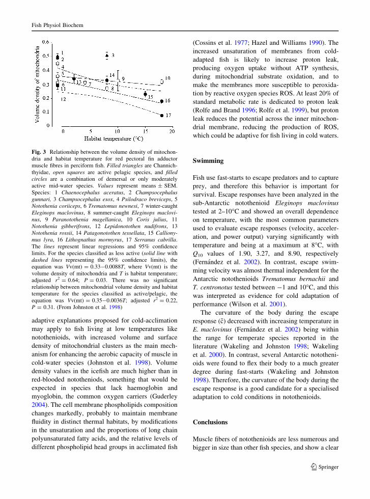
chii (benthic). Johnston et al. (1998) found a
significant inverse relationship between mitochondrial
volume density in slow muscle and habitat temperature
in a research restricted to demersal and moderately
active species (Fig. 3).
Diverse adaptive explanations have been proposed
to account for this fact, including the hypothesis that
increases in mitochondrial volume density partially
compensate for the reduced catalytic capacity at low
temperatures (Johnston 1982; Egginton and Sidell
1989) or otherwise compensate for the reduced
diffusion coefficients of cytosolic metabolites (Tyler
and Sidell 1984; Sidell and Hazel 1987). The same
Fish Physiol Biochem
123
adaptive explanations proposed for cold-acclimation
may apply to fish living at low temperatures like
notothenioids, with increased volume and surface
density of mitochondrial clusters as the main mech-
anism for enhancing the aerobic capacity of muscle in
cold-water species (Johnston et al. 1998). Volume
density values in the icefish are much higher than in
red-blooded nototheniods, something that would be
expected in species that lack haemoglobin and
myoglobin, the common oxygen carriers (Guderley
2004). The cell membrane phospholipids composition
changes markedly, probably to maintain membrane
fluidity in distinct thermal habitats, by modifications
in the unsaturation and the proportions of long chain
polyunsaturated fatty acids, and the relative levels of
different phospholipid head groups in acclimated fish
(Cossins et al. 1977; Hazel and Williams 1990). The
increased unsaturation of membranes from cold-
adapted fish is likely to increase proton leak,
producing oxygen uptake without ATP synthesis,
during mitochondrial substrate oxidation, and to
make the membranes more susceptible to peroxida-
tion by reactive oxygen species ROS. At least 20% of
standard metabolic rate is dedicated to proton leak
(Rolfe and Brand 1996; Rolfe et al. 1999), but proton
leak reduces the potential across the inner mitochon-
drial membrane, reducing the production of ROS,
which could be adaptive for fish living in cold waters.
Swimming
Fish use fast-starts to escape predators and to capture
prey, and therefore this behavior is important for
survival. Escape responses have been analyzed in the
sub-Antarctic notothenioid Eleginops maclovinus
tested at 2–10�C and showed an overall dependence
on temperature, with the most common parameters
used to evaluate escape responses (velocity, acceler-
ation, and power output) varying significantly with
temperature and being at a maximum at 8�C, with
Q10 values of 1.90, 3.27, and 8.90, respectively
(Fernandez et al. 2002). In contrast, escape swim-
ming velocity was almost thermal independent for the
Antarctic notothenioids Trematomus bernachii and
T. centronotus tested between -1 and 10�C, and this
was interpreted as evidence for cold adaptation of
performance (Wilson et al. 2001).
The curvature of the body during the escape
response (c) decreased with increasing temperature in
E. maclovinus (Fernandez et al. 2002) being within
the range for temperate species reported in the
literature (Wakeling and Johnston 1998; Wakeling
et al. 2000). In contrast, several Antarctic nototheni-
oids were found to flex their body to a much greater
degree during fast-starts (Wakeling and Johnston
1998). Therefore, the curvature of the body during the
escape response is a good candidate for a specialised
adaptation to cold conditions in notothenioids.
Conclusions
Muscle fibers of notothenioids are less numerous and
bigger in size than other fish species, and show a clear
Fig. 3 Relationship between the volume density of mitochon-
dria and habitat temperature for red pectoral fin adductor
muscle fibres in perciform fish. Filled triangles are Channich-
thyidae, open squares are active pelagic species, and filledcircles are a combination of demersal or only moderately
active mid-water species. Values represent means ± SEM.
Species: 1 Chaenocephalus aceratus, 2 Champsocephalusgunnari, 3 Champsocephalus esox, 4 Psilodraco breviceps, 5
Notothenia coriiceps, 6 Trematomus newnesi, 7 winter-caught
Eleginops maclovinus, 8 summer-caught Eleginops maclovi-nus, 9 Paranotothenia magellanica, 10 Coris julius, 11
Notothenia gibberifrons, 12 Lepidonotothen nudifrons, 13
Notothenia rossii, 14 Patagonotothen tessellata, 15 Calliony-mus lyra, 16 Lithognathus mormyrus, 17 Serranus cabrilla.
The lines represent linear regressions and 95% confidence
limits. For the species classified as less active (solid line with
dashed lines representing the 95% confidence limits), the
equation was Vv(mt) = 0.33-0.0088T, where Vv(mt) is the
volume density of mitochondria and T is habitat temperature;
adjusted r2 = 0.64; P = 0.03. There was no significant
relationship between mitochondrial volume density and habitat
temperature for the species classified as active/pelagic, the
equation was Vv(mt) = 0.35-0.0036T; adjusted r2 = 0.22,
P = 0.31. (From Johnston et al. 1998)
Fish Physiol Biochem
123
trend to reduction in number and increase in size
from the ancestral to the more derived notothenioids,
but not a geographical pattern. Mitochondria volume
densities in slow muscles of notothenioids are very
high, being likely an adaptation to low temperature.
Antarctic notothenioids also show evidence of an
evolutionary adjustment to temperature in cell cycle
length. Muscle fibers in notothenids may have
evolved to be as large as possible without experienc-
ing diffusion limitations in order to minimize the
sarcolemmal membrane area over which membrane
potential must be maintained. This would make sense
since it would reduce the costs of ionic homeostasis,
which is a considerable fraction of basal metabolic
rate. It is not clear if swimming in notothenioids is
more influenced by phylogeny or by temperature,
since different parameters show different trends.
In brief, some characteristics of the muscles of
nototheniods seem to have a prevalent phylogenetic
component while others seem to be adaptations to
low temperature.
Acknowledgments We would like to thank CONICET,
Agencia de Investigacion Cientıfica y Tecnologica (SeCyT),
Fundacion Antorchas and the European Union for funding
previous projects on muscle of notothenioids. We would also
like to thank present and past members of the laboratory of
Ecophysiology at CADIC for collaboration in these projects.
Sandy Becker and Sheryl Macnie helped to improve the
English of the manuscript. Comments from two anonymous
referees have helped greately to improve the manuscript.
References
Balushkin A (2000) Morphology, classification, and evolution
of notothenioid fishes of the Southern Ocean (Notothe-
nioidei, Perciformes). J Ichthyol 40:S74–S109
Bargelloni L, Marcato S, Zane L et al (2000) Mitochondrial
phylogeny of notothenioids: a molecular approach to
Antarctic fish evolution and biogeography. Syst Biol
49:114–129. doi:10.1080/10635150050207429
Battram JC, Johnston IA (1991) Muscle growth in the Antartic
teleost, Nothothenia neglecta (Nybelin). Antarct Sci 3:29–
33. doi:10.1017/S0954102091000068
Bone Q (1978) Locomotor muscle. In: Hoar WS, Randall DJ
(eds) Fish physiology, vol 7. Academic Press, New York,
pp 361–424
Boyle KL, Dillaman RM, Kinsey ST (2003) Mitochondrial
distribution and glycogen dynamics suggest diffusion
constraints in muscle fibers of the blue crab, Callinectessapidus. J Exp Zoolog A Comp Exp Biol 297:1–16
Brodeur JC, Peck L, Johnston IA (2002) Feeding increases
MyoD and PCNA expression in myogenic progenitor cells
of Notothenia coriiceps. J Fish Biol 60:1475–1485
Brodeur JC, Calvo J, Clarke A et al (2003a) Myogenic cell
cycle duration in Harpagifer species with sub-Antarctic
and Antarctic distributions: evidence for cold compensa-
tion. J Exp Biol 206:1011–1016. doi:10.1242/jeb.00204
Brodeur JC, Calvo J, Johnston IA (2003b) Proliferation of
myogenic progenitor cells following feeding in the sub-
Antarctic notothenioid fish Harpagifer bispinis. J Exp
Biol 206:163–169. doi:10.1242/jeb.00052
Cheng CC, DeVries AL (1991) The role of antifreeze glyco-
peptides and peptides in the freezing avoidance of cold-
water fish. In: Prisco Gd (ed) Life under extreme condi-
tions. Springer, Berlin Heidelberg New York, pp 1–14
Cornelison D, Wold B (1997) Single-cell analysis of regulatory
gene expression in quiescent and activated mouse skeletal
muscle satellite cells. Dev Biol 191:270–283. doi:10.1006/
dbio.1997.8721
Cossins AR, Friedlander MJ, Prosser CL (1977) Correlations
between behavioral temperature adaptations of goldfish and
the viscosity and fatty acid composition of their synaptic
membranes. J Comp Physiol [A] 120:109. doi:10.1007/BF00
619309
Davison W, MacDonald JA (1985) A histochemical study of
the swimming musculature of Antartic fish. NZ J Zool
12:473–483
Devincenti CV, Dıaz AO, Garcıa AM et al (2008) Pectoral fins
of Micropogonias furnieri: a histochemical and ultra-
structural study. Fish Physiol Biochem. doi:10.1007/
s10695-008-9216-3
Dunn JF, Archer SD, Johnston IA (1989) Muscle fibre types
and metabolism in post-larval and adult stages of Not-
othenoid fish. Polar Biol 9:213–223. doi:10.1007/BF00
263769
Eastman JT (1993) Antarctic fish biology: evolution in an
unique environment. Academic Press, New York
Eastman JT (2005) The nature of the diversity of Antarctic
fishes. Polar Biol 28:93–107. doi:10.1007/s00300-004-
0667-4
Eastman JT, De Vries AL (1982) Buoyancy studies of Noto-
thenioid fishes in McMurdo Sound, Antractica. Copeia
2:385–393. doi:10.2307/1444619
Eastman JT, Eakin R (2000) An updated species list for not-
othenioid fish (Perciformes; Notothenioidei), with
comments on Antarctic species. Arch Fisch Meeresforsch
48:11–20
Eastman JT, McCune A (2000) Fishes on the Antarctic conti-
nental shelf: evolution of a marine species flock? J Fish
Biol 57:84–102
Eastman JT, Sidell BD (2002) Measurements of buoyancy for
some Antarctic notothenioid fishes from the South Shet-
land Islands. Polar Biol 25:753–760
Egginton S, Sidell BD (1989) Thermal acclimation induces
adaptive changes in subcellular structure of fish skeletal
muscle. Am J Physiol 256:1–9
Fernandez DA (2000) Caracterizacion histoquimica, distribu-
cion y crecimiento de las fibras musculares en Nototenidos
subantarticos. Analisis inicial de dos factores relacionados
con la natacion: flotabilidad y temperatura. Biology
Department. Universidad de Buenos Aires, Buenos Aires
Fernandez DA, Calvo J, Johnston IA (1999) Characterisation
of the swimming muscles of two sub-Antarctic Notothe-
nioids. Sci Mar 201:477–484
Fish Physiol Biochem
123
Fernandez DA, Calvo J, Franklin CE et al (2000) Muscle fibre
types and size distribution in sub-antarctic notothenioid
fishes. J Fish Biol 56:1295–1311. doi:10.1111/j.1095-
8649.2000.tb02144.x
Fernandez DA, Calvo J, Wakeling JM et al (2002) Escape
performance in the sub-Antarctic notothenioid fish Ele-ginops maclovinus. Polar Biol 25:914–920
Gauvry L, Ennion S, Ettelaie C et al (2000) Characterisation of
red and white muscle myosin heavy chain gene coding
sequences from Antarctic and tropical fish. Comp Bio-
chem Physiol B 127:575–588. doi:10.1016/S0305-0491
(00)00286-8
Guderley H (2004) Metabolic responses to low temperature in
fish muscle. Biol Rev Camb Philos Soc 79:409–427. doi:
10.1017/S1464793103006328
Harrison P, Nicol CJM, Johnston IA (1987) Gross morphology,
fibre composition and mechanical properties of pectoral
fin muscles in the Antartic teleost Notothenia neglecta
Nybelin. Proceedings of the V Congress of European
Ichthyology, Stockholm, pp 459–465
Hazel JR, Williams EE (1990) The role of alterations in
membrane lipid composition in enabling physiological
adaptation of organisms to their physical environment.
Prog Lipid Res 29:167–227. doi:10.1016/0163-7827(90)
90002-3
Hollway G, Currie P (2005) Vertebrate myotome development.
Birth Defects Res C Embryo Today 75:172–179. doi:
10.1002/bdrc.20046
Jobling M (1995) Fish bioenergetics. Chapman & Hall, London
Johnston IA (1981) Structure and function of fish muscle.
Symp Zool Soc Lond 48:71–113
Johnston IA (1982) Capillarisation, oxygen diffusion distances
and mitochondrial content of carp muscles following
acclimation to summer and winter temperatures. Cell
Tissue Res 222:325–337. doi:10.1007/BF00213216
Johnston IA (1987) Respiratory characteristics of muscle fibres
in a fish (Chaenocephalus aceratus) that lacks haem
pigments. J Exp Biol 133:415–428
Johnston IA (1993) Temperature influences muscle differenti-
ation and the relative timing of organogenesis in herring
(Clupea harengus) larvae. Mar Biol (Berlin) 116:363–
379. doi:10.1007/BF00350053
Johnston IA (2003) Muscle metabolism and growth in Ant-
arctic fishes (suborder Notothenioidei): evolution in a cold
environment. Comp Biochem Physiol B Biochem Mol
Biol 136:701–713. doi:10.1016/S1096-4959(03)00258-6
Johnston IA, Patterson S, Ward W et al (1974) The histo-
chemical demonstration of myofibrillar adenosine
triphosphatase activity in fish muscle. Can J Zool 52:871–
877. doi:10.1139/z74-118
Johnston IA, Camm JP, White M (1988) Specialisations of
swimming muscles in the pelagic Antarctic fish Pleura-gramma antarcticum. Mar Biol 100:3–12. doi:10.1007/
BF00392949
Johnston IA, Calvo J, Guderley H et al (1998) Latitudinal vari-
ation in the abundance and oxidative capacities of muscle
mitochondria in perciform fishes. J Exp Biol 201:1–12
Johnston I, Vieira VL, Fernandez DA et al (2003a) Muscle
growth in Polar fish: a study of Harpagifer species with
sub-Antarctic and Antarctic distributions. Fish Sci
68:1023–1028
Johnston IA, Fernandez DA, Calvo J et al (2003b) Reduction in
muscle fibre number during the adaptive radiation of
notothenioid fishes: a phylogenetic perspective. J Exp
Biol 206:2595–2609. doi:10.1242/jeb.00474
Johnston IA, Abercromby M, Vieira VL et al (2004) Rapid
evolution of muscle fibre number in post-glacial popula-
tions of Arctic charr Salvelinus alpinus. J Exp Biol
207:4343–4360. doi:10.1242/jeb.01292
Kinsey ST, Pathi P, Hardy KM et al (2005) Does intracellular
metabolite diffusion limit post-contractile recovery in
burst locomotor muscle? J Exp Biol 208:2641–2652. doi:
10.1242/jeb.01686
Kinsey ST, Hardy KM, Locke BR (2007) The long and winding
road: influences of intracellular metabolite diffusion on
cellular organization and metabolism in skeletal muscle.
J Exp Biol 210:3505–3512. doi:10.1242/jeb.000331
Londraville RL, Sidell BD (1990) Ultrastructure of aerobic
muscle in Antarctic fishes may contribute to maintenance
of diffusive fluxes. J Exp Biol 150:205–220
Mommsen TP (2001) Paradigms of growth in fish. Comp
Biochem Physiol B Biochem Mol Biol 129:207–219. doi:
10.1016/S1096-4959(01)00312-8
Near TJ (2004) Estimating divergence times of notothenioid
fishes using a fossil-calibrated molecular clock. Antarct
Sci 16:37–44. doi:10.1017/S0954102004001798
Near TJ, Pesavento JJ, Cheng C-HC (2004) Phylogenetic
investigations of Antarctic notothenioid fishes (Percifor-mes: Notothenioidei) using complete gene sequences of
the mitochondrial encoded 16S rRNA. Mol Phylogenet
Evol 32:881–891. doi:10.1016/j.ympev.2004.01.002
Near TJ, Kendrick BJ, William Detrich H et al (2007) Con-
firmation of neutral buoyancy in Aethotaxis mitopteryxDeWitt (Notothenioidei: Nototheniidae). Polar Biol
30:443–447. doi:10.1007/s00300-006-0201-y
Nyack AC, Locke BR, Valencia A et al (2007) Scaling of post-
contractile phosphocreatine recovery in fish white muscle:
effect of intracellular diffusion. Am J Physiol Regul Integr
Comp Physiol 292:R2077–R2088. doi:10.1152/ajpregu.
00467.2006
Patterson SE, Mook LB, Devoto SH (2007) Growth in the
larval zebrafish pectoral fin and trunk musculature. Dev
Dyn 237:307–315. doi:10.1002/dvdy.21400
Richardson MK, Allen SP, Wright GM et al (1998) Somite
number and vertebrate evolution. Development 125:
151–160
Rolfe DF, Brand MD (1996) Contribution of mitochondrial
proton leak to skeletal muscle respiration and to standard
metabolic rate. Am J Physiol 271:C1380–C1389
Rolfe DF, Newman JM, Buckingham JA et al (1999) Contri-
bution of mitochondrial proton leak to respiration rate in
working skeletal muscle and liver and to SMR. Am J
Physiol 276:C692–C699
Rowlerson A, Veggetti A (2001) Cellular mechanisms of post-
embryonic growth in aquaculture especies. In: Johnston
IA (ed) Muscle development and growth. Academic Press,
San Diego
Rowlerson A, Scapolo P, Mascarello E et al (1985) Compar-
ative study of myosins present in the lateral muscle of
some fish: species variations in myosin isoforms and their
distribution in red, pink and white muscle. J Muscle Res
Cell Motil 6:601–640. doi:10.1007/BF00711917
Fish Physiol Biochem
123
Sanger AM, Davison W, Egginton S (2005) Muscle fine
structure reflects ecotype in two nototheniids. J Fish Biol
66:1371–1386. doi:10.1111/j.0022-1112.2005.00689.x
Sidell BD, Hazel JR (1987) Temperature affects the diffusion
of small molecules through cytosol of fish muscle. J Exp
Zool 129:191–203
Smialowska E, Kilarski W (1981) Histological analysis of
fibres in myotomes of Antarctic fish (Admiralty Bay, King
George Islands, South Shetland Islands) I. Comparative
analysis of muscle fibre sizes. Pol Polar Res 2:109–129
Stellabotte F, Devoto SH (2007) The teleost dermomyotome.
Dev Dyn 236:2432. doi:10.1002/dvdy.21253
Stellabotte F, Dobbs-McAuliffe B, Fernandez DA et al (2007)
Dynamic somite cell rearrangements lead to distinct
waves of myotome growth. Development 134:1253–1257.
doi:10.1242/dev.000067
Stoiber W, Sanger AM (1996) An electron microscopic
investigation into the possible source of new muscle fibres
in teleost fish. Anat Embryol (Berl) 194:569–579. doi:
10.1007/BF00187470
Stoiber W, Haslett JR, Goldschmid A et al (1998) Patterns of
superficial fibre formation in the European pearlfish (Ru-tilus frisii meidingeri) provide a general template for slow
muscle development in teleost fish. Anat Embryol
197:485–496. doi:10.1007/s004290050159
Tyler S, Sidell BD (1984) Changes in motochondrial disribu-
tion and diffusion distances in muscle of goldfish upon
acclimation to warm and cold temperature. J Exp Zool
232:1–9. doi:10.1002/jez.1402320102
van Raamsdonk W (1982) Differentiation of the muscle fiber
types in the teleost Brachydanio rerio, the zebrafish. Anat
Embryol 164:51–62. doi:10.1007/BF00301878
Wakeling JM, Johnston IA (1998) Muscle power output limits
fast-start performance in fish. J Exp Biol 201:1505–1526
Wakeling JM, Cole NJ, Kemp K et al (2000) The biomechanics
and evolutionary significance of thermal acclimation in
the common carp Cyprinus carpio. Am J Physiol Regul
Integr Comp Physiol 279:657–665
Walesby NJ, Johnston IA (1980) Fibre types in locomotory
muscles of an antarctic teleost Notothenia rosii. Cell
Tissue Res 208:143–164. doi:10.1007/BF00234180
Walworth NC (2000) Cell-cycle checkpoint kinases: checking
in on the cell cycle. Curr Opin Cell Biol 12:697–704. doi:
10.1016/S0955-0674(00)00154-X
Wilson R, Franklin C, Davison W et al (2001) Stenotherms at
sub-zero temperature: thermal dependence of swimming
performance in Antarctic fish. J Comp Physiol [B]
171:263–269. doi:10.1007/s003600000172
Fish Physiol Biochem
123
Related Documents