Veterinary Parasitology 197 (2013) 332–340 Contents lists available at SciVerse ScienceDirect Veterinary Parasitology jou rn al h om epa ge: www.elsevier.com/locate/vetpar First insights into the cattle serological response to tsetse salivary antigens: A promising direct biomarker of exposure to tsetse bites Martin Bienvenu Somda a , Zakaria Bengaly a , Emilie Dama a , Anne Poinsignon b , Guiguigbaza-Kossigan Dayo a , Issa Sidibe a,c , Franck Remoue b , Antoine Sanon d , Bruno Bucheton e,∗ a Centre International de Recherche-Développement sur l’Elevage en zone Subhumide (CIRDES), 01 BP 454, Bobo-Dioulasso 01, Burkina Faso b Institut de Recherche pour le Développement (IRD), Unité Mixte de Recherche 224, Maladies Infectieuses et Vecteurs: Ecologie, Génétique, Evolution et Contrôle (MIVEGEC), 34394 Montpellier Cedex 8, France c Pan African Tsetse and Trypanosomosis Eradication Campaign (PATTEC), Projet de Création de Zones Libérées Durablement de Tsé-tsé et de Trypanosomoses (PCZLD), Bobo-Dioulasso, Burkina Faso d Université de Ouagadougou, UFR/SVT, Laboratoire d’Entomologie Fondamentale et Appliquée (LEFA), 06 BP 9499, Ouagadougou 06, Burkina Faso e Institut de Recherche pour le Développement, Unité Mixte de Recherche IRD-CIRAD 177, Interactions hˆ otes-vecteurs-parasites dans les maladies dues aux Trypanosomatidae, Campus International de Baillarguet, 34398 Montpellier Cedex 5, France a r t i c l e i n f o Article history: Received 2 April 2013 Received in revised form 17 May 2013 Accepted 18 May 2013 Keywords: African animal trypanosomosis Glossina Saliva Antibody Biomarker of exposure a b s t r a c t In the context of the Pan African Tsetse and Trypanosomiasis Eradication Campaign, the value of tsetse saliva antibodies as a biomarker of cattle exposure to tsetse flies was evaluated, as this could provide an alternative and complementary tool to conventional entomological methods. Serum immune reactivity to Glossina (G.) palpalis (p.) gambiensis, G. tachinoides and G. morsitans (m.) submorsitans whole saliva extracts (WSE) were monitored in cattle from both tsetse free and tsetse infested areas, and in cows experimentally exposed to tsetse flies and other hematophagous arthropods. In the tsetse infested area, cattle IgG responses to Glossina WSE were significantly higher during the dry season (p < 0.0001) when herds are most exposed to tsetse flies and in infected animals (p = 0.01) as expected in the case of a biomarker of exposure. Experimental studies further confirmed this as a quick rise of specific IgGs was observed in animals exposed to tsetse flies (within weeks), fol- lowed by a rapid clearance after exposure was stopped. In contrast to the two other tsetse species, G. m. submorsitans WSE enabled to detect exposure to all tsetse species and were associated with low level of cross-reactivity to other blood sucking arthropods. Finally, IgG responses to G. m. submorsitans salivary antigens enabled to distinguish different groups of cows according to exposure levels, thus indicating that tsetse saliva antibodies are not only indicators of tsetse exposure but also are correlated to the intensity of tsetse contacts (p = 0.0031). Implementation of this new sero-epidemiological marker of cattle exposure to tsetse flies in the framework of tsetse elimination campaigns is discussed. © 2013 Elsevier B.V. All rights reserved. ∗ Corresponding author. Tel.: +33 4 67 59 37 08. E-mail addresses: [email protected] (M.B. Somda), [email protected] (Z. Bengaly), [email protected] (E. Dama), [email protected] (A. Poinsignon), [email protected] (G.-K. Dayo), [email protected] (I. Sidibe), [email protected] (F. Remoue), antoine [email protected] (A. Sanon), [email protected] (B. Bucheton). 0304-4017/$ – see front matter © 2013 Elsevier B.V. All rights reserved. http://dx.doi.org/10.1016/j.vetpar.2013.05.018

Welcome message from author
This document is posted to help you gain knowledge. Please leave a comment to let me know what you think about it! Share it to your friends and learn new things together.
Transcript

Fst
MGBa
Fb
Gc
ed
Be
m
ARRA
KAGSAB
aa
0h
Veterinary Parasitology 197 (2013) 332– 340
Contents lists available at SciVerse ScienceDirect
Veterinary Parasitology
jou rn al h om epa ge: www.elsev ier .com/ locate /vetpar
irst insights into the cattle serological response to tsetsealivary antigens: A promising direct biomarker of exposureo tsetse bites
artin Bienvenu Somdaa, Zakaria Bengalya, Emilie Damaa, Anne Poinsignonb,uiguigbaza-Kossigan Dayoa, Issa Sidibea,c, Franck Remoueb, Antoine Sanond,runo Buchetone,∗
Centre International de Recherche-Développement sur l’Elevage en zone Subhumide (CIRDES), 01 BP 454, Bobo-Dioulasso 01, BurkinaasoInstitut de Recherche pour le Développement (IRD), Unité Mixte de Recherche 224, Maladies Infectieuses et Vecteurs: Ecologie,énétique, Evolution et Contrôle (MIVEGEC), 34394 Montpellier Cedex 8, FrancePan African Tsetse and Trypanosomosis Eradication Campaign (PATTEC), Projet de Création de Zones Libérées Durablement de Tsé-tsét de Trypanosomoses (PCZLD), Bobo-Dioulasso, Burkina FasoUniversité de Ouagadougou, UFR/SVT, Laboratoire d’Entomologie Fondamentale et Appliquée (LEFA), 06 BP 9499, Ouagadougou 06,urkina FasoInstitut de Recherche pour le Développement, Unité Mixte de Recherche IRD-CIRAD 177, Interactions hotes-vecteurs-parasites dans lesaladies dues aux Trypanosomatidae, Campus International de Baillarguet, 34398 Montpellier Cedex 5, France
a r t i c l e i n f o
rticle history:eceived 2 April 2013eceived in revised form 17 May 2013ccepted 18 May 2013
eywords:frican animal trypanosomosislossinaalivantibodyiomarker of exposure
a b s t r a c t
In the context of the Pan African Tsetse and Trypanosomiasis Eradication Campaign, thevalue of tsetse saliva antibodies as a biomarker of cattle exposure to tsetse flies wasevaluated, as this could provide an alternative and complementary tool to conventionalentomological methods. Serum immune reactivity to Glossina (G.) palpalis (p.) gambiensis, G.tachinoides and G. morsitans (m.) submorsitans whole saliva extracts (WSE) were monitoredin cattle from both tsetse free and tsetse infested areas, and in cows experimentally exposedto tsetse flies and other hematophagous arthropods. In the tsetse infested area, cattle IgGresponses to Glossina WSE were significantly higher during the dry season (p < 0.0001) whenherds are most exposed to tsetse flies and in infected animals (p = 0.01) as expected in thecase of a biomarker of exposure. Experimental studies further confirmed this as a quickrise of specific IgGs was observed in animals exposed to tsetse flies (within weeks), fol-lowed by a rapid clearance after exposure was stopped. In contrast to the two other tsetsespecies, G. m. submorsitans WSE enabled to detect exposure to all tsetse species and wereassociated with low level of cross-reactivity to other blood sucking arthropods. Finally, IgG
responses to G. m. submorsitans salivary antigens enabled to distinguish different groupsof cows according to exposure levels, thus indicating that tsetse saliva antibodies are notonly indicators of tsetse exposure but also are correlated to the intensity of tsetse contacts(p = 0.0031). Implementation of this new sero-epidemiological marker of cattle exposureto tsetse flies in the frame∗ Corresponding author. Tel.: +33 4 67 59 37 08.E-mail addresses: [email protected] (M.B. Somda), zakaria.bengaly@y
[email protected] (A. Poinsignon), [email protected] (G.-K. Dayo), sambntoine [email protected] (A. Sanon), [email protected] (B. Bucheton).
304-4017/$ – see front matter © 2013 Elsevier B.V. All rights reserved.ttp://dx.doi.org/10.1016/j.vetpar.2013.05.018
work of tsetse elimination campaigns is discussed.
© 2013 Elsevier B.V. All rights reserved.ahoo.fr (Z. Bengaly), [email protected] (E. Dama),[email protected] (I. Sidibe), [email protected] (F. Remoue),

y Parasi
M.B. Somda et al. / Veterinar1. Introduction
Tsetse flies (Diptera: Glossinidae) are found in most ofthe sub-Saharan Africa area (Itard et al., 2003). Humanwelfare is affected by trypanosomes transmitted by tsetseflies because of the chronic and acute forms of sleepingsickness (due to Trypanosoma (T.) brucei gambiense andT. brucei rhodesiense respectively) and of the wasting dis-eases of livestock, grouped under the name African AnimalTrypanosomiasis (AAT) and essentially caused by T. con-golense, T. vivax and T. brucei brucei (Desquesnes and Dia,2003). Tsetse flies are the only known cyclical vectors forthese trypanosome species although they can sometimesbe transmitted mechanically by tsetse flies or other bitinginsects such as tabanids and stable flies (Moloo et al., 2000).In tsetse infested areas, AAT constitutes a major obstacleto efficient and sustainable livestock production systemsand mixed crop-livestock farming (Shaw, 2004) and hasa major impact on the socio-economic development ofthese areas (Kabayo, 2002). Agriculture and livestock pro-duction annual loss due to AAT has been estimated torange between 4 and 4.5 billion US$ per year (Budd, 1999).Glossina species from the palpalis (subgenus Nemorhina)and morsitans group (subgenus Glossina) are the main vec-tors of AAT and HAT. Species from the palpalis group areusually more associated with coastal (including mangrove)habitats, degraded forests of West Africa and riverine vege-tation, nonetheless, some can be found in savannah regionsalong river systems (Solano et al., 2009). All species ofthe morsitans group are restricted to savannah woodlandswhere their density is highly dependent on the presence ofwild fauna. Species of this group are major vectors of AATin Eastern and Southern Africa whereas they are decreasingrapidly in West Africa as the result of increased human den-sity and disappearance of wild animals (Van den Bosscheet al., 2010).
As no vaccine is yet available to prevent infectionby trypanosomes, the control of AAT relies on a vari-ety of strategies including (i) the prophylactic or curativetreatment of animals with trypanocidal drugs, (ii) the pro-motion of West African trypanotolerant taurine breedsable to limit parasitaemia and anemia and remain pro-ductive in enzootic areas and (iii) the control of tsetsepopulations by diverse means such as ground and aerialspraying of insecticides, live-bait technologies, insecticide-treated targets/traps and use of the sterile insect technique(Vreysen et al., 2013). Nevertheless the first strategy islargely hampered because of few prospects for developingnew drugs and because trypanocidal drugs are often usedindiscriminately and unsupervised resulting in increasedresistance of the parasite (Delespaux et al., 2008). Main-taining trypanotolerant cattle has given promising results,nevertheless these animals are not very popular with live-stock owners regarding both their low productivity anddraught power (Holmes, 1997). In this regard controllingtsetse fly populations remains theoretically the best way ofcontrolling AAT. The African Union has recently launched
the Pan African Tsetse and Trypanosomiasis EradicationCampaign (PATTEC), a continental initiative that focuseson the progressive elimination of discrete tsetse-infestedareas. Uganda, Ethiopia, Kenya in East Africa and Mali,tology 197 (2013) 332– 340 333
Ghana, Burkina Faso in West Africa, have started to imple-ment area-wide tsetse eradication campaigns (Schofieldand Kabayo, 2008). In order to reduce tsetse densities effi-ciently, vector control strategies have to lean on accurateentomological evaluations to define adapted and targetedvector control measures (Solano et al., 2009). Currently,Glossina densities are evaluated by the use of tsetse trapsbut this reference method presents several limits: (i) thedeployment of traps is labor intensive, expensive, requiresa complex logistic in certain environmental settings andis thus poorly adapted to large scale interventions; (ii)traps are becoming poorly efficient when tsetse densitiesbecome low possibly due to density-dependant dispersal(Bouyer et al., 2010); (iii) trapping represents an indirectmethod to assess host exposure to tsetse bites. There istherefore an urgent need to develop new alternative toolsto better target populations exposed to tsetse bites andto facilitate the monitoring of entomological interventionsespecially for the declaration of a pest free status (Barclayand Hargrove, 2005).
During the probing and ingestion phase of the feed-ing process, blood sucking arthropods inoculate a complexmixture of pharmacologically active components into thehost skin. The main functions of saliva molecules are toantagonize the vertebrate’s mechanism of blood clotting,platelet aggregation, vasoconstriction, pain and itchingwhich are triggered by tissue destruction, and immunereaction to insect products (Ribeiro et al., 2010). Impor-tantly, salivary proteins are also recognized as foreignantigens and elicit the production of specific antibodiesmaking them potential biomarkers of exposure. Indeed,antibody responses directed against total arthropod saliva,specific recombinant proteins or specific peptides, wereshown to correlate with host exposure to several arthro-pods such as ticks, sand flies, triatomines, Culex, Aedes andAnopheles species (reviewed in Fontaine et al., 2011). Con-cerning Glossina genus, recent studies have shown thathigh IgG responses directed against whole saliva extracts(WSE) of G. m. morsitans, G. fuscipes fuscipes and G. pal-palis (p.) gambiensis were observed in populations fromHAT endemic areas in Uganda (Caljon et al., 2006), Demo-cratic Republic of Congo (Poinsignon et al., 2007, 2008) andGuinea (Dama et al., 2013) respectively. These data suggestthat the antibody response to saliva antigens can be used toevaluate exposure to tsetse flies as for other blood suckingarthropods. Among possible applications of this new sero-epidemiological tool is the monitoring of area-wide tsetseeradication campaigns that are being implemented acrossAfrica (Schofield and Kabayo, 2008). The IgG response totsetse saliva had never been yet evaluated in cattle towardwhich most efforts in terms of vector control are made.Furthermore the use of bovines provides the opportunityto develop “natural” experimental models of exposure thatare most valuable tools to perform kinetics and specificitystudies.
In order to evaluate the value of the cattle antibodyresponses to tsetse saliva as a biomarker of exposure, we
compared IgG responses directed against WSE from G. p.gambiensis in serum samples collected from cattle living intsetse free and infested areas from Burkina Faso. In additionthe kinetics of the anti-saliva response was monitored in
334 M.B. Somda et al. / Veterinary Parasitology 197 (2013) 332– 340
ating th
cmcgod
2
2
tialBcioTbakwo(1tss
Fig. 1. Study areas. Agro-climatic map of Burkina Faso indic
ows experimentally exposed to G. p. gambiensis, G. m. sub-orsitans and to other blood sucking arthropods feeding on
attle (Tabanidae spp, Stomoxys spp, Amblyomma (A.) varie-atum and Anopheles (An.) gambiae) to assess the specificityf the response. Results are discussed in the perspective ofeveloping a biomarker of cattle exposure to tsetse flies.
. Materials and methods
.1. Cattle from tsetse free and infested areas
For the purpose of the study, 162 bovine sampleshat were collected in the framework of previous stud-es and preserved at −80 ◦C in the bovine serum cryobankt Centre International de Recherche-Développement sur’Elevage en zone Subhumide (CIRDES, Bobo-Dioulasso,urkina Faso) were selected. Forty eight samples of themame from non exposed sedentary zebu (Bos indicus) liv-ng in Sambonaye located well above the northern limitf tsetse flies (Courtin et al., 2010) in Burkina Faso (Fig. 1).he remaining 114 samples came from a cohort of 53 cross-red bovine (baoule-zebu peul) from Ouangolodougou in
tsetse infested agro-pastoral area from South-West Bur-ina Faso. These animals, belonging to 10 different owners,ere followed up in time in the framework of a previ-
us study aimed at assessing AAT incidence in this areaDayo et al., 2010). Four animals were sampled four times,
4 animals three times, 21 twice and 14 once. In this areahe climate is of Sudano-Guinean type with two main sea-ons: a dry season (from November to May) and a rainyeason (from June to October). Serum samples collectede study sampling sites and the northern limit of tsetse flies.
from cattle were taken from 2003 to 2005 at various timeof the year (83 during the rainy season and 31 duringthe dry season). We had access to the parasitological sta-tus at the time of sampling, determined by the Buffy-coattechnique (Murray et al., 1977) for all animals. The studyarea is covered by woodland savannah mixed with river-ine forests along the river courses. Main tsetse species areG. p. gambiensis and G. tachinoides which favorite biotopeis riverine vegetation and G. m. submorsitans exclusivelyfound in woody savannah and especially in protected areas.G. medicorum is also present but at very low densities(Pagabeleguem et al., 2012).
2.2. Experimental exposure of cows to Glossina spp. andother hematophagous arthropods
2.2.1. AnimalsAll animal experiments were carried out in compliance
with the CIRDES ethical review comity (03-2011/CE-CIRDES). Two batches of cows were bought and kept in theCIRDES mosquito net-protected cowshed. The first batch ofanimals consisted of six taurine-zebu crossbred cows (2–3years-old) bought in the surroundings of Bobo-Dioulasso.All animals were parasitologically and serologically neg-ative for AAT and had a low level of antibody responseagainst G. p. gambiensis (see Section 2.4). The second batchof cows consisted of 10 zebus (2–3 years-old) from a tsetse
free area in Northern Burkina Faso (Dori). Upon arrivalall animals were given multivitamins (stress-vitam), weretreated for ecto-parasites (vadfène), bacterial infections(Tenaline 20%), helminthes (Ivomec) and trypanosomes
y Parasi
M.B. Somda et al. / Veterinar(diminazene aceturate at 7 mg/kg) and were vaccinatedagainst hemorrhagic septicemia, contagious bovine pleu-ropneumonia, and blackleg disease, in order to avoidinter-recurrent infections. All animals were kept for twomonths in the cowshed before starting experiments.
2.2.2. Blood sucking arthropods used for experimentalexposure
Tsetse flies: Newly emerged unfed male and female (ratio1:1) flies from the G. p. gambiensis and G. m. submorsitansCIRDES colonies were used for experimental exposure.
Ticks: A. variegatum unfed nymphs were obtained fromthe CIRDES colony.
Mosquitoes: Three to five days old females An. gam-biae sensus stricto were kindly provided by the “Institut deRecherche en Sciences de la Santé” (IRSS, Bobo-Dioulasso,Burkina Faso). Mosquitoes were starved for 24 h beforeexperimental exposure.
Tabanids and stomoxes: Female specimens of Taban-idae species (Tabanus taeniola, Tabanus biguttatus, Chrysopsdistinctipennis, Atylotus agrestis, Tabanus par and Tabanusgratus letrouducus) and male and female specimens ofStomoxys species (Stomoxys calcitrans and Stomoxys nigra)were captured in the countryside around Bobo-Dioulassousing N’ZI and monoconical traps. We were concerned withkeeping the proportion of the different species constantfor experimental exposure although this was not alwayspossible depending on the captures of the day.
For experimental exposure, blood sucking insects wereplaced in mosquito net cages that were attached on ashaved area of the cow’s flanks for two hours in the dark. Forticks, nymphs were fixed on the cow’s ear. The ear was thenwrapped in a clothe to prevent them from escaping and tobe collected after feeding. Ticks were left for 6 days beforeremoval. The next exposure on the second ear occurredafter a one week break. All arthropods were collected andvisually examined to insure that feeding was successfulafter exposure. Feeding rates were almost always >70% forall the species used for experimental exposures.
2.2.3. Exposure experimentsIn the first experiment each of the six crossbred cows
were exposed to G. p. gambiensis (20 flies, twice a week),G. m. submorsitans (20 flies, twice a week), Tabanidae spp(20 flies, twice a week), Stomoxys spp (20 flies, twice aweek); An. gambiae (50 mosquitoes, twice a week) and A.variegatum (25 nymphes, every two weeks) respectively.Experimental exposure to Anopheles bites lasted for 12weeks while exposure to other blood sucking arthropodslasted for 23 weeks. For all animals peripheral blood sam-ples (10 ml) were collected in dry vacutainers taken fromthe jugular vein once a week during all the exposure period.Blood samples were centrifuged at 2000 × g for 15 min atroom temperature after clotting and sera were collectedand stored at −80 ◦C until use. The two cows exposed totsetse bites (G. p. gambiensis, G. m. submorsitans) were keptin the cowshed and monitored during 10 more weeks after
the end of experimental exposure.In the second experiment, the 10 zebus were kept inthree groups: (i) a high exposure group (n = 4) in whichanimals were exposed to 50 tsetse flies (G. m. submorsitans)
tology 197 (2013) 332– 340 335
twice a week during 11 weeks; (ii) a low exposure group(n = 4) in which animals were exposed to 10 tsetse flies (G.m. submorsitans) once a week during 11 weeks; and (iii) acontrol group (n = 2) made of unexposed animals. Serumsamples were collected from each animal once a week (asdescribed above) all through the experimental exposureperiod.
2.3. Collection of tsetse saliva
To produce whole saliva extracts, 10–12 days-old maleand female flies from the G. p. gambiensis, G. m. submorsitansand G. tachinoides colonies at CIRDES were used. The tsetsesaliva was collected as described previously (Poinsignonet al., 2007) by a salivation technique that does not requiresthe dissection of salivary glands to avoid the presence ofnon salivary antigens in WSE. Briefly, tsetse flies (4–6 flies)were enclosed in 50 ml Falcon tubes closed by a mosquitonet and placed above a drop of salivation buffer (10 mMHEPES, 150 mM NaCl and 5 mM EDTA, pH 7.2) on warmslides (37 ◦C). The buffer was collected after 10 min of sali-vation and pooled according to tsetse species. A solution ofprotease inhibitor (Mannheim, Germany, Roche diagnos-tics) was added (1:25) to WSEs and protein concentrationwas measured with the BCA (bicinchoninic acid) ProteinAssay Kit (Pierce, Rockford, IL). WSE aliquots were thenfrozen at −80 ◦C before use.
2.4. Serological analysis of bovine specific IgG levels
Total IgG levels directed against tsetse saliva antigenswere measured by indirect-ELISA. Briefly, Polysorp 96-wells plates (Nunc, VWR, France) were coated with 100 �lof WSE at 2 �g/ml in carbonate/bicarbonate buffer for 2 hand 30 min at 37 ◦C. After washing, and saturation withcasein phosphate-buffered saline (PBS) buffer, serum sam-ples (100 �l) were incubated at 37 ◦C for two hours at1/200 dilution in PBS-Tween20 (1%). IgG detection was per-formed by adding a sheep anti-bovine IgG (AbD Serotec,France) at 1/4000 dilution for two hours at 37 ◦C followedby the addition of 2,2′-azino-bis (3-ethylbenzthiazoline 6-sulfonic acid) diammonium (ABTS, Sigma, St. Louis, MO)in 50 mM citrate buffer (pH 4) containing 0.003% H2O2.Optical density (OD) was measured at 405 nm after twohours. Each sample was assessed in duplicate wells (anti-gen well) and in a blank well containing no antigen. Resultsare expressed as the �OD value according to the formula:�OD = ODx − ODn, where ODx represents the mean of anti-gen wells OD values and ODn the OD value of the blank well.A high responder serum was tested on each plate to allowthe normalization of results from the different plates.
2.5. Statistical analysis
All statistical analyses were performed with the JMP7.0 software (SAS Institue Inc.). The non parametricWilcoxon/Kruskal–Wallis test was used to compare the
level of anti-saliva IgG responses between two inde-pendent groups. The factors affecting cattle tsetse salivaantibody responses were also determined by multivariatemixed linear regression models using specific IgG levels as
336 M.B. Somda et al. / Veterinary Parasi
0
0.1
0.2
0.3
0.4
0.5
0.6
0.7
0.8
0.9
Tse tse free area
Rainy season Dry season
Tse tse infested area
∆OD
(WSE
Gpg
)
Fig. 2. Tsetse anti-saliva responses in cattle from tsetse free and tsetseinfested areas. IgG levels measured by ELISA (�OD) to Glossina palpalisgambiensis (Gpg) whole saliva extracts (WSE) are shown for cattle sera col-lected in a tsetse free area (Northern Burkina Faso) and for sera collectedin a tsetse infested area (South-West Burkina Faso) during the rainy anddry seasons. The line across each diamond represents the group mean, thevertical span of each diamond represents the 95% confidence interval (CI)f
√gs
tptfe(
3
3a
atWo(rff�Sssathlp
crit
or each group, overlap marks are drawn ( 2CI)/2 above and below theroup mean. The horizontal span of each diamond is proportional to theample size in the group.
he response; the study covariates (season of sampling andarasitological status for cattle sampled in the field; andsetse exposure levels and time of experimental exposureor cattle experimentally exposed to tsetse flies) as fixedffects and the animal number as a random effect. P-valuesp) < 0.05 were considered significant.
. Results
.1. Anti-saliva responses in cattle from differentgro-pastoral zones
In a first attempt to evaluate the value of the cattlenti-tsetse saliva response as a biomarker of exposureo tsetse bites, IgG responses directed against tsetse
SE were measured by indirect-ELISA in two groupsf cows living in well differentiated agro-pastoral zonesFig. 2). A large inter-individual heterogeneity of antibodyesponses was observed in both groups of animals rangingrom �OD = 0.07–0.52 (mean �OD = 0.30 ± 0.02) in bovinerom Northern Burkina Faso (tsetse free area) and from
OD = 0.13–0.87 (mean �OD = 0.35 ± 0.02) in bovine fromouth-West Burkina Faso (tsetse infested area). The Anti-aliva IgG levels were not significantly different betweenera collected during the rainy season in the tsetse infestedrea (mean �OD = 0.30 ± 0.01) and those collected in thesetse free area (p = 0.67). However highly significantlyigher specific IgG responses were observed in sera col-
ected during the dry season (mean �OD = 0.47 ± 0.03; < 0.0001).
In order to better characterize parameters that are asso-
iated with variation of G. p. gambiensis salivary antigeneactivity, a linear model of specific IgG levels was builtncluding the season of sampling, the parasitological sta-us and the owner as fixed effects, and the animal numbertology 197 (2013) 332– 340
as a random effect (Table 1). Results of this analysis con-firmed the strong season effect (p < 0.0001) and showed asignificant association between high anti-saliva responsesand the risk of infection by trypanosomes (p = 0.01).
3.2. IgG responses against Glossina spp. salivary antigensin cows experimentally exposed to the bite from differenthematophagous arthropods
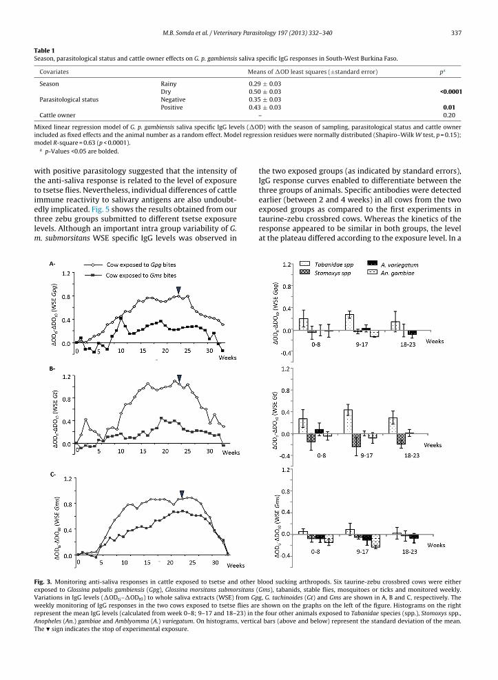
Immune reactivity to G. p. gambiensis, G. m. submorsitansand G. tachinoides WSE of animals experimentally exposedto tsetse flies (G. p. gambiensis and G. m. submorsitans) andto other blood sucking arthropods (tabanids, stable flies,mosquitoes and ticks) is shown in Fig. 3A, B, and C, respec-tively. An important induction of reactivity to WSE from alltsetse species was observed in the animal exposed to G. p.gambiensis. In contrast, IgG responses in the cow exposedto G. m. submorsitans showed less reactivity toward salivaryantigens from the palpalis tsetse group (G. p. gambiensis andG. tachinoides) as compared to those from G. m. submorsi-tans. Anti-saliva IgGs started to increase in between the6th and the 10th week of exposure (between 240 and 400tsetse bites taking into account that in average 80% of bloodmeals were successful) in both cows. Anti-saliva responsesthen reached a plateau from week 16 to 21 and startedto decrease steadily 5–6 weeks after the stop of exposure,returning to pre-exposure levels after 10 weeks.
Experimental exposure of the three other cows to stableflies, mosquitoes and ticks did not induce any cross reactiv-ity with none of the three tsetse species salivary antigens(Fig. 3). Nevertheless, cross reactivity was observed in thecow exposed to tabanids with WSE from G. p. gambiensisand G. tachinoides but not G. m. morsitans. In the view ofthese preliminary results obtained from a limited numberof animals, we postulated that G. m. submorsitans salivaryantigens were better suited to monitor tsetse contacts asit provided a good sensitivity to detect exposure to bothG. m. submorsitans and G. p. gambiensis and appeared to beassociated with a lower degree of cross reactions at least totabanid species. Noteworthy, tabanids are major pests ofcattle in the northern pastoral areas of Burkina Faso wherethey are found in very high densities (Pagabeleguem et al.,2012). This might have explained part of the intermedi-ate responses observed initially in some animals from thetsetse free area (Fig. 2). To test this hypothesis, we selectedserum samples from the tsetse free area (n = 20) and thetsetse infested area (n = 20) but having similar intermedi-ate IgG levels (0.26 ≤ �OD ≤ 0.53) to G. p. gambiensis WSEand repeated the ELISA test with G. m. submorsitans sali-vary antigens (Fig. 4). Confirming our hypothesis, highlysignificant differences were observed when G. m. submor-sitans WSE were used (p = 0.0007), probably because theseantigens provided a better specificity to detect exposure totsetse bites.
3.3. Anti-saliva responses and level of exposure to tsetsebites
In the tsetse infested area, cattle displayed an importantheterogeneity of anti-saliva responses. The fact that IgGlevels are in part explained by the season and are associated
M.B. Somda et al. / Veterinary Parasitology 197 (2013) 332– 340 337
Table 1Season, parasitological status and cattle owner effects on G. p. gambiensis saliva specific IgG responses in South-West Burkina Faso.
Covariates Means of �OD least squares (±standard error) pa
Season Rainy 0.29 ± 0.03Dry 0.50 ± 0.03 <0.0001
Parasitological status Negative 0.35 ± 0.03Positive 0.43 ± 0.03 0.01
Cattle owner – 0.20
Mixed linear regression model of G. p. gambiensis saliva specific IgG levels (�OD) with the season of sampling, parasitological status and cattle ownerel regres
included as fixed effects and the animal number as a random effect. Modmodel R-square = 0.63 (p < 0.0001).a p-Values <0.05 are bolded.
with positive parasitology suggested that the intensity ofthe anti-saliva response is related to the level of exposureto tsetse flies. Nevertheless, individual differences of cattleimmune reactivity to salivary antigens are also undoubt-
edly implicated. Fig. 5 shows the results obtained from ourthree zebu groups submitted to different tsetse exposurelevels. Although an important intra group variability of G.m. submorsitans WSE specific IgG levels was observed inFig. 3. Monitoring anti-saliva responses in cattle exposed to tsetse and other bexposed to Glossina palpalis gambiensis (Gpg), Glossina morsitans submorsitans (Variations in IgG levels (�ODti–�ODt0) to whole saliva extracts (WSE) from Gpweekly monitoring of IgG responses in the two cows exposed to tsetse flies arerepresent the mean IgG levels (calculated from week 0–8; 9–17 and 18–23) in thAnopheles (An.) gambiae and Amblyomma (A.) variegatum. On histograms, verticaThe � sign indicates the stop of experimental exposure.
sion residues were normally distributed (Shapiro–Wilk W test, p = 0.15);
the two exposed groups (as indicated by standard errors),IgG response curves enabled to differentiate between thethree groups of animals. Specific antibodies were detectedearlier (between 2 and 4 weeks) in all cows from the two
exposed groups as compared to the first experiments intaurine-zebu crossbred cows. Whereas the kinetics of theresponse appeared to be similar in both groups, the levelat the plateau differed according to the exposure level. In alood sucking arthropods. Six taurine-zebu crossbred cows were eitherGms), tabanids, stable flies, mosquitoes or ticks and monitored weekly.g, G. tachinoides (Gt) and Gms are shown in A, B and C, respectively. The
shown on the graphs on the left of the figure. Histograms on the righte four other animals exposed to Tabanidae species (spp.), Stomoxys spp.,l bars (above and below) represent the standard deviation of the mean.

338 M.B. Somda et al. / Veterinary Parasitology 197 (2013) 332– 340
0
0.1
0.2
0.3
0.4
0.5
0.6
0.7
0.8
0.9
WSE Gpg
Tsetse free area Tsetse infested area
∆OD
Tsetse free area Tsetse infested area
WSE Gms
p = 0.0980 p = 0.0007
Fig. 4. Antibody responses to G. m. submorsitans and G. p. gambiensis WSEin cattle from tsetse free and infested areas. IgG responses (�OD) to G.morsitans submorsitans (Gms) WSE of 20 animals from the tsetse freearea and 20 animals from the tsetse infested area displaying intermediateresponse to G. palpalis gambiensis (Gpg) WSE are shown. The line acrosseach black diamond represents the group mean, the vertical span of eachdiamond represents the 95% confidence interval (CI) for each group andovs
mm(cp
4
btiei
FdIagGb
Table 2Association between IgG levels directed against G. m. submorsitans WSEand the level of exposure to tsetse bites.
Variables Means of �ODleast squares(±standard error)
pa
Exposure group Control 0.02 ± 0.09Low 0.27 ± 0.07High 0.61 ± 0.07 0.0031
Time of exposure (weeks) – <0.0001
Mixed linear regression model of G. m. submorsitans saliva specific IgG lev-els (�OD) with the exposure group and time of exposure in weeks as fixedeffects and animal as a random effect. Model regression residues were nor-
verlap marks are drawn (√
2CI)/2 above and below the group mean. P-alues calculated by the non parametric Wilcoxon/Kruskal–Wallis test arehown.
ixed linear regression model (Table 2) including the ani-al number as a random effect, both the time of exposure
in weeks) and the exposure group were significantly asso-iated with increased anti-saliva responses (p < 0.0001 and
= 0.0031 respectively).
. Discussion
The host antibody response induced by the saliva oflood sucking arthropods has provided new perspectives
o evaluate host-vector contacts and specific anti-salivarymmunoassays are now more and more widely used inpidemiological studies to assess the disease risk or thempact of vector control measures (Fontaine et al., 2011).0
0,1
0,2
0,3
0,4
0,5
0,6
0,7
0,8
0,9
1
0 1 2 3 4 5 6 7 8 9 10 11
Time of exp osure (weeks)
high exp osure
low exp osure
unexp osed
∆O
Dti-∆
DO
t0(W
SE
Gms)
ig. 5. Monitoring of the tsetse anti-saliva response in cattle exposed toifferent tsetse exposure levels. The graph represents the evolution of
gG responses (�ODti − �ODt0) to Glossina morsitans submorsitans (Gms)ccording to the time of exposure in three groups of cows: high exposureroup (n = 4; 50 Gms flies twice a week); low exposure group (n = 4; 10ms flies once a week); unexposed group (n = 2). Vertical bars above andelow the lines are the standard errors of the group mean.
mally distributed (Shapiro–Wilk W test, p = 0.09); model R-square = 0.72(p < 0.0001).
a p-Values <0.05 are bolded.
An example is given by two recent studies performed toassess the efficacy of insecticide-treated nets (ITNs) in amalaria hypo-endemic transmission area (Drame et al.,2010) and of vector control interventions against Chagasdisease (Schwarz et al., 2011). The aim of this study was toevaluate if the cattle IgG responses directed against Glossinawhole salivary antigens could serve as a biomarker of expo-sure to tsetse flies and to study its main characteristics interms of kinetics, sensitivity and specificity by both fieldbased and experimental approaches.
In the tsetse infested area in South-West Burkina Fasowe found that G. p. gambiensis WSE specific IgG levels weremuch higher during the dry season compared to samplescollected during the rainy season or in cattle from the tsetsefree area. Such seasonal variation of antibody responsesto blood sucking arthropod’s saliva have been previouslyreported for mosquitoes (Elanga Ndille et al., 2012; Remoueet al., 2006) and sand flies (Hostomska et al., 2008), of whichdensities are largely influenced by the climatic conditions.At first sight, our results could appear surprising becausetsetse flies are abundant and more widely dispersed dur-ing the rainy season (Cuisance et al., 1985). But, our datafit well with the herd management system in the studyarea which is a typical pastoral extensive breeding sys-tem of partly sedentary type. In this system cattle spendthe rainy season in the vicinity of villages. During thisperiod cattle are poorly exposed to tsetse flies as water-ing of animals is made around rain-created ponds wheretsetse flies are absent. During the dry season animals areherded in transhumance in search of pastures and wateralong the permanent riverine network and are in close con-tacts with tsetse flies (Dayo et al., 2010; De la Rocque et al.,1999; Koné et al., 2011). In agreement with the hypoth-esis that anti-saliva responses are related to the level ofcattle exposure to tsetse bites is the finding that animalsdisplaying the highest IgG levels had an increased risk ofbeing infected by trypanosomes (p = 0.01). These results arewell in line with those of a previous longitudinal epidemi-ological survey carried out in the area, showing that thedry hot season is associated with an important increaseof AAT incidences (Dayo et al., 2010). Such associations
between anti-saliva IgG levels and the risk of infectionhave previously been described in malaria (Andrade et al.,2009; Drame et al., 2010; Remoue et al., 2006), Lyme dis-ease (Schwartz et al., 1991) and leishmanisasis (Rohousova
y Parasi
M.B. Somda et al. / Veterinarand Volf, 2006). Importantly however, whereas IgG levelsagainst G. p. gambiensis WSE appeared to provide a goodindicator of cattle-tsetse contacts, a number of animalsin the tsetse free area displayed intermediate responses(�OD > 0.4) suggesting that cross reactions with antibodydirected against the saliva of other arthropods may haveoccurred in these animals.
Experimental exposure of animals to different tsetsespecies and other blood sucking arthropods has enabledto confirm the above findings and to investigate the anti-genic properties of the saliva of the different tsetse coloniesavailable at CIRDES (G. m. submorsitans, G. p. gambiensisand G. tachinoides). We were first able to show that theantibody response against tsetse salivary antigens was adynamic process and that specific IgG responses decreasedrapidly (within 10 weeks) to the initial pre-exposure lev-els once exposure was stopped. These results are well inline with the strong season effect observed in cattle fromSouth-West Burkina Faso exposed to varying exposure lev-els to tsetse flies along the year. In addition, analysis ofIgG responses to G. m. submorsitans enabled to differentiatebetween two groups of cows exposed to different intensi-ties of tsetse exposure as shown in previous experimentalstudies led with other blood sucking arthropods such assand flies (Vlkova et al., 2011) or triatomines (Schwarz et al.,2009). Our results thus indicate that in cattle, IgG responsesto tsetse salivary antigens are at least partly related to thetsetse exposure level. Nevertheless important differenceswere also observed between animals of the same expo-sure group or breeds (zebu or taurine crossbreed cows)regarding both IgG levels and kinetics aspects. At the indi-vidual level, evaluation of tsetse contacts by anti-salivaantibodies may thus be impaired by other factors such asthe genetic background of the cow or other factors affectingthe responsiveness of B lymphocytes to salivary antigens.Nevertheless at the herd level IgG responses to tsetse sali-vary antigens appear to be indicative of the average levelof tsetse exposure. Finally, we recommend the use of G. m.submorsitans WSE, at least in West Africa, as these antigensin contrast to those from tsetse species from the palpalisgroup, provided both a good sensitivity and specificity todetect exposure to tsetse bites regardless of the tsetsespecies used for exposure. Furthermore our results suggestthat exposure to tabanids, which are important pests forcattle in many areas, has the potential to induce cross reac-ting antibodies with G. p. gambiensis salivary antigens. Afine analysis of the G. m. submorsitans immuno-sialome bya 2D-electrophoresis approach combined with mass spec-trometry is now currently underway to identify specificimmunogenic antigen candidates to develop a more stan-dardized and specific tool to measure cattle exposure totsetse bites.
With the PATTEC initiative, area-wide tsetse elimina-tion campaigns are multiplying in sub-Saharan Africa withthe objective of creating tsetse free area to promote breed-ing and the socio-economic development of these regions(Schofield and Kabayo, 2008; Vreysen et al., 2013). The
results presented above indicate that the cattle antibodyresponse to tsetse salivary antigens can be used as a directbiomarker of cattle exposure to tsetse flies. It thus consti-tutes an alternative tool complementary to the evaluationtology 197 (2013) 332– 340 339
of tsetse densities by conventional trapping methods. Thefact that cattle breeding is largely of pastoral type, resultsin a high mobility of herds in many regions. Thereforetsetse apparent densities measured by trapping at fixedsites are in many cases poorly representative of cattleexposure to tsetse flies. Monitoring specific IgG responsesto tsetse salivary antigens thus appear a more adaptedmethod to evaluate cattle exposure to tsetse bites. Further-more the rapid kinetics of apparition and disappearance oftsetse saliva antibodies are compatible with the monitor-ing of vector control campaigns (i) to assess their efficacyto reduce cattle-tsetse contacts and (ii) to detect early re-invasion phenomena after interventions.
Conflict of interest
The authors declare that they have no competing inter-ests.
Authors’ contributions
MSB carried out the exposure experiments, immuno-logical analyses and draft the manuscript. ZB conceivedthe study and participated in its design and coordination.AP participated to the study design and implementationof immunological assays and helped draft the manuscript.ED participated in animal experiments and immunologi-cal analyses. GKD participated in the design of the studyand provided cattle serum samples from tsetse infestedareas. IS and AS helped in the interpretation of resultsand drafting the manuscript. BB participated to the studydesign and coordination, analyzed the data and draftedthe manuscript. All authors read and approved the finalmanuscript.
Acknowledgments
We would like to thank Saïdou BOLI who took careof the animals at the cowshed, the CIRDES technicianswho helped in collecting tabanids and stable flies in thefield, Anna COHUET who provided An. gambiae females andIsabelle DALMAU for language editing of the manuscript.The work presented here was supported by the French Min-istry of Foreign Affairs (AIRES-Sud project), the “Institut deRecherche pour le Développment” (IRD) and the Interna-tional Foundation for Science (IFS). MBS received a PhDgrant from the “Service de Coopération et d’Action Cul-turelle” from the French Embassy in Burkina Faso.
References
Andrade, B.B., Rocha, B.C., Reis-Filho, A., Camargo, L.M., Tadei, W.P., Mor-eira, L.A., Barral, A., Barral-Netto, M., 2009. Anti-Anopheles darlingisaliva antibodies as marker of Plasmodium vivax infection and clinicalimmunity in the Brazilian Amazon. Malar. J. 8, 121.
Barclay, H.J., Hargrove, J.W., 2005. Probability models to facilitate a dec-laration of pest-free status, with special reference to tsetse (Diptera
Glossinidae). Bull. Entomol. Res. 95, 1–11.Bouyer, J., Solano, P., Cuisance, D., Itard, J., Frézil, J.L., Authié, E., 2010.Control methods in Trypanosomosis. In: Lefèvre, P.C., Blancou, J., Cher-mette, J., Uilenberg, G. (Eds.), Infectious and Parasitic Diseases ofLivestock, vol. 2. Lavoisier, Paris, pp. 1936–1943.

3 y Parasi
B
C
C
C
D
D
D
D
D
D
E
F
H
H
I
40 M.B. Somda et al. / Veterinar
udd, L., 1999. DFID-Funded Tsetse and Trypanosome Research andDevelopment Since 1980. Economic Analysis, Livestock ProductionProgramme. NRInternational, Chatham Maritime, 123 pp.
aljon, G., Van Den Abbeele, J., Sternberg, J.M., Coosemans, M., De Baetse-lier, P., Magez, S., 2006. Tsetse fly saliva biases the immune responseto Th2 and induces anti-vector antibodies that are a useful tool forexposure assessment. Int. J. Parasitol. 36, 1025–1035.
ourtin, F., Rayaisse, J.B., Tamboura, I., Serdebeogo, O., Koudougou, Z.,Solano, P., Sidibe, I., 2010. Updating the northern tsetse limit in Bur-kina Faso (1949–2009): impact of global change. Int. J. Environ. Res.Public Health 7, 1708–1719.
uisance, D., Février, J., Dejardin, J., Filledier, J., 1985. Dispersion linéaire deGlossina palpalis gambiensis et G tachinoides dans une galerie forestièreen zone soudano-guinéenne (Burkina Faso). Rev. Elev. Méd. Vét. PaysTrop 38, 153–172.
ama, E., Cornelie, S., Somda, M.B., Camara, M., Kambiré, R.F., Jamonneau,C., Demettre, V., Seveno, E., Bengaly, M., Solano, Z., Poinsignon, P.,Remoue, A., Belem, F., Bucheton, A.M.B., 2013. Identification of Glossinapalpalis gambiensis specific salivary antigens: towards the develop-ment of a serologic biomarker of human exposure to tsetse flies inWest Africa. Microb. Infect. 15, 416–427.
ayo, G.K., Bengaly, Z., Messad, S., Bucheton, B., Sidibe, I., Cene, B., Cuny,G., Thevenon, S., 2010. Prevalence and incidence of bovine trypanoso-mosis in an agro-pastoral area of southwestern Burkina Faso. Res. Vet.Sci 88, 470–477.
e la Rocque, S., Bengaly, Z., Michel, J.F., Solano, P., Sidibé, I., Cuisance,D., 1999. Importances des interfaces spatiales et temporelles entreles bovins et les glossines dans la transmission de la trypansomoseanimale en Afrique de l’Ouest. Rev. Elev. Méd. Vét. Pays Trop. 52,215–222.
elespaux, V., Dinka, H., Masumu, J., Van den Bossche, P., Geerts, S., 2008.Five-fold increase in Trypanosoma congolense isolates resistant to dim-inazene aceturate over a seven-year period in Eastern Zambia. Drug.Resist. Updat. 11, 205–209.
esquesnes, M., Dia, M.L., 2003. Trypanosoma vivax: mechanical trans-mission in cattle by one of the most common African tabanids, Atylotusagrestis. Exp. Parasitol. 103, 35–43.
rame, P.M., Poinsignon, A., Besnard, P., Le Mire, J., Dos-Santos,M.A., Sow, C.S., Cornelie, S., Foumane, V., Toto, J.C., Sembene,M., Boulanger, D., Simondon, F., Fortes, F., Carnevale, P., Remoue,F., 2010. Human antibody response to Anopheles gambiae saliva:an immuno-epidemiological biomarker to evaluate the efficacy ofinsecticide-treated nets in malaria vector control. Am. J. Trop. Med.Hyg. 83, 115–121.
langa Ndille, E., Doucoure, S., Damien, G., Mouchet, F., Drame, P.M.,Cornelie, S., Noukpo, H., Yamadjako, S., Djenontin, A., Moiroux, N.,Misse, D., Akogbeto, M., Corbel, V., Henry, M.C., Chandre, F., Baldet,T., Remoue, F., 2012. First attempt to validate human igg anti-body response to Nterm-34 kDa salivary peptide as biomarker forevaluating exposure to Aedes aegypti Bites. PLoS Negl. Trop. Dis. 6,e1905.
ontaine, A., Diouf, I., Bakkali, N., Misse, D., Pages, F., Fusai, T., Rogier,C., Almeras, L., 2011. Implication of haematophagous arthropodsalivary proteins in host–vector interactions. Parasites Vectors 4,187.
olmes, P.H., 1997. New approaches to the integrated control of try-panosomosis. Vet. Parasitol. 71, 121–135.
ostomska, J., Rohousova, I., Volfova, V., Stanneck, D., Mencke, N., Volf,P., 2008. Kinetics of canine antibody response to saliva of the sand fly
Lutzomyia longipalpis. Vector Borne Zoonotic Dis. 8, 443–450.tard, J., Cuisance, D., Tacher, G., 2003. Trypanosomoses: historique-répartition géographique. In: Lefèvre, P.-C., Blancou, J., Chermette,R. (Eds.), Principales maladies infectieuses et parasitaires du bétail,Europe et régions chaudes, vol. 2. Lavoisier, Paris, pp. 1607–1625.
tology 197 (2013) 332– 340
Kabayo, J.P., 2002. Aiming to eliminate tsetse from Africa. Trends Parasitol.18, 473–475.
Koné, N., N’Goran, E.K., Sidibé, I., Kombassere, A.W., Bouyer, J., 2011.Spatio-temporal distribution of tsetse (Diptera Glossinidae) and otherbiting flies (Diptera: Tabanidae and Stomoxinae) in the Mouhoun RiverBasin, Burkina Faso. Med. Vet. Entomol. 25, 156–168.
Moloo, S.K., Kabata, J.M., Gitire, N.M., 2000. Study on the mechanical trans-mission by tsetse fly Glossina morsitans centralis of Trypanosoma vivax,T. congolense or T. brucei brucei to goats. Acta Trop. 74, 105–108.
Murray, M., Murray, P.K., McIntyre, W.I., 1977. An improved parasitologi-cal technique for the diagnosis of African trypanosomiasis. Trans. R.Soc. Trop. Med. Hyg. 71, 325–326.
Pagabeleguem, S., Sangare, M., Bengaly, Z., Akoudjin, M., Belem, A.M.,Bouyer, J., 2012. Climate, cattle rearing systems and african animaltrypanosomosis risk in burkina faso. PLoS One 7, e49762.
Poinsignon, A., Cornelie, S., Remoue, F., Grebaut, P., Courtin, D., Garcia,A., Simondon, F., 2007. Human/vector relationships during humanAfrican trypanosomiasis: initial screening of immunogenic salivaryproteins of Glossina species. Am. J. Trop. Med. Hyg. 76, 327–333.
Poinsignon, A., Remoue, F., Rossignol, M., Cornelie, S., Courtin, D., Grebaut,P., Garcia, A., Simondon, F., 2008. Human IgG antibody response toGlossina saliva: an epidemiologic marker of exposure to Glossina bites.Am. J. Trop. Med. Hyg. 78, 750–753.
Remoue, F., Cisse, B., Ba, F., Sokhna, C., Herve, J.P., Boulanger, D., Simondon,F., 2006. Evaluation of the antibody response to Anopheles salivaryantigens as a potential marker of risk of malaria. Trans. R. Soc. Trop.Med. Hyg. 100, 363–370.
Ribeiro, J.M., Mans, B.J., Arca, B., 2010. An insight into the sialome of blood-feeding Nematocera. Insect Biochem. Mol. Biol. 40, 767–784.
Rohousova, I., Volf, P., 2006. Sand fly saliva: effects on host immuneresponse and Leishmania transmission. Folia Parasitol. (Praha) 53,161–171.
Schofield, C.J., Kabayo, J.P., 2008. Trypanosomiasis vector control in Africaand Latin America. Parasites Vectors 1, 24.
Schwartz, B.S., Ford, D.P., Childs, J.E., Rothman, N., Thomas, R.J., 1991. Anti-tick saliva antibody: a biologic marker of tick exposure that is a riskfactor for Lyme disease seropositivity. Am. J. Epidemiol. 134, 86–95.
Schwarz, A., Juarez, J.A., Richards, J., Rath, B., Machaca, V.Q., Castro, Y.E.,Malaga, E.S., Levy, K., Gilman, R.H., Bern, C., Verastegui, M., Levy,M.Z., 2011. Anti-triatomine saliva immunoassays for the evaluation ofimpregnated netting trials against Chagas disease transmission. Int. J.Parasitol. 41, 591–594.
Schwarz, A., Sternberg, J.M., Johnston, V., Medrano-Mercado, N., Anderson,J.M., Hume, J.C., Valenzuela, J.G., Schaub, G.A., Billingsley, P.F., 2009.Antibody responses of domestic animals to salivary antigens of Tri-atoma infestans as biomarkers for low-level infestation of triatomines.Int. J. Parasitol. 39, 1021–1029.
Shaw, A.P.M., 2004. Economics of African trypanosomiasis. In: Maudlin, I.,Homes, P.H., Miles, M.A. (Eds.), The Trypanosomiasis. CABI Publishing,Wallingford, United Kingdom, pp. 369–402.
Solano, P., Ravel, S., Bouyer, J., Camara, M., Kagbadouno, M.S., Dyer,N., Gardes, L., Herault, D., Donnelly, M.J., De Meeus, T., 2009. Thepopulation structure of Glossina palpalis gambiensis from island andcontinental locations in Coastal Guinea. PLoS Negl. Trop. Dis. 3, e392.
Van den Bossche, P., de La Rocque, S., Hendrickx, G., Bouyer, J., 2010. Achanging environment and the epidemiology of tsetse-transmittedlivestock trypanosomiasis. Trends Parasitol. 26, 236–243.
Vlkova, M., Rohousova, I., Drahota, J., Stanneck, D., Kruedewagen, E.M.,Mencke, N., Otranto, D., Volf, P., 2011. Canine antibody response to
Phlebotomus perniciosus bites negatively correlates with the risk ofLeishmania infantum transmission. PLoS Negl. Trop. Dis. 5, e1344.Vreysen, M.J., Seck, M.T., Sall, B., Bouyer, J., 2013. Tsetse flies: Their biologyand control using area-wide integrated pest management approaches.J. Invertebr. Pathol. 112, S15–S25.
Related Documents