i The epidemiology and management of traumatic facial fractures in children under the age of 15 years recorded in a Johannesburg General hospital over a period of 5 years GERHARD FOUCHE STUDENT NUMBER 685351 MSC DENT SUPERVISOR: DR MZUBANZI MABONGO CO-SUPERVISOR: DR MAPHEFO THEKISO A Research Report submitted to the Department of Maxillofacial and Oral Surgery, Faculty of Oral Health Science, University of Witwatersrand, Johannesburg, in partial fulfilment of the requirements for the degree of Master of Science in Dentistry (Witwatersrand) performed partly in the Department of Maxillofacial and Oral Surgery 18 May 2017

Welcome message from author
This document is posted to help you gain knowledge. Please leave a comment to let me know what you think about it! Share it to your friends and learn new things together.
Transcript
i
The epidemiology and management of traumatic facial fractures
in children under the age of 15 years recorded in a Johannesburg
General hospital over a period of 5 years
GERHARD FOUCHE
STUDENT NUMBER 685351
MSC DENT
SUPERVISOR: DR MZUBANZI MABONGO
CO-SUPERVISOR: DR MAPHEFO THEKISO
A Research Report submitted to the Department of Maxillofacial and Oral Surgery, Faculty of Oral Health Science, University of Witwatersrand, Johannesburg, in partial
fulfilment of the requirements for the degree of Master of Science in Dentistry (Witwatersrand) performed partly in the Department of Maxillofacial and Oral
Surgery
18 May 2017
ii
Declaration
I, Gerhard Fouche, declare that this research report is my own, original work. It is being
submitted for the Degree of Master of Dentistry in the Department Maxillofacial and Oral
Surgery, University of the Witwatersrand, Johannesburg. It has not been submitted before for
any degree or examination in any other University.
____________________________________ [Signature of Candidate]
……… day of …………………… [month], 20…..
iii
Dedication
I dedicate this work to:
The Almighty God, my wife Maryke and our son and in beloved memory of my mother.
Acknowledgments
I would hereby like to thank Dr. M Mabongo for his loyal support, guidance, knowledge, and
expertise. I am truly grateful and I appreciate it.
Special thanks to Dr. M Thekiso for your valuable comments, briefing and academic
knowledge.
I would also like to thank:
- The CEO / Clinical Director of the CMJAH, Ms. G Bogoshi, for granting me access
to the Paediatric Casualty ward and allowing me the usage of patient admission books
and patient records;
- The CEO / Head of Wits Oral Health Centre, Prof P Hlongwa for allowing me the use
of patient records in the Department of Maxillofacial and Oral Surgery;
- The Clinical Head of Department of Surgery, Dr. TE Luvhengo for granting me
access to the Department of Surgery and usage of patient records.
Special thanks to the sisters and staff, working in the wards of Paediatric casualty and
Paediatric surgery for allowing me the time to gain information from the admission books.
I want to express gratitude to the friendly and helpful staff of the CMJAH working in the
patient records storage division for your assistance during my data collection.
I give special thanks to my wife, parents, family, and friends for their loyal support,
compassion and understanding and allowing me the time to do this.
Above all, I am grateful to my Creator. I thank the gracious Lord for His inspiration during
desperate times, His guidance and for always being there.
iv
Abstract
Aim: This study aim was to determine the prevalence of traumatic facial fractures in children
under the age of 15 years who presented at the Charlotte Maxeke Johannesburg Academic
Hospital (Department of Maxillofacial and Oral surgery, Wits Oral Health Centre and
Department of General Surgery) over a period of 5 years from 2011 to 2015.
Objective: This study objective was to determine the prevalence of facial bone fractures, the
age and gender mostly affected, the place and cause of facial fracture, the type and
distribution of facial fractures, the prevalence of associated injuries as well as the
management of facial fractures.
Materials and methods: This is a retrospective study based on data retrieved from patient
records. Four thousand and forty-four files were used for the analysis of this study. Data
collected from existing patient records included: department of admission; date of admission;
age; gender; who accompanied the patient to hospital; ethnicity; medical history; number of
days between date of injury and date of arrival; place of injury; cause of fracture; site of
fracture; type of fracture; teeth affected; associated facial injuries; ophthalmic or globe
involvement; associated bodily injuries; specialized consultation; radiographs; management
and treatment of injuries.
The results: Cases numbering 171 children under the age of 15 years with facial bone
fractures were retrieved from patient records. Majority of the patients were males. Mean age
of patients was 6.45 ± 3.47 years. Most common places of injury included the home, school
and other places which refer to any other environment, surrounding area or public place in the
home or school. Most common causes of paediatric facial fracture injury are pedestrian-
vehicle accidents (PVAs), motor vehicle accidents (MVAs) and falls, with a significant
association between the cause of fracture and the age of the patients. Two hundred and forty
seven facial bone fractures were detected. Most common site of facial fracture was the frontal
bone followed by the orbital bone. Fifty six paediatric patients had multiple facial bone
fractures. Forty nine children had an associated tooth injury. Of the 435 facial soft tissue
injuries (STIs) detected, 91.0% were extra orally. Most common STIs were lacerations,
abrasions and soft tissue swellings. Seventy four of the 117 paediatric patients with
associated bodily injuries, had multiple bodily injuries. Twelve patients with facial bone
fractures showed results of ophthalmic or globe involvement. One hundred and nine (63.7%)
patients with facial bone fractures were managed conservatively, whilst management of 58
v
(34.0%) patients included surgical intervention. Four (2.3%) patient records did not indicate
any treatment.
Conclusion: Most facial bone fractures were recorded in children under the age of 10 years
and male gender was most affected. Aetiology of facial fractures seems to be more similar in
male and female children at a younger age, whereas more variation in aetiology occurs in
gender during adolescence. This study suggests that the school is the safest place for children.
The seasonal variance in terms of paediatric facial fracture prevalence is most likely related
to an increased outdoor activity during the months of summer. Possible reasons that
contribute to home and other places as high-risk areas for facial fractures in children could
either be lack of parental supervision and responsibility, or the absence of safety measures.
More children were involved in PVAs than MVAs. The negligence of drivers, lack of road
safety awareness, insufficient pedestrian safety measures or inadequate parental control is
potential factors to contribute to the high prevalence of MVAs and PVAs as a major
aetiological factor amongst children in these affected communities. From this study, it seems
that the mechanism of injury and stage of facial development shows a noticeable influence on
the type and site of the bone fracture and that the frequency of aetiological factors changes
with age. Management and treatment of paediatric facial fractures should be with a good
understanding of the patterns of anatomical growth and stages of skeletal development.

vi
Table of Contents Declaration ......................................................................................................................................................... ii
Dedication ......................................................................................................................................................... iii
Acknowledgments ............................................................................................................................................. iii
Abstract ............................................................................................................................................................. iv
List of figures ................................................................................................................................................... viii
List of tables ...................................................................................................................................................... ix
Abbreviations ..................................................................................................................................................... x
CHAPTER 1: INTRODUCTION ................................................................................................................................... 1
CHAPTER 2: LITERATURE REVIEW (background) .................................................................................................... 2
CHAPTER 3: RATIONALE .................................................................................................................................... 2324
AIMS AND OBJECTIVES ..................................................................................................................................... 2424
AIM ............................................................................................................................................................... 2424
STUDY OBJECTIVES ....................................................................................................................................... 2424
CHAPTER 4: METHODOLOGY ............................................................................................................................ 2525
4.1 Materials and methods: .................................................................................................................. 2525
4.2 Study design: ................................................................................................................................... 2626
4.3 Study population and Sample ......................................................................................................... 2627
4.4 Data collection ................................................................................................................................ 2727
4.5 Data process and analysis ............................................................................................................... 2728
4.6 Limitations ....................................................................................................................................... 2828
ETHICAL CONSIDERATIONS ............................................................................................................................... 2828
Ethical Clearance .......................................................................................................................................... 2828
Patient confidentiality .................................................................................................................................. 2828
CHAPTER 5: RESULTS ............................................................................................................................................ 29
Department ...................................................................................................................................................... 29
Gender .......................................................................................................................................................... 3029
Referral ......................................................................................................................................................... 3029
Age ................................................................................................................................................................... 30
Ethnicity........................................................................................................................................................ 3130
Medical History ............................................................................................................................................ 3130
Date of arrival ............................................................................................................................................... 3130
Place of injury ............................................................................................................................................... 3131
Date of arrival ............................................................................................................................................... 3131
Cause of fracture .............................................................................................................................................. 32
Statistical analysis ............................................................................................................................................ 35
Site of fracture ................................................................................................................................................. 36
Type of fracture ................................................................................................................................................ 38
vii
Facial side of fracture ....................................................................................................................................... 40
Associated teeth injury..................................................................................................................................... 40
Associated facial soft tissue injuries ................................................................................................................. 40
Associated bodily injuries ................................................................................................................................. 42
Associated ophthalmic or globe involvement .................................................................................................. 42
Specialized consultation ................................................................................................................................... 43
Radiographs ...................................................................................................................................................... 43
Management .................................................................................................................................................... 45
CHAPTER 6: DISCUSSION ...................................................................................................................................... 46
CHAPTER 7: CONCLUSION .................................................................................................................................... 56
REFERENCES .......................................................................................................................................... 58
Appendix A: Facial Fracture Proforma Questionnaire .......................................................................... 63
Appendix B: Letter for ethical clearance ............................................................................................... 66
Appendix C: Letter from CEO of CMJAH, Ms G Bogoshi ....................................................................... 68
Appendix D: Letter from Head of Dental School: Prof P Hlongwa ........................................................ 69
Appendix E: Letter from Clinical Head of Department of Surgery: Dr TE Luvhengo ............................ 70

viii
List of figures
Figure 1: Age category of patients………………………………………………………30
Figure 2: Monthly distribution of paediatric facial fractures in each year………………31
Figure 3: Causes of facial fracture among the patients………………………………….32
Figure 4: Site of fracture………………………………………………………………...36
Figure 5: Associated facial injury……………………………………………………….41
Figure 6: Management of fracture………………………………………………………45
ix
List of tables
Table 1: Number of files used from year of study……………………………………….29
Table 2: Cause of fracture, according to age…………………………………………….34
Table 3: Association between place, year and cause of fracture and demographics…….35
Table 4: Association between place, year, cause, site of fracture and demographics…...35
Table 5: Multinomial logistic regression………………………………………………...35
Table 6: Site of fracture, according to age……………………………………………….38
Table 7: Type of fracture, according to age……………………………………………...40

x
Abbreviations MVA Motor vehicle accident
NOE Naso-orbito-ethmoidal
IMF Intermaxillary fixation
MMF Maxillomandibular fixation
PAN Panorex
PA Posterior anterior
CT Computerized tomography
3D Three-dimensional
WITS University of Witwatersrand
UCT University of Cape Town
CMJAH Charlotte Maxeke Johannesburg Academic Hospital
TMJ Temporomandibular joint
PVA Pedestrian-vehicle accident
FFH Fall from height
MFOS Maxillofacial and Oral surgery
ADHD Attention deficit hyperactivity disorder
STI Soft tissue injury
ENT Ear, nose and throat
FAST Focussed assessment with sonography for trauma
2D Two-dimensional
ORIF Open reduction internal fixation
CRMF Closed reduction maxillary fixation
1
CHAPTER 1: INTRODUCTION
Severe head and facial injuries (intentional or unintentional) have globally been recognized as
some of the leading causes of mortality and morbidity, prolonged hospital admissions and
higher injury severity scores in young children. (1 – 7)
In the United States, it has been reported that head trauma remains the most common cause of
death due to injury in the paediatric age group. (4 - 6) In conjunction, traumatic dental-alveolar
injuries among young children are also being considered as a serious public dental health
problem in many countries. (1, 4, 8 - 12)
Orofacial trauma can be regarded as extra-oral or intra-oral injuries resulting from an external
force. These intra- and extra-oral injuries include trauma to facial soft tissues and facial
bones, including the dental-alveolus, teeth, oral soft tissues, and the tongue. (3, 8, 13)
The impact of facial trauma associated with severe injury often results in a subsequent
functional and aesthetic defects in the growing child, (6, 10, 14) and significantly affects the
normal social functioning and quality of life of children. (15) The important functions, such as
speech, mastication, respiration, and deglutition may adversely be affected. (16)
In conjunction with the specific anatomic features of paediatric patients, the social-economic
impact of social-, cultural-, environmental factors, human behaviour and specific oral factors,
show great influence in the incidence and cause of orofacial trauma in children. (2, 4, 12, 14, 17 -
24)
The variation in the degree of motor development skills is frequently related to the specific
cause or type of injury. (2, 3, 8, 12, 13) Simultaneously in early childhood the development of
sensory systems, neural control mechanisms, cognitive ability and avoidance skills are not yet
sufficient to sustain injury. (7)
The paediatric patient can be categorized according to various stages of growth and
development. (25) (Neonate: newborn up to 1 month; Infant: two months to two years; Child:
three to twelve years; Adolescent: thirteen to sixteen years)

2
CHAPTER 2: LITERATURE REVIEW (background)
According to various studies, facial fractures occur less frequently in children than in adults
and are more often minimally displaced. (26) Paediatric patients are also more likely than
adults to sustain greenstick or incomplete fractures, and fractures that are less likely to have
multiple communications. (26, 27) Facial fractures in children often appear to be long and
irregular in character with the fracture generally running inferiorly and anteriorly. (26)
The reduced frequency of facial fractures in children, compared to adults, are most possibly
due to the following unique anatomic features: (1, 3, 5, 6, 17-19, 21-23, 25-28)
Under-developed facial skeleton and para-nasal air sinuses;
Craniofacial disproportion;
The thick layer of surrounding adipose tissue that covers the more elastic, thin cortical
bones of the paediatric facial skeleton;
un-erupted dentition (presence of tooth buds and developing crypts) of the mandible
and maxilla, and the lack of sinus Pneumatisation provides additional strength and
stability of the jaws;
Abundance of cartilage and cancellous bone, low mineralization and underdeveloped
cortex, along with the more flexible suture lines of the facial bones and indistinct
corticomedullary junction, confer greater intrinsic elasticity and flexibility on the
paediatric facial skeleton;
Increased number of fat pads around the upper and lower jaws.
The striking feature of the newborn skull is the small size of the facial portion in comparison
with the cranial part. At birth the ratio of cranial to facial volume is approximately 8:1 which
decreases significantly with age. By the age of 5 years, these relative proportions are nearly
4:1 and with the completion of growth the ratio is closer to 2.5:1. Furthermore, the face of the
young child is more retruded relative to the protrusive position of the skull. (2, 5, 6, 18, 21-23, 25, 28 -
31)
The “protecting” skull with its larger volume is the unique feature of young children that
associate with the lower incidence of midface and mandibular fractures and higher incidence
of skull/cranial fracture (including frontal, superior orbital and upper nasal injuries),
especially in children under the age of 5 years. (21, 27, 28, 30) The skull absorbs the full force of
the initial impact, thus protecting the face.
3
With age and physiological development, the midface and the mandible becomes more
prominent through the increase of facial growth in a forward and downward direction. From
this development, the lower incidence of cranial and frontal injuries and higher incidence of
facial trauma (specifically bone fracture injuries of the midface, mandible due to its relative
prominence and orbital floor injuries due to the aeration of the maxillary sinus) appears with
age. (5, 6, 18, 21, 22, 23, 28, 31)
With age, the more involvement in sports and unsupervised physical activity give rise to a
peak in fracture incidence during puberty and adolescence. (12, 28) Other peaks of fracture
incidence have been observed between the 6 – 7 years of age, associated with the beginning
of school attendance and a second peak at 12 – 14 years of age, related to physical activity
and participation in various sports. (5, 19, 22, 23)
Seasonal variations are also evident in different countries, with peak frequencies of facial
fractures that occur during certain months of increased outdoor activity and that relate to the
involvement in specific sports. (1, 2, 18, 19, 20, 28, 31, 32)
The previous publications also show a higher incidence (approximately twice as frequently)
of facial fractures in male patients than female patients, in all age groups worldwide. (29)
Although the gender differences show to be less significant and aetiologies more similar in
both sexes at younger ages, substantial variation in fracture incidence occur between sexes
during adolescence, which often attribute to more intensive and frequent involvement in
sports, physical activity and dangerous behaviour among boys. (1, 2, 3, 4, 8, 10, 13, 14, 17, 18, 19, 22, 23,
25, 28, 29, 31, 33, 34, 35)
With interest in paediatric patients involved in polytrauma, children have a higher surface-
area-to-body volume ratio with lower total blood and stroke volumes than adults and
therefore have a higher risk (than adults) for hypotension, hypothermia, and hypoxia.
Especially after massive blood loss, due to the pooling of blood in the peripheral vasculature
rather than supplying the viscera, which can result in rapid decompensation. (1, 8, 25, 28)
Children also have a higher metabolic rate, oxygen demand and cardiac output than adults,
which result in a low physiologic reserve during resuscitation. (25, 28) The smaller body mass
of children compared to adults indicate a greater force per unit of body area during an episode
of trauma. (25) The impact of trauma on children often results in multiple internal organ
injuries due to their incomplete calcified skeleton that is close to the internal organs together
with the presence of less fat and more connective tissue. (1, 8, 25) Not only do children
4
frequently swallow air when injured or frightened, which results in gastric dilatation, but also,
infants have relative narrow nasal air passages that can easily obstruct. (1, 8)
Thus, there are various anatomic, physiologic and psychological differences between children
and adults that significantly influence the consequence and especially the treatment of
trauma. (6, 17, 23, 25)
Previous retrospective studies of maxillofacial trauma in children have shown:
Not only a lower occurrence of facial trauma among children compared to adults
comprising only 3.0% – 6.0% of all facial fractures, (1, 27) but also revealed that
craniofacial skeletal injuries comprise of less than 10.0% of all facial fractures (21)
(some indicate less than 15.0% (28));
That less than 1.0% of facial fractures occurred in children under the age of 5 years; (1
- 3, 5, 8, 19, 21, 25, 28, 29, 36)
That 1.0% - 14.7% of facial fractures occurred in children under 16 years of age; (1 - 3,
5, 8, 19, 22, 23, 25)
That the rate of incidence in children under 12 years of age ranges from 1.5% – 8.0%
of all facial fractures treated in trauma centers. (29, 33, 36)
METHODS OF INJURY
Although the most common aetiology of facial fractures in children varies from one country
to another, this study, therefore, aims to compare local methods of injury with other studies
globally. Various studies from across the world state that the most frequent causes of facial
injuries and fractures in children are: (1-3, 5, 6, 8, 9, 13, 14, 16, 18, 19, 21, 23, 24, 28, 33)
- falls (either from height, slip or trip);
- road traffic crashes or motor vehicle accidents,
- sports-related injuries,
- bicycle,
- social play;
- pedestrians;
- crushes;
- birth;
- violence;

5
- assaults;
- child abuse;
- burns
Motor vehicle accidents
Motor vehicle accidents (MVAs) seem to be one of the major causes of unintentional
maxillofacial and head injuries in the paediatric population. (4-6, 21-25, 27, 28, 37) The prevalence
of these injuries ranges from 34.2% - 57.8% which increases with age (13 – 19 years). (4, 8) A
10-year retrospective study in 2002 from Portugal has reported a 53.3% incidence, which
mostly affected children 16 – 18 years of age. (22) A global childhood unintentional injury
survey of four cities reported MVAs as the major cause of morbidity in children of which
over 70.0% were males above 5 years of age and that pedestrians accounted for most children
suffering road traffic injuries. (37)
Falls
Falls (together with slips and trips), regarded as a low-velocity force are frequently the
initiating event in paediatric trauma, with the major cranial or facial damage typically caused
by a hard or acutely angled object or surface at impact. (2, 7) A child’s head with its large mass
and volume often becomes the major point of contact when the body falls, which results in
the predominance of craniofacial trauma often associated with mild to severe brain injury. (7,
27) Falls are also regarded as one of the main causes of traumatic dental injuries among
preschool children. (2, 3, 8, 13, 14, 16, 18, 24, 28, 34, 35)
A study from the Cheng Kung University Hospital in Taiwan done in 2002, reported a
bimodal incidence, with peaks in children under 1 year of age and between 5 - 8 years of age.
Eighty-three percent of bimodal incidence occurred in children under the age of 6 years,
which confirmed a decrease incidence with the level of development. They also reported
most of the craniofacial injury sites to the anterior of the head in a T-shape distribution,
which involves the forehead, nose, lip, and chin. (2, 7)
A Nigerian study done in 2004 reported falls at a 24.3% incidence rate, which mostly
occurred in children under 5 years of age. (23) Falls have been recorded as the most common
cause of injury at 44.0% in a study from New Zealand done in 2000, which also showed a
predominance in children under the age of 5 years. (13) Two studies were done in Brussels,

6
one at the Royal children’s hospital, Belgium (2005) and the other in Brisbane (2002) also
indicated falls as the predominant cause of injury in boys and girls, associated with
significant trauma to teeth and associated structures. (10, 35) From a study done in 2007 in
Ankara, Turkey, falling while walking or running were reported as the most common
aetiology for oral and traumatic dental injuries frequently among children 6 and 8 - 10 years
of age. (32)
Sports-related injuries
Sports-related injuries in children also contribute to a major part of facial trauma in children.
(24) However, various rates of incidence have been reported from different countries. An
incidence rate of 31.8% has been reported from a study in Austria in 2004 (1) compared to the
0.2% incidence from a study in Chile in 2009. (2) Four independent studies conducted in
India have reported different rates of incidence regarding sports-related injuries in children.
The incidence rates of sports-related injuries from these Indian studies resulted from 13.5% -
42.0%. (3, 8, 18, 15) The type and incidence of facial injuries in children often relate to the
intensity and velocity of sports being played. (15, 24) As motor skills improve between the ages
of 10 - 14 years, sporting injuries become more prevalent. (28) Age, gender, anatomic risk
factors (over jet, incompetent lips, mouth breathing habit, malocclusion, subjects with fixed
orthodontic treatment), type of sport, and seasons when the sport is often played is associated
with sport-related facial injuries in children and adolescents. (24)
Child Abuse
Child abuse is not an uncommon cause of facial injury. (23) Many cases of child abuse involve
trauma to the mouth, face, and head. (38) Repeated injuries, multiple injury sites and
questionable circumstances surrounding the injury should raise suspicion of possible abuse. (28) The various studies have shown that as many as 50.0% – 75.0% of cases of child abuse
involve trauma to the mouth, face, head, and neck (which mostly include soft tissue
lacerations, mandibular and maxillary injuries, and coronal fractures of the maxillary
incisors). (25,38) From a study done by Cavalcanti in Brazil, 56.3% of the abused children
between 0 – 17 years of age had facial injuries, the prevalence of the abused children were
higher among male victims and it showed a higher incidence with age especially in those 11 –
15 years of age and adolescents. (38) This study has also shown a significant association
between a number of injuries and gender and of the number of existing injuries and the
7
presence of oral injuries. (38) Whilst other studies reported a higher frequency of child abuse
in girls and children under 10 years of age. (38)
Violence
Although much research focuses on unintentional injury in the United States, there is a
growing interest in the injury attributable to violence. (9) The incidence of assaults and
interpersonal violence vary from different countries, is an unusual cause of facial fracture in
the Paediatric population and is more commonly seen in older age groups. (9, 23, 28) Different
rates of incidence regarding violence/assault as a major cause of facial fractures in children
have been reported from various previous studies which included the following results: an
incidence rate of 48.0% from a study done in South Africa in 1992 (33), 24.3% from a study in
Indian children in 2012 (18), 38.0% from a study in Korea in 2012 (19), 22.6% from a study in
Brazil in 2005 (39) and 59.0% from a study at 3 trauma centers in Los Angeles in 2008. (9)
Violence has a disproportionate impact on vulnerable youth and the rate of morbidity and
mortality in children. (9) Firearm injuries, stab wounds and blunt trauma are assault-related
injuries that often associated with acts of interpersonal and physical violence and mostly
require medical attention. (9) Previous studies in the United States noted that poverty and
substance abuse (the use of alcohol and drugs in particular) have been closely related to
intentional injury and interpersonal violence among adolescents. (9) Children between 13 - 18
years of age show a higher incidence of facial fractures than of those 0 - 6 years of age. (19) In
a nationwide community sample study among English children between 4 - 15 years of age
the male gender, lower socioeconomic status, single-parent home, Hyperactivity and conduct
disorder children were mostly associated with the occurrence of facial injury. (9) Therefore,
recognizing some of these markers can be used to identify adolescents at risk and possibly
serve as a basis for secondary preventative efforts. (9)
Previous retrospective studies also show the following:
In contrast with the more constant patterns of facial fracture observed in adults, the
wide variety of paediatric injuries represent a combination of mechanical force and
anatomic features unique to the child’s stage of development; (3, 8, 28)
Younger children often sustain injuries from low impact/low-velocity forces such as
falls and older children are more commonly exposed to high impact/ high-velocity
forces; (23, 28)

8
Infants below 2 years of age, more often sustain injuries to the frontal region with
isolated or non-displaced fractures, whereas older children are more prone to injuries
of the chin/mouth region. (28) Thus, with the frontal bone being the most common site
of fracture in young children, an increase in frontal sinus fracture after pubertal sinus
pneumatisation occurs, which often associated with other facial bone fractures as well
as central nervous system involvement. (28)
The most frequent anatomic distribution of fracture/injury in the lower face comprises
the mandible, the mid-face includes the maxillary alveolus, nose, zygomatic bone,
maxilla and Le fort I,II,III fractures and the upper face constitutes mostly the naso-
orbito-ethmoidal (NOE), orbital and frontal-orbital areas. (25)
TYPES OF FRACTURES
Mandibular fractures
Mandibular fractures commonly occur in several locations depending on the type of injury,
direction, and force of the trauma. Mandibular fractures can be classified according to its
anatomic location. The fractures are designated as occurring in either the symphysis, para-
symphysis, alveolus, body, angle, ramus, neck, condyle or coronoid of the mandible. (30)
Mandibular fractures can also be classified according to the type of fracture which
categorizes the fractures either as greenstick, simple, comminute or compound. (30) These
categories describe the condition of the bone fragments at the fracture site and possible
communication with the external environment.
Greenstick fractures involve incomplete fractures with flexible bone and exhibit minimal
mobility on palpation.
A simple fracture is a complete transection of bone with minimal fragmentation at the
fracture site.
In a comminute fracture, the fractured bone is left in multiple segments.
A compound fracture results in communication of the margin of the fractured bone with the
external environment. Bone would be exposed through the oral mucosa, or soft tissues may
be intact when the fracture is in the teeth bearing area. Thus, by definition, any jaw fracture
within a tooth-bearing segment is an open or compound fracture.
Mandibular fractures can either be favourable or unfavourable. (30) In a favourable fracture,
the direction of the fracture line and the muscle pull (of the masseter muscle) resists
9
displacement. An unfavorable fracture results in the displacement of the fractured segments
from the pull of the masseter muscle. (30)
Earlier studies stated that mandibular fractures in the paediatric subpopulation are relatively
prominent, comprise of 20.0% – 50.0 % of all childhood fractures and is reported as the most
common facial fracture site. (2, 3, 8, 14, 16, 21 - 23, 26 - 29, 33, 36, 40) The fracture patterns vary with age
and although the incidence of condylar fractures is initially high (14.5% - 60.0%) (25) and
decrease with age, fractures of mandibular body and angle are initially infrequent but increase
with age. (8, 14, 17, 18, 26, 27) The thin neck and highly vascularized nature of the paediatric
condyle relate to the increased incidence of intra-capsular condyle fractures in children under
the age of 6 years, presenting bilateral in 20.0% of cases. (2, 5, 8, 27, 28, 40) Above the age of 6
years condyle fractures tend to occur more frequently in the sub-condylar and neck region
(extracapsular). (28) Symphysis and para-symphysis fractures also seem to be more typical. (28,
30) Strikingly, a large proportion of paediatric patients with mandibular fractures (30.0% –
60.0%) often have serious associated intra-abdominal, neuro-cranial or orthopaedic injuries
determined by the force required to result in such injuries. (26)
Midfacial fractures
Midfacial fractures include fractures affecting the maxilla, the zygoma, and the NOE
complex. (30) Midfacial fractures can be classified as Le Fort I, II, or III fractures,
zygomaticomaxillary complex fractures, zygomatic arch fractures, NOE fractures, palatal and
dental alveolar fractures. (30)
Le Fort I fracture: frequently results from the application of a horizontal force to the maxilla,
which fractures the maxilla through the maxillary sinus and along the floor of the nose. The
inferior portion of the maxilla is separated in a horizontal fashion, extending from the
piriform aperture of the nose to pterygoid maxillary suture area, thus separating the maxilla
from the pterygoid plates, nasal- and zygomatic structures. (30)
Le Fort II fracture: frequently results from forces that are applied in a more superior position.
It involves the separation of the maxilla and attached nasal complex from the cranial base,
zygomatic orbital rim area, and pterygoid maxillary suture area, but the zygomatic arches are
intact. (30)
Le Fort III fracture: the results when a horizontal force is applied at a level superior enough
to completely separate the midface from the cranial base at the level of the NOE complex and

10
zygomaticofrontal suture area. The fracture also extends through the orbits bilateral and
results in a craniofacial separation. (30) Mid-facial fractures are isolated or occur in a
combination of the above-mentioned injuries. (30)
According to the previous studies, the incidence of midface fractures appears to be infrequent
in children and account for 1.2% - 20.0% of paediatric facial fractures. (2, 5, 21, 23, 28) Less than
5.0% appears to be in children under the age of 12 years with the exception of nasal and
maxillary alveolar defects. (31) Both nasal and dento-alveolar injuries are often managed in the
outpatient setting and are common injuries among children. (1, 33) These injuries seldom
appear in the paediatric facial fractures statistics. (2, 30, 27, 28)
The nasal bones are the least resistant of the facial skeleton, constitute nearly 50.0% of all
facial fractures in children and are often reported as the most common facial bone fracture in
children. (2, 3, 19, 28)
The incidence of dento-alveolar injuries associated with facial fractures has been reported to
be as high as 48.0%, especially in children under the age of 10 years. (27) Even an incidence
rate of 76.3% from a 10-year study in Austria in 2000 has been reported. (1)
The zygomatic complex fractures appear to be the most common midface fracture in children, (28) with an incidence of 7.0% - 41.0% of zygoma fractures. (2) Le Fort fractures are almost
never seen before the age of 2 years, but above the age of 5 years, when the maxillary sinus
expands and the permanent teeth erupt, the incidence of mid-face fractures increases. (5, 22, 23,
28) It appears to affect children between 13 to 15 years of age (after 10 years) more often. (5, 22,
23, 28) Greenstick fractures and the elastic characteristics are often displayed in paediatric
zygomatic and mid-facial fractures where the fracture lines are often “impacted” instead of
being clean breaks with complete displacement. (27) These fractures are often seen with high-
energy injuries, are often multilevel and rarely isolated. (27)
Upper facial fractures
Upper facial fractures or injury would refer to the trauma of the roof of the frontal bone, orbit
and NOE bones. (25)
Orbital fractures constitute 20.0% (5.0% - 25.0% (31) and 3.0% – 45.0% (2)) of facial fractures
in children, often resultant from the transmission of a force directly from the orbital ring to
the thin orbital walls, or indirectly from the hydraulic pressure effect of displaced orbital soft

11
tissues. (28) The orbital cavity itself is bound by the orbital roof, lateral and medial walls, and
orbital floor. Some of these boundaries display changes in structural integrity, closely related
to the maxillary-, frontal- and ethmoid sinus pneumatisation different stages of development.
(2, 21, 31)
Prior to the frontal sinus development, it appears that orbital roof fractures are more apparent
in the very young, (27) however, orbital floor fractures are more apparent in older children due
to the expansion of the maxillary sinus beyond the equator of the globe. (28) The age at which
the probability of an orbital floor fracture exceeds that of an orbital roof fracture is
approximately at 7 years of age. (28) Thus, the orbital floor becomes more susceptible to
fracture in later childhood. (28, 31) It has been noted in previous studies that associated injuries
(to head and neck, neurological such as concussion, depressed skull fractures, intracranial
haemorrhage, long bone fractures, pelvic fractures, chest/abdominal trauma) are more
commonly found together with orbital fractures in children than in adults. These paediatric
patients appear to have more severe associated injuries to the head and chest with a
considerably higher overall mortality. (31)
Orbital fractures should be clinically described based on the mechanism of injury, the precise
anatomic structures involved and the presence/absence of entrapment. (31) Globe involvement
is commonly associated with paediatric orbital fractures and it has therefore been advocated
that a thorough eye examination should be performed with orbital injuries which should
include the assessment of globe integrity, extra-ocular movements, visual fields, visual acuity
and pupillary response. (31) Thus, the acute management of orbital roof fractures is dictated by
ocular and neurological signs and symptoms. (27)
MANAGEMENT OF FRACTURES
Treatment of facial fractures in children requires expertise in the acute management of
fractures and their associated injuries, as well as an understanding of the age-related facial
anatomy and growth biology for long-term follow-up. (27) The anatomical complexity of the
developing mandible, for example, the concerns of the compatibility of implanted hardware,
often mandate the use of surgical techniques that differ markedly from those used in adults. (36)
With the complications and adverse outcomes related to paediatric facial fractures, Mimi et al
have defined three unique types of adverse outcomes that should be considered: (27)
12
Type 1: those intrinsic to, or concomitant with the fracture/injury itself (i.e., the loss of a
permanent tooth with a mandible fracture)
Type 2: those secondary to intervention and surgical management (i.e., marginal mandibular
nerve palsy after open reduction and internal fixation of a mandible fracture)
Type 3: those resulting from subsequent growth and development (i.e., asymmetric
mandibular growth after a condylar fracture)
With the planning of treatment for paediatric patients, it is critical to consider the adverse
effects of post-injury growth disturbances in form and function, especially after severe nasal
septum and mandibular condyle injuries. Thus, with treatment, there should be an emphasis
on the effect of injury or treatment on growth and development. (13, 25, 34) This has both
anatomical, physiological and psychological significance as it may have various effects on
the different stages of development. (7, 25, 27) While children are in their developmental phase,
there are also special considerations such as behavioural disturbances and nutrition that need
to be acknowledged with the planning and treatment of fractures (especially mandibular
fractures). (25)
A treatment which includes an anatomic reduction utilizing a wide exposure and rigid
internal fixation has been the standard care for adults for a long time, but this method of
treatment is seldom effective in children. (26) It is more common and effective to treat facial
fractures in children conservatively compared to adults. (21, 28) Conservative management with
the use of minimal manipulation is recommended, given the high incidence of non-displaced,
minimally displaced or greenstick fractures in children and the greater capacity of the
children’s skeleton for remodelling. (19, 22, 26) Treatment should be non-invasive whenever
possible, and when surgery is necessary the least invasive procedure and least intrusive
devices should be used. (5) With paediatric facial fractures that require treatment, accurate
reduction with or without fixation should be achieved earlier than in adults, as children’s
bones heal much faster. (21, 41) Consequently, it has been emphasized that a decision to
undertake the surgical reduction of a fracture (especially mandibular) in children, should only
be made once the age of the patient and the severity of the fracture have been assessed. (26)
Maxillofacial surgical intervention which includes interdental wiring, occlusal splints, drop
wires, monocortical plates and screws and bio-resorbable systems, (26) is indicated only for
the repair of severely displaced and comminuted fractures that are likely to cause functional
impairment, aesthetic deformity or both. (5) With surgical intervention, it is not only essential
13
to avoid the developing structures during internal fixation, but also to keep debridement and
the manipulation of tooth fragments and bone chips to a minimum. (26)
The paediatric dentition also presents a formidable challenge to traditional surgical
techniques. (26) Arch bars used for intermaxillary fixation (IMF) in adults may be of little
value in children, as the primary teeth and the partially erupted permanent teeth are not a
sufficiently stable foundation, for the pressure exerted by the IMF may avulse the primary
teeth. (26) The conical shape of the primary teeth with their wide cervical margins and tapered
occlusal surfaces, makes the placement of arch bars and or eyelet wires technically
challenging. (26, 40) It has been indicated that IMF using arch bars is safe in children older than
9 years. (26) Other studies have reported the use of mini arch bars which exert less strain on
the developing teeth. (26)
The department of surgery University of Pennsylvania, reported the use of IMF with arch
bars to be safe in patients older than 11 years whose permanent dentition has been able to
form adequate roots. But, before the age of 11 years the use of interdental wiring techniques
with eyelet wires, for example, is suggested (26) children between 2 - 4 years of age a
sufficient number of deciduous teeth is usually present to facilitate the application of arch
bars or eyelet wires, whereas 5 – 8-year-olds present with difficulty owing to the loss or
loosening of deciduous teeth. (40) Also, due to the thinner mandibular cortex, care should be
taken in the placement of circum-mandibular wiring for splints, to avoid pulling a wire
through the mandible. (40)
Clinical and experimental evidence have shown that many fractures in children remodel with
excellence with little or no intervention and that fibrous union during healing process have
shown to be uncommon. (3, 26, 40, 42) The rate of healing also occurs much faster in children,
due to the high metabolic rate of most developing tissues and the increased periosteal bone
remodelling capacity. (3, 21, 26, 41) This has shown significant truth in many minimally displaced
greenstick fractures of the condylar necks that occur early in childhood. (26) It therefore seems
that the growth potential of bones in children may serve to improve the long-term results (i.e.
as with condylar growth after condylar fractures). (26)
Long-term studies have also shown that children in the stages of deciduous and mixed
dentition also demonstrate the capability of spontaneous occlusal readjustment after injury
and treatment (even with the imperfect apposition of bone surfaces), by the paediatrics’ great
remodelling capacity under the influence of masticatory stresses. (26, 40)

14
Open reduction with rigid internal fixation (mini-plates and screws) has been introduced in
the treatment of paediatric facial fractures which increases the chances of a more accurate
reduction and fixation of bone fragments, a stable 3-dimensional reconstruction and a
decrease in the possibility or need for prolonged maxilla-mandibular fixation (MMF). This
treatment permits a rapid return to normal diet, which therefore improves nutrition and
tolerance or compliance are also less of a concern. (25, 41) Internal fixation implies some form
of open approach with subsequent sub-periosteal dissection, which has the potential to
interrupt the bone remodelling potential of the periosteal. (25) Internal fixation with semi-rigid
titanium plates is controversial, because a second surgical intervention is required for the
removal of the fixation devices. (5) Some authors suggest that semi-rigid fixation with small
plating systems (1.0 – 1.3mm outer diameter) currently offer the best fixation alternative and
that placement should be done via limited incisions which adequately expose the fracture
with removal of hardware 2 – 3 months after placement. (25)
Although the development of microplate and screws made it possible to apply fixation
materials in paediatric traumatology, limitations were found in terms of growth restrictions,
stress shielding and corrosion. (36) The introduction of the biodegradable plating system for
internal bone fixation in children added a new dimension to contemporary treatment and is
becoming an alternative treatment in trauma, orthognathic and craniofacial surgery in
children. However, the capability of bioresorbable plates to sufficiently bear masticatory
loads during fracture osteosynthesis is a matter of concern. (29, 36) Controversial potential
problems regarding the use of rigid metal fixation in children include damage to developing
teeth, restriction of growth, stripping of excess periosteal bone, scar development, bone
elasticity, plate migration, increase in healing capacity of bone, corrosion, secondary surgery
and stress shielding. (29, 36)
The department of oral and maxillofacial surgery at the University of Lucknow, India
reported: that the use of bioresorbable plates result in a stable fixation; that no growth
restriction, complication or unstable fixation under bite force were recorded in the mandible 2
weeks, 1, 3 and 6 months post-operative; that the use of the tripolymer
(PLLA/PDLA/PGA/TMC) osteosynthesis system in the management of paediatric fractures
involving the mandible and facial middle third gives excellent results in terms of function,
aesthetics and acceptability. (29) Another case study from India confirmed a satisfactory long-
term result in the use of bioresorbable plates in a 5 year old with a para-symphysis fracture. (36) However, limitations previously reported subject to the use of absorbable plates included

15
its bulkiness, the larger screws, they absorb over a relative long period of time and that
placement requires additional time. (8) One study mentioned that bioresorbable fixation is not
recommended for paediatric trauma. (28)
Treatment of mandibular injuries
Mandibular management depends on the fracture site, stage of skeletal growth and dental
development. (28) Conservative management with observation is the proposed treatment of
choice for:
Fractures of the mandibular body, angle, ramus and symphysis, when the patient is
under 2 years of age; greenstick or minimal displaced bone fractures; the patient
without malocclusion or functional deficit; (25, 26, 28)
Intra-capsular condyle fractures (comminuted or medial pole); high fractures of the
condylar neck; greenstick and minimally displaced fractures of the condyle and
coronoid fractures; when the occlusion is normal and no barrier to movement exist. (21,
25, 26, 27, 28)
The conservative treatment plan for many paediatric mandibular fractures includes
observation, the imposition of a soft diet, rigorous physiotherapy, avoidance of rough
physical contact and symptomatic pain control (analgesics). (3, 25, 26, 28, 40) Indicated advantages
of conservative management include decreased immobilization time, decreased muscular
atrophy, better oral hygiene and a decreased risk of fibrous union or bony ankylosis. (41)
However, in the case of fractures, low in the condylar neck with significant displacement,
open reduction with internal fixation is proposed for children over 9 years of age. (26, 27)
Open reduction should be considered: (25, 40) when occlusion cannot be re-established because
of the position of the fractured condylar segment or presence of mechanical obstruction;
when the segment is displaced in the middle cranial fossa; when a foreign body or penetrating
wound is present or avulsion of the condyle into the capsule; with bilateral condyle fractures
present in midface fractures or in the case of bilateral condylar fractures together with
symphysis or para-symphysis fracture. (28, 36) Bio-resorbable plates can be used for internal
fixation, placed along the inferior mandibular border (28, 36) and especially after eruption of the
mandibular incisors. (29) Stabilization of the symphysis or para-symphysis can facilitate early
mobilization of the mandible with minimal or no need for IMF. (36) Semi-rigid fixation may
be considered for an open approach. (25)
16
Also, with a displaced condylar fracture, a short course (1 - 2 weeks / 7 - 10 days) of MMF or
traction with elastics and a soft diet is effective. (25, 28, 40) Young children (the edentulous
newborn or the partially edentulous child between 5 - 12 years of age), may be effectively
treated with mono-mandibular fixation (by means of an arch bar, acrylic splint or stent, or
thermoplastic material fixed via circum-mandibular wires as skeletal suspension) for body
and Symphysis injuries. (25) Maxillary-mandibular fixation for a period of 3 - 4 weeks is
effective for body, Ramus, angle, or Symphysis injuries is (ideally used for the child between
2 – 6 years of age with 10 teeth in each arch). (25) If semi-rigid fixation is considered, it should
be removed in 2 to 3 months to minimize restrictions to growth and development. Ideally,
treatment should be initiated 4 - 7 days after injury. (25) It is proposed that open reduction with
direct fixation may be used in the body, angle and Ramus. (29)
Treatment of maxillary injury
The maxilla is the least frequent injured facial bone in paediatric patients, which constitutes
1.2% - 20.0% of facial fractures in children. (21, 25, 28) Absolute anatomic reduction is
necessary, to ensure proper growth and development with attention directed to the septum,
nasal-frontal and nasal-maxillary sutures. (25) Closed reduction with MMF for 2 - 3 weeks to
re-establish the occlusion is proposed for minimally displaced fractures. (25, 28) If an open
reduction with semi-rigid internal fixation is chosen or needed, the approach should be via a
vestibular incision, and occlusion should be optimized afterwards to identify optimal
maxillary reduction. (32, 28) If possible, treatment should be initiated within 2 - 4 days. (25)
Treatment of zygoma injuries
Zygoma fractures are relatively frequent in children with an incidence of 7.0% - 41.0%. (25)
Proposed treatments require: observation for minimally displaced or greenstick fractures; an
open approach for displaced or comminuted fractures; (25, 28) intraoral and Gilles approaches
for displaced arch fractures; trans-conjunctivae incisions with lateral canthotomy extensions
for most other zygomatic injuries. (25)
Treatment of nasal injuries
Besides alveolar trauma, nasal injuries account for 1.0% - 45.0% of mid-facial injuries in
children. (25) Oedema frequently mask nasal fractures which could obscure initial diagnosis. (25, 28) Re-fracture or osteotomy of the healing non-union with definitive treatment would then
be required after identification. (25, 28) The definitive treatment includes intranasal packaging
with external splinting via an open approach. Although the closed approach is the most
17
beneficial modality, strict attention to the anatomic reduction of the nasal bones, lateral nasal
cartilages, osseous and cartilaginous septum is mandatory. (25) A displaced, but incomplete
fracture should be mobilized and treated as a complete fracture. (25)
Growth disturbances are often associated with nasal trauma, especially with failure of
adequate treatment of injuries that extend to the nasal-ethmoidal sutures or those that cause
premature ossification of the septal-vomerine suture. (25) Although the compliant nature of the
paediatric nose makes it less susceptible to fracture, it is most susceptible to soft tissue
injuries such as cartilaginous detachment and septal hematoma from direct trauma. (25)
Proposed treatment include direct re-approximation and suturing through an open approach
or support by intranasal packaging, with incision and drainage of septal hematomas to
prevent necrosis and possible growth disturbances. (25, 28)
Treatment of naso-orbito-ethmoidal (NOE) injuries
These injuries occur infrequently with an incidence rate of 1.0% - 8.0%. (25) Observation is
acceptable in the highly unlikely incidence of non-displaced fractures. (25) An open approach
with precise and anatomic reduction is required with displaced fractures, as growth in this
area is dictated by development and suture growth is dictated by the expansion of the cranium
to compensate for the brain at the frontal-ethmoidal, frontal-lacrimal, frontal-maxillary,
ethmoidal-maxillary, nasal-maxillary and septovomerine sutures.(25) Premature ossification or
obliteration of the sutures may result in mid-facial hypoplasia in the vertical and
anterior/posterior direction. Therefore, the use of bioresorbable plates and screws can be
considered when treating these injuries to minimize the need for secondary bi-temporal
incision and flap reflection for the removal of hardware and eliminate hardware migration. (25)
If possible, treatment should be initiated within 4 days. (25)
Orbital and frontal bone injuries
Frontal-orbital injuries constitute 2.9% - 35.0% of Paediatric facial fractures. (25) The
frequency of isolated fractures varies between 10.0% - 13.0%, orbital floor fractures 25.0% -
58.0%, orbital roof fractures 18.0% - 35.0% and medial wall fractures 5.0% - 28.0%. (25) As
mentioned before, the various forms of fracture occur to be age specific. Orbital roof
fractures are frequent before 7 years of age, whereas fractures of the internal orbital roof,
medial wall, lateral wall, and floor, as with frontal sinus fractures, are common after 7 years
of age. (25) Observation is proposed as a treatment in non-displaced or minimally displaced
orbital roof fractures without impairment of extra-ocular movement. (21, 25, 28) A neurosurgical
consultation should always be obtained. If the bones are displaced, extra-ocular muscle

18
movements are inhibited or intracranial injury mandates treatment, (25, 29) where an open
approach by means of a bi-temporal flap is indicated. (25) General indication for treatment is a
large floor defect, greater than 1cm. (28)
Bioresorbable fixation is suggested in order to eliminate secondary, surgical intervention
needed for the removal of hardware which can migrate or cause restriction of growth. (25)
After the age of 7 years, it is suggested that most of the internal orbital injuries occur, as
growth of the midface is complete. Therefore, in displaced fractures, a surgical approach via
open reduction, without the concern of possible growth disturbance can be proposed as a
treatment for anatomic reconstruction. (25) A transconjunctival incision and lateral
canthotomy extension provide adequate access to the floor and lateral wall at this age. (28) A
superior blepharoplasty incision may be required to approach the medial wall or roof.
Titanium micro-screws and plates should have no effect on growth at this time. (25) After the
completion of growth, some authors are still discouraged by the use of alloplasts for internal
orbital reconstruction, although only allergy and intolerance contraindicate their use. Thus, it
is advocated that if a concern exists that orbital growth is not complete, bioresorbable mesh,
film or sheets are accepted media for internal orbital reconstruction. (25, 28) For best results,
treatment should be initiated within 5 - 7 day if possible. (25)
Radiographs
The purpose of radiographs should be to confirm the suspected clinical diagnosis, to obtain
information that may not be clear from the clinical examination, and more accurately,
determine the extent of the injury.
Radiographic examination should also document fractures from different angles or
perspectives. (30)
Radiographic diagnosis of paediatric facial fractures can be confirmed by the following
radiographic images: (16, 30, 33)
Panorex (PAN) view (mandible);
Townes view (mandible);
Posterior-anterior (PA) view (mandible);
Right and left lateral oblique view of the face (mandible);
19
Occlusal or peri-apical views (mandible);
Water’s view (mid-face);
Lateral skull view (mid-face);
Posterior-anterior skull view (mid-face);
Submental vertex view / occipital – mental (OM) (Midface);
CT scans;
Tomographic views
In case of severe facial trauma, cervical spine injuries should be ruled out with complete
cervical spine series x-rays (i.e. cross-table, odontoid and oblique views) before any
manipulation of the neck. (30)
CT (computed tomography) is a sophisticated radiographic technique most commonly used
for evaluation of mid-facial trauma and are therefore defined as the radiographic imaging
technique of choice to confirm the diagnosis of paediatric facial fractures, especially complex
facial fractures of the midface and orbit. It provides the ability to evaluate fractures in several
planes of space and to visualize the skull, midface, and mandible in three-dimensional (3D),
fine and unobstructed anatomic detail.
Thus, with 3D reconstruction, CT scans provide valuable information for the diagnosing,
preoperative planning and treatment of complex facial fractures. (28, 30, 31)
ASSOCIATED INJURIES
Concomitant injuries are often associated with facial bone fractures and might include injury,
either to the head (brain, cranial vault, skull base), neck (cervical, spine), facial soft tissue
(with scalp wounds or soft tissue hematomas), teeth, ocular region or globe. Excessive
bleeding with an injury to the head and neck blood vessels, brain contusions, intracranial
hematomas, bleeding from the nose, ear or throat, uni- or bilateral involvement of one or
more cranial nerves may also occur. Possible thoracic-, abdominal-, pelvis-, upper extremity-,
or lower extremity injuries which can include fractures of bones or contusions to specific
organs may also be present. (1, 6, 19, 20, 22, 23, 25, 31, 33)
Due to a large cranium to body ratio, paediatric facial fractures are highly associated with
injury to the skull and brain. Previous studies have shown a 50.0% occurrence of skull

20
fractures, especially in children under the age of 6 years, whereas a 5.0% incidence of brain
injury has been reported with cranio-maxillofacial trauma. (27)
Teeth injury
Upper incisors are the teeth most commonly affected by trauma in the primary dentition. (1, 11,
12, 13, 35, 43) The average age for the highest incidence of trauma to the primary dentition are
children between 1 and 3 years of age (2 – 4 years (10)) due to the development of motor
coordination. (1, 12, 43) The type of trauma that occurs in children is generally age-related. (43)
The prevalence of traumatic injuries to permanent teeth has been reported to vary at a rate of
7.3% and 58.6%. (32) The rate of incidence seems to increase with age. (10, 11, 32) Some studies
have shown a peak in the fracture incidence of permanent teeth from 8 - 13 years of age,
which they seem to stabilize, (11) or in children 6 and 8 - 10 years of age. (32) The great
variation reported by previous studies may be attributed to a number of factors that include
the type of study, trauma/diagnosis, classification, methodology, study size and population,
geographic location and differences in cultural behaviour. (11, 12, 24, 32)
Injuries to permanent teeth which are common injuries in the maxillofacial region often result
in pain with functional, aesthetic and psychological consequences. (11, 12, 24, 32) The maxillary
incisors are the most commonly affected in dental trauma (1, 11, 12, 28, 34) followed by upper and
lower lateral incisors and the upper canines. (13, 32) However, the rate of incidence in the
maxillary incisors is significantly higher, most possibly due to their exposed position in the
dental arch. (32) Trauma to the permanent incisors show a high prevalence in children 7 - 12
years of age (some studies indicate a peak in 8 - 10 and 9 - 11 years of age (34)), with a
predominance in male children, (10, 12, 32) children with a protruded maxilla and children with a
pronounced overjet. (11, 15, 24, 32) According to the literature, crown fractures are the most
common trauma of permanent maxillary incisors, often reported in older children. (12, 32)
Other studies show that enamel/enamel-dentine fractures are more evident. (10, 32) Reports
from various studies, however, indicate that unspecified accidental falls are the most common
cause of dental injury. (10, 12, 24, 32)-
As presented by Sanders et al., dentoalveolar injuries can be classified into: crown craze or
crack; horizontal or vertical crown fracture; crown-root fracture; horizontal root fracture;

21
sensitivity (i.e. concussion); mobility (i.e., subluxation or looseness); tooth displacement
(intrusion, extrusion, labial displacement, lingual displacement or lateral displacement);
avulsion; alveolar process fracture (1, 2, 12, 30:box 24.1)
Luxation/displacement, subluxation, and avulsion of the incisors appear to be the most
common type of injury in the primary dentition. (10, 12, 13, 34, 43) This most possibly relates to the
pliability of the facial skeleton and of the periodontal ligament, the large volume of teeth in
relation to the bone in the primary and mixed dentition and also the shorter roots of the
primary teeth. (10, 34, 43)
The consequence of trauma to primary teeth often includes colour change, pulp necrosis,
obliteration of the pulp canal, gingival retraction, displacement of primary teeth, pathological
root resorption, alterations in the process of physiological root resorption, or premature loss
of the primary tooth. (43)
The aim of the diagnosis and treatment of traumatic injury to primary teeth should be pain
management, to prevent sequential complications of the developing permanent tooth germ
and long-term prognosis of the permanent tooth. Therefore, treatment of intrusive injuries in
the primary dentition should include determination of the relationship between the primary
and permanent teeth. (43) The anomalous development of permanent teeth, which either
include the coronal part, root region or whole of the permanent tooth germ may result as a
complication from an intrusive injury (impact of force in an axial direction that results in a
displacement of the tooth within the socket) to primary teeth. (43) When a child is between 1
and 3 years of age, the severe intrusion of a primary tooth may result in malformation of the
permanent tooth, due to the invasion of the earliest phases of odontogenesis in the developing
tooth germ. (43) This is when the medial and incisal thirds of the enamel matrix take place. (43)
Development of the permanent tooth may also be altered in the secretory phase of the
ameloblasts or in subsequent stages, changing the root formation process. (43)
Sequential complications in permanent teeth that often affect: the coronal region include
structural alterations with associated enamel hypoplasia, crown dilacerations and white,
yellow or brown discoloration, (43) those affecting the root region often includes root
duplication, root dilacerations and partial or complete arrest of root formation and those
complications affecting the whole tooth germ may either include alterations in the process of
eruption of the permanent tooth, retention of the permanent tooth or malformation of the
permanent tooth germ giving the appearance of an odontoma (any tumour of odontogenic

22
origin or a mixed tumour of odontogenic origin, in which both the epithelial and
mesenchymal cells exhibit complete differentiation resulting in the formation of tooth
structures) (43)
Globe or ophthalmic involvement
Reports have shown an increased likelihood of up to 50.0% of associated ocular trauma with
midface and frontal region fractures and associated blindness due to a traumatic optic nerve
injury or a ruptured globe with orbital and mid-facial fractures. (27) It has been emphasized
that identification of non-reactive pupils and afferent papillary defect is of utmost importance
in the prediction of serious eye injury. A formal ophthalmic consultation is highly
recommended in children with facial fractures, specifically with both blunt and penetrating
eye injuries. (27) Associated post-traumatic nerve injury often occur in children with facial
trauma. (27) Nerve impairments that frequently occur in conjunction with fracture
displacement or surgical intervention fractures are: infra-orbital dysesthesia (associated with
orbital blowout fractures); infra-orbital nerve dysfunction (associated with zygomatic-
maxillary complex fractures); taste and olfactory disturbances (associated with middle and
lower third facial fractures); facial nerve palsy and ophthalmic nerve palsy; sensory nerve
disturbances have been reported to range from 3.8%–23.9%. (27)
Facial soft tissue injury
Soft tissue injuries, often shown as the most associated facial injuries in children, (33) occur in
as many as 29.0% - 56.0 % of facial fracture cases and are often overlooked. (1, 27) These
wounds would often lead to poor scarring as an adverse outcome. (27) Management principles
are much the same as for adults, although treatment if possible should be initiated within
hours due to occurrence of sooner healing. (25) Basic principles of soft tissue injury include
cleansing and debridement of the wound, hemostasis and closure of the wound, with a
prescription for post-operative medication if necessary. (8, 25, 27, 30)
Typical traumatic soft tissue injuries sustained by children: (2)
Contusions (also called a bruise, caused by trauma inflicted with a blunt object,
indicates tissue disruption within tissues, which result in subcutaneous or submucosal
haemorrhage, without a break in the soft tissue surface / skin) (30)
Lacerations (a tear in the epithelial and sub-epithelial tissues commonly caused by a
sharp object, if the object is not sharp, the laceration might be jagged) (30)
23
Abrasions (a wound caused by friction between an object and the surface of the soft
tissue, usually superficial, denudes the epithelium and occasionally involves deeper
layers) (30)
Avulsion (ripping or tearing away of a part either accidently or surgically);
Transfixion (cut from within outwards)
Animal bites
Puncture wounds
Gunshot wounds
Burns
Although facial lacerations are noted to be the most common associated injury, (23, 34)
extensive and devastating paediatric soft tissue injuries often occur from animal bites. (2, 25)
Approximately 8.0% of all bites on the body occur in the head and neck area and primarily
affect the lips and cheek. Most of these injuries occur in children between 5 – 10 years of age. (44)
Soft tissue injury could commonly result in keloids and hypertrophic scars, although better
cosmetic results are often achieved from the immature collagen of children’s soft tissue. (1, 25,
34) Specialized structures, such as the facial nerve and salivary ducts, may require
microvascular repair or in case of a nasolacrimal duct laceration, dacrycostorhinostomy stent
should be placed for 2 - 3 weeks. (25)
Psychosocial counselling may be required for paediatric patients sustaining this form of
traumatic facial injuries, consequently for the trauma and the deforming nature of the soft
tissue injury. (25)

24
CHAPTER 3: RATIONALE
Recent reports from studies done in South Africa regarding maxillofacial fractures in children
were documented in 1996 from a study done at the University of the Witwatersrand (WITS)
in Johannesburg (33) and in 2006 from a study done by the department of paediatric surgery,
University of Cape Town (UCT). (45) It is therefore important to undertake this study, nearly
20 years later at the Charlotte Maxeke Johannesburg Academic Hospital (CMJAH) to
determine the current prevalence and management of facial fractures in children under 15
years of age. The findings of this study were significant in terms of providing a trend
(baseline data) for the planning of treatment, for possible resources (including financial,
budgeting and possible human resources) of facial fractures/injuries in children and for the
comparison of expected demographic changes in the future (e.g. opening of Nelson Mandela
Children’s hospital) and also to have compare our results with similar studies globally.
AIMS AND OBJECTIVES
AIM
The aim of this study was to determine the prevalence, with associated injuries, methods of
treatment and management of traumatic facial fractures in children under the age of 15 years
who were presented to the CMJAH (department of maxillofacial and oral surgery, Wits Oral
Health Centre and the department of general surgery) over a period of 5 years from 2011 to
2015.
STUDY OBJECTIVES
The objectives of this study for patients under the age of 15 years were:
- To determine the prevalence of facial bone fractures;
- To determine the age, gender and ethnicity mostly affected;

25
- To determine the place with the highest risk of injury;
- To determine the most frequent causes of facial fractures;
- To determine the most frequent type and anatomical distribution of facial bone
fracture;
- To determine the prevalence of associated injury (soft tissues, dental, ocular and rest
of the body);
- To describe the management of facial fractures, regarding the various treatment
modalities.
CHAPTER 4: METHODOLOGY
4.1 Materials and methods:
The file numbers of the paediatric patients were retrieved from the annual admission books of
the departments of general surgery and the paediatric casualty ward from 2011 to 2015. The
file numbers retrieved were of children who have been registered or admitted for facial or
head injury, or associated facial or head trauma which either included skull fracture;
concussion; head injury; facial bone fracture; burns; facial swelling; submental or
submandibular swelling; cheek swelling; facial bleeding; facial soft tissue injury; blunt
trauma to face/head; facial/head lacerations, intra-/extra-oral injuries; teeth injuries; eye
trauma; intra/peri-orbital injury/trauma; epistaxis or nasal associated injury; facial/head
haematoma; extra/subdural haemorrhage; degloving head injury; temporomandibular joint
(TMJ) dislocations or incidents which could have led to facial/head trauma, such as a motor
vehicle accident (MVA), pedestrian-vehicle accident (PVA), fall from height (FFH), sporting
injury, assault, violence or gunshot wounds, abuse, dog bite/rat bite or injury from an object
to the face. Many previous research studies did not distinguish between MVAs and PVAs as
an etiological factor, whereas in this study MVAs and PVAs as a cause of paediatric facial
fractures were considered as two separate entities in the data analysis.
These patient files were then analysed. Some of the patient files from the 2015 were stored
electronically and could be scanned on a computer, whereas the rest of the files of 2015 and
the files from 2011 to 2014 were stored in hard copy and had to be scanned manually in a
lightbox.
Various sites of facial fracture were recorded in the patient files which included either one or
more of the following facial bones:
26
Frontal bone. Most of the frontal bone fractures were either associated with the
sphenoid-, orbital-, zygomatic-, parietal-, temporal- or ethmoidal bones. Thus, frontal
fractures were recorded as frontoparietal, fronto-orbital, fronto-zygomatic, fronto-
sphenoid, fronto-temporal or fronto-ethmoidal.
Orbital bone. The orbital bone fractures were recorded in relation to the border of the
orbit, which either included the superior, inferior, medial or lateral border with or
without the inclusion of the laminae papyrycea.
Zygomatic bone. The zygomatic bone fractures were recorded according to its relation
with the arch, zygomatic-maxillary complex, fronto–zygomatic suture, tempero-
zygomatic suture and the maxilla.
Maxillary bone. These fracture sites either included the maxilla or maxillary-alveolus.
Palatal bone
Mandibular bone. Mandibular fractures were recorded according to the specific
anatomical site of the bone, which either included the symphysis, body, angle, ramus,
neck, para-symphysis, alveolus, condyle or coronoid of the mandible.
Nasal bone.
Le Fort fractures were also recorded. These fractures were indicated as either Le Fort
I, II or III.
4.2 Study design:
This was a retrospective study based on data retrieved from patient records.
4.3 Study population and Sample
i. Site of study: Charlotte Maxeke Johannesburg Academic Hospital. (CMJAH)
ii. Study population: Children under the age of 15 years with facial bone fractures, who
was presented at the department of maxillofacial and oral surgery (MFOS), the Wits
Oral Health Center and the department of general surgery.
iii. Subgroups: The children have been grouped into subgroups of age, in order to
correlate the site of facial fractures with the stage of development. The age subgroups
ranged from 0 – 5 years, 6 – 10 years and 11 – 15 years of age.
iv. Sample size: The estimated sample size of 104 (Epi – info 7) over the five-year
period from 2011 till 2015 were calculated by a hypothesized 60.0% frequency of

27
outcome factor and confidence interval of 95.0%, whereas the actual sample size of
this study resulted in a total of 171 after the analysis of the current existing clinical
files.
v. Inclusion criteria:
All children under the age of 15 years with facial fractures were included in the study.
Exclusion criteria:
Children above 15 years of age and adults were excluded from this study.
4.4 Data collection
The data collected were retrieved from the patient records of those children under the
age of 15 years who presented with traumatic facial fractures at the CMJAH
(department of maxillofacial and oral surgery, the Wits Oral Health Center and
department of general surgery selectively) over a period of 5 years, from 2011 to
2015.
The following data were collected: department of admission; date of admission; age;
gender; who accompanied the patient to hospital; ethnicity; medical history; number
of days between date of injury and date of arrival; place of injury; cause of fracture;
site of fracture; type of fracture; teeth affected; associated facial injuries; ophthalmic
or globe involvement; associated bodily injuries; specialized consultation;
radiographs; management and treatment of injuries. The data were then transferred
and categorized into an extensive tabulated data collection sheet. (Appendix A) From
the collection sheet data was columned to an Excel spreadsheet and then into a
statistical software version.
4.5 Data processing and analysis
IBM SPSS statistical software version 23.0 (R) was used for all statistical analysis, with the
level of significance at 5.0%. The associations between categorical variables and traumatic
facial fractures were tested using Pearson’s Chi-squared test. The Fischer’s exact test was
used for variables that had an expected frequency of five or less. Logistic regression was used
to identify factors associated with traumatic facial fractures and odds ratios were used to
determine the strength of associations.
28
4.6 Limitations
Limitations included: lost file numbers, incomplete admission books, lost files or files that
were unable to retrieve, incomplete patient records, patient records with information that
showed a variance in the quality and information on patient records that was difficult to
interpret.
ETHICAL CONSIDERATIONS
Ethical Clearance
Ethical clearance was approved by the University of Witwatersrand Human Research Ethics
Committee, with certificate number: M150833 (Appendix B).
Permission was granted by the Clinical Director of the CMJAH (Appendix C), CEO/Head of
Wits Oral Health Centre (Appendix D) and Clinical Head of the Department of Surgery
(Appendix E).
Patient confidentiality
A reference number was allocated to each patient file to maintain patient confidentiality.

29
CHAPTER 5: RESULTS
Five thousand four hundred and eighteen (5418) file numbers of children with facial or head
trauma were retrieved from the hospital archives of which 1282 file numbers were excluded.
These file exclusions neither included nor associated with facial fractures. (Table 1)
Table 1 shows that 4136 files were retrieved for the analysis of this study.
Included year of study Number of files retrieved from year of study
2011 865
2012 936
2013 875
2014 637
2015 723
Total files 4136

30
Ninety two files were not found. Thus, from the analysed 4044 files a total of 171 cases of
children under the age of 15 years with facial bone fractures was retrieved from patient
records.
Department
Children with facial bone fractures who arrived at the paediatric casualty ward of the CMJAH
was admitted and then transferred accordingly to either one of the two departments:
department of maxillofacial and oral surgery and the department of general surgery. Twenty
seven (15.8%) patients were managed in the department of maxillofacial and oral surgery
whilst 115 (67.3%) patients were managed in the department of general surgery. Twenty nine
(16.9%) patients were managed and treated by both departments.
Gender
One hundred and nine (63.7%) patients were males, with 62 (36.3%) being females. In males,
the facial fractures were primarily due to falls, pedestrian-vehicle accidents (PVAs), motor
vehicle accidents (MVAs), sports injury, violence and by an object to the face, whereas in
females, the major causes of facial fractures were mainly due to PVAs, falls, MVAs and
violence.
Referral
One hundred and twenty-seven (74.3%) patients were brought to the hospital by their parents,
42 (24.5%) by emergency medical services and 2 (1.2%) children were admitted to the
hospital by their guardian.
Age
The mean age was 6.45 ± 3.47 years. Figure 1 shows the prevalence by sub groups of age. A
higher incidence of facial bone fractures was noted amongst children below the age of 10
41%45%
14%
0%
5%
10%
15%
20%
25%
30%
35%
40%
45%
50%
<1-5 years 6-10 years 11-15 years
Per
cen
tage
Age in category

31
years.
Figure 1 shows age category of patients
Ethnicity
The ethnic groups represented amongst in this study population were mostly from various
African nationalities. The Black African racial group was mostly represented with 139
(81.3%) children. Twelve (7.0%) White / Caucasian patients were recorded, with 11 (6.4%)
patients that descended from Indian nationality. No Coloured patients were detected in this
study. A total of 9 (5.3%) paediatric patients were from other nationalities.
Medical History
The majority of children had no underlying predisposing medical condition. One hundred and
sixty-six (97.0%) paediatric patients in this study were healthy. Single cases (0.6%) of
patients with asthma, ADHD and deafness were reported while 2 (1.2%) patients declared a
history of epilepsy.
Date of arrival
One hundred and forty three (83.6%) patients were admitted to the CMJAH on the same day
of injury. Twenty-six (15.2%) paediatric patients were admitted to hospital between 1 and 42
days after the date of injury, whilst ‘date of injury’ was not indicated in the records of 2
(1.2%) patients.
Place of injury
The most familiar places children obtained facial fracture injuries were at home or at school
(which include the day care centers). Seventy-three (42.7%) paediatric patients were injured
at home or close to home, whilst further reports showed that 23 (13.4%) children were
injured at school or nearby the school environment. Seventy-five (43.9%) patients were
injured elsewhere, in other places. Other places of injury, refer to any other environment,
surrounding area or public place in the home or school. Note: the various road sites around /
nearby the home, school or other places, where motor vehicle accidents occur, were included
in those specific places of injury.
Date of arrival
Most cases were reported in 2012 and 2013. In 2011, the facial fracture was fairly equally
distributed between 2-4 cases per month. Only in the month of September 2011, no case was
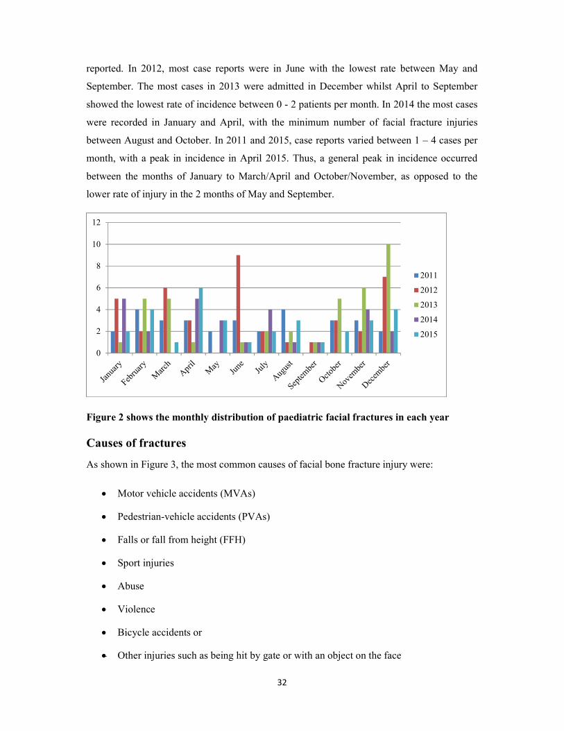
32
reported. In 2012, most case reports were in June with the lowest rate between May and
September. The most cases in 2013 were admitted in December whilst April to September
showed the lowest rate of incidence between 0 - 2 patients per month. In 2014 the most cases
were recorded in January and April, with the minimum number of facial fracture injuries
between August and October. In 2011 and 2015, case reports varied between 1 – 4 cases per
month, with a peak in incidence in April 2015. Thus, a general peak in incidence occurred
between the months of January to March/April and October/November, as opposed to the
lower rate of injury in the 2 months of May and September.
Figure 2 shows the monthly distribution of paediatric facial fractures in each year
Causes of fractures
As shown in Figure 3, the most common causes of facial bone fracture injury were:
Motor vehicle accidents (MVAs)
Pedestrian-vehicle accidents (PVAs)
Falls or fall from height (FFH)
Sport injuries
Abuse
Violence
Bicycle accidents or
Other injuries such as being hit by gate or with an object on the face
0
2
4
6
8
10
12
2011
2012
2013
2014
2015
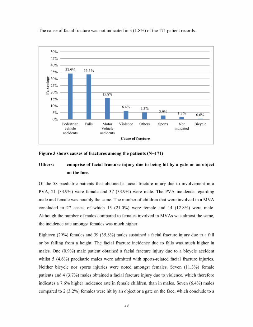
33
The cause of facial fracture was not indicated in 3 (1.8%) of the 171 patient records.
Figure 3 shows causes of fractures among the patients (N=171)
Others: comprise of facial fracture injury due to being hit by a gate or an object
on the face.
Of the 58 paediatric patients that obtained a facial fracture injury due to involvement in a
PVA, 21 (33.9%) were female and 37 (33.9%) were male. The PVA incidence regarding
male and female was notably the same. The number of children that were involved in a MVA
concluded to 27 cases, of which 13 (21.0%) were female and 14 (12.8%) were male.
Although the number of males compared to females involved in MVAs was almost the same,
the incidence rate amongst females was much higher.
Eighteen (29%) females and 39 (35.8%) males sustained a facial fracture injury due to a fall
or by falling from a height. The facial fracture incidence due to falls was much higher in
males. One (0.9%) male patient obtained a facial fracture injury due to a bicycle accident
whilst 5 (4.6%) paediatric males were admitted with sports-related facial fracture injuries.
Neither bicycle nor sports injuries were noted amongst females. Seven (11.3%) female
patients and 4 (3.7%) males obtained a facial fracture injury due to violence, which therefore
indicates a 7.6% higher incidence rate in female children, than in males. Seven (6.4%) males
compared to 2 (3.2%) females were hit by an object or a gate on the face, which conclude to a
33.9% 33.3%
15.8%
6.4% 5.3%2.9% 1.8% 0.6%
0%
5%
10%
15%
20%
25%
30%
35%
40%
45%
50%
Pedestrianvehicle
accidents
Falls MotorVehicle
accidents
Violence Others Sports Notindicated
Bicycle
Per
cent
age
Cause of fracture

34
50.0% higher rate of facial fracture incidence due to other causes amongst males, than in
female children.
For the purpose of this study, children were grouped according to age into age-related sub-
groups. The aim of these relevant sub-groups was to determine the distribution of the cause,
type, and site of fracture according to age.
As illustrated in Table 2, PVAs were the major cause in sub-groups 6 – 10 years and 11 – 15
years of age. PVAs were second most to falls, the greater cause of injury in children between
0 – 5 years of age. Although PVAs were the major cause of facial fracture amongst children 6
– 10 years and those 11 – 15 years of age, the total number and incidence of PVAs in children
0 - 10 years of age, was the highest. MVAs were second to PVAs, the major cause of facial
fracture in children 11 – 15 years of age. Although the 6 (25.0%) children affected by MVA
in the age group 11 – 15 years of age is less than the 13 (16.9%) children 6 - 10 years of age
and the 8 (11.4%) children younger than 5 years of age, the MVA incidence is higher in
children older than 10 years of age. Less children 0 – 5 and 6 – 10 years of age obtained a
facial fracture injury due to MVA than of PVA or falls. The MVA incidence amongst
children in this study has increased with age. Patient records did not reveal whether
passengers were restrained or fastened by a seatbelt at the time of the accident.
Falls were the major cause of facial fracture in children 0 - 5 years of age and the second
major cause in children 6 – 10 years of age, but only 2 (8.3%) patients 10 – 15 years of age
obtained a facial fracture due to falls. In this study, the incidence of falls as the major cause
of facial fracture in children decreased with age.
The bicycle associated facial fracture injury was detected in a single patient (1.3%), in the
sub-group 6 – 10 years of age. Sports-related facial fracture injuries occurred in 2 children
between 6 – 10 and 3 children 11 – 15 years of age. Although the total number of patients
affected in these particular sub-groups of age were almost the same, the 12.5% incidence
amongst children above 10 years of age were much higher than the 2.6% incidence in
children 6 – 10 years of age.
Violence affected children in all sub-groups of age with an almost equal distribution in the
number of cases per age group. However, the incidence of violence varies, especially among
children younger than 10 years and those older than 10 years of age. A lower incidence rate
were detected in children 0 - 5 years and those 6 – 10 years of age, whereas a higher
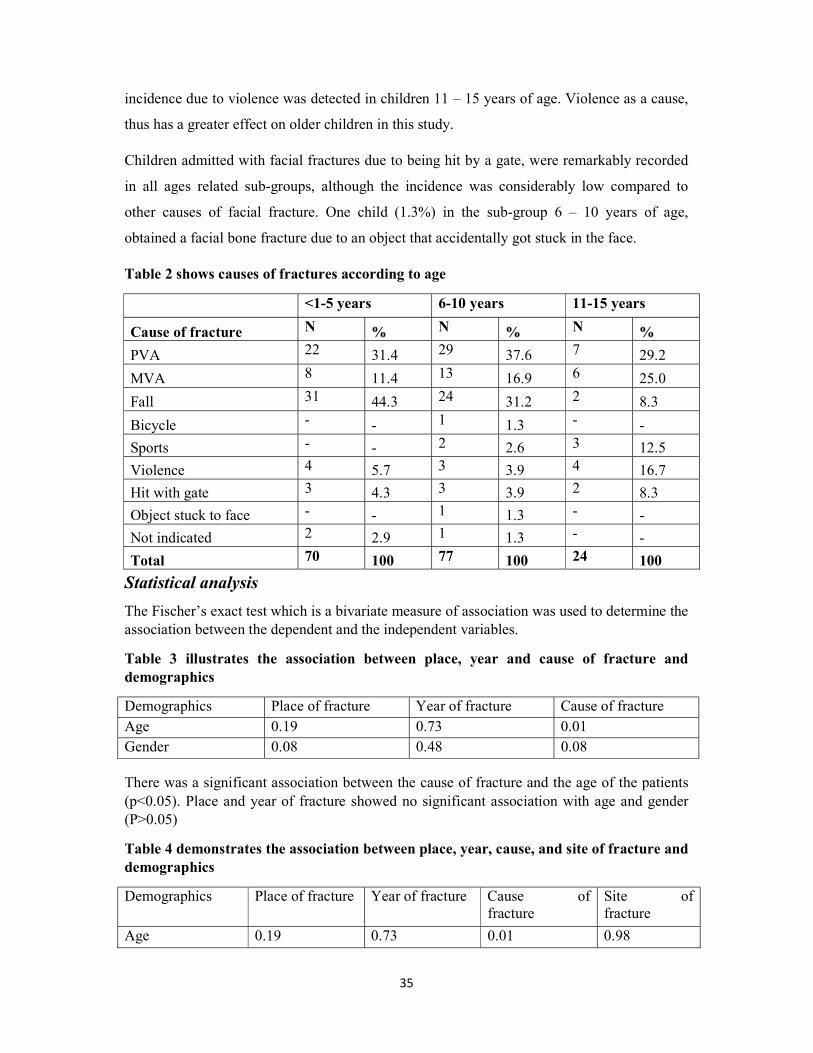
35
incidence due to violence was detected in children 11 – 15 years of age. Violence as a cause,
thus has a greater effect on older children in this study.
Children admitted with facial fractures due to being hit by a gate, were remarkably recorded
in all ages related sub-groups, although the incidence was considerably low compared to
other causes of facial fracture. One child (1.3%) in the sub-group 6 – 10 years of age,
obtained a facial bone fracture due to an object that accidentally got stuck in the face.
Table 2 shows causes of fractures according to age
<1-5 years 6-10 years 11-15 years
Cause of fracture N % N % N %
PVA 22 31.4 29 37.6 7 29.2
MVA 8 11.4 13 16.9 6 25.0
Fall 31 44.3 24 31.2 2 8.3
Bicycle - - 1 1.3 - -
Sports - - 2 2.6 3 12.5
Violence 4 5.7 3 3.9 4 16.7
Hit with gate 3 4.3 3 3.9 2 8.3
Object stuck to face - - 1 1.3 - -
Not indicated 2 2.9 1 1.3 - -
Total 70 100 77 100 24 100
Statistical analysis
The Fischer’s exact test which is a bivariate measure of association was used to determine the association between the dependent and the independent variables.
Table 3 illustrates the association between place, year and cause of fracture and demographics
Demographics Place of fracture Year of fracture Cause of fracture Age 0.19 0.73 0.01 Gender 0.08 0.48 0.08
There was a significant association between the cause of fracture and the age of the patients (p<0.05). Place and year of fracture showed no significant association with age and gender (P>0.05)
Table 4 demonstrates the association between place, year, cause, and site of fracture and demographics
Demographics Place of fracture Year of fracture Cause of fracture
Site of fracture
Age 0.19 0.73 0.01 0.98

36
Gender 0.08 0.48 0.08 0.27
Place, site and year of fracture showed no significant association with age and gender (P>0.05)
Table 5 shows the multinomial logistic regression between the cause of fracture and the age of the patients using the age category 11-15 years as a reference
Age category Exp (B) P value Sig. 95%CI <1-5 years MVA 2.31 0.15 0.75-7.15 Fall 15.50 0.00* 2.83-85.01 Others - - - 6-10 years MVA 2.64 0.08 0.90-7.77 Fall 9.82 0.01* 1.81-53.22 11-15 years MVA Ref Ref Ref Fall Ref Ref Ref
As shown in Table 5, the results showed that children younger than 5 years of age were
approximately 16 times more likely, and those children between 6 – 10 years of age almost
10 times more likely to sustain a facial fracture injury due to falls than children 11 – 15 years
of age.
Site of fracture
There were altogether 247 facial bone fractures amongst the 171 paediatric patients. As the
most familiar site of the facial bone fracture, 74 (30.0%) frontal bone fractures were detected.
The 53 (21.5%) orbital fractures concluded to be the second most common site of facial
fracture. Thirty-nine (15.8%) maxillary and 37 (15.0%) mandibular fractures were noted.
Twenty-seven (10.9%) fractures of the nasal bone and 12 (4.9%) zygomatic bone fractures
were recorded. The 3 (1.2%) Le Fort fractures (1 x Le Fort I and 2 x Le Fort II) together with
the 2 (0.8%) palatal fractures result to 2.0% of all the paediatric facial fractures. (Figure 4)
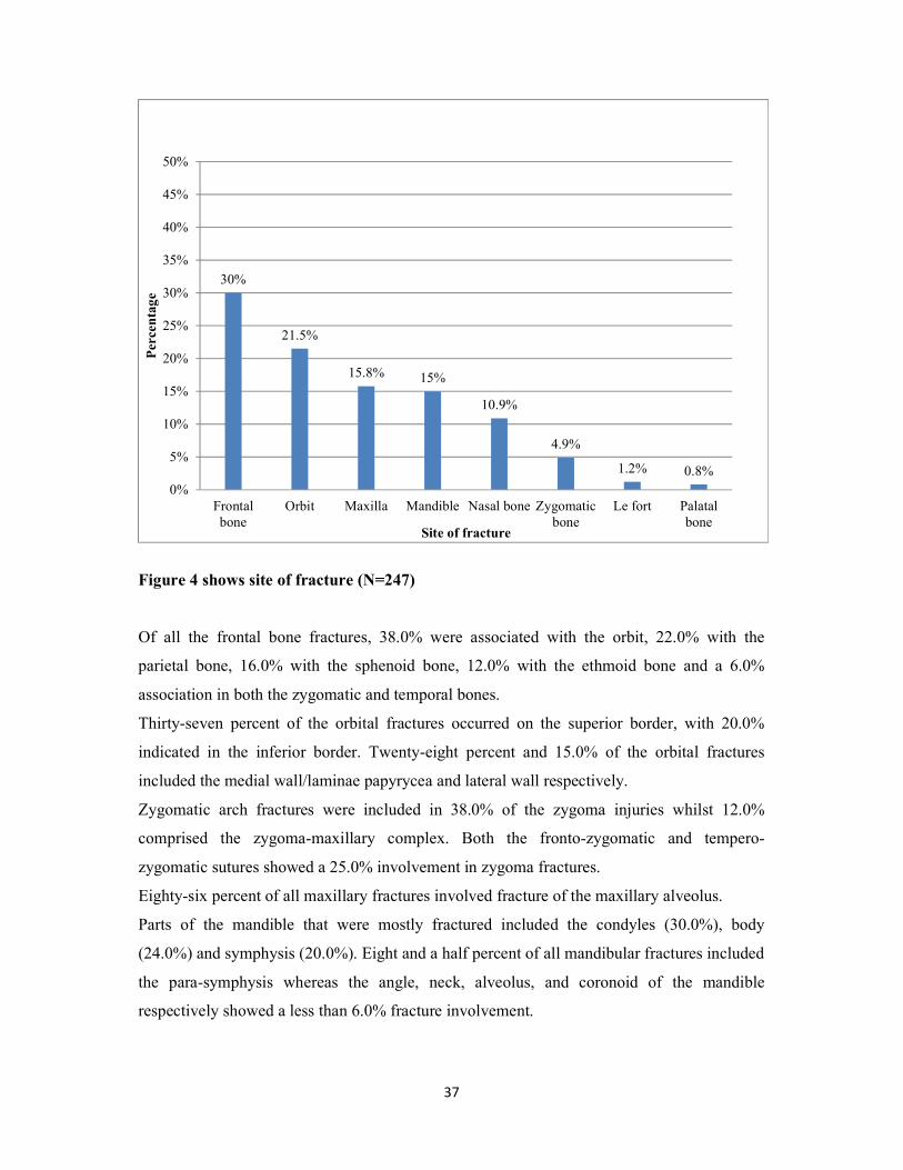
37
Figure 4 shows site of fracture (N=247)
Of all the frontal bone fractures, 38.0% were associated with the orbit, 22.0% with the
parietal bone, 16.0% with the sphenoid bone, 12.0% with the ethmoid bone and a 6.0%
association in both the zygomatic and temporal bones.
Thirty-seven percent of the orbital fractures occurred on the superior border, with 20.0%
indicated in the inferior border. Twenty-eight percent and 15.0% of the orbital fractures
included the medial wall/laminae papyrycea and lateral wall respectively.
Zygomatic arch fractures were included in 38.0% of the zygoma injuries whilst 12.0%
comprised the zygoma-maxillary complex. Both the fronto-zygomatic and tempero-
zygomatic sutures showed a 25.0% involvement in zygoma fractures.
Eighty-six percent of all maxillary fractures involved fracture of the maxillary alveolus.
Parts of the mandible that were mostly fractured included the condyles (30.0%), body
(24.0%) and symphysis (20.0%). Eight and a half percent of all mandibular fractures included
the para-symphysis whereas the angle, neck, alveolus, and coronoid of the mandible
respectively showed a less than 6.0% fracture involvement.
30%
21.5%
15.8% 15%
10.9%
4.9%
1.2% 0.8%0%
5%
10%
15%
20%
25%
30%
35%
40%
45%
50%
Frontalbone
Orbit Maxilla Mandible Nasal bone Zygomaticbone
Le fort Palatalbone
Per
cen
tage
Site of fracture
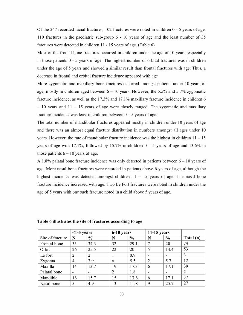
38
Of the 247 recorded facial fractures, 102 fractures were noted in children 0 - 5 years of age,
110 fractures in the paediatric sub-group 6 - 10 years of age and the least number of 35
fractures were detected in children 11 - 15 years of age. (Table 6)
Most of the frontal bone fractures occurred in children under the age of 10 years, especially
in those patients 0 - 5 years of age. The highest number of orbital fractures was in children
under the age of 5 years and showed a similar result than frontal fractures with age. Thus, a
decrease in frontal and orbital fracture incidence appeared with age
More zygomatic and maxillary bone fractures occurred amongst patients under 10 years of
age, mostly in children aged between 6 – 10 years. However, the 5.5% and 5.7% zygomatic
fracture incidence, as well as the 17.3% and 17.1% maxillary fracture incidence in children 6
– 10 years and 11 – 15 years of age were closely ranged. The zygomatic and maxillary
fracture incidence was least in children between 0 – 5 years of age.
The total number of mandibular fractures appeared mostly in children under 10 years of age
and there was an almost equal fracture distribution in numbers amongst all ages under 10
years. However, the rate of mandibular fracture incidence was the highest in children 11 – 15
years of age with 17.1%, followed by 15.7% in children 0 – 5 years of age and 13.6% in
those patients 6 – 10 years of age.
A 1.8% palatal bone fracture incidence was only detected in patients between 6 – 10 years of
age. More nasal bone fractures were recorded in patients above 6 years of age, although the
highest incidence was detected amongst children 11 – 15 years of age. The nasal bone
fracture incidence increased with age. Two Le Fort fractures were noted in children under the
age of 5 years with one such fracture noted in a child above 5 years of age.
Table 6 illustrates the site of fractures according to age
<1-5 years 6-10 years 11-15 years Site of fracture N % N % N % Total (n) Frontal bone 35 34.3 32 29.1 7 20 74
Orbit 26 25.5 22 20 5 14.4 53
Le fort 2 2 1 0.9 - - 3 Zygoma 4 3.9 6 5.5 2 5.7 12 Maxilla 14 13.7 19 17.3 6 17.1 39 Palatal bone - - 2 1.8 - - 2 Mandible 16 15.7 15 13.6 6 17.1 37
Nasal bone 5 4.9 13 11.8 9 25.7 27

39
Total 102 100 110 100 35 100
From a gender perspective, a significant number of 156 facial bone fractures were recorded
amongst the 109 male patients, whilst 91 facial fractures were noted amongst the 62 females.
The incidence of frontal bone fractures was 29.5% in males compared to 30.8% in females.
Orbital fractures were identified in 20.5% of males and in 23.1% of all females. Sixteen point
seven percent of all facial fractures in males were detected in the maxilla followed by a
14.3% occurrence in females. A 15.4% mandibular fracture incidence was noted in males
compared to the 14.3% in females. Ten point three percent of all males and 12.1% of all
females had nasal fractures, with the zygomatic fractures showing a much lower incidence of
5.1% in males compared to 4.4% in females. One palatal fracture was noted in both a male
(0.6%) and a female (1.1%) patient with 3 (1.9%) Le Fort fractures identified in male patients
only.
Although a greater difference in the number of facial bone fractures was noted between male
and female children, the fracture incidence of various bone sites between male and female
was closely ranged.
Types of fractures
Of the 247 facial bone fractures, 115 (67.3%) of the 171 patients had a linear fracture
compared to the 56 (32.7%) patients that had multiple facial bone fractures.
Most patients, which resulted in 121 (70.8%) had a fracture/s that were non-displaced whilst
39 (22.8%) patients had a displaced fracture/s. The type of fracture in terms of displacement
was not indicated in 11 (6.4%) of the patient records.
Also, 2 (1.2%) patients had a compound fracture whereas the fractures of 30 (17.5%) children
were comminuted. The fractures of 4 (2.4%) patients showed clinical or radiographic signs of
crepitus.
Seventy-five (68.8%) males and 40 (64.5%) females had a single linear fracture, whereas 34
(31.2%) males and 22 (35.5%) females showed signs of multiple fractures. This indicates a
fairly similar linear and multiple fracture incidence amongst male and female. An equal
number of compound fractures (1 x male; 1 x female) and fractures with signs of crepitus (2 x
male; 2 x female) were noted in both male and female patients, although a 3.4% higher
incidence of compound fractures and almost double the rate of crepitus incidence in females

40
were noted. The incidence of comminute fractures was almost 10.0% higher amongst the
males.
The 19 (31.1%) displaced fractures detected in females almost correspond to the 20 (20.2%)
noted in males, but show a higher rate of incidence, whereas a higher incidence of non-
displacement were noted in the 79 (79.8%) males, compared to the 42 (68.9%) females.
Most linear fractures appeared in children 6 - 10 years of age, followed secondly in children 0
- 5 years of age. Although the 16 (66.7%) linear fractures documented in children 11 - 15
years of age were much less in number than detected in patients under 10 years of age, the
rate of incidence was fairly similar in all sub-groups of age. Over against that, the incidence
of multiple fractures also corresponded in each sub-group of age, however, the incidence of
linear fracture per age group was almost twice the multiple fracture incidence. (Table 7)
Of the 56 (32.7%) patients recorded with multiple facial bone fractures, 57 facial bone
fractures were detected amongst the 24 (34.3%) paediatric patients 0 - 5 years of age, 56
amongst the 24 (31.2%) children 6 - 10 years of age and 19 were recorded amongst the 8
(33.3%) patients 11 - 15 years of age. Thus, the multiple facial bone fracture incidence of
2.38%, 2.33% and 2.38% per patient, in children 0 – 5 years, 6 – 10 years and those 11 – 15
years of age, indicate a similar incidence amongst all 56 children under 15 years of age.
Also, the total non-displaced fractures were significantly higher in both numbers and
incidence than the displaced fractures documented in both sub-groups 0 – 5 years and 6 – 10
years of age. In the older children of 11 - 15 years of age, the total number of displaced
versus non-displaced fractures were almost equal. (Table 7)
Compound fractures and those fractures with associated crepitus appeared in children above
the age of 6 years. However, 14 (46.7%) comminute fractures, which were the most per age
group, appeared in children 0 - 5 years of age, whilst 11 (36.1%) comminute fractures were
recorded in children 6 - 10 years of age and the least of 5 (16.7%) were noted in children 11 -
15 years of age. Thus, a decrease in the rate of comminute fracture incidence occurred with
age.
Table 7 illustrates type of fractures according to age
<0-5 years 6-10 years 11-15 years Type of fracture N % N % N % Linear 46 65.7 53 68.8 16 66.7
41
Multiple 24 34.3 24 31.2 8 33.3 Total 70 100 77 100 24 100 Displaced 13 20 15 20.8 11 47.8 Non-displaced 52 80 57 79.2 12 52.2
Total 65 100 72 100 23 100
Facial side of fracture
Of the 171 patients, 34.5% had a facial bone fracture injury on the left side, whilst 31.0% of
the patients, showed a facial bone fracture injury on the right side. Twenty-one point one
percent of patients had bone fractures on both the left and right side of their face. A minor
group of 7.6% of patients presented with midline facial fractures. The facial side of fracture
was not indicated in 5.8% of the children’s records.
Associated teeth injury
Forty-nine (28.7%) patients had an associated tooth injury, whereas 122 (71.3%) showed no
teeth affectation. Teeth that were mostly affected included the anterior maxillary and anterior
mandibular incisors and canines of either the primary or permanent dentition. Injury to
molars of the primary dentition was also noted.
The types of dental alveolar injury that were documented, either included mobility, avulsion,
displacement, intrusion, root fracture, crown fracture or any pulpal involvement of the
affected teeth. Mobility of teeth, where the most common dental alveolar injury noted in 28
(57.0%) patients. Avulsion of teeth was the second most common dental alveolar injury noted
in 11 (22.4%) children, followed thirdly by tooth displacement detected in 6 (12.0%)
paediatric patients. Sixteen (33.0%) of the 49 patients with associated tooth injury had
multiple dental alveolar injuries.
Associated facial soft tissue injuries
The most commonly associated facial soft tissue injuries (STIs) observed were lacerations,
abrasions, contusions, stab wounds, animal bites, hematomas, oedema (peri-orbital), soft
tissue swellings, haemorrhage, ecchymosis/raccoon eyes, bleeding/epistaxis or degloving
injuries. Facial soft tissue injuries were also recorded as extraoral or intraoral. Of these STIs,
396 (91.0%) were detected extra orally whilst only 39 (9.0%) were detected intraorally.
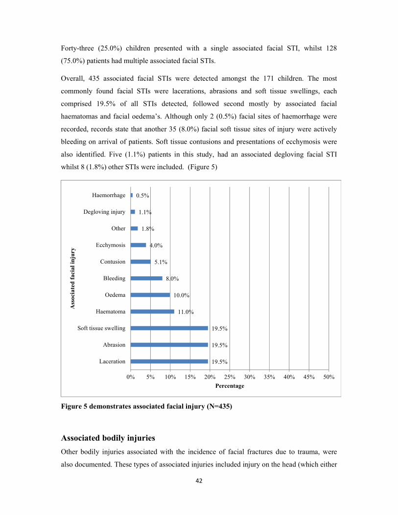
42
Forty-three (25.0%) children presented with a single associated facial STI, whilst 128
(75.0%) patients had multiple associated facial STIs.
Overall, 435 associated facial STIs were detected amongst the 171 children. The most
commonly found facial STIs were lacerations, abrasions and soft tissue swellings, each
comprised 19.5% of all STIs detected, followed second mostly by associated facial
haematomas and facial oedema’s. Although only 2 (0.5%) facial sites of haemorrhage were
recorded, records state that another 35 (8.0%) facial soft tissue sites of injury were actively
bleeding on arrival of patients. Soft tissue contusions and presentations of ecchymosis were
also identified. Five (1.1%) patients in this study, had an associated degloving facial STI
whilst 8 (1.8%) other STIs were included. (Figure 5)
Figure 5 demonstrates associated facial injury (N=435)
Associated bodily injuries
Other bodily injuries associated with the incidence of facial fractures due to trauma, were
also documented. These types of associated injuries included injury on the head (which either
19.5%
19.5%
19.5%
11.0%
10.0%
8.0%
5.1%
4.0%
1.8%
1.1%
0.5%
0% 5% 10% 15% 20% 25% 30% 35% 40% 45% 50%
Laceration
Abrasion
Soft tissue swelling
Haematoma
Oedema
Bleeding
Contusion
Ecchymosis
Other
Degloving injury
Haemorrhage
Percentage
Ass
ocia
ted
faci
al in
jury
43
involved the dura mater, skull/vault or brain/meninges), to a sinus (which indicated any
involvement, opacification or heamosinus), any other body part such as the neck, lung, chest,
abdomen, back, spine, shoulder, arm, hip, leg, hand or foot or other type of life-threatening
injuries such as an airway involvement or obstruction.
Of the 171 patients, 43 (25.0%) patients only had a single associated bodily injury, whereas
74 (43.0%) patients were admitted with multiple bodily injuries associated with facial trauma.
These bodily types of associated injuries were not indicated in 54 (32.0%) of the patient
records.
Of the total 278 associated bodily injuries, the 39 (14.0%) bony skull injuries together with
the 39 (14.0%) cases of sinus involvement were the most. Thirty one (11.1%) dura mater
(extra- or subdural) and 16 (5.8%) brain injuries were noted. Other associated bodily injuries
noted with facial fractures involved 1 (0.4%) neck, 6 (2.2%) spine, 24 (8.6%) shoulder, 7
(2.5%) hand/feet, 17 (6.1%) lung/chest, 13 (4.7%) abdominal and 25 (9.0%) hip injuries. Six
(2.2%) patients with an obstructive airway were admitted.
Other symptoms recorded were excessive pain noted in 32 (18.7%) children whilst 9 (5.3%)
patients with nerve damage or numbness of a bodily part were detected.
Associated ophthalmic or globe involvement
Another finding noted in this study is that 12 (7.0%) of the 171 patients showed results of
ophthalmic or globe involvement. A remarkable 159 (93.0%) had no involvement. Six
(50.0%) of those patients with globe involvement were between 0 - 5 years of age, 4 (33.3%)
between 6 - 10 years of age and 2 (16.6%) between 11 - 15 years, which show by implication
a gradual decrease in ophthalmic or globe incidence with age. This result is compatible with
the decrease in orbital bone fracture incidence with age, revealed in this study.
Specialized consultation
Other medical specialists in the departments of either neurology, opthalmology or ENT (Ear,
nose, and throat) have also been consulted. One hundred and twenty-nine (75.4%) children
required specialized consultation from either of these departments.
44
Consultation with the department of neurology has been permitted solely to 41 (24.0%)
patients with facial bone fracture, followed by the department of ENT in 17 (9.9%) cases and
the department of opthalmology in 12 (7.0%) patients. The department of opthalmology
together with the department of neurology have been included in 39 (22.8%) cases whilst 12
(7.0%) paediatric patients needed consultation from all 3 departments (opthalmology,
neurology, and ENT). Four (2.3%) children required a combined specialist consultation from
the departments of opthalmology and ENT and so have another 4 (2.3%) needed combined
consultation from the departments of neurology and ENT.
The ophthalmology department was consulted with 119 (48.1%) of all the facial fractures
(n=247), mostly for fractures of the orbital bone (34.5%), frontal bone (31.1%) and the
maxillary bone (13.4%). Fewer ophthalmology examinations of which the inclusion ranged
from 7.5% to 1.7% were needed for facial fractures of the zygoma, nasal bone, mandibular
and Le Fort fractures.
The department of neurology has mostly been approached in 144 (58.3%) of all the fractures
(n=247), which concluded to 44.4% for frontal bone, 27.1% of orbital bone and 9.7% of
maxillary bone fractures. Zygoma, nasal, mandibular, Le Fort and palatal bone fractures
occupied 0.7% to 6.25% of all the neurology consultations.
Specialist involvement from ENT was included in 53 (21.5%) facial fractures cases, which
mainly involved fracture of the nasal, maxillary, orbital, mandibular and frontal bones. These
fractures reserved 32.1%, 20.7%, 17.0%, 13.2% and 11.3% of all ENT consultations, whereas
3.8% and 1.8% of all ENT consultations were reserved for Le Fort and zygoma fractures.
Radiographs
Selective radiographs taken either included a panelipse (PAN), lateral skull, posterior-anterior
(PA) or occipito-mental (OM) x-ray, Townes view, Waters view, CT scan (of the brain,
spine, abdomen, chest, arm, leg, or pelvis) or an abdominal FAST (FAST: Focused
assessment with Sonography for trauma). These radiographs were used as part of a diagnostic
tool/measure to confirm the clinical or radiographic diagnosis of a facial bone fracture.
Radiographs were documented in the records of 159 patients. A total of 401 radiographs was
taken amongst this study group (12 patient records didn’t indicate any radiographs). CT scans
were the most widely used radiograph. A significant total of 287 (71.6%) CT scans were

45
taken amongst the 159 indicated patients. CT scans of the brain, spine, abdomen, chest, arm,
pelvis or leg have been taken of different patients to confirm fracture as well as any
associated injuries. CT scans of the brain and spine were recorded mostly which comprised
127 (44.3%) and 105 (36.6%) of all CT’s taken. The remaining 55 (19.1%) CT scans were
mainly taken for the associated bodily injuries of which 5.6% comprised CT of the abdomen,
3.5% of the pelvis, 2.4% of the chest, 1.4% of the arms as well as 1.4% of the legs. However,
4.9% of the total CT scans taken were unspecified.
Remarkable less two-dimensional (2D) views were taken, compared to the CT scans. The
PAN comprised of 30 (7.5%), whilst 24 (6.0%) PA’s, 14 (3.5%) lateral skull’s, 4 (1.0%)
Townes views, 1 (0.2%) Waters view and 1 (0.2%) OM were taken. Five (1.2%) other
radiographs and 22 (5.7%) abdominal FASTs were taken. (Mentioned before, radiographs
were not indicated in the records of 12 patients).
Thirteen (44.0%) PAN’s were to confirm mandibular fracture, whilst 10 (35.0%) PAN’s were
conclusive of maxillary fractures. One (4.7%) PAN was reserved for a fracture of the orbit,
zygoma, palate and nasal bone respectively. Most of the 14 recorded lateral skull radiographs,
were to confirm fracture of the mandible (36.0%) or maxilla (23.0%). Lateral skull views
indicated 9.0% fracture of the frontal bone, the orbital and the nasal bones as well as Le Fort
fractures. Of the 24 posterior-anterior (PA) views taken, 46.0% were diagnostic of
mandibular fracture, 19.0% of maxillary fracture whilst PA views were also conclusive of
frontal, orbital, palatal, nasal bone and Le Fort fractures.
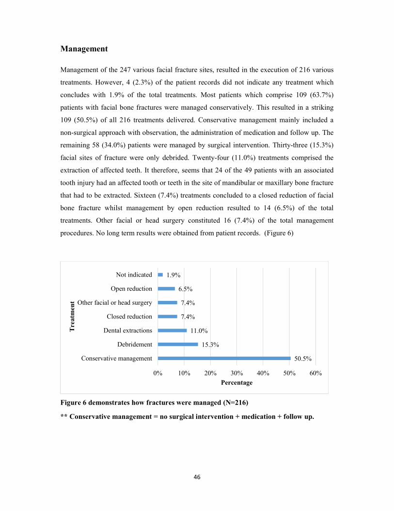
46
Management
Management of the 247 various facial fracture sites, resulted in the execution of 216 various
treatments. However, 4 (2.3%) of the patient records did not indicate any treatment which
concludes with 1.9% of the total treatments. Most patients which comprise 109 (63.7%)
patients with facial bone fractures were managed conservatively. This resulted in a striking
109 (50.5%) of all 216 treatments delivered. Conservative management mainly included a
non-surgical approach with observation, the administration of medication and follow up. The
remaining 58 (34.0%) patients were managed by surgical intervention. Thirty-three (15.3%)
facial sites of fracture were only debrided. Twenty-four (11.0%) treatments comprised the
extraction of affected teeth. It therefore, seems that 24 of the 49 patients with an associated
tooth injury had an affected tooth or teeth in the site of mandibular or maxillary bone fracture
that had to be extracted. Sixteen (7.4%) treatments concluded to a closed reduction of facial
bone fracture whilst management by open reduction resulted to 14 (6.5%) of the total
treatments. Other facial or head surgery constituted 16 (7.4%) of the total management
procedures. No long term results were obtained from patient records. (Figure 6)
Figure 6 demonstrates how fractures were managed (N=216)
** Conservative management = no surgical intervention + medication + follow up.
50.5%
15.3%
11.0%
7.4%
7.4%
6.5%
1.9%
0% 10% 20% 30% 40% 50% 60%
Conservative management
Debridement
Dental extractions
Closed reduction
Other facial or head surgery
Open reduction
Not indicated
Percentage
Tre
atm
ent

47
CHAPTER 6: DISCUSSION
The size of this study population is smaller than a similar South African study approximately
20 years ago that included a sample size of 328 patients. (33) Compared to other global
surveys, the facial fracture prevalence of 4.22% in this study appear to be lower than the
11.5% reported by Gassner et al, (1) 6.0% by Collao-González et al, (2) 4.6% by Imahara et al, (6) 19.0% by Chan (13) and Hyder et al (37), 12.0% by Al-Manik (34) and 12.8% by Kotecha et al. (47) From an author’s perception the variance in prevalence from the above mentioned studies
presumably results from changes in the methodology, which involved a different level of
hospital, either extended over a longer period of time, included more than one hospital or city
or covered a larger metropolitan area. The data collected in this study was from a tertiary
hospital.
The mean age was 6.45 ± 3.47 years which is noticeably younger than the mean age by
Gassner et al who reported the highest facial fracture incidence in children at the age of 9.7 ±
4 years. (1) Of the 247 recorded facial fractures in this study, most were detected in children 6
- 10 years of age (45.0%), followed by those 0 – 5 years of age (41.0%) and the least in
children 11 - 15 years of age (14.0%), which conclude that most facial fractures occurred in
children under 10 years of age. These findings show a discrepancy from related studies that
detected a peak in facial fracture incidence in children between 10 – 15 years of age. (3, 14, 17,
18, 19, 23) Indian studies by Kumaraswamy et al (8) and Karim et al (16) determined a higher
incidence in children 7 – 12 and 5 – 12 years of age. Jung indicated a peak in facial fracture
incidence in children 13 – 17 years of age, (48) which markedly vary from the 14.0%
incidence result obtained in this study of children 11 – 15 years of age. Various literature
supports that the proportional incidence of maxillofacial fractures increases with age, (6, 22, 23,
34, 37, 47) which is in contrast to the author’s results which possibly suggests earlier exposure of
children to the external environment compared to other studies.
PVAs together with MVAs were the major cause of facial fracture amongst children 6 – 10
and 11 – 15 years of age, which concur with some of the literature (4, 5, 6, 21, 22, 24, 25, 27, 28, 37) but
also vary from other studies that reported falls, violence or bicycle accidents as the major
cause of injury. (2, 7, 8, 9, 13, 14, 18, 23, 28, 37, 46, 47) In this study falls, PVAs, MVAs, sports injury,
violence and injury by an object to the face were the most common cause of facial fractures
in paediatric males, which from the author’s point of view, possible result from being more
involved in vigorous play, physical outdoor activities and contact sports. However, most
48
females were involved in PVAs, MVAs, falls and violence. More paediatric males obtained a
facial fracture injury during MVAs and PVAs than females. Although more cases of PVA
than MVA were detected in both male and female children, an almost equal number of males
and females were involved in MVAs. Even though injuries due to motor vehicles were
separated into MVAs and PVAs, these injuries constituted the most common cause of
paediatric facial fractures in this study, which concur with other studies. (6, 22, 23, 37, 45, 46)
Compared to other local studies that separated PVA and MVA, the findings in this study
concurred with Lalloo et al (46) but differed from Bamjee et al who reported MVAs to be
more common in children.(33) Secondly, the author’s results are in sharp contrast to Bamjee et
al that reported violence as the most common cause of facial fractures in children.(33) Since
this study and the one by Bamjee et al were conducted in the same city, albeit 2/3 decades
apart, the increase of PVA as the cause, might be attributed to political change that led to the
influx of young people to the cities in search for opportunities. An anecdotal evidence
suggests that lack of recreational facilities and an increase in the informal settlement areas in
Johannesburg could be an explanation for PVAs being the most common cause of facial
fractures. Other factors from the author’s perception contributing to the high prevalence of
MVAs and PVAs as major aetiological factors amongst children in these affected
communities could be the negligence of drivers, lack of road safety awareness, insufficient
pedestrian safety measures, inadequate parental control or lack of parental supervision. Yet,
the author would like to know what these children were taught in terms of road safety and
traffic rules? Besides being a possible lack of parenting skills, education, public or passenger
safety, could the remarkable higher incidence of PVAs amongst children be the result of
children that act without showing any signs of obedience, discipline, hesitance or judgement?
The author determined falls as the second greatest cause. A remarkable 83.0% of all
paediatric facial fractures resulted from PVAs , MVAs and falls, which relate to previous
studies that indicated such high prevalence of MVAs together with falls (as the second major
cause of fracture). (22, 23, 29) A study done in Seattle support the highest incidence of MVAs
and falls in toddlers (0 – 4 years of age). (6) However, other literature indicated falls as the
major cause of facial fracture injury in children, (2, 8, 14, 18, 37, 46, 47, 49) followed by MVAs as the
second major cause. (8, 14, 16, 37, 49)
Nearly 30.0% of all males and females obtained a facial fracture during falls, whilst a much
smaller number of facial fracture injuries resulted from playing sports, bicycle accidents,
49
violence or abuse or the face being hit by an object or gate. The author’s outcome oppose to
the results obtained by Gassner et al that reported play accidents followed by sporting
accidents as the leading causes of facial fractures in children under 15 years of age. (1) A
study from Japan reported bicycle accidents followed by falls as the main causes, (17) which
from a South African informal settlement perspective clearly indicate a distinct variance (e.g.
in the mainstream mechanism of transport and pedestrian safety measures). A Korean study
reported a 38.0% incidence of violence as the most common cause of facial fracture with a
91.0% male predominance, (19) which is far more than the 11.3% female incidence and 3.7%
male incidence due to violence obtained in this study. However, this study not only revealed
a higher incidence of violence amongst children 11 – 15 years of age, but also showed a
higher incidence of facial fracture due to violence in females which prompt the author’s
suspicion that teenage girls with age, become more exposed to violent activities and abuse at
their homes or nearby home environment.
In this study, a significant association between age and cause of fracture was determined by
making use of the Fischer’s exact test which showed a P-value of 0.01. Thus, from the
author’s perspective and with regards to previous studies, the age at which facial fractures
occur in children is defined by the role of current, type, and degree of physical activity at a
specific age, the effect of social environment and most possible the effect on behaviour and
parental supervision. Mechanism of transport distinct from one country to another markedly
has a definitive impact on the prevalence of paediatric facial fractures.
In terms of the association between cause and age, MVAs with PVAs were the major cause
of facial fracture in all related sub-groups of age (0 – 5 years, 6 – 10 years and 11 – 15 years),
especially in children under the age of 10 years. More cases of PVAs than MVAs were noted
in all age-groups. In accordance with Zhou et al, (49) falls was more apparent in children under
10 years of age, with the highest rate of incidence found in children 5 years and younger,
which concur with other study reports. (3, 33, 45) Falls as a major cause of facial fractures in
children, which decrease in incidence with age, correlate with the literature regarding better
development and control of motoring skills in older children and lack of defence mechanism
in the very young. (7, 28) Sports-related facial fracture injuries essentially affected children
from the age of 6 years, although most occurred in children above 10 years of age. This
finding affirm the higher incidence of sports-related facial fracture injuries with age. (8) More

50
children under 10 years of age obtained a facial fracture injury from being hit by or with an
object, although a higher incidence occurred above 10 years of age. This result corresponds to
a study conveyed in Dunedin where “being hit by an object” was indicated as the main cause
of injury especially in older children. (48) The high prevalence of facial fractures that occurred
in black African children comprised more than 80.0% of the study population, which is from
the author’s point of view a direct dissemination of the drainage area of the CMJAH. The
majority of this study population were male (63.7%), which agree with previous publications
that also show a higher facial fracture incidence in male patients in almost all age groups
worldwide. (1 - 4, 6, 8, 14, 16 - 18, 22, 23, 33, 34, 37, 45 - 49) The data analysis revealed a female to male
ratio of 1:1.75 which correlate with a study by Singh et al, (29) that determined a ratio in males
of approximately twice as frequently to females.
Although more facial bone fractures were detected amongst males, the fracture incidence of
various bone sites between male and female were closely ranged, which resulted in a less
than 1-3% discrepancy. In terms of prevalence by age, the author concurs that the aetiology
of facial fractures seems to be more similar in male and female children at a younger age and
that more substantial variations in aetiology occur between sexes during adolescence.
The author strongly proposes ‘home’ and any ‘other place’ or environment where children
played or visited as high-risk areas for paediatric facial fracture injuries, compared to schools
(lower risk areas) where children are being monitored most of the time in class, during
sporting and other extramural activities. The author’s results correspond with Kumaraswamy
et al (8) and Hyder et al (37) which described the home as the most frequent place where facial
fractures are sustained by children. From an author’s perspective, the possible factors that
contribute to home and other places as high-risk areas for facial fractures in children could
either be lack of parental control, supervision, and responsibility, or the absence of safety
measures.
In terms of seasonal variance, a higher frequency of maxillofacial fracture in children during
the months of January to April and October to December were noted. This seasonal variance
corresponds with other literature. (1, 2, 19, 20) This study result seems to coincide with an
increase in the outdoor activity of children living in or close to this part of Johannesburg
during the months of summer.
The 1.4 average fracture incidence per patient (247 facial bone fractures amongst 171
children) is close to the 1.3 average obtained from a study in Lagos, Nigeria (23) as well as the
51
1.2 average result detected in a study by Mansour. (14) The frontal bone followed by the
orbital bone was the most common fracture site. These results concur with Van As et al that
reported the orbit and frontal bone as the major sites of fracture in children. (45) A lower
incidence of maxillary and mandibular bone fractures were detected. However, the author’s
result is in strong contrast to many other studies that determined the mandible (and in some
instances the maxilla /mid-face as second mostly) as the most familiar facial bone site of
fracture in children. (1, 3, 6, 8, 14, 16, 17, 18, 22, 23, 33, 48) Zhou et al concluded the highest incidence of
mandibular fracture amongst children younger than 12 and adolescents above 13 years of age
but also remarked a higher incidence of maxillary / mid-facial fractures amongst adolescents. (49)
More frontal bone fractures were associated with the orbit and parietal bone than with the
sphenoid, ethmoid, zygomatic and temporal bones. More orbital fractures occurred on the
superior border than the inferior border and medial walls, whereas least were reported on the
lateral walls, which is different from a Korean study that reported the medial wall as the
predominant site of the fracture. (19)
Most of the zygoma fractures recorded included the zygomatic arch, followed by inclusion of
the fronto-zygomatic or tempero-zygomatic sutures. The zygomatic – maxillary complex was
least included. Of all the maxillary fractures, the maxillary alveolus was mostly fractured.
Parts of the mandible that were mostly fractured included the condyles (supported by Ferreira
et al, (22) Kotecha et al (47) and Zhou et al (49)), body and symphysis. Other parts of the
mandible were less affected fractured. Imahara reported a higher fracture incidence of the
symphysis, body, and angle.(6) Kambalimath et al, Ogunlewe et al and Singh et al detected the
highest fracture incidence of the mandibular parasymphysis. (3, 23, 29) Various literature also
reported the mandibular condyles followed by the parasymphysis mostly affected, (8, 14, 17, 18)
compared to Karim et al that detected the symphysis and parasymphysis as the common
mandibular fracture sites. (16)
Most of the frontal and orbital bone fractures occurred in patients under the age of 10 years,
especially in those younger than 5 years of age. A decrease in frontal as well as orbital bone
fracture incidence with age was noted. With regards to the frontal and orbital bones, (firstly
being the bone sites with the highest fracture incidence, secondly being the predominant
facial fracture sites in younger children, especially under 5 years of age, and thirdly showing
a decreased fracture incidence with age), the author also strongly appreciate the representing
anatomical development of a young child’s skull. The larger skull with its greater volume at

52
birth and early years of childhood has a more protrusive protecting position compared to the
smaller retruded face of a young child. (2, 5, 6, 18, 21, 22, 23, 25, 28 - 31)
The frontal and orbital bone as the most frequently fractured sites, justify the result that more
than 80.0% of the paediatric patients included in our study, were under 10 years of age. The
outcome of this study thus correlate with the lower incidence of midfacial fractures and a
higher incidence of cranial fractures detected in early childhood. (21, 27, 28, 30)
Zygomatic and maxillary fractures were mostly detected in children 6 – 10 years of age, with
a corresponding incidence rate in those 11 - 15 years of age, but least in children younger
than 5 years of age. Palatal bone fractures were only noted in children 6 – 10 years of age. An
equivalent number of mandibular fractures were detected amongst all children below 10 years
of age, although the highest incidence appeared in children 11 – 15 years of age. The author’s
results regarding the high incidence of zygomatic, maxillary and mandibular bone fractures
detected in older children, which increase with age, correlate with the physiological
development of facial growth in a downward and forward direction, resulting to further
prominence of the midface and the mandible. Therefore this study accord to the literature
which states that this particular development, may ensue a decrease in cranial and frontal
bone fracture incidence and a possible increase in mandibular fracture due to its relative
prominence as well as midface and orbital floor fracture due to the aeration of the maxillary
sinus. (5, 6, 18, 21 - 23, 28, 31) More nasal bone fractures appeared in paediatric patients above 6
years of age, particularly in children 11 – 15 years of age. The 96.4% nasal bone fracture
incidence by Collao-González et al (2) and 69.0% by Kim et al (19) is remarkable higher than
the author’s results of 10.9%. This variance in nasal fracture incidence most possible result
from method of injury. Conversely, Imahara reported nasal and maxillary fractures as the
most common in infants (0 – 1 year of age). (6) This study reported one Le Fort fracture in a
child under 5 years and one in a child above 5 years of age, compared to Ferreira et al that
only detected Le Fort fractures in children above the age of 10 years. (22)
More single linear than multiple fractures were detected in this study with a similar ratio of
approximately 3/2 in both male and female. This result corresponds Mansour et al, (14)
Ferreira et al, (22) Ogunlewe et al ,(23) a South African study by Bamjee et al (33) and by Van
As et.al. (45) However, only singular fractures were detected in a study by Collao-González et
al. (2) Reports from a study in China revealed a higher incidence of singular fractures in
children under 12 years of age but a greater incidence of multiple severe fractures amongst

53
adolescents above 13 years of age. (49) Most linear fractures appeared in children 6 - 10 years
of age, whereas a higher incidence of multiple fractures was detected with age. The
discrepancy between linear and multiple fractures was less significant in patients 0 - 5 years
of age than in older children. The multiple facial bone fracture incidence of 2.38%, 2.33%
and 2.38% per patient, determined in children 0 – 5 years, 6 – 10 years and those 11 – 15
years of age, indicate a similar incidence amongst all 56 children under 15 years of age. This
result encourage the author to support the literature which substantiate that the unique
anatomic features of children confer greater intrinsic elasticity and flexibility on the
paediatric facial skeleton. Children thus benefit in such way that they are more likely than
adults to sustain greenstick or incomplete fractures and fractures that are likely to have less
multiple communications. (26, 27)
Most fractures were non-displaced. Thirty-nine (22.8%) children had facial fractures that
showed signs of displacement. The number and incidence of non-displaced fractures were
higher in children under 10 years of age, although an almost equal number of displaced and
non-displaced fractures were detected in children above 10 years of age. The author agrees
that the higher occurrence of non-displacement in especially younger children coincide with
the literature regarding the unique anatomic features of children which substantially affirm
the greater intrinsic bone elasticity and flexibility in children. (1, 3, 5, 6, 17, 18, 19, 21-23, 25-28)
Only 49 patients (28.7%) had associated tooth involvement, of which 33.0% presented with
multiple dental alveolar injuries. Teeth affected mostly included the anterior maxillary and
mandibular incisors and canines of either the primary or permanent dentition, and in fewer
cases, the primary molars. Mobility of teeth, was the most common associated dental alveolar
injury, although subluxation of the teeth was confirmed by various studies as the most
frequent dental alveolar injury. (1) The second most common associated dental alveolar injury
was avulsion of the teeth followed by displacement.
Four hundred and thirty-five associated facial soft tissue injuries (STIs) were detected. Most
associated STIs (91.0%) were extra orally. This corresponds to the 8.0% intraoral STI report
of Collao-González et al, (2) although a study in Dunedin reported a 52.6% oral cavity STI
inclusion. (48) Most paediatric patients (75.0%) with facial fractures had multiple associated
facial STIs. The associated facial STI result from this study was significantly higher than the
45.9% incidence detected by Imahara et al, (6) the 29.0% - 56.0% STI incidence by
Kamaraswamy et al, (8) the 21.0% by Kim et al (19) and the 64.5% STI result from a study in
54
Portugal. (22) Lacerations, abrasions and soft tissue swellings were the most commonly found
facial STIs, which correspond with results by Gassner et al (1) and Lalloo et al. (46) Facial
haematoma or oedema both comprised 10.0% of all associated STIs, whilst 8.0% of all facial
STIs were actively bleeding on arrival of the patients. Haemorrhage, contusions, ecchymosis
and degloving facial STIs were also detected.
Sixty eight percent of the paediatric patients had associated bodily injuries, which is
remarkably higher than the 11.0% incidence reported by Collao-González et al, (2) and which
is double from the result by Van As et.al. (45) This discrepancy from the author’s point of
view, most possible result from the difference in mechanism of injury (high impact velocity
versus low impact velocity force). More children with facial fracture trauma had multiple
associated bodily injuries (43.0%) compared to those with a single associated bodily injury
(25.0%). A total of 278 associated bodily injuries was detected. Records revealed that bony
skull injury and sinus involvement both with a 14.0% incidence are the associated injuries,
mostly detected, followed by a lower incidence of extra- or subdural and brain injuries.
Imahara et al also reported skull base fractures, brain injuries, cranial vault fractures and head
injuries to be frequent amongst toddlers 0 – 4 years of age. (6) The high incidence of the
associated skull and sinus injuries correlate with the high frontal bone fracture incidence,
especially amongst children under the age of 10 years, which were mostly involved in PVAs.
Other commonly associated injuries noted involved injury to the neck, airway, spine,
lungs/chest, shoulders, abdomen, hips, hands and feet.
Only 12 (7.0%) paediatric patients showed signs of ophthalmic or globe involvement, most
were between younger than 5 years of age, whereas the incidence appeared to gradually
decrease with age.
Paediatric facial fractures were managed, in both the departments of general surgery and
maxillofacial and oral surgery according to the extent of injury. Management of facial bone
fractures in children involved in this study either included one or more of the following
treatments:
Conservative management, which included non-surgical intervention with
observation, medication and follow-up;
55
Open reduction, which either constitutes one or more of the following entities: open
reduction/internal fixation (ORIF), maxilla-mandibular fixation (MMF), inter-
maxillary fixation (IMF), plates, screws, bioresorbable plates, arch bars, post-op
elastic traction, circum-mandibular wire or any other surgical intervention;
Closed reduction, which either constitutes closed reduction maxillary fixation
(CRMF), IMF, MMF, an acrylic splint, Interdental wires, arch bars, circum-
mandibular wire or elastics;
Observation where no other treatment is required;
Medication (analgesics/antibiotics);
Extractions;
Debridement of fracture site or
Any other facial / head surgery.
The most common type of treatment delivered to patients by the departments of general
surgery and maxillofacial and oral surgery comprised of conservative management (50.5%).
This conservative approach supports Karim which also concluded that the high osteogenic
potential of the paediatric mandible often allows for non-surgical management to be
successful. (16) Kotecha et al also confirmed that conservative management is recommended
as the treatment of choice for especially condylar fractures. (47) Fifteen point three percent of
facial fracture sites were debrided only whilst 11.0% of all treatments included extraction of
affected teeth. Nearly half the patients with a tooth or teeth injury had an affected tooth or
teeth in the site of mandibular or maxillary fracture that had to be extracted. The management
of facial fractures by closed reduction, open reduction or other facial/head surgery,
collectively constituted 21.3% of total treatments. This result is much lower than the 47.0%
of cases from a study in Japan (17) as well as the 84.9% of cases from a study in China, (49) that
were managed by open reduction. Kamabalimath et al have reported that most (83.04%)
mandibular fractures were managed conservatively (IMF in 74.11% of cases and 16.7% by
mini-plating) and therefore also preferred conservative management with observation as
treatment of choice. (3)
Conservative management by closed reduction and MMF were the choice of treatment
reported by a study in Nigeria 2003. (23) A study at the Jordan University managed 91.8% of
mandibular fractures by closed reduction and 8.2% by open reduction, (14) which correspond
to 7.6% open reduction treatment reported by Jung et al. (48) Kim et al reported 95.0%
56
management of nasal bone fractures by closed reduction. (19) This approach is supported by
Ferreira et al, although 78.7% of their study sample (of which 65.0% were older than 13
years of age) received surgical treatment, including open or closed reduction. (22) Singh et al,
however, described the efficient use of bioresorbable plates in the fractured mandible of
children under the age of 15 years, (29) which is supported by Saikrishna et al that described
the bioresorbable method of treatment as a dependable option. (36)
The author perceive correct diagnosis and plan of treatment as an imperative part of
management in paediatric facial fractures. Ninety three percent of paediatric patients had
radiographs of which CT scans were the radiograph of choice, especially CTs of the brain and
spine. This result is in contrast to Bamjee et al who conducted a study at the same academic
hospital from 1989-1992, where conventional plain films were used in general. (33) However,
the study conveyed in 2006 by Van As et al also concluded CT as the gold standard in
assessment of facial fractures. (45) CT as image of choice for mid-face, upper-face and intra-
capsular condyle fractures is also remarked by Karim et al. (16) Most paediatric patients
required specialized consultation from either the department of neurology, opthalmology or
ENT. The department of neurology were consulted mostly, followed by ENT and then
opthalmology. Kambalimath et al also stipulated specialized consultation by neurology as a
prerequisite with craniofacial injury. (3)
Thus, from an author’s perspective, the comprehensive management of paediatric facial
fractures pre- and post-operative is imperative. Therefore, all specialized amenities available
should be utilized to deliver comprehensive treatment. Literature accentuates the acute
management of paediatric facial fractures, understanding the anatomical complexity, age-
related facial anatomy and growth, critical treatment planning with consideration of post-
operative effects and required expertise in treatment of facial bone fracture injuries in
children (7, 13, 25, 27, 34) From the data retrieved regarding treatment and management, the author
substantiate that all possible and available resources in this academic hospital have been
utilized to obtain the above-mentioned principles, which besides the expertise from
specialized surgeons, included the usage of CT scans and conventional radiographs as
diagnostic tools for facial fractures and consultation with other specialized departments.
57
CHAPTER 7: CONCLUSION
In this study, most facial fractures occurred in children under the age of 10 years with a
predominance in paediatric males. PVAs together with MVAs were the most common cause
followed by falls, especially in children under the age of 10 years. More children were
involved in PVAs than MVAs and an almost equal number of males and females were
involved in MVAs. Home and other places (which include roads and any other environment
besides the school that children go to) were the most common places of facial fracture injury.
The aetiology of facial fractures seems more similar in male and female children at a younger
age, with more substantial variations in aetiology between sexes during adolescence.
The frontal bone was the most frequent site of fracture, followed by the orbital bone. Fewer
fractures of the mandible and maxilla were noted. Most fractures were non-displaced and
linear/single, whereas more displaced and multiple fractures were detected in children above
10 years of age. Thus, from this study, it seems that the mechanism of injury and stage of
facial development show a noticeable influence on the type and site of the bone fracture and
that the frequency of aetiological factors change with age.
Less than one-third of all paediatric patients had an associated tooth injury, although more
than four hundred and thirty associated facial soft tissue injuries were detected amongst all
children, mostly extra-orally. Sixty eight percent of the study population had an associated
bodily injury with bony skull injury and sinus involvement detected mostly. Few patient
records revealed ophthalmic or globe involvement, mostly noted in children 0 - 5 years of
age.
Conservative management (which comprised of a non-surgical approach, observation,
medication and follow-up) were the most common type of treatment delivered to paediatric
patients. Open- or closed reduction, or other facial/head surgery constituted less than 10.0%
of all treatments respectively. CT scans have been used as the radiograph of choice.
Although the prevalence of paediatric facial fractures in this study was less compared to
results from other related studies, and although the results were not compared with the
prevalence of facial fractures in adults, the study still bears significance. Every child admitted

58
with a traumatic facial fracture injury, is at a specific stage of development and undergoes a
unique pattern of skeletal growth. Therefore, each child with a facial fracture injury should be
treated in his/her own capacity and the therapeutic approach should be with a good
understanding of the patterns of anatomical growth and stages of skeletal development. Every
treatment of a child who is at a specific stage or phase of skeletal growth and development,
has a consequence, either aesthetically or functionally. The management and treatment of
paediatric facial fractures don’t correspond with the management of fractures in adults, and
even the facial fractures in young children are treated differently from those in older children
or adolescents. Therefore, I believe the emphasis of facial fractures in children, should
essentially be on the accurate evaluation/assessment and identification of paediatric facial
fractures. Not only to meet the importance of immediate treatment and intervention (due to
the greater osteogenic potential, healing capacity and remodelling of bones) but also to avoid
long-term disturbances or effects on the skeletal growth of facial bones.
However, noted from the records of this paediatric study population, no mention regarding
sequential complications specifically, post facial fracture injury was made. These possible
complications include signs of functional and aesthetic facial defects, detrimental loss of
quality of life, social functioning and oral functions such as speech, mastication, respiration,
and deglutition. No records revealed any signs of either permanent facial deformities or soft
tissue scarring post-traumatic facial injury. Although 21.3% of the surgical treatments
delivered which either included open reduction, closed reduction or any other facial or head
surgery, no significant post-operative signs of destruction with regards to permanent brain
injury, globe/ophthalmic injury with loss of sight, occlusal disturbances or complications to
the permanent dentition was noted in the patient records. Possible long-term complications
included the 5 (1.6%) patients with facial nerve damage and the 4 (1.3%) patients who had
numbness of a body part. Also, no reference concerning psychological counselling, post-
facial fracture injury was documented.
Furthermore, this study can possibly be used as a trend for base line data or as a platform for
a next study. Through our results obtained, we could possibly contribute towards the planning
of awareness strategies amongst the public, children, parents, caregivers and healthcare
providers. The concern is however whether these aetiological factors of facial fracture in
children result from an educational, environmental, cultural, public- or road safety matter,
which needs to be addressed. Thus, the value of the work may lead to a reduction in the
frequency of this type of injury, which not only results in unnecessary, possibly lifelong
morbidity but enormous financial costs on a family as well as to an already stressed
59
healthcare system. It may also reduce mortality. The community medicine and dentistry
departments, plus the department of Health should avail themselves to the data analysed and
documented.
REFERENCES
1) Gassner R, Tuli T, Hächl O, Moreira R, Ulmer H. Craniomaxillofacial trauma in
children: a review of 3385 cases with 6060 injuries in 10 years. J Oral Maxillofacial
surgery. (2004); 62: 399 – 407
2) Collao-Gonzalez C, Carrasco-Labra A, Sung-Hsieh HH, Cortés-Araya J.
Epidemiology of paediatric facial trauma in Chile: a retrospective study of 7617 cases
in 3 years. J of Med Oral Patol Oral Cir Bucal. (2014); 19(2): 99 – 105
3) Kambalimath HV, Agarwal SM, Kambalimath DH, Singh M, Jain N, Michael P.
Maxillofacial injuries in children: a 10 year retrospective study. J Maxillofacial Oral
Surg. (2013); 12(2): 140 – 144
4) Cavalcanti AL, De Araujo Lino TH, De Oliveira TBS, et al. Head and maxillofacial
injuries in child and adolescent victims of automotive incidents. The Scientific World
Journal. (2014); Article ID 632720, 6 pages
5) Mukherjee CG, Mukherjee U. Maxillofacial trauma in children. Int J of Clin Pediatr
Dent. (2012); 5(3): 231 – 236
6) Imahara SD, Hopper RA, Wang J, et al. Patterns and outcomes of paediatric facial
fractures in the Unites States: A survey of the National Trauma Data Bank. J Am Coll
Surg. (2008); 207(5): 710 – 716
7) Chang L, Tsai M. Craniofacial injuries from slip, trip and fall accidents of children.
The J of Trauma. (2007); 63(1): 70 – 74
8) Kumaraswamy SV, Nanjappa M, Keerthi R, Deora SS. Paediatric injuries in
maxillofacial trauma: a 5 year study. J Maxillofacial Oral Surg. (2009); 8(2): 150 –
153
9) Murphy DA, Shetty V, et al. Factors associated with orofacial injury and willingness
to participate in interventions among adolescents treated in trauma centres. J Oral
Maxillofacial Surg. (2009); 67(12): 2627 – 2635
60
10) Shayegan A, De Maertelaer V, Abbeele AV. The prevalence of traumatic dental
injuries: a 24-month survey. J of Dent for Children. (2007); 74(3): 194 – 199
11) Marcenes W, Beiruti NA, Tayfour D, Issa S. Epidemiology of traumatic injuries to
the permanent incisors of 9–12–year-old schoolchildren in Damascus, Syria. Endod
Dent Traumatol. (1999); 15: 117 – 123
12) Diaz JA, Bustos L, Brandt AC, Fernández BE. Dental injuries among children and
adolescents aged 1-15 years attending to public hospital in Temuco, Chile. Dental
Traumatology. (2010); 26: 254 – 261
13) Chan YM, Williams S, Davidson LE, Drummond BK. Orofacial and dental trauma of
young children in Dunedin, New Zealand. Dental Traumatology. (2011); 27: 199 –
202
14) Qudah MA, Bataineh AB. A retrospective study of selected oral and maxillofacial
fractures in a group of Jordanian children. J of Oral Surg Oral Med Oral Pathol Oral
Radiol and Endod. (2002); 94: 310 – 314
15) Singh G, Garg S, Damle SG, et al. A study of sports related occurrence of traumatic
orodental injuries and associated risk factors in high school students in North India.
Asian J Sports Med. (2014); 5(3): e22766
16) Karim T, Khan AH, Ahmed SS. Trauma of facial skeleton in children: An Indian
perspective. Indian J Surg. (2010); 72: 232 – 235
17) Iida S, Matsuya T. Paediatric maxillofacial fractures: their aetiological characters and
fracture patterns. J of Craniomaxillofacial Surgery. (2002); 30: 237 – 241
18) Joshi SR, Saluja H, Pendyala GS, Chaudhari S, Mahindra U, Kini Y. Pattern and
prevalence of maxillofacial fractures in rural children of central Maharashtra, India. A
retrospective study. J Maxillofacial Oral Surg. (2013); 12(3): 307 – 311
19) Kim SH, Lee SH, Cho PD. Analysis of 809 facial bone fractures in a paediatric and
adolescent population. J of Arch Plast Surg (2012); 39: 606 – 611
20) Chrcanovic BR. Epidemiology of paediatric head trauma in Guilan. Arch Trauma
Res. (2012); 1(3); 141 – 142
61
21) Wheeler J, Philips J. Paediatric facial fractures and potential long-term growth
disturbances. Craniomaxillofacial trauma and reconstruction. (2011); 4(1): 43 – 52
22) Ferreira PC, Amarante JM, Silva PN, et al. Retrospective study of 1251 maxillofacial
fractures in children and adolescents. Plast and reconstr Surg. (2005); 115(6): 1500 –
1508
23) Ogunlewe MO, James O, Ladeinde AL, Adeyemo WL. Pattern of maxillofacial
fractures in Lagos, Nigeria. Int J of Pediatr Dent. (2006); 16: 358 – 362
24) Glendor U. Aetiology and risk factors related to traumatic dental injuries – a review
of the literature. Dental traumatology. (2009); 25: 19 – 31
25) Haug RH, Foss J. Maxillofacial injuries in the paediatric patient. Oral Surg Oral Med
Oral Path. (2000); 90(2): 126 – 134
26) Smart JM, Low DW, Bartlett SP. The paediatric mandible: management of traumatic
injury or fracture. Plast Reconstr Surg. (2005); 116(2): 28 – 41e
27) Chao MT, Losee JE. Complications in paediatric facial fractures. Craniomaxillofacial
trauma and reconstruction. (2009); 2(2): 103 – 112
28) Cole P, Kaufman Y, Hollier LH. Managing the paediatric facial fracture.
Craniomaxillofacial Trauma and Reconstruction. (2009); 2(2): 77 – 83
29) Singh G, Mohammed S, et al. Bio-resorb able plates as effective implant in paediatric
mandibular fracture. J Maxillofacial Oral Surg. (2012); 11(4): 400 – 406
30) Hupp JR, Ellis III E, Tucker MR. Contemporary Oral and Maxillofacial Surgery, sixth
edition. (2014). By Mosby (ELSEVIER). Part VI; Oral and Maxillofacial Trauma:
496 – 518
31) Oppenheimer AJ, Monson LA, Buchman SR. Paediatric orbital fractures.
Craniomaxillofacial trauma and reconstruction. (2013); 6(1): 9 – 20
32) Altun C, Ozen B, Esenlik E, et al. Traumatic injuries to permanent teeth in Turkish
children, Ankara. Dental Traumatology. (2009); 25: 309 – 313
33) Bamjee Y, Lownie JF, Cleaton-Jones PE, Lownie MA. Maxillofacial injuries in a
group of South Africans under 18 years of age. British J of Oral and Maxillofacial
surgery (1996);34:298 – 302
62
34) Al-manik M. Oral injuries in children attending a hospital in Saudi Arabia. J
Maxillofacial Oral Surg. (2009); 8(1): 34 – 39
35) Batstone MD, Waters C, Porter SAT, Monsour FNT. Treatment delays in paediatric
dent-alveolar trauma at a tertiary referral hospital. Australian Dent J. (2004); 49(1): 28
– 32
36) Saikrishna D, Gupta N. Comparison of circummandibular wiring with resorbable
bone plates in paediatric mandibular fractures. J Maxillofacial Oral Surg. (2010);
9(2): 116 – 118
37) Hyder AA, Sugarman DE, et al. Global childhood unintentional injury surveillance in
four cities in developing countries: a pilot study. Bull World Health organ. (2009); 87:
345 – 352
38) Cavalcanti AL. Prevalence and characteristics of injuries to the head and orofacial
region in physically abused children and adolescents – a retrospective study in a city
of the Northeast of Brazil. Dental Traumatology (2010); 26: 149 – 153
39) Brasileiro BF, Passeri LA. Epidemiological analysis of maxillofacial fractures in
Brazil: a 5 year retrospective study. J of Oral Surg Oral Med Oral Pathol Oral Radiol
and Endod. (2006); 102: 28 – 34
40) Sharma S, Vashistha A, Chugh A, et al. Paediatric mandibular fractures: A review. Int
J of Clin Pediatr Dent. (2009); 2(2): 1 – 5
41) Zimmermann EC, Troulis MJ, Kaban LB. Paediatric facial fractures: recent advances
in prevention, diagnosis and management. Int J of Maxillofacial Surg (2005); 34: 823
– 833
42) Ciechomski J, Aufgang R, et al. Sub cranial approach in paediatric craniofacial
surgery. Craniomaxillofacial trauma and reconstruction. (2010); 3(4): 231 – 236
43) Arenas M, Barberia E, Lucavechi T, Maroto M. Severe trauma in the primary
dentition – diagnosis and treatment of sequelae in permanent dentition. Dental
Traumatology. (2006); 22: 226 – 230
44) Mathog RH. Maxillofacial trauma. (1984) Williams and Wilkens. Ch.7; Binns H.
Early treatment of facial injuries: 89 – 106

63
45) Van As AB, Van Loghem AJ, et al. Causes and distribution of facial fractures in a
group of South African children and the value of computed tomography in their
assessment. Int J of Oral and Maxillofacial Surg. (2006); 35(10): 903 – 906
46) Lalloo R, Van As AB. Profile of children with head injuries treated at the trauma unit
of Red Cross War Memorial Children’s Hospital, 1991 – 2001. SAMJ. (2004); 94(7):
544 – 545
47) Kotecha S, Scannell J, et al. A four year retrospective study of 1062 patients
presenting with maxillofacial emergencies at a specialist pediatric hospital. British J
of Oral and Maxillofacial surgery. (2008); 46: 293 – 296
48) Jung TK, De Silva HL, et al. Trends in maxillofacial trauma presenting to Dunedin
Hospital, 2006 to 2012. New Zealand Dental Journal. (2015); 76 – 80
49) Zhou H, Ongodia D, et al. Incidence and pattern of maxillofacial fractures in children
and adolescents: A 10 year retrospective cohort study. International Journal of
Pediatric Otorhinolaryngology. (2013); 77: 494 – 498
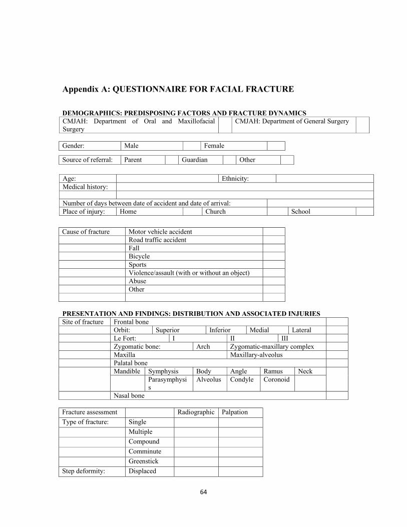
64
Appendix A: QUESTIONNAIRE FOR FACIAL FRACTURE
DEMOGRAPHICS: PREDISPOSING FACTORS AND FRACTURE DYNAMICS CMJAH: Department of Oral and Maxillofacial Surgery
CMJAH: Department of General Surgery
Age: Ethnicity: Medical history: Number of days between date of accident and date of arrival: Place of injury: Home Church School
Cause of fracture Motor vehicle accident Road traffic accident Fall Bicycle Sports Violence/assault (with or without an object) Abuse Other PRESENTATION AND FINDINGS: DISTRIBUTION AND ASSOCIATED INJURIES Site of fracture Frontal bone Orbit: Superior Inferior Medial Lateral Le Fort: I II III Zygomatic bone: Arch Zygomatic-maxillary complex Maxilla Maxillary-alveolus Palatal bone Mandible Symphysis Body Angle Ramus Neck
Parasymphysis
Alveolus Condyle Coronoid
Nasal bone Fracture assessment Radiographic Palpation
Type of fracture: Single
Multiple
Compound
Comminute
Greenstick
Step deformity: Displaced
Gender: Male Female
Source of referral: Parent Guardian Other
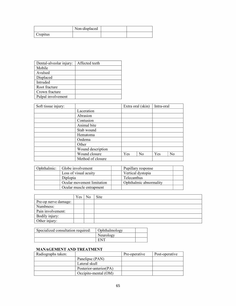
65
Non-displaced
Crepitus
Dental-alveolar injury: Affected teeth Mobile Avulsed Displaced Intruded Root fracture Crown fracture Pulpal involvement Soft tissue injury: Extra oral (skin) Intra-oral Laceration Abrasion Contusion Animal bite Stab wound Hematoma Oedema Other Wound description Wound closure Yes No Yes No Method of closure Ophthalmic: Globe involvement Pupillary response Loss of visual acuity Vertical dystopia Diplopia Telecanthus Ocular movement limitation Ophthalmic abnormality Ocular muscle entrapment Yes No Site Pre-op nerve damage: Numbness: Pain involvement: Bodily injury: Other injury: Specialized consultation required: Ophthalmology Neurology ENT MANAGEMENT AND TREATMENT Radiographs taken: Pre-operative Post-operative Panelipse (PAN) Lateral skull Posterior-anterior(PA) Occipito-mental (OM)

66
Treatment Complications/outcomes Open reduction ORIF (open reduction/internal fixation Malocclusion MMF (maxilla-mandibular fixation) Deformity IMF (inter-maxillary fixation) Infection Plates Screws Bioresorbable Diplopia Arch bars Nerve damage Other Trismus Post-op elastic traction Non-union Closed reduction IMF Acrylic splint Other MMF Interdental wires Arch bars None Observation Medication Analgesics/Antibiotics
Townes view Waters view CT scan Other

67

68
Appendix B: Letter for ethical clearance
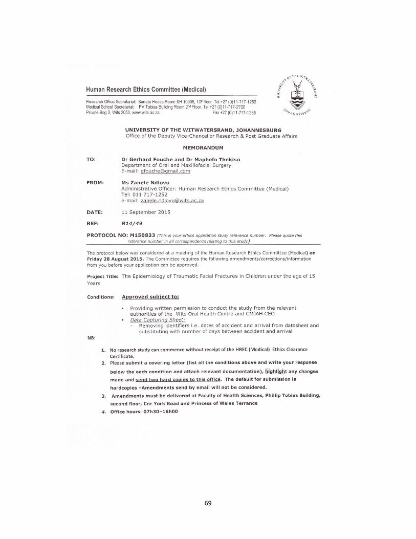
69
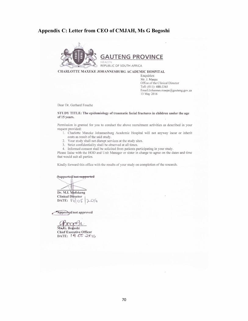
70
Appendix C: Letter from CEO of CMJAH, Ms G Bogoshi
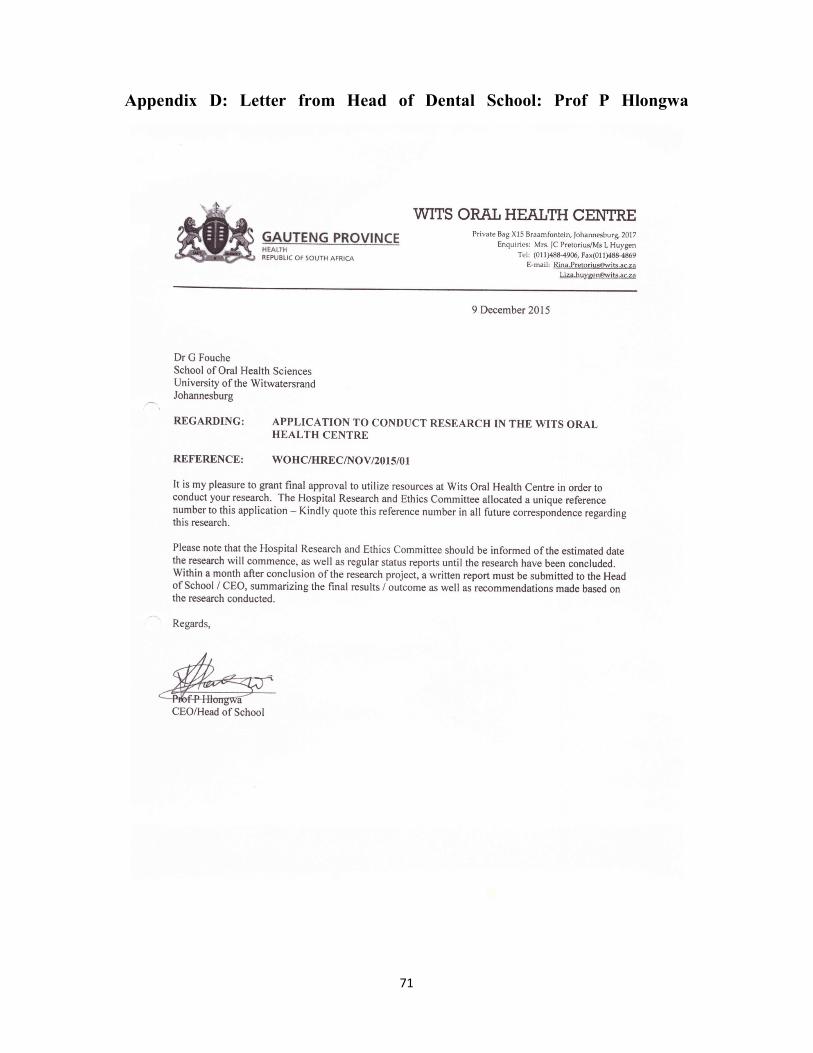
71
Appendix D: Letter from Head of Dental School: Prof P Hlongwa

72
Appendix E: Letter from Clinical Head of Department of Surgery: Dr TE
Luvhengo
Related Documents