CONTINUING STUDIES OF MORTALITY OF ALLIGATORS ON CENTRAL FLORIDA LAKES: PATHOLOGY AND NUTRITION Final report to St. Johns River Water Management District Contract #SE122AA 31 March 2002 James Perran Ross 1 , Dwayne Carbonneau 2 , Scott Terrell 3 , Trenton Schoeb 4 , Dale Honeyfield 5 , Joy Hinterkopf 5 , Adam Finger 6 and Richard Owen 7 1 Author for communications Florida Museum of Natural History, Box 117800, University of Florida, Gainesville FL 32611 352 846 2566 (telephone), 352 392 9367 (fax), <[email protected]> 2 Florida Fish and Wildlife Conservation Commission, Alligator Management Section. 3 College of Veterinary Medicine, University of Florida and Walt Disney World Animal Programs . 4 Department of Genomics and Pathobiology, University of Alabama at Birmingham. 5 Biological Research Division, USGS, Leetown Science Center, Northern Appalachian Research Laboratory, Wellsboro PA. 6 The Institute of Environmental and Human Health at Texas Tech University. 7 Guana River State Park, Florida Department of Environmental Protection, Ponte Vedra Beach, FL.

Welcome message from author
This document is posted to help you gain knowledge. Please leave a comment to let me know what you think about it! Share it to your friends and learn new things together.
Transcript

CONTINUING STUDIES OF MORTALITY OF ALLIGATORS ON CENTRAL FLORIDA LAKES: PATHOLOGY AND NUTRITION
Final report to St. Johns River Water Management District
Contract #SE122AA 31 March 2002
James Perran Ross 1, Dwayne Carbonneau2, Scott Terrell3, Trenton Schoeb4, Dale
Honeyfield5, Joy Hinterkopf5, Adam Finger6 and Richard Owen7
1 Author for communications Florida Museum of Natural History, Box 117800, University of Florida, Gainesville FL 32611 352 846 2566 (telephone), 352 392 9367 (fax), <[email protected]> 2 Florida Fish and Wildlife Conservation Commission, Alligator Management Section. 3 College of Veterinary Medicine, University of Florida and Walt Disney World Animal Programs . 4 Department of Genomics and Pathobiology, University of Alabama at Birmingham. 5 Biological Research Division, USGS, Leetown Science Center, Northern Appalachian Research Laboratory, Wellsboro PA. 6 The Institute of Environmental and Human Health at Texas Tech University. 7 Guana River State Park, Florida Department of Environmental Protection, Ponte Vedra Beach, FL.

ii
EXECUTIVE SUMMARY Investigations into the continuing, unexplained mortality of alligators on Lake Griffin in central Florida were conducted with funding from St. Johns River Water Management District and Lake County Water Authority, and involved cooperative efforts of Florida Museum of Natural History, University of Florida, Florida Fish and Wildlife Conservation Commission (FWC) and USGS - Biological Resource Division Cooperative Fish and Wildlife Research Unit (USGS-COOP). Lake Griffin was surveyed for dead alligators every two weeks throughout the year and field work on dead and impaired alligators conducted February – September, 2001. Seventy five dead alligators were recorded in 2001 and a mortality peak during the spring months March – May was confirmed. Field evaluation of behavioral impairment indicated an average of 1% of alligators in Lake Griffin may be affected. The mortality affects only adult sized alligators. No significant differences in size, body condition or sex ratio is shown between the sick/dead sample and healthy adult alligators on Lake Griffin or Lake Woodruff. Twelve impaired alligators and eight apparently healthy alligators from Lake Griffin and six alligators from other lakes were examined by necropsy for pathology, blood chemistry and histology. No significant findings were noted in gross pathology, blood chemistry or histology except for the following. Five impaired alligators from Lake Griffin showed brain lesions. Three of these were lesions of the torus semicircularis similar to those previously observed. Two individuals had encephalitis of the meninges that is likely to be of different origin. Results from laboratory analyses of cholinesterase reactivation indicated no significant exposure to organophosphates in Lake Griffin or Lake Woodruff alligators. Analysis by Proton Induced X-Ray Emission indicated that Lake Griffin alligators have lower levels of selenium than Lake Woodruff alligators. Extensive re-testing of thiamin levels confirmed that thiamin levels in Lake Griffin alligators are lower than Lake Woodruff alligators and the thiamin is lower in more seriously impaired alligators. Analysis of 63 alligator stomach contents indicated that invertebrates (apple snails and crayfish) occur in 87% of stomachs, fish (catfish and gizzard shad) in 67%, reptiles (turtles) in 32%, mammals in 22% and birds in 13%. Gizzard shad are a significant proportion of both the number of fish ingested and the biomass ingested. Gizzard shad in Lake Griffin ingested forty two phytoplankton species but just two or three species made up a majority of the contents. Potentially toxic blue green algae are a significant proportion (23%-87%) of some gizzards and were present in 13 of 15 stomachs. The relationship between blue green algae blooms, blue green algae toxity, thiaminase production in gizzard shad, gizzard shad in alligator diets and thiamin deficiency in alligators is proposed for detailed additional study.
iii
Table of Contents
EXECUTIVE SUMMARY ...............................................................................................i LIST OF FIGURES ....................................................................................................... iv LIST OF TABLES ............................................................................................................ v LIST OF APPENDIXES..................................................................................................vi ACKNOWLEDGEMENTS ........................................................................................... vii INTRODUCTION ............................................................................................................ 1 ALLIGATOR MONITORING ......................................................................................... 3 Methods..................................................................................................... 3 Results ....................................................................................................... 3 ALLIGATOR COLLECTION AND NECROPSYand PATHOLOGY ........................... 8 Methods................................................................................................................. 8 Results ................................................................................................................. 10 Alligator collection ................................................................................. 10 Blood Values ........................................................................................... 11 Pathology ................................................................................................ 12 MICROBIAL CULTURES ............................................................................................. 14 Methods................................................................................................... 14 Results ..................................................................................................... 14 HISTOLOGY ................................................................................................................. 14 Methods................................................................................................... 14 Results ..................................................................................................... 14 LABORATORY ANALYSIS OF TISSUES.................................................................. 16 Organophosphate … .......................................................................................... 16 Methods................................................................................................... 16 Results ..................................................................................................... 16 Heavy Metal ........................................................................................................ 18 Methods................................................................................................... 18 Results ..................................................................................................... 18 Thiamin ............................................................................................................... 20 Methods................................................................................................... 20 Results ..................................................................................................... 20 DIET AND NUTRITION ............................................................................................... 23 Analysis of Alligator Diet ................................................................................... 23 Methods................................................................................................... 23 Results ..................................................................................................... 24 Analysis of Gizzard Shad Diet ............................................................................ 27 Methods................................................................................................... 27 Results ..................................................................................................... 28 DISCUSSION ................................................................................................................. 30 CONCLUSIONS............................................................................................................. 34 REFERENCES ............................................................................................................... 35

iv
List of Figures
Figure 1. Mortality of alligators on Lake Griffin, FL. 1997-2001 ....................................3 Figure 2. Size of dead alligators on Lake Griffin 2001 ....................................................4 Figure 3. Gender of dead alligators on Lake Griffin 2001 ...............................................4 Figure 4. Body condition (Length versus weight) of alligators in 2001 .........................10 Figure 5. Thiamin levels of alligators from Lake Griffin and Lake Woodruff, 2000, 2001...............................................................................................21 Figure 6. Thiamin levels and impairment level of alligators. .........................................22 Figure 7. Contents of alligator stomachs (% occurrence) 2001 ......................................24 Figure 8. Algae species in the gizzard of Dorosoma cepedianum ..................................28 Figure 9. Gizzard shad size and algae species and quantities eaten ..............................29

v
List of Tables Table 1. The Owen-Carbonneau Alligator Impairment Scale ..........................................6 Table 2. Numbers of alligators exhibiting different avoidance behavior (Owen
Carbonneau Alligator Impairment Scale) Lake Griffin, 2001. .............................7 Table 3. Abbreviated necropsy/veterinary pathology protocol .........................................9 Table 4. Alligators collected and necropsied from lakes in central Florida 2001. .........10 Table 5. Blood analysis values of neurologically impaired and apparently healthy
alligators from Lake Griffin, 2001. .....................................................................12 Table 6. Summarized pathology observations, alligators from Lake Griffin and Lake
Woodruff, 2001. ..................................................................................................13 Table 7. Histology observations and microbial culture results, alligators from Lake
Griffin and Lake Woodruff 2001. .......................................................................15 Table 8. Brain, spinal cord and serum cholinesterase activity of alligators from Lake
Griffin and Lake Woodruff .................................................................................17 Table 9. Metal concentrations (ppb dry weight) for selected alligator tissues from Lake
Griffin and Lake Woodruff. ................................................................................19 Table 10. Total thiamin pmol/g from alligator muscle and liver from Lake Griffin and
Lake Woodruff for 2000 and 20001 ..................................................................21 Table 11. Stomach contents of impaired and not-impaired alligators combined from Lake
Griffin, Lake Woodruff and Lake Apopka. ........................................................25 Table 12a. Species recovered from alligator stomachs, Lake Griffin .............................25 Table 12b. Species recovered from alligator stomachs, Lake Woodruff and
Lake Apopka .......................................................................................................26 Table 13. Occurrence of different species of fish in 31 alligator stomachs from Lake
Griffin .................................................................................................................27 Table 14. Dominant species of phytoplankton recovered from shad gizzards in Lake
Griffin March - May and June 2001 ...................................................................29

vi
List of appendices. The following files are attached as Excel files and provide raw data, and in some cases,
the analyses, of the work summarized and presented in the report. Appendix 1. [App 1. Griffin Alligator surveys.xls] Alligator surveys (summarized in Figures 1, 2, 3). Appendix 2. [App 2. Alligator specimens.xls] Alligators collected for necropsy(summarized in Table 3 and Fig. 4). Appendix 3. [App 3. 2001 Griffin Alligator blood.xls] Alligator blood analyses (summarized in Table 5). Appendix 4. [App 4 . 2001 Alligator necropsy.xls?] Alligator pathology (summarized in Tables 6, 7). Appendix 5. [App 5. Metals in Alligators.xls] Alligator metals (summarized in Table 9). Appendix 6. [App 6. Thiamin in Alligators.xls] Alligator thiamin (summarized in Table 10 and Figures. 5, 6). Appendix 7. [App 7. Alligator diet 2001.xls] Alligator Diet (summarized in Tables 11, 12, 13 and Figure 7) Appendix 8. [App 8. 2001 Shad diet.xls] Algae in Shad diets (summarized in table 14 and Figures 8 and 9)
vii
ACKNOWLEDGEMENTS Tis work is truly a cooperative effort among many people and agencies that it has been my pleasure to coordinate. This work would not be possible without the efforts of the people enumerated below and I thank them all sincerely. The work in 2001 was made possible by funding from the St. Johns River Water Management District ably administered by project officer Walt Godwin. I am also grateful to Carol Lippincott and Mike Coveney for their support of the project. Additional funding from the Lake County Water Authority (LCWA) greatly expanded our activities and I am grateful to Executive Director Mike Perry, Linda Marino, and to all the members of the LCWA board. Ann Griffin, Joe Hill and Bob Taylor all provided additional useful support and information. Field work, necropsies, stomach pumping and data management were conducted with great diligence by Adam Finger and Richard “Bubba” Owen. Franklin Percival, (USGS–COOP) provided boats, vehicles and other logistic support beyond our ability to pay. Scott Terrell conducted the necropsy and pathology work and I am very grateful to Disney’s Animals Kingdom for providing his research facilities. Trenton Schoeb, University of Alabama at Birmingham, provided pathology expertise. Dwayne Carbonneau was our liaison with the FWC and also conducted the biweekly surveys and assisted in many other ways. Allan Woodward provided extensive assistance and helped design the study. I thank Jim Brady FWC Lab director for permission to use their facilities and many staff for tolerating our mess and the alligator necropsies. Additional assistance in the field was provided by Will Strong, Chris Tubbs, Harold Weatherman, P. J. Jones, Bob Godshalk, Ralf Sommerlad, Rogelio Cedeño, Ester Langan, John Kuharik, Robin Bouchton, Adam Britton, and Ben Klien. Matt Milnes, University of Florida Zoology Dept., provided alligators caught on Lake Woodruff and with Richard L’Esperance is analysing lipids in gators and their diet. Tim Gross, Caribbean Science Center, USGS-BRD, provided animals caught on Lake Apopka. Ester Langan, Tony Reppas and Amanda Rice sorted and identified stomach content material. Assistance with identification from collections of the Florida Museum of Natural History is gratefully acknowledged. Algae identification was provided by John Burns and Andy Chapman of Cyano-Labs Inc. Thiamin analyses were provided by Dale Honeyfield and Joy Hinterkopf of the Biological Research Division, USGS, Leetown Science Center, Northern Appalachian Research Laboratory, Wellsboro PA. Cholinesterase data were undertaken by Michael Hooper and Stephanie Schmidt of The Institute of Environmental and Human Health at Texas Tech University. PIXE metals analysis was provided under the direction of professors Henri van Rinsvelt and Gene Dunnam, Dept of Physics, University of Florida, and conducted by Ivan Kravchenko and students Ralf Kelly, John Kuharik and Adele Luta. Skip Goerner provided logistic support and field facilities on Lake Griffin. The residents and manager of Eagles Nest Resort provided lake access when the water level was low. I am grateful to many residents around Lake Griffin for supporting our work, sharing their interesting observations and for tolerating our airboat noise late at night.
1
INTRODUCTION
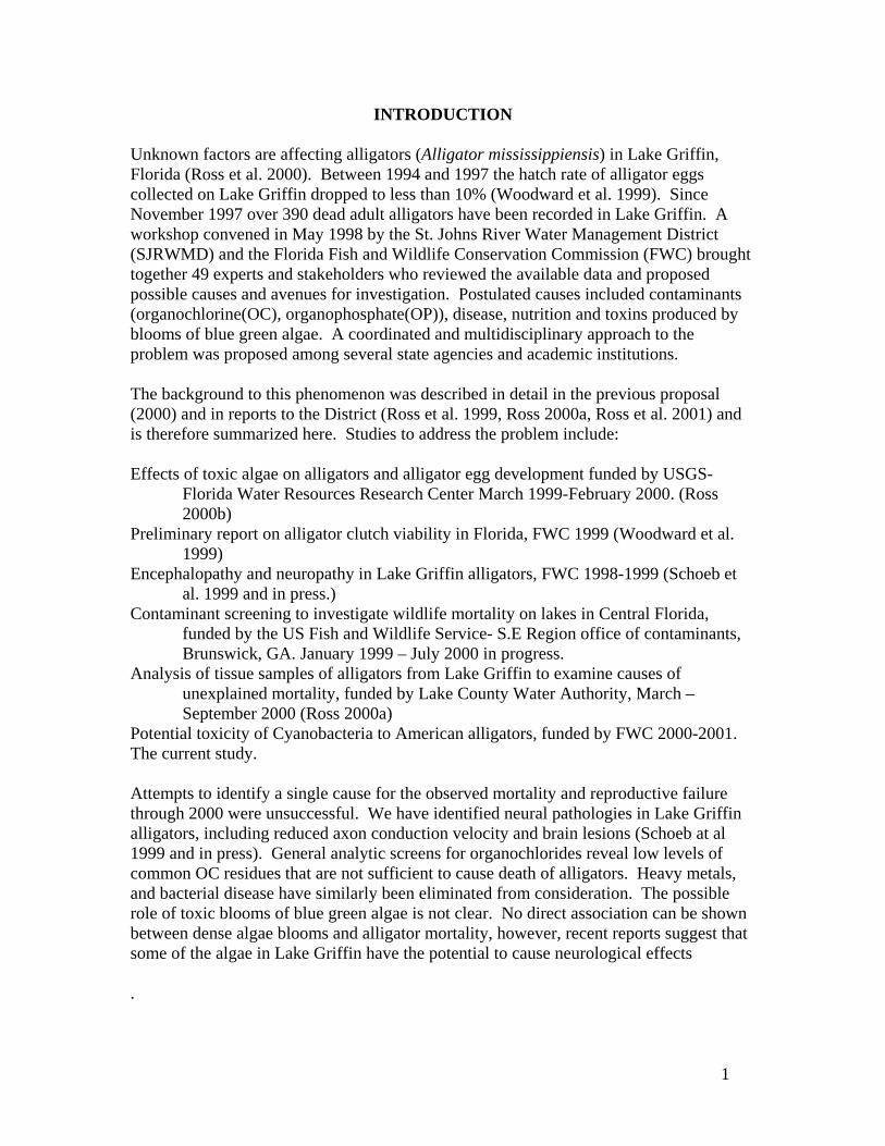
Unknown factors are affecting alligators (Alligator mississippiensis) in Lake Griffin, Florida (Ross et al. 2000). Between 1994 and 1997 the hatch rate of alligator eggs collected on Lake Griffin dropped to less than 10% (Woodward et al. 1999). Since November 1997 over 390 dead adult alligators have been recorded in Lake Griffin. A workshop convened in May 1998 by the St. Johns River Water Management District (SJRWMD) and the Florida Fish and Wildlife Conservation Commission (FWC) brought together 49 experts and stakeholders who reviewed the available data and proposed possible causes and avenues for investigation. Postulated causes included contaminants (organochlorine(OC), organophosphate(OP)), disease, nutrition and toxins produced by blooms of blue green algae. A coordinated and multidisciplinary approach to the problem was proposed among several state agencies and academic institutions. The background to this phenomenon was described in detail in the previous proposal (2000) and in reports to the District (Ross et al. 1999, Ross 2000a, Ross et al. 2001) and is therefore summarized here. Studies to address the problem include: Effects of toxic algae on alligators and alligator egg development funded by USGS-
Florida Water Resources Research Center March 1999-February 2000. (Ross 2000b)
Preliminary report on alligator clutch viability in Florida, FWC 1999 (Woodward et al. 1999)
Encephalopathy and neuropathy in Lake Griffin alligators, FWC 1998-1999 (Schoeb et al. 1999 and in press.)
Contaminant screening to investigate wildlife mortality on lakes in Central Florida, funded by the US Fish and Wildlife Service- S.E Region office of contaminants, Brunswick, GA. January 1999 – July 2000 in progress.
Analysis of tissue samples of alligators from Lake Griffin to examine causes of unexplained mortality, funded by Lake County Water Authority, March – September 2000 (Ross 2000a)
Potential toxicity of Cyanobacteria to American alligators, funded by FWC 2000-2001. The current study. Attempts to identify a single cause for the observed mortality and reproductive failure through 2000 were unsuccessful. We have identified neural pathologies in Lake Griffin alligators, including reduced axon conduction velocity and brain lesions (Schoeb at al 1999 and in press). General analytic screens for organochlorides reveal low levels of common OC residues that are not sufficient to cause death of alligators. Heavy metals, and bacterial disease have similarly been eliminated from consideration. The possible role of toxic blooms of blue green algae is not clear. No direct association can be shown between dense algae blooms and alligator mortality, however, recent reports suggest that some of the algae in Lake Griffin have the potential to cause neurological effects .

2
This study was designed to improve sample efficiency and to pursue additional potential causes of the observed mortality. With support from the SJRWMD and supplementary funding from Lake County Water Authority it was possible to increase field activities and necropsy of alligators, broaden and confirm the previously observed neural pathology and investigate some additional causes. The goals of the project were: 1) Compare sick and healthy alligators from Lake Griffin and Lake Woodruff for the
presence of neural impairment and brain lesions associated with the high mortality in Lake Griffin.
2) Investigate diet and nutrition of alligators from Lake Griffin and Lake Woodruff. The tasks to be conducted were: Task 1. Alligator Monitoring. Observe, monitor and collect alligators using increased facilities and frequency of surveys to refine the data on occurrence of mortality and to collect a minimum sample of eight sick and eight healthy (control or reference) alligators. Task 2. Alligator Collection and Necropsy. Conduct necropsy and clinical evaluations to identify apparent cause of death and confirm previously reported neurological conditions. A pathology team and consulting veterinary pathologist would supervise and assist in the interpretation of pathology information. Complete veterinary pathology screening and archiving of frozen tissues for later analysis Task 3. Laboratory Analysis of Tissues. Conduct laboratory analysis of tissues to examine possible causes of neural impairment including botulism and organophosphate residues and thiamin levels. Task 4. Diet and Nutrition of Alligators. Determine the quantity and quality of alligator diet by developing suitable techniques to obtain stomach contents, calibrate these methods and characterize alligator diets. A sample of at least 12 alligators from Lake Griffin and Lake Woodruff would be captured and sampled by stomach lavage. Stomach contents would be identified and analyzed. Task 5. Report. Integrate and analyze data and prepare reports. Study Sites Study area for this project were Lake Griffin and Lake Woodruff in central Florida. Field work was scheduled to occur from February through December 2001. This report details new data and analyses conducted during 2001 and integrates these results with previous findings. Results are reported by task giving methods, results and general analyses for each topic. A general discussion and conclusions follows. Summarized data in the form of graphs and tables are presented at appropriate positions in the text and the full data are given as a series of electronic appendices to this report.

3
ALLIGATOR MONITORING Methods. Surveys were conducted every two weeks on Lake Griffin. The circumference of the lake was traveled by airboat in a period of 5-8 hours and an experienced observer (D. Carbonneau, FWC) located dead alligators. Dead alligators were marked with spray paint to avoid counting them more than once and their size and gender recorded. Alligator size was determined in the field by measuring the Total Length (TL) with a flexible tape measure. TL is the length from the tip of the snout to the end of the tail measured along the length of the body from either dorsal or ventral aspect. Results. Between January and December 2001, a total of 75 dead alligators were recorded in biweekly surveys. The pattern of alligator mortality continues to be confirmed, with mortality restricted to adult size individuals and occurring in a distinct seasonal peak between February and May (Fig. 1)
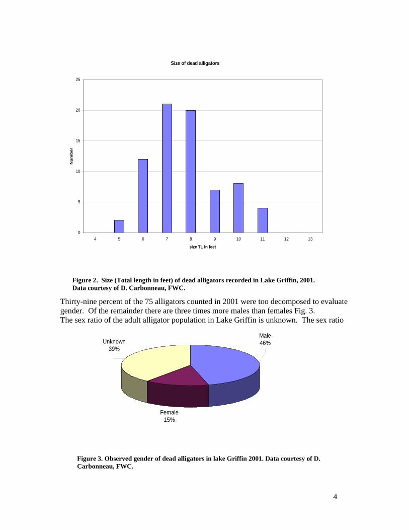
The alligators found dead on Lake Griffin nearly all exceed the TL of 6 feet (Fig. 2). Minimal adult size for alligators in central Florida is around 6 feet TL and alligators greater than about 9 feet TL are usually adult males..
Figure 1. Recorded mortality of alligators on Lake Griffin, FL. 1997-2001. Results of biweekly lake surveys. Data courtesy of D. Carbonneau, FWC.
0
10
20
30
40
50
Aug OctDec Feb Apr Ju
n
Num
ber O
bser
ved
97/98 98/99 99/00 00/01
4
Thirty-nine percent of the 75 alligators counted in 2001 were too decomposed to evaluate gender. Of the remainder there are three times more males than females Fig. 3. The sex ratio of the adult alligator population in Lake Griffin is unknown. The sex ratio
Female15%
Male46%Unknown
39%
Size of dead alligators
0
5
10
15
20
25
4 5 6 7 8 9 10 11 12 13
size TL in feet
Num
ber
Figure 2. Size (Total length in feet) of dead alligators recorded in Lake Griffin, 2001. Data courtesy of D. Carbonneau, FWC.
Figure 3. Observed gender of dead alligators in lake Griffin 2001. Data courtesy of D. Carbonneau, FWC.

5
of a sample of 22 apparently healthy alligators we captured in 2001 (including those released alive after stomach lavage) was 1:1. This suggests that the male dominated sex ratio of dead animals represents a preferential mortality of males. However, with these small sample sizes, the deviations from expected sex ratios are not statistically significant. To quantify apparent neural impairment of live alligators, a graded scale of observed behavior was developed and tested (Table 1). The Owen Carbonneau Alligator Impairment Scale (OCAIS) was designed for this study and evaluates the response of alligators to close daytime approach. Previous observations of sick alligators from Lake Griffin demonstrated that they have slow nerve conduction velocity and focal brain lesions in an area of the brain thought to integrate sensory input and motor response (Schoeb et al. 1999). Empirical field observations indicated a graded degree of reduced motor coordination and lethargy in Lake Griffin alligators. A scale of impairment from 0 or 1 (normal avoidance behavior) to 5 (completely moribund) was assigned to a total of 2,525 alligator encounters on a total of 16 days during the peak of the mortality period. Results (Table 2) indicate that between March and May 2001 an average of 1.4% (range 0%-3.7%) of the alligators observed on Lake Griffin on a given day appear to be suffering some visible level of impaired behavior.

6
Table 1. The Owen-Carbonneau Alligator Impairment Scale (OCAIS) The OCAIS scale allows qualitative evaluation of alligator behavior during daylight when observed from a moving airboat. ______________________________________________________________________ Class 0. Alligators vigorously avoid airboat at first close approach. Alligators approached 50-100 m actively avoid the boat. Alligators on the bank slide into the water and submerge, alligators in shallows move rapidly to deep water, often with considerable splash, alligators in deep water submerge and move underwater. Alligators in class 0 cannot be approached or captured. (note 1) Class 1. Alligators do not avoid airboat at first approach, but behave as class zero on second approach with active avoidance. (note 2) Class 2. Alligators do not avoid airboat on second approach but do not qualify as Class 3. Class 2 alligators cannot be easily approached or captured. Class 2 alligators may continue to stay just out of capture reach, but do not show rigorous avoidance of 0 and 1. Class 3. Alligators approached a second time appear unable or reluctant to move or submerge. Movements appear sluggish, submergence brief and short distance. Class 3 alligators are easily noosed and caught, but may exhibit more active behavior after capture. Class 4. Alligators clearly impaired with obvious motor discoordination including inability to swim or submerge, poor orientation, asymmetrical movements or posture, swimming in circles, refusal to enter the water (alligators on bank). Class 5. Alligators alive but completely moribund, inactive, not moving. ________________________________________________________________________ Notes 1. The great majority, (more than 90%) of alligators observed from an airboat by day
are Class 0, this we assumed to be normal alligator behavior. 2. Class 1 alligators are probably also unimpaired and represent the normal range of
avoidance behavior. Several Class 1 alligators have been observed to be near groups of juveniles and are presumed to be in attendance to these groups and are reluctant to abandon them.
3. Lower air and water temperatures may increase the incidence of class 1 as opposed to class 0. Air and water temperatures should be recorded during surveys.
4. When class evaluation is ambiguous or transitional, use the lower class designation.
5. The OCAIS scale is only for use in daylight from moving boats, not at night or stationary observation.

7
Table 2. Numbers of alligators exhibiting different avoidance behavior (Owen Carbonneau Alligator Impairment Scale) to approach by an airboat during the day during peak of mortality period, Lake Griffin 2001. Classes 0 and 1 are considered normal behaviour.
normal normal ? impaired impaired impaired Percent Class2-5
Date # Search hrs
Class 0 Class 1 Class 2 Class 3 Class 4 Class 5 Total
14-Mar-01 5.5 69 69 0.0% 22-Mar-01 2.0 61 1 1 1 64 3.1% 28-Mar-01 5.0 203 1 204 0.0% 05-Apr-01 3.5 270 15 5 290 1.7% 12-Apr-01 5.5 169 1 3 173 1.7% 18-Apr-01 8.0 160 17 6 183 3.3% 19-Apr-01 7.5 235 6 3 244 1.2% 20-Apr-01 1.0 25 1 1 27 3.7% 26-Apr-01 3.0 133 2 1 136 0.7% 27-Apr-01 8.5 285 1 1 287 0.7% 01-May-01 7.5 320 4 7 331 2.1% 02-May-01 8.5 160 10 1 1 172 1.2% 09-May-01 1.5 96 3 1 100 1.0% 16-May-01 6.0 123 123 0.0% 23-May-01 8.0 138 1 2 1 142 2.1% 13-Jun-01 4.5 78 78 0.0% TOTAL 85.5 2525 62 30 4 2 0 2623 1.4%

8
ALLIGATOR COLLECTION, NECROPSY and PATHOLOGY Methods. With increased manpower and facilities we were able to collect alligators on Lake Griffin each week from February to May. Field teams spent two days and one night each week conducting field work. Lake field trips were conducted by airboat. Alligators were located during the day and those assessed as clearly impaired by the OCAIS scale (impairment level 3 or higher) were captured by hand lasso or capture dart, brought to the boat, secured, sexed and measured. Additional specimens of unimpaired alligators were located at night with a spotlight and captured in the same way. The size of captured alligators was measured by both Total length (TL previously defined) and Snout-Vent length (SVL). SVL is the distance from the tip of the snout to the anterior edge of the cloaca measured on the ventral aspect of the animal. SVL is a more precise measure of alligator size because it is less influenced by the animals mass or condition (stoutness) and because many alligators are missing the tip of their tail. TL is approximately 2 X SVL. A blood sample of approx. 20 ml was drawn by syringe from the vertebral sinus. Blood samples were decanted to lithium heparin (2 tubes) and plain (1 tube) vacutainer tubes and held on ice. Within 2-6 hours the chilled tubes were centrifuged at 8,000 rpm for 10 minutes and serum or plasma decanted to 2 ml cryovials and frozen. Captured, sick alligators were lavaged for stomach contents (see Diet section below) held restrained overnight and euthanized the next morning by cervical section and exsanguination. Alligators were transported to a necropsy facility and a full veterinary necropsy and tissue collection were undertaken. Twenty six euthanized alligators were examined by a qualified veterinary pathologist (Dr. S. Terrell) using a standardized necropsy protocol (Table 3). Seven additional specimens were necropsied by other personnel (Ross, Finger, Owen) using the same protocol. Major organs were examined for gross pathology and samples obtained for additional pathology analysis. Tissue specimens were collected as follows:
• Sterile collection of live tissue, chilled for virus screening and cultured for bacteria.
• Collection of representative tissues into 10% buffered Formalin solution for histology.
• Collection of representative tissues into whirlpacks, frozen for toxicology and other analysis

9
Table 3. Abbreviated necropsy/veterinary pathology protocol. At capture
• Tag and record FWC tag number that becomes identifier on all tissues, specimens. • Record date, time, collectors, location (lake and GPS), sex, snout vent length. • Collect 10-30ml blood from occipital sinus. • 1-2 x red top tube, 2-4 x green top lithium heparin tube • chill-hold on ice, centrifuge, separate plasma/serum to 2ml cryovials, freeze
After euthanasia (All personnel must use protective gloves, aprons, boots and bacteriostatic wash following procedure.)
• Verify specimen number, date, location of origin, sex, • Re-measure and record, Snout vent length, total length • Measure and record specimen weight (note units lb. or kg) • Examine and record specimen condition and external injuries, trauma
Open thoracic-abdominal cavity (lateral incision) and reflect ventral wall.
• Aseptically collect 2-4 ml heart blood to bacteria culture bottle • Aseptically collect 2-4 g spleen, lung to 50ml centrifuge tube, chill/ice for viral screen.
Remove head, cut (striker saw) to remove brain intact.
• Aseptically collect section cerebral cortex to 50ml centrifuge tube, chill/ice for viral screen. • Collect 2 mm sections cerebral cortex, medulla, cerebellum, optic lobe into Trump’s solution
for electron microscopy. Remainder of brain into formalin. • Remove eyes, one in formalin, one chill/ice
Locate, dissect, examine and remove thymus, thyroid, adrenal glands, gonads
• One piece into formalin • Remainder whirlpac chill/ice.
Examine major organs and tissues Abdominal fat, ventral tail fat, Liver, Right and Left Lung, Heart, Spleen, R and L Kidney and tail muscle for appearance, lesions, malformation, parasites and collect from each as possible.
• 0.5 cm piece in formalin • 10-15 g in aluminum foil and whirlpac, chill/ice. • Three x 30 g in whirlpac, chill/ice • Three x 100 g approx. in whirlpac, chill/ice
Dissect and examine Oesophagus, stomach, small and large intestine.
• Examine for lesions and parasites • Preserve 1 cm each in formalin • Collect and preserve stomach contents into formalin
Dissect out following nerves and muscles, adhere to applicator stick and preserve in formalin; Brachial nerve, biceps brachi, sciatic nerve, quadriceps muscle, jaw muscle, lateral tail muscle
Dissect and collect right femur, ziplock bag, chill/ice.
Collect sections of spinal cord from cervical, thoracic, and lumbar sections
• <1cm of each into cryovial, chill/ice • 1 cm into cassette in formalin for histology
Check all labels, transfer chilled/iced samples to –70°F freezer, check and file necropsy record, dispose of carcass, trim and mount all formalin samples for histology.
10
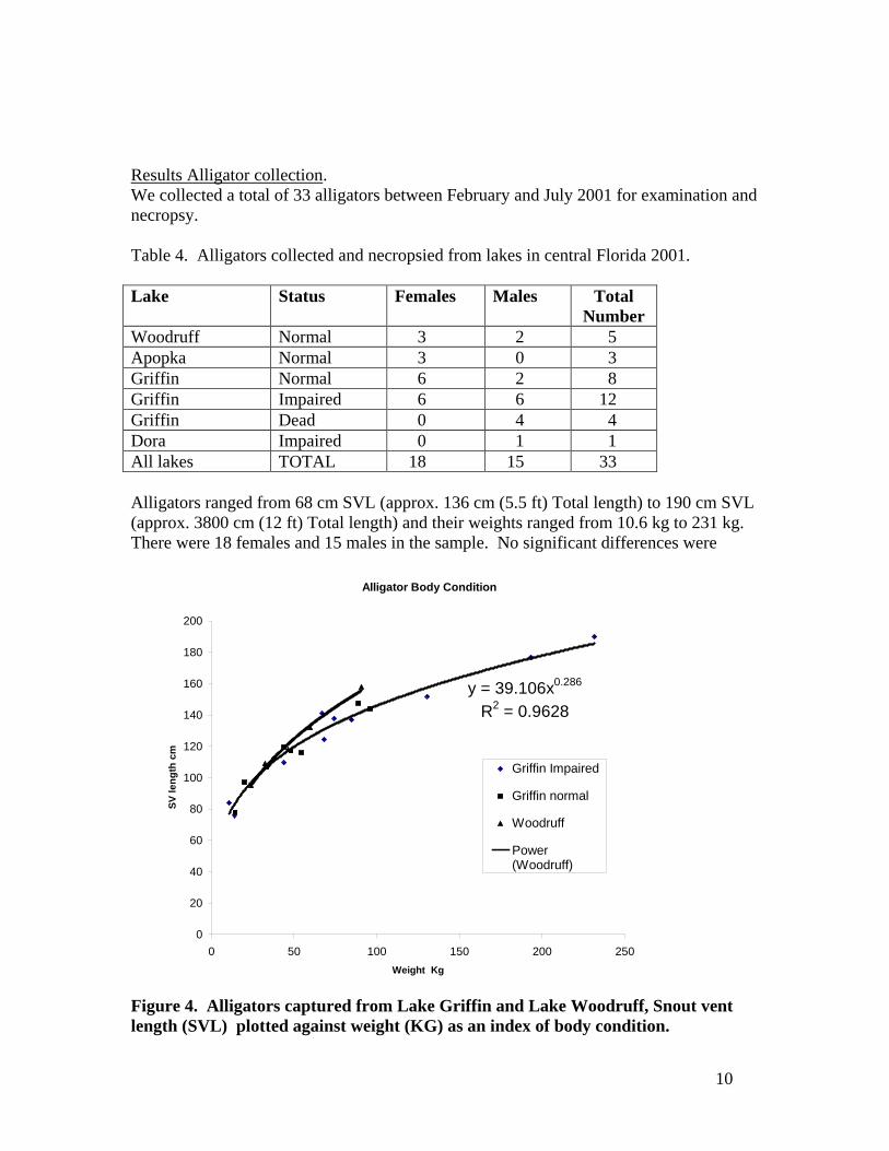
Results Alligator collection. We collected a total of 33 alligators between February and July 2001 for examination and necropsy. Table 4. Alligators collected and necropsied from lakes in central Florida 2001. Lake Status Females Males Total
Number Woodruff Normal 3 2 5 Apopka Normal 3 0 3 Griffin Normal 6 2 8 Griffin Impaired 6 6 12 Griffin Dead 0 4 4 Dora Impaired 0 1 1 All lakes TOTAL 18 15 33 Alligators ranged from 68 cm SVL (approx. 136 cm (5.5 ft) Total length) to 190 cm SVL (approx. 3800 cm (12 ft) Total length) and their weights ranged from 10.6 kg to 231 kg. There were 18 females and 15 males in the sample. No significant differences were
Figure 4. Alligators captured from Lake Griffin and Lake Woodruff, Snout vent length (SVL) plotted against weight (KG) as an index of body condition.
Alligator Body Condition
y = 39.106x0.286
R2 = 0.9628
0
20
40
60
80
100
120
140
160
180
200
0 50 100 150 200 250Weight Kg
SV le
ngth
cm
Griffin Impaired
Griffin normal
Woodruff
Power(Woodruff)

11
observed in length, weight or sex ratio between samples from different lakes or between normal and impaired alligators. Body weight was regressed against snout vent length as an index of body condition (Fig. 4). Lake Woodruff alligators appear to be slightly leaner (lower body weight) than alligators from Lake Griffin In addition, 16 alligators were captured alive, measured, a blood sample collected, and lavaged for stomach contents (see Diet section below) and then released alive at their capture point. Blood samples from these alligators were submitted for chemical screening. Results Blood Values Blood plasma from euthanized and stomach lavaged alligators was stored frozen at –70° F then submitted for standard exotic animal/reptile blood chemistry screen LV2OR to the College of Veterinary Medicine Teaching Hospital. Five healthy Lake Griffin alligators used for necropsy, sixteen Lake Griffin alligators captured for stomach lavage and released alive, and three Lake Woodruff alligators provided a profile of relatively unstressed (and assumed) healthy alligators for comparison with impaired Lake Griffin alligators. The impaired group commonly showed mild to moderate elevations in the metabolic enzymes [Alkaline phosphatase (Alk.Phos), Aspartate aminotransferase (AST), Alanaine aminotransferase( (ALT)], Total protein and Glucose and slightly lowered sodium and chloride (Table 5). These are consistent with organisms that have been stressed. Alkaline phosphatase can originate from several places in the body including the liver, bone, intestine, and kidney. It can be elevated by endogenous steroids (i.e. steroids induced during stress) and it is possible/likely in the absence of liver pathology that the stress of illness or capture is responsible for these elevations. Moderate elevations in AST are likely due to muscle damage, perhaps as a result of the disease process or possibly associated with capture. ALT is another enzyme that originates from the liver, but ALT can be induced as well by endogenous corticosteroids (stress) and can be induced during trauma (capture). Elevations in total protein could be the result of dehydration or may be elevated as a result of a systemic inflammatory process. Total protein is composed of 2 parts: albumin and globulins (GLOB). The globulins are always elevated in the sick animals with high total protein. Globulins can be elevated as a result of acute tissue injury or more long term production of antibodies (immunoglobulins). These can be differentiated by use of serum protein electrophoresis which may be an interesting test to perform on serum from the animals with high total protein as well as a few normal specimens. This could tell us whether these animals are responding to an acute or more chronic agent / process. Elevated blood glucose is a common indicator of stress. Decreased sodium and chloride almost always follow each other and could be the result of renal disease or possibly

12
diarrhea. There are elevations in creatinine and uric acid in a few animals to suggest renal dysfunction. Renal dysfunction could result from dehydration (pre-renal azotemia) or could be the result of enzymes released from damaged muscle that injure the kidney (myoglobinuric nephrosis). Overall, these chemical profiles are consistent with stressed and sick animals but the cause cannot be readily determined. Table 5. Blood analysis values(Mean ± sd) of neurologically impaired and apparently healthy alligators from Lake Griffin, 2001. Standard veterinary symbols as in text. Test Units Alligator Clinical Condition
Abnormal (n=10) Normal (n=24) Alkaline Phosphatase U/L 22.6 ± 8.9 15.7 ± 7.8 AST U/L 278.9 ± 117.7 188.0 ± 74.8 ALT/GPT U/L 32.7 ± 20.0 26.25 ± 19.8 Total Bilirubin mg/dl 0.14 ± 0.1 0.15 ± 0.1 Total protein g/dl 7.24 ± 1.4 5.62 ± 0.9 Albumin g/dl 1.75 ± 0.5 1.23 ± 0.3 Albumin/Globulin ratio 0.32 ± 0.1 0.29 ± 0.1 Globulin g/dl 5.49 ± 1.0 4.39 ± 0.8 Calcium mg/dl 15.92 ± 3.5 13.97 ± 3.4 Phosphorus mg/dL 6.07 ± 1.6 6.12 ± 1.9 Creatinine mg/dL 0.57 ± 0.2 0.46 ± 0.2 BUN1 mg/dL 2.2 ± 0.8 2.8 ± 1.9 Glucose mg/dL 160.2 ± 72.2 107.1 ± 33.2 Cholesterol mg/dL 81.9 ± 24.9 78.3 ± 14.8 Uric Acid mg/dL 2.48 ± 1.9 1.10 ± 0.5 Sodium meq/L 145.3 ± 13.7 155.0 ± 7.9 Potassium meq/L 5.01 ± 1.5 5.30 ± 1.1 Chloride meq/L 97.9 ± 10.8 110.8 ± 8.8 Carbon Dioxide meq/L 12.3 ± 4.7 11.8 ± 4.0 Anion Gap1 40.4 ± 14.8 37.9 ± 12.1 1. BUN= Blood Urea Nitrogen, Anion Gap = sum of anion and cation values Results Pathology. Veterinary pathology examination revealed few external or internal lesions to which death could be attributed (Table 6). Five alligators collected alive from Lake Woodruff showed no abnormalities. Of the 20 live Lake Griffin specimens, 12 were judged impaired by the OCAIS scale. None of these showed significant gross pathology with the exception of minor lesions and stomach ulcers (2 individuals). Five Lake Griffin specimens were judged unimpaired and showed no abnormalities. Three individuals showed obvious signs of traumatic injury, two with extensive boat propeller damage to the body and the other with alligator bite wounds to the head and penetrating the thoracic cavity. However, it is not possible to say if these were the cause of death or post mortem injuries or injuries to sick alligators. Two of the alligators found freshly dead had evidence of water, and in one case, mud, in the lungs indicating drowning as the proximal
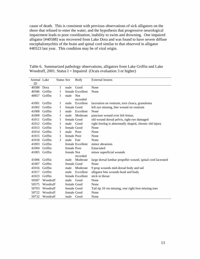
13
cause of death. This is consistent with previous observations of sick alligators on the shore that refused to enter the water, and the hypothesis that progressive neurological impairment leads to poor coordination, inability to swim and drowning. One impaired alligator [#40588] was recovered from Lake Dora and was found to have severe diffuse encephalomyelitis of the brain and spinal cord similar to that observed in alligator #40523 last year. This condition may be of viral origin. Table 6. Summarized pathology observations, alligators from Lake Griffin and Lake Woodruff, 2001. Status I = Impaired (Ocais evaluation 3 or higher) Animal
ID Lake Status Sex Body External lesions
40588 Dora I male Good None 40586 Griffin I female Excellent None 40857 Griffin I male Not
recorded
41001 Griffin I male Excellent laceration on ventrum, torn cloaca, granuloma 41002 Griffin I female Good left eye missing, bite wound on ventrum 41008 Griffin I male Excellent None 41009 Griffin I male Moderate puncture wound over left femur, 41011 Griffin I female Good old wound dorsal pelvis, right eye damaged 41012 Griffin I male Good right foreleg is abnormally shaped, chronic old injury 41013 Griffin I female Good None 41014 Griffin I male Poor None 41015 Griffin I female Poor None 41018 Griffin I male Fair None 41003 Griffin female Excellent minor abrasions 41004 Griffin female Poor Emaciated 41005 Griffin female Not
recorded minor superficial wounds
41006 Griffin male Moderate large dorsal lumbar propeller wound, spinal cord lacerated 41007 Griffin female Good None 41016 Griffin male Moderate 9 prop wounds mid-dorsal body and tail 41017 Griffin male Excellent alligator bite wounds head and body 41023 Griffin female Excellent stick in throat 50587 Woodruff male Good None 50575 Woodruff female Good None 50703 Woodruff female Good Tail tip 10 cm missing, rear right foot missing toes 50722 Woodruff female Good None 50732 Woodruff male Good None

14
MICROBIAL CULTURES Methods Blood was collected from the heart through the pericardium at necropsy using sterile procedures. Approximately 2 ml of fresh blood was introduced to a standard culture medium bottle using a clean, sterile needle. Culture bottles were stored at 0° C then bacterial cultures were submitted to the Microbiology department at the State Veterinary Diagnostic Lab in Kissimmee, FL for processing. Results As in previous years (Schoeb et al. 1999), bacterial cultures were in all cases unremarkable, showing only occasional colonies of common bacteria (Staphlococcus aureus, Escherichia coli, Pseudomonas, Proteus mirabilis) expected to occur in and on alligators. No indication of bacterial infection leading to illness or death were observed (Table 6). HISTOLOGY Methods. Fresh tissue samples of approximately 0.5 cm3 volume were collected at necropsy (Table 4) and fixed and stored in reagent grade 10% buffered formalin. Two mm slices of brain (telencephalic cortex, medulla, cerebellum and optic tectum) were collected and fixed in Trump’s solution for electron microscopy. The remainder of the brain was fixed in formalin. Tissues were prepared routinely for light microscopy according to standard methods for processing, embedding, sectioning and staining (with hematoxylin and eosin) by the histology laboratory of the Department of Pathobiology, College of Veterinary Medicine University of Florida. Slides were then sent to the consulting pathologist, Dr. T. Schoeb, University of Alabama at Birmingham for examination and interpretation. Results. Three hundred eighty-six slides were examined from 21 alligators. In most cases there were no significant findings, that is, findings that would suggest a clinically important condition (Table 7). Exceptions were alligators 41008, 41012, 41013 which had necrotizing encephalopathy similar to that previously seen in Lake Griffin alligators with clinical neurologic impairment. Alligators 41009 and 40588 had lymphocytic meningoencephalitis similar to that previously seen in one clinically ill Lake Griffin alligator. This is an inflammatory, rather than degenerative, process, and therefore is likely to have a completely different cause from the necrotizing encephalopathy. Alligator 41018 had very mild encephalitis localized to one region of the telencephalon. It is unlikely that this lesion was fatal and may not have even been clinically evident.

15
Other lesions listed were considered to be minor incidental findings typical of parasite infestations and other conditions common to wild animals. Table 7. Histology observations and microbial culture results, alligators from Lake Griffin and Lake Woodruff 2001. Animal
ID Lake Bacterial Heart blood culture Internal Lesions and parasites
40588 Dora Not tested encephalitis (viral? See 40523) 40586 Griffin Not tested None noted 40857 Griffin Not tested None noted 41001 Griffin no growth lung: focal hyperplasia, thickening, hyperemia of
esophageal mucosal near cardia, trematodes in small intestine
41002 Griffin scant Staph aureus fibrinonecrotic lesion in the distal esophagus 41008 Griffin no growth few parasites in the liver, 3cm gastric ulcer, small
blood in mid intestine, brain lesions 41009 Griffin Scant mixed colonies parasites in lung and liver, gastric ulceration /
laceration, encephalitis 41011 Griffin Not tested Right eye damaged, old wound on pelvis. 41012 Griffin no growth Right foreleg abnormal shape chronic. linear ulcer /
laceration in stomach with large amounts of blood in lumen, brain lesions
41013 Griffin Not tested Brain lesions 41014 Griffin no growth parasites in stomach, stomach contained another gator 41015 Griffin no growth Multiple small cystic lesions associated with repro tract 41018 Griffin no growth gastric ulcers present with parasites, mild encephalitis 41003 Griffin no growth None noted 41004 Griffin scant Proteus mirabilis large numbers of parasites in liver, fibrinonecrotic
esophageal lesions 41005 Griffin no growth not noted 41006 Griffin no growth Spinal cord transected (propeller wound) 41007 Griffin no growth few parasites in the liver 41016 Griffin Not tested Internal lesions (propeller wounds) 41017 Griffin Not tested heart damaged, internal hemorrhage (Alligator bite
wounds) 41023 Griffin Not tested Blood and oedema right lung and oesophagus (injury) 50587 Woodruff Not tested None noted 50575 Woodruff Not tested None noted 50703 Woodruff Not tested Missing tail tip and right rear toes 50722 Woodruff Not tested None noted 50732 Woodruff Not tested None noted

16
LABORATORY ANALYSIS OF TISSUES ORGANOPHOSPHATE Methods. Samples of serum, brain and spinal cord tissue from 18 alligators collected prior to 2001 were analyzed by The Institute of Environmental and Human Health at Texas Tech University for Cholinesterase activity and reactivation to evaluate exposure to organophosphates (OP). Brain and spinal cord tissue samples of 0.2 g – 0.4 g received frozen from Florida were macerated and homogenized in 1:9 weight/volume Tris0.05 M pH 7.4 buffer. Cholinesterase in two forms ( AchE and BchE) was released with Triton-X 100 (1%). Cholinesterase activity was measured using the method of Ellman et al. 1961 as modified by Gard and Hooper 1993 for use on a SPECTROmax 96 well spectrophotometer plate reader. Reactivation analysis was performed with 2-PAM to displace ChE inhibiting organophosphates (Hooper and Schmidt, 2001). Results. Nine brain, nine spinal cord and 18 serum samples were assessed for ChE activity and reactivation. These represent samples from 9 Lake Woodruff (normal), 4 Lake Griffin (normal) and 5 Lake Griffin (impaired) alligators. ChE levels are expressed in standard ‘units’/g = umoles acetylthiocholine hydrolysed/minute/g
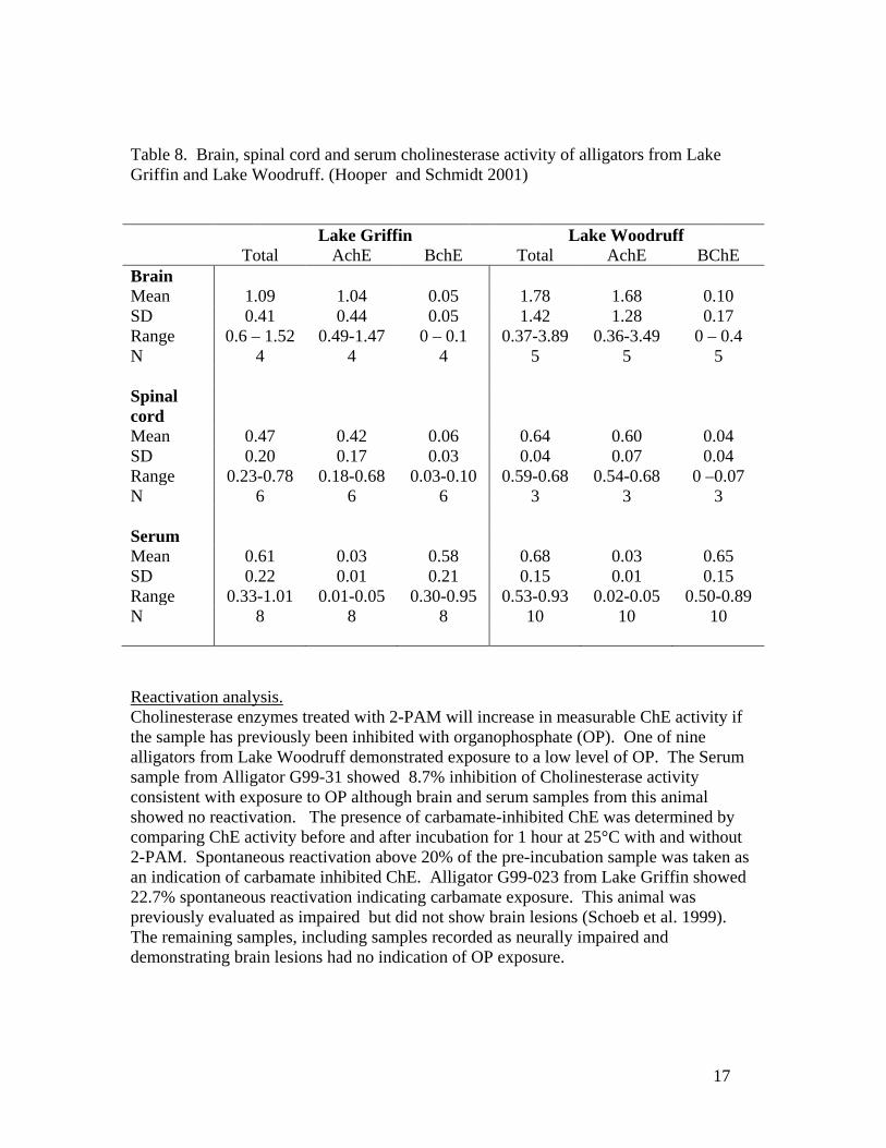
17
Table 8. Brain, spinal cord and serum cholinesterase activity of alligators from Lake Griffin and Lake Woodruff. (Hooper and Schmidt 2001) Lake Griffin Lake Woodruff Total AchE BchE Total AchE BChE Brain Mean 1.09 1.04 0.05 1.78 1.68 0.10 SD 0.41 0.44 0.05 1.42 1.28 0.17 Range 0.6 – 1.52 0.49-1.47 0 – 0.1 0.37-3.89 0.36-3.49 0 – 0.4 N 4 4 4 5 5 5 Spinal cord
Mean 0.47 0.42 0.06 0.64 0.60 0.04 SD 0.20 0.17 0.03 0.04 0.07 0.04 Range 0.23-0.78 0.18-0.68 0.03-0.10 0.59-0.68 0.54-0.68 0 –0.07 N 6 6 6 3 3 3 Serum Mean 0.61 0.03 0.58 0.68 0.03 0.65 SD 0.22 0.01 0.21 0.15 0.01 0.15 Range 0.33-1.01 0.01-0.05 0.30-0.95 0.53-0.93 0.02-0.05 0.50-0.89 N 8 8 8 10 10 10 Reactivation analysis. Cholinesterase enzymes treated with 2-PAM will increase in measurable ChE activity if the sample has previously been inhibited with organophosphate (OP). One of nine alligators from Lake Woodruff demonstrated exposure to a low level of OP. The Serum sample from Alligator G99-31 showed 8.7% inhibition of Cholinesterase activity consistent with exposure to OP although brain and serum samples from this animal showed no reactivation. The presence of carbamate-inhibited ChE was determined by comparing ChE activity before and after incubation for 1 hour at 25°C with and without 2-PAM. Spontaneous reactivation above 20% of the pre-incubation sample was taken as an indication of carbamate inhibited ChE. Alligator G99-023 from Lake Griffin showed 22.7% spontaneous reactivation indicating carbamate exposure. This animal was previously evaluated as impaired but did not show brain lesions (Schoeb et al. 1999). The remaining samples, including samples recorded as neurally impaired and demonstrating brain lesions had no indication of OP exposure.

18
HEAVY METALS Methods. Analysis of heavy metals was conducted by Department of Physics, University of Florida using Proton Induced X-ray Emissions (PIXE). PIXE is a proven spectroscopic technique which has been widely used for several decades in non-destructive simultaneous trace multi-elemental analysis in a variety of research fields such as biology, medicine, botany, zoology, geology, archeology, metallurgy, and environmental sciences (Johansson and Johansson 1976). PIXE X-ray spectra are generated by bombarding the sample atoms with a beam of energetic (1-2.5 MeV) protons. Emitted X-ray quanta include 'characteristic' X-rays that possess energies uniquely identifying the atom from which each originates. The number of emitted X-rays or 'intensity' is proportional to the number of corresponding elemental atoms in the sample being analyzed. The minimum detection limit is at the ppm level or below (1 ppm=10-6 g/g) with the highest sensitivity obtained for the atomic numbers 20<Z<40 and Z>75 (Johansson and Campbell 1988). Tissue samples averaging a few grams each were taken from frozen and fresh material (using nonmetallic, disposable scalpels to minimize contamination) digested with nitric acid in teflon lined bombs, pipetted onto polycarbonate film backings and vacuum dried. These were individually mounted in nylon target holders for irradiation in the accelerator. Average irradiation charge was 50 micro-Coulombs, at proton beam currents averaging 30 nanoamperes, during which period the X-ray spectra were collected with a Kevex Si(Li) detector. Peaks in the X-ray spectra were calibrated against standards of known concentration and metals present calculated to parts per million dry weight in the original sample. Results. Samples of kidney, liver and spinal cord were analyzed from 12 sick Lake Griffin alligators, five apparently healthy Lake Griffin alligators and five Lake Woodruff alligators. Initially 5-8 duplicate runs were conducted on two samples to establish sample variance and calibrate the equipment. Subsequently, 2-3 runs per sample were adequate to give repeatable results and the values were pooled for the different alligator groups. Each spectrum demonstrated peaks of x-ray absorption corresponding to 16 metals but most of these showed no significant patterns. For simplicity, only values of copper, iron, rubidium, selenium and zinc are considered here (Table 9). The values obtained for Cu, Fe and Zn are in the same range as previously measured by different techniques in a small sample of Lake Griffin and Lake Woodruff alligator livers and kidneys (Ross 2000b). In that study selenium was below the detectable limit in most samples but fell in the same range of values 1 – 2.5 ppm wet weight (calculated assuming water content of the 2001 samples is similar to that of earlier samples, around 75%). One published value for selenium in alligator livers is also comparable, 0.641 + 0.09 ppm wet weight (Burger et al. 2000). We therefore feel confident that the values obtained in our current study are accurate.

19
Iron values are highly variable and we suspect that contamination from steel equipment (scalpels, knives) during necropsy may be detected by this very sensitive technique. Alligators from Lake Griffin appear to have depleted levels of selenium in their liver and kidney. We propose to expand these data to determine if this effect is statistically significant. Table 9. Metal concentrations (ppb dry weight) for selected alligator tissues from Lake Griffin and Lake Woodruff (Pooled mean +1 SE). (Note, dry weight concentrations are approximately 4 X wet weight values for alligator tissues). Kidney (N) Cu Fe Rb Se Zn Griffin sick (23)
7.6+0.5 324+48 13.0+0.7 2.5+0.2 67.3+3.4
Griffin healthy (6)
7.0+0.4 166+23 17.4+1.1 2.3+0.4 77.2+6.6
Woodruff (24)
8.3+0.3 388+23 24.4+4.9 10.0+0.3 69.3+1.5
Liver Griffin sick (10)
36.0+5.9 2139+484 10.2+2.2 1.3+0.8 81.5+6.1
Griffin healthy (6)
31.7+6.8 1844+707 15.3+4.1 3.7+1.4 84.3+12.1
Woodruff (7)
8.2+0.8 14059+2695 17.8+2.0 9.4+1.2 55.1+3.6
Alligators from Lake Griffin have significantly lower levels of selenium than alligators from Lake Woodruff for both Liver (t = 2.9 P<0.05, 23 df) and Kidney (t= 4.5,P<0.01 df=67). Impaired alligators have significantly lower selenium in the kidney than unimpaired alligators (t= 4.2,P<<0.01, df=58). However the difference in selenium in the liver (one tailed t= 1.5, P= 0.06, df =66) just fails to be significant at alpha=0.05. None of the other metals show significant differences.
20
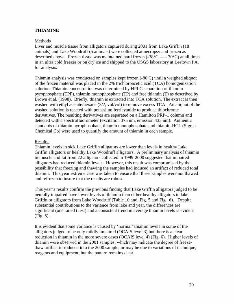
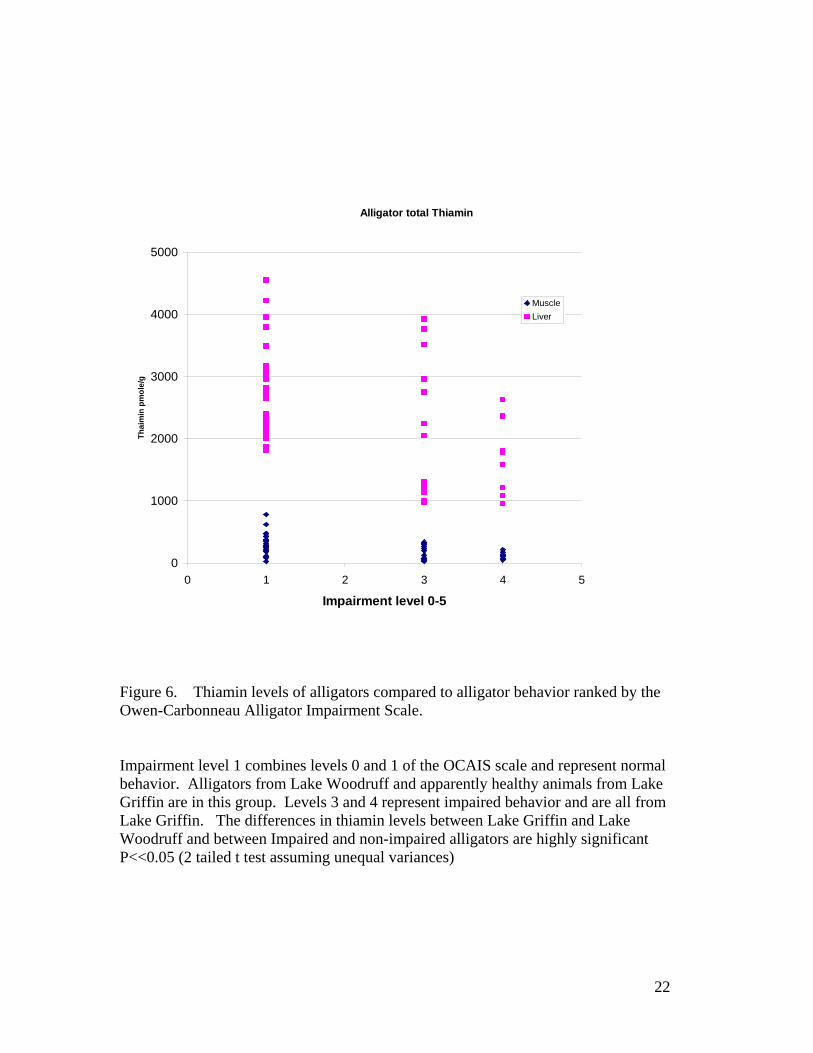
THIAMINE Methods Liver and muscle tissue from alligators captured during 2001 from Lake Griffin (18 animals) and Lake Woodruff (5 animals) were collected at necropsy and frozen as described above. Frozen tissue was maintained hard frozen (-38°C — - 70°C) at all times in an ultra cold freezer or on dry ice and shipped to the USGS laboratory at Leetown PA. for analysis. Thiamin analysis was conducted on samples kept frozen (-80 C) until a weighed aliquot of the frozen material was placed in the 2% trichloroacetic acid (TCA) homogenization solution. Thiamin concentration was determined by HPLC separation of thiamin pyrophosphate (TPP), thiamin monophosphate (TP) and free thiamin (T) as described by Brown et al, (1998). Briefly, thiamin is extracted into TCA solution. The extract is then washed with ethyl acetate:hexane (3/2, vol/vol) to remove excess TCA. An aliquot of the washed solution is reacted with potassium ferricyanide to produce thiochrome derivatives. The resulting derivatives are separated on a Hamilton PRP-1 column and detected with a spectrofluorometer (excitation 375 nm, emission 433 nm). Authentic standards of thiamin pyrophosphate, thiamin monophosphate and thiamin-HCL (Sigma Chemical Co) were used to quantify the amount of thiamin in each sample. Results. Thiamin levels in sick Lake Griffin alligators are lower than levels in healthy Lake Griffin alligators or healthy Lake Woodruff alligators. A preliminary analysis of thiamin in muscle and fat from 22 alligators collected in 1999-2000 suggested that impaired alligators had reduced thiamin levels. However, this result was compromised by the possibility that freezing and thawing the samples had induced an artifact of reduced total thiamin. This year extreme care was taken to ensure that these samples were not thawed and refrozen to insure that the results are robust. This year’s results confirm the previous finding that Lake Griffin alligators judged to be neurally impaired have lower levels of thiamin than either healthy alligators in lake Griffin or alligators from Lake Woodruff (Table 10 and, Fig. 5 and Fig. 6). Despite substantial contributions to the variance from lake and year, the differences are significant (one tailed t test) and a consistent trend in average thiamin levels is evident (Fig. 5). It is evident that some variance is caused by ‘normal’ thiamin levels in some of the alligators judged to be only mildly impaired (OCAIS level 3) but there is a clear reduction in thiamin in the more severe cases (OCAIS level 4) (Fig. 6). Higher levels of thiamin were observed in the 2001 samples, which may indicate the degree of freeze-thaw artifact introduced into the 2000 sample, or may be due to variations of technique, reagents and equipment, but the pattern remains clear.

21
Table 10. Total thiamin pmol/g from alligator muscle and liver from Lake Griffin and Lake Woodruff for years 2000 and 20001. Mean + 1 SE (sample size N)
Griffin Impaired Griffin Healthy Woodruff Muscle 2000 92.7 + 25.1 (8) 143 0 + 52.9 (4) 312.0 + 54.8 (10) 2001 177.4 + 26.8 (13) 316.9 + 127.1 (5) 358.4 + 40.5 (5) Liver 2000 1327 + 137 (8) 2172 + 112 (4) 2679 + 165 (10) 2001 2373 + 276 (13) 2922 + 529 (5) 3324 + 324 (5)
Figure 5. Average thiamin levels pmol/g of muscle and liver from alligators from Lake Griffin and Lake Woodruff for years 2000 and 2001. One standard error of the mean is indicated . Alligators are grouped by relative level of impairment. Differences are not statistically significant.
Average Thiamine
woodruffgrif okgrif sick
woodruffgrif OKgriffin sick
0
500
1000
1500
2000
2500
3000
3500
4000
4500
0 1 2 3 4 5 6 7 8 9
Muscle Liver
Thia
min
e pm
ol/g
2000 2001
22
Figure 6. Thiamin levels of alligators compared to alligator behavior ranked by the Owen-Carbonneau Alligator Impairment Scale. Impairment level 1 combines levels 0 and 1 of the OCAIS scale and represent normal behavior. Alligators from Lake Woodruff and apparently healthy animals from Lake Griffin are in this group. Levels 3 and 4 represent impaired behavior and are all from Lake Griffin. The differences in thiamin levels between Lake Griffin and Lake Woodruff and between Impaired and non-impaired alligators are highly significant P<<0.05 (2 tailed t test assuming unequal variances)
Alligator total Thiamin
0
1000
2000
3000
4000
5000
0 1 2 3 4 5
Impairment level 0-5
Thai
min
pm
ole/
g
MuscleLiver
23
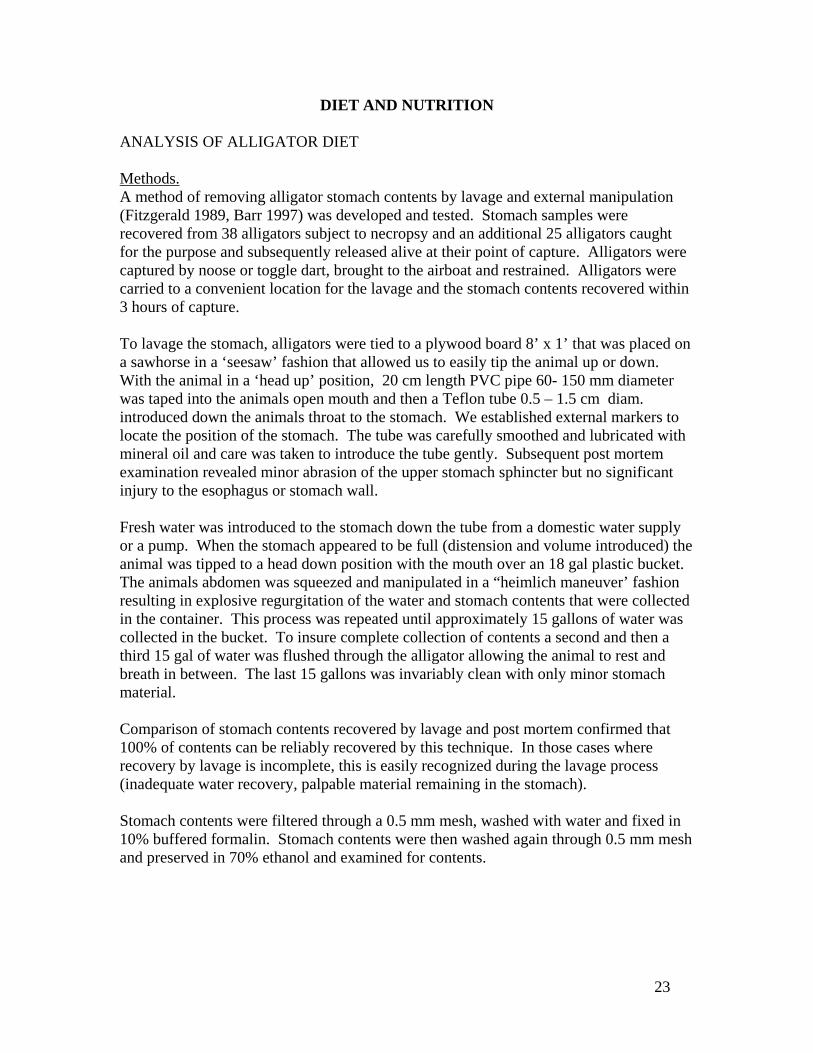
DIET AND NUTRITION ANALYSIS OF ALLIGATOR DIET Methods. A method of removing alligator stomach contents by lavage and external manipulation (Fitzgerald 1989, Barr 1997) was developed and tested. Stomach samples were recovered from 38 alligators subject to necropsy and an additional 25 alligators caught for the purpose and subsequently released alive at their point of capture. Alligators were captured by noose or toggle dart, brought to the airboat and restrained. Alligators were carried to a convenient location for the lavage and the stomach contents recovered within 3 hours of capture. To lavage the stomach, alligators were tied to a plywood board 8’ x 1’ that was placed on a sawhorse in a ‘seesaw’ fashion that allowed us to easily tip the animal up or down. With the animal in a ‘head up’ position, 20 cm length PVC pipe 60- 150 mm diameter was taped into the animals open mouth and then a Teflon tube 0.5 – 1.5 cm diam. introduced down the animals throat to the stomach. We established external markers to locate the position of the stomach. The tube was carefully smoothed and lubricated with mineral oil and care was taken to introduce the tube gently. Subsequent post mortem examination revealed minor abrasion of the upper stomach sphincter but no significant injury to the esophagus or stomach wall. Fresh water was introduced to the stomach down the tube from a domestic water supply or a pump. When the stomach appeared to be full (distension and volume introduced) the animal was tipped to a head down position with the mouth over an 18 gal plastic bucket. The animals abdomen was squeezed and manipulated in a “heimlich maneuver’ fashion resulting in explosive regurgitation of the water and stomach contents that were collected in the container. This process was repeated until approximately 15 gallons of water was collected in the bucket. To insure complete collection of contents a second and then a third 15 gal of water was flushed through the alligator allowing the animal to rest and breath in between. The last 15 gallons was invariably clean with only minor stomach material. Comparison of stomach contents recovered by lavage and post mortem confirmed that 100% of contents can be reliably recovered by this technique. In those cases where recovery by lavage is incomplete, this is easily recognized during the lavage process (inadequate water recovery, palpable material remaining in the stomach). Stomach contents were filtered through a 0.5 mm mesh, washed with water and fixed in 10% buffered formalin. Stomach contents were then washed again through 0.5 mm mesh and preserved in 70% ethanol and examined for contents.

24
Results. Only 7 of the 63 stomachs did not contain evidence of any recent meal. Analysis by percentage occurrence (percentage of stomachs in the sample in which an item is present) indicate invertebrates (87% of stomachs, primarily apple snails but also crustaceans and insects) and fish (67%) are the most common dietary items with reptiles (32%, mostly turtles), mammals (22%) and birds (13%) also present. Stomachs were also notable for the large quantity of anthropogenic materials including golf balls, plastic toys, fishing lures and in one case, 6 spark plugs!
Figure 7. Contents of alligator stomachs from Lake Griffin identified to major animal class and presented as percentage of stomachs in the sample containing representatives of that class (% occurrence). Our sample comprised 48 from Lake Griffin, 10 from Lake Woodruff and 5 from Lake Apopka. The sample sizes do not allow statistical comparison, but the occurrence of mammals in 100% of the Lake Apopka sample is striking. Otherwise, the diets appeared similar. There was no significant difference in the proportion of empty stomachs between healthy and impaired alligators (Table 11).
MammalBird
Reptile
Amphib.
Fish
Invert.
25
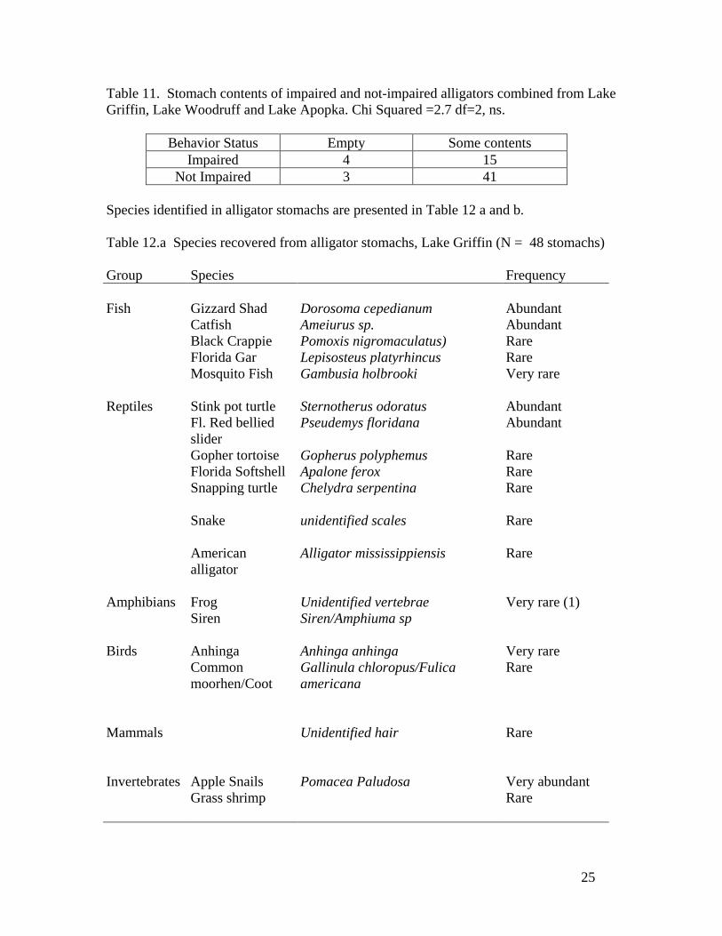
Table 11. Stomach contents of impaired and not-impaired alligators combined from Lake Griffin, Lake Woodruff and Lake Apopka. Chi Squared =2.7 df=2, ns.
Behavior Status Empty Some contents Impaired 4 15
Not Impaired 3 41 Species identified in alligator stomachs are presented in Table 12 a and b. Table 12.a Species recovered from alligator stomachs, Lake Griffin (N = 48 stomachs) Group Species Frequency Fish Gizzard Shad Dorosoma cepedianum Abundant Catfish Ameiurus sp. Abundant Black Crappie Pomoxis nigromaculatus) Rare Florida Gar Lepisosteus platyrhincus Rare Mosquito Fish Gambusia holbrooki Very rare Reptiles Stink pot turtle Sternotherus odoratus Abundant Fl. Red bellied
slider Pseudemys floridana Abundant
Gopher tortoise Gopherus polyphemus Rare Florida Softshell Apalone ferox Rare Snapping turtle Chelydra serpentina Rare Snake unidentified scales Rare American
alligator Alligator mississippiensis Rare
Amphibians Frog Unidentified vertebrae Very rare (1) Siren Siren/Amphiuma sp Birds Anhinga Anhinga anhinga Very rare Common
moorhen/Coot Gallinula chloropus/Fulica americana
Rare
Mammals Unidentified hair Rare Invertebrates Apple Snails Pomacea Paludosa Very abundant Grass shrimp Rare
26
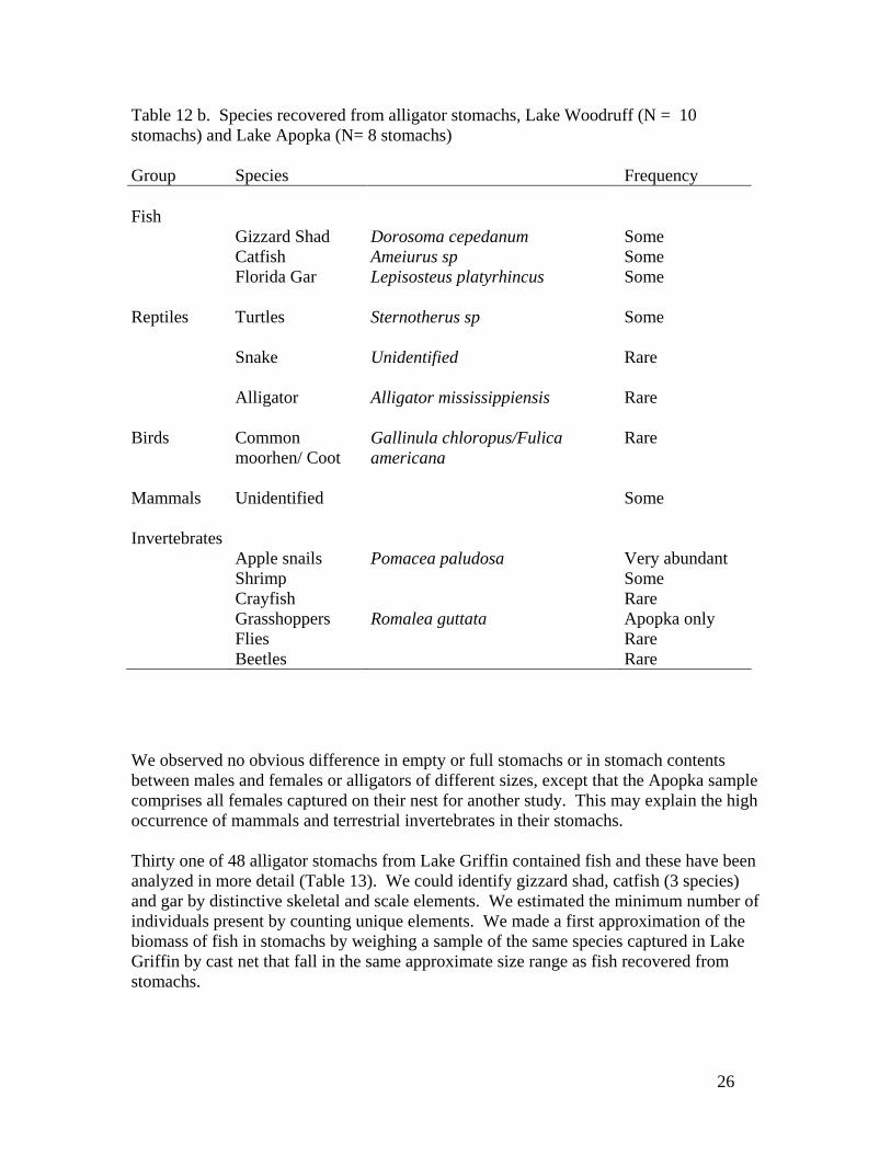
Table 12 b. Species recovered from alligator stomachs, Lake Woodruff (N = 10 stomachs) and Lake Apopka (N= 8 stomachs) Group Species Frequency Fish Gizzard Shad Dorosoma cepedanum Some Catfish Ameiurus sp Some Florida Gar Lepisosteus platyrhincus Some Reptiles Turtles Sternotherus sp Some Snake Unidentified Rare Alligator Alligator mississippiensis Rare Birds Common
moorhen/ Coot Gallinula chloropus/Fulica americana
Rare
Mammals Unidentified Some Invertebrates Apple snails Pomacea paludosa Very abundant Shrimp Some Crayfish Rare Grasshoppers Romalea guttata Apopka only Flies Rare Beetles Rare We observed no obvious difference in empty or full stomachs or in stomach contents between males and females or alligators of different sizes, except that the Apopka sample comprises all females captured on their nest for another study. This may explain the high occurrence of mammals and terrestrial invertebrates in their stomachs. Thirty one of 48 alligator stomachs from Lake Griffin contained fish and these have been analyzed in more detail (Table 13). We could identify gizzard shad, catfish (3 species) and gar by distinctive skeletal and scale elements. We estimated the minimum number of individuals present by counting unique elements. We made a first approximation of the biomass of fish in stomachs by weighing a sample of the same species captured in Lake Griffin by cast net that fall in the same approximate size range as fish recovered from stomachs.

27
Table 13. Occurrence of different species of fish in 31 alligator stomachs from Lake Griffin that contained fish. Shad – (Dorosoma cepedianum), Catfish = (Ameiurus sp.), Gar = (Lepisosteus platyrhincus) Shad Catfish Gar Other fish a) Frequency of occurrence, X fish /in Y stomachs
19 / 8 14 / 11 2 / 2 13 / 13
b) % of 31 stomachs containing 26% 35% 6% 42% c) % of all individual fish eaten 40% 29% 4% 27% d) Avg. # fish in stomachs containing that species
2.4 1.3 1 1
e) Estimated avg. weight of each species occurring in stomachs (g)
212g 334g 500g 50g
f) Estimated biomass of that species ingested/stomach.
503g 425g 500g 50g
g) % of all fish biomass eaten 24% 52% 14% 9% Shad occurred in 26% of the stomachs with fish and comprised 40% of the individuals and 24% of the fish biomass ingested. Catfish occurred in 35% of the stomachs and comprised 29% of the individuals but 52% of the biomass. However, the average biomass of fish in any given stomach is around 500g whether it is shad or catfish or both. Gar also comprise about 500g of biomass in those stomachs in which they occur. Because fish bones dissolve completely in about 24 hours (Barr 1997) we believe these contents represent recent (same day) feeding by alligators. We conclude from this analysis that alligators in our sample ate about the same amount of fish when they ate fish at all. Shad and catfish are the dominant fish eaten, with catfish, by virtue of greater average weight, making a larger proportion of mass eaten. However, in order to ingest the same approximate amount, alligators eat more individual shad, reflected in the greater number of individuals eaten and fish per stomach (Table 9). More detailed analysis to establish the diet by species, biomass, and with corrections for digestibility and residence time (Barr 1997) will refine our understanding of diet in these alligators. For the present we confirm that Gizzard shad are a prominent item in the diet of Lake Griffin alligators. ANALYSIS OF GIZZARD SHAD DIET Methods. Gizzard shad (Dorosoma cepedianum) were taken in Lake Griffin in March, May and June 2001 by cast net. Representative specimens of various sizes were eviscerated and the whole gizzard, forestomach and intestine preserved in vials in 70% Ethanol. Analysis of contents was done by Dr. A. Chapman of Cyano-Labs Inc. Gut contents were processed through a series of sieves (425- 75 mu) and the final fraction preserved in Lugol’s iodine solution to facilitate settling of the material. Samples were allowed to settle for one week then volume was adjusted to 100ml for all samples. A sub-sample was removed from each sample and placed in an Utermol settling chamber. The volume
28
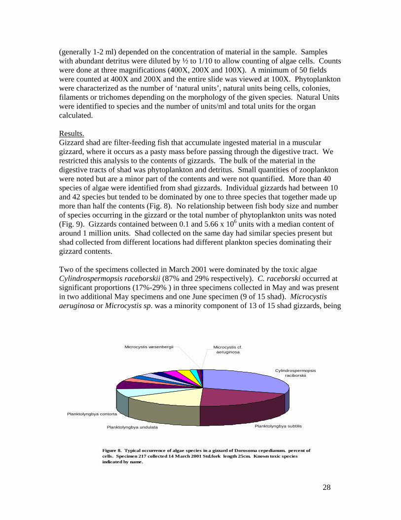
(generally 1-2 ml) depended on the concentration of material in the sample. Samples with abundant detritus were diluted by ½ to 1/10 to allow counting of algae cells. Counts were done at three magnifications (400X, 200X and 100X). A minimum of 50 fields were counted at 400X and 200X and the entire slide was viewed at 100X. Phytoplankton were characterized as the number of ‘natural units’, natural units being cells, colonies, filaments or trichomes depending on the morphology of the given species. Natural Units were identified to species and the number of units/ml and total units for the organ calculated. Results. Gizzard shad are filter-feeding fish that accumulate ingested material in a muscular gizzard, where it occurs as a pasty mass before passing through the digestive tract. We restricted this analysis to the contents of gizzards. The bulk of the material in the digestive tracts of shad was phytoplankton and detritus. Small quantities of zooplankton were noted but are a minor part of the contents and were not quantified. More than 40 species of algae were identified from shad gizzards. Individual gizzards had between 10 and 42 species but tended to be dominated by one to three species that together made up more than half the contents (Fig. 8). No relationship between fish body size and number of species occurring in the gizzard or the total number of phytoplankton units was noted (Fig. 9). Gizzards contained between 0.1 and 5.66 x 10⁶ units with a median content of around 1 million units. Shad collected on the same day had similar species present but shad collected from different locations had different plankton species dominating their gizzard contents. Two of the specimens collected in March 2001 were dominated by the toxic algae Cylindrospermopsis raceborskii (87% and 29% respectively). C. raceborski occurred at significant proportions (17%-29% ) in three specimens collected in May and was present in two additional May specimens and one June specimen (9 of 15 shad). Microcystis aeruginosa or Microcystis sp. was a minority component of 13 of 15 shad gizzards, being
Figure 8. Typical occurrence of algae species in a gizzard of Dorosoma cepedianum. percent of cells. Specimen 217 collected 14 March 2001 Std.fork length 25cm. Known toxic species indicated by name.
Cylindrospermopsis raciborskii
Planktolyngbya subtilisPlanktolyngbya undulata
Planktolyngbya contorta
Microcystis wesenbergii Microcystis cf. aeruginosa

29
present in all three months (Table 14). Table 14. Dominant species of phytoplankton recovered from shad gizzards in Lake Griffin, March - May and June 2001. Sample Date Dominant algae species Other potentially toxic
species present March 14 C. raceborskii,
Planktolyngbya contorta, P. subtilis, P. undulata, Aulacoseira sp. Aphanocapsa sp.
Microcystis aeruginosa
May 1 Aphanocapsa sp., Aulacoseira sp. Planktolyngbya contorta, ?, P. undulata, C. raceborskii, Limnothrix sp.
Microcystis aeruginosa
June 13 Aphanocapsa sp., Aulacoseira sp
Microcystis aeruginosa, C. raceborskii
Figure 9. Gizzard shad (Dorosoma cepedianum) showing weak relationship of size (standard fork length cm) to number of phytoplankton species and total number of phytoplankton units identified in the gizzard.
0
10
20
30
40
50
60
0.0 5.0 10.0 15.0 20.0 25.0 30.0
Standard Fork length cm
Phyt
oplan
kton
100K
units
/ Sp
ecies total cells
# species

30
DISCUSSION The cause of alligator mortality in Lake Griffin remains unknown. However, our work in 2001 has confirmed some earlier conjectures, provided some significant new data and opened the door to an intriguing hypothesis for further testing. Tasks 1 and 2 have confirmed the findings of previous years, broadening our sample size and increasing confidence in the robustness of the findings. The marked seasonal pattern of alligator mortality is strongly confirmed and any explanation of the deaths must take this into account. Dead alligators comprise a majority of adults and a predominance of males. However, no statistically significant differences in size, sex or condition between the dead alligators and the general adult population in Lake Griffin or in other lakes can be determined from this sample. Juvenile and subadult size classes remain conspicuously absent from the dead sample. Prior to 2001 primarily long dead and decomposing alligators were observed in the biweekly surveys. Our more intense field activities on the lake this year resulted in the discovery of four freshly dead alligators, but still no juveniles and we feel confident that the mortality event is affecting only the larger size classes. We have developed, tested and applied a quantitative field assessment for behavioral impairment of alligators in the field that indicates that no more than 3% of alligators appear affected at any time during the spring mortality peak. Necropsy and pathology results confirm the presence of brain lesions in many sick alligators. We have now examined a total of 20 sick Lake Griffin alligators (8 by Schoeb et al. 1999 and 12 this study) of which 11 demonstrate physical indications of neural problems, primarily lesions of the torus semicircularis of the brain. Three of these demonstrate a lymphocytic meningoencephalitis that is probably of different origin to the lesions. Blood values, microbial culture, gross pathology and histological examination of major organ systems fail to indicate any other pathology in these specimens. The absence of histologically evident brain abnormalities in some impaired alligators is not unexpected. Many variables may affect the expression of the lesions and our ability to detect them including progression and severity of the syndrome, artifacts of sampling, location and orientation of the section, and incomplete brain removal or preservation in a few samples. The presence of lesions in over half the sample is compelling evidence that the association between alligator mortality on Lake Griffin, behavioral impairment and neural lesions is robust. Our survey results have refined our understanding of the mortality syndrome process and the affected alligator population. The seasonal nature of the mortality is now completely clear and reproducible. This is an early spring phenomenon affecting adult alligators. We can speculate on several causes why this might be. The causal agent or agents may be more active or intense in the early part of the year or the alligators may be more vulnerable or both. Alligators are ectotherms and spend much of the cooler period of the year quietly in burrows with little movement. Metabolism and movement are reduced

31
and feeding is rare. Body temperatures converge to ambient temperatures that are below optimum for alligator physiology and opportunities for thermoregulation (basking) are reduced (Howarter 1999). It is probable that body reserves (fat, stored micronutrients) are slowly depleted during this period, although the level of depletion will depend upon many factors including the alligator’s size, recent feeding history, reproductive activities etc. Spring, as the ambient temperature increases, is a busy time for alligators. They become active, feed, and adults begin territorial defense and courting. Stored reserves may be mobilized and further depleted while new material is assimilated from food. Changes in hormonal status and stress levels may further complicate the internal metabolic picture. At the same time, many other components of the lacustrine ecosystem are going through their own spring activities. Algae bloom, fish and birds breed, human activities in and around the lake change (lawn fertilizing, restoration management activities such as water level manipulation, fish removal). Changes in weather (rainfall, temperature) impose their own ecological effects. Sublethal effects of low levels of contaminants have been identified as a contributor to reduced immune competence in alligators (Crain and Guillette 1997). So that alligators in contaminated lakes may be more vulnerable to these effects. If the agent(s) responsible for alligator mortality are entirely external to alligators, then we seek a process or event that occurs in the spring. If the agent(s) are derived from within the alligator, perhaps as an interaction of stored materials and metabolic activity, then we seek a process that responds to spring changes. We should also not overlook the fact that reproduction in Lake Griffin alligators is characterized by low hatch success for similarly unknown causes. An agent or chain of events that also explains reproductive inhibition would be particularly useful. We are now confident that the proximal cause of alligator death is derived from severe neuropathy involving both central (torus semicircularis) and peripheral nervous structures. Death by lack of coordination and inanition leading to drowning is plausible and supported by some observations. Our attention was first drawn to thiamin by the similarity between brain lesions reported for thiamin deficient fish and the lesions we observed in alligators. Reproduction of salmonid fish in the Great Lakes, Finger Lakes (New York) and the Baltic Sea is reduced by fry mortality due to a thiamin responsive syndrome (Brown and Honeyfield 1999; Bengtsson, Brown and Honeyfield 1999; Whyte and Honeyfield 2000). This syndrome, called Early Mortality syndrome (EMS), Cayuga syndrome or M74, was first reported in the early 1990s and has received intensive study. The syndrome is recently reported to also affect adult lake trout, coho salmon, baltic salmon and steelhead trout (Honeyfield and Brown (personal comm.). EMS in salmonids is thought to be a result of ingestion of prey species (fish) that contain the enzyme thiaminase which destroys thiamin. Thiamin deficient salmon, lay eggs that are thiamin deficient and the resultant fry have been found with brain lesions and suffer high incidences of mortality. Thiamin deficiency in

32
salmonid eggs can be reversed by injecting gravid females three weeks before spawning or by immersing eggs (or fry) in a thiamin bath (Brown and Honeyfield 1999). The primary prey species that contain thiaminase are the alewife (Alosa pseudoharengus) and smelt (Osmerus mordax) in the Great Lakes and herring (clupeids) in the Baltic Sea, all filter feeders. In the Great Lakes and New York Finger Lakes alewives, thiaminase levels are highly variable both seasonally, within lakes and among lakes. The cause of this variability in thiaminase in these filter feeding fish is unknown, but thiaminase positive algae have been isolated from alewife. (Honeyfield et al., in press). There is also a positive correlation (r = 0.84) between summer abundance of blue green algae, particularly Microcystis sp. and incidence of EMS in coho (Hinterkopf et al. 1999). Although overt mortality from environmental contaminants has not been found in either the Great Lakes or Baltic salmonids, the role of contaminants has not been completely excluded. Synergistic interaction between low thiamin and exposure to pollutants (dioxin like compounds) have been reported (Brown and Honeyfield 1999). Apoptosis in lake trout fry exposed to dioxin-like compounds was found to be greater in thiamin deficient fish (Whyte et al. 2001). The similarity between Florida alligators in Lake Griffin and EMS in salmonids is striking; reproductive failure due to embryo mortality, adult neural impairment and mortality, brain lesions and the association with abundant filter feeding forage fish. Prompted by these similarities we have established contact and a cooperative research program with Dale Honeyfield, U. S. Geological Survey, Wellsboro, PA. Several straightforward questions should be answered. Do Lake Griffin shad have thiaminase? If so, do they exhibit seasonal patterns and how much? Would thiamin treatment reverse the alligator neuropathy? We confirm in this study that gizzard shad do filter toxic blue green algae and can ingest, concentrate and carry significant quantities (up to millions of cells). We have not quantified the mass of shad gizzard contents but estimate that in larger specimens these may amount to 1-2 g of material. The role of gizzard shad in alligator diets, and the interrelationship of algae, shad and the alligator mortality syndrome requires investigation. The role of selenium, and the apparent deficiency of this metal in Lake Griffin alligators is of unknown significance. Selenium interacts with biological systems and metabolism in several contradictory ways. An excess of selenium causes toxicity and developmental abnormalities. However, the metal also interacts with some enzymes as a co-factor and a deficiency may be either a cause or a symptom of a metabolic disorder. We have not yet been able to discover a link between selenium and thiamin metabolism and research on this point continues.

33
We have initiated studies of the relationship of blue green algae, gizzard shad and alligators, demonstrating that there is a direct and short food chain linking these organisms with the potential to rapidly pass toxic materials to alligators.

34
CONCLUSIONS
While the cause of alligator mortality in Lake Griffin remains unknown, these results confirm previous advances and provide some indications of fruitful additional work. We have confirmed and strengthened the strong seasonal occurrence of mortality. No convincing evidence supports either organochloride or organophosphate pollution as the direct cause of the observed neural impairment and death. We have confirmed the widespread occurrence of specific lesions in the mid-brain in many Lake Griffin alligators and have an indication that at least two separate kinds of brain lesions occur- meningoencephalitis and necrotizing encephalopathy. We have added the observations that sick Lake Griffin alligators demonstrate reduced levels of thiamin and selenium in some tissues. A continuing mystery is why the phenomenon is restricted to Lake Griffin. We have investigated several alligator deaths in other lakes (Dora, Apopka) and established that these do not show the same characteristics as Lake Griffin. However, Lake Griffin does not appear to have any unique features that are not shared by many other Florida lakes. Nutrient loading, flocculent material, stabilized water levels, algae blooms, weed control, adjacent land use, contaminant levels, and gizzard shad are all shared in abundance by many other lakes. We continue to speculate that it is not the components of the system, but the particular way in which they interact that is causing Lake Griffin alligators to die. In this regard, continuing a multifaceted, interdisciplinary and flexible approach continues to be the strategy we think is most useful. We continue to follow the general hypothesis that events in Lake Griffin are associated with ecological changes, and particularly disruption and contamination. However, our studies to date indicate that this is not a direct toxicity due to any of the better known contaminants. It continues to appear plausible that we are investigating a chain of related events in which changes to the system are modulated through several species and several trophic levels to express themselves eventually as alligator mortality and reproductive failure. As the process of restoration of this and other lake systems in Florida proceeds, it becomes more important to understand the nature of such interactions. Ecosystems are complex balances of multiple components. We attempt interventions to address previous abuse, such as reduce dissolved nutrients or reinstitute dynamic water levels. It should not be a surprise that these sometimes have unpredicted results or that the system resists our attempts to change it. Untangling the mystery of alligator mortality in Lake Griffin should provide useful information on which to base more effective restoration and management.

35
REFERENCES Barr, B. 1997. Food habits of the American alligator. PhD Dissertation, University of
Miami:244 pp. Bengtsson B-E, S. Brown and D. Honeyfield. 1999. International Council for the
Exploration of the Seas, Annual Science Conference Sept 29-Oct 6, 1999. Stockholm Sweden. Session report: M74 syndrome and similar reproductive disturbances in marine animals. 5 pp.
Brown S. and D. Honeyfield. 1999. Report on Early Mortality Syndrome Workshop. Great Lakes Fishery Commission, Board of Technical Experts: 33 pp.
Brown, S., D. C. Honeyfield, and L. Vandenbyllaardt. 1998. Thiamin analysis in fish egg and tissues by high pressure liquid chromatography. In G. McDonald, J. Fitzsimons, and D.C. Honeyfield (Eds). Early life stage mortality syndrome in fishes of the Great Lakes and Baltic Sea. Symposium 21, p 73-81. American Fisheries Society, Bethesda, MD 20814.
Burger, J., M. Gochfeld, A. A. Rooney, E. F. Orlando, A. R. Woodward and L. J. Guillette. 2000. Metals and metaloids in tissues of American alligators in three Florida lakes. Arch. Environ. Contam. Toxicol. 38:501-508.
Crain D. A. and L. J. Guillette. 1997. Endocrine disrupting contaminats and reproduction in vertebrate wildlife. Reviews in Toxicology 1(1997):47-70.
Fitzgerald, L. A. 1989. An evaluation of stomach flushing techniques for crocodilians. J. Herpetology. Vol 23 (2):170-172.
Hinterkopf, J. P., D. C. Honeyfield, J. Markarewicz and T. Lewis. 1999. Abundance of plankton species in Lake Michigan and the incidence of early mortality syndrome from 1983 to 1992. Board of Technical Experts Briefing Book, October 1999, Great Lakes Fishery Commission. p 206-207.
Honeyfield, D.C. J. P. Hinterkopf and S.B. Brown. In press. Isolation of thiaminase-positive bacteria from alewife. Transactions of the American Fisheries Society.
Hooper and Schmidt 2001. Cholinesterase activity determinations for American alligator tissues. Final report TIEHH project No. T0111. Unpub report:1-14.
Howarter S. 1999. Thermal ecology of the American alligator in the Everglades. Masters dissertation. University of Florida: 70p.
Johansson S. A. E and T. B. Johansson, 1976. Analytical Application of Particle Induced x-ray Emission, Nucl.Instr. Meth. 137 p. 473 - 516.
Johansson, S. and J. L. Campbell, 1988. PIXE: A Novel Technique for Elemental Analysis. John Wiley and Sons.
Ross, J. P., H. F. Percival, A. Woodward, D. Carbonneau, T. R. Schoeb, J. Burns, C. Williams and W. Carmichael. 2000. Alligator mortality and algal toxins: a preliminary report. American Fisheries Society, Florida chapter 20th Annual meeting March 2000. Abstract.
Ross, J.P. 2000a. Central Florida Lakes Wildlife Initiative, Analyses of tissue samples of alligators from Lake Griffin to examine causes of unexplained mortality. Report to Lake County Water Authority, Contract No. #00032702, 30 September 2000:
Ross, J. P. 2000 b. Effect of toxic algae on alligators and alligator egg development. Tech Research Report Project FL 03 to Water Resources Research Center USGS- University of Florida: 23pp.
36
Ross, J. P., H.F. Percival , D. Carbonneau , A. Woodward and T. Schoeb. 2001. Investigations of mortality and reproductive failure of alligators in Lake Griffin, Central Florida. Florida Lake Management Association, Annual Meeting Tallahassee May 2001. Abstract.
Schoeb, T. R., R. M. Clemmons and T. G. Heaton-Jones. 1999. Encephalopathy and neuropathy in Florida Alligators. Report to Florida Fish and Wildlife Conservation Commission and submitted J. Wildlf. Disease.
Whyte J. J. and D. C. Honeyfield. 2000. Lake Trout Restoration in the Great Lakes: Early Life Stage Survival. Int. Assoc. for Great Lake Research, 43rd Conference on Great Lakes and St. Lawrence River research. May 2000. Abstracts
Whyte, J.J., H.A. Allert, C.C. Edsall, D.C. Honeyfield and D.E. Tillitt. 2001. Apoptosis in embryonic lake trout developing under nutrient and contaminant stress. SETAC Annual meeting, Nov 11-15, 2001. Baltimore, MD.: 35.
Woodward, A. R., D.S. Bermudez and D. A. Carbonneau. 1999. A Preliminary report on Alligator Clutch Viability in Florida. Florida Fish and Wildlife Conservation Commission internal report.
Related Documents