Welcome message from author
This document is posted to help you gain knowledge. Please leave a comment to let me know what you think about it! Share it to your friends and learn new things together.
Transcript

300 SBAs
Final FRCA

London • Philadelphia • Panama City • New Delhi
Kariem El-Boghdadly MBBS BSc (Hons) FRCASpecialist Registrar in Anaesthesia,
South East School of Anaesthesia, London, UK
Imran Ahmad MBBS FRCAConsultant Anaesthetist,
Guy’s and St Thomas’ NHS Foundation Trust, London, UKHonorary Senior Lecturer, King’s College London, UK
Training Programme Director Anaesthesia South London (ASL), London, UK
300 SBAs
Final FRCA
© 2015 JP Medical Ltd. Published by JP Medical Ltd83 Victoria Street, London, SW1H 0HW, UKTel: +44 (0)20 3170 8910 Fax: +44 (0)20 3008 6180Email: [email protected] Web: www.jpmedpub.com
The rights of Kariem El-Boghdadly and Imran Ahmad to be identified as editors of this work have been asserted by them in accordance with the Copyright, Designs and Patents Act 1988.
All rights reserved. No part of this publication may be reproduced, stored or transmitted in any form or by any means, electronic, mechanical, photocopying, recording or otherwise, except as permitted by the UK Copyright, Designs and Patents Act 1988, without the prior permission in writing of the publishers. Permissions may be sought directly from JP Medical Ltd at the address printed above.
All brand names and product names used in this book are trade names, service marks, trademarks or registered trademarks of their respective owners. The publisher is not associated with any product or vendor mentioned in this book.
Medical knowledge and practice change constantly. This book is designed to provide accurate, authoritative information about the subject matter in question. However readers are advised to check the most current information available on procedures included and check information from the manufacturer of each product to be administered, to verify the recommended dose, formula, method and duration of administration, adverse effects and contraindications. It is the responsibility of the practitioner to take all appropriate safety precautions. Neither the publisher nor the editors assume any liability for any injury and/or damage to persons or property arising from or related to use of material in this book.
This book is sold on the understanding that the publisher is not engaged in providing professional medical services. If such advice or services are required, the services of a competent medical professional should be sought.
Every effort has been made where necessary to contact holders of copyright to obtain permission to reproduce copyright material. If any have been inadvertently overlooked, the publisher will be pleased to make the necessary arrangements at the first opportunity.
ISBN: 978-1-909836-18-1
British Library Cataloguing in Publication DataA catalogue record for this book is available from the British Library
Library of Congress Cataloging in Publication DataA catalog record for this book is available from the Library of Congress
Commissioning Editor: Steffan ClementsEditorial Assistant: Sophie WoolvenDesign: Designers Collective Ltd
Copy-edited, typeset, printed and bound in India.
v
Preface
Recent pass rates for the Final FRCA written examination have fallen by around 25%, a significant drop that suggests candidates require more practice ahead of the exam. There are few single best answer (SBA) revision books or past papers currently available for the Final FRCA. Recognising this problem and following on from our experience of writing Primary FRCA: 450 MTFs and SBAs, we decided to write this book of 300 practice SBAs.
The written element of the Final FRCA consists of a 3-hour short answer question (SAQ) section of 12 compulsory questions and a multiple choice question (MCQ) section. This comprises 90 MCQs to be completed in 3 hours; 60 multiple-true-false (MTF) questions and 30 SBA questions (the latter introduced in September 2010). The content of the exam is aligned to the intermediate training CCT curriculum, with variable weighting applied to different aspects of the curriculum. The 30 question SBA section generally includes 20 questions in clinical anaesthesia, 5 in intensive care medicine and 5 in pain management. The SBAs are often seen as the most challenging section of the MCQ paper, because they test the application of knowledge in the clinical setting, as opposed to the MTFs which test factual recall.
Each SBA question consists of a clinical scenario (the ‘stem’), and a direct ‘lead-in’ question, followed by five answer options. One of these is the ideal response, although all options are possible solutions. Four marks are awarded for each correct answer and no marks are lost for an incorrect answer, therefore a total of 120 marks is possible. Due to the heavy relative weighting of the SBAs, they are an important aspect of the written examination, and it is crucial that candidates practise as much possible in order to answer them correctly.
The 10 papers of 30 SBAs in this book cover every aspect of the Final FRCA syllabus. We have included 20 clinical anaesthesia, 5 intensive care medicine and 5 pain management questions in each paper to reflect the exam. Each question is up-to-date at the time of writing and reflects the standard and type of question that candidates will encounter.
Failing to pass the Final FRCA is both expensive and upsetting, so it is prudent to be well prepared. It requires candidates to have an in-depth knowledge spanning the syllabus, and to have practised their examination technique. We believe this book provides candidates with both the knowledge and the technique, and that it will help candidates succeed in the Final FRCA SBA exam.
Kariem El-BoghdadlyImran AhmadJanuary 2015

Contents
vii
Preface vContributors ix
Chapter 1 Mock Paper 1 1 Questions 1 Answers 12
Chapter 2 Mock Paper 2 41 Questions 41 Answers 51
Chapter 3 Mock Paper 3 81 Questions 81 Answers 92
Chapter 4 Mock Paper 4 121 Questions 121 Answers 131
Chapter 5 Mock Paper 5 159 Questions 159 Answers 170
Chapter 6 Mock Paper 6 203 Questions 203 Answers 214
Chapter 7 Mock Paper 7 247 Questions 247 Answers 258
Chapter 8 Mock Paper 8 287 Questions 287 Answers 297

viii
Chapter 9 Mock Paper 9 327 Questions 327 Answers 337
Chapter 10 Mock Paper 10 365 Questions 365 Answers 375
Index 405
Contributors
Dr Jonathan Aron MBBS BSc MRCP FRCA, Specialist Registrar in Anaesthetics and Intensive Care Medicine, South East School of Anaesthesia, London, UK
Dr Sheela Badiger MB/BChir MA FRCA, Specialist Registrar in Anaesthetics, South East School of Anaesthesia, London, UK
Dr Jonathan Ball MRCP EDIC FCCP FFICM MSc MD, Consultant and Honorary Senior Lecturer in General and Neuro Intensive Care, St George’s Hospital and Medical School, London, UK
Dr Toby Dixson BSc (Hons) AlBiol MBBS FRCA, Specialist Registrar in Anaesthetics and Intensive Care Medicine, South East School of Anaesthesia, London, UK
Dr Dragos Dragnea MBBS FRCA, Specialist Registrar in Anaesthetics, South East School of Anaes-thesia, London, UK
Dr Heng Gan MRCPCH FRCA, Specialist Registrar in Anaesthetics, South East School of Anaesthe-sia, London, UK
Dr Akhil Gupta MBBS BSc FRCA, Specialist Registrar in Anaesthetics, South East School of Anaes-thesia, London, UK
Dr Martin John MBBS BSc(Hons) MRCP FRCA, Specialist Registrar in Anaesthetics, South East School of Anaesthesia, London, UK
Dr Mubeen Khan MBBS DA FCPS DNB FRCA, Consultant Anaesthetist, King’s College Hospital NHS Foundation Trust, London, UK
Dr Desire N Onwochei MBBS BSc (Hons) FRCA, Specialist Registrar in Anaesthetics, South East School of Anaesthesia, London, UK
Dr David Pang MBChB FRCA FFPMRCA, Consultant in Pain Medicine, St Thomas’ Hospital NHS Trust, London, UK
Dr Shital Patel MBBS BSc MRCP FRCA, Specialist Registrar in Anaesthetics, South East School of Anaesthesia, London, UK
Dr Isabelle Reed MBChB FRCA, Specialist Registrar in Anaesthetics, South East School of Anaesthe-sia, London, UK
Dr Ramai Santhirapala MBBS BSc (Hons) FRCA FFICM, Specialist Registrar in Anaesthetics and Intensive Care Medicine, South East School of Anaesthesia, London, UK
Dr Husham Al-shather MBChB ICO FRCA EDRA, Specialist Registrar in Anaesthetics, South East School of Anaesthesia, London, UK
Dr Michael Shaw BSc (Hons) MBChB (Hons) FRCA, Specialist Registrar in Anaesthetics, South East School of Anaesthesia, London, UK
Dr Philippa Webb MBBS MSc FRCA, Locum Consultant Neuroanaesthetist, St George’s Hospital, London, UK
ix

Mock Paper 1
Chapter 1
Questions1. You are called to anaesthetise a claustrophobic patient who requires an MRI scan.
The patient has a cervical fixation device in place to stabilise a recent C-spine fracture, and the neurosurgeons have requested that it remains in situ until after the scan results.
Which of the following factors would most likely mean that an MRI scan is contraindicated?
A The provision of a standard anaesthetic machine in the MRI suiteB The patient having a permanent pacemaker (PPM) in situC The patient recalling that he has a foreign body in his eyeD The provision of standard infusion pumps in the MRI suiteE A Halo device for cervical stabilisation
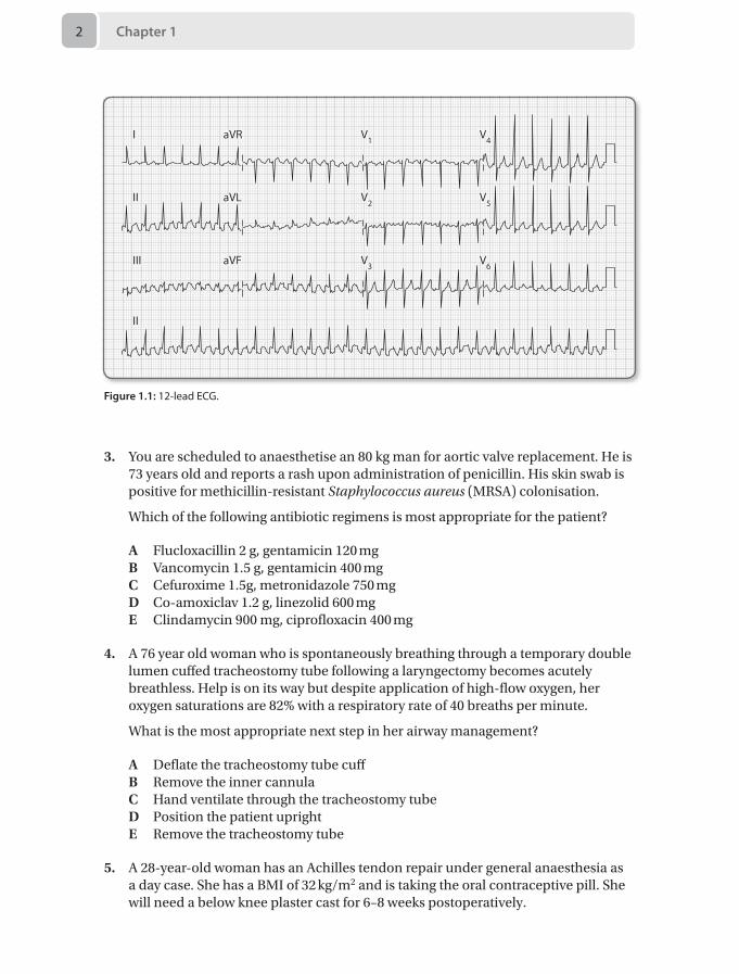
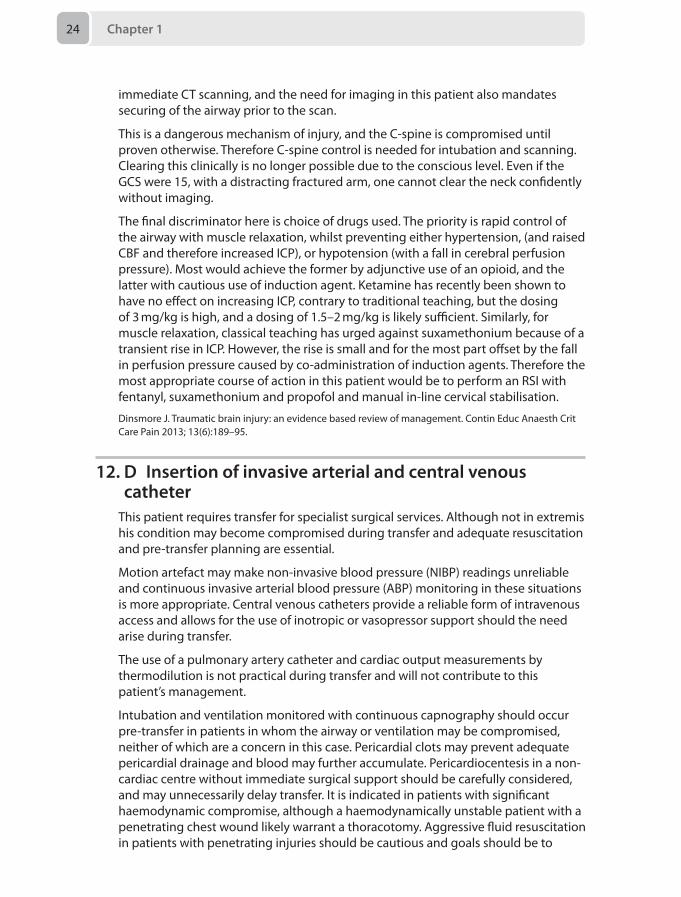
2. A 77-year-old man arrived in the intensive care unit 2 hours ago following coronary artery bypass grafting (CABG). He has a background of interstitial lung disease and hypertension. He is intubated, ventilated and sedated and on a noradrenaline infusion at 0.05 μg/kg/min. Atrial pacing wires are in situ. You are called to see him as the nurse looking after him thinks the ECG has changed. His blood pressure is 110/80 mmHg and the cardiac index reading on the PiCCO is 1.5 L/min/m2. The readings an hour ago were 130/80 and 2.4 L/min/m2 respectively. His 12-lead ECG is shown in Figure 1.1.
What is the most appropriate course of action?
A 1 mg intravenous metoprololB 300 mg amiodarone over 30 minutesC Synchronised DC cardioversion with 100JD Atrial pacing at 100 beats per minuteE 250 µg intravenous digoxin
Chapter 12
3. You are scheduled to anaesthetise an 80 kg man for aortic valve replacement. He is 73 years old and reports a rash upon administration of penicillin. His skin swab is positive for methicillin-resistant Staphylococcus aureus (MRSA) colonisation.
Which of the following antibiotic regimens is most appropriate for the patient?
A Flucloxacillin 2 g, gentamicin 120 mgB Vancomycin 1.5 g, gentamicin 400 mgC Cefuroxime 1.5g, metronidazole 750 mgD Co-amoxiclav 1.2 g, linezolid 600 mgE Clindamycin 900 mg, ciprofloxacin 400 mg
4. A 76 year old woman who is spontaneously breathing through a temporary double lumen cuffed tracheostomy tube following a laryngectomy becomes acutely breathless. Help is on its way but despite application of high-flow oxygen, her oxygen saturations are 82% with a respiratory rate of 40 breaths per minute.
What is the most appropriate next step in her airway management?
A Deflate the tracheostomy tube cuffB Remove the inner cannulaC Hand ventilate through the tracheostomy tubeD Position the patient uprightE Remove the tracheostomy tube
5. A 28-year-old woman has an Achilles tendon repair under general anaesthesia as a day case. She has a BMI of 32 kg/m2 and is taking the oral contraceptive pill. She will need a below knee plaster cast for 6–8 weeks postoperatively.
I
II
III
II
aVR
aVL
aVF
V1
V2
V3
V4
V5
V6
Figure 1.1: 12-lead ECG.
Questions 3
What is the best form of venous thromboembolism prophylaxis for her?
A Advice on mobilisation and fluid intakeB Graduated compression stockings and pneumatic compression device on day
of surgeryC Graduated compression stockings post discharge for 7 daysD Single dose low molecular weight heparin on day of surgeryE Extended course of low molecular weight heparin post discharge
6. A 65-year-old woman is scheduled for an extended abdominal hysterectomy. She is not on any anticoagulants but 2 years ago she developed a blood clot following a total hip replacement. At that time her treatment injections caused a wound haematoma, and she was put on a ‘blood thinning’ infusion for several days. At the end of the treatment she remembers having investigations for low platelets in her blood. These results are unavailable.
For immediate perioperative prophylaxis this time the best treatment would be:
A A heparin infusion started 6 hours following surgery if bleeding is controlledB Treatment dose low-molecular-weight heparin (LMWH) injection once dailyC Fondaparinux injection once dailyD Low-dose aspirin orally throughout the perioperative periodE A danaparoid infusion
7. A 21-year-old woman is undergoing a Le Fort I transverse osteotomy to correct her maxillary retrusion. A nasal tube is used and anaesthesia is maintained by propofol and remifentanil infusions. During the down-fracture, her pulse rate falls to 29 bpm and her blood pressure reads 70/30 mmHg. Her oxygen saturation, end tidal CO2 and airway pressures remain unchanged.
What is the most likely cause of her haemodynamic compromise?
A HaemorrhageB Venous air embolusC Endotracheal tube damageD Trigeminocardiac reflexE Remifentanil
8. A 64-year-old man with no previous cardiac or respiratory morbidity is attending for his second treatment of electroconvulsive therapy (ECT). After his previous treatment, he had a supraventricular tachycardia with a peak blood pressure of 198/105 mmHg, which resolved spontaneously within 5 minutes. For his anaesthetic he had received propofol 90 mg and suxamethonium 40 mg.
What is the most appropriate course of action for this second treatment?
A Perform procedure with defibrillation pads on his chestB Pre-medicate with oral atenololC Use intravenous esmolol during procedureD Use remifentanil infusion during procedureE Use sublingual nifedipine during procedure
Chapter 14
9. You are called to see a 65-year-old patient in the surgical ward 3 days following an elective abdominal aortic aneurysm repair. A thoracic epidural catheter is in situ. He is febrile and complains of back pain and lower limb weakness.
What would be the most appropriate next step?
A Stop the epidural infusion and contact the neurosurgeonB Stop the epidural infusion, do regular neurological observations and monitor
the epidural catheter siteC Arrange an urgent MRI scan and inform the neurosurgeonD Stop the epidural infusion and start empirical antibioticsE Remove the epidural catheter and do a full neurological examination
10. A 70 kg, 36-year-old man is scheduled for foot surgery under a regional anaesthetic approach.
Which of the following needles would you use to perform a lateral popliteal nerve block?
A 50 mm length, short bevel peripheral nerve block needleB 50 mm length, long bevel peripheral nerve block needleC 150 mm length, short bevel peripheral nerve block needleD 100 mm length, short bevel peripheral nerve block needleE 100 mm length, long bevel peripheral nerve block needle
11. A 28-year-old pedestrian struck by a bus presents to the emergency department. In hospital, he has had a primary survey which reveals an obvious head injury; he also appears to have a fracture to his right arm. His chest appears clear and a FAST scan of the abdomen is negative. Because he had been confused, the emergency medicine registrar has asked you to sedate him for a CT scan of his head. On examination, he grimaces and groans to a deep painful stimulus, but does not open his eyes. He flexes his left arm and leg.
The safest option for CT scan would be to:
A Titrate small doses of propofol to effect with continuous monitoring including waveform capnography
B Refuse to give sedative drugs on account of his depressed conscious state, but accompany him to the scanner
C Perform a rapid sequence induction (RSI) with propofol and suxamethonium 1.5 mg/kg, and transfer with a propofol infusion
D Perform a modified RSI with 1.5 mg/kg suxamethonium, after 2 µg/kg fentanyl and propofol and manual in-line stabilisation of the cervical spine
E Fit a Miami J collar and blocks and then perform a modified RSI with 1 mg/kg rocuronium and 3 mg/kg ketamine.
12. A 19-year-old man presents to a district general hospital emergency department 8 hours after suffering a penetrating injury to his anterior chest. He has a Glasgow
Questions 5
coma scale (GCS) of 15, heart rate of 105 beats per minute, blood pressure of 95/50 (MAP 65) mmHg, saturations of 99% on oxygen and a haemoglobin of 105 g/L. Transthoracic echocardiogram shows a haemopericardium for which he requires transfer to a nearby cardiothoracic centre for exploration.
What pre-transfer intervention is most appropriate?
A Needle pericardiocentesis B Intubation and ventilationC Insertion of a pulmonary artery catheter for cardiac output monitoring D Insertion of invasive arterial and central venous catheterE Transfusion of 2 units packed red cells
13. A 62-year-old man who sustained an isolated non-penetrating chest injury resulting in lung contusions and rib fractures is on the intensive care unit intubated and ventilated. He has deteriorated over the past 72 hours and now has a Po2:Fio2 ratio (PFR) of 50 mmHg with a Fio2 of 1.0 and a positive end-expiratory pressure (PEEP) of 5 cmH2O. The investigations suggest he has developed Acute Respiratory Distress Syndrome (ARDS).
The most important first intervention is:
A Furosemide bolus of 40 mg intravenously and commence an infusion aiming for a negative fluid balance
B Administer a neuromuscular blocking agentC Perform a recruitment manoeuvre and incrementally increase the PEEP to
above 14 cmH2O D Adjust the ventilator settings to ensure tidal volumes of 6 mL/kg and a peak
pressure of less than 30 cmH2OE Prone the patient
14. A 26-year-old woman with a past medical history of self-harm was found unconscious at home with empty alcohol and amitriptyline bottles on the floor. These had been ingested within half an hour. On arrival to the emergency department her Glasgow coma scale (GCS) was 5 (E1, V1, M3). She was intubated for airway protection. The patient subsequently developed a blood pressure of 80/60 mmHg associated with a heart rate of 150 beats per min, a QRS width of 100 msec and multiple ventricular ectopic beats.
The next most important intervention is:
A Nasogastric tube insertion and administration of activated charcoalB Intravenous crystalloid bolus of 20 mL/kg followed by a noradrenaline infusion
to maintain blood pressureC 500 ml intravenous sodium bicarbonate 1.26% for treatment of a broadened
QRS complexD Lignocaine 2 mg/kg for the management of ventricular ectopic beatsE Lipid emulsion 20% 1.5 mL/kg for intravascular sequestration of tricyclic drug
Chapter 16
15. A 28-year-old woman presents with progressive and ascending motor weakness. She reports a recent history of coryzal symptoms.
The following would be an early indicator of the requirement for intubation:
A Respiratory rate > 35 breaths per minuteB Pao2 < 8 kPaC Paco2 > 6.5 kPaD Vital capacity < 15 mL/kgE Absence of bulbar weakness
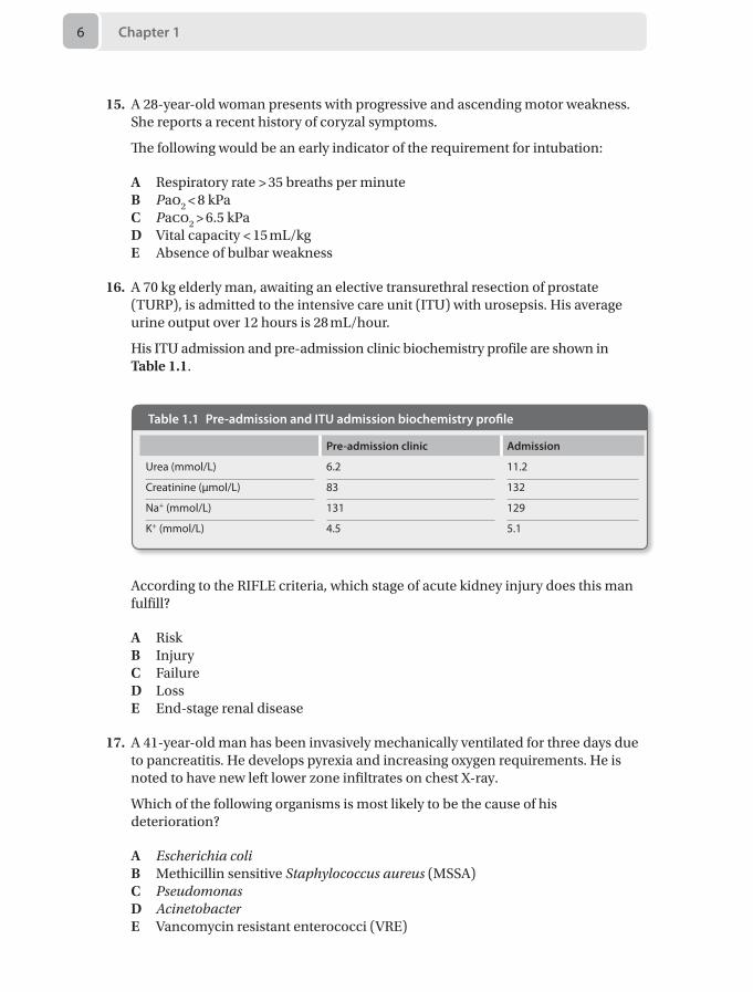
16. A 70 kg elderly man, awaiting an elective transurethral resection of prostate (TURP), is admitted to the intensive care unit (ITU) with urosepsis. His average urine output over 12 hours is 28 mL/hour.
His ITU admission and pre-admission clinic biochemistry profile are shown in Table 1.1.
Table 1.1 Pre-admission and ITU admission biochemistry profile
Pre-admission clinic Admission
Urea (mmol/L) 6.2 11.2
Creatinine (μmol/L) 83 132
Na+ (mmol/L) 131 129
K+ (mmol/L) 4.5 5.1
According to the RIFLE criteria, which stage of acute kidney injury does this man fulfill?
A RiskB InjuryC FailureD LossE End-stage renal disease
17. A 41-year-old man has been invasively mechanically ventilated for three days due to pancreatitis. He develops pyrexia and increasing oxygen requirements. He is noted to have new left lower zone infiltrates on chest X-ray.
Which of the following organisms is most likely to be the cause of his deterioration?
A Escherichia coliB Methicillin sensitive Staphylococcus aureus (MSSA)C PseudomonasD Acinetobacter E Vancomycin resistant enterococci (VRE)
Questions 7
18. A 32-year-old primigravid patient with a body mass index (BMI) of 55 is on the labour ward. It is 10 pm; she is currently 7 cm dilated and requesting an epidural. The baby is in the occiput posterior (OP) position. You are unable to palpate her spinous processes. On your third attempt, with difficulty, you perform a lumbar epidural at L3/4 and accidentally cause a dural tap.
What is the best line of management in this situation?
A Repeat your attempt at an adjacent lumbar level and use a smaller test doseB Request help from a colleague to attempt the epiduralC Use the ultrasound to help locate the depth of the epidural space before re-
attemptingD Abandon your attempt and institute a remifentanil PCAE Site a spinal catheter, inform midwife and perform subsequent top-ups
yourself
19. You are fast bleeped to the emergency department (ED) where a 22-year-old woman who is 28/40 pregnant has presented with a history of seizures for the past 45 minutes. A wedge has been placed under the right side of the patient and large bore intravenous access has been secured. Her blood pressure is 180/110 mmHg, heart rate 154 beats per minute, respiratory rate 24 breaths per minute and an arterial blood gas sample reveals a pH of 7.2 with an elevated lactate. The obstetric registrar is present and suspects this is an eclamptic fit. 4 g of intravenous magnesium sulphate (MgSO4) has been given over 5 minutes and anti-hypertensive medication has been started. The patient is still fitting.
What should the next stages of management be?
A Secure airway with endotracheal tube (ETT) and perform emergency Caesarean section in the ED
B Commence MgSO4 infusion at 1 g/hour, secure airway with ETT and perform emergency Caesarean in the ED
C Commence MgSO4 infusion at 1 g/hour, give a further 2 g MgSO4 bolus, secure airway with ETT and continue supportive management
D Give a further 2 g MgSO4 bolus and if no response administer phenytoin 15 mg/kg
E Commence MgSO4 infusion at 1 g/hour, give a further 2 g MgSO4 bolus, secure airway with ETT and perform emergency Caesarean section in the ED
20. A 5-day-old boy presents to a local emergency department with a 2-day history of increasing respiratory distress. He is lethargic with a heart rate of 184 beats per minute, a respiratory rate of 68 breaths per minute, a blood pressure of 66/32 mmHg, capillary refill time of 6 seconds, Spo2 96% on air on the right hand, but unrecordable from the other limbs. His axillary temperature is 36.1°C, but his extremities are mottled and feel cool to touch. The chest sounds clear and the heart sounds seem normal with weakly palpable femoral pulses. He was given a bolus of 10 mL/kg of 0.9% saline and broad-spectrum intravenous antibiotics. A rapid sequence induction was performed, and the patient intubated and ventilated.
Chapter 18
The most appropriate next step in his management is:
A Start prostaglandin E2 intravenous infusion and refer to tertiary centre for possible coarctation of the aorta
B Arrange for an urgent chest X-rayC Insert a nasogastric tube to decompress the stomach to aid ventilationD Perform arterial blood gas analysisE Keep the infant warm with radiant heater
21. A 20 kg 5-year-old child was brought to the emergency department of a district general hospital with 15% burns from scalding to neck, chest, abdomen and right upper limb having already received 20 mL/kg (400 mL) Hartmann's and 20 µg/kg intravenous (IV) morphine for analgesia. It is 4 hours since the time of injury. On examination, the child appears comfortable, with a heart rate of 110 beats per minute, blood pressure of 124/82 mmHg, a respiratory rate of 22 breaths per minute and Spo2 of 99% on air.
The next most appropriate step in the management of this patient for the next 4 hours is:
A IV Hartmann’s at 110 mL/hour. Refer to tertiary centre for further managementB IV Hartmann’s at 110 mL/hour. Admit for further observation and
managementC IV Hartmann’s at 75 mL/hour. Refer to tertiary centre for further managementD Intubate and ventilate. IV Hartmann’s at 110 mL/hour. Refer to tertiary centre
for further managementE Give IV antibiotic prophylaxis. IV Hartmann’s at 110 mL/hour. Admit for
further observation and management
22. A 57-year-old woman presents with a history of severe facial pain that occurs in sudden episodes of a few minutes and only affect her right cheek. It starts with a sharp ’electric shock‘ which then becomes an ache before it abruptly disappears. Treatment with carbamazepine was commenced at 100 mg b.d. this week, and this has provided only modest relief.
The most appropriate next step in her treatment is:
A Increase dose of carbamazepineB Microvascular decompressionC Add amitriptylineD Add lamotrigineE Cognitive-behavioural therapy
23. You are presented with a 43-year-old woman who had a mastectomy 7 years ago, followed by neoadjuvant radiothearpy and chemotherapy for left sided breast cancer. She is currently taking hormonal therapy and has had pain over the left chest wall for the past 2 years.
Questions 9
Which of the following is most correct regarding this patient’s chest wall pain?
A Urgent referral for investigation of recurrence is neededB Phantom pain is rare in post-mastectomy patientsC Long-term opioids should be commencedD Brachial plexus pathology is the likely causeE The pain will usually respond to anticonvulsants
24. A 46-year-old right-handed violinist presents with a 3-month history of worsening severe pain in his right wrist, which commenced suddenly after a long performance in a concert. He has noticed the painful wrist going pale and cold at times, and swelling occasionally. Sometimes it sweats, and it has become stiff and difficult to use. It appears smaller than his left hand, and the nails of his right fingers have become brittle and discoloured. He admits to being very distressed and anxious as he is no longer able to perform. Treatment with paracetamol and amitriptyline has been commenced.
The most appropriate next step in his management is:
A Pregabalin 75 mg b.d.B AcupunctureC Patient education and psychological supportD Application of 5% lignocaine patchesE Mirror therapy
25. A 30-year-old woman with chronic lower back pain is assessed in an outpatient clinic. She tells you that her pain has improved with exercise and local heat application, but when she thinks about the pain it seems to get worse.
Regarding this gate theory of pain, which of the following is most accurate?
A It applies mostly to nociceptive painB It is the basis of how transcutaneous electrical nerve stimulation (TENS)
machines workC A supraspinal input is requiredD Inhibition occurs via Aδ fibresE This theory does not apply to children
26. A 73-year-old woman with metastatic breast cancer presents with a 4-month history of severe pain in her back, upper arms and legs. She has been on increasing doses of modified release oral morphine and paracetamol, and while this combination provides her some relief, she is troubled by drowsiness, pruritus, and constipation. At times she feels this is more distressing than her initial pain. Additionally, she is on warfarin for atrial fibrillation.
The most appropriate next step in her management would be:
A Add diclofenac
Chapter 110
B Reduce the dose of morphineC Add naloxoneD Opioid rotationE Prescribe a laxative
27. A 35-year-old man has been admitted to the intensive care unit with a 55% total body surface area (BSA) burn. He is intubated and has been resuscitated as per the Parkland formula.
Which of the following statements is correct?
A Should temperature spike above 38°C, take blood cultures and start broad spectrum antibiotics
B Enteral nutrition should be started as soon as possibleC Steroids are indicated as there is greater than 40% BSA burnsD Fluid resuscitation should be continued according to the Parkland formula
even if polyuria developsE If fluid management is optimal generalised oedema is unlikely to develop
28. A hypertensive 68-year-old man on amlodipine is undergoing an elective abdominal aortic aneurysm repair. At the end of the operation the surgeon is prepared to release the infrarenal aortic cross-clamp.
Which of the following interventions would mitigate the ensuing hypotension?
A Starting an infusion of noradrenaline at 0.5 µg/kg/min after cross-clamp release
B Starting an infusion of dobutamine at 5 µg/kg/min after the cross-clamp release
C Rapid infusion of 500 mL of colloid during cross-clamp releaseD Tilting the table in reverse Trendelenburg positionE Optimising the intravascular volume during aortic cross-clamping
29. A 74-year-old man is brought to the emergency department with palpitations. He has a heart rate of 156 beats per minute and atrial fibrillation on his ECG.
Which one of the following would not warrant immediate direct current cardioversion?
A. Blood pressure of 84/30 mmHgB. GCS of 12/15 C. Bi-basal creptitations and tachypneaD. Sweating with cold clammy handsE. T wave inversion in lead aVR
30. A 3-year-old boy suffers from dry and scaly skin, oral thrush, dandruff and dry hair, as well as poor vision in the dark. On examination he has xerosis and Bitot’s spots.

Questions 11
The most likely deficiency is:
A Vitamin AB Vitamin DC Vitamin CD Vitamin B12E Vitamin K
Chapter 112
Answers1. C The patient recalling that he has a foreign
body in his eyeMagnetic resonance imaging (MRI) scans are often utilised for investigating the central nervous system as they provide images that show improved distinction between tissue types compared with computed tomography (CT) scans.
MRI scanning takes advantage of the fact that atomic nuclei within tissues naturally spin, generating their own small magnetic field. By applying a larger external field to a tissue, these spinning nuclei align with the field which has been applied. A second external field is then pulsed in a perpendicular fashion causing some nuclei to be pulled to an angle. This incorporates nuclear energy absorption, and they begin to wobble or precess – a term used to describe rotation around an axis different to that of original spin. Precession results in tissues producing rotational magnetic fields, the amplitude and specific frequency of which can be detected and used to form an image. As the nuclei return to their previous positions between pulses, they emit the energy they previously absorbed at the same frequency. The rate of their return depends on the elemental content of the nucleus (e.g. hydrogen or phosphorous) in addition to the molecule of which it is a part (e.g. water or fat). Different tissue types therefore return at different rates. By using a combination of magnetic field gradients and pulse configurations, detailed cross-sectional views can be obtained.
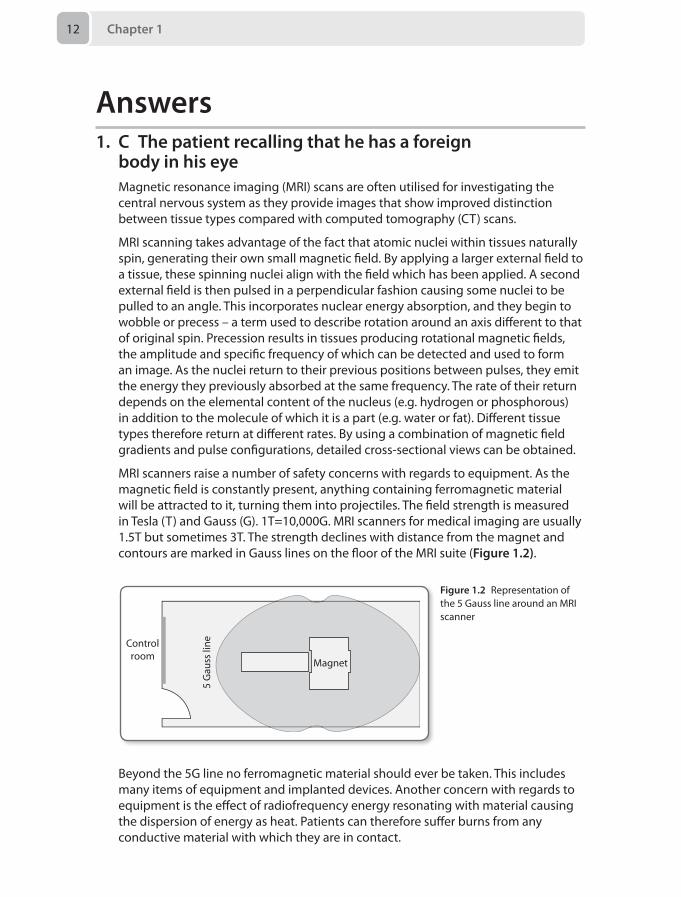
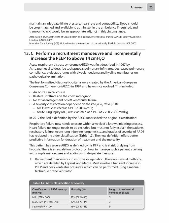
MRI scanners raise a number of safety concerns with regards to equipment. As the magnetic field is constantly present, anything containing ferromagnetic material will be attracted to it, turning them into projectiles. The field strength is measured in Tesla (T) and Gauss (G). 1T=10,000G. MRI scanners for medical imaging are usually 1.5T but sometimes 3T. The strength declines with distance from the magnet and contours are marked in Gauss lines on the floor of the MRI suite (Figure 1.2).
Controlroom
Magnet
5 G
auss
line
Figure 1.2 Representation of the 5 Gauss line around an MRI scanner
Beyond the 5G line no ferromagnetic material should ever be taken. This includes many items of equipment and implanted devices. Another concern with regards to equipment is the effect of radiofrequency energy resonating with material causing the dispersion of energy as heat. Patients can therefore suffer burns from any conductive material with which they are in contact.
Answers 13
As a result of the above, all equipment is classified according to the hazard it poses under certain conditions such as magnetic field strength or in view of radiofrequency absorption. MR safe equipment can be used in all MR settings, MR conditional in specified environments, and MR unsafe in none of the aforementioned situations.
Monitoring in the MRI suite is essential and has evolved accordingly. MR compatible monitoring is standard, with many units using telemetric equipment to avoid any induced currents in long cables. MR compatible anaesthetic machines and infusion pumps are available; however standard equipment can be used with extensions to beyond the 5G line. The anaesthetic machine must be securely fixed to the wall and the pumps attached to their extensions through a port into the control room.
Pacemakers and implanted cardiac defibrillators are at risk of malfunctioning or displacing and so were, until recently, a strict contraindication to having an MRI. Technology has, however, advanced and there are now some MR-compatible models. There are also MR strategies and guidelines that have been described to limit risk in the event that an MRI is absolutely necessary for a patient with a standard device.
Cervical fixators, such as the halo device, vary in their classification. Some are MR safe and this, or the hazard of any other item, can be easily checked by referring to a list on www.MRIsafety.com.
Foreign bodies in the eye have the potential to migrate and cause bleeding into the vitreous, therefore contraindicating an MRI scan.
Reddy U, White MJ, Wilson SR. Anaesthesia for magnetic resonance imaging. Contin Educ Anaesth Crit Care Pain 2012; 12(3):140–44.Association of Anaesthetists of Great Britain and Ireland. Safety in magnetic resonance units: an update. Anaesthesia 2010; 65:766–70.
2. C Synchronised DC cardioversion with 100JThe ECG shows atrial flutter. Up to 40% of coronary artery bypass grafting (CABG) patients will develop postoperative atrial fibrillation or flutter. The majority of these dysrhythmias occur within 48-hours and may be recurrent. Presence of atrial fibrillation or flutter is associated with an increase in hospital mortality as well as other complications including stroke.
Risk factors for the development of atrial fibrillation or flutter include:
• Atrial injury during cannulation• Ischaemia• Prolonged cardiopulmonary bypass time• Use of post operative catecholamines• Hypokalaemia and hypomagnesaemia
Although accompanied with reasonable haemodynamics, there is a clear fall in cardiac index (and hence output) as measured by the PiCCO. Atrial flutter may change to atrial fibrillation and rate control is rarely an option. Restoration of sinus rhythm should be the aim in this circumstance and this is best achieved with synchronised DC cardioversion.
Chapter 114
Although amiodarone is frequently used for atrial flutter, data concerning its use in this setting is surprisingly lacking. When using amiodarone, cardioversion may take hours rather than minutes. Another reason to avoid amiodarone in this circumstance would be the history of interstitial lung disease, which is a risk factor for exacerbation of any lung fibrosis that may be caused by amiodarone.
Rate controlling agents such as metoprolol and digoxin would not be optimal treatment here.
Atrial pacing is a viable option but would usually be performed at a rate 10–15 bpm higher than the atrial flutter rate. If the ventricular rate rises to match the atrial rate, the pacemaker frequency can then be reduced (i.e. the rhythm is entrained) to an acceptable rate. This may lead to conversion to sinus rhythm (or atrial fibrillation!). Given this patient is already sedated and ventilated, it is quicker and more effective to perform DC cardioversion.
As well as addressing strategies for cardioversion, it is also imperative that other contributing factors for the development of any dysrhythmias are addressed:
• Hypoxaemia• Hypercarbia• Electrolyte disturbances• Other causes of myocardial ischaemia e.g. graft failure
European Society of Cardiology. Guidelines for the management of atrial fibrillation. Eur Heart J 2010; 31:2369–429.
3. B Vancomycin 1.5 g, gentamicin 400 mgCommon pathogens in cardiac surgery are Staphylococcus aureus and Staphylococcus epidermidis. In addition, this man has evidence of methicillin-resistant staphylococcus aureus (MRSA) colonisation, so any prophylactic antibiotics must cover this organism (vancomycin or linezolid). Most centres also administer some gram negative cover such as an aminoglycoside (gentamicin at 5 mg/kg) or a fluoroquinolone (ciprofloxacin).
Although the skin reaction reported after penicillin may not be significant, it is prudent to avoid penicillins thus flucloxacillin and co-amoxiclav should be avoided. Vancomycin 1.5 g with gentamicin 400 mg provides gram-positive (including MRSA) and gram-negative cover and is the correct regimen for this patient. The dose of vancomycin is 15 mg/kg and should be given as an infusion. The combination of cefuroxime and metronidazole does not have MRSA cover and although clindamycin with ciprofloxacin gives good gram positive, gram negative and MRSA cover, the use of untargeted ciprofloxacin is often discouraged due to the speed by which plasmid mediated resistance can occur.
Other elements of perioperative care that may reduce the incidence of surgical site infection include patient warming, tight glycaemic control, hair removal and the sterility of instruments and the surgical field.
Bratzler DW, Dellinger EP, Olsen KM, et al. 2013 Clinical practice guidelines for antimicrobial prophylaxis in surgery. Surg Infect 2013; 14(1):73.
Answers 15
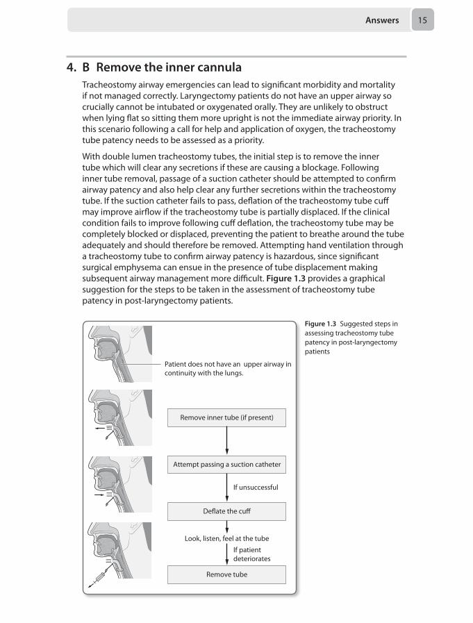
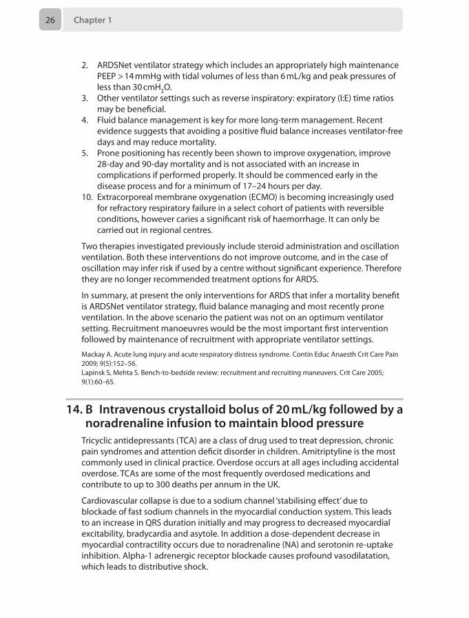
4. B Remove the inner cannulaTracheostomy airway emergencies can lead to significant morbidity and mortality if not managed correctly. Laryngectomy patients do not have an upper airway so crucially cannot be intubated or oxygenated orally. They are unlikely to obstruct when lying flat so sitting them more upright is not the immediate airway priority. In this scenario following a call for help and application of oxygen, the tracheostomy tube patency needs to be assessed as a priority.
With double lumen tracheostomy tubes, the initial step is to remove the inner tube which will clear any secretions if these are causing a blockage. Following inner tube removal, passage of a suction catheter should be attempted to confirm airway patency and also help clear any further secretions within the tracheostomy tube. If the suction catheter fails to pass, deflation of the tracheostomy tube cuff may improve airflow if the tracheostomy tube is partially displaced. If the clinical condition fails to improve following cuff deflation, the tracheostomy tube may be completely blocked or displaced, preventing the patient to breathe around the tube adequately and should therefore be removed. Attempting hand ventilation through a tracheostomy tube to confirm airway patency is hazardous, since significant surgical emphysema can ensue in the presence of tube displacement making subsequent airway management more difficult. Figure 1.3 provides a graphical suggestion for the steps to be taken in the assessment of tracheostomy tube patency in post-laryngectomy patients.
Figure 1.3 Suggested steps in assessing tracheostomy tube patency in post-laryngectomy patients
Patient does not have an upper airway incontinuity with the lungs.
Remove inner tube (if present)
Attempt passing a suction catheter
If unsuccessful
De�ate the cu�
Look, listen, feel at the tube
If patientdeteriorates
Remove tube
Chapter 116
Therefore, this patient requires removal of the inner cannula for further assessment and management of the cause of her respiratory distress.
McGrath BA, Bates L, Atkinson D, Moore JA. Multidisciplinary guidelines for the management of tracheostomy and laryngectomy airway emergencies. Anaesthesia 2012; 67(9):1025–41.Regan K, Hunt K. Tracheostomy management. Contin Educ Anaesth Crit Care Pain 2008; 8(1):31–35.
5. E Extended course of low molecular weight heparin post dischargeThe risk of venous thromboembolic disease (VTE) after a day surgery procedure is lower than after in-patient procedures as surgery is generally less invasive and mobilisation is earlier. However, more complex and longer procedures in higher risk patients are increasingly being performed in this setting. The 2010 National Institute for Health and Care Excellence (NICE) guidelines for the prevention of VTE includes day surgery as a specific cohort of patients and recommends that mechanical prophylaxis should be used if one or more risk factors are present. Pharmacological prophylaxis should be added depending on ‘patient factors and clinical judgement’.
• Surgical procedure with a total anaesthetic and surgical time of more than 90 minutes, or 60 minutes if the surgery involves the pelvis or lower limb
• Acute surgical admission with inflammatory or intra-abdominal condition • Expected significant reduction in mobility • One or more of the risk factors below:
– Active cancer or cancer treatment – Age over 60 years – Critical care admission – Dehydration – Known thrombophilia – Obesity (body mass index [BMI] over 30 kg/m2) – One or more significant medical comorbidities (for example: heart disease,
metabolic, endocrine or respiratory pathologies, acute infectious diseases, inflammatory conditions)
– Personal history or first-degree relative with a history of VTE – Use of hormone replacement therapy – Use of oestrogen-containing contraceptive therapy – Varicose veins with phlebitis
Pharmacological prophylaxis should be continued for 5–7 days if significantly reduced mobility is expected. This patient has three risk factors and extended pharmacological prophylaxis is indicated. In addition she will have a lower limb plaster cast, where NICE recommends that prophylaxis should be continued until the cast is removed after discussion with the patient and evaluation of risks and benefits. The exact duration will vary between centres.
National Institute for Health and Care Excellence (NICE). Venous thromboembolism – reducing the risk. CG no 92. London: NICE, 2010.British Association of Day Surgery (BADS). Organisational issues in pre operative assessment for day surgery. London: BADS, 2010.
Answers 17
6. E A danaparoid infusionVenous thromboembolism (VTE), is of major clinical significance given that up to a quarter of inpatients with risk factors may be affected, albeit subclinically. Candidates will be familiar with the risk factors for VTE (see above) but also should be comfortable with the drug treatment strategies available and their complications.
Mechanical methods
Anti-embolism stockings or thromboembolic deterrent stockings (TEDS), are graded to provide increased compression from distal to proximal. They are effective at promoting venous return and increasing the speed of blood flow, but not suitable for all patients, such as those with arteriopathy. Intermittent calf and thigh compression devices produce pressures of approximately 40 mmHg 10 times per minute to emulate the limb muscle pump.
Heparins
Unfractionated heparin is a naturally occurring antithrombin binder. This inhibits factor Xa and thrombin and in higher doses also has an antiplatelet function. Low-molecular-weight heparin (LMWH) is more effective than subcutaneous heparin, has a lower risk of bleeding, and less anti-platelet effects. It is more convenient with once daily administration, but is less controllable than a heparin infusion, and accumulates in renal failure. It will not usually affect the activated partial thromboplastin time (aPTT), which is a useful monitor of unfractionated heparinisation.
Warfarin
Warfarin has the advantage of being given orally, and has similar risks of bleeding as LMWH. It can be monitored using the international normalised ratio (INR).
Fondaparinux
Fondaparinux is a synthetic saccharide which emulates the structure of the heparin anti-thrombin binding site. It indirectly inhibits factor Xa, and is given by subcutaneous daily injection. It is more effective at preventing VTE than LMWH, but also at producing bleeding. The half-life is long, and the drug-free time before neuraxial block is thus 36 hours. It has a lower incidence of HIT, and has been used as a LMWH substitute in this condition.
Others
Lepirudin is a hirudin derivative made as a recombinant protein in yeast, whose main use is in heparin-induced thrombocytopenia (HIT). It directly inhibits thrombin and due to its short half-life is administered by a continuous infusion and is monitored with the aPTT. Due to manufacturer cessation of production in April 2012, lepirudin is no longer available in the UK. Notably, this withdrawal was not due to any safety concerns.
Danaparoid is a heparinoid that inhibits factor Xa, and can be used in patients with HIT. There is a need for close monitoring as some HIT cross reactivity does occur. It has now replaced the use of lepirudin in the management of HIT in the UK due to the aforementioned withdrawal.
Chapter 118
Dabigatran is an orally administered direct thrombin inhibitor licensed for VTE prophylaxis after surgery. It does not require monitoring but also lacks any method to reverse the anticoagulant effect.
Rivaroxaban is a direct oral inhibitor of factor Xa that is becoming more common. Previously only for postoperative VTE prophylaxis, it is now being used in atrial fibrillation and in Europe as an adjunct to aspirin and clopidogrel in acute coronary syndromes.
The likely diagnosis in the above patient is an episode of heparin-induced thrombocytopenia (HIT). HIT is an immune-mediated IgG response to an immunogenic component of heparin, leading to thrombocytopenia. This occurs in around 3% of patients as a consequence of treatment with unfractionated heparin, and less-so at a rate of 0.1–1% with LMWH preparations. Paradoxically, the risk of thrombosis is increased to 50% at this time, so alternate forms of anticoagulation are needed. Platelet counts should be monitored from day 4 – 14, which is the risk period for antibody formation.
Although the diagnosis is not absolutely confirmed, the question forces you to respond and treat in the safest way possible. If HIT is a possibility then a heparin infusion should be avoided, as should LMWH, as this can also precipitate the condition. In addition, the dose of LMWH is probably too high, given that she is no longer on anticoagulant treatment. Aspirin may or may not be indicated for this patient in terms of primary cardiovascular prophylaxis, but does not have any role in thromboprophylaxis. Of the two HIT-safe options, fondaparinux and danaparoid, only danaparoid has no association with HIT. Fondaparinux has a very low rate of giving rise to HIT and is sometimes used off-license as a treatment. However, in this scenario the long half-life makes fondaparinux irreversible and uncontrollable in the immediate postoperative phase. From day 2 onwards, without bleeding, fondaparinux would represent a good choice for prophylaxis with adequate monitoring of platelets.
Barker RC, Marval P. Venous thromboembolism: risks and prevention. Contin Educ Anaesth Crit Care Pain 2011; 11(1):18–23.
7. D Trigeminocardiac reflexThe horizontal Le Fort I osteotomy is a common procedure used to correct maxillary deformities and knowledge of the surgical technique and relevant anatomy is useful in recognising and treating complications. Surgery involves an intraoral incision and the formation of a transverse maxillary osteotomy that extends to the pterygomaxillary junction. The maxilla is then separated from the upper face along this osteotomy plane by a down-fracture and fully mobilised to aid surgery.
Bleeding is a recognised complication during the down-fracture since the bony mid-face receives a rich blood supply and is in close proximity to an extensive venous plexus. The blood vessels most likely to be injured during the down-fracture are the pterygoid vessels, palatine and alveolar arteries, or on rare occasions the internal carotid. In order to visualise the source of bleeding and achieve haemostasis, completion of the down-fracture is often required. It is unusual for an acute haemorrhage to present with a severe bradycardia as described in the above case.
Answers 19
Venous air emboli can occur during any head and neck surgery where open veins are exposed to the atmosphere. However, end-tidal carbon dioxide levels would be expected to fall as a result of an increase in physiological dead space and intrapulmonary shunting which is not observed in the above case.
A nasal endotracheal tube is usually the airway of choice when correcting for maxillary retrusion, since the jaw is frequently closed and wired to ensure normal alignment of the upper and lower teeth. During the osteotomy and down fracture, the nasal tube may be damaged resulting in impaired gas exchange and secondary haemodynamic compromise. In such a situation, the airway (which is now likely to be difficult) needs to be re-established. This scenario is unlikely in the above case since the oxygen saturations, end tidal carbon dioxide levels and airway pressures remain unchanged.
The Le Fort I osteotomy can also cause nerve damage and pressure effects to cranial nerves II-VII due to their proximity to the surgical field. A recognised complication of the maxillary down-fracture in particular is the generation of the trigeminocardiac reflex. This reflex occurs as a result of pressure on the cranial nerve V (trigeminal nerve) initiating a vagal reflex causing a severe bradycardia which may even progress to asystole. Cessation of the down-fracture and return of the jaw to its original position can increase the heart rate, as can administration of anticholinergic drugs. The isolated bradycardia and hypotension in relation to the down-fracture in the above scenario makes this reflex the most likely cause.
Remifentanil use in maxillofacial surgery is increasing in popularity due to its favourable pharmacokinetic profile and its useful contribution to deliberate hypotension. Severe bradycardia and hypotension are recognised complications of remifentanil use, however the temporal relationship between the down-fracture and the bradycardia in the above case make the trigeminal reflex more likely.
Beck J, Johnston K. Anaesthesia for cosmetic and functional maxillofacial surgery. Contin Educ Anaesth Crit Care Pain 2013 doi:10.1093/bjaceaccp/mkt027.Miloro M, Kolokythas A. Management of complications in oral and maxillofacial surgery. 1st Ed. New York:John Wiley & Sons Inc, 2012.
8. C Use intravenous esmolol during procedureDuring electroconvulsive therapy (ECT) an electrical current is applied to the brain via transcutaneous electrodes to induce a generalised therapeutic seizure lasting between 10–120 seconds. There is a biphasic physiological response. The seizure causes an immediate direct stimulation of the vagal parasympathetic outflow, which can lead to transient bradycardia and hypotension, and rarely asystole.
Premedication with an anticholinergic agent is often used to attenuate this effect. This is followed by a more prominent catecholamine mediated sympathetic response, which peaks 3–5 minutes after therapy, causing a tachycardia, and hypertension and may give rise to tachyarrhythmias.
This sympathetic response can be attenuated using a variety of agents. Beta blockers have been shown to be the most effective in controlling both heart rate and mean arterial pressure. Due to the risk of initial bradycardia short acting agents such as
Chapter 120
esmolol or labetalol given just prior or during the procedure may avoid accentuating the parasympathetic response compared to longer acting agents. Esmolol is preferred as it reduces the peak systolic blood pressure more than labetalol while labetalol may be associated with a shorter seizure duration.
Calcium channel blockers can also be effective to control arterial pressures but reflex tachycardia may occur with nifedipine. Remifentanil has been shown to reduce both the heart rate and blood pressure and does not have an effect on seizure duration, though use of an infusion may not be available or suitable for these short procedures.
Uppal V, Dourish J, Macfarlane A. Anaesthesia for electroconvulsive therapy. Contin Educ Anaesth Crit Care Pain 2010; 10(6):192–197.
9. C Arrange an urgent MRI scan and inform the neurosurgeonThere are many benefits for neuroaxial drug delivery. However, we need to balance the advantages against the risk of complications such as infection, nerve damage and haematoma.
In the third National Audit Project (NAP 3) report, the incidence of epidural abscess after central neuraxial block (CNB) was quoted as 2.1 in 100,000. Although this is considerably lower than previous studies, epidural abscess is still a very serious complication of CNB and can lead to permanent neurological damage. In the above scenario, the patient has signs and symptoms of an established epidural abscess that needs decompression immediately.
We should recognise patients at increase risk of spinal infection before commencing the CNB, with risk factors including:
• Immune compromised patients• Patient with local or systemic infection• Long-term vascular access• Long duration of epidural catheterisation• Difficult CNB or a bloody tap after epidural • Prolonged hospital stay• Disruption of the spinal column, e.g. surgery or trauma
Following epidural catheter insertion, catheter site checks and regular temperature monitoring are very important to recognise epidural abscess.
The classical presentation of epidural abscess is of pyrexia, back pain and progressive abnormal neurology of the lower half of the body. However, 1 in 4 patients have no back pain. Therefore, a high index of suspicion is required to diagnose epidural abscess.
Advice from neurosurgeons, neurologists, radiologists and microbiologists are needed early in established epidural abscess.
The most important action when we suspect epidural abscess is to organise MRI with gadolinium. This will help to decide whether open or percutaneous drainage should be used.
Answers 21
In this example, arranging an MRI scan and informing the neurosurgeons are the first and most important steps to perform because early diagnosis and surgical decompression is needed. Although option E is correct, it is time consuming and delays the diagnosis. Once muscle weakness is present, only 20% patients regain full function, even after surgery.
A full infection screen including blood cultures is mandatory if an epidural abscess is suspected. At the same time, it is essential to remove the epidural catheter, as well as stop the epidural infusion, and send the catheter tip for culture and sensitivity. As solely stopping the infusion is inadequate, options A, B and D are insufficient management options.
The most common microorganism found in spinal infection is Staphylococcus. Initial antibiotic therapy should be empirical and then modified depending on the culture and sensitivity results, while treatment must be guided by microbiological input.
Intravenous antibiotics are required initially for 2–4 weeks, followed by a prolonged course of oral antibiotics. Regularly checking of inflammatory markers, back pain and neurology should be used to monitor the response to antibiotics.
Royal College of Anaesthetists. Major complications of central neuraxial blocks in the United Kingdom: the 3rd National Audit Project (NAP3) of the Royal College of Anaesthetists, 2009. Br J Anaesth 2009; 102(2):179–90.Simpson KH, Al-Makadma YS. Epidural drug delivery and spinal Infection. Contin Educ Anaesth Crit Care Pain 2007; 7(4):112–15. Gosavi C, Bland D, Poddar R, Horst C. Epidural abscess complicating insertion of epidural catheters. Br J Anaesth 2004; 92:294–95.
10. D 100 mm length, short bevel peripheral nerve block needle
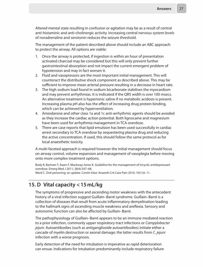
When performing nerve blocks, the length of the block needle is an essential consideration. Longer needles might have potential tissue damage if advanced further than needed, while the shorter needle may not be long enough to reach the nerve.
The ideal needle length for peripheral nerve blocks is:
• 25 mm – Interscalene• 50 mm – Cervical plexus, supraclavicular, axillary, femoral and popliteal ('posterior
approach')• 100 mm – Infraclavicular, paravertebral, lumbar plexus, sciatic (‘posterior
approach’) and popliteal (‘lateral approach’)• 150 mm – Sciatic (‘anterior approach’)
There are two types of nerve block needles: cutting tip needle and pencil point tip needle (see Figure 1.4).
Cutting tip needles might be long bevel (15°) or short bevel (30–45°). Long bevel needles are more likely to cause nerve damage by causing sharp nerve penetration. Although the nerve damage caused by a short bevel needle is less frequent, the damage will be more severe.
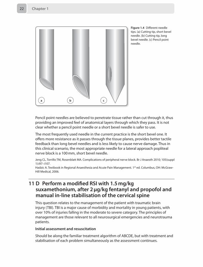
Chapter 122
Pencil point needles are believed to penetrate tissue rather than cut through it, thus providing an improved feel of anatomical layers through which they pass. It is not clear whether a pencil point needle or a short bevel needle is safer to use.
The most frequently used needle in the current practice is the short bevel one. It offers more resistance as it passes through the tissue planes, provides better tactile feedback than long bevel needles and is less likely to cause nerve damage. Thus in this clinical scenario, the most appropriate needle for a lateral approach popliteal nerve block is a 100 mm, short bevel needle.
Jeng CL, Torrillo TM, Rosenblatt MA. Complications of peripheral nerve block. Br J Anaesth 2010; 105(suppl 1):i97–i107.Hadzic A. Textbook in Regional Anaesthesia and Acute Pain Management. 1st ed. Columbus, OH: McGraw-Hill Medical, 2006.
11 D Perform a modified RSI with 1.5 mg/kg suxamethonium, after 2 µg/kg fentanyl and propofol and manual in-line stabilisation of the cervical spineThis question relates to the management of the patient with traumatic brain injury (TBI). TBI is a major cause of morbidity and mortality in young patients, with over 10% of injuries falling in the moderate to severe category. The principles of management are those relevant to all neurosurgical emergencies and neurotrauma patients.
Initial assessment and resuscitation
Should be along the familiar treatment algorithm of ABCDE, but with treatment and stabilisation of each problem simultaneously as the assessment continues.
Figure 1.4 Different needle tips. (a) Cutting tip, short bevel needle. (b) Cutting tip, long bevel needle. (c) Pencil point needle.
a b c
Answers 23
Haemodynamics
Even one episode of hypotension has been shown to double mortality. The aim is to maintain cerebral perfusion pressure (CPP), in the face of raised intracranial pressure (ICP) as CPP = MAP – ICP. International targets differ slightly, but a widespread MAP target is 90 mmHg. Fluid resuscitation to normovalaemia would be the logical first step, with vasopressors following if required. Crystalloid is best, with some evidence of harm with albumin colloid. Hypotonic dextrose solutions must be avoided (unless hypoglycaemic), as they contribute to tissue oedema.
Airway/ventilation
Intubation is indicated for a deterioration in Glasgow coma scale (GCS), or a GCS of < 8, or if there is a failure of the patient's protective reflexes. Any disturbance in oxygenation or ventilation leading to hypoxia or hypercapnia is likewise a mandatory indication as hypoxic episodes are shown to worsen prognosis. Hypercapnia will increase cerebral blood flow (CBF) and thus ICP, so must be controlled; similarly a patient hyperventilating to hypocapnia must move to controlled ventilation as they will compromise their cerebral perfusion. Other indications include those that may compromise the airway if not dealt with, such as bilateral mandibular fractures, oral bleeding, or seizures. Targets again differ, but a Pao2 of > 10 kPa and a Paco2 in the normal range of 4.5–5 kPa are assumed to be safe.
Managing ICP
Outside of a neurosurgical centre, intracranial hypertension is either a presumptive diagnosis, or made when so severe as to bring about herniation and associated pupillary unresponsiveness. Where facilities exist for monitoring, the level at which treatment should begin, is > 20–25 mmHg.
Hyperosomolar fluids such as mannitol and 3% saline draw fluid from the intracellular space back into the interstitium and vasculature. Fewer complications are seen with hypertonic saline and doses depend on the concentrations available, but 3 ml/kg of 3% or 2 mL/kg of 5% are reasonable, titrated to a serum sodium of < 155 mmol/L.
Hyperventilation has been shown to compromise cerebral perfusion, and is thus reserved for severe cases resistant to other treatments. A temporary course of hyperventilation titrated to a Paco2 of 4–4.5 kPa may be used.
Hypothermia reduces ICP and cerebral metabolic rate of oxygen (CMRO2), and is used in neurosurgical centres for this reason. The target temperature, and duration to achieve benefit are not known, as no benefit has yet been reliably shown. Most would ensure mild hypothermia (35°C), and ensure prevention of fevers, which are known to be harmful.
Adequate sedation (reducing CMRO2) and muscle relaxation preventing coughing and associated rises in ICP is essential. This is extrapolated using barbiturates for burst suppression in some cases of raised ICP, but is associated with significant hypotension.
In the above patient, the GCS is E1 V2 M3 = 6/15, and, in the context of a head injury this represents an indication for intubation. The patient meets the criteria for
Chapter 124
immediate CT scanning, and the need for imaging in this patient also mandates securing of the airway prior to the scan.
This is a dangerous mechanism of injury, and the C-spine is compromised until proven otherwise. Therefore C-spine control is needed for intubation and scanning. Clearing this clinically is no longer possible due to the conscious level. Even if the GCS were 15, with a distracting fractured arm, one cannot clear the neck confidently without imaging.
The final discriminator here is choice of drugs used. The priority is rapid control of the airway with muscle relaxation, whilst preventing either hypertension, (and raised CBF and therefore increased ICP), or hypotension (with a fall in cerebral perfusion pressure). Most would achieve the former by adjunctive use of an opioid, and the latter with cautious use of induction agent. Ketamine has recently been shown to have no effect on increasing ICP, contrary to traditional teaching, but the dosing of 3 mg/kg is high, and a dosing of 1.5–2 mg/kg is likely sufficient. Similarly, for muscle relaxation, classical teaching has urged against suxamethonium because of a transient rise in ICP. However, the rise is small and for the most part offset by the fall in perfusion pressure caused by co-administration of induction agents. Therefore the most appropriate course of action in this patient would be to perform an RSI with fentanyl, suxamethonium and propofol and manual in-line cervical stabilisation.
Dinsmore J. Traumatic brain injury: an evidence based review of management. Contin Educ Anaesth Crit Care Pain 2013; 13(6):189–95.
12. D Insertion of invasive arterial and central venous catheter
This patient requires transfer for specialist surgical services. Although not in extremis his condition may become compromised during transfer and adequate resuscitation and pre-transfer planning are essential.
Motion artefact may make non-invasive blood pressure (NIBP) readings unreliable and continuous invasive arterial blood pressure (ABP) monitoring in these situations is more appropriate. Central venous catheters provide a reliable form of intravenous access and allows for the use of inotropic or vasopressor support should the need arise during transfer.
The use of a pulmonary artery catheter and cardiac output measurements by thermodilution is not practical during transfer and will not contribute to this patient’s management.
Intubation and ventilation monitored with continuous capnography should occur pre-transfer in patients in whom the airway or ventilation may be compromised, neither of which are a concern in this case. Pericardial clots may prevent adequate pericardial drainage and blood may further accumulate. Pericardiocentesis in a non-cardiac centre without immediate surgical support should be carefully considered, and may unnecessarily delay transfer. It is indicated in patients with significant haemodynamic compromise, although a haemodynamically unstable patient with a penetrating chest wound likely warrant a thoracotomy. Aggressive fluid resuscitation in patients with penetrating injuries should be cautious and goals should be to
Answers 25
maintain an adequate filling pressure, heart rate and contractility. Blood should be cross-matched and available to administer in the ambulance if required, and tranexamic acid would be an appropriate adjunct in this circumstance.
Association of Anaesthetists of Great Britain and Ireland. Interhospital transfer. AAGBI Safety Guideline. London: AAGBI, 2009.Intensive Care Society (ICS). Guidelines for the transport of the critically ill adult. London: ICS, 2002.
13. C Perform a recruitment manoeuvre and incrementally increase the PEEP to above 14 cmH2O
Acute respiratory distress syndrome (ARDS) was first described in 1967 by Ashbaugh et al to describe tachypnoea, pulmonary infiltrates, decreased pulmonary compliance, atelectatic lungs with alveolar oedema and hyaline membranes on pathological examination.
The first formalised diagnostic criteria were created by the American-European Consensus Conference (AECC) in 1994 and have since evolved. This included:
• An acute clinical course • Bilateral infiltrates on the chest radiograph • No atrial enlargement or left ventricular failure • A severity classification dependent on the Pao2/Fio2 ratio (PFR)
– ARDS was classified as a PFR < 200 mmHg – Acute lung injury (ALI) was classified as a PFR of > 200 < 300 mmHg
In 2012 the Berlin definition by the AECC superseded the original classification:
Respiratory failure now needs to occur within a week of a known initiating process. Heart failure no longer needs to be excluded but must not fully explain the patients respiratory failure. Acute lung injury no longer exists, and grades of severity of ARDS has replaced the older classification (Table 1.2). The new definition offers better predictive information for duration of treatment and the mortality.
This patient has severe ARDS as defined by his PFR and is at risk of dying from hypoxia. There is an escalation protocol on how to manage such a patient, starting with simple manoeuvres and ending with desperate measures:
1. Recruitment manoeuvres to improve oxygenation. There are several methods, which are detailed by Lapinsk and Mehta. Most involve a transient increase in PEEP and peak ventilator pressures, which can be performed using a manual technique or the ventilator.
Table 1.2 ARDS classification of severity
Classification of ARDS severity (mmHg)
Mortality (%) Length of mechanical ventilation (days)
Mild (PFR < 300) 27% (CI 24–30) 5
Moderate (PFR 100–200) 32% (CI 29–34) 7
Severe (PFR < 100) 45% (CI 42–48) 9
Chapter 126
2. ARDSNet ventilator strategy which includes an appropriately high maintenance PEEP > 14 mmHg with tidal volumes of less than 6 mL/kg and peak pressures of less than 30 cmH2O.
3. Other ventilator settings such as reverse inspiratory: expiratory (I:E) time ratios may be beneficial.
4. Fluid balance management is key for more long-term management. Recent evidence suggests that avoiding a positive fluid balance increases ventilator-free days and may reduce mortality.
5. Prone positioning has recently been shown to improve oxygenation, improve 28-day and 90-day mortality and is not associated with an increase in complications if performed properly. It should be commenced early in the disease process and for a minimum of 17–24 hours per day.
10. Extracorporeal membrane oxygenation (ECMO) is becoming increasingly used for refractory respiratory failure in a select cohort of patients with reversible conditions, however caries a significant risk of haemorrhage. It can only be carried out in regional centres.
Two therapies investigated previously include steroid administration and oscillation ventilation. Both these interventions do not improve outcome, and in the case of oscillation may infer risk if used by a centre without significant experience. Therefore they are no longer recommended treatment options for ARDS.
In summary, at present the only interventions for ARDS that infer a mortality benefit is ARDSNet ventilator strategy, fluid balance managing and most recently prone ventilation. In the above scenario the patient was not on an optimum ventilator setting. Recruitment manoeuvres would be the most important first intervention followed by maintenance of recruitment with appropriate ventilator settings.
Mackay A. Acute lung injury and acute respiratory distress syndrome. Contin Educ Anaesth Crit Care Pain 2009; 9(5):152–56.Lapinsk S, Mehta S. Bench-to-bedside review: recruitment and recruiting maneuvers. Crit Care 2005; 9(1):60–65.
14. B Intravenous crystalloid bolus of 20 mL/kg followed by a noradrenaline infusion to maintain blood pressure
Tricyclic antidepressants (TCA) are a class of drug used to treat depression, chronic pain syndromes and attention deficit disorder in children. Amitriptyline is the most commonly used in clinical practice. Overdose occurs at all ages including accidental overdose. TCAs are some of the most frequently overdosed medications and contribute to up to 300 deaths per annum in the UK.
Cardiovascular collapse is due to a sodium channel ‘stabilising effect’ due to blockade of fast sodium channels in the myocardial conduction system. This leads to an increase in QRS duration initially and may progress to decreased myocardial excitability, bradycardia and asytole. In addition a dose-dependent decrease in myocardial contractility occurs due to noradrenaline (NA) and serotonin re-uptake inhibition. Alpha-1 adrenergic receptor blockade causes profound vasodilatation, which leads to distributive shock.
Answers 27
Altered mental state resulting in confusion or agitation may be as a result of central anti-histaminic and anti-cholinergic activity. Increasing central nervous system levels of noradrenaline and serotonin reduces the seizure threshold.
The management of the patient described above should include an ABC approach to protect the airway. All options are viable:
1. Once the airway is protected, if ingestion is within an hour of presentation activated charcoal may be considered but this will only prevent further gastrointestinal absorption and not impact the current emergent problem of hypotension and may in fact worsen it.
2. Fluid and vasopressors are the most important initial management. This will counteract the distributive shock component as described above. This may be sufficient to improve mean arterial pressure resulting in a decrease in heart rate.
3. The high sodium load found in sodium bicarbonate stabilises the myocardium and may prevent arrhythmias. It is indicated if the QRS width is over 100 msecs. An alternative treatment is hypertonic saline if no metabolic acidosis is present. Increasing plasma pH also has the effect of increasing drug protein binding, which can be achieved by hyperventilation.
4. Amiodarone and other class 1a and 1c anti-arrhythmic agents should be avoided as they increase the cardiac action potential. Both lignocaine and magnesium have been used for arrhythmia management in TCA overdose.
5. There are case reports that lipid emulsion has been used successfully in cardiac arrest secondary to TCA overdose by sequestering plasma drug and reducing the active concentration. If used, this should follow the same protocol as for local anaesthetic toxicity.
A multi-faceted approach is required however the initial management should focus on airway control, volume expansion and management of vasoplegia before moving onto more complex treatment options.
Body R, Bartram T, Azam F, Mackway-Jones K. Guideline for the management of tricyclic antidepressant overdose. Emerg Med J 2011; 28(4):347–68.Ward C. Oral poisoning: an update. Contin Educ Anaesth Crit Care Pain 2010; 10(1):6–11.
15. D Vital capacity < 15 mL/kgThe symptoms of progressive and ascending motor weakness with the antecedent history of a viral infection suggest Guillain–Barré syndrome. Guillain–Barré is a collection of diseases that result from acute inflammatory demyelination leading to the hallmark signs of ascending muscle weakness and areflexia. Sensory and autonomic function can also be affected by Guillain–Barré.
The pathophysiology of Guillain–Barré appears to be an immune mediated reaction to a prior infection, commonly upper respiratory tract infections or Campylobacter jejuni. Autoantibodies (such as antiganglioside autoantibodies) initiate either a cascade of myelin destruction or axonal damage; the latter results from C. jejuni infection with a worse prognosis.
Early detection of the need for intubation is imperative as rapid deterioration can ensue. Indications for intubation predominantly include respiratory failure
Chapter 128
secondary to muscle weakness or the presence of bulbar symptoms necessitating airway protection.
Whilst clinical signs of respiratory failure such as tachypnoea, hypoxia and hypercapnia will strongly suggest the requirement for intubation, these are relatively late signs. Serial vital capacity (VC) measurements should be taken and management in a critical care area should be considered when VC < 20 mL/kg. Intubation should be carefully considered when measurements fall below 15 mL/kg in the presence of bulbar symptoms or rapidly progressive disease. Rapid sequence induction is recommended due to the raised aspiration risk. In addition to full AAGBI monitoring, invasive arterial blood pressure monitoring should be instituted, particularly in the presence of autonomic instability. Due to reports of an exaggerated hyperkalaemic response, depolarising muscle relaxants should be avoided.
Critical care management should include consideration of immunotherapy in liaison with specialist teams. Intravenous immunoglobulin or plasma exchange is the mainstay of current management, while steroids do not appear to have a role.
Richards KJC, Cohen AT. Guillain‐Barré syndrome. BJA CEPD Reviews.2003;3(2):46–49.Yuki N, Hartung HP. Guillain–Barré syndrome. N Engl J Med 2012; 366(24):2294–304.
16. B InjuryAcute kidney injury (AKI) describes an abrupt decline in renal function. A number of classification systems have been devised to further the definition and staging of AKI. Three in common use are the RIFLE criteria (2004), AKIN criteria (2009) and KDIGO (2012). All three rely on a defined creatinine rise with or without criteria for urine output. There is an increasing recognition that serum creatinine may not detect early AKI and the role of renal injury biomarkers, such as neutrophil gelatinase-associated lipocalin (NGAL), is under investigation.
Of the three criteria to describe AKI, none have shown clinical superiority, although the AKIN criteria are more sensitive. The question uses the RIFLE criteria as this is commonly referred to in UK practice. According to RIFLE, AKI is subdivided into five progressive stages based on creatinine rise and urine output. Further information regarding the AKIN criteria can be found in question 10.13.
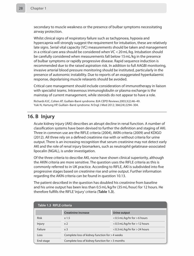
The patient described in the question has doubled his creatinine from baseline and his urine output has been less than 0.5 mL/kg/hr (35 mL/hour) for 12 hours. He therefore fulfills the RIFLE ‘injury’ criteria (Table 1.3).
Table 1.3 RIFLE criteria
Creatinine increase Urine output
Risk x 1.5 < 0.5 mL/kg/hr for > 6 hours
Injury x 2 < 0.5 mL/kg/hr for > 12 hours
Failure x 3 < 0.3 mL/kg/hr for > 24 hours
Loss Complete loss of kidney function for > 4 weeks
End-stage Complete loss of kidney function for > 3 months
Answers 29
The morbidity and associated mortality from AKI is high both within and outside the critical care environment. Prevention, early recognition (using criteria) and good adherence to the principles of management should be a part of routine care. The principles of management include treating the underlying cause, optimising renal perfusion, withholding nephrotoxic agents and, where appropriate, renal replacement therapy.
The clinical importance of AKI has lead to a recent National Institute for Health and Care Excellence (NICE) guideline and the London Acute Kidney Injury Network releasing an ‘AKI bundle.’
National Institute for Health and Care Excellence. Acute Kidney Injury (CG169). 2013. London: National Institute for Health and Care Excellence. Available from: http://guidance.nice.org.uk/CG169London Acute Kidney Injury Network. Acute Kidney Injury Bundle. 2013. Available from: http://www.londonaki.net/news/downloads/AKI_bundle-GSTH.pdf
17. B Methicillin sensitive Staphylococcus aureus (MSSA)The case described has features consistent with the development of pneumonia during the provision of invasive mechanical ventilation (MV). Ventilator-associated pneumonia (VAP) has traditionally been defined as tracheobronchitis or pneumonia occurring more than 48 hours after initiation of MV. This definition relies on a high index of suspicion with confirmation based on clinical, radiological and microbiological evidence. The definition of VAI remains a controversial area. A recent Society of Critical Care Medicine (SCCM) guideline has suggested that VAP may not require a positive microbiological diagnosis.
Clinical scoring systems, such as the Clinical Pulmonary Infection Score (CPIS), can help to objectify the diagnosis of VAP. CPIS is based on temperature, white cell count, appearance of tracheal secretions, new infiltrates on chest radiology and oxygenation indices. A score of greater than 6 is suggestive of VAP. However the validity of this score has been questioned.
The most commonly associated causative organisms tend to relate to the timing of the infection from the onset of MV. In the case described here, the onset is early (< 5 days) and as such is most commonly associated with MSSA and Haemophilus influenzae. The commonest bacterial pathogens in late onset VAP (> 5 days) are aerobic gram negative bacilli (AGNB) such as Klebsiella, Pseudomonas, Enterobacter and Acinetobacter. Drug resistant microbes such as methicillin-resistant Staphylococcus aureus (MRSA) and vancomycin-resistant Enterococci (VRE) are also causative organisms for late onset VAP. The value of bronchoalveolar lavage (BAL) over and above blind endotracheal aspiration is keenly debated.
The management of VAP is supportive with continuous microbiological surveillance and antimicrobial therapy based on local policies in combination with microbiological advice. The prevention of VAP is the focus of a Department of Health (DoH) High Impact Intervention Care bundle. This document outlines six key areas of recommended good practice:
• Elevation of the head of the bed to 30–45° (though trials of supine and even head down positioning are currently in progress)
Chapter 130
• Daily sedation level assessment (though many would advocate mandatory daily cessation for which there is a strong evidence base)
• Oral hygiene with chlorhexidine 6 hourly and tooth brushing 12 hourly• Subglottic aspiration in patients expected to be intubated for > 72 hours (complex
and controversial evidence for this intervention)• Tracheal cuff pressure measured 4 hourly and maintained between 20–30 cmH2O• Stress ulcer prophylaxis prescribed only in high risk patients according to local
protocols and reviewed daily (though this contradicts the earlier DoH, ‘Ventilator care bundle’)
Kalanuria AA, Zai W, Mirski M. Ventilator-associated pneumonia in the ICU. Crit Care. 2014;18(2):208.Department of Health. High impact intervention care bundle to reduce ventilation-association pneumonia. London: Department of Health, 2010.
18. E Site a spinal catheter, inform midwife and perform subsequent top-ups yourself
Accidental dural puncture (ADP) is a well-known complication of epidural anaesthesia, being said to occur in 0.2–4% of cases. Parturients are at risk due to difficulty in positioning and being ‘moving targets’. In this case, there is the added risk of multiple attempts at insertion and a high body mass index (BMI). This case is typical of a pressurised situation on labour ward and the clinical setting must be taken into account when deciding how to manage the ADP.
Repeating the attempt is a potential option but is not the best line of management due to the difficulty of finding the space with multiple attempts already having been undertaken.
The chance of a colleague being available to assist at this time is slim, as opposed to during a day shift. Although this is a possibility, a colleague may also find this epidural difficult to perform, and there may be a significant delay before they can attend to help.
Ultrasound can help to locate the depth, but even with optimum conditions, this will be difficult in a lady of this size, especially if you are unfamiliar with this technique. Finding the ultrasound machine and the correct probe may also prove challenging in the clinical circumstances.
A remifentanil PCA is a potential alternative, but in this situation, the lady has a high risk of needing further intervention due to the position of the baby. Therefore, a spinal catheter is the best option. To reduce the risks of neurological damage, no more than 3 cm is left inside the subarachnoid space. The catheter must be clearly labelled as intrathecal, and both the midwife and mother must be informed. Top-ups must be given only by the anaesthetist. A suggested regime may be 2.5 mL of low dose mixture (0.1% levobupivacaine + 2 μg/mL fentanyl) every 2–4 hours. This can also be used if the patient goes on to need any operative intervention. There is also some evidence that introducing a spinal catheter may reduce the incidence of post dural puncture headache.
Sharpe P. Accidental dural puncture in obstetrics. BJA CEPD Reviews 2001; 1(3):81–84.Palmer CM. Continuous spinal anesthesia and analgesia in obstetrics. Anesth Analg 2010;111(6):1476-9.
Answers 31
19. C Commence MgSO4 infusion at 1 g/hour, give a further 2 g MgSO4 bolus, secure airway with ETT and continue supportive management
Pre-eclampsia is one of the causes of maternal death and since it is well managed, it is not often that patients present with eclamptic seizures. The MAGPIE Trial demonstrated that MgSO4 significantly reduces the risk of eclampsia and this is standard management in these cases. The initial dose of MgSO4 is 4 g over 5–10 minutes followed by an infusion at 1 g/hour for 24 hours post-partum. Any subsequent seizures may be treated with a further bolus of 2 g MgSO4. Phenytoin and diazepam have no place in the management of eclampsia.
Every effort should be made to stabilise the mother (e.g. correct hypoxia, control blood pressure and stop seizures) before undertaking a Caesarean section, as this is a risky procedure to undertake whilst the mother is unstable. Also, the emergency department is a dirty environment therefore the risk of postoperative infection is high. There is unlikely to be diathermy available and the patient may bleed significantly, which is a risk due to the disorders of coagulation that can occur in eclampsia. Furthermore, the baby is only 28/40 and a course of steroid treatment would help lung maturation if delivery were expected to be undertaken. It may be better for the baby to undergo in utero recovery from hypoxia and hypercarbia. If the mother were to arrest in the emergency department then an emergency Caesarean section would be necessary.
National Institute for Health and Care Excellence Clinical Guideline. Hypertension in pregnancy: the management of hypertensive disorders during pregnancy. August 2010 (revised reprint January 2011)Hart E, Coley S. The diagnosis and management of pre-eclampsia. BJA CEPD Rev 2003; 3(2): 38–42.The Magpie Trial Collaborative Group. Do women with pre-eclampsia, and their babies, benefit from magnesium sulphate? The Magpie Trial: a randomised placebo-controlled trial. Lancet 2002; 359:1877–90.Munro P. Management of eclampsia in the accident and emergency department. J Accid Emerg Med 2000; 17(1):7–11.
20. A Start prostaglandin E2 intravenous infusion and refer to tertiary centre for possible coarctation of the aorta
Stabilisation of critically ill neonates, as with all paediatric patients, should prioritise securing the airway, then establishing breathing and maintaining adequate circulation. Endotracheal intubation and fluid resuscitation are usually required in critically ill neonates and these procedures, if indicated, should not be delayed while waiting for diagnostic evaluation. Establishing optimal ventilation and oxygenation is often sufficient to improve both respiratory and cardiac insufficiency; however, continued IV fluids and resuscitation may be required in the gravely ill neonate. An initial bolus of 10 mL/kg of isotonic fluid (0.9% saline) should be given if necessary. Sepsis is one of the most common causes of critical illness in the neonate and prompt empirical treatment with antibiotics is almost always indicated. If the history and physical examination suggest a possible cardiac diagnosis, a continuous infusion of prostaglandin E2 (Prostin, PGE2, 0.01–0.1 μg/kg/min) should be promptly initiated and paediatric cardiology consulted.
Chapter 132
Coarctation of the aorta is a congenital narrowing of the descending thoracic aorta at or near the connection of the ductus arteriosus. It is the sixth most common congenital heart disease, constituting 8% of heart anomalies. The most dramatic presentation of aortic coarctation occurs in the neonate who is dependent on a patent ductus arteriosus (PDA) for blood flow to the distal aorta. After a relatively asymptomatic period of days to weeks, the PDA will close and immediately limit blood flow distal to the coarctation. The ensuing left ventricular failure and systemic hypoperfusion manifest as respiratory distress, cold and pale lower extremities, markedly decreased or absent pulses, metabolic and respiratory acidosis.
Kim UO, Brousseau DC, Konduri G. Evaluation and management of the critically ill neonate in the emergency department. Clin Pediatr Emerg Med 2008; 9:140–148.Landsman IS, Davis P J. Aortic coarctation: anesthetic considerations. Semin Cardiothor Vasc Anesth 2001; 5:91–97.
21. A IV Hartmann’s at 110 mL/hour. Refer to tertiary centre for further management
Burns are a common cause of injury in children. Most burns occur at home, usually in the kitchen and bathroom. The aetiology changes with age; younger children suffer more scalds, older children more flame burns.
The anaesthetist’s roles include resuscitation, analgesia, sedation, anaesthesia and intensive care management of these patients. Adequate early fluid resuscitation maintains organ perfusion and controls the extent of the burn injury itself. Early excision and covering of non-viable skin reduces morbidity, mortality, and the extent of inflammatory response. Adequate pain management is an obligation and may help to alleviate psychological sequelae.
Resuscitation fluid volumes are calculated using the Parkland formula: for the first 24 hours after the burn, give 4 mL/kg per % body surface area (BSA) burn of crystalloid, half of this volume should be delivered in the first 8 hours post-burn, the other half in the next 16 hours. In this case:
4 mL x 15 kg x 20% = 1200 mL over 24 hours
Since 400 mL has already been given in the first 4 hours, only 200 mL should be given in the next 4 hours, i.e. 50 mL/hour.
Maintenance fluid is calculated using the 4/2/1 rule:
• 4 mL/kg/hour for the first 10 kg body weight• 2 mL/kg/hour for the next 10 kg body weight• 1 mL/kg/hour for each kg body weight above 20 kg
For this 20 kg child, this works out to 60 mL/hour.
Therefore resuscitation (50 mL/hour) and maintenance fluid (60 mL/hour) together is 110 mL/hour. Glucose containing intravenous fluids are not generally indicated though may be required for infants when early feeding cannot be established.
Specialist services are required for full-thickness burns exceeding 5% of BSA, partial thickness exceeding 10% BSA, inhalation burns or burns to the airway, face,
Answers 33
hands, feet, and perineum. Antibiotics are indicated only in suspected or confirmed infection, not for prophylaxis.
Fenlon S, Nene S. Burns in children. Contin Educ Anaesth Crit Care Pain 2007;7:76–80.
22. A Increase dose of carbamazepineThis patient has trigeminal neuralgia.
Trigeminal neuralgia (TN), also known as tic douloureux, is a condition characterised by paroxysmal episodes of intense, severe, lancinating facial pain. A typical episode lasts seconds to minutes, and starts and ends abruptly. The pain itself is often described as feeling like “electric shocks” or “stabbing”, and almost always affects just one side of the face, frequently the right. Triggers are usually trivial and may include washing the face, chewing food, brushing teeth, vibrations, or exposure to wind. In particular, the maxillary and/or mandibular distributions of the trigeminal nerve are most commonly affected. Attacks vary significantly in frequency, and may range from just one or two, to several hundred per day.
Over time, TN follows a relapsing and remitting course. A relapse may last days or months, and remission may be as long as several years.
Occurring more commonly in females, the peak incidence of TN is in the fifth and sixth decades of life.
In approximately 80% of cases, there is compression of the trigeminal ganglion, most commonly due to an aberrant arterial or venous loop. Other causes may include multiple sclerosis, Chiari malformation, tumours such as schwannomas or meningiomas, or pontine lesions.
Diagnosis is purely clinical. Differential diagnoses include post-herpetic neuralgia, temporomandibular joint dysfunction, migraine, cluster headaches, and atypical facial pain.
Magnetic resonance imaging (MRI) may be helpful to exclude other causes of pain if the diagnosis is less obvious. It is the most sensitive test available for the diagnosis of multiple sclerosis, and useful in visualising the posterior fossa where tumours associated with TN such as schwannomas and meningiomas are found.
While TN may go into remission spontaneously, the symptoms are usually severe enough to warrant treatment. Treatment options may be medical or surgical.
The drug of choice is carbamazepine. It is thought to be so effective that lack of response, some authors suggest, should prompt reconsideration of the diagnosis. It’s keto-derivative, oxcarbazepine, can be used with equal effect and with fewer unwanted effects. Treatment should be commenced at a low dose, and then gradually increased every 3–7 days until a dose suitable to that patient is found. Carbamazepine, for example, is started at 100 mg b.d. and may need to be increased to 200 mg q.d.s. Alternatives such as lamotrigine or baclofen may be tried if carbamazepine or oxcarbazepine are contra-indicated or poorly tolerated.
Surgical options include microvascular decompression, rhizotomy, and stereotactic radiosurgery, but due to the invasive nature of these interventions, they are reserved
Chapter 134
for refractory cases. In this patient, therefore, the most appropriate intervention at this stage would be to increase the dose of carbamazepine appropriately.
Zakrzewska JM, Linskey ME. Trigeminal neuralgia. Br Med J 2014; 348:g474.National Institute for Health and Care Excellence (NICE). Trigeminal neuralgia. Clinical Knowledge Summary (CKS). London: NICE, 2013.
23. E The pain will usually respond to anticonvulsantsChronic post-surgical pain (CPSP) is an increasingly common phenomenon and its exact aetiology is unknown. Most of these pains are benign and local cancer recurrence is unlikely at 2 years. The nature of the pain is mixed neuropathic and nociceptive but phantom pain can occur in 22–64% of post-mastectomy patients and it is appropriate to use anticonvulsants such as gabapentin to treat such pain. The use of long-term opioids is not recommended as tolerance usually develops and long-term hormonal and immune suppression is increasingly recognised. Although brachial plexopathy can cause chest wall pain, these patients often have arm symptoms rather than pure chest wall pain. Also, brachial plexopathy is usually only found if the cancer has recurred, while the 2-year history would make it unlikely in this circumstance. Therefore the most appropriate statement is that this patient's pain will probably respond to anticonvulsants.
Alves Nogueira Fabro E, Bergmann A, do Amaral E Silva B, et al. Post-mastectomy pain syndrome: incidence and risks. Breast 2012; 21(3):321–5.
24. C Patient education and psychological supportThis patient has developed symptoms and signs of complex regional pain syndrome (CRPS) type 1.
CRPS is a chronic, debilitating and painful condition. It is uncommon and usually develops following an injury, but may occur spontaneously. It affects one or more limbs and typically demonstrates a collection of symptoms and signs described as sensory, vasomotor, sudomotor, trophic and inflammatory. The presentation varies from mild and self-limiting to a chronic and disabling disease process.
Diagnosis is clinical. In 1994, the International Association for the Study of Pain (IASP) established a definition, but the latest definition came from Budapest in 2004.
The Budapest criteria classify symptoms and signs into four broad categories (Table 1.4).
According to the Budapest criteria, all of the following must be met in order to make a diagnosis of CRPS:
• The patient has continuing pain which is disproportionate to the inciting event• The patient has at least one sign in two or more categories• The patient has at least one symptom in three or more categories• No other diagnosis can better explain the signs and symptoms
CRPS can be academically classified as being type 1 (not associated with major nerve damage) or type 2 (associated with major nerve damage), but this does not influence subsequent management or prognosis.
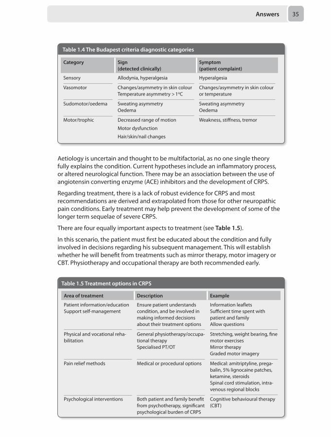
Answers 35
Aetiology is uncertain and thought to be multifactorial, as no one single theory fully explains the condition. Current hypotheses include an inflammatory process, or altered neurological function. There may be an association between the use of angiotensin converting enzyme (ACE) inhibitors and the development of CRPS.
Regarding treatment, there is a lack of robust evidence for CRPS and most recommendations are derived and extrapolated from those for other neuropathic pain conditions. Early treatment may help prevent the development of some of the longer term sequelae of severe CRPS.
There are four equally important aspects to treatment (see Table 1.5).
In this scenario, the patient must first be educated about the condition and fully involved in decisions regarding his subsequent management. This will establish whether he will benefit from treatments such as mirror therapy, motor imagery or CBT. Physiotherapy and occupational therapy are both recommended early.
Table 1.4 The Budapest criteria diagnostic categories
Category Sign(detected clinically)
Symptom(patient complaint)
Sensory Allodynia, hyperalgesia Hyperalgesia
Vasomotor Changes/asymmetry in skin colour Temperature asymmetry > 1oC
Changes/asymmetry in skin colour or temperature
Sudomotor/oedema Sweating asymmetry Oedema
Sweating asymmetry Oedema
Motor/trophic Decreased range of motion
Motor dysfunction
Hair/skin/nail changes
Weakness, stiffness, tremor
Table 1.5 Treatment options in CRPS
Area of treatment Description Example
Patient information/education Support self-management
Ensure patient understands condition, and be involved in making informed decisions about their treatment options
Information leaflets Sufficient time spent with patient and family Allow questions
Physical and vocational reha-bilitation
General physiotherapy/occupa-tional therapy Specialised PT/OT
Stretching, weight bearing, fine motor exercises Mirror therapy Graded motor imagery
Pain relief methods Medical or procedural options Medical: amitriptyline, prega-balin, 5% lignocaine patches, ketamine, steroids Spinal cord stimulation, intra-venous regional blocks
Psychological interventions Both patient and family benefit from psychotherapy, significant psychological burden of CRPS
Cognitive behavioural therapy (CBT)
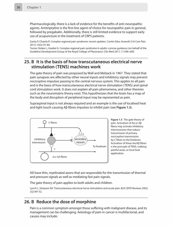
Chapter 136
Pharmacologically, there is a lack of evidence for the benefits of anti-neuropathic agents. Amitriptyline is the first line agent of choice for neuropathic pain in general, followed by pregabalin. Additionally, there is still limited evidence to support early use of acupuncture in the treatment of CRPS patients.
Ganty P, Chawla R. Complex regional pain syndrome: recent updates. Contin Educ Anaesth Crit Care Pain 2013; 14(2):79–84.Turner-Stokes L, Goebel A. Complex regional pain syndrome in adults: concise guidance (on behalf of the Guideline Development Group of the Royal College of Physicians). Clin Med 2011; 11:596–600.
25. B It is the basis of how transcutaneous electrical nerve stimulation (TENS) machines work
The gate theory of pain was proposed by Wall and Melzack in 1967. They stated that pain synapses are affected by other neural inputs and inhibitory signals may prevent nociceptive impulses passing to the central nervous system. This applies to all pain and is the basis of how transcutaneous electrical nerve stimulation (TENS) and spinal cord stimulation work. It does not explain all pain phenomena, and other theories such as the neuromatrix theory exist. This hypothesizes that the brain has a map of the body and disruption of peripheral input may be represented as pain.
Supraspinal input is not always required and an example is the use of localised heat and light touch causing Aβ fibres impulses to inhibit pain (see Figure 1.5).
To forebrain
––
+
+
Inhibitoryinterneuron
C bres
Αα/Αβ bres
secondaryneuron
Figure 1.5 The gate theory of pain. Activation of Aα or Aβ fibres may activate inhibitory interneurones that reduce transmission of primary nociceptive transmission by C fibres to the forebrain. Activation of these Aα/Aβ fibres is the principle of TENS, rubbing painful areas, or local heat application.
Aδ have thin, myelinated axons that are responsible for the transmission of thermal and pressure signals as well as mediating fast pain signals.
The gate theory of pain applies to both adults and children.
Lynch L, Simpson KH. Transcutaneous electrical nerve stimulation and acute pain. BJA CEPD Reviews 2002; 2(2):49–52.
26. B Reduce the dose of morphinePain is a common symptom amongst those suffering with malignant disease, and its management can be challenging. Aetiology of pain in cancer is multifactorial, and causes may include:
Answers 37
• Mass effect of the tumour• Effect of treatment, e.g. radiotherapy, chemotherapy• Paraneoplastic syndromes• Conditions associated with the disease process, e.g. osteoporosis, hypercalcaemia• Pre-existing pain from other chronic conditions• Combination of any of the above
Table 1.6 summarises some of the strategies that may be employed in controlling pain.
Opioids are very effective in management of cancer-related pain in general. To begin with, most patients are started on longer acting oral opioids to treat background pain, supplemented with additional doses to treat breakthrough pain. Oral morphine is a suitable agent for most patients, but different people may display an unpredictably different benefit-side-effect profile with different opioids.
Table 1.6 Strategies for pain control
Method of pain control
Examples
Pharmacological WHO analgesic ladder is a guide, but sometimes patients may need to start with strong opioids in the early stages of treatment Adjuvant medications such as ketamine Anti-neuropathic agents such as amitriptyline, pregabalin Steroids reduce oedema and inflammation around tumour growth, hence reducing local mass effects Bisphosphonates for bone pain from osteoporosis or metastases Antispasmodics
Oncological Chemotherapy Radiotherapy Hormonal therapy, e.g. tamoxifen Immunotherapy, e.g. Herceptin
Surgical Treatment of gastrointestinal obstruction Reduction and treatment of pathological fractures Curative surgery CSF shunt procedures
Psychological Grief counselling Cognitive behavioural therapy Relaxation techniques Pain management programmes
Complementary Therapies
Acupuncture Reflexology Herbal remedies
Interventional techniques
Spinal cord stimulation
In general, a low dose oral preparation is commenced. The dose is then increased by 30–50% gradually over intervals of 2–3 days until adequate control of symptoms is achieved. If using morphine as an example, an appropriate dose for breakthrough pain would be 1/6 of the total daily morphine requirement.
Chapter 138
It is not uncommon to use parenteral routes to administer opioids. In palliative care settings, opioids are frequently administered subcutaneously by syringe driver. Those on very large doses of oral morphine (> 60 mg per 24 hours) may benefit from a fentanyl patch.
Long-term opioid use is associated with significant unwanted effects. These include dependence, tolerance, drowsiness, constipation, nausea, vomiting, pruritus, urinary retention and respiratory depression. Sometimes, patients may report a paradoxical worsening of their pain. This is a phenomenon known as opioid-induced hyperalgesia. Unwanted side effects will usually settle as tolerance develops. However, at times they may be more distressing than the pain itself, as in this scenario.
If adverse effects of opioids cause the patient notable distress or discomfort they warrant specific treatment. The reversal agent naloxone has an extremely short plasma half-life, and while it is useful for acute opioid poisoning, it has no role in a chronic setting. In the first instance, the dose of opioid should be reduced, but other adjuvant analgesia should be prescribed, e.g. paracetamol or ketamine. Anti-pruritic drugs, laxatives, and other medication for symptom relief may be considered. Failing this, opioid rotation may be attempted. This is a method by which one opioid is substituted for an equipotent dose of another (e.g. oxycodone for morphine).
Prescribing a laxative is not the best option here as it only tackles constipation, rather than treating the problem of unwanted effects as a whole.
Scott-Warren J, Bhaskar A. Cancer pain management – Part 1: General principles. Contin Educ Anaesth Crit Care Pain 2014, doi: 10.1093/bjaceaccp/mkt070.Ripamonti CI, Bandieri E, Roila F. Management of cancer pain: ESMO. Ann Oncol 2012; 23(suppl 7):vii39–vii154.
27. B Enteral nutrition should be started as soon as possibleFollowing a significant burns injury, the resultant increase in basal metabolic rate is proportional to the size of the burn and is thought to peak at 7–10 days. Early enteral nutrition is essential and has been shown to improve survival. Good nutritional support minimises protein breakdown, improves wound healing and reduces infective complications. A high protein and carbohydrate diet is required.
Patients with severe burns reset their baseline temperature to 38.5°C. It is therefore important to prevent hypothermia by ensuring ambient room temperature is kept between 28–32°C as well as all the usual warming measures. There is no evidence for prophylactic antibiotics in patients with burns. Diagnosis of sepsis in these patients is difficult as the burn itself results in a systemic inflammatory response syndrome (SIRS) that mimics sepsis. It is important to be vigilant to the presence of infection as this represents a significant source of morbidity and mortality in major burns. To reduce resistance, antibiotics should only be started following positive cultures or when there is a high index of clinical suspicion. Skin grafting, aseptic dressing changes and patient isolation are all measures that reduce the risk of infection.
Prophylactic steroids are not indicated in the routine management of burns patients.
Answers 39
The Parkland formula (volume to be infused in first 24 hours = 4 mL/kg/% body surface area burn) is merely a guide to fluid resuscitation in burns and will almost always need to be adjusted. Too much fluid can be as detrimental as too little. The complications of over resuscitation include abdominal compartment syndrome, the need for fasciotomies to prevent extremity necrosis and pulmonary oedema. The resultant SIRS leads to increase capillary permeability, so even if fluid management is optimal there will still be a degree of oedema.
Bishop S, Maguire S. Anaesthesia and intensive care for major burns. Contin Educ Anaesth Crit Care Pain 2012; 12(3): 118–122.
28. E Optimising the intravascular volume during aortic cross-clamping
Hypotension following the application of aortic cross-clamp is a significant intra-operative event. The most effective interventions that can minimise this haemodynamic instability include optimal fluid resuscitation during the pre-release phase and gradual release of the aortic cross-clamp by the surgeon.
Although they have a role, vasoconstrictors and inotropes are not as effective if initiated following aortic cross-clamp release. Hypoperfusion to the lower limbs leads to anaerobic metabolism, metabolic acidosis and hyperkalaemia. This in turn reduces the sensitivity of adrenergic receptors in the affected areas. If used, vasoconstrictors will increase the systemic vascular resistance causing vasoconstriction in the upper limbs, potentially diverting the blood away from the central compartment and thereby compromising cardiac output.
Administration of fluids is less effective in the post-release phase, and a 500 mL colloid bolus during the release of the cross-clamp is often insufficient resuscitation. Tilting the table head up would only decreased the left-ventricular preload and cause hypotension. Therefore, carefully optimising the patients intravascular status throughout the operation is the safest method to mitigate the predictable hypotension following aortic cross-clamping.
Gelman S. The pathophysiology of aortic cross-clamping and unclamping. Anesthesiology 1995; 82:1026–60.
29. E T wave inversion in lead aVRArrhythmias may occur preceding a myocardial infarction or may lead to ventricular fibrillation, thus it is necessary to identify them and treat them without a delay.
As with any other medical emergency there needs to be an ABCDE approach. A 12-lead ECG would differentiate between broad QRS complex and narrow QRS complex tachyarrhythmias.
It is important to look for features suggesting clinical instability in view of the arrhythmia. In the presence of such adverse features it is necessary to perform direct current cardioversion in attempt to restore sinus rhythm. These features include:
• Shock: hypotension (systolic pressure <90mmHg), pallor, cold clammy extremities, sweating, confusion or impaired consciousness
Chapter 140
• Syncope: transient unconsciousness due to compromised cerebral blood flow• Myocardial ischaemia: clinical features of chest pain or ECG changes suggesting
the same• Heart failure: pulmonary oedema and raised jugular venous pressure (with or
without peripheral oedema or enlarged liver)
T wave inversion in aVR is normal ECG morphology, and therefore not an adverse feature necessitating urgent intervention.
Resuscitation Council (UK). Peri-arrest arrhythmias. In: Advanced Life Support, 6th ed. London: Resuscitation Council (UK), 2010.
30. A Vitamin AThis patient presents with clinical features suggesting vitamin A deficiency. Vitamin A is an essential nutrient whose deficiency leads to eye and skin manifestations. It is found as retinol and beta-carotene in the diet. Retinol comes from animal sources like liver, fish and eggs whereas beta-carotene is found in fruits and green vegetables. Peak incidence for vitamin A deficiency occurs in children aged 2-5 years due to poor nutrition .
Skin effects are seen as repeated skin infections, acne and dry skin. It can also lead to thrush and dry hair.
Vitamin A deficiency leads to dry conjunctivae (xerosis) and night blindness. Small, raised, grey plaques are found in the interpalpebral conjunctivae called Bitot’s spots. Corneal ulceration and perforation may occur in extreme cases.
Treatment is by administering retinol palmitate intramuscularly or beta-carotene orally.
Collier J, Longmore M, Scally P. Oxford Handbook of Clinical Specialties, 7th ed. Oxford: Oxford University Press, 2007.
Mock Paper 2
Chapter 2
Questions1. 5 days ago a 25-year-old woman suffered a traumatic brain injury. Despite a
normal CT, she demonstrated bilateral motor posturing. She therefore had an intracranial pressure (ICP) bolt inserted shortly after admission in order to monitor her ICP. You begin your morning review by analysing her ICP waveform.
Which of the following would concern you most acutely?
A Lundberg A wavesB A flat traceC A baseline value of 20 mmHgD Lundberg B wavesE The most prominent upstroke being the P2 (tidal) wave
2. You are called to the emergency department to assist with a 20-year-old man who presented following a grand-mal seizure. He has begun fitting again and his conscious level has remained below baseline for 30 minutes. He has a history of epilepsy and has not been taking his usual medication since his prescription ran out 1 week ago. He is otherwise well.
Which of the following is the most appropriate immediate action?
A Intubate the patient to protect his airway and optimise ventilationB Check his blood glucose and treat any hypoglycaemia with intravenous
dextroseC Take blood for full blood count, electrolytes, urea, creatinine, liver function
tests and anti-epileptic medication levelsD Administer his anti-epileptic medication intravenouslyE Apply pulse oximetry, blood pressure and ECG monitoring
3. You are asked to pre-assess a 68-year-old man for elective open umbilical hernia repair. He suffers from exertional angina (three flights of stairs) and had coronary angiography 6 months ago, which showed right coronary and circumflex artery disease. He had a myocardial infarction 2 years ago. His symptoms have been stable over the past year and he is currently taking bisoprolol 2.5 mg once a day (o.d.), aspirin 75 mg o.d. and ramipril 2.5 mg o.d. His creatinine is 120 μmol/L. He is not diabetic and has not had a stroke. He is independent and performs all his own housework.
Chapter 242
Which of the following is the most appropriate next step in this patient’s management?
A Schedule the patient for surgery. Cease the ramipril 24 hours preoperatively. Continue the aspirin
B Schedule the patient for surgery. Cease the bisoprolol 2 days preoperatively. C Obtain non-invasive stress testingD Refer for repeat coronary angiogramE Refer for coronary artery bypass surgery
4. A healthy 30-year-old man due for an appendicectomy is undergoing a rapid sequence induction. After full pre-oxygenation and administration of thiopentone and suxamethonium, three attempts at intubation have failed with an unexpected grade III Cormack & Lehane laryngoscopy view. His oxygen saturations are 92% and full cricoid pressure is being applied.
What action is most appropriate in this situation?
A Reattempt intubation using a different laryngoscope B Reattempt intubation whilst reducing the cricoid pressureC Attempt insertion of a supraglottic airway deviceD Postpone surgery and awaken the patient C Attempt cannula cricothyroidotomy
5. An 80-year-old woman, admitted with pneumonia 14 days ago, suffers an episode of large haematemesis requiring endoscopic investigation. You anaesthetise her and secure her airway uneventfully with a size 7.5 endotracheal tube (ETT) secured at 24 cm to the teeth. Oxygen saturations are 99% (Fio₂ 0.4). The gastroenterologist then extends her head. At this point she becomes progressively hypoxic and chest wall movement becomes limited to the right side.
What is the most likely reason for her desaturation?
A Aspiration of gastric contentsB BronchospasmC Sputum plugging of a bronchus D PneumothoraxE Endobronchial intubation
6. A 56-year-old man is taken to the intensive care unit following a laparotomy a week ago for bowel obstruction. He is hypertensive, has a body mass index (BMI) of 42, and has had an ileus for the last 7 days. His abdomen is tender and tensely distended. There are no signs of sepsis, but he has worsening renal function and a poor urine output, despite a very positive fluid balance. He had become increasingly breathless with type II respiratory failure, and after a failed trial of non-invasive ventilation, is now mechanically ventilated. His ventilator pressures are worsening and his base excess and lactate are increasing.
Which of the following is the most correct regarding this patient?
Questions 43
A A measurement of intra-abdominal pressure is most safely performed with an intraperitoneal catheter and pressure transducer
B An intravesical measurement is performed at the end of inspirationC Abdominal compartment syndrome may provoke a cardiac arrest at
laparotomyD An abdominal pressure of 24 mmHg can be considered normalE The only effective treatment is re-laparotomy leaving the abdomen open with a
Bogota bag
7. A 60-year-old man awaiting surgical resection for his tracheal carcinoma presents to the emergency department with worsening dyspnoea. As part of his pulmonary function tests, a bedside flow-volume loop (spirogram) is ordered.
What will be the most likely appearance of his flow-volume loop?
A Flattening of the inspiratory limb only B Flattening of the expiratory limb only C Flattening of both the inspiratory and expiratory limbs D Saw-tooth pattern in both the inspiratory and expiratory limbs E Normal appearance of both inspiratory and expiratory limbs
8. A 32-year-old woman was recently diagnosed with brainstem death following a subarachnoid haemorrhage. She was initially hypertensive, then developed polyuria and gradually became hypotensive. The patient has received 30 mL/kg of crystalloid boluses.
What is the next pharmacological intervention needed to restore the patient's blood pressure?
A DopamineB AdrenalineC VasopressinD NoradrenalineE Further bolus of colloid
9. You have been asked to see a 32-year-old patient in recovery following a retrograde intramedullary femoral nailing. He works as a builder and fell from a ladder sustaining bilateral femoral fractures. The plan is to fix the other leg in the next few days. He is desaturating to 88% on oxygen via facemask and is confused, and one of the recovery staff has also noticed a petechial rash on the patient’s neck.
In diagnosis and treatment of the likely condition, which of the following tests is most important?
A Arterial blood gas analysisB Bronchoscopic alveolar lavage (BAL) for macrophagesC Trial of methylprednisolone for 48 hoursD A CT of the chestE Urine microscopy for lipid droplets
Chapter 244
10. A 50-year old woman for elective hand surgery is to have an ultrasound guided axillary brachial plexus block.
Which of the following would be the most appropriate ultrasound probe to use?
A Curved array probeB Linear array probeC Low frequency (2–5 MHz) probeD Phased array probeE Hockey stick footprint probe
11. A 38-year-old man has sustained a stab wound in the left flank following an altercation. In the emergency department he is awake and has saturations of 99% on room air, a respiratory rate of 25 breaths per minute, a heart rate of 110 beats per minute and a blood pressure of 85/40 mmHg. Initial resuscitation has started with intravenous access and blood samples taken. Any other injuries have been excluded apart from a dry puncture wound to the left flank associated with mild abdominal tenderness and no peritonism.
What is the next step in the ongoing resuscitation of this patient?
A A fluid bolus of 10 ml/kg of colloidB A fluid bolus of 10 ml/kg of crystalloidC No fluid bolusD Insert an arterial lineE Admit the patient to the emergency department observation ward
12. A 55-year-old woman with a 1-month history of headaches presents with a severe headache followed by a collapse. In the emergency department of a large teaching hospital her Glasgow coma scale (GCS) was 3 and she was intubated and ventilated appropriately. A CT scan demonstrated a large subarachnoid haemorrhage (SAH) with blood in the intraventricular system and dilated lateral ventricles. A CT angiogram demonstrated a large posterior communicating artery aneurysm. Her pupillary response to light is sluggish.
The most beneficial intervention in this patient would be:
A Immediate transfer to neurosurgical theatre for an extraventricular drain (EVD)
B Immediate transfer to neurosurgical theatre for insertion of an intracranial bolt monitor
C Immediate transfer to the intensive care unit for medical stabilisation before embolisation of the aneurysm
D Administration of hypertonic saline and mannitol E Administration of intravenous phenytoin
13. A 52-year-old man is admitted to the emergency department one hour ago with worsening shortness of breath, fevers and productive sputum. His observations include a temperature of 38°C, Spo2 95% on high-flow oxygen, blood pressure 88/49 mmHg, heart rate 126 beats per minute and respiratory rate 28 breaths per minute. His arterial blood gas is below (Table 2.1):
Questions 45
Blood cultures have been taken and a dose of co-amoxiclav and clarithromycin has been administered.
The most appropriate next step would be:
A Intubation and ventilationB Central venous access and noradrenalineC Central venous access and measurement of Scv–o2D Non-invasive ventilationE 30 mL/kg crystalloid bolus
14. An 18-year-old woman presents to the intensive care unit with diabetic ketoacidosis (DKA) for which she has had multiple admissions in the past. On examination her heart rate is 110 beats per minute and her blood pressure is 100/60 mmHg. Her Glasgow coma scale (GCS) is 14 (E3, V5, M6). Her laboratory glucose was 39 mmol/L and the arterial blood gas demonstrated the following (Table 2.2):
Current treatment is an insulin infusion of 5 units/hour and 500 mL of intravenous Hartmann’s solution being administered over 4 hours.
Table 2.2 Arterial blood gas test results
Parameter Result
pH 6.9
Pao2 14.1 kPa
Paco2 2.1 kPa
Bicarbonate concentration (HCO3–) 3.4 mmol/L
Lactate 2.2 mmol/L
Potassium concentration (K+) 4.6 mmol/L
Table 2.1 Arterial blood gas test results
Parameter Result
Fio2 0.4
pH 7.21
Paco2 4.8 kPa
Pao2 11.4 kPa
Base excess –5.6 mmol/L
Bicarbonate concentration (HCO3–) 20.6 mmol/L
Potassium concentration (K+) 4.8 mmol/L
Sodium concentration (Na+) 138 mmol/L
Lactate 4.6 mmol/L
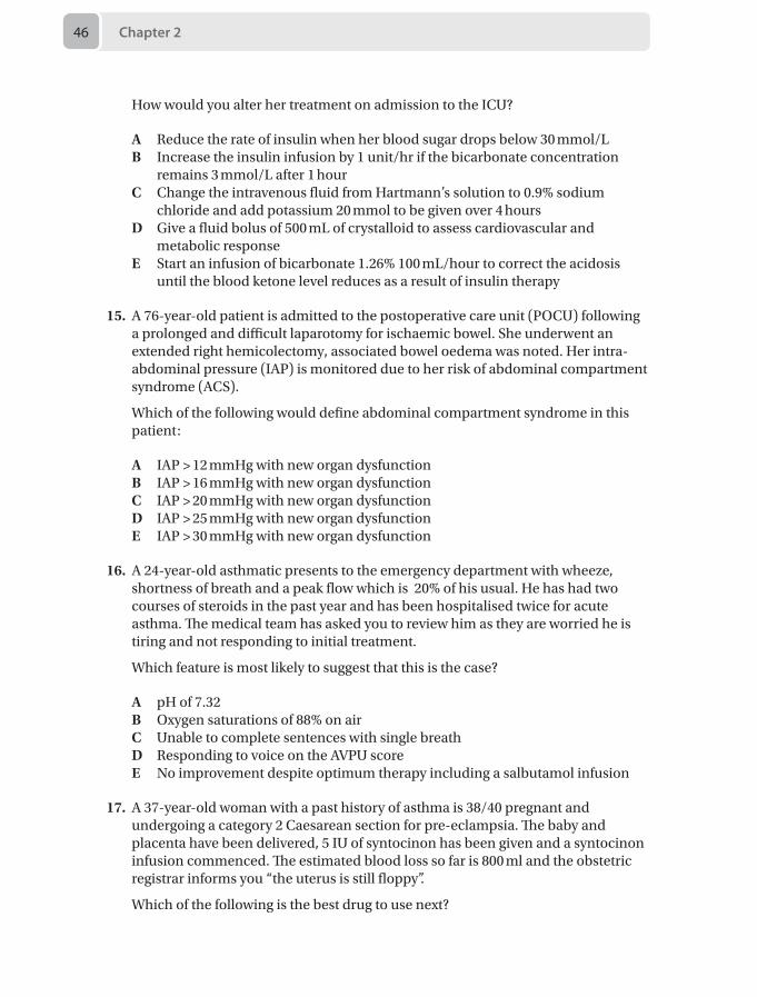
Chapter 246
How would you alter her treatment on admission to the ICU?
A Reduce the rate of insulin when her blood sugar drops below 30 mmol/LB Increase the insulin infusion by 1 unit/hr if the bicarbonate concentration
remains 3 mmol/L after 1 hourC Change the intravenous fluid from Hartmann’s solution to 0.9% sodium
chloride and add potassium 20 mmol to be given over 4 hoursD Give a fluid bolus of 500 mL of crystalloid to assess cardiovascular and
metabolic responseE Start an infusion of bicarbonate 1.26% 100 mL/hour to correct the acidosis
until the blood ketone level reduces as a result of insulin therapy
15. A 76-year-old patient is admitted to the postoperative care unit (POCU) following a prolonged and difficult laparotomy for ischaemic bowel. She underwent an extended right hemicolectomy, associated bowel oedema was noted. Her intra-abdominal pressure (IAP) is monitored due to her risk of abdominal compartment syndrome (ACS).
Which of the following would define abdominal compartment syndrome in this patient:
A IAP > 12 mmHg with new organ dysfunctionB IAP > 16 mmHg with new organ dysfunctionC IAP > 20 mmHg with new organ dysfunctionD IAP > 25 mmHg with new organ dysfunctionE IAP > 30 mmHg with new organ dysfunction
16. A 24-year-old asthmatic presents to the emergency department with wheeze, shortness of breath and a peak flow which is 20% of his usual. He has had two courses of steroids in the past year and has been hospitalised twice for acute asthma. The medical team has asked you to review him as they are worried he is tiring and not responding to initial treatment.
Which feature is most likely to suggest that this is the case?
A pH of 7.32B Oxygen saturations of 88% on air C Unable to complete sentences with single breathD Responding to voice on the AVPU scoreE No improvement despite optimum therapy including a salbutamol infusion
17. A 37-year-old woman with a past history of asthma is 38/40 pregnant and undergoing a category 2 Caesarean section for pre-eclampsia. The baby and placenta have been delivered, 5 IU of syntocinon has been given and a syntocinon infusion commenced. The estimated blood loss so far is 800 ml and the obstetric registrar informs you “the uterus is still floppy”.
Which of the following is the best drug to use next?
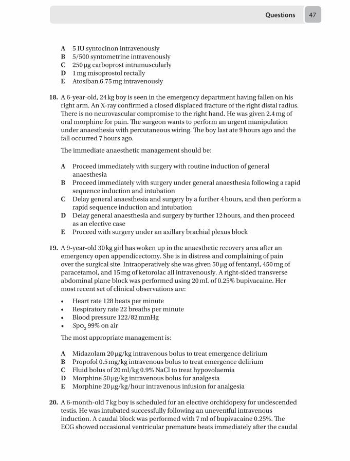
Questions 47
A 5 IU syntocinon intravenouslyB 5/500 syntometrine intravenouslyC 250 μg carboprost intramuscularlyD 1 mg misoprostol rectallyE Atosiban 6.75 mg intravenously
18. A 6-year-old, 24 kg boy is seen in the emergency department having fallen on his right arm. An X-ray confirmed a closed displaced fracture of the right distal radius. There is no neurovascular compromise to the right hand. He was given 2.4 mg of oral morphine for pain. The surgeon wants to perform an urgent manipulation under anaesthesia with percutaneous wiring. The boy last ate 9 hours ago and the fall occurred 7 hours ago.
The immediate anaesthetic management should be:
A Proceed immediately with surgery with routine induction of general anaesthesia
B Proceed immediately with surgery under general anaesthesia following a rapid sequence induction and intubation
C Delay general anaesthesia and surgery by a further 4 hours, and then perform a rapid sequence induction and intubation
D Delay general anaesthesia and surgery by further 12 hours, and then proceed as an elective case
E Proceed with surgery under an axillary brachial plexus block
19. A 9-year-old 30 kg girl has woken up in the anaesthetic recovery area after an emergency open appendicectomy. She is in distress and complaining of pain over the surgical site. Intraoperatively she was given 50 μg of fentanyl, 450 mg of paracetamol, and 15 mg of ketorolac all intravenously. A right-sided transverse abdominal plane block was performed using 20 mL of 0.25% bupivacaine. Her most recent set of clinical observations are:
• Heart rate 128 beats per minute• Respiratory rate 22 breaths per minute • Blood pressure 122/82 mmHg• Spo2 99% on air
The most appropriate management is:
A Midazolam 20 μg/kg intravenous bolus to treat emergence deliriumB Propofol 0.5 mg/kg intravenous bolus to treat emergence deliriumC Fluid bolus of 20 ml/kg 0.9% NaCl to treat hypovolaemiaD Morphine 50 μg/kg intravenous bolus for analgesiaE Morphine 20 μg/kg/hour intravenous infusion for analgesia
20. A 6-month-old 7 kg boy is scheduled for an elective orchidopexy for undescended testis. He was intubated successfully following an uneventful intravenous induction. A caudal block was performed with 7 ml of bupivacaine 0.25%. The ECG showed occasional ventricular premature beats immediately after the caudal

Chapter 248
block was performed. Shortly after that, the ECG showed a regular broad complex tachycardia at 160 beats per minute. You notice the end tidal CO2 trace becomes flat despite good bilateral chest movement. There are no palpable pulses.
Your immediate first action is:
A Announce cardiac arrest, call for help, and start chest compressionsB Give 1.5 mL/kg of 20% lipid emulsion as an intravenous bolus over 1 minute,
followed by an infusion of 15 mL/kg/hourC Request for the cardiac defibrillator and deliver a DC shock of 4 J/kgD Request the local anaesthetic toxicity boxE Give 0.1 mL/kg of 1:10,000 epinephrine intravenously
21. A 6-year-old girl is admitted with acute appendicitis. After uncomplicated surgery she is noted to be in discomfort and crying.
In the assessment this child’s pain, which of the following is most correct:
A The FLACC scale is for 1 month and aboveB Self-reporting is reliable over the age of 10C The visual analogue scale (VAS) can be used in patients aged 12 years and
aboveD Physiological measures such as heart rate can be used aloneE The Wong-Baker FACES pain scale consist of 5 hand drawn faces
22. A 45 kg, 83-year-old woman with dementia and chronic kidney disease is admitted with a right fractured neck of femur following a fall. She is in severe pain, is confused and agitated. She has been given regular paracetamol, and is booked for a dynamic hip screw.
Which of the following is the best method to provide her with adequate analgesia intra- and postoperatively?
A Fascia liaca blockB Femoral nerve blockC Spinal anaesthesiaD Codeine phosphateE Intravenous morphine
23. A 61-year-old woman presents with worsening left sided chest pain over the last 3 months. She describes an intense burning sensation over the T5 distribution, which began after an episode of shingles 4 months ago. The pain gets worse through the day, and especially during the night when it disturbs her sleep. It may be triggered by cold wind blowing over the painful part, or her clothes rubbing against it. She has been taking co-codamol with only modest effect.
Which of the following is the most appropriate next step in her management?
A Lignocaine patchB Oral morphine sulphate
Questions 49
C Topical capsaicinD GabapentinE Thoracic sympathectomy
24. A 45-year-old man has undergone multiple spinal surgeries in the past and has been on 80 mg daily of slow release morphine for the past year. He is asking for an increase in his medication.
Which of the following most appropriately describes this patient’s opioid therapy?
A There is no upper limit on the efficacy of opioidsB Respiratory depression is a significant risk at this doseC Hyperalgesia may develop with increasing dosageD Constipation is usually resolved after 1 yearE Levels of serum adrenocorticotrophic hormone (ACTH) may be increased
25. A 31-year-old woman with a history of acute intermittent porphyria presents with severe abdominal pain. She is tachycardic, hypertensive and vomiting. A diagnosis of small bowel obstruction is made and she is to go to theatre for a laparotomy.
Which of the following is the best option for her postoperative analgesia?
A Epidural with levobupivacaine and fentanylB Titrated intravenous ketamineC Intravenous morphineD OxycodoneE Wound infiltration of 0.25% levobupivacaine
26. A 32-year-old man is undergoing emergency surgery under general anaesthesia for a penetrating eye injury. During the procedure, the surgeon palpates the globe and tells you that the intraocular pressure (IOP) is too high.
Which of the following measures is least likely to reduce the IOP?
A Adequate muscle relaxationB Trendelenburg positioningC Increasing minute ventilationD Intravenous acetazolamideE Intravenous mannitol bolus
27. A 71-year-old man is undergoing a bilateral ilio-femoral bypass graft with the aid of a cell salvage device. The surgeons have completed the right ilio-femoral bypass and are preparing to begin operating on the left side when you notice that the blood returned to the patient from the cell salvage device is 1,900 mL.
After sending a clotting sample to the lab, what is the most appropriate next step in the management of this patient’s blood loss?
A Await the lab results and treat the coagulation accordinglyB Request cryoprecipitate and platelets from the blood bank
Chapter 250
C Administer a 1 g bolus of tranexamic acid intavenouslyD Use hypotensive anaesthesia to decrease further blood lossE Check the patients activated clotting time (ACT)
28. A 67-year-old man is admitted to the emergency department with a sudden onset of severe chest pain and breathlessness. His ECG shows ST elevation in the following leads: I, aVL and V2–V6.
Which of the following is the most likely affected blood vessel?
A Left anterior descending arteryB Right coronary arteryC Left circumflex arteryD Left coronary arteryE Posterior descending artery
29. A 7-year-old boy presents following a head injury by falling from a height of 3 metres. On assessing his neurological status he opens his eyes to a painful stimulus, cries to pain and withdraws from a painful stimulus.
Which score on the paediatric Glasgow coma scale best represents the findings?
A Eyes 2 Verbal 3 Motor 4 B Eyes 1 Verbal 2 Motor 4C Eyes 2 Verbal 2 Motor 3D Eyes 3 Verbal 3 Motor 4E Eyes 2 Verbal 4 Motor 4
30. You are called to urgently assess a 54-year-old man in outpatient clinic who has acutely deteriorated following administration of oral antibiotics. On assessment his heart rate is 145 beats per minute, blood pressure is 74/36 mmHg and saturations are 93% on 6 L/minute of oxygen via Hudson mask. He is wheezy and has a generalised rash all over his body.
Which of the following would be the most appropriate pharmacological intervention for the patient?
A intravenous adrenaline 3 mg bolusB intravenous adrenaline 0.5 mL of 1:1000C intramuscular adrenaline 1 mL of 1:1000D intramuscular adrenaline 0.5 mL of 1:1000E intramuscular adrenaline 0.5 mL of 1:10000
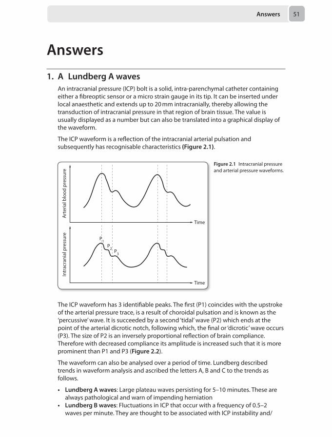
Answers 51
Answers
1. A Lundberg A wavesAn intracranial pressure (ICP) bolt is a solid, intra-parenchymal catheter containing either a fibreoptic sensor or a micro strain gauge in its tip. It can be inserted under local anaesthetic and extends up to 20 mm intracranially, thereby allowing the transduction of intracranial pressure in that region of brain tissue. The value is usually displayed as a number but can also be translated into a graphical display of the waveform.
The ICP waveform is a reflection of the intracranial arterial pulsation and subsequently has recognisable characteristics (Figure 2.1).
Time
Art
eria
l blo
od p
ress
ure
Intr
acra
nial
pre
ssur
e
Time
P1
P3
P2
The ICP waveform has 3 identifiable peaks. The first (P1) coincides with the upstroke of the arterial pressure trace, is a result of choroidal pulsation and is known as the ‘percussive’ wave. It is succeeded by a second ‘tidal’ wave (P2) which ends at the point of the arterial dicrotic notch, following which, the final or ‘dicrotic’ wave occurs (P3). The size of P2 is an inversely proportional reflection of brain compliance. Therefore with decreased compliance its amplitude is increased such that it is more prominent than P1 and P3 (Figure 2.2).
The waveform can also be analysed over a period of time. Lundberg described trends in waveform analysis and ascribed the letters A, B and C to the trends as follows.
• Lundberg A waves: Large plateau waves persisting for 5–10 minutes. These are always pathological and warn of impending herniation
• Lundberg B waves: Fluctuations in ICP that occur with a frequency of 0.5–2 waves per minute. They are thought to be associated with ICP instability and/
Figure 2.1 Intracranial pressure and arterial pressure waveforms.
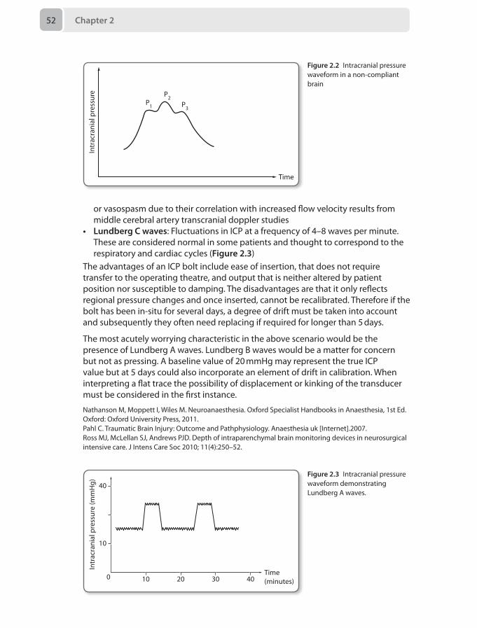
Chapter 252
or vasospasm due to their correlation with increased flow velocity results from middle cerebral artery transcranial doppler studies
• Lundberg C waves: Fluctuations in ICP at a frequency of 4–8 waves per minute. These are considered normal in some patients and thought to correspond to the respiratory and cardiac cycles (Figure 2.3)
The advantages of an ICP bolt include ease of insertion, that does not require transfer to the operating theatre, and output that is neither altered by patient position nor susceptible to damping. The disadvantages are that it only reflects regional pressure changes and once inserted, cannot be recalibrated. Therefore if the bolt has been in-situ for several days, a degree of drift must be taken into account and subsequently they often need replacing if required for longer than 5 days.
The most acutely worrying characteristic in the above scenario would be the presence of Lundberg A waves. Lundberg B waves would be a matter for concern but not as pressing. A baseline value of 20 mmHg may represent the true ICP value but at 5 days could also incorporate an element of drift in calibration. When interpreting a flat trace the possibility of displacement or kinking of the transducer must be considered in the first instance.
Nathanson M, Moppett I, Wiles M. Neuroanaesthesia. Oxford Specialist Handbooks in Anaesthesia, 1st Ed. Oxford: Oxford University Press, 2011.Pahl C. Traumatic Brain Injury: Outcome and Pathphysiology. Anaesthesia uk [Internet].2007. Ross MJ, McLellan SJ, Andrews PJD. Depth of intraparenchymal brain monitoring devices in neurosurgical intensive care. J Intens Care Soc 2010; 11(4):250–52.
Intr
acra
nial
pre
ssur
e
Time
P1 P3
P2
Figure 2.2 Intracranial pressure waveform in a non-compliant brain
Intr
acra
nial
pre
ssur
e (m
mH
g) 40
10
0 2010 30 40Time(minutes)
Figure 2.3 Intracranial pressure waveform demonstrating Lundberg A waves.
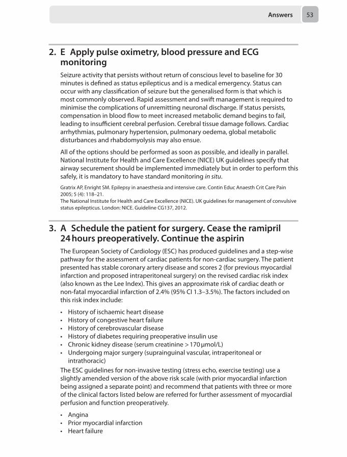
Answers 53
2. E Apply pulse oximetry, blood pressure and ECG monitoringSeizure activity that persists without return of conscious level to baseline for 30 minutes is defined as status epilepticus and is a medical emergency. Status can occur with any classification of seizure but the generalised form is that which is most commonly observed. Rapid assessment and swift management is required to minimise the complications of unremitting neuronal discharge. If status persists, compensation in blood flow to meet increased metabolic demand begins to fail, leading to insufficient cerebral perfusion. Cerebral tissue damage follows. Cardiac arrhythmias, pulmonary hypertension, pulmonary oedema, global metabolic disturbances and rhabdomyolysis may also ensue.
All of the options should be performed as soon as possible, and ideally in parallel. National Institute for Health and Care Excellence (NICE) UK guidelines specify that airway securement should be implemented immediately but in order to perform this safely, it is mandatory to have standard monitoring in situ.
Gratrix AP, Enright SM. Epilepsy in anaesthesia and intensive care. Contin Educ Anaesth Crit Care Pain 2005; 5 (4): 118–21.The National Institute for Health and Care Excellence (NICE). UK guidelines for management of convulsive status epilepticus. London: NICE. Guideline CG137, 2012.
3. A Schedule the patient for surgery. Cease the ramipril 24 hours preoperatively. Continue the aspirinThe European Society of Cardiology (ESC) has produced guidelines and a step-wise pathway for the assessment of cardiac patients for non-cardiac surgery. The patient presented has stable coronary artery disease and scores 2 (for previous myocardial infarction and proposed intraperitoneal surgery) on the revised cardiac risk index (also known as the Lee Index). This gives an approximate risk of cardiac death or non-fatal myocardial infarction of 2.4% (95% CI 1.3–3.5%). The factors included on this risk index include:
• History of ischaemic heart disease• History of congestive heart failure• History of cerebrovascular disease• History of diabetes requiring preoperative insulin use• Chronic kidney disease (serum creatinine > 170 μmol/L)• Undergoing major surgery (suprainguinal vascular, intraperitoneal or
intrathoracic)The ESC guidelines for non-invasive testing (stress echo, exercise testing) use a slightly amended version of the above risk scale (with prior myocardial infarction being assigned a separate point) and recommend that patients with three or more of the clinical factors listed below are referred for further assessment of myocardial perfusion and function preoperatively.
• Angina• Prior myocardial infarction• Heart failure
Chapter 254
• Stroke/transient ischaemic attack• Renal dysfunction with serum creatinine > 170 μmol/L• Diabetes requiring insulin therapyIn patients with extensive stress-induced ischaemia, referral for coronary revascularisation may be indicated. The method of achieving this will depend on the potential benefit of the procedure (percutaneous coronary intervention vs coronary artery bypass grafting) against the risk of the procedure.
Pre-operative diagnostic angiography is indicated in those patients with:
• Acute ST elevation myocardial infarction (STEMI)• Non-STEMI (NSTEMI) and unstable angina• Angina not controlled with medical therapyIt may also be considered in stable patients undergoing high-risk surgery. Referral for any procedure must take into account the risk to the patient as well as the potential benefit. In this situation, the patient is relatively low-risk and has had a reasonably recent angiogram; it is therefore unlikely that a repeat angiography justifies the risk.
In patients already established on beta-blockers, these should be continued throughout the perioperative period. Although a topic of some debate, usual practice is to omit ACE-inhibitors 24 hours before surgery due to an increased risk of intraoperative hypotension. Aspirin should be continued unless there are concerns regarding haemostasis during surgery (certain neurosurgical procedures, for example).
The use of biomarkers including Brain Natriuretic Peptide (BNP) is under evaluation and may be of some use in the risk stratification of cardiac patients.
Kristensen SD, Knuuti J, Saraste A, et al. 2014 ESC/ESA Guidelines on non-cardiac surgery: cardiovascular assessment and management: The Joint Task Force on non-cardiac surgery: cardiovascular assessment and management of the European Society of Cardiology (ESC) and the European Society of Anaesthesiology (ESA). Eur J Anaesthesiol 2014; 31:517-573.
4. D Postpone surgery and awaken the patientThe management of an unanticipated difficult intubation during a rapid sequence induction can often be a challenging experience for the anaesthetist and clear guidelines by the Difficult Airway Society have been developed to help keep patients safe should this occur. During routine inductions, four initial intubation attempts are allowed, however this is reduced to three in the rapid sequence scenario. Patients receiving a rapid sequence induction have an increased risk of regurgitation so their risk of aspirating during repeated attempts at tracheal intubation is higher. Furthermore, the short half-life of suxamethonium will also increase the risk of laryngospasm and difficult laryngoscopy should repeated intubation attempts be made when neuromuscular function is recovering. The safest action in the above scenario is to postpone surgery and awaken the patient.
The first attempt at laryngoscopy should always be performed with the patient in the optimum intubating position after full pre-oxygenation. If intubation is unsuccessful, then reducing the cricoid pressure, using introducers or an alternative
Answers 55
• Cannula cricothyroidotomy• Surgical cricothyroidotomy
• LMA insertion (reduce cricoid during insertion) and attempt ventilation
• Use face mask, 1 or 2 person mask technique (with oral +/– nasal airway) to maintain oxygenation and ventilation. • Reduce cricoid force if ventilation di�cult.
• Pre-oxygenate, apply cricoid pressure, optimise patient position, external laryngeal manipulation by laryngoscopist.• If poor view: reduce cricoid force, use bougie and/or alternative laryngoscope.• Not more than three attempts maintaining oxygenation with facemask, cricoid pressure and anaesthesia.
Plan A Initial tracheal
intubation plan
Plan CMaintain
oxygenationand awaken
Plan DRescue "can't
intubatecan't ventilate"
techniques
FAILED INTUBATION
IF FAILED FACE MASK OXYGENATION ( SpO2 < 90%, FIO2 1.0)
FAILED VENTILATION AND OXYGENATION
laryngoscope can all improve the success rate. However these changes need to be performed within the allocated initial three attempts.
The use of laryngeal mask airways do have a role during rapid sequence inductions, but should only be used after a failed initial intubation plan and failed face mask oxygenation whilst waiting for the patient to awaken. Failure of oxygenation is defined as oxygen saturations of < 90% whilst receiving 100% oxygen.
Only in a ‘cannot intubate and cannot ventilate’ scenario with increasing hypoxemia should a cannula or surgical cricothyroidotomy be performed (Figure 2.4).
Figure 2.4 Unanticipated difficult tracheal intubation during rapid sequence induction of anaesthesia in the non-obstetric adult patient
Henderson J, Popat M, Latto I, Pearce A. Difficult Airway Society guidelines for management of the unanticipated difficult intubation. Anaesthesia 2004; 59(7):675–94.
5. E Endobronchial intubationWhen intubating a patient it is imperative that tube position is assessed clinically. It is important to bear in mind that change in patient position for a procedure may cause the endotracheal tube (ETT) to migrate. This lady’s ETT is originally placed at a
Chapter 256
depth most likely to be too great for a female patient. Extending her head probably resulted in confirming endobronchial intubation.
Aspiration of gastric contents would most likely result in right-sided decompensation or, in this scenario, be evident from viewing the oral cavity. She may have plugged off her left bronchus, although her saturations are surprisingly robust for a patient with the amount of sputum that would be present for such an event. Bronchospasm and pneumothorax may occur independently or as a consequence of endobronchial intubation.
Sitzwohl C, Langheinrich A, Schober A, et al. Endobronchial intubation detected by insertion depth of endotracheal tube, bilateral auscultation, or observation of chest movements: randomised trial. BMJ 2010;341:c5943doi:10.1136/bmj.c5943.
6 C Abdominal compartment syndrome may provoke a cardiac arrest at laparotomyThe management of abdominal hypertension and compartment syndrome can be imagined in a similar way to elevated intracranial pressure. Thus the initial management relies on the maintenance of abdominal perfusion pressure (APP), which is mean arterial pressure (MAP) minus intra-abdominal pressure (IAP). Maintenance of an APP > 60 mmHg has been shown to improve outcome in a condition with an untreated mortality otherwise approaching 100%.
Definitions and measurement Normal IAP is in the region of 5–7 mmHg in the critical care setting, and this is increased by presence of intra-abdominal fluid or collections, upright position, and mechanical ventilation. Intra-abdominal hypertension is diagnosed with a pressure persisting above 12 mmHg, and this is termed compartment syndrome when the pressure is consistently above 20 mmHg with new organ failure.
Measurement can be direct, using an intraperitoneal needle or catheter and transducer system, or indirect via intra-vesical, intra-gastric, intrauterine or rectal routes. The commonest route is the bladder, which can make use of a normal Foley catheter and gives usable results with lower risk than direct methods. The process involves connecting a transducer to a three-way tap, which measures the pressure of 25 mL of sterile saline instilled into an empty bladder, after the catheter is clamped. Readings should be taken at end-expiration and one minute following instillation of the saline to allow for bladder relaxation. The patient should have a relaxed abdominal wall, and not be coughing straining.
Systemic pathological effects
Elevated intra-abdominal pressure has wide-ranging systemic effects; high abdominal pressure produces collapsing of the vasculature. On the venous side, this gives rise to reductions in venous return and reduced stroke volume. Arteriolar compression gives an increased systemic vascular resistance (SVR) and afterload. The combined effects are those of reduced cardiac output that can rapidly spiral to produce gut ischaemia.
Answers 57
Respiratory features are those of respiratory failure due to atelectasis, collapse and shunt. There is a reduction in chest wall compliance and diaphragmatic excursion. In ventilated patients the increased pressures required to maintain oxygenation and ventilation can compromise cardiac output further.
Renal function is impaired as a combined result of reduced cardiac output and a reduction in filtration gradient (FG). The filtration gradient is the pressure difference across the glomerulus, and is therefore the difference between the perfusion pressure and the pressure in the proximal tubule. Raised IAP compresses the collapsible renal outflow tracts and collecting systems, such that tubular pressure rises, thereby lowering FG. Thus a high IAP both reduces the APP and raises the tubular pressure all of which reduce the FG required to drive filtration.
High IAPs also increase intracranial pressure (ICP). This occurs by a combination of high intrathoracic pressure and reduced venous return, and ventilatory difficulties and the consequences for arterial carbon dioxide tension. The combination of pre-existing intracranial hypertension and abdominal compartment syndrome is particularly dangerous and may mandate surgical abdominal decompression to lower ICP.
Management Non-surgical management is directed toward the joint aims of lowering intra-abdominal pressure and maintaining APP with careful fluid resuscitation to normovolaemia followed by vasopressor support if required. Over enthusiastic fluid therapy may contribute to gut oedema and further compromise tissue perfusion. Pressure in the abdomen may be lowered by reducing the volumes of the contents, with nasogastric tubes, endoscopic evacuation or invasive drainage of gas or fluid. In addition, in the ventilated patient consideration can be given to optimal sedation and muscle relaxation to reduce straining and asynchrony.
Surgical management involves laparotomy and decompression. The abdomen is then left open and covered with a temporary closure of a Bogota bag or vacuum dressing. It remains important to measure abdominal pressure even after decompression, as 25% of patients with a Bogota bag go on to develop secondary hypertension. Serious caution should be exercised at laparotomy for raised IAP as profound haemodynamic instability may ensue as a consequence of abdominal reperfusion alongside a sudden fall in SVR. This can be massive and of sufficient magnitude to bring about cardiac arrest.
As described above, indirect methods of measurement reduce the risks of complications associated with needle or catheter techniques. Measurement takes place at the end of expiration. A pressure of 24 mmHg is more than double the cut-off for hypertension, and within the range of compartment syndrome, if sustained. Management strategies are discussed above, with non-surgical options also available. Laparotomy is indeed a risk factor for cardiac arrest given the massive physiological changes that occur in this situation.
Berry N, Fletcher S. Abdominal compartment syndrome. Contin Educ Anaesth Crit Care Pain 2012; 12 (3):110–116.
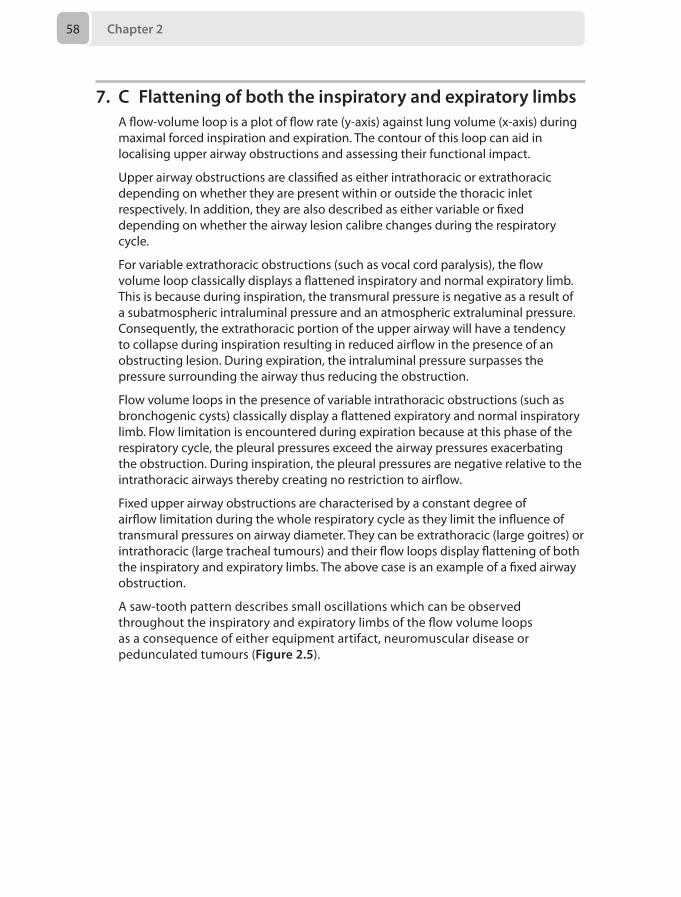
Chapter 258
7. C Flattening of both the inspiratory and expiratory limbsA flow-volume loop is a plot of flow rate (y-axis) against lung volume (x-axis) during maximal forced inspiration and expiration. The contour of this loop can aid in localising upper airway obstructions and assessing their functional impact.
Upper airway obstructions are classified as either intrathoracic or extrathoracic depending on whether they are present within or outside the thoracic inlet respectively. In addition, they are also described as either variable or fixed depending on whether the airway lesion calibre changes during the respiratory cycle.
For variable extrathoracic obstructions (such as vocal cord paralysis), the flow volume loop classically displays a flattened inspiratory and normal expiratory limb. This is because during inspiration, the transmural pressure is negative as a result of a subatmospheric intraluminal pressure and an atmospheric extraluminal pressure. Consequently, the extrathoracic portion of the upper airway will have a tendency to collapse during inspiration resulting in reduced airflow in the presence of an obstructing lesion. During expiration, the intraluminal pressure surpasses the pressure surrounding the airway thus reducing the obstruction.
Flow volume loops in the presence of variable intrathoracic obstructions (such as bronchogenic cysts) classically display a flattened expiratory and normal inspiratory limb. Flow limitation is encountered during expiration because at this phase of the respiratory cycle, the pleural pressures exceed the airway pressures exacerbating the obstruction. During inspiration, the pleural pressures are negative relative to the intrathoracic airways thereby creating no restriction to airflow.
Fixed upper airway obstructions are characterised by a constant degree of airflow limitation during the whole respiratory cycle as they limit the influence of transmural pressures on airway diameter. They can be extrathoracic (large goitres) or intrathoracic (large tracheal tumours) and their flow loops display flattening of both the inspiratory and expiratory limbs. The above case is an example of a fixed airway obstruction.
A saw-tooth pattern describes small oscillations which can be observed throughout the inspiratory and expiratory limbs of the flow volume loops as a consequence of either equipment artifact, neuromuscular disease or pedunculated tumours (Figure 2.5).
Answers 59
Nethercott D, Strang T, Krysiak P. Airway stents: anaesthetic implications. Contin Educ Anaesth Crit Care Pain 2010; 10(2):53–8.Rendleman N. Quinn S. The answer is blowing in the wind: a pedunculated tumour with saw tooth flow-volume loop. J Laryngol Otol 1998; 112(10):973–5.
8. C VasopressinBrainstem herniation causes relative hypotension and bradycardia after an initial period of hypertension due to catecholamine release. This is due to loss of autonomic control of vasomotor tone and loss of vagal tone. Treating episodes of hypotension with adrenergic vasoactive drugs exacerbates end-organ ischaemia and they are contraindicated in a potential organ donor. Vasopressin is the agent of choice to treat hypotension and maintain vascular tone following brainstem herniation as it improves organ perfusion and corrects the polyuric component of diabetes insipidus caused by hypothalamic ischaemia. Fluid boluses are useful to maintain euvolaemia, but over filling patients also risks detrimental outcomes to organs.
Gordon JK. Physiological changes after brain stem death and management of heart-beating donor. Contin Educ Anaesth Crit Care Pain 2012; 12(5):225–29.
Extrathoracic variableobstruction
Intrathoracic variableobstruction
Intrathoracic xedobstruction
Flow
Volume
Normal ow – volume loop
Flow
Volume
Expi
ratio
n(L
/min
)In
spira
tion
(L/m
in)
Expi
ratio
n(L
/min
)In
spira
tion
(L/m
in)
++
++
––
––
––
––
––
––
++
++
++
++
Extrathoracic xedobstruction
––
––
++
++
Figure 2.5 Flow-volume loops with different configurations of extrathoracic and intrathoracic obstructions.
Chapter 260
9. A An arterial blood gas analysisFat embolism indicates the presence of fat within the pulmonary or peripheral circulation, often without symptoms. Fat embolism syndrome is the clinical consequences of fat embolism occurring in some patients and has a quoted mortality of between 5–15%. The commonest implicated fractures are the closed fractures of the long bones and pelvis. The greater the number of fractures, the higher the incidence. A third of patients with bilateral femoral fractures go on to develop the syndrome.
Presentation and diagnosis
The syndrome most often presents at 1–3 days. There are a variety of sets of published diagnostic criteria, but all have in common either an arterial blood gas (ABG) or clinical features of profound type I respiratory failure. Clinically, the two other major tenets are neurological dysfunction, most commonly acute confusion, and the classical petechial rash. The rash may be a late sign and only present in 60% of cases.
Blood indices most often reflect a marked anaemia with thrombocytopenia. Other features such as fat in sputum, urine and alveolar macrophages may support the diagnosis, but equally do not correlate well with the severity of the syndrome.
Pathophysiology
Two main theories exist. In the mechanical theory, physical disruption forces adipose marrow into the venous system and bones with high marrow content are indeed most associated with the syndrome. However, studies have directly demonstrated embolic load in patients during orthopaedic surgery who have not progressed to develop the clinical syndrome. The mechanical theory also does not explain why, despite the fact that embolism is greatest around the time of injury, occurrence of the syndrome peaks at 24–72 hours.
The biochemical theory ascribes toxicity to the hydrolytic degradation of neutral marrow fat to free fatty acids. These free acids are shown to be able to induce acute lung injury in animal models. C-reactive protein may also have a role in abnormal lipid metabolism in these patients.
Treatment
The treatment is essentially supportive with good oxygenation and ventilation in an intensive care environment if indicated. Correction of blood indices may be needed.
Prevention is with operative fixation after early immobilisation, and some operative techniques exist to reduce intraosseous pressure during drilling or reaming to reduce the embolic load.
Some advocate steroid prophylaxis with methylprednisolone for those patients at highest risk, with some evidence to support this. There is no evidence to support the use of steroids as a treatment however. Aspirin may speed recovery of platelets and possibly lung injury.
In this question, the stem highlights the diagnosis of fat embolism syndrome, which most candidates are likely to discern. The real question relates to the most helpful test,
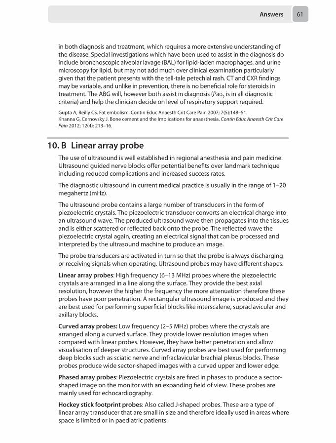
Answers 61
in both diagnosis and treatment, which requires a more extensive understanding of the disease. Special investigations which have been used to assist in the diagnosis do include bronchoscopic alveolar lavage (BAL) for lipid-laden macrophages, and urine microscopy for lipid, but may not add much over clinical examination particularly given that the patient presents with the tell-tale petechial rash. CT and CXR findings may be variable, and unlike in prevention, there is no beneficial role for steroids in treatment. The ABG will, however both assist in diagnosis (Pao2 is in all diagnostic criteria) and help the clinician decide on level of respiratory support required.
Gupta A, Reilly CS. Fat embolism. Contin Educ Anaesth Crit Care Pain 2007; 7(5):148–51.Khanna G, Cernovsky J. Bone cement and the Implications for anaesthesia. Contin Educ Anaesth Crit Care Pain 2012; 12(4): 213–16.
10. B Linear array probeThe use of ultrasound is well established in regional anesthesia and pain medicine. Ultrasound guided nerve blocks offer potential benefits over landmark technique including reduced complications and increased success rates.
The diagnostic ultrasound in current medical practice is usually in the range of 1–20 megahertz (mHz).
The ultrasound probe contains a large number of transducers in the form of piezoelectric crystals. The piezoelectric transducer converts an electrical charge into an ultrasound wave. The produced ultrasound wave then propagates into the tissues and is either scattered or reflected back onto the probe. The reflected wave the piezoelectric crystal again, creating an electrical signal that can be processed and interpreted by the ultrasound machine to produce an image.
The probe transducers are activated in turn so that the probe is always discharging or receiving signals when operating. Ultrasound probes may have different shapes:
Linear array probes: High frequency (6–13 MHz) probes where the piezoelectric crystals are arranged in a line along the surface. They provide the best axial resolution, however the higher the frequency the more attenuation therefore these probes have poor penetration. A rectangular ultrasound image is produced and they are best used for performing superficial blocks like interscalene, supraclavicular and axillary blocks.
Curved array probes: Low frequency (2–5 MHz) probes where the crystals are arranged along a curved surface. They provide lower resolution images when compared with linear probes. However, they have better penetration and allow visualisation of deeper structures. Curved array probes are best used for performing deep blocks such as sciatic nerve and infraclavicular brachial plexus blocks. These probes produce wide sector-shaped images with a curved upper and lower edge.
Phased array probes: Piezoelectric crystals are fired in phases to produce a sector-shaped image on the monitor with an expanding field of view. These probes are mainly used for echocardiography.
Hockey stick footprint probes: Also called J-shaped probes. These are a type of linear array transducer that are small in size and therefore ideally used in areas where space is limited or in paediatric patients.
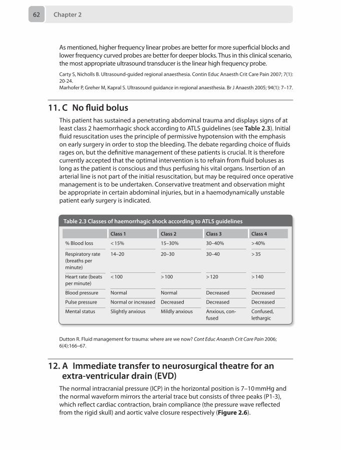
Chapter 262
As mentioned, higher frequency linear probes are better for more superficial blocks and lower frequency curved probes are better for deeper blocks. Thus in this clinical scenario, the most appropriate ultrasound transducer is the linear high frequency probe.
Carty S, Nicholls B. Ultrasound-guided regional anaesthesia. Contin Educ Anaesth Crit Care Pain 2007; 7(1): 20-24.Marhofer P, Greher M, Kapral S. Ultrasound guidance in regional anaesthesia. Br J Anaesth 2005; 94(1): 7–17.
11. C No fluid bolusThis patient has sustained a penetrating abdominal trauma and displays signs of at least class 2 haemorrhagic shock according to ATLS guidelines (see Table 2.3). Initial fluid resuscitation uses the principle of permissive hypotension with the emphasis on early surgery in order to stop the bleeding. The debate regarding choice of fluids rages on, but the definitive management of these patients is crucial. It is therefore currently accepted that the optimal intervention is to refrain from fluid boluses as long as the patient is conscious and thus perfusing his vital organs. Insertion of an arterial line is not part of the initial resuscitation, but may be required once operative management is to be undertaken. Conservative treatment and observation might be appropriate in certain abdominal injuries, but in a haemodynamically unstable patient early surgery is indicated.
Table 2.3 Classes of haemorrhagic shock according to ATLS guidelines
Class 1 Class 2 Class 3 Class 4
% Blood loss < 15% 15–30% 30–40% > 40%
Respiratory rate (breaths per minute)
14–20 20–30 30–40 > 35
Heart rate (beats per minute)
< 100 > 100 > 120 > 140
Blood pressure Normal Normal Decreased Decreased
Pulse pressure Normal or increased Decreased Decreased Decreased
Mental status Slightly anxious Mildly anxious Anxious, con-fused
Confused, lethargic
Dutton R. Fluid management for trauma: where are we now? Cont Educ Anaesth Crit Care Pain 2006; 6(4):166–67.
12. A Immediate transfer to neurosurgical theatre for an extra-ventricular drain (EVD)
The normal intracranial pressure (ICP) in the horizontal position is 7–10 mmHg and the normal waveform mirrors the arterial trace but consists of three peaks (P1-3), which reflect cardiac contraction, brain compliance (the pressure wave reflected from the rigid skull) and aortic valve closure respectively (Figure 2.6).
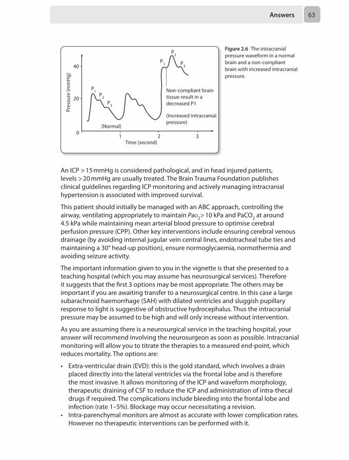
Answers 63
An ICP > 15 mmHg is considered pathological, and in head injured patients, levels > 20 mmHg are usually treated. The Brain Trauma Foundation publishes clinical guidelines regarding ICP monitoring and actively managing intracranial hypertension is associated with improved survival.
This patient should initially be managed with an ABC approach, controlling the airway, ventilating appropriately to maintain Pao2> 10 kPa and PaCo2 at around 4.5 kPa while maintaining mean arterial blood pressure to optimise cerebral perfusion pressure (CPP). Other key interventions include ensuring cerebral venous drainage (by avoiding internal jugular vein central lines, endotracheal tube ties and maintaining a 30° head-up position), ensure normoglycaemia, normothermia and avoiding seizure activity.
The important information given to you in the vignette is that she presented to a teaching hospital (which you may assume has neurosurgical services). Therefore it suggests that the first 3 options may be most appropriate. The others may be important if you are awaiting transfer to a neurosurgical centre. In this case a large subarachnoid haemorrhage (SAH) with dilated ventricles and sluggish pupillary response to light is suggestive of obstructive hydrocephalus. Thus the intracranial pressure may be assumed to be high and will only increase without intervention.
As you are assuming there is a neurosurgical service in the teaching hospital, your answer will recommend involving the neurosurgeon as soon as possible. Intracranial monitoring will allow you to titrate the therapies to a measured end-point, which reduces mortality. The options are:
• Extra-ventricular drain (EVD): this is the gold standard, which involves a drain placed directly into the lateral ventricles via the frontal lobe and is therefore the most invasive. It allows monitoring of the ICP and waveform morphology, therapeutic draining of CSF to reduce the ICP and administration of intra-thecal drugs if required. The complications include bleeding into the frontal lobe and infection (rate 1–5%). Blockage may occur necessitating a revision.
• Intra-parenchymal monitors are almost as accurate with lower complication rates. However no therapeutic interventions can be performed with it.
Time (second)
(Normal)
Pres
sure
(mm
Hg)
40
(Increased intracranialpressure)
Non-compliant braintissue result in adecreased P1
20
01 2
P1
P1
P2
P2
P3
P3
3
Figure 2.6 The intracranial pressure waveform in a normal brain and a non-compliant brain with increased intracranial pressure.
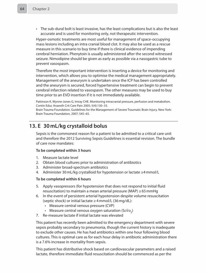
Chapter 264
• The sub-dural bolt is least invasive, has the least complications but is also the least accurate and is used for monitoring only, not therapeutic intervention.
Hyper-osmotic treatments are most useful for management of space-occupying mass lesions including an intra cranial blood clot. It may also be used as a rescue measure in this scenario to buy time if there is clinical evidence of impending cerebral herniation. Phenytoin is usually administered after the second witnessed seizure. Nimodipine should be given as early as possible via a nasogastric tube to prevent vasospasm.
Therefore the most important intervention is inserting a device for monitoring and intervention, which allows you to optimise the medical management appropriately. Management of the aneurysm is undertaken once the ICP has been controlled and the aneurysm is secured, forced hypertensive treatment can begin to prevent cerebral infarction related to vasospasm. The other measures may be used to buy time prior to an EVD insertion if it is not immediately available.
Pattinson K, Wynne-Jones G, Imray CHE. Monitoring intracranial pressure, perfusion and metabolism. Contin Educ Anaesth Crit Care Pain 2005; 5(4):130–33.Brain Trauma Foundation. Guidelines for the Management of Severe Traumatic Brain Injury. New York: Brain Trauma Foundation, 2007; S45–65.
13. E 30 mL/kg crystalloid bolusSepsis is the commonest reason for a patient to be admitted to a critical care unit and therefore the 2012 Surviving Sepsis Guidelines is essential revision. The bundle of care now mandates:
To be completed within 3 hours
1. Measure lactate level2. Obtain blood cultures prior to administration of antibiotics3. Administer broad-spectrum antibiotics4. Administer 30 mL/kg crystalloid for hypotension or lactate ≥4 mmol/L
To be completed within 6 hours
5. Apply vasopressors (for hypotension that does not respond to initial fluid resuscitation) to maintain a mean arterial pressure (MAP) ≥ 65 mmHg
6. In the event of persistent arterial hypotension despite volume resuscitation (septic shock) or initial lactate ≥ 4 mmol/L (36 mg/dL):• Measure central venous pressure (CVP)• Measure central venous oxygen saturation (Scv–o2)
7. Re-measure lactate if initial lactate was elevated
This patient has recently been admitted to the emergency department with severe sepsis probably secondary to pneumonia, though the current history is inadequate to exclude other causes. He has had antibiotics within one hour following blood cultures. This is optimal care as for each hour delay in antibiotic administration there is a 7.6% increase in mortality from sepsis.
This patient has distributive shock based on cardiovascular parameters and a raised lactate, therefore immediate fluid resuscitation should be commenced as per the
Answers 65
Surviving Sepsis Guidelines above. 30 mL/kg should be given in the next two hours. If hypotension persists despite this fluid resuscitation, then noradrenaline should be considered titrated to Scv–o2, MAP and CVP. The lactate level should be re-evaluated.
This patient has evidence of a raised alveolar-arterial gradient representing a probable diffusion defect and ventilation/perfusion mismatch with shunting. However clinically his predominant problem is hypoperfusion and shock so should be fluid resuscitated in the first instance. Currently he does not have an indication for non-invasive ventilation (NIV) or intubation and ventilation.
Dellinger RP, Levy MM, Rhodes A, et al. Surviving sepsis campaign: International guidelines for management of severe sepsis and septic shock. Crit Care Med 2013;41:580–637.
14. B Increase the insulin infusion by 1 unit/hour if the bicarbonate concentration remains 3 mmol/L after 1 hour
Diabetic ketoacidosis (DKA) is an endocrine emergency that presents in diabetic patients who do not produce endogenous insulin. Relative or absolute lack of insulin (endogenous or exogenous) results in a triad of hyperglycaemia, a ketotic state and acidaemia. Eleven percent of Type 1 diabetics presented with DKA in 2004–2009. Mortality is still 2%, most commonly attributable to cerebral oedema.
Glucose forces renal diuresis and results in profound hypovolaemia. The patient presents with a metabolic acidosis (often partially compensated by a monumental minute ventilation) with a large anion gap due to unmeasured blood ketones. Blood ketones can now be measured at the bedside and indicate severity of disease and response to treatment. The patient is often potassium-deplete which may not be immediately obvious from initial lab results as a high level of acidosis causes intracellular potassium ions to migrate into the plasma. As acidosis resolves and potassium once again returns to the intracellular space, the plasma potassium concentration decreases rapidly.
Management of DKA in critical care areas focuses on the following issues:
• Fluid management including resuscitation and replacement of ongoing losses• Blood glucose control and providing glucose once the blood glucose drops below
15 mmol/L• Acidosis and electrolyte management (in particular potassium maintained
between 4 and 5 mmol/L)• Insulin administration and titration• Septic screening and appropriate antibiotics• Thromboprophylaxis• Monitoring for complications including cerebral oedema, sepsis, hypokalaemia,
ischaemic cardiac events, acute kidney injury (AKI) and acute respiratory distress syndrome
• Management of psycho-social issues• Involving of specialist teams for ongoing managementThe management of DKA has undergone a recent change in 2011 and guidelines have been published by Diabetes UK. The sliding scale is no longer incorporated and titrating insulin to biochemical markers is the primary objective. To summarise some of the new recommendations:
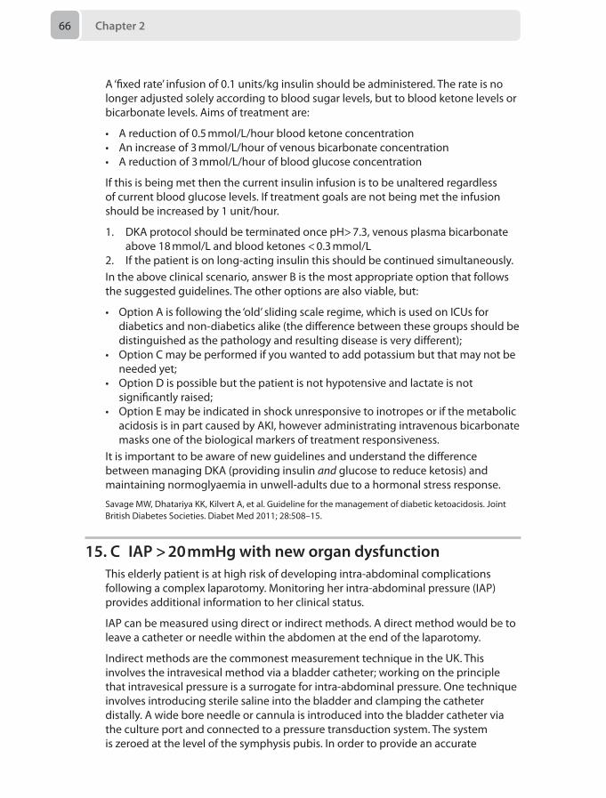
Chapter 266
A ‘fixed rate’ infusion of 0.1 units/kg insulin should be administered. The rate is no longer adjusted solely according to blood sugar levels, but to blood ketone levels or bicarbonate levels. Aims of treatment are:
• A reduction of 0.5 mmol/L/hour blood ketone concentration• An increase of 3 mmol/L/hour of venous bicarbonate concentration• A reduction of 3 mmol/L/hour of blood glucose concentration
If this is being met then the current insulin infusion is to be unaltered regardless of current blood glucose levels. If treatment goals are not being met the infusion should be increased by 1 unit/hour.
1. DKA protocol should be terminated once pH> 7.3, venous plasma bicarbonate above 18 mmol/L and blood ketones < 0.3 mmol/L
2. If the patient is on long-acting insulin this should be continued simultaneously.In the above clinical scenario, answer B is the most appropriate option that follows the suggested guidelines. The other options are also viable, but:
• Option A is following the ‘old’ sliding scale regime, which is used on ICUs for diabetics and non-diabetics alike (the difference between these groups should be distinguished as the pathology and resulting disease is very different);
• Option C may be performed if you wanted to add potassium but that may not be needed yet;
• Option D is possible but the patient is not hypotensive and lactate is not significantly raised;
• Option E may be indicated in shock unresponsive to inotropes or if the metabolic acidosis is in part caused by AKI, however administrating intravenous bicarbonate masks one of the biological markers of treatment responsiveness.
It is important to be aware of new guidelines and understand the difference between managing DKA (providing insulin and glucose to reduce ketosis) and maintaining normoglyaemia in unwell-adults due to a hormonal stress response.
Savage MW, Dhatariya KK, Kilvert A, et al. Guideline for the management of diabetic ketoacidosis. Joint British Diabetes Societies. Diabet Med 2011; 28:508–15.
15. C IAP > 20 mmHg with new organ dysfunctionThis elderly patient is at high risk of developing intra-abdominal complications following a complex laparotomy. Monitoring her intra-abdominal pressure (IAP) provides additional information to her clinical status.
IAP can be measured using direct or indirect methods. A direct method would be to leave a catheter or needle within the abdomen at the end of the laparotomy.
Indirect methods are the commonest measurement technique in the UK. This involves the intravesical method via a bladder catheter; working on the principle that intravesical pressure is a surrogate for intra-abdominal pressure. One technique involves introducing sterile saline into the bladder and clamping the catheter distally. A wide bore needle or cannula is introduced into the bladder catheter via the culture port and connected to a pressure transduction system. The system is zeroed at the level of the symphysis pubis. In order to provide an accurate
Answers 67
measurement, the reading should be taken in the supine patient at the end of expiration. The measurement should be repeated four hourly.
Normal IAP is 5–7 mmHg. The World Society of the Abdominal Compartment Syndrome (WSACS) classifies intra-abdominal hypertension as a sustained IAP > 12 mmHg. Abdominal compartment syndrome is a sustained IAP > 20 mmHg with new organ dysfunction, hence making option C correct.
Risk factors can be subdivided into:
• Intraluminal such as gastroparesis, ileus or pseudo-obstruction• Extraluminal such as ascites or pelvic tumours• Decreased abdominal wall compliance such as abdominal surgery with fascial
closure, obesity, abdominal burns or trauma• Capillary leak or excessive fluid resuscitation such as pancreatitis, sepsis or
massive transfusionThis patient is at high risk due to the risk of ileus, risk of capillary leak from a systemic inflammatory response syndrome and reduced abdominal wall compliance following a laparotomy.
The principle of management is to optimise abdominal perfusion pressure. The strategies used broadly fall into medical and surgical categories.
Medical management involves:
• IAP monitoring if risk factors are present• Draining abdominal fluid collections such as ascites or abscesses• Reducing intraluminal contents; nasogastric and rectal decompression and the
cautious use of prokinetics• Improving abdominal wall compliance such as sedation and analgesia with
possible neuromuscular blockade and removal of constrictive dressings or escharotomy
• Maintain oxygen delivery with the use of fluid challenges and cardiovascular support. Excessive fluid administration should be avoided and diuretics may have a role
The main surgical option is decompression via a laparostomy with delayed closure. Close liaison with the surgical team is mandated in the management of high-risk patients, such as this case, and thresholds ascertained which alert the teams to consider a surgical option.
Berry N, Fletcher S. Abdominal compartment syndrome. Contin Educ Anaesth Crit Care Pain 2012; 12(3):110–117.
16. D Responding to voice on the AVPU scoreAsthma is characterised by reversible airways obstruction due to bronchial smooth muscle contraction, airway inflammation and increased airway secretion production. Chronic management focuses on a step-wise approach to therapy escalation aiming for minimal requirement for rescue therapy. Around 1400 people a year still die from asthma despite improvements in chronic management.
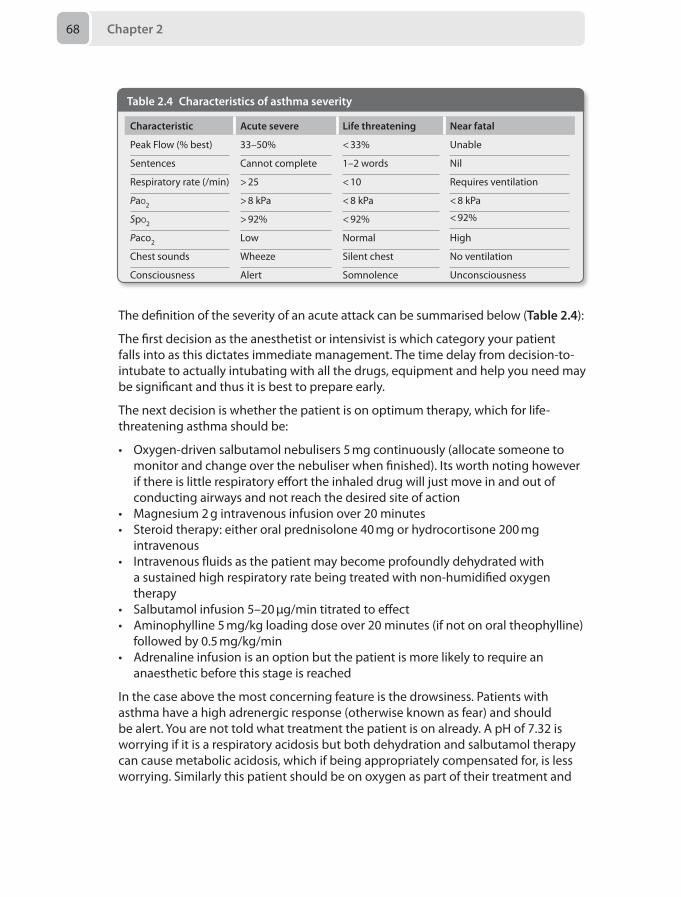
Chapter 268
The definition of the severity of an acute attack can be summarised below (Table 2.4):
The first decision as the anesthetist or intensivist is which category your patient falls into as this dictates immediate management. The time delay from decision-to-intubate to actually intubating with all the drugs, equipment and help you need may be significant and thus it is best to prepare early.
The next decision is whether the patient is on optimum therapy, which for life-threatening asthma should be:
• Oxygen-driven salbutamol nebulisers 5 mg continuously (allocate someone to monitor and change over the nebuliser when finished). Its worth noting however if there is little respiratory effort the inhaled drug will just move in and out of conducting airways and not reach the desired site of action
• Magnesium 2 g intravenous infusion over 20 minutes• Steroid therapy: either oral prednisolone 40 mg or hydrocortisone 200 mg
intravenous• Intravenous fluids as the patient may become profoundly dehydrated with
a sustained high respiratory rate being treated with non-humidified oxygen therapy
• Salbutamol infusion 5–20 μg/min titrated to effect• Aminophylline 5 mg/kg loading dose over 20 minutes (if not on oral theophylline)
followed by 0.5 mg/kg/min• Adrenaline infusion is an option but the patient is more likely to require an
anaesthetic before this stage is reached
In the case above the most concerning feature is the drowsiness. Patients with asthma have a high adrenergic response (otherwise known as fear) and should be alert. You are not told what treatment the patient is on already. A pH of 7.32 is worrying if it is a respiratory acidosis but both dehydration and salbutamol therapy can cause metabolic acidosis, which if being appropriately compensated for, is less worrying. Similarly this patient should be on oxygen as part of their treatment and
Table 2.4 Characteristics of asthma severity
Characteristic Acute severe Life threatening Near fatal
Peak Flow (% best) 33–50% < 33% Unable
Sentences Cannot complete 1–2 words Nil
Respiratory rate (/min) > 25 < 10 Requires ventilation
Pao2 > 8 kPa < 8 kPa < 8 kPa
< 92%Spo2 > 92% < 92%
Paco2 Low Normal High
Chest sounds Wheeze Silent chest No ventilation
Consciousness Alert Somnolence Unconsciousness
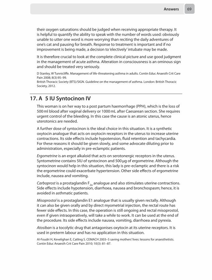
Answers 69
their oxygen saturations should be judged when receiving appropriate therapy. It is helpful to quantify the ability to speak with the number of words used: obviously unable to utter one word is more worrying than reciting the daily adventures of one’s cat and pausing for breath. Response to treatment is important and if no improvement is being made, a decision to ‘electively’ intubate may be made.
It is therefore crucial to look at the complete clinical picture and use good judgment in the management of acute asthma. Alteration in consciousness is an ominous sign and should be treated very seriously.
D Stanley, W Tunnicliffe. Management of life-threatening asthma in adults. Contin Educ Anaesth Crit Care Pain 2008; 8(3):95–99.British Thoracic Society (BTS)/SIGN. Guideline on the management of asthma. London: British Thoracic Society, 2012.
17. A 5 IU Syntocinon IVThis woman is on her way to a post partum haemorrhage (PPH), which is the loss of 500 ml blood after vaginal delivery or 1000 mL after Caesarean section. She requires urgent control of the bleeding. In this case the cause is an atonic uterus, hence uterotonics are needed.
A further dose of syntocinon is the ideal choice in this situation. It is a synthetic oxytocin analogue that acts on oxytocin receptors in the uterus to increase uterine contractions. Its side effects include hypotension, fluid retention and tachycardia. For these reasons it should be given slowly, and some advocate diluting prior to administration, especially in pre-eclamptic patients.
Ergometrine is an ergot alkaloid that acts on serotonergic receptors in the uterus. Syntometrine contains 5IU of syntocinon and 500 µg of ergometrine. Although the syntocinon would help in this situation, this lady is pre-eclamptic and there is a risk the ergometrine could exacerbate hypertension. Other side effects of ergometrine include, nausea and vomiting.
Carboprost is a prostaglandin F2α analogue and also stimulates uterine contractions. Side effects include hypotension, diarrhoea, nausea and bronchospasm; hence, it is avoided in asthmatic patients.
Misoprostol is a prostaglandin E1 analogue that is usually given rectally. Although it can also be given orally and by direct myometrial injection, the rectal route has fewer side effects. In this case, the operation is still ongoing and rectal misoprostol, even if given intraoperatively, will take a while to work. It can be used at the end of the procedure. Its side effects include nausea, vomiting, diarrhoea and pyrexia.
Atosiban is a tocolytic drug that antagonises oxytocin at its uterine receptors. It is used in preterm labour and has no application in this situation.
Al-Foudri H, Kevelighan E, Catling S. CEMACH 2003–5 saving mothers’ lives: lessons for anaesthetists. Contin Educ Anaesth Crit Care Pain 2010; 10(3): 81–87.
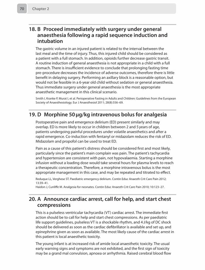
Chapter 270
18. B Proceed immediately with surgery under general anaesthesia following a rapid sequence induction and intubation
The gastric volume in an injured patient is related to the interval between the last meal and the time of injury. Thus, this injured child should be considered as a patient with a full stomach. In addition, opioids further decrease gastric transit. A routine induction of general anaesthesia is not appropriate in a child with a full stomach. There is insufficient evidence to conclude that prolonging fasting time pre-procedure decreases the incidence of adverse outcomes, therefore there is little benefit in delaying surgery. Performing an axillary block is a reasonable option, but would not be feasible in a 6-year old child without sedation or general anaesthesia. Thus immediate surgery under general anaesthesia is the most appropriate anaesthetic management in this clinical scenario.
Smith I, Kranke P, Murat I, et al. Perioperative Fasting in Adults and Children: Guidelines from the European Society of Anaesthesiology. Eur J Anaesthesiol 2011; 28(8):556–69.
19. D Morphine 50 μg/kg intravenous bolus for analgesiaPostoperative pain and emergence delirium (ED) present similarly and may overlap. ED is more likely to occur in children between 2 and 5 years of age, patients undergoing painful procedures under volatile anaesthetics and after a rapid emergence. Co-induction with fentanyl or midazolam reduces the risk of ED. Midazolam and propofol can be used to treat ED.
Pain as a cause of this patient’s distress should be considered first and most likely, particularly since the patient’s main complain was pain. The patient’s tachycardia and hypertension are consistent with pain, not hypovalaemia. Starting a morphine infusion without a loading dose would take several hours for plasma levels to reach a therapeutic concentration. Therefore, a morphine intravenous bolus is the most appropriate management in this case, and may be repeated and titrated to effect.
Reduque LL, Verghese ST. Paediatric emergency delirium. Contin Educ Anaesth Crit Care Pain 2012; 13:39–41. Haidon J, Cunliffe M. Analgesia for neonates. Contin Educ Anaesth Crit Care Pain 2010; 10:123–27.
20. A Announce cardiac arrest, call for help, and start chest compressions
This is a pulseless ventricular tachycardia (VT) cardiac arrest. The immediate first action should be to call for help and start chest compressions. As per paediatric life support guidelines, pulseless VT is a shockable rhythm, and 4 J/kg of DC shock should be delivered as soon as the cardiac defibrillator is available and set up, and epinephrine given as soon as available. The most likely cause of the cardiac arrest in this patient is local anaesthetic toxicity.
The young infant is at increased risk of amide local anaesthetic toxicity. The usual early warning signs and symptoms are not exhibited, and the first sign of toxicity may be a grand mal convulsion, apnoea or arrhythmia. Raised cerebral blood flow
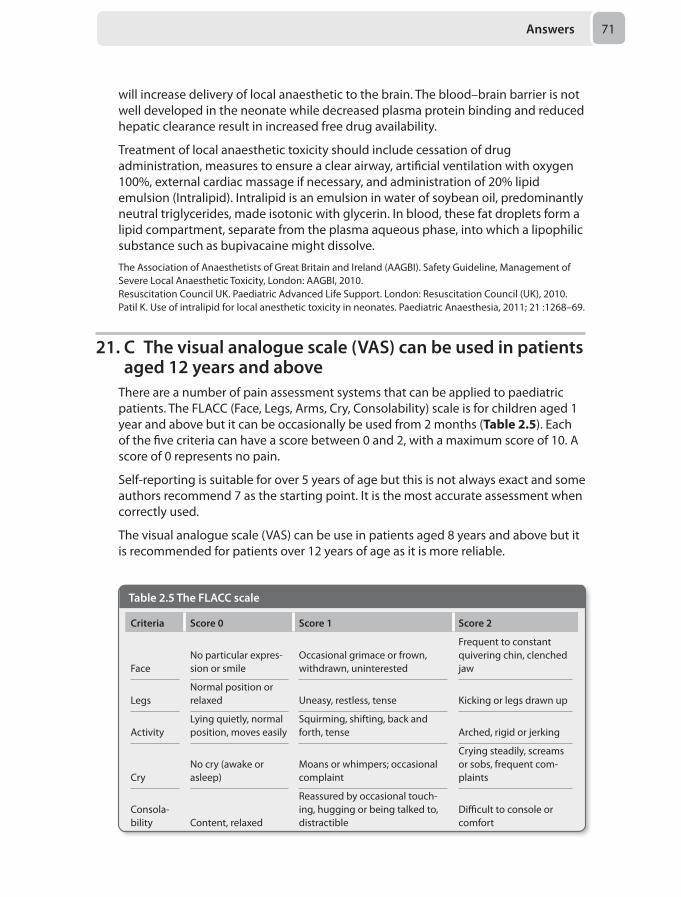
Answers 71
will increase delivery of local anaesthetic to the brain. The blood–brain barrier is not well developed in the neonate while decreased plasma protein binding and reduced hepatic clearance result in increased free drug availability.
Treatment of local anaesthetic toxicity should include cessation of drug administration, measures to ensure a clear airway, artificial ventilation with oxygen 100%, external cardiac massage if necessary, and administration of 20% lipid emulsion (Intralipid). Intralipid is an emulsion in water of soybean oil, predominantly neutral triglycerides, made isotonic with glycerin. In blood, these fat droplets form a lipid compartment, separate from the plasma aqueous phase, into which a lipophilic substance such as bupivacaine might dissolve.
The Association of Anaesthetists of Great Britain and Ireland (AAGBI). Safety Guideline, Management of Severe Local Anaesthetic Toxicity, London: AAGBI, 2010.Resuscitation Council UK. Paediatric Advanced Life Support. London: Resuscitation Council (UK), 2010.Patil K. Use of intralipid for local anesthetic toxicity in neonates. Paediatric Anaesthesia, 2011; 21 :1268–69.
21. C The visual analogue scale (VAS) can be used in patients aged 12 years and above
There are a number of pain assessment systems that can be applied to paediatric patients. The FLACC (Face, Legs, Arms, Cry, Consolability) scale is for children aged 1 year and above but it can be occasionally be used from 2 months (Table 2.5). Each of the five criteria can have a score between 0 and 2, with a maximum score of 10. A score of 0 represents no pain.
Self-reporting is suitable for over 5 years of age but this is not always exact and some authors recommend 7 as the starting point. It is the most accurate assessment when correctly used.
The visual analogue scale (VAS) can be use in patients aged 8 years and above but it is recommended for patients over 12 years of age as it is more reliable.
Table 2.5 The FLACC scale
Criteria Score 0 Score 1 Score 2
FaceNo particular expres-sion or smile
Occasional grimace or frown, withdrawn, uninterested
Frequent to constant quivering chin, clenched jaw
LegsNormal position or relaxed Uneasy, restless, tense Kicking or legs drawn up
ActivityLying quietly, normal position, moves easily
Squirming, shifting, back and forth, tense Arched, rigid or jerking
CryNo cry (awake or asleep)
Moans or whimpers; occasional complaint
Crying steadily, screams or sobs, frequent com-plaints
Consola-bility Content, relaxed
Reassured by occasional touch-ing, hugging or being talked to, distractible
Difficult to console or comfort
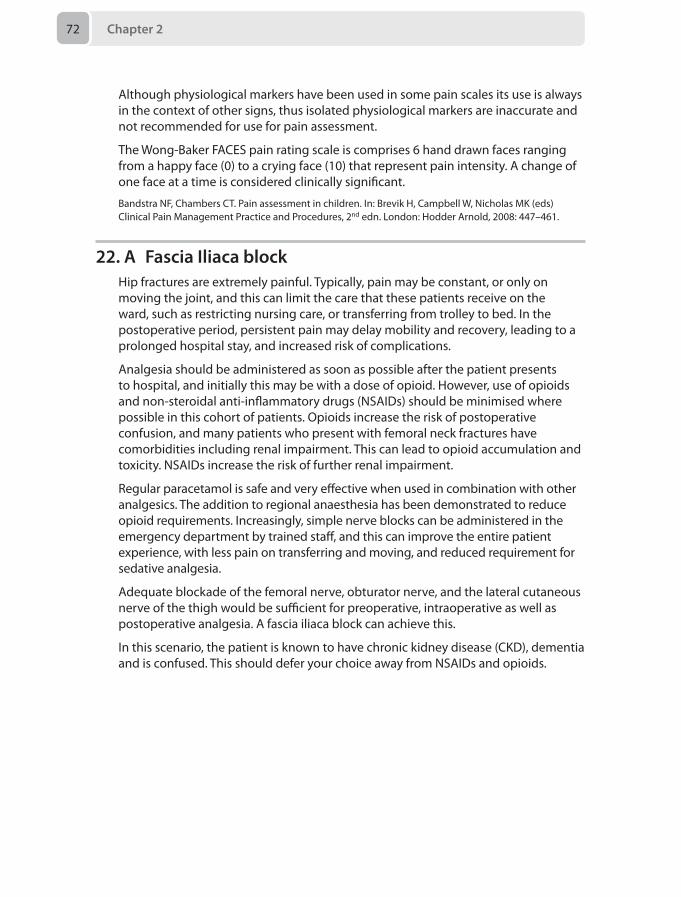
Chapter 272
Although physiological markers have been used in some pain scales its use is always in the context of other signs, thus isolated physiological markers are inaccurate and not recommended for use for pain assessment.
The Wong-Baker FACES pain rating scale is comprises 6 hand drawn faces ranging from a happy face (0) to a crying face (10) that represent pain intensity. A change of one face at a time is considered clinically significant.
Bandstra NF, Chambers CT. Pain assessment in children. In: Brevik H, Campbell W, Nicholas MK (eds) Clinical Pain Management Practice and Procedures, 2nd edn. London: Hodder Arnold, 2008: 447–461.
22. A Fascia Iliaca blockHip fractures are extremely painful. Typically, pain may be constant, or only on moving the joint, and this can limit the care that these patients receive on the ward, such as restricting nursing care, or transferring from trolley to bed. In the postoperative period, persistent pain may delay mobility and recovery, leading to a prolonged hospital stay, and increased risk of complications.
Analgesia should be administered as soon as possible after the patient presents to hospital, and initially this may be with a dose of opioid. However, use of opioids and non-steroidal anti-inflammatory drugs (NSAIDs) should be minimised where possible in this cohort of patients. Opioids increase the risk of postoperative confusion, and many patients who present with femoral neck fractures have comorbidities including renal impairment. This can lead to opioid accumulation and toxicity. NSAIDs increase the risk of further renal impairment.
Regular paracetamol is safe and very effective when used in combination with other analgesics. The addition to regional anaesthesia has been demonstrated to reduce opioid requirements. Increasingly, simple nerve blocks can be administered in the emergency department by trained staff, and this can improve the entire patient experience, with less pain on transferring and moving, and reduced requirement for sedative analgesia.
Adequate blockade of the femoral nerve, obturator nerve, and the lateral cutaneous nerve of the thigh would be sufficient for preoperative, intraoperative as well as postoperative analgesia. A fascia iliaca block can achieve this.
In this scenario, the patient is known to have chronic kidney disease (CKD), dementia and is confused. This should defer your choice away from NSAIDs and opioids.
Answers 73
The choice between the blocks should lead you to choose the fascia iliac block. A femoral nerve block would not anaesthetise the lateral cutaneous nerve of the thigh, which supplies sensation to the incision site. Additionally, a spinal anaesthetic has not been shown to provide longer lasting analgesia than a peripheral nerve block. Many would therefore advocate performing a spinal anaesthetic for intraoperative management, as well as a fascia iliaca block for postoperative analgesia.
Maxwell L, White S. Anaesthetic management of patients with hip fractures: an update. Cont Educ Anaesth Crit Care Pain 2013; 13(5):179–183.Scottish Intercollegiate Guidelines Network (SIGN). Management of hip fracture in older people. National CG No. 111. Edinburgh: Scottish Intercollegiate Guidelines Network, 2009.
23. D Gabapentin‘Shingles’, also known as herpes zoster, is caused by reactivation of the varicella zoster virus (VZV), a virus in the same family as the herpes simplex virus (Alphaherpesvirinae). Herpes zoster and herpes simplex are, however, independent diseases. After causing its primary infection (chickenpox) the VZV lies dormant in a sensory ganglion, until it is given such conditions when it can re-emerge. The clinical manifestation of this re-emergence is called shingles. The most common cause is immunosuppression, which may be due to malignancy, acquired syndromes including HIV, or immunosuppressive medication such as steroids.
On reactivation, VZV causes pain and a characteristic rash, limited to the dermatomal distribution of the dorsal root ganglion in which it lies. In this scenario the dorsal root ganglion affected is T5. Some patients may describe a painful prodrome, followed by development of skin lesions. The associated pain can be intense, severe and neuropathic in character. Classically described as throbbing or burning, it is worse as the day progresses and maximal at night. Associated sleep disturbances are common, and it may subsequently lead to depression, fatigue and concentration difficulties. The commonest site of pain is in the thoracic dermatomes, but it may also affect the ophthalmic division of the trigeminal nerve, making it a key differential diagnosis for trigeminal neuralgia.
In the majority of patients, pain is acute, lasting less than 30 days, or subacute, lasting less than 120 days. In some patients, however, it lasts more than 3 months after skin lesions have healed, and this is described as post-herpetic neuralgia (PHN).
PHN may last several years and can be severe and debilitating. Risk factors include increasing age, female gender, presence of a prodrome, more severe pain in the acute phase, and a more intense rash, as well as psychosocial factors such as higher anxiety levels.
Management of PHN can be challenging. Patients should be well educated about their condition, and given appropriate support and counselling. Psychological therapies such as cognitive behavioural therapy may be of benefit.
In terms of pharmacological management, recommended first line agents include gabapentinoids (gabapentin, pregabalin) or tricyclic antidepressants (amitriptyline, nortriptyline). Lignocaine patches have been used and are recommended but robust evidence for their benefit is lacking. Opioids help reduce severity of the pain but
Chapter 274
long-term use is associated with several unwanted effects, and topical capsaicin has been used successfully for PHN, but may be painful to apply.
In this scenario, each of the options is possible, but the first line agent, and hence the most appropriate next step, would be gabapentin. Subsequently, other options can be explored, depending on the patient’s response.
Gupta R, Smith PF. Post-herpetic neuralgia. Cont Educ Anaesth Crit Care Pain 2012; 12(4):181–85.
24. C Hyperalgesia may develop with increasing dosageOpioid-induced hyperalgesia occurs when pain is uncontrolled despite increasing use of long-term oioids.
The use of opioids has commonly been described without a ceiling in cancer pain and palliative care, but in this clinical picture is inappropriate. The use of opioids in non-cancer pain is becoming recognised as ineffective and potentially harmful due to its suppression of the immune and hormonal systems. Opioids can act directly on the hypothalamo-pituitary axis to reduce gonadotrophin releasing hormone (GnRH), luteinizing hormone (LH) as well as gonadal testosterone. Opioids also reduce adrenocorticotrophic hormone (ACTH) and cortisol with long-term use.
Respiratory depression is not observed at stable doses of opioids administered long-term.
The constipating effects of opioids are long-term and do not resolve unless the opioids are withdrawn.
Seyfreid O, Hester J. Opioids and endocrine dysfunction. Br J Pain 2012; 6(1):17–24.
25. C Intravenous morphineThe expression ‘porphyria’ encompasses a collection of inherited disorders of haem synthesis. Haem is a key constituent of haemoglobin and an example of a naturally occurring porphyrin. Its synthetic pathway consists of a series of enzyme-dependent steps. Genetic mutation causing a deficit of a particular enzyme in this pathway results in accumulation of the intermediate substrates that rely on it for their metabolism.
The primary step in the haem synthetic pathway is the combination of glycine and succinyl CoA to form the compound 5-aminolevulinic acid (5-ALA) by the action of ALA synthase. Deficiency of any enzyme downstream from this step will cause the accumulation of 5-ALA. In all acute porphyrias, 5-ALA levels are elevated. Whether this itself is the cause of the clinical manifestations of acute porphyria is unclear.
In any form of acute porphyria, an acute neurovisceral crisis may be triggered by anything that increases the demand for haem, including infection, dehydration, starvation and drugs. The key presenting features are abdominal pain, neurological symptoms and tachycardia. As these are rather non-specific presentations, diagnosis is often made late. In this scenario, it is not a surgical condition causing the patient’s pain, but this may additionally trigger a porphyric crisis.
Answers 75
A number of drugs used in common anaesthetic practice may be unsafe for use in patients with porphyria. The majority are, in fact, safe, and the Table 2.6 below summarises drugs that are either known to be dangerous or those which are yet unclassified for use in porphyric patients.
Morphine is a safe, effective, titratable drug that can be used in porphyria, and would be the drug of choice here. The patient is tachycardic with small bowel obstruction, and likely to be hypovolaemic. This would preclude the use of an epidural. Levobupivacaine has indeterminate safety profile so should be avoided and relying on local infiltration of the surgical wound would not provide lasting analgesia. Oxydone and ketamine are unsafe.
Findley H, Philips A, Cole D, Nair A. Porphyrias: implications for anaesthesia, critical care, and pain medicine. Contin Educ Anaesth Crit Care Pain 2011; 12(3): 128–133.
26. B Trendelenburg positioningDuring surgery for penetrating eye injury, control of intraocular pressure (IOP) is important to prevent expulsion of the contents of the orbit. Episcleral venous pressure, which is influenced by central venous pressure (CVP), is the most important factor determining IOP acutely; hence measures to prevent venous congestion must be employed. Such measures include taping, not tying, the endotracheal tube, keeping the head in the neutral position, reverse Trendelenburg positioning and avoiding coughing by ensuring adequate muscle relaxation. Hypercarbia can lead to an increase in IOP; hence increasing the minute ventilation will reduce CO2 and help to reduce IOP.
Both acetazolamide and mannitol can be used to reduce IOP in the acute setting. Acetazolamide is a carbonic anhydrase inhibitor that reduces the production of aqueous humour, while mannitol dehydrates the eye by its action as an osmotic diuretic. Both have systemic effects that must be taken into account when administered, such as electrolyte disturbance and dehydration.
Trendelenburg positioning is the correct answer, as the head down position will reduce venous drainage from the orbit and cause an increase in CVP and therefore the IOP.
Murgatroyd H, Bembridge J. Intraocular pressure. Contin Educ Anaesth Crit Care Pain 2008; 8(3):100–103.
Table 2.6 Safety of different drug classes for use in porphyria.
Class of drug Unsafe Unclassified
Intravenous induction agents Thipentone ketamine
Inhalational anaesthetic agents Sevoflurane
Local anaesthetic agents Levobupivacaine ropivacaine
Analgesics Oxycodone diclofenac Mefanamic acid pentazocine
Vasopressors Ephedrine Metaraminol vasopressin
Antibiotics Erythromycin rifampicin
Chapter 276
27. B Request cryoprecipitate and platelets from the blood bank
Cell salvage is the process of utilising a patients own blood retrieved from the surgical field to produce autologously transfused blood by filtering and washing it. Cell salvage therefore returns red cells and reduces the requirement for allogenic transfusions, with the following indications:
• Expected blood loss of > 20% blood volume or > 1 litre• Operations with an expected blood transfusion requirement• Pro-haemorrhagic states• Preoperative anaemia• Rare blood groups• Patients with rare blood antibodies• Patient refusal for allogenic blood transfusionAs cell salvage only returns red blood cells, other blood products such as plasma, inflammatory mediators and, importantly platelets and clotting factors, are therefore discarded. Patients receiving more than 2 litres of autologous red cells are at a significant risk of developing coagulopathy.
In this clinical scenario, the patient is likely to require coagulopathic support, due to anticipated further bleeding, in the form of platelets and cryoprecipitate.
Tranexamic acid is useful in reducing on-going bleeding as an antifibrinolytic, but it will not prevent the massive blood loss coagulopathy expected. Hypotensive anaesthesia is not a recommended anaesthetic technique in vascular surgery and carries a significant hazard of complications in high-risk patients undergoing vascular intervention. Point of care measurement of activated clotting time (ACT) guides intraoperative heparin therapy, but will not assist in the treatment of massive haemorrhage coagulopathy. Finally, waiting for laboratory blood results will merely delay the required intervention of administering clotting products in a timely manner. Thus, requesting clotting products is the most appropriate step at this point as it prepares the anaesthetist for the ensuing coagulopathy.
Kuppurao L, Wee M. Perioperative cell salvage. Contin Educ Anaesth Crit Care Pain 2010; 10 (4):104–108.
28. D Left coronary arterySudden occlusion of major coronary arteries is the most common cause of myocardial infarction (MI). The area of myocardial ischaemia depends on the coronary circulation. Early recognition of acute coronary obstruction and quick reperfusion is essential for good outcome after MI.
A good anatomical knowledge is essential for successful MI management (Figure 2.7). The blood to the heart is mainly supplied by two main coronary arteries (CAs), the right coronary artery (RCA) and the left coronary artery (LCA).
The RCA arises form the right anterior aortic sinus. It mainly supplies blood to the right side of the heart. However, the RCA also gives twigs to the right atrium and left ventricle (LV).
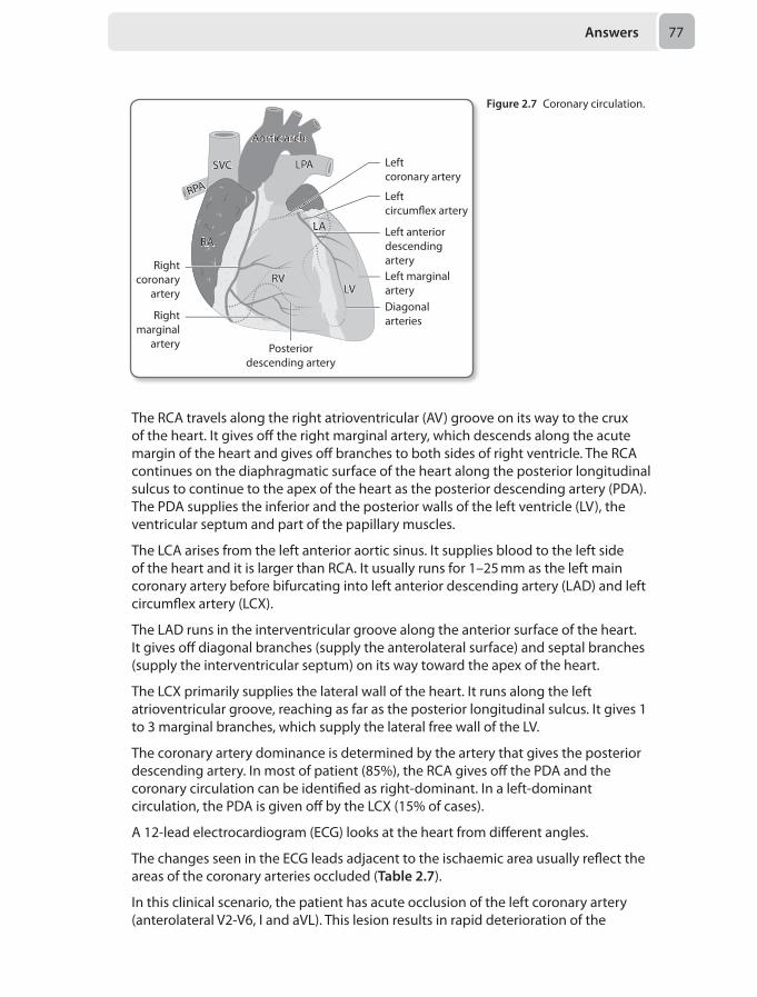
Answers 77
The RCA travels along the right atrioventricular (AV) groove on its way to the crux of the heart. It gives off the right marginal artery, which descends along the acute margin of the heart and gives off branches to both sides of right ventricle. The RCA continues on the diaphragmatic surface of the heart along the posterior longitudinal sulcus to continue to the apex of the heart as the posterior descending artery (PDA). The PDA supplies the inferior and the posterior walls of the left ventricle (LV), the ventricular septum and part of the papillary muscles.
The LCA arises from the left anterior aortic sinus. It supplies blood to the left side of the heart and it is larger than RCA. It usually runs for 1–25 mm as the left main coronary artery before bifurcating into left anterior descending artery (LAD) and left circumflex artery (LCX).
The LAD runs in the interventricular groove along the anterior surface of the heart. It gives off diagonal branches (supply the anterolateral surface) and septal branches (supply the interventricular septum) on its way toward the apex of the heart.
The LCX primarily supplies the lateral wall of the heart. It runs along the left atrioventricular groove, reaching as far as the posterior longitudinal sulcus. It gives 1 to 3 marginal branches, which supply the lateral free wall of the LV.
The coronary artery dominance is determined by the artery that gives the posterior descending artery. In most of patient (85%), the RCA gives off the PDA and the coronary circulation can be identified as right-dominant. In a left-dominant circulation, the PDA is given off by the LCX (15% of cases).
A 12-lead electrocardiogram (ECG) looks at the heart from different angles.
The changes seen in the ECG leads adjacent to the ischaemic area usually reflect the areas of the coronary arteries occluded (Table 2.7).
In this clinical scenario, the patient has acute occlusion of the left coronary artery (anterolateral V2-V6, I and aVL). This lesion results in rapid deterioration of the
RPARPA
Left coronary artery
Left circum�ex artery
Left anteriordescendingarteryLeft marginalartery
Posteriordescending artery
Rightmarginal
artery
LVLV
LALARARA
SVCSVC
Aortic archAortic arch
LPALPA
Rightcoronary
arteryDiagonalarteries
RVRV
Figure 2.7 Coronary circulation.
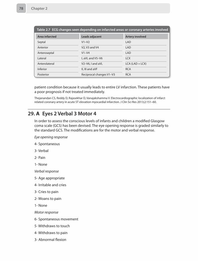
Chapter 278
patient condition because it usually leads to entire LV infarction. These patients have a poor prognosis if not treated immediately.
Thejanandan CS, Reddy D, Rajasekhar D, Vanajakshamma V. Electrocardiographic localization of infarct related coronary artery in acute ST elevation myocardial infarction. J Clin Sci Res 2013;2:151–60.
29. A Eyes 2 Verbal 3 Motor 4In order to assess the conscious levels of infants and children a modified Glasgow coma scale (GCS) has been devised. The eye opening response is graded similarly to the standard GCS. The modifications are for the motor and verbal response.
Eye opening response
4- Spontaneous
3- Verbal
2- Pain
1- None
Verbal response
5- Age appropriate
4- Irritable and cries
3- Cries to pain
2- Moans to pain
1- None
Motor response
6- Spontaneous movement
5- Withdraws to touch
4- Withdraws to pain
3- Abnormal flexion
Table 2.7 ECG changes seen depending on infarcted areas or coronary arteries involved
Area infarcted Leads adjacent Artery involved
Septal V1–V2 LAD
Anterior V2, V3 and V4 LAD
Anteroseptal V1–V4 LAD
Lateral I, aVL and V5–V6 LCX
Anterolateral V2–V6, I and aVL LCA (LAD + LCX)
Inferior II, III and aVF RCA
Posterior Reciprocal changes V1–V3 RCA
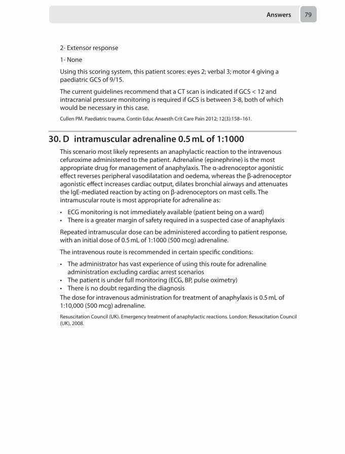
Answers 79
2- Extensor response
1- None
Using this scoring system, this patient scores: eyes 2; verbal 3; motor 4 giving a paediatric GCS of 9/15.
The current guidelines recommend that a CT scan is indicated if GCS < 12 and intracranial pressure monitoring is required if GCS is between 3-8, both of which would be necessary in this case.
Cullen PM. Paediatric trauma. Contin Educ Anaesth Crit Care Pain 2012; 12(3):158–161.
30. D intramuscular adrenaline 0.5 mL of 1:1000This scenario most likely represents an anaphylactic reaction to the intravenous cefuroxime administered to the patient. Adrenaline (epinephrine) is the most appropriate drug for management of anaphylaxis. The α-adrenoceptor agonistic effect reverses peripheral vasodilatation and oedema, whereas the β-adrenoceptor agonistic effect increases cardiac output, dilates bronchial airways and attenuates the IgE-mediated reaction by acting on β-adrenoceptors on mast cells. The intramuscular route is most appropriate for adrenaline as:
• ECG monitoring is not immediately available (patient being on a ward)• There is a greater margin of safety required in a suspected case of anaphylaxis
Repeated intramuscular dose can be administered according to patient response, with an initial dose of 0.5 mL of 1:1000 (500 mcg) adrenaline.
The intravenous route is recommended in certain specific conditions:
• The administrator has vast experience of using this route for adrenaline administration excluding cardiac arrest scenarios
• The patient is under full monitoring (ECG, BP, pulse oximetry)• There is no doubt regarding the diagnosisThe dose for intravenous administration for treatment of anaphylaxis is 0.5 mL of 1:10,000 (500 mcg) adrenaline.
Resuscitation Council (UK). Emergency treatment of anaphylactic reactions. London: Resuscitation Council (UK), 2008.

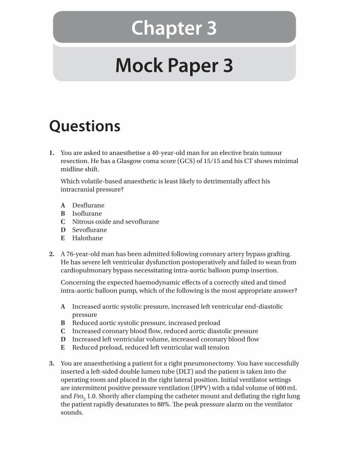
Mock Paper 3
Chapter 3
Questions1. You are asked to anaesthetise a 40-year-old man for an elective brain tumour
resection. He has a Glasgow coma score (GCS) of 15/15 and his CT shows minimal midline shift.
Which volatile-based anaesthetic is least likely to detrimentally affect his intracranial pressure?
A DesfluraneB IsofluraneC Nitrous oxide and sevofluraneD SevofluraneE Halothane
2. A 76-year-old man has been admitted following coronary artery bypass grafting. He has severe left ventricular dysfunction postoperatively and failed to wean from cardiopulmonary bypass necessitating intra-aortic balloon pump insertion.
Concerning the expected haemodynamic effects of a correctly sited and timed intra-aortic balloon pump, which of the following is the most appropriate answer?
A Increased aortic systolic pressure, increased left ventricular end-diastolic pressure
B Reduced aortic systolic pressure, increased preloadC Increased coronary blood flow, reduced aortic diastolic pressureD Increased left ventricular volume, increased coronary blood flowE Reduced preload, reduced left ventricular wall tension
3. You are anaesthetising a patient for a right pneumonectomy. You have successfully inserted a left-sided double lumen tube (DLT) and the patient is taken into the operating room and placed in the right lateral position. Initial ventilator settings are intermittent positive pressure ventilation (IPPV) with a tidal volume of 600 mL and Fio2 1.0. Shortly after clamping the catheter mount and deflating the right lung the patient rapidly desaturates to 88%. The peak pressure alarm on the ventilator sounds.
Chapter 382
What is the first appropriate course of action?
A Deflate the bronchial cuffB Apply 5 cm H2O CPAPC Check the position of the tube with a fibrescopeD Administer 5 L/minute oxygen to the deflated lungE Advance the DLT 2 cm
4. A 70-year-old woman is receiving laser therapy to vocal cord polyps. Her grade 1 intubation with a laser-resistant cuffed tube filled with methylene blue was uneventful and she is being ventilated with 2% sevoflurane and 50% oxygen. Suddenly the surgeon notices flames and methylene blue around the endotracheal tube so stops lasering and floods the site with saline.
What is the most appropriate next step in managing her airway?
A Reduce the Fio2 and perform bronchoalveolar lavageB Stop the flow of all airway gases and remove the endotracheal tubeC Reduce the Fio2 and pack wet swabs around the endotracheal cuffD Reduce the Fio2 and perform a tracheostomyE Reduce the Fio2 and inspect the endotracheal tube lumen via fibrescope
5. A 27-year-old man with type 1 diabetes is listed for day case shoulder arthroscopy under general anaesthesia. He is listed first on an afternoon list and will take an early breakfast on the morning of surgery. He takes Novomix 30 twice a day after his morning and evening meals. His HbA1c measured last month was 53 mmol/mol (7%).
What is the most appropriate advice for his insulin adjustment on the day of surgery?
A Take usual morning dose, and usual insulin with evening mealB Halve usual morning dose, take usual insulin with evening mealC Omit usual morning dose, take normal insulin with evening mealD Halve usual morning dose, halve evening doseE Omit usual morning dose, halve evening dose
6. A 25-year-old woman is having a hysteroscopic myomectomy for infertility under general anaesthesia. The procedure has been prolonged and the irrigation bags have been changed several times. On screen you can see some blood and bubbles in the uterine cavity. She is positioned in the lithotomy position and is slightly head down. You suddenly notice a stepwise decrement in end-tidal waveform capnography, the patient desaturates and there is new T wave inversion on her ECG.
Which of the following is the most appropriate immediate management?
A Call for help, ask the surgeon to stop. Place the patient flat. Give a fluid challenge and high-flow oxygen.
Questions 83
B Call for help, ask the surgeon to flood the uterus with irrigation. Place the patient flat. Give a fluid challenge and high-flow oxygen.
C Call for help, ask the surgeon to externally compress the uterus. Place the patient lateral. Give a fluid challenge and high-flow oxygen.
D Call for help, ask the surgeon to empty the uterus. Place the patient head up. Give a fluid challenge and high-flow oxygen.
E Call for help. Steepen the head down to insert a central venous line into the right internal jugular vein, pass it into the right atrium and attempt to aspirate air.
7. A 68-year-old man undergoing vocal cord surgery is receiving high frequency jet ventilation via a subglottic catheter with the following standard settings: Fio2 0.9, driving pressure 2 atmospheres, frequency 150 min-1, inspiratory time 50%. Blood gas analysis shows a respiratory acidosis and clinically there is no evidence of air trapping.
Which action is most likely to improve the respiratory acidosis?
A Increasing the driving pressureB Increasing the ventilator frequencyC Increasing the inspiratory timeD Increasing the Fio2E Increasing the expiratory time
8. A patient 10 days post coronary artery bypass grafting requires an MRI brain scan for neurological deterioration.
Which of the following is most likely to be a safety hazard during the scan?
A History of a total hip replacement B Presence of sternal wires C Use of an endotracheal tube with metallic spring in the pilot balloonD Epicardial pacing wiresE Invasive arterial blood pressure transducer
9. A 27-year-old woman is to have femoral nerve block with the aid of nerve stimulator.
What is the most important feature of an electrical peripheral nerve stimulator?
A Short pulse durationB Stimulation frequency of 2 HzC Constant current generatorD Negative stimulation polarityE Actual current digital display
10. You have been called to site an epidural for a 32-year-old term primigravida patient. She is now 3 cm dilated with slow progress and very distressed with each contraction. While you are placing the Tuohy needle in the epidural space, you see an obvious flow of clear fluid through the needle.
Chapter 384
What would be the most appropriate next course of action?
A Pull the needle out and arrange alternative analgesiaB Inject 10 mL of normal saline into the subarachnoid space to prevent post-
dural puncture headache (PDPH) and then re-site the epidural catheter in another interspace
C Remove the needle and re-site the epidural catheter in another interspace. Tell the midwife to use 10 mL of epidural mixture for each top-up
D Thread the epidural catheter through the punctured dural hole and use it as intrathecal catheter. Make sure only an anaesthetist gives every top-up dose
E Pull the needle out and call the consultant on call to perform a blood patch to prevent PDPH
11. You have been called to an acute medical ward to help manage a man who has become distressed and angry with the medical management of a relative, and is now behaving violently toward one of the ward staff. When you arrive the situation is heated, security and porter staff are already in attendance. The sister tells you and the on call psychiatrist that the man is unreasonable and needs to be sedated for safety.
How do you proceed?
A Offer the man a dose of oral lorazepamB Attempt to assess his capacity, and discuss with your consultant. Defer
immediate management to the security staff and policeC Give intramuscular lorazepam and haloperidolD Give a dose of intramuscular ketamine and transfer to a monitored environmentE Organise the security staff to restrain the man, and give intramuscular lorazepam
12. A 24-year-old 60 kg woman has been injured in a house fire. She has sustained 40% full thickness lower limb and abdominal burns. Her burns were sustained at 11 pm. She is intubated in the emergency department for suspected inhalational injury and resuscitated with 2 litres of crystalloid in total. At 3 am she is ready for transfer to the regional burns unit that is 4 hours away.
What is the most appropriate fluid regime during transfer?
A Colloid solution, at 500 mL/hourB Compound sodium lactate, at 500 mL/hourC 0.9% sodium chloride, at 500 mL/hour D Colloid solution, at 700 mL/hourE Compound sodium lactate, at 700 mL/hour
13. A previously well 28-year-old man presents to the emergency department with a 48-hour history of feeling generally unwell and complains of polyuria and abdominal pain.
His arterial blood gas on room air is shown in Table 3.1.
His biochemistry profile is shown in Table 3.2.
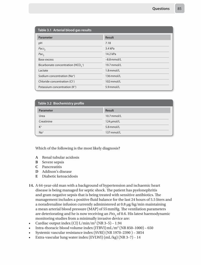
Questions 85
Which of the following is the most likely diagnosis?
A Renal tubular acidosisB Severe sepsisC PancreatitisD Addison’s diseaseE Diabetic ketoacidosis
14. A 64-year-old man with a background of hypertension and ischaemic heart disease is being managed for septic shock. The patient has pyelonephritis and gram negative sepsis that is being treated with sensitive antibiotics. The management includes a positive fluid balance for the last 24 hours of 5.5 liters and a noradrenaline infusion currently administered at 0.8 μg/kg/min maintaining a mean arterial blood pressure (MAP) of 55 mmHg. The ventilation parameters are deteriorating and he is now receiving an Fio2 of 0.6. His latest haemodynamic monitoring studies from a minimally invasive device are:
• Cardiac output index (CI) L/min/m2 (NR 3–5) – 1.94 • Intra-thoracic blood volume index (ITBVI) mL/m2 (NR 850–1000) – 650• Systemic vascular resistance index (SVRI) (NR 1970–2390 ) – 3854• Extra-vascular lung water index (EVLWI) (mL/kg) (NR 3–7) – 14
Table 3.1 Arterial blood gas results
Parameter Result
pH 7.18
Paco2 3.4 kPa
Pao2 14.2 kPa
Base excess –8.8 mmol/L
Bicarbonate concentration (HCO3–) 19.7 mmol/L
Lactate 1.8 mmol/L
Sodium concentration (Na+) 136 mmol/L
Chloride concentration (Cl–) 102 mmol/L
Potassium concentration (K+) 5.9 mmol/L
Table 3.2 Biochemistry profile
Parameter Result
Urea 10.7 mmol/L
Creatinine 124 μmol/L
K+ 5.8 mmol/L
Na+ 137 mmol/L
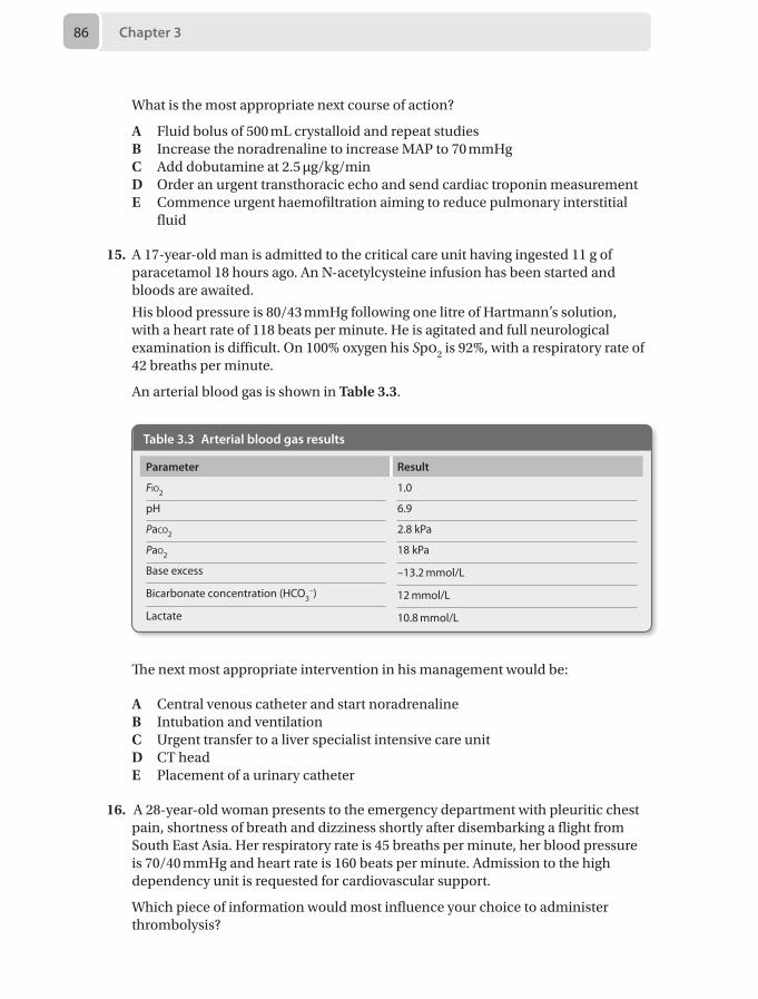
Chapter 386
What is the most appropriate next course of action?
A Fluid bolus of 500 mL crystalloid and repeat studiesB Increase the noradrenaline to increase MAP to 70 mmHgC Add dobutamine at 2.5 μg/kg/minD Order an urgent transthoracic echo and send cardiac troponin measurementE Commence urgent haemofiltration aiming to reduce pulmonary interstitial
fluid
15. A 17-year-old man is admitted to the critical care unit having ingested 11 g of paracetamol 18 hours ago. An N-acetylcysteine infusion has been started and bloods are awaited.
His blood pressure is 80/43 mmHg following one litre of Hartmann’s solution, with a heart rate of 118 beats per minute. He is agitated and full neurological examination is difficult. On 100% oxygen his Spo2 is 92%, with a respiratory rate of 42 breaths per minute.
An arterial blood gas is shown in Table 3.3.
Table 3.3 Arterial blood gas results
Parameter Result
Fio2 1.0
pH 6.9
Paco2 2.8 kPa
Pao2 18 kPa
Base excess –13.2 mmol/L
Bicarbonate concentration (HCO3–) 12 mmol/L
Lactate 10.8 mmol/L
The next most appropriate intervention in his management would be:
A Central venous catheter and start noradrenalineB Intubation and ventilationC Urgent transfer to a liver specialist intensive care unitD CT headE Placement of a urinary catheter
16. A 28-year-old woman presents to the emergency department with pleuritic chest pain, shortness of breath and dizziness shortly after disembarking a flight from South East Asia. Her respiratory rate is 45 breaths per minute, her blood pressure is 70/40 mmHg and heart rate is 160 beats per minute. Admission to the high dependency unit is requested for cardiovascular support.
Which piece of information would most influence your choice to administer thrombolysis?
Questions 87
A Currently on anticoagulationB Information from a CT pulmonary angiographyC Information from a bedside transthoracic echoD A history of being 20 weeks pregnantE A history of peptic ulcer disease
17. A 46-year-old man is sedated and intubated for respiratory failure secondary to community acquired pneumonia. He is known to have recurrent admissions with pneumonia. Your junior colleague sustains a needlestick injury whilst inserting an arterial line, is anxious and would like you to test this patient for HIV.
With regards to HIV testing in this patient, the best course of action is:
A Request consent from the next of kinB Test the patient for HIVC Do not test this patient for HIV because you suspect he is low riskD Do not test this patient for HIV as it is unethicalE Wait until the patient has capacity and gain consent from the patient for HIV
testing
18. A 25-year-old woman is undergoing a category 1 lower segment Caesarean section (LSCS) for fetal bradycardia. You perform a rapid sequence induction with thiopentone 500 mg and suxamethonium 100 mg but are unable to intubate after three attempts. The patient is desaturating and you are unable to ventilate with a face mask and Guedel airway or with a laryngeal mask airway, despite cricoid pressure being reduced and then released. You successfully perform a needle cricothyroid puncture and an ENT surgeon has been informed and is on their way. The fetal heart rate remains at 60 bpm.
What is the next appropriate step?
A Continue oxygenation and proceed with emergency LSCSB Delay LSCS until ENT have performed surgical tracheostomyC Once ENT arrive, perform surgical tracheostomy and LSCS at the same timeD Abandon LSCS and continue oxygenation until patient wakes upE Continue oxygenation until patient wakes up and then perform a spinal
anaesthetic
19. A 30-year-old woman who is 32/40 pregnant is undergoing an open appendicectomy under general anaesthesia. She had a rapid sequence induction with thiopentone 500 mg and suxamethonium 100 mg and was maintained on sevoflurane in oxygen and air. At the end of the procedure, in the recovery room, the patient complains of abdominal discomfort. The midwife from labour ward performs a cardiotocograph (CTG), which suggests the patient is going into preterm labour.
Chapter 388
Which of the following is the least likely cause of her preterm labour?
A Third trimester pregnancyB Acute appendicitisC Surgical manipulation of the uterusD SevofluraneE Maternal pyrexia
20. A 4-month-old 4.5-kg boy is on your elective day case surgical list for bilateral inguinal hernia repair. He was born at 27 weeks gestation and his current corrected gestational age is 44 weeks. The patient was ventilated for 10 days and was oxygen dependent for 6 weeks. He was discharged home 3 weeks previously, but re-presented last week with an apparently obstructed hernia that was reduced by the on-call surgeon. He is on iron and folic acid for anaemia of prematurity.
The most appropriate anaesthetic management would be:
A Proceed with day case surgery under general anaesthesia supplemented with regional anaesthesia
B Proceed with day case surgery under regional anaesthesia supplemented with minimal intravenous sedation
C Postpone surgery until 60 weeks gestational ageD Postpone surgery until 52 weeks gestational ageE Proceed with surgery under general anaesthesia supplemented with regional
anaesthesia and admit postoperatively for apnoea monitoring
21. A 16 kg 4-year-old girl with diabetic ketoacidosis (DKA) is given 40 mL/kg of 0.9% saline for resuscitation and 85 mL/hour of 0.45% saline with 5% glucose as replacement and maintenance fluid. An insulin infusion was commenced at 0.1 unit/kg/hour after an initial bolus dose of 0.1 unit/kg.
2 hours later she has become drowsy with a blood glucose of 11 mmol/l. She appears lethargic and somnolent, and responds to voice. Her heart rate is 96 beats per minute, blood pressure is 128/68 mmHg and respiratory rate is 38 breaths per minute.
The most likely cause of her reduced level of consciousness is:
A Cerebral oedemaB MeningitisC HypokalaemiaD DehydrationE Exhaustion
22. A 65-year-old man was scheduled for a below knee amputation for peripheral vascular disease. He is currently on 10 mg morphine sulphate twice daily for analgesia.
What would be the most appropriate pre-medication to reduce the risk of development of chronic pain?
Questions 89
A Morphine sulphate 15 mgB Gabapentin 900 mgC Amitriptyline 25 mgD Ketamine 20 mgE Epidural analgesia 24 hours preoperatively
23. A 34-year-old woman presents with a 3-month history of poor sleep, fatigue, and worsening widespread muscular pain. On examination, she is extremely tender on finger palpation of several muscle groups and a diagnosis of fibromyalgia is made.
Regarding management of her symptoms, which of the following is the most appropriate first step?
A Trigger point injectionB MorphineC AmitriptylineD IbuprofenE Gabapentin
24. A 3-year-old boy presents for circumcision. He is previously fit and well, and weighs approximately 10 kg. You decide to give him intravenous paracetamol intraoperatively.
Which of the following is the correct dose?
A 150 mgB 200 mgC 75 mgD 100 mgE 500 mg
25. A 49-year-old diabetic man with peripheral vascular disease presents for right below knee amputation. He has been in severe pain in his right leg for several months and no oral analgesia has provided any relief. An epidural is to be commenced preoperatively and continued into the postoperative period.
Which of the following factors increase his risk of developing chronic post surgical pain?
A Severe preoperative painB Increasing ageC Male genderD Use of an epiduralE Diabetes mellitus
26. A 30-year-old man has sustained a partial brachial plexus injury to his left arm. He describes constant, sharp pain with spontaneous burning and shock-like symptoms. Light touch elicits painful episodes and there are constant tingling sensations.
Chapter 390
Which of these statements about pain classifications would apply?
A Neuropathic pain requires a disease or lesion of the somatosensory system to be present
B Dysaesthesias are not always unpleasantC Paraesthesia is usually painfulD Complex regional pain syndrome results from serious traumaE Hyperalgesia is pain from a non-painful stimuli
27. A 33-year-old man is brought to the emergency department with an 18% body surface area (BSA) burn.
Regarding fluid resuscitation, what is the most appropriate statement?
A The Parkland formula should not be used as he is an adult with < 20% BSA burn
B 4 mL/kg/% burn predicts the fluid required in the first 24 hours. Half the fluid should be given in the first 8 hours and the remaining half over the next 16 hours from presentation to hospital
C Fluid requirement should be calculated as per the Baxter formula: 4 mL/kg/% burn. Half the fluid should be given in the first 8 hours and the remaining half over the next 16 hours from the time of burn
D Fluid requirement should be calculated as per the modified Brooke formula: 2 mL/kg/% burn
E Fluid requirement should be calculated as per the Parkland formula. Half should be given as colloid and the other half as crystalloid to reduced the complications of massive fluid resuscitation
28. During the initial surgical dissection of an aortic abdominal aneurysm, the surgeon warns you that he needs to apply the aortic cross-clamp above the coeliac arteries, following which the patient undergoes significant haemodynamic changes.
What are the changes in left ventricle preload and afterload that are caused by the aortic cross-clamp?
A Increased preload, increased afterloadB Decrease preload, increased preloadC No change in preload or afterloadD No change in preload, increased afterloadE Increased preload, decreased afterload
29. A 45-year-old chronic alcoholic in the emergency department was found lying unconscious on the floor of his flat amidst claims that he was not contactable for almost 48 hours. On arrival his Glasgow coma scale (GCS) is 8, (eyes 1; verbal 3; motor 4). His has a heart rate of 104 beats per minute and a blood pressure of 80/60 mmHg. His blood gas shows severe metabolic acidosis and hyperkalaemia. His urine output in the last hour has been 5 mL and is brown in colour.
Questions 91
The most appropriate investigation would be:
A Creatinine kinaseB Urininary haemC MyoglobinuriaD Blood ureaE Alkaline phosphatase
30. A malnourished 93-year-old man with dementia complains of lethargy, breathlessness, myalgia and bone pain, and has petechiae, bleeding gums and gingivitis.
Which of the following nutritional deficiencies would most likely be attributed to this clinical condition:
A Ascorbic acidB IronC ThiamineD Vitamin AE Vitamin D
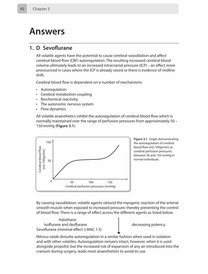
Chapter 392
Answers
1. D SevofluraneAll volatile agents have the potential to cause cerebral vasodilation and affect cerebral blood flow (CBF) autoregulation. The resulting increased cerebral blood volume ultimately leads to an increased intracranial pressure (ICP) – an effect more pronounced in cases where the ICP is already raised or there is evidence of midline shift.
Cerebral blood flow is dependent on a number of mechanisms:
• Autoregulation• Cerebral metabolism coupling• Biochemical reactivity• The autonomic nervous system• Flow dynamics
All volatile anaesthetics inhibit the autoregulation of cerebral blood flow which is normally maintained over the range of perfusion pressures from approximately 50 – 150 mmHg (Figure 3.1).
100
50
0 50 100 150Cerebral perfusion pressures (mmHg)
Cere
bral
blo
od �
ow(m
L/10
0g/m
in)
Figure 3.1 Graph demonstrating the autoregulation of cerebral blood flow (mL/100g/min) at cerebral perfusion pressures between 50 and 150 mmHg in normal individuals.
By causing vasodilation, volatile agents obtund the myogenic reaction of the arterial smooth muscle when exposed to increased pressure, thereby preventing the control of blood flow. There is a range of effect across the different agents as listed below.
Halothane Isoflurane and desflurane decreasing potencySevoflurane (minimal effect ≤ MAC 1.5)
Nitrous oxide disturbs autoregulation in a similar fashion when used in isolation and with other volatiles. Autoregulation remains intact, however, when it is used alongside propofol, but the increased risk of expansion of any air introduced into the cranium during surgery, leads most anaesthetists to avoid its use.
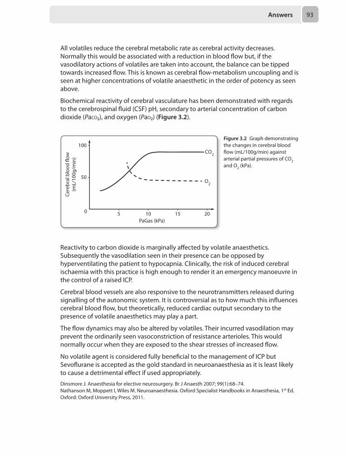
Answers 93
All volatiles reduce the cerebral metabolic rate as cerebral activity decreases. Normally this would be associated with a reduction in blood flow but, if the vasodilatory actions of volatiles are taken into account, the balance can be tipped towards increased flow. This is known as cerebral flow-metabolism uncoupling and is seen at higher concentrations of volatile anaesthetic in the order of potency as seen above.
Biochemical reactivity of cerebral vasculature has been demonstrated with regards to the cerebrospinal fluid (CSF) pH, secondary to arterial concentration of carbon dioxide (Paco₂), and oxygen (Pao₂) (Figure 3.2).
100
50
0 5 10
CO2
O2
15 20PaGas (kPa)
Cere
bral
blo
od �
ow(m
L/10
0g/m
in)
Figure 3.2 Graph demonstrating the changes in cerebral blood flow (mL/100g/min) against arterial partial pressures of CO2 and O2 (kPa).
Reactivity to carbon dioxide is marginally affected by volatile anaesthetics. Subsequently the vasodilation seen in their presence can be opposed by hyperventilating the patient to hypocapnia. Clinically, the risk of induced cerebral ischaemia with this practice is high enough to render it an emergency manoeuvre in the control of a raised ICP.
Cerebral blood vessels are also responsive to the neurotransmitters released during signalling of the autonomic system. It is controversial as to how much this influences cerebral blood flow, but theoretically, reduced cardiac output secondary to the presence of volatile anaesthetics may play a part.
The flow dynamics may also be altered by volatiles. Their incurred vasodilation may prevent the ordinarily seen vasoconstriction of resistance arterioles. This would normally occur when they are exposed to the shear stresses of increased flow.
No volatile agent is considered fully beneficial to the management of ICP but Sevoflurane is accepted as the gold standard in neuroanaesthesia as it is least likely to cause a detrimental effect if used appropriately.
Dinsmore J. Anaesthesia for elective neurosurgery. Br J Anaesth 2007; 99(1):68–74.Nathanson M, Moppett I, Wiles M. Neuroanaesthesia. Oxford Specialist Handbooks in Anaesthesia, 1st Ed. Oxford: Oxford University Press, 2011.
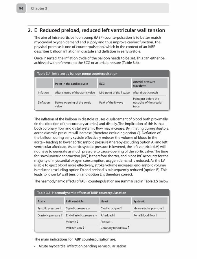
Chapter 394
2. E Reduced preload, reduced left ventricular wall tensionThe aim of Intra-aortic balloon pump (IABP) counterpulsation is to better match myocardial oxygen demand and supply and thus improve cardiac function. The physical premise is one of ‘counterpulsation’, which in the context of an IABP describes balloon inflation in diastole and deflation in early systole.
Once inserted, the inflation cycle of the balloon needs to be set. This can either be achieved with reference to the ECG or arterial pressure (Table 3.4).
Table 3.4 Intra-aortic balloon pump counterpulsation
Point in the cardiac cycle ECGArterial pressure waveform
Inflation After closure of the aortic valve Mid-point of the T wave After dicrotic notch
Deflation Before opening of the aortic valve
Peak of the R wavePoint just before the upstroke of the arterial trace
The inflation of the balloon in diastole causes displacement of blood both proximally (in the direction of the coronary arteries) and distally. The implication of this is that both coronary flow and distal systemic flow may increase. By inflating during diastole, aortic diastolic pressure will increase (therefore excluding option C). Deflation of the balloon during early systole effectively reduces the volume of blood in the aorta – leading to lower aortic systolic pressure (thereby excluding option A) and left ventricular afterload. As aortic systolic pressure is lowered, the left ventricle (LV) will not have to generate as much pressure to cause opening of the aortic valve. The time for isovolumetric contraction (IVC) is therefore shorter, and, since IVC accounts for the majority of myocardial oxygen consumption, oxygen demand is reduced. As the LV is able to eject blood more effectively, stroke volume increases, end-systolic volume is reduced (excluding option D) and preload is subsequently reduced (option B). This leads to lower LV wall tension and option E is therefore correct.
The haemodynamic effects of IABP counterpulsation are summarised in Table 3.5 below:
Table 3.5 Haemodynamic effects of IABP counterpulasation
Aorta Left ventricle Heart Systemic
Systolic pressure ↓ Systolic pressure ↓ Cardiac output ↑ Mean arterial pressure ↑
Diastolic pressure ↑ End-diastolic pressure ↓ Afterload ↓ Renal blood flow ↑
Volume ↓ Preload ↓
Wall tension ↓ Coronary blood flow ↑
The main indications for IABP counterpulsation are:
• Acute myocardial infarction pending re-vascularisation
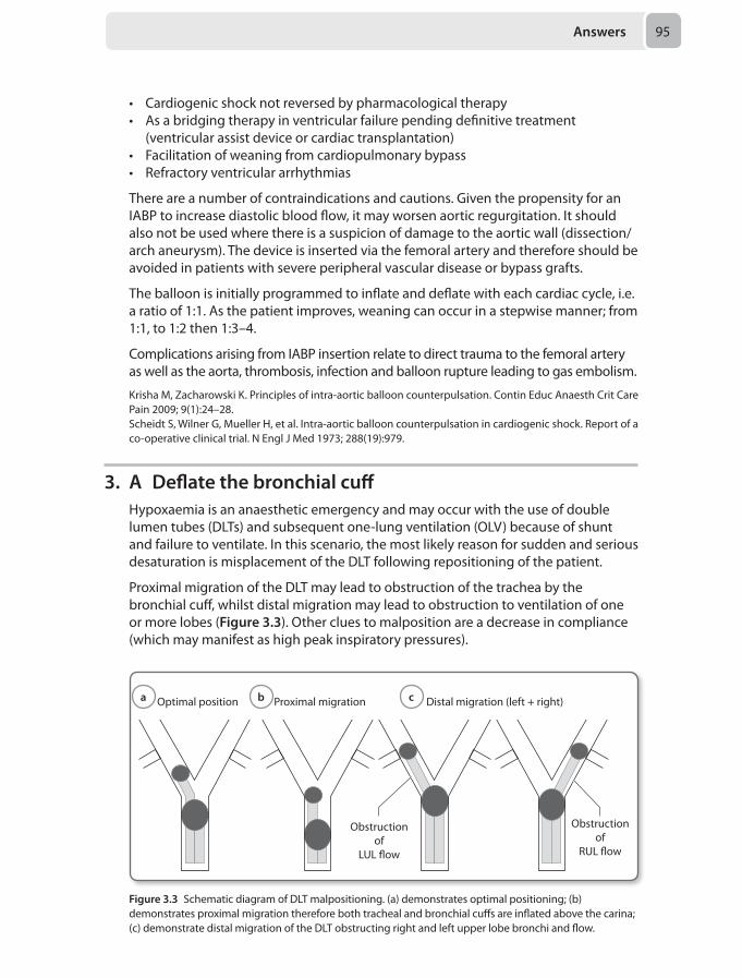
Answers 95
• Cardiogenic shock not reversed by pharmacological therapy• As a bridging therapy in ventricular failure pending definitive treatment
(ventricular assist device or cardiac transplantation)• Facilitation of weaning from cardiopulmonary bypass• Refractory ventricular arrhythmias
There are a number of contraindications and cautions. Given the propensity for an IABP to increase diastolic blood flow, it may worsen aortic regurgitation. It should also not be used where there is a suspicion of damage to the aortic wall (dissection/arch aneurysm). The device is inserted via the femoral artery and therefore should be avoided in patients with severe peripheral vascular disease or bypass grafts.
The balloon is initially programmed to inflate and deflate with each cardiac cycle, i.e. a ratio of 1:1. As the patient improves, weaning can occur in a stepwise manner; from 1:1, to 1:2 then 1:3–4.
Complications arising from IABP insertion relate to direct trauma to the femoral artery as well as the aorta, thrombosis, infection and balloon rupture leading to gas embolism.
Krisha M, Zacharowski K. Principles of intra-aortic balloon counterpulsation. Contin Educ Anaesth Crit Care Pain 2009; 9(1):24–28.Scheidt S, Wilner G, Mueller H, et al. Intra-aortic balloon counterpulsation in cardiogenic shock. Report of a co-operative clinical trial. N Engl J Med 1973; 288(19):979.
3. A Deflate the bronchial cuffHypoxaemia is an anaesthetic emergency and may occur with the use of double lumen tubes (DLTs) and subsequent one-lung ventilation (OLV) because of shunt and failure to ventilate. In this scenario, the most likely reason for sudden and serious desaturation is misplacement of the DLT following repositioning of the patient.
Proximal migration of the DLT may lead to obstruction of the trachea by the bronchial cuff, whilst distal migration may lead to obstruction to ventilation of one or more lobes (Figure 3.3). Other clues to malposition are a decrease in compliance (which may manifest as high peak inspiratory pressures).
Optimal position Proximal migration Distal migration (left + right)
Obstructionof
LUL �ow
Obstructionof
RUL �ow
Figure 3.3 Schematic diagram of DLT malpositioning. (a) demonstrates optimal positioning; (b) demonstrates proximal migration therefore both tracheal and bronchial cuffs are inflated above the carina; (c) demonstrate distal migration of the DLT obstructing right and left upper lobe bronchi and flow.
a b c
Chapter 396
Immediate deflation of the bronchial cuff (option A) may allow two-lung ventilation (via the tracheal lumen) if the DLT has migrated proximally or restoration of ventilation to the upper lobes in the case of distal migration. This manoeuver may restore oxygenation and can then be swiftly followed by definitive repositioning of the DLT with a fibrescope (option C). Advancement of the DLT blindly and without deflating the bronchial cuff (option E) is not optimal management due to the risk of bronchial injury.
Options B and D are accepted methods to improve oxygenation in the event of hypoxaemia that is not due to tube malposition.
Campos JH, Hallam EA, Van Natta T, Kernstine KH. Devices for lung isolation used by anesthesiologists with limited thoracic experience: comparison of double-lumen endotracheal tube, Univent torque control blocker, and Arndt wire-guided endobronchial blocker. Anesthesiology 2006; 104(2):261–66.Ng A, Swanevelder J. Hypoxaemia during one lung ventilation. Br J Anaesth 2011; 106(6): 761–63.
4. B Stop the flow of all airway gases and remove the endotracheal tubeLaser technology allows precision microsurgery and improved haemostasis which makes it an appealing surgical tool for operating in the airway. However, since lasers generate a considerable amount of thermal energy, they also represent a potential fire hazard. Fire requires the presence of an oxidiser (oxygen), ignition source (laser) and fuel (endotracheal tube), all of which are present within the airway in this scenario.
Although laser-resistant endotracheal tubes offer some protection against airway fires, they are still flammable under certain conditions. The endotracheal cuff is particularly susceptible to puncture from misdirected laser energy which will lead to oxygen enrichment around the surgical site and an increased risk of catastrophic airway fire. The most important step after stopping lasering and flooding the site with saline is to stop the flow of all airway gases (oxidisers) and remove the endotracheal tube (fuel). Wet swabs placed around the surgical site prior to lasering can minimise the risk but should not be used to treat an airway fire. In the event of an airway fire, all flammable materials should be removed from the airway. The patient can subsequently be bag valve mask ventilated with air once the fire is out.
To assess for airway damage, rigid bronchoscopy is recommended afterwards. Gentle bronchoalveolar lavage and fibreoptic assessment of the more distal airways is of benefit and if the damage is severe, a tracheostomy may be indicated. Smoke inhalation and thermal damage to the lungs may necessitate prolonged intubation and mechanical ventilation.
Kitching A, Edge C. Lasers and surgery. Contin Educ Anaesth Crit Care Pain 2003; 3(5):143–146.Apfelbaum J, Caplan RA, Barker SJ, et al. Practice advisory for the prevention and management of operating room fires: an updated report by the American Anesthesiologists Task Force on operating room fires. Anesthesiology 2013; 118(2):271–90.
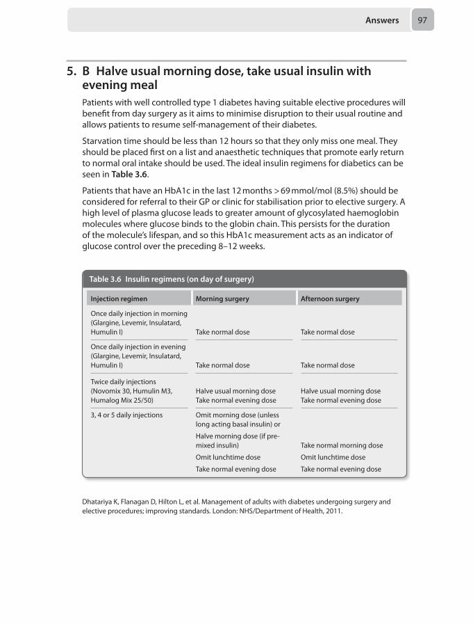
Answers 97
5. B Halve usual morning dose, take usual insulin with evening mealPatients with well controlled type 1 diabetes having suitable elective procedures will benefit from day surgery as it aims to minimise disruption to their usual routine and allows patients to resume self-management of their diabetes.
Starvation time should be less than 12 hours so that they only miss one meal. They should be placed first on a list and anaesthetic techniques that promote early return to normal oral intake should be used. The ideal insulin regimens for diabetics can be seen in Table 3.6.
Patients that have an HbA1c in the last 12 months > 69 mmol/mol (8.5%) should be considered for referral to their GP or clinic for stabilisation prior to elective surgery. A high level of plasma glucose leads to greater amount of glycosylated haemoglobin molecules where glucose binds to the globin chain. This persists for the duration of the molecule’s lifespan, and so this HbA1c measurement acts as an indicator of glucose control over the preceding 8–12 weeks.
Table 3.6 Insulin regimens (on day of surgery)
Injection regimen Morning surgery Afternoon surgery
Once daily injection in morning (Glargine, Levemir, Insulatard, Humulin I) Take normal dose Take normal dose
Once daily injection in evening (Glargine, Levemir, Insulatard, Humulin I) Take normal dose Take normal dose
Twice daily injections (Novomix 30, Humulin M3, Humalog Mix 25/50)
Halve usual morning dose Take normal evening dose
Halve usual morning dose Take normal evening dose
3, 4 or 5 daily injections Omit morning dose (unless long acting basal insulin) or
Halve morning dose (if pre-mixed insulin)
Omit lunchtime dose
Take normal evening dose
Take normal morning dose
Omit lunchtime dose
Take normal evening dose
Dhatariya K, Flanagan D, Hilton L, et al. Management of adults with diabetes undergoing surgery and elective procedures; improving standards. London: NHS/Department of Health, 2011.
Chapter 398
6. D Call for help, ask the surgeon to empty the uterus. Place the patient head up. Give a fluid challenge and high-flow oxygenGas embolism can occur either into the venous system or the arterial system, and may involve gases other than air e.g. carbon dioxide in the case of laparoscopic misadventure. The pathophysiology and principles of management should be clear in your mind.
Incidence
In the clinical arena, clinically obvious air embolism is thankfully rare; however in studies looking for the condition, venous air embolism (VAE) was discovered in 100% of seated craniotomies, 40% of Caesarean sections and 30% of hip replacements. Dose of air is important, as is the size of the patient and the rate of air ingress. Rapid air entry is worst with a lethal dose of around 1 mL/kg.
Pathophysiology
Gas entering the venous system returns to the right heart and can cause a right ventricular outflow obstruction as the air is compressible and causes mechanical dysfunction. Distal flow of air causes trapping in the pulmonary vessels and a massive increase in pulmonary vascular resistance (PVR) and fulminant right heart failure. Some micro-emboli may pass into the distal tree and produce non-cardiogenic pulmonary oedema by secondary mechanisms. Air may also pass through the lungs and on into the systemic circulation or through a patent foramen ovale. Clinically the patient will exhibit tachypnoea, tachycardia and cardiovascular collapse if the volume is great enough. Smaller doses may give chest pain, shortness of breath and mental disturbance.
During anaesthesia the end-tidal CO2 falls as shunt increases, and desaturation will occur with variable ECG changes and cardiovascular instability. Pulmonary oedema may develop as a later sign, and central venous pressure can be elevated.
Detection
Clinical suspicion and vigilance are imperative in the detection of VAE. Classically described tools for detection such as the precordial and oesophageal stethoscope have poor sensitivity and are not often used. Doppler, in transoesophageal or transcranial positions are very sensitive detectors of air.
Treatment
Immediate treatment is supportive and resuscitative along the ABC algorithm. It should then focus on preventing further air entry, reducing the size of the embolus, and overcoming the mechanical obstruction in the right side.
Preventing further air ingress can be achieved surgically at the site of bleeding, either by covering or compressing bleeding areas and flooding the area with saline. Venous pressure can be increased by positioning the site lower than the heart, administering intravenous fluids, performing a Valsalva manoeuvre or increasing intrathoracic pressure.
Answers 99
Reducing the size of an embolus is achieved by immediate discontinuation of nitrous oxide and giving 100% oxygen that promotes nitrogen diffusion out of the bubble. Aspiration from the right atrium via a central line may be possible if the embolus is large and the patient positioned either lateral or head down position to prevent further forward movement of air out of the right ventricle. This would depend on the line already having been situated and the tip positioned in the right atrium. Hyperbaric oxygen therapy will also promote rapid clearance of emboli.
Overcoming mechanical obstruction relies on judicious use of fluid and inotropic agents to support a right ventricle against the sudden rise in pulmonary vascular resistance.
One may not classically attribute gynaecological surgery with a high risk of venous air embolism, but as described hitherto the only requirements are pressurised gas and an open vascular system.
The key elements here in terms of management are:
• Recognise this as an anaesthetic emergency and summon help• Preventing further air ingress
The aim therefore is to promote higher venous pressure at the site to reduce air entrainment. Given the uterine site of bleeding, a head up position will be best. Bear in mind the need for resuscitation may necessitate laying the patient flat if cardiac arrest ensues. Other measures include compressing the wound edges by the surgeon, and flooding with saline. In this case, with a full uterine cavity, external compression may increase the pressure gradient and promote residual gas inflow. The safest compromise is likely to be to empty the cavity such that the pressure driving gas into the venous system is removed.
Supportive measures
Increasing the fraction of inspired oxygen to 100% will increase the partial pressure of oxygen and favours nitrogen washout from bubbles to the alveoli. This will also ensure that, if used, nitrous oxide would also be washed out.
A fluid bolus will assist in raising the venous pressure, and also assist the right ventricle combating elevated pulmonary pressures, as well as support systemic perfusion pressure.
Aspirating the air lock within the right atrium may be possible if there is a central line already in situ. However, insertion after diagnosis is likely to distract from immediate management and is not recommended.
Webber S, Andrzejowski J, Francis G. Gas embolism in anaesthesia. Br J Anaesth CEPD Reviews 2002; 2(2):53–57.
7. A Increasing the driving pressureHigh frequency jet ventilation is a specialised ventilation modality designed to achieve gas exchange by using high frequency, low tidal volume breaths. It differs from conventional ventilation since gas exchange is not primarily dependent on bulk flow to the alveoli (as the tidal volumes are often lower than the dead space).
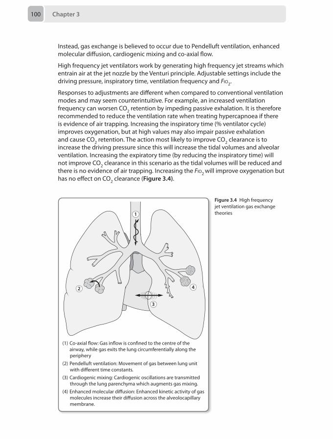
Chapter 3100
Instead, gas exchange is believed to occur due to Pendelluft ventilation, enhanced molecular diffusion, cardiogenic mixing and co-axial flow.
High frequency jet ventilators work by generating high frequency jet streams which entrain air at the jet nozzle by the Venturi principle. Adjustable settings include the driving pressure, inspiratory time, ventilation frequency and Fio2.
Responses to adjustments are different when compared to conventional ventilation modes and may seem counterintuitive. For example, an increased ventilation frequency can worsen CO2 retention by impeding passive exhalation. It is therefore recommended to reduce the ventilation rate when treating hypercapnoea if there is evidence of air trapping. Increasing the inspiratory time (% ventilator cycle) improves oxygenation, but at high values may also impair passive exhalation and cause CO2 retention. The action most likely to improve CO2 clearance is to increase the driving pressure since this will increase the tidal volumes and alveolar ventilation. Increasing the expiratory time (by reducing the inspiratory time) will not improve CO2 clearance in this scenario as the tidal volumes will be reduced and there is no evidence of air trapping. Increasing the Fio2 will improve oxygenation but has no effect on CO2 clearance (Figure 3.4).
1
2
3
4
(1) Co-axial �ow: Gas in�ow is con ned to the centre of the airway, while gas exits the lung circumferentially along the
periphery(2) Pendelluft ventilation: Movement of gas between lung unit
with di�erent time constants.(3) Cardiogenic mixing: Cardiogenic oscillations are transmitted
through the lung parenchyma which augments gas mixing.(4) Enhanced molecular di�usion: Enhanced kinetic activity of gas
molecules increase their di�usion across the alveolocapillary membrane.
Figure 3.4 High frequency jet ventilation gas exchange theories
Answers 101
Evans E, Biro P, Bedforth N. Jet ventilation. Contin Educ Anaesth Crit Care Pain 2007; 7(1):2–5.Brice JW, Davis WB. High frequency ventilation in the adult. Clin Pulm Med 2004; 11(2):101–6.
8. D Epicardial pacing wiresSafety considerations when anaesthetising patients for MRI scans include:
• Remote location• Limited access to the patient • The projectile effect of the magnetic field on ferromagnetic equipment• The heating effect and risk of burns from conductive wires and coils• Risk of equipment malfunction• Noise protection• Risk of contrast reactions
MRI scanners operate with magnetic field strengths of 0.5–3 Tesla (T). The strength of the field decays exponentially with distance from the magnet, which is always kept on. Zones around the magnet are described in terms of Gauss (G), where 1T = 10,000G. Within 50G zone a significant attractive force will risk ferromagnetic objects to act as projectiles potentially causing serious harm or damage, and an area of 5G is set where no unscreened personnel should enter (see Figure 1.2). Currents may be generated in cables especially in coils causing a local heating effect that may cause burns to the patient.
Special MRI safe anaesthetic machines, ventilators and monitors should be available. MRI compatible ECG electrodes, and fibreoptic pulse oximetry should be used and all cables should be kept away from the area being imaged, in straight lines away from the patient to avoid burns. ST-T wave artefacts may occur due to the heating effect of blood around the aortic arch. Blood pressure cuffs without metal connectors and invasive pressure transducers may be used. Gas sampling lines will be longer, increasing the lag time and dead space. Epicardial pacing wires and pulmonary artery catheters containing conductive wires risk myocardial heating and burns, and scans cannot be performed in cases where these cannot be removed. The spring in an endotracheal tube pilot balloon is safe, however it should be taped away from the area being scanned as it may cause image artefact. Only essential infusions should be continued and long extensions attached to pass through a wall port so that the syringe pumps can stay in the control room.
Most orthopaedic prostheses, surgical clips and sternal wires are safe, but may distort images. Other implanted metallic devices such as pacemakers, cochlear implants, intraocular metallic objects and aneurysm clips will cause internal heating, malfunction or risk movement and so would contraindicate a scan.
Reddy U, White MJ, Wilson SR. Anaaesthesia for magnetic resonance imaging. Contin Educ Anaesth Crit Care Pain 2012; 12(3):140–44.
9. C Constant current generatorThe use of a peripheral nerve stimulator to localise nerves is still the commonest technique employed worldwide in regional anaesthesia. The method is based on the
Chapter 3102
principle that muscle contraction becomes obvious when enough electrical current is applied to the nerve in order to depolarise the nerve fibres.
The main objective in regional anaesthesia is to stop nerve conduction by infiltrating enough local anaesthetic close to the nerve fibres and provide motor and sensory block. This goal is possible due to the capability of nerve stimulators to approximate the distance between the nerve and the needle tip and hence can localise the optimal injection site.
Nerve fibres differ anatomically according to their thickness and degree of myelination. Stimulating motor fibers with nerve stimulator is easier than stimulating sensory fibres because Aα motor fibers have the maximal velocity of impulse spreading and a relatively low threshold for extrinsic activation because of a large diameter and high degree of myelination. In contrast, C-fibres have a small diameter and very little or no myelin sheath, hence have a high threshold for external stimulation and relatively slow action potential propagation.
An ideal nerve stimulator should have the following features:
1. Constant current generator: They must be able to supply a constant electrical current between the negative pole and the positive pole irrespective of the wide range of impendences encountered by the tissue around the nerve. Nerve stimulators provide a current range between 0.01–5 mA.
When performing a nerve block, the ideal initial current is 1–2 mA. The needle is then inserted until the desired muscle contraction is seen. The stimulating current is then gradually decreased until twitches are still seen at a final range of 0.2–0.5 mA which is the most acceptable current threshold. A current above 0.5 mA may mean the needle tip is far from the nerve and the block may not be successful. Twitches should not be seen below 0.2 mA because motor response below 0.2 mA may mean the needle is inside the nerve and injecting maybe harmful.
2. Stimulation polarity: In order to get maximum benefit from the delivered current, the needle should be connected to the negative pole (cathode) to depolarise nerve fibres. Stimulating nerves with the anode will lead hyperpolarisation of the fibres, thus a stronger current will be required to depolarise the nerve. Modern nerve stimulators are designed in order to only allow the needle to be connected to the cathode electrode.
3. Stimulation frequency: This affects the speed of nerve localisation. The ideal current frequency is 1–2 Hz, where a higher frequency makes nerve detection faster, but causes more patient discomfort. Therefore, the most common frequency used is 2 Hz.
4. Pulse duration: This is the time for which the electrical current is applied to the nerve. A short pulse duration (0.1 ms) ensures motor neurons are stimulated but not the sensory neurons.
5. Accuracy: The current generated by the nerve stimulator must be similar to the displaced one on the digital screen. Accurate current generation is mandatory for correct needle insertion and successful nerve block.
Answers 103
The above features of nerve stimulators are all essential for successful nerve block. However, the most important characteristic of peripheral nerve stimulators is the constant current generation. This allows the current to remain the same regardless of resistance variation encountered by tissue, thus decreases the chance of nerve damage or unsuccessful nerve block.
Sardesai AM. Lyer U. Nerve stimulation for peripheral nerve blockade. Anaesthesia Tutorial of the Week no 149. London: World Federation of Societies of Anaesthesiologists, 2009.Shariat AN, Horan PM, Gratenstein K, McCally C, Frulla AP. Electrical nerve stimulators and localization of peripheral nerves, New York: The New York School of Regional Anaesthesia, 2013.
10. D Thread the epidural catheter through the punctured dural hole and use it as intrathecal catheter. Make sure only an anaesthetist gives every top-up dose
Inadvertent dural puncture can occur following many procedures, for example spinal surgery. However, in anaesthetic practice it is usually a consequence of epidural analgesia and anaesthesia.
Every year, approximately 140,000 labouring women receive an epidural in the UK. 1,400 (1%) will suffer an accidental dural puncture. The commonest reported complication is post-dural puncture headache (PDPH), with an incidence varying from 60% to 90%. Headache usually develops 12–24 hours following dural puncture. PDPH may cause significant morbidity and mothers may be unable to properly care for their newborn or themselves for some time.
Puncturing the dura with a 16G Touhy needle usually results in an obvious cerebrospinal fluid (CSF) flow through the epidural needle. The CSF is warm to touch and tests positive to glucose with urine dipstick.
Each obstetric unit should have a plan of action for accidental dural tap.
At the time of recognised dural puncture, the anaesthetist must decide whether to use the catheter for a continuous subarachnoid (intrathecal) technique or re-site the epidural in another interspace. Whichever decision is made, only the anaesthetist should give every dose of local anaesthetic top-up. Therefore, stem D is inappropriate here as the midwife gives the top-up.
The patient should be informed about the dural puncture and counseled regarding potential complications and further management options. A senior anaesthetic colleague, the obstetric registrar and the midwife should also be informed, and the operator must ensure everything is documented in the patient’s notes.
If no PDPH is present during labour, pushing at full dilation can be encouraged. However, if a headache is present, forceps delivery may be advised.
After delivery, remove the catheter as usual. There is little evidence that leaving a subarachnoid catheter in place for 24 hours can reduce PDPH.
If no headache is present post delivery, do not restrict patient’s mobilisation, as immobilisation will not prevent the development PDPH. However, if a PDPH
Chapter 3104
develops, encourage bed rest, regular analgesia, oral fluid and oral caffeine intake, and consider epidural blood patch. Epidural blood patch is the gold standard therapy for PDPH, however if performed within 24 hours of onset of symptoms there is a failure rate of 70%, which reduces to 4% if performed after 24 hours. Therefore, it is preferable to perform blood patch more than 24 hours after the dural tap occured.
Studies investigating the use of epidural saline as a preventative measure against PDPH failed to reach statistical significance. In addition, pooled results of the four randomised trials failed to show statistical significance for the use of epidural blood patch as prophylactic measure to treat PDPH. The use of sumatriptan and adrenocorticotrophic hormone (ACTH) has also previously been advocated, but again there is a large disparity in the evidence base for their use.
Royal College of Anaesthetists. Major complications of central neuraxial blocks in the United Kingdom: the 3rd National Audit Project (NAP3). Br J Anaesth 2009; 102(2): 179–90. Apfel CC, Saxena A, Cakmakkaya OS, et al. Prevention of postdural puncture headache after accidental dural puncture: A quantitative systematic review. Br J Anaesth 2010; 105(3):255–63.Sharpe P. Accidental dural puncture in obstetrics. BJA CEPD Reviews 2001; 1(3):81–84.
11. B Attempt to assess his capacity, and discuss with your consultant. Defer immediate management to the security staff and police
There are multidisciplinary rapid response teams in some hospitals, for the purpose of sedating disturbed psychiatric inpatients, which do include anaesthetists. The Royal College of Anaesthetists has issued position statement guidance in tandem with the Royal College of Psychiatrists, the noteworthy points include:
• Anaesthetists should only act as part of a multidisciplinary response team incorporating mental healthcare professionals including a psychiatrist
• Trainee anaesthetists should not routinely be involved in initiating pharmacological restraint, but if the urgency of the clinical situation dictates they must only act within their competence and, whenever possible, after consultation with a consultant anaesthetist
• When rapid tranquillisation is deemed appropriate the minimum intervention possible should be used as guided by the local protocol
• The full range of ventilatory/resuscitation equipment and trained assistance must be immediately available when rapid tranquillisation is administered
• The College does not support under any circumstances the use of rapid tranquillisation to manage violence or aggression in visitors or other individuals on hospital premises
This question, like some past exam SBAs, reflects some of the more difficult ‘real-life’ clinical situations we can find ourselves in as practising anaesthetists. The important points to draw from the stem are the lack of any evidence of confusion or signs of organic mental illness. The man is aggressive and violent, but as a visitor is not a patient of the hospital. This means that his behaviour should be managed by security staff, who have been specially trained to do so, and if necessary by the police. If the police after their initial assessment were to believe him to be mentally disturbed, they would convey him to the emergency department or other place of
Answers 105
safety for specialist assessment. This does not remove our responsibility to respond to treat visitors in an emergency, such as in the case of a collapse or cardiac arrest.
Royal College of Anaesthetists. Position statement on the involvement of anaesthetists in restraint teams. London: Royal College of Anaesthetists, 2014.National Institute for Health and Care Excellence (NICE). Violence: The short-term management of disturbed/violent behaviour in psychiatric in-patient settings and emergency departments. CG No 25. London: NICE, 2005.
12. E Compound sodium lactate, at 700 mL/hourFluid resuscitation after burns injury is widely guided by the Parkland formula: 4 mL/kg/% body surface area burn, where half of this volume is given in the first 8 hours after the burn injury, and the second half over the next 16 hours. Thus for the above patient:
4 x 60 x 40 = 9600 mL
Therefore this patient requires 4800 mL in the first 8 hours.
Two litres has already been given in the first 4 hours since the burn, and so 2800 mL should be given over the next 4 hours, which is 700 mL/hour.
Intravenous fluids are given in cases of more than 15% total body surface area (TBSA) burns or 10% with smoke inhalation. The fluid of choice is a balanced crystalloid solution, and initially a urine output of greater than 0.5 mL/kg/hour is used to guide adequate resuscitation.
Criteria for referral to a specialist burns unit include:
• > 2% TBSA in children or > 3% in adults • Full thickness burns • Circumferential burns • Unhealed burn after 2 weeks• Any suspicion of non-accidental injury
Discuss with a burns specialist with an opinion to transfer:
• Burns to hands, feet, face, perineum or genitalia • Chemical, electrical or friction burns• Cold injuries• Febrile child with a burn • Co-morbidities affecting treatment or healing of the burn
Intubation for a burn is indicated in the presence of stridor, oropharyngeal oedema, deep facial or neck burns, a GCS < 8, hypoxia or hypercapnia, or suspicion of inhalational injury. Facial oedema can progress rapidly and an early intubation using a large uncut endotracheal tube should be performed prior to transfer if there are any concerns. A rapid sequence induction using suxamethonium is acceptable if within the first 24 hours after the burn.
Bishop S, Maguire S. Anaesthesia and intensive care for major burns. Contin Educ Anaesth Crit Care Pain 2012; 12(3):118–22.National Network for Burn Care. National Burn Care Referral Guidance. London: NHS Specialised Services, 2012.
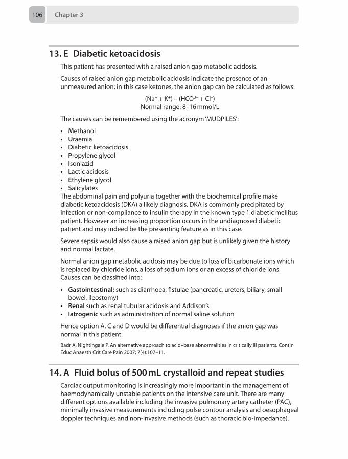
Chapter 3106
13. E Diabetic ketoacidosisThis patient has presented with a raised anion gap metabolic acidosis.
Causes of raised anion gap metabolic acidosis indicate the presence of an unmeasured anion; in this case ketones, the anion gap can be calculated as follows:
(Na+ + K+) – (HCO3– + Cl–)Normal range: 8–16 mmol/L
The causes can be remembered using the acronym ‘MUDPILES’:
• Methanol• Uraemia• Diabetic ketoacidosis• Propylene glycol• Isoniazid• Lactic acidosis• Ethylene glycol• SalicylatesThe abdominal pain and polyuria together with the biochemical profile make diabetic ketoacidosis (DKA) a likely diagnosis. DKA is commonly precipitated by infection or non-compliance to insulin therapy in the known type 1 diabetic mellitus patient. However an increasing proportion occurs in the undiagnosed diabetic patient and may indeed be the presenting feature as in this case.
Severe sepsis would also cause a raised anion gap but is unlikely given the history and normal lactate.
Normal anion gap metabolic acidosis may be due to loss of bicarbonate ions which is replaced by chloride ions, a loss of sodium ions or an excess of chloride ions. Causes can be classified into:
• Gastointestinal; such as diarrhoea, fistulae (pancreatic, ureters, biliary, small bowel, ileostomy)
• Renal such as renal tubular acidosis and Addison’s• Iatrogenic such as administration of normal saline solution
Hence option A, C and D would be differential diagnoses if the anion gap was normal in this patient.
Badr A, Nightingale P. An alternative approach to acid–base abnormalities in critically ill patients. Contin Educ Anaesth Crit Care Pain 2007; 7(4):107–11.
14. A Fluid bolus of 500 mL crystalloid and repeat studiesCardiac output monitoring is increasingly more important in the management of haemodynamically unstable patients on the intensive care unit. There are many different options available including the invasive pulmonary artery catheter (PAC), minimally invasive measurements including pulse contour analysis and oesophageal doppler techniques and non-invasive methods (such as thoracic bio-impedance).
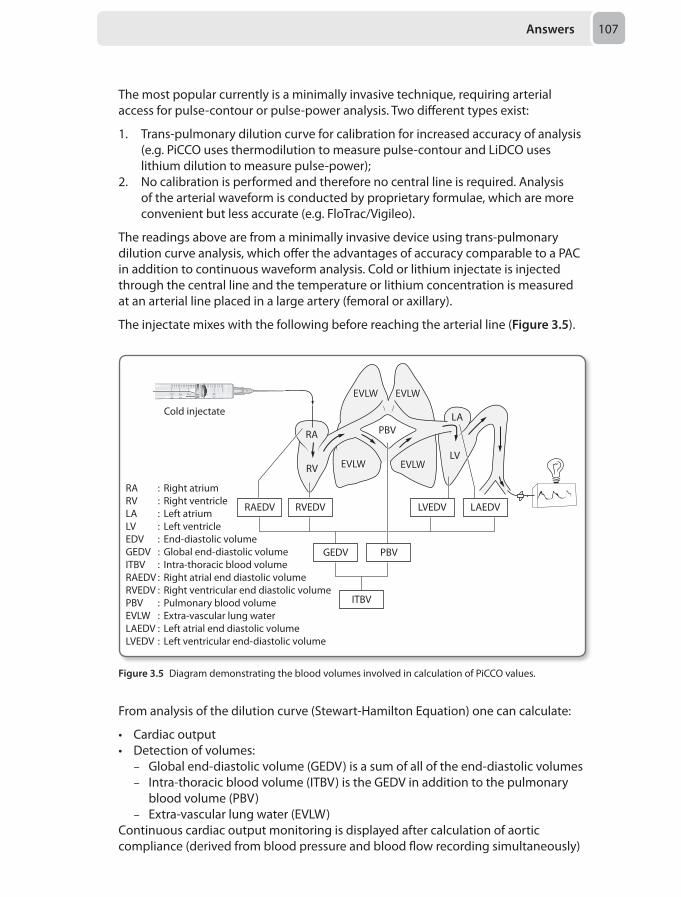
Answers 107
The most popular currently is a minimally invasive technique, requiring arterial access for pulse-contour or pulse-power analysis. Two different types exist:
1. Trans-pulmonary dilution curve for calibration for increased accuracy of analysis (e.g. PiCCO uses thermodilution to measure pulse-contour and LiDCO uses lithium dilution to measure pulse-power);
2. No calibration is performed and therefore no central line is required. Analysis of the arterial waveform is conducted by proprietary formulae, which are more convenient but less accurate (e.g. FloTrac/Vigileo).
The readings above are from a minimally invasive device using trans-pulmonary dilution curve analysis, which offer the advantages of accuracy comparable to a PAC in addition to continuous waveform analysis. Cold or lithium injectate is injected through the central line and the temperature or lithium concentration is measured at an arterial line placed in a large artery (femoral or axillary).
The injectate mixes with the following before reaching the arterial line (Figure 3.5).
Cold injectate
RA : Right atriumRV : Right ventricleLA : Left atriumLV : Left ventricleEDV : End-diastolic volumeGEDV : Global end-diastolic volumeITBV : Intra-thoracic blood volumeRAEDV : Right atrial end diastolic volumeRVEDV : Right ventricular end diastolic volume PBV : Pulmonary blood volume EVLW : Extra-vascular lung water LAEDV : Left atrial end diastolic volumeLVEDV : Left ventricular end-diastolic volume
RA
LA
LVRV
PBV
RAEDV RVEDV
GEDV PBV
ITBV
LVEDV LAEDV
EVLW
EVLW
EVLW
EVLW
From analysis of the dilution curve (Stewart-Hamilton Equation) one can calculate:
• Cardiac output• Detection of volumes:
– Global end-diastolic volume (GEDV) is a sum of all of the end-diastolic volumes – Intra-thoracic blood volume (ITBV) is the GEDV in addition to the pulmonary
blood volume (PBV) – Extra-vascular lung water (EVLW)
Continuous cardiac output monitoring is displayed after calculation of aortic compliance (derived from blood pressure and blood flow recording simultaneously)
Figure 3.5 Diagram demonstrating the blood volumes involved in calculation of PiCCO values.
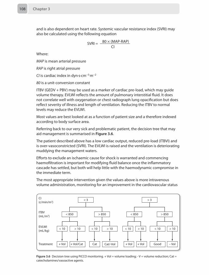
Chapter 3108
and is also dependent on heart rate. Systemic vascular resistance index (SVRI) may also be calculated using the following equation
SVRI = 80 × (MAP-RAP) CI
Where:
MAP is mean arterial pressure
RAP is right atrial pressure
CI is cardiac index in dyn·s·cm−5·m−2
80 is a unit-conversion constant
ITBV (GEDV + PBV) may be used as a marker of cardiac pre-load, which may guide volume therapy. EVLWI reflects the amount of pulmonary interstitial fluid. It does not correlate well with oxygenation or chest radiograph lung opacification but does reflect severity of illness and length of ventilation. Reducing the ITBV to normal levels may reduce the EVLWI.
Most values are best looked at as a function of patient size and a therefore indexed according to body surface area.
Referring back to our very sick and problematic patient, the decision tree that may aid management is summarised in Figure 3.6.
The patient described above has a low cardiac output, reduced pre-load (ITBVI) and is over-vasoconstricted (SVRI). The EVLWI is raised and the ventilation is deteriorating muddying the management waters.
Efforts to exclude an ischaemic cause for shock is warranted and commencing haemofiltration is important for modifying fluid balance once the inflammatory cascade has settled, but both will help little with the haemodynamic compromise in the immediate term.
The most appropriate intervention given the values above is more intravenous volume administration, monitoring for an improvement in the cardiovascular status
CI(c/min/m2)
< 3
< 850 < 850> 850 > 850
< 10 > 10 < 10 > 10 < 10 > 10 < 10 > 10
+ Vol + Vol – Vol+ Vol/Cat + Vol GoodCat Cat/-Vol
> 3
ITBV(mL/m2)
EVLWI(mL/kg)
Treatment
Figure 3.6 Decision tree using PiCCO monitoring. + Vol = volume loading; - V = volume reduction; Cat = catecholamines/vasoactive agents.
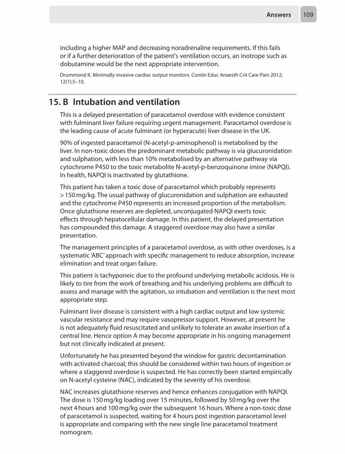
Answers 109
including a higher MAP and decreasing noradrenaline requirements. If this fails or if a further deterioration of the patient's ventilation occurs, an inotrope such as dobutamine would be the next appropriate intervention.
Drummond K. Minimally invasive cardiac output monitors. Contin Educ Anaesth Crit Care Pain 2012; 12(1):5–10.
15. B Intubation and ventilationThis is a delayed presentation of paracetamol overdose with evidence consistent with fulminant liver failure requiring urgent management. Paracetamol overdose is the leading cause of acute fulminant (or hyperacute) liver disease in the UK.
90% of ingested paracetamol (N-acetyl-p-aminophenol) is metabolised by the liver. In non-toxic doses the predominant metabolic pathway is via glucuronidation and sulphation, with less than 10% metabolised by an alternative pathway via cytochrome P450 to the toxic metabolite N-acetyl-p-benzoquinone imine (NAPQI). In health, NAPQI is inactivated by glutathione.
This patient has taken a toxic dose of paracetamol which probably represents > 150 mg/kg. The usual pathway of glucuronidation and sulphation are exhausted and the cytochrome P450 represents an increased proportion of the metabolism. Once glutathione reserves are depleted, unconjugated NAPQI exerts toxic effects through hepatocellular damage. In this patient, the delayed presentation has compounded this damage. A staggered overdose may also have a similar presentation.
The management principles of a paracetamol overdose, as with other overdoses, is a systematic ‘ABC’ approach with specific management to reduce absorption, increase elimination and treat organ failure.
This patient is tachyponeic due to the profound underlying metabolic acidosis. He is likely to tire from the work of breathing and his underlying problems are difficult to assess and manage with the agitation, so intubation and ventilation is the next most appropriate step.
Fulminant liver disease is consistent with a high cardiac output and low systemic vascular resistance and may require vasopressor support. However, at present he is not adequately fluid resuscitated and unlikely to tolerate an awake insertion of a central line. Hence option A may become appropriate in his ongoing management but not clinically indicated at present.
Unfortunately he has presented beyond the window for gastric decontamination with activated charcoal; this should be considered within two hours of ingestion or where a staggered overdose is suspected. He has correctly been started empirically on N-acetyl cysteine (NAC), indicated by the severity of his overdose.
NAC increases glutathione reserves and hence enhances conjugation with NAPQI. The dose is 150 mg/kg loading over 15 minutes, followed by 50 mg/kg over the next 4 hours and 100 mg/kg over the subsequent 16 hours. Where a non-toxic dose of paracetamol is suspected, waiting for 4 hours post ingestion paracetamol level is appropriate and comparing with the new single line paracetamol treatment nomogram.
Chapter 3110
This patient clearly needs to be discussed with the local liver specialist intensive care unit as an urgent priority. Once resuscitated, a liver transplant may represent his only hope for survival. The King’s College Hospital Criteria for consideration for liver transplantation in paracetamol overdose are:
Either:
• pH < 7.3 (or 7.25 if on NAC)
Or all of the following:
• Prothrombin time (PT) > 100 seconds (INR > 6.5)• Creatinine > 300 μmol/L• Grade 3 or above encephalopathy
He will require a urinary catheter to measure urine output. The cause for his agitation is most likely his acidosis and possible encephalopathy. If the circumstances around his overdose raised suspicion of trauma or collapse a CT head should be considered, however at present is not an immediate action.
Maclure P, Salman B. Management of acute liver failure in critical care. Anaesthesia Tutorial of the Week 251. London: World Federation of Societies of Anaesthesiologists, 2012.
16. C Information from a bedside transthoracic echoThe incidence of pulmonary embolism (PE) is around 60–70 per 10,000, and more than half occur in hospital. Prompt recognition and consideration of the diagnosis is essential to prevent progression of the embolism and death.
Pulmonary embolism may be categorised into small, non-massive (or sub-massive), and massive, the major difference being the degree of cardiac instability:
• A small PE is an embolism that does not cause hypotension or right ventricular dysfunction
• A non-massive PE is one in which the patient remains normotensive but right ventricular dysfunction can be demonstrated
• A massive PE is onme that causes right ventricular dysfunction and hypotension
In the majority of patients CT pulmonary angiography (CTPA) is the investigation of choice. This is a contrast-enhanced scan, which is conducted rapidly (less than one second) to avoid movement artifact due to respiration and cardiac pulsation and reduce the radiation exposure, making it safe in pregnancy. A filling defect that is seen in the pulmonary vasculature is diagnostic. The problem with this ‘snap-shot’ image is that it gives little information over the course of the cardiac cycle. A bed-side echo is essential for obtaining detailed information regarding pulmonary artery pressures, right ventricular dysfunction and right atrial enlargement when considering a potentially life-threatening intervention. In addition if the embolism is intra-cardiac or within the proximal pulmonary artery it may be directly visualised using this technique.
Returning to our scenario, the assumption is that this young lady has a massive PE and that she is hypotensive as a result. There are relative and absolute contraindications for thrombolysis.
Answers 111
Absolute contra-indications:
• Previous intracranial bleeding at any time• Stroke within the last 6 months • Closed head or facial trauma within 3 months • Suspected aortic dissection • Uncontrolled high blood pressure (> 180 systolic or > 100 diastolic) • Known structural cerebral vascular lesion, arteriovenous malformations,
aneurysm or brain tumors• Thrombocytopenia or known coagulation disorders• Pericardial effusion • Septic emboli
Relative contra-indications:
• Current anticoagulant use • Invasive or surgical procedure in the last 2 weeks • Prolonged cardiopulmonary resuscitation• Pregnancy • Hemorrhagic or diabetic retinopathies • Active peptic ulcer • Controlled severe hypertension
This patient has several relative contraindications and in this critical situation these factors will not help you make a final decision regarding the need for thrombolysis as clearly a young healthy patient has everything to loose from not receiving treatment. The CTPA may diagnose a PE (which appears clinically obvious) but the patient must undergo a hazardous transfer and the scan takes valuable time to be performed and reported. The test that will give most information at this stage is the bedside echo which will visualise right ventricular dysfunction. This is seen as a dilated ventricle equal or larger in diameter when compared to the left ventricle with paradoxical septal movement, which in conjunction with hypotension means the diagnosis is massive PE. The treatment is therefore thrombolysis and the sooner it is administered the better.
van Beek EJR. Diagnosis and initial treatment of patients with suspected pulmonary thromboembolism. Contin Educ Anaesth Crit Care Pain 2009; 9(4):119–24.
17. B Test the patient for HIVHIV testing remains a contentious topic. Recent guidance recommends that, where possible, consent from the patient should be sought. However within the intensive care environment, valid consent in a patient with capacity is challenging. HIV testing can be performed without antecedent consent, where there is reasonable clinical suspicion of HIV, and it is in the best interests of the patient. The prognosis following a diagnosis of HIV is significantly improved with early diagnosis and treatment.
This man has recurrent pneumonia and his current episode is severe requiring intubation. This should prompt the clinician to consider underlying diagnoses that lead to immunosuppression including HIV. A HIV test is indicated in the clinical
Chapter 3112
management of this patient. The result of the HIV test will impact on the length of post exposure prophylaxis for the junior colleague, who sustained a needle stick injury. However it must be emphasised, this is not the primary indication for performing a HIV test in this patient.
Consent by the next of kin is not valid unless there is a lasting power of attorney or advance directive in place. Assent can be gained from the next of kin in certain situations such as the decision to perform a percutaneous tracheostomy. Due to confidentiality issues surrounding the result of a HIV test, it is not appropriate to request assent from the relatives to perform a HIV test. In the sedated patient with no prior documented advance directive or lasting power of attorney, it is the clinician’s role to act in the best interests of the patient.
This man has a severe pneumonia and it is currently unclear when he is likely to gain capacity. It does not seem appropriate to wait for this, especially as this may delay starting treatment in the presence of HIV.
Taegtmeyer M, Beeching N. Practical approaches to HIV testing in the intensive care unit. J Intens Care Soc 2008; 9(1):37–41.Jones AB, Hughes A, Barton SE. Guidance on occupational-related HIV post-exposure prophylaxis (PEP) in the intensive care setting. J Intens Care Soc 2012; 13(4):332–36.British HIV Association (BHIVA). BHIVA Guidelines for HIV testing. London: BHIVA, 2008.
18. B Delay LSCS until ENT have performed surgical tracheostomy
In this scenario there is a lot to consider. Your priority as the anaesthetist is the life of the mother, even if you are under pressure from the obstetricians to save the baby. Airway always comes first!
Although you have successfully managed to oxygenate the mother, a needle cricothyroid puncture is not a definitive airway. Allowing the surgeons to proceed with lower segment Caesarean section (LSCS) places the mother at further risk of morbidity and mortality, especially if there is excessive bleeding intraoperatively. Furthermore, there is still a risk of aspiration and you are not ventilating the mother, as carbon dioxide clearance is not effective. There are the added risks of barotrauma and surgical emphysema with the high pressures needed to pass gas through the cannula, and the LSCS may take longer than expected.
The LSCS should be delayed until a definitive airway is placed, in this case a surgical tracheostomy. As soon as the ENT surgeon has secured the airway, the obstetricians should proceed with the Caesarean section. Hence, they should already be prepared with the patient cleaned and draped as far as is feasible in this situation.
Performing a tracheostomy and Caesarean at the same time is not appropriate, as it would not be possible to adequately deal with any complications occurring from either surgical site without putting the mother in jeopardy. Again, airway always comes first!
Completely abandoning the LSCS and waking the patient up would be correct if this was an elective procedure; however, as this is an emergency it needs to go ahead
Answers 113
once the airway is secured. If there was a threat to the mother’s life, then it may be necessary to proceed before ENT arrives.
Performing a spinal anaesthetic at this point would use up even more valuable time, considering the tension of the situation.
There are many newer devices now available for emergency cricothyroidotomy that are actually cuffed tubes. Whether it is appropriate to perform emergency surgery using any of these is debateable and would very much be a case specific decision.
Rucklidge M, Hinton C. Difficult and failed intubation in obstetrics. Contin Educ Anaesth Crit Care Pain 2012; 2(2):86–91.Morris S. Management of difficult and failed intubation in obstetrics. BJA CEPD Reviews 2001; 1(4):117–21.
19. D SevofluraneAnaesthesia for urgent surgery in the pregnant patient can be a challenge and the maternal physiological changes of pregnancy must be considered in these cases. Although anaesthetic drugs have not been shown to be teratogenic in clinical doses, surgery occurring in the first trimester does have a high miscarriage rate. The ideal time to perform urgent surgery is the second trimester, as the risk of preterm labour increases as the pregnancy progresses. Elective surgery must be postponed until at least six weeks after delivery. Obviously, if emergency surgery is needed, it should not be delayed, and a discussion with obstetricians, surgeons and paediatricians must take place to decide whether early delivery is necessary before surgery.
The disease process and the associated pyrexia can cause preterm labour due to uterine irritability. This risk is very high in both appendicitis and peritonitis. Non-intentional surgical interference with the uterus can also lead to preterm labour and often laparoscopy may be the preferred technique.
Volatile agents reduce uterine tone and therefore actually help to reduce uterine contractions. Therefore, sevoflurane is the least likely cause of this patient’s preterm labour.
Nejdlova M, Johnson T. Anaesthesia for non-obstetric procedures during pregnancy. Contin Educ Anaesth Crit Care Pain 2012; 12(4):203–6.Walton NKD, Melachuri VK. Anaesthesia for non-obstetric surgery during pregnancy. Contin Educ Anaesth Crit Care Pain 2006; 6(2):83–85.
20. E Proceed with surgery under general anaesthesia supplemented with regional anaesthesia and admit post-operatively for apnoea monitoring
Prematurity, anaemia and gestational age less than 46 weeks are all risk factors for postoperative apnoea in this patient. He is at significant risk of postoperative apnoea regardless of anaesthetic technique, and should be admitted for postoperative apnoea monitoring, making day case surgery inappropriate. The recent history of obstructed hernia increases the risk of incarceration; therefore postponing surgery is also not appropriate. The most appropriate anaesthetic management of this patient
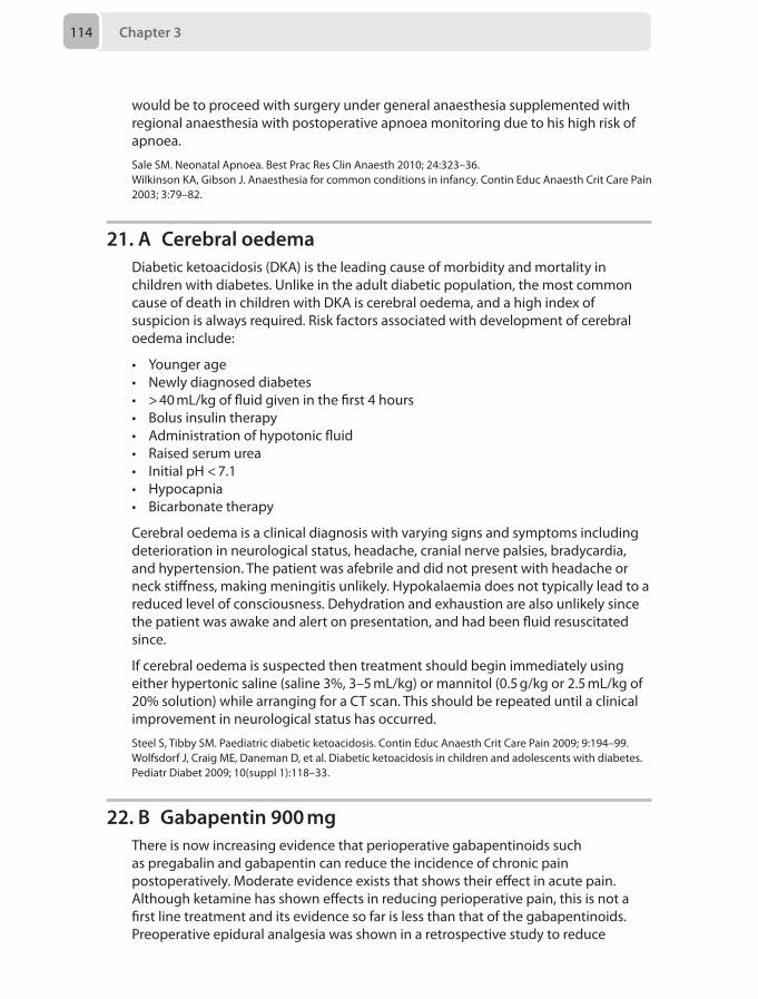
Chapter 3114
would be to proceed with surgery under general anaesthesia supplemented with regional anaesthesia with postoperative apnoea monitoring due to his high risk of apnoea.
Sale SM. Neonatal Apnoea. Best Prac Res Clin Anaesth 2010; 24:323–36.Wilkinson KA, Gibson J. Anaesthesia for common conditions in infancy. Contin Educ Anaesth Crit Care Pain 2003; 3:79–82.
21. A Cerebral oedemaDiabetic ketoacidosis (DKA) is the leading cause of morbidity and mortality in children with diabetes. Unlike in the adult diabetic population, the most common cause of death in children with DKA is cerebral oedema, and a high index of suspicion is always required. Risk factors associated with development of cerebral oedema include:
• Younger age• Newly diagnosed diabetes• > 40 mL/kg of fluid given in the first 4 hours• Bolus insulin therapy• Administration of hypotonic fluid• Raised serum urea• Initial pH < 7.1• Hypocapnia• Bicarbonate therapy
Cerebral oedema is a clinical diagnosis with varying signs and symptoms including deterioration in neurological status, headache, cranial nerve palsies, bradycardia, and hypertension. The patient was afebrile and did not present with headache or neck stiffness, making meningitis unlikely. Hypokalaemia does not typically lead to a reduced level of consciousness. Dehydration and exhaustion are also unlikely since the patient was awake and alert on presentation, and had been fluid resuscitated since.
If cerebral oedema is suspected then treatment should begin immediately using either hypertonic saline (saline 3%, 3–5 mL/kg) or mannitol (0.5 g/kg or 2.5 mL/kg of 20% solution) while arranging for a CT scan. This should be repeated until a clinical improvement in neurological status has occurred.
Steel S, Tibby SM. Paediatric diabetic ketoacidosis. Contin Educ Anaesth Crit Care Pain 2009; 9:194–99.Wolfsdorf J, Craig ME, Daneman D, et al. Diabetic ketoacidosis in children and adolescents with diabetes. Pediatr Diabet 2009; 10(suppl 1):118–33.
22. B Gabapentin 900 mgThere is now increasing evidence that perioperative gabapentinoids such as pregabalin and gabapentin can reduce the incidence of chronic pain postoperatively. Moderate evidence exists that shows their effect in acute pain. Although ketamine has shown effects in reducing perioperative pain, this is not a first line treatment and its evidence so far is less than that of the gabapentinoids. Preoperative epidural analgesia was shown in a retrospective study to reduce
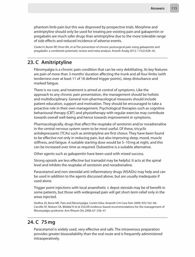
Answers 115
phantom limb pain but this was disproved by prospective trials. Morphine and amitriptyline should only be used for treating pre-existing pain and gabapentin or pregabalin are much safer drugs than amitriptyline due to the more tolerable range of side effects and reduced incidence of adverse events.
Clarke H, Bonin RP, Orser BA, et al.The prevention of chronic postsurgical pain using gabapentin and pregabalin: a combined systematic review and meta-analysis. Anesth Analg 2012; 115(2):428–42.
23. C AmitriptylineFibromyalgia is a chronic pain condition that can be very debilitating. Its key features are pain of more than 3 months’ duration affecting the trunk and all four limbs (with tenderness over at least 11 of 18 defined trigger points), sleep disturbance and marked fatigue.
There is no cure, and treatment is aimed at control of symptoms. Like the approach to any chronic pain presentation, the management should be holistic and multidisciplinary. General non-pharmacological measures should include patient education, support and motivation. They should be encouraged to take a proactive role in their own management. Psychological therapies such as cognitive behavioural therapy (CBT) and physiotherapy with regular exercise may contribute towards overall well-being and hence towards improvement in symptoms.
Pharmacologically, drugs that affect the reuptake of serotonin and/or noradrenaline in the central nervous system seem to be most useful. Of these, tricyclic antidepressants (TCAs) such as amitriptyline are first choice. They have been found to be effective not only in reducing pain, but also improving sleep, mood, muscle stiffness, and fatigue. A suitable starting dose would be 5–10 mg at night, and this can be increased over time as required. Duloxetine is a suitable alternative.
Other agents such as gabapentin have been used with mixed success.
Strong opioids are less effective but tramadol may be helpful. It acts at the spinal level and inhibits the reuptake of serotonin and noradrenaline.
Paracetamol and non-steroidal anti-inflammatory drugs (NSAIDs) may help and can be used in addition to the agents discussed above, but are usually inadequate if used alone.
Trigger point injections with local anaesthetic ± depot steroids may be of benefit in some patients, but those with widespread pain will get short-term relief only in the areas injected.
Dedhia JD, Bone ME. Pain and fibromyalgia. Contin Educ Anaesth Crit Care Pain 2009; 9(5):162–66.Carville SF, Nielsen SA, Bliddal H et al. EULAR evidence-based recommendations for the management of fibromyalgia syndrome. Ann Rheum Dis 2008; 67: 536–41
24. C 75 mgParacetamol is widely used, very effective and safe. The intravenous preparation provides greater bioavailability than the oral route and is frequently administered intraoperatively.
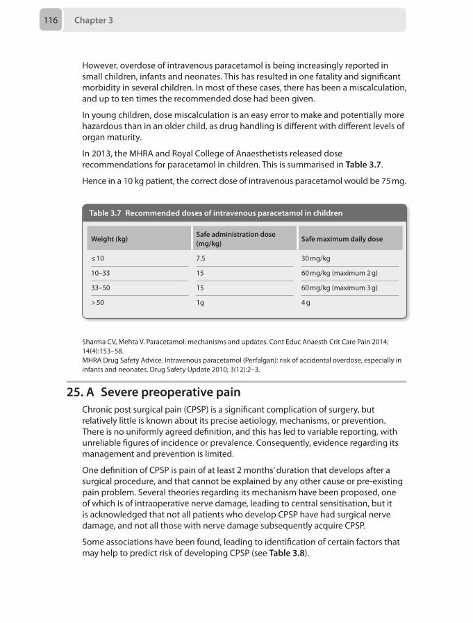
Chapter 3116
However, overdose of intravenous paracetamol is being increasingly reported in small children, infants and neonates. This has resulted in one fatality and significant morbidity in several children. In most of these cases, there has been a miscalculation, and up to ten times the recommended dose had been given.
In young children, dose miscalculation is an easy error to make and potentially more hazardous than in an older child, as drug handling is different with different levels of organ maturity.
In 2013, the MHRA and Royal College of Anaesthetists released dose recommendations for paracetamol in children. This is summarised in Table 3.7.
Hence in a 10 kg patient, the correct dose of intravenous paracetamol would be 75 mg.
Table 3.7 Recommended doses of intravenous paracetamol in children
Weight (kg)Safe administration dose (mg/kg)
Safe maximum daily dose
≤ 10 7.5 30 mg/kg
10–33 15 60 mg/kg (maximum 2 g)
33–50 15 60 mg/kg (maximum 3 g)
> 50 1g 4 g
Sharma CV, Mehta V. Paracetamol: mechanisms and updates. Cont Educ Anaesth Crit Care Pain 2014; 14(4):153–58.MHRA Drug Safety Advice. Intravenous paracetamol (Perfalgan): risk of accidental overdose, especially in infants and neonates. Drug Safety Update 2010; 3(12):2–3.
25. A Severe preoperative painChronic post surgical pain (CPSP) is a significant complication of surgery, but relatively little is known about its precise aetiology, mechanisms, or prevention. There is no uniformly agreed definition, and this has led to variable reporting, with unreliable figures of incidence or prevalence. Consequently, evidence regarding its management and prevention is limited.
One definition of CPSP is pain of at least 2 months’ duration that develops after a surgical procedure, and that cannot be explained by any other cause or pre-existing pain problem. Several theories regarding its mechanism have been proposed, one of which is of intraoperative nerve damage, leading to central sensitisation, but it is acknowledged that not all patients who develop CPSP have had surgical nerve damage, and not all those with nerve damage subsequently acquire CPSP.
Some associations have been found, leading to identification of certain factors that may help to predict risk of developing CPSP (see Table 3.8).
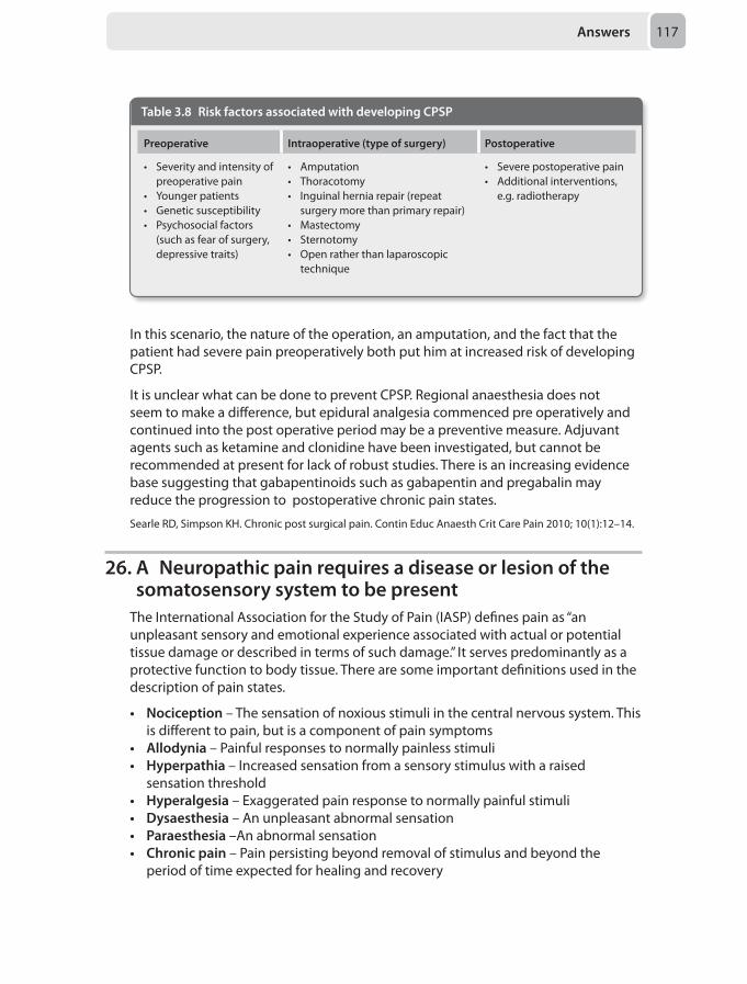
Answers 117
Table 3.8 Risk factors associated with developing CPSP
Preoperative Intraoperative (type of surgery) Postoperative
• Severity and intensity of preoperative pain
• Younger patients• Genetic susceptibility• Psychosocial factors
(such as fear of surgery, depressive traits)
• Amputation• Thoracotomy• Inguinal hernia repair (repeat
surgery more than primary repair)• Mastectomy• Sternotomy• Open rather than laparoscopic
technique
• Severe postoperative pain• Additional interventions,
e.g. radiotherapy
In this scenario, the nature of the operation, an amputation, and the fact that the patient had severe pain preoperatively both put him at increased risk of developing CPSP.
It is unclear what can be done to prevent CPSP. Regional anaesthesia does not seem to make a difference, but epidural analgesia commenced pre operatively and continued into the post operative period may be a preventive measure. Adjuvant agents such as ketamine and clonidine have been investigated, but cannot be recommended at present for lack of robust studies. There is an increasing evidence base suggesting that gabapentinoids such as gabapentin and pregabalin may reduce the progression to postoperative chronic pain states.
Searle RD, Simpson KH. Chronic post surgical pain. Contin Educ Anaesth Crit Care Pain 2010; 10(1):12–14.
26. A Neuropathic pain requires a disease or lesion of the somatosensory system to be present
The International Association for the Study of Pain (IASP) defines pain as “an unpleasant sensory and emotional experience associated with actual or potential tissue damage or described in terms of such damage.” It serves predominantly as a protective function to body tissue. There are some important definitions used in the description of pain states.
• Nociception – The sensation of noxious stimuli in the central nervous system. This is different to pain, but is a component of pain symptoms
• Allodynia – Painful responses to normally painless stimuli• Hyperpathia – Increased sensation from a sensory stimulus with a raised
sensation threshold• Hyperalgesia – Exaggerated pain response to normally painful stimuli• Dysaesthesia – An unpleasant abnormal sensation• Paraesthesia –An abnormal sensation • Chronic pain – Pain persisting beyond removal of stimulus and beyond the
period of time expected for healing and recovery
Chapter 3118
• Neuropathic pain – Defined by the IASP as pain caused by a lesion or disease of the somatosensory nervous system
• Complex regional pain syndrome – A chronic, painful condition that can be either spontaneously occurring or due to nerve injury characterised by a collection of sensory, vasomotor, sudomotor and trophic skin changes
Thus it is clear that of the options given, the most correct answer describes neuropathic pain as requiring a disease or lesion of the somatosensory nervous system.
Merskey H, Bogduk N. Classification of chronic pain. Descriptions of chronic pain syndromes and definitions of pain terms. Seattle, WA: IASP Press; 1994:209–13.
27. C Fluid requirement should be calculated as per the Baxter formula: 4 mL/kg/% burn. Half the fluid should be given in the first 8 hours and the remaining half over the next 16 hours from the time of burn
Major burns cause a systemic inflammatory response syndrome resulting in increased capillary permeability and large fluid shifts. It is therefore important to maintain intravascular volume to ensure end organ perfusion. However, it should be remembered that hypovolaemic shock in the first few hours following a burn is never due solely to the burn and an alternative injury/source should be sought.
Two large bore IV cannulae should be inserted ideally through unburned skin. The groins are often spared making femoral CVC access practical. However fluid resuscitation should not be delayed if IV access through unburned skin is proving difficult.
IV fluid resuscitation is required in adults if the burn involves more than 15% BSA or 10% with smoke inhalation.
There are various formulae that attempt to estimate the fluid deficit but these clearly provide no more than a rough guide. The Parkland formula (also known as the Baxter formula) is the most widely used. The modified Brooke formula (2 mL/kg/% burn in the first 24 hours) can also be used but is less well known and was hoping to address the issues of ‘fluid creep’.
Parkland formula: 4 mL/kg/% burn in the first 24 hours
Half of the fluid is given in the first 8 hours and the remaining half is given over the next 16 hours.
The calculation should start from the time of the initial burn and not the time of presentation to hospital. It is generally accepted that the fluid of choice is Hartmann’s solution. Any pre-hospital fluid administered should be deducted from the calculated requirement.
Given the volume of fluid often administered and the propensity of burns patients to hypothermia, the Hartmann’s should be warmed. After the first 24–48 hours capillary permeability should have returned to normal and fluid balance is much less of an issue.
Bishop S, Maguire S. Anaesthesia and intensive care for major burns. Contin Educ Anaesth Crit Care Pain 2012; 12(3):118–22.
Answers 119
28. A Increased preload, increased afterloadApplication of aortic cross-clamp can cause significant rise in systemic blood pressure. This is caused by an increase in left ventricular afterload, due to the cross clamp. Reflex venoconstriction below the clamp, particularly in the splanchnic circulation, causes an increase in the blood volume in the inferior and superior vena cavae, hence causing increase in preload.
The change in afterload is consistent. The change in preload is variable when aortic cross clamping is applied infrarenally, but is present in 90% of cases when the clamp is applied above the coeliac arteries.
Gelman S. The pathophysiology of aortic cross-clamping and unclamping. Anesthesiology 1995; 82:1026–60.
29. A Creatinine kinaseThis patient has presented with clinical features suggestive of rhabdomyolysis.
Rhabdomyolysis is a clinical spectrum, which occurs as a result of the breakdown of striated muscle. The muscle degradation leads to elevated intracytoplasmic calcium, which causes myocyte constituents to be released into blood, eventually leading to acute kidney injury and hyperkalaemia.
The aetiology of rhabdomyolysis can be traumatic, including crush injuries, electrocution or blunt trauma, or non-traumatic such as infection (legionella, tetanus), drugs (cocaine, alcohol) or immune mediated (polymoyositis, dermatomyositis).
Alcohol is thought to be involved in up to 20% of cases of rhabdomyolysis in the UK. Alcohol-induced coma leads to prolonged immobilisation and muscle compression with ischaemia. It also leads to myopathy and damages cell sarcolemma, increasing sodium permeability. This influx of sodium activates the sodium-calcium exchange pump, which increases cytosolic calcium along with direct influx of calcium due to cellular damage.
It presents with a spectrum, from asymptomatic to hypovolaemic shock accompanied with life threatening electrolyte imbalance and acute kidney injury. Clinically, it is syptomised by malaise, muscle pains, fever and tachycardia.
Myocyte injury leads to severe hyperkalaemia, hyperuricaemia and hyperphosphataemia, while muscle necrosis causes lactic acidosis, further exacerbating the hyperkalaemia.
The most sensitive test for rhabdomyolysis is serum creatinine kinase (CK), an enzyme released by striated muscle when damaged. An five-fold increase in serum CK is expected in rhabdomyolysis. Serum CK concentrations greater than 5000 units/litre are associated with a >50% incidence of acute renal failure.
High concentrations of myoglobin released can cause myoglobinuria, which is responsible for the brown discolouration of urine. Routine dipstick test of urine cannot differentiate between haemoglobinuria (haemoglobin in urine), haematuria (red blood cells in urine) or myoglobinuria (haem-containing myoglobin in urine)
Chapter 3120
as it detects presence of haem which is positive in all cases. As the liver normally metabolises myoglobin rapidly, an absence of it in blood or urine does not eliminate the diagnosis of rhabdomyolysis.
Treatment consists of prompt fluid resuscitation, control of hyperkalaemia and renal support with filtration if indicated.
Hunter JD, Greeg K, Damani Z. Rhabdomyolysis. Contin Educ Anaesth Crit Care Pain 2006; 6(4):141-143.
30. A Ascorbic acidThis patient has clinical features suggestive of scurvy, caused by vitamin C (ascorbic acid) deficiency. It is rarely seen in developed countries, except in extremes of age most commonly due to malnourishment, but can occur due to dietary vitamin C insufficiency. As vitamin C is an essential component for collagen synthesis it’s deficiency leads to fragile capillaries, impaired wound healing, poor bone formation and muscle symptoms. Administering a diet rich in vitamin C including products such as citrus fruits, tomatoes and bell peppers can prevent it.
Iron deficiency leads to anaemia, which could cause lethargy, breathlessness and palpitations.
Vitamin B1 or thiamine deficiency leads to beriberi, which is characterised, by heart failure, oedema (wet) and neuropathies (dry).
Vitamin A deficiency leads to predominant visual symptoms including xeropthalmos and blindness, while vitamin D deficiency leads to osteogenic defects such as rickets disease and osteomalacia.
Raynaud-Simon A, Cohen-Bittan J, Guoronnec A, et al. Scurvy in hospitalized elderly patients. J Nutr Health Aging 2010; 14(6):407-410.Longmore M, Wilkinson I, Torok E. Oxford Handbook of Clinical Medicine, 8th ed. Oxford: Oxford University Press, 2010.
Mock Paper 4
Chapter 4
Questions1. A 32-year-old man is admitted to the intensive care unit. 2 weeks ago he suffered a
bout of gastroenteritis, following which he noticed bilateral leg pain and weakness which then progressed proximally and he soon had difficulty coughing and swallowing. Since admission he has been persistently tachycardic and sweaty with episodes of hypertension and hypotension.
Which of the following clinical features is most likely to confirm his diagnosis?
A Progressive areflexic weakness in more than one limbB Progressive rise in CSF protein levels > 0.4 g/LC Symmetrical weaknessD Autonomic dysfunctionE Bulbar palsy
2. A 44-year-old woman with a past history of hypothyroidism has suffered a traumatic brain injury and is on the neurointensive care unit. She had an isolated seizure following a blunt force to her cranium and remains intubated and ventilated. She is clinically euvolaemic but her investigations reveal the following:• Serum sodium 122 mmol/L (normal range 135–145 mmol/L)• Serum osmolality 270 mOsm/kg (normal range 278–305 mOsm/kg)• Urine osmolality 300 mOsm/kg (normal range 500–800 mOsm/kg)
Which of the following is the most likely cause of her biochemical derangement?
A Hypotonic fluid administrationB Syndrome of inappropriate antidiuretic hormone secretion (SIADH)C Cerebral salt-wasting syndrome (CSWS)D Phenytoin administrationE Myxoedema
3. You are asked to see a 60-year-old woman with a suspected myocardial infarction. She is known to have a permanent pacemaker and implantable cardioverter-defibrillator (ICD). Shortly after arriving she suffers a cardiac arrest. The monitor shows ventricular fibrillation.
Which of the following best describes the optimum position of the defibrillation pads?
Chapter 4 122
A Anterior-posterior positionB Directly over the pacemakerC At least 8 cm from the generator positionD Anterior-lateral positionE No defibrillation pads should be applied
4. A 7-year-old child is experiencing breathing difficulties. After suffering from coryza and a 3-day low-grade fever, his parents noticed a rapid deterioration in his breathing overnight. He is now stridulous with a high-grade fever and is producing copious secretions. He is lying flat, moving his neck freely and there is no response to nebulised adrenaline and steroids.
What is the most likely diagnosis?
A EpiglottitisB Viral croupC BronchiolitisD Retropharyngeal abscessE Bacterial tracheitis
5. You are involved in a critical incident in the neuroradiology suite. A patient undergoing an angiogram received a total of 7 mg/kg of plain lignocaine to anaesthetise the groin for femoral arterial access. The patient then lost cardiac output which returned following administration of intravenous lipid emulsion, according to national guidelines. They have since been transferred to the intensive care unit.
Which of the following describes your most appropriate action immediately after the event?
A Report the case to the NHS Commissioning Board Special Health AuthorityB Ensure that you have fully documented the event in the patient records C Contact your medical indemnity providerD Organise an ‘after action review’ with all personnel involvedE Instigate the local reporting mechanism for critical incidents
6. An 80-year-old man for an open oesophagectomy has a cardiopulmonary exercise test (CPET) as follows:• Anaerobic threshold (AT) 10 mL/kg/min• Peak oxygen consumption (V
.O2 max) is 75% of that predicted
When describing the results the most correct description includes:
A His AT gives him a higher rate of complicationsB His AT gives him a higher risk of deathC His V
.O2 max max gives him a low risk of death
D His V.O2 max max is associated with the highest risk of complications
E He should be nursed in intensive care postoperatively
Questions 123
7. A 76-year-old frail lady is undergoing an awake fibreoptic nasal intubation. She received intravenous glycopyrrolate as an antisialagogue and midazolam for anxiolysis. Her nasal mucosa was prepared with co-phenylcaine, and the anaesthetist is using a “spray as you go” and nebuliser anaesthetic technique with lignocaine. Remifentanil is used for sedation. She suddenly complains of lightheadedness, tinnitus, confusion and peri-oral paraesthesia.
Which is the most likely drug responsible for her symptoms?
A GlycopyrrolateB MidazolamC LignocaineD PhenylephrineE Remifentanil
8. A 45-year-old man is brought to the emergency department following a 30 minute out-of-hospital cardiac arrest. He has a return of spontaneous circulation, is intubated, has a blood pressure of 110/60 mmHg, a heart rate of 80 beats per minute in sinus rhythm, GCS 3/15, blood glucose 5 mmol/L and core temperature of 34.6°C.
What is the most appropriate step to consider post-cardiac arrest management?
A Transfer the patient to intensive care for further managementB Start surface cooling with ice packsC Insert an arterial and central lineD Call his family and discuss likelihood of poor prognosisE Start sedation and muscle relaxation
9. A 76-year-old man has undergone an arthroscopic acromioclavicular joint decompression. He had an interscalene block and a general anaesthetic. His past medical history includes obesity, moderate chronic obstructive pulmonary disease (COPD) and obstructive sleep apnoea (not on CPAP). In the recovery room he is awake, but struggling to breathe. He has four twitches and no fade to peripheral nerve stimulation, and saturations are 96% on oxygen via facemask. There is minimal wheeze on auscultation.
The best treatment is likely to involve:
A Another dose of neostigmine reversalB Perform an arterial blood gas analysisC BiPAP until the block wears offD CPAP until opioid freeE Nebulisers and steroids
10. A 70-year-old man is to have a number of tendons repaired in his hand. After discussion with the patient, a regional anaesthetic technique has been agreed; your preferred approach is an infraclavicular block under ultrasound guidance.
Chapter 4 124
Which part of the brachial plexus is most likely to be blocked by this approach?
A RootsB TrunksC Divisions D CordsE Branches
11. A 20 kg 5-year-old is admitted to the emergency department with a fractured femur following a bicycle fall. His leg was splinted prior to arrival and he received two crystalloid boluses of 400 mL. His has a respiratory rate of 40 breaths per minute, a capillary refill time of 4 seconds and heart rate of 160 beats per minute.
What is the next step in the resuscitation phase while waiting for surgery?
A A further bolus of 200 mL of colloidB 200 mL bolus of O negative bloodC Call the transfusion lab and order crossmatched blood, fresh frozen plasma
(FFP) and plateletsD Change the temporary femoral splint to a plaster of paris castE Insert an arterial line
12. You are caring for a 35-year-old patient on the neurointensive care unit who has a serious traumatic brain injury following an assault.
According to the Academy of Medical Royal Colleges 2008 Code of Practice for the Diagnosis and Confirmation of Death, brainstem death should only be diagnosed when:
A The blood glucose is 4.0–8.0 mmo/LB Prior to an apnoea test the Paco2 must be 6.0-8.0 kPa and the arterial pH 7.20–
7.40C Two complete sets of tests have been performed a minimum of 4 hours apartD The serum sodium is > 124 mmol/LE After 5 minutes of observed apnoea the Pao2 is < 8 kPa
13. A 47-year-old woman collapsed due to a grade 3 sub-arachnoid haemorrhage. An extra-ventricular drain was inserted and she is ventilated on the intensive care unit. On examination the blood pressure is 90/60 mmHg, heart rate is 110 beats per minute and the peripheral capillary refill time is 6 seconds. She has passed 1000 mL of urine since catheterisation 4 hours ago.
What is the most useful course of action?
A Insert a femoral central line and commence a noradrenaline infusion B Request urinary and plasma osmolarity measurementsC Administer 20 mL/kg intravenous crystalloid D Request urinary sodium concentration measurementE Administer intravenous desmopressin (DDAVP) 0.4 mg.
Questions 125
14. A 67-year-old woman presents with an acute onset illness and progressive physiological deterioration. She has a pyrexia of 39.7°C, a heart rate of 135 beats per minute (in atrial fibrillation), a systemic blood pressure of 85/48 mmHg, a respiratory rate of 28 breaths per minutes, oxygen saturations of 89% on a non-rebreathe reservoir face mask with oxygen at 15 L/min, is drowsy and has a capillary blood glucose in 12.2 mmol/L.
The best choice of fluid type to bolus as a first step in cardiovascular resuscitation would be:
A 0.9% sodium chlorideB A balanced crystalloidC A gelatin solutionD 4.5% human albumin in 0.9% sodium chlorideE A starch solution
15. A 54-year-old man with known alcoholic liver disease presents to the emergency department with confusion. On examination he has stigmata of decompensated liver disease and is oedematous with marked ascites. His respiratory rate is 30 breaths per minute, the oxygen saturations are 94% on air, his blood pressure is 90/60 mmHg and his heart rate is 120 beats per minute. After catheterisation he produces 10 mL of urine in the first hour.
What would be the most useful investigation to establish the cause of his confusion?
A CT headB Rectal examinationC Arterial blood gasD Renal function testsE Amylase
16. The cardiologists have inserted a temporary transvenous pacing wire in an 83-year-old man in the intensive care unit.
In which of the following scenarios is the urgent placement of a temporary cardiac pacing system the best treatment option?
A Verapamil overdose with shockB Incidental finding of a heart rate of 38 beats per minute, sinus rhythm with
a PR interval of 260 ms and very frequent, multifocal, supraventricular and ventricular ectopic beats without shock
C Hyperkalaemia with shockD A patient with a PR interval of 220 ms and left bundle branch block who
requires emergency surgery under general anaesthesiaE Second degree heart block with frequent pauses > 2 seconds associated with
syncope
Chapter 4 126
17. A 24-year-old primagravid woman presents in the anaesthetic antenatal clinic for pre-assessment for a high body mass index (BMI). She is currently 26/40 pregnant and already has a BMI of 49.
What is the best line of advice to give to her at this stage?
A Lose weight before the due dateB Early intravenous access, early epidural for labour and risk of difficult
neuraxial blockade or intubation in an emergency Caesarean sectionC Early intravenous access, remifentanil PCA for labour and risk of difficult
intubation in an emergency Caesarean sectionD Aim for normal delivery without any anaesthetic interventionE Aim for normal delivery but be prepared for difficult epidural or spinal
anaesthesia if needed
18. A 5-year-old 20 kg girl is scheduled for an elective adenotonsillectomy for obstructive sleep apnoea (OSA) as a day case. She has Trisomy 21. Her parents inform you that she snores when she sleeps. A recent sleep study showed significant periods of apnoea and desaturation, with an apnoea/hypopnoea index of 12 (severe OSA). An echocardiogram done in her first year of life showed normal intra-cardiac anatomy. Her thyroid function is also normal.
The most important preoperative preparation for this case is:
A Check the full blood count for polycythaemia secondary to recurrent desaturations
B Request a repeat echocardiogramC Arrange an inpatient bed so that the patient can be admitted overnight for
respiratory monitoring postoperativelyD Prescribe a sedative premedicationE Apply local anaesthetic cream to potential sites for intravenous access
19. A 38 kg 14-year-old girl is in recovery after scoliosis correction surgery. Apart from idiopathic scoliosis, there is no other past medical history of note and no known drug allergies. She was given 7 mg of morphine near the end of surgery, and was started on a morphine patient controlled analgesia (PCA) with 1 mg bolus doses, 5 minute lock-out time, and 1 mg/hour background infusion.
After 4 hours of observation in recovery, she appears drowsy, but responds to voice. Her respiratory rate is 10 breaths per minute. On 2 L/min of oxygen via nasal cannulae her oxygen saturation is 96%. Her pupils measure 2 mm bilaterally, and are equally reactive to light.
The PCA pump shows a total of 18 mg of morphine had been delivered with the most recent patient requested bolus an hour ago.
The most appropriate immediate action is:
A Inform the ward to keep her on oxygen to maintain Spo2 over 94%
Questions 127
B Give a 2 µg/kg bolus dose of naloxone and repeat if necessary and reduce PCA bolus and background doses
C Keep her in recovery for further observationD Request an arterial blood gas analysis from the arterial lineE Ask the patient not to use the PCA for the next hour
20. A 4-year-old 18 kg boy fractured his left forearm and was put on the emergency theatre list for a manipulation under anaesthesia (MUA) and K-wire insertion.
After induction of anaesthesia with fentanyl, propofol and atracurium, a laryngeal mask airway was inserted and the patient ventilated on oxygen, air and sevoflurane. You gave him 540 mg of co-amoxiclav intravenously. A few minutes later, his heart rate increased from 100 to 160 beats per minute, his oxygen saturation drops from 100% to 97% on 50% oxygen, and you cannot get an automated blood pressure reading. You noticed on auscultation that he has bilateral wheeze and a rash appears around the patient’s neck, arms and torso.
Your first immediate action is:
A Increase the inspired oxygen concentration to 100% B Give chlorpheniramine 2.5 mg intravenouslyC Give an intravenous fluid bolus of 20 mL/kg 0.9% salineD Give adrenaline 10 µg/kg intramuscularlyE Give epinephrine 1:10 000, 1 µg/kg intravenously
21. An 86-year-old woman is on the trauma list for a hemiarthroplasty following a mechanical fall. She weighs 45 kg and can manage two flights of stairs. Her ECG is unremarkable and a recent echocardiogram showed preserved left ventricular function with no significant valvular defects. Bloods show a haemoglobin of 101 g/L, platelets of 200 ×109/L, normal coagulation studies and her creatinine is 87 μmol/L. She has a past medical history of hypertension, hypercholesterolaemia and ischaemic heart disease. She takes aspirin, clopidogrel, bisoprolol, ramipril and atorvastatin.
What would be the most appropriate way to manage her postoperative pain relief?
A Combined spinal and epidural anaesthesia. Placing 2.5 mL of heavy 0.5% bupivacaine and 300 µg of diamorphine intrathecally and using standard low dose mixture as a patient controlled epidural analgesia in the postoperative phase
B Spinal anaesthesia with 2.8 mL of heavy 0.5% bupivacaine. Regular intravenous paracetamol 1g 6 hourly and tramadol 50 mg up to 6 hourly as required for breakthrough pain
C General anaesthesia with a lumbar plexus block. Regular oral paracetamol 1g 6 hourly and ibuprofen 400 mg up to 8 hourly as required for breakthrough pain
D General anaesthesia with a fascia iliaca block. Regular oral paracetamol 1 g 6 hourly and immediate release morphine 5–10 mg up to 4 hourly as required for breakthrough pain
Chapter 4 128
E General anaesthesia with no regional component. Regular oral paracetamol 1 g 6 hourly and ibuprofen 400 mg 8 hourly with a morphine PCA
22. A 75-year-old man with severe debilitating osteoarthritis in both knees presents with worsening pain despite treatment with multiple analgesic medication including paracetamol, ibuprofen, oral morphine sulphate and buprenorphine patch. He is unfit for surgery, but is keen to try acupuncture for symptomatic relief.
Which of the following would preclude his use of acupuncture?
A Insufficient QiB Cellulitis over needle insertion siteC Local anaesthetic hypersensitivityD Abnormal anatomyE Use of buprenorphine patch
23. A 62-year-old woman presents with severe episodes of pain in the distribution of the right mandibular branch of the trigeminal nerve. The attacks are usually precipitated by cold wind and are short lived. She has no relief from paracetamol or tramadol, and is on no other medication.
What would be the most appropriate first line treatment?
A CarbamazepineB Microvascular decompressionC Percutaneous trigeminal continuous radiofrequency neurotomyD GabapentinE Glycerol gangliolysis of the Gasserian ganglion
24. A 70-year-old patient develops pain after a stroke. He has weakness and sensory loss of his right arm and leg.
Which feature of his pain is most accurate:
A Morphine is a good choice of analgesia for the acute phaseB Pain as a result of the stroke is rareC He is unlikely to develop allodyniaD Pain usually occurs in an area of altered sensationE A thalamic lesion has lead to his post-stroke pain
25. A 73-year-old man with advanced pancreatic cancer presents with worsening upper abdominal pain. Despite treatment with opioids and adjuvant medication, he has no pain relief and is being considered for a procedural intervention.
Which of the following procedures is the most appropriate option for this patient?
A Thoracic epidural injectionB Coeliac plexus blockC Lumbar sympathetic plexus blockD Superior hypogastric plexus blockE Sphenopalatine ganglion block
Questions 129
26. A 40-year-old man is fast-tracked through the emergency department with a penetrating eye injury. He is haemodynamically stable, has no other injuries, no past medical history and has not been fasted. He needs to undergo an urgent operation and you are due to perform the anaesthetic.
Which of the following drugs should be avoided in this patient?
A SuxamethoniumB ThiopentoneC RocuroniumD PropofolE Ketamine
27. You are called to review an 82-year-old man with type 2 diabetes in recovery. He underwent an uneventful right common femoral and popliteal angioplasty and stenting in the endovascular radiology suite. The procedure took approximately 8 hours. The nurse is concerned because he continues to ooze from the right groin despite continuous application of pressure on the wound site.
What would be the next step in diagnosing the potential cause of his ongoing bleeding?
A Check his haemoglobinB Perform an arterial blood gasC Check his blood sugarD Check activated clotting time (ACT)E Check his core temperature
28. A 23-year-old man for elective foot surgery is to have a popliteal nerve block for postoperative analgesia. You use an ultrasound-guided technique and infiltrate 20 mL of 0.5% bupivacaine.
Which of the following nerve fibre modalities is most likely to be blocked first once the local anaesthetic has been infiltrated?
A Touch and pressure B Motor C Pain and temperature D Preganglionic autonomic E Proprioception and muscle tone
29. A 78-year-old patient is admitted to the intensive care unit (ITU) following an exacerbation of chronic obstructive pulmonary disease (COPD). He has known prolonged QTc syndrome. His list of medications includes salbutamol, nifedipine for hypertension, glyceryl trinitrate for ischaemic heart disease and digoxin and warfarin for atrial fibrillation. Whilst on the ITU he requires an amiodarone infusion for fast atrial fibrillation (AF) and intravenous cefuroxime for a suspected chest infection. He has now developed ‘torsades de pointes’ syndrome.
Chapter 4 130
Which one of his medications would most likely have been responsible for the dysrhythmia?
A SalbutamolB NifedipineC DigoxinD AmiodaroneE Cefuroxime
30. Two days following a right hemicolectomy for bowel carcinoma, a 64-year-old man develops breathlessness and pleuritic chest pain. His observations reveal:• Pulse rate of 88 beats per minute• Blood pressure of 120/74 mmHg• Oxygen saturation of 94% on 2 L/minute of oxygen
His heart sounds are normal and chest sounds are clear.
Which one of the following investigations would be the most appropriate to confirm a diagnosis in this scenario:
A 12-lead ECGB CT angiographyC Transoesophageal echocardiographyD Serum D-dimerE Arterial blood gas
Answers 131
Answers
1. A Progressive areflexic weakness in more than one limbThe history described is classical of the development of Guillain–Barré syndrome.
Guillain-Barré syndrome is a progressive, infective, demyelinating neuropathy. It has an incidence of 1–2 per 100,000 and usually has a precursor of gastric or respiratory viral illness in its history. Diagnostic features are progressive weakness accompanied by areflexia in more than one limb. Features that support identification of the syndrome include symmetry of limb signs, cranial nerve involvement, respiratory muscle weakness, autonomic dysfunction, mild sensory symptoms and the investigative findings of increasing cerebrospinal fluid (CSF) protein levels (> 4.0 g/L) over subsequent days or slowed nerve conduction studies. Therefore the most relevant diagnostic clinical feature in this patient is progressive areflexia in more than one limb.
Pollard BJ. Handbook of Clinical Anaesthesia, 2nd edn. Philadelphia: Elsevier, 2003. Singer M, Webb AR. Oxford Handbook of Critical Care, 2nd edn. Oxford: Oxford University Press, 2005.
2. B Syndrome of inappropriate antidiuretic hormone secretion (SIADH)Sodium and water go hand in hand to maintain intravascular homeostasis. Serum osmolality is predominantly dictated by sodium concentration, and the control of sodium is a powerful determinant of water distribution. If the serum osmolality increases, hypothalamic osmoreceptors signal the production of antidiuretic hormone (ADH, vasopressin) to reduce water excretion and thereby rectify the imbalance. Total body sodium itself, is controlled by the sympathetic nerves and natriuretic peptides that govern its renal reabsorption once it has been filtered at the glomerulus.
Hyponatraemia can be classified as a serum concentration < 135 mmol/L and may be associated with hyper-, hypo- or euvolaemia. The most common causes of hyponatraemia in a brain injured patient are syndrome of inappropriate ADH secretion (SIADH) and cerebral salt-wasting syndrome (CSWS).
SIADH leads to unregulated ADH release and lack of feedback response such that water is indiscriminately reabsorbed. It is characterised by:
1. Serum sodium < 135 mmol/L2. Reduced serum osmolality < 280 mOsm/kg3. Urine osmolality greater than serum osmolality4. Low urine output5. Normovolaemia (occasionally hypervolaemia)
Treatment includes fluid restriction, furosemide (to encourage water excretion) and demeclocycline (to inhibit renal ADH response, or direct ADH receptor antagonists).
Chapter 4132
CSWS is not fully understood but is associated with increased natriuretic peptides and ultimately involves increased renal sodium loss and subsequently, water is lost in tandem. It is characterised by:
1. Normal or low serum sodium2. Normal or low serum osmolality3. Normal or high urine osmolality4. Normal or high urine output5. Hypovolaemia
Treatment involves replacement of sodium and water. This is usually commenced with 0.9% saline solution but hypertonic 1.8% or 3% solutions may be required if the loss has been acute and the patient is symptomatic.
The negative fluid balance is usually the distinguishing feature between CSWS and SIADH, but can be hard to assess clinically. Very infrequently CSWS can biochemically masquerade as SIADH. In this instance the induced hypovolaemia is such that it results in a consequential rise in ADH.
Iatrogenic hyponatraemia can be seen after hypotonic fluid infusions or as the side effect of some medications such as anticonvulsants, especially carbamazepine and phenytoin.
Systemic disease, such as hypothyroidism, can also be associated with hyponatraemia. Hypothyroid coma or myxoedema is rare but may be triggered by trauma, particularly in the absence of replacement medication.
SIADH is therefore the most likely cause from the options given.
Bradshaw K, Smith M. Disorders of sodium balance after brain injury. Contin Educ Anaesth Crit Care Pain 2008; 8(4):129–133.
3. C At least 8 cm from the generator positionThe patient has a shockable rhythm and requires defibrillation immediately. In patients with an implantable cardioverter-defibrillator (ICD) or permanent pacemaker, it is important not to delay defibrillation; the ICD may not be functional or failed to have detected the dysrhythmia.
Although the strength of evidence is low, the pad should be placed at least 8 cm from the generator; it is possible that automated external defibrillator (AED) devices may sense pacing spikes and so not detect ventricular fibrillation (VF). The other pad can then be placed in the lateral position or the posterior position. Placement of defibrillation pads over the pacemaker/ICD may cause subsequent malfunction of the device and should be avoided.
Resuscitation Council (UK). Adult Resuscitation Guidelines. London: Resuscitation Council (UK), 2010.
4. E Bacterial tracheitisCertain childhood respiratory tract infections have the potential to progress to life threatening airway obstruction if they are not diagnosed and managed correctly. Children with acute severe stridor represent an anaesthetic challenge as
Answers 133
any agitation from the child might precipitate complete obstruction. Intravenous cannulation and throat examination in this scenario should therefore not be attempted. Early, experienced anaesthetic and ENT involvement is recommended and the priority is to examine and secure the airway under anaesthesia.
Bacterial tracheitis is a rare but life threatening condition commonly caused by Staphylococcus aureus and characterised by subglottic oedema with thick mucopurulent secretions compromising the airway. Typically, the child experiences viral upper respiratory tract prodromal symptoms for 2–3 days which is followed by a rapid clinical deterioration over 8–10 hours. At this stage the child may appear toxic, stridulous and have a high fever as described in the above case. A distinguishing feature from epiglottitis is the usual ability of the child to lie flat and the absence of drooling and dysphagia.
Croup is the most common cause of acute stridor in children but usually affects younger age groups (6 months to 3 years). Commonly caused by the parainfluenza virus family, sufferers classically display a barking cough preceded by a prodrome of nasal congestion and rhinorrhea. The deterioration is not as marked as in bacterial tracheitis and copious secretions are not typical features. Furthermore, children often want to sit upright as opposed to lie flat and may show a marked clinical improvement following nebulized adrenaline and steroids.
Since the introduction of the Haemophilus influenzae type b vaccine, epiglottitis has become rare. Epiglottitis normally affects children aged 2–6 years and usually presents abruptly with a high fever, dysphagia, stridor and drooling. The child may prefer leaning forwards with their mouth open to keep their airway open. The presence of antecedent viral symptoms, current secretions as well as the child’s position in the case above makes epiglottitis not the most likely diagnosis.
A retropharyngeal abscess arises in the space between the posterior pharyngeal wall and prevertebral fascia and can cause airway obstruction by physical expansion. The abscess can be formed after a penetrating pharyngeal injury or infected lymph nodes associated with an upper respiratory tract infection. Crucially these patients commonly complain of limited neck movement contrary to the above scenario.
Bronchiolitis is a common and usually self-limiting lower respiratory tract infection caused by the respiratory syncytial virus. Children under 2 years old are most commonly affected and present acutely with rhinorrhea, cough and a low grade fever preceded by a prodrome of several days. Since it is a lower respiratory tract infection, stridor is not usually present. Treatment is supportive and includes oxygen and intravenous fluid therapy as needed. Conflicting evidence remains as to the effectiveness of steroids and nebulised adrenaline in treating this condition.
Maloney E, Meakin G. Acute stridor in children. Contin Educ Anaesth Crit Care Pain 2007; 7(6):183–186.Rafei K, Lichenstein R. Airway infectious disease emergencies. Pediatr Clin North Am 2006; 53(2):215–242.
5. B Ensure that you have fully documented the event in the patient recordsClinical risk management is at the centre of ensuring patient safety and may be prospective or retrospective. Prospective management can be at an individual level,
Chapter 4134
e.g. planning a patient-specific anaesthetic, or at a department level to comply with Clinical Negligence Scheme for Trust (CNST) regulations. In order to manage risk there are five sequential stages of process to be completed.
1. Awareness 2. Identification3. Assessment4. Management5. Re-evaluation
Awareness
A critical incident or Patient Safety Incident (PSI) is that which could or did cause harm, be it unexpected or unintended. It has been reported in the literature that up to 50% of PSIs are preventable.
Identification
Risk is identified in several ways. Local incident reporting mechanisms, by clinical staff or patients, and national data from the NHS Commissioning Board Special Health Authority, formerly the National Patient Safety Agency (NPSA), serve to highlight threats. Case note review is fundamental for recognition and education regarding events. Root cause analysis (RCA) provides a more formal and structured investigation to identify failings in a system.
RCA is undertaken by a team of risk managers including clinicians and, on occasion, lay people. RCA aims to analyse each case thoroughly from documented data (from the whole admission), construction of accurate timelines and personnel contribution to an event, and subsequent interrogation of all information collected to identify the cause. It detects barriers to safe practice which are classified as physical, natural (temporal or distance related), human action and administrative.
Assessment
Identified risk can then be scored according to its potential severity and frequency. This enables a trust to stratify its resources accordingly for the prevention of risk recurrence.
Management
This describes the arrangements implemented to reduce the risk to as low a level as possible. It involves improving those barriers to patient safety identified through RCA. At a local level it may be prudent to hold an after action review (AAR). This is an informal discussion between the staff involved in an incident. It is led by an independent and objective facilitator with the aim of identifying problems and improvements without the allocation of blame.
Re-evaluation
This is essential in order to confirm the absence of renewed risk in light of any changes made. Although all of the options are applicable to action following a
Answers 135
critical incident, full contemporaneous documentation provides the basis of all further risk analysis.
Bould M, Hunter D, Haxby E. Clinical Risk Management in Anaesthesia. Contin Educ Anaesth Crit Care Pain 2006; 6(6):240–43.
6. B His AT gives him a higher risk of deathCardiopulmonary exercise testing (CPET) is no longer the reserve of research labs or specialist centres, and is now widespread. The detailed physiological data produced gives a wealth of information, the correct use and interpretation of which takes skill and experience. Whilst not expecting this standard of candidates, the examiners have recently demonstrated their willingness to scrutinise candidates on the subject.
Risk stratification is an ever-changing topic, and thus there are several factors one has to consider in terms of any question on the subject. Firstly a thorough history and patient examination are a sound foundation, and from there one can incorporate the information into relevant scoring systems such as the ASA, Goldman, Lee and Detsky. Frequently, these give a figure based on single organ, especially cardiac, demise. A detailed history to reveal functional ability is an essential tenet of any pre-assessment and this can be assimilated into a tool such as the Duke Activity Index. Unfortunately the revealed levels of activity may not truly reflect ability and history is subject to bias and recall error. So, in search of more objective data we arrive in the arena of testing. The traditional tests of function, such as echocardiography and spirometry give limited data as they are non-dynamic tests performed at rest.
In addressing this need for another functional test, CPET has evolved into the gold standard for objective functional assessment and measurement.
Performing the test
The test requires two staff and special equipment. One member of staff will attend to and coach the patient whilst the other attends to the data and testing equipment.
The equipment has two parts; a fixed exercise cycle, the resistance of which can be adjusted by the control computer to increase or decrease the work done by the patient. The other element is the metabolic cart, which is formed by a facemask with a gas analyser to measure oxygen use, carbon dioxide evolution, and a pneumotachograph to quantify gas flows and volumes. A 12-lead ECG is also connected to the computer of the cart which assimilates all the gas and ECG data to produce live displays of results alongside a continuous ECG with ST segment analysis.
Pre-test, the exercise bike seat has to be adjusted for height and the facemask straps adjusted and tested for a good seal. A Spo2 probe is attached and a non-invasive blood pressure (NIBP) cuff fitted alongside the 12-lead ECG. The patient then enters the warm up phase, pedalling unloaded at 60 rpm, while baseline spirometry is performed. This unloaded phase continues into the first 3 minutes of testing, followed by sequential increased loading to the pre-calculated ramp protocol. At the end of the test the patient has a cool down period, and remains monitored for a further 10 minutes to observe recovery.
Chapter 4136
Safety
The quoted mortality of the test is in the region of three patients per 100,000 tests, and full resuscitation facilities must be immediately available. Certain conditions preclude testing, such as severe or unstable cardiac/respiratory conditions, thrombosis and dissection and those conditions which may preclude cooperation such as mental disabilities. Whilst wearing the facemask patients cannot talk, so a set of previously agreed signals are used to indicate fatigue and chest pain.
Measurements and results
Gas exchange measurements include oxygen consumption (V.o2), carbon dioxide
production (V.co2), and the respiratory exchange ratio (RER). Ventilatory measures
of respiratory rate, minute ventilation (VE) and tidal volume (VT) are taken, as are cardiovascular parameters of NIBP, ECG, heart rate and oxygen pulse (Vo2/hour). Pulmonary exchange calculations can be taken from the ventilatory equivalents for O2, CO2 and Spo2.
Cardiac output
The oxygen pulse V.o2/hour, is an approximation of stroke volume. Increased work
requires more oxygen to fuel energy usage, and so oxygen consumption increases. Cardiac output is seen to increase in a linear fashion alongside V
.o2, until a peak
oxygen extraction ratio of 75% is reached. The gradient of the V.o2 increase is a
measure of the exercise driven increase in cardiac output.
Anaerobic threshold/V.o2 max
This oft quoted measure is a marker of the efficiency of the cardiorespiratory system. It is also largely unchanged with age, and is unaffected by effort or motivation and is reliable and repeatable for a given patient.
The anaerobic threshold (AT) gives a value for the point at which the oxygen demand outstrips supply as work increases, and therefore anaerobic respiration is evoked. The production of lactate generates an extra acid load to the system and thus increases the production of CO2 (V
.co2). Thus the AT is the inflection point of a
graph of V.co2against O2. In other terms, the AT is also the point at which the RER rises
above 1, and is the lowest point on the plot of ventilatory equivalents for oxygen.
Patients can exercise well beyond their AT, and in most tests this represents roughly the half way mark. V
.o2 max, is the peak V
.o2 usually measured at the time the test is
terminated.
It is should be remembered that the variables discussed are part of a whole testing package and a raft of results which should ideally not be considered in isolation. The results can be considered to be interlinked in physiological terms and in terms of complications. For example a complication can give rise to mortality if of sufficient severity.
The AT is shown to correlate with mortality, and the key ‘cut off’ figure in this regard is considered to be 11 mL/kg/min. Thus he has a higher risk of inpatient postoperative mortality. V
.o2 max has more often been shown to correlate with
Answers 137
complications, but as alluded to the delineation between morbidity and mortality is not always established in studies. However, a V
.O2max of < 60% predicted is known
to be associated with both of these bad outcomes. The AT certainly suggests the patient would benefit from critical care postoperatively, but in most centres this would be normal for other oesophagectomy patients as well.
Agnew N. Preoperative cardiopulmonary exercise testing. Contin Educ Anaesth Crit Care Pain 2010; 10 (2):33–37.
7. C LignocaineAwake fibre-optic intubation is an invaluable anaesthetic tool to help safely manage patients with difficult airways. Its successful execution requires not only familiarity with handling of the scope, but also effective sedation and topical anaesthesia. Multiple agents are frequently given to optimise the intubating conditions and an awareness of common or serious side effects of these drugs is important.
When anaesthetising the airway, it is important to keep a close track of the amount of local anaesthetic administered to prevent inadvertent drug toxicity. Lignocaine is frequently given in different concentrations via various routes and is also present in co-phenylcaine which is sometimes not appreciated. In practice, not all patients are weighed, and caution should be exercised when administering local anaesthetics in the elderly. Lignocaine is a sodium channel blocker and during systemic toxicity, firstly inhibits the inhibitory central nervous system neurons which manifests as confusion, tinnitus and paraesthesia before culminating in convulsions. As further toxicity ensues, a more global central inhibition occurs which results in the loss of consciousness and respiratory depression. Negative inotropy and dysrhythmias which are difficult to treat may also be seen at this stage. The British Thoracic Society recommends that the total dose of lignocaine applied during bronchoscopy should be limited to 8.2 mg/kg. Local anaesthetic toxicity is the most likely answer in the above case in view of the specific excitatory symptoms occurring after multiple administrations of lignocaine to the elderly patient.
Glycopyrrolate is an anti-cholinergic drug which is frequently used to reduce the amount of secretions produced in the patient’s upper airway to aid visualisation during bronchoscopy. Anticholinergics act by competitive antagonism at the muscarinic acetylcholine receptor and toxic central effects include agitation, delirium, hallucinations and seizures. Glycopyrrolate however, has a quaternary ammonium group and therefore does not cross the blood-brain barrier as freely as other anticholinergics such as atropine or hyoscine. Central effects are therefore minimal.
Midazolam is a short acting benzodiazepine which produces amnesia, anxiolysis and sedation. Paradoxical excitement can occur, although this is very rare, and not the most likely explanation for the above scenario.
Phenylephrine is found in co-phenylcaine and provides vasoconstriction to the nasal mucous membrane via α1agonism. Absorption across the mucous membrane can occur which may cause hypertension and reflex bradycardias. Central nervous system effects are unusual and not the most likely cause for the symptoms in the above case.
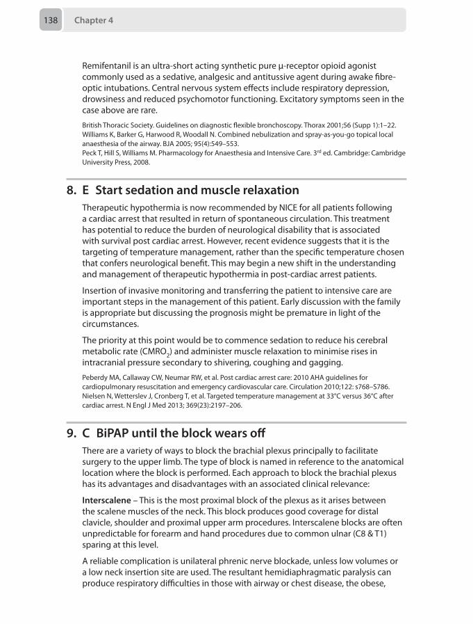
Chapter 4138
Remifentanil is an ultra-short acting synthetic pure μ-receptor opioid agonist commonly used as a sedative, analgesic and antitussive agent during awake fibre-optic intubations. Central nervous system effects include respiratory depression, drowsiness and reduced psychomotor functioning. Excitatory symptoms seen in the case above are rare.
British Thoracic Society. Guidelines on diagnostic flexible bronchoscopy. Thorax 2001;56 (Supp 1):1–22.Williams K, Barker G, Harwood R, Woodall N. Combined nebulization and spray-as-you-go topical local anaesthesia of the airway. BJA 2005; 95(4):549–553.Peck T, Hill S, Williams M. Pharmacology for Anaesthesia and Intensive Care. 3rd ed. Cambridge: Cambridge University Press, 2008.
8. E Start sedation and muscle relaxationTherapeutic hypothermia is now recommended by NICE for all patients following a cardiac arrest that resulted in return of spontaneous circulation. This treatment has potential to reduce the burden of neurological disability that is associated with survival post cardiac arrest. However, recent evidence suggests that it is the targeting of temperature management, rather than the specific temperature chosen that confers neurological benefit. This may begin a new shift in the understanding and management of therapeutic hypothermia in post-cardiac arrest patients.
Insertion of invasive monitoring and transferring the patient to intensive care are important steps in the management of this patient. Early discussion with the family is appropriate but discussing the prognosis might be premature in light of the circumstances.
The priority at this point would be to commence sedation to reduce his cerebral metabolic rate (CMRO2) and administer muscle relaxation to minimise rises in intracranial pressure secondary to shivering, coughing and gagging.
Peberdy MA, Callaway CW, Neumar RW, et al. Post cardiac arrest care: 2010 AHA guidelines for cardiopulmonary resuscitation and emergency cardiovascular care. Circulation 2010;122: s768–S786.Nielsen N, Wetterslev J, Cronberg T, et al. Targeted temperature management at 33°C versus 36°C after cardiac arrest. N Engl J Med 2013; 369(23):2197–206.
9. C BiPAP until the block wears offThere are a variety of ways to block the brachial plexus principally to facilitate surgery to the upper limb. The type of block is named in reference to the anatomical location where the block is performed. Each approach to block the brachial plexus has its advantages and disadvantages with an associated clinical relevance:
Interscalene – This is the most proximal block of the plexus as it arises between the scalene muscles of the neck. This block produces good coverage for distal clavicle, shoulder and proximal upper arm procedures. Interscalene blocks are often unpredictable for forearm and hand procedures due to common ulnar (C8 & T1) sparing at this level.
A reliable complication is unilateral phrenic nerve blockade, unless low volumes or a low neck insertion site are used. The resultant hemidiaphragmatic paralysis can produce respiratory difficulties in those with airway or chest disease, the obese,
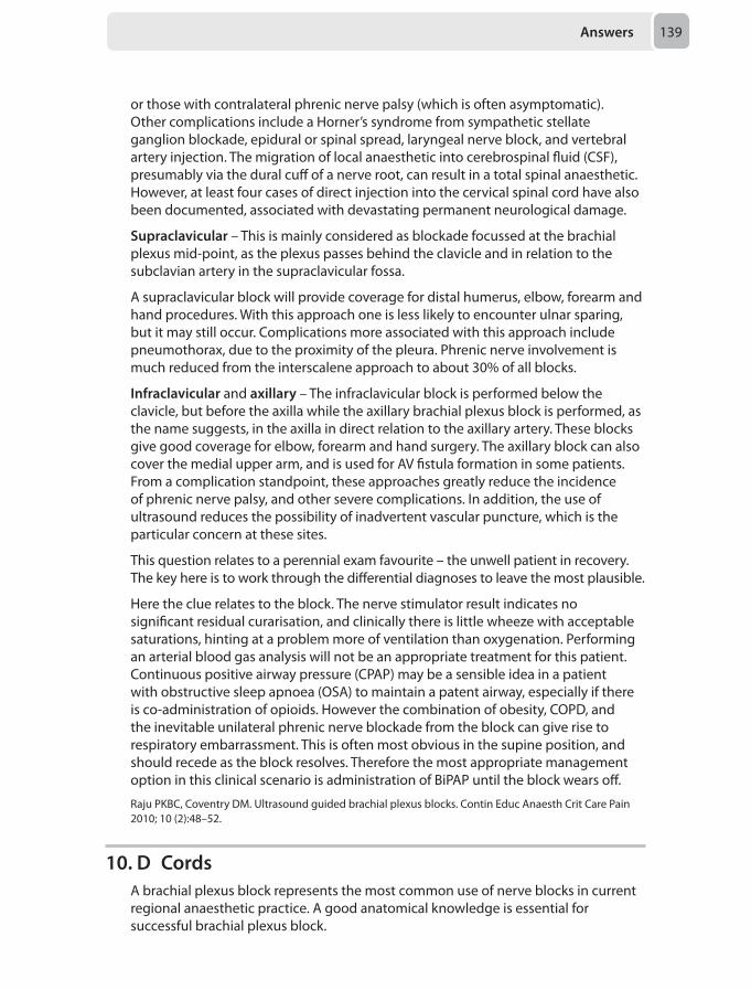
Answers 139
or those with contralateral phrenic nerve palsy (which is often asymptomatic). Other complications include a Horner’s syndrome from sympathetic stellate ganglion blockade, epidural or spinal spread, laryngeal nerve block, and vertebral artery injection. The migration of local anaesthetic into cerebrospinal fluid (CSF), presumably via the dural cuff of a nerve root, can result in a total spinal anaesthetic. However, at least four cases of direct injection into the cervical spinal cord have also been documented, associated with devastating permanent neurological damage.
Supraclavicular – This is mainly considered as blockade focussed at the brachial plexus mid-point, as the plexus passes behind the clavicle and in relation to the subclavian artery in the supraclavicular fossa.
A supraclavicular block will provide coverage for distal humerus, elbow, forearm and hand procedures. With this approach one is less likely to encounter ulnar sparing, but it may still occur. Complications more associated with this approach include pneumothorax, due to the proximity of the pleura. Phrenic nerve involvement is much reduced from the interscalene approach to about 30% of all blocks.
Infraclavicular and axillary – The infraclavicular block is performed below the clavicle, but before the axilla while the axillary brachial plexus block is performed, as the name suggests, in the axilla in direct relation to the axillary artery. These blocks give good coverage for elbow, forearm and hand surgery. The axillary block can also cover the medial upper arm, and is used for AV fistula formation in some patients. From a complication standpoint, these approaches greatly reduce the incidence of phrenic nerve palsy, and other severe complications. In addition, the use of ultrasound reduces the possibility of inadvertent vascular puncture, which is the particular concern at these sites.
This question relates to a perennial exam favourite – the unwell patient in recovery. The key here is to work through the differential diagnoses to leave the most plausible.
Here the clue relates to the block. The nerve stimulator result indicates no significant residual curarisation, and clinically there is little wheeze with acceptable saturations, hinting at a problem more of ventilation than oxygenation. Performing an arterial blood gas analysis will not be an appropriate treatment for this patient. Continuous positive airway pressure (CPAP) may be a sensible idea in a patient with obstructive sleep apnoea (OSA) to maintain a patent airway, especially if there is co-administration of opioids. However the combination of obesity, COPD, and the inevitable unilateral phrenic nerve blockade from the block can give rise to respiratory embarrassment. This is often most obvious in the supine position, and should recede as the block resolves. Therefore the most appropriate management option in this clinical scenario is administration of BiPAP until the block wears off.
Raju PKBC, Coventry DM. Ultrasound guided brachial plexus blocks. Contin Educ Anaesth Crit Care Pain 2010; 10 (2):48–52.
10. D CordsA brachial plexus block represents the most common use of nerve blocks in current regional anaesthetic practice. A good anatomical knowledge is essential for successful brachial plexus block.
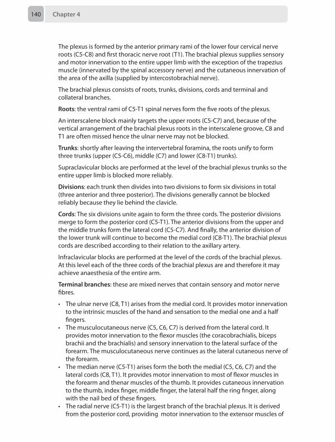
Chapter 4140
The plexus is formed by the anterior primary rami of the lower four cervical nerve roots (C5-C8) and first thoracic nerve root (T1). The brachial plexus supplies sensory and motor innervation to the entire upper limb with the exception of the trapezius muscle (innervated by the spinal accessory nerve) and the cutaneous innervation of the area of the axilla (supplied by intercostobrachial nerve).
The brachial plexus consists of roots, trunks, divisions, cords and terminal and collateral branches.
Roots: the ventral rami of C5-T1 spinal nerves form the five roots of the plexus.
An interscalene block mainly targets the upper roots (C5-C7) and, because of the vertical arrangement of the brachial plexus roots in the interscalene groove, C8 and T1 are often missed hence the ulnar nerve may not be blocked.
Trunks: shortly after leaving the intervertebral foramina, the roots unify to form three trunks (upper (C5-C6), middle (C7) and lower (C8-T1) trunks).
Supraclavicular blocks are performed at the level of the brachial plexus trunks so the entire upper limb is blocked more reliably.
Divisions: each trunk then divides into two divisions to form six divisions in total (three anterior and three posterior). The divisions generally cannot be blocked reliably because they lie behind the clavicle.
Cords: The six divisions unite again to form the three cords. The posterior divisions merge to form the posterior cord (C5-T1). The anterior divisions from the upper and the middle trunks form the lateral cord (C5-C7). And finally, the anterior division of the lower trunk will continue to become the medial cord (C8-T1). The brachial plexus cords are described according to their relation to the axillary artery.
Infraclavicular blocks are performed at the level of the cords of the brachial plexus. At this level each of the three cords of the brachial plexus are and therefore it may achieve anaesthesia of the entire arm.
Terminal branches: these are mixed nerves that contain sensory and motor nerve fibres.
• The ulnar nerve (C8, T1) arises from the medial cord. It provides motor innervation to the intrinsic muscles of the hand and sensation to the medial one and a half fingers.
• The musculocutaneous nerve (C5, C6, C7) is derived from the lateral cord. It provides motor innervation to the flexor muscles (the coracobrachialis, biceps brachii and the brachialis) and sensory innervation to the lateral surface of the forearm. The musculocutaneous nerve continues as the lateral cutaneous nerve of the forearm.
• The median nerve (C5-T1) arises form the both the medial (C5, C6, C7) and the lateral cords (C8, T1). It provides motor innervation to most of flexor muscles in the forearm and thenar muscles of the thumb. It provides cutaneous innervation to the thumb, index finger, middle finger, the lateral half the ring finger, along with the nail bed of these fingers.
• The radial nerve (C5-T1) is the largest branch of the brachial plexus. It is derived from the posterior cord, providing motor innervation to the extensor muscles of
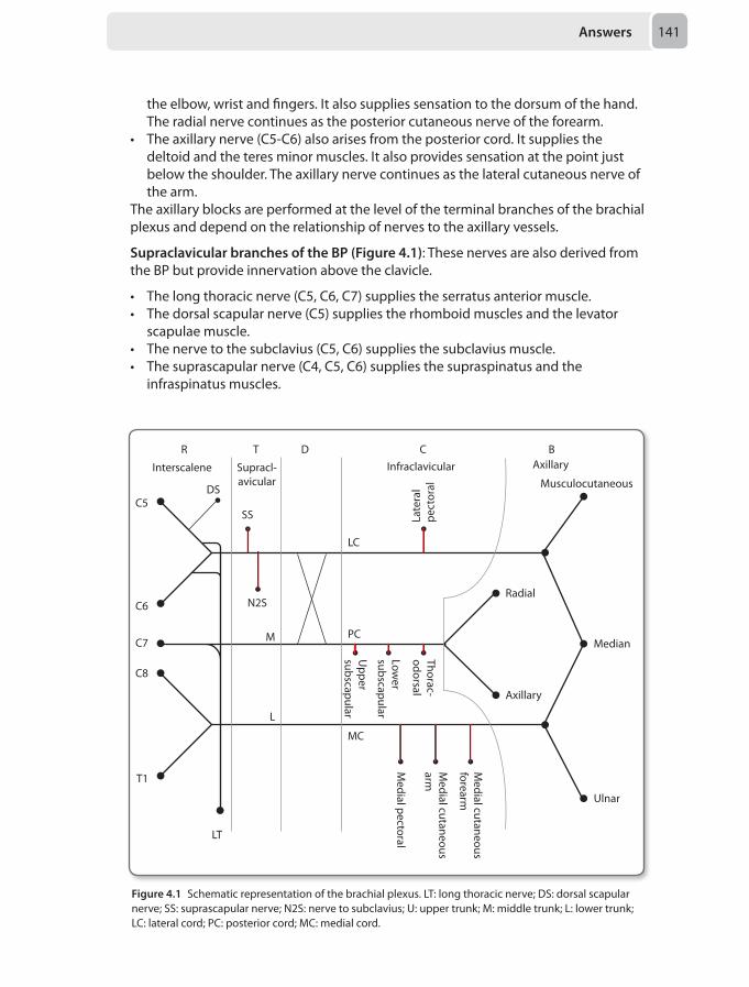
Answers 141
the elbow, wrist and fingers. It also supplies sensation to the dorsum of the hand. The radial nerve continues as the posterior cutaneous nerve of the forearm.
• The axillary nerve (C5-C6) also arises from the posterior cord. It supplies the deltoid and the teres minor muscles. It also provides sensation at the point just below the shoulder. The axillary nerve continues as the lateral cutaneous nerve of the arm.
The axillary blocks are performed at the level of the terminal branches of the brachial plexus and depend on the relationship of nerves to the axillary vessels.
Supraclavicular branches of the BP (Figure 4.1): These nerves are also derived from the BP but provide innervation above the clavicle.
• The long thoracic nerve (C5, C6, C7) supplies the serratus anterior muscle.• The dorsal scapular nerve (C5) supplies the rhomboid muscles and the levator
scapulae muscle.• The nerve to the subclavius (C5, C6) supplies the subclavius muscle.• The suprascapular nerve (C4, C5, C6) supplies the supraspinatus and the
infraspinatus muscles.
C5DS
N2S
SS
PC
MC
M
Ulnar
Median
Musculocutaneous
LC
L
C6
C7
C8
T1
LT
Medial cutaneous
forearm
Medial cutaneous
arm
Medial pectoral
Thorac-odorsal
Lower
subscapular
Upper
subscapular
Radial
Axillary
Interscalene Supracl-avicular
AxillaryInfraclavicular
Late
ral
pect
oral
R T D C B
Figure 4.1 Schematic representation of the brachial plexus. LT: long thoracic nerve; DS: dorsal scapular nerve; SS: suprascapular nerve; N2S: nerve to subclavius; U: upper trunk; M: middle trunk; L: lower trunk; LC: lateral cord; PC: posterior cord; MC: medial cord.
Chapter 4142
Branches of the cords: see Table 4.1
Table 4.1 Branches of the cords of the brachial plexus
Posterior cord branches ULTRA Upper subscapular, lower subscapular, thoracodorsal, radial, axillary
Lateral cord branches LLM Lateral pectoral, lateral root of the median nerve, musculocutaneous
Medial cord branches MMMUM Medial pectoral, medial cutaneous nerve of arm, medial cutaneous nerve of forearm, ulnar, medial root of the median nerve
Al-Haddad MF, Coventry DM. Brachial plexus blockade. BJA CEPD Reviews 2002; 2(2): 33–36.Neal JM, Gerancher JC, Hebl JR. Upper extremity regional anesthesia: essentials of our current understanding. Reg Anesth Pain Med 2009 Mar-Apr;34(2):134–70.
11. B 200 mL bolus of O negative bloodIn the management of paediatric trauma, it is crucial to recognise what the normal physiological parameters are for different aged patients (Table 4.2).
Table 4.2 The normal physiological parameters for different aged children
Age (years) Respiratory rate (breaths per minute)
Heart rate (beats per minute)
Systolic blood pressure (mmHg)
< 1 30–40 110–160 70–90
1–2 25–35 100 –150 80–90
2–5 25– 30 95–140 80– 105
5–2 20–25 80–120 90– 110
> 12 15–20 60–100 100–120
It is thus clear that this patient is expressing deranged parameters and has had significant haemorrhage from the trauma sustained. Fluid resuscitation in paediatric trauma is similar to adult trauma but with some key differences. If the patient fails to respond to repeated boluses of crystalloid or colloid up to a maximum of 40 mL/kg, the next most appropriate step is to use blood and blood products. The dose of packed red cells is 10 mL/kg. This patient has already had the allocated 40 mL/kg and still demonstrates instability therefore should be transfused O negative blood. If the bleeding continues following this, fresh frozen plasma and platelets need to be administered to avoid the coagulopathy worsening the bleeding.
Changing the temporary splint may result in the bone fragments being disrupted causing further bleeding, and will not immediately assist in replenishing the significant blood loss. Insertion of an arterial line will become necessary to monitor the patient intra-operatively but in the current clinical scenario is unlikely to add more to the clinical picture.
Cullen P. Paediatric trauma. Contin Educ Anaesth Crit Care Pain 2012; 12(3):157–161.
Answers 143
12. B Prior to an apnoea test the Paco2 must be 6.0–8.0 kPa and the arterial pH 7.20–7.40The Academy of Medical Royal Colleges 2008 code of practice for the diagnosis and confirmation of death is the current reference manual for brainstem death (BSD) testing in the UK.
In order to undertake BSD testing the following preconditions must be fulfilled:
• An unresponsive coma and apnoea with a recognised date and time of onset• Evidence of irreversible brain damage of known aetiology
You must then exclude potentially reversible causes for the coma and/or apnoea. These exclusions are:
• The presence or persistence of depressant drugs (or their active metabolites)• A body temperature ≤ 34°C• The presence of a reversible circulatory, metabolic or endocrine disorder• Respiratory failure due to neuromuscular blocking agents, other drugs
or potentially reversible causes of apnoea (e.g. cervical injury, profound neuromuscular weakness)
• Sodium levels at time of coma onset must be 115–160 mmol/L• Sodium levels at time of first test. must be 115–160 mmol/L and not have
changed by > 0.5 mmol/L per hour between time of coma onset and first test• Potassium levels at time of first test must be > 2 mmol/L• Phosphate levels at time of first test must be 0.5–3.0 mmol/L• Magnesium levels at time of first test must be 0.5–3.0 mmol/L• Glucose levels at time of first test must be 3.0–20.0 mmol/L
To confirm BSD, the following bedside tests must be conducted. (Note, two complete sets must be performed, however these can be conducted successively without any fixed interval.):
• Absence of any pupillary reaction to light• Absence of any eyelid movements when each cornea is touched in turn• Absence of nystagmus or any eye movement when each ear is instilled with 50 mL
ice cold water• Absence of a gag reflex• Absence of a cough reflex when a suction catheter is passed down into the
trachea • Absence of any motor response when supraorbital pressure is applied• Absence of any spontaneous breathing efforts for 5 minutes
• Preconditions to apnoea test: Paco2 6.0-8.0 kPa and arterial pH 7.20–7.40• Apnoea test only valid if, after 5 minutes: Paco2 increases by > 0.5 kPa; and Pao2
is > 5 kPa; and the systemic mean arterial pressure is ≥ 60 mmHg throughout.If one or more of these preconditions are not met or the bedside tests cannot be performed then ancillary investigations are required to confirm the diagnosis. The legal time of death is when the first set of tests indicates death due to the absence of brain-stem reflexes.
Academy of Medical Royal Colleges. A Code of Practice for the Diagnosis and Confirmation of Death. London: Academy of Medical Royal Colleges, 2008.
Chapter 4144
13. B Request urinary and plasma osmolarity measurementsThis question requests the most useful option, not the first action. The most useful strategy to aid ongoing management would be to work out why the patient is hypotensive and polyuric so that treatment can be tailored appropriately. The hypotension and poor peripheral perfusion will most likely respond to intravenous crystalloid and a noradrenaline infusion but will not aid identification and correction of the underlying condition which is most likely to be diabetes insipidus given the clinical history.
Diabetes inspidus (DI)
The clinical manifestations are due to the failure of release of anti-diuretic hormone (ADH) or reduced activity of ADH on the renal collecting duct. Without ADH there is no re-absorption of water at the collecting duct of the nephron, resulting in loss of plasma as urine which results in hypovolaemia. As a result of renal conservation of sodium, a high plasma sodium concentration and a normal urinary sodium concentration is seen.
DI is caused by:
• Central nervous system DI• Traumatic brain injury (35%) • Sub-arachnoid haemorrhage • Intra-cerebral haemorrhage • Pituitary surgery • End-stage cerebral oedema
• Nephrogenic DI• Renal DI may be caused by lithium treatment
The differential diagnosis includes:
• Diabetes mellitus • Osmotic diuretics (which may have been given in this case)
Diagnosis may be made with the following criteria:
• Increased urine volume > 3000 mL per day or > 1000 mL in 4 hours• High serum sodium > 145 mmol/L• High serum osmolarity > 305 mmol/kg• Low urine osmolarity < 350 mmol/Kg
A plasma osmolarity measurement would be the most useful intervention out of the available options to confirm the diagnosis. The diagnosis of DI will enable correction of her fluid-balance status by administrating exogenous anti-diuretic hormone.
Bradshaw K, Smith M. Disorders of sodium balance after brain injury. Contin Educ Anaesth Crit Care Pain 2008; 8(4):129–33.
14. B A balanced crystalloidThe clinical scenario is suggestive of septic shock. Fluid resuscitation is indicated and should commence with a 10 mL/kg bolus of crystalloid. Though controversial,
Answers 145
there is a strong theoretical argument, though an absence of definitive evidence to suggest that balanced solutions are less harmful than the unphysiological 0.9% sodium chloride. 0.9% sodium chloride is mildly hyperosmotic and contains 50% more chloride ions per litre than plasma and hence infusion of any significant volume may result in a hyperchloraemic acidosis. Although the acidosis is rapidly buffered, the effects of hyperchloraemia are several and include impaired mental function, nausea, gastrointestinal dysfunction, renal vasoconstriction, hyperkalaemia, impaired coagulation and a pro-inflammatory response. What is less clear is whether these effects are clinically important.
Starch solutions have been withdrawn from the UK market due to concerns over safety and questionable evidence. Albumin has not been demonstrated to confer any survival benefit in sepsis but carry a more significant risk of reactions. Finally, gelatins are unbalanced solutions, and again have not been demonstrated to be superior to balanced crystalloid solutions in these circumstances.
Therefore the most appropriate choice of fluid would be a balanced crystalloid solution.
Dellinger RP, Levy MM, Rhodes A. Surviving Sepsis Campaign: International Guidelines for Management of Severe Sepsis and Septic Shock. Crit Care Med 2013; 41(2):580–637.Handy JM, Soni N. Physiological effects of hyperchloraemia and acidosis. Br J Anaesth 2008; 101(2):141–50.
15. B Rectal examinationAlcoholic chronic liver disease is a physiological disaster, with some of the clinician’s concerns being:
• Airway:• an obtunded patient with a full stomach (possibly blood)
• Breathing:• Fluid overload results in pulmonary oedema, • Pleural effusions • Ascites may splint the diaphragm
• Cardiovascular:• Myocardial disease (the symptoms of which may be difficult to differentiate
from reflux or pancreatitis) due to: – Chronic hypervolaemia – Hypertension – Alcohol-induced cardiomyopathy – Pericardial effusion – Ischaemic myopathy – Loss of occult blood (chronic or acute) and coagulopathy: – Gastritis and ulcer disease – Mallory–Weiss tear – Varices
• Disability (an acute deterioration in cognition may be multi-factorial):• Hypoglycaemia• Intoxication• Withdrawal
Chapter 4146
• Seizures• Hyponatraemia • Delayed presentation of head trauma• Encephalopathy
• Others:• Hypothermia• Infections and sepsis• Hepatorenal syndrome • Immuno-suppression • Pancreatitis • Diabetes mellitus • Peripheral neuropathy • Dementia syndromes • Malnourishment • Self-harm and depression
You are presented with such a patient who has an acute change in cognition associated with hypotension and low urine output. As is often the case you do not have further detailed information regarding this gentleman’s prior medical history.
Given the complicated picture the appropriate approach is a prioritisation-centred examination and treatment management pathway (A-B-C-D-E). The most pressing concern is cardiovascular instability and a rectal examination looking particularly for occult blood (be it altered or otherwise) is indicated as a matter of urgency. The finding of rectal blood will focus this scenario from a complicated differential diagnosis into haemorrhagic shock in a patient with a presumed coagulopathy. An arterial blood gas does have a haemoglobin measurement, but early on in haemorrhagic shock the concentration may remain static and chronic anaemia may complicate the interpretation. The other tests are all important but excluding immediately life-threatening conditions must be your first priority and a rectal examination in this group of patients is mandatory.
Lai WK. Management of acute liver failure. Contin Educ Anaesth Crit Care Pain 2004; 4(2):40–43.
16. E Second degree heart block with frequent pauses >2 seconds associated with syncope
Temporary transvenous cardiac pacing is a last resort in the management of brady-dysrhythmias due both to the logistical difficulties related to insertion and the high complication rates of this procedure. In asymptomatic brady-dysrhythmias / cardiac conduction abnormalities, the indications for, and values of, permanent pacemaker insertion are reasonably well defined.
In acute brady-dysrhythmias, the indications for treatment include shock, syncope, malignant escape ventricular tachy-dysrhythmias and asystoles > 2 seconds. Primary treatment should simultaneously include definitive treatment of any acutely reversible cause and a trial of positive chronotropes. The later can be considered in two groups, parasympathetic antagonists (atropine and glycopyrronium) and sympathetic agonists (isoprenaline adrenaline, dobutamine, dopexamine and salbutamol). Failure of this simultaneous treatment approach should lead to an
Answers 147
immediate trial of rescue, transcutaneous cardiac pacing, but only as a bridging therapy either to definitive treatment of the reversible cause or insertion of a temporary (or permanent) transvenous system.
In the above question, the specific treatment for calcium channel overdose is hyperinsulinaemic euglycaemia. Failure to respond to the combination of this and positive chronotrope therapy may warrant the insertion of a temporary transvenous pacing system.
Scenario B warrants consideration of a permanent pacemaker but gives no indication for a temporary system.
Hyperkalaemia causing brady-dysrhythmia should be treated immediately with a combination of intravenous calcium, continuous insulin and dextrose infusion (safer than a bolus infusion, which may be followed by a rebound hyperkalaemia) with the possible additions of salbutamol and/or sodium bicarbonate, followed by definitive therapy such as haemofiltration or haemodialysis.
The patient in scenario D is at significant risk of brady-dysrhythmias and their complications. As such, discussion with the cardiology team and formation of a plan(s), should this occur, would be prudent.
Scenario E represents the clearest indication amongst the given scenarios for insertion of a temporary pacing system, though, depending upon the setting and circumstances, a permanent system may be more appropriate.
Brignole M, Auricchio A, Baron-Esquivias G, et al. 2013 ESC Guidelines on cardiac pacing and cardiac resynchronization therapy: the Task Force on cardiac pacing and resynchronization therapy of the European Society of Cardiology (ESC). Developed in collaboration with the European Heart Rhythm Association (EHRA). Eur Heart J 2013; 34(29):2281–2329.
17. B Early intravenous access, early epidural for labour and risk of difficult neuraxial blockade or intubation in an emergency Caesarean sectionObesity in pregnancy is becoming more commonplace and is associated with many complications, such as gestational diabetes, pre-eclampsia, post-partum haemorrhage, macrosomia, miscarriage and stillbirth. Joint guidelines produced by the Centre for Maternal and Child Enquiries (CMACE) and the Royal College of Obstetricians and Gynaecologists (RCOG) in 2010 recommend that pregnant women with a body mass index (BMI) ≥ 40 should be seen by an obstetric anaesthetist antenatally. When assessing these patients, the following should be considered:
• Airway – higher incidence of difficult intubation and difficult mask ventilation• Respiratory – desaturate quicker and higher incidence of obstructive sleep
apnoea• Cardiovascular – aortocaval compression may be more pronounced in the supine
position. Hypertension, myocardial ischaemia, heart failure and cardiomyopathy are more common, while venous thromboembolism is also a greater risk
• Gastrointestinal – higher aspiration risk due to higher gastric volumes• Anaesthesia – difficult venous access, difficult epidural or spinal anaesthesia• Delivery – increased risk of instrumental delivery and Caesarean section
Chapter 4148
The patient should be advised of the main risks surrounding her delivery (intravenous access, regional anaesthesia, failed intubation), but also be able to trust you as her anaesthetist, as she is already likely to be anxious. It may be difficult for the patient to lose weight during the pregnancy, and this should not be the mainstay of the advice given. Early intravenous access and an early labour epidural should be advised, due to the potential for difficulty and the need to accomplish these outside of an emergency situation. The risks of general anaesthesia should also be explained. A normal delivery without any anaesthetic intervention is the ideal situation, but practically, complications must be prepared for.
Gupta A, Faber P. Obesity in pregnancy. Contin Educ Anaesth Crit Care Pain 2011; 11 (4):143–46.Modder J, Fitzsimons KJ. CMACE/RCOG Joint Guideline: Management of women with obesity in pregnancy. London: Centre for Maternal and Child Enquiries, and Royal College of Obstetrics and Gynaecologists, 2010.
18. C Arrange an inpatient bed so that the patient can be admitted overnight for respiratory monitoring postoperativelyObstructive sleep apnoea (OSA) is common in children, and can be associated with significant morbidity. OSA belongs to a spectrum of diagnoses known as sleep-related breathing disorders in which the airway is completely (apnoea) or partially (hypopnoea) occluded during sleep despite continued respiratory efforts. In young children, adenotonsillar hypertrophy is the most common anatomical abnormality associated with OSA, and adenotonsillectomy is, therefore, the most common surgical intervention. Other risk factors for OSA include midface hypoplasia, macroglossia, muscular hypotonia (all three are features of Down’s syndrome), micrognathia, and obesity.
The apnoea/hypopnoea index (AHI), counts the number of apnoea or hypoapnoea events secondary to obstructive events during sleep for 60 min. An AHI > 10 in children is classified as severe OSA. Respiratory complications after adenotonsillectomy are more common in children with severe OSA. Other risk factors for postoperative complications include age < 3 years, obesity, Down’s syndrome, failure to thrive, history of prematurity and neuromuscular diseases.
Optimal perioperative management of OSA includes thorough preoperative assessment, opioid-sparing anaesthetic and analgesic approaches, and close respiratory observation and monitoring. Postoperative respiratory monitoring of children with OSA should include continuous pulse oximetry to assess oxygenation and clinical observation with measurement of the respiratory rate at frequent intervals as a secondary assessment of the adequacy of ventilation. Measurement of a full blood count for polycythaemia is not an effective method of assessing desaturations. As this patient has had an echocardiograph showing normal intra-cardiac anatomy, a repeat echocardiograph is unlikely to provide information that would change management. Sedative premedications increase the risks of airway complications in OSA patients and are unlikely to benefit this patient. Finally, application of topical anaesthesia at potential intravenous access sites is valuable, but not the most important preoperative preparation for this case.
Patino M, Sadhasivam S, Mahmoud M.Obstructive sleep apnoea in children: perioperative considerations. Br J Anaesth 2013;111(suppl 1): i83–i95.
Answers 149
19. B Give a 2 µg/kg bolus dose of naloxone and repeat if necessary and reduce PCA bolus and background dosesPatient controlled analgesia (PCA) using intravenous morphine is an excellent way to manage postoperative pain in older children. It has better analgesic efficacy and patient satisfaction compared to other common analgesia modalities. However, it is not without side effects or complications – itching and respiratory depression being two of the most common. The risk is higher when a PCA is used with a continuous background.
The history, symptoms and signs of this patient are all consistent with opioid induced drowsiness and respiratory depression. A small dose of naloxone, a µ-receptor antagonist, is appropriate in this case. Naloxone has a shorter half-life than morphine, so repeated doses may be required. Ideally, the naloxone would be titrated to reverse the drowsiness and respiratory depression, but not the analgesic effects. In severe overdoses, airway and ventilator support may be indicated.
Different patients have different sensitivity to opioid. The initial PCA setting was evidently inappropriate for this patient. The bolus dose should be reduced, and the background infusion either reduced or removed.
Association of Paediatric Anaesthetists of Great Britain and Ireland (APAGBI). Good Practice in Postoperative and Procedural Pain Management, 2nd ed. Paed Anaesth 2012;22 (suppl 1):1–79.Macintyre PA. Safety and efficacy of patient-controlled analgesia. Br J Anaesth 2001;87:36–46.
20. A Increase the inspired oxygen to 100%This is a case of suspected anaphylaxis. It is a life-threatening allergic reaction triggered by a wide range of antigens and involves multiple organ systems. Signs suggestive of anaphylaxis include hypotension, difficulty in ventilation, cutaneous flushing, coughing, an urticarial rash, desaturation and wheeze.
The most common causes of anaphylaxis during anaesthesia are neuromuscular blocking agents (most commonly succinylcholine), latex, colloids and antibiotics.
Every anaesthetist should be familiar with an anaphylaxis drill. The one published by AAGBI (revised 2009) is widely used in the UK. The immediate management includes:
• Stop the administration of all agents likely to have caused the anaphylaxis • Call for help • Maintain the airway, give oxygen 100% and lie the patient flat with the legs
elevated • Give adrenaline (epinephrine) 1:10 000. Adult dose: 50 µg (0.5 mL); paediatric
dose: 1 µg/kg• Give intravenous fluid bolus (avoiding colloids that have a higher incidence of
allergy) Adult: 500–1000 mL; paediatric 20 mL/kg
Subsequent management:
• Give antihistamines (chlorpheniramine 2.5 mg intravenously for a child aged 6 months to 6 years)
• Give corticosteroids (hydrocortisone 50 mg IV for a child aged 6 months to 6 years)
Chapter 4150
• Bronchodilators may be required for persistent bronchospasm• Catecholamine infusion as cardiovascular instability may last several hours • Take blood samples for mast cell tryptase
Ryder SA. Anaphylaxis. Cont Educ Anaesth Crit Care Pain 2004;4:111–113.Mills AT, Sice PJ, Ford SM. Anaesthesia-related anaphylaxis: investigation and follow-up. Cont Educ Anaesth Crit Care Pain 2014; 14 (2): 57-62.
21. D General anaesthesia with a fascia iliaca block. Regular oral paracetamol 1 g 6 hourly and immediate release morphine 5–10 mg up to 4-hourly as required for breakthrough painPatients with hip fractures are often frail and elderly with multiple co-morbidities. As such they have a relatively high incidence of perioperative morbidity and mortality. High quality care requires a co-ordinated and timely multidisciplinary approach. Early fixation not only aids analgesia but also reduces morbidity and mortality. Hip fractures are painful and optimal pain relief will aid rehabilitation and ultimately improve outcomes. Extracapsular fractures are more painful than intracapsular fractures. It is important that both static and dynamic pain scores are considered so that physiotherapy can be as effective as possible. As with all pain, a multimodal approach should be adopted. Local anaesthetic nerve blocks reduce the amount of supplemental opioids required. A number of different approaches such as femoral nerve block, lumbar plexus block or fascia iliaca block could be used. Central neuraxial blockade is also useful for both intraoperative anaesthesia and postoperative analgesia. However this would not be the method of choice in a patient taking both aspirin and clopidogrel. Clearly care must be taken when performing fascia iliaca blocks and ultrasound may reduce the incidence of haematoma formation.
Paracetamol should be used as first line therapy as it is not only very effective but also has a good side-effect profile. The dose of intravenous paracetamol should be reduced in those weighing less than 50 kg. Non-steroidal anti-inflammatory drugs (NSAIDs) should largely be avoided as many of this patient population have impaired renal function. In the example above the creatinine may only be 87 μmol/L but when weighing 45 kg with reduced muscle mass this will no doubt represent a degree of renal impairment. Codeine and tramadol should also be avoided. Opioids should be used with extreme care at reduced doses due to renal impairment and increased postoperative confusion.
R Griffiths. Anaesthesia for patients undergoing hip fracture surgery. Association of Paediatric Anaesthetists of Great Britain and Ireland (APAGBI): Core Topics in Anaesthesia, 2012.
22. B Cellulitis over needle insertion siteAcupuncture is a widely practiced complementary therapy, with its origins China approximately 3000 years ago. It is based on the principle that the flow of vital energy (Qi) through the body along set pathways (known as meridians) is the key to good health. An imbalance between two opposing forces, yin (cold, slow,
Answers 151
passive elements), and yang (hot, excited, active elements) disrupts this flow, and is the cause of ill-health, including pain.
There are said to be 12 main and 8 secondary meridians. These are connected by over 2000 specified points on the body, which are used as acupuncture points. Depending on where the imbalance is thought to lie, fine needles are placed in the relevant points on the appropriate part of the body, and left in place for seconds to minutes.
How or why this is meant to work is a matter of hypothesis, and theories include placebo or psychological effects, intrinsic release of endorphins which then act on the descending inhibitory pain pathways, or confounding factors such the spontaneous resolution of the disease process anyway.
Convincing evidence for or against the use of acupuncture is scarce, mainly due to the lack of well-conducted studies. However, it seems to have found a place in pain management, mainly as a last resort where all other methods have failed. Pain of osteoarthritis, chronic neck pain, chronic low back pain, and labour analgesia are some of its more common uses in clinical practice by those who believe it may help.
Risks of needling are small but are not dissimilar to any other needle-based procedure to which we may be more accustomed. These potentially include pneumothorax, infection, bleeding, bruising, and local pain or discomfort. Contraindications include patient refusal, poor cooperation, systemic sepsis, local burns or cellulitis and severe coagulopathy or bleeding diatheses.
Wilkinson J, Faleiro R. Acupuncture in pain management. Contin Educ Anaesth Crit Care Pain 2007; 7(4): 135–138.Surah A, Baranidharan G, Morley S. Chronic pain and depression. Contin Educ Anaesth Crit Care Pain 2014; 14 (2): 85–89.
23. A CarbamazepineTrigeminal neuralgia is neuropathic pain in the distribution of the trigeminal nerve, which most commonly presents in middle age. It is often characterised as lancinating and the worst pain imaginable. It most frequently occurs in the maxillary or mandibular divisions of the trigeminal nerve and is always unilateral in nature. It is usual triggered by benign stimuli such as hair brushing or shaving. There is complete resolution of pain between the episodes, which usually last only seconds, and there is no associated neurological deficit.
About 70% of patients with trigeminal neuralgia can be managed medically. Carbamazepine is the most effective agent, probably followed by gabapentin. Amongst the surgical options microvascular decompression is the most effective but is invasive. Glycerol gangliolysis has a greater success rate than alcohol injection of the trigeminal nerve at various points along its course. Radiofrequency ablation is looking promising but is not yet well validated.
W Rea, S Kapur, H Mutagi. Radiofrequency therapies in chronic pain. Contin Educ Anaesth Crit Care Pain 2011; 11(2): 35–38.K Farooq, P Williams. Headache and chronic facial pain. Contin Educ Anaesth Crit Care Pain 2008; 8(4):138–142.
Chapter 4152
24. D Pain usually occurs in an area of altered sensationThis clinical scenario provides an example of central post-stroke pain. It is poorly recognised but common and can occur in up to 8% of all stroke patients at one year. It was once thought to be only a consequence of thalamic lesions but any lesion in the spinothalamic tract can cause this pain. Given the motor changes, it is unlikely that this patient has suffered a thalamic lesion. Post-stroke pain occurs generally in an area with altered sensation, and 70% of patients also suffer from allodynia (pain in response to non-painful stimuli). Treatment of these patients is difficult and opioids have been shown to have poor efficacy. The most appropriate therapy in this patient is anticonvulsants such as lamotrigine.
Klit H, Finnerup NB, Jensen TS. Central post-stroke pain: clinical characteristics, pathophysiology, and management. Lancet Neurol 2009; 8(9):857–868.
25. B Coeliac plexus blockThe pathophysiology of chronic pain is complex and incompletely understood. It is thought to have a somatic component, but also a contribution from the sympathetic nervous system (SNS). For this reason, blockade of sympathetic ganglia is widely used to provide relief to those in whom other forms of analgesia have been inadequate. Robust evidence for the use of sympathetic blocks is lacking, and often pain is not completely relieved with these techniques alone, but may be reduced. If the patient is taking chronic pain medication, this should usually be continued after the procedure.
The SNS is thought to have a particular role in pain of neuropathic nature, or of vascular or visceral origin. There may be more than one mechanism. While neurological sensitisation is suggested in neuropathic pain, sympathetic mediated vasoconstriction may contribute to chronic vascular pain such as what occurs in Raynaud’s disease.
Indications for a sympathetic block are seen in Table 4.3.
Table 4.3 Indications for sympathetic block
Vascular disease Visceral pain Neuropathic pain Others
Acute vasospastic conditions e.g. cold injury, traumatic spasm, in-advertent arterial drug injection
Chronic pancreatitis Complex regional pain syndrome (CRPS) types 1 and 2
Hyperhydrosis
Chronic vasospastic conditions e.g. Raynaud’s disease, chronic spinal cord injury
Upper abdominal malignancy
Postherpetic neu-ralgia
Chronic arterial occlusion syn-dromes e.g. atherosclerosis
Upper abdominal surgery
Neuropathy, e.g. dia-betic, carcinomatous
Microvascular surgery e.g. arterio-venous fistula formation
Perineal/pelvic cancer
Cardiac pain, e.g. refractory angina
Chronic non-malignant pelvic pain
Answers 153
Contraindications include patient refusal, bleeding diathesis or coagulopathy, local infection, local malignancy and hypersensitivity to the drugs used in the block.
There are several types of sympathetic block. These are summarised in Table 4.4.
Table 4.4 Types of sympathetic block
Diagnostic block Prognostic block Therapeutic block
Assesses the sympathetic com-ponent of chronic pain, which can then be used to plan future treatment
Assesses the effect of the block, prior to neuro-ablative techniques
Longer lasting block, mainly for vascular disease and malig-nancy
False positive results are common because of concomi-tant inadvertent blockade of somatic nerves
Because false positives may oc-cur, should be performed more than once to assess results before decision to proceed with neuroablation
Not permanent
1% lignocaine commonly drug of choice
0.25–0.5% bupivacaine com-monly used
Commonly used drugs include 0.25–0.5% bupivacaine and neurolytics such as phenol or alcohol
Neurolytics cause non-specific neural tissue destruction, and can cause neuritis in other nerves
Blocks are usually performed under sedation or general anaesthesia in a setting that has facilities and skills for resuscitation. Image intensifier is used for accurate needle placement.
In this scenario, the patient has a chronic upper gastrointestinal malignancy, and a coeliac plexus block would be the most suitable choice for him. A lumbar sympathetic block is better for lower limb pain (e.g. CRPS or vascular) or rectal pain. A superior hypogastric plexus block is the best choice for pelvic pain. Sphenopalatine ganglion blocks are used to treat intractable headaches and facial pain.
Menon R, Swanepoel A. Sympathetic blocks. Contin Educ Crit Care Pain 2010; 10(3):88–92.
26. E KetaminePenetrating eye injury is an ophthalmic emergency and patients require urgent surgical intervention to avoid the loss of sight. The concern in anaesthesia is that a rise in intraocular pressure (IOP) may lead to expulsion of the contents of the globe through the injured opening.
The normal IOP is 10–20 mmHg and is influenced by aqueous humour regulation, choroidal blood flow and extraocular muscle tone. Most anaesthetic induction agents and volatile agents actually reduce IOP, hence propofol and thiopentone are safe to use. Ketamine, however, causes an increase in IOP, probably by increasing arterial pressure, and it should be avoided in this scenario.
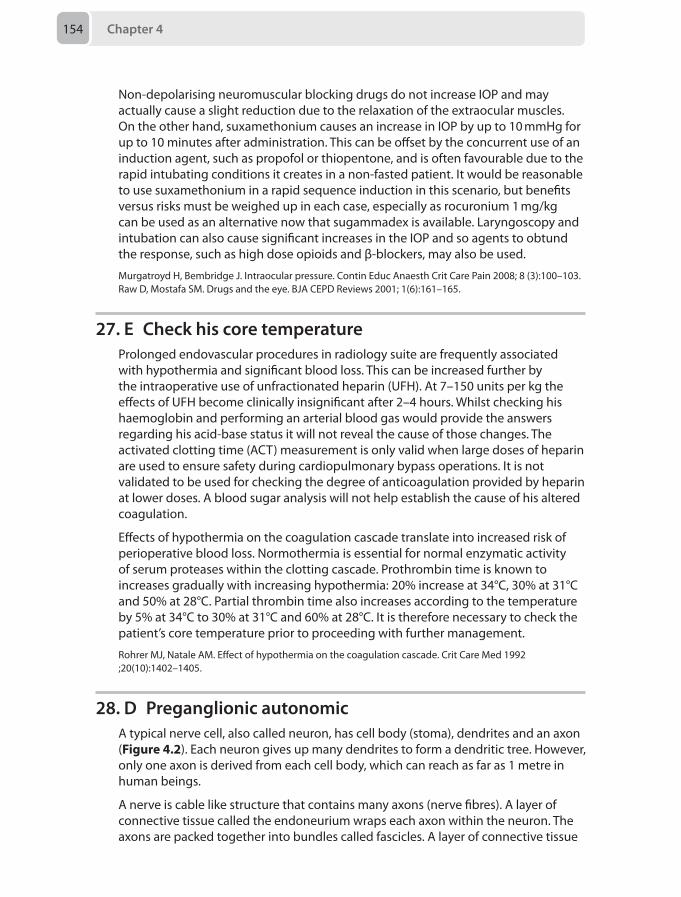
Chapter 4154
Non-depolarising neuromuscular blocking drugs do not increase IOP and may actually cause a slight reduction due to the relaxation of the extraocular muscles. On the other hand, suxamethonium causes an increase in IOP by up to 10 mmHg for up to 10 minutes after administration. This can be offset by the concurrent use of an induction agent, such as propofol or thiopentone, and is often favourable due to the rapid intubating conditions it creates in a non-fasted patient. It would be reasonable to use suxamethonium in a rapid sequence induction in this scenario, but benefits versus risks must be weighed up in each case, especially as rocuronium 1 mg/kg can be used as an alternative now that sugammadex is available. Laryngoscopy and intubation can also cause significant increases in the IOP and so agents to obtund the response, such as high dose opioids and β-blockers, may also be used.
Murgatroyd H, Bembridge J. Intraocular pressure. Contin Educ Anaesth Crit Care Pain 2008; 8 (3):100–103.Raw D, Mostafa SM. Drugs and the eye. BJA CEPD Reviews 2001; 1(6):161–165.
27. E Check his core temperatureProlonged endovascular procedures in radiology suite are frequently associated with hypothermia and significant blood loss. This can be increased further by the intraoperative use of unfractionated heparin (UFH). At 7–150 units per kg the effects of UFH become clinically insignificant after 2–4 hours. Whilst checking his haemoglobin and performing an arterial blood gas would provide the answers regarding his acid-base status it will not reveal the cause of those changes. The activated clotting time (ACT) measurement is only valid when large doses of heparin are used to ensure safety during cardiopulmonary bypass operations. It is not validated to be used for checking the degree of anticoagulation provided by heparin at lower doses. A blood sugar analysis will not help establish the cause of his altered coagulation.
Effects of hypothermia on the coagulation cascade translate into increased risk of perioperative blood loss. Normothermia is essential for normal enzymatic activity of serum proteases within the clotting cascade. Prothrombin time is known to increases gradually with increasing hypothermia: 20% increase at 34°C, 30% at 31°C and 50% at 28°C. Partial thrombin time also increases according to the temperature by 5% at 34°C to 30% at 31°C and 60% at 28°C. It is therefore necessary to check the patient’s core temperature prior to proceeding with further management.
Rohrer MJ, Natale AM. Effect of hypothermia on the coagulation cascade. Crit Care Med 1992 ;20(10):1402–1405.
28. D Preganglionic autonomicA typical nerve cell, also called neuron, has cell body (stoma), dendrites and an axon (Figure 4.2). Each neuron gives up many dendrites to form a dendritic tree. However, only one axon is derived from each cell body, which can reach as far as 1 metre in human beings.
A nerve is cable like structure that contains many axons (nerve fibres). A layer of connective tissue called the endoneurium wraps each axon within the neuron. The axons are packed together into bundles called fascicles. A layer of connective tissue
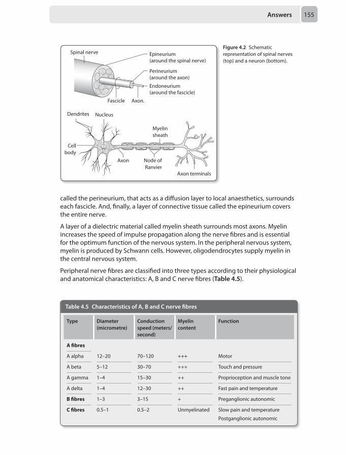
Answers 155
called the perineurium, that acts as a diffusion layer to local anaesthetics, surrounds each fascicle. And, finally, a layer of connective tissue called the epineurium covers the entire nerve.
A layer of a dielectric material called myelin sheath surrounds most axons. Myelin increases the speed of impulse propagation along the nerve fibres and is essential for the optimum function of the nervous system. In the peripheral nervous system, myelin is produced by Schwann cells. However, oligodendrocytes supply myelin in the central nervous system.
Peripheral nerve fibres are classified into three types according to their physiological and anatomical characteristics: A, B and C nerve fibres (Table 4.5).
Nucleus
Cellbody
Dendrites
Node of Ranvier
Axon terminals
Axon
Myelin sheath
Spinal nerve Epineurium(around the spinal nerve)
Endoneurium(around the fascicle)
Perineurium(around the axon)
Fascicle Axon.
Figure 4.2 Schematic representation of spinal nerves (top) and a neuron (bottom).
Table 4.5 Characteristics of A, B and C nerve fibres
Type Diameter (micrometre)
Conduction speed (meters/second)
Myelin content
Function
A fibres
A alpha 12–20 70–120 +++ Motor
A beta 5–12 30–70 +++ Touch and pressure
A gamma 1–4 15–30 ++ Proprioception and muscle tone
A delta 1–4 12–30 ++ Fast pain and temperature
B fibres 1–3 3–15 + Preganglionic autonomic
C fibres 0.5–1 0.5–2 Unmyelinated Slow pain and temperature
Postganglionic autonomic
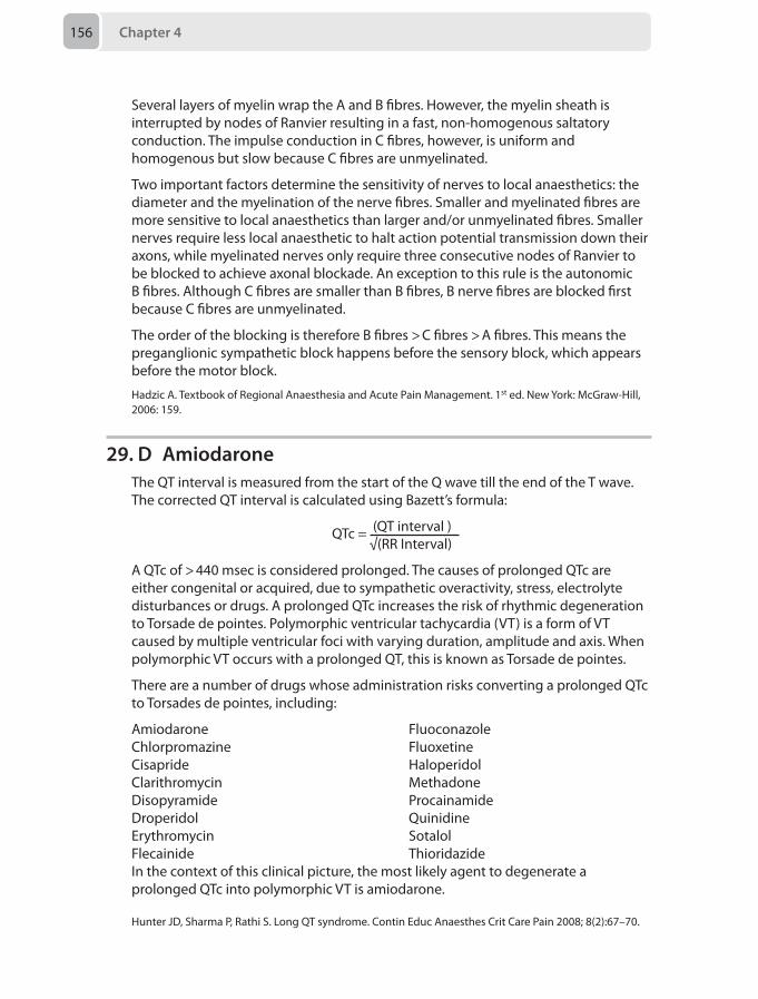
Chapter 4156
Several layers of myelin wrap the A and B fibres. However, the myelin sheath is interrupted by nodes of Ranvier resulting in a fast, non-homogenous saltatory conduction. The impulse conduction in C fibres, however, is uniform and homogenous but slow because C fibres are unmyelinated.
Two important factors determine the sensitivity of nerves to local anaesthetics: the diameter and the myelination of the nerve fibres. Smaller and myelinated fibres are more sensitive to local anaesthetics than larger and/or unmyelinated fibres. Smaller nerves require less local anaesthetic to halt action potential transmission down their axons, while myelinated nerves only require three consecutive nodes of Ranvier to be blocked to achieve axonal blockade. An exception to this rule is the autonomic B fibres. Although C fibres are smaller than B fibres, B nerve fibres are blocked first because C fibres are unmyelinated.
The order of the blocking is therefore B fibres > C fibres > A fibres. This means the preganglionic sympathetic block happens before the sensory block, which appears before the motor block.
Hadzic A. Textbook of Regional Anaesthesia and Acute Pain Management. 1st ed. New York: McGraw-Hill, 2006: 159.
29. D AmiodaroneThe QT interval is measured from the start of the Q wave till the end of the T wave. The corrected QT interval is calculated using Bazett’s formula:
QTc = (QT interval ) √(RR Interval)
A QTc of > 440 msec is considered prolonged. The causes of prolonged QTc are either congenital or acquired, due to sympathetic overactivity, stress, electrolyte disturbances or drugs. A prolonged QTc increases the risk of rhythmic degeneration to Torsade de pointes. Polymorphic ventricular tachycardia (VT) is a form of VT caused by multiple ventricular foci with varying duration, amplitude and axis. When polymorphic VT occurs with a prolonged QT, this is known as Torsade de pointes.
There are a number of drugs whose administration risks converting a prolonged QTc to Torsades de pointes, including:
AmiodaroneChlorpromazineCisaprideClarithromycinDisopyramideDroperidolErythromycinFlecainide
FluoconazoleFluoxetineHaloperidolMethadoneProcainamideQuinidineSotalolThioridazide
In the context of this clinical picture, the most likely agent to degenerate a prolonged QTc into polymorphic VT is amiodarone.
Hunter JD, Sharma P, Rathi S. Long QT syndrome. Contin Educ Anaesthes Crit Care Pain 2008; 8(2):67–70.
Answers 157
30. B CT angiographyThe most likely diagnosis in this clinical scenario is that of a pulmonary embolism (PE).
The European Society of Cardiology published guidelines in 2014 on the diagnosis and management of acute PEs. There are a number of prognosticating tests of PE severity, of which the Pulmonary Embolism Severity Index (PESI) and simplified PESI (sPESI) are recommended. Due to ease of application and validation, the sPESI is used most frequently, taking in to account:
• Age > 80–1 point• Cancer–1 point• Chronic heart failure or pulmonary disease – 1 point• Pulse rate > 110 beats per minute –1 point• Systolic blood pressure < 100 mmHg –1 point• Arterial oxyhaemoglobin saturations < 90% – 1 point0 points gives patients a 30-day mortality of 1.0%, while ≥ 1 point gives a 30-day mortality of 10.9%.
Additional markers are also applied to stratify patients:
Clinical: Shock, hypotension
Right ventricular (RV) dysfunction: RV dilatation, hypokinesia or pressure overload on echocardiogram, RV dilatation on CT or elevated right heart pressure on cardiac catheterisation.
Cardiac laboratory biomarkers: Cardiac troponin T or I, brain natriuretic peptide (BNP) elevation
These can then be put together to quantify the risk severity for patients (Table 4.6)
Table 4.6. Classification of acute PE based on 30-day mortality
30-day mortality risk Risk scores and parameters
Shock or hypo-tension
sPESI ≥ 1 RV dysfunction on imaging
Cardiac lab biomarkers
High risk + + +
Intermediate risk
Intermediate-high – + Both positive
Intermediate-low – + One or none positive
Low risk – – Both negative
It can be seen therefore that a hypotensive or shocked patient will immediately be classified as high risk. In these patients imaging with transthoracic echocardiography or CT angiography is recommended. Treatment of the PE must not be delayed for imaging if there is a high clinical suspicion.
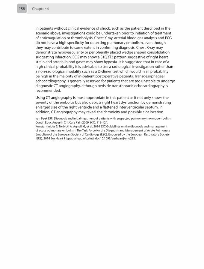
Chapter 4 158
In patients without clinical evidence of shock, such as the patient described in the scenario above, investigations could be undertaken prior to initiation of treatment of anticoagulation or thrombolysis. Chest X ray, arterial blood gas analysis and ECG do not have a high specificity for detecting pulmonary embolism, even though they may contribute to some extent in confirming diagnosis. Chest X-ray may demonstrate hypovascularity or peripherally placed wedge shaped consolidation suggesting infarction. ECG may show a S1Q3T3 pattern suggestive of right heart strain and arterial blood gases may show hypoxia. It is suggested that in case of a high clinical probability it is advisable to use a radiological investigation rather than a non-radiological modality such as a D-dimer test which would in all probability be high in the majority of in-patient postoperative patients. Transoesophageal echocardiography is generally reserved for patients that are too unstable to undergo diagnostic CT angiography, although bedside transthoracic echocardiography is recommended.
Using CT angiography is most appropriate in this patient as it not only shows the severity of the embolus but also depicts right heart dysfunction by demonstrating enlarged size of the right ventricle and a flattened interventricular septum. In addition, CT angiography may reveal the chronicity and possible clot location.
van Beek EJR. Diagnosis and initial treatment of patients with suspected pulmonary thromboembolism Contin Educ Anaesth Crit Care Pain 2009; 9(4): 119-124.Konstantinides S, Torbicki A, Agnelli G, et al. 2014 ESC Guidelines on the diagnosis and management of acute pulmonary embolism: The Task Force for the Diagnosis and Management of Acute Pulmonary Embolism of the European Society of Cardiology (ESC). Endorsed by the European Respiratory Society (ERS). 2014 Eur Heart J (epub ahead of print). doi:10.1093/eurheartj/ehu283.
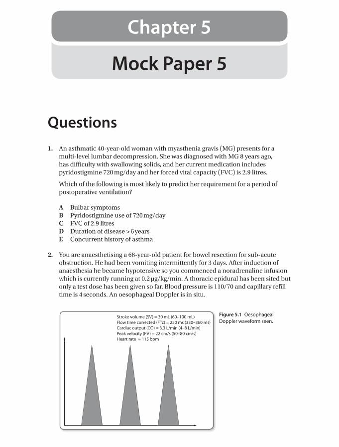
Mock Paper 5
Chapter 5
Questions1. An asthmatic 40-year-old woman with myasthenia gravis (MG) presents for a
multi-level lumbar decompression. She was diagnosed with MG 8 years ago, has difficulty with swallowing solids, and her current medication includes pyridostigmine 720 mg/day and her forced vital capacity (FVC) is 2.9 litres.
Which of the following is most likely to predict her requirement for a period of postoperative ventilation?
A Bulbar symptomsB Pyridostigmine use of 720 mg/dayC FVC of 2.9 litresD Duration of disease > 6 yearsE Concurrent history of asthma
2. You are anaesthetising a 68-year-old patient for bowel resection for sub-acute obstruction. He had been vomiting intermittently for 3 days. After induction of anaesthesia he became hypotensive so you commenced a noradrenaline infusion which is currently running at 0.2 µg/kg/min. A thoracic epidural has been sited but only a test dose has been given so far. Blood pressure is 110/70 and capillary refill time is 4 seconds. An oesophageal Doppler is in situ.
Stroke volume (SV) = 30 mL (60–100 mL)Flow time corrected (FTc) = 250 ms (330–360 ms)Cardiac output (CO) = 3.3 L/min (4–8 L/min)Peak velocity (PV) = 22 cm/s (50–80 cm/s)Heart rate = 115 bpm
Figure 5.1 Oesophageal Doppler waveform seen.
Chapter 5160
Based on the waveform and data shown in Figure 5.1, what is the appropriate first course of action?
A Increase the noradrenaline infusionB Commence a dobutamine infusionC Commence the epidural infusion and leave the noradrenalineD Commence a GTN infusion and leave the noradrenalineE Administer 250 mL of Hartmann’s solution and decrease the noradrenaline
3. A patient in the cardiac intensive care unit suffers a cardiac arrest following three vessel coronary artery bypass grafting. He has epicardial pacing wires with the box set to DDD. The monitor shows pulseless electrical activity with pacing spikes. Cardiopulmonary resuscitation (CPR) is commenced.
What is the most appropriate next step?
A 1 mg adrenaline IVB 300 mg bolus of amiodaroneC Institution of external pacingD Exclusion of a tension pneumothoraxE Turn off the pacemaker
4. A 70-year-old smoker with limited mouth opening having previously undergone a neck dissection with adjuvant radiotherapy is scheduled for surgery to treat his right middle lobe tumour. A difficult airway is anticipated and it is likely that post-operative ventilatory support will be required.
Which of the following is the most appropriate airway management strategy?
A Fibreoptic intubation with a single-lumen tube and a right sided bronchial blocker
B Fibreoptic intubation with a single lumen tube and a left sided bronchial blocker
C Videolaryngoscopy and insertion of a left sided double-lumen tubeD Fibreoptic left sided endobronchial intubation with a single-lumen tubeE Awake tracheostomy and insertion of right sided double-lumen endobronchial
tracheostomy tube
5. A 75-year-old man is to have a cystoscopy and bladder biopsy as a day surgery case. He has a 40 pack year history of smoking. Recent spirometry has shown his FEV1/FVC is 0.6, and echocardiography has shown an ejection fraction of 40%. He has been consented for a spinal anaesthetic.
What is the most appropriate solution for the spinal injection?
A Hyperbaric bupivacaine 0.5% 2 mL B Hyperbaric bupivacaine 0.5% 2 mL with 300 µg diamorphine C Plain bupivacaine 0.5% 2 mL with 10 µg fentanyl D Hyperbaric prilocaine 2% 2 mL with 10 µg fentanylE Hyperbaric lignocaine 2% 2 mL with 10 µg fentanyl
Questions 161
6. You are asked to review a confused 72-year-old man in recovery. He has had a transurethral resection of his prostate for benign prostatic hyperplasia (BPH). A brief assessment reveals him to be disorientated in time and place, and restless. Whilst you review his anaesthetic chart he has a short seizure, which resolves spontaneously.
After assessing his airway breathing and circulation, which of the following would be the best immediate management:
A Administration 2 mg of intravenous midazolamB Starting an infusion of magnesium sulphateC Sending an urgent U&Es, FBC and osmolality, and prepare intravenous
lorazepam in case of further seizureD Administering 20–40 mg of intravenous frusemide E Infusing 1–2 mL/kg 3% NaCl
7. An obese 45-year-old woman with progressive conductive hearing loss secondary to chronic suppurative otitis media is due to undergo tympanoplasty. During the preoperative safety check list, the team is informed by the surgeon that intubation and facial nerve monitoring is required.
Which of the following would be the most appropriate to use as part of your anaesthetic technique?
A Remifentanil infusionB Ketamine bolus C Nitrous oxideD Clonidine infusionE Magnesium sulphate infusion
8. A 63-year-old man with a confirmed inherited pseudocholinesterase deficiency (EuEa) is attending for his first course of electroconvulsive therapy.
Which of the following drug combinations is most appropriate for his induction of anaesthesia?
A Propofol and mivacuriumB Propofol and rocuroniumC Propofol and alfentanilD Thiopentone and rocuroniumE Thiopentone and alfentanil
9. A 70-year old man is scheduled for foot surgery under general anaesthesia and a sciatic nerve block. There are no ultrasound machines available and you decide on a landmark technique to perform the block.
Which one of the following described techniques results in the most proximal approach to performing a sciatic nerve block?
A Mansour’s approach B Raj’s approach
Chapter 5162
C Labat’s approach D Beck’s approach E Guardini’s approach
10. A 68-year old man with emphysema is listed for elbow surgery under regional anaesthesia.
Which of the following would be the most appropriate nerve block for this patient?
A Interscalene brachial plexus blockB Supraclavicular brachial plexus blockC Medial infraclavicular brachial plexus blockD Axillary brachial plexus block E Mid-arm peripheral nerve block
11. You have been called to assist in the care of a 17-year-old girl who has become increasingly agitated in the emergency department. She has a history of mental illness and has recently been behaving strangely. Now her actions are violent and compromising her safety and that of those around her. You are unable to assess her formally, and she has not had any blood tests, intravenous access or observations. Security officers are present, and the emergency department registrar tells you he would like to perform bloods, a CT head and a lumbar puncture. The plan has been approved by the girl’s mother and the paediatric consultant.
How will you proceed?
A Use security staff to hold the patient, insert intravenous access, and give 2 mg midazolam and 2 µg/kg fentanyl in the room
B Use security staff to hold the patient, and give intramuscular 4 mg/kg ketamine, then transfer to the resuscitation bay
C Do nothing, and refuse to get involved with this caseD Encourage her to take 20 mg oral temazepam and review E Using security staff to hold the patient, transfer to theatre, and perform an
inhalational induction with sevoflurane
12. A 34-year-old man sustained a traumatic brain injury 3 days ago and is currently intubated and ventilated on the intensive care unit. The nurse informs you during your daily review that the plasma sodium concentration is 121 mmol/L.
What other piece information would be most useful in establishing the cause?
A Urine output volume measurementB Central venous pressure measurementC Degree of peripheral oedemaD Urinary osmolarity measurementE Plasma osmolarity measurement
13. A 13-year-old boy presented to the emergency department with acute severe asthma 1 hour ago. His usual peak expiratory flow (PEF) is 68%, and takes long acting β 2 agonist and high dose corticosteroid inhalers with montelukast tablets. You are
Questions 163
called for advice as despite 4 x 2.5 mg nebulised salbutamol, 500 µg nebulised ipratropium and 40 mg of soluble prednisolone the patient’s PEF remains 35% predicted, respiratory rate 32 per minute, speaking words, Spo2 93% on 10L/min of warm humidified supplemental oxygen and transcutaneous carbon dioxide level of 5.1 kPa.
Which of the following should be the next intervention?
A Rapid sequence induction using thiopentone and suxamethonium following by positive pressure ventilation on an anaesthetic machine using isoflurane to maintain anaesthesia and ease bronchoconstriction
B Commence an intravenous salbutamol infusion at 10 µg/minuteC Give 20 mmol of intravenous magnesium sulphate over 10–20 minutesD Give a further 5 mg nebulised salbutamolE Give a loading dose of 5 mg/kg aminophylline followed by an infusion at
500 µg/kg/hour
14. During the high dependency unit ward round you are called to the bedside of a 64-year-old gentleman with a background of hypertension who is awaiting primary angioplasty planned for the following day after being admitted with a non-ST segment elevation myocardial infarction. He is feeling anxious and has central chest pain. The heart rate has recently increased to 150 beats per minute and the blood pressure is 90/60 mmHg. The ECG shows atrial fibrillation and widespread ST segment depression.
What is your immediate course of action?
A Ring the anaesthetist on call and arrange for direct current (DC) cardioversion in theatre
B Ring the anaesthetist on call and arrange for direct current (DC) cardioversion on the HDU
C Administer amiodarone 300 mg intravenously over 30 minutesD Administer 2 g intravenous magnesium and optimise the serum potassium
concentrationE Ring the cardiologist on call and organise an urgent angiography
15. A 72-year-old man on the intensive care unit has an APACHE II score of 48.
Which of the following variables is the most heavily weighted in intensive care severity of illness scoring systems?
A AgeB Glasgow coma scaleC Systolic blood pressure / dose of vasopressorD Pao2:Fio2 (PF ratio)E Arterial lactate concentration
16. A 34-year-old woman with end-stage liver disease due to auto-immune hepatitis presents with a 2-day history of productive cough and breathlessness and has been commenced on antibiotics for a chest infection. She has stigmata
Chapter 5164
of decompensated liver disease with peripheral oedema and ascites. Her blood pressure is 70/40 mmHg, her heart rate is 130 beats per minute and her temperature 38.9oC. An arterial blood gas analysis returned a lactate concentration 0f 6.8 mmol/L.
Which of the following treatment options do you administer first?
A 20 mL/kg Hartmann’s solutionB 20 mL/kg 0.9% sodium chlorideC 10 mL/kg gelofusineD 10 mL/kg human albumin solution (HAS) 20%E Commence vasopressor support as soon as possible
17. A 29-year-old woman, with a history of rheumatic fever is undergoing a category 1 Caesarean section under general anaesthesia for cord prolapse. After delivery of the baby and placenta, the estimated blood loss is 1100 mL. The patient receives a total of 10 IU of syntocinon intravenously plus 10 IU/hour intravenous syntocinon infusion, intravenous ergometrine 500 µg, 250 µg intramuscular carboprost, misoprostol 1 mg rectally, four units of packed red blood cells and two units of fresh frozen plasma. As the obstetricians are closing, the patient begins to produce frothy pink sputum up the endotracheal tube.
Which of the following is the least likely cause of the pink frothy sputum?
A SyntocinonB ErgometrineC CarboprostD Blood productsE Misoprostol
18. You have been fast bleeped to one of the delivery rooms on labour ward where a 38-year-old multiparous woman who is in the first stage of labour has suddenly become short of breath. Initial observations show oxygen saturations of 87%, a respiratory rate of 35, heart rate of 110 beats per minute and a blood pressure of 85/40 mmHg.
What is the least likely cause of her presentation?
A Amniotic fluid embolismB Pulmonary embolismC Antepartum haemorrhageD AnaphylaxisE Tension pneumothorax
19. A 6-week-old boy presents with a 3-day history of progressive non-bilious vomiting and poor feeding. An ultrasound scan confirms the diagnosis of pyloric stenosis. The capillary blood gas is shown in Table 5.1.
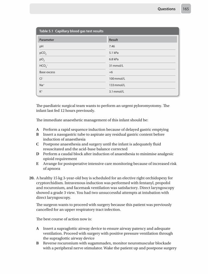
Questions 165
The paediatric surgical team wants to perform an urgent pyloromyotomy. The infant last fed 12 hours previously.
The immediate anaesthetic management of this infant should be:
A Perform a rapid sequence induction because of delayed gastric emptyingB Insert a nasogastric tube to aspirate any residual gastric content before
induction of anaesthesiaC Postpone anaesthesia and surgery until the infant is adequately fluid
resuscitated and the acid-base balance correctedD Perform a caudal block after induction of anaesthesia to minimise analgesic
opioid requirement E Arrange for postoperative intensive care monitoring because of increased risk
of apnoea
20. A healthy 15 kg 3-year-old boy is scheduled for an elective right orchidopexy for cryptorchidism. Intravenous induction was performed with fentanyl, propofol and rocuronium, and facemask ventilation was satisfactory. Direct laryngoscopy showed a grade 3 view. You had two unsuccessful attempts at intubation with direct laryngoscopy.
The surgeon wants to proceed with surgery because this patient was previously cancelled for an upper respiratory tract infection.
The best course of action now is:
A Insert a supraglottic airway device to ensure airway patency and adequate ventilation. Proceed with surgery with positive pressure ventilation through the supraglottic airway device
B Reverse rocuronium with sugammadex, monitor neuromuscular blockade with a peripheral nerve stimulator. Wake the patient up and postpone surgery
Table 5.1 Capillary blood gas test results
Parameter Result
pH 7.46
pCO2 5.1 kPa
pO2 6.8 kPa
HCO3– 31 mmol/L
Base excess +6
Cl– 100 mmol/L
Na+ 133 mmol/L
K+ 3.1 mmol/L
Chapter 5166
C Insert a supraglottic airway device to ensure airway patency and adequate ventilation. Perform a fibreoptic intubation through the supraglottic airway device and proceed with surgery
D Ensure airway patency and adequate ventilation using facemask. Perform a fibreoptic intubation and proceed with surgery
E Have two further attempts at laryngoscopy. Check head and neck position, laryngoscopy technique, external laryngeal manipulation and adequate paralysis. Consider using a different laryngoscope, a smaller tube, a stylet or a bougie
21. A 30-year-old man with Crohn’s disease has had a right hemicolectomy. You are asked to see him in recovery for uncontrolled abdominal pain. He declined an epidural preoperatively. Intraoperatively he had paracetamol and 20 mg morphine. So far in recovery he has had a total of 25 mg morphine with little effect. He is alert but very distressed with a normal respiratory rate. He does not normally take any strong opiates and has no allergies.
What would be the most appropriate next step in the management of his acute pain?
A Further titrate morphine up to a maximum of 20 mg provided there are no overt signs of overdose
B Give 2 mg midazolam C Give 10 mg ketamine intravenously D Give 100 mg tramadol intravenously E Give 50 µg clonidine intravenously
22. A 23-year-old woman had an above knee amputation for a localised osteosarcoma. She has developed unusual sensations at the amputated limb, pain at the stump, and a feeling that the limb is still there.
Which of the following features are most likely to suggest true phantom limb pain in this patient?
A Painful sensations at the stumpB Good response to opioid analgesiaC Intermittent painD Inadequately working preoperative epidural analgesiaE Sensations that the amputated limb is still present
23. A 62-year-old woman presents for a left lobectomy. She is generally fit and well, and not on any regular medications.
Which of the following is the best option for analgesic management:
A Lumbar epiduralB Intrathecal diamorphineC PCA morphineD Interpleural blockE Thoracic epidural
Questions 167
24. A 48-year-old man is referred to the pain clinic with poorly controlled upper abdominal pain. He is known to have pancreatic cancer, which is now palliative. As a result of his medication he is now feeling increasingly tired and is having difficulty concentrating.
He is on the following medication:
• Paracetamol 1g four times daily• Gabapentin 900 mg three times daily• Modified release oxycodone 30 mg twice daily• Immediate release oxycodone 10 mg for breakthrough pain
What would be the most appropriate treatment option?
A Increase modified release oxycodone to 40 mg twice daily and immediate release oxycodone to 15 mg for breakthrough pain
B Add amitriptyline C Perform a coeliac plexus blockD Perform a lumbar sympathetic blockE Perform a thoracic paravertebral block T10–T12
25. A 4.2 kg, 26-day-old neonate is admitted for repair of an inguinal hernia. He was born at term by normal vaginal delivery. There are no other known medical problems and no allergies.
What would be the most appropriate analgesic plan for post operative pain relief?
A Single shot caudal combined with 15 mg/kg paracetamol intravenously 6-hourly and codeine phosphate 1 mg/kg orally 6-hourly as required
B Single shot caudal combined with 10 mg/kg paracetamol orally 6-hourly and ibuprofen 5 mg/kg orally 8-hourly as required
C Single shot caudal combined with 10 mg/kg paracetamol orally 6-hourly and codeine phosphate 1 mg/kg orally 6-hourly as required
D Local anaesthetic infiltration by surgeon combined with paracetamol 7.5 mg/kg intravenously 6-hourly and codeine phosphate 1 mg/kg orally 6-hourly as required
E Local anaesthetic infiltration by surgeon combined with paracetamol 15 mg/kg orally 6-hourly and morphine sulphate 100 µg/kg orally 4–6-hourly as required
26. A 26-year-old woman was pulled unconscious from a campervan fire and was intubated at the scene. She has 40% burns with moderate inhalational injury, and has been fluid resuscitated according to the Parkland formula.
Her heart rate is 96 beats per minute, blood pressure 110/77 mmHg, capillary refill time <2 seconds, temperature 38°C, and urine output is 70 ml/hour. Arterial blood gas analysis on a Fio2 of 50% shows: pH 7.12, Pao2 40 kPa, Paco2 3.3 kPa, Hco3
– 16 mmol/L, base excess –4.0, lactate 12.3 mmol/L.
Chapter 5168
What is most likely to account for the lactic acidosis:
A SepsisB Volume depletionC Acute kidney injury from rhabdomyolysisD Cynaide poisoningE Ethanol poisoning
27. You are anaesthetising a 72-year-old man for an elective open abdominal aneurysm. He is a known hypertensive and type II diabetic and his medication includes amlodipine 5 mg, ramipril 10 mg and metformin 1 gm b.d.
What is the single most important intervention that would limit potential postoperative renal impairment?
A Start dopamine at 2.5 µg/kg/minB Administer mannitol 20% 0.5 g/kgC Optimise his circulatory blood volumeD Keep his circulating haemoglobin > 90 g/LE Administer furosemide 40 mg
28. A 6-year-old boy is admitted with fulminant hepatic failure, bleeding oesophageal varices, ascites and marked splenomegaly.
His liver function tests show an elevated bilirubin, alanine transaminases and aspartate transaminases. He has low albumin, prolonged prothrombin time and examination of his cornea on slit lamp examination demonstrate a brown, dark ring encircling his iris.
Which of the following is the most likely diagnosis for this clinical picture?
A Alpha-1 antitrypsin deficiencyB Wilson’s diseaseC Primary biliary cirrhosisD HaemochromatosisE Sclerosing cholangitis
29. A 92-year-old man is brought to emergency department with a fractured neck of femur following a fall.
To be categorised using the fraility phenotype, which of the following characteristics would not be assessed?
A Self-reported exhaustionB Mental stateC Weight lossD Grip strengthE Low activity
Questions 169
30. A new antiemetic drug is being evaluated. The percentage of patients who suffered postoperative nausea and vomiting (PONV) after administration of either the drug or placebo is reported:• percentage of patients with PONV after drug A = 20% • percentage of patients with PONV after placebo = 25%
Which of the following is the number needed to treat (NNT)?
A 20B 25C 5D 75E 1
Chapter 5170
Answers
1. D Duration of disease > 6 yearsMyasthenia gravis (MG) is an autoimmune disease with a prevalence between 1 in 10,000–100,000. Women are more likely to be affected with a female:male ratio of 3:2. The disease is caused by IgG antibodies to the post-synaptic acetylcholine (ACh) receptors at the neuromuscular junction of skeletal muscle. These receptors are occupied by the antibodies and ultimately destroyed through complement-mediated immune processes. MG is therefore associated with fatiguing muscle weakness, as only a limited response to ACh released at the neuromuscular junction is possible and any subsequent stimulation results in fewer and fewer receptors available for activation.
The extent of muscle involvement and severity of disease was classified by Osserman as seen in Table 5.2.
Table 5.2 Osserman Classification for the severity of MG
CLASS I Limited to eye muscle involvement
CLASS IIa Mild and generalised, responding to treatment
CLASS IIb Moderate and generalised, response to treatment not satisfactory
CLASS III Severe and generalised, including respiratory dysfunction
CLASS IV Requiring ventilation
15% of patients fall into Class I, the remaining 85% suffer from generalised MG. Cardiac and smooth muscle is entirely unaffected.
An anaesthetic and surgery can impact on a patient with MG in a number of ways. The physiological stress in itself can exacerbate symptoms and, for a patient who may be unable to achieve adequate tidal volumes or cough ordinarily, lack of pre-operative planning could prove fatal.
There are four recognised risk factors that are associated with an increased likelihood of requiring a period of postoperative ventilation.
1. MG duration of > 6 years – this has the greatest predictive value2. Concurrent history of chronic respiratory disease3. Pyridostigmine requirements of > 750 mg/day in the preceding 48 hours4. Forced vital capacity < 2.9 litres
Other considerations for trying to predict the need for respiratory support include surgery – type, length and need for intubation; anaesthetic – general +/– local, need for muscle relaxation and perhaps reversal; medication – opiate use in a patient with affected respiratory reserve, drugs such as aminoglycosides or beta-blockers that can cause an exacerbation of MG and administration of the patient's normal
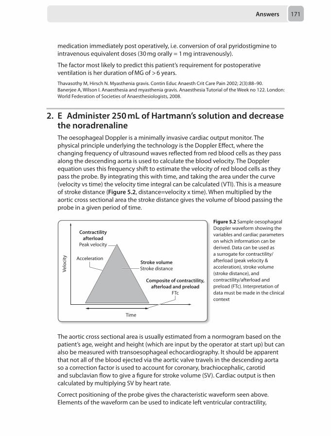
Answers 171
medication immediately post operatively, i.e. conversion of oral pyridostigmine to intravenous equivalent doses (30 mg orally = 1 mg intravenously).
The factor most likely to predict this patient’s requirement for postoperative ventilation is her duration of MG of > 6 years.
Thavasothy M, Hirsch N. Myasthenia gravis. Contin Educ Anaesth Crit Care Pain 2002; 2(3):88–90.Banerjee A, Wilson I. Anaesthesia and myasthenia gravis. Anaesthesia Tutorial of the Week no 122. London: World Federation of Societies of Anaesthesiologists, 2008.
2. E Administer 250 mL of Hartmann’s solution and decrease the noradrenalineThe oesophageal Doppler is a minimally invasive cardiac output monitor. The physical principle underlying the technology is the Doppler Effect, where the changing frequency of ultrasound waves reflected from red blood cells as they pass along the descending aorta is used to calculate the blood velocity. The Doppler equation uses this frequency shift to estimate the velocity of red blood cells as they pass the probe. By integrating this with time, and taking the area under the curve (velocity vs time) the velocity time integral can be calculated (VTI). This is a measure of stroke distance (Figure 5.2, distance=velocity x time). When multiplied by the aortic cross sectional area the stroke distance gives the volume of blood passing the probe in a given period of time.
Contractilityafterload
Peak velocity
Acceleration
Velo
city
Stroke volumeStroke distance
Composite of contractility,afterload and preload
FTc
Time
Figure 5.2 Sample oesophageal Doppler waveform showing the variables and cardiac parameters on which information can be derived. Data can be used as a surrogate for contractility/afterload (peak velocity & acceleration), stroke volume (stroke distance), and contractility/afterload and preload (FTc). Interpretation of data must be made in the clinical context
The aortic cross sectional area is usually estimated from a normogram based on the patient’s age, weight and height (which are input by the operator at start up) but can also be measured with transoesophageal echocardiography. It should be apparent that not all of the blood ejected via the aortic valve travels in the descending aorta so a correction factor is used to account for coronary, brachiocephalic, carotid and subclavian flow to give a figure for stroke volume (SV). Cardiac output is then calculated by multiplying SV by heart rate.
Correct positioning of the probe gives the characteristic waveform seen above. Elements of the waveform can be used to indicate left ventricular contractility,
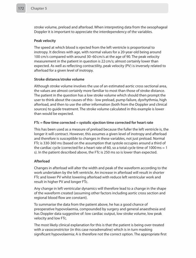
Chapter 5172
stroke volume, preload and afterload. When interpreting data from the oesophageal Doppler it is important to appreciate the interdependency of the variables.
Peak velocity
The speed at which blood is ejected from the left ventricle is proportional to inotropy. It declines with age, with normal values for a 20-year-old being around 100 cm/s compared with around 30–60 cm/s at the age of 90. The peak velocity measurement in the patient in question is 22 cm/s; almost certainly lower than expected. As well as reflecting contractility, peak velocity (PV) is inversely related to afterload for a given level of inotropy.
Stroke distance/stroke volume
Although stroke volume involves the use of an estimated aortic cross sectional area, the values are almost certainly more familiar to most than those of stroke distance. The patient in the question has a low stroke volume which should then prompt the user to think about the causes of this - low preload, pump failure, dysrhythmia, high afterload, and then to use the other information (both from the Doppler and clinical sources) to guide treatment. The stroke volume calculated in this example is lower than would be expected.
FTc = flow time corrected = systolic ejection time corrected for heart rate
This has been used as a measure of preload because the fuller the left ventricle is, the longer it will contract. However, this assumes a given level of inotropy and afterload and therefore is susceptible to changes in these variables, not just preload. Normal FTc is 330-360 ms (based on the assumption that systole occupies around a third of the cardiac cycle (corrected for a heart rate of 60, so a total cycle time of 1000 ms = 1 s). In the patient described above, the FTc is 250 ms so is lower than expected.
Afterload
Changes in afterload will alter the width and peak of the waveform according to the work undertaken by the left ventricle. An increase in afterload will result in shorter FTc and lower PV whilst lowering afterload with reduce left ventricular work and result in higher PV and longer FTc.
Any change in left ventricular dynamics will therefore lead to a change in the shape of the waveform created (assuming other factors including aortic cross section and regional blood flow are constant).
To summarise the data from the patient above, he has a good chance of preoperative hypovolaemia, compounded by surgery and general anaesthesia and has Doppler data suggestive of: low cardiac output, low stroke volume, low peak velocity and low FTc.
The most likely clinical explanation for this is that the patient is being over-treated with a vasoconstrictor (in this case noradrenaline) which is in turn masking significant hypovolaemia; A is therefore not the correct option. The appropriate first
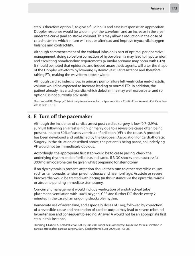
Answers 173
step is therefore option E; to give a fluid bolus and assess response; an appropriate Doppler response would be widening of the waveform and an increase in the area under the curve (and so stroke volume). This may allow a reduction in the dose of catecholamine which in turn will reduce afterload and improve myocardial oxygen balance and contractility.
Although commencement of the epidural infusion is part of optimal perioperative management, doing so before correction of hypovolaemia may lead to hypotension and escalating noradrenaline requirements (a similar scenario may occur with GTN). It should be noted that epidurals, and indeed anaesthetic agents, will alter the shape of the Doppler waveform by lowering systemic vascular resistance and therefore raising FTc, making the waveform appear wider.
Although cardiac index is low, in primary pump failure left ventricular end-diastolic volume would be expected to increase leading to normal FTc. In addition, the patient already has a tachycardia, which dobutamine may well exacerbate, and so option B is not currently advisable.
Drummond KE, Murphy E. Minimally invasive cardiac output monitors. Contin Educ Anaesth Crit Care Pain 2012; 12 (1): 5-10.
3. E Turn off the pacemakerAlthough the incidence of cardiac arrest post cardiac surgery is low (0.7–2.9%), survival following an arrest is high, primarily due to a reversible cause often being present. In up to 50% of cases ventricular fibrillation (VF) is the cause. A protocol has been developed and published by the European Association for Cardiothoracic Surgery. In the situation described above, the patient is being paced, so underlying VF would not be immediately obvious.
Accordingly, the appropriate first step would be to cease pacing, check the underlying rhythm and defibrillate as indicated. If 3 DC shocks are unsuccessful, 300 mg amiodarone can be given whilst preparing for sternotomy.
If no dysrhythmia is present, attention should then turn to other reversible causes such as tamponade, tension pneumothorax and haemorrhage. Asystole or severe bradycardia would be treated with pacing (in this instance via the epicardial wires) or atropine pending immediate sternotomy.
Concurrent management would include verification of endotracheal tube placement, ventilation with 100% oxygen, CPR and further DC shocks every 2 minutes in the case of an ongoing shockable rhythm.
Immediate use of adrenaline, and especially doses of 1mg, followed by correction of a reversible cause and restoration of cardiac output may lead to severe rebound hypertension and consequent bleeding. Answer A would not be an appropriate first step in this instance.
Dunning J, Fabbri A, Kolh PH, et al. EACTS Clinical Guidelines Committee. Guideline for resuscitation in cardiac arrest after cardiac surgery. Eur J Cardiothorac Surg 2009; 36(1):3–28.
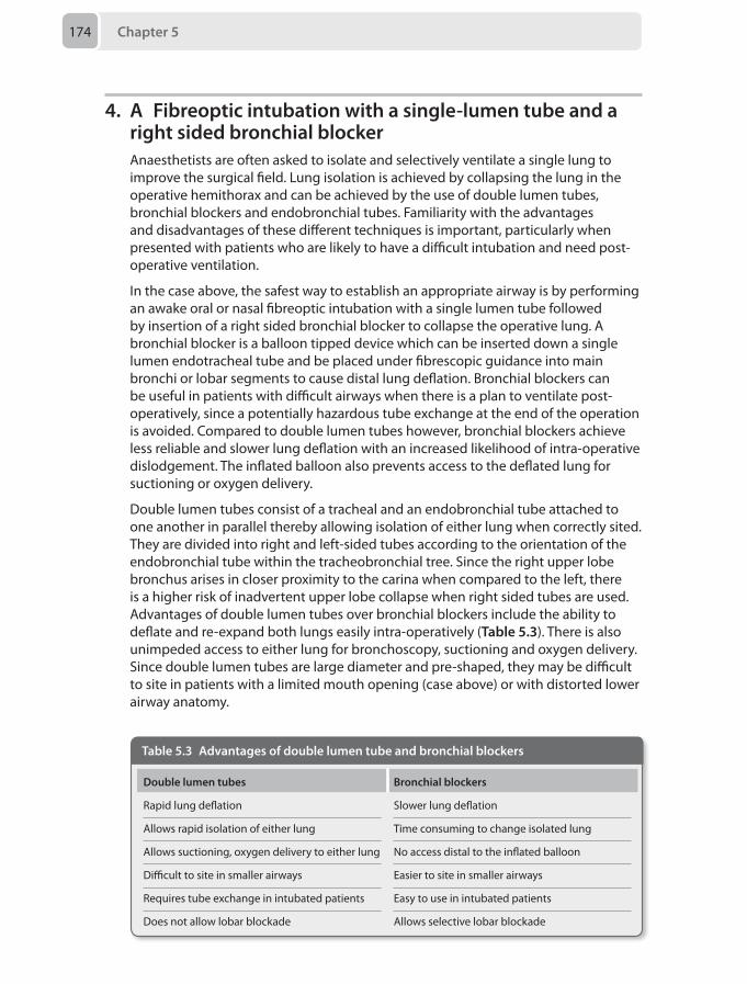
Chapter 5174
4. A Fibreoptic intubation with a single-lumen tube and a right sided bronchial blockerAnaesthetists are often asked to isolate and selectively ventilate a single lung to improve the surgical field. Lung isolation is achieved by collapsing the lung in the operative hemithorax and can be achieved by the use of double lumen tubes, bronchial blockers and endobronchial tubes. Familiarity with the advantages and disadvantages of these different techniques is important, particularly when presented with patients who are likely to have a difficult intubation and need post-operative ventilation.
In the case above, the safest way to establish an appropriate airway is by performing an awake oral or nasal fibreoptic intubation with a single lumen tube followed by insertion of a right sided bronchial blocker to collapse the operative lung. A bronchial blocker is a balloon tipped device which can be inserted down a single lumen endotracheal tube and be placed under fibrescopic guidance into main bronchi or lobar segments to cause distal lung deflation. Bronchial blockers can be useful in patients with difficult airways when there is a plan to ventilate post-operatively, since a potentially hazardous tube exchange at the end of the operation is avoided. Compared to double lumen tubes however, bronchial blockers achieve less reliable and slower lung deflation with an increased likelihood of intra-operative dislodgement. The inflated balloon also prevents access to the deflated lung for suctioning or oxygen delivery.
Double lumen tubes consist of a tracheal and an endobronchial tube attached to one another in parallel thereby allowing isolation of either lung when correctly sited. They are divided into right and left-sided tubes according to the orientation of the endobronchial tube within the tracheobronchial tree. Since the right upper lobe bronchus arises in closer proximity to the carina when compared to the left, there is a higher risk of inadvertent upper lobe collapse when right sided tubes are used. Advantages of double lumen tubes over bronchial blockers include the ability to deflate and re-expand both lungs easily intra-operatively (Table 5.3). There is also unimpeded access to either lung for bronchoscopy, suctioning and oxygen delivery. Since double lumen tubes are large diameter and pre-shaped, they may be difficult to site in patients with a limited mouth opening (case above) or with distorted lower airway anatomy.
Table 5.3 Advantages of double lumen tube and bronchial blockers
Double lumen tubes Bronchial blockers
Rapid lung deflation Slower lung deflation
Allows rapid isolation of either lung Time consuming to change isolated lung
Allows suctioning, oxygen delivery to either lung No access distal to the inflated balloon
Difficult to site in smaller airways Easier to site in smaller airways
Requires tube exchange in intubated patients Easy to use in intubated patients
Does not allow lobar blockade Allows selective lobar blockade
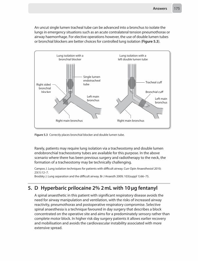
Answers 175
An uncut single lumen tracheal tube can be advanced into a bronchus to isolate the lungs in emergency situations such as an acute contralateral tension pneumothorax or airway haemorrhage. For elective operations however, the use of double lumen tubes or bronchial blockers are better choices for controlled lung isolation (Figure 5.3).
Tracheal cu�
Bronchial cu�
Right main bronchus
Right sidedbronchial
blocker
Single lumenendotrachealtube
Right main bronchus
Single
ronchial cu�
chus chusRight main bro Right main broR
Right sidedbronchial
blockerblocker
eal
Left mainbronchus Left main
bronchus
Lung isolation with abronchial blocker
Lung isolation with aleft double lumen tube
Figure 5.3 Correctly places bronchial blocker and double lumen tube.
Rarely, patients may require lung isolation via a tracheostomy and double lumen endobronchial tracheostomy tubes are available for this purpose. In the above scenario where there has been previous surgery and radiotherapy to the neck, the formation of a tracheostomy may be technically challenging.
Campos J. Lung isolation techniques for patients with difficult airway. Curr Opin Anaesthesiol 2010; 23(1):12–7.Brodsky J. Lung separation and the difficult airway. Br J Anaesth 2009; 103(suppl 1):66–75.
5. D Hyperbaric prilocaine 2% 2 mL with 10 µg fentanylA spinal anaesthetic in this patient with significant respiratory disease avoids the need for airway manipulation and ventilation, with the risks of increased airway reactivity, pneumothorax and postoperative respiratory compromise. Selective spinal anaesthesia is a technique favoured in day surgery that describes a block concentrated on the operative site and aims for a predominately sensory rather than complete motor block. In higher risk day surgery patients it allows earlier recovery and mobilisation and avoids the cardiovascular instability associated with more extensive spread.
Chapter 5176
The ideal agent for such a block would:
• have a rapid onset• provide a dense predictable sensory block• have a short duration of action to allow early recovery and ambulation• have a favourable side-effect and safety profile
Hyperbaric prilocaine 2% has been licensed for spinal use in the UK since 2010 and is now widely accepted as the agent of choice for day surgery. It has both a rapid onset and resolution of block and confers a higher degree of cardiovascular stability compared to bupivacaine.
Low doses of hyperbaric bupivacaine can achieve selective blockade but doses higher than 7 mg are associated with urinary retention especially in those having urology procedures and those older than 70 years. Plain solutions are slightly hypobaric at 37oC and less predictable as they produce greater variability in spread, so are less likely to result in a selective block.
Lignocaine has an early onset time and a short duration of action, but has been associated with TNS (transient neurological symptoms – self-limiting pain and dysaesthesia in the buttocks) especially after day surgery in the lithotomy position.
Fentanyl acts synergistically with the local anaesthetic allowing reduced dose and prolonged analgesia. Using small doses (10–25 µg) avoids respiratory depression and is less likely to cause pruritus.
Before attempting to mobilise after a spinal anaesthetic, patients should have return of perianal sensation, plantar flexion of the foot to pre-operative strength and proprioception of the big toe, and discharged after voiding and other standard criteria have been met.
British Association of Day Surgery (BADS). Spinal anaesthesia for day surgery patients; a practical guide. 3rd Ed. London: BADS, 2013.Black AS, Newcombe GN, Plummer JL, McLeod GH, Martin DK. Spinal anaesthesia for ambulatory arthroscopic surgery of the knee: a comparison of low dose prilocaine and fentanyl with bupivacaine and fentanyl. Br J Anaesth 2011; 106(2):183–8.
6. C Sending an urgent U&Es, FBC and osmolality, and prepare intravenous lorazepam in case of further seizureTransurethral resection of the prostate (TURP) is a common procedure, and the best available treatment for benign prostatic hyperplasia (BPH) with obstructive lower urinary tract symptoms. TURP syndrome is caused by the absorption of hypotonic irrigation fluid. The quantity of absorbed fluid is important and the probability of developing TURP syndrome increases with the following factors which all increase absorption:
• Length of resection, especially > 1 hour• Significant bleeding, implying large quantities of open vessels• Bladder or prostatic capsular perforation, (fluid is rapidly absorbed from the
peritoneum)• Height of the irrigation fluid bag. This corresponds to the hydrostatic pressure
within the bladder. Heights > 70 cm are unusualThe syndrome is caused by changes in:
Answers 177
Volume
This is biphasic in nature. Initially, the circulation absorbs large volumes and there can be hypertension with a reflex bradycardia. This may also cause signs and symptoms of volume overload with left sided heart failure and pulmonary oedema. Later as the irrigation fluid shifts to the extracellular space (due to its hypotonic composition), there may be a relative hypovolaemia and hypotension. It’s worth bearing in mind that the first stage of hypertension is often masked by the low systemic vascular resistance (SVR) state of a spinal sympathetic block.
Treatment of hypervolaemia resulting in left ventricular failure (LVF) involves frusemide, but hypervolaemia without LVF is better treated with mannitol as this lowers serum sodium less than frusemide. Hypotension and reduced heart rates are addressed with vasoconstrictors, calcium and atropine.
Plasma sodium and osmolality
The hyponatraemia of TURP syndrome is mainly dilutional, and produces headaches, nausea and vomiting, seizures and coma. However, of the two values, osmolality is by far the more important. This is because rapid falls in osmolality cannot be countered by pumps in neuronal cell walls, and the cells thus get flooded with water down its osmotic gradient, leading to cerebral oedema and increased intracranial pressure. If osmolality is relatively normal, low sodium does not need treatment in an asymptomatic patient. Sodium should only be corrected slowly to prevent pontine demyelination, and only correct until symptoms resolve, not to normal values.
Treatment of hyponatraemia should be titrated to symptoms, slowly, not > 1 mmol/L/hour. Remember to measure the osmolality also. If the sodium is < 120 mmol/L, give 3% NaCl (weight in kg x 0.6 x 2 = number of mL 3% NaCl needed to elevate sodium by 1 mmol/L).
Glycine
Although glycine is an inhibitory neurotransmitter, it does have effects increasing NMDA receptor activity. This can produce the phenomenon of dis-inhibition, such that the first neurological symptoms may be of irritability and seizures. As concentrations increase, coma may follow. Due to its physiological antagonist action at NMDA receptors magnesium is a useful second line treatment for seizures associated with TURP syndrome.
Treatment of seizures is supportive and if needed involves benzodiazepines such as lorazepam and if required magnesium.
The first stems A and B suggest treatments primarily focussed on seizure control. While the possibility of a further seizure should be at the forefront of one’s mind, a self-terminating isolated seizure may not require treatment. Instead the focus should be on identifying the severity of the condition and thus determining whether specific treatment is required, as in stem C. Empirical treatment without knowing the sodium or osmolarity first may be dangerous (E), and treatment with frusemide (D) is reserved for heart failure secondary to fluid overload.
Milligan L J, Bellamy M C. Anaesthesia for transurethral resection of the prostate. Contin Educ Anaesth Crit Care Pain 2009; 9(3):92–96.
Chapter 5178
7. A Remifentanil infusionThe middle ear is a delicate air filled cavity containing three ossicles which transmit sound vibrations from the eardrum to the cochlea. Due to its small size, location and fragile content, the provision of anaesthesia for surgery to this unique site is especially challenging.
Maintaining the surgical field is difficult since small amounts of bleeding or movements can significantly degrade the view during microsurgery. Furthermore, the use of neuromuscular blocking drugs to provide akinesia is frequently restricted due to the need for intraoperative facial nerve monitoring. A smooth, cough-free wake up is desirable to avoid compromising the surgical result, and patients are at an increased risk of developing post-operative nausea and vomiting.
Remifentanil is the most appropriate drug to use in this scenario since it addresses a number of problems associated with middle ear surgery anaesthesia in addition to providing adequate intraoperative analgesia. To minimise blood loss, remifentanil can be used to rapidly control the blood pressure to deliver safe hypotensive anaesthesia and a stable pulse in suitable patients. Remifentanil also allows mechanical ventilation without neuromuscular blocking agents which enables uninterrupted facial nerve monitoring. Remifentanil also attenuates coughing on emergence, and if used in conjunction with propofol as part of a total intravenous anaesthetic, reduces the incidence of post-operative nausea and vomiting.
Ketamine produces intense analgesia and dissociative anaesthesia via NMDA receptor antagonism at both spinal cord and central sites. It can however cause hypertension due to an increased sympathetic outflow which can result in bleeding into the surgical field. Another drawback is the risk of emergence delirium and coughing due to hypersalivation after extubation. For these reasons, it is not the most appropriate option.
Nitrous oxide produces analgesia by inducing endogenous opioid release centrally. Unfortunately, since the relative solubility of nitrous oxide in blood is far greater than that of nitrogen, it will diffuse into the middle ear cavity at a more rapid rate than nitrogen can leave. Subsequent raised middle ear pressures can cause displacement of tympanoplasty grafts and promote nausea and vomiting, making this option inappropriate.
Clonidine is a central acting presynaptic α2 adrenoceptor agonist with numerous effects which lend themselves favourably to anaesthesia for middle ear surgery. Not only does clonidine provide intraoperative analgesia, but also a reduction in sympathetic outflow and therefore hypotension to minimise blood loss. Its sedative effects may also contribute to a smooth wake up. In contrast to remifentanil however, clonidine does not obviate the need to administer neuromuscular blocking drugs which will interfere with facial nerve monitoring.
Magnesium is a versatile drug also with many favourable pharmacodynamic properties. As a result of its NMDA receptor antagonism, magnesium provides analgesia. It also inhibits smooth muscle contraction and has a direct vasodilator effect which causes hypotension. Magnesium does impede neuromuscular transmission by inhibiting acetylcholine release at the pre-synaptic nerve terminal,
Answers 179
but this is not enough on its own to cause paralysis and allow safe, controlled ventilation.
Ravi R, Howell T. Anaesthesia for paediatric ear, nose and throat surgery. Contin Educ Anaesth Crit Care Pain 2007; 7(2):33–37.Liang S, Irwin M. Review of anaesthesia for middle ear surgery. Anesthesiol Clin 2010; 28(3):519–28.Stoelting R, Hillier S. Pharmacology and Physiology in Anesthetic Practice, 4th ed. Philadelphia: Lippincott Williams & Wilkins, 2005.
8. B Propofol and rocuroniumThe choice of anaesthetic agents for ECT depends on the ability to:
• provide rapid onset and recovery from unconsciousness• provide adequate muscle relaxation to avoid injury from an uncontrolled tonic-
clonic seizure• have minimal effect on the seizure duration or quality
The original gold standard was methohexital as it has minimal anticonvulsant properties, rapid induction and recovery, and a wide therapeutic range. However, it has now been replaced by newer hypnotic agents, and the widespread availability of propofol, its good cardiovascular stability profile and quick emergence properties, mean that it is the most commonly used agent. Low doses such as <1 mg/kg are used to avoid reducing duration of seizures. Etomidate may reduce seizure threshold allowing lower currents to be used, but has a pronounced hyperdynamic response and long emergence times. Thiopentone reduces the duration of seizures and there is an increased arrhythmia risk. Inhalational induction with sevoflurane has a reduced seizure duration compared to methohexital and is time consuming for the anaesthetist. It is important that whichever agent is chosen, the same one is used throughout the course of treatment to avoid influencing changes in seizure threshold. Combining with opioids may reduce seizure duration but overall has an induction agent sparing effect.
Muscle relaxants are essential in preventing uncontrolled convulsions and musculoskeletal injury. Succinylcholine is still the most commonly used, typically a dose of 0.5 mg/kg.
Mivacurium is short acting and doses 0.15 mg/kg should be used to control muscle movements. Individuals with variations in the genes coding for the pseudocholinesterase enzyme exhibit prolonged neuromuscular blockade. 4 alleles are described depending on the degree of enzyme inhibition; normal (Eu), atypical or dibucaine resistance (Ea), fluoride resistant (Es) and silent (Es). 96% of the population is homozygotes for the normal gene. Homozygotes for the atypical or silent gene exhibit prolonged paralysis for up to 4 hours and homozygotes for the fluoride resistant up to 2 hours. Heterozygotes exhibit mild prolonged paralysis up to 10 minutes. Both suxamethonium and mivacurium are contraindicated in cases of pseudocholinesterase deficiency, even in heterozygotes with intermediate dibucaine numbers. Rocuronium or vecuronium are the most appropriate alternatives, in view of the increasing availability of sugammadex.
Uppal V, Dourish J, Macfarlane A. Anaesthesia for electroconvulsive therapy. Contin Educ Anaesth Crit Care Pain 2010; 10(6):192–97.
Chapter 5180
9. A Mansour’s approachThe merger of the anterior rami of spinal nerves L4, L5, S1, S2, S3 and S4 forms the sacral plexus. This plexus provides sensory and motor innervation to the posterior thigh, most of the lower leg and the foot. The two most important branches for the lower limb surgery are the sciatic nerve and the posterior femoral cutaneous nerve of the thigh.
The sciatic nerve is derived from the ventral rami of L4–S3 and is the longest and widest nerve in the body. It supplies the posterior thigh and almost the entire lower limb below the knee. It exits the pelvis through the greater sciatic notch below the piriformis muscle to enter the lower limb between the ischial tuberosity and the greater trochanter. The sciatic nerve then descends in the posterior thigh toward the popliteal fossa where it runs posterolateral to the popliteal vessels in the upper part of the fossa.
The sciatic nerve is actually a mixture of two nerves from its origin (tibial and common peroneal nerves). In the pelvis, the two nerves are packed together by connective tissues to form the sciatic nerve. At the proximal pole of the popliteal fossa, the sciatic nerve divides into its component nerves. Sometimes, the two components separate early at the upper thigh or even in the pelvis.
The posterior femoral cutaneous nerve (PFCN) is found in the pelvis from the anterior rami of S1, S2 and S3. This is purely a sensory nerve and it descends with the sciatic nerve in the upper part of the thigh. It gives off the inferior cluneal nerve (sensation to the lower buttock), perineal branches (sensation to the external genitalia), and femoral and sural branches (sensation to the back of the thigh and calf ). It ends in the popliteal fossa where it anastomoses with the sural nerve.
The most common indications for sciatic nerve block are anaesthesia and postoperative analgesia for foot and ankle surgery. It is also useful for operations above the knee, and for management of chronic pain conditions in the lower limbs such as sciatic neuropathy.
Various approaches have been described to block the sciatic nerve because of its deep location and the difficulties associated with positioning.
Mansour’s parasacral block: Mansour describes this block in 1993. It is the most proximal approach to sciatic nerve and mainly used to provide analgesia following major ankle and foot surgeries. It is more than an isolated sciatic nerve block because it may block the entire sacral plexus, and this is advantageous for knee and above the knee operations when compared with distal sciatic nerve approaches. It reliably blocks the two components of sciatic nerve and the PFCN.
The patient is positioned in the lateral decubitus position and a line is drawn connecting the posterior superior iliac spine (PSIS) and the ischial tuberosity. The point of insertion is 6 cm caudal from PSIS along this line. A 100 mm insulated block needle is used because the nerve is deep in this area. The motor response is inversion and planter flexion (tibial) or dorsiflexion and eversion (peroneal) that can be elicited at a depth of 7–9 cm.
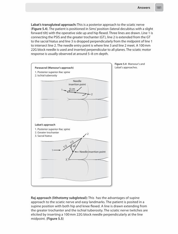
Answers 181
Labat’s transgluteal approach: This is a posterior approach to the sciatic nerve (Figure 5.4). The patient is positioned in Sims’ position (lateral decubitus with a slight forward tilt) with the operative side up and hip flexed. Three lines are drawn. Line 1 is connecting the PSIS and the greater trochanter (GT), line 2 is extended from the GT to the sacral hiatus and line 3 is dropped perpendicularly from the midpoint of line 1 to intersect line 2. The needle entry point is where line 3 and line 2 meet. A 100 mm 22G block needle is used and inserted perpendicular to all planes. The sciatic motor response is usually observed at around 5–8 cm depth.
1. Posterior superior iliac spine2. Ischial tuberosity
Parasacral (Mansour’s approach)
Labat’s approach
1. Posterior superior iliac spine2. Greater trochanter3. Sacral hiatus
6 cm1
2
Needleinsertion point
1. Posterior supe2.2. IscIschiahi l tuberos
Parasacral (Man
Labat’s approac
ea
11/2
1/2
90º
2
3
Needle insertion point
2. Greater trocha3. 3. SacSacralr hiatus
pp
1. Posterior supe2 Greater trocha
1 ion point
Figure 5.4 Mansour's and Labat's approaches.
Raj approach (lithotomy subgloteal): This has the advantages of supine approach to the sciatic nerve and easy landmarks. The patient is posited in a supine position with both hip and knee flexed. A line is drawn extending from the greater trochanter and the ischial tuberosity. The sciatic nerve twitches are elicited by inserting a 100 mm 22G block needle perpendicularly at the line midpoint. (Figure 5.5)
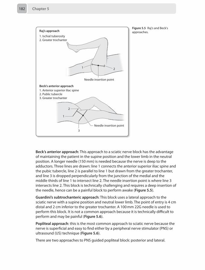
Chapter 5182
Beck’s anterior approach: This approach to a sciatic nerve block has the advantage of maintaining the patient in the supine position and the lower limb in the neutral position. A longer needle (150 mm) is needed because the nerve is deep to the adductors. Three lines are drawn: line 1 connects the anterior superior iliac spine and the pubic tubercle, line 2 is parallel to line 1 but drawn from the greater trochanter, and line 3 is dropped perpendicularly from the junction of the medial and the middle thirds of line 1 to intersect line 2. The needle insertion point is where line 3 intersects line 2. This block is technically challenging and requires a deep insertion of the needle, hence can be a painful block to perform awake (Figure 5.5).
Guardini’s subtrochanteric approach: This block uses a lateral approach to the sciatic nerve with a supine position and neutral lower limb. The point of entry is 4 cm distal and 2 cm inferior to the greater trochanter. A 100 mm 22G needle is used to perform this block. It is not a common approach because it is technically difficult to perform and may be painful (Figure 5.6).
Popliteal approach: this is the most common approach to sciatic nerve because the nerve is superficial and easy to find either by a peripheral nerve stimulator (PNS) or ultrasound (US) technique (Figure 5.6).
There are two approaches to PNS guided popliteal block: posterior and lateral.
1. Ischial tuberosity2. Greater trochanter
1. Anterior superior iliac spine2. Public tubercle3. Greater trochanter
Beck’s anterior approach
Raj’s approach
Needle insertion point
1 2
Needle insertion point
1
2
3
Figure 5.5 Raj's and Beck's approaches.
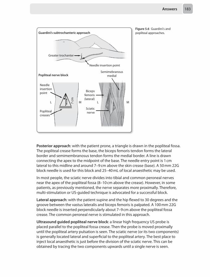
Answers 183
Posterior approach: with the patient prone, a triangle is drawn in the popliteal fossa. The popliteal crease forms the base, the biceps femoris tendon forms the lateral border and semimembranosus tendon forms the medial border. A line is drawn connecting the apex to the midpoint of the base. The needle entry point is 1 cm lateral to this midline and around 7–9 cm above the skin crease (base). A 50 mm 22G block needle is used for this block and 25–40 mL of local anaesthetic may be used.
In most people, the sciatic nerve divides into tibial and common peroneal nerves near the apex of the popliteal fossa (8–10 cm above the crease). However, in some patients, as previously mentioned, the nerve separates more proximally. Therefore, multi-stimulation or US-guided technique is advocated for a successful block.
Lateral approach: with the patient supine and the hip flexed to 30 degrees and the groove between the vastus lateralis and biceps femoris is palpated. A 100 mm 22G block needle is inserted perpendicularly about 7–9 cm above the popliteal fossa crease. The common peroneal nerve is stimulated in this approach.
Ultrasound guided popliteal nerve block: a linear high frequency US probe is placed parallel to the popliteal fossa crease. Then the probe is moved proximally until the popliteal artery pulsation is seen. The sciatic nerve (or its two components) is generally located lateral and superficial to the popliteal artery. The best place to inject local anaesthetic is just before the division of the sciatic nerve. This can be obtained by tracing the two components upwards until a single nerve is seen.
Figure 5.6 Guardini's and popliteal approaches.
Popliteal creases
Needle insertion point
Guardini’s subtrochanteric approach
Needleinsertionpoint
L M7 cm
Greater trochanter
Semimebranousmedial
Bicepsfemoris(lateral)
L
Sciaticnerve
M
Popliteal nerve block
Chapter 5184
Enneking K, Chan V, Greger J. Lower-extremity peripheral nerve blockade: essentials of our current understanding. Reg Anesth Pain Med 2005; 30(1):4-35.Al-Haddad MF, Coventry DM. Major nerve blocks of the lower limb. Br J Anaesth CEPD Reviews 2003; 3(4):102–105.
10. D Axillary brachial plexus blockPatients undergoing upper limb surgery have several options of anaesthesia, including local, regional and general anaesthesia. Brachial plexus block represents the most common use of nerve blocks in current regional anaesthetic practice.
There are different approaches to block the brachial plexus. According to the site of the surgery they are divided into two groups: blocks above the clavicle (interscalene and supraclavicular) and blocks below the clavicle (infraclavicular and axillary).
Interscalene blocks are mainly indicated for shoulder and upper arm surgery. This approach targets the upper roots (C5-C7) and, because of the vertical arrangement of the brachial plexus roots in the interscalene groove, C8 and T1 nerve roots are missed, hence the ulnar nerve may not be blocked. Therefore, this approach is unreliable for hand and forearm surgery. Because the phrenic nerve runs anterior to the anterior scalene muscle, local anaesthetic injection at this level will almost always lead to a phrenic nerve blockade and may present a significant problem in patients with respiratory compromise, while the rare but important side effect of pneumothorax is also a possibility. The interscalene approach to a brachial plexus block is therefore is unsuitable in this clinical scenario.
Supraclavicular blocks are performed at the level where the brachial plexus trunks are in close proximity to each other so the entire upper limb is blocked reliably. The usual indications of this block are arm and hand surgery, but it can also be used for shoulder surgery if a suprascapular block is performed separately. Ulnar sparing might be a problem with the supraclavicular approach because of the medial location of the inferior trunk in this area; however the use of ultrasound might overcome this problem. The incidence of pneumothorax in supraclavicular blocks is high due to the proximity of the brachial plexus to the dome of the pleura at this level. However, the risk of pneumothorax can be reduced with a modified approach and the use of ultrasound. This block may present a danger for patients with respiratory problems such as emphysema, hence should be avoided unless performed by anaesthetists with extensive skill in this technique.
Infraclavicular blocks are performed at the level of the cords of the brachial plexus (See Figure 4.1). It blocks each of the three cords of the brachial plexus and therefore it anesthetises the entire arm successfully. It is a simple block to perform without the aid of ultrasound; however, the infraclavicular approach to brachial plexus block is not popular because of the fairly high failure rate and the risk of pneumothorax. The use of ultrasound significantly improves the success rate of this block but is remains a difficult procedure to perform. This block is not suitable for patients with respiratory compromise, again due to the high pneumothorax risk, particularly with a medial infraclavicular approach.
The safest and the most commonly used and studied brachial plexus approach is the axillary block. It has few side-effects and usually covers the entire upper limb
Answers 185
with the exception of the lateral part of the arm and the forearm, which requires additional musculocutaneous nerve block. This approach blocks the brachial plexus terminal branches and depends on the relationship of nerves to the axillary vessels. It is usually performed for elbow, arm and hand surgery. With no risk of pneumothorax and phrenic nerve block, the axillary block is the most suitable brachial plexus approach for patients with respiratory problems and lung diseases, and is therefore the most appropriate choice of block in this scenario.
Radial, ulnar and median nerves can all be easily blocked in the arm as well. However, the duration of the regional anaesthesia tends to be shorter than with brachial plexus blocks. It is also limited by the requirement to block several nerves and the application tourniquet for most surgery. Therefore, this mid-arm peripheral nerve block is not the optimal option to consider in this clinical scenario.
Al-Haddad MF. Brachial plexus blockade. Br J Anaesth CEPD Reviews 2002;2;2: 33-36.Neal JM. Upper extremity regional anesthesia. Essentials of our current understanding. Reg Anaesth Pain Med 2009; 34(2):134–70.
11. B Use security staff to hold the patient, and give 4 mg/kg ketamine intramuscularly, then transfer to rhesus
The usual tenets of sedation applicable in the elective situation are not necessarily appropriate in the emergency setting. The important issues here are consent, holding/restraint as well as the provision of safe sedation.
Consent
At the age of 17 the patient is legally still a child. If she were able to demonstrate maturity and understanding and be judged to be Gillick competent, she would be able to give her consent. When it comes to refusing treatment the child may not do this in the same way, even if competent. A parent may still be able to consent for the child in this case. In the case of a parent refusing treatment on behalf of their child, (which the medical team believe is indicated), an interim care order may be granted by the Courts allowing treatment.
In this scenario, the child lacks capacity. In England a doctor may act to provide treatment in the best interests of a child, even without parental consent. In this case parental assent/consent was available. All clinical information should be nevertheless clearly documented, alongside the reasons for the treatment plan, and a consent Form 4 could also be used for procedures, e.g. the CT/lumbar puncture.
Holding and restraint
In general, the principle is to use restraint only as a technique of last resort. Minimal force required for safety (of staff and patient) should be employed, by appropriate numbers of experienced and trained staff. The plan should be discussed with the parent beforehand, and opportunity for a discussion with parent and child should exist afterwards.
Answering notes
In this instance, oral medication is impractical (in option D), and as outlined above the legal case to intervene is clear, ruling out the attractive option of C. This leaves
Chapter 5186
the use of intramuscular, intravenous or inhalational methods alongside minimal restraint. Most emergency departments would lack the facility of an anaesthetic machine, thus the degree and duration of restraint needed to transfer this girl to theatre and perform an inhalational induction makes option E impractical and dangerous. Holding to achieve intravenous access may be reasonable, but the choice of agents is not, as the CT scan and bloods should be relatively painless therefore the fentanyl in option A may not be required, as such the increased number of agents only serves to increase risks. In addition, a single dose of midazolam is unlikely to be successful for the duration of the investigations required. Thus, the option which minimises holding, involves only one agent, and provides an appropriate duration would be intramuscular ketamine. Thought does have to be given to inserting intravenous access and establishing safe monitoring for transfer, which is probably best achieved in a resuscitation area.
Tan L, Meakin GH. Anaesthesia for the uncooperative child. Contin Educ Anaesth Crit Care Pain 2010; 10(2):48–52.
12. A Urine output volume measurementThis question is testing your knowledge and reasoning in an attempt to differentiate between two common causes of hyponatraemia in a patient with a head injury. The differential diagnosis is between syndrome of inappropriate anti-diuretic hormone (SIADH) and cerebral salt wasting (CSW).
Hyponatraemia is serious: in-hospital mortality is increased by 2–4 times and a difference in survival outcome is still present at 1-year follow-up. Correcting the serum sodium concentration is also hazardous and if done too rapidly may precipitate severe neurologic complications, such as central pontine myelinosis, which can produce spastic quadriparesis, swallowing dysfunction and pseudobulbar palsy.
The classic way to differentiate between causes of hyponatraemia is to assess fluid balance (see Table 5.4).
CSW is a condition that is poorly understood. Proposed mechanisms include increased sympathetic activity causing a higher glomerular filtration rate and excess atrial natriuretic peptide (ANP) and brain natriuretic peptide (BNP) release resulting in reduced renal water re-absorption.
It occurs most commonly in traumatic brain injury and presents in the first week after injury and is normally self-limiting. The key clinical feature is hypovolaemia with a high urine output production. The serum osmolarity may be normal or high and urinary sodium is usually raised. The management involves replacing sodium and water with 0.9% sodium chloride and if symptoms develop (anorexia, confusion, unconsciousness and seizures) hypertonic saline may be indicated.
SIADH occurs as a result of traumatic brain injury, sub-arachnoid haemorrhage, brain tumors and meningitis. Excess ADH results in increased water absorption from the collecting duct of the nephron. The key clinical feature is hypervolaemia and low urinary volume. The plasma has a low serum osmolarity due to the dilutional effect of excess water and the urine osmolarity is usually high. The management involves restricting water intake.
Answers 187
In the intensive care unit great care is paid to getting the ‘numbers’ right. The fluid balance is often adjusted according to a planned daily target. Central venous pressure is of dubious benefit and a discrete value as is offered here is unhelpful. Peripheral oedema may be multi-factorial and may be apparent even in the presence of intravascular volume depletion. Osmolarity measurements are important in making the diagnosis but in both differential diagnoses it may be normal and a urinary sodium concentration is the more discerning test.
Considering the available options in the question above, urine output is the most important piece of information: a high urine volume being produced in CSW and a low urine volume being produced in SIADH.
Bradshaw K, Smith M. Disorders of sodium balance after brain injury. Contin Educ Anaesth Crit Care Pain 2008; 8(4):129–33.Sushrut P, Waikar S. Mortality after hospitalization with mild, moderate, and severe hyponatremia. Am J Med 2009; 122(9):857–65.
13. C Give 20 mmol of intravenous magnesium sulphate over 10–20 minutes
This child has acute severe asthma and has failed to respond adequately despite optimal first line therapy. Though at risk of further deterioration, the severity of his current condition does not warrant intubation and positive pressure ventilation – both of which may be hazardous. There is no evidence to support an additional dose of nebulised salbutamol, or the use of intravenous bronchodilators.
Table 5.4 Establishing the cause of hyponatraemia by assessing fluid status
Fluid status Pathophysiology Conditions
Hypovolaemic Sodium and water loss Cerebral salt wastingRenal loss:• Diuretics• Ketonuria• Glycosuria• Addison’s diseaseGastrointestinal loss:• Diarrhoea and vomitingOther:• Sweating• Bleeding
Normovolaemic Sodium loss without water loss
SIADHDiureticsAddison’s diseaseHypothyroidismIatrogenic
Hypervolaemic Excess water SIADHCardiac failureHepatic failureRenal failureIatrogenic
Chapter 5188
The next intervention likely to reverse his current pathophysiology is intravenous (as opposed to nebulised) magnesium sulphate although the optimal regime remains controversial.
Somasundaram K, Ball J. Medical emergencies: pulmonary embolism and acute severe asthma. Anaesthesia 2013; 68(suppl 1):102–16.
14. D Administer 2 g intravenous magnesium and optimise the serum potassium concentration
Atrial fibrillation (AF) is a common problem in the critical care environment with up to 15% of medical critical care patients developing AF at some point during their stay.
The risk factors for developing AF are:
Patient factors:
• Age > 65 years old• Disease severity• Hypertension• Previous AF• Congestive heart failure • Chronic obstructive pulmonary disease• Previous use of calcium-channel blockers, beta-blocker or angiotensin-converting
enzyme-inhibitor and withdrawal of catecholamine infusions
Acute illness:
• Hypoxia• Cardiac ischaemia
Sepsis or systemic inflammatory response syndrome
• Fluid shifts (hypervolaemia and hypovolaemia)• Low serum magnesium and potassium
Iatrogenic
• Intra-cardiac catheter: central line or pulmonary artery catheter
AF probably occurs as a result of a final atrial insult (the last straw) on the background of chronic disease most commonly hypertensive or ischaemic myopathy. A sudden change in atrial dimensions as a result of filling pressures (either too high with fluid or too low with dehydration and sepsis), a change in electrochemical gradients across the myocyte (potassium and magnesium flux) or an ischaemic event are the common precipitating factors.
In health the atria contribute around 10% of cardiac output, increasing to 30% during exercise. This is well tolerated in patients with normal left ventricular function but in patients who depend on the higher filling pressures, loss of the ‘atrial kick’
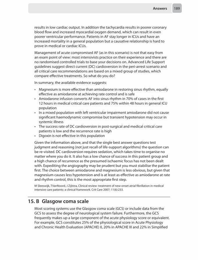
Answers 189
results in low cardiac output. In addition the tachycardia results in poorer coronary blood flow and increased myocardial oxygen demand, which can result in even poorer ventricular performance. Patients in AF stay longer in ICUs and have an increased mortality in a general population but a causative relationship is hard to prove in medical or cardiac ICUs.
Management of acute compromised AF (as in this scenario) is not that easy from an exam point of view: most intensivists practice on their experience and there are no randomised controlled trials to base your decisions on. Advanced Life Support guidelines suggest direct current (DC) cardioversion in the peri-arrest scenario and all critical care recommendations are based on a mixed group of studies, which compare effective treatments. So what do you do?
In summary, the available evidence suggests:
• Magnesium is more effective than amiodarone in restoring sinus rhythm, equally effective as amiodarone at achieving rate control and is safe
• Amiodarone infusion converts AF into sinus rhythm in 70% of cases in the first 12 hours in medical critical care patients and 75% within 48 hours in general ICU population
• In a mixed population with left ventricular impairment amiodarone did not cause significant haemodynamic compromise but transient hypotension may occur in systemic illness
• The success rate of DC cardioversion in post-surgical and medical critical care patients is low and the recurrence rate is high
• Digoxin is not effective in this population
Given the information above, and that the single best answer questions test judgment and reasoning (not just recall of life-support algorithms) the question can be re-visited. DC cardioversion requires sedation, which takes time to organise no matter where you do it. It also has a low chance of success in this patient group and a high chance of recurrence as the presumed ischaemic focus has not been dealt with. Expediting the angiography may be prudent but you must stabilise the patient first. The choice between amiodarone and magnesium is less obvious, but given that magnesium causes less hypotension and is at least as effective as amiodarone at rate and rhythm control, this is the most appropriate first step.
M Sleeswijk, TVanNoord, J Zijlstra, Clinical review: treatment of new-onset atrial fibrillation in medical intensive care patients: a clinical framework. Crit Care 2007; 11(6):233.
15. B Glasgow coma scaleMost scoring systems use the Glasgow coma scale (GCS) or include data from the GCS to assess the degree of neurological system failure. Furthermore, the GCS frequently makes up a large component of the acute physiology score or equivalent. For example, GCS constitutes 25% of the physiological score in Acute Physiology and Chronic Health Evaluation (APACHE) II, 20% in APACHE III and 22% in Simplified
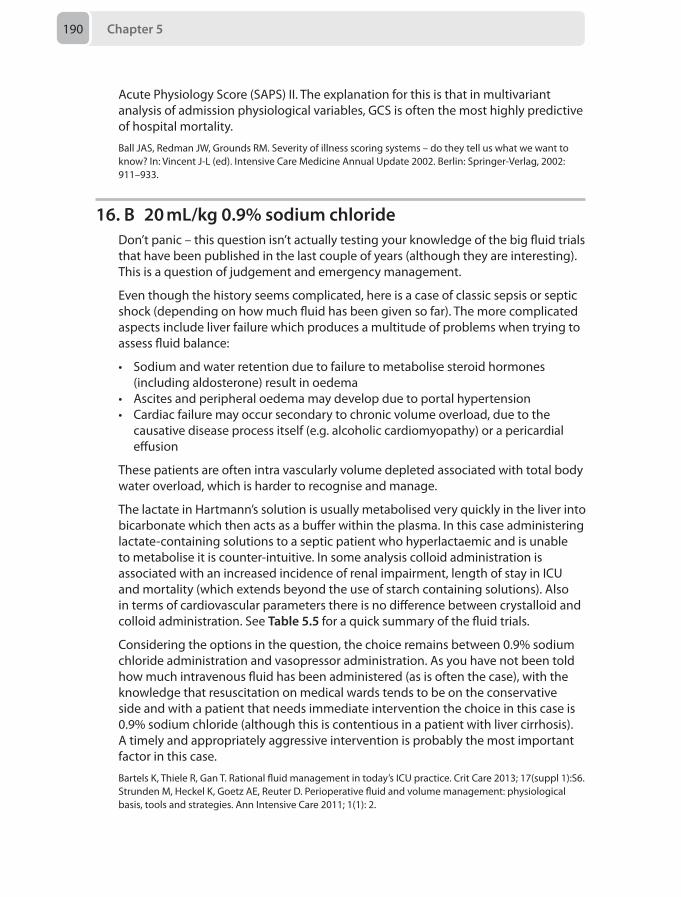
Chapter 5190
Acute Physiology Score (SAPS) II. The explanation for this is that in multivariant analysis of admission physiological variables, GCS is often the most highly predictive of hospital mortality.
Ball JAS, Redman JW, Grounds RM. Severity of illness scoring systems – do they tell us what we want to know? In: Vincent J-L (ed). Intensive Care Medicine Annual Update 2002. Berlin: Springer-Verlag, 2002: 911–933.
16. B 20 mL/kg 0.9% sodium chlorideDon’t panic – this question isn’t actually testing your knowledge of the big fluid trials that have been published in the last couple of years (although they are interesting). This is a question of judgement and emergency management.
Even though the history seems complicated, here is a case of classic sepsis or septic shock (depending on how much fluid has been given so far). The more complicated aspects include liver failure which produces a multitude of problems when trying to assess fluid balance:
• Sodium and water retention due to failure to metabolise steroid hormones (including aldosterone) result in oedema
• Ascites and peripheral oedema may develop due to portal hypertension • Cardiac failure may occur secondary to chronic volume overload, due to the
causative disease process itself (e.g. alcoholic cardiomyopathy) or a pericardial effusion
These patients are often intra vascularly volume depleted associated with total body water overload, which is harder to recognise and manage.
The lactate in Hartmann’s solution is usually metabolised very quickly in the liver into bicarbonate which then acts as a buffer within the plasma. In this case administering lactate-containing solutions to a septic patient who hyperlactaemic and is unable to metabolise it is counter-intuitive. In some analysis colloid administration is associated with an increased incidence of renal impairment, length of stay in ICU and mortality (which extends beyond the use of starch containing solutions). Also in terms of cardiovascular parameters there is no difference between crystalloid and colloid administration. See Table 5.5 for a quick summary of the fluid trials.
Considering the options in the question, the choice remains between 0.9% sodium chloride administration and vasopressor administration. As you have not been told how much intravenous fluid has been administered (as is often the case), with the knowledge that resuscitation on medical wards tends to be on the conservative side and with a patient that needs immediate intervention the choice in this case is 0.9% sodium chloride (although this is contentious in a patient with liver cirrhosis). A timely and appropriately aggressive intervention is probably the most important factor in this case.
Bartels K, Thiele R, Gan T. Rational fluid management in today’s ICU practice. Crit Care 2013; 17(suppl 1):S6.Strunden M, Heckel K, Goetz AE, Reuter D. Perioperative fluid and volume management: physiological basis, tools and strategies. Ann Intensive Care 2011; 1(1): 2.
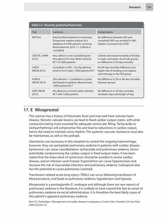
Answers 191
17. E MisoprostolThis woman has a history of rheumatic fever and may well have valvular heart disease. Stenotic valvular lesions can lead to fixed cardiac output states, with atrial contraction being more essential for adequate ventricular filling. Tachycardia or tachyarrhythmias will compromise this and lead to reductions in cardiac output, hence the need to maintain sinus rhythm. The systemic vascular resistance must also be maintained, as well as the preload.
Uterotonics are necessary in this situation to control the ongoing haemorrhage, however, they can precipitate pulmonary oedema in patients with cardiac disease. Syntocinon can cause vasodilatation, tachycardia and pulmonary oedema, hence potentially compromising the cardiac output in fixed output states. It has been stated that the bolus dose of syntocinon should be avoided in severe cardiac disease, and an infusion used instead. Ergometrine can cause hypertension and increase the risk of myocardial infarction and pulmonary oedema. Carboprost also has the potential to cause pulmonary overload.
Transfusion-related acute lung injury (TRALI) can occur following transfusion of blood products, and leads to pulmonary oedema, hypotension and hypoxia.
Misoprostol is a prostaglandin E1 analogue and although there are rare reports of pulmonary oedema in the literature, it is unlikely to have caused this fast an onset of pulmonary oedema via rectal administration. It is therefore the least likely cause of this patient's apparent pulmonary oedema.
Burt CC, Durbridge J. Management of cardiac disease in pregnancy. Contin Educ Anaesth Crit Care Pain 2009; 9(2):44–47.
Table 5.5 Recently published fluid trials
Trial Summary Interpretation
RaFTinG Rational fluid treatment in Germany. Prospective registry analysis of a database of 4,500 patients receiving fluid treatment 2010–11. Colloid vs crystalloid
No difference between HES and crystalloid (HES was avoided in AKI). Gelatins increased risk of AKI
CRISTAL (JAMA 2013)
Any colloid vs and crystalloid given throughout ICU stay. Multi-national RCT of 2,800 patients
Colloids decreased mortality at 90 days in septic and septic shock sub groups, no difference in 28-day mortality
CHEST(NEJM 2012)
Crystalloid vs HES. 7 mL/kg adminis-tered in first 4 days. 7,000 patients RCT
No 90-day mortality difference, but higher risk of needing renal replace-ment therapy in the HES group
ALBIOS (NEJM 2014)
20% albumin + crystalloid vs crystal-loid based on patients albumin level. 1,800 patients RCT
No difference in 28-or 90-day mortality between groups
SAFE (NEJM 2012)
4% albumin vs normal saline, blinded RCT with 7,000 patients
No difference in 28-day mortality, ventilator days and length of stay
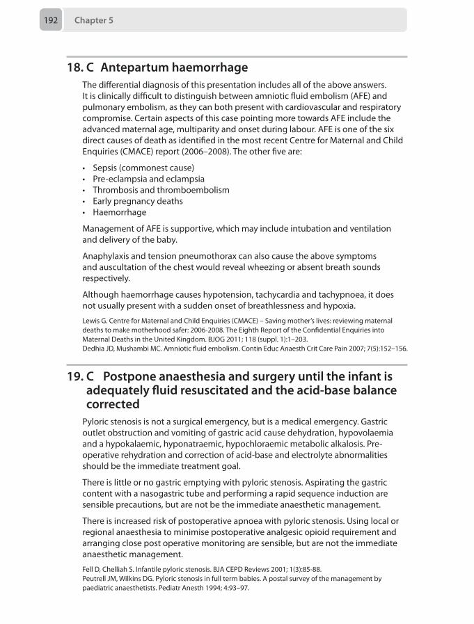
Chapter 5192
18. C Antepartum haemorrhageThe differential diagnosis of this presentation includes all of the above answers. It is clinically difficult to distinguish between amniotic fluid embolism (AFE) and pulmonary embolism, as they can both present with cardiovascular and respiratory compromise. Certain aspects of this case pointing more towards AFE include the advanced maternal age, multiparity and onset during labour. AFE is one of the six direct causes of death as identified in the most recent Centre for Maternal and Child Enquiries (CMACE) report (2006–2008). The other five are:
• Sepsis (commonest cause)• Pre-eclampsia and eclampsia• Thrombosis and thromboembolism• Early pregnancy deaths• Haemorrhage
Management of AFE is supportive, which may include intubation and ventilation and delivery of the baby.
Anaphylaxis and tension pneumothorax can also cause the above symptoms and auscultation of the chest would reveal wheezing or absent breath sounds respectively.
Although haemorrhage causes hypotension, tachycardia and tachypnoea, it does not usually present with a sudden onset of breathlessness and hypoxia.
Lewis G. Centre for Maternal and Child Enquiries (CMACE) – Saving mother’s lives: reviewing maternal deaths to make motherhood safer: 2006-2008. The Eighth Report of the Confidential Enquiries into Maternal Deaths in the United Kingdom. BJOG 2011; 118 (suppl. 1):1–203.Dedhia JD, Mushambi MC. Amniotic fluid embolism. Contin Educ Anaesth Crit Care Pain 2007; 7(5):152–156.
19. C Postpone anaesthesia and surgery until the infant is adequately fluid resuscitated and the acid-base balance corrected
Pyloric stenosis is not a surgical emergency, but is a medical emergency. Gastric outlet obstruction and vomiting of gastric acid cause dehydration, hypovolaemia and a hypokalaemic, hyponatraemic, hypochloraemic metabolic alkalosis. Pre-operative rehydration and correction of acid-base and electrolyte abnormalities should be the immediate treatment goal.
There is little or no gastric emptying with pyloric stenosis. Aspirating the gastric content with a nasogastric tube and performing a rapid sequence induction are sensible precautions, but are not be the immediate anaesthetic management.
There is increased risk of postoperative apnoea with pyloric stenosis. Using local or regional anaesthesia to minimise postoperative analgesic opioid requirement and arranging close post operative monitoring are sensible, but are not the immediate anaesthetic management.
Fell D, Chelliah S. Infantile pyloric stenosis. BJA CEPD Reviews 2001; 1(3):85-88.Peutrell JM, Wilkins DG. Pyloric stenosis in full term babies. A postal survey of the management by paediatric anaesthetists. Pediatr Anesth 1994; 4:93–97.
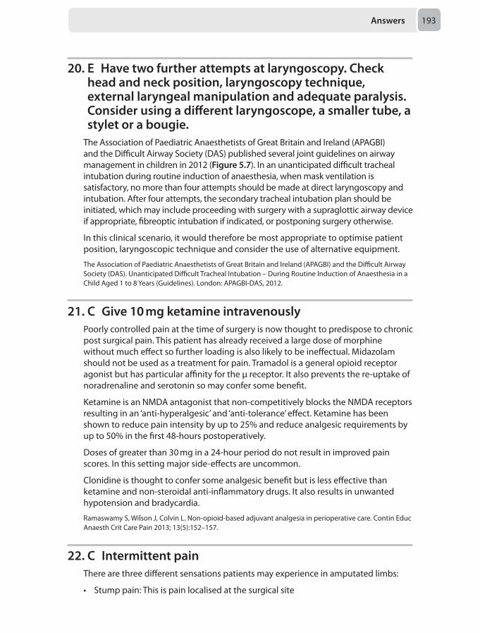
Answers 193
20. E Have two further attempts at laryngoscopy. Check head and neck position, laryngoscopy technique, external laryngeal manipulation and adequate paralysis. Consider using a different laryngoscope, a smaller tube, a stylet or a bougie.
The Association of Paediatric Anaesthetists of Great Britain and Ireland (APAGBI) and the Difficult Airway Society (DAS) published several joint guidelines on airway management in children in 2012 (Figure 5.7). In an unanticipated difficult tracheal intubation during routine induction of anaesthesia, when mask ventilation is satisfactory, no more than four attempts should be made at direct laryngoscopy and intubation. After four attempts, the secondary tracheal intubation plan should be initiated, which may include proceeding with surgery with a supraglottic airway device if appropriate, fibreoptic intubation if indicated, or postponing surgery otherwise.
In this clinical scenario, it would therefore be most appropriate to optimise patient position, laryngoscopic technique and consider the use of alternative equipment.
The Association of Paediatric Anaesthetists of Great Britain and Ireland (APAGBI) and the Difficult Airway Society (DAS). Unanticipated Difficult Tracheal Intubation – During Routine Induction of Anaesthesia in a Child Aged 1 to 8 Years (Guidelines). London: APAGBI-DAS, 2012.
21. C Give 10 mg ketamine intravenouslyPoorly controlled pain at the time of surgery is now thought to predispose to chronic post surgical pain. This patient has already received a large dose of morphine without much effect so further loading is also likely to be ineffectual. Midazolam should not be used as a treatment for pain. Tramadol is a general opioid receptor agonist but has particular affinity for the μ receptor. It also prevents the re-uptake of noradrenaline and serotonin so may confer some benefit.
Ketamine is an NMDA antagonist that non-competitively blocks the NMDA receptors resulting in an ‘anti-hyperalgesic’ and ‘anti-tolerance’ effect. Ketamine has been shown to reduce pain intensity by up to 25% and reduce analgesic requirements by up to 50% in the first 48-hours postoperatively.
Doses of greater than 30 mg in a 24-hour period do not result in improved pain scores. In this setting major side-effects are uncommon.
Clonidine is thought to confer some analgesic benefit but is less effective than ketamine and non-steroidal anti-inflammatory drugs. It also results in unwanted hypotension and bradycardia.
Ramaswamy S, Wilson J, Colvin L. Non-opioid-based adjuvant analgesia in perioperative care. Contin Educ Anaesth Crit Care Pain 2013; 13(5):152–157.
22. C Intermittent painThere are three different sensations patients may experience in amputated limbs:
• Stump pain: This is pain localised at the surgical site
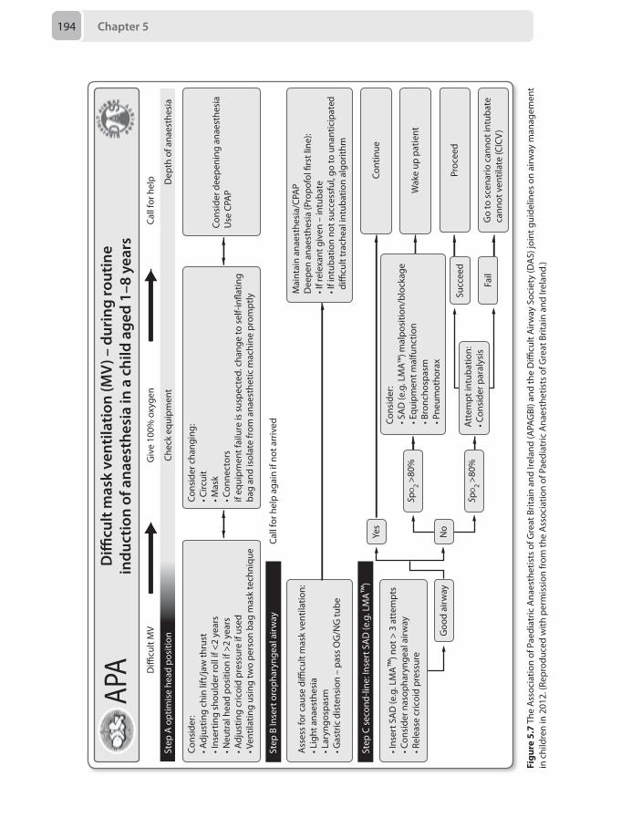
Chapter 5194
Di�
cult
MV
Giv
e 10
0% o
xyge
n
Call
for h
elp
agai
n if
not a
rriv
ed
Ass
ess
for c
ause
di�
cult
mas
k ve
ntila
tion:
• Lig
ht a
naes
thes
ia• L
aryn
gosp
asm
• Gas
tric
dis
tens
ion
– pa
ss O
G/N
G tu
be
• Ins
ert S
AD
(e.g
. LM
A™
) not
> 3
att
empt
s• C
onsi
der n
asop
hary
ngea
l airw
ay• R
elea
se c
ricoi
d pr
essu
re
Goo
d ai
rway
SpO
2 >8
0%
SpO
2 >8
0%A
ttem
pt in
tuba
tion:
• Con
side
r par
alys
is
Cons
ider
:• S
AD
(e.g
. LM
A™
) mal
posi
tion/
bloc
kage
• Equ
ipm
ent m
alfu
nctio
n• B
ronc
hosp
asm
• Pne
umot
hora
x
Cons
ider
dee
peni
ng a
naes
thes
iaU
se C
PAP
Mai
ntai
n an
aest
hesi
a/CP
AP
Dee
pen
anae
sthe
sia
(Pro
pofo
l �rs
t lin
e):
• If r
elex
ant g
iven
– in
tuba
te• I
f int
ubat
ion
not s
ucce
ssfu
l, go
to u
nant
icip
ated
di�
cult
trac
heal
intu
batio
n al
gorit
hm
Go
to s
cena
rio c
anno
t int
ubat
eca
nnot
ven
tilat
e (C
ICV
)
Succ
eed
Fail
Cons
ider
cha
ngin
g:• C
ircui
t• M
ask
• Con
nect
ors
if eq
uipm
ent f
ailu
re is
sus
pect
ed. c
hang
e to
sel
f-in�
atin
gba
g an
d is
olat
e fr
om a
naes
thet
ic m
achi
ne p
rom
ptly
Call
for h
elp
Cons
ider
:• A
djus
ting
chin
lift
/jaw
thru
st• I
nser
ting
shou
lder
roll
if <2
yea
rs• N
eutr
al h
ead
posi
tion
if >2
yea
rs• A
djus
ting
cric
oid
pres
sure
if u
sed
• Ven
tilat
ing
usin
g tw
o pe
rson
bag
mas
k te
chni
que
Di�
cult
mas
k ve
ntila
tion
(MV
) – d
urin
g ro
utin
ein
duct
ion
of a
naes
thes
ia in
a c
hild
age
d 1–
8 ye
ars
APA
Step
A o
ptim
ise
head
pos
ition
Chec
k eq
uipm
ent
Dep
th o
f ana
esth
esia
Step
B In
sert
oro
phar
ynge
al a
irway
Step
C s
econ
d-lin
e: In
sert
SA
D (e
.g. L
MA
™)
Cont
inue
Wak
e up
pat
ient
Proc
eed
Yes
No
Figu
re 5
.7 T
he A
ssoc
iatio
n of
Pae
diat
ric A
naes
thet
ists
of G
reat
Brit
ain
and
Irela
nd (A
PAG
BI) a
nd th
e D
ifficu
lt A
irway
Soc
iety
(DA
S) jo
int g
uide
lines
on
airw
ay m
anag
emen
t in
chi
ldre
n in
201
2. (R
epro
duce
d w
ith p
erm
issi
on fr
om th
e A
ssoc
iatio
n of
Pae
diat
ric A
naes
thet
ists
of G
reat
Brit
ain
and
Irela
nd.)
Answers 195
• Phantom sensations: This occurs when the patient feels that the limb is present but is not always painful
• Phantom pain: This is pain that arises in the imagined, amputated limb
Phantom pain, occurring in 30–85% of post-amputation patients, is very difficult to treat and tends to respond poorly to opioids. Various pharmacological therapies have been trialled, with some success with calcitonin, amitriptyline and gabapentin, although non-pharmacological therapies have an important role. Pre-emptive epidural analgesia has not been shown to reduce the incidence of development of phantom limb pain. The pain is most commonly intermittent and only rarely does the pain become constant. In this patient therefore, true phantom pain is most likely suggested by pain that is intermittent in nature.
Nikolajsen L, Jensen TS. Phantom limb pain. Br J Anaesth 2001; 87(1):107–116.
23. E Thoracic epiduralThoracotomy ranks amongst the most painful of surgical procedures. Apart from patient discomfort, the consequences of post-thoracotomy pain lead to several other complications. Reduced tidal volumes from shallow breathing and the inability to cough may result in hypoxia and hypercarbia, placing the patient at increased risk of developing respiratory failure and postoperative pneumonia. This is further complicated by increased myocardial oxygen demand from the sympathetic response to pain, which may precipitate an adverse cardiac event.
There are several possible options for achieving analgesia. What method is chosen depends on patient, anaesthetic and surgical preference (Table 5.6).
In the above clinical scenario therefore, the most appropriate analgesic option would be a thoracic epidural.
Hughes R, Gao F. Pain control for thoracotomy. Contin Educ Anaesth Crit Care Pain 2005; 5(2):56–60.
24. C Perform a coeliac plexus blockA coeliac plexus block can be used to relieve autonomically mediated pain from non-pelvic abdominal organs. It is most commonly used in intractable malignant pain. Firstly it would be performed with local anaesthetic to assess efficacy and then can be repeated as a neurolytic block with 6% aqueous phenol.
The coeliac plexus consists of coeliac ganglia that lie on either side of the L1 vertebral body. It provides the autonomic supply to the non-pelvic abdominal organs and the first two-thirds of the large intestine.
It consists of:
• Greater splanchnic nerve: T5–T6 to T9–T10• Lesser splanchnic nerve: T10–T11• Least splanchnic nerve: T11–T12
Figu
re 5
.7 T
he A
ssoc
iatio
n of
Pae
diat
ric A
naes
thet
ists
of G
reat
Brit
ain
and
Irela
nd (A
PAG
BI) a
nd th
e D
ifficu
lt A
irway
Soc
iety
(DA
S) jo
int g
uide
lines
on
airw
ay m
anag
emen
t in
chi
ldre
n in
201
2. (R
epro
duce
d w
ith p
erm
issi
on fr
om th
e A
ssoc
iatio
n of
Pae
diat
ric A
naes
thet
ists
of G
reat
Brit
ain
and
Irela
nd.)
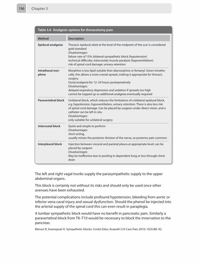
Chapter 5196
Table 5.6 Analgesic options for thoracotomy pain
Method Description
Epidural analgesia Thoracic epidural sited at the level of the midpoint of the scar is considered gold standardDisadvantages: failure rate of 15%; bilateral sympathetic block (hypotension) technical difficulty; intercostals muscle paralysis (hypoventilation) risk of spinal cord damage; urinary retention
Intrathecal mor-phine
Morphine is less lipid-soluble than diamorphine or fentanyl. Given intrathe-cally, this allows a more cranial spread, making it appropriate for thoracic surgery. Good analgesia for 12–24 hours postoperatively Disadvantages: delayed respiratory depression and sedation if spreads too high cannot be topped up so additional analgesia eventually required
Paravertebral block Unilateral block, which reduces the limitations of a bilateral epidural block, e.g. hypotension, hypoventilation, urinary retention. There is also less risk of spinal cord damage. Can be placed by surgeon under direct vision, and a catheter can be left in situDisadvantages: only suitable for unilateral surgery
Intercostal block Quick and simple to performDisadvantages: short acting, usually misses the posterior division of the nerve, so posterior pain common
Interpleural block Injection between visceral and parietal pleura at appropriate level; can be placed by surgeonDisadvantages: May be ineffective due to pooling in dependent lung or loss through chest drain
The left and right vagal trunks supply the parasympathetic supply to the upper abdominal organs.
This block is certainly not without its risks and should only be used once other avenues have been exhausted.
The potential complications include profound hypotension, bleeding from aortic or inferior vena caval injury and sexual dysfunction. Should the phenol be injected into the arterial supply of the spinal cord this can even result in paraplegia.
A lumbar sympathetic block would have no benefit in pancreatic pain. Similarly a paravertebral block from T8–T10 would be necessary to block the innervation to the pancreas.
Menon R, Swanepoel A. Sympathetic blocks. Contin Educ Anaesth Crit Care Pain 2010; 10(3):88–92.
Answers 197
25. C Single shot caudal combined with 10 mg/kg paracetamol orally 6-hourly and codeine phosphate 1 mg/kg orally 6-hourly as required
Despite having immature nociceptive pathways, it is now well accepted that neonates experience pain and as such, post operative pain needs to be well managed. As with adults, a multimodal approach should be used to minimise side-effects.
Paracetamol is the most commonly used analgesic both in the adult and paediatric populations and should be used as a base in most analgesic regimes.
However as per the British National Formulary (BNF) the paracetamol dose varies:
Orally:
• Preterm neonate 28–32 weeks post-conceptual age 20 mg/kg loading dose followed by 10–15 mg/kg 8–12-hourly. Maximum dose 30 mg/kg daily
• Neonate over 32 weeks post-conceptual age 20 mg/kg loading dose followed by 10–15 mg 6–8-hourly. Maximum dose 60 mg/kg daily
Intravenously:
• Preterm neonate over 32 weeks post-conceptual age 7.5 mg/kg 8-hourly. Maximum dose 25 mg/kg daily
• Neonate 10 mg/kg 4–6 hourly. Maximum dose 30 mg/kg daily
Non steroidal anti-inflammatory drugs (NSAIDs) should also be used in children wherever possible. However, ibuprofen is not recommended for neonates or infants under 5 kg and diclofenac is not recommended for infants under 6 months.
Opiates should also be used with care in this setting. Codeine is a pro-drug that is metabolised to morphine via the p450 cytochrome. The particular enzyme responsible, CYP2D6, has low activity at birth and becomes more effective with age. In addition to its immaturity, it also exhibits genetic polymorphism leading to significant differences in efficacy between patients. This explains its good safety profile in the young but is perhaps not the best analgesic agent. However, the combination of paracetamol and codeine has been shown to reduce postoperative pain in infants.
Morphine is also commonly used and can be administered via a number of different routes. Reduced doses should be used in neonates due to their increased potency. This is due to a combination of an immature blood-brain barrier and reduced protein binding. Morphine will also have a longer duration of action due to slower metabolism (immature enzymes) and reduced excretion (reduced glomerular filtration rate).
Local anaesthetic, whether in the form of surgical wound infiltration, nerve block or regional technique, should be used wherever possible. Caudals remain the most popular central neuroaxial technique used in neonates. Opioids are not commonly added to the local anaesthetic due to concerns over postoperative respiratory depression. In the neonates, caudals result in adequate analgesia below T10.
Chapter 5198
Williams G. Analgesic regimens for children. In: AAGBI Core Topics in Anaesthesia. Johnston I, Harrop-Griffiths W, Gemmell L, eds. London: AAGBI, 2012.Haidon JL, Cunliffe M. Analgesia for neonates. Contin Educ Anaesth Crit Care Pain 2010; 10(4): 123–27.Gormley SMC, Crean PM. Basic principles of anaesthesia for neonates and infants. BJA CEPD Reviews 2001; 1(5):130–33.
26. D Cyanide poisoningIn the Western world, the most common cause of cyanide poisoning is house fires and subsequent smoke inhalation. When exposed to high temperatures, products containing carbon and nitrogen may liberate cyanide during combustion. These materials include wool, silk and synthetic polymers.
Cyanide toxicity is dangerous as it prevents aerobic cell metabolism, causing intracellular hypoxia by reversibly binding to cytochrome oxidase a3 within the mitochondria, which is essential for oxidative phosphorylation. Oxidative phosphorylation is crucial to the synthesis of adenosine triphosphate (ATP) and the continuation of cellular respiration. As supplies of ATP become depleted, mitochondria cannot extract or use the oxygen they are exposed to. As a result, metabolism shifts to glycolysis through anaerobic metabolism which produces lactate. Production of lactate results in a high anion-gap metabolic acidosis. The cells are no longer able to use oxygen for aerobic metabolism which results in high venous oxygen saturations.
Signs and symptoms of cyanide poisoning occur soon after exposure. Early manifestations include anxiety, headache and mydriasis. As hypoxia progresses, GCS drops and seizures can occur.
Cyanide poisoning should be suspected in burns patients with an unexplained and persistent lactic acidosis despite adequate fluid resuscitation.
Currently there is no test available for rapid diagnosis of cyanide poisoning, so treatment should be based on clinical suspicion. The treatment of choice for cyanide poisoning in burns patients is hydroxocobalamin. Hydroxocobalamin binds to cyanide to form the non-toxic cyanocobalamin (vitamin B12) which is then excreted by the kidneys. Cyanide has a greater affinity for hydroxocobalamin than for cytochrome oxidase a3 within the mitochondria. This then allows the mitochondria to perform aerobic metabolism. As it binds with cyanide without forming methaemoglobin, it is particularly helpful in burns patients who may have part of their haemoglobin in the form of carboxyhaemoglobin thus already reducing their oxygen carrying capacity.
Cyanide (CN–) is metabolised in any of the three following pathways:
• Converted by rhodanase in liver to thiocyanate (SCN–) which is then excreted in urine. This is the major pathway, accounting for 80% of CN– metabolism
• Combines with vitamin B12 to form cyanocobalamine (non-toxic) which is also excreted in the urine
• Combines with methemoglobin to form cyanomethemoglobin (non-toxic)
Sepsis can be a major problem in the burns patient but not at this early stage and prophylactic antibiotics are not advocated. Volume depletion is also a likely
Answers 199
contender but not in the context of a good urine output and normal capillary refill time. Acute kidney injury (AKI) in burns patients can also be problematic but again too early to show such gross acid base anomalies. AKI may be due to either volume depletion or rhabdomyolysis, the latter of which is a particular problem in electrical burns.
Substance abuse must always be considered in this group of patients and alcohol intoxication also leads to a lactic acidosis but not of this severity.
Bishop S, Maguire S. Anaesthesia and intensive care for major burns. Contin Educ Anaesth Crit Care Pain 2012; 12(3):118–22.Hamel J. A review of acute cyanide poisoning with a treatment update. Crit Care Nurse 2011; 31(1):72–81.
27. C Optimise his circulatory blood volumeRenal impairment is a significant postoperative complication following elective aortic aneurysm repairs. It is associated with prolonged hospital stay, risk of progression to renal replacement therapy and increased mortality. The pathophysiology of renal impairment during an aortic aneurysm repair is multifactorial. It involves decreased blood flow across the renal arteries during aortic cross-clamping, cholesterol emboli as a result of clamping a heavily calcified aorta, the use of nephrotoxic medication in the preoperative phase and ischaemia-reperfusion syndrome. Over the last decade several drugs listed as options in the question have been researched in an attempt to find one to prevent the development of acute kidney injury. Low dose dopamine and dopexamine were used in an attempt to stimulate diuresis postoperatively. Free radical scavengers such as mannitol given before or soon after aortic cross-clamping also failed to show a decrease in postoperative renal impairment. Except for achieving an optimum intravascular volume by replacing losses and correcting any coagulation abnormalities, all the other strategies have failed to produce a positive result.
Frederick JR, Woo YJ. Thoracoabdominal aortic aneurysm. Ann Cardiothorac Surg 2012; 1(3):277–28.
28. B Wilson’s diseaseKayser-Fleischer rings (brown or dark rings encircling the iris) are pathognomonic of Wilson’s disease which is an autosomal recessive inherited disorder characterised by toxic accumulation of copper in the liver and brain. The ATP7B enzyme prevents excessive accumulation of copper by either combining it with caeruloplasmin and releasing it into the bloodstream, or secreting it in the bile. The functions of this enzyme are affected in Wilson’s disease, causing the toxic accumulation of copper in the blood.
Children usually present with hepatic complications such as hepatitis, cirrhosis or fulminant hepatic failure. Adults tend to present with neurophsychiatric signs including dysarthria, tremors, seizures, migraine, ataxia, cognitive decline and behavioral disturbances.
Kayser-Fleischer rings are caused by deposition of copper in the Descemet’s (basement) membrane and can be visualised by slit lamp examination. Treatment for
Chapter 5200
Wilson’s disease consists of administering chelating agents like penicillamine or in extreme cases hepatic transplantation.
Alpha-1 antitrypsin is a protease inhibitor and its deficiency leads to cholestasis and pulmonary symptoms including emphysema. Primary biliary cirrhosis is caused by damage of interlobular bile ducts, leading to cholestasis coupled with portal hypertension and cirrhosis. Haemochromatosis is caused by increased iron absorption and deposition in the liver, heart or pancreas. Primary sclerosing cholangitis is inflammation of intra- and extra-hepatic ducts, which leads to liver failure and death.
Longmore M, Wilkinson I, Torok E. Oxford Handbook of Clinical Medicine, 8th ed. Oxford: Oxford University Press, 2010.
29. B Mental stateThe elderly proportion of society is expected to increase significantly over the coming years, leading to a higher rate of surgery in this age group. The postoperative morbidity associated with surgery in the geriatric population is high due to multiple comorbidities and the influence of geriatric syndromes. These syndromes can be collectively termed as a phenotype as they cannot be termed into a disease process or an organ specific pathology. Frailty is thus termed as ‘a condition associated with a decreased physiological reserve across various organ systems that can lead to an increased vulnerability to physiological stressors'. Having a tool to measure frailty helps to stratify this population according to their risks. Fried et al. originally described the frailty phenotype based upon features observed across more than 500 patients over 65 years.
• Weight loss (unintentional): > 10 pounds from baseline in prior year• Sarcopenia (loss of muscle mass): grip strength - lowest 20th population centile
(by gender and body mass index)• Weakness: self-reported exhaustion• Poor endurance: Walking time per 15 feet, slowest 20th population centile (by
gender and height)• Slowness: kcal per week, lowest 20th population centile • Low activity: males < 383 kcal/week, females < 270 kcal per week
Mental state is not incorporated in frailty assessment.
Positive frailty phenotype: > 3 criteria present
Intermediate or pre-frail : 1 or 2 criteria present
The frailty phenotype is predictive of progressive decline, repeated falls, hospital visits and eventually death.
Griffiths R, Mehta M. Frailty and anaesthesia: what we need to know. Contin Educ Anaesth Crit Care Pain 2014; 14(6):273-277.
Answers 201
30. A 20The number needed to treat (NNT) is the number of patients to whom a clinician would need to administer a particular treatment for one patient to receive benefit from it. The NNT is calculated either as:
• 100/absolute risk reduction (ARR) expressed as a percentage, or• 1/ARR expressed as a proportion
The absolute risk reduction is defined as:
Control event rate – experimental event rate; which in the example given above equates to:
25–20% = 5%
The NNT is therefore:
100/5 or 1/0.05 depending on whether the ARR is expressed as a percentage or number. The NNT is therefore 20.
NNTs have a number of important limitations. The true value of a NNT can be higher or lower than the value given; It is therefore useful to know the 95% confidence intervals of the NNT. If there is a large confidence interval there can be less certainty in the reported NNT and so clinical decisions based on this must be made with caution.
An additional important point is that the NNT depends on the baseline frequency of a given event. So, in the case of PONV, advances in perioperative care and surgical techniques may mean that the baseline frequency of PONV changes over time; A NNT of 8 observed for agent A based on a study from 1970 may not necessarily be comparable to a NNT of 10 for agent B based on a similar study conducted in 2010.
McQuay HJ, Moore RA. Using numerical results from systemic reviews in clinical practice. Ann Intern Med 1997; 126:712–720.Lalkhen AG, McCluskey A. Statistics V: Introduction to clinical trials and systematic reviews. Contin Educ Anaes Crit Care Pain 2008; 8:143–146.

Mock Paper 6
Chapter 6
Questions1. A 44-year-old woman is on the intensive care unit having had a grade 3
subarachnoid haemorrhage secondary to an anterior communicating artery aneurysm one day ago. She is currently stable neurologically. Her past medical history comprises of hypercholesterolaemia, hypertension and smoking. She has a drug history of simvastatin and lisinopril.
Which of the following would most likely prevent the development of delayed cerebral ischaemia in this patient?
A ‘Triple H therapy’B Magnesium administrationC Statin administrationD Nimodipine administrationE Antiplatelet therapy
2. A 29-year-old woman who suffered a blow to the left side of her skull vault with a resulting depressed fracture is awaiting transfer to a tertiary centre. She lost consciousness for approximately 1 minute after the incident. Her GCS is currently 14/15 (E4 V4 M6).
Which of the following, in isolation, indicates that intubation is essential before transfer?
A Pao₂ of 13 kPa an Fio₂ of 0.6B A discrete and short-lived seizure en route to your hospitalC Drop in GCS from E4 V4 M6 to E3 V4 M5 in the emergency departmentD An increase in respiratory rate leading to a Paco₂ of 4.0kPaE Blood in the oropharynx
3. A 65-year-old man presents to the emergency department with acute central chest pain radiating to the back. He has a history of hypertension and smoking. The ECG shows evidence of left ventricular hypertrophy and his blood pressure is 190/100 mmHg, heart rate 105 beats per minute. There is a collapsing pulse and an early diastolic murmur.
Chapter 6204
What is the next most appropriate management step?
A Commencement of sodium nitroprusside infusionB Site an arterial lineC Arrange urgent aortographyD Titrate intravenous morphineE Arrange transfer to a cardiothoracic centre
4. A 65-year-old woman is recovering from an uneventful total thyroidectomy as treatment for a large substernal goitre. On the third postoperative day, she becomes progressively more stridulous and wheezy. She is tachypnoeic, confused and complaining of circumoral paraesthesia. There is no obvious neck swelling or pain.
What is the most likely cause of her symptoms?
A Bilateral vocal cord paralysisB TracheomalaciaC HaematomaD Tracheal necrosisE Hypocalcaemia
5. A 36-year-old woman with an impacted food bolus needs to go to theatre imminently. She has been unable to swallow her saliva for 24 hours. On inquiring about her anaesthetic history she reports collapsing due to a severe allergic reaction under anaesthesia, but she is unsure which agent was responsible. There are no notes available, nor relatives to elaborate on the history.
Which of the following should you avoid as the most likely causative agent?
A RocuroniumB Latex C Morphine D Chlorhexidine E Gelofusine
6. A 34-year-old man presents for laparoscopic excision of his left adrenal gland for phaeochromocytoma. During your preoperative assessment, he tells you that he has been taking medication for blood pressure for about a month.
Which of the following is most likely to indicate that he is prepared for surgery?
A Good exercise tolerance, but a history of dizziness on standingB Lack of a history of palpitations, and a normal ECGC A normal echocardiogram, and chest X-rayD History of dizziness on standing, a 5-minute ECG with no premature
ventricular complexes, and nasal congestionE Several blood pressure recordings of < 160/90 mmHg
Questions 205
7. A 45-year-old man is admitted to the surgical ward with a fever, toothache and neck discomfort. Whilst waiting for surgery you are called to his bedside as he is more breathless and complaining of substernal pain. On examination he is hypotensive and there is tender, ’woody‘ induration of his neck. On auscultation you hear a pericardial rub.
Which investigation is most appropriate to guide management in this scenario?
A Cervical and chest ultrasoundB Cervical and chest computed tomography C Cervical and chest magnetic resonance imagingD Cervical and chest radiographE Echocardiogram
8. A 35-year-old cyclist suffered a severe traumatic brain injury with a large subdural haematoma and an associated C2–C3 cervical spine fracture. He is comatose and apnoeic, with neurosurgeons confirming that he is not a candidate for surgery due to poor prognosis. Confirmation of brainstem death is underway, with examination of cranial nerves just being completed.
What is the next most appropriate test that will support the neurological diagnosis of death?
A Apnoea testingB Somatosensory evoked potentialsC No further tests necessaryD A second neurological examination of the cranial nervesE Electroencephalogram
9. An 84-year-old woman with an extracapsular hip fracture is scheduled for a dynamic hip screw on your morning trauma list. On examination she has an ejection systolic murmur in the aortic area and anaemia with a haemoglobin of of 90 g/L. She has previously had a coronary stent and is on both aspirin and clopidogrel. Your hospital's echocardiogram technician is unavailable.
How do you proceed?
A Postpone the surgery until an echocardiogram can be performed urgentlyB Proceed with the case under general anaesthesia and invasive blood pressure
monitoring, with one unit of packed red blood cellsC Perform a spinal anaesthetic after administration of one pool of plateletsD Insert a lumbar epidural and use small volume incremental top-ups to achieve
a surgical blockE Proceed with the case under general anaesthesia and an ultrasound-guided
fascia iliaca block with invasive blood pressure monitoring
10. A 35-year old man for elective ankle surgery is to have an ultrasound guided popliteal nerve block.
Chapter 6206
What is the most frequently used combination of ultrasound view and needle visualisation for this nerve block?
A Short-axis view with in-plane needle approachB Long-axis view with out-of-plane needle approachC Short-axis view with out-of-plane needle approachD Long-axis view with in-plane needle approach E Any of the above combinations
11. You are called to the emergency department to assess a young woman that was rescued from a house fire following a gas leak after being trapped confined in a room. She is awake, with normal observations but suffered 10% body surface area (BS) partial thickness burns over her arms and face. You are asked to transfer her to the nearest burns unit that is 2 hours away. On examination she has singed nasal hair, a normal airway and no change in voice. Burns resuscitation is underway with intravenous fluids and analgesia.
What is the next step in ensuring her safe transfer?
A Add the operating department practitioner to your transfer teamB Full monitoring including invasive blood pressure measurementC Prepare difficult airway equipment for the transferD Prepare Intubating equipment and drugsE Elective intubation of the patient
12. A 72-year-old man has been on the intensive care unit after being treated for an infective exacerbation of his chronic obstructive pulmonary disease. He has been mechanically ventilated for 5 days and has acceptable gas exchange. He has been weaned to pressure support ventilation requiring 12 cmH2O inspiratory support and 5 cmH2O of positive end-expiratory pressure (PEEP) with an inspired oxygen concentration of 0.35. He is currently obeying commends.
How would you best assess his suitability for extubation?
A Change the patient to continuous positive airway pressure (CPAP) and assess ventilation and cardiovascular parameters for 30 minutes
B Reduce the pressure support gradually over the next 48 hours by 2 cmH2O per 12 hours and assess ventilation and cardiovascular parameters
C Reduce the inspired oxygen fraction to 0.25 and repeat an arterial blood gas 30 minutes later
D Repeat a chest radiograph to ensure resolution of his consolidative processE Assess the patient’s sputum production and send a repeat sample for
microscopy to ensure clearance of the infective process
13. You are asked to review a 72-year-old man who was admitted to your intensive care unit 6 hours ago following elective coronary artery bypass grafts. He is haemodynamically stable with no evidence of end organ hypoperfusion. The concern is that he has been slowly bleeding into his drains (total 570 mL since theatre) and has slow oozing through his sternotomy wound and around his lines
Questions 207
and drains. His core temperature is 36.2°C and pH 7.32. An urgent full blood count and clotting tests were sent 30 minutes ago and the results have just come back and show: Haemoglobin concentration 78 g/L; platelet count 102 ×109/L; INR 1.4; aPTTr 1.6; fibrinogen 1.8 g/L; and ionised calcium 0.9 mmol/L. The patient is on long-term aspirin 75 mg daily (not stopped for surgery). He received heparin in theatre that was reversed with protamine. He also received a single dose of 1 g tranexamic acid.
Given this information the most appropriate treatment strategy is:
A 1 unit packed red blood cells (pRBC) + 1 pool of platelets + 15 mL/kg fresh frozen plasma (FFP) + 1 dose of cryoprecipitate
B 20 mmol of calcium chloride + protamine + 1 pool of platelets + tranexamic acid
C 2 units packed red blood cells (pRBC) D 20 mmol of calcium chloride + 1 pool of platelets + 15 mL/kg fresh frozen
plasma (FFP)E Perform a thromboelastogram
14. A 60 kg, 55-year-old woman has been admitted to the intensive care unit with severe community acquired pneumonia. Two days later she develops worsening hypoxaemia with new bilateral infiltrates on chest radiography. She is currently ventilated with the following settings:• Fio2 1.0• Inspiratory pressure (Pinsp) 35 cmH2O• Positive end expiratory pressure (PEEP) 12 cmH2O• Inspiratory:expiratory (I:E) ratio 1:1• Tidal volume (Vt) 250 mL
An arterial blood gas reveals results shown in Table 6.1.
Based on current evidence, which of the following would be an appropriate next step to improve her oxygenation and reduce mortality?
Table 6.1 Arterial blood gas test results
Parameter Result
pH 7.28
Paco2 8.6 kPa
Pao2 7.1 kPa
Base excess –3.4 mmol/L
Bicarbonate concentration (HCO3–) 21.4 mmol/L
Lactate 2.3 mmol/L
Haemoglobin concentration (Hb) 96 g/L
Glucose concentration 6.7 mmol/L
Chapter 6208
A Extracorporeal membrane oxygenationB Prone positioningC Inhaled nitric oxideD High frequency oscillation ventilationE Increase Pinsp
15. A 58-year-old man is brought in by ambulance following a house fire in an enclosed area. He is confused with a GCS 14/15.
On examination he has singed facial hair with voice changes. He is noted to have partial thickness burns to the front of his torso, bilateral palms and palmar aspect of upper limbs. His body weight is 70 kg.
According to the Parkland formula his estimated fluid requirement in the first 8 hours following his burn is:
A 7560 mLB 4850 mLC 4620 mLD 3910 mLE. 780 mL
16. A male motorcyclist of unknown age has been transferred to hospital after having a high-speed accident. The paramedics report states that the patient is unresponsive, has chest, abdominal, pelvic injuries and a traumatic right leg amputation currently secured with a tourniquet. The respiratory rate is 10 breaths per minute, there is a weak carotid pulse and the Glasgow coma score is 3.
The trauma team members are present and you decide to prepare to intubate the patient.
What technique is most appropriate?
A Modified rapid sequence with rocuronium 1 mg/kg and midazolam 0.05 mg/kgB Rapid sequence induction with thiopentone 3 mg/kg and suxamethonium
1 mg/kg C Modified rapid sequence induction with midazolam 0.05 mg/kg, fentanyl
1–3 µg/kg and rocuronium 1 mg/kgD Modified rapid sequence induction with propofol 1–2 mg/kg, fentanyl 1–3 µg/
kg and rocuronium 1 mg/kg E Modified rapid sequence with ketamine 2 mg/kg, fentanyl 1–3 µg/kg and
rocuronium 1 mg/kg
17. The obstetric registrar has asked you to review a 22-year-old woman on the postnatal ward who underwent a Category 2 Caesarean section for chorioamnionitis 2 days ago. She was otherwise previously fit and well. She has a respiratory rate of 28 breaths per minute, a heart rate of 100 beats per minute, blood pressure of 92/50 mmHg and oxygen saturations of 91% on air. She was prescribed intravenous antibiotics postoperatively, but had only received one dose before being changed to oral
Questions 209
antibiotics as her cannula had tissued and the team had been unable to re-site another. Her temperature is 38.7°C and she is complaining of abdominal tenderness.
What is the next most appropriate line of management?
A High flow oxygen, blood cultures, intravenous fluids and urgent discussion with microbiology consultant
B High flow oxygen, intravenous fluids and intravenous broad spectrum antibioticsC High flow oxygen, intravenous fluids and intravenous paracetamolD High flow oxygen, blood cultures, intravenous fluids and oral antibioticsE High flow oxygen, blood cultures and intravenous fluids
18. A 4-year-old 18 kg girl is scheduled for elective squint surgery. She was born at 31 week gestation, was ventilated for 1 week, and then was on CPAP for a month. She now suffers from recurrent episodes of wheeze and hospital admissions requiring nebuliser therapy.
She takes salbutamol and beclomethasone inhalers regularly. Her mother reported she had just recovered from another viral respiratory tract infection a week ago, but no longer had any cough or coryzal symptoms. On examination, she is comfortable with no respiratory distress. Her respiratory rate is 16 breaths per minute and her oxygen saturation is 98% on air. On auscultation, there is a soft bilateral expiratory wheeze.
The most appropriate management plan for this patient is:
A Ask the mother to give the patient an extra dose of her salbutamol inhaler before induction of anaesthesia
B Give the patient nebulised salbutamol before induction of anaesthesiaC Reschedule the surgery for when the patient is 6 weeks from the most recent
viral respiratory illnessD Give the patient a dose of intravenous steroid intraoperativelyE Refer the patient to the paediatric respiratory team for further management
19. A 10-year-old 24kg girl is scheduled on your day surgery list for an upper gastro-intestinal endoscopy to investigate her unexplained recurrent abdominal pain. There is no other significant past medical history. On preassessment, the patient was anxious but both mother and patient agreed to a gaseous induction of anaesthesia.
On arrival in the anaesthetic room, the child is crying, combative and refusing to cooperate. After 10 minutes in the anaesthetic room, the child only allowed you to put on a pulse oximeter.
She is the final patient on the morning list, and the endoscopist has a clinic to attend in the afternoon.
Your best plan of action is:
A Send the child back to the waiting area to have a sedative pre-medicationB Ask the mother to help restrain the child for a quick gas induction
Chapter 6210
C Give the child a dose of sublingual midazolam in the anaesthetic room as premedication before induction of anaesthesia
D Give the child a dose of intramuscular ketamine in the anaesthetic room as premedication before induction of anaesthesia
E Reschedule for another day with a plan for midazolam pre-medication on the ward
20. A 6-year-old 20 kg girl is scheduled to have an emergency laparoscopic appendicectomy. She is clinically stable and appears comfortable at rest. You discover in your preoperative assessment that the patient’s maternal uncle has a possible history of malignant hyperthermia, but the patient and both her parents have not been investigated for malignant hyperthermia susceptibility (MHS).
The most appropriate anaesthetic management is:
A Postpone anaesthesia and surgery until the possibility of MHS in the patient has been investigated
B Postpone anaesthesia and surgery until more information is available about the uncle’s history of malignant hyperthermia
C Proceed with anaesthesia and surgery, but with modified anaesthesia technique to avoid known triggers for malignant hyperthermia
D Proceed with anaesthesia and surgery. Malignant hyperthermia is not maternally inherited, so modification of anaesthetic technique is not required.
E Proceed with anaesthesia and surgery, but with a high vigilance for malignant hyperthermia.
21. A 73-year-old woman suffering with depression and poorly controlled chronic back pain who is taking paracetamol, diclofenac and fluoxetine is started on tramadol. The following day, she presents to the emergency department with tremor, confusion and restlessness. On examination she is febrile, hyperreflexic and has mydriasis.
What is the most likely cause of her symptoms?
A Opioid toxicityB Opioid withdrawalC HyponatraemiaD Serotonin syndromeE Anaphylaxis
22. A 41-year-old woman presents for repeated wide local excision for breast cancer, and is due to have adjuvant radiotherapy. Her past medical history includes diabetes and depression. She is worried about the operation, especially pain after her surgery.
Which of the following is not a risk factor for chronic post-surgical pain?
A DiabetesB Fear of surgery
Questions 211
C Repeated surgeryD Younger ageE Adjuvant radiotherapy
23. A 65-year-old man presents to the pain clinic with long-standing poorly controlled lower back pain. He is frightened by the painful sensations and admits to feeling depressed since he is no longer able to walk unaided.
Which of the following is the most appropriate assessment tool to evaluate his painful experience?
A Numeric rating scaleB Visual analog scaleC Verbal descriptor scaleD McGill pain questionnaireE Wong–Baker FACES scale
24. You are asked to review an 84-year-old woman overnight on the ward with a right hip fracture. The orthopaedic core trainee is unable to control her pain despite administering paracetamol and 15 mg Oramorph.
What is the most appropriate next step for managing this patient’s pain?
A Add gabapentinB Give a stat one off dose of ibuprofenC Start a patient controlled analgesia (PCA)D This patient needs emergency surgeryE Perform a nerve block
25. A 59-year-old man with a 2 year history of type I complex regional pain syndrome affecting his left leg presents to the pain clinic after a failed trial of epidural injections and physiotherapy. He has a fentanyl patch and is taking paracetamol, ibuprofen, amitriptyline and gabapentin. Despite this, he suffers from severe debilitating leg pain, allodynia and hyperalgesia.
What intervention is the most appropriate next step in managing his symptoms?
A Spinal cord stimulationB Radiofrequency lumbar sympathectomyC Below knee amputationD Guanethidine-sympathetic blockadeE Non-invasive brain stimulation
26. You are pre-assessing a 6-year-old child in the day unit for re-do strabismus surgery to the right eye. The mother tells you that the child underwent the procedure six months prior and had to stay overnight due to intractable post-operative nausea and vomiting.
Which of the following is least likely to prevent a repeat of this?
Chapter 6212
A Avoidance of perioperative opioidsB Atropine 20 µg/kg at inductionC Ondansetron 0.15 mg/kgD Sevoflurane maintenance over desfluraneE Ondansetron 0.15 mg/kg plus dexamethasone 0.15 mg/kg
27. You are called to the emergency department to assess a 65-year-old non-insulin dependent diabetic man presenting with an ischaemic foot. He also has hypertension and exertional angina. He admits to getting progressively short of breath over the last 6 months but he is able to climb one flight of stairs without stopping. Physical examination reveals no basal crackles and heart sounds are normal.
What is the next most appropriate step in the management of his acutely ischaemic foot?
A Delay surgery until an echocardiogram can be performedB Proceed to surgery without delayC Proceed to surgery after discussing the case with your consultantD Proceed to surgery after booking a bed in HDU/ITUE Delay surgery until an arteriogram to identify the location of the blockage can
be performed
28. An obese 45-year old patient has undergone an inguinal hernia repair under general anaesthesia and a first generation supraglottic airway device was used. In recovery, he becomes hypoxic and short of breath. You are suspecting a pulmonary aspiration of gastric contents.
Which of the following lung segments is most likely to be contaminated following an episode of aspiration during a general anaesthetic?
A Apical segments of the lower lobesB Posterior basal segments of the lower lobesC Lateral segment of the right middle lobe D Lateral basal segments of the lower lobesE Apical segments of the upper lobes
29. A 17-year-old girl with a body mass index (BMI) of 15 has been brought to the emergency department with a heart rate of 42 beats per minute and a blood pressure of 76/34mmHg. Her respiratory rate is 10 breaths per minute and she is complaining of epigastric discomfort. Her investigations reveal an atrioventricular block and her blood gases demonstrate a metabolic alkalosis. Her mother states that she has lost weight and has had amenorrhoea for the past six months.
The most likely diagnosis is:
A Ectopic pregnancy B Duodenal perforationC Anorexia nervosa
Questions 213
D Acute hypothyroidismE Opioid overdose
30. A 33-year-old man who suffers from chronic alcohol and drug abuse was admitted to the intensive care unit with a head injury 5 days ago. He has been intubated and ventilated since admission and has been receiving enteral nutrition. Whilst on the unit he has developed refeeding syndrome.
Which of the following is the most appropriate management in this patient?
A Omitting fluids containing potassiumB Avoiding thiamine in his enteral feedC Infusion of 10% dextrose to correct hypoglycaemiaD Infusion of phosphateE Infusion of calcium chloride to counter hyperkalaemia

Chapter 6214
Answers
1. D Nimodipine administrationDelayed cerebral ischaemia describes neurological deterioration that occurs secondary to ischaemia alone (i.e. not hydrocephalus or seizure activity) and persists for greater than 1 hour. It develops in more than 60% of subarachnoid haemorrhage (SAH) patients and confers a less favourable outcome. Patients are at greatest risk of ischaemia from day 3 to day 10 post-SAH. Their risk is also augmented by a poor grade of SAH (Table 6.2), a large volume haemorrhage within the subarachnoid space or extending to the ventricles and a smoking history. Delayed ischaemia is frequently labelled as intracranial vasospasm, but until confirmed by investigation the two terms should be separately defined. They are treated in an identical fashion.
Table 6.2 Grade of SAH as classified by the World Federation of Neurosurgical Societies (WFNS)
Grade WFNS classification
1 No motor deficit + GCS 15
2 No motor deficit + GCS 13–14
3 Motor deficit + GCS 13–14
4 GCS 7–12 (motor testing irrelevant for score)
5 GCS 3–6(motor testing irrelevant for score)
The use of triple H therapy (hypertension, hypervolaemia and haemodilution) is now controversial. Those who advocate it do so in order to improve cerebral blood flow by increasing cerebral perfusion pressure (CPP), volume status and blood rheology. Targets for each are CPP > 70 mmHg, CVP 12–15 mmHg and haematocrit 0.3 respectively. More recent studies have failed to show conclusive benefits from any element but it is widely accepted that hypovolaemia and hypotension are deleterious. The patient’s premorbid blood pressure must also be acknowledged when calculating a suitable target.
Hypermagnesaemia has been promoted as reducing the risk of cerebral ischaemia as magnesium is a vasodilator and thought to have a role in neuroprotection. The 2011 IMASH trial (Intravenous Magnesium Sulphate in Aneurysmal Subarachnoid Haemorrhage) failed to show any benefit but it is felt that further studies are needed to ascertain the optimum level required to improve outcome. A magnesium level below the normal range should definitely be corrected.
Answers 215
In addition to treating hypercholesterolaemia, statins have been found to modulate the cytokine response. They also reduce the quantity of reactive oxygen molecules produced in brain injury. Overall, the subsequent inflammatory response is minimised and they have therefore been suggested as part of the treatment for SAH to prevent vasospasm and delayed ischaemic injury. However, data from the international, multicentre, randomised controlled STASH trial (Simvastatin in Aneurysmal Subarachnoid Haemorrhage) published in 2014 suggests that there is no short-term or long-term benefit to using statins in these patients, despite earlier enthusiasm with the idea.
In 2007, a Cochrane review noted that antiplatelet therapy was associated with a non-significant trend indicating a benefit to outcome in patients at risk of delayed cerebral ischaemia. Unsurprisingly, this trend was counteracted by a parallel increase in haemorrhage. Therefore antiplatelet agents, in this setting, are restricted to use following endovascular stenting for SAH management.
The only proven effective treatment in the prevention of delayed cerebral ischaemia is nimodipine. As a calcium antagonist it is thought to protect against vasospasm and there is level 1 evidence that it improves outcome. Every patient with a diagnosis of SAH should be started on nimodipine (60 mg every 4 hours) for a course of 21 days. A side-effect can be systemic hypotension which can be avoided by the more frequent administration of half doses. If this does not remedy the situation, the blood pressure should take precedence.
All of these treatments have been considered in the prevention of delayed cerebral ischaemia. Nimodipine is the only one to have withstood repeated testing with consistent results.
Luoma A, Reddy U. Acute management of aneurysmal subarachnoid haemorrhage. Contin Educ Anaesth Crit Care Pain 2013; 13(2):52–58.
2. B A discrete and short-lived seizure en-route to your hospitalThis patient has suffered a head injury by a mechanism significant enough to cause a depressed skull fracture. This will most probably lead to an evolving brain injury secondary to underlying contusions. It is important that she is managed in an appropriate environment, to expedite swift treatment of any complications, and is likely to involve further transfer to a tertiary hospital with on-site neurosurgical care.
Prior to transfer it is imperative to assess her ability to maintain her physiology such that secondary brain injury is avoided as much as possible. This includes adequate ventilation via a patent airway, preservation of an appropriate blood pressure [cerebral perfusion pressure (CPP) = mean arterial pressure (MAP) - intracranal pressure (ICP)] and optimisation of cerebral metabolism. The aim is to minimise further rises in ICP and secure brain tissue perfusion following the suspected injury. The following are suggested targets during transfer:
• Pao₂ > 13 kPa• Paco₂ 4.5–5.0 kPa
Chapter 6216
• MAP > 80 mmHg• Adequate analgesia• Sufficient sedation (and therefore intubation) if agitated• Treatment of any seizures• Normothermia• Blood glucose 6–10 mmol• Optimal cerebral venous drainage – head-up, avoidance of neck ties
In the scenario given you are asked to choose an instance that would obligate you intubate the patient in order to maintain each target en route. The AAGBI has published guidelines for the safe transfer of head injured patients and they include indications that should initiate intubation and ventilation before any journey:
• Glasgow coma score < 8/15• Glasgow coma score drop of 2 points in the motor score• Pao₂ < 13kPa with oxygen administration• Paco₂ < 4.0 or > 6.0 kPa• Concern regarding laryngeal reflexes• Seizure(s) since the injury• Bilaterally fractured mandible• Significant bleeding threatening the airway
A Pao₂ of 13kPa whilst receiving an Fio₂ of 0.6 implies a significant alveolar to arterial gradient. The value for Pao₂, however, is acceptable and there is scope to improve it with optimal positioning and increased oxygen administration.
A drop in GCS from E4 V4 M6 to E3 V4 M5 is a drop of 2 points and significant enough to warrant consideration of intubation prior to transfer but guidelines allow for individual clinical decision making. Intubation is regarded as essential if 2 points are dropped within the motor score.
An increased respiratory rate leading to hypocapnia in this patient could be as a result of pain. If, despite treatment, this continues and reduces further to jeopardise cerebral circulation then control of ventilation may be warranted.
Blood in the oropharynx may be small and resolved or ongoing, potentially interfering with ventilation. Clinical examination and judgement are required to assess whether this, in isolation, would necessitate intubation.
Seizures in the period following head trauma imply increased severity of the injury and may recur to further increase intracranial pressure and cerebral metabolic requirements. All of the options could trigger a decision to secure the airway before transfer, but seizure activity makes it essential.
Dinsmore J. Traumatic brain injury: an evidence-based review of management. Contin Educ Anaesth Crit Care Pain 2013; 13(6):189–195.The Association of Anaesthetists of Great Britain and Ireland. Recommendations for the Safe Transfer of Patients with Brain Injury. London: The Association of Anaesthetists of Great Britain and Ireland, 2006.
Answers 217
3. D Titrate intravenous morphineThe history and clinical signs are suggestive of aortic dissection with aortic regurgitation. Other clinical signs relate to the area of the aorta involved and are summarised in Table 6.3.
Table 6.3 Features associated with vascular anatomical areas involved
Anatomical area involved Clinical feature
Aortic valve Aortic regurgitation, cardiac failure
Coronary ostia Coronary ischaemia
Carotid/brachiocephalic artery Stroke, syncope, seizure
Subclavian artery Limb ischaemia
Intercostal arteries (spinal arteries) Lower limb weakness
Coeliac trunk, mesenteric arteries Abdominal pain, bowel ischaemia
Renal arteries Flank pain, renal failure
There are a number of risk factors for aortic dissection, including:
• Hypertension (72% of patients)• Smoking• Trauma – deceleration and falls from height• Aortic surgery/cannulation• Vasculitis• Collagen disorders
There are two different classification systems of which the Stanford system is most widely used. It denotes that dissections involving the ascending aorta are Type A with all others as Type B.
The priorities are to make an accurate diagnosis, limit the stress on the aortic lumen (by lowering systolic blood pressure and left ventricular contractility) and forming a definitive treatment plan, which may include urgent transfer to a cardiothoracic centre.
It is particularly important to diagnose Type A dissections (i.e. those involving the ascending aorta) as these are considered surgical emergencies. Non-invasive diagnostic methods have superseded traditional aortography (option C) with CT, transthoracic and transoesophageal echo being the most common modalities employed. Transthoracic echocardiography can be performed at the bedside but is not able visualise the distal ascending and descending aorta reliably.
The management steps outlined by the European Society of Cardiology guidance are shown below:
Chapter 6218
• Detailed medical history and physical examination• Intravenous line, blood samples, cardiac enzymes• ECG, heart rate and blood pressure monitoring (both sides)• Pain relief• Reduction of systolic blood pressure using beta-blockers/calcium channel
blockers + additional vasodilators if needed• Diagnostic imaging• Intensive care level monitoring – right radial arterial line as standard
Although pharmacological control of systolic hypertension may be required, a large proportion of patients will have pain which may of course exacerbate hypertension. Titrated morphine is therefore the most appropriate first step in this scenario. If further blood pressure control is required, beta-blockers are recommended before pure vasodilators such as sodium nitroprusside. Attainment of clinical stability and institution of invasive blood pressure monitoring would usually be obtained before transfer to a surgical centre, however planning for this possible eventuality early will ensure timely subsequent management.
Survival after surgical repair of Type A dissection is 96% and 91% at 1 and 3 years respectively. Complicated Type B aortic dissections may be amenable to treatment with endovascular stents, although some centres are also treating Type A dissections in this manner as well.
Poor prognostic factors at presentation include:
• Age > 70 years• Hypotension, shock or tamponade at presentation• Preoperative renal failure• Preoperative bleeding/massive transfusion• Prior myocardial infarction• Abnormal ECG
Erbel R, Alfonso F, Boileau C, et al. Diagnosis and management of aortic dissection. Eur Heart J 2001; 22(18):1642–1681.Hebballi R, Swanevelder J. Diagnosis and management of aortic dissection. Contin Educ Anaesth Crit Care Pain 2009; 9 (1):14-18.
4. E HypocalcaemiaIt is important to remain vigilant for any signs of respiratory distress after head and neck surgery since progression can be rapid with catastrophic consequences. After thyroid surgery, there are a number of complications which can cause respiratory difficulties and an appreciation of the associated signs can help identify them.
Iatrogenic injury to the recurrent laryngeal nerve resulting in vocal cord damage is a recognised complication following thyroid surgery. Post-operative symptoms depend on whether both the left and right recurrent laryngeal nerves are involved. Unilateral injury manifests as a hoarse voice, difficulties phonating and aspiration on swallowing whereas bilateral injuries present acutely following extubation with stridor necessitating reintubation and tracheostomy formation. Bilateral vocal cord paralysis is not the most likely cause in the above scenario, as the stridor only presents after four days. Furthermore, bilateral vocal cord paralysis does not directly cause circumoral paraesthesia or confusion.
Answers 219
Tracheomalacia is believed to occur as a result of longstanding extrinsic tracheal compression causing a loss of tracheal cartilage rigidity. Removal of this compressive source (thyroidectomy) may then precipitate life threatening dynamic airway collapse. It is a very rare complication and does not explain the confusion and paraesthesia in the above scenario.
Post-operative haemorrhage is a well recognised complication following thyroid surgery and can result in a rapidly expanding haematoma compromising airway patency. The haematoma usually presents as a large, tense and immobile swelling under the wound, which will have to be re-opened at the bedside if there is impending airway obstruction. The majority of bleeds occur within 24 hours and presenting symptoms can include stridor, dyspnoea, neck pain, dysphagia and confusion. Although an important differential to consider, it is not the most likely diagnosis in the above case due to the normal neck examination and lack of pain. Symptom occurrence on day four postoperatively and the presence of circumoral paraesthesia is also not typical.
The blood supply to the upper trachea is primarily from small branches of the inferior thyroid artery and life threatening tracheal necrosis due to excessive cautery near the trachea has been described. Tracheal disruption is unlikely to be causing the symptoms in the case described since there is no subcutaneous emphysema (formed from the tracheal air leak). Furthermore, stridor, confusion and paraesthesia are not usually associated with this very rare complication.
Hypocalcaemia is the most common complication following thyroidectomy and the most likely cause of the clinical picture described. Since the parathyroid glands are located on the posterior surface of the thyroid gland, these can be damaged or devascularised following surgery to this area. The fall in calcium levels generally occurs within 24–48 hours post-operatively and can be sufficient to produce symptoms. Hypocalcaemia directly increases neuromuscular excitability and many of the clinical manifestations stem from this underlying problem. The stridor described in the above case is due to laryngospasm which is an exaggeration of the normal glottic closure reflex. Circumoral paraesthesia and bronchospasm also arise as a consequence of neuromuscular irritability.
Malhotra S, Sodhi V. Anaesthesia for thyroid and parathyroid surgery. Contin Educ Anaesth Crit Care Pain 2007; 7(2):55–58.Findlay J, Sadler G, Bridge H, Mihai R. Post-thyroidectomy tracheomalacia: minimal risk despite significant tracheal compression. Br J Anaesth 2011; 106(6):903–906.Chauhan A, Ganguly M, Saidha N, Gulia P. Tracheal necrosis with surgical emphysema following thyroidectomy. J Postgrad Med 2009; 55(3):193–195.
5. A RocuroniumThis patient’s limited anaesthetic history raises the suspicion of a previous episode of anaphylaxis. Without prior records available it is prudent to avoid agents most likely to cause such a reaction.
Anaphylaxis is an immune reaction that is triggered by hypersensitivity to an antigen, e.g. the β lactam ring found in some antibiotics. It results in IgE antibody production and a subsequent IgE-antigen mediated cascade of events. This leads to the widespread release of inflammatory mediators such as histamine, leukotrienes
Chapter 6220
and prostaglandins. The reaction results in an increase in vascular permeability, bronchial hyper-reactivity and subsequent circulatory compromise that can be fatal (10% of those reported to the UK Medicines Control Agency).
Similar, and often indistinguishable, reactions may occur that do not involve IgE release in response to an antigen. They manifest secondary to direct histamine release or activation of the complement pathway by other means. They are known as anaphylactoid reactions. An example of which could be initiated by morphine which acts directly on mast cells to cause histamine release.
The culture of reporting anaphylactic reactions is variable between countries and thus the frequency of its occurrence (based on information from Australia and France) ranges from 1 in 10 000 to 1 in 20 000. The 6th National Audit Project (Perioperative Anaphylaxis) may help determine the incidence of anaphylaxis in the UK, which is currently unknown.
The following table (Table 6.4) lists the most commonly known triggers for anaphylaxis and their proposed incidence when associated with anaesthesia.
Table 6.4 Incidence of anaphylaxis with common triggers used in anaesthetic practice
Agent Incidence
Muscle relaxants 60–70%
Latex 12–20%
Antibiotics 2–15%
Colloids 4%
Induction agents rare
Opioids 1.7%
Local anaesthetics rare
Disinfectant and antiseptic agents Unknown but increasing
As muscle relaxants are reported to be the agents with the highest risk of triggering anaphylaxis, rocuronium should be avoided in this scenario if at all possible. If the use of a muscle relaxant is necessary, using a benzyl-isoquinolinium instead of an aminosteroid may reduce the risk as they are less associated with such a reaction. To further avoid histamine release, and therefore the possibility of an anaphylactoid reaction, cisatracurium may be the best option.
The remaining agents can also be associated with anaphylaxis. Further modifications to the anaesthetic, such as fentanyl instead of histamine-producing morphine or iodine in place of chlorhexidine and avoidance of all colloids, can be simple enough to make. The majority of theatres are now run as ‘latex-free’ or can easily be made so these days.
The Association of Anaesthetists of Great Britain and Ireland. Suspected anaphylactic reactions associated with anaesthesia. Anaesthesia 2009;64:199–211.Ryder SA, Waldmann C. Anaphylaxis. Contin Educ Anaesth Crit Care Pain 2004; 4(4):111–113.
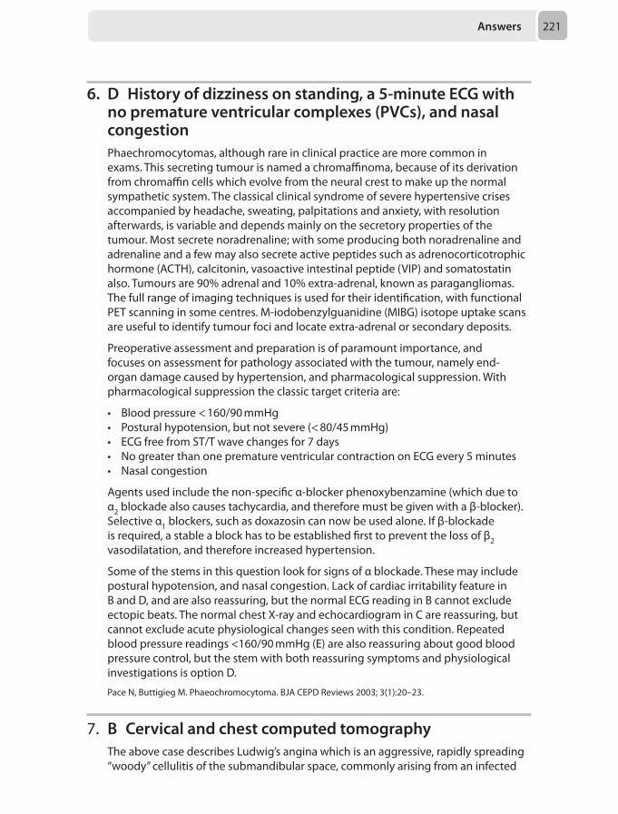
Answers 221
6. D History of dizziness on standing, a 5-minute ECG with no premature ventricular complexes (PVCs), and nasal congestionPhaechromocytomas, although rare in clinical practice are more common in exams. This secreting tumour is named a chromaffinoma, because of its derivation from chromaffin cells which evolve from the neural crest to make up the normal sympathetic system. The classical clinical syndrome of severe hypertensive crises accompanied by headache, sweating, palpitations and anxiety, with resolution afterwards, is variable and depends mainly on the secretory properties of the tumour. Most secrete noradrenaline; with some producing both noradrenaline and adrenaline and a few may also secrete active peptides such as adrenocorticotrophic hormone (ACTH), calcitonin, vasoactive intestinal peptide (VIP) and somatostatin also. Tumours are 90% adrenal and 10% extra-adrenal, known as paragangliomas. The full range of imaging techniques is used for their identification, with functional PET scanning in some centres. M-iodobenzylguanidine (MIBG) isotope uptake scans are useful to identify tumour foci and locate extra-adrenal or secondary deposits.
Preoperative assessment and preparation is of paramount importance, and focuses on assessment for pathology associated with the tumour, namely end-organ damage caused by hypertension, and pharmacological suppression. With pharmacological suppression the classic target criteria are:
• Blood pressure < 160/90 mmHg • Postural hypotension, but not severe (< 80/45 mmHg)• ECG free from ST/T wave changes for 7 days• No greater than one premature ventricular contraction on ECG every 5 minutes• Nasal congestion
Agents used include the non-specific α-blocker phenoxybenzamine (which due to α2 blockade also causes tachycardia, and therefore must be given with a β-blocker). Selective α1 blockers, such as doxazosin can now be used alone. If β-blockade is required, a stable a block has to be established first to prevent the loss of β2 vasodilatation, and therefore increased hypertension.
Some of the stems in this question look for signs of α blockade. These may include postural hypotension, and nasal congestion. Lack of cardiac irritability feature in B and D, and are also reassuring, but the normal ECG reading in B cannot exclude ectopic beats. The normal chest X-ray and echocardiogram in C are reassuring, but cannot exclude acute physiological changes seen with this condition. Repeated blood pressure readings <160/90 mmHg (E) are also reassuring about good blood pressure control, but the stem with both reassuring symptoms and physiological investigations is option D.
Pace N, Buttigieg M. Phaeochromocytoma. BJA CEPD Reviews 2003; 3(1):20–23.
7. B Cervical and chest computed tomographyThe above case describes Ludwig’s angina which is an aggressive, rapidly spreading “woody” cellulitis of the submandibular space, commonly arising from an infected
Chapter 6222
molar tooth. There is a lack of lymphadenopathy since the typically polymicrobial infection spreads along fascial planes as opposed to the lymphatic system. Two life-threatening complications of Ludwig’s angina are upper airway obstruction and descending necrotising mediastinitis. Sufferers are at risk of airway obstruction due to posterior infective extension and tongue distension with posterior displacement. Descending necrotising mediastinitis describes the spread of infection from neck to mediastinum via contiguous fascial planes which is promoted by gravity and the negative intrathoracic pressure. Since the disease is rare and early symptoms often nebulous, diagnosis and treatment can be delayed with fatal consequences.
Computed tomography is the imaging modality of choice for acute deep-seated neck infections and the correct answer to the above scenario. Imaging the neck allows a rapid assessment of the depth of involvement as well as the presence of abscesses which may be amenable to surgical drainage. Chest imaging provides confirmation and allows an assessment of the extent of mediastinal involvement which is important for surgical planning. Since a pericardial rub was heard in the above scenario, computed tomography will also be useful in assessing for secondary pericardial involvement and the presence of an effusion.
Magnetic resonance imaging does provide excellent soft tissue resolution and diffusion weighted imaging can help delineate complex fluid collections. This imaging modality is particularly useful for infections involving the retropharyngeal space where extension into the spinal column is suspected. However, it is more time consuming than computed tomography and patients may feel claustrophobic during scanning. Patient compatibility also needs to be assessed. In the above scenario where the airway patency can deteriorate rapidly and an early diagnosis and treatment plan is needed, computed tomography is more appropriate.
Cervical ultrasound can be useful in characterising soft tissue masses and collections but is unable to penetrate bone or air filled structures. It is also operator dependent and not as accurate as computed tomography in assessing the extent of mediastinal involvement.
Plain radiography is easily accessible but is of little value in planning the management of descending necrotising fasciitis complicating Ludwig’s angina. A lateral cervical radiograph can highlight pretracheal gas bubbles and a loss of the normal lordosis, whereas a chest radiograph may show a widened mediastinum and an enlarged cardiac silhouette if there is mediastinitis or a pericardial effusion respectively. Computed tomography however provides a much more accurate picture of the severity of the infection.
An echocardiogram can provide information regarding the extent of the pericardial effusion and whether it is affecting cardiac function. Echocardiography is not the most appropriate investigation to plan management since it provides no information on the degree of cervical involvement or whether there are any collections amenable to drainage.
Hurley M, Heran M. Imaging studies for head and neck infections. Infect Dis Clin N Am 2007; 21(2):305–353.Wippold F. Head and neck imaging: the role of CT and MRI. J Magn Reson Imaging 2007; 25(3):453–465.Boscollo-Rizzo P, Da Mosto MC. Submandibular space infection: a potentially lethal infection. Int J Infect Dis 2009; 13(3):327–333.
Answers 223
8. E ElectroencephalogramThe patient fulfils the prerequisites for brainstem testing because he has suffered irreversible brain injury and he is in an apnoeic coma. The neurological confirmation of death consists of cranial nerve II – XI examination and apnoea testing performed by two doctors at two different times. At the end of each set of cranial nerves examinations an apnoea test occurs. In a patient with a high cervical spine injury, apnoea might not be due to a central cause but due to spinal cord injury, therefore ancillary tests are employed to confirm de the diagnosis. Electroencephalogram (EEG) is the most widely used and validated assessment in this circumstance.
The second battery of brainstem tests cannot be performed in isolation without the apnoea testing; therefore an EEG is the next most appropriate step to support the diagnosis of death by neurological criteria. Somatosensory evoked potentials are used for monitoring of depth of anaesthesia and play no part in the diagnosis of death.
Oram J, Murphy P. Diagnosis of death. Contin Educ Anaesth Crit Care Pain2011; 11(3):77–81.
9. E Proceed with the case under general anaesthesia and a ultrasound-guided fascia iliaca block with invasive blood pressure monitoringThe management of patients presenting for operative fixation of proximal femoral fractures is clinically and politically of huge importance, the examiners recognise this. Therefore it is essential to be familiar with at least one of the national or international consensus guidelines on the issue. The key elements of these guidelines are as follows:
Timing of surgery and delays
The Department of Health (UK) guidance recommends surgery within 36 hours of admission, and evidence clearly shows adverse outcomes in terms of mortality, complications and stay length if fixation is delayed past 48 hours. Nevertheless, this cannot override the obvious sensibility of stabilisation and resuscitation of an unstable patient. The AAGBI lists several situations in which an operative delay may be acceptable to allow for interim treatment. This list includes reversible coagulopathic states, severe glucose or electrolyte disorders, uncontrolled arrhythmias with heart rates above 120 beats per minute, overt heart failure, and chest sepsis.
Echocardiography
The presence of a murmur may indicate serious valvular heart disease, and indeed aortic stenosis (AS) is more common in hip fracture patients occurring in 20-40%, ten times the rate of the general elderly population. That said, some studies demonstrate similar early postoperative mortality in patients with AS and those without. One could argue also that an echo demonstrating AS will not change management, in that surgery is still required and that therefore the best way to proceed in these
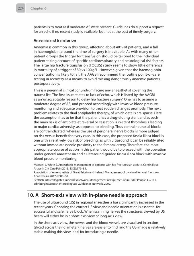
Chapter 6224
patients is to treat as if moderate AS were present. Guidelines do support a request for an echo if no recent study is available, but not at the cost of timely surgery.
Anaemia and transfusion
Anaemia is common in this group, affecting about 40% of patients, and a fall in haemoglobin around the time of surgery is inevitable. As with many other patient groups the trigger for transfusion should be tailored to the individual patient taking account of specific cardiorespiratory and neurological risk factors. The large hip fracture transfusion (FOCUS) study seems to show little difference in mortality of a trigger of 80 vs 100 g/L. However, given that the haemoglobin concentration is likely to fall, the AAGBI recommend the routine point-of-care testing in recovery as a means to avoid missing dangerously anaemic patients postoperatively.
This is a perennial clinical conundrum facing any anaesthetist covering the trauma list. The first issue relates to lack of echo, which is listed by the AAGBI as an ‘unacceptable reason to delay hip fracture surgery’. One has to assume a moderate degree of AS, and proceed accordingly with invasive blood pressure monitoring and adequate provision to treat sudden changes promptly. The next problem relates to the dual antiplatelet therapy, of which details are sparse. Here the assumption has to be that the patient has a drug-eluting stent and as such the main risk is of antiplatelet reversal or cessation is in-stent thrombosis leading to major cardiac adversity, as opposed to bleeding. Thus central neuraxial blocks are contraindicated, whereas the use of peripheral nerve blocks is more judged on risk versus benefit for every case. In this case, the proposed fascia iliaca block is one with a relatively low risk of bleeding, as with ultrasound it can be reliably sited without immediate needle proximity to the femoral artery. Therefore, the most appropriate course of action in this patient would be to proceed with the operation under general anaesthesia and a ultrasound-guided fascia iliaca block with invasive blood pressure monitoring.
Maxwell L, White S. Anaesthetic management of patients with hip fractures: an update.Contin Educ Anaesth Crit Care Pain 2013; 13(5):179–83.Association of Anaesthetists of Great Britain and Ireland. Management of proximal femoral fractures. Anaesthesia 2012;67:85–98.Scottish Intercollegiate Guidelines Network. Management of Hip Fracture in Older People. CG 111. Edinburgh: Scottish Intercollegiate Guidelines Network, 2009.
10. A Short-axis view with in-plane needle approachThe use of ultrasound (US) in regional anaesthesia has significantly increased in the recent years. Choosing the correct US view and needle orientation is essential for successful and safe nerve block. When scanning nerves the structures viewed by US beam will either be in a short-axis view or long-axis view.
In the short-axis view, the nerves and the blood vessels are visualised in section (sliced across their diameter), nerves are easier to find, and the US image is relatively stable making this view ideal for introducing a needle.
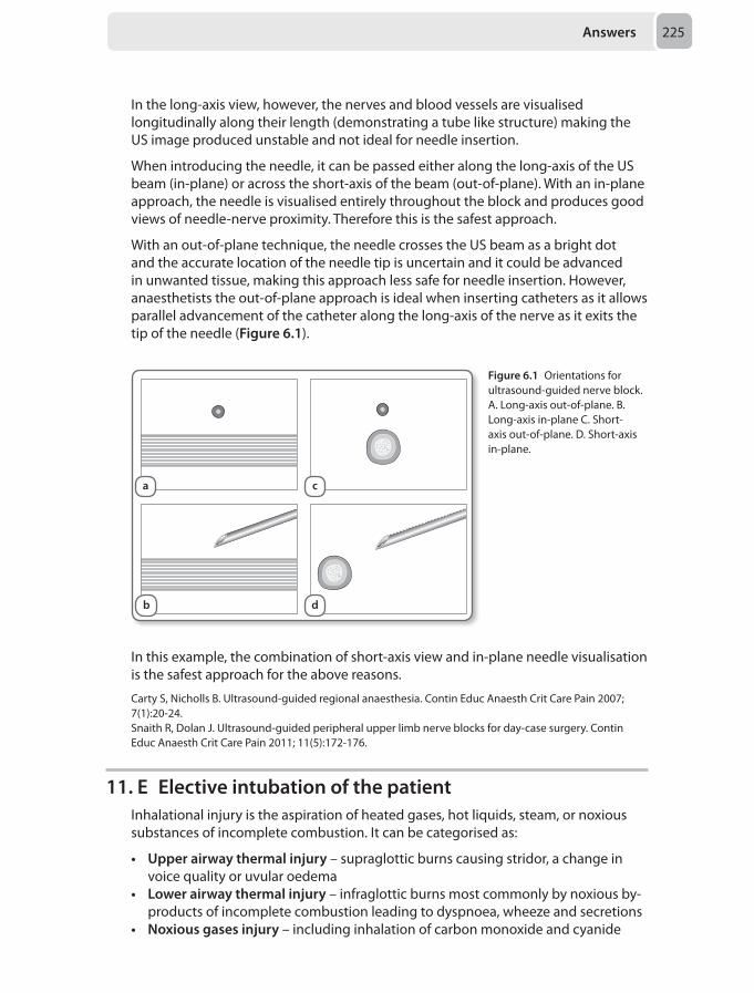
Answers 225
In the long-axis view, however, the nerves and blood vessels are visualised longitudinally along their length (demonstrating a tube like structure) making the US image produced unstable and not ideal for needle insertion.
When introducing the needle, it can be passed either along the long-axis of the US beam (in-plane) or across the short-axis of the beam (out-of-plane). With an in-plane approach, the needle is visualised entirely throughout the block and produces good views of needle-nerve proximity. Therefore this is the safest approach.
With an out-of-plane technique, the needle crosses the US beam as a bright dot and the accurate location of the needle tip is uncertain and it could be advanced in unwanted tissue, making this approach less safe for needle insertion. However, anaesthetists the out-of-plane approach is ideal when inserting catheters as it allows parallel advancement of the catheter along the long-axis of the nerve as it exits the tip of the needle (Figure 6.1).
Figure 6.1 Orientations for ultrasound-guided nerve block. A. Long-axis out-of-plane. B. Long-axis in-plane C. Short-axis out-of-plane. D. Short-axis in-plane.
In this example, the combination of short-axis view and in-plane needle visualisation is the safest approach for the above reasons.
Carty S, Nicholls B. Ultrasound-guided regional anaesthesia. Contin Educ Anaesth Crit Care Pain 2007; 7(1):20-24.Snaith R, Dolan J. Ultrasound-guided peripheral upper limb nerve blocks for day-case surgery. Contin Educ Anaesth Crit Care Pain 2011; 11(5):172-176.
11. E Elective intubation of the patientInhalational injury is the aspiration of heated gases, hot liquids, steam, or noxious substances of incomplete combustion. It can be categorised as:
• Upper airway thermal injury – supraglottic burns causing stridor, a change in voice quality or uvular oedema
• Lower airway thermal injury – infraglottic burns most commonly by noxious by-products of incomplete combustion leading to dyspnoea, wheeze and secretions
• Noxious gases injury – including inhalation of carbon monoxide and cyanide
a c
b d
Chapter 6226
This patient has a high risk of inhalation injury due to an enclosed space fire with significant burns to the face. The onset of airway oedema is often unpredictable, but fluid resuscitation is likely to worsen any impending oedema, while the relatively long duration of transfer indicates the need to have a secure airway during transfer. Therefore it is appropriate to plan elective intubation of the patient in controlled circumstances with senior support, a difficult airway trolley and skilled assistance. Adding a competent team member to the transfer is reassuring and can help should complications arise during transfer, but it is often impractical. All transfers should have full monitoring, including ECG, pulse oximetry and non-invasive blood pressures, but invasive blood pressure monitoring is only indicated if you anticipate cardiovascular instability or it is required to guide ongoing therapy. Availability of difficult airway equipment is necessary once elective intubation has been decided, and devices such as video laryngoscopes are useful to have when a patient is being transferred. However, the most appropriate approach would be to ensure a secure airway prior to transfer.
Bishop S, Maguire S. Anaesthesia and intensive care for major burns. Contin Educ Anaesth Crit Care Pain 2012; 12 (3):118–122.
12. A Change the patient to continuous positive airway pressure (CPAP) and assess ventilation and cardiovascular parameters for 30 minutes
The majority of patients who receive mechanical ventilation have acute respiratory failure in the postoperative period, pneumonia, congestive heart failure, sepsis, trauma or acute respiratory distress syndrome (ARDS). Respiratory muscle weakness may not be a contributing factor to their respiratory failure and once the acute pathophysiological problem is resolved, invasive ventilation may be downgraded and patients extubated. The duration of mechanical ventilation is often unnecessarily prolonged in the setting of a short period of ventilator support (less than 7 days) with the weaning process accounting for up to 50% of the total ventilation time. A delay of 48 hours in extubation results in an increased risk of extubation failure, ventilator acquired pneumonia, thromboembolic disease, longer intensive care and hospital stay and increased mortality.
Weaning involves progression from a controlled mode of ventilation to a support mode and then reduction of support delivered until a trial of readiness for extubation. This trial is termed a spontaneous breathing trial (SBT).
Typical readiness criteria for attempted weaning include:
• Improvement in the underlying condition that caused the respiratory failure• Pulmonary: fractional inspired oxygen Ratio (PFR) of more than 200 with a
positive end-expiratory pressure (PEEP) of 5 cmH2O • Haemodynamic stability • No electrolyte, metabolic, haematological or nutritional deficits• Neurologically appropriate with cough and gag reflexes present
Once deemed suitable, a SBT may be initiated with minimal pressure support such as 5 cmH2O, CPAP or a T-piece or tracheostomy mask (no PEEP). A SBT should be
Answers 227
attempted for a minimum of 30 minutes but should be terminated and deemed unsuccessful if:
• The respiratory rate remains above 35 breathes per minute for 5 minutes • Oxygen saturations of less than 90%• Heart rate increases to over 140 beats per minute• Systolic blood pressure > 180 mmHg or < 90 mmHg• Panic or diaphoresis
The following classification of the results of the spontaneous breathing trial may be applied:
• Simple: successful first trial followed by extubation• Difficult: up to three spontaneous trials but discontinuation of ventilation within
7 days • Prolonged: more than three unsuccessful trials or more than 7 days of mechanical
ventilation
10–20% of ventilated patients may have prolonged weaning and in-hospital mortality is increased in this group. In patients who fail a SBT, the strategy is to reduce the support the patient is receiving and try again. A period of rest between SBTs is advocated of 24–48 hours. Gradual reductions in the pressure support by 2–4 cmH2O per 24 hours or a short SBT period every hour with increasing the duration are both advocated.
Even assessing in a careful manner such as described above, 10–15% of extubations fail, necessitating re-intubation. If this occurs the mortality rate in this group is increased.
In the patient above, the criteria for initiating a SBT is met and if successful, a trial of extubation is warranted. In this patient with COPD, a greater-than-average sputum production and poorer gas-exchange may have been present prior to the acute infection and must be accepted in order to avoid the complications of on-going mechanical ventilation.
McConville JF, Kress JP. Weaning patients from the ventilator. N Engl J Med 2012;367:2233–2239.
13. D 20 mmol of calcium chloride + 1 pool of platelets + 15 mL/kg fresh frozen plasma (FFP)
In order to form effective blood clots a patient needs an adequate number of functioning platelets, adequate levels of all the clotting factors, an adequate haematocrit, an adequate level of ionised calcium, a relatively normal pH and an absence of significant hypothermia. The critical levels of these variables cannot be defined and are mutually dependent. The clinical scenario described suggests that there is ongoing bleeding due to a coagulopathy rather than a failure of surgical haemostasis. The temperature, pH, haematocrit and platelet count are acceptable. However, the patient has been receiving long term antiplatelet therapy and has been on cardiopulmonary bypass, thus, in the absence of a platelet function test it is reasonable to deduce that platelet transfusion is warranted to correct the coagulopathy. The clotting tests suggest there is a consumptive and /or dilutional
Chapter 6228
component to this coagulopathy. Given the degree of abnormality, a dose of FFP should elevate the levels of all factors, including fibrinogen, without the need to give additional cryoprecipitate. Administration of FFP and platelets is likely to result in a further drop in ionised calcium, it would be prudent to administer a replacement dose. Given the scenario and timings, a further dose of protamine is likely to result in an anti-coagulant effect. In the absence of evidence for hyperfibrinolysis, a second dose of tranexamic acid is not indicated at this stage. The threshold for pRBC transfusion in this context is <70g/L. A thromboelastogram would refine the diagnosis further and repetition after intervention guide further therapy. This is a common practice in many centres but not universal.
Dyke C, Aronson S, Dietrich W, et al. Universal definition of perioperative bleeding in adult cardiac surgery. J Thorac Cardiovasc Surg 2014; 147(5):1458–63.Gorlinger K, Shore-Lesserson L, Dirkmann D, Hanke AA, Rahe-Meyer N, Tanaka KA. Management of hemorrhage in cardiothoracic surgery. J Cardiothorac Vasc Anesth 2013; 27(Suppl 4):S20–34.Davidson S. State of the art - How I manage coagulopathy in cardiac surgery patients. Br J Haematol 2014; 146(6):779–89.
14. B Prone positioning The worsening hypoxaemia, new bilateral radiology infiltrates and low Pao2:Fio2 (P:F ratio) within one week of the onset of severe pneumonia suggests acute respiratory distress syndrome (ARDS).
ARDS is an acute, diffuse inflammatory lung syndrome that results in respiratory failure. The 1994 American-European Consensus Conference definition of ARDS has now been superseded by the 2012 Berlin Definition (Table 6.5).
Table 6.5 The 2012 Berlin Definition of ARDS
Severe ARDS Moderate ARDS Mild ARDS
Timing Within one week of known clinical insult or new or worsening respiratory symptoms
Chest Imaging Bilateral opacities not explained by effusions, lobar/lung collapse or nodules
Paco2 (kPa):Fio2Paco2 (mmHg):Fio2
< 13.3< 100
13.3-26.7100-200
26.7-40200-300
PEEP (cmH2O) ≥ 5 ≥ 5 ≥ 5
Mortality (%) 45 32 27
The cause of ARDS in this patient is severe pneumonia, which is a direct (or pulmonary) cause. Other direct causes of ARDS include aspiration, lung contusions and inhalational injury. Indirect (non-pulmonary) causes include sepsis, trauma, pancreatitis and burns.
The pathophysiology of ARDS is complex and involves the interplay of various body systems. A simplified view of this pathogenesis is presented here but this is an area of ongoing exploration.
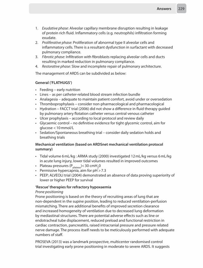
Answers 229
1. Exudativephase: Alveolar capillary membrane disruption resulting in leakage of protein rich fluid. Inflammatory cells (e.g. neutrophils) infiltration forming exudate.
2. Proliferativephase: Proliferation of abnormal type II alveolar cells and inflammatory cells. There is a resultant dysfunction in surfactant with decreased pulmonary compliance.
3. Fibroticphase: Infiltration with fibroblasts replacing alveolar cells and ducts resulting in marked reduction in pulmonary compliance.
4. Restorativephase: Slow and incomplete repair of pulmonary architecture.
The management of ARDS can be subdivided as below:
General (‘FLATHUGS’)
• Feeding – early nutrition• Lines – as per catheter-related blood stream infection bundle• Analagesia – adequate to maintain patient comfort, avoid under or oversedation• Thromboprophylaxis – consider non-pharmacological and pharmacological• Hydration – FACCT trial (2006) did not show a difference in fluid therapy guided
by pulmonary artery flotation catheter versus central venous catheter• Ulcer prophylaxis – according to local protocol and review daily• Glycaemic control – no definitive evidence for tight glycemic control, aim for
glucose < 10 mmol/L• Sedation/Spontaneous breathing trial – consider daily sedation holds and
breathing trials
Mechanical ventilation (based on ARDSnet mechanical ventilation protocol summary)
• Tidal volume 6 mL/kg : ARMA study (2000) investigated 12 mL/kg versus 6 mL/kg in acute lung injury, lower tidal volumes resulted in improved outcomes
• Plateau pressures (Pplateau)< 30 cmH20• Permissive hypercapnia, aim for pH > 7.3• PEEP; ALVEOLI trial (2004) demonstrated an absence of data proving superiority of
lower or higher PEEP for survival
‘Rescue’ therapies for refractory hypoxaemia Pronepositioning
Prone positioning is based on the theory of recruiting areas of lung that are non-dependent in the supine position, leading to reduced ventilation-perfusion mismatching. There are additional benefits of improved secretion clearance and increased homogeneity of ventilation due to decreased lung deformation by mediastinal structures. There are potential adverse effects such as line or endotracheal tube displacement, reduced preload and functional restriction in cardiac contraction, pancreatitis, raised intracranial pressure and pressure related nerve damage. The process itself needs to be meticulously performed with adequate numbers of staff.
PROSEVA (2013) was a landmark prospective, multicenter randomised control trial investigating early prone positioning in moderate to severe ARDS. It suggests
Chapter 6230
benefit in terms of oxygenation and mortality. Previous studies appeared to show improved oxygenation, but no clear mortality benefit.
Inhalednitricoxide
Nitric oxide (NO) is known to cause pulmonary vasodilatation and hence improve pulmonary blood flow. The inhaled route delivers NO selectively to ventilated lung units and hence improves oxygenation. Although inhaled nitric oxide improves oxygenation, there does not appear to be a mortality benefit.
Extracoporealmembraneoxygenation(ECMO)
ECMO involves insertion of large cannulae into central vessels. It is similar to a simple cardiopulmonary bypass circuit. Blood leaves a central vessel and is pumped around a circuit through a membrane oxygenator to allow gas exchange, then returned to the patient via a central vessel. As oxygenation is predominantly achieved through the extracorporeal circuit, ultra low tidal volumes can be used to ventilate the patient minimising ventilator associated lung injury. ECMO requires systemic anticoagulation, carrying a risk of bleeding.
The CESAR trial (2009) was a multicentre randomised control trial investigating conventional management or referral to consideration for treatment by ECMO in severe potentially reversible respiratory failure. It concluded that referral to a tertiary respiratory centre for consideration of ECMO resulted in improved survival. It is unclear what proportion of this benefit is attributed to optimum conventional ventilation in a tertiary referral centre.
Highfrequencyoscillationventilation(HFOV)
HFOV works on the principle of high frequency (120–600 breaths/min) oscillation around a continuous high distending airway pressure. This results in lower tidal volumes, approximating at 3 mL/kg. The OSCAR (2013) and OSCILLATE (2013) trials were two multicentre randomised control trials investigating the role of HFOV in ARDS. The failed to show a benefit and possibly showed harm with HFOV compared to conventional ventilation. At present, HFOV cannot be recommended in refractory hypoxemia secondary to ARDS in adults.
Of the options given in this question, based on current evidence the options for refractory hypoxaemia in ARDS appear to be prone positioning and ECMO. Given that ECMO remains controversial and requires a specialist centre, prone positioning would be the most appropriate option in this patient. If an option was given for ‘referral to a tertiary respiratory centre’, that would also be appropriate.
Pharmacological
No proven mortality benefit but many have been trialed including surfactant replacement therapy, glucocorticoids, and β-adrenoceptor agonists.
Ranieri VM, Rubenfeld GD, Thompson BT, et al. ARDS Definition Task Force: Acute respiratory distress syndrome: the Berlin Definition. JAMA. 2012 ;307(23):2526–2533.National Heart, Lung and Blood Institute Acute Respiratory Distress Syndrome (ARDS) Clinical Trials Network. Comparison of two fluid-management strategies in acute lung injury. N Engl J Med 2006; 354:2564–2575.
Answers 231
The Acute Respiratory Distress Syndrome Network. Ventilation with low volumes as compared with traditional tidal volumes for acute lung injury and acute respiratory distress syndrome. N Engl J Med 2000;342:1301–1308.Brower RG, Lanken PN, MacIntyre N, et al. National Heart, Lung, and Blood Institute ARDS Clinical Trials Network. Higher versus lower positive end-expiration pressures in patients with the acute respiratory distress syndrome. N Engl J Med 2004;351(4):327–336.Guérin C, Reignier J, Richard JC, et al. Prone positioning in severe acute respiratory distress syndrome. N Engl J Med 2013;368(23):2159–2168.Taylor RW, Zimmerman JL, Dellinger RP, et al. Low-dose inhaled nitric oxide in patients with acute lung injury: a randomized controlled trial. JAMA 2004; 291(13):1603–1609.Peek J, Mugford M, Tiruvoipati R, et al. The CESAR trial collaboration: Efficacy and economic assessment of conventional ventilatory support versus extracorporeal membrane oxygenation for severe adult respiratory failure (CESAR): a multicentre randomised controlled trial. Lancet 2009; 374:1351–1363.Young D, Lamb SE, Shah S, et al. The OSCAR Study Group. High-frequency oscillation for acute respiratory distress syndrome. N Engl J Med 2013; 368(9):806–813.Ferguson ND, Cook DJ, Guyatt GH, et al. The OSCILLATE Trial Investigators; Canadian Critical Care Trials Group. High-frequency oscillation in early acute respiratory distress syndrome. N Engl J Med 2013;368(9):795–805.
15. E 3780 mLThis patient has sustained a significant thermal injury with evidence of inhalational injury. Significant burns cause a profound systemic inflammatory response syndrome and early aggressive management is paramount. Mortality from major burns is in the order of 10–20% with multiorgan failure and sepsis being leading causes.
Management should follow ALTS guidelines, especially where the mechanism is unknown. During the primary survey, early intubation is advised where airway compromise or significant inhalational injury is suspected. A rapid sequence induction is advised and intubation performed with an uncut cuffed endotracheal tube; ideally size 8 or larger to aid assessment of the airway via bronchoscopy. Suxamethonium is considered safe in the first 24 hours following injury, an exaggerated hyperkalaemic response may occur after this time frame.
As part of the ‘Breathing’ assessment, carbon monoxide poisoning should be excluded. In this case the confusion at presentation may be an early sign and an arterial blood gas should be done urgently. Normal carbon monoxide levels can be up to 10% in smokers and a level greater than 20% raises the suspicion of significant inhalation injury and carbon monoxide poisoning. It is important to note that pulse oximetry overestimates Spo2 in the presence of carbon monoxide. Therefore the saturations of 100% in this case should be corroborated with arterial gas analysis via co-oximetry. High-flow oxygen decreases the half-life of carbon monoxide from 4 to 1 hours, and should be administered empirically until carboxyhaemoglobin (HbCO) levels are attained.
Another point of concern in this patient as part of the ‘Breathing’ assessment is the anterior torso burn. The chest wall should be examined for evidence of circumferential burn which may require early escharotomies. There is evidence to support that, where possible, these should be done in specialist burns centres.
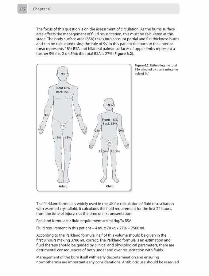
Chapter 6232
The focus of this question is on the assessment of circulation. As the burns surface area affects the management of fluid resuscitation, this must be calculated at this stage. The body surface area (BSA) takes into account partial and full thickness burns and can be calculated using the ‘rule of 9s’. In this patient the burn to the anterior torso represents 18% BSA and bilateral palmar surfaces of upper limbs represent a further 9% (i.e. 2 x 4.5%); the total BSA is 27% (Figure 6.2).
Front 18%Back 18%
9% 9%
18%
Adult Child
18%
1%
9%
Front 18%Back 18%
9% 9%
13.5% 13.5%
1%
18%
Figure 6.2 Estimating the total BSA affected by burns using the 'rule of 9s.'
The Parkland formula is widely used in the UK for calculation of fluid resuscitation with warmed crystalloid. It calculates the fluid requirement for the first 24 hours, from the time of injury, not the time of first presentation.
Parkland formula for fluid requirement = 4 mL/kg/% BSA
Fluid requirement in this patient = 4 mL x 70 kg x 27% = 7560 mL
According to the Parkland formula, half of this volume should be given in the first 8 hours making 3780 mL correct. The Parkland formula is an estimation and fluid therapy should be guided by clinical and physiological parameters; there are detrimental consequences of both under and over resuscitation with fluids.
Management of the burn itself with early decontamination and ensuring normothermia are important early considerations. Antibiotic use should be reserved
Answers 233
to where there is a strong clinical suspicion of active infection, there appears to be little evidence for prophylactic antibiotics. There are burns specific criteria for diagnosing sepsis which can guide clinical decision making.
This patient represents a BSA greater than 10%, with burns to hands and a possible inhalational injury mandating discussion and transfer to a regional burns centre. The British Burns Association criteria for referral to a burns centre are shown in Table 6.6 below.
Table 6.6 British Burns Association ciritera for referral to a regional burns centre
Criteria
Age (years) ≤ 5 or ≥ 60
Site Face, hands, feet, perineum, circumferential
BSA (%) ≥ 10% in adults, ≥ 5% in children
Injury Inhalational, chemical, electrical or complex trauma
Comorbidities Significant cardiorespiratory disease, diabetes mellitus, immunocompromised, liver disease
Bishop S, Maguire S. Anaesthesia and intensive care for major burns. Contin Educ Anaesth Crit Care Pain 2012; 12(3):118–122.
16. E Modified rapid sequence with ketamine 2 mg/kg, fentanyl 1–3 µg/kg and rocuronum 1 mg/kg
Anaesthesia service representation in the trauma team may come from the anaesthetic department or intensive care unit (ICU). The answer to this question is often the technique in which the clinician has the most experience with because a stressful situation is not the ideal time to be trying out novel methods; however, consideration of the different options is still important.
Your responsibility is to secure the airway while your colleagues simultaneously manage some of the other issues. Your choice of induction technique is vital with some of the popular options listed above. The considerations include:
• Speed of induction: – The patient has been obtunded for some time and therefore performing
an induction designed to progress from unconsciousness to endotracheal intubation in as rapid a time as possible is less vital. The classic rapid sequence induction of thiopentone and suxamethonium causes vasodilation, reflex tachycardia, decreased myocardial contractility and suxamethonium causes a transient rise in intracranial pressure.
• Cardiovascular stability is the most important aspect of this situation. Therefore the use of anaesthetic agents known to cause vasodilatation and decreased myocardial contractility should be avoided, at least in standard doses. The options include:
Chapter 6234
– Propofol and thiopentone may cause cardiovascular instability due to vasodilation and decreased contractility. The use of vasoconstrictor medication may be required, which may result in rebound hypertension and could worsen active bleeding.
– Midazolam may also cause hypotension, but less marked than propofol and thiopentone and may therefore be an appropriate choice.
– Fentanyl is a potent analgesic, which does not release histamine and therefore maintains cardiac stability. It is hypnotic-sparing and may reduce the cardiovascular side-effect profile of induction agents. In addition it obtunds the cardiovascular reflex to laryngoscopy, which is beneficial to prevent an increase in blood pressure and intracranial pressure.
– Rocuronium is a steroid non-depolarising paralytic agent. Administration does not cause the release of histamine and therefore maintains cardiovascular stability. In addition when used in larger doses of 0.9–1.2 mg/kg, the onset of intubating conditions is rapid, occurring between 60 and 90 seconds.
– Ketamine is a non-competitive N-methyl-D-aspartate (NMDA) receptor antagonist at the glutamate pre-synaptic calcium channel and is used to provide ‘dissociative anaesthesia’. It has become the drug of choice in the pre-hospital setting in combination with fentanyl due to its cardiovascular effect profile. It causes an increase in systemic vascular resistance and maintains blood pressure via this mechanism. It should be noted that as a calcium antagonist it decreases myocardial contractility and therefore may result in a drop in blood pressure in extreme hypovolaemia. Ketamine has a prolonged length of action of 30–40 minutes negating the need for an infusion. When used in conjunction with controlled mechanical ventilation it does not increase intracranial pressure as described in the 1970s, and may in-fact offer neuro-protection preventing cellular apoptosis.
Pre-hospital care consensus has advocated the use of fentanyl 3 µg/kg, ketamine 2 mg/kg and rocuronium 1 mg/kg (remembered as 3/2/1). This is considered the safest induction technique at the scene of the accident for the reasons described above. Whether or not we can translate this technique directly to hospital care is not certain, but securing the airway in a poly-trauma patient in hospital has the same priorities as at the scene of the accident. Therefore the most appropriate anaesthesia induction technique is one that has been tried and tested on this group of patients.
Sengupta S. Trauma anaesthesia and critical care: the post trauma network era. Contin Educ Anaesth Crit Care Pain 2014;14 (1): 32–37.
17. A High flow oxygen, blood cultures, intravenous fluids and urgent discussion with microbiology consultant
With a temperature > 38.3°C, a heart rate > 90 beats per minute and tachypnoea, this lady meets the diagnostic criteria for sepsis based on the general variables as outlined by the Surviving Sepsis Campaign. She is also at risk of developing severe sepsis, as her blood pressure and oxygen saturations are low. The site of infection must be investigated, as she may have developed intra-abdominal sepsis post surgery, amongst other possibilities.
Answers 235
Genital tract sepsis was the commonest direct cause of maternal death in the last triennium, as outlined by the most recent Centre for Maternal and Child Enquiries (CMACE) report (2006–2008), with Group A streptococcal disease being the responsible pathogen in many cases. Recommendations were made that high dose intravenous broad spectrum antibiotics should be administered within 1 hour of recognition of sepsis as mortality increases with each hour of delay.
In this case, the patient has been on antibiotics via an inadequate route. She has become more unwell, and blood cultures should be taken and an urgent discussion with the consultant microbiologist made to determine the most appropriate antibiotics given her recent antibiotic therapy. This treatment should ideally be commenced within 1 hour. She is likely to need an escalation in treatment and admission to a critical care area may be warranted. Fluid challenges should be given and there should be a low threshold for bladder catheterisation to ensure a urine output of at least 0.5 mL/kg/hour. Oral antibiotic treatment is not appropriate and IV paracetamol will not treat the sepsis. Option B is incorrect, as her management must include the taking of blood cultures.
Dellinger RP, Levy MM, Rhodes A, et al. Surviving Sepsis Campaign: International Guidelines for Management of Severe Sepsis and Septic Shock: 2012. Crit Care Med 2013; 41(2):580–637.Lewis G. Centre for Maternal and Child Enquiries (CMACE): Saving mother’s lives: reviewing maternal deaths to make motherhood safer: 2006-2008. BJOG 2011; 118(suppl 1):1–203.
18. C Reschedule the surgery for when the patient is 6 weeks from the most recent viral respiratory illness
Asthma is one of the most common pulmonary disorders encountered by paediatric anaesthetists. Asthma patients carry a small but significantly increased risk for perioperative complications. Paediatric asthmatic patients require careful preoperative evaluation and preparation.
Essential points to review in the preoperative evaluation are the level of asthma control and the current medication regimen. In addition, review of the level of activity, use of rescue medications, hospital visits (tracheal intubation or intravenous infusions required), allergies, and previous anaesthetic history are important. A Also an inquiry regarding cough and sputum production should also occur. Although otherwise healthy children can often be anaesthetised safely during an acute upper respiratory infection, the risk of bronchospasm in asthmatics is very high. They should ideally be postponed 4–6 weeks after such an event, particularly if the surgery is non-urgent, as is the case with the patient in this question.
Preoperative preparation for a controlled asthmatic can include administration of inhaled β2 adrenergic agonist 1–2 hours before surgery. For moderately controlled asthma, additional optimisation with an inhaled corticosteroid and regular use of inhaled β2 agonists 1 week before surgery can be instituted. Poorly controlled asthmatics might need addition of systemic corticosteroid 3–5 days before surgery.
Lauer R, Vadi M, Mason L. Anaesthetic management of the child with co-existing pulmonary disease. Br J Anaesth 2012; 109(suppl 1):i47–i59.Bhatia N, Barber N. Dilemmas in the preoperative assessment of children. Contin Educ Anaesth Crit Care Pain2011;11:214–218.
Chapter 6236
19. E Reschedule for another day with a plan for midazolam pre-medication on the ward
Anaesthetists frequently have to cope with a child who is uncooperative at induction of anaesthesia and must be familiar with strategies for preventing and dealing with this problem.
Psychological and pharmacological interventions aimed at reducing preoperative anxiety can improve compliance at induction and reduce postoperative behavioural changes. Psychological interventions include preoperative ward visit, play therapy, parental presence at induction, music, lighting and distraction. Various drugs can be used as premedication for the uncooperative child, midazolam being the most common. The preferred route of administration is oral, followed by nasal. The rectal and intramuscular route should be avoided if possible.
Uncooperative children are often preschool or young children with an anxious temperament, anxious parents, or both. These patients may appear cooperative when interviewed in the surgical ward, but then become uncooperative in the anaesthetic room or at induction of anaesthesia. Fortunately, they are usually amenable to reasoning and encouragement possibly backed up by sedative premedication. The use of physical restraint (overpowering), holding still (immobilising), and containing (preventing escape or self-harm) in children raises ethical, legal, and practical problems, and should only be used as a last resort.
If the surgery is elective, as in the case above, then the option of postponing the procedure should be considered. Postponing the procedure gives more time for planning, but may not be convenient for the parents. Giving premedication in a day surgery environment may not be appropriate, so rescheduling the operation for another day, as inpatient, is the best plan of action in this case.
Tan L, Meakin GH. Anaesthesia for the uncooperative child. Contin Educ Anaesth Crit Care Pain 2010; 10:48–52.
20. C Proceed with anaesthesia and surgery, but with modified anaesthesia technique to avoid known triggers for malignant hyperthermia
Malignant hyperthermia (MH) is an inherited disorder of skeletal muscle that can be pharmacologically triggered to produce a potentially fatal combination of hypermetabolism, muscle rigidity and muscle breakdown. Malignant hyperthermia susceptibility (MHS) is inherited in an autosomal dominant fashion. However, a parent with MHS may not necessarily have a positive history of MH. Anaesthetic technique must be modified to avoid known triggers for MH (halothane, enflurane, isoflurane, sevoflurane, desflurane, and succinylcholine) in any cases of suspected or confirmed MHS. The anaesthetic machine should be prepared by removal of vapourisers and flushing through the machine and ventilator with 100% oxygen at maximal flows for 20–30 minutes, and a new breathing circuit should be used.
The key to successful management of MH is its early diagnosis and the rapid instigation of several modes of treatment simultaneously. Administration of volatile anaesthetics should be discontinued and the patient’s lungs hyperventilated
Answers 237
using 100% oxygen with fresh gas flows and type of breathing circuit optimised to eliminate the anaesthetic from the body. Anaesthesia should be maintained with intravenous drugs while surgery is concluded as rapidly as possible. Active cooling measures should be commenced. At the onset of treatment, one member of staff must be assigned to the preparation of dantrolene sodium for infusion. Repeated doses of dantrolene (1 mg/kg up to maximum of 20 mg) should be administered intravenously as soon as possible until the tachycardia, rise in CO2 production and pyrexia start to subside. Up to 10 mg/kg may be required.
Postponing surgery for further information and investigation is not an option in this case due to the urgency of the surgery. Proceeding with an 'MH-safe' anaesthetic is the most appropriate approach in this clinical scenario.
Halsall PJ, Hopkins PM. Malignant hyperthermia. ContinEducAnaesthCritCarePain 2003;3:5–9.
21. D Serotonin syndromeSerotonin syndrome is a potentially lethal condition resulting from excess agonist activity at central and peripheral serotonergic receptors. It can result from therapeutic drug use, intentional self-poisoning or interactions between drugs, many of which anaesthetists are involved with. The syndrome is characterised by neuromuscular excitability, autonomic hyperactivity, and altered mental status. It is a clinical diagnosis and the presence of tremor, clonus, or akathisia without extrapyramidal signs should lead clinicians to consider the syndrome, particularly if the patient is taking drugs known to elevate serotonin levels. Tramadol is a commonly prescribed analgesic and works by activation of central μ-opioid receptors. In addition to its opioid receptor effects, tramadol also inhibits neuronal reuptake of serotonin and noradrenaline. This property of tramadol can lead to elevated plasma serotonin levels which increases the risk of developing the serotonin syndrome. When tramadol is taken in conjunction with serotonergic agents like SSRIs (such as in the case above), this risk increases. Treatment is generally supportive and involves removing the offending agent(s) and controlling the agitation, autonomic instability and hyperthermia. The antihistamine cyproheptadine, which is also a serotonin antagonist, is reserved for severe cases. Serotonin syndrome is the most likely diagnosis in the case above since there are clinical signs of neuromuscular excitability, autonomic hyperactivity and altered mental status in a patient known to be taking two agents which can increase serotonin levels.
Opioid toxicity can occur with tramadol administration, although symptoms such as drowsiness and lethargy would be more likely than the restlessness described. Furthermore, miosis as opposed to mydriasis would be expected on examination of the pupils. Hyperreflexia, tremor and pyrexia are also not typical presentations of opioid toxicity.
Opioid withdrawal shares many of the symptoms and signs seen in the serotonin syndrome such as restlessness, tremor, mydriasis and tachycardia. The temporal relationship between tramadol use and the symptoms in the above scenario however makes opioid withdrawal unlikely. Withdrawal symptoms usually occur following cessation of opioids after several weeks of steady use in which physical dependence is attained. In the above scenario tramadol was only taken for 1 day, and there was no history of its cessation.
Chapter 6238
The symptoms associated with hyponatraemia are predominantly neurological due to cerebral oedema associated with a reduced serum osmolality. The muscle hyperactivity and confusion as seen in the above case are typical symptoms of hyponatraemia which can progress to seizures, coma and respiratory arrest if the hyponatraemia is not corrected. Tramadol can cause hyponatraemia on rare occasions, thought to be due to opioid and serotonin receptor induced ADH release. The fever described in the above case however is not typical of hyponatraemia, but is frequently seen in the serotonin syndrome due to autonomic hyperactivity.
Anaphylaxis should always be in the differential diagnosis for any patient who becomes unwell after starting a new medication. In the above case however, the tremor and hyperreflexia suggest an alternative diagnosis.
Boyer E, Shannon M. The serotonin syndrome. NEJM 2005; 352(11):1112–20.Frank C. Recognition and treatment of serotonin syndrome. Can Fam Physician. 2008; 54(7):988–92.Lota A, Dubrey S, Wills P. Profound hyponatremia following a tramadol overdose. QJM 2012; 105(4): 397–8.
22. A DiabetesChronic post-surgical pain (CPSP) is recognised as:
• Pain developing after a surgical procedure• Pain of at least 2 months duration• Other causes of pain excluded (such as infection)• Pain continuing from a pre-existing pain problem excluded
Risk factors for development of CPSP can be patient factors or surgical factors. Surgical factors include type of procedure (breast surgery, amputation, thoracotomy), length of surgery and repeat surgery for the same pathology. Surgical approach is also important, as the use of a laparoscopic technique results in less CPSP for cholecystectomy and hernia repairs. The use of adjuvant radiotherapy is also associated with a significantly increased risk of CPSP.
Patient factors include age (CPSP after breast cancer surgery decreases by 5% for each yearly increase in the patient’s age ), genetic susceptibility and psychosocial risk factors. For example, fear of surgery after breast surgery is associated with worse pain and a higher risk of progression to CPSP. Additionally, the severity of postoperative pain positively correlates with the incidence of development of CPSP.
Diabetes is not a recognised risk factor for the development of CPSP.
Searle RD, Simpson KH. Chronic post-surgical pain. Contin Educ Anaesth Crit Care Pain 2010; 10(1):12–14.
23. D McGill pain questionnairePain is a complex, subjective experience which often requires specialised assessment tools to fully evaluate and quantify. Numerous pain rating scales have been developed over the years and it is important as anaesthetists to appreciate the context in which they should be used. Unidimensional pain scales are useful for evaluating acute pain of clear aetiology (e.g. postoperative pain) since they allow quick assessment of pain intensity and response to treatment. However, they are less effective in evaluating chronic pain, since they often fail to measure the associated affective and disabling
Answers 239
components. Multidimensional pain scales are more appropriate in these cases since they allow measurement of these other facets of the pain experience.
The McGill pain questionnaire is one of the most extensively tested multidimensional scales, and is the most appropriate tool to use in the above clinical scenario. The three-part questionnaire assesses not only the sensory aspects but also the affective component of pain which the above patient is suffering from. This assessment tool may also help identify whether there are any specific pain syndromes (such as neuropathic pain) present.
The numeric rating scale is a commonly used unidimensional pain scale where patients rate their pain intensity on a ten point scale with a score of ten representing 'the worst imaginable pain'. It is easy to use and has been validated in numerous settings and pain types. However, it only measures one dimension of pain which limits its usefulness in the chronic pain setting. It is also less reliable in patients with cognitive impairment.
The visual analog scale is an assessment tool composed of a ten centimetre line representing the spectrum of pain intensity from ‘no pain’ to the ‘worst pain imaginable’. Patients are instructed to mark a point on the line which corresponds to their level of pain, and the distance between this mark and zero is measured. This tool is sensitive for variations in pain intensity with treatment and is reproducible. However, like the numeric rating scale, it only measures one component of the complex multidimensional nature of chronic pain.
The verbal descriptor scale is a six point categorical scale of descriptive words from ‘no pain’ to ‘worst possible pain’ which the patient can use to express their pain experience. It allows for a rapid assessment of pain intensity and is easy to use at the bedside. However, it forces the patient to use someone else’s words to describe their pain and does not measure the multidimensional components of chronic pain.
The Wong–Baker FACES scale is another categorical scale with faces conveying expressions of pain with increasing severity. Patients are instructed to select the face which best matches how they are feeling to provide a crude measure of their pain experience. It is a useful assessment tool for children and patients with cognitive impairment who may otherwise have difficulties in quantifying their pain but it is not the most appropriate scale to use in patients with chronic pain.
Breivik H, Borchgrevink P, Allen S, et al. Assessment of pain. Br J Anaesth 2008; 101(1);17–24.Garra G, Singer A, Taira B, et al. Validation of the Wong-Baker FACES Pain Rating Scale in pediatric emergency department patients. Acad Emerg Med 2010; 17(1):50–54.
24. Perform a nerve blockPreoperative pain management for fractured neck of femur patients is a significant problem. As such, the National Institute for Health and Care Excellence (NICE) have issued guidelines (CG124) aiming to optimise analgesic management for these patients whilst awaiting definitive surgery. This guideline states:
• Offer immediate analgesia to all patients presenting to hospital with a suspected hip fracture, including people with cognitive impairment
• Ensure analgesia is sufficient to allow movements necessary for investigations and nursing care
Chapter 6240
• Offer paracetamol 6 hourly unless contraindicated• Offer additional opiates if paracetamol alone does not provide sufficient
preoperative pain relief • Considering adding a nerve block if paracetamol and opioids do not provide
sufficient preoperative pain relief, or to limit opioid dosage • Non-steroidal anti-inflammatory drugs (NSAIDs) are not recommended
This patient would therefore most likely benefit from an ultrasound-guided femoral nerve block or a fascia iliaca block. This is relatively simple to perform and has been demonstrated to have a significant impact on preoperative analgesia.
National Institute for Health and Care Excellence (NICE) guidelines: Hip fracture: The management of hip fracture in adults. CG no 124. London: NICE, 2011.
25. A Spinal cord stimulationComplex regional pain syndrome (CRPS) is a debilitating, painful condition which is classified into type I and II subtypes, depending on the absence or presence of an antecedent peripheral nerve injury respectively. The lead symptom of CRPS is limb-confined pain, but the syndrome also encompasses autonomic, motor, skin and bone changes. If the pain is unrelenting and the physical impairment persists for more than 2 years, the condition is considered long-term.
The aim of medication is to minimise pain and support physical rehabilitation. Although no drugs are licensed to treat CRPS in the UK, national guidelines encourage the use of drugs targeting neuropathic pain if simple medication is unsuccessful after 4 weeks.
In the UK, the only National Institute for Health and Care Excellence (NICE) approved method to treat CRPS is spinal cord stimulation, and should be considered in patients who have not responded to appropriate integrated management. Stimulation of the spinal cord is achieved by application of an electrical current to the dorsal columns of the spinal cord through a catheter inserted into the epidural space. The exact mechanism of action is unclear but some investigators suggest that spinal cord stimulation may activate Aβ afferents which modulate the transmission of pain based on the gate control theory. Others suggest that spinal cord stimulation may block spinothalamic tract transmission or enhance descending inhibitory mechanisms. In the above case where simple analgesia and medication targeting neuropathic pain have failed, spinal cord stimulation is an appropriate next management step. In order to assess the potential benefit of spinal cord stimulation, the electrodes are initially stimulated by an external stimulating device prior to permanent pulse generator insertion. Patient satisfaction is generally high, although there is some evidence that the efficacy of this treatment generally declines over time.
The autonomic vasomotor changes seen in CRPS have led observers to previously view the associated pain as sympathetically mediated, and although common in early CRPS, it is actually rare in long-term cases. The use of guanethidine (which depletes the limb autonomic nerve endings of noradrenaline) to achieve chemical sympathectomy in the affected limb has been shown to be ineffective in randomised controlled trials, and is not recommended.
Answers 241
Lower limb sympathectomy can also be achieved by creating a thermal lesion from the application of a high frequency current to lumbar sympathetic ganglia via percutaneous electrodes. Proponents of this intervention (termed radiofrequency ablation), suggest that it is less invasive than surgical resection and can help break the cycle of pain. The most recent Cochrane review however, judged that there was no evidence from controlled trials that sympathectomy (including radiofrequency) was no more effective than placebo or no treatment.
Amputation should not be used to provide pain relief in CRPS, and should only be considered in rare cases of intractable infection of the affected limb. Amputation may worsen CRPS, with symptoms recurring in the stump.
An interesting approach to tackle the symptoms of CRPS is through the use of non-invasive brain stimulation. The treatment is based on the hypothesis that CRPS is primarily a disease of cortical organisation which results in changes in the way somatosensory systems process tactile, noxious and thermal information. The aim of non-invasive brain stimulation is to induce cortical modulation through the application of a repetitive current to improve symptoms. This treatment is an interesting concept, however further research is needed to evaluate its efficacy.
O’Connell N, Wand B, McAuley J, et al. Interventions for treating pain and disability in adults with complex regional pain syndrome. Cochrane Database Syst Rev 2013 Apr 30;4:CD009416.Goebel A. Complex regional pain syndrome in adults. Rheumatology 2011; 50:1739–50.Goebel A, Barker CH, Turner-Stokes L, et al. Complex regional pain syndrome in adults: UK guidelines for diagnosis, referral and management in primary and secondary care. London: Royal College of Physicians, 2012.National Institute for Health and Clinical Excellence (NICE). Spinal cord stimulation for chronic pain of neuropathic or ischaemic origin. TA guidance 159. London: NICE, 2008.
26. D Sevoflurane maintenance over desfluraneStrabismus surgery is one of the most common paediatric ophthalmic operations. The oculocardiac reflex and postoperative nausea and vomiting (PONV) are major anaesthetic concerns in this procedure.
The oculocardiac reflex occurs due to traction on the extraocular muscles, which causes bradycardia. This can be attenuated by release of surgical traction or the administration of anticholinergic medication such as atropine. Hypercarbia can also increase the incidence of bradycardia, therefore ventilating the patient to maintain a normal CO2 is a technique often used.
PONV is more likely to occur in children who demonstrate the oculocardiac reflex, therefore preventing this reflex should prevent PONV. Atropine 20 µg/kg is advocated as a result, and so option B is not the correct answer. Intraoperative intravenous fluids, avoiding opioid analgesia and using antiemetic prophylaxis helps to reduce PONV incidence. Evidence suggests that ondansetron in combination with dexamethasone is more effective than ondansetron alone.
All the volatile agents increase the risk of PONV and there is currently no evidence to show that any one is less emetogenic than the other, hence D is the correct answer.
Carr AS, Courtman S, Holtby H, et al. Guidelines on the prevention of post-operative vomiting in children. London: The Association of Paediatric Anaesthetists of Great Britain and Ireland, Spring 2009. James I. Anaesthesia for paediatric eye surgery. Contin Educ Anaesth Crit Care Pain 2008; 8(1): 5–10
Chapter 6242
27. C Proceed to surgery after discussing the case with your consultant
The patient has two risk factors for moderate postoperative cardiac risk: stable congestive heart failure and stable angina. According to the ACC/AHA 2007 guidelines for managing cardiac risk of patients for non-cardiac surgery, surgical risk and urgency are used in conjunction with clinical risk and clinical assessment of exercise tolerance to outline the best approach of managing complex situations such as this. The patients with the highest risk of major adverse cardiac events (MACE), i.e. death or myocardial infarction, are those that possess several clinical diagnoses:
• Unstable angina• Overt congestive cardiac failure• Uncontrolled arrhythmias• Severe stenotic valvular disease• Recent myocardial infarction (within 4 weeks).
This only applies to non-emergent situations. An emergency would override those considerations in view of the risk of MACE being higher if the operation would be delayed.
Stable heart failure, stable angina, rate controlled atrial fibrillation, chronic renal impairment or history of cerebrovascular event are risk factors for MACE and increase the burden of post-operative morbidity, however delaying life or limb saving operations to further investigate them would expose the patient to a higher than necessary risk of MACE. The evidence presented in the guidelines suggests that an initial assessment and improvement of the above conditions may result in a decrease of MACE.
In this case it is a potential life or limb saving procedure has a lower risk than waiting for a cardiology opinion to optimise his heart failure and angina. Conditioning life or limb saving surgery to availability of a high care bed is not advisable. Early senior involvement is the most likely step towards a safe and effective intra-operative management for this patient.
Fleisher L, Beckman JA, Brown KA, et al. ACC/AHA 2007 Guidelines on perioperative cardiovascular evaluation and care for noncardiac surgery. Circulation 2007; 116:e418–e450.
28. A Apical segments of the lower lobes Pulmonary aspiration of gastric contents is one of the most serious complications after general anaesthesia. Acid aspiration may cause immediate lung tissue injury and subsequent severe inflammatory response.
The anatomy of the lung lobes and bronchopulmonary tree affects zonal contamination if aspiration of gastric contents happens.
The trachea is a 10–12 cm long tube that connects the larynx to the lungs. In the first year of life, the tracheal diameter is 3 mm or less. The diameter then increases by about 1 mm per year until it reaches the adult size (around 20–25 mm). The trachea comprises fifteen to twenty C-shaped cartilaginous rings. These rings are incomplete posteriorly allowing the trachea to collapse slightly during the passage of the food in the esophagus.
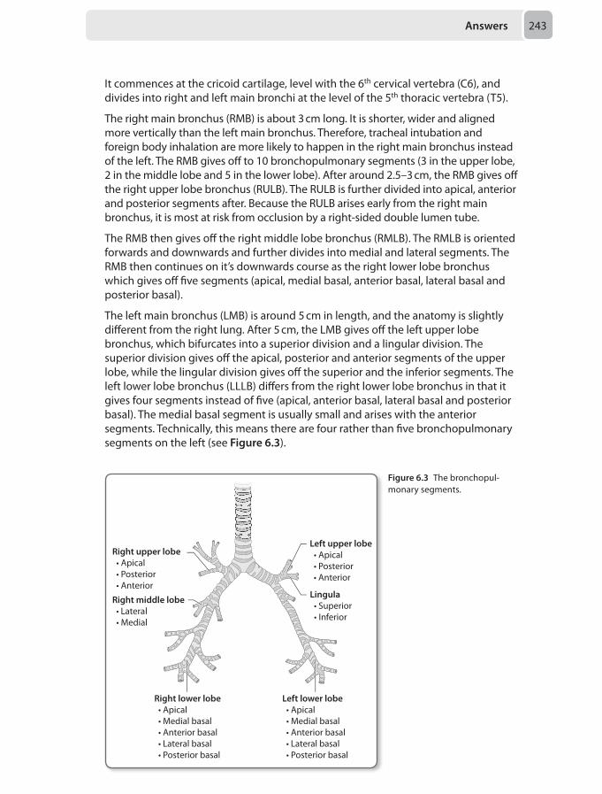
Answers 243
It commences at the cricoid cartilage, level with the 6th cervical vertebra (C6), and divides into right and left main bronchi at the level of the 5th thoracic vertebra (T5).
The right main bronchus (RMB) is about 3 cm long. It is shorter, wider and aligned more vertically than the left main bronchus. Therefore, tracheal intubation and foreign body inhalation are more likely to happen in the right main bronchus instead of the left. The RMB gives off to 10 bronchopulmonary segments (3 in the upper lobe, 2 in the middle lobe and 5 in the lower lobe). After around 2.5–3 cm, the RMB gives off the right upper lobe bronchus (RULB). The RULB is further divided into apical, anterior and posterior segments after. Because the RULB arises early from the right main bronchus, it is most at risk from occlusion by a right-sided double lumen tube.
The RMB then gives off the right middle lobe bronchus (RMLB). The RMLB is oriented forwards and downwards and further divides into medial and lateral segments. The RMB then continues on it’s downwards course as the right lower lobe bronchus which gives off five segments (apical, medial basal, anterior basal, lateral basal and posterior basal).
The left main bronchus (LMB) is around 5 cm in length, and the anatomy is slightly different from the right lung. After 5 cm, the LMB gives off the left upper lobe bronchus, which bifurcates into a superior division and a lingular division. The superior division gives off the apical, posterior and anterior segments of the upper lobe, while the lingular division gives off the superior and the inferior segments. The left lower lobe bronchus (LLLB) differs from the right lower lobe bronchus in that it gives four segments instead of five (apical, anterior basal, lateral basal and posterior basal). The medial basal segment is usually small and arises with the anterior segments. Technically, this means there are four rather than five bronchopulmonary segments on the left (see Figure 6.3).
Right upper lobe • Apical • Posterior • Anterior
Left upper lobe • Apical • Posterior • Anterior
Lingula • Superior • Inferior
Right lower lobe • Apical • Medial basal • Anterior basal • Lateral basal • Posterior basal
Left lower lobe • Apical • Medial basal • Anterior basal • Lateral basal • Posterior basal
Right middle lobe • Lateral • Medial
Figure 6.3 The bronchopul-monary segments.
Chapter 6244
Zonal contamination of the lung lobes and the bronchopulmonary segments after aspirating is dependent on the patient’s position during the aspiration.
In a supine patient the apical segment of the lower lobe is more likely to be contaminated because of the direct posterior orientation of the segment. If the patient is prone, then aspiration is more likely to affect the right middle lobe or the lingula because of their forward and downward projection. If in the upright sitting position, the lateral or posterior basal segments of the lower lobes will be the site of the problem, and in the lateral position, the upper lobes would be contaminated.
Engelhardt T, Webster NR. Pulmonary aspiration of gastric contents in anaesthesia. Br J Anaesth 1999; 83(3):453–460.Bricker S. The Anaesthesia Science Viva Book, 2nd ed. Cambridge: Cambridge University Press, 2008 pp 39–41.
29. C Anorexia nervosaAnorexia nervosa is a psychiatric disorder which affects numerous systems in the body and involves the following diagnostic criteria:
• Body weight is less than 15% of expected or body mass index (BMI) less than or equal to 17.5
• Self-induced weight loss involving food avoidance, purging, self-induced vomiting or using diuretics
• Distorted body image• Multiple endocrine dysfunctions involving hypothalamic-pituitary-gonadal axis• Delayed pubertal events if it manifests prior to puberty
The condition affects numerous physiological systems in the body including:
• Cardiovascular – hypotension, bradycardia, myocardial dysfunction, mitral valve prolapse, cardiomyopathy and arrhythmias
• Respiratory – metabolic alkalosis, decreased lung compliance, aspiration pneumonia
• Gastrointestinal – enlarged salivary glands, dental caries, Mallory–Weiss tears, oesophagitis, gastric dilatation/perforation, increased amylase, abnormal liver function tests
• Renal – proteinuria, reduced serum Na+, K+, Cl-, H+, Mg2+, Ca2+ and renal calculi• Endocrine – reduced FSH, LH, GnRH, T3, T4 and glucose, increased serum cortisol• Haematological – anaemia, leucopenia, thrombocytopenia• Neurological – reduced cognitive function, seizures, coma, neuropathy• Musculoskeletal – osteopenia, pathological fractures, myalgia
An ectopic pregnancy, duodenal perforation or peritonitis would lead to tachycardia along with hypotension. Hypothyroidism would lead to a reduced basal metabolism, which would prevent weight loss. An opioid overdose would cause respiratory acidosis as the major component in an arterial blood gas.
Denner AM. Anorexia nervosa: perioperative complications. Contin Educ Anaesth Crit Care Pain 2009; 9(2):61–64.
Answers 245
30. D Infusion of phosphateRefeeding syndrome is defined as the shift of electrolytes and fluids that can occur in patients who are malnourished and have been started on artificial feeding (enteral or parenteral) and could have potentially fatal consequences. The hallmark feature is hypophosphatemia but may also include hypokalaemia, hypomagnesaemia along with abnormal sodium and fluid balance. Patients with long-standing nutritional deficiencies like anorexia, chronic alcoholics, oncological disorders and chronic malnutrition are at high risk. Malnourishment increases ketone bodies as fatty acids are used as the primary source of energy. Intracellular minerals such as phosphate are depleted, although the serum levels may be normal due to contraction of the intracellular compartment and reduced excretion. On refeeding, glycaemia causes insulin secretion which stimulates glycogen, fat and protein synthesis. This leads to consumption of minerals like phosphate and magnesium, as well as co-factors such as thiamine. Insulin secretion causes intracellular migration of glucose and water along with a phosphate and magnesium shift thereby causing acute depletion.
Management of refeeding syndrome consists of sequential screening of serum levels of potassium, magnesium, calcium and potassium. The levels of phosphate, potassium, magnesium and calcium need to be replenished along with administration of thiamine.
Excessive administration of glucose in a patient suffering from refeeding syndrome could lead to hyperglycaemia induced diuresis, dehydration, metabolic acidosis and ketoacidosis.
In this patient, it is therefore most appropriate to supplement the probable hypophosphatemia with a phosphate infusion.
Mehanna HM, Moledina J, Travis J. Refeeding syndrome: what it is, and how to prevent and treat it. BMJ 2008; 336:1495–1498.

Mock Paper 7
Chapter 7
Question1. You are anaesthetising a previously well 43-year-old woman for a craniotomy to
remove a frontoparietal meningioma. The patient is supine, with a 30° head-up tilt. 1 hour into the operation her oxygen saturations suddenly drop from 98% to 65%, her end-tidal CO₂ from 4.5 kPa to 2 kPa and her blood pressure, which initially rises, begins to fall rapidly.
Which of the following best describes your initial step in the management of the situation?
A Administer 100% oxygen B Insert a right internal jugular central venous pressure catheter and aspirate any
airC Raise the patient’s venous pressure at the operative site by levelling the table
+/– inotropic agent +/– performing a Valsalva manoeuvreD Alert the surgeons and ask them to flood the operative site E Turn the patient into the left lateral, head down position
2. You are caring for a 70 kg man undergoing coronary artery bypass grafting. Long term 75 mg aspirin (once daily) was discontinued 5 days preoperatively. His separation from cardiopulmonary bypass (CPB) was uneventful but during sternal wiring the surgeon states that the patient is ‘oozy’ and you note there is already 500 mL in the mediastinal drain. The activated clotting time (ACT) is 115 seconds. You send a sample for thromboelastography (TEG).
Based on the results shown below in Table 7.1, what is the most appropriate treatment?
Table 7.1
Parameter Value Reference range
r time 32 mm 15–30 mm
k time 12 mm 6–12 mm
α° 40° 40–50°
Maximum amplitude (MA) 40 mm 50–60 mm
Ly30 5.0% < 7.5%
ACT 115 seconds 90–130 seconds
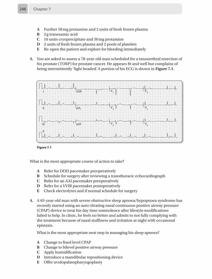
Chapter 7248
A Further 50 mg protamine and 2 units of fresh frozen plasmaB 2 g tranexamic acidC 10 units cryoprecipitate and 50 mg protamineD 2 units of fresh frozen plasma and 2 pools of plateletsE Re-open the patient and explore for bleeding immediately
3. You are asked to assess a 78-year-old man scheduled for a tansurethral resection of his prostate (TURP) for prostate cancer. He appears fit and well but complains of being intermittently ‘light headed’. A portion of his ECG is shown in Figure 7.1.
I
II
III
II
aVR
aVL
aVF
V1
V2
V3
V4
V5
V6
Figure 7.1
What is the most appropriate course of action to take?
A Refer for DDD pacemaker preoperativelyB Schedule for surgery after reviewing a transthoracic echocardiographC Refer for an AAI pacemaker preoperativelyD Refer for a VVIR pacemaker postoperativelyE Check electrolytes and if normal schedule for surgery
4. A 65-year-old man with severe obstructive sleep apnoea/hypopnoea syndrome has recently started using an auto-titrating nasal continuous positive airway pressure (CPAP) device to treat his day time somnolence after lifestyle modifications failed to help. In clinic, he feels no better and admits to not fully complying with the treatment because of nasal stuffiness and irritation at night with occasional epistaxis.
What is the most appropriate next step in managing his sleep apnoea?
A Change to fixed level CPAPB Change to bilevel positive airway pressureC Apply humidification D Introduce a mandibular repositioning deviceE Offer uvulopalatopharyngoplasty
Questions 249
5. A 39-year-old woman with a body mass index of 46 kg/m2 for umbilical hernia repair is seen in day surgery pre-assessment clinic. She has well controlled hypertension. She has been told she snores loudly but sleeps well with no daytime somnolence. Her neck circumference is 35 cm, and her oxygen saturation on air is 96%. Her ECG is normal.
Which of the following options is the most appropriate next action?
A She can proceed for day case surgery B She should be listed for inpatient surgeryC She should be referred for sleep studies D She should have a glucose tolerance testE She should be advised to lose weight prior to surgery
6. A 73-year-old man in the recovery room is extremely confused, combative and is tachypnoeic. The recovery staff are struggling to perform any other observations. He has had a radical robotic prostatectomy for locally confined prostatic carcinoma. The surgery was technically complex and the procedure duration was nearly 7 hours. On examination the only obvious signs are his severe delirium and agitation, and you also notice significant periorbital swelling.
The immediate treatment for the likely condition includes:
A Non-invasive humidified CPAP by maskB Non-invasive BiPAP by maskC Heliox with added entrained oxygen and urgent ENT referralD Ophthalmology opinionE Reintubation and head-up positioning
7. A 50-year-old man awaiting surgery for a mediastinal tumour becomes progressively more tachypnoeic and stridulous on room air. Heliox is administered as a holding measure until more definitive treatment is instituted and his symptoms slowly improve.
Which physical property of helium is most important in improving his respiratory distress?
A A reduced density compared to airB A higher viscosity compared to airC A higher thermal conductivity compared to airD A lower blood: gas solubility coefficient compared to airE Its non-reactivity within the airway
8. A 55-year-old man is undergoing emergency coronary angioplasty for myocardial infarction in the cardiac catheterisation suite after return of spontaneous circulation from a ventricular fibrillation (VF) cardiac arrest. You have been urgently called to provide a general anaesthetic as he is becoming increasingly drowsy and confused with a Glasgow coma score of 10/15. There is an anaesthetic machine present in the room.
Chapter 7250
What should you prioritise as your first action?
A Check the anaesthetic machineB Ensure suction and a tipping trolley is presentC Take a history and perform a brief neurological examinationD Draw up the emergency drugsE Call for anaesthetic assistance
9. A 75-year old woman with chronic anaemia and angina is to have a Colles' fracture reduction under Bier’s block.
Which local anaesthetic agent would be the most appropriate to use for this block?
A LevobupivacaineB LignocaineC RopivacaineD PrilocaineE Chloroprocaine
10. A 65-year-old woman is to have a palmar fasciectomy of the middle finger under axillary nerve block. 30 minutes after performing the block, it is apparent that the median nerve is spared. You decide to perform a supplementary median nerve block.
Which of the following approaches to the median nerve would be the most appropriate for this case?
A Wrist B Mid-forearm C Axillary D Antecubital fossaE Supraclavicular
11. A 6-year-old boy with global developmental delay is first on your surgical list for an orchidopexy procedure. At your pre-assessment visit his mother tells you he can be a “nightmare” and is not up to date with vaccinations after a bad experience at their local health centre. She doesn’t think he will cooperate with induction, and is clearly anxious herself. The child will not interact with you and runs off to the play area as you approach. In the anaesthetic room, you make a single attempt for intravenous access, which is unsuccessful. The child is inconsolable and the mother is visibly distressed.
The best way to proceed would be:
A Cancel this elective case and explain to the mother counselling/play therapy will be required before rebooking
B Overpower the child and proceed with an inhalational induction with sevoflurane at 8% in oxygen
Questions 251
C Overpower the child and proceed with an inhalational induction with sevoflurane at 8% in oxygen and nitrous oxide
D Send the child back to the ward and prescribe an oral midazolam premedication at a dose of 0.5 mg/kg, resending for the child at 15 minutes post dose
E Send the child back to the ward and prescribe an oral ketamine premedication at a dose of 5 mg/kg, resending for the child at 15 minutes post dose
12. A 35-year-old man has presented with a syncopal episode the day after a fall during a rugby match, and CT scan has confirmed an extra-dural haematoma.
What features would indicate that intubation should be performed before transfer to a neurosurgical centre?
A Glasgow coma score (GCS) 11/15B An episode of vomitingC A seizureD Suspected skull fractureE A drop in GCS by 1 point on the verbal scale
13. A 45-year-old man has suffered an isolated, catastrophic, irrecoverable traumatic brain injury. In the last few minutes he has become progressively tachycardic, hypotensive and polyuric despite aggressive filling with intravenous crystalloid. His observations include: heart rate 100 beats per minute sinus rhythm, blood pressure 75/45 mmHg, stroke volume 82 mL.
The first vasoactive drug of choice in this scenario is:
A AdrenalineB DopamineC LabetalolD VasopressinE Metaraminol
14. A 76-year-old man has undergone an uneventful 3-vessel on-pump coronary artery bypass grafting (CABG) 4 hours ago and is currently sedated and ventilated on the intensive care unit. He has normal ventricular function demonstrated on a pre-operative transthoracic echo (TTE). On review the noradrenaline dose has increased from 0.08 µg/kg/min to 0.2 µg/kg/min to maintain a target blood pressure while the central venous pressure is static at 12 mmHg. There is a total of 300 mL of blood in the chest drains. An arterial blood gas demonstrates a worsening metabolic acidosis.
What is the next appropriate intervention?
A Organise an urgent TTEB Request the cardiothoracic surgeon to attend immediatelyC Give sequential intravenous crystalloid boluses of 100 mLD Commence dobutamine at 2.5 µg/kg/minE Insert a pulmonary artery catheter to guide fluid therapy
Chapter 7252
15. A 65-year-old man with an established history of moderate COPD was admitted with an acute, infective exacerbation 5 days ago. He has never required invasive ventilation and has a good exercise tolerance.
Following a sedation hold, the patient is awake and co-operative. He appears comfortable on CPAP 5 cmH2O with 18 cmH2O of inspiratory pressure support (iPS). His Pao2 is 8.5 kPa on a Fio2 of 0.28. He coughs spontaneously with moderate strength but has a significant secretion load. He is cardiovascularly stable. A spontaneous breathing trial is performed, but within 5 minutes he has rapid shallow breaths and looks to be struggling, while a repeat blood gas shows a significant increase in his Paco2, recurrence of a mild acute respiratory acidosis and a modest fall in his Pao2.
On the basis of this spontaneous breathing trial the best strategy is:
A Extubate onto mask ventilationB Perform a percutaneous tracheotomy later today and wean the iPS as toleratedC Institute protocolised gradual reduction in pressure supportD Initiate titrated interval sprint weaning (work and rest cycles)E Re-sedate and recommence synchronised intermittent mandatory ventilation
(SIMV)
16. A 74-year-old man has been ventilated on the intensive care unit for 3 days after having an emergency laparotomy for bowel obstruction, which was complicated by acute kidney injury requiring filtration. Overnight he spiked a temperature and required an increase in his noradrenaline infusion and inspired oxygen delivery.
Which investigation is going to be most useful in determining the most appropriate choice of immediate empirical antibiotic treatment?
A Blood cultures from a peripheral site and from the central lineB CT abdomenC Sputum and urine culturesD Stool sampleE Chest radiograph
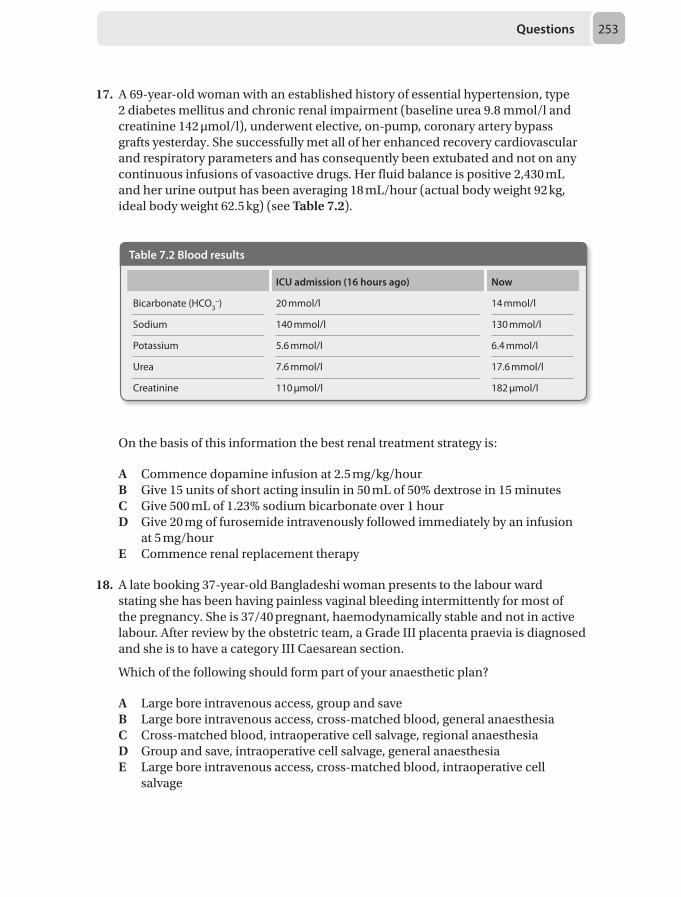
Questions 253
17. A 69-year-old woman with an established history of essential hypertension, type 2 diabetes mellitus and chronic renal impairment (baseline urea 9.8 mmol/l and creatinine 142 μmol/l), underwent elective, on-pump, coronary artery bypass grafts yesterday. She successfully met all of her enhanced recovery cardiovascular and respiratory parameters and has consequently been extubated and not on any continuous infusions of vasoactive drugs. Her fluid balance is positive 2,430 mL and her urine output has been averaging 18 mL/hour (actual body weight 92 kg, ideal body weight 62.5 kg) (see Table 7.2).
Table 7.2 Blood results
ICU admission (16 hours ago) Now
Bicarbonate (HCO3–) 20 mmol/l 14 mmol/l
Sodium 140 mmol/l 130 mmol/l
Potassium 5.6 mmol/l 6.4 mmol/l
Urea 7.6 mmol/l 17.6 mmol/l
Creatinine 110 µmol/l 182 µmol/l
On the basis of this information the best renal treatment strategy is:
A Commence dopamine infusion at 2.5 mg/kg/hourB Give 15 units of short acting insulin in 50 mL of 50% dextrose in 15 minutesC Give 500 mL of 1.23% sodium bicarbonate over 1 hourD Give 20 mg of furosemide intravenously followed immediately by an infusion
at 5 mg/hourE Commence renal replacement therapy
18. A late booking 37-year-old Bangladeshi woman presents to the labour ward stating she has been having painless vaginal bleeding intermittently for most of the pregnancy. She is 37/40 pregnant, haemodynamically stable and not in active labour. After review by the obstetric team, a Grade III placenta praevia is diagnosed and she is to have a category III Caesarean section.
Which of the following should form part of your anaesthetic plan?
A Large bore intravenous access, group and saveB Large bore intravenous access, cross-matched blood, general anaesthesiaC Cross-matched blood, intraoperative cell salvage, regional anaesthesiaD Group and save, intraoperative cell salvage, general anaesthesiaE Large bore intravenous access, cross-matched blood, intraoperative cell
salvage
Chapter 7254
19. A 27-year-old woman is rushed into theatre from the midwifery-led birthing centre with a post-partum haemorrhage (PPH) of 800 mL. The obstetric registrar has diagnosed uterine inversion and has tried manual reduction without success. The patient has a blood pressure of 100/60 mmHg and a heart rate of 95 beats per minute. She has adequate intravenous access, is receiving a second litre of crystalloid and is comfortable on Entonox.
What is the best line of management to undertake next?
A General anaesthesia and immediate laparotomyB Administration of a tocolytic agentC Immediate infusion of 2 units of O– bloodD Regional anaesthesia to relax the uterusE Rapid infusion of 500 mL of crystalloid, then regional anaesthesia
20. A 3-year-old, 16 kg child is scheduled for adenotonsillectomy for recurrent tonsillitis and mild obstructive sleep apnoea. He is the second case on the afternoon ENT list. His mother gave him a light breakfast at 7 am, and he has had nothing to eat or drink since. The morning list is overrunning, it is now 1 pm, and the child is unlikely to be anaesthetised until 4 pm. The nurse on the ward informs you that the child is getting upset because he is hungry and thirsty.
The best course of action is:
A Postpone the case for another day, and let the child eat and drinkB Let the child drink clear water until 2 pm, with plan to anaesthetise the child at
4 pmC Start an intravenous infusion of 0.9% salineD Start an intravenous infusion of 0.9% saline with 5% dextroseE Continue to fast the child, and aim to do the child as soon as possible
21. A 2-month-old, 6 kg boy is having an emergency laparotomy for bowel obstruction. The temperature from a nasopharyngeal thermistor reads 35.8 °C.
The best way to reduce heat loss through radiation is:
A Turn the theatre temperature upB Use a warm air blanketC Use an overhead radiant heaterD Cover the patient’s head with a hatE Use warm irrigation fluid
22. A 46-year-old man developed back pain after heavy lifting 6 months ago, and still has pain in his lower back radiating to his buttocks. He finds that he has to limit his gardening and play golf due to the pain.
Questions 255
Which of the following is the most likely cause of his pain?
A Epidural adhesionsB Facet jointC Disc prolapseD DiscogenicE Sacroiliac pain
23. A 36-year-old primigravida who is 38/40 pregnant presents to the labour ward. She has a body mass index (BMI) of 40 and is known to have pre-eclampsia for which she takes labetalol. Currently her blood pressure is 158/96 mmHg. A vaginal examination reveals she is 6cm dilated and she is coping well with her contractions. Bloods show platelets of 98 x 109/L with normal clotting.
What would be the most appropriate way to manage her labour analgesia?
A Remifentanil patient-controlled analgesiaB Intramuscular pethidineC EntonoxD Epidural with patient controlled epidural analgesiaE She does not require any analgesia at present
24. A 69-year-old man is undergoing elective hip replacement surgery. He has a history of significant chronic obstructive pulmonary disease (COPD) which is controlled with regular inhalers. You offer him a spinal anaesthetic for his surgery.
Which of the following would be the most appropriate post operative analgesia option?
A Patient controlled analgesia with morphineB Patient controlled analgesia with fentanylC Oral opioids on a regular basisD Intrathecal diamorphineE Femoral nerve catheter and infusion of bupivacaine
25. A 38-year-old woman who is well known to the pain clinic presents with acute-on-chronic lower back pain. There is no radiation of pain and there are no red flag symptoms. Previous MRI was unremarkable. She has had facet joint injections in the past which have been effective for up to 2 weeks. She is currently taking paracetamol 1 g four times daily and Oxycontin 20 mg twice daily.
What would be the most appropriate next step in the management of this patient’s ongoing pain?
A Increase Oxynorm to 30 mg twice daily with Oxynorm 5 mg for breakthrough pain
B Book for further facet joint injectionsC Book for lumbar epiduralD Add amitriptylineE Referral to a pain management program
Chapter 7256
26. A 30-year-old man with ulcerative colitis is undergoing a total colectomy. He has been on long term opioids via a fentanyl patch at 50 µg per hour for the past year. He has refused an epidural for post operative analgesia.
What would be the most appropriate option for pain relief?
A Patient controlled analgesia (PCA) with morphine 1 mg bolus and keeping his fentanyl patch on
B Doubling the dose of the fentanyl patch C Bilateral transverse abdominis plane (TAP) blocksD Increasing the fentanyl patch to 75 µg per hour and using a PCA using fentanyl
bolus of 20 µg onlyE PCA with fentanyl bolus 10 μg and a background infusion of 10 µg per hour
27. You review a 7-year-old boy in the emergency department of a district general hospital. He has an 11% total body surface area (TBSA) burn involving his chest from hot cooking oil. It looks mostly partial thickness in nature. His vital signs including GCS are stable.
Which of the information provided above meets referral criteria to a specialised burns centre?
A Being 7 years old B Having an 11% TBSA scaldC A likely significant inhalational injuryD Any burn involving hot oilE He does not meet any of the criteria required for referral to a specialised burns
centre
28. A 72-year-old man had an elective above-knee amputation under a combined spinal-epidural. The operation was uneventful and finished at 2 pm. The surgeon is keen to start the thromboprophylaxis with dalteparin 2,500 units subcutaneously at 10 pm.
What specific instructions do you need to convey to the ward nurses concerning removal of the epidural catheter and administration of further doses of thromboprophylaxis?
A Remove the catheter after checking the prothrombin time and activated prothrombin time
B Remove the catheter after 10 am the following day and then administer the dalteparin immediately afterwards
C Remove the catheter at 10 pm the following evening and then administer the dalteparin immediately afterwards
D Remove the epidural catheter at 11 am the following day and administer the dalteparin after 4 hours
E Keep the epidural catheter in situ and wait for anaesthetic advice
Questions 257
29. A 42-year-old female being pre-assessed for gastric banding surgery is known to snore at night and has a body mass index (BMI) of 48 kg/m2.
As per the STOP-BANG questionnaire, which of the following assessment criteria would she need to fulfill in order to be termed high risk for obstructive sleep apnoea (OSA)?
A Collar size of 38 cmB High blood pressureC DiabetesD Pulmonary hypertensionE Collar size of 33 cm
30. A 64-year-old man is brought to the emergency department with dizziness and difficulty in breathing. He has a heart rate of 68 beats per minute and a blood pressure of 76/40 mmHg. On auscultation he has generalised rhonchi and is wheezy. There is no rash on examination. He states that he suffers from chronic glaucoma and his ophthalmologist has recently changed his eye drops.
Which of the following eye drops is the most likely cause for this clinical picture?
A PhenylephrineB AdrenalineC BrimonidineD LevobunololE Apraclonidine
Chapter 7258
Answers
1. D Alert the surgeons and ask them to flood the operative siteVenous air embolism is a potentially fatal clinical situation. Aspiration of approximately 1mL/kg can generate an ‘air locked’ pulmonary circulation. It can occur in any surgical position providing the operative site is above the level of the heart. If the hydrostatic gradient between the site and the right atrium is negative, air can potentially move into the venous circulation and directly into the right atrium. From here it passes into the right ventricle and on to the pulmonary artery. If large enough it will entirely obstruct flow of blood through the ventricular outflow tract. Subsequently, an air embolism initially increases right heart pressures and critically impairs gas exchange. Cardiac output, end-tidal CO₂ and O₂ saturations decrease. Ultimately, such deterioration can lead to cardiac arrest. Neurosurgical procedures are especially high risk as veins may be held open by boney structures.
Management priorities are to stop further air inflow, reduce the volume or remove any air that has accumulated and to treat any development of cardiovascular (CVS) collapse. The initial action should therefore be to immediately alert the surgeons who should obstruct any further air entry by flooding or applying a wet swab to the site. 100% oxygen should then be administered, followed by methods to increase venous pressure at the site. This can be achieved by levelling the table, applying pressure to the neck, administering a fluid challenge +/- an inotrope or conducting a Valsalva manoeuvre. If a central venous line is in situ, it should be aspirated. If CVS collapse occurs the patient should then be turned into the left lateral, head down position if possible, and cardiopulmonary resuscitation initiated.
Webber S, Andrzejowski J, Francis G. Gas embolism in anaesthesia. Contin Educ Anaesth Crit Care Pain 2002; 2(2):53–57. Pollard BJ (ed). Handbook of Clinical Anaesthesia, 2nd edn. London: Elsevier Science, 2003.
2. D 2 units of fresh frozen plasma and 2 pools of plateletsIt has been reported that up to 20% of cardiac surgery patients bleed significantly postoperatively. The need for resternotomy increases the chance of further complications including prolonged mechanical ventilation, adult respiratory distress syndrome (ARDS) and wound infection. In addition to obvious surgical causes of bleeding, dysfunction of the coagulation cascade can occur for a variety of reasons. Causes of perioperative coagulopathy can have the mnemonic ‘ACHE’:
• Antiplatelet agents• Contact with cardiopulmonary bypass circuit• Haemodilution• Heparin• Hypothermia• Excessive fibrinolysis
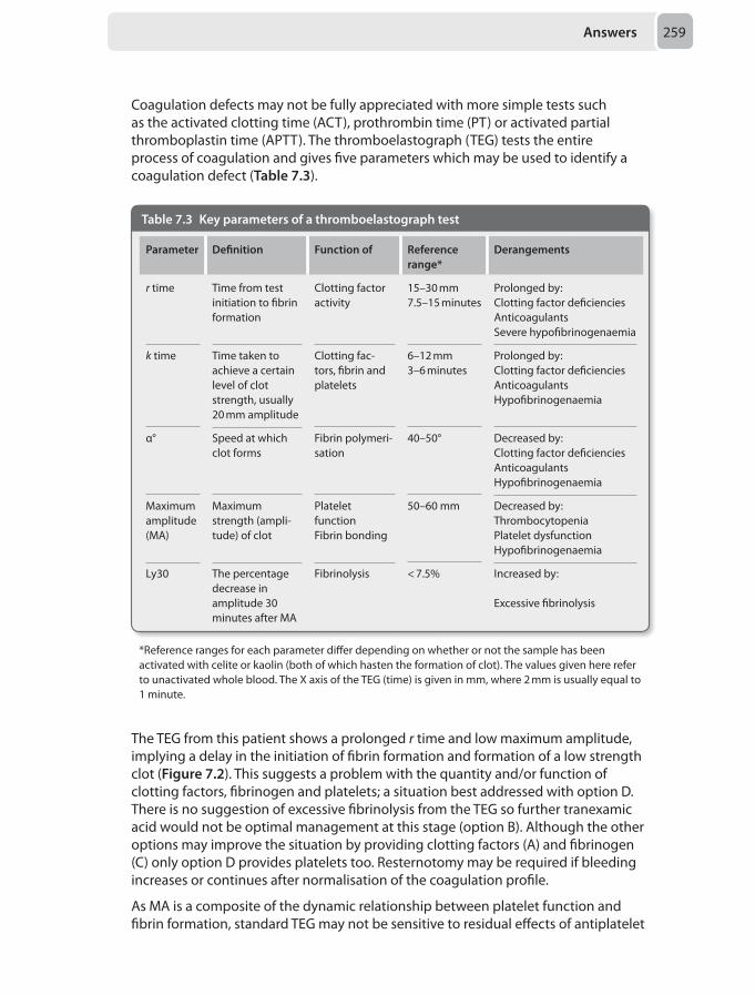
Answers 259
Coagulation defects may not be fully appreciated with more simple tests such as the activated clotting time (ACT), prothrombin time (PT) or activated partial thromboplastin time (APTT). The thromboelastograph (TEG) tests the entire process of coagulation and gives five parameters which may be used to identify a coagulation defect (Table 7.3).
Table 7.3 Key parameters of a thromboelastograph test
Parameter Definition Function of Reference range*
Derangements
r time Time from test initiation to fibrin formation
Clotting factor activity
15–30 mm7.5–15 minutes
Prolonged by:Clotting factor deficienciesAnticoagulantsSevere hypofibrinogenaemia
k time Time taken to achieve a certain level of clot strength, usually 20 mm amplitude
Clotting fac-tors, fibrin and platelets
6–12 mm 3–6 minutes
Prolonged by:Clotting factor deficienciesAnticoagulantsHypofibrinogenaemia
α° Speed at which clot forms
Fibrin polymeri-sation
40–50° Decreased by:Clotting factor deficienciesAnticoagulants Hypofibrinogenaemia
Maximum amplitude (MA)
Maximum strength (ampli-tude) of clot
Platelet functionFibrin bonding
50–60 mm Decreased by:ThrombocytopeniaPlatelet dysfunctionHypofibrinogenaemia
Ly30 The percentage decrease in amplitude 30 minutes after MA
Fibrinolysis < 7.5% Increased by:
Excessive fibrinolysis
*Reference ranges for each parameter differ depending on whether or not the sample has been activated with celite or kaolin (both of which hasten the formation of clot). The values given here refer to unactivated whole blood. The X axis of the TEG (time) is given in mm, where 2 mm is usually equal to 1 minute.
The TEG from this patient shows a prolonged r time and low maximum amplitude, implying a delay in the initiation of fibrin formation and formation of a low strength clot (Figure 7.2). This suggests a problem with the quantity and/or function of clotting factors, fibrinogen and platelets; a situation best addressed with option D. There is no suggestion of excessive fibrinolysis from the TEG so further tranexamic acid would not be optimal management at this stage (option B). Although the other options may improve the situation by providing clotting factors (A) and fibrinogen (C) only option D provides platelets too. Resternotomy may be required if bleeding increases or continues after normalisation of the coagulation profile.
As MA is a composite of the dynamic relationship between platelet function and fibrin formation, standard TEG may not be sensitive to residual effects of antiplatelet
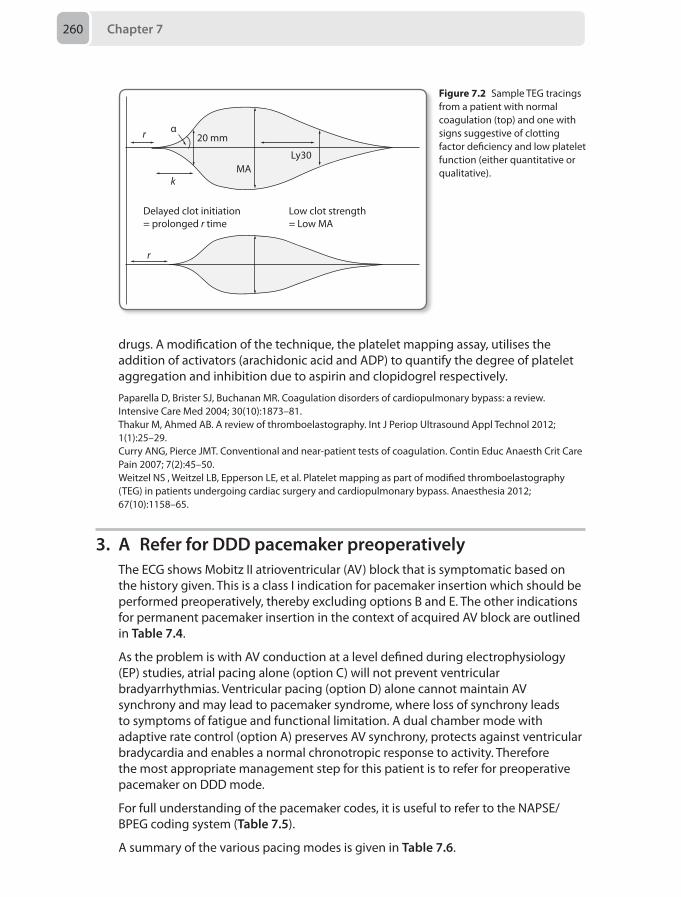
Chapter 7260
drugs. A modification of the technique, the platelet mapping assay, utilises the addition of activators (arachidonic acid and ADP) to quantify the degree of platelet aggregation and inhibition due to aspirin and clopidogrel respectively.
Paparella D, Brister SJ, Buchanan MR. Coagulation disorders of cardiopulmonary bypass: a review. Intensive Care Med 2004; 30(10):1873–81.Thakur M, Ahmed AB. A review of thromboelastography. Int J Periop Ultrasound Appl Technol 2012; 1(1):25–29.Curry ANG, Pierce JMT. Conventional and near-patient tests of coagulation. Contin Educ Anaesth Crit Care Pain 2007; 7(2):45–50.Weitzel NS , Weitzel LB, Epperson LE, et al. Platelet mapping as part of modified thromboelastography (TEG) in patients undergoing cardiac surgery and cardiopulmonary bypass. Anaesthesia 2012; 67(10):1158–65.
3. A Refer for DDD pacemaker preoperativelyThe ECG shows Mobitz II atrioventricular (AV) block that is symptomatic based on the history given. This is a class I indication for pacemaker insertion which should be performed preoperatively, thereby excluding options B and E. The other indications for permanent pacemaker insertion in the context of acquired AV block are outlined in Table 7.4.
As the problem is with AV conduction at a level defined during electrophysiology (EP) studies, atrial pacing alone (option C) will not prevent ventricular bradyarrhythmias. Ventricular pacing (option D) alone cannot maintain AV synchrony and may lead to pacemaker syndrome, where loss of synchrony leads to symptoms of fatigue and functional limitation. A dual chamber mode with adaptive rate control (option A) preserves AV synchrony, protects against ventricular bradycardia and enables a normal chronotropic response to activity. Therefore the most appropriate management step for this patient is to refer for preoperative pacemaker on DDD mode.
For full understanding of the pacemaker codes, it is useful to refer to the NAPSE/BPEG coding system (Table 7.5).
A summary of the various pacing modes is given in Table 7.6.
r α
k
20 mm
MA
Delayed clot initiation = prolonged r time
r
Low clot strength = Low MA
Ly30
Figure 7.2 Sample TEG tracings from a patient with normal coagulation (top) and one with signs suggestive of clotting factor deficiency and low platelet function (either quantitative or qualitative).
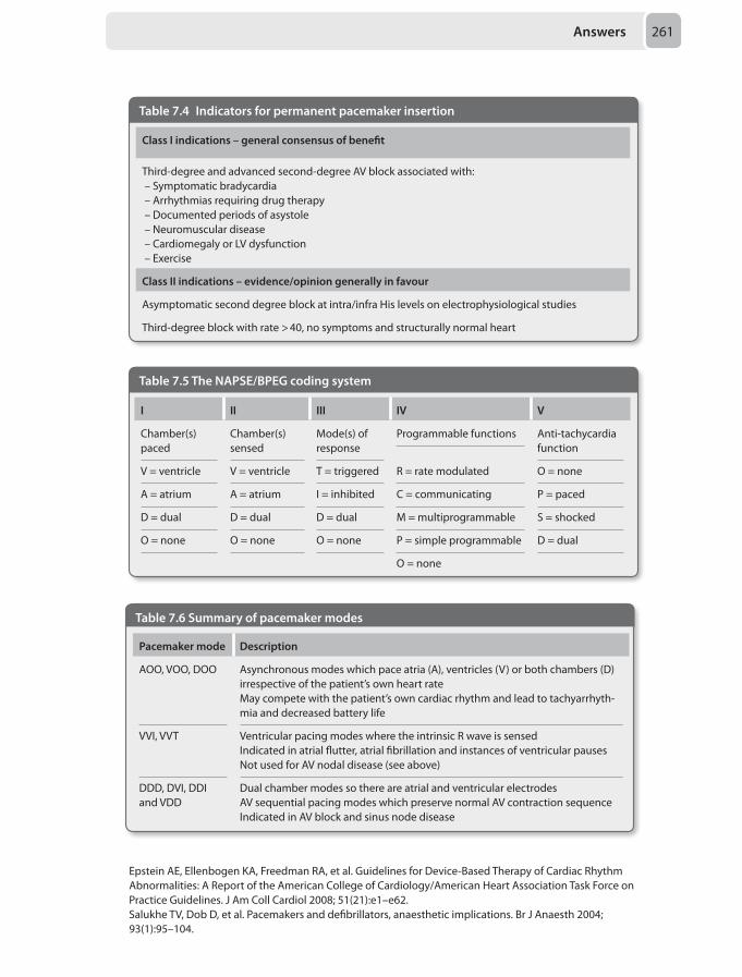
Answers 261
Table 7.4 Indicators for permanent pacemaker insertion
Class I indications – general consensus of benefit
Third-degree and advanced second-degree AV block associated with: – Symptomatic bradycardia – Arrhythmias requiring drug therapy – Documented periods of asystole – Neuromuscular disease – Cardiomegaly or LV dysfunction – Exercise
Class II indications – evidence/opinion generally in favour
Asymptomatic second degree block at intra/infra His levels on electrophysiological studies
Third-degree block with rate > 40, no symptoms and structurally normal heart
Table 7.5 The NAPSE/BPEG coding system
I II III IV V
Chamber(s) paced
Chamber(s) sensed
Mode(s) of response
Programmable functions Anti-tachycardia function
V = ventricle V = ventricle T = triggered R = rate modulated O = none
A = atrium A = atrium I = inhibited C = communicating P = paced
D = dual D = dual D = dual M = multiprogrammable S = shocked
O = none O = none O = none P = simple programmable D = dual
O = none
Table 7.6 Summary of pacemaker modes
Pacemaker mode Description
AOO, VOO, DOO Asynchronous modes which pace atria (A), ventricles (V) or both chambers (D) irrespective of the patient’s own heart rateMay compete with the patient’s own cardiac rhythm and lead to tachyarrhyth-mia and decreased battery life
VVI, VVT Ventricular pacing modes where the intrinsic R wave is sensedIndicated in atrial flutter, atrial fibrillation and instances of ventricular pauses Not used for AV nodal disease (see above)
DDD, DVI, DDI and VDD
Dual chamber modes so there are atrial and ventricular electrodesAV sequential pacing modes which preserve normal AV contraction sequenceIndicated in AV block and sinus node disease
Epstein AE, Ellenbogen KA, Freedman RA, et al. Guidelines for Device-Based Therapy of Cardiac Rhythm Abnormalities: A Report of the American College of Cardiology/American Heart Association Task Force on Practice Guidelines. J Am Coll Cardiol 2008; 51(21):e1–e62. Salukhe TV, Dob D, et al. Pacemakers and defibrillators, anaesthetic implications. Br J Anaesth 2004; 93(1):95–104.
Chapter 7262
4. C Apply humidificationObstructive sleep apnoea/hypopnoea syndrome (OSAHS) is a common disorder characterised by intermittent upper airway collapse during sleep. An apnoea is defined as a ten second breathing pause due to complete airway closure, whereas a hypopnoea describes an episode where ventilation is reduced by at least 50% for 10 seconds due to partial collapse. OSAHS is graded into mild, moderate and severe categories by the apnoea-hypopnoea index (number of events per hour of sleep) and the severity of symptoms.
In order to improve daytime somnolence, the treatment aim is to reduce the frequency of nocturnal apnoeas/hypopnoeas with options including lifestyle modification, dental devices, surgery and the application of continuous positive airway pressure (CPAP). The National Institute for Health and Care Excellence (NICE) have recently recommended that all moderate to severe symptomatic cases of OSAHS should be offered CPAP therapy. There is also a role for CPAP therapy in symptomatic mild cases of OSAHS, but only if lifestyle modification has failed to make a difference.
CPAP devices work by producing a continuous positive pressure (set between 5 and 20 cmH2O) which prevents upper airway collapse and subsequent apnoeas or hypopnoeas during sleep. Problems with compliance to therapy are common since upper airway symptoms such as nasal dryness, bleeding and throat irritation can occur as a result of high flows of dry, cool air through the nose. Humidification devices are now frequently used in conjunction with CPAP devices to prevent these symptoms. In the above case, application of a humidifier is the most appropriate next management step since this may improve CPAP compliance in order to accurately assess treatment effect before exploring other options.
Fixed CPAP devices as the name suggests, deliver air at a set pressure throughout the night which can lead to non-adherence due to pressure intolerance. To minimise these side effects and reduce mean airway pressures, auto-titrating CPAP devices have been developed. These devices vary the treatment pressure applied automatically based on feedback from changes in airflow resistance. In the above scenario, the patient is already using an auto-titrating CPAP device and is not complaining of pressure intolerance so changing to a fixed device is therefore unlikely to improve adherence.
Bilevel positive airway pressure (BiPAP) delivers positive airway pressure at different levels during inspiration and expiration. BiPAP not only prevents upper airway collapse but also augments tidal volume and can achieve lower mean airway pressures when compared to CPAP. In relation to the above case, it is unlikely to improve the upper airway symptoms affecting compliance however.
Mandibular repositioning devices are designed to improve upper airway patency by protruding the mandible to expand the posterior airspace. However, the maximum attainable airspace expansion is perceived to be modest, and currently these devices are only considered appropriate for mild to moderate OSAHS. Mandibular repositioning devices can be used in patients who refuse to use or fail to respond to CPAP. It is not the most appropriate next management step in the featured case
Answers 263
since the OSAHS is severe and the treatment benefit of CPAP has not yet fully been established.
In the absence of a resectable obstructing lesion such as tonsillar hypertrophy, the role of surgery in treating OSAHS remains contentious.
Uvulopalatopharyngoplasty (UPPP) is a common surgical approach which involves resection of the uvula, retrolingual soft tissue and palatine tonsillar tissue in an attempt to improve airway patency in this context. However, surgery does not guarantee symptom improvement and may compromise future CPAP therapy by promoting mouth leakage and limiting the maximum pressure level tolerated. In the above case, surgery is not the most appropriate management step as there is no obvious obstructing lesion, and symptoms may improve by increasing adherence to the CPAP machine alone.
Appraisal Committee and National Institute for Health and Care Excellence (NICE) project team. Continuous positive airway pressure for the treatment of obstructive sleep apnoea/hypopnoea syndrome. Technology appraisal guidance 139. London: NICE, 2008.McDaid C, Griffin S, Weatherley H, et al. Continuous positive airway pressure devices for the treatment of obstructive sleep apnoea-hypopnoea syndrome: a systematic review and economic analysis. Health Technol Assess 2009; 13(4):1–119,143–274. Scottish Intercollegiate Guidelines Network (SIGN).Guideline development group. Management of obstructive sleep apnoea/hypopnoea syndrome in adults. A national clinical guideline. Edinburgh: SIGN, 2003. Weaver T, Grunstein R. Adherence to continuous positive airway pressure therapy. The challenge of effective treatment. Proc Am Thorac Soc 2008; 5:173–178.
5. A She can proceed for day case surgeryAAGBI guidelines for perioperative management of obese patients recommend that patients should not be excluded from day surgery on the basis of their BMI alone. Units with appropriate resources and experienced staff can safely manage these cases where their management would not be changed by overnight admission and in fact benefit from early mobilisation. Patients with morbid obesity should be carefully pre-assessed for symptoms of cardiac, respiratory and metabolic disease. Stable OSA with established CPAP also does not preclude day surgery, but measures such as avoiding long acting opioids and careful postoperative monitoring are required.
A validated questionnaire STOP-BANG has been developed to identify and risk-stratify patients:
• Snoring – do you snore loudly? (loud enough to be heard through a closed door)• Tired – do you often feel tired or sleepy during the daytime?• Observed – has anyone observed you stop breathing in your sleep?• Blood pressure – do you have or are you treated for high blood pressure?• Body mass Index > 35 kg/m2• Age > 50 years• Neck circumference > 40 cm• Gender – male
A score of greater than 5 requires further investigation and careful perioperative management as does the presence of any other features such as poor functional
Chapter 7264
capacity, abnormal ECG, uncontrolled hypertension or ischaemic heart disease, saturations less than 94% on air, concurrent airways disease and previous venous thromboembolism.
In this case, the STOP-BANG score is 3 and further cardio-respiratory investigation is not required. Sleep studies are not indicated unless symptoms of excessive daytime sleepiness or witnessed apnoeas in the presence of other risk factors are reported. In-patient surgery or overnight admission is not required if her postoperative Spo2 is maintained at baseline levels on air without stimulation, and routine discharge criteria can be met. Although diabetes should be screened for with a random blood glucose check a formal glucose tolerance test is not indicated. Pre-assessment clinics are an ideal place for advice regarding lifestyle modification, however weight loss must be carefully controlled and monitored and is unlikely to alter management in this case if surgery is postponed.
Association of Anaesthetists of Great Britain and Ireland (AAGBI). Peri-operative Management of the Morbidly Obese Patient. London: AAGBI, 2007. Chung F, Subramanyam R, Liao P, et al. High STOP-Bang score indicates a high probability of obstructive sleep apnoea. Br J Anaesth 2012; 108(5):768–775.Society for Obesity and Bariatric Anaesthesia. Anaesthesia for the Obese Patient: BMI> 35 kg/m2. London: Society for Obesity and Bariatric Anaesthesia, 2014.
6. E Reintubation and head-up positioningRobotic surgery is becoming increasingly widespread, and may now be found in many centres and specialties including general surgery and gynaecology. In the UK by far the largest body of established work involves urology, and specifically prostatectomy. The perceived benefits include increased nerve preservation within the pelvic field and thus higher chances of retaining urinary continence and erectile function. There may also be some advantages in terms of comfort/analgesia and reduced blood loss. Indeed, it is now not uncommon for patients to be discharged in the first 24 hours following surgery.
The robot
The da Vinci system is the most common system in use in the UK at the current time. This system is made up of a surgical control console with an immersive high-definition visual display, a computer tower, and the robotic surgical manipulator. The manipulator is a large, heavy trolley comprising the surgical arms which is then ‘docked’ to the patients table. One arm supports the camera, and others are then inserted into the ports. A scrubbed assistant is still required, while the unscrubbed surgeon sits at the console, which may be distant from the patient. The robot has no autonomy in function; it merely acts as a ‘telemanipulator’ transmitting the surgeon’s movements from the console. There are case reports of surgery having been performed with the surgical console being situated in a different country from the patient.
The advantage over standard laparoscopic surgery comes from several sources. First, fewer assistants are required, with one scrubbed surgeon and a scrub nurse. The camera contains dual optical apparatus meaning that a stereoscopic picture is possible in the display console, allowing for depth perception. The robot arms have extra jointed articulations allowing advanced movements and greater degrees of

Answers 265
freedom compared to normal laparoscopic instruments. The apparatus filters tremor and automatically scales movements, all greatly facilitating microsurgery.
Specific physiological considerations
For the most part the considerations are those of laparoscopic surgery, however access to the patient is severely limited, and the position is very extreme. This exaggerates all the physiological changes such that complications may ensue if precautions are not taken. Due to the time taken to uncouple the robot from the patient, (may be several minutes) a plan for emergency access to the patient must be rehearsed.
The surgery requires steep head-down in the Trendelenburg position which may be as steep as 45°. For this reason, the attention to detail during positioning is vital. Strapping of the shoulders to prevent patient slipping can produce traction on the brachial plexus, and the lower limbs must be carefully positioned to reduce the risk of well leg syndrome and thromboembolism. The transition to this position can cause movement of the tracheal tube, due to migration of the tube in either direction, but also movement of the trachea and the diaphragm upward. Once in this position the added cardiovascular insult of pneumoperitoneum can cause major haemodynamic instability which if not resolved by countermeasures, may necessitate conversion to an open procedure.
The degree and duration of Trendelenburg present a series of problems less common in other types of surgery, but still thankfully rare. Reflux of gastric secretions can cause chemical damage to the mucosa of the mouth and also unprotected eyes. Antacid premedication can be helpful. The increase in systemic vascular resistance, mean arterial pressures and intracranial pressures accompanied by decreased venous return can cause oedema of the dependent head and neck tissues, and patients are often warned to expect facial and eye swelling postoperatively. This has been associated with laryngeal oedema and stridor, and cerebral oedema with marked confusion, requiring reintubation and head up positioning for some hours before successful extubation. For this reason, and to reduce ureteric flow a conservative fluid strategy is often adopted once the head down position is achieved.
Confusion in recovery is a popular exam topic with a vast array of differentials. The clue here is the type and duration of surgery, and knowing something about the position involved during the robotic technique obviously helps. The patient could be hypoxic, but in this condition is unlikely to tolerate non-invasive ventilation anyway. Airway oedema can occur in these patients, but no mention is made of stridor in the stem. Facial swelling and cerebral oedema should subside in hours with supportive measures and head up position. CT scanning should also be considered.
Irvine M, Patil V. Anaesthesia for robot-assisted laparoscopic surgery. Contin Educ Anaesth Crit Care Pain 2009; 9(4):125–129.
7. A A reduced density compared to airHelium is an odourless, colourless and biologically inert noble gas whose unique physical properties can be exploited when managing an upper airway obstruction.
Chapter 7266
It is presented as Heliox 21, which is the generic name for the mixture of 21% oxygen and 79% helium. In order to appreciate its role in the management of an upper airway obstruction, the types of flow within the airway needs to be revisited.
Gas flow within the respiratory tract can be either laminar or turbulent. Laminar flow is unidirectional and smooth with molecules moving in parallel creating a parabolic flow profile. This is an efficient type of flow. Turbulent flow on the other hand has an essentially flat flow profile with molecules swirling in eddies and vortices rather than an orderly way. This is an inefficient form of flow and conversion from laminar to turbulent flow approximately halves the flow rate for a given pressure drop.
Whether or not flow is turbulent or laminar depends on a dimensionless number called the Reynolds number (Re) (turbulent flow being more likely if Re > 2000):
Re = ρvd/μg
ρ = density, v = linear velocity, d = diameter of the tube, μ = viscosity, g = gravitational factor.
In constricted upper airways, the airflow is turbulent and inefficient. Since heliox is approximately three times less dense than air, its administration will cause a reduction in the Reynolds number thus increasing the likelihood of more efficient laminar flow. Even in situations where turbulent flow persists, heliox still flows much easier when compared to air since the flow rate is inversely related to the density of the carrying gas.
The viscosity of helium is in fact very similar to air, and does not explain why heliox is beneficial in upper airway obstruction scenarios. Flow rate is inversely related to the gas viscosity in established laminar flow, which is different to the turbulent flows seen in the above case.
Helium does have a high thermal conductivity and prolonged administration may be associated with a lowering of body temperature. This however does not significantly affect the airflow patterns within the respiratory tree.
Another useful physical property of helium is its very low blood: gas solubility. The helium dilution technique works on the principle that helium remains within the lung due to its low solubility in blood allowing the functional residual capacity to be estimated.
Helium is chemically inert because its filled valence orbitals are less able to interact with other compounds. Helium has no direct pharmacological effects and is not a treatment in its own right. It should be viewed as a bridging therapy whilst waiting for the effects of other treatments to have effect (Table 7.7).
Table 7.7 Physical properties of helium, oxygen and nitrogen at 20°C and 1 atmosphere
Helium Oxygen Nitrogen
Density (ρ; g/L) 0.1785 1.251 1.429
Viscosity (μ; micropoises) 188.7 167.4 192.6
Thermal conductivity (K; μcal/s/°K) 352 58 58.5
Answers 267
Mitchell V. Gas, tubes and flow. Anaesthesia and intensive care medicine. 2010; 11(1):32–35.Harris P, Barnes R. The uses of helium and xenon in current clinical practice. Anaesthesia 2008; 63:284–293.Gainnier M, Forel J. Clinical review: use of helium-oxygen in critically ill patients. Crit Care 2006; 10(6):241.
8. E Call for anaesthetic assistanceAnaesthesia in remote locations is associated with risk. It represents an unfamiliar environment, using anaesthetic equipment and monitoring, which may be only used on occasion, and personnel that do not routinely work together and are unfamiliar with anaesthetic practices. In the cardiac catheter lab the radiology equipment often makes it difficult to access and visualise the patient and the table may be fixed and unable to tilt head down. The focus of the staff is often on the revascularisation and in these challenging situations especially when dealing with patients that are critically unstable, communication and effective team working are paramount.
The 2013 Royal College of Anaesthetists (RCoA) guidelines on anaesthesia in non-theatre settings outline the staffing, drug, equipment and safety requirements that should be met when anaesthesia is provided in these remote locations. Equipment for induction, maintenance and emergence from anaesthesia should be available as is in theatre, and monitoring should be ideally separate from that used by the cardiologist. Space and equipment should be set up to deal with the possibility of cardiac arrest.
In this scenario, there are many essential checks and tasks that need to be swiftly performed before anaesthesia can be administered. The RCoA guidelines state that anaesthesia in remote locations cannot be performed by a single individual, and that a dedicated, qualified and skilled anaesthetic assistant should always be available, and provide exclusive help to anaesthetist. Clearly the machine, drug and equipment checks are all important, however, calling early for anaesthetic assistance is essential and the other tasks can be conducted once this has been requested.
Peden C. Guidelines for provision of services for anaesthetic care in the non theatre environment 2013. Guidelines on the provision of anaesthetic services. London: Royal College of Anaesthetists, 2013.
9. B LignocaineBier’s block anaesthesia is a form of intravenous regional anaesthesia (IVRA) that was first introduced by the German surgeon August Bier in 1908. It involves the administration of local anaesthetic (LA) intravenously into a tourniquet-blocked limb thus localising the anaesthetic in that limb. The technique is based on the principle that local anaesthetic diffuses from the vascular bed to the capillary plexus surrounding the nerve, causing conduction block in the nerve involved.
IVRA is primarily indicated for surgical procedures on the elbow, forearm or hand requiring anaesthesia for up to one 1 hour, such as fracture manipulation. It can also be successfully performed on quick lower limb procedures of the foot, ankle and lower leg. However, the block is difficult to perform in the lower limb and requires larger amount of local anaesthetic.
The steps to perform a Bier’s block involve:
1. Before commencing the Bier’s block, patient should be informed and consented adequately and fully starved. IVRA should be performed in a safe environment
Chapter 7268
where the patient is fully monitored with resuscitation equipment and emergency drugs available.
2. Two intravenous cannulae are established, one in the operated arm (as distal as possible) and another in the contralateral limb to administer sedation or other drugs if required.
3. A double cuff tourniquet is applied on the arm involved. The arm is then exsanguinated either by applying Esmarch bandage or raising it for two minutes while compressing the axillary artery.
4. The distal cuff is inflated to at least 100 mmHg above the patient’s systolic blood pressure followed by inflating the proximal cuff to the same pressure.
5. Once the tourniquet is secure, the distal cuff can be deflated.6. The LA solution is injected in the operated arm after confirming the absence of a
radial pulse. It is very important to inject the local anaesthetic slowly to prevent the peak venous pressure from exceeding the tourniquet occlusion pressure and hence leakage of LA to the systemic circulation.
7. Once the injection is completed, remove the cannula and apply pressure on the puncture site.
8. After 10–15 minutes, when the anaesthesia is established, the distal cuff is inflated followed by the deflation of the proximal cuff to relieve the tourniquet pain below the proximal cuff. The tourniquet must not be deflated before 20 minutes because releasing the tourniquet early may result in a large amount of LA being released immediately into the systemic circulation, increasing the danger of LA toxicity.
9. Once the surgical operation has been completed, the tourniquet should be deflated in two stages. By deflating the tourniquet for 10 seconds then reinflating it for 1 minute before the final release, the chance of systemic toxicity is reduced by gradually washing out the LA from the operated limb.
10. It is mandatory to continue monitoring the patient for at least 10 minutes after the procedure.
Although IVRA is a simple and safe technique, specific knowledge in local anaesthetic pharmacology is required in order to avoid rare but serious complications.
A variety of local anaesthetic agents have been used to perform a Bier’s block, however prilocaine and lignocaine are currently the most commonly used drugs.
In the UK, 0.5% prilocaine is the drug of choice for IVRA. It is the least toxic LA as it is the most rapidly metabolised of the amides. Prilocaine is an amide LA, the recommended dose is 3 mg/kg (maximum dose is 6 mg/kg), and usually 40 mL of a 0.5% solution is injected in the operated arm.
Prilocaine is associated with methaemoglobinaemia, especially when the dose exceeds 600 mg. Although this is clinically insignificant in most patients, small amounts of methaemoglobin can cause a significant decrease in oxygen-carrying capacity in patients with anaemia and heart disease, hence it should be avoided. Therefore prilocaine is not appropriate for the patient in this clinical scenario.
In North America, lignocaine remains the most frequent used amide LA in a dose of not more than 3 mg/kg. Many emergency doctors and anaesthetists in the UK are
Answers 269
still using lignocaine as their first choice due to its availability and reliability in IVRA. The New York School of Regional Anesthesia (NYSORA) has recommended 12–15 mL of 2% lignocaine for upper limb procedures or 30–40 mL of 2% for lower extremities. It would be the most suitable agent for this clinical scenario.
Bupivacaine is another amide LA. In addition to blocking neurotransmission, it also affects the myocardium and is avoided in IVRA because of its cardiotoxicity. Death has also been reported in some studies; therefore its use is contraindicated in many centres.
Although levobupivacaine and ropivacaine are safer and less cardiotoxic than bupivacaine, the use of these local anaesthetics does not provide rapid onset of anaesthesia or superior analgesia, and they are not recommended for IVRA.
Chloroprocaine is an ester local anaesthetic. It is a vasoconstrictor and has a rapid onset time of 3–5 minutes. It is less toxic than lignocaine and has a shorter duration of action. However, it is not used in IVRA in the UK due urticarial rash and venous irritation following cuff release in some patients.
Although many drugs have been used as additives to local anaesthetics in IVRA such as neostigmine, ketamine, clonidine, muscle relaxants and dexamethasone, ketorolac 20 mg is the primary drug that has demonstrated some evidence in relieving tourniquet pain and prolonging postoperative analgesia.
The New York School of Regional Anesthesia (NYSORA). Bier’s block. www.nysora.com/techniques/3071–bier-block.htmlRivera JJ, Villecco DJ, Dehner BK, et al. The efficacy of ketorolac as an adjunct to the Bier block for controlling postoperative pain following nontraumatic hand and wrist surgery. AANA J 2008; 76(5):341–345.
10. D Antecubital fossaUpper limb peripheral nerve blocks are used to provide analgesia and anaesthesia for elbow, forearm, wrist and hand surgery. They may also be used to augment a brachial plexus block or provide perioperative analgesia after a general anaesthesia.
The median nerve (C5-T1) arises from both the medial (C5, C6, C7) and the lateral cords (C8, T1) of the brachial plexus. In the arm, the nerve passes lateral to the brachial artery, which it then crosses, and descends on its medial side to the antecubital fossa. In the forearm, the median nerve lies between the bellies of flexor digitorum profundus and flexor digitorum superficialis. And at the wrist, it lies medial to flexor carpi radials and lateral to the tendon of palmaris longus.
It supplies sensory innervation to the radial side of the palm, and the palmar surface of the lateral 3 and half fingers, including their dorsal tip to the first interphalangeal joint. It provides motor innervation to most of flexor muscles in the forearm and thenar muscles of the thumb.
One of the most important branches of median nerve is the anterior interosseous nerve. This nerve arises from the median nerve just distal to the antecubital fossa. It descends between the ulna and the radius along the interosseous membrane. The anterior interosseous nerve supplies the flexor pollicis longus, the flexor digitorum profundus (lateral half ) and the pronator quadratus. It is essential to block the anterior interosseous nerve for successful median nerve block.
Chapter 7270
The median nerve can be blocked at various places and can be performed using peripheral nerve stimulator, landmark technique and/or ultrasound (US) guided with a high frequency probe.
At the brachial plexus: the median nerve lies in close relation to the axillary artery and vein in the axilla and can be blocked independently or in conjunction with the ulnar, radial and musculocutaneous nerves here. See question 4.10 for further details.
At the mid-arm level: the nerve lies above the brachial artery. Using a high frequency US probe or nerve stimulator, a single injection of 5–7 mL of local anaesthetic is enough to block the nerve.
At the antecubital fossa: using a high frequency US probe, the median nerve is seen as a single hyperechoic elliptical structure immediately medial to the brachial artery. 5–7 mL of local anaesthetic is injected after visualising the nerve. With a peripheral nerve stimulator technique, the needle is directed perpendicularly and the nerve should be found within 1–2 cm depth, medial to the brachial artery pulsation. After stimulating the median nerve (pronation, finger flexion and thumb opposition), 5–7 mL of local anaesthetic is injected.
This approach successfully blocks the anterior interosseous nerve, and for this clinical scenario it is the correct answer.
At the mid-forearm: a high frequency US probe is moved laterally to visualise the median nerve in axial section as a hyperechoic structure. Again, 5–7 mL of local anaesthetic is injected around the nerve.
At the wrist: the nerve lies between the tendons of flexor carpi radialis and palmaris longus. It can easily be blocked by ultrasound or nerve stimulator techniques, 2 cm proximal to the wrist crease.
In this scenario, the best place to block the median nerve is in the antecubital fossa because it is essential to block the anterior interosseous nerve for successful median nerve block. The anterior interosseous nerve is usually missed in the mid-forearm and the wrist approach. The axillary approach is not an option in this scenario because it has already been attempted and was unsuccessful. Spared nerves should be augmented with local anaesthetic injections distally and not proximally, so a supraclavicular approach is not the best option.
The radial and ulnar nerves can also be blocked throughout their course. The radial nerve (C5-T1) is the largest branch of the brachial plexus. It is derived from the posterior cord. During its course, it gives branches to the triceps muscle and then enters the spiral groove where it lies behind the humerus. In the spiral groove, the median nerve gives off the posterior cutaneous nerve of the forearm. It then descends in the elbow between the brachioradialis and the brachialis muscles. At the lateral epicondyle of the humerus, it divides into superficial and deep terminal branches. The superficial branch supplies sensation to the dorsum of the hand, while the deep branch becomes the posterior interosseous nerve, which provides motor innervation to the extensor muscles of the elbow, wrist and fingers.
The radial nerve block is not usually performed below the elbow because it of its division into superficial and deep branches just proximal to the elbow.
Answers 271
With a peripheral nerve stimulator technique, the nerve is usually found 1-2 cm above the brachial crease between the biceps tendon and the brachioradialis muscle. Around 5–7 mL of local anaesthetic is injected after stimulating the nerve (fingers and wrist extension). Using a high frequency US probe, the radial nerve can be blocked at the spiral groove below the triceps and above the humerus. It can also been blocked at the antecubital fossa. The probe is placed in the antecubital crease and then moved lateral and proximal. At this area, the radial nerve is visualised as an elliptical structure that divides into superficial and deep branches between the brachioradialis and the brachialis muscle. Again, 5–7 mL of local anaesthetic is injected around the two branches.
The ulnar nerve (C8, T1) arises from the medial cord of the brachial plexus. During its course, it passes behind the medial epicondyle to enter the forearm between the heads of flexor carpi ulnaris. It supplies the flexor carpi ulnaris and half of the flexor digitorum profundus. It provides motor innervation to the intrinsic muscles of the hand and sensation to the medial one and a half fingers.
The ulnar nerve can be blocked below or above the elbow. At the elbow level, the ulnar nerve lies between the medial epicondyle and the olecranon process. Blocking the nerve at this level could cause ischaemia due to high compartment pressures and should be avoided.
The safest approach is distal to the elbow. Placing a high frequency US probe on the flexor surface of the forearm, the nerve is seen on the medial side of the forearm, lying medial to the ulnar artery. Follow the nerve with the probe until the ulnar nerve separates from the artery.
With a peripheral nerve stimulator, the nerve is usually found 3–4 cm proximal to the ulnar groove. Around 5–7 mL of local anaesthetic is injected after stimulating the nerve (ulnar deviation of the wrist and medial finger flexion).
Brennan A, Jones M, Gordon J. Ultrasound-guided local anaesthetic blocks of the forearm. Anaesthesia Tutorial of the Week 208. London: World Federation of Societies of Anaesthesiologists, 2011. McCahon RA, Bedforth NM. Peripheral nerve block at the elbow and wrist. Contin Educ in Anaesth Crit Care Pain 2007; 7(2):42–44.
11. Send the child back to the ward and prescribe an oral midazolam premedication at a dose of 0.5 mg/kg, resending for the child at 15 minutes post-doseHaving an uncooperative child at induction is not uncommon, in studies distress at induction occurs in a third of children, with a quarter requiring some form of physical restraint. There are several factors which predict problems during anaesthetic induction, and eliciting these can help in making an induction strategy and preparing the parents beforehand.
Risk factors for induction distress
• Withdrawn, shy, introverted demeanour• Anxious children• Ages 1–3 (increased separation anxiety)
Chapter 7272
• Previous negative hospital experiences• Previous turbulent reaction at vaccination
Drug treatments
Midazolam is the most widespread sedative premedicant in the UK. The oral dose is 0.5 mg/kg, giving an onset at 5-10 minutes peaking at 20–30 minutes. It may also be given intranasally or sublingually at a dose of 0.2 mg/kg. The intranasal route may be possible if oral medication is rejected, but it can give a burning sensation.
Fentanyl can be given transmucosally in a lollipop, with a bioavailability of 33% via this route. A dose of 15–20 µg/kg will produce sedation at 20 minutes with a peak at 30–40 minutes. The whole host of opioid side-effects including respiratory depression are a drawback.
Vallergan (trimeprazine) is a sedating antihistamine from the phenothiazine class. Like other phenothiazines, it is also beneficial in being antiemetic and antimuscarinic. Since midazolam has been shown to be superior, Vallergan is now used less often.
Ketamine can be given orally at a dose of 5–8 mg/kg, with an onset at 10 minutes and peak at 25 minutes. Intramuscular ketamine at a dose of 4–5 mg/kg works in 5 minutes and is reserved for those patients in whom all other strategies have failed and who may be displaying aggressive /combative behaviour. Ketamine side effects include tachypnoea, hypersalivation, ballistic limb movements and the classical emergence hallucinations. Where ketamine has been used patients should be nursed in a calm, quiet area with standby provision of resuscitation equipment.
Holding and restraint
In general, the principle is to use restraint only as a technique of last resort. Minimal force required for safety (of staff and patient) should be employed, by appropriate numbers of experienced/trained staff. The plan should be discussed with the parent(s) beforehand, and opportunity for debrief discussions with parent and child should exist afterwards.
In this case the induction process has clearly broken down, and the risk of proceeding with a distressed child and mother has to be balanced against the urgency of the procedure. In an emergency if the child had already failed with premedication and the mother had been warned and was happy to proceed/participate with an inhalational induction, this might be the next step. However the risk of laryngospasm in a crying, anxious and distressed child is unacceptable here. Cancellation is an option, but even with further preparation and psychological input the risk of induction distress still persists, thus sending the child back to the ward and trying a premed is valid. Midazolam is the first line in this situation.
Tan L , Meakin GH. Anaesthesia for the uncooperative child. Contin Educ Anaesth Crit Care Pain 2010; 10(2):48–52.
12. C A seizureIntubation for transfer is indicated in patients who have:
• GCS 8 or less
Answers 273
• Drop in GCS of 2 points or 1 point in the motor scale• Loss of protective laryngeal reflexes• Ventilatory insufficiency: Pao2 < 13 kPa on oxygen, Paco2 > 6 kPa• Spontaneous hyperventilation with Paco2< 4 kPa• Irregular respiratory pattern• Seizures • Unstable facial fractures • Bleeding into the airway
Principles during transfer should be to reduce and avoid surges in intracranial pressure, maintain cerebral perfusion pressure and prevent secondary brain injury.
• The patient should receive sedation and analgesia via a syringe driver and adequate muscle relaxation. Aims should be for a Pao2 > 13 kPa, Paco2 4.5–5 kPa unless there is clinical or radiological evidence of raised intracranial pressure where hyperventilation to a Paco2> 4 kPa with a higher Fio2 is justified
• Endotracheal tubes should be secured but tight tube ties avoided. The patient should be placed in a 15–30 degree head up position
• Avoid hypotension. Hypovolaemia is poorly tolerated during transfer due to the effects of motion, and circulating volume should be normalised before departure. Inotropes may be indicated to achieve an adequate mean arterial pressure (> 80 mmHg) after volume expansion if hypotension persists
• Consider loading with an anticonvulsant, e.g. phenytoin prior to transfer• Avoid hypoglycaemia
National Institute for Health and Care Excellence (NICE). Triage, assessment, investigation and early management of head injury in children, young people and adults. CG No 176. London: NICE, 2014.Association of Anaesthetists of Great Britain and Ireland. Recommendations for the safe transfer of patients with brain injury. London: AAGBI, 2006.
13. D VasopressinAs catastrophic brain injury progresses into brainstem death, dramatic changes in cardiovascular physiology often occur due to one or more of the following:
• hypovolaemia secondary to diabetes insipidus caused by acute posterior pituitary failure
• myocardial depression due to catecholamine and cytokine toxicity• the transition from hypertensive catecholamine excess into vasoplegic
hypotension
In managing this clinical situation, a rapid, systematic approach to the cardiovascular system is essential and must encompass cardiac rate and rhythm, preload, contractility and afterload. The cardiovascular observations given suggest the patient has diabetes insipidus, has received adequate volume resuscitation but is vasoplegic. Current expert recommendations and limited trial evidence supports the use of vasopressin as the optimal first line agent in this scenario.
Bugge JF. Brain death and its implications for management of the potential organ donor. Acta Anaesthesiologica Scandinavica 2009; 53(10):1239–1250.Ball J. Optimal management of the potential organ donor following catastrophic brain injury. ICU Management 2013; 13(2):10–13.Callahan DS, Neville A, Bricker S, et al. The effect of arginine vasopressin on organ donor procurement and lung function. J Surg Res 2014; 186(1):452–457.
Chapter 7274
14. C Give sequential intravenous crystalloid boluses of 100 mL
The priorities after coronary artery bypass grafting are as follows: warm, wean, and wake. This simple list (which has obviously been designed by a surgeon) allows the consideration of problems at each stage:
Warming• As warming occurs vasodilation may occur which may result in relative
hypovolaemia • Reperfusion may result in transient metabolic disturbances
Weaning• Refers to reducing vasoconstrictor, inotrope and ventilator requirements, which
should be routine if there are no complications as a result of surgery or anaesthesiaWaking
There are several causes of prolonged waking which may be respiratory function related, metabolic, temperature-related, or anaesthetic and analgesic related, but most concerning are:
• ‘Pump-head’ a multi-factorial syndrome causing global cerebral dysfunction due to micro-emboli (particulate and gas) and hypo-perfusion
• Embolic stroke (regional deficit)• Haemorrhagic stroke (regional deficit)
The complications that may occur which disrupt weaning include:
Cardiac pump-failure• Global myocardial dysfunction due to pre-existing disease, myocardial
stunning, metabolic and electrolyte disturbances or inappropriate vasoconstrictor and inotrope usage
• Regional myocardial dysfunction due to a thrombosed graft, embolic obstruction, a kinked graft or poor cardioplegia delivery
• Arrhythmias: – Tachycardia (atrial fibrillation is the most common) treated with
pharmacological measures or rarely electrical cardioversion – Bradycardia treated with either atrial pacing (if no atrioventricular
conduction delay is present), ventricular pacing (if an atrioventricular block is present), or with atrioventricular sequential pacing
Mechanical obstruction• Tension pneumothorax (pleura surgically opened if internal mammary arteries
used for grafting)• Cardiac tamponade• Haemothorax if drains are obstructed with blood clot formation
Bleeding which may be assessed by monitoring the chest drain output• Surgical
– Graft anastomotic site – Venous graft tributary – Site of cannulation (aortic or atrial), suture lines, sternal wire holes
• ‘Anaesthetic’ – Dilutional coagulopathy – Inadequately reversed unfractionated heparin
Answers 275
In the case above there is evidence of worsening haemodynamic performance (an increased vasoconstrictor requirement) and end-organ perfusion (a worsening metabolic acidosis) without an obvious bleeding source and without an increased central venous pressure. An ABC approach is needed to identify which of the differential diagnosis are most likely. The immediate intervention, if no obvious cause is found, is a trial of intravenous fluid therapy (100 – 250 mL boluses) and assessment of fluid-responsiveness. As mentioned before, the combination of warming, reperfusion, fluid re-distribution and a small amount of blood loss may be all that is wrong.
A transthoracic or oesophageal echo is an extremely useful investigation. A visualised tamponade or regional wall abnormality that may indicate a graft-malfunction indicates the need to return to theatre however a collection of blood posterior to the heart causing tamponade may not be visualised. Global dysfunction seen on the echo due to myocardial stunning may be treated with correction of metabolic and electrolyte abnormalities or an inotrope such as dobutamine or milrinone.
A repeat full blood count and clotting analysis including a thrombelastogram (TEG) is helpful to guide blood product administration if bleeding is suspected.
The surgical team should be informed of the developments early. Cardiothoracic surgeons are experienced in weaning patients after bypass grafting and often have useful insights regarding the particulars of the surgery. If the patient continues to deteriorate, a repeat thoracotomy in theatre (or on the intensive care unit in extreme situations) may be required.
Walcot N, Marchbank A. Postoperative care of adult cardiac surgery patients. Surgery 2012; 30(1):13–16.
15. D Initiate titrated interval sprint weaning (work and rest cycles)
The scenario suggests a mixed picture of good and bad prognostic factors. In particular, declining exercise tolerance, low body mass index and/or significant recent weight loss and more than two hospital admissions per year are poor prognostic markers in patients with chronic obstructive pulmonary disease (COPD). This patient fulfils all the criteria for a spontaneous breathing trial, the purpose of which is to assess the likelihood of successful extubation. He resoundingly fails the trial by all criteria.
Ventilatory management in this population is challenging and arguably more of an art than a science. The best answer suggested here is controversial.
Though there is increasing enthusiasm for extubation and immediate application of mask ventilation in scenarios such as that outlined, the risks and benefits are complex and the relative merits of this approach are currently the subject of a number of large, randomised control trials. This patient has a relative contra-indication in having a heavy secretion load with only a moderate strength cough. Non-invasive ventilation (NIV) will increase his difficulty in secretion clearance and therefore places him at significant risk of ventilator failure despite NIV and requiring re-intubation. Should this occur, this sequence of events is associated with a higher morbidity and mortality that continuing invasive support.
Chapter 7276
Performing a tracheostomy at day 5, would be considered too early by most practitioners, unless, the educated guess was that a patient would clearly need > 10–14 days of invasive support. There is no clear evidence to support either an ‘always early’ or ‘always late’ strategy.
Protocolised weaning is advocated by many practitioners. Its success is probably more attributable to the organisational and logistic benefits rather than any physiological rationale. However, given that respiratory muscle fatigue is the principal cause of weaning failure in COPD patients, there is a growing body of evidence ranging from exercise physiology to cardiac rehabilitation to support a titrated work rest cycle approach utilising short bursts of high activity with prolonged periods of effective rest. Hence D is considered the best answer.
In the scenario given there is no rationale to re-sedate and SIMV is proven to be detrimental to weaning as opposed to CPAP with iPS, which promotes it.
Burns KE, Meade MO, Premji A, Adhikari NK. Noninvasive positive-pressure ventilation as a weaning strategy for intubated adults with respiratory failure. Cochrane Database Syst Rev 2013;12:CD004127.Gomes Silva BN, Andriolo RB, Saconato H, Atallah AN, Valente O. Early versus late tracheostomy for critically ill patients. Cochrane Database Syst Rev 2012;3:CD007271.Blackwood B, Alderdice F, Burns K, et al. Use of weaning protocols for reducing duration of mechanical ventilation in critically ill adult patients: Cochrane systematic review and meta-analysis. Br Med J 2011;342:c7237.Gist NH, Fedewa MV, Dishman RK, Cureton KJ. Sprint interval training effects on aerobic capacity: a systematic review and meta-analysis. Sports Med 2014; 44(2):269–79.
16. E Chest radiographThis is a very common scenario on the intensive care unit. Patients are predisposed to getting infections due to immobility, sedation, invasive tubes, relative immunosuppression and being handled by staff continually.
The differential diagnosis as to the source of infection in this gentleman is broad and therefore it is worth narrowing it down to the most likely culprit in order to choose an appropriate antibiotic. In general the first organ systems to consider are those that have contact with the external environment (skin including invasive lines, urinary tract, gastrointestinal tract and respiratory tract). These are also the easiest to investigate, as samples from these organs are relatively easy. If these are negative then consideration of more anatomically-isolated systems are needed (hepatobiliary, intra-abdominal, endocardial, central nervous system and bone). Finally if these are negative, rarer causes such as non-bacterial infections, malignancy, inflammatory, antibiotic related and neurologically-mediated should be investigated.
The term ventilator-associated pneumonia (VAP) refers to pneumonia occurring in patients more than 48 hours after endotracheal intubation and mechanical ventilation. The risk factors include:
• Patient• Extremes of age • Immunosuppression • Respiratory co-morbidities
Answers 277
• Airway• Emergency intubation• Presence of endotracheal tube
– No coughing – Decreased mucociliary clearance – Micro-aspiration of sub-glottic – Intra-luminal formation of biofilm
• Increased duration of intubation – Early onset < 96 hours – Late onset > 96 hours
• Late tracheostomy (contentious)• Head injury or altered consciousness (including sedation)• Poor mouth care and tracheobronchial toileting
• Gastrointestinal• Nasogastric feeding• Prolonged use of proton-pump inhibitors• Prolonged supine position
Early onset VAP commonly results from community-acquired pathogens such as Staphylococcus aureus, Streptococcus pneumoniae and Haemophilus influenzae. Late onset VAP is often a result of infection with drug resistant organisms such as Pseudomonas sp. and methicillin resistant Staphylococcus aureus (MRSA).
There are several scoring criteria published, which have been suggested in which to aid the early diagnosis. These include:
• Clinical signs• Temperature above 38°C or below 36°C• Leukocytosis or leucopenia• New purulent secretion production• Worsening gas exchange or increased oxygen requirement
• Imaging• Radiographic signs of new consolidation (generalised or focal)
• Microbiology• Sputum or bronchial lavage samples
In this scenario you are not given specific clinical details so diagnosis is more difficult. However, infections being common at this early stage of the patient’s clinical course, a chest infection would be high on the differential diagnosis. In addition a chest radiograph will give an immediate answer allowing prompt empirical treatment whereas all other investigations listed will take more time to return with useful information.
Rea-Neto A, Youssef NCM, Tuche F, et al. Diagnosis of ventilator-associated pneumonia: a systematic review of the literature. Crit Care 2008; 12:R56.Sundaram R. Nosocomial pneumonia. Anaesthesia UK, 2006.
Chapter 7278
17. E Commence renal replacement therapyIn patients with normal renal function, on-pump cardiac surgery is associated with a 10–30% risk of acute kidney injury and < 5% risk of needing acute renal replacement therapy. These risks are significantly increased in patients with pre-existing renal impairment. Other peri-procedural risk factors include on versus off-pump, longer bypass times, haemodynamic instability/need for high-dose or protracted inotropes and/or vasopressors.
In the scenario described, the patient has clearly developed acute kidney injury (AKI), with oliguria, a metabolic acidosis, hyperkalaemia and uraemia. There is no place for low dose dopamine or diuretics in either the prevention or management of AKI. Given the rate of evolution of this patient’s metabolic derangement and apparently normal renal perfusion, temporising therapy to improve the acidosis and reduce the serum potassium are unlikely to prevent the need for renal replacement therapy, hence this is the best answer.
Of note:
• Most centres use bicarbonate based fluids for renal replacement therapy • Although there is no universally agreed thresholds for commencing renal
replacement therapy in AKI a reasonable suggestion would include:• Hyperkalaemia (K+ > 6.5 mmol/l or K+ > 5.5 mmol/l and rapidly rising at
> 0.25 mmol/hour for 2 or more hours)• Correction of severe/unresolving acidosis (pH < 7.1) in particular, acidosis
associated with cardiovascular compromise (shock i.e. end-organ hypoperfusion) / high vasoactive drug requirements (noradrenaline > 0.5 µg/kg/min/dobutamine > 10 µg/kg/min)
• Uraemia (urea > 40 mmol/L or rising by > 12 mmol/24 hours)• Fluid overload causing severe hypertension and/or problematic oedema (e.g.
abdominal compartment syndrome) and/or contributing to hypoxaemia/poor lung compliance
• There is no evidence to support any specific modality over another in this setting
The use of bolus insulin and dextrose mixtures is a poor practice as it is associated with a very high incidence of acute, severe dysglycaemia and rapid rebound hyperkalaemia. If temporisation of hyperkalaemia is required, continuous infusions of insulin and dextrose are safer and more effective. Adjunctive use of nebulised salbutamol and intravenous bicarbonate can also be very helpful. If cardiac toxicity is evident, acute protection is afforded by a slow bolus of intravenous calcium either as gluconate or chloride.
Bellomo R, Chapman M. Low-dose dopamine in patients with early renal dysfunction: a placebo-controlled randomised trial. Australian and New Zealand Intensive Care Society (ANZICS) Clinical Trials Group. Lancet 2000; 356(9248):2139–43.Karajala V, Mansour W, Kellum JA, et al. Diuretics in acute kidney injury. Minerva Anestesiol 2009; 75(5):251–7.Palevsky PM. Renal replacement therapy in acute kidney injury. Adv Chronic Kidney Dis 2013; 20(1):76–84.
Answers 279
18. E Large bore intravenous access, cross-matched blood, intraoperative cell salvage
Placenta praevia occurs when the placenta is implanted in the lower uterine segment close to the internal cervical os. Painless vaginal bleeding in mid to late pregnancy is a classical presentation. It is divided into four grades:
• Grade I – Placenta in the lower segment but not reaching the internal os• Grade II – Placenta reaches internal os but does not cover it• Grade III – Placenta partially covers internal os• Grade IV – Placenta completely covers internal os
Risk factors include advanced maternal age, previous Caesarean section and multiparity. The obvious concern is the propensity to bleed during labour and often a Caesarean section is indicated. Uterine contraction may be impaired in the lower segment, the placenta may be abnormally adherent to the uterine wall and the placental position may make surgical access difficult. Hence, the risk of blood loss is significant and the anaesthetic plan should account for this.
The woman must be advised regarding the high risk of blood loss and large bore intravenous access must be secured, hence C and D are incorrect. At least 4 units of cross-matched blood should be ready and available for immediate use, i.e. in the labour ward fridge, if this facility exists. There must be a multidisciplinary approach to management, with involvement of the obstetric and anaesthetic teams, haematologists and paediatricians. Intraoperative cell salvage should be used and a rapid infusion device available and ready. Invasive monitoring should be established if needed.
Cases like this were often performed under general anaesthesia, but there has been a massive shift to regional techniques, such as spinal or combined spinal epidural. Ultimately, the decision lies with the anaesthetist and patient and if regional anaesthesia is chosen, the patient should be advised about the possibility of conversion to general anesthesia intraoperatively. Option B is incorrect, as intraoperative cell salvage should be part of the anaesthetic plan.
Banks A, Norris A. Massive haemorrhage in pregnancy. Contin Educ Anaesth Crit Care Pain 2005; 5(6):195–8.Walfish M, Neuman A, Wlody D. Maternal haemorrhage. Br J Anaes 2009; 103(suppl. 1):i47–i56.
19. B Administration of a tocolytic agentUterine inversion is an obstetric emergency, although relatively uncommon. The uterine fundus becomes displaced above or through the cervix, during the third stage of labour. The placenta remains attached in many instances, which can lead to massive haemorrhage. The accompanying haemodynamic instability is classically stated to be out of proportion to the blood loss, due to traction of uterine ligaments causing parasympathetic effects. This, however, may not be accurate, as blood loss may simply be underestimated in many cases.
Chapter 7280
Management focuses on uterine relaxation and reduction, and the management of haemorrhage. In this case, the patient has lost 800 mL of blood and may lose more. Basic resuscitation should be applied, namely, intravenous access, fluids and cross-match. As attempts at manual reduction have failed, tocolytic therapy should be the next line of management, such as terbutaline, magnesium sulphate or glyceryl trinitrate. Intravenous terbutaline at a dose of 0.25 mg has been recommended in relatively stable patients, otherwise 4 g of magnesium sulphate intravenously is an alternative. 100 µg of glyceryl trinitrate intravenously is relatively familiar to anaesthetists and has a quick onset of action.
If tocolytic therapy fails, then general anaesthesia to relax the uterus is indicated and a laparotomy may be required if the uterus still cannot be reduced vaginally. Regional anaesthesia does not cause uterine relaxation, so D and E are incorrect. Answer C is incorrect, as the patient has stabilised with intravenous fluids and does not need immediate O negative blood; hence it is not the best next line of management.
Abouleish E, Ali V, Joumaa B, Lopez M, Gupta D. Anaesthetic management of acute puerperal uterine inversion. Br J Anaesth 1995; 75(4):486–7. Beringer RM, Patteril M. Puerperal uterine inversion and shock. Br J Anaesth 2004; 92(3):439–41.
20. B Let the child drink clear water until 2 pm, with plan to anaesthetise the child at 4 pm
The reason for preoperative fasting is to reduce the risk of aspiration pneumonitis at induction of anaesthesia. However, prolonged fasting does not further reduce the risk of a harmful event for the patient, but adversely affects patient comfort and hydration. Therefore, the period of preoperative fasting should be minimised as close as possible to 6 hours for food and formula milk, 4 hours for breast milk and 2 hours for clear fluid, as per published consensus guidelines. If prolonged fasting is unavoidable, such as in patients with bowel obstruction, then intravenous hydration should be instituted.
In the case described above, there is no contra-indication to oral hydration up to 2 hours preoperatively, therefore intravenous hydration is not warranted. Postponing the case is disruptive, unnecessary and would not be the preferred option in the first instance.
The Association of Anaesthetists of Great Britain and Ireland (AAGBI). Safety Guideline: Pre-Operative Assessment and Patient Preparation: the Role of the Anaesthetist. London: AAGBI, 2010.Royal College of Nursing (RCN). Perioperative Fasting in Adults and Children: An RCN Guideline for the Multidisciplinary Team. London: RCN, 2005.
21. A Turn the theatre temperature upPerioperative hypothermia is associated with increased energy expenditure, deranged coagulation, increased risk of postoperative infection, and decreased patient satisfaction. Heat loss can be from evaporation, conduction, convection and radiation. The increased body surface area to weight ratio of infants and small children, combined with their deranged thermoregulation under anaesthesia, make
Answers 281
them particularly susceptible to inadvertent perioperative hypothermia and the associated perioperative morbidity.
Ways to reduce heat loss from a patient include using a warm air blanket and an overhead heater, covering the patient’s heat with an insulating hat, using low fresh gas flow and a heat moisture exchanger, warming any intravenous fluid and irrigation fluid, and increasing the theatre temperature and humidity. The most effective way of reducing heat loss from radiation is to warm the theatre to minimise the temperature difference between the patient and the surroundings.
Sessler DI. Temperature monitoring and perioperative thermoregulation. Anesthesiology 2008; 109:318–38.Pearce B, Christensen R, Voepel-Lewis T. Perioperative hypothermia in the pediatric population: prevalence, risk factors and outcomes. J Anesthes Clin Res 2010, doi:10.4172/2155-6148.1000102.
22. D DiscogenicBack pain is very common and usually settles within 3 months; if it persists then it is considered chronic back pain. Chronic back pain may be simple musculoskeletal pain (95%), spinal nerve root pain (4–5%) or serious spinal pathology (1%).
Simple musculoskeletal pain is mechanical in nature and occurs in a younger population (20–55 years). It is usually described as a dull aching pain over the lumbrosacral and gluteal area, and can be associated with referred leg pain which is limited to the thighs. The pain usually varies with physical activity.
Pain from the intervertebral discs (discogenic) accounts for 40% of mechanical back pain. Sacroiliac joint pain accounts for 20%, lower lumber facet joint pain is the cause in 10–15% of young patients and 40% of elderly patients with mechanical back pain.
Jackson A, Simpson K. Chronic back pain. Contin Educ Anaesth Crit Care Pain 2006; 6(4):152–155.
23. D Epidural with patient controlled epidural analgesiaThis woman should be advised to have an epidural early in labour not only for pre-eclampsia but also for a raised body mass index (BMI). A working epidural will attenuate the hypertensive response to pain during labour and also improve placental blood flow. A low dose local anaesthetic mixture should be used to reduce the risk of profound hypotension. A working epidural can also be extended should a Caesarean section be required. This would avoid the risks of a potentially difficult airway associated with pregnancy, obesity and pre-eclampsia.
It is sensible to perform the epidural with up-to-date bloods as the platelet count can drop with increasing severity of pre-eclampsia. If there are any concerns it should also be checked prior to removing the epidural catheter. There is no absolute cut off for platelet number and local guidelines should be adhered to. It is however generally accepted that an epidural can be inserted with platelets of 98 x 109 /L provided clotting is normal. Informed consent is a must.
E Hart, S Coley. The diagnosis and management of pre-eclampsia. Br J Anaesth CEPD reviews 2003; 3(2):38–42.
Chapter 7282
24. C Oral opioids on a regular basisThe risk of opioid induced respiratory depression is greater with patient-controlled analgesia (PCA) than intrathecal opioids, therefore option D is inappropriate when considering this patient has underlying severe COPD. In this scenario, fentanyl offers no advantages compared with morphine and has a shorter duration of action. A femoral catheter is good for knee surgery but it will not cover the hip well, unless it is in the fascia iliaca. The motor block from peripheral nerve catheters is undesirable and will inhibit early rehabilitation. Oral opioids can be continued for a number of days and will allow mobilisation without motor block so will be the most appropriate choice out of the available options.
Barrington JW, Halaszynski TM, Sinatra RS, Expert Working Group On Anesthesia And Orthopaedics Critical Issues. Perioperative pain management in hip and knee replacement surgery.In: Hip And Knee Replacement Arthroplasty FT. Am J Orthop 2014;43(4 Suppl):S1-S16.Grant CKR, Checketts MR. Analgesia for primary hip and knee arthroplasty: the role of regional anaesthesia. Contin Educ Anaesth Crit Care Pain 2008; 8(2):56-61.
25. E Referral to a pain management programChronic lower back pain is back pain that persists for greater than 3 months. Simple musculoskeletal back pain accounts for the vast majority. Troublesome simple back pain should be managed within a pain management setting using a multidisciplinary approach.
There is evidence supporting the use of non-steroidal anti-inflammatory drugs (NSAIDs) and opioids in the management of chronic back pain. Care should however be taken in prescribing long-term NSAIDs due to their adverse effects. Strong opioids should also be used with caution and immediate release opioids should usually be avoided. Opioid usage should follow the principles of good opioid prescribing set out by the British Pain Society.
There is strong evidence to support psychological approaches such as cognitive behavior therapy (CBT) and exercise therapy. This would therefore be the most appropriate next step in this patient’s management.
There is no data to support the use of caudal or lumbar epidural injections in the treatment of simple back pain.
Transcutaneous electrical nerve stimulation (TENS) may be helpful short term but there is no evidence of any longer term benefits.
Jackson MA, Simpson KH. Chronic back pain. Contin Educ Anaesth Crit Care Pain 2006; 6(4):152–155.The British Pain Society. Opioids for persistent pain: good practice. London: The British Pain Society, 2010.
26. E Patient-controlled analgesia with fentanyl bolus 10 µg and a background infusion of 10 µg per hour
This man is opioid tolerant and takes a background fentanyl patch 50 µg per hour. Its conversion to morphine is variable but equates to approximately 135–224 µg oral morphine daily. Although continuing the fentanyl patch may seem sensible
Answers 283
to keep the background doses, the absorption will be varied due to fluid shifts and skin circulatory changes as a result of the surgery. It is more reliable to start a background infusion to add to the PCA background infusion has been shown to increase the incidence of respiratory depression in patients and there is also an increase in the incidence of programming errors when this additional feature is used. Therefore care must be taken when using a background infusion. Although background infusions should not be used routinely, they can certainly be useful in patients like in this example who are already on high doses of opioids. The background dose is primarily included to prevent withdrawal. Increases in opioid requirements perioperatively is roughly 20% but this also depends on the type of surgery being performed.
Stone M, Wheatley R. Patient‐controlled analgesia. BJA CEPD Reviews 2002; 2(3):79-82.Tordoff SG, Ganty P. Chronic pain and prescription opioid misuse. Contin Educ Anaesth Crit Care Pain 2010;10(5):158-161.Mercadante S, Caraceni A. Conversion ratios for opioid switching in the treatment of cancer pain: a systematic review. Palliat Med 2011; 25(5):504-15.
27. B Having an 11% TBSA scaldThe British Burns Association has devised referral criteria for patients with burn injuries. They can be viewed in full online, but include the following.
• Patients at extremes of age < 5 or > 60• < 16 with greater than 5% TBSA burn (dermal or full thickness) or > 16 with greater
than 10% TBSA burn (dermal or full thickness)• Dermal or full-thickness loss to face, perineum, hands, feet and flexures• Any significant inhalational injury excluding pure carbon monoxide poisoning• Significant co-morbidities• Associated injuries, e.g. fractures and head injuries• Any suspicion of non-accidental injury
Option C is incorrect in this context as you would not get an associated inhalational injury with this mechanism.
Bishop S, Maguire S. Anaesthesia and intensive care for major burns. Contin Educ Anaesth Crit Care Pain 2012; 12(3):118–122.
28. D Remove the epidural catheter at 11 am the following day and administer the dalteparin after 4 hours
APTT and PT are not influenced by the administration of low molecular weight heparin (LMWH) therapy and cannot provide reassurance that the effects of LMWH are no longer active. Waiting for anaesthetic advice is potentially beneficial but could cause unnecessary delay in the patient’s mobilisation, therefore option E is undesirable. Essentially it is recommended that a minimum of 12 hours should elapse from the last subcutaneous dose and a 4 hour gap should exist until the next LMWH dose is administered following the catheter removal. The American Society of Regional Anaesthesia (ASRA) and the European Society of Regional Anaesthesia (ESRA) guidelines of managing neuroaxial blocks in anticoagulated patients states
Chapter 7284
that single dose LMWH has to be administered at least 2 hours after insertion of spinal/epidural. Removal of the epidural catheter is only allowed after 10–12 hours following a prophylactic dose of LMWH, therefore option D is the most appropriate choice of instructions. Coagulation parameters are unaffected by the either low or high dose of LMWH and therefore cannot be used to monitor its effect.
Association of Anaesthetists of Great Britain & Ireland (AAGBI), Obstetric Anaesthetists’ Association (OAA), Regional Anaesthesia UK (RA-UK). A guidance document produced by a Joint Working Party. Regional Anaesthesia in Patients with Abnormalities in Coagulation. London: AAGBI/RA-UK, 2011.
29. B High blood pressureObstructive sleep apnoea (OSA) affects 5-10% of the population, with the highest incidence occurring in the obese, middle-aged population. Recognising this risk group is necessary in order to take relevant preventive measures for the postoperative period. Patients suffering from OSA can be diagnosed from clinical history, questionnaires and tests such us sleep studies (polysomnography).
The STOP-BANG questionnaire is one such measure. Of the various parameters, fulfilling 3 or more would place the patient in the high-risk category for suffering from OSA:
S: Snore - loud snoring
T: Tired - daytime tiredness
O: Observed apnoeas
P: Pressure – Have or being treated for high blood pressure
B: Body mass index (BMI) – BMI >35 kg/m2
A: Age >50 years
N: Neck circumference >40 cm
G: Gender – male
Using polysomnography, OSA can be classified based upon the incidence of Apnoea/ Hypopnea Index (AHI), which is the number of apnoea or hypopnea episodes lasting more than 10 secs per hour of sleep:
• Mild OSA – AHI ≥5• Moderate OSA – AHI ≥15• Severe OSA – AHI ≥30
Of the options listed, only high blood pressure is a risk factor counted towards the STOP-BANG questionnaire.
Martinez G, Faber P. Obstructive sleep apnoea. Contin Educ Anaesth Crit Care Pain 2011; 11(1):5-8.
Answers 285
30. D LevobunololNormal intraocular pressure (IOP) is 15-20 mmHg. Glaucoma is considered if the IOP is greater than 20 mmHg. It contributes towards optic disc cupping and nerve damage, which eventually leads to visual field defects and blindness if untreated. Medical management includes topical eye drops. Circulatory absorption of drugs is rapid through the nasolacrimal duct and the conjunctival capillaries.
Sympathomimetic agents such as 1% adrenaline, 0.1% dipivefrine (an adrenergic prodrug), 0.2% brimonidine (α2-agonist) and 0.5% apraclonidine (α2-agonist) can be used. They act by reducing rate of aqueous humor production and increased outflow via the trabecular meshwork. Side effects include hypertension, arrhythmias and myocardial ischaemia due to coronary vasospasm.
Adrenoceptor blocking agents including timolol, betaxolol and levobunolol act by β-adrenoceptor blockage and reducing rate of aqueous humor production. Systemic absorption of these can cause bradycardia, hypotension, bronchospasm and heart failure.
This patient has presented with clinical features suggestive of bronchospasm secondary to β-blocker, the most likely drug of which is levobunolol.
Raw D, Mostafa SM. Drugs and the eye. Contin Educ Anaesth Critical Care Pain 2001; 1(6):161-165.

Mock Paper 8
Chapter 8
Questions1. A 26-year-old man who suffered an isolated blunt force head injury a week ago, is
showing no clinical signs of improvement. He is ventilator dependent and his family are aware of the situation. You are called to the neurointensive care unit to aid in the performance of brainstem death testing as there are no consultants available.
Which factor is most likely to make testing inappropriate within the next hour?
A Recent cessation of an infusion of midazolamB HyperglycaemiaC Temperature of 34°CD Administration of atracurium 25 mg 40 minutes agoE Availability of two physicians to perform testing on this shift – an ST7
anaesthetist (yourself) and an ST7 respiratory physician
2. A 70-year-old man with a history of well-controlled hypertension and smoking, has a large meningioma resected. It is a 6-hour procedure during which the blood loss is 600 mL. He coughs briefly on extubation and is transferred to the recovery room obeying commands. Half an hour later he rapidly drops his GCS to 3/15, becomes bradycardic and hypoxic. His blood pressure is maintained.
Which of the following is the most likely cause for his deterioration?
A Myocardial infarctionB Air embolismC Morphine analgesiaD Intracranial haematomaE Hypovolaemia
3. You are anaesthetising a 70 kg patient for coronary artery bypass grafting (CABG). He suffered a non-ST elevation myocardial infarction (NSTEMI) 10 days ago but has been becoming increasingly breathless on minimal exertion. Recent angiography shows an 80% stenosis of the left main coronary artery. Baseline blood pressure is 130/80 mmHg. Following induction with fentanyl, midazolam and propofol, the systolic blood pressure drops to 65 mmHg and there is anterior ST elevation on the ECG. The blood pressure rises to 120/78 after 1.0 mg of metaraminol. The transoesophageal echo shows worsening anterior hypokinesis.
Chapter 8288
What is the next most appropriate step?
A Give 500 mL of colloid statB Give 30,000 units of heparin C Give 1 mg of metaraminol and wait for 2 minutesD Ask the surgeon to insert an intra-aortic balloon pumpE Commence milrinone
4. A 45-year-old woman presents for urgent repair of a LeFort III fracture with involvement of the small bones of the nasal complex following a motor vehicle accident. Her mandibular molar teeth are loose and she has poor mouth opening and a clear cervical spine. It is anticipated she will be a difficult intubation and will require postoperative ventilation.
Which route for airway control is the most appropriate to use in this scenario?
A Nasal intubation B Retromolar intubationC Surgical tracheostomyD Oral intubation via direct laryngoscopyE Submental intubation
5. You are called to assist a junior colleague who has just topped-up an epidural to enable an emergency Caesarean section for prolonged labour. The block level was confirmed to be satisfactory. Upon securing the surgical drapes the patient complained of a strange sensation around her mouth and double vision before becoming unconscious followed by loss of cardiac output.
What is the most likely diagnosis?
A Concealed haemorrhageB Local anaesthetic (LA) toxicityC Amniotic fluid embolusD Pulmonary embolusE Total spinal block
6. A 54-year-old woman is to undergo an abdominal hysterectomy with lymph node resection for endometrial adenocarcinoma. She has mild asthma, managed on inhalers, recently diagnosed diet controlled type II diabetes, and a body mass index (BMI) of 38. She admits to snoring and daytime sleepiness. Her exercise tolerance is reasonable.
The most appropriate analgesic plan would include:
A Gabapentin premedication. Simple analgesia and intravenous morphine intraoperatively with transversus abdominis plane (TAP) blocks at the end of the procedure. Postoperative PCA fentanyl in the high dependancy unit (HDU)
B Lumbar epidural with plain levobupivacaine and simple analgesia. Oral morphine postoperatively
Questions 289
C Simple analgesia and intravenous morphine intraoperatively with TAP blocks at the end of the procedure. Postoperative PCA morphine in HDU
D Simple analgesia, lumbar epidural PCEA with levobupivacaine/fentanyl mixture
E Intraoperative Remifentanil target-controlled infusion, asleep spinal at the end of the procedure, PCA morphine
7. A 27-year-old man is scheduled for surgical exploration and repair of his fractured mandible after being assaulted the previous night with injuries to the side of his face. After administration of 100 μg fentanyl, 200 mg propofol and 50 mg rocuronium, his mouth will not open despite adequate force. He is afebrile with normal end tidal carbon dioxide levels.
What is the most likely cause of his persistent mouth closure?
A PainB Masseter spasmC Sub-masseteric abscessD Depressed zygomatic fractureE Anterior dislocation of the mandible
8. 48 hours following a severe traumatic brain injury, a 25-year-old man remains intubated and ventilated and haemodynamically stable. Sedation was discontinued 36 hours earlier but his GCS remains 3/15 and he continues to make no respiratory effort. The critical care team believes the patient has suffered irreversible traumatic brain injury.
What would be the next step in assessing the suitability of the patient to be an organ donor?
A Apnoea testingB CT scan of brain to confirm brainstem herniationC Brainstem testingD Cerebral circulation angiogramE Sensory and motor evoked potentials measurement
9. A 66-year-old man with a history of stable angina on exertion presents for revision of a left total hip replacement, his medications include a statin and low-dose aspirin. The primary joint has been cemented, and the surgeon is concerned that the procedure may be ’difficult‘. The patient had haemoglobin of 110 g/L at his pre-assessment visit. You find a note from the pre-assessment nurse telling you that the patient is also a Jehovah’s Witness.
The best blood conservation strategy, which is also likely to be acceptable to the patient would involve:
A Autologous pre-donation for one month with oral iron supplementation and recombinant erythropoeitin (EPO). Rescue therapy for severe bleeding with recombinant factor VII
Chapter 8290
B Autologous pre-donation for one month with intravenous iron supplementation. Rescue therapy for bleeding with recombinant factor VII
C Preoperative iron supplementation and EPO. Acute hypervolaemic haemodilution during the procedure. Rescue therapy for bleeding with recombinant factor VII
D Preoperative iron supplementation and erythropoietin (EPO). Acute hypervolaemic haemodilution during the procedure. Rescue therapy for bleeding with fresh frozen plasma and fibrinogen concentrate
E Preoperative iron supplementation and EPO. Acute normovolaemic haemodilution, during the procedure. Rescue therapy for bleeding with recombinant factor VII and platelets
10. A 45-year-old patient requires abdominal surgery to be supplemented by a regional anaesthetic block.
In which one of the following operations is a unilateral transversus abdominis plane (TAP) block most reliably able to provide perioperative analgesia?
A Laparoscopic inguinal hernia repairB Open appendicectomyC Paraumbilical hernia repairD Open cholecystectomyE Laparoscopic appendicectomy
11. A 70-year-old man was admitted to your neurosurgical unit following a fall down flights of stairs. He sustained bilateral subdural haematomas and a C7 fracture. You are called to secure his airway as his GCS is 9 and he is agitated and confused.
What is the most appropriate method of applying cricoid pressure for the rapid sequence intubation?
A Remove the collar completely and apply single handed cricoid pressureB Double handed cricoid pressure application without the collarC Keep the collar on and apply single handed cricoid pressureD Do not use cricoid pressure as it worsens laryngoscopyE Awake fiberoptic intubation without cricoid pressure
12. A 55-year-old man is admitted to your intensive care unit following an emergency laparotomy for faecal peritonitis secondary to a perforated sigmoid diverticulum. He is paralysed and sedated, intubated and ventilated. His haemodynamic observations are as follows: heart rate 90 beats per minute sinus rhythm; mean arterial pressure (MAP) 62 mmHg on 0.36 µg/kg/min of noradrenaline; stroke volume index (SVI) is 19 mL/m2/beat before, and 20 mL/m2/beat after a 250 mL bolus of Hartmann’s solution given over 150 seconds; haemoglobin concentration is 84 g/L; arterial blood lactate is 4.8 mmol/L; central venous oxygen saturation (Scvo2 is 56%; central venous to arterial carbon dioxide difference is 1.4 kPa; highly sensitive cardiac troponin T (hs-cTropT) levels are 150 times the upper reference limit.
Given this information the best treatment strategy is:
Questions 291
A Increase the rate of the noradrenaline infusion to achieve a MAP of 75–85 mmHg
B Give a 50 mg bolus of hydrocortisone and repeat 6-hourlyC Give two units of packed red blood cells (pRBC) D Request an urgent cardiology review as the patient needs an emergency
coronary angiogram and the insertion of an intra-aortic balloon pumpE Commence a titrated dobutamine infusion starting at 5 µg/kg/min
13. A 69-year-old woman with an established history of essential hypertension, type 2 diabetes mellitus and chronic renal impairment (baseline urea 9.8 mmol/L and creatinine 142 µmol/L), underwent elective, on-pump coronary artery bypass grafts yesterday. She successfully met all of her enhanced recovery cardiovascular and respiratory parameters and has consequently been extubated and not on any vasoactive drugs. Her fluid balance is + 2430 mL since arrival in the intensive care unit. Her urine output has been averaging 18 mL/hour (actual body weight 92 kg, ideal body weight 62.5 kg). See Table 8.1 below for this patient’s blood results.
Table 8.1 Blood test results
Parameter ICU admission (16 hours ago) Now
Bicarbonate concentration 20 mmol/L 14 mmol/L
Sodium concentration 140 mmol/L 130 mmol/L
Potassium 5.6 mmol/L 6.4 mmol/L
Urea 7.6 mmol/L 17.6 mmol/L
Creatinine 110 µmol/L 182 µmol/L
On the basis of this information, the best renal treatment strategy is:
A Commence a dopamine infusion at 2.5 mg/kg/hourB Give 15 units of short acting insulin in 50 mL of 50% dextrose in 15 minutesC Give 500 mL of 1.23% sodium bicarbonate over 1 hourD Give 20 mg of furosemide intravenously followed immediately by an infusion
at 5 mg/hourE Commence renal replacement therapy
14. A 44-year-old woman presented with a grade 5 subarachnoid hemorrhage 7 days ago. Brainstem death testing conducted in the appropriate manner has revealed intact gag and deep tracheal stimulation reflexes. The multi-disciplinary team agrees that this patient has suffered a non-survivable brain injury.
What is the next most appropriate course of action?
A Monitor for deterioration and conduct brain stem testing at a later dateB Inform the family the results of the testing and approach the subject of heart-
beating organ donation
Chapter 8292
C Escalate life support therapy to ensure end-organ perfusion is maintained in case organ donation is accepted
D Contact the transplant coordinatorE Discuss the case with the coroner
15. A 42-year-old morbidly obese woman is admitted to the high dependency unit (HDU) after an elective laparoscopic sleeve gastrectomy. Her background includes type 2 diabetes, a previous deep vein thrombosis and obstructive sleep apnoea. The patient had a grade 3 laryngoscopy view at intubation and had an internal jugular central line and right radial arterial line inserted. Surgery was uneventful but 2 hours after admission the patient becomes agitated and breathless and an arterial blood gas demonstrates hypercapnic respiratory failure. A trial of non-invasive ventilation (NIV) was not tolerated and made her more agitated, tachycardic and hypoxic.
What is the next immediate step?
A Intubate and ventilateB Examine the respiratory systemC Request an urgent chest radiographD Request an urgent CT pulmonary angiogramE Organise an urgent transthoracic echo
16. A 56-year-old woman with a background of chronic kidney disease presents with a pulmonary embolism. She was admitted to the high dependency unit and commenced on an unfractionated heparin infusion for anticoagulation. Five days later she developed hypoxic respiratory failure and became cardiovascularly unstable. A repeat CT pulmonary angiogram demonstrated an extension of her pulmonary embolism.
What is the most useful investigation?
A Thrombophilia screenB Full blood countC Transthoracic echocardiogramD Lower limb vein ultrasoundE Serotonin release assay
17. A 20-year-old woman who is 37/40 pregnant with twins presents for an elective Caesarean section. She is normally fit and well and a spinal anaesthetic is performed. Immediately after performing the spinal she is placed in the supine position with a left lateral tilt and begins to complain of weak arms and difficulty in breathing. Her blood pressure drops to 80/50 mmHg, heart rate 43 beats per min and she loses consciousness. Her pupils are dilated and she has become apnoeic.
Which of the following is the most likely diagnosis?
A AnxietyB Hypoglycaemia
Questions 293
C Total spinalD High spinal-induced cardiovascular collapseE Aortocaval compression
18. The obstetric team wants to deliver a term baby by emergency Caesarean section for foetal distress. A spinal anaesthetic was quickly established, and the baby was delivered within minutes. However, the newborn appears floppy and pale. The midwife asked for help with resuscitation the newborn. A neonatal crash call has been put out, but the neonatal team has not yet attended. The mother is stable, and you leave her under the care of a second anaesthetist to assist the midwife.
The first step in the resuscitation of a newborn is:
A Open the airway by maintaining the head in a neutral positionB Establish the Apgar score by assessing the newborn’s appearance, pulse,
grimace, activity and respirationC Deliver five inflation breaths at 30 cmH2O airway pressure for a term babyD Dry and stimulate the newborn with a towel, replace the wet towel and cover
the babyE Assess the heart rate by auscultating over the precordium
19. A 3-year-old 16 kg child with sickle cell disease is scheduled for adeno-tonsillectomy. He is the second case on the ENT list, but the first case is taking much longer than anticipated. He had dinner at 7 pm the previous night, and a drink at 9 pm before going to bed. He has had nothing to eat or drink since. It is now 10 am, and the child is unlikely to be anaesthetised until 1 pm. The nurse on the ward informs you that the child is getting upset because he is hungry and thirsty.
The best course of action is:
A Postpone the case for another day, and let the child eat and drinkB Let the child eat and drink until 10 am, with a plan to anaesthetise the child at
4 pmC Cancel the case, and re-consider the indication for adenotonsillectomy given
the increased risk of anaesthesia and surgery in sickle cell diseaseD Start an intravenous infusion of warmed isotonic crystalloidE Continue to fast the child, and aim to do the child as soon as possible
20. An 11-year-old boy was involved in a road traffic accident. He was brought in to the emergency department of a local district general hospital, where he was tachypnoeic, tachycardic, and responsive only to painful stimuli. He was intubated and ventilated and given 40 mL/kg of crystalloid intravenously. A CT head was performed after the child was stabilised, and showed an acute subdural haematoma and signs of raised intracranial pressure. After discussion with the nearest neurosurgical unit, the decision was made to transfer the child for urgent haematoma evacuation. The nearest neurosurgical unit is 1 hour away by ambulance, but the regional paediatric transport team will not be available for at least another 3 hours.
Chapter 8294
The best option for the transfer of this patient is:
A Keep the child in the emergency department until the regional paediatric transport team is available to transfer the child
B Move the child to theatre while waiting for the regional paediatric transport team to be available to transfer the child
C Move the child to the intensive care unit while waiting for the regional paediatric transport team to transfer the child
D Use a local non-specialist team to transfer the child immediately to the nearest neurosurgical unit
E Find an alternative specialist paediatric transport team to transfer the child to the nearest neurosurgical unit
21. A 39/40 week primiparous patient presents in labour. She is unable to have an epidural sited due to florid eczema over her back. Other analgesic options have failed, you have discussed and agreed on a remifentanil patient controlled analgesia (PCA).
With regards to starting the PCA which of the following options is least correct?
A Patients require 1:1 midwife careB They require routine oxygen deliveryC It is safe to use within 4 hours of other parenteral opiatesD Requires a dedicated cannulaE Patients require CTG monitoring
22. A 70-year-old woman presents to the pain clinic with a 4-month history of neuropathic pain in the distribution of the left T10 dermatome. This was preceded by skin lesions in the same distribution. She is taking regular paracetamol and has been taking moclobemide for many years for depression. She also has a pacemaker for sick sinus syndrome.
What is the most appropriate first line treatment for her pain?
A Transcutaneous electrical nerve stimulation (TENS) B Ten days of acyclovir and steroidsC AmitriptylineD PregabalinE Thoracic sympathetic block
23. A 40-year-old woman with chronic pain from degenerative lumbar spine disease develops increasing pain in her left leg with weakness and altered sensation.
Which of these statements is the most appropriate course of action?
A An urgent MRI is indicated to assess for nerve dysfunctionB Initial treatment is conservative with simple analgesicsC Early surgery should be considered to prevent further disease progressionD Urgent surgical referral is needed if pain spreads to both legsE Lumbar spine radiography is useful if MRI not available
Questions 295
24. A 48-year-old man presents for an elective open rotator cuff repair. He is fit and well and has no known allergies.
What would be the most appropriate perioperative analgesic plan?
A Interscalene block with 20 mL 0.5% levobupivacaine and regular paracetamol and ibuprofen
B Suprascapular block with 20 mL 0.5% levobupivacaine and regular paracetamol and ibuprofen
C Axillary brachial plexus block with 20 mL 0.5% levobupivacaine and regular paracetamol and ibuprofen
D Regular paracetamol, ibuprofen and morphine patient controlled analgesia (PCA)
E Intra-articular injection with 10 mg morphine and 20 mL 0.5% levobupivacaine by surgeons at the end of surgery combined with regular paracetamol and ibuprofen
25. A 26-year-old woman developed a headache 36 hours following a suspected dural puncture with a 16G Tuohy needle whilst having an epidural for labour analgesia. She had an epidural blood patch that was effective for 24 hours, but the headache has now returned. Neurological examination is normal and she is afebrile.
What step would most likely be beneficial in the management of this ongoing headache?
A SumatriptanB 400 mg caffeine intravenously twice daily C Bed restD Intravenous fluidsE Second blood patch
26. A 73-year-old man is attending the day surgery unit for a cataract operation. He has a past medical history of atrial fibrillation, well-controlled chronic obstructive pulmonary disease (COPD) and type II diabetes mellitus. He has had retinal detachment surgery on the same eye previously and would prefer to have the procedure performed under regional anaesthesia.
Which of the following is the most significant risk factor for this patient having a sub-Tenon’s block?
A INR of 2.0B COPDC Previous retinal detachment surgeryD Age > 70E Blood glucose of 8 mmol/L
27. The recovery nurses are concerned about a 74-year-old woman following a right carotid endarterectomy under superficial cervical plexus block. They noticed the right pupil is smaller than the left. The patient is asymptomatic but you also observe ptosis of the right eye. The anaesthetic chart shows that the anaesthetist used 20 mL of 0.25% levobupivacaine for the block.
Chapter 8296
What is the explanation you provide to the concerned patient and nurses?
A This is a surgical complication; need to contact the surgical teamB This is a regional anaesthetic related complication, reassure and continue to
observe the patient in recovery for resolutionC The patient had a stroke, alert the surgical team pleaseD Administer intralipid 20%, to the patientE This is a congenital abnormality
28. A 65-year-old man is listed for an elective total knee replacement.
What would be the most appropriate nerve block to manage his perioperative analgesia?
A Femoral nerve block B 3-in-1 block C Fascia iliaca blockD Adductor canal blockE Sciatic and femoral nerve block
29. A 68-year-old lady has been admitted to hospital with suspected infective endocarditis and has positive blood cultures for Staphylococcus aureus.
Which of the following criterion needs to be met in order to reach a conclusive diagnosis of infective endocarditis as per the modified Duke's criteria?
A Temperature >38°CB A dilated right ventricle on echocardiogramC Jayneway lesionsD Intracardiac mass on echocardiogramE Pericardial effusion on echocardiogram
30. A 73-year-old man is undergoing a laparotomy for perforative peritonitis. He has a history of ischaemic heart disease and asthma. Intraoperative cardiac output monitoring is performed using an oesophageal Doppler probe. The readings of the Doppler are as follows: • Stroke volume index (SVI) - 110 mL/m2 (35-65 normal range)• Flow time corrected (FTc) - 490 ms (330-360 normal range)• Heart rate – 106 beats per minute• Non-invasive blood pressure - 80/56 mmHg
Following fluid bolus of 200 mL of colloid his SVI changes to 115mL/m2 and FTc remains 490 ms with minimal change in vital parameters.
Using the current data, which of the following options is the next most appropriate step in the management of this patient:
A Further 200 mL colloid bolusB Dobutamine infusion to titrate blood pressureC Noradrenaline infusion to titrate blood pressureD Observe for another twenty minutes and repeat measurementsE 500 mL crystalloid bolus
Answers 297
Answers
1. E Availability of two physicians to perform testing on this shift – an ST7 anaesthetist (yourself) and an ST7 respiratory physicianBrainstem death is confirmed when testing demonstrates irreversible loss of brainstem function in the event of brain damage with known, untreatable aetiology. To ascertain the irreversibility of the patient’s state, certain criteria must be met as a pre-condition to testing.
Pre-testing criteria
• Confirmation of the absence of medications at a plasma level significant enough to cause central nervous system depression. Plasma levels may therefore be requested in anticipation – those of midazolam should be < 1.0 µg/L
• Resolution of all primary circulatory, metabolic and endocrine disturbances. Blood glucose is accepted between 3–20 mmol/L
• Temperature > 34.0 °C. The patient may require active warming• Ventilator dependence without residual muscle relaxation. Effects of recent
administration can be assessed with a nerve stimulator +/- reversed
Providing the above conditions have been met, brainstem testing may be carried out. For this, 2 physicians are required. They should both have at least 5 years registration with the General Medical Council and one must be a consultant. Neither physician should be a member of an organ donation/transplant team.
There is no consultant available to assist with brainstem testing during this shift. Even if all the other factors where managed to meet pre-testing criteria within the next hour, including bringing the temperature to above 34°C, reversal of neuromuscular blockade, corrrection of hyperglycaemia and a reduction in the plasma concentrations of midazolam, the tests would remain invalid.
Allman K, Wilson I. Oxford Handbook of Anaesthesia, 2nd edn. Oxford: Oxford University Press, 2006.Academy of Medical Royal Colleges. A Code of Practice for the Diagnosis and Confirmation of Death. London: Academy of Medical Royal Colleges, 2010.The NHS Institute for Innovation and Improvement. Neurological Determination of Death. London: The NHS Institute for Innovation and Improvement, 2010.
2. D Intracranial haematomaMeningiomas are more commonly seen in women than men and account for 15% of central nervous system (CNS) tumours. They arise from cells in the arachnoid mater and often grow very slowly to a great size before symptomatic presentation. 90% are benign, although even benign meningiomas can invade locally into the dura and neighbouring bone. Fewer than 10% are multiple but they may be seen in patients with neurofibromatosis and other genetic syndromes. Meningiomas can be highly vascular and therefore confer an elevated risk of significant intraoperative bleeding. Postoperative bleeding usually occurs within hours of surgery and subsequently,
Chapter 8298
often reveals itself in recovery. Factors that may contribute to this event include size and location of the tumour, coughing on extubation, postoperative vomiting and inadequate pain relief leading to surges in hypertension and therefore intracranial pressure.
Although postoperative myocardial infarction is possible with the co-morbidities, acute onset bradycardia and reduced Glasgow coma score (GCS) makes this diagnosis less likely. Venous air embolism in intracranial surgery is a recognised complication due to the positioning of patients in the head up position. The most common presentation is a reduction in end-tidal CO2 followed by cardiovascular embarrassment. The timing of a postoperative course of deterioration following a stable intraoperative course makes venous air embolism less likely as it often presents acutely intraoperatively. A morphine overdosing may also present similarly but the rapidity of the deterioration as well as the timing of it makes this less likely. Hypovolaemia would be expected to present with the telltale signs of high intraoperative blood loos, hypotension and tachycardia, which is not apparent in this case. The rapidity of his neurological demise following this particular procedure makes an intracranial haematoma therefore the most likely cause.
Nathanson M, Moppett I, Wiles M. Neuroanaesthesia. Oxford Specialist Handbooks in Anaesthesia. 1st Ed. Oxford: Oxford University Press, 2011.
3. B Give 30,000 units of heparinThis is a high-risk cardiac patient given his symptoms, recent non-ST elevation myocardial infarction (NSTEMI) and the extent of left main coronary disease. Based on the information given, it appears he is suffering from acute myocardial ischaemia, which is persisting (as evidenced by worsening wall motion abnormalities on transoesophageal echo (TOE)) despite restoration of perfusion pressures. Although the dysfunction may be due to the negative inotropic action of the induction agents, emergency institution of cardiopulmonary bypass may be indicated, therefore early administration of heparin with the aim of fully anticoagulating the patient prior to exposure to the bypass circuit is the best option here. The usual dose given is 300–400 IU/kg aiming for an activated clotting time (ACT) of over 480 seconds.
Further metaraminol may increase myocardial oxygen consumption by increasing afterload. An inotrope such as milrinone would most likely be used following revascularisation rather than before. Fluid administration may be warranted when guided by real time TOE, but is not the best initial option here. Although insertion of an intra-aortic balloon pump (IABP) may be beneficial, especially when weaning from cardiopulmonary bypass (CPB) in this patient, it is unlikely that one can be placed without slowing institution of CPB (unless there are a plethora of surgeons or cardiologists available).
Machin D, Allsager C. Principles of cardiopulmonary bypass. Contin Educ Anaesth Crit Care Pain 2006; 5;176–181.Wasnick J. Cardiac Anesthesia and Transesophageal Echocardiography. New York: McGraw-Hill, 2011.
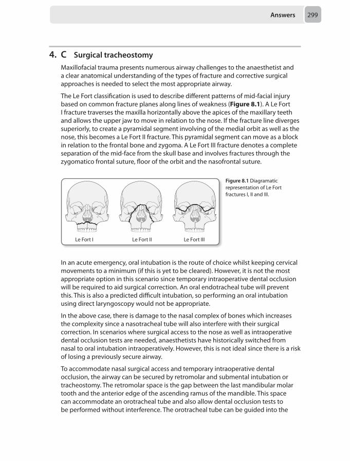
Answers 299
4. C Surgical tracheostomyMaxillofacial trauma presents numerous airway challenges to the anaesthetist and a clear anatomical understanding of the types of fracture and corrective surgical approaches is needed to select the most appropriate airway.
The Le Fort classification is used to describe different patterns of mid-facial injury based on common fracture planes along lines of weakness (Figure 8.1). A Le Fort I fracture traverses the maxilla horizontally above the apices of the maxillary teeth and allows the upper jaw to move in relation to the nose. If the fracture line diverges superiorly, to create a pyramidal segment involving of the medial orbit as well as the nose, this becomes a Le Fort II fracture. This pyramidal segment can move as a block in relation to the frontal bone and zygoma. A Le Fort III fracture denotes a complete separation of the mid-face from the skull base and involves fractures through the zygomatico frontal suture, floor of the orbit and the nasofrontal suture.
Le Fort I Le Fort II Le Fort III
Figure 8.1 Diagramatic representation of Le Fort fractures I, II and III.
In an acute emergency, oral intubation is the route of choice whilst keeping cervical movements to a minimum (if this is yet to be cleared). However, it is not the most appropriate option in this scenario since temporary intraoperative dental occlusion will be required to aid surgical correction. An oral endotracheal tube will prevent this. This is also a predicted difficult intubation, so performing an oral intubation using direct laryngoscopy would not be appropriate.
In the above case, there is damage to the nasal complex of bones which increases the complexity since a nasotracheal tube will also interfere with their surgical correction. In scenarios where surgical access to the nose as well as intraoperative dental occlusion tests are needed, anaesthetists have historically switched from nasal to oral intubation intraoperatively. However, this is not ideal since there is a risk of losing a previously secure airway.
To accommodate nasal surgical access and temporary intraoperative dental occlusion, the airway can be secured by retromolar and submental intubation or tracheostomy. The retromolar space is the gap between the last mandibular molar tooth and the anterior edge of the ascending ramus of the mandible. This space can accommodate an orotracheal tube and also allow dental occlusion tests to be performed without interference. The orotracheal tube can be guided into the
Chapter 8300
space following conventional oral intubation or pass through the space en route to the trachea with the aid of a Bonfils or flexible fibre-optic scope in difficult airways. The tube is usually held in place by ties to the adjacent tooth which in the above scenario is loose. It is also not ideal for patients expected to need prolonged postoperative ventilation.
Submental intubation involves performing conventional oral intubation then passing the endotracheal tube through a surgical incision in the floor of the mouth. This keeps the mouth and nose free for the surgical access and avoids the need to perform a tracheostomy. Compared to a tracheostomy, it also leaves a more aesthetic scar and carries less serious complications. It is not the ideal airway for prolonged postoperative ventilation however.
An awake, formal tracheostomy prior to surgery is the most appropriate airway for the above scenario since surgical field interfere is avoided and a safe, stable airway for postoperative ventilation is provided.
Curran J. Anaesthesia for facial trauma. Anaesth Intensive Care Med 2011; 12(8):354–359.Kellman R, Losquadro W. Comprehensive airway management of patients with maxillofacial trauma. Craniomaxillofac Trauma Reconstr 2008; 1(1):39–47.Jain G, Dhama S, Singh D. Role of retromolar intubation for airway management in trauma. Adv Trop Med Pub Health Int 2011; 1(1):21–32.
5. B Local anaesthetic (LA) toxicityThe rapid topping up of an epidural, following its prolonged use, is encumbered with the significant risk of reaching the threshold of local anaesthetic (LA) toxicity. Presentation can occur almost immediately up to an hour after LA administration. It classically begins with signs and symptoms of central nervous system (CNS) excitability followed by CNS depression and ultimately, cardiovascular instability. A wide range of arrhythmias may ensue, resulting in cardiac arrest. The other diagnoses should all be considered but, in light of her anaesthetic history during labour and systematic progression of signs and symptoms, LA toxicity is most likely and it should therefore be treated as such.
The Association of Anaesthetists of Great Britain & Ireland (AAGBI). Management of severe local anaesthetic toxicity. London: AAFGI, 2010.
6. D Simple analgesia, lumbar epidural PCEA with levobupivacaine/fentanyl mixtureGynaecological surgery presents a number of challenges, and the added aspects of gynaecological oncology may provide further problems to the anaesthetist, especially in the era of more radical surgery for pelvic disease.
Good quality analgesia tailored to the patient is of paramount importance. Chronic pain after abdominal hysterectomy may occur in up to 30% of patients, and poor perioperative pain control represents a modifiable risk factor. Interestingly, much of the pain seems to relate to the visceral trauma of surgery, as there is no difference in rates of chronic pain when comparing open and laparoscopic approaches to hysterectomy surgery.
Answers 301
Most recommendations for analgesia in hysterectomy involve categorising a patient’s risk of postoperative pain as high or low and then treating accordingly. Risk factors for postoperative pain are:
• Chronic pain conditions and ambulatory opioid use• Illicit intravenous drug abuse• Previous chemo/radiotherapy• Surgery with extensive tissue trauma
In a web review of procedure specific pain guidance (PROSPECT), recommendations in abdominal hysterectomy are for epidural analgesia for high risk patients, whereas in low risk, patients wound infiltration and opioids are deemed sufficient. Oral gabapentin is used by some for high risk patients, but side effects include sedation and dizziness, and thus this should not be given with other sedating agents. It is often started pre-emptively as a premedicant before induction. Ketamine infusions, usually in combination with PCA opioids have also been used with success in high risk chronic pain patients.
In ovarian adenocarcinoma, patients with epidural anaesthesia have been shown to have improved rates of survival at 3 and 5 years. This is postulated to relate to preservation of immune function by avoidance of anaesthetic and analgesic drugs allowing for greater immunosurveillance and tumour cell clearance at the time of surgery.
The key points with this patient are that she is obese, and suffers with respiratory disease. Of significance, there is some evidence that she may suffer with obstructive sleep apnoea (OSA). Given the presence of OSA, the ideal postoperative environment is the high dependency unit (HDU) especially if patient controlled opiates are to be used. Gabapentin should be avoided. Given the respiratory picture, removing or minimising the need for systemic opioids would be beneficial, therefore an epidural is the best choice, in the first instance a low dose mixture should be tried, and other opiates avoided. A multimodal approach to managing this patient's postoperative analgesia is required, therefore simple analgesia in combination with patient controlled epidural analgesia (PCEA) with a levobupivacaine/fentanyl mixture is the ideal choice of analgesia for this patient.
Morosan M, Popham P. Anaesthesia for gynaecological oncological surgery. Contin Educ Anaesth Crit Care Pain 2014; 14(2):63–68.
7. D Depressed zygomatic fractureAirway management decisions for mandibular fractures are difficult due to the numerous and often co-existing causes of the associated trismus. Trismus describes the inability to open the mouth and can occur as a result of pain, muscle spasm, swelling or any mechanical obstruction. During the preoperative assessment, pain associated with mouth opening may mask other underlying contributors to trismus and also limit the airway examination. It is therefore important to communicate with the surgeons if they have any concerns regarding the mechanics of mouth opening before induction.
With adequate depth of anaesthesia and neuromuscular blockade (as in the case above), it is expected that trismus secondary to purely pain will be reversed. In cases
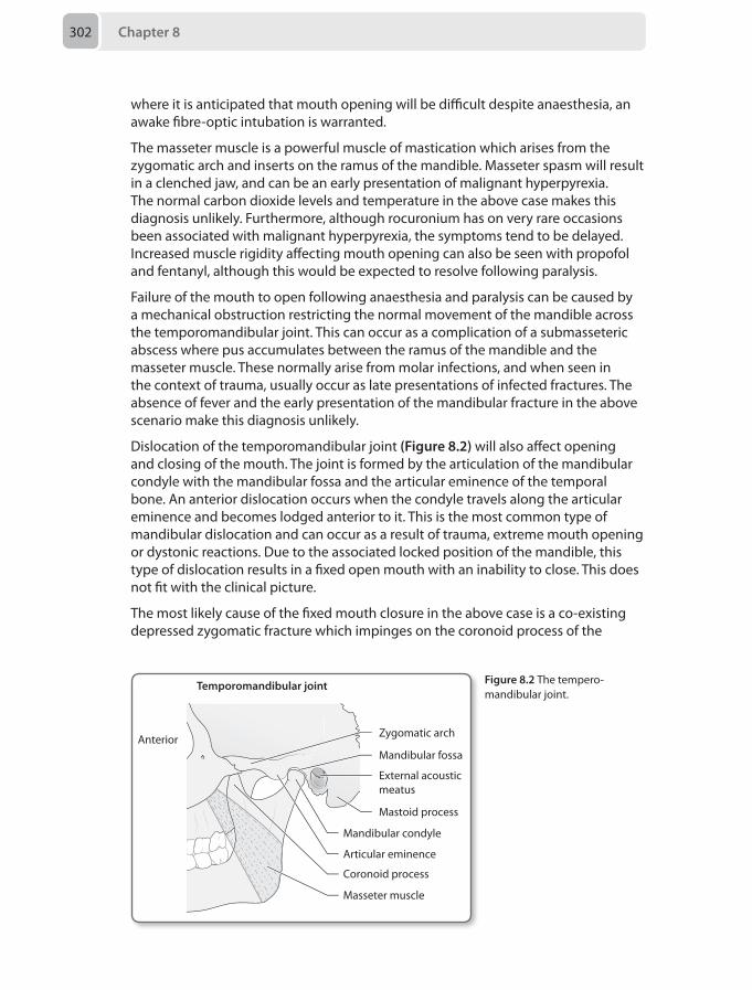
Chapter 8302
where it is anticipated that mouth opening will be difficult despite anaesthesia, an awake fibre-optic intubation is warranted.
The masseter muscle is a powerful muscle of mastication which arises from the zygomatic arch and inserts on the ramus of the mandible. Masseter spasm will result in a clenched jaw, and can be an early presentation of malignant hyperpyrexia. The normal carbon dioxide levels and temperature in the above case makes this diagnosis unlikely. Furthermore, although rocuronium has on very rare occasions been associated with malignant hyperpyrexia, the symptoms tend to be delayed. Increased muscle rigidity affecting mouth opening can also be seen with propofol and fentanyl, although this would be expected to resolve following paralysis.
Failure of the mouth to open following anaesthesia and paralysis can be caused by a mechanical obstruction restricting the normal movement of the mandible across the temporomandibular joint. This can occur as a complication of a submasseteric abscess where pus accumulates between the ramus of the mandible and the masseter muscle. These normally arise from molar infections, and when seen in the context of trauma, usually occur as late presentations of infected fractures. The absence of fever and the early presentation of the mandibular fracture in the above scenario make this diagnosis unlikely.
Dislocation of the temporomandibular joint (Figure 8.2) will also affect opening and closing of the mouth. The joint is formed by the articulation of the mandibular condyle with the mandibular fossa and the articular eminence of the temporal bone. An anterior dislocation occurs when the condyle travels along the articular eminence and becomes lodged anterior to it. This is the most common type of mandibular dislocation and can occur as a result of trauma, extreme mouth opening or dystonic reactions. Due to the associated locked position of the mandible, this type of dislocation results in a fixed open mouth with an inability to close. This does not fit with the clinical picture.
The most likely cause of the fixed mouth closure in the above case is a co-existing depressed zygomatic fracture which impinges on the coronoid process of the
Temporomandibular joint
Anterior Zygomatic arch
External acousticmeatus
Mastoid process
Mandibular fossa
Mandibular condyle
Articular eminence
Coronoid process
Masseter muscle
Figure 8.2 The tempero-mandibular joint.
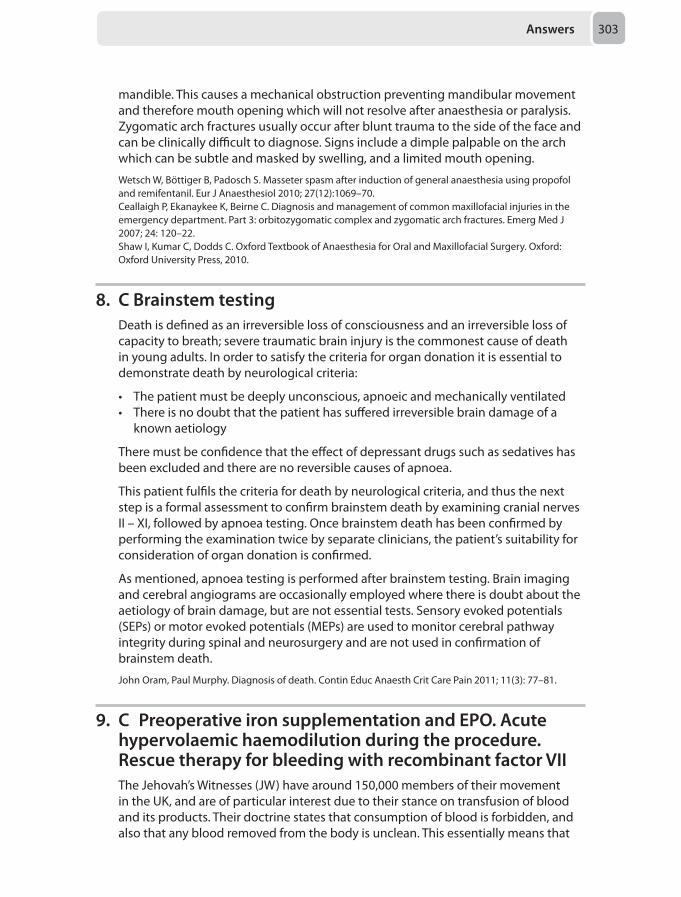
Answers 303
mandible. This causes a mechanical obstruction preventing mandibular movement and therefore mouth opening which will not resolve after anaesthesia or paralysis. Zygomatic arch fractures usually occur after blunt trauma to the side of the face and can be clinically difficult to diagnose. Signs include a dimple palpable on the arch which can be subtle and masked by swelling, and a limited mouth opening.
Wetsch W, Böttiger B, Padosch S. Masseter spasm after induction of general anaesthesia using propofol and remifentanil. Eur J Anaesthesiol 2010; 27(12):1069–70.Ceallaigh P, Ekanaykee K, Beirne C. Diagnosis and management of common maxillofacial injuries in the emergency department. Part 3: orbitozygomatic complex and zygomatic arch fractures. Emerg Med J 2007; 24: 120–22.Shaw I, Kumar C, Dodds C. Oxford Textbook of Anaesthesia for Oral and Maxillofacial Surgery. Oxford: Oxford University Press, 2010.
8. C Brainstem testingDeath is defined as an irreversible loss of consciousness and an irreversible loss of capacity to breath; severe traumatic brain injury is the commonest cause of death in young adults. In order to satisfy the criteria for organ donation it is essential to demonstrate death by neurological criteria:
• The patient must be deeply unconscious, apnoeic and mechanically ventilated• There is no doubt that the patient has suffered irreversible brain damage of a
known aetiology
There must be confidence that the effect of depressant drugs such as sedatives has been excluded and there are no reversible causes of apnoea.
This patient fulfils the criteria for death by neurological criteria, and thus the next step is a formal assessment to confirm brainstem death by examining cranial nerves II – XI, followed by apnoea testing. Once brainstem death has been confirmed by performing the examination twice by separate clinicians, the patient’s suitability for consideration of organ donation is confirmed.
As mentioned, apnoea testing is performed after brainstem testing. Brain imaging and cerebral angiograms are occasionally employed where there is doubt about the aetiology of brain damage, but are not essential tests. Sensory evoked potentials (SEPs) or motor evoked potentials (MEPs) are used to monitor cerebral pathway integrity during spinal and neurosurgery and are not used in confirmation of brainstem death.
John Oram, Paul Murphy. Diagnosis of death. Contin Educ Anaesth Crit Care Pain 2011; 11(3): 77–81.
9. C Preoperative iron supplementation and EPO. Acute hypervolaemic haemodilution during the procedure. Rescue therapy for bleeding with recombinant factor VIIThe Jehovah’s Witnesses (JW) have around 150,000 members of their movement in the UK, and are of particular interest due to their stance on transfusion of blood and its products. Their doctrine states that consumption of blood is forbidden, and also that any blood removed from the body is unclean. This essentially means that
Chapter 8304
the transfusion of blood or of major blood products is not accepted, and any forms of blood removal and storage are generally not permitted. Therefore a number of perioperative management plans must be instituted in this group of patients to reduce the risks of anaemic morbidity and mortality.
Preoperative
The aim should be to assess and optimise the haemoglobin concentration (Hb).
Thus, anaemia should be investigated and treated vigorously, and any medications which interfere with clotting, such as antiplatelets and non-steroidal anti-inflammatory drugs (NSAIDs), should be discontinued if possible. Recombinant erythropoietin (EPO) is useful in Jehovah’s witnesses and patients with anaemia who are also unable to accept transfusion. EPO should be given with iron, and if deficient or malnourished, B12 and folic acid, to prevent iron deficiency. Some regard intravenous iron as superior when used with EPO. If time allows, allogenic donation of blood could be considered, here a patient donates their own red cells for several weeks prior to surgery. This reduces the risks of infection and blood incompatibility, but will often be undertaken with EPO/ iron to stimulate regeneration and avoid anaemia. Unfortunately, as this involves storage of blood this is not often acceptable to Jehovah’s Witness patients.
Perioperative
It is important to remember that there are various techniques available other than just drug treatments and cell salvage. Surgically, large procedures can be staged and use of laser diathermy and haemostatic gels and glues may reduce blood loss. From an anaesthetic technique viewpoint, a good principle to follow is to avoid anything which increases venous pressure and thus blood loss, such as high PEEP/intrathoracic pressures or hypercapnia. Lowering systemic pressures, using deliberate hypotension can reduce blood loss, but this is offset by the risk to the patient’s physiological status. Similarly, regional techniques have been shown to reduce operative losses, but with neuraxial techniques, the risk of massive blood loss and subsequent coagulopathy and propensity to form an epidural haematoma has to be considered. General measures such as warming are mandatory to avoid coagulopathy.
Haemodilution
Acute normovolaemic haemodilution is the perioperative removal of whole blood, prior to the stage of surgery involving haemorrhage. This is replaced with crystalloid or colloid, to maintain normovolaemic status, and thus when bleeding does occur, the actual number of red cells lost per unit volume is lower. There is also the attractive option of returning whole blood with a normal composition of clotting factors and platelets at the end of the procedure to assist with haemostasis. Again, due to the removal and storage considerations, this is often unacceptable to Jehovah’s Witness patients. Acute hypervolaemic haemodilution is the dilution of the circulating blood as above, but without removal and storage This
Answers 305
technique is acceptable, clearly a balance has to be achieved between dilution for haemodynamics and reducing red cell loss, and avoiding coagulopathy. A reduction target in packed cell volume (PCV) of 30% was well tolerated in a study of Jehovah’s Witness patients.
Cell salvage
Widely accepted by Jehovah’s Witness patients, this involves the centrifugal separation, washing and re-suspension of red cells for transfusion. Importantly, the circuit should remain in continuity with the patient to avoid the objections over removal and storage. The washing stage produces red cells and is not designed to recover platelets or clotting factors. There are several cautions to the use of the cell saver, and these usually relate to the re-transfusion of undesirable agents e.g. bacteria or tumour cells. Thus, use in sepsis, tumour beds or direct suction of amniotic fluid is avoided.
Drugs/blood fractions/recombinant factors
Antifibrinolytics such as tranexamic acid are useful and acceptable to Jehovah’s Witness patients. Fractionated components of blood such as fresh frozen plasma (FFP), are usually not acceptable, although this is not universally the case. However, the acceptance of recombinant factors is widespread such as with the established use of recombinant Factor VII (NovoSeven). This is likely also the case with other recombinant protein factors, but not certain with all newer compounds such as fibrinogen concentrate. Indeed, the only way to be sure, is to ask the patient explicitly about all eventualities, including their views on transfusion if all else fails and they are close to death. This should be witnessed independently, and not by family or Jehovah’s Witness advocates. Some Trusts now have useful and extensive tick-box consent forms listing all agents especially for those with objections to conventional allogenic transfusion.
The most important principle in clinical practice, also relevant in the examination situation, is the individual nature of the patient’s decision. The patient has the choice over what they are willing to accept, and this can vary in many cases, especially in the grey area of fractionated products/recombinant agents. The discussion must obviously take place in advance, and should be open and frank about the risks involved including death or prolonged intensive care admission. Ideally, discussions should take place in private, as patients may express different views in front of relatives/other community members.
The first answer stems, A and B feature pre-donation, which is not an acceptable practice for Jehovah’s Witness patients. D and E list rescue strategies involving blood fractions not normally acceptable, but as aforementioned this does vary. Stem E also features intraoperative normovolaemic dilution, which may also not be acceptable as it involves blood storage. C remains as the combination of therapies most likely to be found acceptable by Jehovah’s Witness patients.
Milligan L J, Bellamy M C. Anaesthesia and Critical Care of Jehovah’s Witnesses. Contin Educ Anaesth Crit Care Pain 2004;4(2): 35–39.
Chapter 8306
10. B Open appendicectomyThe transversus abdominis plane (TAP) block can provide good perioperative analgesia when used for the appropriate operation. Its use can decrease opioid consumption allowing patients to breath comfortably and speed mobilisation and early discharge.
Innervation of the abdominal wall derives from the anterior rami of the lower six thoracic nerves and the first lumbar nerve (T7-L1).
These nerves enter the transversus abdominis facial plane between the internal oblique and transversus abdominis muscles.
The TAP block is essentially injecting a large volume of local anaesthetic (LA) in the transversus abdominis plane targeting the T7 to L1 nerves. This block will provides adequate analgesia for the abdominal wall but not for the abdominal viscera.
Although the early studies were able show blocks extending from T7 to L1, other and more recent studies have unable to demonstrate a spread higher than T10. Therefore, TAP blocks are currently recommended for infraumbilical surgery.
The injection can be done unilaterally or bilaterally depending on the type of the surgery.
Unilateral block: Open appendicectomy and hernia repair below the umbilicus.
Bilateral block: abdominal hysterectomy, radical prostatectomy, Caesarean section, midline incision and laparoscopic operations.
The addition of subcostal TAP block can achieve a higher block up to T7 and can successfully be used with posterior TAP block for supraumbilical procedures such as cholecystectomies.
In the above question, a right-sided TAP block can provide adequate analgesia for open appendicectomy procedures. The other options require either bilateral TAP block or an additional sub-costal block to achieve reliable analgesia.
The TAP block can be performed either by using ultrasound or a landmark technique. The landmark technique, which was originally described by McDonnell et al, access to the transversus abdominis plane is achieved through the triangle of Petit. The iliac crest bound this triangular area inferiorly, anteriorly by the external oblique muscle and posteriorly by the latissimus dorsi muscle (see Figure 8.3). After passing through the skin, the needle should be advanced until two pops are felt, at which point the needle lies in the transversus abdominis plane, immediately superficial to the transversus abdominis muscle (see figure 8.3). A minimum of 20 mL LA is injected in each side after aspiration.
In the ultrasound (US) technique, a linear high frequency probe is placed transversely in the mid-axillary line between the iliac crest and the 12th rib.
The muscle layers and the transversus abdominis plane can easily be identified in this area. A short bevel 100 mm needle is advanced with an in-plane approach until reaches this plane, when 20 mL of LA is injected once again superficial to the transversus

Answers 307
abdominis muscle. The LA spread in the plane will form an elliptical hypoechoic shape between the transversus abdominis and the internal oblique muscles.
McDonnell JG, O’Donnell B, Curley G, et al. The analgesic effect of transversus abdominis plane block after abdominal surgery. Anaesth Analg 2007; 104(1):193–197.Tran TMN, Ivanusic JJ, Hebbard P, Barrington MJ. Determination of spread of injectate after ultrasound guided transversus abdominis plane block: A cadaveric study. Br J Anaesth 2009; 102(1):123–127.
11. B Double handed cricoid pressure application without the collar
The use of cricoid pressure for rapid sequence induction (RSI) aims to prevent regurgitation of gastric content, although the true aims of an RSI are to rapidly and safely secure a definitive airway. The utility and necessity of cricoid pressure has a very questionable evidence base currently, with many practitioners doing away with cricoid pressure completely as it is known to distort the view at direct laryngoscopy. The pragmatic approach would be to utilise cricoid pressure carefully and advocate early release if laryngoscopic views are worsened by it.
Although the debate rages on, and is likely to do so for some time, the question asked here is how to apply cricoid pressure assuming it will be used as part of your standard RSI technique for a patient with an unstable cervical spine, therefore not applying cricoid pressure at all is an incorrect answer. An awake fibreoptic intubation is inappropriate in a patient with a reduced GCS and agitation, thus is best avoided.
The aims here should be to ensure cervical spine stability and control without sacrificing a safe RSI technique. Therefore removing the collar and having an assistant provide manual in-line stabilisation (MILS) whilst another providing cricoid pressure is important. Single-handed cricoid pressure is suitable should the posterior component of a cervical collar be left in situ as this has been demonstrated to produce minimal vertebral movement. If the collar is removed completely, single-handed cricoid pressure leaves the patient at risk of vertebral instability. However, the safest way to apply cricoid pressure would be with a bimanual approach with collar removed completely and MILS. This is thought to provide better laryngoscopic views, maintain the integrity of cervical flexion, and avoid excessive pressure being applied.
Cranshaw J. Airway management after major trauma. Contin Educ Anaesth Crit Care Pain 2006; 6(3):124–127.Lockey D.J, Crewdson K, HM Lossiusm HM. Pre-hospital anaesthesia: the same but different. Br J Anaesth 2014; 113(2):211–219.
Latissimusdorsi
Triangle ofPetit
Externaloblique
Iliac crest
Latissimusdorsi
Externaloblique
Triangle ofPetitIliac crest
Figure 8.3 Location of the Triangle of Petit for a TAP block.
Chapter 8308
12. E Commence a titrated dobutamine infusion starting at 5 µg/kg/min
The patient described is in septic shock despite fluid resuscitation and high dose vasopressors. The profound degree of shock is evidenced by the lactate, Scvo2 levels and carbon dioxide gap, all of which are useful measures of global oxygen supply demand imbalance.
The adequacy of fluid resuscitation is evidenced by the ~5% increase in stroke volume index (SVI) following the rapid fluid bolus. As a general guide, a ≤ 10% increase in SVI is considered to be fluid unresponsive. A ≥ 15% increase in SVI is considered fluid responsive and should prompt the consideration of a further fluid bolus until the response is ≤ 10% (SVI maximisation). Changes in the 10–15% range are equivocal and further fluid boluses should balance the risks and benefits. However, the stroke volume index and hence cardiac index (HR x SVI) is low, despite optimal heart rate and rhythm, which is consistent with significant myocardial contractility impairment. This may be due to septic cardiomyopathy and/or ischaemic heart disease. The highly sensitive cardiac troponin T (hs-cTropT) elevation does not differentiate between these two diagnoses. A 12-lead ECG with acute/dynamic changes consistent with ischaemia/infarction in a specific coronary territory, and echocardiogram showing new regional wall motion abnormalities would be highly suggestive of a type 1 myocardial infarction, and should be performed. However, even if the clinical evidence points towards this diagnosis, the role and optimal timing of acute percutaneous coronary intervention and the use of mechanical support are highly controversial. The best immediate management therefore is to commence a positive inotrope, such as dobutamine. There is no definitive trial evidence to support the choice of one positive inotrope over any other.
In this scenario, the threshold for considering packed red blood cell transfusion should be < 70 g/L and, as the patient is euvolaemic, the risk of fluid overload is very high hence pRBC transfusion is not indicated. There is no value in increasing the mean arterial pressures further as this is likely to have a negative impact on myocardial function and worsen rather than improve both coronary and global perfusion. The role of ‘stress dose’ glucocorticoid therapy in this scenario as a treatment for functional hypoadrenalism remains controversial. It may be worth considering a therapeutic trial depending upon the patient’s response to the positive inotrope.
Somasundaram K, Ball J. Medical emergencies: atrial fibrillation and myocardial infarction. Anaesthesia 2013; 68(S1):84–101.
13. E Commence renal replacement therapyIn patients with normal renal function, on-pump cardiac surgery is associated with a 10-30% risk of acute renal injury and a < 5% risk of needing acute renal replacement therapy. These risks are significantly increased in patients with pre-existing renal impairment. Other peri-procedural risk factors include on versus off-pump, longer bypass times, haemodynamic instability, need for high dose or protracted inotropes and/or vasopressors.
Answers 309
In the scenario described, the patient has clearly developed acute kidney injury (AKI), with oliguria, a metabolic acidosis, hyperkalaemia and uraemia. There is no place for low dose dopamine or diuretics in either the prevention or management of AKI. Given the rate of evolution of this patient’s metabolic derangement and apparently normal renal perfusion, temporising therapy to improve the acidosis and reduce the serum potassium are unlikely to prevent the need for renal replacement therapy, hence this is the best answer.
There are a number of considerations to take in to account when considering renal replacement therapy in the acute setting:
• Most centres use bicarbonate based fluids for renal replacement therapy • Although there is no universally agreed thresholds for commencing renal
replacement therapy in ARF a reasonable suggestion would be:• Hyperkalaemia (K+ > 6.5 mmol/L or o K+ > 5.5 mmol/L and rapidly rising at
> 0.25 mmol/hr for 2 or more hours). • Correction of severe/unresolving acidosis (pH < 7.1); acidosis associated with
cardiovascular compromise (end organ hypoperfusion)/high vasoactive drug requirements (noradrenaline > 0.5 µg/kg/min / dobutamine > 10 µg/kg/min).
• Uraemia (urea > 40 mmol/L or rising by > 12 mmol/24 hrs)• Fluid overload causing severe hypertension and/or problematic oedema (e.g.
abdominal compartment syndrome) and/or contributing to hypoxaemia / poor lung compliance.
• There is no evidence to support any specific modality over another in this setting • The use of bolus insulin and dextrose mixtures is a poor practice as it is associated
with a very high incidence of acute, severe dysglycaemia and rapid rebound hyperkalaemia. If temporisation of hyperkalaemia is required, continuous infusions of insulin and dextrose are safer and more effective. Adjunctive use of nebulised salbutamol and intravenous bicarbonate can also be very helpful. If cardiac toxicity is evident, acute protection is afforded by a slow bolus of intravenous calcium either as gluconate or chloride.
Bellomo R, ChapmanM, et al. Low-dose dopamine in patients with early renal dysfunction: a placebo-controlled randomised trial. Australian and New Zealand Intensive Care Society (ANZICS) Clinical Trials Group. Lancet 2000; 356(9248):2139–2143.Karajala V, Mansour W, et al. Diuretics in acute kidney injury. Minerva Anestesiol 2009; 75(5): 251–257.Palevsky P M. Renal replacement therapy in acute kidney injury. Adv Chron Kidney Dis 2013; 20(1):76–84.
14. D Contact the transplant coordinatorWhen organ transplantation began all organs were retrieved from patients immediately after cardiorespiratory arrest. In 1976 brainstem testing allowed retrieval of heart-beating donor organs which has become the principal source of organs for transplantation for the last 25–30 years. Non heart-beating organ donation (NHBOD) however is re-gaining popularity. This is partly because the demand for organs is increasing. In addition, the number of heart-beating donors is declining for two reasons: fewer younger people are dying as a result of severe injury or catastrophic cerebrovascular events, and improvements in diagnosis and management of severe brain injuries mean that fewer fulfill the brainstem testing criteria.

Chapter 8310
Suitability criteria for consideration for NHBOD include:
• A decision has been made to withdraw treatment• They are expected to die within 2 hours• They have organs suitable for transplantation
The only absolute contra-indications are human immunodeficiency virus (unless the recipient is also HIV positive) or Creutzfeldt–Jakob disease. The final decision regarding suitability is made by the retrieval and transplant surgeons.
Comprehensive guidance from the Intensive Care Society and General Medical Council exist. The decision to withdraw treatment must be separate from the one to donate organs. A protocol may be useful to dictate a withdrawal of care approach, which ensures the interests of the dying patient remain the primary focus. The family should first be made aware that further treatment is not in the patient’s best interests. They can then be approached about donation after the transplant coordinator has been contacted, the organ donor register checked and consent obtained from the coroner. The coordinator and senior clinician will ideally approach the family together.
The key consideration here is what is deemed to be in the patient’s best interests and does not cause harm or distress to them or their family. If they have expressed a wish to donate organs then blood testing and maintenance of life-sustaining treatment is acceptable (including escalation of treatment). Similarly delaying withdrawal or moving the patient to facilitate transplantation is considered to be in the patient's best interests if their wish to donate is known. Systemic heparinisation, femoral cannulation and CPR are not acceptable as they have a significant risk of harm.
Withdrawal of active treatment should not vary from local practice because organ donation is being considered and should proceed in accordance with the usual practice of the critical care unit. Commencing treatment to ensure lack of distress prior to extubation, termination of ventilation or cardiovascular support is the usual practice. After withdrawal of care the patient may continue to have a cardiac output for some time. If the patient is hypoxic or hypotensive for this period of time then the same physiological conditions that occur in warm ischaemic time are present. Therefore if the process of dying lasts more than 2–3 hours the retrieval may be abandoned.
The ethical issues are numerous and include: the withdrawal of treatment, appropriateness of ongoing treatment to facilitate donation, the method of diagnosis of death and the time left between cessation of cardiorespiratory function and confirmation of death.
Returning to the scenario above, the next step (which is what the question is asking for) is to contact the transplant coordinator. They will analyse the case and decide whether or not it is suitable to continue before putting the family in a position to make a decision. Breaking the news to the family that their relative will not survive and approaching the topic of organ donation should be separate conversations if possible. In addition the transplant coordinator should be present to answer logistical questions you may not be aware of.
Intensive Care Society Working Group on Organ and Tissue Donation. Guidelines for adult organ and tissue donation. London: Intensive Care Society, 2005.Ridley S. UK guidance for non-heart-beating donation, Br J Anaesth 2005; 95(5):592–595.
Answers 311
15. B Examine the respiratory systemObesity is a health epidemic facing Western countries. Obesity is defined as a body mass index (BMI) > 30 kg/m2, ‘morbidly’ obese a BMI > 35 and ‘super morbidly’ obese> 55 kg/m2.. Recent figures suggest that up to 23% of men and 25% of women in the UK are obese.
Bariatric surgery presents many challenges intra-and postoperatively. Management of the patient’s co-morbidities is often a reason for high-dependency care postoperatively as in this case. Complications are increased in this group and this case explores the differential diagnosis of an immediate postoperative complication.
The list of differential diagnoses (which may not be exhaustive) include:
• Airway obstruction due to obstructive sleep apnoea (OSA), sedation, carbon dioxide narcosis, anaphylaxis
• Breathing• Hypoventilation resulting in hypercapnic respiratory failure
– OSA – Residual anaesthesia or long-acting analgesia – Abdominal splinting with residual pneumoperitoneum or sub-optimal
position in bed• Aspiration of gastric contents at induction or extubation• Pneumothorax as a result of the central venous cannulation or laparoscopic
surgery• Pulmonary embolism• Negative pressure pulmonary oedema• Gas embolism
• Circulation• Perioperative myocardial ischemic event• Postoperative bleeding (may be seen in drain output)
• Disability• Residual sedative drugs• Focal deficit due to stroke• Global deficit due to hypoperfusion (e.g. relative hypotension or prolonged
reverse-Trendelenburg position)• Exposure
• Hypoglycemia• Hypothermia• Pain (not only operative, e.g. long standing back pain)• Urinary retention
The best way to approach a complex patient is perform a rapid examination looking to exclude some of the dangerous causes above. ‘Agitated’ implies that the airway is patent for now and with a grade three laryngoscopy (in the optimum conditions present in theatre) caution and planning is required in order to re-intubate. Hypercapnic respiratory failure is her main complaint and the focus of management should be to find a reversible cause of this deterioration in order to prevent re-intubation. This could potentially avoid significant morbidity and mortality associated with a prolonged ventilatory period postoperatively.
Chapter 8312
The question requests the next immediate step and therefore a careful examination of the respiratory system would be of great use. Non-invasive ventilation (NIV) could convert an undiagnosed simple pneumothorax (caused by a difficult central line insertion as in this case) to a tension pneumothorax and examination findings suggestive of this diagnosis would enable rapid decompression and resolution of the acute deterioration. Aspiration of gastric contents or pulmonary oedema may also be appreciated on clinical examination. As with all sick patients checking the electrocardiogram, the surgical drain output, the blood glucose level, the degree of residual neuromuscular blockade and the temperature are all part of the initial survey.
Clinical examination should not be forgotten in the intensive care unit as it guides further investigation and management in a more focused and efficient manner.
Sabharwal A. Anaesthesia for bariatric surgery, Contin Educ Anaesth Crit Care Pain 2010; 10(4):99–103.
16. B Full blood countThis scenario describes a case of Heparin Induced Thrombocytopenia (HIT), which is seen in 2.5% of patients treated with unfractionated heparin and 0.1% of those on low molecular weight heparin. It presents with reduced platelets, arterial and venous thrombosis (30%), a systemic reaction consisting of anaphylactoid symptoms (25%) and a skin rash, which represents dermal ischemia and necrosis as a result of emboli (4%).The syndrome is further classified into two types.
Type 1 HIT is a benign decrease in platelet numbers, which appears to be a physical interaction causing platelet aggregation. It results in a mild drop in the platelet count and may occur hours after treatment is commenced and usually recovers once heparin is stopped without any intervention. It is not associated with an increased risk of thrombosis.
Type 2 HIT is an immune-modulated process caused by formation of Immunoglobulin G (IgG) or rarely IgM antibodies, against heparin bound to a protein called Platelet Factor 4 (PF4). The onset is often delayed (day 5–10 after commencing treatment) but may present within hours if the patient has had previous exposure to heparin. The tail of the antibody binds to the Fcylla receptor on the platelet surface resulting in activation, aggregation, clot formation and a consumptive decrease in numbers. The platelet count usually drops below 50% of baseline values and a pro-thrombotic state occurs and results in arterial and venous thrombi. The antibodies persist in the plasma for 2–3 months.
The diagnosis of HIT is a 3-stage process. A scoring system is used to identify patients requiring further testing. This is called the 4T score and has been validated by Wakentin and Heddle in 2003. A low score (0–3 out of 8) has a negative predictive value of 0.998 whereas an intermediate (4–5) or high (5–8) score has a positive predictive value of 0.14 and 0.68 respectively, thus warranting further investigation (see Table 8.2).
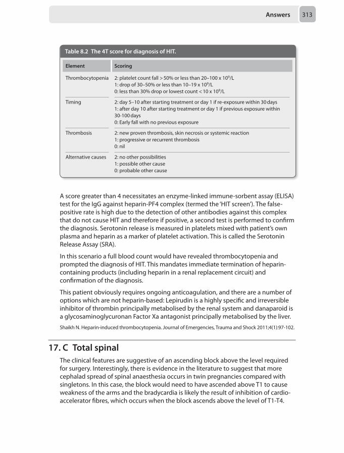
Answers 313
A score greater than 4 necessitates an enzyme-linked immune-sorbent assay (ELISA) test for the IgG against heparin-PF4 complex (termed the ‘HIT screen’). The false-positive rate is high due to the detection of other antibodies against this complex that do not cause HIT and therefore if positive, a second test is performed to confirm the diagnosis. Serotonin release is measured in platelets mixed with patient’s own plasma and heparin as a marker of platelet activation. This is called the Serotonin Release Assay (SRA).
In this scenario a full blood count would have revealed thrombocytopenia and prompted the diagnosis of HIT. This mandates immediate termination of heparin-containing products (including heparin in a renal replacement circuit) and confirmation of the diagnosis.
This patient obviously requires ongoing anticoagulation, and there are a number of options which are not heparin-based: Lepirudin is a highly specific and irreversible inhibitor of thrombin principally metabolised by the renal system and danaparoid is a glycosaminoglycuronan Factor Xa antagonist principally metabolised by the liver.
Shaikh N. Heparin-induced thrombocytopenia. Journal of Emergencies, Trauma and Shock 2011;4(1):97-102.
17. C Total spinalThe clinical features are suggestive of an ascending block above the level required for surgery. Interestingly, there is evidence in the literature to suggest that more cephalad spread of spinal anaesthesia occurs in twin pregnancies compared with singletons. In this case, the block would need to have ascended above T1 to cause weakness of the arms and the bradycardia is likely the result of inhibition of cardio-accelerator fibres, which occurs when the block ascends above the level of T1-T4.
Table 8.2 The 4T score for diagnosis of HIT.
Element Scoring
Thrombocytopenia 2: platelet count fall > 50% or less than 20–100 x 109/L1: drop of 30–50% or less than 10–19 x 109/L0: less than 30% drop or lowest count < 10 x 109/L
Timing 2: day 5–10 after starting treatment or day 1 if re-exposure within 30 days1: after day 10 after starting treatment or day 1 if previous exposure within 30-100 days0: Early fall with no previous exposure
Thrombosis 2: new proven thrombosis, skin necrosis or systemic reaction1: progressive or recurrent thrombosis0: nil
Alternative causes 2: no other possibilities1: possible other cause0: probable other cause
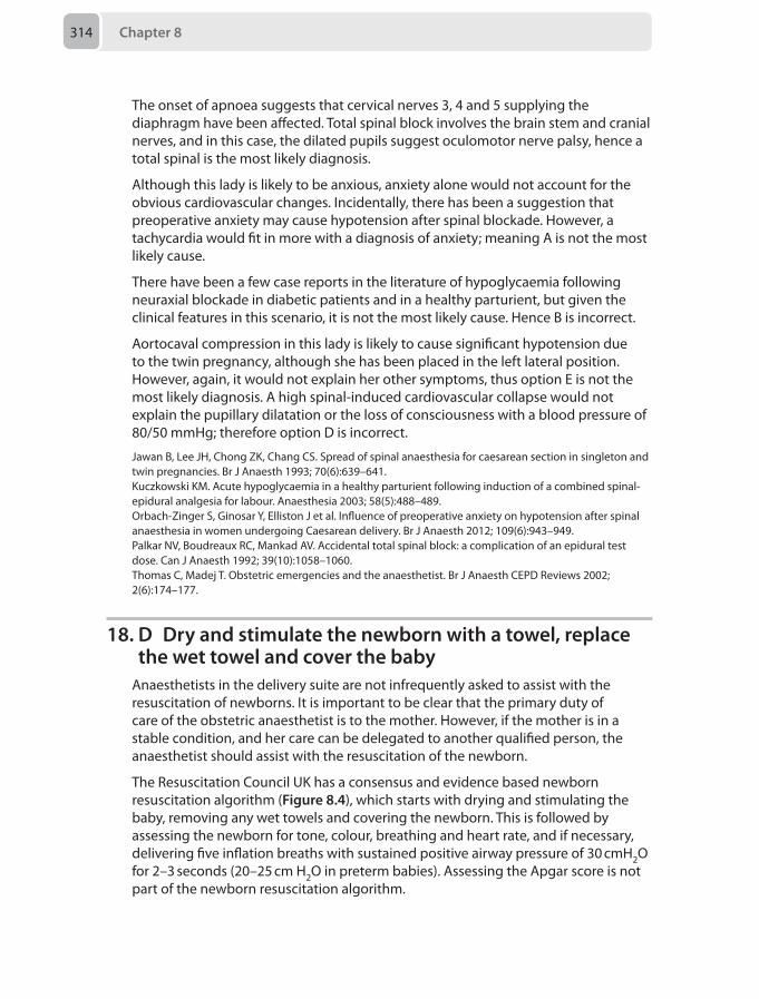
Chapter 8314
The onset of apnoea suggests that cervical nerves 3, 4 and 5 supplying the diaphragm have been affected. Total spinal block involves the brain stem and cranial nerves, and in this case, the dilated pupils suggest oculomotor nerve palsy, hence a total spinal is the most likely diagnosis.
Although this lady is likely to be anxious, anxiety alone would not account for the obvious cardiovascular changes. Incidentally, there has been a suggestion that preoperative anxiety may cause hypotension after spinal blockade. However, a tachycardia would fit in more with a diagnosis of anxiety; meaning A is not the most likely cause.
There have been a few case reports in the literature of hypoglycaemia following neuraxial blockade in diabetic patients and in a healthy parturient, but given the clinical features in this scenario, it is not the most likely cause. Hence B is incorrect.
Aortocaval compression in this lady is likely to cause significant hypotension due to the twin pregnancy, although she has been placed in the left lateral position. However, again, it would not explain her other symptoms, thus option E is not the most likely diagnosis. A high spinal-induced cardiovascular collapse would not explain the pupillary dilatation or the loss of consciousness with a blood pressure of 80/50 mmHg; therefore option D is incorrect.
Jawan B, Lee JH, Chong ZK, Chang CS. Spread of spinal anaesthesia for caesarean section in singleton and twin pregnancies. Br J Anaesth 1993; 70(6):639–641.Kuczkowski KM. Acute hypoglycaemia in a healthy parturient following induction of a combined spinal-epidural analgesia for labour. Anaesthesia 2003; 58(5):488–489.Orbach-Zinger S, Ginosar Y, Elliston J et al. Influence of preoperative anxiety on hypotension after spinal anaesthesia in women undergoing Caesarean delivery. Br J Anaesth 2012; 109(6):943–949.Palkar NV, Boudreaux RC, Mankad AV. Accidental total spinal block: a complication of an epidural test dose. Can J Anaesth 1992; 39(10):1058–1060.Thomas C, Madej T. Obstetric emergencies and the anaesthetist. Br J Anaesth CEPD Reviews 2002; 2(6):174–177.
18. D Dry and stimulate the newborn with a towel, replace the wet towel and cover the baby
Anaesthetists in the delivery suite are not infrequently asked to assist with the resuscitation of newborns. It is important to be clear that the primary duty of care of the obstetric anaesthetist is to the mother. However, if the mother is in a stable condition, and her care can be delegated to another qualified person, the anaesthetist should assist with the resuscitation of the newborn.
The Resuscitation Council UK has a consensus and evidence based newborn resuscitation algorithm (Figure 8.4), which starts with drying and stimulating the baby, removing any wet towels and covering the newborn. This is followed by assessing the newborn for tone, colour, breathing and heart rate, and if necessary, delivering five inflation breaths with sustained positive airway pressure of 30 cmH2O for 2–3 seconds (20–25 cm H2O in preterm babies). Assessing the Apgar score is not part of the newborn resuscitation algorithm.
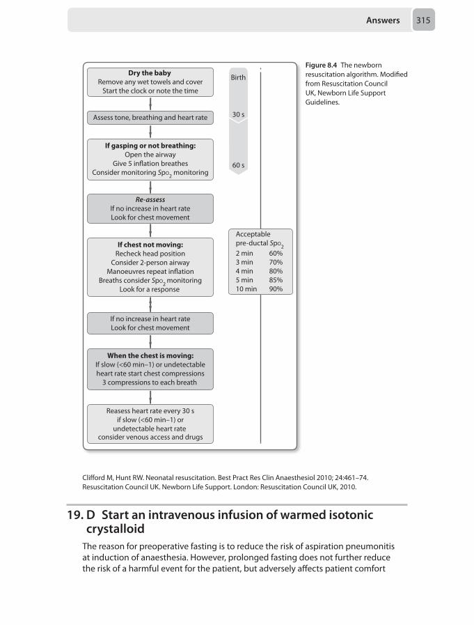
Answers 315
Dry the baby Remove any wet towels and cover
Start the clock or note the time
Assess tone, breathing and heart rate
If gasping or not breathing:Open the airway
Give 5 in�ation breathesConsider monitoring SpO2 monitoring
Re-assessIf no increase in heart rateLook for chest movement
If chest not moving:Recheck head position
Consider 2-person airwayManoeuvres repeat in�ation
Breaths consider SpO2 monitoringLook for a response
If no increase in heart rateLook for chest movement
When the chest is moving:If slow (<60 min–1) or undetectableheart rate start chest compressions
3 compressions to each breath
Reasess heart rate every 30 sif slow (<60 min–1) or
undetectable heart rate consider venous access and drugs
Acceptablepre-ductal SpO22 min 60%3 min 70%4 min 80%5 min 85%10 min 90%
Birth
60 s
30 s
Figure 8.4 The newborn resuscitation algorithm. Modified from Resuscitation Council UK, Newborn Life Support Guidelines.
Clifford M, Hunt RW. Neonatal resuscitation. Best Pract Res Clin Anaesthesiol 2010; 24:461–74. Resuscitation Council UK. Newborn Life Support. London: Resuscitation Council UK, 2010.
19. D Start an intravenous infusion of warmed isotonic crystalloid
The reason for preoperative fasting is to reduce the risk of aspiration pneumonitis at induction of anaesthesia. However, prolonged fasting does not further reduce the risk of a harmful event for the patient, but adversely affects patient comfort
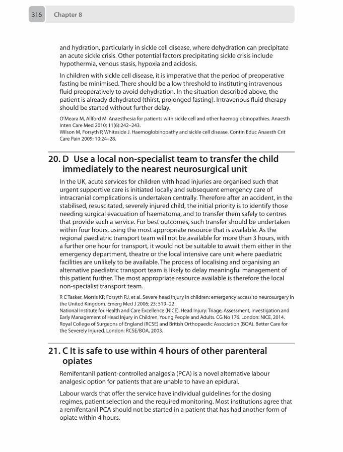
Chapter 8316
and hydration, particularly in sickle cell disease, where dehydration can precipitate an acute sickle crisis. Other potential factors precipitating sickle crisis include hypothermia, venous stasis, hypoxia and acidosis.
In children with sickle cell disease, it is imperative that the period of preoperative fasting be minimised. There should be a low threshold to instituting intravenous fluid preoperatively to avoid dehydration. In the situation described above, the patient is already dehydrated (thirst, prolonged fasting). Intravenous fluid therapy should be started without further delay.
O’Meara M, Allford M. Anaesthesia for patients with sickle cell and other haemoglobinopathies. Anaesth Inten Care Med 2010; 11(6):242–243.Wilson M, Forsyth P, Whiteside J. Haemoglobinopathy and sickle cell disease. Contin Educ Anaesth Crit Care Pain 2009; 10:24–28.
20. D Use a local non-specialist team to transfer the child immediately to the nearest neurosurgical unit
In the UK, acute services for children with head injuries are organised such that urgent supportive care is initiated locally and subsequent emergency care of intracranial complications is undertaken centrally. Therefore after an accident, in the stabilised, resuscitated, severely injured child, the initial priority is to identify those needing surgical evacuation of haematoma, and to transfer them safely to centres that provide such a service. For best outcomes, such transfer should be undertaken within four hours, using the most appropriate resource that is available. As the regional paediatric transport team will not be available for more than 3 hours, with a further one hour for transport, it would not be suitable to await them either in the emergency department, theatre or the local intensive care unit where paediatric facilities are unlikely to be available. The process of localising and organising an alternative paediatric transport team is likely to delay meaningful management of this patient further. The most appropriate resource available is therefore the local non-specialist transport team.
R C Tasker, Morris KP, Forsyth RJ, et al. Severe head injury in children: emergency access to neurosurgery in the United Kingdom. Emerg Med J 2006; 23: 519–22.National Institute for Health and Care Excellence (NICE). Head Injury: Triage, Assessment, Investigation and Early Management of Head Injury in Children, Young People and Adults. CG No 176. London: NICE, 2014.Royal College of Surgeons of England (RCSE) and British Orthopaedic Association (BOA). Better Care for the Severely Injured. London: RCSE/BOA, 2003.
21. C It is safe to use within 4 hours of other parenteral opiates
Remifentanil patient-controlled analgesia (PCA) is a novel alternative labour analgesic option for patients that are unable to have an epidural.
Labour wards that offer the service have individual guidelines for the dosing regimes, patient selection and the required monitoring. Most institutions agree that a remifentanil PCA should not be started in a patient that has had another form of opiate within 4 hours.
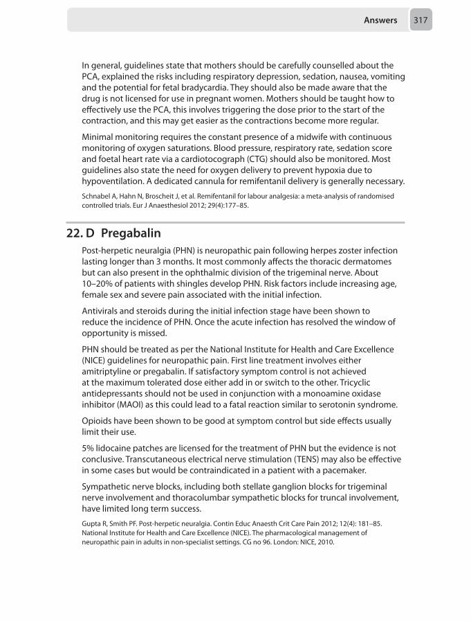
Answers 317
In general, guidelines state that mothers should be carefully counselled about the PCA, explained the risks including respiratory depression, sedation, nausea, vomiting and the potential for fetal bradycardia. They should also be made aware that the drug is not licensed for use in pregnant women. Mothers should be taught how to effectively use the PCA, this involves triggering the dose prior to the start of the contraction, and this may get easier as the contractions become more regular.
Minimal monitoring requires the constant presence of a midwife with continuous monitoring of oxygen saturations. Blood pressure, respiratory rate, sedation score and foetal heart rate via a cardiotocograph (CTG) should also be monitored. Most guidelines also state the need for oxygen delivery to prevent hypoxia due to hypoventilation. A dedicated cannula for remifentanil delivery is generally necessary.
Schnabel A, Hahn N, Broscheit J, et al. Remifentanil for labour analgesia: a meta-analysis of randomised controlled trials. Eur J Anaesthesiol 2012; 29(4):177–85.
22. D PregabalinPost-herpetic neuralgia (PHN) is neuropathic pain following herpes zoster infection lasting longer than 3 months. It most commonly affects the thoracic dermatomes but can also present in the ophthalmic division of the trigeminal nerve. About 10–20% of patients with shingles develop PHN. Risk factors include increasing age, female sex and severe pain associated with the initial infection.
Antivirals and steroids during the initial infection stage have been shown to reduce the incidence of PHN. Once the acute infection has resolved the window of opportunity is missed.
PHN should be treated as per the National Institute for Health and Care Excellence (NICE) guidelines for neuropathic pain. First line treatment involves either amitriptyline or pregabalin. If satisfactory symptom control is not achieved at the maximum tolerated dose either add in or switch to the other. Tricyclic antidepressants should not be used in conjunction with a monoamine oxidase inhibitor (MAOI) as this could lead to a fatal reaction similar to serotonin syndrome.
Opioids have been shown to be good at symptom control but side effects usually limit their use.
5% lidocaine patches are licensed for the treatment of PHN but the evidence is not conclusive. Transcutaneous electrical nerve stimulation (TENS) may also be effective in some cases but would be contraindicated in a patient with a pacemaker.
Sympathetic nerve blocks, including both stellate ganglion blocks for trigeminal nerve involvement and thoracolumbar sympathetic blocks for truncal involvement, have limited long term success.
Gupta R, Smith PF. Post-herpetic neuralgia. Contin Educ Anaesth Crit Care Pain 2012; 12(4): 181–85.National Institute for Health and Care Excellence (NICE). The pharmacological management of neuropathic pain in adults in non-specialist settings. CG no 96. London: NICE, 2010.
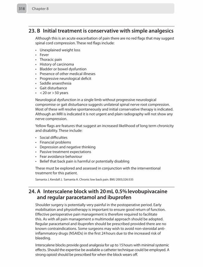
Chapter 8318
23. B Initial treatment is conservative with simple analgesicsAlthough this is an acute exacerbation of pain there are no red flags that may suggest spinal cord compression. These red flags include:
• Unexplained weight loss• Fever• Thoracic pain• History of carcinoma• Bladder or bowel dysfuntion• Presence of other medical illneses• Progressive neurological deficit• Saddle anaesthesia• Gait disturbance• < 20 or > 50 years
Neurological dysfunction in a single limb without progressive neurological compromise or gait disturbance suggests unilateral spinal nerve root compression. Most of these will resolve spontaneously and initial conservative therapy is indicated. Although an MRI is indicated it is not urgent and plain radiography will not show any nerve compression.
Yellow flags are features that suggest an increased likelihood of long term chronicity and disability. These include:
• Social difficulties• Financial problems• Depression and negative thinking• Passive treatment expectations• Fear avoidance behaviour• Belief that back pain is harmful or potentially disabling
These must be explored and assessed in conjunction with the interventional treatment for this patient.
Samanta J, Kendall J, Samanta A. Chronic low back pain. BMJ 2003;326:535
24. A Interscalene block with 20 mL 0.5% levobupivacaine and regular paracetamol and ibuprofen
Shoulder surgery is potentially very painful in the postoperative period. Early mobilisation and physiotherapy is important to ensure good return of function. Effective perioperative pain management is therefore required to facilitate this. As with all pain management a multimodal approach should be adopted. Regular paracetamol and ibuprofen should be prescribed provided there are no known contraindications. Some surgeons may wish to avoid non-steroidal anti-inflammatory drugs (NSAIDs) in the first 24 hours due to the increased risk of bleeding.
Interscalene blocks provide good analgesia for up to 15 hours with minimal systemic effects. Should the expertise be available a catheter technique could be employed. A strong opioid should be prescribed for when the block wears off.
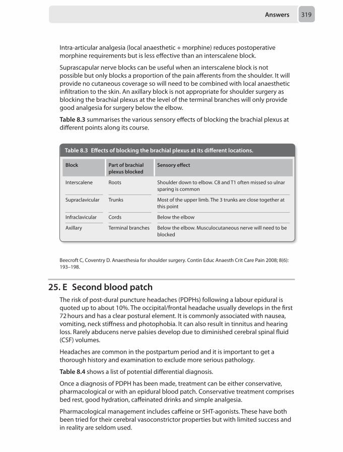
Answers 319
Intra-articular analgesia (local anaesthetic + morphine) reduces postoperative morphine requirements but is less effective than an interscalene block.
Suprascapular nerve blocks can be useful when an interscalene block is not possible but only blocks a proportion of the pain afferents from the shoulder. It will provide no cutaneous coverage so will need to be combined with local anaesthetic infiltration to the skin. An axillary block is not appropriate for shoulder surgery as blocking the brachial plexus at the level of the terminal branches will only provide good analgesia for surgery below the elbow.
Table 8.3 summarises the various sensory effects of blocking the brachial plexus at different points along its course.
Table 8.3 Effects of blocking the brachial plexus at its different locations.
Block Part of brachial plexus blocked
Sensory effect
Interscalene Roots Shoulder down to elbow. C8 and T1 often missed so ulnar sparing is common
Supraclavicular Trunks Most of the upper limb. The 3 trunks are close together at this point
Infraclavicular Cords Below the elbow
Axillary Terminal branches Below the elbow. Musculocutaneous nerve will need to be blocked
Beecroft C, Coventry D. Anaesthesia for shoulder surgery. Contin Educ Anaesth Crit Care Pain 2008; 8(6): 193–198.
25. E Second blood patchThe risk of post-dural puncture headaches (PDPHs) following a labour epidural is quoted up to about 10%. The occipital/frontal headache usually develops in the first 72 hours and has a clear postural element. It is commonly associated with nausea, vomiting, neck stiffness and photophobia. It can also result in tinnitus and hearing loss. Rarely abducens nerve palsies develop due to diminished cerebral spinal fluid (CSF) volumes.
Headaches are common in the postpartum period and it is important to get a thorough history and examination to exclude more serious pathology.
Table 8.4 shows a list of potential differential diagnosis.
Once a diagnosis of PDPH has been made, treatment can be either conservative, pharmacological or with an epidural blood patch. Conservative treatment comprises bed rest, good hydration, caffeinated drinks and simple analgesia.
Pharmacological management includes caffeine or 5HT-agonists. These have both been tried for their cerebral vasoconstrictor properties but with limited success and in reality are seldom used.
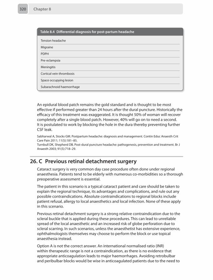
Chapter 8320
An epidural blood patch remains the gold standard and is thought to be most effective if performed greater than 24 hours after the dural puncture. Historically the efficacy of this treatment was exaggerated. It is thought 50% of woman will recover completely after a single blood patch. However, 40% will go on to need a second. It is postulated to work by blocking the hole in the dura thereby preventing further CSF leak.
Sabharwal A, Stocks GM. Postpartum headache: diagnosis and management. Contin Educ Anaesth Crit Care Pain 2011; 11(5):181–85.Turnbull DK, Shepherd DB. Post-dural puncture headache: pathogenesis, prevention and treatment. Br J Anaesth 2003; 91(5):718–29.
26. C Previous retinal detachment surgeryCataract surgery is very common day case procedure often done under regional anaesthesia. Patients tend to be elderly with numerous co-morbidities so a thorough preoperative assessment is essential.
The patient in this scenario is a typical cataract patient and care should be taken to explain the regional technique, its advantages and complications, and rule out any possible contraindications. Absolute contraindications to regional blocks include patient refusal, allergy to local anaesthetics and local infection. None of these apply in this scenario.
Previous retinal detachment surgery is a strong relative contraindication due to the scleral buckle that is applied during these procedures. This can lead to unreliable spread of the local anaesthetic and an increased risk of globe perforation due to scleral scarring. In such scenarios, unless the anaesthetist has extensive experience, ophthalmologists themselves may choose to perform the block or use topical anaesthesia instead.
Option A is not the correct answer. An international normalised ratio (INR) within therapeutic range is not a contraindication, as there is no evidence that appropriate anticoagulation leads to major haemorrhages. Avoiding retrobulbar and peribulbar blocks would be wise in anticoagulated patients due to the need to
Table 8.4 Differential diagnosis for post-partum headache
Tension headache
Migraine
PDPH
Pre-eclampsia
Meningitis
Cortical vein thrombosis
Space occupying lesion
Subarachnoid haemorrhage
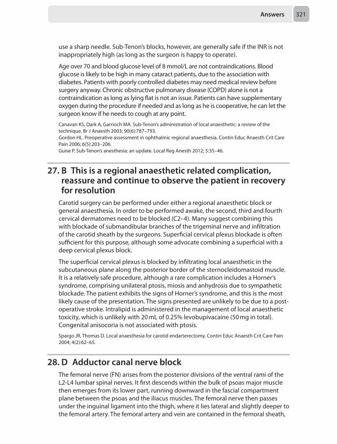
Answers 321
use a sharp needle. Sub-Tenon’s blocks, however, are generally safe if the INR is not inappropriately high (as long as the surgeon is happy to operate).
Age over 70 and blood glucose level of 8 mmol/L are not contraindications. Blood glucose is likely to be high in many cataract patients, due to the association with diabetes. Patients with poorly controlled diabetes may need medical review before surgery anyway. Chronic obstructive pulmonary disease (COPD) alone is not a contraindication as long as lying flat is not an issue. Patients can have supplementary oxygen during the procedure if needed and as long as he is cooperative, he can let the surgeon know if he needs to cough at any point.
Canavan KS, Dark A, Garrioch MA. Sub-Tenon’s administration of local anaesthetic: a review of the technique. Br J Anaesth 2003; 90(6):787–793.Gordon HL. Preoperative assessment in ophthalmic regional anaesthesia. Contin Educ Anaesth Crit Care Pain 2006; 6(5):203–206.Guise P. Sub-Tenon’s anesthesia: an update. Local Reg Anesth 2012; 5:35–46.
27. B This is a regional anaesthetic related complication, reassure and continue to observe the patient in recovery for resolution
Carotid surgery can be performed under either a regional anaesthetic block or general anaesthesia. In order to be performed awake, the second, third and fourth cervical dermatomes need to be blocked (C2–4). Many suggest combining this with blockade of submandibular branches of the trigeminal nerve and infiltration of the carotid sheath by the surgeons. Superficial cervical plexus blockade is often sufficient for this purpose, although some advocate combining a superficial with a deep cervical plexus block.
The superficial cervical plexus is blocked by infiltrating local anaesthetic in the subcutaneous plane along the posterior border of the sternocleidomastoid muscle. It is a relatively safe procedure, although a rare complication includes a Horner’s syndrome, comprising unilateral ptosis, miosis and anhydrosis due to sympathetic blockade. The patient exhibits the signs of Horner’s syndrome, and this is the most likely cause of the presentation. The signs presented are unlikely to be due to a post-operative stroke. Intralipid is administered in the management of local anaesthetic toxicity, which is unlikely with 20 mL of 0.25% levobupivacaine (50 mg in total). Congenital anisocoria is not associated with ptosis.
Spargo JR, Thomas D. Local anaesthesia for carotid endarterectomy. Contin Educ Anaesth Crit Care Pain 2004; 4(2):62–65.
28. D Adductor canal nerve blockThe femoral nerve (FN) arises from the posterior divisions of the ventral rami of the L2-L4 lumbar spinal nerves. It first descends within the bulk of psoas major muscle then emerges from its lower part, running downward in the fascial compartment plane between the psoas and the iliacus muscles. The femoral nerve then passes under the inguinal ligament into the thigh, where it lies lateral and slightly deeper to the femoral artery. The femoral artery and vein are contained in the femoral sheath,
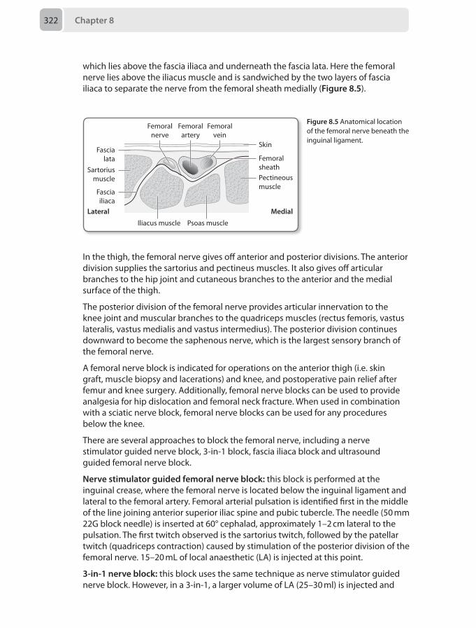
Chapter 8322
which lies above the fascia iliaca and underneath the fascia lata. Here the femoral nerve lies above the iliacus muscle and is sandwiched by the two layers of fascia iliaca to separate the nerve from the femoral sheath medially (Figure 8.5).
Femoralnerve
Femoralartery
Femoralvein
Femoralsheath
Medial
Pectineousmuscle
Psoas muscleIliacus muscle
Lateral
Fasciailiaca
Sartoriusmuscle
Fascialata
Skin
Figure 8.5 Anatomical location of the femoral nerve beneath the inguinal ligament.
In the thigh, the femoral nerve gives off anterior and posterior divisions. The anterior division supplies the sartorius and pectineus muscles. It also gives off articular branches to the hip joint and cutaneous branches to the anterior and the medial surface of the thigh.
The posterior division of the femoral nerve provides articular innervation to the knee joint and muscular branches to the quadriceps muscles (rectus femoris, vastus lateralis, vastus medialis and vastus intermedius). The posterior division continues downward to become the saphenous nerve, which is the largest sensory branch of the femoral nerve.
A femoral nerve block is indicated for operations on the anterior thigh (i.e. skin graft, muscle biopsy and lacerations) and knee, and postoperative pain relief after femur and knee surgery. Additionally, femoral nerve blocks can be used to provide analgesia for hip dislocation and femoral neck fracture. When used in combination with a sciatic nerve block, femoral nerve blocks can be used for any procedures below the knee.
There are several approaches to block the femoral nerve, including a nerve stimulator guided nerve block, 3-in-1 block, fascia iliaca block and ultrasound guided femoral nerve block.
Nerve stimulator guided femoral nerve block: this block is performed at the inguinal crease, where the femoral nerve is located below the inguinal ligament and lateral to the femoral artery. Femoral arterial pulsation is identified first in the middle of the line joining anterior superior iliac spine and pubic tubercle. The needle (50 mm 22G block needle) is inserted at 60° cephalad, approximately 1–2 cm lateral to the pulsation. The first twitch observed is the sartorius twitch, followed by the patellar twitch (quadriceps contraction) caused by stimulation of the posterior division of the femoral nerve. 15–20 mL of local anaesthetic (LA) is injected at this point.
3-in-1 nerve block: this block uses the same technique as nerve stimulator guided nerve block. However, in a 3-in-1, a larger volume of LA (25–30 ml) is injected and
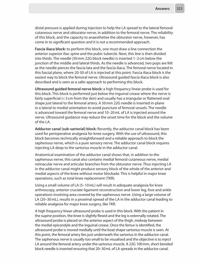
Answers 323
distal pressure is applied during injection to help the LA spread to the lateral femoral cutaneous nerve and obturator nerve, in addition to the femoral nerve. The reliability of this block, and the capacity to anaesthetise the obturator nerve, however, has come in to significant question and it is not a recommended approach.
Fascia iliaca block: to perform this block, one must draw a line connection the anterior superior iliac spine and the pubic tubercle. Next, this line is then divided into thirds. The needle (50 mm 22G block needle) is inserted 1–2 cm below the junction of the middle and lateral thirds. As the needle is advanced, two pops are felt as the needle pierces the fascia lata and the fascia iliaca. The femoral nerve located in this fascial plane, where 20-30 of LA is injected at this point. Fascia iliaca block is the easiest way to block the femoral nerve. Ultrasound guided fascia iliaca block is also described and is seen as a safer approach to performing this block.
Ultrasound guided femoral nerve block: a high frequency linear probe is used for this block. This block is performed just below the inguinal crease where the nerve is fairly superficial (< 3 cm from the skin) and usually has a triangular or flattened oval shape just lateral to the femoral artery. A 50 mm 22G needle is inserted in-plane in a lateral to medial orientation to avoid puncture of femoral vessels. The needle is advanced toward the femoral nerve and 10–20 mL of LA is injected around the nerve. Ultrasound guidance may reduce the onset time for the block and the volume of the LA.
Adductor canal (sub-sartorial) block: Recently, the adductor canal block has been used for perioperative analgesia for knee surgery. With the use of ultrasound, this block becomes technically straightforward and a reliable approach to block the saphenous nerve, which is a pure sensory nerve. The adductor canal block requires injecting LA deep to the sartorius muscle in the adductor canal.
Anatomical examination of the adductor canal shows that, in addition to the saphenous nerve, this canal also contains medial femoral cutaneous nerve, medial retinacular nerve and articular branches from the obturator nerve. Thus injecting LA in the adductor canal might produce sensory block of the whole of the anterior and medial aspects of the knee without motor blockade. This is helpful in major knee operations, such as total knee replacement (TKR).
Using a small volume of LA (5–10 mL) will result in adequate analgesia for knee arthroscopy, anterior cruciate ligament reconstruction and lower leg, foot and ankle operations involving area covered by the saphenous nerve. Using a large volume of LA (20–30 mL), results in a proximal spread of the LA in the adductor canal leading to reliable analgesia for major knee surgery, like TKR.
A high frequency linear ultrasound probe is used in this block. With the patient in the supine position, the knee is slightly flexed and the leg is externally rotated. The ultrasound probe is placed on the anterior aspect of the thigh, midway between the medial epicondyle and the inguinal crease. Once the femur is identified, the ultrasound probe is moved medially until the boat shape sartorius muscle is seen. At this point, the femoral artery lies just underneath the sartorius in the adductor canal. The saphenous nerve is usually too small to be visualised and the objective is to inject LA around the femoral artery under the sartorius muscle. A 22G 100 mm, short beveled block needle is inserted ensuring that 20–30 mL of LA spreads in the adductor canal.
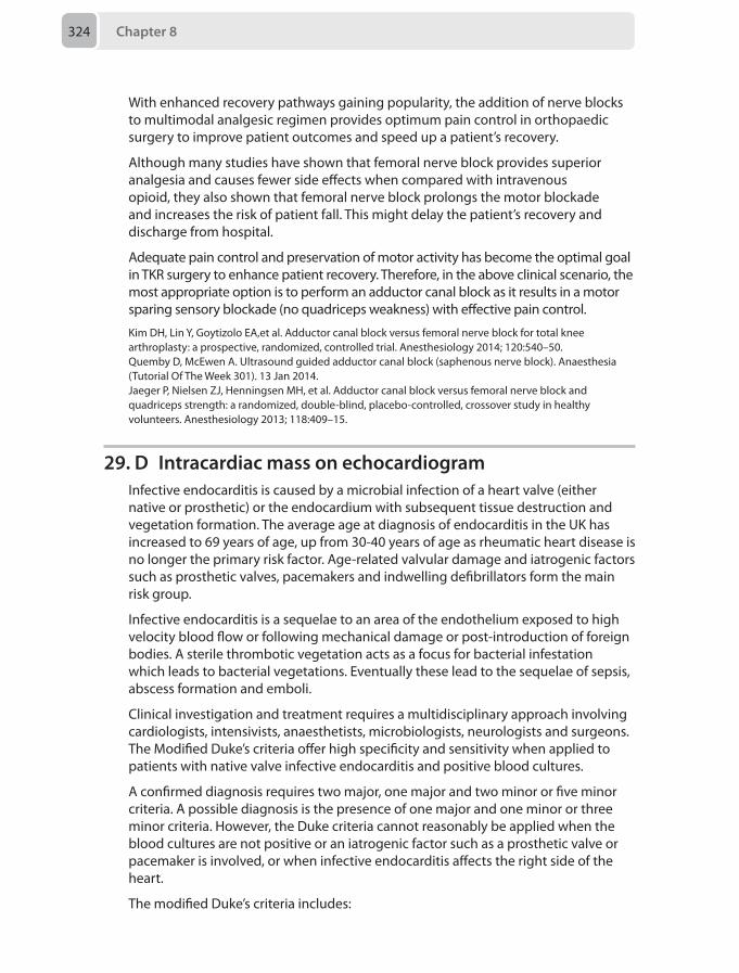
Chapter 8324
With enhanced recovery pathways gaining popularity, the addition of nerve blocks to multimodal analgesic regimen provides optimum pain control in orthopaedic surgery to improve patient outcomes and speed up a patient’s recovery.
Although many studies have shown that femoral nerve block provides superior analgesia and causes fewer side effects when compared with intravenous opioid, they also shown that femoral nerve block prolongs the motor blockade and increases the risk of patient fall. This might delay the patient’s recovery and discharge from hospital.
Adequate pain control and preservation of motor activity has become the optimal goal in TKR surgery to enhance patient recovery. Therefore, in the above clinical scenario, the most appropriate option is to perform an adductor canal block as it results in a motor sparing sensory blockade (no quadriceps weakness) with effective pain control.
Kim DH, Lin Y, Goytizolo EA,et al. Adductor canal block versus femoral nerve block for total knee arthroplasty: a prospective, randomized, controlled trial. Anesthesiology 2014; 120:540–50.Quemby D, McEwen A. Ultrasound guided adductor canal block (saphenous nerve block). Anaesthesia (Tutorial Of The Week 301). 13 Jan 2014.Jaeger P, Nielsen ZJ, Henningsen MH, et al. Adductor canal block versus femoral nerve block and quadriceps strength: a randomized, double-blind, placebo-controlled, crossover study in healthy volunteers. Anesthesiology 2013; 118:409–15.
29. D Intracardiac mass on echocardiogramInfective endocarditis is caused by a microbial infection of a heart valve (either native or prosthetic) or the endocardium with subsequent tissue destruction and vegetation formation. The average age at diagnosis of endocarditis in the UK has increased to 69 years of age, up from 30-40 years of age as rheumatic heart disease is no longer the primary risk factor. Age-related valvular damage and iatrogenic factors such as prosthetic valves, pacemakers and indwelling defibrillators form the main risk group.
Infective endocarditis is a sequelae to an area of the endothelium exposed to high velocity blood flow or following mechanical damage or post-introduction of foreign bodies. A sterile thrombotic vegetation acts as a focus for bacterial infestation which leads to bacterial vegetations. Eventually these lead to the sequelae of sepsis, abscess formation and emboli.
Clinical investigation and treatment requires a multidisciplinary approach involving cardiologists, intensivists, anaesthetists, microbiologists, neurologists and surgeons. The Modified Duke’s criteria offer high specificity and sensitivity when applied to patients with native valve infective endocarditis and positive blood cultures.
A confirmed diagnosis requires two major, one major and two minor or five minor criteria. A possible diagnosis is the presence of one major and one minor or three minor criteria. However, the Duke criteria cannot reasonably be applied when the blood cultures are not positive or an iatrogenic factor such as a prosthetic valve or pacemaker is involved, or when infective endocarditis affects the right side of the heart.
The modified Duke’s criteria includes:
Answers 325
Major criteria
1. Positive blood culture2. Echocardiogram positive for:
• Oscillating intracardiac mass• Intracardiac abscess• New partial dehiscence of prosthetic valve
Minor criteria
• Fever• Predisposed heart condition or intravenous drug use• Vascular or immunological phenomena like major arterial emboli, septic
pulmonary infarcts, mycotic aneurysm, intracranial or conjunctival haemorrhagic lesions, Janeway lesions
• Microbiological evidence such as polymerase chain reaction (PCR), serological tests, or positive blood cultures not meeting a major criterion
Of the options given in this clinical scenario, only the presence of an intracardiac mass or abscess is classified as a major criterion of the Modified Duke’s criteria, although all the other options are possible occurrences in a patient with infective endocarditis. Jayneway lesions are haemorrhagic nodules found on the palms and feet in infective endocarditis patients due to microabscesses caused by septic emboli. Although a temperature of > 38°C is a minor criterion, it is not required for the diagnosis of infective endocarditis.
Martinez G, Valchanov K. Infective endocarditis. Contin Educ Anaesth Crit Care Pain 2012; 12(3):134-139.
30. C Noradrenaline infusion titrated to blood pressureThe oesophageal Doppler monitor is a cardiac output monitoring device that can be utilised for intraoperative fluid optimisation. Once correctly placed in the oesophagus, it provides a velocity-time waveform by measuring the velocity of blood flow in the descending aorta. This helps guide intraoperative fluid therapy based upon derived parameters.
Parameters calculated are:
• Stroke distance (SD) - the distance that a column of blood moves down the aorta with each contraction. Values are age and size dependent
• Stroke volume (SV) - the volume of blood ejected from the left ventricle during each contraction. Normal range of 60-100 mL
• Stroke volume index (SVI) - the stroke volume divided by the body surface area (BSA). Normal range of 35-65 mL/m2. A low SVI could be either due to hypovolaemia or a high after load. A high SVI may be caused by decreased afterload
• Flow time corrected (FTc) - the duration of systolic aortic blood flow corrected for heart rate. Normal range is 330-360 ms. A low FTc may be due to hypovolaemia or an increased afterload. A high FTc may be seen by a low afterload.
The original oesophageal Doppler reading in this patient suggests a peripherally vasodilated circulation which could be attributed to a septic response to peritonitis.
Chapter 8326
As per the fluid management protocol suggested for oesophageal Doppler monitoring, following a bolus of 200 mL of colloid if the SV were to increase by greater than 10% another bolus could be considered. Under the given circumstances it would be most appropriate to initiate vasopressor therapy with noradrenaline. Dobutamine would not be appropriate as its chief action would be to increase cardiac output rather than cause peripheral vasoconstriction, and may in fact worsen the vasodilation and hypotension.
Drummond KE, Murphy E. Minimally invasive cardiac output monitors. Contin Educ Anaesth Crit Care Pain (2012) 12 (1): 5-10.
Mock Paper 9
Chapter 9
Questions1. A 61-year-old man has been brought to the emergency department intubated and
ventilated. Examination reveals a large frontal haematoma and a single dilated, but reactive, pupil. His abnormal observations are a blood pressure of 180/100 mmHg, heart rate of 45 bpm and temperature of 35.5°C. An arterial blood gas shows Pao₂ 13 kPa, Paco₂ of 6.9 kPa and blood glucose 8 mmol/L.
Which of the following parameters should be your priority when attempting to acutely improve this patient's cerebral perfusion?
A TemperatureB Paco₂C Blood pressureD Pao₂E Blood glucose
2. You are anaesthetising a 78-year-old man for a right upper lobectomy and lymphadenectomy for adenocarcinoma via video assisted thoracoscopic surgical approach (VATS). He is a long-term smoker, has chronic obstructive pulmonary disease (COPD) and takes aspirin 75 mg o.d. His FEV1 is 1.5 L. Despite your best efforts, you fail to site a thoracic epidural.
Which of the following would be the most appropriate technique to optimise this gentleman's perioperative analgesia?
A Single shot paravertebral injection at T6B Ask the surgeon to site a paravertebral catheterC Run a remifentanil infusion perioperatively and leave the patient intubated
overnightD Ask the surgeon to site an intrapleural catheterE Perform intercostal blocks at T5–8
3. You are asked to urgently review a 57-year-old man 7 days post left pneumonectomy. He remained intubated and ventilated for 24 hours post operatively due to intraoperative bleeding and hypothermia. A left sided intercostal drain was removed 24 hours ago. He is now complaining of cough, shortness of breath and chest pain. His oxygen saturations are 89% on 15 L/min oxygen.
Chapter 9328
On examination there is new subcutaneous emphysema of the chest wall. Heart rate is 60 beats per minute and blood pressure is 80/50 mmHg.
What is the most appropriate next step?
A Urgent chest radiograph (CXR)B Needle thoracocentesis followed by insertion of 22F intercostal drainC Immediate insertion of a 12F intercostal drain by the Seldinger techniqueD Urgent bronchoscopyE Urgent CT scan and thoracic surgical opinion
4. A 3-year-old boy is under general anaesthesia for the removal of a foreign body partially obstructing his right main bronchus via rigid bronchoscopy. He is breathing spontaneously and receiving sevoflurane in air. Foreign body instrumentation is difficult and after prolonged grasping attempts and suctioning, he becomes bradycardic with a heart rate of 25 beats per minute.
What is the most likely cause of his clinical deterioration?
A HypoxiaB Depth of anaesthesia C HypothermiaD HypercarbiaE Vasovagal reflex
5. A 48-year-old woman has had an arthroscopic rotator cuff repair. She has received a general anaesthetic, a supraglottic airway was inserted and had an interscalene block. Her surgery finished at midday.
Which of the following is most likely to prevent her from being discharged on the day of surgery?
A Lives in a rural location 30 minutes by car to the nearest hospital B Has an adult relative to act as carer at home only until 20.00C Hasn’t yet passed urineD Is taking a public taxi home with an adult relativeE Has residual upper arm weakness
6. A 78-year-old man is listed for a transurethral resection of his prostate (TURP) under spinal anaesthesia. He has moderate to severe chronic obstructive pulmonary disease (COPD) with ongoing steroid use, ischaemic heart disease, and had a coronary stent inserted 15 months ago. He normally takes aspirin and clopidogrel, but has not been taking the latter for “a few weeks”. He has also recently started taking rivaroxaban 10 mg at night for an irregular heart rate.
What is the safest way to proceed?
A Ensuring 18 hours after the last dose of rivaroxaban, give a spinal, and then start a heparin infusion postoperatively
B Give a spinal now and use treatment dose low molecular weight heparin (LMWH) from 2 hours postoperatively
Questions 329
C Wait until 24 hours after the last dose of rivaroxaban, then proceed with a spinal, and give the next dose immediately postoperatively
D Ensure 12 hours after the last dose of rivaroxaban, and give prophylactic LMWH 6 hours postoperatively
E Discuss with the patient the increased risks of central neuraxial blockade and proceed under general anaesthesia
7. A 64-year-old man undergoes hip surgery under general anaesthesia. He is positive pressure ventilated through a size 5 laryngeal mask airway and anaesthesia is maintained with nitrous oxide and sevoflurane. In recovery, he complains of paraesthesia over the right anterior aspect of his tongue. There is no dysphagia or dysarthria and tongue appearance and movements are normal.
What is the most likely cause of his neurological signs in recovery?
A Hypoglossal nerve injuryB Lingual nerve injuryC Recurrent laryngeal nerve injuryD Inferior alveolar nerve injuryE Venous drainage obstruction
8. A patient with an acute subarachnoid haemorrhage is undergoing coil embolisation of the aneurysm in the interventional neuroradiology suite. Anaesthesia is induced with alfentanil, propofol and rocuronium. Maintenance of anaesthesia is with sevoflurane and remifentanil infusion. Shortly after intubation the observations are as follows:• Blood pressure: 220/110 mmHg• Heart rate: 90 beats per minute• Spo2 98%• ETCO2 4.9 kPa• End-tidal sevoflurane 1.9%
What is the most appropriate initial management?
A Alert radiologist B Increase depth of anaesthesia C Increase minute ventilationD Give mannitol 1 g/kgE Start intravenous esmolol infusion
9. A 10 kg child with no comorbidities is scheduled for an elective umbilical hernia repair as a day case.
Which of the following would be the best regime of injectate for caudal epidural analgesia?
A 10 mL of 0.25% levobupivacaine with 10 μg fentanylB 10 mL of 0.25% levobupivacaineC 10 mL of 0.25% plain bupivacaine with 300 μg diamorphineD 10 mL of 0.25% levobupivacaine with 25 μg clonidineE 10 mL of 0.25% bupivacaine
Chapter 9330
10. A 65-year-old man for elective thoracotomy and pulmonary lobectomy is to have a thoracic epidural for perioperative analgesia.
Which of the following is the best approach for epidural insertion?
A A midline mid-thoracic epidural under general anaesthesiaB A paramedian mid-thoracic epidural under light sedation or awakeC A paramedian lower-thoracic epidural under general anaesthesiaD A midline upper lumbar epidural under light sedation or awakeE A midline mid-thoracic epidural under light sedation or awake
11. An 84-year-old ASA 3 woman is listed for multilevel facet joint injections and a caudal epidural by the orthopaedic surgeons. The patient will need to lie in the prone position. Comorbidities include moderate chronic obstructive pulmonary disease (COPD), angina, hypertension and chronic lower back pain. Alongside all her cardiovascular medications she takes regular co-dydramol and amitriptyline for her pain.
The safest anaesthetic technique for this procedure is:
A 0.25–0.5 mg/kg intravenous ketamine B Local anaesthesia only with no sedationC Infusion of remifentanil at 0.25 mg/kg/minD Target controlled infusion of propofol at a 1 mg/mLE 0.5 μg/kg fentanyl followed after several minutes by small doses of intravenous
midazolam titrated to effect
12. A 62-year-old non-diabetic woman presents to the intensive care unit with severe urosepsis.
Which of the following glucose levels would be the most appropriate to target?
A > 4 mmol/LB 4–6 mmol/LC 6–8 mmol/LD < 10 mmol/LE < 15 mmol/L
13. A 19-year-old male motorcyclist is admitted following a high speed road traffic accident. The retrieval team report he has clinical evidence of bilateral flail segments and a significant neurological injury. He is intubated and sedated by the retrieval service with intermittent doses of ketamine, propofol and rocuronium and arrives to the intensive care unit. He has been haemodynamically stable with moderate and escalating ventilator requirements.
The most appropriate sedation regime for this patient on the intensive care unit would be:
A Propofol and fentanylB Clonidine and fentanyl
Questions 331
C Midazolam and fentanylD Ketamine and fentanylE Fentanyl alone
14. A 26-year-old woman who is 32/40 pregnant had a witnessed collapse whilst shopping. She received bystander cardiopulmonary resuscitation (CPR) and advanced life support (ALS) by the paramedics for one hour prior to transfer to a teaching hospital. In hospital, a Caesarean section was performed immediately. ALS continued for a further 45 minutes without return of spontaneous circulation and a profound metabolic acidosis developed.
What now is the most appropriate management option?
A Terminate life support and organise a team debriefB Administer thrombolysis and continue ALSC Continue ALS until the intensive care consultant arrivesD Commence extra-corporeal membrane oxygenation (ECMO)E Administer 10–20 mL of 8.4% sodium bicarbonate
15. A 76-year-old woman has had an upper gastrointestinal bleed and presented with an acute kidney injury. After resuscitation and an oesophago-duedenoscopy she is admitted to the intensive care unit for renal replacement therapy. The nurse requests that you prescribe the particulars of renal haemofiltration including the anticoagulation.
The most appropriate choice is:
A Unfractionated heparin loading dose followed by a pre-filter infusionB No anti-coagulationC Prostacyclin infusion D Sodium citrate pre-filter infusionE Increasing the fraction of replacement fluid added before the filter
16. A 65-year-old man is recovering on the high dependency unit after an emergency laparotomy for small bowel perforation for which he received an effective epidural. His background includes treated hypertension, a smoker of 20 cigarettes per day and mild depression. Overnight he becomes very agitated and confused and attempts to remove his invasive lines and monitoring. Examination, review of his blood science investigations and blood gas results are all unremarkable.
What is the most appropriate course of treatment?
A Reassure the patient regarding his situationB Call his wife to hospital to help calm him downC Prescribe vitamin replacement therapy and benzodiazepine sedationD Prescribe haloperidolE Commence sedation with clonidine
Chapter 9332
17. A 26-year-old woman who is 3 days post-partum has returned to the labour ward complaining of an ongoing headache. She delivered vaginally after having a lumbar epidural for labour. On the first day postpartum she had complained of a frontal headache that worsened with coughing and had been diagnosed with a post-dural puncture headache (PDPH). At home, she has been taking simple analgesia and drinking plenty of water for the past 2 days but the headache is persisting.
What is the next best line of management in this situation?
A Encourage her to drink coffee and 3 L of water per dayB Encourage her to drink coffee and prescribe sumatriptanC Admit her overnight for intravenous fluid therapy, regular analgesia and
further assessmentD Offer her an epidural blood patchE Offer her an epidural blood patch and perform routine blood cultures at the
same time
18. A 21-year-old woman in antenatal clinic is due to have an elective Caesarean section for breech presentation within the next two weeks. She is concerned about having a spinal anaesthetic as she has been diagnosed with gestational thrombocytopenia. You review her blood results and her platelet count has been low but steady.
Which of the following blood results would prevent this lady from having a spinal?
A Platelet count < 50 × 109/LB Activated partial thromboplastin (APTT) time of 30 secondsC Platelet count < 100 × 109/LD Platelet count < 70 × 109/LE Prothrombin time (PT) of 12 seconds
19. A 15 kg, 3-year-old girl was brought to the emergency department with a history of choking on a piece of apple 6 hours previously. She appears comfortable and not in respiratory distress. Her chest sounds clear on auscultation but a chest X-ray shows a right lung that is more inflated and radiolucent compared to the left, particularly on the expiration film.
Suspecting the child has inhaled the piece of apple, the ENT team want to perform an urgent examination under anaesthesia (EUA) with a rigid bronchoscopy and removal of foreign body.
The most appropriate anaesthetic plan for this case is:
A Perform a rapid sequence induction and intubate to secure the airwayB Routine intravenous induction with muscle relaxant. Intubate and ventilate
until rigid bronchoscopyC Routine intravenous induction with muscle relaxant. Insert a supraglottic
airway device for ventilation until rigid bronchoscopyD Routine intravenous induction without muscle relaxant. Facemask ventilation
until rigid bronchoscopy
Questions 333
E Inhalational anaesthetic induction and maintenance with sevoflurane, without muscle relaxant. Maintain spontaneous respiration throughout the case
20. An 18-month old boy is scheduled for an inguinal hernia repair as a day case. His mother reports that he developed an anxiety to needles since a hospital admission for pneumonia 5 months previously, and has not had his MMR vaccination. His mother requests a gas induction and asks if he could receive his MMR vaccination while under general anaesthesia.
The best course of action is:
A Administer the MMR vaccination after induction of anaesthesiaB Ask the surgical team to administer the MMR vaccination whilst under general
anaesthesiaC Ask the paediatric team to administer the MMR vaccination postoperatively
prior to dischargeD Arrange for the GP to administer the MMR vaccination 4 weeks postoperativelyE Cancel the surgery until the child has had his MMR vaccination
21. A 58-year-old woman is listed for an elective hysterectomy. She states that she has a morphine allergy which made her eyes and lip swell in the past.
Which of the following analgesics would be unsafe in this patient?
A PethidineB TramadolC BuprenorphineD MethadoneE Fentanyl
22. A 64-year-old woman with a history of chronic pain is listed for a shoulder replacement. She normally takes gabapentin 300 mg three times a day, paracetamol 1 g as needed and a buprenorphine patch at 20 μg/hour.
What is the most appropriate postoperative analgesic regimen for this patient?
A. Paracetamol, ibuprofen, gabapentin, MST 25 mg twice daily, Oramorph 10–20 mg 4-hourly
B. Paracetamol, diclofenac, a morphine PCA 2 mg bolus with 5 minute lockoutC. Paracetamol, ibuprofen, gabapentin, fentanyl PCA with 25 μg bolus with 5
minute lockoutD. Paracetamol, codeine, tramadol and Oramorph 10–20 mg 4-hourlyE. Paracetamol, ibuprofen, gabapentin, Oxynorm 15 mg twice daily
23. A 35-year-old man with a chronic history of intravenous heroin use and schizophrenia presents to the emergency department with a perforated duodenal ulcer. He is septic, coagulopathic and haemodynamically unstable, so is rushed to theatre for resuscitation and an emergency laparotomy.
What is the most appropriate analgesic regimen to manage his postoperative pain?
Chapter 9334
A Thoracic epidural with plain bupivacaineB Intravenous methadone and ketamine infusionC Oral methadone and intravenous morphine as requiredD Intravenous methadone and intravenous morphine as requiredE Morphine patient controlled analgesia (PCA) with a background infusion
24. A 68-year-old woman with advanced breast cancer and poor intravenous access is suffering from intractable bone pain in her distal right femur. A recent MRI scan has confirmed a solitary metastases in her right femur and ruled out a fracture. Management is at a palliative stage and she is currently taking paracetamol, ibuprofen and morphine sulphate.
What is the most appropriate next step in controlling her pain?
A Internal fixation of femurB BisphosphonatesC Localised external beam radiotherapyD Radioisotope treatmentE Gabapentin
25. A 75-year-old woman with metastatic breast cancer is currently on 70 mg MST twice a day and 20 mg of Oramorph 4-hourly for breakthrough pain. She continues to suffer from back pain. An MRI excludes any cord compression but confirms the presence of vertebral bone deposits.
What is next best step in treating her pain?
A Converting the patient to oxycodoneB Start calcitoninC Increase the dose of MSTD RadiotherapyE Start bisphosphonates
26. A 22-year-old man is brought into a district general emergency department after being pulled from a burning house with 35% body surface area burns. They include partial thickness facial and anterior chest wall burns. He has a hoarse voice with carbonaceous sputum. His Glasgow coma score is 15 and other observations are as follows:• Heart rate 98 beats per minute• Blood pressure 169/82 mmHg• Respiratory rate 25 breaths per minute• Saturations 100% on high flow oxygen• Temperature 38.0°C
There are no other injuries.
What is the most appropriate immediate course of action?
Questions 335
A Perform a modified rapid sequence induction with alfentanil, propofol and rocuronium and intubate with a size 8.0 cuffed oral tracheal tube cut to 24 cm to reduce dead space
B Perform a rapid sequence induction with thiopentone and suxamethonium using an uncut size 8.0 cuffed oral tracheal tube
C Refer and transfer to regional burns centre without delay for definitive treatment
D Give 200 mg hydrocortisone intravenouslyE Give 1.5 g ceftriaxone intravenously
27. One of the high dependency unit nurses calls you to review a 73-year-old woman 72 hours post-carotid endarterectomy. The patient appears confused, agitated and her blood pressure is 210/100 mmHg. The nurse administered 1 g of oral paracetamol for a persistent headache 1 hour ago after which the patient vomited.
What is the next most appropriate step in the management of her condition?
A Administer a broad spectrum intravenous antibioticB Administer 50 mL of 20% mannitolC Catheterise the patientD Administer a stat dose of oral amlodipine 10 mg E Administer a bolus dose of intravenous labetalol 10 mg
28. A 38-year-old man scheduled to have a revision of his arteriovenous fistula in the next 8 weeks is being assessed in the anaesthetic pre-assessment clinic. He suffers from chronic kidney disease and is on dialysis. His recent blood count shows a haemoglobin of 68 g/L with a low reticulocyte but a normocytic mean corpuscular volume.
The most appropriate preoperative strategy for treating this patient's anaemia is:
A Blood transfusionB Human erythropoietinC Perioperative blood transfusionD Folic acid injectionsE Vitamin B12 injections
29. A 34-year-old parturient had epidural analgesia for a full-term normal delivery. 4 days later, she complains of constant severe back pain along with paraesthesia in her left leg. On examination she is febrile and has a motor power of 4/5 in both of her legs and normal power in her upper limbs.
The immediate investigation of choice would be:
A Lumbar punctureB MRI lumbar spineC MRI whole spineD C-reactive protein (CRP)E Erythrocyte sedimentation rate (ESR)
Chapter 9336
30. You are reviewing a study that randomised two groups of patients to receive sedation either at the discretion of the caregivers or by following a strict protocol. The study hypothesis is that protocolisation reduces the total cumulative dose of sedative medications.
Which of the following statistical tests would be most appropriate to analyse the results of this pilot study?
A Unpaired Student's t-testB Paired Student's t-testC Chi Squared testD Mann-Whitney U testE Paired ANOVA
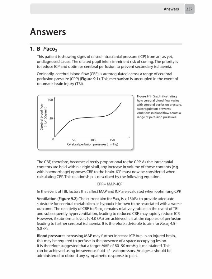
Answers 337
Answers
1. B Paco₂This patient is showing signs of raised intracranial pressure (ICP) from an, as yet, undiagnosed cause. The dilated pupil infers imminent risk of coning. The priority is to reduce ICP and optimise cerebral perfusion to prevent secondary ischaemia.
Ordinarily, cerebral blood flow (CBF) is autoregulated across a range of cerebral perfusion pressure (CPP) (Figure 9.1). This mechanism is uncoupled in the event of traumatic brain injury (TBI).
100
50
0 50 100 150Cerebral perfusion pressures (mmHg)
Cere
bral
blo
od �
ow(m
L/10
0g/m
in)
Figure 9.1 Graph illustrating how cerebral blood flow varies with cerebral perfusion pressure. Autoregulation prevents variations in blood flow across a range of perfusion pressures.
The CBF, therefore, becomes directly proportional to the CPP. As the intracranial contents are held within a rigid skull, any increase in volume of those contents (e.g. with haemorrhage) opposes CBF to the brain. ICP must now be considered when calculating CPP. This relationship is described by the following equation:
CPP= MAP–ICP
In the event of TBI, factors that affect MAP and ICP are evaluated when optimising CPP.
Ventilation (Figure 9.2): The current aim for Pao₂ is > 13 kPa to provide adequate substrate for cerebral metabolism as hypoxia is known to be associated with a worse outcome. The reactivity of CBF to Paco₂ remains relatively robust in the event of TBI and subsequently hyperventilation, leading to reduced CBF, may rapidly reduce ICP. However, if subnormal levels (< 4.0 kPa) are achieved it is at the expense of perfusion leading to further cerebral ischaemia. It is therefore advisable to aim for Paco₂ 4.5–5.0 kPa.
Blood pressure: Increasing MAP may further increase ICP but, in an injured brain, this may be required to perfuse in the presence of a space occupying lesion. It is therefore suggested that a target MAP of 80–90 mmHg is maintained. This can be achieved using intravenous fluid +/– vasopressors. Analgesia should be administered to obtund any sympathetic response to pain.
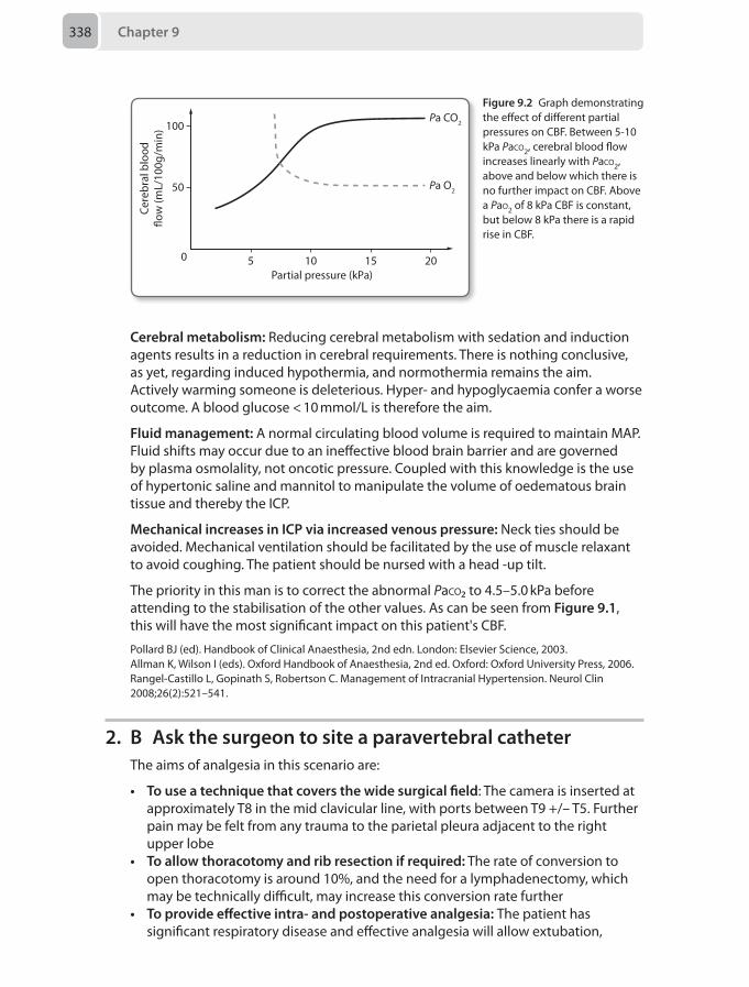
Chapter 9338
Cerebral metabolism: Reducing cerebral metabolism with sedation and induction agents results in a reduction in cerebral requirements. There is nothing conclusive, as yet, regarding induced hypothermia, and normothermia remains the aim. Actively warming someone is deleterious. Hyper- and hypoglycaemia confer a worse outcome. A blood glucose < 10 mmol/L is therefore the aim.
Fluid management: A normal circulating blood volume is required to maintain MAP. Fluid shifts may occur due to an ineffective blood brain barrier and are governed by plasma osmolality, not oncotic pressure. Coupled with this knowledge is the use of hypertonic saline and mannitol to manipulate the volume of oedematous brain tissue and thereby the ICP.
Mechanical increases in ICP via increased venous pressure: Neck ties should be avoided. Mechanical ventilation should be facilitated by the use of muscle relaxant to avoid coughing. The patient should be nursed with a head -up tilt.
The priority in this man is to correct the abnormal Paco₂ to 4.5–5.0 kPa before attending to the stabilisation of the other values. As can be seen from Figure 9.1, this will have the most significant impact on this patient's CBF.
Pollard BJ (ed). Handbook of Clinical Anaesthesia, 2nd edn. London: Elsevier Science, 2003.Allman K, Wilson I (eds). Oxford Handbook of Anaesthesia, 2nd ed. Oxford: Oxford University Press, 2006.Rangel-Castillo L, Gopinath S, Robertson C. Management of Intracranial Hypertension. Neurol Clin 2008;26(2):521–541.
2. B Ask the surgeon to site a paravertebral catheterThe aims of analgesia in this scenario are:
• To use a technique that covers the wide surgical field: The camera is inserted at approximately T8 in the mid clavicular line, with ports between T9 +/– T5. Further pain may be felt from any trauma to the parietal pleura adjacent to the right upper lobe
• To allow thoracotomy and rib resection if required: The rate of conversion to open thoracotomy is around 10%, and the need for a lymphadenectomy, which may be technically difficult, may increase this conversion rate further
• To provide effective intra- and postoperative analgesia: The patient has significant respiratory disease and effective analgesia will allow extubation,
100
10
50
50 15 20
Cere
bral
blo
od�o
w (m
L/10
0g/m
in)
Partial pressure (kPa)
Pa CO2
Pa O2
Figure 9.2 Graph demonstrating the effect of different partial pressures on CBF. Between 5-10 kPa Paco2, cerebral blood flow increases linearly with Paco2, above and below which there is no further impact on CBF. Above a Pao2 of 8 kPa CBF is constant, but below 8 kPa there is a rapid rise in CBF.
Answers 339
spontaneous ventilation and coughing. Prompt extubation reduces the risk of ventilator associated complications in the critical care unit, therefore option C is not the best choice here
Although thoracic epidural analgesia is seen as the gold standard for this scenario, injection of local anaesthetic into the paravertebral space aims to block spinal nerves as they leave the intervertebral foramina; providing unilateral analgesia with a degree of sympathetic blockade. A single shot injection may give analgesia for over 20 hours but use of a catheter allows infusion of local anaesthetic in the post operative period and is the best option of those listed here (option B).
In light of failed attempts to site a thoracic epidural, it may be kinder to perform further procedures when the patient is asleep; surgically placed catheters during VATS have been described and it would be worth asking the surgeon whether they can perform this procedure in the first instance.
Intrapleural local anaesthetic, that is administration of local anaesthetic into the space between the parietal and visceral pleura, would diffuse around the intercostal nerves as they travel between the inner and innermost intercostal muscles. However, disruption of the pleura leads to erratic absorption, potential leakage into any intercostal drains sited and so less effective analgesia. Systemic absorption via this route is high so option D is neither the safest nor the most effective of those given. Intercostal blocks (option E) in general do not have adequate duration for this scenario and offer inadequate analgesia compared with paravertebral techniques.
Tighe, SQM, Greene MD, Rajadurai N. Paravertebral block. Contin Educ Anaesth Crit Care Pain 2010; 10(5):133–137.
3. B Needle thoracocentesis followed by insertion of 22F intercostal drainThe clinical signs are suggestive of a massive air leak, possibly from breakdown of the bronchial stump. There are signs of cardiovascular impairment (including paradoxical bradycardia) suggesting impending cardiovascular collapse. The most likely diagnosis is a bronchopleural fistula leading to tension pneumothorax that should be decompressed immediately by needle thoracocentesis. Other diagnoses could include delayed infection and bleeding so it would be prudent to follow needle decompression with a larger bore (22F) intercostal drain. A smaller 12F drain inserted via the Seldinger technique may not drain blood/purulent matter adequately and takes more time to site. Obtaining a chest radiograph often takes time that may be detrimental in this scenario.
Risk factors for bronchopleural fistulae include increased age, poor wound healing, pneumonectomy, previous chemo/radiotherapy and prolonged mechanical ventilation postoperatively.
Although bronchoscopy +/– CT thorax may be needed to make the diagnosis and assess for any other complications (e.g. empyema) when the patient stabilises, the priority is restoration of oxygenation and adequate cardiovascular parameters.
Chapter 9340
Early bronchial stump breakdown often requires surgical treatment with direct closure or coverage with an intercostal flap.
Darling GE, Abdurahman A, Yi QL, et al. Risk of a right pneumonectomy: role of bronchopleural fistula. Ann Thorac Surg 2005; 79(2):433.
4. A HypoxiaForeign body aspiration is a dangerous condition most frequently seen in infants where inadvertent aspiration of objects disrupts the normal airway structure and function. The classic triad of symptoms consists of paroxysmal coughing, wheezing and reduced breath sounds on the affected side occurring after a witnessed choking episode. It is a leading cause of death in 1–3 year olds and its safe management is challenging to both surgeon and anaesthetist.
The gold standard for managing foreign body aspiration in children is removal via rigid bronchoscopy under general anaesthesia. The instrument most commonly used in children is the Storz ventilating bronchoscope which consists of a metal tube and a removable optical scope (Hopkins rod). During instrumentation, the optical scope is within the lumen of the bronchoscope and provides excellent visualisation of the airway. The scope however significantly reduces the lumen of the bronchoscope available for ventilation and should only be used for short periods. Hypoventilation is a real possibility especially if the patient is spontaneously ventilating.
Bradycardias during bronchoscopy are uncommon and should be assumed to be secondary to hypoxia until proven otherwise. Hypoxia can occur if the scope is placed in a bronchus or if instrumentation triggers bronchospasm. Furthermore, when excessive suctioning is performed, there may be atelectasis and a reduction in the inspired oxygen concentration. Also, a feared complication which can cause hypoxia acutely is dislodgement of the foreign body into the trachea creating complete obstruction of the airway.
In order to reduce the risk of foreign body dislodgement whilst allowing spontaneous ventilation, anaesthesia needs to be deep enough to minimise coughing and moving without paralysis. Excessive anaesthesia to achieve this can trigger bradycardias, but it is not the most likely cause in the above scenario. The arrhythmia occurred after prolonged instrumentation which would have restricted the spontaneous ventilation and elevated the boy to a lighter plane of anaesthesia.
Children are commonly affected by inhaled foreign bodies and it is important for the anaesthetist to also be aware of the challenges of paediatric anaesthesia. Children are at more risk of becoming hypothermic during anaesthesia which if severe, can cause arrhythmias. The patient's core temperature in the above case however is highly unlikely to be sufficiently low to produce this response.
There are vagal sensory nerves within the conducting airways and stimulation by bronchoscopy can cause reflex spasm and bradycardia. Coughing during bronchoscopy may also illicit a vagal response. The fact that the bradycardia occurred after prolonged instrumentation (as opposed to during), and no coughing occurred makes this diagnosis less likely.
Answers 341
Hypercarbia occurs frequently during rigid bronchoscopy, particularly in spontaneously ventilating patients and where there is frequent use of the optical scope or forceps restricting ventilation. Hypercarbia however is more likely to cause an initial tachycardia as opposed to a bradycardia due to an increased sympathoadrenal tone.
Roberts S, Thornington R. Paediatric bronchoscopy. Contin Educ Anaesth Crit Care Pain 2005; 5(2):41–44.Farrell P.Rigid bronchoscopy for foreign body removal: anaesthesia and ventilation. Paediatr Anaesth 2004; 14(1):84–89.
5. B Has an adult relative to act as carer at home only until 20.00In the ‘ten high impact changes’ document published by the NHS Modernisation Agency it is outlined that day surgery, rather than inpatient surgery, should be treated as the norm for all elective surgery. Locally agreed protocols exist in most day case units for selection and exclusion criteria. These fall broadly into medical, surgical and social considerations (Table 9.1).
Table 9.1 Selection criteria that should be met for appropriateness for day case surgery.
Medical Surgical Social
Preoperative assessment of a patient’s health status should be made to determine eligibility rather than use of arbitrary limits, e.g. BMI, ASA, age
Procedure should not pose any serious complications, e.g. risk of haemorrhage and cardiovascular instability
Patients must understand procedure, postoperative care and be able to consent to day surgery
Chronic stable disease may be better treated as day case
Oral medications and local anaesthetic techniques should be sufficient to manage post operative symptoms
A responsible adult should escort the patient home and be present for the first 24 hours postoperatively
Procedure should not prevent resumption of oral intake within a few hours
Domestic circumstances should be appropriate for postoperative care
Patients should be able to mobilise but full mobilisation is not always required
It is important to note that patients can be discharged with residual effects after nerve blockade so long as the duration of effects are explained and the patient has received written and verbal instructions of what to expect. Mandatory oral intake and ability to void are no longer considered essential discharge criteria unless manipulation of the bladder has occurred. Distance from the hospital should be individually assessed, but some units set 60 minutes as a practical limit. An escort home and for the first 24 hours are still necessary although this may be relaxed in the future for minor procedures with very short anaesthetics where patient is not compromised by the time of discharge.
Verma R, Alladi R, Jackson I, et al. Day case and short stay surgery: 2. Anaesthesia 2011: 66;417–434.
Chapter 9342
6. A Ensuring 18 hours after the last dose of rivaroxaban, give a spinal, and then start a heparin infusion postoperativelyVast numbers of patients present for surgery on antiplatelet drugs. The perioperative management of these medications commonly falls to anaesthetists to coordinate, and there is a significant overlap also in the assessment of cardiac risk for non-cardiac surgery. A solid understanding of these issues will help in preparation for both the written and viva elements of the Final FRCA.
Aspirin and clopidogrel
Aspirin is an irreversible inhibitor of platelet cyclooxygenase (COX), and thus normal platelet function relies on new platelet manufacture, which takes approximately 7 days. Aspirin is not contraindicated in central neuraxial blockade (CNB), as the risk of haematoma is not elevated.
Conversely, clopidogrel is associated with haematoma formation in case reports. It is a thienopyridine adenosine diphosphate (ADP) blocker, and published advice is to avoid for at least 7 days prior to CNB. Prasugrel, a more potent thienopyridine, should be avoided for 7–10 days and not restarted until 6 hours after block or catheter removal, where clopidogrel can be given just afterwards.
Tirofiban/abciximab
These two are glycoprotein IIb/IIIa blockers, in the case of abciximab this is via binding of a monoclonal antibody. Tirofiban is the shorter acting of the two, and CNB can be attempted after 8 hours, whereas antibody persistence means a duration of 24–48 hours is needed for abciximab.
Warfarin
An international normalised ratio (INR) of ≤ 1.5 is known to be associated with clotting factor levels of > 40% and is regarded as safe for CNB.
Dabigatran/rivaroxaban
Dabigatran is an oral thrombin inhibitor only licensed for venous thromboembolism (VTE) prophylaxis after surgery. CNB should not be established in patients already on this drug, as it is contraindicated by the manufacturer. It can be started 6 hours after the risk period. Rivaroxaban is a direct oral inhibitor of factor Xa. It is becoming more common as the list of approved indications increases. Previously only for postoperative VTE prophylaxis, it is now being used in AF and in Europe as an adjunct to aspirin and clopidogrel in acute coronary syndromes. CNB should be 12–18 hours post-dose, and the drug should only be given 6 hours after a block or catheter removal.
Heparins
With the low molecular weight heparins (LMWH), the duration between administration and safe block depends on dosage. Therapeutic dose requires a 24-hour delay, whereas prophylactic dose, a gap of 12 hours. In both cases 2–4 hours is the duration until restarting following block or catheter removal.
Answers 343
Fondaparinux
Fondaparinux is another factor Xa inhibiting drug, although with a long half-life of 21 hours. It has little effect on thrombin and no antiplatelet effect. At treatment doses no CNB is permitted, but in prophylactic doses, CNB can be performed after 36 hours, with 6 hours to elapse before the next dose, but 12 hours after epidural catheter removal.
Use of dual antiplatelet therapy (DAPT) is essential for the prevention of stent thrombosis following coronary stenting, particularly after a drug eluting stent. Here the minimum recommended time for DAPT is normally 12 months, and for stopping or withholding DAPT, the cardiac risk of stent thrombosis often exceeds the operative risks of bleeding. This gentleman has been off clopidogrel greater than the minimum of 7 days, such that his clopidogrel is no longer an issue.
However, the confounding issue is now the rivaroxaban, which is appearing in the drug history of more and more patients. Mainly used for postoperative venous thromboprophylaxis there are now indications for atrial fibrillation and in the treatment of acute coronary syndrome patients. The recommended omission time here is 12–18 hours, and restarting should be 6 hours after block or catheter removal. The other important safety issue with rivaroxaban is the lack of any mechanism of reversal, which probably makes its use here in the immediate postoperative phase unwise. Therefore the safest approach for this patient is to have the required 18 hours off rivaroxaban before preforming a spinal anaesthetic with a heparin infusion postoperatively.
Davies G, Checketts MR. Regional anaesthesia and antithrombotic drugs. Contin Educ Anaesth Crit Care Pain 2013; 12(1):11–16.
7. B Lingual nerve injuryThe laryngeal mask airway (LMA) is a versatile supraglottic airway device which consists of a tube connected to an inflatable cuff which surrounds a mask designed to seal off the laryngeal inlet from the gastrointestinal tract. However, it is not a definitive airway and vigilance against aspiration is advised particularly when used in conjunction with positive pressure ventilation. Another recognised complication associated with laryngeal mask airway ventilation is pressure neurapraxia to anatomically vulnerable nerves within the pharynx or oral cavity.
A neurapraxia refers to a localised and transient conduction block along a nerve without any anatomical interruption, which in the above case is likely to be caused by pressure from the cuff. Predisposing factors include the use of nitrous oxide, cuff over-inflation, using an undersized laryngeal mask airway, the lateral position and a difficult insertion.
The lingual nerve is a branch from the mandibular division of the trigeminal nerve and supplies sensory innervation to the anterior 2/3 of the tongue. It also carries sensory taste fibres from the anterior tongue to the facial nerve via the chorda tympani. Damage to the lingual nerve characteristically produces a loss of sensation and taste confined to one side of the anterior tongue without any motor dysfunction. Although rare, lingual nerve neurapraxia is a recognised complication
Chapter 9344
of laryngeal mask use and is the most likely pathology in the above scenario. The lingual nerve is vulnerable to compression as it enters the mouth below the superior constrictor and continues against the medial aspect of the mandible.
The hypoglossal nerve carries purely motor fibres and supplies all the intrinsic muscles of the tongue. Like its name suggests, it can be found below the tongue and is vulnerable to compression as it travels above the hyoid bone. Case reports of damage to this nerve following LMA usage have been documented, although the symptoms and signs are different from the case above. Sufferers may complain of difficulties in swallowing, articulating speech and the tongue feeling ‘heavy’. The protruded tongue will also deviate towards the side of the lesion due to the unopposed action of the contralateral genioglossus muscle.
The recurrent laryngeal nerve is a branch of the vagus nerve which conveys sensory and motor innervation to the larynx. This nerve can also be damaged following LMA siting and symptoms include dysphonia, stridor, dysphagia and postoperative lung aspiration, but not tongue paraesthesia as in the case above. The recurrent laryngeal nerve is at risk of being compressed against the cricoid cartilage as it enters the larynx at the apex of the piriform fossa.
The inferior alveolar nerve is the largest branch of the mandibular division of the trigeminal nerve, and supplies motor fibres to the floor of the mouth and sensory innervation to the lower teeth and chin via the mental nerve. Commonly anaesthetised for dental procedures, blockage of this nerve can also occur following LMA use. The nerve runs a superficial course between the last molar and the ramus of the mandible, making it vulnerable to compression there. The featured case lacked dental or facial symptoms, making this answer incorrect.
The LMA can also impede the venous drainage of the tongue and there have been case reports of postoperative oedema and cyanosis with or without associated tongue paraesthesia. The venous drainage of the tongue occurs via the dorsal and deep lingual veins which can potentially be occluded by the LMA. In the case above, the normal appearance of the tongue counts against this diagnosis.
Twigg S, Brown JM, Williams R. Swelling and cyanosis of the tongue associated with the use of a laryngeal mask airway. Anaesth Intensive Care 2000; 28:449–450.Hanumanthaiah D, Masud S, Ranganath A. Inferior alveolar nerve injury with laryngeal mask airway: a case report. J Med Case Rep 2011; 5:122.Brimacombe J, Clarke G, Keller C. Lingual nerve injury associated with the ProSeal laryngeal mask airway: a case report and review of the literature. Br J Anaesth 2005; 95(3):420–423. Lowinger D, Benjamin B, Gadd L. Recurrent laryngeal nerve injury caused by a laryngeal mask airway. Anaesth Intensive Care 1999; 27:202–205.
8. A Alert radiologistGeneral anaesthesia is often used for aneurysm coiling as it allows control over parameters to provide optimal cerebral perfusion pressure (CPP), and provides an immobile patient. These procedures are carried out often in a site remote from the theatre complex and can be long.
A sudden rise in blood pressure should alert the anaesthetist to the possibility of aneurysm rupture, which has an intraoperative incidence of 2–19%. Rupture can
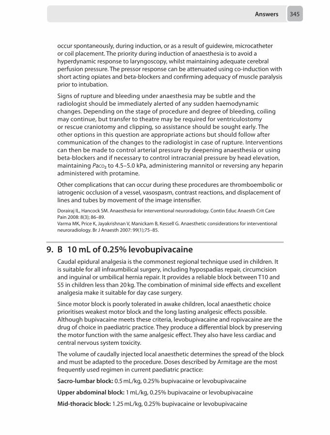
Answers 345
occur spontaneously, during induction, or as a result of guidewire, microcatheter or coil placement. The priority during induction of anaesthesia is to avoid a hyperdynamic response to laryngoscopy, whilst maintaining adequate cerebral perfusion pressure. The pressor response can be attenuated using co-induction with short acting opiates and beta-blockers and confirming adequacy of muscle paralysis prior to intubation.
Signs of rupture and bleeding under anaesthesia may be subtle and the radiologist should be immediately alerted of any sudden haemodynamic changes. Depending on the stage of procedure and degree of bleeding, coiling may continue, but transfer to theatre may be required for ventriculostomy or rescue craniotomy and clipping, so assistance should be sought early. The other options in this question are appropriate actions but should follow after communication of the changes to the radiologist in case of rupture. Interventions can then be made to control arterial pressure by deepening anaesthesia or using beta-blockers and if necessary to control intracranial pressure by head elevation, maintaining Paco₂ to 4.5–5.0 kPa, administering mannitol or reversing any heparin administered with protamine.
Other complications that can occur during these procedures are thromboembolic or iatrogenic occlusion of a vessel, vasospasm, contrast reactions, and displacement of lines and tubes by movement of the image intensifier.
Dorairaj IL, Hancock SM. Anaesthesia for interventional neuroradiology. Contin Educ Anaesth Crit Care Pain 2008: 8(3); 86–89.Varma MK, Price K, Jayakrishnan V, Manickam B, Kessell G. Anaesthetic considerations for interventional neuroradiology. Br J Anaesth 2007: 99(1);75–85.
9. B 10 mL of 0.25% levobupivacaineCaudal epidural analgesia is the commonest regional technique used in children. It is suitable for all infraumbilical surgery, including hypospadias repair, circumcision and inguinal or umbilical hernia repair. It provides a reliable block between T10 and S5 in children less than 20 kg. The combination of minimal side effects and excellent analgesia make it suitable for day case surgery.
Since motor block is poorly tolerated in awake children, local anaesthetic choice prioritises weakest motor block and the long lasting analgesic effects possible. Although bupivacaine meets these criteria, levobupivacaine and ropivacaine are the drug of choice in paediatric practice. They produce a differential block by preserving the motor function with the same analgesic effect. They also have less cardiac and central nervous system toxicity.
The volume of caudally injected local anaesthetic determines the spread of the block and must be adapted to the procedure. Doses described by Armitage are the most frequently used regimen in current paediatric practice:
Sacro-lumbar block: 0.5 mL/kg, 0.25% bupivacaine or levobupivacaine
Upper abdominal block: 1 mL/kg, 0.25% bupivacaine or levobupivacaine
Mid-thoracic block: 1.25 mL/kg, 0.25% bupivacaine or levobupivacaine
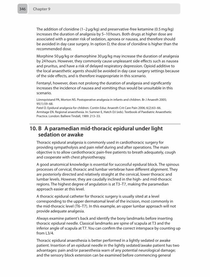
Chapter 9346
The addition of clonidine (1–2 μg/kg) and preservative-free ketamine (0.5 mg/kg) increases the duration of analgesia by 5–10 hours. Both drugs at higher dose are associated with a greater risk of sedation, apnoea or nausea, and therefore should be avoided in day case surgery. In option D, the dose of clonidine is higher than the recommended dose.
Morphine 50 μg/kg or diamorphine 30 μg/kg may increase the duration of analgesia by 24 hours. However, they commonly cause unpleasant side effects such as nausea and pruritus, and have a risk of delayed respiratory depression. Opioid additive to the local anaesthetic agents should be avoided in day case surgery settings because of the side effects, and is therefore inappropriate in this scenario.
Fentanyl, however, does not prolong the duration of analgesia and significantly increases the incidence of nausea and vomiting thus would be unsuitable in this scenario.
Lönnqvistand PA, Morton NS. Postoperative analgesia in infants and children. Br J Anaesth 2005; 95(1):59–68.Patel D. Epidural analgesia for children. Contin Educ Anaesth Crit Care Pain 2006; 6(2):63–66.Armitage EN. Regional anaesthesia. In: Sumner E, Hatch DJ (eds). Textbook of Paediatric Anaesthetic Practice. London: Balliere Tindall, 1989: 213–33.
10. B A paramedian mid-thoracic epidural under light sedation or awake
Thoracic epidural analgesia is commonly used in cardiothoracic surgery for providing sympatholysis and pain relief during and after operations. The main objective is to allow cardiothoracic pain-free patients to breath adequately, cough and cooperate with chest physiotherapy.
A good anatomical knowledge is essential for successful epidural block. The spinous processes of cervical, thoracic and lumbar vertebrae have different alignment. They are posteriorly directed and relatively straight at the cervical, lower thoracic and lumbar levels. However, they are caudally inclined in the high- and mid-thoracic regions. The highest degree of angulation is at T3–T7, making the paramedian approach easier at this level.
A thoracic epidural catheter for thoracic surgery is usually sited at a level corresponding to the upper dermatomal level of the incision, most commonly in the mid-thoracic level (T6–T7). In this example, an upper lumbar approach will not provide adequate analgesia.
Always examine patient’s back and identify the bony landmarks before inserting thoracic epidural needle. Classical landmarks are spine of scapula at T3 and the inferior angle of scapula at T7. You can confirm the correct interspace by counting up from L3/4.
Thoracic epidural anaesthesia is better performed in a lightly sedated or awake patient. Insertion of an epidural needle in the lightly sedated/awake patient has two advantages: pain and/or paraesthesia warn of any potential neurological damage; and the sensory block extension can be examined before commencing general

Answers 347
anaesthesia. Thus in the above clinical scenario, a paramedian mid-thoracic epidural in a lightly sedated or awake patient would be most appropriate.
McLeod GA, Cumming C. Thoracic epidural anaesthesia and analgesia. Contin Educ Anaesth Crit Care Pain 2004; 4(1):16-19.McLeod G, Davies H, Munnoch N, Bannister J, MacRae W. Postoperative pain relief using thoracic epidural analgesia: outstanding success and disappointing failures. Anaesthesia 2001; 56:75–81.Hughes R, Gao F. Pain control for thoracotomy. Contin Educ Anaesth Crit Care Pain 2005; 5(2):56–60.
11. E 0.5 μg/kg fentanyl followed after several minutes by small doses of intravenous midazolam titrated to effect
Sedation is required in a myriad of clinical settings and across many specialties. Complications arise not uncommonly and not just from the inappropriate use of agents, but from the inadequate skills and training of operators, poor patient assessment, and lack of or failure to use appropriate levels of monitoring. Despite the fact that anaesthetists have the detailed knowledge and skills required to give sedation safely, few had received any formal training in sedation per se. Thus, since August 2010, the curriculum for anaesthetic training now includes sections on sedation.
Pre-assessment
Poor or absent pre-assessment is a theme common to many adverse events in sedation practice. Thus, the pre-assessment must be thorough, and to the same standard as that required for formal general anaesthesia. This must include all relevant drugs, allergies, comorbidities and an airway assessment to predict features associated with difficult intubation and/or mask ventilation.
Monitoring
Guidance recommends the use of non-invasive blood pressure, pulse oximetry and ECG which may be modified to suit the needs of the patient, or the degree of sedation being provided. Verbal communication is essential as a monitor of depth of sedation, and if verbal communication is lost the patient requires the same standard of care as for general anaesthesia. Capnography is very useful, and may be essential where clinical assessment during the case is limited by access to the patient, e.g. MRI. Some would recommend capnography for all patients.
Levels of sedation
• Minimal: Normal verbal response, airway normal and responds to pain• Moderate: Responds purposefully to voice or pain, no airway intervention
required• Deep: Only responds after repeated pain, may need airway and ventilator support
Principles and drugs
• Single agents are easier to titrate and tend to be safer• Synergistic effects (e.g. benzodiazepines following an opioid), may increase risks
by reducing safety margins
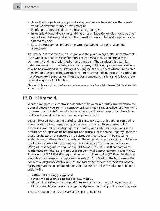
Chapter 9348
• Anaesthetic agents such as propofol and remifentanil have narrow therapeutic windows and thus reduced safety margins
• Painful procedures need to include an analgesic agent• In an opioid/benzodiazepine combination technique, the opioid should be given
and allowed to have a full effect. Then small amounts of benzodiazepine may be titrated to effect
• Loss of verbal contact requires the same standard of care as for a general anaesthetic
The key here is that the procedure (and also the positioning) itself is uncomfortable, even with local anaesthesia infiltration. The patient also takes an opioid in the community, and has established chronic back pain. Thus analgesia is essential. Ketamine would provide sedation and analgesia, but the sympathomimetic effects may be best avoided in the setting of her angina, the severity of which is not stated. Remifentanil, despite being a nearly ideal short-acting opioid, carries the significant risk of respiratory suppression. Thus the best combination is fentanyl, followed later by small aliquots of midazolam.
Blayney MR. Procedural sedation for adult patients: an overview. Contin Educ Anaesth Crit Care Pain 2012; 12 (4):176–180.
12. D < 10 mmol/LWhilst poor glycaemic control is associated with worse morbidity and mortality, the optimal glucose level remains controversial. Early trials suggested benefit from tight glycaemic control (4–6 mmol/L), however recent evidence suggest that there is no additional benefit and in fact, may cause possible harm.
Leuven I was a single centre trial of surgical intensive care unit patients comparing intensive (tight) to conventional glucose control. The results suggested a 34% decrease in mortality with tight glucose control, with additional reductions in the occurrence of sepsis, acute renal failure and critical illness polyneuropathy. However these results were not concurred in a subsequent trial (Leuven II) by the same author in medical intensive care patients. The uncertainty lead to a large multicentre randomised control trial (Normoglycemia in Intensive Care Evaluation-Survival Using Glucose Algorithm Regulation; NICE SUGAR) in 2009. 6,000 patients were randomised to tight (4.5–6 mmol/L) or conventional glucose control (< 10 mmol/L). The results of NICE SUGAR suggested an increase in mortality (27.5% vs 24.9%) and a significant increase in hypoglycemic events (6.8% vs 0.5%) in the tight versus the conventional glucose control groups. The trial evidence was incorporated into the ‘2010 International recommendations for glucose control in the adult non-diabetic critically ill’:
• < 10 mmol/L strongly suggested• severe hypoglycemia is defined as < 2.2 mmol/L• glucose levels should be sampled from arterial rather than capillary or venous
blood, using laboratory or blood gas analysers rather than point of care anaylsers
This is reiterated in the 2012 Surviving Sepsis guidelines:
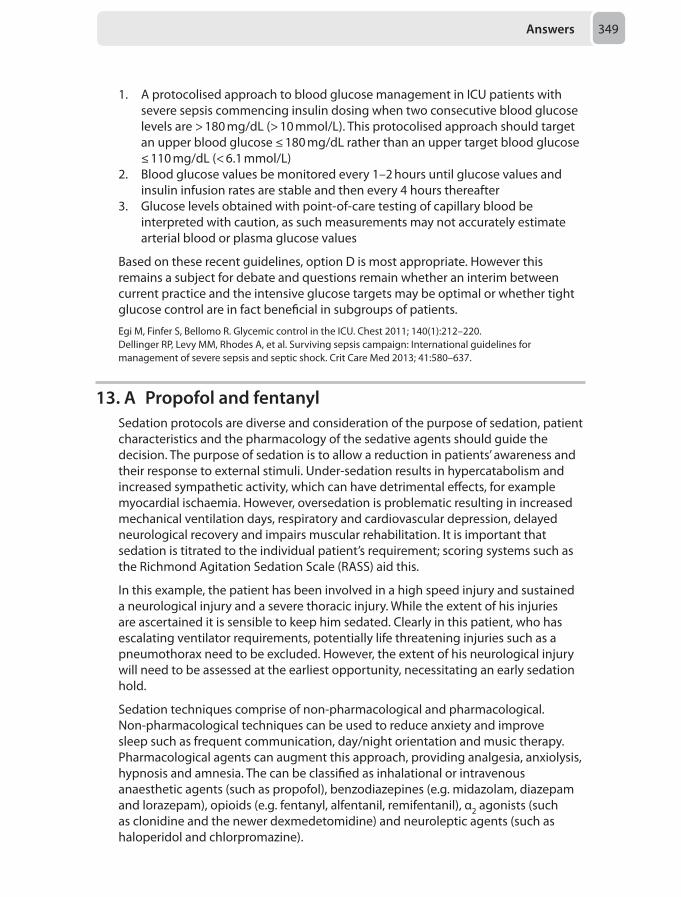
Answers 349
1. A protocolised approach to blood glucose management in ICU patients with severe sepsis commencing insulin dosing when two consecutive blood glucose levels are > 180 mg/dL (> 10 mmol/L). This protocolised approach should target an upper blood glucose ≤ 180 mg/dL rather than an upper target blood glucose ≤ 110 mg/dL (< 6.1 mmol/L)
2. Blood glucose values be monitored every 1–2 hours until glucose values and insulin infusion rates are stable and then every 4 hours thereafter
3. Glucose levels obtained with point-of-care testing of capillary blood be interpreted with caution, as such measurements may not accurately estimate arterial blood or plasma glucose values
Based on these recent guidelines, option D is most appropriate. However this remains a subject for debate and questions remain whether an interim between current practice and the intensive glucose targets may be optimal or whether tight glucose control are in fact beneficial in subgroups of patients.
Egi M, Finfer S, Bellomo R. Glycemic control in the ICU. Chest 2011; 140(1):212–220.Dellinger RP, Levy MM, Rhodes A, et al. Surviving sepsis campaign: International guidelines for management of severe sepsis and septic shock. Crit Care Med 2013; 41:580–637.
13. A Propofol and fentanylSedation protocols are diverse and consideration of the purpose of sedation, patient characteristics and the pharmacology of the sedative agents should guide the decision. The purpose of sedation is to allow a reduction in patients’ awareness and their response to external stimuli. Under-sedation results in hypercatabolism and increased sympathetic activity, which can have detrimental effects, for example myocardial ischaemia. However, oversedation is problematic resulting in increased mechanical ventilation days, respiratory and cardiovascular depression, delayed neurological recovery and impairs muscular rehabilitation. It is important that sedation is titrated to the individual patient’s requirement; scoring systems such as the Richmond Agitation Sedation Scale (RASS) aid this.
In this example, the patient has been involved in a high speed injury and sustained a neurological injury and a severe thoracic injury. While the extent of his injuries are ascertained it is sensible to keep him sedated. Clearly in this patient, who has escalating ventilator requirements, potentially life threatening injuries such as a pneumothorax need to be excluded. However, the extent of his neurological injury will need to be assessed at the earliest opportunity, necessitating an early sedation hold.
Sedation techniques comprise of non-pharmacological and pharmacological. Non-pharmacological techniques can be used to reduce anxiety and improve sleep such as frequent communication, day/night orientation and music therapy. Pharmacological agents can augment this approach, providing analgesia, anxiolysis, hypnosis and amnesia. The can be classified as inhalational or intravenous anaesthetic agents (such as propofol), benzodiazepines (e.g. midazolam, diazepam and lorazepam), opioids (e.g. fentanyl, alfentanil, remifentanil), α2 agonists (such as clonidine and the newer dexmedetomidine) and neuroleptic agents (such as haloperidol and chlorpromazine).
Chapter 9350
In the example of a patient who has sustained extensive chest wall injuries, an opioid to provide analgesia would seem sensible. Whilst clonidine would also provide an analgesic component, it can result in haemodynamic compromise and is unadvisable until visceral or vascular injury has been excluded.
While a pure opioid regime can in theory provide an element of hypnosis, in practice this is difficult to achieve. The ideal characteristic of the choice of hypnotic agent should include ease of titration, short half-life, minimal pharmacodynamics adverse effect, cheap and familiar to both nursing and medical teams.
Of the agents listed, propofol is closest to these ideal characteristics. Midazolam has active metabolites and there are concerns regarding dependence. Ketamine provides dissociative anaesthesia with associated neuropsychiatric sequelae. Ketamine is also a sympathomimetic which may be detrimental especially in the presence of a severe head injury.
Recent trials show promise with dexmedetomidine, when compared to propofol and midazolam. However it remains expensive and is still associated with significant side effects, so its role in the intensive care needs to be further investigated.
Rowe K, Fletcher S. Sedation in the intensive care unit. Contin Educ Anaesth Crit Care Pain 2008; 8(2):50–55.Jakob SM, Ruokonen E, Grounds RM et al. Dexmedetomidine vs midazolam or propofol for sedation during prolonged mechanical ventilation: two randomized controlled trials. JAMA 2012; 307(11):1151–1160.
14. D Commence extra-corporeal membrane oxygenation (ECMO)
This scenario is based on a real case and this patient and her child both survived to discharge neurologically intact.
ECMO uses technology refined from cardiopulmonary bypass circuits used for cardiac surgery. As the technology advances and with the opportunity to gain experience in its use (the H1N1 swine-flu epidemic of 2009) the complication rates have decreased. The CESAR trial evaluated the benefits of ECMO in adult respiratory distress syndrome (ARDS) and demonstrated that patients transferred to a centre offering ECMO had a better outcome (less death or severe disability at 6 months) than those treated at the original hospital with conventional therapy. However, treatment at the ECMO centre did not always involve ECMO and the improvement in outcome was not shown when comparing ECMO verses conventional ventilation at the ECMO centre.
An ECMO circuit can be set up in three ways:
• Venoarterial ECMO: blood is pumped from the venous to the arterial side allowing gas exchange and haemodynamic support
• Venovenous: blood is removed from the venous side and then pumped back into it facilitating gas exchange only
• Arteriovenous: arterial pressure moves the blood from the arterial side to the venous side and facilitates gas exchange. No mechanical pump is required.
The large-bore cannulae are placed surgically or with a percutaneous approach under ultrasound or X-ray guidance. The circuit is more effective at carbon dioxide removal than oxygenation due to differences in solubility between the two gases. Anticoagulation is required as the circuit activates the coagulation cascade.
Answers 351
Complications include:
• Haemorrhagic complications (50% of patients): – 50% of these due to the cannulation, especially at the arterial site – Intracranial bleeding (5%) – Bleeding may occur in any organ
• Thrombosis in the circuit can: – Affect the function of the pump or the oxygenator – Cause stroke – Result in leg ischaemia
• Infective complications can be related to the invasive lines or primary pathology• Technical complications include:
– ECMO circuit failure or breakage – Cannula displacement – Mechanical pump failure
A meta-analysis of studies using ECMO as rescue therapy during cardiopulmonary resuscitation (CPR) demonstrated an increased survival in younger patients after instigating ECMO during or immediately after manual CPR. There is an increasing drive to consider early initiation of ECMO, and the emergency department ECMO project strives to initiate pre-hospital and emergency department ECMO CPR.
Returning to the scenario, this young woman who has had continuous CPR and has not responded to support measures should be considered for ECMO if it is available. To ensure the best outcome, oxygenated blood flow to the brain should be restored as early as possible. Pregnancy is an absolute contra indication to thrombolysis as is having a major operation within 14 days. After a rushed emergency department cesarean section with a low cardiac output state (and therefore difficulty identifying bleeding points) thrombolysis would have a high complication rate may only be considered if no alternative was available.
Martinez G. Extracorporeal membrane oxygenation in adults. Contin Educ Anaesth Crit Care Pain 2012; 12(2):57–61.Cardarelli MG. Use of extracorporeal membrane oxygenation for adults in cardiac arrest (E-CPR): a meta-analysis of observational studies. ASAIO J 2009; 55(6):581–586.
15. C Prostacyclin infusionOne third of critically ill adults develop an acute kidney injury and 5% of these will require renal replacement therapy. This question highlights some of the complexities of managing a patient on renal replacement therapy.
The indications for renal replacement therapy include:
• Fluid balance management• Hyperkalaemia (potassium over 6.5 mmol/L)• Metabolic acidosis (pH < 7.1)• Raised urea (> 30 mmol/L) or symptomatic ureamia• Severe sepsis to remove inflammatory mediators• Removal of water-soluble, low protein-bound drugs, e.g. some antibiotics to
increase dose administered.
Chapter 9352
The different types of renal replacement therapy are:
• Haemo-(ultra)filtration: venous blood is pumped into an extra-corporeal circuit which creates a hydrostatic pressure gradient across a semi-permeable membrane. Plasma (ultrafiltrate) and molecules of less than 50,000 Daltons are forced across by convection. The plasma is replaced by fluid either before or after the filter to maintain volume and haematocrit
• Haemodialysis: venous blood is pumped into a dialyser in which blood is separated by a semi-permeable membrane from a countercurrent flow of dialysis solution. Solute moves along its concentration gradient from blood to dialysis solution (e.g. urea) or from solution to blood (e.g. bicarbonate) by diffusion
• Haemodiafiltration: this is a combination of the two.
The methods can be applied intermittently or continuously, with continuous methods (filtration or dialysis) being preferred on the intensive care unit due to cardiovascular stability.
Anticoagulation is required as all extra-corporeal circuits activate the clotting cascade. Clot that forms within the catheter causes an access pressure alarm, whereas clot that forms in the filter will cause a trans-membrane alarm. The latter will reduce the efficiency of the filter and if it clots of completely then blood within the circuit is lost.
Non-pharmacological methods to prevent clot formation include:
• Ensure adequate driving pressure (venous pressure)• Ensure adequate flow rates through the vascular-catheter (vascath):
– Correct site choice (femoral preferred over right internal jugular which is in turn preferred over left internal jugular veins)
– Good insertion technique – Catheter position and care
• Adding replacement fluid before the filter (pre-dilution) lowers the haematocrit and reduces the chance of filter clot but reduces the efficacy of the filtration process
If the patient has a coagulopathy (INR > 2, APTT > 60 seconds) no anti-coagulation is required, however most patients require pharmacological treatment to prolong the life of the filter.
Pharmacological methods include:
• Unfractionated heparin: increases anti-thrombin III formation. It is the most commonly used, fully reversible by protamine and the anticoagulation effect can be easily titrated by measuring the activated partial thromboplastin ratio (APTR). Given into the circuit before the filter results in less systemic anticoagulation then when given systemically, but monitoring is required and the APTR should be less than 2. There is an increased risk of bleeding and heparin-induced thrombocytopenia
• Prostacyclin or prostaglandin E2 inhibit platelet function and has a short half-life resulting in rapid reversal of the anti-coagulation effect on termination. It causes vasodilatation and therefore can worsen cardiovascular instability, hypoxia in patients dependent on hypoxic pulmonary vasoconstriction, and intracranial hypertension
Answers 353
• Regional citrate: infused pre-filter chelates calcium and prevents clot-formation. Calcium is added post-filter. It is effective and reduces bleeding rates, but may cause metabolic abnormalities and is rarely used in the UK
• Danaparoid and lepirudin may be used instead of heparin if heparin-induced thrombocytopenia develops but is no more effective, bleeding rates are equivocal and they are not easily reversed.
Therefore, in this patient who has been resuscitated (which in the context of a gastrointestinal bleed means the coagulation has been normalised), the safest option in this case would be prostacyclin infused into the filter, which will result in minimal systemic anti-coagulation and may be reversed by terminating the infusion. Heparin given into the circuit still causes systemic anticoagulation and increases the bleeding risk.
Baker A, Green R. Renal replacement therapy in critical care. Anaesthesia Tutorial of the Week No 194. London: World Federation of Societies of Anaesthesiologists, 2010.
16. D Prescribe haloperidolDelirium in critically ill patients is common, with 60–80% of patients being affected. It is characterised by an acute change in cognition and disturbance of consciousness and may follow a fluctuating course. There is an increased length of ventilation, intensive care stay, hospital stay, risk of infection, risk of long-term cognitive impairment and mortality. There is a much higher rate of adverse incidents such as self-extubation and removal of catheters and lines.
The different types of delirium are:
• Hyperactive delirium (5–22%), which is the case described above, and includes agitation, hallucinations and aggression
Table 9.2 Risk factors associated with developing delirium in critically ill patients
Patient AgeSubstance abuse (alcohol, smoking, illicit drugs)HypertensionDepressionExisting cognitive deficiencySensory loss (deafness or blindness)
Clinical conditions Metabolic and electrolyte disturbances (particularly hyponatraemia)SepsisHypoxia or hypercapniaHypotensionIschaemic myocardial eventDisturbances in blood glucose controlPostoperative pain, urinary retention, constipation
Iatrogenic Sedation or analgesic medicationDay-night cycle disruptionImmobilization
Chapter 9354
• Hypoactive delirium is more common, presenting with inattention and decreased situational awareness, but may be peaceful and compliant so is often not diagnosed
• Mixed delirium is a fluctuation between the two extremes above
The risk factors for developing delirium are show in Table 9.2.
There are two different delirium assessment methods in the intensive care unit to be aware of for the exam, both of which are described in detail in the references below:
• Intensive Care Delirium Screening Checklist (ICDSC): Patients are scored for alertness from waking, and then their attention, orientation, agitation or retardation, hallucinations, speech and mood and sleep cycle is scored daily. A score of 4 or more has a sensitivity of 99% but a low specificity of 64%
• Confusion Assessment Method in the intensive care unit (CAM-ICU): This is designed for ventilated patients and has a high sensitivity and specificity. Following a level of consciousness assessment (Richmond agitation sedation scale is commonly used), attention, organised thinking and ability to follow instructions is assessed
Preventative management includes ensuring a correct and adequate sleep pattern, constant information and reassurance regarding their situation and as consistent as possible attendants (family members are the best). The management of delirium is multi-factorial, and includes exclusion of reversible organic causes as described above, which requires an examination and review of relevant investigations.
Pharmacological management can be considered in an escalating fashion:
• Haloperidol 2.5 mg intravenously, doubling the dose every 30 minutes until settled followed by a regular regime. Side-effects include prolongation of the QT interval and an extra-pyramidal movement disorders
• Olanzapine 5 mg orally or intramuscularly may be considered as an alternative• Quetiapine, an atypical antipsychotic is being used increasingly as an alternative
to haloperidol, has equal efficacy and safety, without extra-pyramidal side effects • Dexmedetomidine, an α2 adrenoceptor agonist, similar to clonidine, has also been
used in ventilated patients with delirium and is as effective as haloperidol. A bolus of 0.1 mg/kg followed by 0.2-0.7 mg/kg/hour may be used.
It is worth noting that other sedation methods including opioids and benzodiazepines may contribute to delirium, although benzodiazepines do have a role in alcohol withdrawal.
The question describes a patient in danger of harming himself in the immediate-term, and therefore the preventative measures are likely to be unhelpful. There is no firm evidence of alcohol consumption excess and it is too early in the patients clinical course to blame this on alcohol withdrawal, therefore benzodiazepine treatment may exacerbate his condition. The first line treatment is haloperidol, followed by other measures if unsuccessful.
Cavallazzi R. Delirium in the ICU: an overview. Ann Intensive Care 2012; 2:49.King K, Gratrix A. Delirium in intensive care. Contin Educ Anaesth Crit Care Pain 2009; 9(5):144–147.
Answers 355
17. D Offer her an epidural blood patchPost dural puncture headache (PDPH) is a well-known complication of central neuraxial blockade. In epidural anaesthesia, dural puncture is not always obvious at the time of the procedure, as the Tuohy needle may nick the dura, but not enough to cause a frank CSF spill. Patients with PDPH usually present within 72 hours of the incident with the typical low-pressure headache – worse on standing, coughing, straining and better on lying supine. The headache is usually frontal or occipital and may be associated with symptoms such as neck stiffness, photophobia, nausea or tinnitus. Gutsche’s test may be positive – pressing over the liver with the patient at 45o relieves the headache.
When assessing a patient with suspected PDPH it is important to take a full history and complete a full neurological examination in order to try to rule out more sinister differential diagnosis such as meningitis, cortical vein thrombosis, cerebral infarction and subarachnoid haemorrhage. Other causes such as tension headache, migraine and sinusitis should also be considered, which a thorough history will help with.
Management can be conservative with hydration, paracetamol and non-steroidal anti-inflammatory drugs. Bed rest is also encouraged, but this can be impractical for a nursing mother. Caffeine causes cerebral vasoconstriction and so may provide some relief of the headache, although concrete evidence of benefit is lacking. Sumatriptan is a serotonin receptor agonist used for the treatment of migraine, which again lacks evidence of benefit in PDPH.
This patient has already tried conservative therapies for the past 2 days and the fact that she has returned to the labour ward suggests that she is not coping. Although intravenous fluids may help, it is not the best line of management to take next, as an epidural blood patch (EBP) has the best chance of curing her symptoms. The patient should be offered an EBP with all the risks and benefits explained. Blood cultures were at one point taken routinely at the time of an EBP, but this has fallen out of favour in many units since patients are usually apyrexial and cultures come back negative. In fact, if a patient was pyrexial, this should deter the performance of an EBP and prompt further investigation of another cause for the headache.
Sabharwal A, Stocks GM. Postpartum headache: diagnosis and management. Contin Educ Anaesth Crit Care Pain 2011; 11(5):181–185.
18. A Platelet count <50 x 109/LThe risk of developing a spinal or epidural haematoma as a result of central neuraxial blockade increases in the presence of abnormal coagulation. In the obstetric population, there may be a number of reasons for abnormal coagulation, including pre-eclampsia, disseminated intravascular coagulation (DIC) and gestational thrombocytopenia. If there is any reason to suspect a clotting problem in a patient who may need central neuraxial blockade, a full blood count and clotting screen should be checked.
Chapter 9356
Gestational thrombocytopenia occurs due to a combination of haemodilution and increased platelet turnover, particularly in the third trimester. The platelet count is usually > 70 x 109/L. The increased risks of regional anaesthesia and haemorrhage during delivery should be explained to the mother so she can make an informed decision on choice of anaesthetic.
The lowest acceptable platelet count for performing neuraxial blockade has gradually reduced over the past few years. Current expert opinion is that neuraxial blockade can be performed in parturients with platelet counts > 50 x 109/L, as long as the count is stable and all other clotting indices are normal. Hence, option A is the only scenario in this case that would prevent this lady having a spinal.
Different hospitals have different protocols for regional analgesia, and in general, advocate that a discussion with the consultant anaesthetist take place if the platelet count is less than 70 or 80 x 109/L. In this case, the lady is coming for an elective Caesarean and hence, a consultant anaesthetist should be on the labour ward on the day and often may choose to perform the block themselves instead of the trainee.
An APTT of 30 seconds and a PT of 12 seconds are both within normal limits, hence would not prevent this lady from having a spinal.
Bedson R, Riccoboni A. Physiology of pregnancy: clinical anaesthetic implications. Contin Educ Anaesth Crit Care 2014; 14(2):69-72.Kam PCA, Thompson SA, Liew ACS. Thrombocytopenia in the parturient. Anaesthesia 2004; 59(3):255–264.Lyons G, Hunt BJ. Platelet counts and obstetric analgesia and anaesthesia. Clinical Guidelines: Regional anaesthesia and coagulation. London: Obstetric Anaesthetists’ Association, www.oaa-anaes.ac.uk
19. E Inhalational anaesthetic induction and maintenance with sevoflurane, without muscle relaxant. Maintain spontaneous respiration throughout the case
Presentation of inhaled foreign body can vary from asymptomatic, to partial obstruction with coughing, wheezing, stridor and dyspnoea, to complete obstruction of the upper airway with hypoxia and cardiac arrest. Most foreign bodies are radiolucent and the chest X-ray will often be normal. Therefore, a positive history and clinical signs of aspiration alone may be enough evidence for endoscopy. A chest X-ray in inspiration and expiration may aid location of the foreign body and show any atelectasis, pneumonia, or air trapping.
Whatever anaesthetic technique is used, spontaneous respiration is best preserved, although inhalation induction may be prolonged in the presence of hypoventilation. Sevoflurane in 100% oxygen and topical anaesthesia to the airway is the technique of choice. Care must be taken to maintain spontaneous breathing or gentle assisted ventilation as positive pressure may drive the foreign body distally.
Roberts S. Paediatric bronchoscopy. Contin Educ Anaesth Crit Care Pain 2005; 5:41–44.Fidkowski CW, Zheng H, Firth PG. The anesthetic considerations of tracheobronchial foreign bodies in children: a literature review of 12,979 cases. Anesth Analg 2010; 111:1016–1025.
Answers 357
20. D Arrange for the GP to administer the MMR vaccination 4 weeks postoperatively
The Association of Paediatric Anaesthetists of Great Britain and Ireland (APAGBI) published a guideline on the subject of vaccination around the time of anaesthesia and surgery. One of the questions addressed was: should vaccines be given opportunistically during anaesthetic procedures? The APAGBI concluded that in general, vaccination should not be administered during anaesthesia, in order that paracetamol or other anti-inflammatory agents can be used freely as part of the anaesthetic technique and post-surgical care. This is due to concerns that paracetamol and non-steroidal anti-inflammatory drugs reduce the efficacy and antibody responses to vaccines.
If indicated, vaccination may be given when the child has recovered, but before discharge. However, in the case of this infant with an inguinal hernia repair, paracetamol and other anti-inflammatory drugs are useful for post operative analgesia, so vaccination is best delayed for at least 72 hours. As this clinical scenario refers to a day-case operation, the most appropriate course of action is to arrange for the GP to administer the vaccination after a suitable interval postoperatively.
The Association of Paediatric Anaesthetists of Great Britain and Ireland (APAGBI). Immunisation Guideline: The Timing of Vaccination with Respect to Anaesthesia and Surgery. London: APAGBI, 2013. Prymula R, Siegrist CA, Chlibek R, et al. Effect of prophylactic paracetamol administration at time of vaccination on febrile reactions and antibody responses in children: two open-label, randomised controlled trials. Lancet 2009; 374:1339–1350.
21. C BuprenorphineTrue morphine allergy is rare, but when it does occur patients can safely be prescribed alternate opioids as long as they are structurally different.
Structural classes:
• Diphenylheptanes: methadone• Phenanthrenes: morphine, codeine, buprenorphine, oxycodone• Phenylpiperidines: fentanyl, remifentanil, pethidine
Tramadol is a cyclohexanol derivative and is structurally different to morphine.
Methadone shows no cross-tolerance with other opioids and can be used safely in a true morphine allergy.
Fentanyl and pethidine are synthetic opioids of the phenylpiperidine class. This class of opioid has structures different enough that they can be given to a patient intolerant to the natural or semi-synthetics without fear of cross reactivity. They are also very different from others in this same class.
Buprenorphine is a semi-synthetic opioid and therefore has some structural similarities to morphine, suggesting that there maybe some cross reactivity.
Chapter 9358
Barnett M. Alternative opioids to morphine in palliative care: a review of current practice and evidence. Postgrad Med J 2001; 77:371–378.DeDea L. Prescribing opioids safely in patients with an opiate allergy. JAPA 2012; 25(1):17.
22. A Paracetamol, ibuprofen, gabapentin, MST 25 mg twice daily, Oramorph 10–20 mg 4-hourly
The conversion of transdermal buprenorphine to oral morphine is 1:100.
20 μg/hour = 20 x 24 = 480 μg/day
480 μg x 100 = 48,000 μg = 48 mg per day
Therefore option A gives a background dose to cover the patch and then an as required (PRN) dose which is one-sixth of the daily usage. This is a safe starting point.
B is unsafe with a 2 mg bolus with a short lockout time of 5 minutes as longer lockout times are advocated with larger bolus doses.
C gives a standard fentanyl bolus protocol and there is no cover for background requirement of opiate this patient clearly will need.
D is a standard protocol and does not consider the patient's normal opiate requirement
E Oxycodone is twice as potent as oral morphine therefore a 15 mg b.d. of a modified release oxycodone (Oxycontin) would give a sufficient background. However, the immediate release Oxynorm is not suitable for this purpose.
British National Formulary (BNF). Section 4.7. Pain management with opioids. Analgesics. London: BNF, 2014 www.bnf.org
23. E Morphine patient controlled analgesia (PCA) with a background infusion
Anaesthetists have a fundamental role in providing safe and adequate analgesia for surgical patients, which sometimes includes individuals who are already taking recreational opioids or have preceding chronic pain issues. The salient features in the above case include the history of intravenous heroin use (and likely physiological dependence), the type of surgery (affecting the postoperative route of administration and absorption), as well as the diagnosis of schizophrenia (affecting suitable analgesic choices).
The term ‘opioid’ describes all substances active at the opioid receptor, which includes heroin (diamorphine) and morphine. Chronic opioid use leads to suppression of the noradrenergic system and a compensatory up-regulation of the cyclic adenosine monophosphate signalling pathways in the neurons involved in noradrenaline release. If opioid intake then ceases abruptly, patients will experience a ‘noradrenergic storm’ of withdrawal, which includes shivering, goose bumps, anxiety, and lacrimation. Patients presenting for surgery with a chronic history of heroin use need to be protected against withdrawal, by maintaining adequate opioid receptor agonist, which is commonly achieved by administering methadone
Answers 359
or morphine. There are means of estimating oral methadone equivalence with street heroin, but these are not always reliable.
The problem with oral methadone in this clinical scenario is the anticipated post-operative nil by mouth status and the unpredictable absorption following major abdominal surgery. It is not appropriate to use intravenous methadone as dose equivalence with street heroin can be difficult to determine.
Neuraxial blockade can provide excellent postoperative analgesia following major abdominal surgery, but should not be the sole form of analgesia in the above scenario since the plain bupivacaine epidural solution will not address the opioid dependency. Siting an epidural in the presence of sepsis and coagulopathy will also increase the risk of developing an epidural abscess or haematoma.
Care needs to be taken when providing complex pain management to patients with schizophrenia since certain medications can exacerbate a psychosis. Ketamine and cannabinoid receptor agonists are both associated with this side-effect and should not be used.
The most appropriate way to manage the above patient is to address both his opioid dependency and analgesic requirements by commencing a morphine patient controlled analgesia regime on top of a continuous morphine infusion. These patients need to be monitored closely to assess adequacy of pain relief and for any signs of respiratory depression. Early involvement of the pain team is also advised.
The British Pain Society. Pain and substance misuse: improving the patient experience. London: The British Pain Society, 2007:1–60.Maldonado R. Participation of noradrenergic pathways in the expression of opiate withdrawal: biochemical and pharmacological evidence. Neurosci Biobehav Rev 1997; 21(1):91–104.
24. C Localised external beam radiotherapyThe neurophysiology of cancer pain is complex and can encompass inflammatory, neuropathic, ischaemic as well as compressive processes from multiple sites. It is therefore important when assessing cancer pain to not only identify the location and severity but also recognise the underlying aetiology to help guide management.
Skeletal pain in cancer patients is most commonly associated with bony metastases and management can be tailored to a solitary site or multi-focal areas depending on symptoms. In the above scenario, where the pain is localised to a single metastasis which is refractory to opioids, the most appropriate next step is to apply targeted radiotherapy. The efficacy of this treatment modality in managing metastatic bone pain has been confirmed in a Cochrane review and it can be applied in the palliative setting. The exact mechanism by which radiotherapy provides analgesia is not known, although a reduction in tumour load and local osteoclast activity is believed to play a role. The pain relief evolves consistently over 4–6 weeks from the start of treatment and approximately 80% of patients will have a recorded response. Symptoms such as nausea and increased stool frequency are recognised side-effects of treatment, but are more likely to occur when radiotherapy is applied to bony areas with a significant amount of surrounding bowel (such as the pelvis or lumbar spine).
Chapter 9360
Stabilising surgery is a recognised treatment option for metastatic bony pain in cancer patients, but only in the presence of a pathological fracture. If this is inoperable, patients can still achieve pain relief through application of localised radiotherapy to the fracture site. Since there is no femoral fracture in the above scenario, surgery is not appropriate.
Bisphosphonates are a group of drugs which inhibit osteoclastic bone resorption and can be used in the management of cancer induced bone pain and to prevent skeletal events. However, they do not specifically target solitary metastases, and are more appropriate for when the pain is scattered. Bisphosphonates also have a very poor oral bioavailability and are usually given as intravenous infusions, which may be difficult in patients with poor intravenous access such as the case above.
Radioisotope treatment involves the delivery of radiotherapy to multiple bony metastases via the intravenous administration of a bone seeking radio isotope (usually Strontium 89). It has a similar efficacy to wide field radiotherapy but with a better side effect profile. However, it is more appropriate for patients with pain from multiple bony metastases as opposed to a single site.
Neuropathic pain is a feature which can occur in conjunction with bony metastases (particularly vertebral) to heighten the nociceptive experience. In such cases, drugs such as gabapentin which specifically target the neuropathic component may be applicable. However, it is not the most appropriate next management step in the above scenario since there is no evidence of neuropathic pain.
McQuay H, Collins S, Carroll D. Radiotherapy for the palliation of painful bone metastases. Cochrane Database Syst Rev 2000; 2:CD001793.The British Pain Society. Cancer Pain Management. London: The British Pain Society, 2010: 1–116.
25. D RadiotherapyMetastatic bone pain is a common problem in patients with disseminated malignancy and can be difficult to control with opioid analgesia alone.
In this case increasing her MST is unlikely to help as despite large dose of intermittent Oramorph, pain remains an issue. Opioid rotation can be effective in patients that are developing tolerance to morphine; however this is not the best option here.
Radiotherapy is a very effective treatment for localised bone pain, as shown by two Cochrane reviews. Relief was achieved in 60% of patients with a number needed to treat (NNT) of 3.6 (95% CI 3.2–3.9).
There is evidence to suggest that the use of adjuvant bisphosphonates reduces morbidity from bone metastasis. Results from a Cochrane review suggested that there is only a modest reduction in pain when used in addition to analgesics.
Finally, there is no evidence for the use calcitonin to control pain from bone metastases currently.
The British Pain Society. Cancer Pain Management. London: The British Pain Society, 2010: 1–116.McQuay H, Carroll D, Moore RA. Radiotherapy for painful bone metastases: a systematic review. Clin Oncol 1997; 9:150–154.
Answers 361
Ross JR, Saunders Y, Edmonds PM, et al. Systematic review of role of bisphosphonates on skeletal morbidity in metastatic cancer. Br Med J 2003; 327:469–472.Wong R, Wiffen PJ. Bisphosphonates for the relief of pain secondary to bone metastases. Cochrane Database of Systematic Reviews 2002; 2:CD002068.Martinez MJ, Roqué M. Calcitonin for metastatic bone pain. Cochrane Database of Systematic Reviews 2006; 3:CD003223.
26. B Perform a rapid sequence induction with thiopentone and suxamethonium using an uncut size 8 cuffed oral tracheal tube
As with all emergency situations an ABC approach should be used. While assessing the airway high flow oxygen should be administered via a non-rebreathing mask.
The following features are suggestive of an airway which is at risk:
• Burns sustained in an enclosed space. Flash burns rarely cause an inhalational injury
• Singed eyebrows and nasal hair• Carbonaceous sputum• Erythematous and swollen oral mucosa and uvula• Difficulty swallowing• Hoarse voice• Stridor• Deep facial and neck burns
If there is any concern over the airway it is safer to intubate early. Early intubation is technically easier as the oropharyngeal swelling is not yet established. An uncut large oral tracheal tube should be used. This will allow for any subsequent facial swelling. The large calibre tube will facilitate later bronchoscopy to assess inhalation injury.
Suxamethonium is safe to use in the 24 hours following a burn injury but should be avoided thereafter for up to a year. This is thought to be due to extra-junctional acetylcholine receptors that are expressed following burns which, when activated, leads to a massive efflux of potassium resulting in possible cardiac arrest.
Following a burn injury, the thermostatic centre in the hypothalamus is reset, resulting in a core temperature 1–2 degrees higher than normal.
There is no evidence for the use of prophylactic antibiotics or steroids in burns patients.
Bishop S, Maguire S. Anaesthesia and intensive care for major burns. Contin Educ Anaesth Crit Care Pain 2012; 12(3):118–122.
27. E Administer a bolus of intravenous labetalol 10 mgConfusion in the high dependency unit following carotid endarterectomy (CEA) has a number differential diagnoses but in the context of the above presentation points towards a rare but potentially fatal complication called cerebral reperfusion
Chapter 9362
syndrome. It complicates 1% of carotid endarterectomies. Its presentation ranges from 2 to 7 days, and occurs due to a combination of sustained hypertension associated with various neurological signs and symptoms of cerebral oedema. It is associated with 60% mortality and the mainstay of its treatment is accurate and rapid control of raised blood pressure. The pathophysiology involves lack of auto-regulation of surges in blood pressure due to operation around the carotid bifurcation. This will result in carotid baroreceptor being injured by the surgery and as a result the exposure of intracranial circulation to a rapidly elevated perfusion pressure, resulting in cerebral oedema.
The first option is true if the patient had meningitis, but a normal temperature and no signs of meningism makes it unlikely. Urinary retention is a common cause of confusion in the immediate postoperative phase but is unlikely 3 days later. Mannitol administration is a temporising measure to control intracranial pressure in a patient at risk of cerebral or cerebellar herniation, which is unlikely in a conscious patient. The definitive treatment of this rare syndrome is rapid control of hypertension which means intravenous hypotensive agent such as labetalol.
Ladak N. General or local anaesthesia for carotid endarterectomy? Contin Educ Anaesth Crit Care Pain 2012; 12(2):92–96.
28. B Human erythropoietinPreoperative anaemia is associated with increased perioperative morbidity and should be diagnosed and optimised prior to surgery. In order to differentiate the causes of anaemia, a reticulocyte count can be performed to gauge bone marrow response. A high reticulocyte count suggests regenerative anaemia, which is associated with blood loss or haemolysis. A low reticulocyte count can be further differentiated based on mean corpuscular volume (MCV) into microcytic, normocytic or macrocytic anaemia.
• Microcytic anaemia is associated with iron deficiency or β-thalassaemia• Macrocytic anaemia can be associated with folate and B12 deficiency if
megaloblasts are visualised in a peripheral blood smear. Chronic alcoholism, liver disorders and thyroid disease can cause non-megaloblastic macrocytic anaemia
• Normocytic anaemia with low reticulocytes is associated with renal and hepatic dysfunction, chronic anaemia and myelodysplasia
Erythropoiesis is controlled by erythropoietin, which is a glycoprotein hormone released by the renal cortices. Recombinant human erythropoietin is recommended for anaemia caused by chronic kidney disease and is administered as subcutaneous injections three times a week.
Hans GA, Jones N. Preoperative anaemia. Contin Educ Anaesth Crit Care Pain 2013; 13(3):103-105.
Answers 363
29. C MRI whole spineThe classical triad of fever, backache and neurological symptoms can be seen in patients with suspected epidural abscess. Back pain is the commonest symptom followed by fever and neurological symptoms. Neurological manifestations are noticed late and the diagnosis should be suspected prior to the onset of these signs.
The neurological features occur due to pressure symptoms coupled with vascular effects including ischaemia or thrombosis. Leucocytosis occurs in two thirds of patients and an elevated erythrocyte sedimentation rate (ESR) is much more commonly associated. Normal C-reactive protein (CRP) values cannot exclude epidural abscess.
Magnetic resonance imaging (MRI) with gadolinium is the investigation of choice and should include the whole spine as the catheter tip may lie proximal as compared to the lumbar entry site.
Lumbar punctures may not be positive and more importantly could potentially spread the infection or cause coning in case of elevated intracranial pressures.
Management must be expeditious and use a multidisciplinary approach including radiology, neurosurgeons, intensive care and anaesthesia.
Simpson KH, Al-Makadma YH. Epidural drug delivery and spinal infection. Contin Educ Anaesth Crit Care Pain 2007; 7(4):112-115.
30. D Mann-Whitney U testStatistics is an essential part of critical appraisal. To ascertain the most appropriate statistical test to be applied, a flow chart may help (see Figure 9.3). First the data needs to be ascertained as either qualitative (categorical) or quantitative (continuous).
Qualitative data is descriptive data such as gender, eye colour or ethnicity. The Chi squared test is a good example of a statistical test used for analysis of qualitative data. For smaller samples where the results can be collated by a ‘2 by 2’ table, the Fisher’s exact test may be more appropriate.
Quantitative data can be classified as either parametric (normal) or non-parametric. Further decisions can be guided by whether there are two groups or more than two groups in the study. Within the groups the data can be paired, that is the data was collected from the same sample group. An example would be a study investigating blood pressure measurements in a group of patients before and after a trial antihypertensive is given. Unpaired data suggests two different groups were studied. For example, this study, which compares two groups of patients; those who received discretionary sedation versus those who received protocolised sedation.
From the statistical test employed, a p-value is derived. The p-value reflects the probability the result happened by chance. A commonly applied threshold p-value in clinical trials is < 0.05. This means there is a less than 5% (or 1 in 20) chance of the result occurring by chance. As clinicians, the fundamental outcome has to be considered on the basis of clinical significance, rather than pure statistical significance.
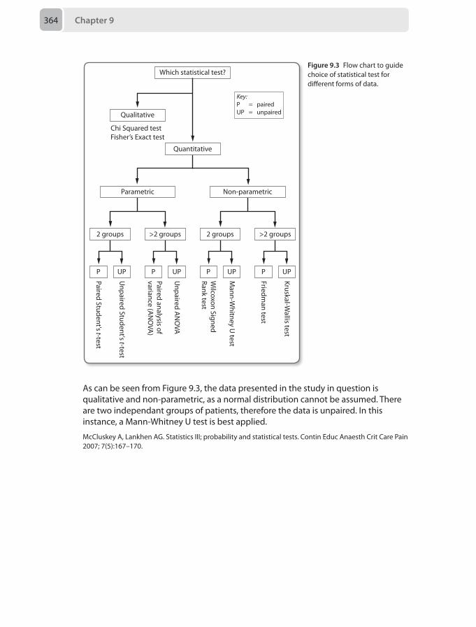
Chapter 9364
As can be seen from Figure 9.3, the data presented in the study in question is qualitative and non-parametric, as a normal distribution cannot be assumed. There are two independant groups of patients, therefore the data is unpaired. In this instance, a Mann-Whitney U test is best applied.
McCluskey A, Lankhen AG. Statistics III; probability and statistical tests. Contin Educ Anaesth Crit Care Pain 2007; 7(5):167–170.
Which statistical test?
Chi Squared testFisher’s Exact test
Qualitative
Key:P = pairedUP = unpaired
Parametric Non-parametricKruskal-W
allis test
Friedman test
Mann-W
hitney U test
Wilcoxon Signed
Rank test
Unpaired A
NO
VA
Paired analysis ofvariance (A
NO
VA)
Unpaired Student's t-test
Paired Student's t-test
2 groups >2 groups 2 groups >2 groups
P UPP UPP UPP UP
Quantitative
Figure 9.3 Flow chart to guide choice of statistical test for different forms of data.
Mock Paper 10
Chapter 10
Questions1. A 36-year-old man requires trans-sphenoidal surgery for a large anterior pituitary
tumour with suprasellar extension.
Prior to induction, which of the following are you most likely to need to prepare?
A Fibreoptic scopeB Lumbar drainC Invasive arterial blood pressure monitoring D Intravenous insulin administrationE Postoperative ventilation
2. A 20-year-old male trauma patient has arrived in the emergency department was had flaccid paralysis of both lower limbs before being intubated at scene for transfer. A full body CT scan revealed a complex vertebral fracture of T5 with a retropulsed fragment and an undisplaced sacral fracture. Despite 2 litres of fluid resuscitation his blood pressure remains 80/40 mmHg with a heart rate of 69 beats per minute.
Which of the following is the most likely reason for his fluid resistant hypotension?
A Spinal shockB Tension pneumothoraxC HaemorrhageD Pulmonary embolusE Neurogenic shock
3. You are asked to anaesthetise a 68-year-old man for rigid bronchoscopy for biopsy of a posterior tracheal mass. He has normal mouth opening and neck extension.
Which of the following is the most appropriate anaesthetic technique?
A Spontaneous ventilation with lidocaine topicalisation of the airway and sedation with intravenous midazolam
B Inhalational induction and maintenance of deep anaesthesia with sevoflurane and oxygen via a facemask removed when the bronchoscope is inserted
C Intravenous induction with propofol and atracurium. Oxygen and sevoflurane maintenance with intermittent positive pressure ventilation (IPPV) via a microlaryngeal tube
Chapter 10366
D Intravenous induction with propofol and atracurium followed by low frequency jet ventilation via the bronchoscope. Maintenance of anaesthesia with propofol and remifentanil target controlled infusion (TCI)
E Placement of cricothyroid cannula followed by anaesthesia with propofol and remifentanil TCI. Jet ventilation via the cricothyroid cannula
4. A 33-year-old man is extracted from a house fire and admitted to the emergency department. He cannot remember being rescued and on examination has singed nasal hair, burns across his neck and productive carbonaceous sputum. He is receiving high flow oxygen through a non-rebreathing mask and is not in respiratory distress.
Which investigation will be most useful in assessing and managing his upper airway?
A Pulse oximetryB Chest X-rayC Computed tomographyD Arterial blood gas E Flexible bronchoscopy
5. A previously well 9-year-old boy is having a laparoscopic appendicectomy. Anaesthesia was induced uneventfully with thiopentone and suxamethonium, followed by a sevoflurane/oxygen mix. 10 minutes after port insertion his airway pressures increase and his oxygen saturations begin to fall. Despite adjusted mechanical ventilation, his end-tidal CO₂ reaches 9.0 kPa. His heart rate rises to 180 beats per minute and his temperature to 40°C.
Which of the following actions will ameliorate the situation most definitively?
A Hyperventilation with 100% oxygen B Cooling the patientC Switching maintenance of anaesthesia to propofolD Treating any hyperkalaemiaE Dantrolene 1–10 mg/kg
6. A 53-year-old man presents for a laparoscopic gastric bypass procedure. He has a BMI of 46 kg/m2 and is an ex-smoker. He has a diagnosis of obstructive sleep apnoea, but doesn’t really use his CPAP machine. He has severe reflux and a neck circumference of 46 cm, his Mallampati score is 1 and he has good mouth opening.
The most appropriate anaesthetic plan includes:
A Ramped head-up position, preoxygenation, and a rapid sequence induction (RSI) with suxamethonium and cricoid pressure. ITU postoperatively
B Ramped head-up position, preoxygenation, and a RSI with rocuronium and cricoid pressure. HDU postoperatively
C Ramped head-up position, preoxygenation, and a RSI with suxamethonium and cricoid pressure. HDU postoperatively
D An awake fibreoptic intubation. Ward level postoperative care.E Ramped head-up position, preoxygenation, standard induction.
Questions 367
7. A 62-year-old woman who presented for a laparoscopic cholecystectomy experienced an unanticipated difficult intubation. An endotracheal tube was eventually correctly sited following multiple attempts and the use of an intubating stylet. The following day she is febrile with cervical surgical emphysema and complains of neck stiffness and dysphagia.
What is the most likely cause of her symptoms?
A Oesophageal perforationB Uvular necrosisC Tracheal ruptureD Arytenoid dislocationE Aspiration pneumonia
8. A cardiac arrest call brings you to a 78-year-old man admitted to coronary care following urgent percutaneous coronary intervention for inferior myocardial infarction. The coronary care nurses administered a total of 3 mg of atropine 5 minutes ago for bradycardia. He is now has a blood pressure of 80/40 mmHg, a heart rate of 35 beats per minute (regular), but is alert.
What is the next appropriate step in the management of his condition?
A Administer 500 μg atropine B Start a dopamine infusionC Urgent transvenous electrical pacingD Start an adrenaline infusionE Give a fluid bolus of 250 mL of colloid
9. A 17-year-old girl presents for surgical correction of a spinal scoliosis. She has dysmenorrhoea and menorrhagia for which she takes oral iron supplementation, and is otherwise fit and well. Her haemoglobin concentration is 101 g/L. The surgeon reminds you that his current practice involves spinal cord monitoring in these cases.
Along with two large-bore peripheral cannulae, which of the following would be the most appropriate anaesthetic technique for this case:
A Volatile with target controlled infusion (TCI) remifentanil maintenance. An internal jugular central line and a radial arterial line. Tranexamic acid.
B A radial arterial line. Propofol and remifentanil TCI. Cell salvage and tranexamic acid.
C Volatile with nitrous oxide maintenance. A radial arterial line. Cell salvage and tranexamic acid.
D Propofol and remifentanil TCI. A radial arterial line. A femoral central line and cell salvage.
E TCI propofol with boluses of fentanyl. A radial arterial line with an internal jugular central line. Tranexamic acid.
Chapter 10368
10. A 75-year-old man with significant co-morbidities is admitted for elective foot surgery under an ultrasound-guided ankle block.
In order to minimise the amount of time required to wait for the block to be adequate for surgery, which one of the following nerves needs to be blocked first?
A Superficial peroneal nerveB Deep peroneal nerveC Sural nerveD Saphenous nerveE Tibial nerve
11. You are asked to transfer a 27-year-old man to the local neurosurgical centre who was admitted two hours ago with an acute traumatic subdural haematoma. On admission he was alert but unable to recall the event, and he vomited twice. On your assessment, he is asleep but rousable to voice, has slurred speech and is obeying commands. His observations include a blood pressure of 180/90 mmHg, heart rate of 90 beats per minute sinus rhythm and pupils of equal size and reacting to light. He continues to vomit in spite of antiemetics.
What the most appropriate next step before the transfer?
A Perform an arterial blood gasB Repeat the CT scanC Rapid sequence inductionD Administer mannitol 20 % 0.5 g/kgE Cool the patient
12. A 42-year-old man presents to the emergency department after an out-of-hospital cardiac arrest. He was successfully defibrillated out of ventricular fibrillation after 20 minutes of cardiopulmonary resuscitation (CPR). The airway was secured and the patient was transferred to hospital. The blood pressure is 120/76 mmHg without the need for inotropes or vasopressors and he is coughing on the endotracheal tube. There is no ischaemia demonstrated on his electrocardiogram (ECG).
What is the clinical action that is likely to be most beneficial?
A Administrating anti-platelet therapy via a nasogastric tubeB An urgent coronary angiogram C An urgent transthoracic echocardiogram D A CT pulmonary angiogram E Targeted temperature management for 24 hours
13 A 59-year-old woman is on the high dependency unit following a bowel resection for adenocarcinoma which finished 8 hours ago. She weighs 60 kg, has a past medical history of type 2 diabetes and has a baseline creatinine of 120 μmol/L. Postoperatively she has been passing 40 mL/hour of urine, which has decreased to 10 mL/hour for the past 2 hours despite her maintaining a normal blood pressure. A repeat blood sample demonstrates a creatinine of 180 μmol/L.
Questions 369
Which of this patient's risk factors is most significant?
A Chronic renal impairment preoperativelyB Major intra-abdominal surgeryC OliguriaD Serum creatinine riseE Diabetes
14. A 72-year-old man with hypertension has been referred to you 2 days after having an emergency laparotomy for an incarcerated hernia. His oxygen saturations are 94% on an inspired oxygen concentration of 60%, his respiratory rate is 28 breaths per minute and on auscultation there is bi-basal crepitus. On examination the blood pressure is 100/60 mmHg, the pulse is regular, the heart rate is 110 beats per minute and the jugular venous pressure (JVP) is visible at 6 cm. He has passed 10 mL of urine per hour for the last 6 hours and is agitated.
What is the next appropriate course of action?
A Urgent blood tests to assess renal functionB Continuous positive airway pressure (CPAP) support in the high dependency
unitC Urgent fluid administration of 5–10 mL/kg of fluid and assess responseD Admit to the intensive care unit for haemofiltrationE Intravenous administration of 40 mg of furosemide and 2.5 mg of diamorphine
15. The intensive care unit dietician suggests commencement of enteral nutrition on a 67-year-old patient.
Which of the follow represents an absolute contraindication to starting enteral nutrition?
A Ischaemic bowelB Small bowel anastomosisC Short gut syndromeD Paralytic ileusE Pancreatitis
16. A 52-year-old hypertenisve woman presents to the emergency department with a history of a sudden onset, severe headache, photophobia and confusion. Her Glasgow coma score (GCS) suddenly declines to 7/15 (E2, V2, M3) with no focal neurology elicited. She is intubated for a CT head, which shows extensive subarachnoid blood. Her current location does not have neurosurgical facilities. According to the World Federation of Neurological Surgeons Scale (WFNSS), her clinical grade is:
A IB IIC IIID IVE V
Chapter 10370
17. A 38-year-old woman presents for a category 2 lower segment Caesarean section (LSCS) for breech presentation. She has recently arrived in the UK from Burma, is 37/40 pregnant and contracting. On examination of her back you notice skin dimpling and a patch of hair at the base of her spine. She tells you that her mother said she was born with an ‘abnormal spinal cord’ but she does not have any further details. She is otherwise fit and well and functions normally.
What is the best line of management for her delivery?
A Urgent MRI before the LSCS, then spinal anaesthesia if an acceptable lumbar level is unaffected
B Perform the LSCS under spinal anaesthesia without prior MRIC Perform the LSCS under epidural anaesthesia without prior MRID Perform the LSCS under general anaesthesiaE Request that the obstetricians deliver her vaginally
18. An 11-year-old boy has been hit by a car travelling at 20 mph.
On arrival to the emergency department. His head and neck are immobilised in a cervical collar, and his respiratory rate is 24 breaths per minute, with oxygen saturations of 94% on 15 litres per minute of oxygen. There is reduced air-entry on the left. His heart rate is 158 beats per minute and blood pressure is 88/32 mmHg, with a peripheral capillary refill time of 4 seconds. He has noticeable bruising to the left side of his chest and abdomen. He does not open his eyes, makes no sounds, and does not respond to commands, but tries to withdraw his arm when an intravenous cannula is sited.
A chest X-ray shows consolidation of the left lung, but no obvious pneumothorax or haemothorax. Bloods have been sent for full blood count and cross match. A 500 mL bag of 0.9% saline is running through his intravenous cannula, and a second intravenous cannula is being inserted.
The next immediate step in his management should be:
A Perform a rapid sequence induction and secure his airway with an endotracheal tube
B Arrange for an urgent CT head, chest and abdomenC Perform a peritoneal lavageD Rapid transfusion of 20 mL/kg of O-negative packed red cellsE Perform a Focused Assessment with Sonography for Trauma (FAST) scan
19. A fit and well 7-month-old 6 kg boy is scheduled for an elective inguinal hernia repair as a day case. As you were inserting an intravenous cannula, you noticed bruises of different ages on both arms and legs. When the infant was positioned for a caudal block, you again noticed bruises of different ages on his back and buttocks. The patient had an eventful surgery and is now in recovery.
What is the most appropriate action regarding the bruises?
Questions 371
A Send a blood sample for an urgent coagulation screenB Speak to the parents and ask them to explain the bruisesC Discuss the case with the hospital’s child protection teamD Advise the surgical team to follow-up the bruisesE Document the bruises in the patient’s medical notes
20. A 19 kg 3-year-old boy is scheduled for an elective dental procedure under general anaesthesia. After an inhalational induction, you have four unsuccessful attempts at placing an intravenous cannula. The patient is maintained under general anaesthesia with oxygen and sevoflurane, breathing spontaneously via a face mask.
The most appropriate next step is:
A Have further attempts at intravenous access until successfulB Increase the inhalational anaesthesia and secure the airway before further
attempts at intravenous accessC Ask another colleague to attempt intravenous accessD Wake the patient up and postpone the procedureE Insert an intra-osseous needle
21. A 61-year-old man has been suffering from long-term low back pain. His MRI one year ago shows multiple degenerative changes and there is no evidence of nerve root compression. He has tried simple analgesics, non-steroidal anti-inflammatory drugs (NSAIDs) and weak opioids without success. Injection therapy has also been unsuccessful.
What would be the most appropriate next step in this patient’s management?
A Add gabapentin 300 mg three times a dayB Repeat the MRIC Referral to surgeryD Referral to a pain management programmeE Offer transcutaneous electrical nerve stimulation (TENS)
22. A 26-year-old woman presents with a 5-year history of generalised pain, stiffness and poor sleep. Extensive investigations have ruled out serious causes and she has been given a diagnosis of fibromyalgia.
Which of the following would apply to this patient?
A Rheumatoid arthritis co-exists in 10%B The incidence decreases with ageC Complete resolution of symptoms is expectedD Pharmacological therapy should be avoidedE Serotonin is increased in most patients
23. A 64-year-old asthmatic man presents with severe burns to his upper left arm and abdomen. The wounds on his left arm extend as far as his shoulder, and now have a foul smelling purulent discharge. He complains of a constant aching pain, which
Chapter 10372
can become very severe at times, both in his arm and his abdomen. The surgeons want to take him to theatre for debridement of his arm wounds.
The most appropriate option for his postoperative analgesia is:
A Left interscalene blockB Paracetamol and ibuprofenC Left axillary blockD Intraoperative opioids and postoperative patient-controlled analgesia (PCA)E Left supraclavicular block
24. A 3-week-old neonate is to have a hernia repair under general anaesthesia with caudal analgesia. His parents ask about alternative methods of pain relief, rather than a caudal block.
Which of the following would be an appropriate alternative?
A Intravenous paracetamol and codeine phosphateB Intravenous morphine intraoperativelyC Paracetamol, ibuprofen, codeine phosphateD Ibuprofen, morphine, and local anaesthetic infiltration to the wound by
surgeonsE Paracetamol, local anaesthetic infiltration to the wound, titrated morphine
25. A 42-year-old man with obstructive sleep apnoea and known sickle cell disease presents with an acutely painful crisis. He suffers from recurrent crises, and on previous hospital admissions he has required significant amounts of opioids administered frequently to control his pain. He regularly takes paracetamol and ibuprofen.
Which of the following is the most appropriate option for his analgesia?
A Morphine PCAB PethidineC CodeineD Fentanyl PCAE Tramadol
26. A 78-year-old woman is admitted with a type 4 (Crawford classification) aortic aneurysm. She is haemodynamically stable but the surgical team is asking for your help in siting an arterial line in preparation for an endovascular repair of her aneurysm using a fenestrated aortic graft.
Which of the following sites would be most appropriate for inserting an arterial line?
A Right radial arteryB Left radial arteryC Right dorsalis pedis arteryD Left dorsalis pedis arteryE Right femoral artery
Questions 373
27. A 72-year-old man requires a unilateral intercostal nerve block for severe pain caused by a fractured rib.
When performing the intercostal nerve block, where would be the most appropriate place to infiltrate the local anaesthetic to achieve the highest success rate for the block?
A At the angle of the ribB Any point proximal to the mid-axillary lineC Medial to the angle of the ribD At the mid-axillary line E Lateral to the angle of the rib
28. A 60-year-old patient with a 5-year history of myasthenia gravis is scheduled for a transcervical thymectomy. He has generalised moderate muscle weakness and has recently been requiring higher doses of pyridostigmine to control his symptoms. His preoperative investigations show forced vital capacity (FVC) of 3.5 litres.
Which of the following would be the most appropriate postoperative management for this patient:
A Reversal and extubation followed by same day discharge B Transfer to the intensive care unit for overnight ventilationC Reversal and extubation followed by monitoring in the high dependency unitD Reversal and use of airway exchange catheter in case re-intubation requiredE Use of elective plasma exchange to assist chances of successful extubation
29. An 83-year-old man is admitted intubated to the intensive care unit following a laparotomy for perforative peritonitis. On the fourth day of his admission his oxygen requirements increase and he begins to desaturate. His oxygen saturation on 75% Fio2 is 86%. His findings are as follows:• Pulse 96 beats per minute• Blood pressure 104/74 mmHg without inotropic support• Arterial blood gas on 75% Pio2 is pH 7.31, Pao2 7.11 kPa, Paco2, 6.14 kPa• Chest X-ray shows a positive ‘Luftsichel sign’.
The most likely lung finding is:
A Left upper lobe collapseB Right upper lobe collapseC Right lower lobe collapseD Right middle lobe collapseE Left lower lobe collapse
30. You have been asked to re-write the departmental guidelines for the treatment of postoperative nausea and vomiting. As part of the research you are reviewing a recent meta-analysis of pharmacological therapy.
With regards to the forest plot in Figure 10.1 which of the following answers is most appropriate?
A All studies would be expected to be weighted equally
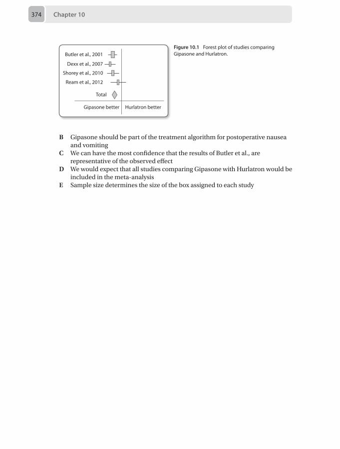
Chapter 10374
B Gipasone should be part of the treatment algorithm for postoperative nausea and vomiting
C We can have the most confidence that the results of Butler et al., are representative of the observed effect
D We would expect that all studies comparing Gipasone with Hurlatron would be included in the meta-analysis
E Sample size determines the size of the box assigned to each study
Butler et al., 2001
Dexx et al., 2007
Shorey et al., 2010
Ream et al., 2012
Total
Gipasone better Hurlatron better
Figure 10.1 Forest plot of studies comparing Gipasone and Hurlatron.
Answers 375
Answers
1. C Invasive arterial blood pressure monitoringThe majority of pituitary tumours are approached surgically by the trans-sphenoidal route. This involves passage through the sphenoid sinus and removal of the floor of the pituitary fossa (sella turcica). The fossa is defined laterally by the cavernous sinus and superiorly by the sella diaphragma. It is important to remain aware that the cavernous sinus contains portions of the carotid arteries – a cause of significant and rapid blood loss in the event of surgical trauma. For this reason, in addition to the potential requirement of careful blood pressure manipulation, invasive arterial pressure monitoring is essential for all trans-sphenoidal pituitary surgery.
The most common pituitary tumours arise from the anterior lobe and are usually adenomas, 75% of which are hormone secreting. Hyper-or hyposecretion of growth hormone (GH), adrenocorticotrophic hormone (ACTH), prolactin (PRL) and thyroid-stimulating hormone (TSH) can occur depending on the cell-origin or mass effect of the tumour. If an adenoma is present that leads to GH or ACTH secretion, the patient may develop acromegaly or Cushing’s disease respectively. In either case, this may lead to glucose intolerance or frank diabetes requiring insulin.
Acromegalic patients often present with soft tissue changes particularly of the larynx and oropharynx. This leads to thickened mucosa, affecting visualisation of the airway, and glottic stenosis. They may also have an enlarged mandible and maxilla resulting in poor occlusion of the dental aperture. This renders them at risk of a difficult airway which can be graded from 1–4. Grade 1 is classified as minimal mucosal involvement, grade 2 as mucosal hypertrophy in the region of the naso pharynx and oropharynx, grade 3 as isolated glottic changes and grade 4 as comprising of elements from both 2 and 3. It is recommended that a fibreoptic intubation is considered for grades 1–2 and a surgical tracheostomy for grades 3–4.
Many patients who suffer Cushingoid or acromegalic effects from their tumour acquire obstructive sleep apnoea. This obligates consideration of safe postoperative airway management and possible ventilation strategies as any positive pressure ventilation applied nasally is prohibited in the immediate period following trans-sphenoidal surgery.
Large pituitary tumours may still be resected trans-sphenoidally, provided they remain midline. If suprasellar extension has occurred a lumbar drain may be required preoperatively. This enables aliquots of normal saline to be introduced during surgery thereby causing increased intracranial pressure and subsequent protrusion of the tumour for improved surgical access. It is also used for drainage postoperatively in the event of cerebrospinal fluid leak from the surgical site.
Trans-sphenoidal surgery may require preparation for all of the options but invasive arterial pressure monitoring is always indicated.
Pollard BJ. Handbook of Clinical Anaesthesia. 2nd ed. London: Elsevier Science, 2003.Smith M, Hirsch NP. Pituitary disease and anaesthesia. Br J Anaesth 2000; 85:3–14.
Chapter 10376
2. E Neurogenic shockA tension pneumothorax should always be considered in a patient who is undergoing positive pressure ventilation after intubation. As a result of increasing pressure within the pleural cavity, the lung collapses and ultimately the mediastinum shifts. This leads to obstructed venous return and therefore persistent hypotension until the pressure is released by needle decompression or chest drain insertion. Hypotension is therefore a relatively late sign and considering the recent CT scan did not show an existing pneumothorax this is not the most likely reason.
All trauma patients with hypotension should be treated with ongoing suspicion of haemorrhage. Most sources of significant blood loss, without obvious external injury, should be identified by a CT scan using contrast media. Even though this patient has had negative imaging, a sacral fracture can lead to the development of a retroperitoneal haematoma. The fracture is, however, undisplaced and although this is not currently the most likely reason it is still one to bear in mind.
Pulmonary embolism is defined as the obstruction of a pulmonary artery or arteriole by intravascular matter such as air, thrombus or fat. If large, it may lead to prevention of flow to the left heart, failure of the right heart and subsequent circulatory collapse. Pulmonary emboli in trauma patients mainly occur as fat (classically secondary to long bone fractures) or thrombus (more often after significant periods of lower limb immobilisation). Although this should be considered it is less likely within the time frame, or associated with the injuries described.
Neurogenic shock occurs when the autonomic pathways are interrupted as in a spinal cord injury. It leads to hypotension and bradycardia. High thoracic injuries are particularly associated with these signs as the cardiac sympathetic fibres originate from T2-T5 thereby resulting in reduced inotropy, unchallenged vagal tone and decreased systemic vascular resistance. This is the most likely reason in this example.
Spinal shock is described as the absence of reflexes below the level of injury. This would produce the flaccid areflexia noted in this case and although normally seen with hypotension from neurogenic shock, does not best define the reason for the patients fluid resistant hypotension.
Bonner S, Smith C. Initial management of acute spinal cord injury. Contin Educ Anaesth Crit Care Pain 2013; 13(6):224–31.
3 D Intravenous induction with propofol and atracurium followed by low frequency jet ventilation via the bronchoscope. Maintenance of anaesthesia with propofol and remifentanil TCIAnaesthesia for airway surgery raises a number of issues including:
• Shared airway with conflicting needs of the surgeon (clear, unobstructed views of the operative field) and anaesthetist (airway protection and maintenance of oxygenation, ventilation and anaesthesia)
• Co-morbidities of the patient group who may have malignant, respiratory and cardiovascular disease
Answers 377
Procedures such as the one outlined above are often relatively short (30 minutes) but intensely stimulating; smooth balanced anaesthesia is essential to reduce the risk of perioperative myocardial ischaemia.
The rigid bronchoscope is a large instrument and it is highly unlikely that a patient would be able to tolerate the procedure without general anaesthesia. Although inhalational induction may be a valid technique, deep anaesthesia alone may not be sufficient (without paralysis) and the use of intermittently removing the facemask will increase the risk of awareness as well as hypoxaemia. Use of a microlaryngeal tube may be acceptable for certain procedures (e.g. supraglottic) but a microlaryngeal tube may occlude the posterior trachea and when inflated the cuff will mean any lesions in all but the most proximal part of the trachea would be inaccessible. These are therefore unfeasible options in this case.
Low frequency jet ventilation is delivered via a handheld trigger device (e.g. Manujet) attached via a Luer lock connector to the rigid bronchoscope. The operator can manually deliver oxygen under pressure at a rate of 10–20 breaths per minute. Volatile anaesthetic agents cannot be delivered via the rigid bronchoscope so total intravenous anaesthesia (TIVA) is required. Intravenous induction with propofol and atracurium with low frequency bronchoscopic jet ventilation and TIVA provides good surgical conditions as well as anaesthesia and is best response given here. It should be noted that there is a risk of barotrauma and gas trapping when using jet ventilation and it is not possible to accurately monitor end tidal carbon dioxide or airway pressures.
High frequency jet ventilation can be delivered via a cricothyroid cannula which may be left in place for emergency perioperative oxygenation in patients felt to be ‘at risk’; for example those will difficult laryngoscopy. This would therefore not be the first choice technique in this case based on the information given.
As these patients are at risk of complete airway obstruction or complications including bleeding, airway oedema, laryngospasm and barotrauma/pneumothoraces it is prudent to be familiar with the difficult airway trolley and have equipment and personnel available to deal with any complications.
English J, Norris A, Bedforth N. Anaesthesia for airway surgery. Contin Educ Anaesth Crit Care Pain 2006; 6(1):28–31.
4. E Flexible bronchoscopySmoke inhalation injury is a serious complication of burns and significantly increases patient morbidity and mortality. Airway injuries in this context can be difficult to safely manage and requires an appreciation of the risk factors, natural progression and appropriate investigations available.
During a fire, the upper airway may be injured from chemical irritation and direct thermal insult resulting in oedema, erythema and ulceration, which can threaten airway patency. Other factors detrimental to the airway include the systemic inflammatory response, aggressive fluid administration and accompanying neck burns causing external compression. The airway oedema is variable but generally peaks at 24 hours and clinical symptoms such as stridor or dyspnoea may not be
Chapter 10378
obvious until this is substantial. A timely and controlled intubation to protect the airway is preferable to an emergency procedure so determining which patients are at risk of upper airway injury or obstruction is important.
Patients who have lost consciousness and been exposed to heat or flames in an enclosed space for a prolonged time are at higher risk of airway injury. Physical signs suggestive of airway injury include facial burns, singed nasal hairs, carbonaceous sputum, stridor, hoarseness and drooling. Certain investigations can also guide assessment and management of inhalational airway injuries
Flexible bronchoscopy is considered the gold standard for early evaluation of the upper airway in patients with smoke inhalational injuries and it is the correct answer for the above scenario. Bronchoscopy allows direct visualisation of the laryngeal structures and an appreciation of any oedema, ulceration, necrosis or soot contaminating and threatening the airway. Furthermore, bronchoscopy allows removal of any airway debris, and the placement of an endotracheal tube if indicated. Repeat examinations can also be performed to assess the progression of airway injury.
Pulse oximetry provides continuous non-invasive monitoring of the haemoglobin oxygen saturation in the arterial blood. It is an important monitor for patients with suspected smoke inhalation injury as desaturations may indicate worsening associated upper or lower airway damage. However, in the presence of carbon monoxide, the monitor will provide an inaccurately high saturation reading since it cannot distinguish between carboxyhaemoglobin and oxyhaemoglobin. Unlike bronchoscopy, it cannot diagnose nor grade the severity of the upper airway injury.
Admission chest X-rays are frequently performed in patients admitted with burns but are insensitive for an inhalational injury diagnosis. Since burns patients are at risk of developing chest infections and acute lung injury during their illness, the admission chest radiograph is however still important for establishing a baseline.
Computed tomography has a role in selected burns patients where inhalation injury is suspected. For example the bronchial wall thickness measured by this imaging modality can be useful as a predictor for the number of ventilator days and the development of pneumonia. Unlike bronchoscopy however, direct visualisation and interventions to treat upper airway pathology is not possible.
Arterial blood gas analysis provides important information concerning the adequacy of ventilation and acid base status of burns patients. If there is co-existing carbon monoxide poisoning, this can also be assessed by carboxyhaemoglobin levels. However, a normal blood gas result does not rule out an inhalation injury, and the investigation provides no direct information on whether the upper airway is threatened.
Micak R, Suman O, Herndon D. Respiratory management of inhalation injury. Burns 2007; 33:2–13.Palmieri T, Gamelli R. Diagnosis and management of inhalation injury. In: Handbook of Burns. Kamolz LP, Jeschke MG, Brychta P et al, eds. New York: Springer, 2012, pp163–72.Yamamura H, Kaga S, Kanada K, Mizobata Y. Chest computed tomography performed on admission helps predict the severity of smoke inhalation injury. Crit Care 2013; 17(3):R95.
Answers 379
5. E Dantrolene 1–10 mg/kgIn the event of an unexplained significant rise in end-tidal CO₂ and heart rate with simultaneous increased oxygen requirements, the possibility of malignant hyperthermia must not be overlooked.
Malignant hyperthermia is a genetically determined error of skeletal muscle metabolism that is triggered by suxamethonium and volatile anaesthetic agents. It is thought to arise from a defective gene for the ryanodine/dihydropyridine receptor on chromosome 19. This leads to an uncontrolled inward flux of calcium ions and subsequent rapid development of skeletal muscle rigidity. This generates a hypermetabolic state producing dramatic rises in CO₂, O₂ consumption and temperature. As the condition persists, haemodynamic instability, rhabdomyolysis, hyperkalaemia, metabolic acidosis and disseminated intravascular coagulation develop.
The mainstays of initial treatment are to rapidly acquire dantrolene at the earliest point of suspicion, whilst simultaneously minimising the deleterious effects of the process. These include informing the team of your diagnosis, calling for help, hyperventilating the patient on 100% oxygen, removing volatile anaesthesia from the circuit, maintaining anaesthesia via intravenous agents, cooling the patient and treating any biochemical abnormalities.
Dantrolene is the only known antidote to malignant hyperthermia and is therefore the most definitive treatment. The Association of Anaesthetists of Great Britain and Ireland (AAGBI) guidelines on the recommended stages of management are available on the AABGI website
Halsall PJ, Hopkins PM. Malignant hyperthermia. Contin Educ Anaesth Crit Care Pain 2003; 3(1):5–9.Yentis SM, Hirsch NP, Smith GB. Anaesthesia and Intensive Care A-Z. An Encyclopaedia of Principles and Practice. 3rd ed. London: Elsevier Science, 2003.
6. C Ramped head-up position, preoxygenation, and a RSI with suxamethonium and cricoid pressure. HDU postoperativelyObesity is on the rise in the UK. Weight loss surgery is also a growing field. The procedures fall into two categories:
Restrictive
The commonest example of this type is the adjustable gastric band (AGB). Here a fluid-filled band is placed around the proximal stomach creating a small pouch that fills quickly with food creating the sensation of fullness. The band can be adjusted by saline insufflation via a subcutaneous port. The AGB is now more popular than the other types of restrictive treatment, such as the sleeve gastrectomy, and luminal gastric balloon. The laparoscopic AGB is minimally invasive, reversible and technically easier and safer than malabsorptive surgery. Complications often relate to relative obstruction or reflux of food or gastric contents, such as oesophagitis.
Chapter 10380
Malabsorptive
The most common procedure of this type is the Roux-en-Y gastric bypass. This surgically creates a small pouch of proximal stomach which is then plumbed directly to the jejunum, ‘bypassing’ the duodenum altogether. Thus the stomach firstly has an element of volume restriction, with the added benefit of a degree of malabsorption. This makes the gastric bypass the gold standard weight loss surgery, with body mass index (BMI) reductions of 10 kg/m2 possible in the first year alone. It is irreversible, more complex, and has added complications including nutritional deficiency and dumping syndrome.
Preoperative assessment
Airway assessment should include neck measurement. Studies have shown that obesity alone doesn’t predict difficult laryngoscopy, but alongside a Mallampati grade III/IV or a high neck circumference, it does. Difficulty rates were 5% with a 40 cm neck, rising to 35% with a 60 cm neck. Medical co-morbidities should be assessed in the usual manner, but particular attention paid to screening for obstructive sleep apnoea, pulmonary hypertension, right heart dysfunction and heart failure. Functional testing in the form of cardiopulmonary exercise testing or stress echocardiography may be indicated. Bariatric patients are regarded as high risk of aspiration regardless of reflux symptoms and prokinetics, and antacids are the norm.
Operative factors
Induction often occurs in theatre on table to avoid moving and handling concerns, but if required a hover mattress may be used for moving patients. The ideal position is with the patient ‘ramped’ or ‘stacked’, this uses pillows and blankets to raise the upper torso, shoulders and head to align the tragus of the ear with the angle of Louis. This has been shown to improve direct laryngoscopy and should facilitate preoxygenation by increasing functional residual capacity. A proprietary pillow, the Oxford HELP pillow, is marketed in the UK for this purpose. The surgical position is usually a modification of the Lloyd–Davis with steep head-up. A shelf is put at the foot of the table to avoid slippage, and the arms are often out on arm boards. The physiological strain of pneumoperitoneum is often well-tolerated, and ventilation is sometimes better than expected due to the degree of positioning the chest above the abdomen. Pressure point protection must be fastidious, as obese patients are at high risk. Greatest risks are from venous thromboembolism, with an incidence of about 5%, and low molecular weight heparin doses must be adjusted to weight as per local protocol.
The crucial elements of the stem here are the presence of untreated obstructive sleep apnoea (OSA), in determining postoperative care, and the airway assessment influencing induction planning. From the outset this gentleman requires higher than ward level care for his OSA and the gastric bypass procedure. HDU should suffice unless he encounters any intraoperative obstacles. In terms of the induction, as discussed the presence of obesity alone doesn’t equal a difficult laryngoscopy, but increasing neck circumference is shown to correlate. His neck circumference does
Answers 381
not put him into the highest risks group. In any event, ramped positioning is crucial to facilitate preoxygenation, laryngoscopy and mask ventilation (if required).
Sabharwal A, Cristelis N. Anaesthesia for Bariatric Surgery. Contin Educ Anaesth Crit Care Pain 2010; 10(4):99–103.
7. A Oesophageal perforationRepeated instrumentation during a difficult intubation can lead to significant damage to the airway and surrounding structures resulting in potentially fatal complications. An unrecognised oesophageal perforation can lead to retropharyngeal abscess formation, acute mediastinitis, pneumonia and eventually multi-organ failure and death. Early symptoms and signs can be non-specific; therefore a high index of suspicion is crucial to avoid delays in management. The case above contains strong risk factors for an oesophageal perforation which includes female gender, age older than sixty years and a difficult intubation. Early symptoms of perforation include sore throat, cervical pain, and cough, whilst fever and dysphagia may indicate secondary bacterial invasion and abscess formation. Air may also dissect along cervical fascial planes resulting in subcutaneous emphysema, pneumomediastinum and pneumothorax. Management depends on lesion severity and includes cessation of oral intake, intravenous antibiotics, parenteral nutrition and if indicated surgical repair.
Tracheal rupture can also occur following a forceful difficult intubation and repeated trauma from airway adjuncts. Following atraumatic intubations, tracheal injury can still occur if the endotracheal tube is incorrectly sized or the tube cuff over-inflated. The most common clinical signs are subcutaneous emphysema, mediastinal emphysema and pneumothorax, which often develop soon after extubation. Other signs include dyspnoea, dysphonia, cough, haemoptysis and pneumoperitoneum. The history of fever and dysphagia in the case above make oesophageal perforation more likely. The management of a tracheal rupture can be conservative (intubation with the cuff distal to the rupture, tracheal aspiration, pleural drain if required and empirical antibiotics) or involve surgical correction.
Uvular necrosis is a rare occurrence and can result from mechanical trauma during intubation or suctioning. Intraoperative impingement from the endotracheal tube compromising uvular blood flow has also been described. Symptoms include a foreign body sensation, sore throat, pain on swallowing, coughing and in severe cases airway obstruction. Subcutaneous cervical emphysema as described in the above case is not a usual presentation of uvular necrosis. Treatment is conservative and management options reported in the literature includes steroids, antibiotics, topical adrenaline administration and antihistamines.
Arytenoid dislocation can occur as a consequence of direct trauma to the cricoarytenoid joint during endotracheal intubation. Symptoms include persistent hoarseness, sore throat dysphagia and stridor. Prompt diagnosis and early operative correction is important to prevent articular adhesions and ankylosis. A primary arytenoid dislocation does not cause surgical emphysema as described in the case above.
Chapter 10382
Aspiration of gastric contents into the lung can occur following repeated intubation attempts to a difficult airway. The clinical manifestations are wide ranging and depend partly on the type and amount of aspirate. Solid matter aspiration can lead to an acute airway obstruction resulting in rapidly progressive hypoxia, whereas gastric acid contamination can result in an aspiration pneumonitis and the acute respiratory distress syndrome. Infection from bacteria that normally reside in the stomach or upper airway can give rise to pyrexia, wheezes and crackles. Treatment is mainly supportive and sometimes prolonged mechanical ventilation is necessary.
Antibiotics should only be administered to patients who develop pneumonia. Surgical emphysema, dysphagia and neck stiffness are not common presentations of aspiration pneumonia.
Domino K, Posner K, Caplan R, Cheney F. Airway Injury during Anaesthesia: a closed claims analysis. Anesthesiology 1999; 91(6):1703–11.Miñambres E, Burón J, Ballesteros M, et al. Tracheal rupture after endotracheal intubation: a literature systematic review. Eur J Cardiothorac Surg 2009; 35(6):1056–62.Hagberg C, Georgi R, Krier C. Complications of managing the airway. Best Pract Res Clin Anaesthesiol 2005; 19(4):641–59.
8. D Start an adrenaline infusionThe patient most likely has a symptomatic complete heart block that is usually associated with an inferior myocardial infarction (MI). He demonstrates adverse features in the form of hypotension; others to be concerned about include syncope, heart failure or myocardial ischaemia.
The initial management would be administration of 500 μg of atropine intravenously and assess the patient’s response. If the patient fails to respond to 3 mg of Atropine in total, the next step is to start a second line agent such as an isoprenaline infusion at 5 μg/minute or an adrenaline infusion at 2–10 μg/minute. Alternative drugs can be considered such as aminophylline, dopamine, glucagon or glycopyrronium bromide, however in a hypotensive patient, adrenaline is a more appropriate option.
Fluids are an appropriate option to treat hypotension without bradyarrhythmia following an inferior MI but will not correct the cause of hypotension.
Transcutaneous and transvenous cardiac pacing are suitable alternatives to the pharmacological treatment, the latter requiring significant expertise that might not be immediately available, but is ultimately the aim.
Resuscitation Council (UK). ALS guidelines for bradyarrhythmias (2010 Resuscitation Guidelines). London: Resuscitation Council (UK), 2010.
9. B A radial arterial line. Propofol and remifentanil TCI. Cell salvage and tranexamic acidOperations on the spine are liable to cause serious complications, and the understanding of the type of surgery and the general principles is essential to prevent attendant morbidity. Massive haemorrhage requiring transfusion is one of the most common, but respiratory or airway compromise, eye injuries (including blindness), and spinal cord damage may also occur.
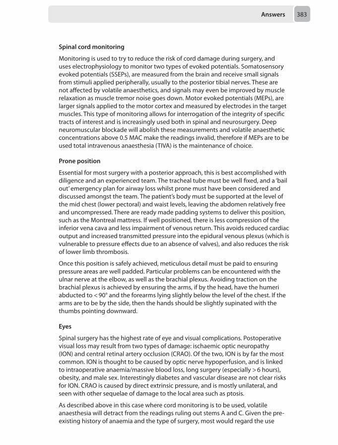
Answers 383
Spinal cord monitoring
Monitoring is used to try to reduce the risk of cord damage during surgery, and uses electrophysiology to monitor two types of evoked potentials. Somatosensory evoked potentials (SSEPs), are measured from the brain and receive small signals from stimuli applied peripherally, usually to the posterior tibial nerves. These are not affected by volatile anaesthetics, and signals may even be improved by muscle relaxation as muscle tremor noise goes down. Motor evoked potentials (MEPs), are larger signals applied to the motor cortex and measured by electrodes in the target muscles. This type of monitoring allows for interrogation of the integrity of specific tracts of interest and is increasingly used both in spinal and neurosurgery. Deep neuromuscular blockade will abolish these measurements and volatile anaesthetic concentrations above 0.5 MAC make the readings invalid, therefore if MEPs are to be used total intravenous anaesthesia (TIVA) is the maintenance of choice.
Prone position
Essential for most surgery with a posterior approach, this is best accomplished with diligence and an experienced team. The tracheal tube must be well fixed, and a ‘bail out’ emergency plan for airway loss whilst prone must have been considered and discussed amongst the team. The patient’s body must be supported at the level of the mid chest (lower pectoral) and waist levels, leaving the abdomen relatively free and uncompressed. There are ready made padding systems to deliver this position, such as the Montreal mattress. If well positioned, there is less compression of the inferior vena cava and less impairment of venous return. This avoids reduced cardiac output and increased transmitted pressure into the epidural venous plexus (which is vulnerable to pressure effects due to an absence of valves), and also reduces the risk of lower limb thrombosis.
Once this position is safely achieved, meticulous detail must be paid to ensuring pressure areas are well padded. Particular problems can be encountered with the ulnar nerve at the elbow, as well as the brachial plexus. Avoiding traction on the brachial plexus is achieved by ensuring the arms, if by the head, have the humeri abducted to < 90° and the forearms lying slightly below the level of the chest. If the arms are to be by the side, then the hands should be slightly supinated with the thumbs pointing downward.
Eyes
Spinal surgery has the highest rate of eye and visual complications. Postoperative visual loss may result from two types of damage: ischaemic optic neuropathy (ION) and central retinal artery occlusion (CRAO). Of the two, ION is by far the most common. ION is thought to be caused by optic nerve hypoperfusion, and is linked to intraoperative anaemia/massive blood loss, long surgery (especially > 6 hours), obesity, and male sex. Interestingly diabetes and vascular disease are not clear risks for ION. CRAO is caused by direct extrinsic pressure, and is mostly unilateral, and seen with other sequelae of damage to the local area such as ptosis.
As described above in this case where cord monitoring is to be used, volatile anaesthesia will detract from the readings ruling out stems A and C. Given the pre-existing history of anaemia and the type of surgery, most would regard the use
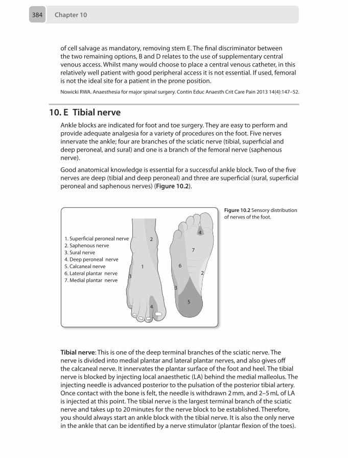
Chapter 10384
of cell salvage as mandatory, removing stem E. The final discriminator between the two remaining options, B and D relates to the use of supplementary central venous access. Whilst many would choose to place a central venous catheter, in this relatively well patient with good peripheral access it is not essential. If used, femoral is not the ideal site for a patient in the prone position.
Nowicki RWA. Anaesthesia for major spinal surgery. Contin Educ Anaesth Crit Care Pain 2013 14(4):147–52.
10. E Tibial nerveAnkle blocks are indicated for foot and toe surgery. They are easy to perform and provide adequate analgesia for a variety of procedures on the foot. Five nerves innervate the ankle; four are branches of the sciatic nerve (tibial, superficial and deep peroneal, and sural) and one is a branch of the femoral nerve (saphenous nerve).
Good anatomical knowledge is essential for a successful ankle block. Two of the five nerves are deep (tibial and deep peroneal) and three are superficial (sural, superficial peroneal and saphenous nerves) (Figure 10.2).
1
3
4
2
3
5
2
7
6
1. Super�cial peroneal nerve2. Saphenous nerve3. Sural nerve4. Deep peroneal nerve5. Calcaneal nerve6. Lateral plantar nerve7. Medial plantar nerve
4
Tibial nerve: This is one of the deep terminal branches of the sciatic nerve. The nerve is divided into medial plantar and lateral plantar nerves, and also gives off the calcaneal nerve. It innervates the plantar surface of the foot and heel. The tibial nerve is blocked by injecting local anaesthetic (LA) behind the medial malleolus. The injecting needle is advanced posterior to the pulsation of the posterior tibial artery. Once contact with the bone is felt, the needle is withdrawn 2 mm, and 2–5 mL of LA is injected at this point. The tibial nerve is the largest terminal branch of the sciatic nerve and takes up to 20 minutes for the nerve block to be established. Therefore, you should always start an ankle block with the tibial nerve. It is also the only nerve in the ankle that can be identified by a nerve stimulator (plantar flexion of the toes).
Figure 10.2 Sensory distribution of nerves of the foot.
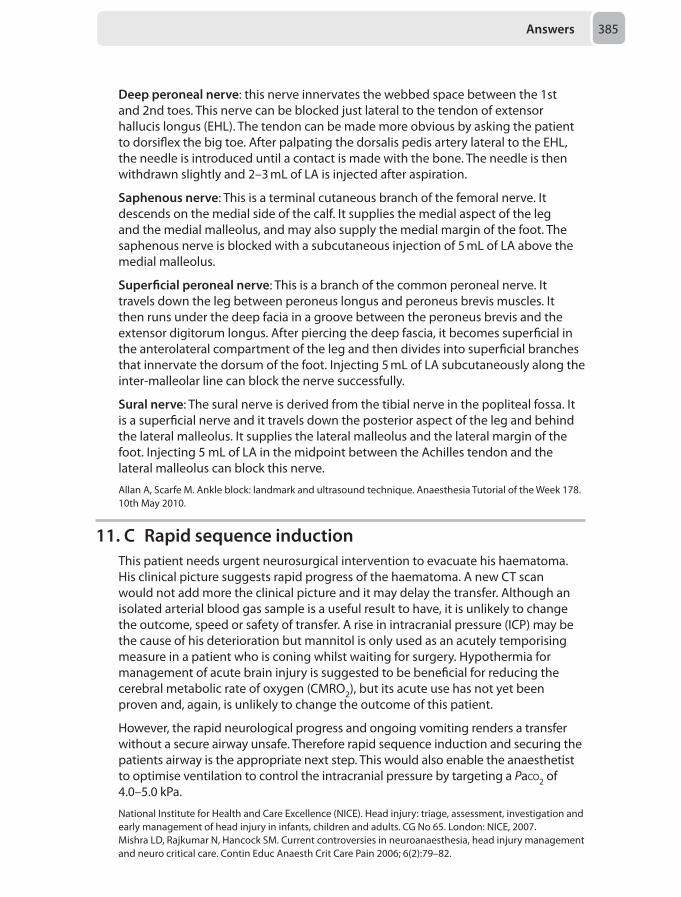
Answers 385
Deep peroneal nerve: this nerve innervates the webbed space between the 1st and 2nd toes. This nerve can be blocked just lateral to the tendon of extensor hallucis longus (EHL). The tendon can be made more obvious by asking the patient to dorsiflex the big toe. After palpating the dorsalis pedis artery lateral to the EHL, the needle is introduced until a contact is made with the bone. The needle is then withdrawn slightly and 2–3 mL of LA is injected after aspiration.
Saphenous nerve: This is a terminal cutaneous branch of the femoral nerve. It descends on the medial side of the calf. It supplies the medial aspect of the leg and the medial malleolus, and may also supply the medial margin of the foot. The saphenous nerve is blocked with a subcutaneous injection of 5 mL of LA above the medial malleolus.
Superficial peroneal nerve: This is a branch of the common peroneal nerve. It travels down the leg between peroneus longus and peroneus brevis muscles. It then runs under the deep facia in a groove between the peroneus brevis and the extensor digitorum longus. After piercing the deep fascia, it becomes superficial in the anterolateral compartment of the leg and then divides into superficial branches that innervate the dorsum of the foot. Injecting 5 mL of LA subcutaneously along the inter-malleolar line can block the nerve successfully.
Sural nerve: The sural nerve is derived from the tibial nerve in the popliteal fossa. It is a superficial nerve and it travels down the posterior aspect of the leg and behind the lateral malleolus. It supplies the lateral malleolus and the lateral margin of the foot. Injecting 5 mL of LA in the midpoint between the Achilles tendon and the lateral malleolus can block this nerve.
Allan A, Scarfe M. Ankle block: landmark and ultrasound technique. Anaesthesia Tutorial of the Week 178. 10th May 2010.
11. C Rapid sequence inductionThis patient needs urgent neurosurgical intervention to evacuate his haematoma. His clinical picture suggests rapid progress of the haematoma. A new CT scan would not add more the clinical picture and it may delay the transfer. Although an isolated arterial blood gas sample is a useful result to have, it is unlikely to change the outcome, speed or safety of transfer. A rise in intracranial pressure (ICP) may be the cause of his deterioration but mannitol is only used as an acutely temporising measure in a patient who is coning whilst waiting for surgery. Hypothermia for management of acute brain injury is suggested to be beneficial for reducing the cerebral metabolic rate of oxygen (CMRO2), but its acute use has not yet been proven and, again, is unlikely to change the outcome of this patient.
However, the rapid neurological progress and ongoing vomiting renders a transfer without a secure airway unsafe. Therefore rapid sequence induction and securing the patients airway is the appropriate next step. This would also enable the anaesthetist to optimise ventilation to control the intracranial pressure by targeting a Paco2 of 4.0–5.0 kPa.
National Institute for Health and Care Excellence (NICE). Head injury: triage, assessment, investigation and early management of head injury in infants, children and adults. CG No 65. London: NICE, 2007.Mishra LD, Rajkumar N, Hancock SM. Current controversies in neuroanaesthesia, head injury management and neuro critical care. Contin Educ Anaesth Crit Care Pain 2006; 6(2):79–82.
Chapter 10386
12. E Targeted temperature management for 24 hoursThis is a difficult question. All are appropriate actions to ensure complete management of your patient. The differential diagnosis of a sudden collapse is broad, but most significantly could be:
• Arrhythmogenic (sudden acute arrhythmia such as ventricular fibrillation or complete heart block);
• Cardiogenic (such as an acute myocardial infarction causing myocardial failure); • Vascular (aortic aneurysm rupture);• Obstructive (such as a pulmonary embolism);• Intracranial event (such as a subarachnoid hemorrhage).
Making efforts to diagnose the cause of arrest will no doubt help in management. For the options above:
1. There is no evidence of ischaemia on the ECG, suggesting no sudden occlusion of a coronary vessel. Therefore anti-platelets may not be the key intervention. Similarly a coronary angiogram may demonstrate unobstructed coronary arteries.
2. An urgent echocardiogram is vital to look at cardiac function. It is particularly helpful at diagnosing a sudden ischaemic event (with regional wall abnormality) or suggesting a pulmonary embolism (with a dilated right ventricle and high pulmonary pressures). However since blood pressure is maintained without augmentation it is questionable whether the echocardiogram will provide you with information that will alter the management in the immediate-term.
3. A CT pulmonary angiogram is an important test and may provide a diagnosis. With a normal blood pressure thrombolysis is not indicated. Treatment with anticoagulation may be presumptively commenced (also indicated for coronary vessel disease).
4. There is no option for an urgent CT head but this would also be a vital investigation as an intra-cranial event may require emergency surgical management.
The question is asking for the most beneficial intervention, not the first. Therapeutic hypothermia after out-of-hospital cardiac arrest has got a good body of evidence that suggests improved neurological outcome and mortality outcomes at 6 months. A recent study has demonstrated that a targeted temperature management approach of 36°C is non-inferior to targeting a temperature of 33°C, the suggestion is that a targeted approach and avoidance of pyrexia are more important than the specific temperature chosen. Many departments would still opt to target a temperature of 33°, but this practice appears to be decreasing.
Should a temperature of 32–34°C be chosen, cooling should be commenced in the emergency department with infusion of cool crystalloids and application of cooling blankets if possible. In the intensive care unit this is continued until a temperature of 32–34°C is achieved within 4 hours of return of spontaneous circulation. This may be done with cooling blankets or intravascular extra-corporeal devices, controlled electronically and titrated to maintain desired temperature. Hypothermia is maintained for 12–24 hours before re-warming commences at 0.25–0.5°C/hour and neurological function can then be assessed. Complications should be managed in a supportive manner including shivering, which should be treated with
Answers 387
pharmaceutical paralysis as it is metabolically demanding and increases myocardial oxygen demand. It remains to be seen if this therapeutic method will remain accepted practice in the future, but what is accepted is that targeted temperature management is perhaps more important than the specific temperature chosen.
Luscombe M. Clinical applications of induced hypothermia. Contin Educ Anaesth Crit Care Pain 2006; 6 (1):23–27.Deakin CD, Nolan JP, Soar J, et al. European Resuscitation Council guidelines for resuscitation 2010, Section 4: adult advanced life support. Resuscitation 2010;81:1305–52.Nielsen N, Wetterslev J, Cronberg T, et al. Targeted temperature management at 33°C versus 36°C after cardiac arrest. N Engl J Med 2013; 369(23):2197–206.
13. D Serum creatinine riseAcute kidney injury (AKI), is defined as an abrupt deterioration in renal function occurring over 48 hours. The prevalence in hospitals is 1–7% and even a small rise in creatinine is associated with an increased mortality, ranging from 10% to 80%.
In 2009 the National Confidential Enquiry into Patient Outcome and Death (NCEPOD) published ‘Adding Insult to Injury’ which found that we were deficient in identifying patients at risk of AKI and in 50% of cases our management could have been more timely to prevent renal deterioration. This prompted the National Institute for Health and Care Excellence (NICE) to publish recommendations highlighting the importance of identification of risk factors and of prompt treatment.
Risk factors include:
• Age over 65 years old • Male gender • Pre-existing renal disease• Co-morbidities:
– Congestive cardiac disease – Hypertension – Diabetes – Ascites
• Surgery: – Emergency – Prolonged, major surgery – Intra-peritoneal
• Anaesthesia: – Hypotension – Hypovolaemia – Blood transfusion – Nephrotoxic drug administration
The diagnosis may be made with either the RIFLE, AKIN or KDIGO classifications (the latter two being very similar). They are based on RIFLE (Risk, Injury, Failure, Loss, End-stage), which is a useful way to remember the steps to disaster, but the recent modification is more sensitive and reflects recent evidence that even a small increase in the serum creatinine concentration results in significantly increased morbidity and mortality (see Tables 10.1 and 10.2).
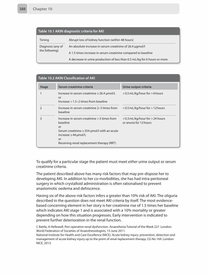
Chapter 10388
To qualify for a particular stage the patient must meet either urine output or serum creatinine criteria.
The patient described above has many risk factors that may pre-dispose her to developing AKI. In addition to her co-morbidities, she has had intra-peritoneal surgery in which crystalloid administration is often rationalised to prevent anastomotic oedema and dehiscence.
Having six of the above risk factors infers a greater than 10% risk of AKI. The oliguria described in the question does not meet AKI criteria by itself. The most evidence-based concerning element in her story is her creatinine rise of 1.5 times her baseline which indicates AKI stage 1 and is associated with a 10% mortality or greater depending on how this situation progresses. Early intervention is indicated to prevent further deterioration in the renal function.
C Battle, A Hellewell. Peri-operative renal dysfunction. Anaesthesia Tutorial of the Week 227. London: World Federation of Societies of Anaesthesiologists, 13 June 2011. National Institute for Health and Care Excellence (NICE). Acute kidney injury: prevention, detection and management of acute kidney injury up to the point of renal replacement therapy. CG No 169. London: NICE, 2013.
Table 10.2 AKIN Classification of AKI
Stage Serum creatinine criteria Urine output criteria
1 Increase in serum creatinine ≥ 26.4 μmol/L or Increase > 1.5–2 times from baseline
< 0.5 mL/kg/hour for > 6 hours
2 Increase in serum creatinine 2–3 times from baseline
< 0.5 mL/kg/hour for > 12 hours
3 Increase in serum creatinine > 3 times from baselineorSerum creatinine ≥ 354 μmol/l with an acute increase ≥ 44 μmol/LorReceiving renal replacement therapy (RRT)
< 0.3 mL/kg/hour for > 24 hours or anuria for 12 hours
Table 10.1 AKIN diagnostic criteria for AKI
Timing Abrupt loss of kidney function (within 48 hours)
Diagnosis (any of the following)
An absolute increase in serum creatinine of 26.4 μgmol/l
A 1.5 times increase in serum creatinine compared to baseline
A decrease in urine production of less than 0.5 mL/kg for 6 hours or more
Answers 389
14. E Intravenous administration of 40mg of furosemide and 2.5 mg of diamorphine
This man has the following clinical issues:
1. Clinical evidence of pulmonary oedema with a raised jugular venous pressure (JVP)
2. Hypotension with evidence of end-organ dysfunction (agitation)3. Acute kidney injury (AKI) Stage 1 on the basis of his urine output being less than
0.5 mL/kg/hour for 6 hours, according to the AKIN classification (see Table 10.2, Question 13).
The pulmonary oedema may be due to different etiologies, either due to excess intravascular fluid or ineffective cardiac activity. Excess intravenous fluid administration for the treatment of oliguria post-operatively may be implicated. Alternative possibilities include acute cardiac decompensation due to an ischaemic cardiac event, electrolyte disturbance or a cardiac arrhythmia.
Acute heart failure should be managed with an ABC approach. Sitting the patient upright reduces the central venous pressure and therefore the preload, which can result in improved cardiac output. Oxygen therapy, intravenous access, electrocardiogram, a chest radiograph, an arterial blood gas and repeat blood science analysis including serum troponin concentration are all immediately indicated. Treatment options include:
• Treat reversible causes: – Coronary reperfusion therapies – Anti-arrhythmic agents – Electrolyte correction
• To reduce the preload via venodilation: – Loop diuretic administration results in immediate venodilation – Diamorphine or morphine administration reduces central sympathetic activity
resulting in venodilation – Glyceryl trinitrate (GTN) infusion
• To reduce the preload by reducing circulating volume: – Loop diuretic administration results in delayed diuresis – Haemofiltration for accurate control of fluid balance
• To augment cardiac output: – Continuous positive airway pressure or non-invasive ventilation reduces
ventricular distention and improves cardiac contractility – Inotrope administration – Mechanical augmentation such as a intra-aortic balloon pump
• To provide cardiac or respiratory replacement: – Extra-corporeal membrane oxygenation
Regarding the options in the question, all interventions may be needed at some stage. Investigations are important but take time to yield results and more urgent action is required in this situation. Fluid administration may be used to treat hypotension and oliguria but in the presence of pulmonary oedema (the more pressing clinical priority) may be detrimental. That leaves us with management of
Chapter 10390
his heart failure with the most appropriate therapy at this stage being the easiest to administer, the least invasive and the most rapid to instigate.
Valchanov K. Inpatient management of advanced heart failure. Contin Educ Anaesth Crit Care Pain 2008; 8(5):167–71.
15. A Ischaemic bowelDuring critical illness or the perioperative period, it is important to consider every patient’s nutritional status and requirements. Nutritional requirements should aim to address any pre-existing malnutrition, support the catabolic response to surgery or critical illness and aid rehabilitation. Malnutrition is associated with impaired immunological function and increased morbidity and mortality.
The detection of acute malnutrition can be challenging and at present no specific markers exist. Surrogate markers for chronic nutritional status include anthropometric measures and biomarkers such as albumin, pre-albumin, transferring and haemoglobin.
Following the advent of acute critical illness, the early (within 24–48 hours) instigation of nutritional support has been demonstrated to improve patient outcomes, however conflicting evidence exists concerning the merits of hypocaloric verses normocaloric targets. Furthermore, the optimal method (continuous verses bolus) and composition remains undefined.
The National Institute for Health and Care Excellence (NICE) recommendations for assessing a patient’s nutritional requirements are summarised below:
1. Screening on admission to include:• assessment of body mass index (BMI)• percentage unintentional weight loss and time over which nutrient intake has
been unintentionally reduced• likelihood of future impaired nutrient intake
2. Recognise• Malnutrition
– BMI < 18.5 kg/m2
– unintentional weight loss > 10% in past 3–6 months – BMI <20 kg/m2 or > 5% unintentional weight loss – over 3–6 months
• At risk of malnutrition – eaten little or nothing for more than 5 days and/or likely to eat little or
nothing for the next 5 days or longer – poor absorptive capacity, and/or high nutrient losses and/or increased
nutritional needs from causes such as catabolism3. Treat
• Oral if safe swallow• Enteral if unsafe swallow or inadequate oral intake and gastrointestinal tract
accessible• Parenteral if unsafe swallow or inadequate enteral intake and gastrointestinal
tract inaccessible• Correct prescribing
Answers 391
4. Monitor indications, route, risks, benefits and goals of nutrition support at regular intervals
There are published guidelines by NICE and The American Society for Parenteral and Enteral Nutrition (ASPEN) in partnership with the Society of Critical Care Medicine (SCCM), covering all aspects of nutritional support in the acute patient and critical care settings. Though much debate continues with regard to the appropriate indications for parenteral nutrition, most agree that it should not be commenced unless the enteral route is inaccessible and likely to remain so for > 7days. The UK CALORIES Trial published in October 2014 found no mortality difference between enteral and parenteral routes of feeding critically ill patients. The current balance of evidence suggests significant advantages of the enteral route that include less infectious complications, thought to be secondary to reduced villous atrophy and bacterial translocation within the gastrointestinal tract.
Previous theories mandating ‘resting of the gut’ have been disproven. Traditionally enteral nutrition was avoided in pancreatitis, however the British Society of Gastroenterology now recommend enteral feed; there appears to be little difference in outcomes between nasogastric or nasojejunal routes. Similarly paralytic ileus does not preclude enteral nutrition and starting a low rate with vigilance for intolerance is advised. Intolerance should be monitored through 4-hourly feeding tube aspirates and prokinetics such as metoclopramide and erythromycin can be added pending no contraindications. Bowel anastomosis should not prevent enteral nutrition unless a concern regarding anastomotic leak exists. Short bowel syndrome results in problems with malabsorption and high output stoma / fistulae. Enteral nutrition can be trialed with the use of thickening agents; however it is likely that a combined enteral and parenteral approach may need to be adopted. Enteral nutrition may induce or worsen bowel ischaemia especially in the presence of hypotension and is therefore not recommended in suspected or proven bowel ischaemia.
National Institute for Health and Care Excellence (NICE). Nutritional support in adults. CG No 32. London: NICE, 2006. Edmondson WC. Nutritional support in critically ill patients. Contin Educ Anaesth Crit Care Pain 2007; 7(6):199–202.Fremont RD, Rice TW. How soon should we start interventional feeding in the ICU? Curr Opin Gastroenterol 2014; 30(2):178–81. McClave SA, Martindale RG, Vanek VW et al. The ASPEN Board of Directors, and the American College of Critical Care Medicine. Guidelines for the Provision and Assessment of Nutrition Support Therapy in the Adult Critically Ill Patient. J Parenter Enteral Nutr 2009; 33:277–316.Working Party of the British Society of Gastroenterology, Association of Surgeons of Great Britain and Ireland, Pancreatic Society of Great Britain and Ireland, Association of Upper GI Surgeons of Great Britain and Ireland. UK Guidelines for the Management of Acute Pancreatitis. Gut 2005; 54(3):1–9.Harvey SE, Parrott F, Harrison DA, et al. CALORIES Trial Investigators. Trial of the route of early nutritional support in critically ill adults. N Engl J Med 2014;371(18):1673-84.
16. D IVSubarachnoid haemorrhage (SAH) is a neurological emergency. Anaesthetists may be involved at presentation, intraoperatively during neurosurgical procedures or to manage the patient in a critical care environment.
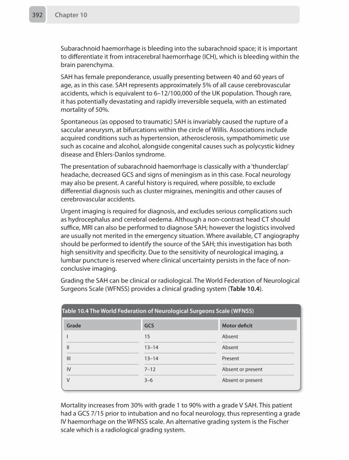
Chapter 10392
Subarachnoid haemorrhage is bleeding into the subarachnoid space; it is important to differentiate it from intracerebral haemorrhage (ICH), which is bleeding within the brain parenchyma.
SAH has female preponderance, usually presenting between 40 and 60 years of age, as in this case. SAH represents approximately 5% of all cause cerebrovascular accidents, which is equivalent to 6–12/100,000 of the UK population. Though rare, it has potentially devastating and rapidly irreversible sequela, with an estimated mortality of 50%.
Spontaneous (as opposed to traumatic) SAH is invariably caused the rupture of a saccular aneurysm, at bifurcations within the circle of Willis. Associations include acquired conditions such as hypertension, atherosclerosis, sympathomimetic use such as cocaine and alcohol, alongside congenital causes such as polycystic kidney disease and Ehlers-Danlos syndrome.
The presentation of subarachnoid haemorrhage is classically with a ‘thunderclap’ headache, decreased GCS and signs of meningism as in this case. Focal neurology may also be present. A careful history is required, where possible, to exclude differential diagnosis such as cluster migraines, meningitis and other causes of cerebrovascular accidents.
Urgent imaging is required for diagnosis, and excludes serious complications such as hydrocephalus and cerebral oedema. Although a non-contrast head CT should suffice, MRI can also be performed to diagnose SAH; however the logistics involved are usually not merited in the emergency situation. Where available, CT angiography should be performed to identify the source of the SAH; this investigation has both high sensitivity and specificity. Due to the sensitivity of neurological imaging, a lumbar puncture is reserved where clinical uncertainty persists in the face of non-conclusive imaging.
Grading the SAH can be clinical or radiological. The World Federation of Neurological Surgeons Scale (WFNSS) provides a clinical grading system (Table 10.4).
Mortality increases from 30% with grade 1 to 90% with a grade V SAH. This patient had a GCS 7/15 prior to intubation and no focal neurology, thus representing a grade IV haemorrhage on the WFNSS scale. An alternative grading system is the Fischer scale which is a radiological grading system.
Table 10.4 The World Federation of Neurological Surgeons Scale (WFNSS)
Grade GCS Motor deficit
I 15 Absent
II 13–14 Absent
III 13–14 Present
IV 7–12 Absent or present
V 3–6 Absent or present
Answers 393
This patient did not present to a neurosurgical centre and timely management will affect prognosis. The decrease in GCS mandated intubation and ventilation in view of the diagnosis. As with other neurological emergencies, neuroprotection to prevent secondary injury is vital.
Specific to the management of a subarachnoid haemorrhage are supportive and surgical strategies. Supportive strategies include:
• Adequate sedation and analgesia• Blood pressure < 200/100 mmHg in unsecured aneurysms, balancing the risks of
further bleeding against those of hypoperfusion • Avoid hypomagnesaemia, however hypermagnesaemia does not provide
additional benefit (IMASH Trial 2010)• Nimodipine – to reduce the incidence and severity of cerebral arterial vasospasm
(calcium channel antagonist)• Non-pharmacological venous thromboprophylaxis and stress ulcer prophylaxis
Fundamentally, urgent neurosurgical advice and intervention should be sought. Interventional options are:
• neuroradiological: coiling of intracranial aneurysm• neurosurgical: clipping intracranial aneurysm
The ISAT trial (2005) was a multicenter, randomised controlled trial comparing coiling to clipping. The trial suggested more independent survivors with coiling at one year; however the coiling group also had a higher risk of re-bleeding. Neurosurgical centres now follow local protocols, but increasingly clipping is reserved for aneurysms not amenable to coiling. Coiling is less expensive and avoids the patient undergoing a craniotomy.
Securing the aneurysm in a timely fashion is core to the management of SAH. However a number of complications affect the course of the patient with SAH, which need to be sought on presentation and during the critical care management.
• Re-bleeding: particularly within first 2 weeks• Vasospasm: challenging to definitely diagnose, transcranial Doppler maybe
useful. Mainstay of management is currently supportive, intra-arterial vasodilators are controversial
• Hydrocephalus: relatively common (20–30%), diagnosed on CT and requires urgent external ventricular drainage
• Seizures: no evidence for prophylactic anti-seizure medications, but aggressive management of seizures is paramount
• Endogenous catecholamine induced severe myocardial depression: diagnosed clinically using biomarkers such as troponin and brain natriuretic peptide (BNP) and echocardiography. Management is supportive
• Endocrine abnormalities including cerebral salt wasting syndrome (SWS) and syndrome of inappropriate antidiuretic hormone (SIADH, see paper 4, question 2)
Wong GK, Poon WS, Chan MT, et al. Intravenous magnesium sulphate for aneurysmal subarachnoid hemorrhage (IMASH): a randomized, double-blinded, placebo-controlled, multicenter phase III trial. Stroke 2010; 41(5):921–26.Molyneux A, Kerr R, Stratton I, et al. International Subarachnoid Aneurysm Trial (ISAT) of neurosurgical
Chapter 10394
clipping versus endovascular coiling in 2143 patients with ruptured intracranial aneurysms: a randomised trial. Lancet 2002; 360:1267–74.Luoma A, Reddy U. Acute management of aneurysmal subarachnoid haemorrhage. Contin Educ Anaesth Crit Care Pain 2013;13(2):52–58.Dorhout Mees SM, Rinkel GJ, Feigin VL, et al. Calcium antagonists for aneurysmal subarachnoid haemorrhage. Cochrane Database Syst Rev 2007;(3):CD000277.
17. D Perform the LSCS under general anaesthesiaThis patient is suspected of having spina bifida and, unfortunately, there is no information about her condition. Immigrants often present late antenatally and organising their care can be a challenge for the obstetricians and anaesthetists.
An urgent MRI may be possible prior to delivery in certain hospital settings, but in this case it is impractical since the patient is already contracting. It will be uncomfortable for her to undergo an MRI in her present state. Although there is no evidence that MRI is unsafe in pregnancy, it is best performed when there is an urgent clinical need, such as neurological compromise, which does not exist in this case.
Performing the LSCS under central neuraxial blockade is an option, but without a prior scan, there is no way of knowing at what level the abnormality lies. Even if this level were to be known, both epidural and spinal local anaesthetic spread can be wildly unpredictable, with a high block or even an inadequate block being possible. For these reasons amongst others, many anaesthetists would steer well clear of any central neuraxial blockade in such patients.
Performing the LSCS under general anaesthetic is the best option in this case. This should obviously be explained to the mother, with the risks and benefits being made clear. There is always a difficult airway risk in the obstetric population, but patients with spina bifida may also have a difficult airway. Full airway assessment must be undertaken and difficult airway equipment checked and ready.
Vaginal delivery of the breech baby, although possible, is not without risks, and if undertaken, there is still the risk that an even more urgent Caesarean section may become necessary. Obviously, a Caesarean section would be best undertaken with as much planning as possible. This is not the kind of case you want to be doing in a rush.
Griffiths S, Durbridge JA. Anaesthetic implications of neurological disease in pregnancy. Contin Educ Anaesth Crit Care Pain 2011; 11(5):157–61.
18. A Perform a rapid sequence induction and secure his airway with an endotracheal tube
The first priority in managing a child with trauma is to ensure a patent and protected airway. Indications for intubation are similar to adults: respiratory inadequacy, reduced Glasgow coma scale (<8), suspected raised intracranial pressure, need for prolonged ventilation, and need for transport to tertiary centre. This child has a Glasgow coma scale of 6 (eyes 1, motor 4, verbal 1), and had a high probability of head injury with raised intracranial pressure. His airway should first be secured
Answers 395
with an endotracheal tube, before proceeding to other treatment (further fluid resuscitation, blood transfusion, chest drain) and investigations (FAST scan, X-rays and CT scan).
Advanced Life Support Group. Advanced Paediatric Life Support: The Practical Approach, 5th ed. London: BMJ Publishing Group, 2011).Cullen PM. Paediatric trauma. Contin Educ Anaesth Crit Care Pain 2012; 12:157–61.Kochanek PM, Carney N, Adelson PD, et al. Guidelines for the Acute Medical Management of Severe Traumatic Brain Injury in Infants, Children, and Adolescents, 2nd Ed. Pediatr Crit Care Med 2012; 13 (suppl 1): S1–82.
19. C Discuss the case with the hospital’s child protection team
Child abuse is not uncommon. Evidence from a national UK survey suggests that the prevalence of serious physical abuse in childhood is around 7%, while sexual, emotional abuse and neglect each have a prevalence of around 6%. Increased awareness and familiarity with reporting procedures forms part of an effective preventive strategy for all healthcare professionals. Anaesthetists may encounter abused children during resuscitation in the emergency department, in the paediatric intensive care unit, during routine preoperative assessment, or intraoperatively during the course of a surgical procedure.
Clinical features that should raise concern or suspicion of non-accidental injury include:
• Unusual or excessive bruising, particularly in the non ambulant baby/child• Cigarette burns• Bite marks• Unusual injuries in inaccessible areas e.g. neck, ear, hands, feet & buttocks• Intra-oral trauma• Damage to intra-oral frena, or unexplained frenum injury in a non-ambulant child• Genital/anal trauma where no clear history of direct trauma is offered or part of
the clinical presentation• Trauma without adequate history eg. intra-abdominal injury
The Royal College of Anaesthetists in association with a number of stakeholders have developed a flow-chart to guide anaesthetists in the operating theatre who have child protection concerns. Hospitals generally have designated child protection doctors, nurses, and midwives to whom more serious concerns can be raised and cases discussed both formally and informally. There should also be an on call rota for emergency referrals, and it is important that anaesthetists are familiar with their local procedures and policies. This often involves the on call consultant paediatrician who also has a clear safeguarding role, and may be the first person to consult.
Royal College of Anaesthetists, Association of Anaesthetists of Great Britain and Ireland, Royal College of Paediatrics and Child Health, The Association of Paediatric Anaesthetists of Great Britain and Ireland. Child Protection and the Anaesthetist : Safeguarding Children in the Operating Theatre. 2014Melarkode K, Wilkinson K. Child protection issues and the anaesthetist. Contin Educ Anaesth Crit Care Pain 2012;12:123–27.
Chapter 10396
20. C Ask another colleague to attempt intravenous accessIntravenous access in children can be difficult, time-consuming and frustrating, particularly in the small premature infant, the chubby child, and in children who have undergone multiple previous cannulations. Unhurried preoperative examination for possible cannulation sites, avoidance of long starvation times and the use of adjuncts including topical local anaesthetic creams and oral sedatives improve success rates. Despite these measures, failure of intravenous cannulation will still occur. A range of alternative methods to access the circulation include ultrasound guided peripheral intravenous cannulation and central venous cannulation. Occasionally, drug administration via other routes can be considered, for example intramuscular or intra-osseous.
Instrumenting the airway without intravenous access is a subject of debate both in the literature and among practicing anaesthetists. In paediatric anaesthesia, this practice generally considered unsafe because of the risk of precipitating laryngospasm without an established route for its management.
In most situations, including the one described above, asking a colleague with a fresh pair of eyes and hands is the most appropriate next step before the other options are considered.
Tremlett M, Bajwa S. Failed intravenous access in children. Anaesth Intens Care Med 2009; 10:87–92.Martin R. Instrumentation of the airway in the absence of intravenous access. Paediatr Anaesth 2003; 13:366.Mohiuddin S, Mayhew JF. Tracheal Intubation without intravenous access. Paediatr Anaesth 2004; 14:621.
21. D Referral to a pain management programmePatients with long-term back pain often do not recover and the risk of their symptoms persisting is high. He is not a likely candidate for surgery because there is no evidence of nerve root compression, and the MRI is unlikely to change unless he develops new symptoms or signs. There is no evidence that gabapentin is effective in chronic nociceptive pain and its use in chronic low back pain is not based on any good randomised trials. Transcutaneous electrical nerve stimulation (TENS) is a common treatment that uses electrical stimulation over the back to mask the patient’s pain. TENS delivers high-frequency pulses at sub-motor intensities to modulate the pain pathways by ‘closing the gate’ to pain fibres. However, all the evidence thus far points out its lack of effect. This leaves a pain management programme that aims to improve patient’s ability to cope and manage their symptoms rather than a reduction in their pain. It has good evidence on improving functional outcomes and is the most appropriate step in this clinical scenario.
National Institute for Health and Care Excellence (NICE). Low back pain: Early management of persistent non-specific low back pain. CG 88. London: NICE, 2009
22. D Pharmacological therapy should be avoidedFibromyalgia is a common cause of widespread pain and is diagnosed clinically as more than 3 months of widespread pain with 11 out of 18 tender points on clinical examination. It occurs mostly in the 20–50 year age group but the incidence
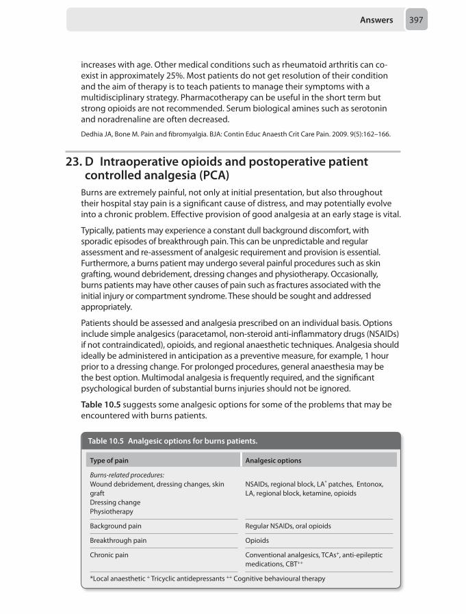
Answers 397
increases with age. Other medical conditions such as rheumatoid arthritis can co-exist in approximately 25%. Most patients do not get resolution of their condition and the aim of therapy is to teach patients to manage their symptoms with a multidisciplinary strategy. Pharmacotherapy can be useful in the short term but strong opioids are not recommended. Serum biological amines such as serotonin and noradrenaline are often decreased.
Dedhia JA, Bone M. Pain and fibromyalgia. BJA: Contin Educ Anaesth Crit Care Pain. 2009. 9(5):162–166.
23. D Intraoperative opioids and postoperative patient controlled analgesia (PCA)
Burns are extremely painful, not only at initial presentation, but also throughout their hospital stay pain is a significant cause of distress, and may potentially evolve into a chronic problem. Effective provision of good analgesia at an early stage is vital.
Typically, patients may experience a constant dull background discomfort, with sporadic episodes of breakthrough pain. This can be unpredictable and regular assessment and re-assessment of analgesic requirement and provision is essential. Furthermore, a burns patient may undergo several painful procedures such as skin grafting, wound debridement, dressing changes and physiotherapy. Occasionally, burns patients may have other causes of pain such as fractures associated with the initial injury or compartment syndrome. These should be sought and addressed appropriately.
Patients should be assessed and analgesia prescribed on an individual basis. Options include simple analgesics (paracetamol, non-steroid anti-inflammatory drugs (NSAIDs) if not contraindicated), opioids, and regional anaesthetic techniques. Analgesia should ideally be administered in anticipation as a preventive measure, for example, 1 hour prior to a dressing change. For prolonged procedures, general anaesthesia may be the best option. Multimodal analgesia is frequently required, and the significant psychological burden of substantial burns injuries should not be ignored.
Table 10.5 suggests some analgesic options for some of the problems that may be encountered with burns patients.
Table 10.5 Analgesic options for burns patients.
Type of pain Analgesic options
Burns-relatedprocedures:Wound debridement, dressing changes, skin graftDressing change Physiotherapy
NSAIDs, regional block, LA* patches, Entonox, LA, regional block, ketamine, opioids
Background pain Regular NSAIDs, oral opioids
Breakthrough pain Opioids
Chronic pain Conventional analgesics, TCAs+, anti-epileptic medications, CBT++
*Local anaesthetic + Tricyclic antidepressants ++ Cognitive behavioural therapy
Chapter 10398
In this scenario, the patient has an infected wound that would make a suitable regional block inappropriate. A history of asthma and the fact that significant burns may be associated with a coagulopathy would make NSAIDs less preferable. His abdominal pain, together with his postoperative arm pain would best be managed with a suitable opioid regime.
Norman AT, Judkins KC. Pain in the patient with burns. Contin Educ Anaesth Crit Care Pain 2004; 4(2):57–61.European Burns Association. European Best Practice Guidelines for Burn Care. Vienna: European Burns Association, 2013.
24. E Paracetamol, local anaesthetic infiltration to the wound, titrated morphine
Neonatal pain is often unrecognised and inadequately addressed. Apart from discomfort to the patient, mismanagement of pain at this stage may have a significant influence on the way pain is processed in later life. There is some evidence to suggest that insufficiently treated pain in the first few weeks of life may lead to hyperalgesia when painful experiences are encountered later.
As with any other age group, pain management should follow a structured approach. Non-pharmacological techniques which may help include skin-to-skin contact 10-15 minutes before a painful procedure, oral sucrose administration, or sucking on a soother. The latter seems to produce a calming effect in the newborn.
Paracetamol is used commonly, and is safe when given in the correct dose. The oral route is preferred. Rectal absorption is unpredictable, and bioavailability is higher.
Non-steroidal anti-inflammatory drugs (NSAIDs) are generally avoided, unless they are used to close a patent ductus arteriosus. Use of NSAIDs non-specifically inhibits prostaglandins that are essential in the development of many organ systems. They may also cause additional unwanted effects such as impaired thermoregulation, alterations in renal and cerebral blood flow and disrupted sleep.
Codeine phosphate is often used to good effect in neonates via the oral, rectal, or intramuscular routes. It should never be administered intravenously as it may cause severe hypotension.
Morphine is used widely, both intra- and postoperatively. It is often given as a nurse controlled analgesia (NCA) arrangement. Reduced protein binding and immature renal and hepatic function mean that the free fraction of morphine in the plasma is higher, and the plasma half-life is longer. Hence, the dose requirement is less than one may expect. The unwanted effects are the same in neonates as in any other age group, but may be more difficult to detect. Respiratory rate can be monitored, but pruritus and sedation for example, often go unnoticed. After receiving morphine, neonates should be nursed in an observed area where they can be monitored with apnoea alarms, oxygen saturation, and electrocardiogram.
Regional anaesthesia remains an excellent choice of analgesia and can often avoid many of the unwanted effects of systemic analgesia.
In this scenario the most appropriate option is to administer paracetamol, local anaesthetic infiltration to the wound and titrated morphine.
Haidon JL, Cunliffe L. Analgesia for neonates. Contin Educ Anaesth Crit Care Pain 2010; 10(4):123–127.
Answers 399
25. D Fentanyl PCASickle cell crises are a frequent reason for referral to pain teams and can be challenging to treat. Pain itself can precipitate a crisis in those with sickle cell disease.
The National Institute for Health and Care Excellence (NICE) recommend that patients should be offered analgesia within 30 minutes of hospital presentation. Paracetamol and non-steroidal anti-inflammatory drugs (NSAIDs) should be given regularly if there are no contraindications, in conjunction with an opioid. If no analgesia has been taken before, a weak opioid, such as codeine, can be used initially.
Pethidine should be avoided altogether in sickle cell crises, as there is increasing evidence that it is detrimental to outcome, namely due to the accumulation of its metabolite, norpethidine. Norpethidine is an irritant to the central nervous system and can cause dysphoria and convulsions. It is renally excreted, and patients with sickle cell crises may have a degree of renal impairment, possibly due to dehydration, NSAID use, or a vaso-occlusive event. Furthermore, a single dose is unlikely to be effective, necessitating repeated administration.
Judging by previous opioid requirements, a weak opioid such as codeine or tramadol is unlikely to provide this patient relief, and a PCA seems the most sensible option.
In view of his obstructive sleep apnoea, morphine is best avoided if possible, and a fentanyl PCA would be the most appropriate option in this patient although he should continue to take his paracetamol and NSAID throughout.
National Institute for Health and Care Excellence. Sickle cell acute painful episode: management of an acute painful sickle cell episode in hospital. CG 143. London: NICE, 2012.Stone M, Wheatley B. Patient-controlled analgesia. Cont Educ Anaesth Crit Care Pain 2002; 2(3):79–82.
26. B Left radial arteryCrawford classification of the aortic thoracoabdominal aneurysms (TAAA) consists of five types:
• Type I involves most of the descending thoracic aorta from the origin of the left subclavian to the suprarenal abdominal aorta
• Type II is the most extensive, extending from the subclavian to the aortoiliac bifurcation
• Type III involves the distal thoracic aorta to the aortoiliac bifurcation• Type IV TAAAs are limited to the abdominal aorta below the diaphragm• Safi’s group have modified this scheme by adding Type V, which extends from the
distal thoracic aorta including the celiac and superior mesenteric origins but not the renal arteries
Deploying a fenestrated aortic graft requires multiple guide wires to correctly align the fenestrations with the renal arteries and the mesenteric arteries. Right axillary/subclavian access is usually required for deploying the superior mesenteric artery guide wire due to the angle of take off of the artery from the aorta which makes it impossible to wire from the common femoral or external iliac arteries. The aortic stent is inserted via the common femoral artery which makes this site and pedal
Chapter 10400
arteries site impractical for insertion of arterial lines. This means the left upper limb arteries are the only possible arterial monitoring access points that are possible and practical.
Frederick JR, Woo YJ et al. Thoraco-abdominal aortic aneurysm. Ann Cardiothorac Surg 2012; 1(3):277–85.
27. A At the angle of the ribThe intercostal nerve (ICN) block is used to provide effective analgesia for breast surgery, thoracic and upper abdominal surgery (thoracotomy, cholecystectomy and gastrectomy) and fracture ribs. It also used for acute and chronic pain conditions like herpes zoster, post-thoracotomy pain and cancer related pain. The analgesia provided by the block is usually lasts for up to 12 hours.
The thoracic nerve roots exit from their respective intervertebral foramina, then divide into ventral and dorsal primary rami. The dorsal ramus supplies the paravertebral skin and muscles. The ventral ramus continues as the intercostal nerve, which enters the subcostal groove of the corresponded rib to lie initially between the pleura and the posterior intercostal membrane (medial to the rib angle). At the angle of the rib, the ICN passes between the internal intercostal and the innermost muscles. At the mid-axillary line, it gives off the lateral cutaneous branch that supplies the skin and the muscles of the lateral chest and abdominal walls. The final branch arises as the anterior cutaneous branch which is the continuation of the ICN, and innervates the skin and the muscles of the anterior thoracic and abdominal walls.
During its course in the subcostal groove of the rib, the ICN runs in a neurovascular bundle containing the intercostal vein superiorly, the intercostal artery in the middle and the intercostal nerve inferiorly.
There are some specific differences at certain levels. T1 intercostal nerve lacks the lateral and the anterior cutaneous branches. T2 and T3 contribute to the intercostobrachial nerve. T12 is really a subcostal nerve and some its fibres join with fibres from the lumbar plexus to form ilioinguinal, iliohypogastric and genitofemoral nerves.
An ICN block can be performed in different positions, including the prone position,, lateral decubitus, supine or sitting position. However, it is best performed in prone position with a pillow under the abdomen and the arms hangings from the sides of the bed. This accentuates the intercostal spaces and rotates the scapula laterally
The injection point for an ICN block must be proximal to the mid-axillary line before the the nerve divides. Performing the block at or lateral to the mid-axillary line might fail to anaesthetise the lateral cutaneous branch. The optimal place to perform the ICN block is at the angle of the rib where the rib is thicker and the intercostal space is wider, decreasing the chance to contact the pleura (Figure 10.3). The chance of pneumothorax increases if the block performed lateral to the angle of the rib.
If the injection site is medial to the angle of the rib, there is a possibility of subarachnoid spread because the dural sack can extend up to 8 cm from the midline.
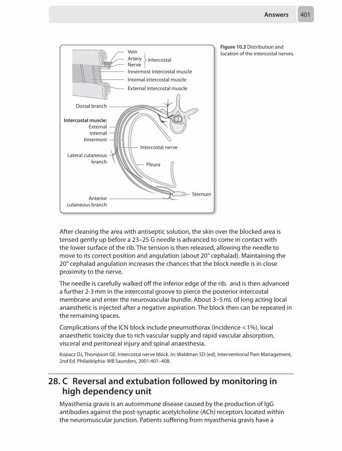
Answers 401
After cleaning the area with antiseptic solution, the skin over the blocked area is tensed gently up before a 23–25 G needle is advanced to come in contact with the lower surface of the rib. The tension is then released, allowing the needle to move to its correct position and angulation (about 20° cephalad). Maintaining the 20° cephalad angulation increases the chances that the block needle is in close proximity to the nerve.
The needle is carefully walked off the inferior edge of the rib. and is then advanced a further 2-3 mm in the intercostal groove to pierce the posterior intercostal membrane and enter the neurovascular bundle. About 3–5 mL of long acting local anaesthetic is injected after a negative aspiration. The block then can be repeated in the remaining spaces.
Complications of the ICN block include pneumothorax (incidence < 1%), local anaesthetic toxicity due to rich vascular supply and rapid vascular absorption, visceral and peritoneal injury and spinal anaesthesia.
Kopacz DJ, Thompson GE. Intercostal nerve block. In: Waldman SD (ed), Interventional Pain Management, 2nd Ed. Philadelphia: WB Saunders, 2001:401–408.
28. C Reversal and extubation followed by monitoring in high dependency unit
Myasthenia gravis is an autoimmune disease caused by the production of IgG antibodies against the post-synaptic acetylcholine (ACh) receptors located within the neuromuscular junction. Patients suffering from myasthenia gravis have a
Dorsal branch
Intercostal muscle:ExternalInternal
Innermost
Lateral cutaneousbranch
Anteriorcutaneous branch
Pleura
Intercostal nerve
Sternum
VeinArteryNerve
Intercostal
Innermost intercostal muscle
Internal intercostal muscle
External intercostal muscle
Figure 10.3 Distribution and location of the intercostal nerves.
Chapter 10402
reduction in the number of ACh receptors to just 30% of normal. It is clinically represented by weakness, which is exacerbated by exercise and relieved by rest. It can be classified by the clinical symptoms into four grades:
Grade I: Ocular signs and symptomsGrade IIA: Generalised mild muscle weakness responding well to therapyGrade IIB: Generalised moderate muscle weakness responding poorly to therapyGrade III: Acute fulminant condition and/or respiratory symptomsGrade IV: Myasthenic crisis requiring artificial ventilation
The factors which determine elective ventilation post-thymectomy include:
• Long standing disease (> 6 years)• Forced vital capacity (FVC) < 2.9 litres• History of chronic respiratory disease• Type III or Type IV myasthenia gravis
There is a greater chance of requiring postoperative ventilation for a trans-sternal thymectomy. Preoperatively well controlled patients can be extubated following surgery but should be monitored in a high dependency unit postoperatively due to the high risk of respiratory weakness and decompensation. As this patient has a vital capacity which is well above the threshold and is not having a trans-sternal procedure it would be judicious to extubate him and then monitor him overnight for any respiratory complications if they were to develop.
Thavasothy M, Hirsch N. Myasthenia gravis. Contin Educ Anaesth Crit Care Pain 2002; 2(3):88–90.
29. A Left upper lobe collapseAtelectasis is the loss of lung volume due to collapse of either whole or part of the lung. It can be broadly classified into obstructive or non-obstructive.
Obstructive atelectasis is commoner and caused by:
• Tumour• Mucus plugs• Inflammation and infections• Foreign bodies• Blood clots
Non-obstructive atelectasis is less common and is caused by:
• Interstitial disease• Collapse under anaesthesia• Thoraco-abdominal surgery• Chest trauma• Pleural effusion• Pneumothorax• Pneumonia
The loss of lung volume affects the ventilation/perfusion ratio, impairing compliance and oxygenation and also can cause increased pulmonary vascular resistance.
Questions 403
A chest X-ray can be diagnostic in assessment of lung collapse. There can either be a direct radiographic signs, such as loss of lung volume, or indirect signs including mediastinal shift A unilateral complete ‘white-out’ suggests involvement of an entire lung. More often, atelectasis involves a particular lobe with characteristic findings for each involved lobe.
• Right upper lobe: elevation of the right hilum and oblique fissure on an antero-posterior view. The oblique fissure on the lateral view appears convex superiorly, unless there is a mass lesion inferiorly which may cause it to appear concave superiorly. This is called the ‘Golden S’ sign
• Right middle lobe: This is most often overlooked in lobar collapse. There is radiographic loss of the right heart border silhouette. On a lateral view the right horizontal and oblique fissures move towards each other leading to a wedge shaped opacity
• Right lower lobe: There is a triangular opacity along the right heart border along with obliteration of the right hemidiaphragm
• Left upper lobe: Due to lack of a left horizontal fissure, a left sided upper lobe collapse leads to a veil-like opacity extending from the hilum and fading as it progresses inferiorly. On lateral view the oblique (major) fissure is displaced anteriorly and a hyperexpanded superior segment of the left lower lobe is apparent. In half the cases this hyperexpanded lobe is positioned between a collapsed upper lobe and the aortic arch below which gives the appearance of a crescent of aerated lung called the ‘Luftsichel sign’.
• Left lower lobe: This leads to a retrocardiac opacity, which silhouettes the left hemidiaphragm.
There are various therapeutic measures which can be utilised to deal with lung collapse including continuous positive airway pressure, positive end-expiratory pressure, bronchoscopy and washout, prone position ventilation and high frequency oscillatory ventilation. The method selected depends on the condition of the patient, etiology and co-morbidities.
Ray K, Bodenmham A, Paramasivam E. Pulmonary atelectasis in anaesthesia and critical care. Contin Educ Anaesth Crit Care Pain 2014; 14(5):237-245.
30. C We can have the most confidence that the results of Butler et al., are representative of the observed effect
A meta-analysis is a means to combine the results of a number of studies statistically, thus aiming to increase the power of any subsequent analysis and the accuracy and precision of any conclusion drawn from the data.
The process begins with a systematic review of the relevant literature. Prior to starting the review the reviewers must draw up criteria that each study must fulfill in order to be included. Studies which do not fulfill these criteria are rejected as significant methodological flaws may distort the results and lead to incorrect conclusions. Such methodological considerations include the randomisation process, blinding, placebo-control and number of participants. Answer D is therefore not the best one given here.
Chapter 10404
Once the appropriate studies have been identified and appraised, data can be extracted and the studies can then be weighted. There are a number of ways in which this is done but the principle is to assign more weight to those studies that provide more information about the treatment in question, in this case the ability of a drug to treat postoperative nausea and vomiting. It is expected that larger studies exhibit less variance than smaller ones, and therefore large studies are often weighted more; making answer A incorrect. The effect of the weighting process could be that smaller, valid studies have less impact on the final position of the ‘diamond’.
Methods of weighting used, for example, by the Cochrane Collaboration take into account the sample size and the event rate. The statistical concept encompassing these is the variance. The degree of weighting is shown by the size of the box. Answer E is therefore only partially correct, in addition to sample size; the event rate is needed to calculate variance.
Although the results of this fictitious meta-analysis suggest that Gipatron is a superior treatment for post operative nausea and vomiting, integration into clinical practice requires further considerations pertaining to side effect profile, cost, availability, routes of administration etc. More information is therefore needed before choosing option B.
The horizontal line is the confidence interval and a measure of how uncertain we are about the described effect. A longer line therefore implies less confidence in the effect and therefore the true value described in the study. It can be seen that the study by Butler et al. appears to have the narrowest confidence intervals and so statistically speaking we can be more confident in those results not being due to chance.
Higgins JPT, Green S. Cochrane Handbook for Systematic Reviews of Interventions 4.2.6. The Cochrane Library 2006; 4, 8a.4.Egger M, Smith GD, Phillips AN. Meta-analysis: principles and procedures. Br Med J 1997; 315;1533–37.
Index
Note: Page numbers in bold or italic refer to tables or figures respectively.
AAbciximab 342Abdominal compartment syndrome 56–57, 66–67
management 67risk factors 67
Abdominal perfusion pressure (APP) 56Abdominal trauma, fluid resuscitation in 62Absolute risk reduction (ARR) 201Accidental dural puncture (ADP) 30Activated clotting time (ACT) 76, 154Activated partial thromboplastin time (aPTT) 17Acupuncture, in pain management 150–151Acute kidney injury (AKI) 28, 387
diagnosis 387, 388principles of management of 29RIFLE criteria 28, 28risk factors 387stage 1 388
Acute renal failure (ARF) 278Acute respiratory distress syndrome (ARDS) 25, 228
Berlin definition 228causes of 228classification of severity 25extracoporeal membrane oxygenation in 230general management 229high frequency oscillation ventilation in 230inhaled nitric oxide in 230mechanical ventilation in 229pathophysiology of 228–229pharmacological management 230prone positioning in 229–230severe, management of 25–26
Adductor canal block 321–324Adjustable gastric band (AGB) 379Adrenaline
in anaphylaxis management 79in hypotension with bradyarrhythmia 382
Adrenoceptor blocking agents 285Aerobic gram negative bacilli (AGNB) 29After action review (AAR) 134Airway management, in children 193, 194Airway surgery, anaesthesia for 376–377Alcoholic chronic liver disease 145–146Alcohol use, and rhabdomyolysis 119Allodynia 117Alpha-1 antitrypsin 200Amiodarone 156
in atrial flutter 14
Amitriptylinein fibromyalgia 115in neuropathic pain 36overdose, management of 26–27
Amniotic fluid embolism (AFE) 192Anaemia, preoperative 362Anaesthesia
for airway surgery 376–377anaphylaxis during 149–150in remote locations 267for urgent surgery in pregnant patient 113
Anaphylactoid reactions 220Anaphylaxis 149–150, 219–220
triggers for 220Aneurysm coiling, general anaesthesia for 344–345Anion gap metabolic acidosis 106Ankle blocks 384–385Anorexia nervosa 244Anterior interosseous nerve 269, 270Anticonvulsants, in post surgical pain 34Anticubital fossa 269, 270Anti-diuretic hormone (ADH) 131, 144Anti-embolism stockings 17Antimicrobial prophylaxis, in surgery 14Aortic aneurysm repair, and renal impairment 199Aortic cross-clamping 39, 118–119Aortic dissection
with aortic regurgitation 217, 217–218classification systems 217management of 217–218risk factors for 217
Apnoea/hypopnea index (AHI) 148, 284Apnoea testing 303ARDSNet ventilator strategy 26Arrhythmias 39
direct current cardioversion in 39–40Arterial blood gas (ABG) analysis
burns patients 378fat embolism syndrome 60–61
Arytenoid dislocation 381Ascorbic acid deficiency 120Aspiration pneumonia 382Aspirin 342Asthma 67, 187–188
in children 235management of 68–69preoperative evaluation and preparation 235severity of 68
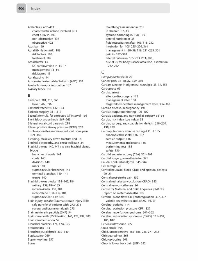
Index406
Atelectasis 402–403characteristic of lobe involved 403chest X-ray in 403non-obstructive 402obstructive 402
Atosiban 69Atrial fibrillation (AF) 188
risk factors 188treatment 189
Atrial flutter 13DC cardioversion in 13–14management 13–14risk factors 13
Atrial pacing 14Automated external defibrillator (AED) 132Awake fibre-optic intubation 137Axillary block 139
BBack pain 281, 318, 363
lower 282, 396Bacterial tracheitis 132–133Bariatric surgery 311–312Bazett’s formula, for corrected QT interval 156Bier’s block anaesthesia 267–269Bilateral vocal cord paralysis 218Bilevel positive airway pressure (BPAP) 262Bisphosphonates, in cancer induced bone pain
359–360Bleeding, maxillary down-fracture and 18Brachial plexopathy, and chest wall pain 34Brachial plexus 140, 141 see also Brachial plexus
blocksbranches of cords 142cords 140divisions 140roots 140supraclavicular branches 141terminal branches 140–141trunks 140
Brachial plexus blocks 138–142, 184axillary 139, 184–185infraclavicular 139, 184interscalene 138–139, 184supraclavicular 139, 184
Brain injury see also Traumatic brain injury (TBI)safe transfer of patients with 272–273severe, and brainstem death 273
Brain natriuretic peptide (BNP) 54Brainstem death (BSD) testing 143, 223, 297, 303Brainstem herniation 59Bronchial blockers 174, 174, 175Bronchiolitis 133Bronchopleural fistula 339–340Bupivacaine 269Buprenorphine 357Burns
‘Breathing’ assessment in 231in children 32–33cyanide poisoning in 198–199enteral nutrition in 38fluid resuscitation after 105, 118, 232intubation for 105, 225–226, 361management in 38–39, 118, 231–233, 361pain in 397–398referral criteria in 105, 233, 233, 283rule of 9s, for body surface area (BSA) estimation
232, 232
CCampylobacter jejuni 27Cancer pain 36–38, 37, 359–360Carbamazepine, in trigeminal neuralgia 33–34, 151Carboprost 69Cardiac arrest
after cardiac surgery 173management after 138targeted temperature management after 386–387
Cardiac disease, in pregnancy 191Cardiac output monitoring 106–109Cardiac patients, and non-cardiac surgery 53–54Cardiac risk index (Lee Index) 53Cardiac surgery, and coagulation defects 258–260,
259, 260Cardiopulmonary exercise testing (CPET) 135
anaerobic threshold 136–137cardiac output 136measurements and results 136performing test 135safety 136
Carotid endarterectomy (CEA) 361–362Carotid surgery, anaesthesia for 321Caudal epidural analgesia 345–346Cell salvage 76Central neuraxial block (CNB), and epidural abscess
20–21Central post-stroke pain 152Central retinal artery occlusion (CRAO) 383Central venous catheters 24Centre for Maternal and Child Enquiries (CMACE)
report, on maternal deaths 192Cerebral blood flow (CBF) autoregulation 337, 337
volatile anaesthetics and 92, 92–93, 93Cerebral oedema 114Cerebral perfusion pressure (CPP) 337Cerebral reperfusion syndrome 361–362Cerebral salt-wasting syndrome (CSWS) 131–132,
186, 187Cervical ultrasound 222Child abuse 395Child, uncooperative 185–186, 236, 271–272Chi squared test 363Chloroprocaine 269Chronic lower back pain (LBP) 282

Index 407
Chronic obstructive pulmonary disease (COPD)protocolised weaning in 276ventilatory management in 275–276weaning failure in 276
Chronic post surgical pain (CPSP) 116–117, 117, 238Clinical Negligence Scheme for Trust (CNST)
regulations 134Clinical Pulmonary Infection Score (CPIS) 29Clinical risk management 133–134
assessment 134awareness 134identification 134management 134re-evaluation 134–135
Clonidine, as anaesthesia for middle ear surgery 178Clopidogrel 342Coagulopathy in cardiac surgery patients,
management of 227–228Coarctation of the aorta 31–32Codeine, in neonates 197Codeine phosphate 398Coeliac plexus block 195–196
in chronic upper gastrointestinal malignancy 152–153
Cognitive behavior therapy (CBT) 282Complex regional pain syndrome (CRPS) 34, 117,
240–241Budapest criteria 34, 35diagnosis 34–35treatment 35, 35–36
Computed tomography (CT)burns patients 378neck infections 222
Confusion Assessment Method in the intensive care unit (CAM-ICU) 354
Continuous positive airway pressure (CPAP) 139, 262Coronary artery bypass grafting (CABG) 13
care after 274–275Coronary artery dominance 77Crawford classification, of aortic thoracoabdominal
aneurysms (TAAA) 399Creatinine kinase (CK) 119Cricoid pressure, use of, for rapid sequence induction
307Croup 133CT pulmonary angiography (CTPA) 110–111Cyanide poisoning 198–199Cyproheptadine 237
DDabigatran 18, 342Danaparoid 17Dantrolene, in malignant hyperthermia 379Da Vinci system 264–265Day case surgery, selection criteria for 341, 341Death, criteria 303Delayed cerebral ischaemia 214–215
Delirium, in critically ill patients 353, 353–354assessment methods 354management of 354risk factors associated with 353types of 353–354
Dexametomidate, in delirium 354Diabetes inspidus (DI) 144Diabetic ketoacidosis (DKA) 65, 106
in children with diabetes 114management of 65–66
Diabetic patient, and surgery 96insulin regimens 96
Dobutamine, in septic shock 308Double lumen tubes 174, 175
and bronchial blockers 174, 174malpositioning 95, 95–96
Dual antiplatelet therapy (DAPT) 343Duke Activity Index 135Duke’s criteria, for infective endocarditis 324–325Dural puncture, and post-dural puncture headache
103–104Dysaesthesia 117
EEchocardiography 222Electroconvulsive therapy (ECT) 19
anaesthesia for 19–20, 179and biphasic physiological response 19
Electroencephalogram (EEG), for diagnosis of death 223
Emergence delirium (ED) 70Endobronchial intubation 55–56Endotracheal tube (ETT) 55Epidural abscess, diagnosis and management of
20–21, 363Epidural analgesia 281Epidural blood patch (EBP) 355Epiglottitis 133Ergometrine 69Erythropoiesis 362European Society of Cardiology (ESC) guidelines,
for assessment of cardiac patients for non-cardiac surgery 53–54
Exercise therapy 282Extracorporeal membrane oxygenation (ECMO) 26,
230, 350–351Extra-vascular lung water (EVLW) 107, 108Extra-ventricular drain (EVD) 63–64Eye injury, penetrating 153–154
FFascia liaca block, in hip fractures 72–73Fat embolism syndrome 60–61Femoral catheter 282Femoral nerve (FN) 321–322, 322
fascia iliaca block 3233-in-1 nerve block 322–323
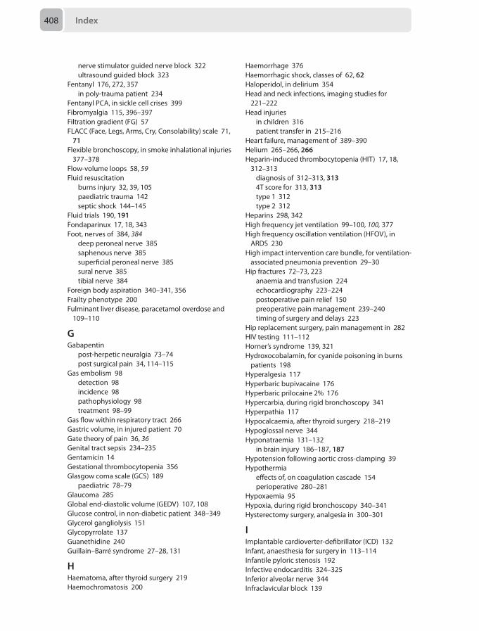
Index408
nerve stimulator guided nerve block 322ultrasound guided block 323
Fentanyl 176, 272, 357in poly-trauma patient 234
Fentanyl PCA, in sickle cell crises 399Fibromyalgia 115, 396–397Filtration gradient (FG) 57FLACC (Face, Legs, Arms, Cry, Consolability) scale 71,
71Flexible bronchoscopy, in smoke inhalational injuries
377–378Flow-volume loops 58, 59Fluid resuscitation
burns injury 32, 39, 105paediatric trauma 142septic shock 144–145
Fluid trials 190, 191Fondaparinux 17, 18, 343Foot, nerves of 384, 384
deep peroneal nerve 385saphenous nerve 385superficial peroneal nerve 385sural nerve 385tibial nerve 384
Foreign body aspiration 340–341, 356Frailty phenotype 200Fulminant liver disease, paracetamol overdose and
109–110
GGabapentin
post-herpetic neuralgia 73–74post surgical pain 34, 114–115
Gas embolism 98detection 98incidence 98pathophysiology 98treatment 98–99
Gas flow within respiratory tract 266Gastric volume, in injured patient 70Gate theory of pain 36, 36Genital tract sepsis 234–235Gentamicin 14Gestational thrombocytopenia 356Glasgow coma scale (GCS) 189
paediatric 78–79Glaucoma 285Global end-diastolic volume (GEDV) 107, 108Glucose control, in non-diabetic patient 348–349Glycerol gangliolysis 151Glycopyrrolate 137Guanethidine 240Guillain–Barré syndrome 27–28, 131
HHaematoma, after thyroid surgery 219Haemochromatosis 200
Haemorrhage 376Haemorrhagic shock, classes of 62, 62Haloperidol, in delirium 354Head and neck infections, imaging studies for
221–222Head injuries
in children 316patient transfer in 215–216
Heart failure, management of 389–390Helium 265–266, 266Heparin-induced thrombocytopenia (HIT) 17, 18,
312–313diagnosis of 312–313, 3134T score for 313, 313type 1 312type 2 312
Heparins 298, 342High frequency jet ventilation 99–100, 100, 377High frequency oscillation ventilation (HFOV), in
ARDS 230High impact intervention care bundle, for ventilation-
associated pneumonia prevention 29–30Hip fractures 72–73, 223
anaemia and transfusion 224echocardiography 223–224postoperative pain relief 150preoperative pain management 239–240timing of surgery and delays 223
Hip replacement surgery, pain management in 282HIV testing 111–112Horner’s syndrome 139, 321Hydroxocobalamin, for cyanide poisoning in burns
patients 198Hyperalgesia 117Hyperbaric bupivacaine 176Hyperbaric prilocaine 2% 176Hypercarbia, during rigid bronchoscopy 341Hyperpathia 117Hypocalcaemia, after thyroid surgery 218–219Hypoglossal nerve 344Hyponatraemia 131–132
in brain injury 186–187, 187Hypotension following aortic cross-clamping 39Hypothermia
effects of, on coagulation cascade 154perioperative 280–281
Hypoxaemia 95Hypoxia, during rigid bronchoscopy 340–341Hysterectomy surgery, analgesia in 300–301
IImplantable cardioverter-defibrillator (ICD) 132Infant, anaesthesia for surgery in 113–114Infantile pyloric stenosis 192Infective endocarditis 324–325Inferior alveolar nerve 344Infraclavicular block 139
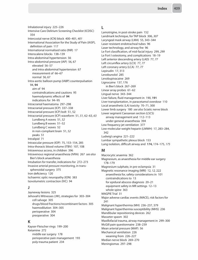
Index 409
Inhalational injury 225–226Intensive Care Delirium Screening Checklist (ICDSC)
354Intercostal nerve (ICN) block 400–401, 401International Association for the Study of Pain (IASP),
definition of pain 117International normalised ratio (INR) 17Interscalene blocks 138–139Intra-abdominal hypertension 56Intra-abdominal pressure (IAP) 56, 67
elevated 56–57and intra-abdominal hypertension 67measurement of 66–67normal 56, 67
Intra-aortic balloon pump (IABP) counterpulsation 94, 94
aim of 94contraindications and cautions 95haemodynamic effects of 94indications for 94–95
Intracranial haematoma 297–298Intracranial pressure (ICP) 337–338Intracranial pressure (ICP) bolt 51, 52Intracranial pressure (ICP) waveform 51, 51, 62–63, 63
Lundberg A waves 51, 52Lundberg B waves 51–52Lundberg C waves 52in non-compliant brain 51, 52peaks 51
Intralipid 71Intraocular pressure (IOP) 75, 153–154, 285Intra-thoracic blood volume (ITBV) 107, 108Intravenous access, in children 396Intravenous regional anaesthesia (IVRA) 267 see also
Bier’s block anaesthesiaIntubation for transfer, indications for 272–273Invasive arterial pressure monitoring, in trans-
sphenoidal surgery 375Iron deficiency 120Ischaemic optic neuropathy (ION) 383Isovolumetric contraction (IVC) 94
JJayneway lesions 325Jehovah’s Witnesses (JW), strategies for 303–304
cell salvage 305drugs/blood fractions/recombinant factors 305haemodilution 304–305perioperative 304preoperative 304
KKayser-Fleischer rings 199–200Ketamine 272
middle ear surgery 178perioperative pain management 193poly-trauma patient 234
LLamotrigine, in post-stroke pain 152Landmark technique, for TAP block 306, 307Laryngeal mask airway (LMA) 55, 343–344Laser-resistant endotracheal tubes 96Laser technology, and airway fire 96Le Fort classification, of mid-facial injury 299, 299Le Fort I osteotomy, and complications 18–19Left anterior descending artery (LAD) 77, 77Left circumflex artery (LCX) 77, 77Left coronary artery (LCA) 77, 77Lepirudin 17, 313Levobunolol 285Levobupivacaine 269Lignocaine 137, 176
in Bier’s block 267–269Linear array probes 61–62Lingual nerve 343–344Liver failure, fluid management in 190, 191Liver transplantation, in paracetamol overdose 110Local anaesthetic (LA) toxicity 70–71, 300Lower limb surgery 180 see also Sciatic nerve blockLower segment Caesarean section (LSCS)
airway management and 112–113under general anaesthesia 394
Low frequency jet ventilation 377Low-molecular-weight heparin (LMWH) 17, 283–284,
342Ludwig’s angina 221–222Lumbar sympathetic plexus block 153Lung isolation, difficult airway and 174, 174–175, 175
MMacrocytic anaemia 362Magnesium, as anaesthesia for middle ear surgery
178–179Magnesium sulphate, in pre-eclampsia 31Magnetic resonance imaging (MRI) 12, 12, 222
anaesthesia for, safety considerations in 101contraindications to 13for epidural abscess diagnosis 20–21equipment safety in MR settings 12–13whole spine 363
MAGPIE Trial 31Major adverse cardiac events (MACE), risk factors for
241Malignant hyperthermia (MH) 236–237, 379Malignant hyperthermia susceptibility (MHS) 236Mandibular repositioning devices 262Masseter spasm 302Maxillofacial trauma, airway management in 299–300McGill pain questionnaire 238–239Mean arterial pressure (MAP) 56Mechanical ventilation 226
weaning from 226–227Median nerve block 269–270Meningiomas 297–298
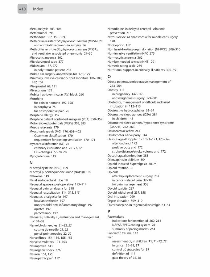
Index410
Meta-analysis 403–404Metaraminol 298Methadone 357, 358–359Methicillin-resistant Staphylococcus aureus (MRSA) 29
and antibiotic regimens in surgery 14Methicillin sensitive Staphylococcus aureus (MSSA),
and ventilator associated pneumonia 29–30Microcytic anaemia 362Microlaryngeal tube 377Midazolam 137, 272
in poly-trauma patient 234Middle ear surgery, anaesthesia for 178–179Minimally invasive cardiac output monitors 106–109,
107, 108Misoprostol 69, 191Mivacurium 179Mobitz II atrioventricular (AV) block 260Morphine
for pain in neonate 197, 398in porphyria 75for postoperative pain 70
Morphine allergy 357Morphine patient controlled analgesia (PCA) 358–359Motor evoked potentials (MEPs) 303, 383Muscle relaxants 179Myasthenia gravis (MG) 170, 401–402
Osserman classification 170requirement for post op ventilation 170–171
Myocardial infarction (MI) 76coronary circulation and 76–77, 77ECG changes 77–78, 78
Myoglobinuria 119
NN-acetyl cysteine (NAC) 109N-acetyl-p-benzoquinone imine (NAPQI) 109Naloxone 149Nasal endotracheal tube 19Neonatal apnoea, postoperative 113–114Neonatal pain, analgesia for 398Neonatal resuscitation 314–315, 315Neonates, analgesia for 197
local anaesthetics 197non steroidal anti-inflammatory drugs 197opiates 197paracetamol 197
Neonates, critically ill, evaluation and management of 31–32
Nerve block needles 21–22, 22cutting tip needle 21, 22pencil point needles 22, 22
Nerve fibres 154–156, 155, 155Nerve stimulators 101–103Neurapraxia 343Neurogenic shock 376Neuron 154, 155Neuropathic pain 117
Nimodipine, in delayed cerebral ischaemia prevention 215
Nitrous oxide, as anaesthesia for middle ear surgery 178
Nociception 117Non heart-beating organ donation (NHBOD) 309–310Non-invasive ventilation (NIV) 275Normocytic anaemia 362Number needed to treat (NNT) 201Numeric rating scale 239Nutritional support, in critically ill patients 390–391
OObese patients, perioperative management of
263–264Obesity 311
in pregnancy 147–148and weight loss surgery 379–381
Obstetrics, management of difficult and failed intubation in 112–113
Obstructive hydrocephalus 63–64Obstructive sleep apnoea (OSA) 284
in children 148Obstructive sleep apnoea/hypopnoea syndrome
(OSAHS) 262–263Oculocardiac reflex 241Oculomotor nerve palsy 314Oesophageal Doppler 171, 171–173, 325–326
afterload and 172peak velocity and 172stroke distance/stroke volume and 172
Oesophageal perforation 381Olanzapine, in delirium 354Opioid-induced hyperalgesia 38, 74Opioid rotation 38Opioids
after hip replacement surgery 282in cancer-related pain 37–38for pain management 358
Opioid toxicity 237Opioid withdrawal 237, 358Oral intubation 299Organ donation 309–310Oxcarbazepine, in trigeminal neuralgia 33–34
PPacemakers
indications for insertion of 260, 261NAPSE/BPEG coding system 261summary of pacing modes 261
Paediatric trauma 142Pain
assessment of, in children 71, 71–72, 72in cancer 36–38, 37control of, strategies for 37definition of 117gate theory of 36, 36
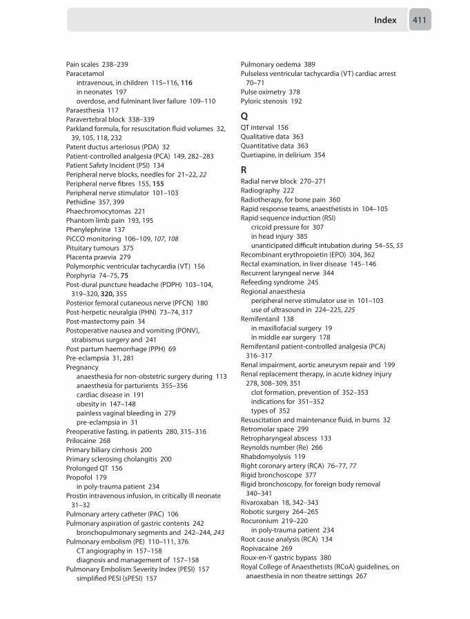
Index 411
Pain scales 238–239Paracetamol
intravenous, in children 115–116, 116in neonates 197overdose, and fulminant liver failure 109–110
Paraesthesia 117Paravertebral block 338–339Parkland formula, for resuscitation fluid volumes 32,
39, 105, 118, 232Patent ductus arteriosus (PDA) 32Patient-controlled analgesia (PCA) 149, 282–283Patient Safety Incident (PSI) 134Peripheral nerve blocks, needles for 21–22, 22Peripheral nerve fibres 155, 155Peripheral nerve stimulator 101–103Pethidine 357, 399Phaechromocytomas 221Phantom limb pain 193, 195Phenylephrine 137PiCCO monitoring 106–109, 107, 108Pituitary tumours 375Placenta praevia 279Polymorphic ventricular tachycardia (VT) 156Porphyria 74–75, 75Post-dural puncture headache (PDPH) 103–104,
319–320, 320, 355Posterior femoral cutaneous nerve (PFCN) 180Post-herpetic neuralgia (PHN) 73–74, 317Post-mastectomy pain 34Postoperative nausea and vomiting (PONV),
strabismus surgery and 241Post partum haemorrhage (PPH) 69Pre-eclampsia 31, 281Pregnancy
anaesthesia for non-obstetric surgery during 113anaesthesia for parturients 355–356cardiac disease in 191obesity in 147–148painless vaginal bleeding in 279pre-eclampsia in 31
Preoperative fasting, in patients 280, 315–316Prilocaine 268Primary biliary cirrhosis 200Primary sclerosing cholangitis 200Prolonged QT 156Propofol 179
in poly-trauma patient 234Prostin intravenous infusion, in critically ill neonate
31–32Pulmonary artery catheter (PAC) 106Pulmonary aspiration of gastric contents 242
bronchopulmonary segments and 242–244, 243Pulmonary embolism (PE) 110–111, 376
CT angiography in 157–158diagnosis and management of 157–158
Pulmonary Embolism Severity Index (PESI) 157simplified PESI (sPESI) 157
Pulmonary oedema 389Pulseless ventricular tachycardia (VT) cardiac arrest
70–71Pulse oximetry 378Pyloric stenosis 192
QQT interval 156Qualitative data 363Quantitative data 363Quetiapine, in delirium 354
RRadial nerve block 270–271Radiography 222Radiotherapy, for bone pain 360Rapid response teams, anaesthetists in 104–105Rapid sequence induction (RSI)
cricoid pressure for 307in head injury 385unanticipated difficult intubation during 54–55, 55
Recombinant erythropoietin (EPO) 304, 362Rectal examination, in liver disease 145–146Recurrent laryngeal nerve 344Refeeding syndrome 245Regional anaesthesia
peripheral nerve stimulator use in 101–103use of ultrasound in 224–225, 225
Remifentanil 138in maxillofacial surgery 19in middle ear surgery 178
Remifentanil patient-controlled analgesia (PCA) 316–317
Renal impairment, aortic aneurysm repair and 199Renal replacement therapy, in acute kidney injury
278, 308–309, 351clot formation, prevention of 352–353indications for 351–352types of 352
Resuscitation and maintenance fluid, in burns 32Retromolar space 299Retropharyngeal abscess 133Reynolds number (Re) 266Rhabdomyolysis 119Right coronary artery (RCA) 76–77, 77Rigid bronchoscope 377Rigid bronchoscopy, for foreign body removal
340–341Rivaroxaban 18, 342–343Robotic surgery 264–265Rocuronium 219–220
in poly-trauma patient 234Root cause analysis (RCA) 134Ropivacaine 269Roux-en-Y gastric bypass 380Royal College of Anaesthetists (RCoA) guidelines, on
anaesthesia in non theatre settings 267
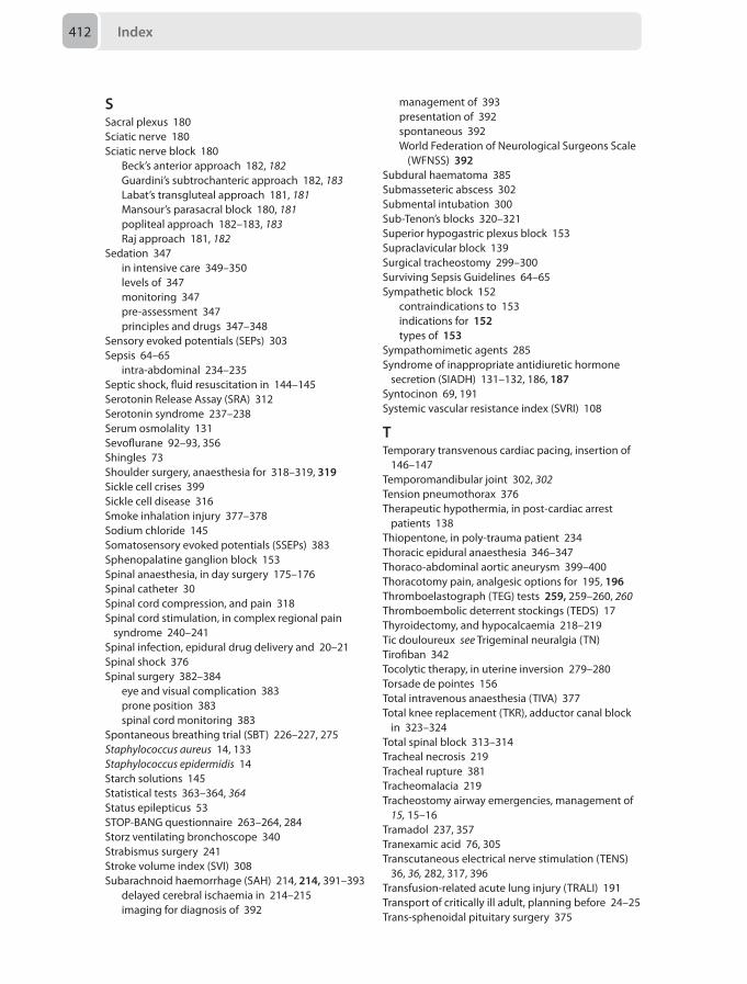
Index412
SSacral plexus 180Sciatic nerve 180Sciatic nerve block 180
Beck’s anterior approach 182, 182Guardini’s subtrochanteric approach 182, 183Labat’s transgluteal approach 181, 181Mansour’s parasacral block 180, 181popliteal approach 182–183, 183Raj approach 181, 182
Sedation 347in intensive care 349–350levels of 347monitoring 347pre-assessment 347principles and drugs 347–348
Sensory evoked potentials (SEPs) 303Sepsis 64–65
intra-abdominal 234–235Septic shock, fluid resuscitation in 144–145Serotonin Release Assay (SRA) 312Serotonin syndrome 237–238Serum osmolality 131Sevoflurane 92–93, 356Shingles 73Shoulder surgery, anaesthesia for 318–319, 319Sickle cell crises 399Sickle cell disease 316Smoke inhalation injury 377–378Sodium chloride 145Somatosensory evoked potentials (SSEPs) 383Sphenopalatine ganglion block 153Spinal anaesthesia, in day surgery 175–176Spinal catheter 30Spinal cord compression, and pain 318Spinal cord stimulation, in complex regional pain
syndrome 240–241Spinal infection, epidural drug delivery and 20–21Spinal shock 376Spinal surgery 382–384
eye and visual complication 383prone position 383spinal cord monitoring 383
Spontaneous breathing trial (SBT) 226–227, 275Staphylococcus aureus 14, 133Staphylococcus epidermidis 14Starch solutions 145Statistical tests 363–364, 364Status epilepticus 53STOP-BANG questionnaire 263–264, 284Storz ventilating bronchoscope 340Strabismus surgery 241Stroke volume index (SVI) 308Subarachnoid haemorrhage (SAH) 214, 214, 391–393
delayed cerebral ischaemia in 214–215imaging for diagnosis of 392
management of 393presentation of 392spontaneous 392World Federation of Neurological Surgeons Scale
(WFNSS) 392Subdural haematoma 385Submasseteric abscess 302Submental intubation 300Sub-Tenon’s blocks 320–321Superior hypogastric plexus block 153Supraclavicular block 139Surgical tracheostomy 299–300Surviving Sepsis Guidelines 64–65Sympathetic block 152
contraindications to 153indications for 152types of 153
Sympathomimetic agents 285Syndrome of inappropriate antidiuretic hormone
secretion (SIADH) 131–132, 186, 187Syntocinon 69, 191Systemic vascular resistance index (SVRI) 108
TTemporary transvenous cardiac pacing, insertion of
146–147Temporomandibular joint 302, 302Tension pneumothorax 376Therapeutic hypothermia, in post-cardiac arrest
patients 138Thiopentone, in poly-trauma patient 234Thoracic epidural anaesthesia 346–347Thoraco-abdominal aortic aneurysm 399–400Thoracotomy pain, analgesic options for 195, 196Thromboelastograph (TEG) tests 259, 259–260, 260Thromboembolic deterrent stockings (TEDS) 17Thyroidectomy, and hypocalcaemia 218–219Tic douloureux see Trigeminal neuralgia (TN)Tirofiban 342Tocolytic therapy, in uterine inversion 279–280Torsade de pointes 156Total intravenous anaesthesia (TIVA) 377Total knee replacement (TKR), adductor canal block
in 323–324Total spinal block 313–314Tracheal necrosis 219Tracheal rupture 381Tracheomalacia 219Tracheostomy airway emergencies, management of
15, 15–16Tramadol 237, 357Tranexamic acid 76, 305Transcutaneous electrical nerve stimulation (TENS)
36, 36, 282, 317, 396Transfusion-related acute lung injury (TRALI) 191Transport of critically ill adult, planning before 24–25Trans-sphenoidal pituitary surgery 375
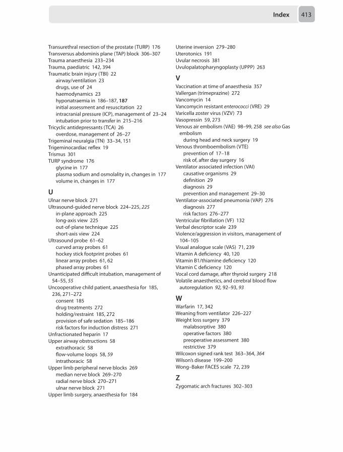
Index 413
Transurethral resection of the prostate (TURP) 176Transversus abdominis plane (TAP) block 306–307Trauma anaesthesia 233–234Trauma, paediatric 142, 394Traumatic brain injury (TBI) 22
airway/ventilation 23drugs, use of 24haemodynamics 23hyponatraemia in 186–187, 187initial assessment and resuscitation 22intracranial pressure (ICP), management of 23–24intubation prior to transfer in 215–216
Tricyclic antidepressants (TCA) 26overdose, management of 26–27
Trigeminal neuralgia (TN) 33–34, 151Trigeminocardiac reflex 19Trismus 301TURP syndrome 176
glycine in 177plasma sodium and osmolality in, changes in 177volume in, changes in 177
UUlnar nerve block 271Ultrasound-guided nerve block 224–225, 225
in-plane approach 225long-axis view 225out-of-plane technique 225short-axis view 224
Ultrasound probe 61–62curved array probes 61hockey stick footprint probes 61linear array probes 61, 62phased array probes 61
Unanticipated difficult intubation, management of 54–55, 55
Uncooperative child patient, anaesthesia for 185, 236, 271–272
consent 185drug treatments 272holding/restraint 185, 272provision of safe sedation 185–186risk factors for induction distress 271
Unfractionated heparin 17Upper airway obstructions 58
extrathoracic 58flow-volume loops 58, 59intrathoracic 58
Upper limb peripheral nerve blocks 269median nerve block 269–270radial nerve block 270–271ulnar nerve block 271
Upper limb surgery, anaesthesia for 184
Uterine inversion 279–280Uterotonics 191Uvular necrosis 381Uvulopalatopharyngoplasty (UPPP) 263
VVaccination at time of anaesthesia 357Vallergan (trimeprazine) 272Vancomycin 14Vancomycin resistant enterococci (VRE) 29Varicella zoster virus (VZV) 73Vasopressin 59, 273Venous air embolism (VAE) 98–99, 258 see also Gas
embolismduring head and neck surgery 19
Venous thromboembolism (VTE)prevention of 17–18risk of, after day surgery 16
Ventilator associated infection (VAI)causative organisms 29definition 29diagnosis 29prevention and management 29–30
Ventilator-associated pneumonia (VAP) 276diagnosis 277risk factors 276–277
Ventricular fibrillation (VF) 132Verbal descriptor scale 239Violence/aggression in visitors, management of
104–105Visual analogue scale (VAS) 71, 239Vitamin A deficiency 40, 120Vitamin B1/thiamine deficiency 120Vitamin C deficiency 120Vocal cord damage, after thyroid surgery 218Volatile anaesthetics, and cerebral blood flow
autoregulation 92, 92–93, 93
WWarfarin 17, 342Weaning from ventilator 226–227Weight loss surgery 379
malabsorptive 380operative factors 380preoperative assessment 380restrictive 379
Wilcoxon signed rank test 363–364, 364Wilson’s disease 199–200Wong–Baker FACES scale 72, 239
ZZygomatic arch fractures 302–303

Related Documents