Methodological Advances Filter centrifugation as a sampling method for miniaturization of extracellular fungal enzyme activity measurements in solid media J. HEINONSALO a , G. KABIERSCH a, *, R.M. NIEMI b , S. SIMPANEN b,c , H. ILVESNIEMI d , M. HOFRICHTER e , A. HATAKKA a , K.T. STEFFEN a a Viikki Biocenter, Department of Food and Environmental Sciences, Faculty of Agriculture and Forestry, P.O. Box 56, FIN-00014 University of Helsinki, Helsinki, Finland b Finnish Environment Institute, Research Programme for Biodiversity, P.O. Box 140, FIN-00251 Helsinki, Finland c Department of Environmental Sciences, University of Helsinki, Niemenkatu 73, 15140 Lahti, Finland d Finnish Forest Research Institute, Vantaa Research Station, P.O. Box 18, FIN-01301 Vantaa, Finland e Department of Environmental Biotechnology, International Graduate School of Zittau, Markt 23, 02763 Zittau, Germany article info Article history: Received 16 December 2010 Revision received 12 May 2011 Accepted 20 July 2011 Available online 21 September 2011 Corresponding editor: Petr Baldrian Keywords: Enzyme extraction Fungi Peroxidase Sampling method Screening abstract A novel sampling method to evaluate extracellular fungal enzyme activities was developed and the validity tested for agar media. The method is based on centrifugation of small agar pieces taken from growing fungal solid-state cultures. Centrifuge tubes that allow spinning liquid out from small samples containing, for example, the hyphal front of a growing mycelium are essential for the protocol. Centrifugation recovers a liquid phase from the samples, which contains soluble material including many enzymes. The recovery of two added model enzymes, namely laccase and manganese peroxidase (MnP), from agar media was sufficient (ranging from 50 % to 75 %) but the addition of humic material into agar decreased the observed MnP activity significantly to approx. 25 % of the stock solution. Using growing cultures, the presence of humus as well as Scots pine sawdust on Hagem’s agar plates induced the production of laccase and peroxidase in certain fungi, which indicates that the method is suitable for screening enzyme activities on different growth media or with variable additives or growth conditions. The use of the presented sampling method for functional enzyme fingerprinting of different fungi may be a promising tool for investigating the behaviour and ecological role of forest soil fungi. This method also allows obtaining spatial data from very small and defined areas of solid fungal cultures, e.g. from microcosms. ª 2011 Elsevier Ltd and The British Mycological Society. All rights reserved. Introduction Analysis of enzymatic activities in samples obtained from solid media or solid environmental samples usually requires the addition of water and/or buffer. This may lead to the dilution of enzymes below the detection limit. As a conse- quence, additional laborious and costly working steps to concentrate the sample may be required. A common way to extract enzymes from solid media is to add buffer (e.g. 50 mM sodium acetate, pH 5.0) or water, soak and shake the samples * Corresponding author. Tel.: þ358 9 191 59 318; fax: þ358 9 191 59 322. E-mail address: grit.kabiersch@helsinki.fi (G. Kabiersch). available at www.sciencedirect.com journal homepage: www.elsevier.com/locate/funeco 1754-5048/$ e see front matter ª 2011 Elsevier Ltd and The British Mycological Society. All rights reserved. doi:10.1016/j.funeco.2011.07.008 fungal ecology 5 (2012) 261 e269

Welcome message from author
This document is posted to help you gain knowledge. Please leave a comment to let me know what you think about it! Share it to your friends and learn new things together.
Transcript

.sciencedirect.com
f u n g a l e c o l o g y 5 ( 2 0 1 2 ) 2 6 1e2 6 9
available at www
journal homepage: www.elsevier .com/locate/ funeco
Methodological Advances
Filter centrifugation as a sampling method for miniaturizationof extracellular fungal enzyme activity measurements in solidmedia
J. HEINONSALOa, G. KABIERSCHa,*, R.M. NIEMIb, S. SIMPANENb,c, H. ILVESNIEMId,M. HOFRICHTERe, A. HATAKKAa, K.T. STEFFENa
aViikki Biocenter, Department of Food and Environmental Sciences, Faculty of Agriculture and Forestry, P.O. Box 56,
FIN-00014 University of Helsinki, Helsinki, FinlandbFinnish Environment Institute, Research Programme for Biodiversity, P.O. Box 140, FIN-00251 Helsinki, FinlandcDepartment of Environmental Sciences, University of Helsinki, Niemenkatu 73, 15140 Lahti, FinlanddFinnish Forest Research Institute, Vantaa Research Station, P.O. Box 18, FIN-01301 Vantaa, FinlandeDepartment of Environmental Biotechnology, International Graduate School of Zittau, Markt 23, 02763 Zittau, Germany
a r t i c l e i n f o
Article history:
Received 16 December 2010
Revision received 12 May 2011
Accepted 20 July 2011
Available online 21 September 2011
Corresponding editor:
Petr Baldrian
Keywords:
Enzyme extraction
Fungi
Peroxidase
Sampling method
Screening
* Corresponding author. Tel.: þ358 9 191 59 3E-mail address: [email protected]
1754-5048/$ e see front matter ª 2011 Elsevdoi:10.1016/j.funeco.2011.07.008
a b s t r a c t
A novel sampling method to evaluate extracellular fungal enzyme activities was developed
and the validity tested for agar media. The method is based on centrifugation of small agar
pieces taken from growing fungal solid-state cultures. Centrifuge tubes that allow spinning
liquid out from small samples containing, for example, the hyphal front of a growing
mycelium are essential for the protocol. Centrifugation recovers a liquid phase from the
samples, which contains soluble material including many enzymes. The recovery of two
added model enzymes, namely laccase and manganese peroxidase (MnP), from agar media
was sufficient (ranging from 50 % to 75 %) but the addition of humic material into agar
decreased the observed MnP activity significantly to approx. 25 % of the stock solution. Using
growing cultures, the presence of humus as well as Scots pine sawdust on Hagem’s agar
plates induced the production of laccase and peroxidase in certain fungi, which indicates
that themethod is suitable for screening enzyme activities on different growthmedia or with
variable additives or growth conditions. The use of the presented sampling method for
functional enzyme fingerprinting of different fungi may be a promising tool for investigating
the behaviour and ecological role of forest soil fungi. This method also allows obtaining
spatial data from very small and defined areas of solid fungal cultures, e.g. frommicrocosms.
ª 2011 Elsevier Ltd and The British Mycological Society. All rights reserved.
Introduction dilution of enzymes below the detection limit. As a conse-
Analysis of enzymatic activities in samples obtained from
solid media or solid environmental samples usually requires
the addition of water and/or buffer. This may lead to the
18; fax: þ358 9 191 59 32(G. Kabiersch).ier Ltd and The British M
quence, additional laborious and costly working steps to
concentrate the sample may be required. A common way to
extract enzymes from solid media is to add buffer (e.g. 50 mM
sodium acetate, pH 5.0) or water, soak and shake the samples
2.
ycological Society. All rights reserved.

262 J. Heinonsalo et al.
for a certain time and then filter the suspensions through filter
paper. The enzyme activities are afterwards measured in the
filtrates (�Snajdr & Baldrian 2006; Steffen et al. 2007; Elisashvili
et al. 2008; Isikhuemhen et al. 2009). As a consequence, many
current methods use relatively large samples (several grams)
for extraction (Dinis et al. 2009; Valent�ın et al. 2010). Further-
more, destructive harvesting for enzyme activity analysis
increases the need of replicates (e.g. culture flasks, bottles,
etc), especially when the effects ofmultiple factors on enzyme
production are screened.
To improve enzyme recovery, it is an advantage if the
amount of required steps is minimized, since each phase in
processing the samples usually results in a loss of activity.
Reduction in working effort, preparation costs and analysis
methods, as well as lowering the number of replicate growth
units, are important for practical and financial reasons. One
approach is to increase measurement sensitivity by using
fluorogenic substrates and measurement in suspensions
without extraction (e.g. Veps€al€ainen et al. 2001, 2004).
Here we describe a method where small pieces of agar
media with or without supplements of natural organic matter
can be sampled and analysed for extracellular enzyme activ-
ities. This facilitates studies on the impact of different ingre-
dients and growth conditions on enzyme production and
secretion, and the comparison of enzyme patterns of different
fungal strains. At optimal performance, this method does not
need any additional liquid for the extraction of enzymes.
Consequently, even low enzyme activities can be detected
allowing the determination of enzyme fingerprints and
spatially representative data. The aim of the study was to
describe the method, test its robustness and suitability for
screening experiments and discuss its potential application in
ecological studies on fungi.
Table 1 e The fungal strains and their ecology, strain and cult
Name Ecology
Irpex lacteus White-rot
Phanerochaete velutina White-rot
Physisporinus rivulosus White-rot
Sphaerobolus stellatus White-rot
Piptoporus betulinus Brown-rot
Postia caesia Brown-rot
Serpula lacrymans Brown-rot
Agrocybe praecox Litter-decomposing
Collybia dryophila Litter-decomposing
Hypholoma fasciculare Litter-decomposing
Stropharia aeruginosa Litter-decomposing
Stropharia hornemannii Litter-decomposing
Stropharia rugosoannulata Litter-decomposing
Cenococcum geophilum Ectomycorrhizal
Lactarius rufus Ectomycorrhizal
Piloderma olivaceum Ectomycorrhizal
Suillus bovinus Ectomycorrhizal
Suillus variegatus Ectomycorrhizal
Cadophora finlandica Root-endophytic
Meliniomyces variabilis Root-endophytic
Ophiostoma abietinum Root-endophytic
Phialocephala fortinii Root-endophytic
Rhizoscyphus ericae aggr. Root-endophytic
a FBCC¼ Fungal Biotechnology Culture Collection at University of Helsin
Materials and methods
Organisms, growth media and enzyme preparations
The strains used in the experiments are listed in Table 1 and
are currently deposited in Fungal Biotechnology Culture
Collection (FBCC) of the University of Helsinki, Finland (fbcc@
helsinki.fi).
Cultures were routinely grown at room temperature
(þ22 �C) on Petri dishes containing Hagem’s agar (HA; (Modess
1941)) with or without added humus or sawdust. HA contains
per litre: 0.5 g KH2PO4, 0.4 g NH4Cl, 0.5 g MgSO4� 7H2O, 0.5 ml
FeCl3 (1 % aqueous solution), 5 g glucose, 5 g malt extract, 15 g
agar. The pH was adjusted to 4.5e4.8. Humus was obtained
from Finnish boreal Scots pine (Pinus sylvestris) dominated
forest in M€ammil€a at Hyyti€al€a forest research station of the
University of Helsinki (for detailed site description, see
Ilvesniemi et al. 2000), sieved through a 4 mm mesh and dried
at room temperature before being autoclaved twice at 121 �Cfor 15 min. Sawdust was obtained from dry, debarked Scots
pine logs. The sawdust was dried and autoclaved like humus.
Humus and sawdust plates were prepared by adding 1 ml
suspensions (containing approx. 340 and 85 mg solidmaterial,
respectively) on top of freshly poured but not yet solidified
agar plates. In this way, the humus and sawdust were incor-
porated into the top layer of the agar and could come into
immediate contact with the growing fungus.
In another experiment, special agar plates were prepared
with spent coffee ground (CG), an organic waste product. It
contains caffeine, tannins and polyphenols (Fan et al. 2000),
which may have an impact on production of lignin-degrading
enzymes. The CG medium contained 9.5 g l�1 (20 g l�1 wet
ure collection number information
Strain Collection number
CCB-196 FBCCa 384
T244i FBCC 941
T241i FBCC 397
PO203 FBCC 253
T35 FBCC 703
T91 FBCC 757
Md1144/VTT FBCC 1009
TM 70.84 FBCC 476
K209 FBCC 626
CCBAS 281 FBCC 1034
K47 FBCC 521
K122 FBCC 565
11372 FBCC 475
JH101 FBCC 1388
JH84 FBCC 1389
JH33 FBCC 1391
JH95 FBCC 1392
JH35 FBCC 1390
JH163 FBCC 1393
JH31 FBCC 1394
JH142 FBCC 1395
JH103 FBCC 1396
JH135 FBCC 1397
ki.

Filter centrifugation as a sampling method 263
weight) autoclaved, spent coffee ground that was obtained
after preparing espresso from Puro� Fairtrade espresso beans
(Miko Koffie NV, Turnhout, Belgium), and 15 g l�1 agar.
Laccase (34.7 Umg�1 protein from Trametes versicolor) and
manganese peroxidase (MnP) (22 Umg�1 protein from Nem-
atoloma frowardii) preparations used in the tests were obtained
from Jena Bios GmbH (Jena, Germany).
Enzyme sampling
A circular agar plug (diameter 8 mm, volume approx. 250 ml)
from the growing hyphal front (age of themyceliumwas 1e2 d
for quickly growing species and 1e6 d for slowly growing
species) was cut using a cork borer. The plug was placed into
a Costar� Spin-X� CLS8162 (Corning Inc., NY, USA) centrifuge
tube filter containing a cellulose acetate membrane with
a pore size of 0.45 mm and then centrifuged at þ20 �C and
15 700�g for 30 min; in centrifugal force tests, plugs were
treated in the same way but at different rotation centrifugal
force (200, 2700, 7900 and 15 700�g). Approximately 150e200 ml
of solution was obtained after spinning and the whole volume
was used immediately for enzyme assays.
Enzyme assays
To assess the activity of laccase and peroxidase in a coupled
assay, 2,20-azino-bis-(3-ethylthiazoline-6-sulfonate) (ABTS)
was used as substrate and the resulting colour change was
measured spectrophotometrically at a wavelength of 420 nm
(Hofrichter et al. 1998). Briefly, 0.99 ml of the reaction solution
contained: 50 mM sodium malonate (pH 4.5), 0.5 mM MnSO4,
1 mMABTS, sample. 10 ml H2O2wasaddedafter 20 s togive1 ml
final reaction volume and a final concentration of 0.1 mM, and
ABTS oxidation was followed for another 20 s. Peroxidase
activity was corrected through the value of laccase activity.
To evaluate the suitability of the centrifugation sampling
method for functional enzyme activity fingerprinting, the
ZymProfiler� method (Veps€al€ainen et al. 2001, 2004) was also
used. In brief, using fluorogenic substrate analogues, the
activities of a-glucosidase, b-glucosidase, b-xylosidase, cello-
biosidase, N-acetylglucosaminidase, phosphomonoesterase,
phosphodiesterase, arylsulphatase, leucine-aminopeptidase
and alanine-aminopeptidase were measured. Substrate and
4-methylumbelliferone (MUF) standard solutions were freeze-
dried in 96-well plates and the latter stored at �20 �C. Enzyme
activities were measured with four replicates using a substrate
concentration of 500 mM, except for N-acetylglucosaminidase
where 200 mM substrate was used. Standards were measured
with three replicates at MUF concentrations ranging from
0.5 mM to 100 mM. No standard curve was used for aminopepti-
dases and only the intensity of the fluorescence signal was
analysed. The samples were diluted with MUB (modified
universal buffer (Tabatabai 1994)) at pH 4.0 so that the final
dilutionswere1:100and1:1000.MUBstocksolutioncontainsper
litre: tris(hydroxymethyl)aminomethane 12.1 g, maleic acid
11.6 g, citric acid 14.0 g, boric acid 6.3 g, sodium hydroxide
(1 mol l�1) 488 ml, deionizedwater added tomakeup to 1000 ml.
For theworkingbuffer, pHof 200 ml stock solutionwasadjusted
with 0.1 mol l�1 hydrochloric acid or sodium hydroxide, and
deionizedwaterwasadded toobtainafinal volumeof1000 ml.A
volume of 200 ml of the diluted samples was added into the
substrate and standard plates. The plates were incubated at
þ20 �C for 3 hr, 6 hr, 24 hr and 30 hr. Fluorescence values were
measured with Wallac 1420 Victor2 (PerkinElmer, Inc., USA)
multilabel plate reader using excitation at l¼ 355 nm and
emission at l¼ 460.
Testing the sampling method
Three experiments were performed to test the reliability of
tube centrifugation as a sampling method for enzyme assays.
Laccase and MnP were used as model enzymes due to their
simple and rapid analysis procedure in the laboratory.
In the first experiment, different growth media and
different supplements were tested to estimate the recovery of
a known amount of added enzyme from a stock solution
injected into the agar plugs (20 ml). After injection, the agar
with the added enzyme was incubated for 1 hr at room
temperature. Then the samplewas centrifuged at 15 700�g for
30 min before beingmeasured for enzyme activities. Theword
recovery is used in this study to indicate the detected enzyme
activity after addition of a measured and known amount of
tested enzyme.
In the second experiment, we evaluated the effect of the
centrifugal force (from 200 to 15 700�g) on extractable enzyme
activities from living fungal agar cultures. The aim was to
estimate whether a high centrifugal force may result in the
partial disruption of fungal cells, which would liberate e
against the scope of the method e intracellular enzymes into
the measurement solutions. A sonication treatment was also
applied to see if sonication (30 min) in combination with
centrifugation (15 700�g) could release intracellular or cell
wall-bound enzymes to the supernatant thus increasing
oxidative enzyme activities.
In the third experiment, the effect of the liquid volume on
the recovery of added enzyme activities was analysed. The
same quantities of model enzymes were added, without agar,
into the centrifugation tubes but diluted to different volumes
(50, 150 and 300 ml). The aim was to evaluate whether
a possible retaining of enzymes in the centrifugation tube is
volume-dependent and whether the results could be affected
by the moisture content of the samples.
Culture tests
A set of three different experiments was performed to test the
suitability of the method for: (i) the screening fungal culture
collections for laccase and peroxidase producers; (ii) spatial
distribution of enzyme activities; and (iii) functional enzyme
activity fingerprinting.
In the first experiment, a set of 23 different fungal strains
was studied for their laccase and peroxidase production.
These strains comprised white-rot, brown-rot, litter-
decomposing, ectomycorrhizal and root-endophytic fungi,
thus covering the major eco-physiological groups of fungi
found in boreal forest soils (Table 1). The strains were grown
on HA and on HA agar plates supplemented with humus or
sawdust. The samples were taken from the hyphal front
growing on the agar media and the liquid was recovered by
centrifugation at 15 700�g for 30 min. In addition to absolute

Fig 1 e The effect of growth media on activity of added
laccase and MnP in the centrifugation assay. Stock solution
activity describes the activity before addition to agar and
before extraction; extracted stock shows the activity of
stock solution after centrifugation treatment in the
absence of agar medium. Different letters above the
standard deviation bars indicate statistically significant
(P £ 0.05) differences between treatments (N[ 3).
264 J. Heinonsalo et al.
activities, the results are presented as corrected values using
the correction coefficient obtained from the recovery tests.
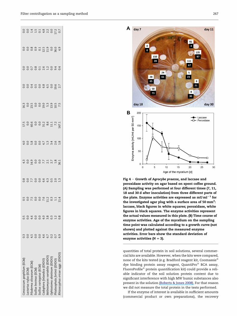
In the second experiment, the litter-decomposer Agrocybe
praecoxwas grown on CG agar. On four different days, samples
were taken from different parts of plates representing
different ages of the mycelium. Laccase and peroxidase
activities were determined in the liquids obtained after
centrifugation.
In the third experiment, two fungal strains of different
eco-physiological groups (a litter-decomposing and an ecto-
mycorrhizal fungus), A. praecox and Piloderma olivaceum, pre-
grown on HA or HA supplemented with Mn(II) (as
MnCl2� 4H2O 100 mg l�1), humus or sawdust, were tested for
the production/secretion of 12 different enzymes that are
important in carbon, nitrogen, sulfur and phosphorus utili-
zation from organic sources. Sampling was performed as
described for the first experiment.
Statistical analyses
The data were analysed using a one-way ANOVA following
Tukey’s HSD post hoc-test using P¼ 0.05 as limit for statistical
significance. Homogeneity of variances and the normality of
the data were tested and in case the requirements of ANOVA
were not met even after log or square root transformations,
the non-parametric ManneWhitney test was used. The
statistical analyses were carried out using SPSS for Windows
version 16.0 software (SPSS Inc. USA).
Results
Verification of the sampling method
Known amounts of laccase and MnP were added to different
agar media, left to settle for 1 hr at room temperature before
centrifugation sampling. In the case of laccase, enzyme
activity in the agar media was generally and significantly
(P� 0.05) lower than the added enzyme activity (25 % to 45 %
less) but no differences were found between the agar media
(Fig 1). In the case of MnP,mediawith humus addition showed
significantly lower enzyme activity in relation to the added
stock solution (Fig 1). However, there were no significant
differences (P> 0.05), probably due to the large variation
(25e75 %). The correction coefficient of enzyme activity (100
divided by the average activity percentage) for laccase on HA,
HAwith humus and HAwith sawdust were 1.84, 1.35 and 1.62,
respectively. The coefficients for MnP were 1.66, 4.08 and 1.84,
respectively.
The effect of the centrifugal forces on laccase and peroxi-
dase activity was studied using living cultures of A. praecox
and Collybia dryophila (Fig 2). In general, a force of 15 700�g
gave the highest activity, although the difference was in most
cases not significant (P> 0.05). Only the peroxidase activity of
A. praecox increased with increasing force. Sonication did not
further increase the detectable enzyme activities, so it seems
that a partial lysis of hyphae did not occur at these centrifugal
forces and intracellular enzymes (e.g. some laccase isoen-
zymes) were not liberated into the solution; on the contrary,
enzyme activities always decreased after the sonication
treatment (Fig 2). The volume of the enzyme solution did not
have any significant effect on the percentage of enzyme
activity indicating that the filter does not retain comparatively
more enzymes when small volumes are used (Fig 3).
Culture tests
Laccase (Table 2A) and peroxidase (Table 2B) activities were
measured for a set of 23 basidiomycetous fungi and corrected
by the correction coefficients obtained from Fig 1. In the case
of laccase, higher activities weremeasured when themedium
contained humus or sawdust, sometimes the activities were
several hundred-times higher (e.g. in case of A. praecox).
However, the effect of additives differed clearly between the
species. In the case of peroxidase, the same trend was
observed with considerable enhancement of activities on
plates with organic additives. When the data were corrected
using the coefficients, they suggest low but significant
peroxidase activities also for some ectomycorrhizal and
root-endophytic ascomycetous fungi (Table 2B). Whether
these activities are really based on MnP enzyme proteins or
may be the result of other (bio)chemical processes slowly
oxidizing Mn(II) or even just assay artefacts will have to be
clarified in additional studies.
In the second experiment, the spatial distribution of lac-
case and peroxidase activities of A. praecox was determined
(Fig 4). Agar plugs were taken on different days and from
different areas of the plate. The use of the filter centrifugation
method for single agar plugs allowed visualizing the spatial
distribution of laccase and peroxidase (Fig 4A). Based on
a growth curve (data not shown), the actual age of the myce-
lium at the sampling time point was calculated and plotted
together with themeasured enzyme activities to obtain a time

Fig 3 e The effect of sample volume on laccase and MnP
activity. Different letters above the standard deviation bars
indicate statistically significant (P £ 0.05) differences
between treatments (N[ 3). Separate statistical tests are
performed for different enzymes.
Fig 2 e The effect of centrifugal forces on laccase and
peroxidase activity. The test was performed using living
cultures of the litter-decomposing fungi A. praecox and C.
dryophila growing on HA medium. Sonic means that the
sample was ultrasonicated for 30 min before
centrifugation. Different letters above the standard
deviation bars indicate statistically significant (P £ 0.05)
differences between treatments (N[ 3). Separate statistical
tests are performed for different fungi and enzymes.
Filter centrifugation as a sampling method 265
course of enzyme activities (Fig 4B). High laccase activities
were found in the growing part of the mycelium. Laccase
activity appeared to be stable before its activity decreased in
the area of around 11-d-old mycelium. Peroxidase seemed to
be secreted later than laccase and after hyphae had success-
fully established on the growth substratum. Also peroxidase
activity appeared to remain stable up to 10 d in agar plates
before measurable activity started to decrease.
In the third experiment, the sampling method was used to
analyse the activities of 12 enzymes from two fungal strains,
A. praecox and P. olivaceum; the species represent two different
eco-physiological fungal groups in soil-litter-decomposing
and ectomycorrhizal fungi respectively. Clear differences in
the enzyme fingerprint between studied species were evident
(Table 3). Mn(II) addition stimulated most enzymes in both
species, humus particularly induced activities in A. praecox
and sawdust in P. olivaceum. A. praecox did not show any
activity of arylsulfatase nor of a-glucosidase. P. olivaceum did
not show any activities of leucine- and alanine-
aminopeptidase, nor of peroxidase and hardly any activity of
a-glucosidase. Also, the level of enzyme activities differed
substantially between the species.
Discussion
Sampling method
The application of centrifuge tube filters as a method for
fine-scale sampling of fungal enzymes proved to be useful in
Petri dish experiments. During centrifugation, aqueous and
soluble compounds including enzymes were recovered. The
percentage detected activity of the model enzymes (laccase
and MnP) was sufficient, although that of MnP was probably
affected by humus (Fig 1). This could be due to adsorption of
the enzyme or an interference of humic ingredients with the
enzyme activity measurement. The inhibitory effect of HA on
laccase has been very well demonstrated by Zavarzina et al.
(2004) and was considered to be due to the functional
groups, molecular weight and hydrophobicity of the HA
preparations (Zavarzina et al. 2004). Furthermore, in our
particular test, a definite activity of enzyme was added by
injection to the agar. So it is possible that the injection created
a hole in the agar and only part of the enzyme added was
absorbed into the agar.
Veps€al€ainen et al. (2001) concluded that soil extracts cannot
be used to assess the enzyme activities of bulk soil due to very
low protein recovery. In our case, although so far not estab-
lished with authentic soil but with soil-like organic additives,
the apparent enzyme activity was inmost cases between 50 %
and 75 % of that added, which is quite high compared to
previous studies. The agar media used in our study differ in
the adsorption properties fromnatural samples but they serve
as suitable model to mimic the fate of enzymes in complex
solid materials. Especially the addition of humus resulted in
low enzyme activities. It is uncertain whether this is due to
protein absorption, enzyme inactivation or the interference of
humic substances with the enzyme assays. To determine the

Table 2 e The production of laccase (2A) peroxidase (2B) on HAwith and without added sterilized boreal forest humus and Scots pine sawdust by a selection of white-rot,brown-rot, litter-decomposing, ectomycorrhizal and root-endophytic fungi. The values that are corrected using correction factors obtained in tests presented in Fig 1 arealso presented. The factors are for laccase on HA, HA with humus and HA with sawdust 1.84, 1.35 and 1.62, and for MnP 1.66, 4.08 and 1.84, respectively
HA HAþhumus HAþ sawdust
Measuredaverage
stdev Correctedaverage
stdev Measuredaverage
stdev Correctedaverage
stdev Measuredaverage
stdev Correctedaverage
stdev
A. Laccase U/cm2
Irpex lacteus (WR) 1.0 0.7 1.8 1.4 0.0 0.0 0.0 0.0 7.6 6.6 12.3 10.7
Phanerochaete velutina (WR) 2.8 2.8 5.2 5.2 0.0 0.0 0.0 0.0 12.0 3.7 19.4 6.0
Physisporinus rivulosus (WR) 68.3 3.7 125.9 6.9 679.8 69.6 919.4 94.2 272.2 128.7 440.4 208.2
Sphaerobolus stellatus (WR) 28.8 5.4 53.1 10.0 124.8 112.4 168.8 152.1 63.5 35.1 102.8 56.7
Piptoporus betulinus (BR) 0.0 0.0 0.0 0.0 0.0 0.0 0.0 0.0 0.0 0.0 0.0 0.0
Postia caesia (BR) 7.9 7.5 14.5 13.9 2.0 2.2 2.7 2.9 0.3 0.5 0.5 0.8
Serpula lacrymans (BR) 1.0 1.8 1.9 3.3 0.0 0.0 0.0 0.0 0.0 0.0 0.0 0.0
Agrocybe praecox (LDF) 0.6 1.0 1.1 1.9 256.0 85.5 346.2 115.6 1.1 1.9 1.8 3.1
Collybia dryophila (LDF) 253.7 52.6 468.0 97.0 307.1 57.3 415.3 77.6 412.1 46.1 666.8 74.7
Hypholoma fasciculare (LDF) 42.1 13.0 77.7 24.0 59.2 11.0 80.1 14.9 24.5 10.3 39.6 16.7
Stropharia aeruginosa (LDF) 1.2 1.1 2.2 2.1 28.3 43.9 38.3 59.4 19.1 2.5 30.9 4.0
Stropharia hornemannii (LDF) 8.2 1.4 15.1 2.5 326.6 45.4 441.7 61.4 114.5 18.3 185.3 29.7
Stropharia rugosoannulata (LDF) 0.0 0.0 0.0 0.0 4.9 8.4 6.7 11.3 2.1 3.1 3.4 5.0
Cenococcum geophilum (ECM) 9.9 2.6 18.4 4.7 33.9 1.9 45.9 2.6 10.3 1.0 16.6 1.7
Lactarius rufus (ECM) 38.0 3.4 70.0 6.3 0.0 0.0 0.0 0.0 131.5 4.0 212.8 6.5
Piloderma olivaceum (ECM) 20.7 7.8 38.1 14.5 100.3 8.8 135.7 12.0 146.6 66.2 237.1 107.2
Suillus bovinus (ECM) 0.0 0.0 0.0 0.0 0.0 0.0 0.0 0.0 0.0 0.0 0.0 0.0
Suillus variegatus (ECM) 0.0 0.0 0.0 0.0 0.1 0.1 0.1 0.2 0.0 0.0 0.0 0.0
Cadophora finlandica (ENDO) 4.8 2.9 8.9 5.3 0.3 0.5 0.4 0.6 4.8 7.5 7.8 12.1
Meliniomyces variabilis (ENDO) 0.0 0.0 0.0 0.0 0.0 0.0 0.0 0.0 1.3 1.9 2.1 3.1
Ophiostoma abietinum (ENDO) 0.0 0.0 0.0 0.0 0.2 0.3 0.3 0.5 0.0 0.0 0.0 0.0
Phialocephala fortinii (ENDO) 1.3 2.3 2.4 4.2 0.5 0.9 0.7 1.3 4.5 1.4 7.3 2.3
Rhizoscyphus ericae aggr. (ENDO) 0.1 0.2 0.2 0.3 0.7 1.0 0.9 1.4 0.1 0.2 0.2 0.3
B. Peroxidase U/cm2
Irpex lacteus (WR) 3.4 3.3 5.6 5.5 42.1 13.1 171.5 53.4 45.9 34.6 84.4 63.5
Phanerochaete velutina (WR) 199.0 44.5 330.1 73.9 0.0 0.0 0.0 0.0 153.3 211.2 281.6 387.9
Physisporinus rivulosus (WR) 92.9 22.8 154.0 37.8 123.8 13.2 504.6 53.9 51.3 36.8 94.2 67.6
Sphaerobolus stellatus (WR) 77.2 24.5 128.0 40.6 195.6 99.5 797.3 405.5 113.0 37.7 207.6 69.3
Piptoporus betulinus (BR) 6.5 11.3 10.8 18.7 2.0 3.4 8.0 13.9 0.0 0.0 0.0 0.0
Postia caesia (BR) 3.6 3.8 6.1 6.4 9.2 3.7 37.3 15.2 7.5 2.7 13.8 5.0
Serpula lacrymans (BR) 0.0 0.0 0.0 0.0 0.0 0.0 0.0 0.0 0.0 0.0 0.0 0.1
Agrocybe praecox (LDF) 1.4 1.1 2.3 1.9 59.0 20.3 240.5 82.8 3.7 3.2 6.8 5.8
Collybia dryophila (LDF) 0.0 0.0 0.0 0.0 20.2 13.3 82.3 54.3 1359.5 485.8 2496.8 892.2
Hypholoma fasciculare (LDF) 505.0 415.5 837.5 689.2 4.3 2.6 17.7 10.5 239.8 185.8 440.5 341.2
Stropharia aeruginosa (LDF) 82.2 83.7 136.3 138.9 725.2 862.5 2956.2 3515.8 223.4 57.4 410.2 105.4
Stropharia hornemannii (LDF) 32.1 28.5 53.2 47.3 234.6 33.3 956.2 135.7 182.1 93.7 334.4 172.1
Stropharia rugosoannulata (LDF) 13.2 6.0 21.9 9.9 171.1 38.6 697.4 157.4 168.1 48.3 308.7 88.6
266
J.Heinonsa
loet
al.
Cen
ococcu
mgeop
hilum
(ECM)
0.3
0.5
0.5
0.8
4.3
4.0
17.5
16.3
0.0
0.0
0.0
0.0
Lactariusrufus(ECM)
0.0
0.0
0.0
0.0
0.0
0.0
0.0
0.0
0.0
0.0
0.0
0.0
Pilod
ermaolivaceum
(ECM)
4.5
4.6
7.5
7.7
14.9
5.8
60.6
23.5
0.4
0.7
0.8
1.4
Suillusbov
inus(ECM)
0.0
0.0
0.0
0.0
0.0
0.0
0.0
0.0
0.5
0.8
0.9
1.5
Suillusvariegatus(ECM)
0.0
0.0
0.0
0.0
0.0
0.0
0.0
0.0
0.0
0.1
0.1
0.1
Cadop
horafinlandica(ENDO)
4.6
4.0
7.6
6.6
7.7
4.7
31.2
19.0
6.8
2.6
12.5
4.8
Meliniomyces
variabilis
(ENDO)
6.7
3.8
11.2
6.3
2.1
1.3
8.6
5.3
6.5
1.2
11.9
2.2
Ophiostom
aabietinum
(ENDO)
0.2
0.3
0.3
0.5
2.7
3.4
11.1
13.9
0.0
0.0
0.0
0.0
Phialocephala
fortinii(ENDO)
0.7
1.2
1.1
2.0
1.8
2.6
7.2
10.5
19.6
9.4
36.0
17.2
Rhizoscyphusericaeaggr.
(ENDO)
6.9
0.8
11.4
1.3
36.1
1.8
147.1
7.5
2.7
0.4
4.9
0.7
Fig 4 e Growth of Agrocybe praecox, and laccase and
peroxidase activity on agar based on spent coffee ground.
(A) Sampling was performed at four different times (7, 11,
18 and 30 d after inoculation) from three different parts of
the plate. Enzyme activities are expressed as mUmlL1 for
the investigated agar plug with a surface area of 50 mm2:
laccase, black figures in white squares; peroxidase, white
figures in black squares. The enzyme activities represent
the actual values measured in this plate. (B) Time course of
enzyme activities. Age of the mycelium on the sampling
time point was calculated according to a growth curve (not
shown) and plotted against the measured enzyme
activities. Error bars show the standard deviation of
enzyme activities (N[ 3).
Filter centrifugation as a sampling method 267
quantities of total protein in soil solutions, several commer-
cial kits are available. However, when the kits were compared,
none of the kits tested (e.g. Bradford reagent kit, Coomassie�
dye binding protein assay reagent, QuantiPro� BCA assay,
FluoroProfile� protein quantification kit) could provide a reli-
able indicator of the soil solution protein content due to
significant interference with high MW humic substances also
present in the solution (Roberts & Jones 2008). For that reason
we did not measure the total protein in the tests performed.
If the enzyme of interest is available in sufficient amounts
(commercial product or own preparations), the recovery

Table 3 e The summary of the 12 enzyme activities analysed (semiquantitative from no activity[L to strong activity[DDD). The fungi were grown on Hagem’s agar without supplements or supplemented with Mn II, humus or Scots pinesawdust
Enzyme activity A. praecox P. croceum Control
Mn II Humus Sawdust Hagem’s Mn II Humus Sawdust Hagem’s Hagem’s
Arylsulphatase � � � � � � � � �a-glucosidase � � � � � � � � �b-glucosidase þ � � � þ þ þþþ � �Cellobiosidase � � � � � � � � �b-xylosidase þ � � � � � þþþ � �Phosphomonoesterase þþ � � � þþ � þþþ � �Phosphodiesterase þ � � � � � þþþ � �N-acetylglucosamidase þþ � � � þþþ � � � �Leucine-aminopeptidase � � � � � � � � �Alanine-aminopeptidase � � � � � � � � �Laccase þþþ þþþ � � � þ þ � �Peroxidase þþþ þ � � � � � � �
268 J. Heinonsalo et al.
percentage can be calculated to obtain a correction coefficient
that can be later used to adjust results of strain screening
experiments. In our case, the correction coefficient for MnP in
agar plates supplemented with humus was 4.1, which signif-
icantly affected the results obtained. Despite the reduced
enzyme activities observed after artificial supplementation of
the agar with model enzymes (probably caused by the organic
additives/ingredients), the lack of a dilution effect in this
sampling method results in good sensitivity and improved
detection of enzymatic activities even at very low levels.
In the centrifugal force test, the aim was to verify that the
fungal hyphae do not lyse and release intracellular enzymes
to the sample solution. In three out of four tests, there was no
significant (P> 0.05) difference between the highest and
lowest centrifugal force, which demonstrates that the cells do
not break due to the centrifugal force. In the case of A. praecox
peroxidase the highest force yielded a higher activity than the
lower centrifugal forces (Fig 2) indicating a correlation
between centrifugal force and recoverable peroxidase activity.
Sonication decreased the activities both of laccase and
peroxidase suggesting that ultrasound waves have damaging
effects on the enzyme proteins. This phenomenon has been
known for decades and is a general problem of intracellular
enzyme preparation using sonication for cell disruption
(Coakley et al. 1973).
Different amounts of added enzymes did not have any
effect on the activities in the volumetric test, which means
that also in case of small volumes (around 50 ml) enzymes are
as efficiently recovered from the filters as in the case of larger
solution volumes (up to 300 ml). This is important if the
method is used to detect activities in small fine-scale samples
with no water (or other buffer/extractant) additions and
variable moisture content.
Culture tests
The filter centrifugation method proved to be useful in
detecting potential laccase and peroxidase producers in pure
cultures. It also facilitated the use of different solid or liquid
supplements in simple Petri dishes, which makes it easier to
screen growth media for optimization of enzyme production.
Fungi producing high amounts of enzymes in liquid cultures
did so also on agar plates (data not shown).
The spatial distribution of laccase and peroxidase activity
determined at several points during the cultivation of A.
praecox on agar plates also reflected the temporal develop-
ment of its colony. Traditionally, extracellular enzyme activ-
ities have been localized and visualized by staining agar plates
(White & Boddy 1992; Hiscox et al. 2010). In a study of Dinis
et al. (2009), enzyme production of four fungi during growth
on wheat straw was investigated. At each sampling time the
whole culture was harvested. In our experiment, samples at
different time points and from different areas were just taken
from one set of agar plates (three replicate plates). Estimation
of themycelium age based on a growth curve allowed plotting
of the enzyme activities as a time course of measured enzyme
activities during solid-state cultivation (Fig 4B). It was neither
necessary to stain the plates nor to apply laborious extraction
steps to determine and quantify enzyme activities. Further-
more the results may give hints about which part of the
mycelium actively produces enzymes at a certain time point.
Enzyme activity fingerprints for two different types of fungi
were obtained using the centrifugation sampling method. Two
species, A. praecox and P. olivaceum, had clearly different func-
tional profiles with different levels of activities. A. praecox
produced all envisaged enzymes except arylsulfatase and
a-glucosidase. P. olivaceum produced all others except a-gluco-
sidase, leucine- and alanine-aminopeptidase and peroxidase.
Enzyme production by P. olivaceum and A. praecox was strongly
stimulated by addition of sawdust and humus, respectively.
This kind of functional profiling may be useful to identify and
segregate functional groups of fungi, which in turn could be
used to highlight roles of cryptic species in forest soil
ecosystems.
Applicability of the sampling method
The method presented here facilitates studies on fungal
ecology using small, fine-scale sampling from fungal cultures
growing on different agar media. The recovery of model
enzymes was reasonably good from agar media. Thus, the
method allows screening for a variety of enzymes and also

Filter centrifugation as a sampling method 269
facilitates investigation of the effect of various supplements
on growth and enzyme production. Even the conversion of
colourless toxic additives might be studied, if subsequent
analysing tools for small volumes become available.
Further studies are needed to test the suitability of the
method for soil samples, since low recoveries with several
extraction methods were previously reported for soil samples
(Veps€al€ainen et al. 2001). The possibility of taking small-scale
samples may allow the detection of spatial and temporal
variation in enzyme activities in different kinds of micro- and
mesocosms where fungal hyphae can be harvested and
samples taken with precision. Our proposed method may be
applicable for the use of small amounts of soil, or even wood,
to give sufficient amounts of enzyme-containing liquid easily
utilisable for further analysis.
Acknowledgement
This research was funded by Academy of Finland (project
number 1212915 and 130984 for J.H., project number 206085 for
H.I., and project number 1133022 for G.K.) and EnSTe Graduate
School (for G.K.).
r e f e r e n c e s
Coakley WT, Brown RC, James CJ, Gould RK, 1973. Theinactivation of enzymes by ultrasonic cavitation at 20 kHz.Archives of Biochemistry and Biophysics 159: 722e729.
Dinis MJ, Bezerra RMF, Nunes F, Dias AA, Guedes CV, Ferreira LMM,Cone JW, Marques GSM, Barros ARN, Rodrigues MAM, 2009.Modification of wheat straw lignin by solid state fermentationwith white-rot fungi. Bioresource Technology 100: 4829e4835.
Elisashvili V, Penninckx M, Kachlishvili E, Tsiklauri N, Metreveli E,Kharziani T, Kvesitadze G, 2008. Lentinus edodes and Pleurotusspecies lignocellulolytic enzymes activity in submerged andsolid-state fermentation of lignocellulosic wastes of differentcomposition. Bioresource Technology 99: 457e462.
Fan L, Pandey A, Mohan R, Soccol CR, 2000. Use of various coffeeindustry residues for the cultivation of Pleurotus ostreatus insolid state fermentation. Acta Biotechnologica 20: 41e52.
Hiscox J, Baldrian P, Rogers HJ, Boddy L, 2010. Changes in oxidativeenzyme activity during interspecific mycelial interactions
involving thewhite-rot fungusTrametes versicolor. Fungal Geneticsand Biology 47: 562e571.
Hofrichter M, Ziegenhagen D, Vares T, Friedrich M, J€ager MG,Fritsche W, Hatakka A, 1998. Oxidative decomposition ofmalonic acid as basis for the action ofmanganese peroxidase inthe absence of hydrogen peroxide. FEBS Letters 434: 362e366.
Ilvesniemi H, Giesler R, van Hees P, Magnussson T, Melkerud PA,2000. General description of the sampling techniques and thesites investigated in the Fennoscandinavian podzolizationproject. Geoderma 94: 109e123.
Isikhuemhen O, Mikiashvili N, Kelkar V, 2009. Application of solidwaste from anaerobic digestion of poultry litter in Agrocybeaegerita cultivation: mushroom production, lignocellulolyticenzymes activity and substrate utilization. Biodegradation 20:351e361.
Modess O, 1941. Zur Kenntnis der Mykorrhizabildner von Kieferund Fichte. Symbolae Botanicae Upsalienses 5: 1e147.
Roberts P, Jones DL, 2008. Critical evaluation of methods fordetermining total protein in soil solution. Soil Biology &Biochemistry 40: 1485e1495.
�Snajdr J, Baldrian P, 2006. Production of lignocellulose-degradingenzymes and changes in soil bacterial communities duringthe growth of Pleurotus ostreatus in soil with different carboncontent. Folia Microbiologica 51: 579e590.
Steffen KT, Cajthaml T, �Snajdr J, Baldrian P, 2007. Differentialdegradation of oak (Quercus petraea) leaf litter by litter-decomposing basidiomycetes. Research in Microbiology 158:447e455.
Tabatabai MA, 1994. Soil enzymes. In: Weaver RW, Angle S,Bottomley P, Bezdicek D, Smith S, Tabatabai A, Wollum A (eds),Methods of Soil Analysis. Part 2. Microbial and Biochemical Properties.Soil Science Society of America, Madison, pp. 775e833.
Valent�ın L, Kluczek-Turpeinen B, Willf€or S, Hemming J, Hatakka A,Steffen K, Tuomela M, 2010. Scots pine (Pinus sylvestris) barkcomposition and degradation by fungi: potential substrate forbioremediation. Bioresource Technology 101: 2203e2209.
Veps€al€ainen M, Erkomaa K, Kukkonen S, Vestberg M,Wallenius K, Niemi R, 2004. The impact of crop plantcultivation and peat amendment on soil microbial activity andstructure. Plant and Soil 264: 273e286.
Veps€al€ainen M, Kukkonen S, Vestberg M, Sirvi€o H, Maarit Niemi R,2001. Application of soil enzyme activity test kit in a fieldexperiment. Soil Biology and Biochemistry 33: 1665e1672.
White NA, Boddy L, 1992. Extracellular enzyme localizationduring interspecific fungal interactions. FEMS MicrobiologyLetters 98: 75e79.
Zavarzina AG, Leontievsky AA, Golovleva LA, Trofimov SYa, 2004.Biotransformation of soil humic acids by blue laccase of Panustigrinus 8/18: an in vitro study. Soil Biology & Biochemistry 36:359e369.
Related Documents