Journal of Structural Biology 150 (2005) 170–179 www.elsevier.com/locate/yjsbi 1047-8477/$ - see front matter. Published by Elsevier Inc. doi:10.1016/j.jsb.2005.02.007 Filaments of the Ure2p prion protein have a cross- core structure Ulrich Baxa a,b , Naiqian Cheng a , Dennis C. Winkler a , Thang K. Chiu c , David R. Davies c , Deepak Sharma d , Hideyo Inouye d , Daniel A. Kirschner d , Reed B. Wickner b , Alasdair C. Steven a,¤ a Laboratory of Structural Biology, National Institute of Arthritis, Musculoskeletal, and Skin Diseases, National Institutes of Health, Bethesda, MD 20892, USA b Laboratory of Biochemistry and Genetics, National Institute of Diabetes and Digestive and Kidney Diseases, National Institutes of Health, Bethesda, MD 20892, USA c Laboratory of Molecular Biology, National Institute of Diabetes and Digestive and Kidney Diseases, National Institutes of Health, Bethesda, MD 20892, USA d Biology Department, Boston College, Chestnut Hill, MA 02467, USA Received 22 December 2004, and in revised form 5 February 2005 Available online 14 March 2005 Abstract Formation of Wlaments by the Ure2 protein constitutes the molecular mechanism of the [URE3] prion in yeast. According to the “amyloid backbone” model, the N-terminal asparagine-rich domains of Ure2p polymerize to form an amyloid core Wbril that is sur- rounded by C-terminal domains in their native conformation. Protease resistance and Congo Red binding as well as -sheet content detected by spectroscopy—all markers for amyloid—have supported this model, as has the close resemblance between 40 Å N- domain Wbrils and the Wbrillar core of intact Ure2p Wlaments visualized by cryo-electron microscopy and scanning transmission elec- tron microscopy. Here, we present electron diVraction and X-ray diVraction data from Wlaments of Ure2p, of N-domains alone, of fragments thereof, and of an N-domain-containing fusion protein that demonstrate in each case the 4.7 Å reXection that is typical for cross- structure and highly indicative of amyloid. This reXection was observed for specimens prepared by air-drying with and with- out sucrose embedding. To conWrm that the corresponding structure is not an artifact of air-drying, the reXection was also demon- strated for specimens preserved in vitreous ice. Local area electron diVraction and X-ray diVraction from partially aligned specimens showed that the 4.7 Å reXection is meridional and therefore the underlying structure is cross-. Published by Elsevier Inc. Keywords: Electron diVraction; X-ray diVraction; Yeast prion; Natively unfolded protein 1. Introduction The non-chromosomal element [URE3] in the yeast, S. cerevisiae, was identiWed as an aggregated, infectious (prion) form of the cytosolic protein Ure2p (Edskes et al., 1999; Masison and Wickner, 1995; Speransky et al., 2001; Wickner, 1994). The normal, soluble, form of Ure2p has a regulatory function in the nitrogen catabo- lite repression pathway (NCR) 1 (Cooper, 2002). When wild-type yeast cells are grown on a rich nitrogen source like ammonia, Ure2p binds to the transcription factor Gln3p and blocks its entry into the nucleus, thereby shutting oV genes that are needed to utilize poor nitro- gen sources (Beck and Hall, 1999; Blinder et al., 1996; Drillien et al., 1973). In [URE3] strains, this regulatory function is lost. ¤ Corresponding author. Fax: +1 301 443 7651. E-mail address: [email protected] (A.C. Steven). 1 Abbreviations used: GFP, green Xuorescent protein; NCR, nitrogen catabolite repression.

Welcome message from author
This document is posted to help you gain knowledge. Please leave a comment to let me know what you think about it! Share it to your friends and learn new things together.
Transcript
Journal of Structural Biology 150 (2005) 170–179
www.elsevier.com/locate/yjsbi
Filaments of the Ure2p prion protein have a cross-� core structure
Ulrich Baxa a,b, Naiqian Cheng a, Dennis C. Winkler a, Thang K. Chiu c, David R. Davies c, Deepak Sharma d, Hideyo Inouye d, Daniel A. Kirschner d,
Reed B. Wickner b, Alasdair C. Steven a,¤
a Laboratory of Structural Biology, National Institute of Arthritis, Musculoskeletal, and Skin Diseases, National Institutes of Health, Bethesda, MD 20892, USA
b Laboratory of Biochemistry and Genetics, National Institute of Diabetes and Digestive and Kidney Diseases, National Institutes of Health, Bethesda, MD 20892, USA
c Laboratory of Molecular Biology, National Institute of Diabetes and Digestive and Kidney Diseases, National Institutes of Health, Bethesda, MD 20892, USA
d Biology Department, Boston College, Chestnut Hill, MA 02467, USA
Received 22 December 2004, and in revised form 5 February 2005Available online 14 March 2005
Abstract
Formation of Wlaments by the Ure2 protein constitutes the molecular mechanism of the [URE3] prion in yeast. According to the“amyloid backbone” model, the N-terminal asparagine-rich domains of Ure2p polymerize to form an amyloid core Wbril that is sur-rounded by C-terminal domains in their native conformation. Protease resistance and Congo Red binding as well as �-sheet contentdetected by spectroscopy—all markers for amyloid—have supported this model, as has the close resemblance between 40 Å N-domain Wbrils and the Wbrillar core of intact Ure2p Wlaments visualized by cryo-electron microscopy and scanning transmission elec-tron microscopy. Here, we present electron diVraction and X-ray diVraction data from Wlaments of Ure2p, of N-domains alone, offragments thereof, and of an N-domain-containing fusion protein that demonstrate in each case the 4.7 Å reXection that is typical forcross-� structure and highly indicative of amyloid. This reXection was observed for specimens prepared by air-drying with and with-out sucrose embedding. To conWrm that the corresponding structure is not an artifact of air-drying, the reXection was also demon-strated for specimens preserved in vitreous ice. Local area electron diVraction and X-ray diVraction from partially aligned specimensshowed that the 4.7 Å reXection is meridional and therefore the underlying structure is cross-�.Published by Elsevier Inc.
Keywords: Electron diVraction; X-ray diVraction; Yeast prion; Natively unfolded protein
1. Introduction lite repression pathway (NCR)1 (Cooper, 2002). When
The non-chromosomal element [URE3] in the yeast,S. cerevisiae, was identiWed as an aggregated, infectious(prion) form of the cytosolic protein Ure2p (Edskeset al., 1999; Masison and Wickner, 1995; Speranskyet al., 2001; Wickner, 1994). The normal, soluble, form ofUre2p has a regulatory function in the nitrogen catabo-
¤ Corresponding author. Fax: +1 301 443 7651.E-mail address: [email protected] (A.C. Steven).
1047-8477/$ - see front matter. Published by Elsevier Inc.doi:10.1016/j.jsb.2005.02.007
wild-type yeast cells are grown on a rich nitrogen sourcelike ammonia, Ure2p binds to the transcription factorGln3p and blocks its entry into the nucleus, therebyshutting oV genes that are needed to utilize poor nitro-gen sources (Beck and Hall, 1999; Blinder et al., 1996;Drillien et al., 1973). In [URE3] strains, this regulatoryfunction is lost.
1 Abbreviations used: GFP, green Xuorescent protein; NCR, nitrogencatabolite repression.
U. Baxa et al. / Journal of Structural Biology 150 (2005) 170–179 171
The N-terminal domain of Ure2p (N-domain, resi-dues 1–90) is both necessary and suYcient for prion con-version and propagation (Masison et al., 1997; Masisonand Wickner, 1995), whereby Ure2p forms Wlaments inthe infected cells (Speransky et al., 2001). Hence it is alsoreferred to as the prion-forming domain or, simply,prion domain. This domain is unusually rich in the polaruncharged residues, asparagine, glutamine and serine.Yeast cells expressing only the C-terminal domain ofUre2p (residues 90–354) still have a functional, althoughsomewhat leaky, NCR but are unable to convert to the[URE3] form (Maddelein and Wickner, 1999; Masisonand Wickner, 1995). The crystal structure of the C-termi-nal domain has been solved (Bousset et al., 2001a;Umland et al., 2001), conWrming the expectation basedon sequence similarity that it has the same fold as gluta-thione S-transferase (Coschigano and Magasanik, 1991).In fact, the C-domain binds glutathione (Bousset et al.,2001b) and has glutathione peroxidase activity (Baiet al., 2004).
Recombinant Ure2p can also form Wlaments in vitro(Taylor et al., 1999), and in this form, retains both gluta-thione binding (Bousset et al., 2002) and glutathione per-oxidase activity (Bai et al., 2004). These observationsindicate that the C-terminal domains are still folded inthe Wlamentous state of Ure2p, supporting the earlierinference to this eVect based on the activities of variousenzymes in Wlaments of fusion proteins in which theywere coupled to the N-domain of Ure2p (Baxa et al.,2002). The inference that the C-domain remains foldedin Wlaments is strongly supported by diVerential scan-ning calorimetry of Ure2p Wlaments, soluble Ure2p andsoluble C-terminal domains (Baxa et al., 2004). The C-terminal domain responded to thermal perturbation inthe same way in all three states, indicating that it doesnot change its structure upon entering Wlaments.
In the soluble state of Ure2p, the N-domain isnatively unfolded, as attested by its sensitivity to prote-ases (Baxa et al., 2003; Thual et al., 1999), as well as datafrom physical chemistry (Galani et al., 2002; Perrett etal., 1999; Zhou et al., 2001), diVerential scanning calo-rimetry (Baxa et al., 2004), and NMR spectroscopy(Pierce et al., 2004). Upon entering the Wlamentous state,it undergoes a drastic conformational change as attestedby its becoming highly resistant to proteases (Baxa et al.,2003; Ripaud et al., 2004; Taylor et al., 1999) and otherlines of evidence (Baxa et al., 2004; Schlumpberger et al.,2000; Taylor et al., 1999). The predisposition for thisconformational change seems to lie mainly in its aminoacid composition, since Wve randomizations of the orderof the amino acids of the N-domain sequence producedUre2p variants that retained the ability to form Wla-ments in vitro and become prions in vivo (Ross et al.,2004).
High resolution negative staining as well as cryo-elec-tron microscopy showed that Ure2p Wlaments have a
thin core Wbril with globular domains arranged aroundit in a left handed spiral (Baxa et al., 2003; Kajava et al.,2004). In diameter and appearance, the core Wbrilmatches Wbrils assembled in vitro from N-domains, aswell as residual Wlaments remaining after proteolysis ofintact Ure2p Wlaments (Baxa et al., 2003; Taylor et al.,1999). The observations summarized above underlie the“amyloid backbone” model, whereby residues 1 to »70polymerize to form an amyloid Wlament that is sur-rounded by native C-terminal domains (residues »90 to354) and connected to them by a Xexible linker (residues»70 to »90) (Baxa et al., 2002, 2003; Kajava et al., 2004;Speransky et al., 2001; Taylor et al., 1999).
The evidence that the core Wbril is amyloid is exten-sive but somewhat circumstantial: (i) it is strikingly resis-tant to digestion with proteinase K (Baxa et al., 2003;Ripaud et al., 2004; Taylor et al., 1999); (ii) Wlaments ofall N-domain-containing constructs examined to datebind Congo Red and show yellow-green birefringence(Baxa et al., 2003; Taylor et al., 1999); (iii) N-domainWlaments are rich in �-structure and an increase of �-structure content has been observed upon Wlament for-mation by Ure2p (Schlumpberger et al., 2000; Taylor etal., 1999); and (iv) core Wbrils are straight and smooth-sided (Taylor et al., 1999), and resemble in these respectsother Wlaments accepted to be amyloids, e.g., Alzheimer�-peptide and amylin (Goldsbury et al., 1997, 2000).However, direct evidence from diVraction methods forthe presence of cross-� structure has been lacking. Herewe Wll this gap by reporting electron diVraction and X-ray diVraction studies.
2. Experimental procedures
2.1. Preparation of Ure2p Wlaments
Protein was produced as described previously (Baxaet al., 2002, 2003; Speransky et al., 2001; Taylor et al.,1999). Filaments were made by incubation of proteinsolutions (usually at about 1 mg/ml) on a shaker for 16 hat 4 °C. Proteinase K-digested Ure2p Wlaments were pro-duced as described (Baxa et al., 2003). Before Wlamentpreparations were used in experiments, they were sedi-mented repeatedly at 40000g for 1 h at 4 °C to removeany soluble protein. Prior to air drying with or withoutsucrose embedding, Wlaments were taken up in water orvery dilute buVer (<2 mM phosphate) to reduce diVrac-tion from salt crystals after drying. At all times, the Wla-ments were maintained at room temperature or 4 °C.
2.2. Electron microscopy and diVraction
Samples were negatively stained for assessment ofyield and quality. Samples were adsorbed onto freshlyglow-discharged carbon-coated grids, rinsed with water,
172 U. Baxa et al. / Journal of Structural Biology 150 (2005) 170–179
and stained with 1% uranyl acetate. For sucrose embed-ding, samples were mixed with an equal volume of 1%sucrose and adsorbed onto freshly glow-discharged car-bon-coated grids. After 5 min, excess solution was blot-ted oV and the sample was allowed to dry. Both kinds ofspecimens were examined in a CM120 microscope (FEI,Hillsboro, OR). For cryo-EM, drops of sample wereadsorbed to holey carbon Wlms, blotted, and vitriWed byplunging into liquid ethane. These grids were examinedin a CM200-FEG microscope (FEI, Hillsboro, OR) witha Gatan 626 cryo-holder (Cheng et al., 1999). DiVractionpatterns were taken from areas of »6 �m diameter forpowder spectra and »1 �m for micro-diVraction fromlocally ordered areas, with doses of 1–4 e¡/Å2. Subse-quently, one image was taken in defocused diVractionmode and another one in image mode of the area usedfor diVraction, to assess Wlament orientation and density.DiVraction spacings were calibrated with graphitizedcarbon, evaporated thallous chloride, and evaporatedaluminium (Electron Microscopy Science, HatWeld, PA).Rotational registration between the diVraction patternand the image was calibrated with a grid of negativelystained catalase crystals, prepared as described by Wrig-ley (1968).
2.3. X-ray diVraction and analysis
To prepare the Ure2p10–39 specimen, lyophilized pep-tide was suspended in ultra-pure 18 M� water (U.S. Fil-ter, Lowell, MA), vortexed, and brieXy centrifuged at16000g to sediment any precipitate. About 2–4�L of thesupernatant was then slowly drawn into a 0.5–0.7 mm-diameter siliconized, thin-walled glass capillary tube(Charles A. Supper, South Natick, MA), which was thensealed, and placed in a 2-Tesla permanent magnet(Charles A. Supper; Oldenbourgh and Phillips, 1986). Apin-hole was punched through the wax at the wide end ofthe capillary to allow gradual drying (under ambient tem-perature and humidity) of the solution over 1–2 weeks.When the sample was a small uniform disk, the capillarytube was removed from the magnetic Weld and trans-ferred to the sample holder for analysis by X-ray diVrac-tion. The Ure2p1–89 Wlament sample was examined as awet concentrated pellet after sealing in a capillary tube.
DiVraction patterns were recorded on Kodak DEFWlm using double mirror focused CuK� radiation gener-ated by an Elliot GX-20 rotating anode (Marconi Avi-onics, UK). The specimen-to-Wlm distances of the X-raycameras were calibrated using known standards (calcite,d D 3.035 Å; NaCl, d D 2.821 Å). Exposure time rangedfrom one to three days and the observed spacings werecalculated using Bragg’s law from positions of the reXec-tions measured directly oV the Wlm. The diVraction pat-terns were digitized using ScanMaker 5 (MicrotekLaboratory, 3715 Doolittle Drive, Redondo Beach, CA,USA). The ScanMaker 5, using 300 dpi (corresponding
to 84.66 �m pixels), was operated using MacIntosh pro-gram ScanWizard PPC 3.2.2. The machine readout wascalibrated using the known optical density of a KodakStep Calibration Tablet (No.2; range from 0.04 to 3.03OD). The digitized image was displayed on NIHIMAGE (developed at the U.S. National Institutes ofHealth, and available at http://rsb.info.nih.gov/nih-image/). The intensities along the equatorial directionand radial direction crossing the oV-meridional intensitymaximum were plotted by selecting narrow windows.The background curves were approximated as polyno-mials and subtracted from the measured intensity. EachreXection was assumed to be a Gaussian function. Mea-surements of the integral widths and integral intensitiesof the reXections were obtained by Wtting the back-ground corrected intensity proWle to multiple Gaussianfunctions. The integral width of the direct beam, deter-mined after Gaussian approximation, was 397 �m forthe Ure2p10–39 sample, and 332 �m for Ure2p1–89 whichcorrespond to 2.95 £ 10¡3 Å¡1 and 2.46 £ 10¡3 Å¡1.
Samples of Ure2p full-length Wlaments were dried inbromosilicate capillaries (Charles Supper, Natick, MA)with or without a magnetic Weld (»1.5 T). X-ray diVrac-tion images were recorded using a Rigaku rotatinganode source (CuK�) and a Raxis IV++ detector. Expo-sure times were 5–15 min at a specimen to detector dis-tance of 71.2 mm.
3. Results
Recombinant Ure2p forms Wlaments under physio-logical buVer conditions (Taylor et al., 1999). Rigorousproteinase K treatment digests away the C-domains,leaving thin Wbrils composed of N-terminal material(Baxa et al., 2003). The N-domain alone Wbrillizes rap-idly in physiological buVers (Taylor et al., 1999), as doN-domain fragments, e.g., Ure2p10–39 (Kajava et al.,2004). Moreover, fusion proteins in which the C-domainis exchanged for other globular proteins such as thegreen Xuorescent protein (GFP) readily form Wlaments(Baxa et al., 2002). We set out to investigate Wlaments ofall these kinds by diVraction analysis.
3.1. Electron diVraction
Electron diVraction has been used on previous occa-sions to detect repetitive secondary structures in proteinWlaments (e.g. Watts et al., 1998), including cross-� struc-ture in amyloids (King and Diaz-Avalos, 2004; Serpellet al., 2000). It has the advantage that it is highly sensi-tive; moreover, since Wlaments tend to be deposited onEM grids so as to present side-views, Bragg-angle sam-pling of axial periodicities occurs for a larger fraction ofthe sample than for a three-dimensional pellet of ran-domly oriented Wlaments.
U. Baxa et al. / Journal of Structural Biology 150 (2005) 170–179 173
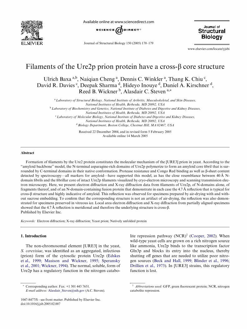
Filaments of Ure2p, Ure2p1–65, Ure2p1–89, Ure2p10–39
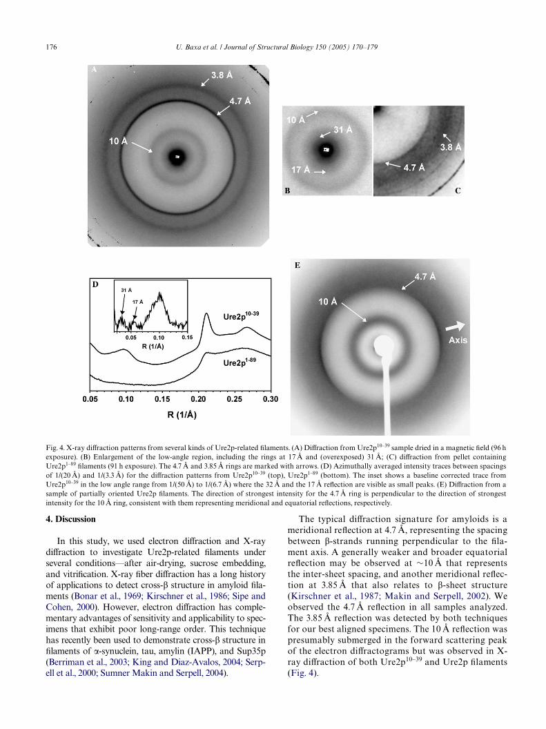
and Ure2p1–80-GFP, as well as Ure2p core Wbrils pro-duced by proteinase K digestion, were embedded insucrose on carbon Wlm-bearing EM grids (see Section 2)and examined in the electron microscope. In each case,the grids were scanned for promising areas and electrondiVractograms were recorded from areas of »6 �m to »1 �m across, and images of these areas were thenrecorded after suitably defocusing the diVracted electronbeam (Fig. 1). Strong diVraction rings at a spacing of4.7 Å were observed in all cases, but were not seen incontrol grids prepared with soluble Ure2p or withsucrose solution alone (data not shown). Ure2p10–39 Wla-ments produced the strongest reXections and for thesespecimens, an additional ring at 3.85 Å (Fig. 1E) was
observed. In one case, the second order of the 4.7 ÅreXection was also observed (data not shown). Asexpected for a protein-associated feature, the 4.7 ÅreXection was radiation-sensitive and decayed com-pletely after a dose of »25 e¡/Å2, even though the overallappearance of the Wlaments in the accompanying imageswas unaltered.
We used sucrose embedding to prepare these samplessince this treatment has been observed to mitigate dry-ing-induced damage to native protein structure (Akeyand Edelstein, 1983; Bourguignon et al., 1978; Kesselet al., 1986; Tokuyasu, 1973). However, we obtainedessentially the same results when samples were air-driedwithout embedding in sucrose (data not shown), indicat-ing that the structure responsible for generating the 4.7 Å
Fig. 1. Electron diVraction analysis of six kinds of Ure2p-related Wlaments. The preparations of Wlaments were Wrst checked by negative stain electronmicroscopy. Four examples are shown: (A) Ure2p10–39; (B) Ure2p1–65; (C) full-length Ure2p; and (D) Ure2p1–89. EM grids bearing sucrose-embeddedspecimens were then examined by electron diVraction. Quarter-plane patterns corresponding to samples (A–D) are shown in (E–H), respectively. Thespectra were digitized and azimuthally averaged. Similar analyses were also performed for Wlaments of the Ure2p1–80-GFP fusion protein, and forcore Wbrils remaining after proteinase K digestion of Ure2p Wlaments. The resulting traces between spatial frequencies indicated below (F) from 1/(7.1 Å) to 1/(2.77 Å) are shown in (I). From the top, they are from: Ure2p10–39, Ure2p1–80-GFP, Ure2p1–65, core Wbrils; Ure2p1–89, and Ure2p. In eachcase, a prominent reXection at »0.21 Å¡1 or 1/(4.7 Å) was detected.
174 U. Baxa et al. / Journal of Structural Biology 150 (2005) 170–179
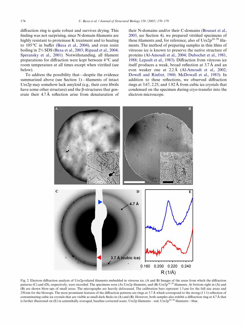
diVraction ring is quite robust and survives drying. ThisWnding was not surprising, since N-domain Wlaments arehighly resistant to proteinase K treatment and to heatingto 105 °C in buVer (Baxa et al., 2004), and even resistboiling in 2% SDS (Baxa et al., 2003; Ripaud et al., 2004;Speransky et al., 2001). Notwithstanding, all Wlamentpreparations for diVraction were kept between 4 °C androom temperature at all times except when vitriWed (seebelow).
To address the possibility that—despite the evidencesummarized above (see Section 1)—Wlaments of intactUre2p may somehow lack amyloid (e.g., their core Wbrilshave some other structure) and the �-structures that gen-erate their 4.7 Å reXection arise from denaturation of
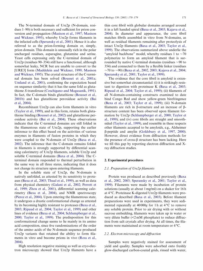
their N-domains and/or their C-domains (Bousset et al.,2003, see Section 4), we prepared vitriWed specimens ofthese Wlaments and, for reference, also of Ure2p10–39 Wla-ments. The method of preparing samples in thin Wlms ofvitreous ice is known to preserve the native structure ofproteins (Al-Amoudi et al., 2004; Dubochet et al., 1981,1988; Lepault et al., 1983). DiVraction from vitreous iceitself produces a weak, broad reXection at 3.7 Å and aneven weaker one at 2.2 Å (Al-Amoudi et al., 2002;Dowell and Rinfret, 1960; McDowall et al., 1983). Inaddition to these reXections, we observed diVractionrings at 3.67, 2.25, and 1.92 Å from cubic ice crystals thatcondensed on the specimen during cryo-transfer into theelectron microscope.
Fig. 2. Electron diVraction analysis of Ure2p-related Wlaments embedded in vitreous ice. (A and B) Images of the areas from which the diVractionpatterns (C) and (D), respectively, were recorded. The specimens were (A) Ure2p Wlaments, and (B) Ure2p10–39 Wlaments. At bottom right in (A) and(B) are shown blow-ups of small areas. The micrographs are heavily defocussed. The calibration bars represent 1.5 �m for the full size areas and250 nm for the blowups. The most prominent features of the diVraction patterns are rings at 3.7 Å which correspond to the strong (1 1 1) reXection ofcontaminating cubic ice crystals that are visible as small dark Xecks in (A) and (B). However, both samples also exhibit a diVraction ring at 4.7 Å that
is further illustrated on (E) in azimuthally averaged, baseline corrected scans: Ure2p Wlaments—red; Ure2p10–39 Wlaments—blue.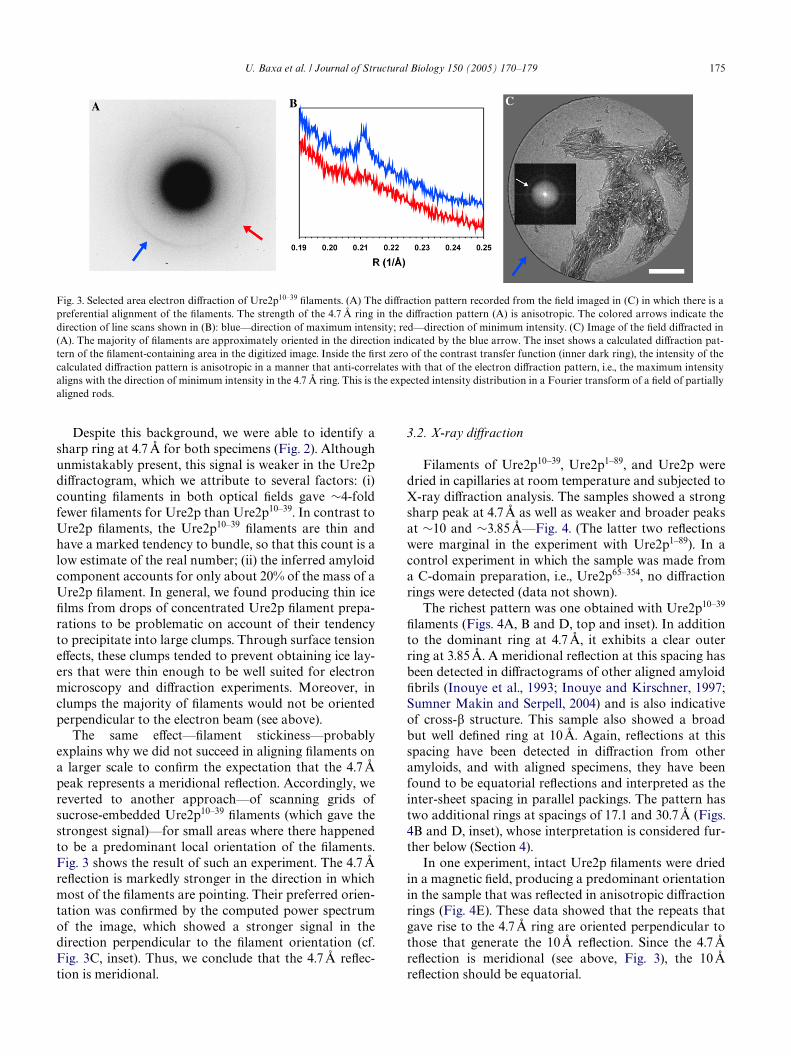
U. Baxa et al. / Journal of Structural Biology 150 (2005) 170–179 175
Despite this background, we were able to identify asharp ring at 4.7 Å for both specimens (Fig. 2). Althoughunmistakably present, this signal is weaker in the Ure2pdiVractogram, which we attribute to several factors: (i)counting Wlaments in both optical Welds gave »4-foldfewer Wlaments for Ure2p than Ure2p10–39. In contrast toUre2p Wlaments, the Ure2p10–39 Wlaments are thin andhave a marked tendency to bundle, so that this count is alow estimate of the real number; (ii) the inferred amyloidcomponent accounts for only about 20% of the mass of aUre2p Wlament. In general, we found producing thin iceWlms from drops of concentrated Ure2p Wlament prepa-rations to be problematic on account of their tendencyto precipitate into large clumps. Through surface tensioneVects, these clumps tended to prevent obtaining ice lay-ers that were thin enough to be well suited for electronmicroscopy and diVraction experiments. Moreover, inclumps the majority of Wlaments would not be orientedperpendicular to the electron beam (see above).
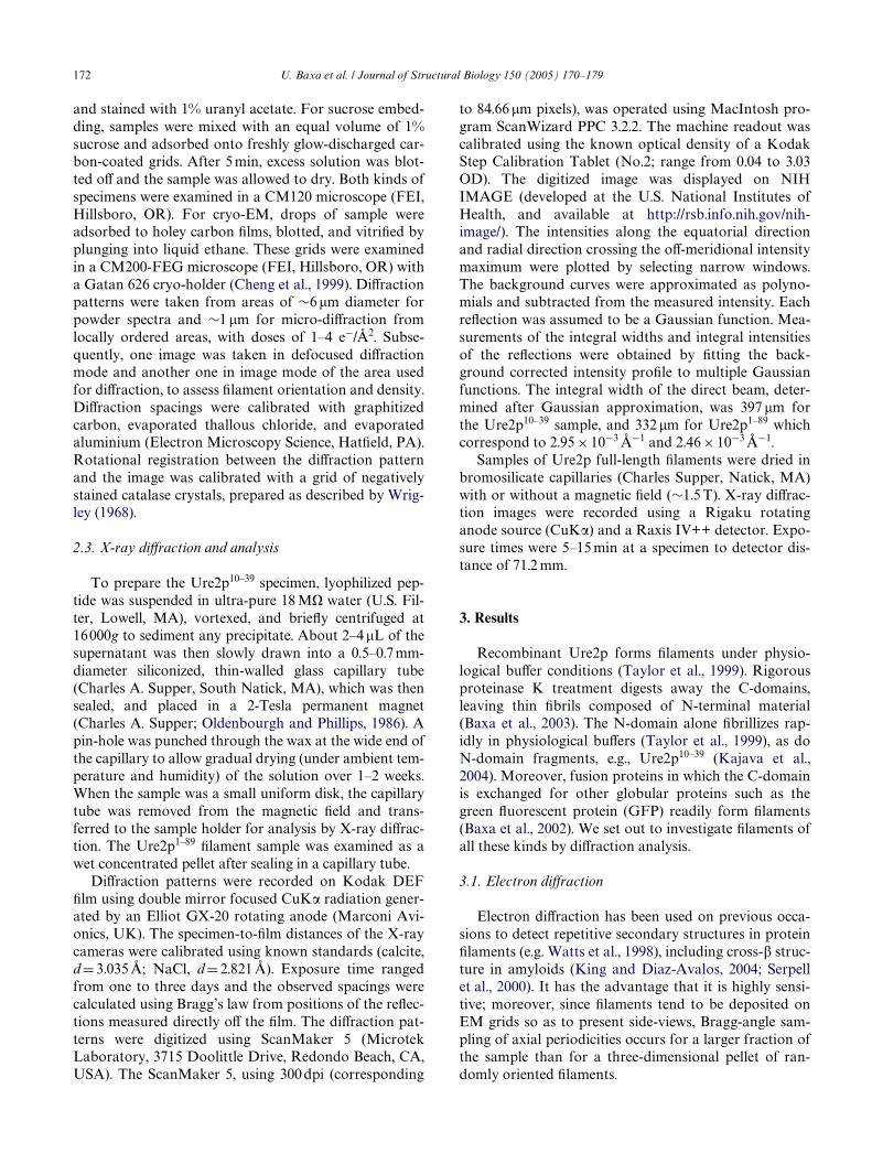
The same eVect—Wlament stickiness—probablyexplains why we did not succeed in aligning Wlaments ona larger scale to conWrm the expectation that the 4.7 Åpeak represents a meridional reXection. Accordingly, wereverted to another approach—of scanning grids ofsucrose-embedded Ure2p10–39 Wlaments (which gave thestrongest signal)—for small areas where there happenedto be a predominant local orientation of the Wlaments.Fig. 3 shows the result of such an experiment. The 4.7 ÅreXection is markedly stronger in the direction in whichmost of the Wlaments are pointing. Their preferred orien-tation was conWrmed by the computed power spectrumof the image, which showed a stronger signal in thedirection perpendicular to the Wlament orientation (cf.Fig. 3C, inset). Thus, we conclude that the 4.7 Å reXec-tion is meridional.
3.2. X-ray diVraction
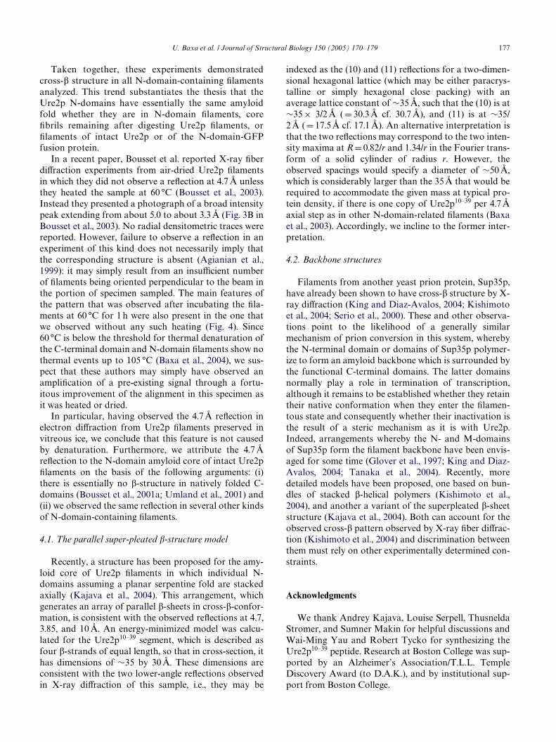
Filaments of Ure2p10–39, Ure2p1–89, and Ure2p weredried in capillaries at room temperature and subjected toX-ray diVraction analysis. The samples showed a strongsharp peak at 4.7 Å as well as weaker and broader peaksat »10 and »3.85 Å—Fig. 4. (The latter two reXectionswere marginal in the experiment with Ure2p1–89). In acontrol experiment in which the sample was made froma C-domain preparation, i.e., Ure2p65–354, no diVractionrings were detected (data not shown).
The richest pattern was one obtained with Ure2p10–39
Wlaments (Figs. 4A, B and D, top and inset). In additionto the dominant ring at 4.7 Å, it exhibits a clear outerring at 3.85 Å. A meridional reXection at this spacing hasbeen detected in diVractograms of other aligned amyloidWbrils (Inouye et al., 1993; Inouye and Kirschner, 1997;Sumner Makin and Serpell, 2004) and is also indicativeof cross-� structure. This sample also showed a broadbut well deWned ring at 10 Å. Again, reXections at thisspacing have been detected in diVraction from otheramyloids, and with aligned specimens, they have beenfound to be equatorial reXections and interpreted as theinter-sheet spacing in parallel packings. The pattern hastwo additional rings at spacings of 17.1 and 30.7 Å (Figs.4B and D, inset), whose interpretation is considered fur-ther below (Section 4).
In one experiment, intact Ure2p Wlaments were driedin a magnetic Weld, producing a predominant orientationin the sample that was reXected in anisotropic diVractionrings (Fig. 4E). These data showed that the repeats thatgave rise to the 4.7 Å ring are oriented perpendicular tothose that generate the 10 Å reXection. Since the 4.7 ÅreXection is meridional (see above, Fig. 3), the 10 ÅreXection should be equatorial.
Fig. 3. Selected area electron diVraction of Ure2p10–39 Wlaments. (A) The diVraction pattern recorded from the Weld imaged in (C) in which there is apreferential alignment of the Wlaments. The strength of the 4.7 Å ring in the diVraction pattern (A) is anisotropic. The colored arrows indicate thedirection of line scans shown in (B): blue—direction of maximum intensity; red—direction of minimum intensity. (C) Image of the Weld diVracted in(A). The majority of Wlaments are approximately oriented in the direction indicated by the blue arrow. The inset shows a calculated diVraction pat-tern of the Wlament-containing area in the digitized image. Inside the Wrst zero of the contrast transfer function (inner dark ring), the intensity of thecalculated diVraction pattern is anisotropic in a manner that anti-correlates with that of the electron diVraction pattern, i.e., the maximum intensityaligns with the direction of minimum intensity in the 4.7 Å ring. This is the expected intensity distribution in a Fourier transform of a Weld of partiallyaligned rods.
176 U. Baxa et al. / Journal of Structural Biology 150 (2005) 170–179
4. Discussion
In this study, we used electron diVraction and X-raydiVraction to investigate Ure2p-related Wlaments underseveral conditions—after air-drying, sucrose embedding,and vitriWcation. X-ray Wber diVraction has a long historyof applications to detect cross-� structure in amyloid Wla-ments (Bonar et al., 1969; Kirschner et al., 1986; Sipe andCohen, 2000). However, electron diVraction has comple-mentary advantages of sensitivity and applicability to spec-imens that exhibit poor long-range order. This techniquehas recently been used to demonstrate cross-� structure inWlaments of �-synuclein, tau, amylin (IAPP), and Sup35p(Berriman et al., 2003; King and Diaz-Avalos, 2004; Serp-ell et al., 2000; Sumner Makin and Serpell, 2004).
The typical diVraction signature for amyloids is ameridional reXection at 4.7 Å, representing the spacingbetween �-strands running perpendicular to the Wla-ment axis. A generally weaker and broader equatorialreXection may be observed at »10 Å that representsthe inter-sheet spacing, and another meridional reXec-tion at 3.85 Å that also relates to �-sheet structure(Kirschner et al., 1987; Makin and Serpell, 2002). Weobserved the 4.7 Å reXection in all samples analyzed.The 3.85 Å reXection was detected by both techniquesfor our best aligned specimens. The 10 Å reXection waspresumably submerged in the forward scattering peakof the electron diVractograms but was observed in X-ray diVraction of both Ure2p10–39 and Ure2p Wlaments(Fig. 4).
Fig. 4. X-ray diVraction patterns from several kinds of Ure2p-related Wlaments. (A) DiVraction from Ure2p10–39 sample dried in a magnetic Weld (96 hexposure). (B) Enlargement of the low-angle region, including the rings at 17 Å and (overexposed) 31 Å; (C) diVraction from pellet containingUre2p1–89 Wlaments (91 h exposure). The 4.7 Å and 3.85 Å rings are marked with arrows. (D) Azimuthally averaged intensity traces between spacingsof 1/(20 Å) and 1/(3.3 Å) for the diVraction patterns from Ure2p10–39 (top), Ure2p1–89 (bottom). The inset shows a baseline corrected trace fromUre2p10–39 in the low angle range from 1/(50 Å) to 1/(6.7 Å) where the 32 Å and the 17 Å reXection are visible as small peaks. (E) DiVraction from asample of partially oriented Ure2p Wlaments. The direction of strongest intensity for the 4.7 Å ring is perpendicular to the direction of strongestintensity for the 10 Å ring, consistent with them representing meridional and equatorial reXections, respectively.
U. Baxa et al. / Journal of Structural Biology 150 (2005) 170–179 177
Taken together, these experiments demonstratedcross-� structure in all N-domain-containing Wlamentsanalyzed. This trend substantiates the thesis that theUre2p N-domains have essentially the same amyloidfold whether they are in N-domain Wlaments, coreWbrils remaining after digesting Ure2p Wlaments, orWlaments of intact Ure2p or of the N-domain-GFPfusion protein.
In a recent paper, Bousset et al. reported X-ray WberdiVraction experiments from air-dried Ure2p Wlamentsin which they did not observe a reXection at 4.7 Å unlessthey heated the sample at 60 °C (Bousset et al., 2003).Instead they presented a photograph of a broad intensitypeak extending from about 5.0 to about 3.3 Å (Fig. 3B inBousset et al., 2003). No radial densitometric traces werereported. However, failure to observe a reXection in anexperiment of this kind does not necessarily imply thatthe corresponding structure is absent (Agianian et al.,1999): it may simply result from an insuYcient numberof Wlaments being oriented perpendicular to the beam inthe portion of specimen sampled. The main features ofthe pattern that was observed after incubating the Wla-ments at 60 °C for 1 h were also present in the one thatwe observed without any such heating (Fig. 4). Since60 °C is below the threshold for thermal denaturation ofthe C-terminal domain and N-domain Wlaments show nothermal events up to 105 °C (Baxa et al., 2004), we sus-pect that these authors may simply have observed anampliWcation of a pre-existing signal through a fortu-itous improvement of the alignment in this specimen asit was heated or dried.
In particular, having observed the 4.7 Å reXection inelectron diVraction from Ure2p Wlaments preserved invitreous ice, we conclude that this feature is not causedby denaturation. Furthermore, we attribute the 4.7 ÅreXection to the N-domain amyloid core of intact Ure2pWlaments on the basis of the following arguments: (i)there is essentially no �-structure in natively folded C-domains (Bousset et al., 2001a; Umland et al., 2001) and(ii) we observed the same reXection in several other kindsof N-domain-containing Wlaments.
4.1. The parallel super-pleated �-structure model
Recently, a structure has been proposed for the amy-loid core of Ure2p Wlaments in which individual N-domains assuming a planar serpentine fold are stackedaxially (Kajava et al., 2004). This arrangement, whichgenerates an array of parallel �-sheets in cross-�-confor-mation, is consistent with the observed reXections at 4.7,3.85, and 10 Å. An energy-minimized model was calcu-lated for the Ure2p10–39 segment, which is described asfour �-strands of equal length, so that in cross-section, ithas dimensions of »35 by 30 Å. These dimensions areconsistent with the two lower-angle reXections observedin X-ray diVraction of this sample, i.e., they may be
indexed as the (10) and (11) reXections for a two-dimen-sional hexagonal lattice (which may be either paracrys-talline or simply hexagonal close packing) with anaverage lattice constant of »35 Å, such that the (10) is at»35 £ 3/2 Å ( D 30.3 Å cf. 30.7 Å), and (11) is at »35/2 Å ( D 17.5 Å cf. 17.1 Å). An alternative interpretation isthat the two reXections may correspond to the two inten-sity maxima at R D 0.82/r and 1.34/r in the Fourier trans-form of a solid cylinder of radius r. However, theobserved spacings would specify a diameter of »50 Å,which is considerably larger than the 35 Å that would berequired to accommodate the given mass at typical pro-tein density, if there is one copy of Ure2p10–39 per 4.7 Åaxial step as in other N-domain-related Wlaments (Baxaet al., 2003). Accordingly, we incline to the former inter-pretation.
4.2. Backbone structures
Filaments from another yeast prion protein, Sup35p,have already been shown to have cross-� structure by X-ray diVraction (King and Diaz-Avalos, 2004; Kishimotoet al., 2004; Serio et al., 2000). These and other observa-tions point to the likelihood of a generally similarmechanism of prion conversion in this system, wherebythe N-terminal domain or domains of Sup35p polymer-ize to form an amyloid backbone which is surrounded bythe functional C-terminal domains. The latter domainsnormally play a role in termination of transcription,although it remains to be established whether they retaintheir native conformation when they enter the Wlamen-tous state and consequently whether their inactivation isthe result of a steric mechanism as it is with Ure2p.Indeed, arrangements whereby the N- and M-domainsof Sup35p form the Wlament backbone have been envis-aged for some time (Glover et al., 1997; King and Diaz-Avalos, 2004; Tanaka et al., 2004). Recently, moredetailed models have been proposed, one based on bun-dles of stacked �-helical polymers (Kishimoto et al.,2004), and another a variant of the superpleated �-sheetstructure (Kajava et al., 2004). Both can account for theobserved cross-� pattern observed by X-ray Wber diVrac-tion (Kishimoto et al., 2004) and discrimination betweenthem must rely on other experimentally determined con-straints.
Acknowledgments
We thank Andrey Kajava, Louise Serpell, ThusneldaStromer, and Sumner Makin for helpful discussions andWai-Ming Yau and Robert Tycko for synthesizing theUre2p10–39 peptide. Research at Boston College was sup-ported by an Alzheimer’s Association/T.L.L. TempleDiscovery Award (to D.A.K.), and by institutional sup-port from Boston College.
178 U. Baxa et al. / Journal of Structural Biology 150 (2005) 170–179
References
Agianian, B., Leonard, K., Bonte, E., Van der Zandt, H., Becker, P.B.,Tucker, P.A., 1999. The glutamine-rich domain of the DrosophilaGAGA factor is necessary for amyloid Wbre formation in vitro, butnot for chromatin remodelling. J. Mol. Biol. 285, 527–544.
Akey, C.W., Edelstein, S.J., 1983. Equivalence of the projected struc-ture of thin catalase crystals preserved for electron microscopy bynegative stain, glucose or embedding in the presence of tannic acid.J. Mol. Biol. 163, 575–612.
Al-Amoudi, A., Dubochet, J., Studer, D., 2002. Amorphous solid waterproduced by cryosectioning of crystalline ice at 113 K. J. Microsc.207, 146–153.
Al-Amoudi, A., Norlen, L.P., Dubochet, J., 2004. Cryo-electron micros-copy of vitreous sections of native biological cells and tissues. J.Struct. Biol. 148, 131–135.
Bai, M., Zhou, J.M., Perrett, S., 2004. The yeast prion protein Ure2shows glutathione peroxidase activity in both native and Wbrillarforms. J. Biol. Chem. 279, 50025–50030.
Baxa, U., Ross, P.D., Wickner, R.B., Steven, A.C., 2004. The N-terminalprion domain of Ure2p converts from an unfolded to a thermallyresistant conformation upon Wlament formation. J. Mol. Biol. 339,259–264.
Baxa, U., Speransky, V., Steven, A.C., Wickner, R.B., 2002. Mechanismof inactivation on prion conversion of the Saccharomyces cerevisiaeUre2 protein. Proc. Natl. Acad. Sci. USA 99, 5253–5260.
Baxa, U., Taylor, K.L., Wall, J.S., Simon, M.N., Cheng, N., Wickner,R.B., Steven, A.C., 2003. Architecture of Ure2p prion Wlaments: theN-terminal domains form a central core Wber. J. Biol. Chem. 278,43717–43727.
Beck, T., Hall, M.N., 1999. The TOR signalling pathway controlsnuclear localization of nutrient-regulated transcription factors.Nature 402, 689–692.
Berriman, J., Serpell, L.C., Oberg, K.A., Fink, A.L., Goedert, M., Crow-ther, R.A., 2003. Tau Wlaments from human brain and from in vitroassembly of recombinant protein show cross-beta structure. Proc.Natl. Acad. Sci. USA 100, 9034–9038.
Blinder, D., Coschigano, P.W., Magasanik, B., 1996. Interaction of theGATA factor Gln3p with the nitrogen regulator Ure2p in Saccha-romyces cerevisiae. J. Bacteriol. 178, 4734–4736.
Bonar, L., Cohen, A.S., Skinner, M.M., 1969. Characterization of theamyloid Wbril as a cross-beta protein. Proc. Soc. Exp. Biol. Med.131, 1373–1375.
Bourguignon, L.Y., Tokuyasu, K.T., Singer, S.J., 1978. The capping oflymphocytes and other cells, studied by an improved method forimmunoXuorescence staining of frozen sections. J. Cell. Physiol. 95,239–257.
Bousset, L., Belrhali, H., Janin, J., Melki, R., Morera, S., 2001a. Struc-ture of the globular region of the prion protein Ure2 from the yeastSaccharomyces cerevisiae. Structure 9, 39–46.
Bousset, L., Belrhali, H., Melki, R., Morera, S., 2001b. Crystal struc-tures of the yeast prion Ure2p functional region in complex withglutathione and related compounds. Biochemistry 40, 13564–13573.
Bousset, L., Briki, F., Doucet, J., Melki, R., 2003. The native-like con-formation of Ure2p in Wbrils assembled under physiologically rele-vant conditions switches to an amyloid-like conformation uponheat-treatment of the Wbrils. J. Struct. Biol. 141, 132–142.
Bousset, L., Thomson, N.H., Radford, S.E., Melki, R., 2002. Theyeast prion Ure2p retains its native alpha-helical conformationupon assembly into protein Wbrils in vitro. EMBO J. 21, 2903–2911.
Cheng, N., Conway, J.F., Watts, N.R., Hainfeld, J.F., Joshi, V., Powell,R.D., Stahl, S.J., WingWeld, P.E., Steven, A.C., 1999. Tetrairidium, afour-atom cluster, is readily visible as a density label in three-dimensional cryo-EM maps of proteins at 10–25 Å resolution. J.Struct. Biol. 127, 169–176.
Cooper, T.G., 2002. Transmitting the signal of excess nitrogen in Sac-charomyces cerevisiae from the Tor proteins to the GATA factors:connecting the dots. FEMS Microbiol. Rev. 26, 223–238.
Coschigano, P.W., Magasanik, B., 1991. The URE2 gene product ofSaccharomyces cerevisiae plays an important role in the cellularresponse to the nitrogen source and has homology to glutathione S-transferases. Mol. Cell. Biol. 11, 822–832.
Dowell, L.G., Rinfret, A.P., 1960. Low-temperature forms of ice asstudied by X-ray diVraction. Nature 188, 1144–1148.
Drillien, R., Aigle, M., Lacroute, F., 1973. Yeast mutants pleiotropi-cally impaired in the regulation of the two glutamate dehydrogen-ases. Biochem. Biophys. Res. Commun. 53, 367–372.
Dubochet, J., Adrian, M., Chang, J.J., Homo, J.C., Lepault, J., McDo-wall, A.W., Schultz, P., 1988. Cryo-electron microscopy of vitriWedspecimens. Q. Rev. Biophys. 21, 129–228.
Dubochet, J., Booy, F.P., Freeman, R., Jones, A.V., Walter, C.A., 1981.Low temperature electron microscopy. Annu. Rev. Biophys. Bio-eng. 10, 133–149.
Edskes, H.K., Gray, V.T., Wickner, R.B., 1999. The [URE3] prion is anaggregated form of Ure2p that can be cured by overexpression ofUre2p fragments. Proc. Natl. Acad. Sci. USA 96, 1498–1503.
Galani, D., Fersht, A.R., Perrett, S., 2002. Folding of the yeast prionprotein Ure2: kinetic evidence for folding and unfolding intermedi-ates. J. Mol. Biol. 315, 213–227.
Glover, J.R., Kowal, A.S., Schirmer, E.C., Patino, M.M., Liu, J.J., Lind-quist, S., 1997. Self-seeded Wbers formed by Sup35, the proteindeterminant of [PSI+], a heritable prion-like factor of S. cerevisiae.Cell 89, 811–819.
Goldsbury, C.S., Cooper, G.J., Goldie, K.N., Muller, S.A., SaaW, E.L.,Gruijters, W.T., Misur, M.P., Engel, A., Aebi, U., Kistler, J., 1997.Polymorphic Wbrillar assembly of human amylin. J. Struct. Biol.119, 17–27.
Goldsbury, C.S., Wirtz, S., Muller, S.A., Sunderji, S., Wicki, P., Aebi, U.,Frey, P., 2000. Studies on the in vitro assembly of A� 1-40: implica-tions for the search for a beta Wbril formation inhibitors. J. Struct.Biol. 130, 217–231.
Inouye, H., Fraser, P.E., Kirschner, D.A., 1993. Structure of beta-crys-tallite assemblies formed by Alzheimer beta-amyloid protein ana-logues: analysis by X-ray diVraction. Biophys. J. 64, 502–519.
Inouye, H., Kirschner, D.A., 1997. X-ray diVraction analysis of scrapieprion: intermediate and folded structures in a peptide containingtwo putative alpha-helices. J. Mol. Biol. 268, 375–389.
Kajava, A.V., Baxa, U., Wickner, R.B., Steven, A.C., 2004. A model forUre2p prion Wlaments and other amyloids: the parallel super-pleated �-structure. Proc. Natl. Acad. Sci. USA 101, 7885–7890.
Kessel, M., Buhle, E.L., Glavaris, E., Aebi, U., 1986. A novel methodfor preventing collapse of air-dried specimens for heavy metalshadowing. In: Proceedings of the 44th Annual Meeting of EMSA.San Francisco Press, San Francisco, pp. 228–229.
King, C.Y., Diaz-Avalos, R., 2004. Protein-only transmission of threeyeast prion strains. Nature 428, 319–323.
Kirschner, D.A., Abraham, C., Selkoe, D.J., 1986. X-ray diVractionfrom intraneuronal paired helical Wlaments and extraneuronalamyloid Wbers in Alzheimer disease indicates cross-beta conforma-tion. Proc. Natl. Acad. Sci. USA 83, 503–507.
Kirschner, D.A., Inouye, H., DuVy, L.K., Sinclair, A., Lind, M., Selkoe,D.J., 1987. Synthetic peptide homologous to beta protein from Alz-heimer disease forms amyloid-like Wbrils in vitro. Proc. Natl. Acad.Sci. USA 84, 6953–6957.
Kishimoto, A., Hasegawa, K., Suzuki, H., Taguchi, H., Namba, K.,Yoshida, M., 2004. Beta-Helix is a likely core structure of yeastprion Sup35 amyloid Wbers. Biochem. Biophys. Res. Commun. 315,739–745.
Lepault, J., Booy, F.P., Dubochet, J., 1983. Electron microscopy of fro-zen biological suspensions. J. Microsc. 129, 89–102.
Maddelein, M.L., Wickner, R.B., 1999. Two prion-inducing regions ofUre2p are nonoverlapping. Mol. Cell. Biol. 19, 4516–4524.
U. Baxa et al. / Journal of Structural Biology 150 (2005) 170–179 179
Makin, O.S., Serpell, L.C., 2002. Examining the structure of the matureamyloid Wbril. Biochem. Soc. Trans. 30, 521–525.
Masison, D.C., Maddelein, M.L., Wickner, R.B., 1997. The prion modelfor [URE3] of yeast: spontaneous generation and requirements forpropagation. Proc. Natl. Acad. Sci. USA 94, 12503–12508.
Masison, D.C., Wickner, R.B., 1995. Prion-inducing domain of yeastUre2p and protease resistance of Ure2p in prion-containing cells.Science 270, 93–95.
McDowall, A.W., Chang, J.J., Freeman, R., Lepault, J., Walter, C.A.,Dubochet, J., 1983. Electron microscopy of frozen hydrated sec-tions of vitreous ice and vitriWed biological samples. J. Microsc. 131,1–9.
Oldenbourgh, R., Phillips, W.C., 1986. Small permanent magnet forWelds up to 2.6 T. Rev. Sci. Instrum. 57, 2362–2365.
Perrett, S., Freeman, S.J., Butler, P.J., Fersht, A.R., 1999. Equilibriumfolding properties of the yeast prion protein determinant Ure2. J.Mol. Biol. 290, 331–345.
Pierce, M.M., Baxa, U., Steven, A.C., Bax, A., Wickner, R.B., 2004. Isthe prion domain of soluble Ure2p unstructured? Biochemistry 44,321–328.
Ripaud, L., Maillet, L., Immel-Torterotot, F., Durand, F., Cullin, C.,2004. The [URE3] yeast prion results from protein aggregates thatdiVer from amyloid Wlaments formed in vitro. J. Biol. Chem. 279,50962–50968.
Ross, E.D., Baxa, U., Wickner, R.B., 2004. Scrambled prion domainsform prions and amyloid. Mol. Cell. Biol. 24, 7206–7213.
Schlumpberger, M., Wille, H., Baldwin, M.A., Butler, D.A., Hersko-witz, I., Prusiner, S.B., 2000. The prion domain of yeast Ure2pinduces autocatalytic formation of amyloid Wbers by a recombinantfusion protein. Protein Sci. 9, 440–451.
Serio, T.R., Cashikar, A.G., Kowal, A.S., Sawicki, G.J., Moslehi, J.J.,Serpell, L., Arnsdorf, M.F., Lindquist, S.L., 2000. Nucleated confor-mational conversion and the replication of conformational infor-mation by a prion determinant. Science 289, 1317–1321.
Serpell, L.C., Berriman, J., Jakes, R., Goedert, M., Crowther, R.A.,2000. Fiber diVraction of synthetic alpha-synuclein Wlaments shows
amyloid-like cross-beta conformation. Proc. Natl. Acad. Sci. USA97, 4897–4902.
Sipe, J.D., Cohen, A.S., 2000. Review: history of the amyloid Wbril. J.Struct. Biol. 130, 88–98.
Speransky, V.V., Taylor, K.L., Edskes, H.K., Wickner, R.B., Steven,A.C., 2001. Prion Wlament networks in [URE3] cells of Saccharomy-ces cerevisiae. J. Cell Biol. 153, 1327–1336.
Sumner Makin, O., Serpell, L.C., 2004. Structural characterisation ofislet amyloid polypeptide Wbrils. J. Mol. Biol. 335, 1279–1288.
Tanaka, M., Chien, P., Naber, N., Cooke, R., Weissman, J.S., 2004.Conformational variations in an infectious protein determine prionstrain diVerences. Nature 428, 323–328.
Taylor, K.L., Cheng, N., Williams, R.W., Steven, A.C., Wickner, R.B.,1999. Prion domain initiation of amyloid formation in vitro fromnative Ure2p. Science 283, 1339–1343.
Thual, C., Komar, A.A., Bousset, L., Fernandez-Bellot, E., Cullin, C.,Melki, R., 1999. Structural characterization of Saccharomyces cere-visiae prion-like protein Ure2. J. Biol. Chem. 274, 13666–13674.
Tokuyasu, K.T., 1973. A technique for ultracryotomy of cell suspen-sions and tissues. J. Cell Biol. 57, 551–565.
Umland, T.C., Taylor, K.L., Rhee, S., Wickner, R.B., Davies, D.R.,2001. The crystal structure of the nitrogen regulation fragment ofthe yeast prion protein Ure2p. Proc. Natl. Acad. Sci. USA 98, 1459–1464.
Watts, N.R., Misra, M., WingWeld, P.T., Stahl, S.J., Cheng, N., Trus,B.L., Steven, A.C., Williams, R.W., 1998. Three-dimensional struc-ture of HIV-1 Rev protein Wlaments. J. Struct. Biol. 121, 41–52.
Wickner, R.B., 1994. [URE3] as an altered URE2 protein: evidencefor a prion analog in Saccharomyces cerevisiae. Science 264, 566–569.
Wrigley, N., 1968. The lattice spacing of crystalline catalase as an inter-nal standard of length in electron microscopy. J. Ultrastruct. Res.24, 454–464.
Zhou, J.M., Zhu, L., Balny, C., Perrett, S., 2001. Pressure denaturationof the yeast prion protein Ure2. Biochem. Biophys. Res. Commun.287, 147–152.
Related Documents


