Figure 1 shows a human cheek cell viewed under a light microscope. Figure 1 © Ed Reschke/Photolibrary/Getty Images (a) Label the nucleus and cell membrane on Figure 1. (2) (b) Cheek cells are a type of body cell. Body cells grow through cell division. What is the name of this type of cell division? Tick one box. Differentiation Mitosis Specialisation (1) (c) Ribosomes and mitochondria are not shown in Figure 1. What type of microscope is needed to see ribosomes and mitochondria? (1) Page 1 of 23 1

Welcome message from author
This document is posted to help you gain knowledge. Please leave a comment to let me know what you think about it! Share it to your friends and learn new things together.
Transcript
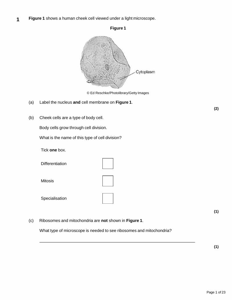
Figure 1 shows a human cheek cell viewed under a light microscope.
Figure 1
© Ed Reschke/Photolibrary/Getty Images
(a) Label the nucleus and cell membrane on Figure 1.
(2)
(b) Cheek cells are a type of body cell.
Body cells grow through cell division.
What is the name of this type of cell division?
Tick one box.
Differentiation
Mitosis
Specialisation
(1)
(c) Ribosomes and mitochondria are not shown in Figure 1.
What type of microscope is needed to see ribosomes and mitochondria?
(1)
Page 1 of 23
1
(d) What is the advantage of using the type of microscope you named in part (c)?
Tick one box.
Cheaper
Higher magnification
Lower resolution
(1)
(e) The cheek cell in Figure 2 is magnified 250 times.
The width of the cell is shown by the line D to E.
Figure 2
Calculate the width of the cheek cell in micrometres (µm).
Complete the following steps.
Measure the width of the cell using a ruler mm
Use the equation to work out the real width of the cell in mm:
real size = mm
Convert mm to µm µm
(3)
Page 2 of 23
(f) A red blood cell is 8 µm in diameter.
A bacterial cell is 40 times smaller.
Calculate the diameter of the bacterial cell.
Tick one box.
0.02 µm
0.2 µm
2.0 µm
20.0 µm
(1)
(Total 9 marks)
Page 3 of 23
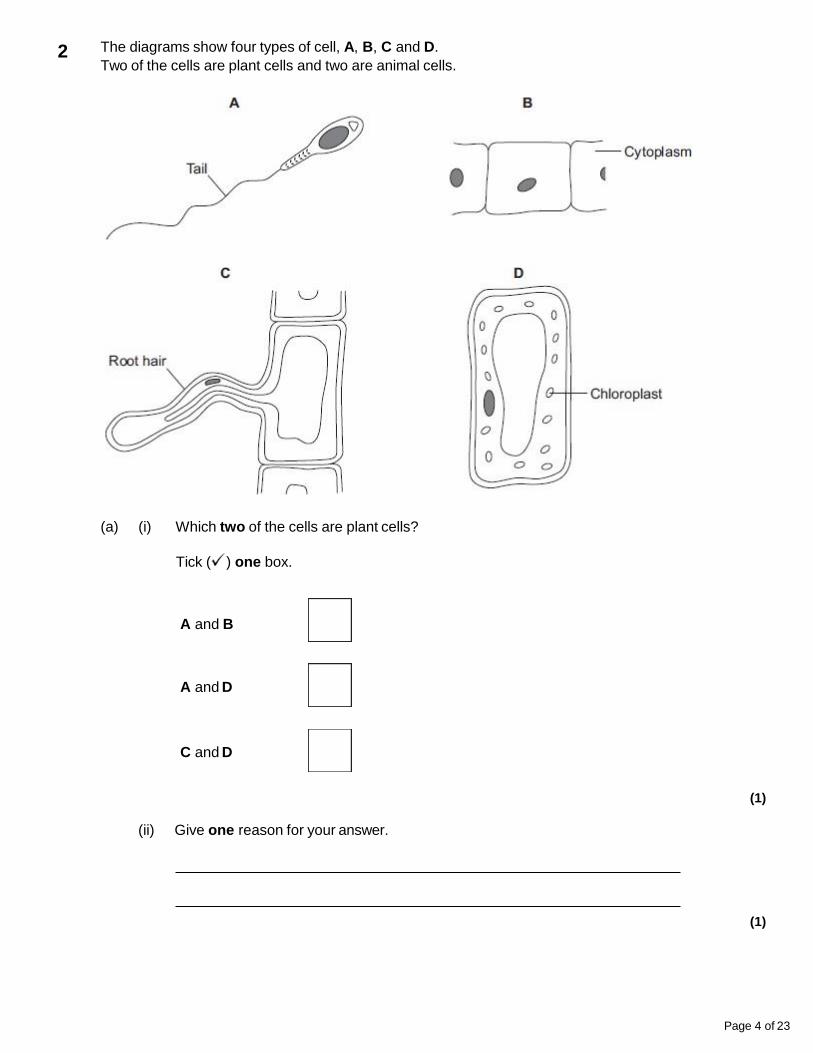
The diagrams show four types of cell, A, B, C and D.
Two of the cells are plant cells and two are animal cells.
(a) (i) Which two of the cells are plant cells?
Tick ( ) one box.
A and B
A and D
C and D
(1)
(ii) Give one reason for your answer.
(1)
Page 4 of 23
2
(b) (i) Which cell, A, B, C or D, is adapted for swimming?
(1)
(ii) Which cell, A, B, C or D, can produce glucose by photosynthesis?
(1)
(c) Cells A, B, C and D all use oxygen.
For what process do cells use oxygen?
Draw a ring around one answer.
osmosis photosynthesis respiration
(1)
(Total 5 marks)
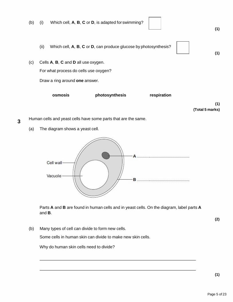
Human cells and yeast cells have some parts that are the same.
(a) The diagram shows a yeast cell.
3
Parts A and B are found in human cells and in yeast cells. On the diagram, label parts A
and B.
(2)
(b) Many types of cell can divide to form new cells.
Some cells in human skin can divide to make new skin cells.
Why do human skin cells need to divide?
(1)
Page 5 of 23
(c) Human stem cells can develop into many different types of human cell.
(i) Use the correct answer from the box to complete the sentence.
embryos hair nerve cells
Human stem cells may come from
(1)
(ii) Use the correct answer from the box to complete the sentence.
cystic fibrosis paralysis polydactyly
Human stem cells can be used to treat
(1)
(Total 5 marks)
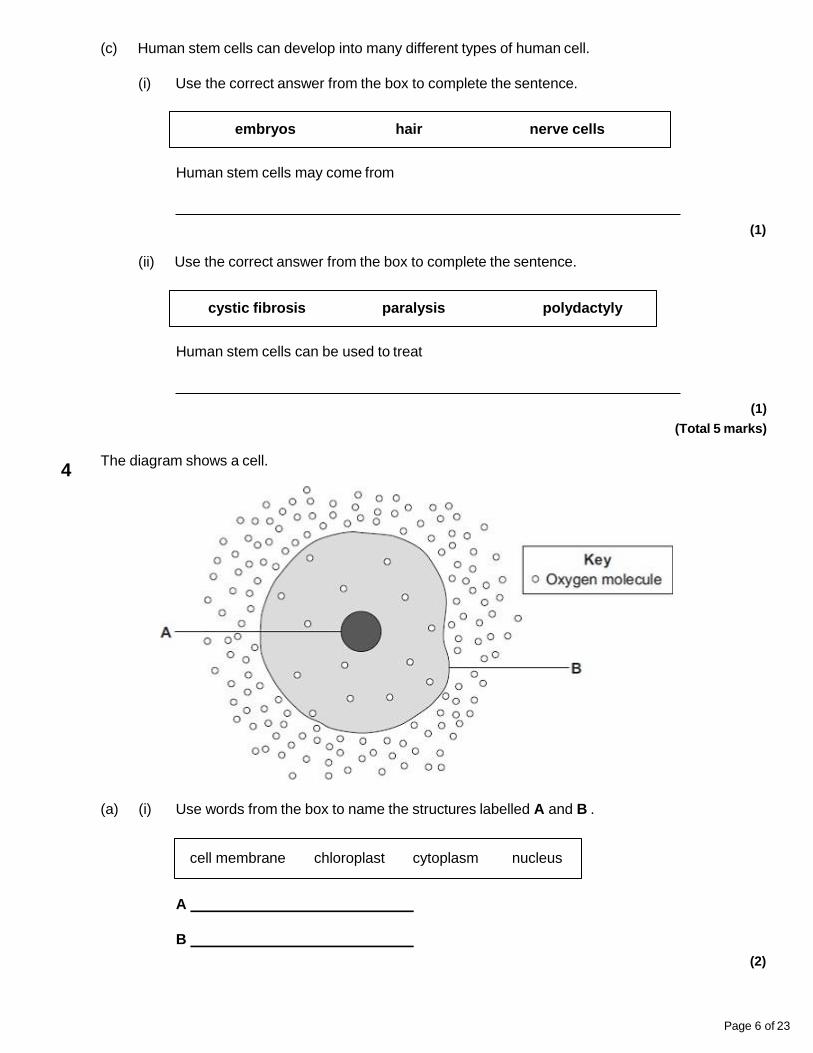
The diagram shows a cell.
(a) (i) Use words from the box to name the structures labelled A and B .
Page 6 of 23
cell membrane chloroplast cytoplasm nucleus
A
B
(2)
4
(ii) The cell in the diagram is an animal cell.
How can you tell it is an animal cell and not a plant cell?
Give two reasons.
1.
2.
(2)
(b) Oxygen will diffuse into the cell in the diagram.
Why?
Use information from the diagram.
(1)
Page 7 of 23
(c) The cell shown in the diagram is usually found with similar cells.
Draw a ring around the correct answer to complete the sentence.
Scientists call a group of similar cells
an organ.
a system.
a tissue.
(1)
(Total 6 marks)
Substances can move into cells and out of cells.
(a) Draw a ring around the correct answer to complete each sentence.
Water moves into cells and out of cells by
active transport.
osmosis.
reabsorption.
5
The water moves through a membrane.
freely permeable
non-permeable
partially permeable
(2)
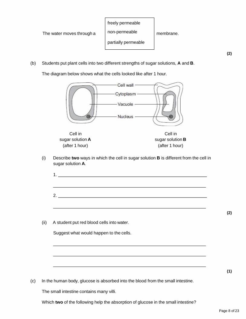
(b) Students put plant cells into two different strengths of sugar solutions, A and B.
The diagram below shows what the cells looked like after 1 hour.
Cell in
sugar solution A
(after 1 hour)
Cell in
sugar solution B
(after 1 hour)
(i) Describe two ways in which the cell in sugar solution B is different from the cell in
sugar solution A.
1.
2.
(2)
(ii) A student put red blood cells into water.
Suggest what would happen to the cells.
(1)
Page 8 of 23
(c) In the human body, glucose is absorbed into the blood from the small intestine.
The small intestine contains many villi.
Which two of the following help the absorption of glucose in the small intestine?
Tick ( ) two boxes.
Villi have a cell wall.
Villi are covered in thick mucus.
Villi give the small intestine a large surface area.
Villi have many blood capillaries.
(2)
(Total 7 marks)
Page 9 of 23
Nucleus Plasmids Cytoplasm
Prokaryotic cell
Eukaryotic cell
Cells can be classified according to their structure.
(a) Complete Table 1 to show which features each cell type has.
Write a tick or a cross in each box.
Table 1
(2)
6
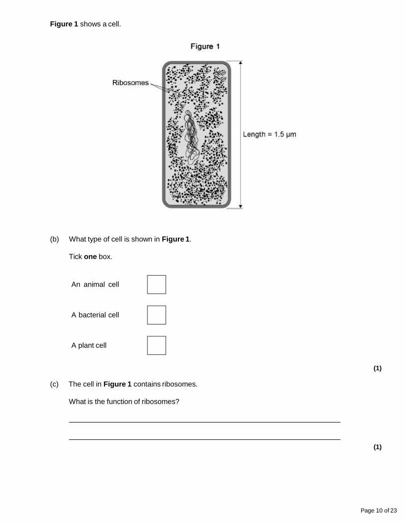
Figure 1 shows a cell.
(b) What type of cell is shown in Figure 1.
Tick one box.
An animal cell
A bacterial cell
A plant cell
(1)
(c) The cell in Figure 1 contains ribosomes.
What is the function of ribosomes?
(1)
Page 10 of 23
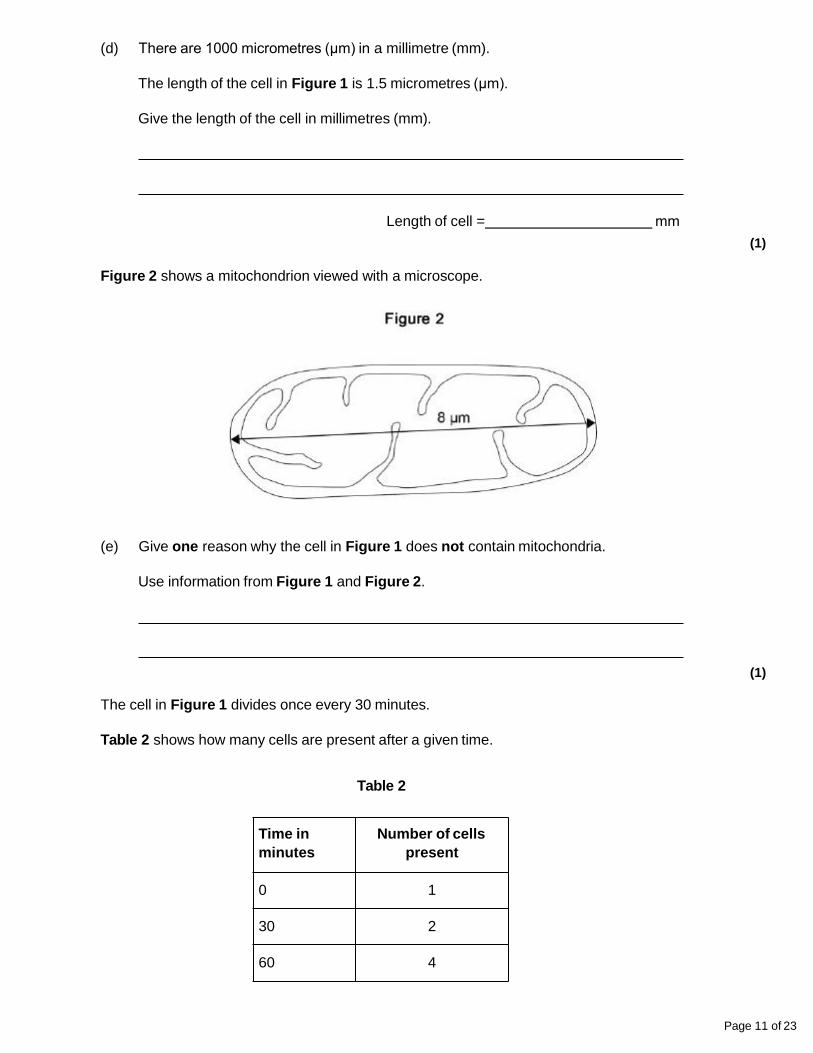
(d) There are 1000 micrometres (μm) in a millimetre (mm).
The length of the cell in Figure 1 is 1.5 micrometres (μm).
Give the length of the cell in millimetres (mm).
Length of cell = mm
(1)
Figure 2 shows a mitochondrion viewed with a microscope.
(e) Give one reason why the cell in Figure 1 does not contain mitochondria.
Use information from Figure 1 and Figure 2.
(1)
Page 11 of 23
Time in
minutes
Number of cells
present
0 1
30 2
60 4
The cell in Figure 1 divides once every 30 minutes.
Table 2 shows how many cells are present after a given time.
Table 2
(f) Calculate how many cells will be present after 2 hours.
Number of cells =
(2)
Cells like the one in Figure 1 are kept in a culture solution for 25 hours.
The graph below shows the number of live cells present.
(g) Describe the changes in the number of live cells shown in the graph above in the first 20
hours.
Use data from the graph in your answer.
(3)
Page 12 of 23
(h) Suggest one reason why the number of live cells decreases after 20 hours.
(1)
(Total 12 marks)
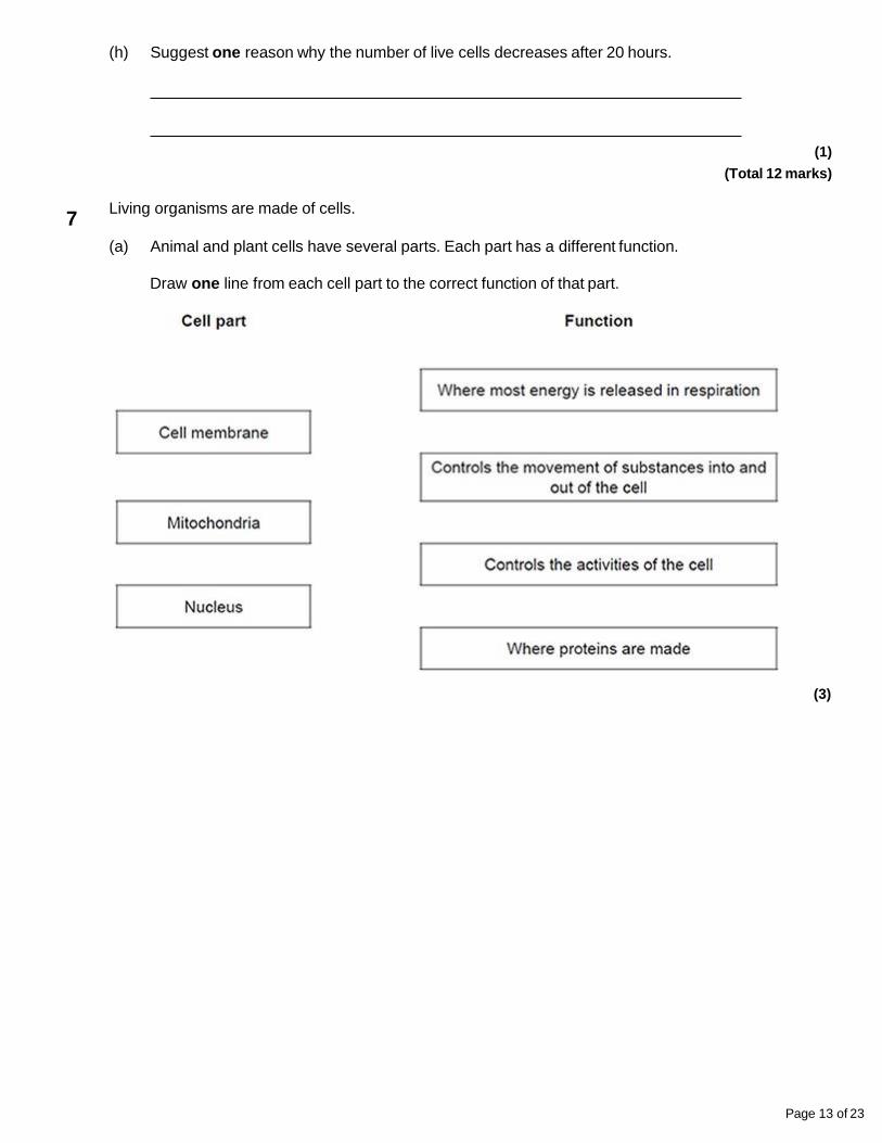
Living organisms are made of cells.
(a) Animal and plant cells have several parts. Each part has a different function.
Draw one line from each cell part to the correct function of that part.
(3)
Page 13 of 23
7
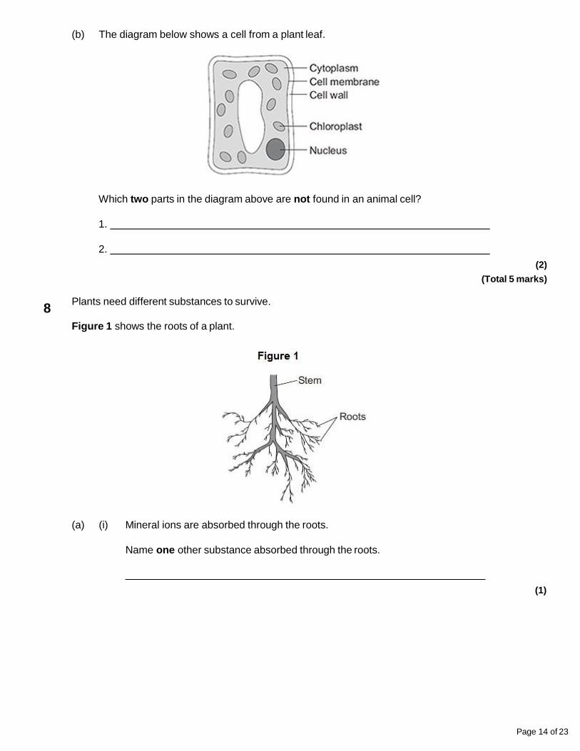
(b) The diagram below shows a cell from a plant leaf.
Which two parts in the diagram above are not found in an animal cell?
1.
2.
(2)
(Total 5 marks)
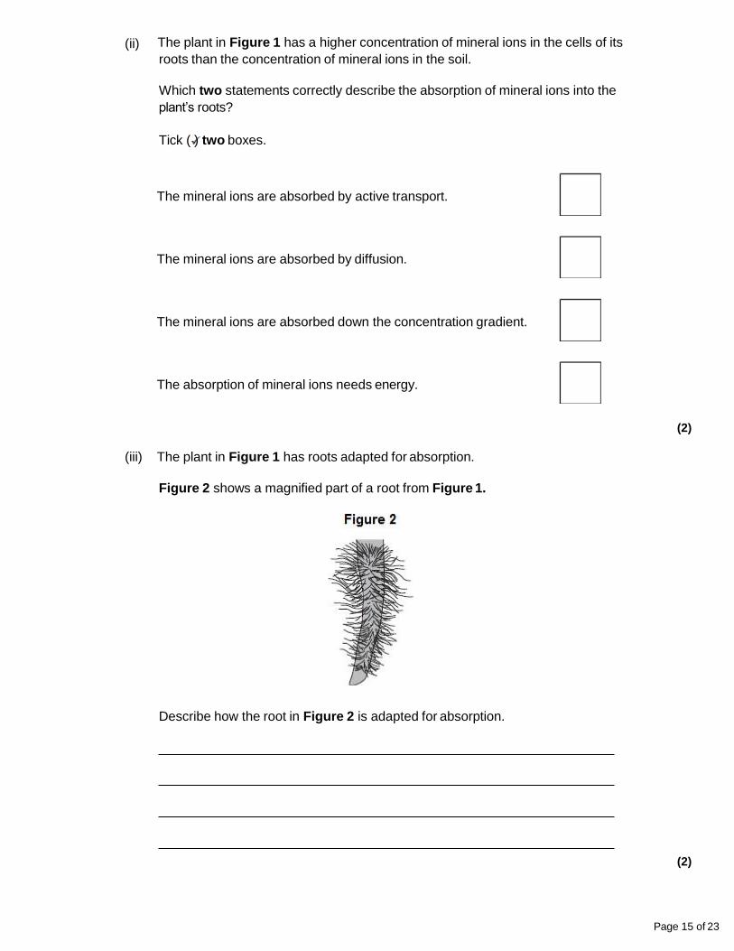
Plants need different substances to survive.
Figure 1 shows the roots of a plant.
(a) (i) Mineral ions are absorbed through the roots.
Name one other substance absorbed through the roots.
(1)
Page 14 of 23
8
(ii) The plant in Figure 1 has a higher concentration of mineral ions in the cells of its
roots than the concentration of mineral ions in the soil.
Which two statements correctly describe the absorption of mineral ions into the
plant’s roots?
Tick ( ) two boxes.
The mineral ions are absorbed by active transport.
The mineral ions are absorbed by diffusion.
The mineral ions are absorbed down the concentration gradient.
The absorption of mineral ions needs energy.
(2)
(iii) The plant in Figure 1 has roots adapted for absorption.
Figure 2 shows a magnified part of a root from Figure 1.
Describe how the root in Figure 2 is adapted for absorption.
(2)
Page 15 of 23
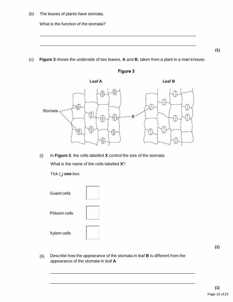
(b) The leaves of plants have stomata.
What is the function of the stomata?
(1)
(c) Figure 3 shows the underside of two leaves, A and B, taken from a plant in a man’shouse.
(i) In Figure 3, the cells labelled X control the size of the stomata.
What is the name of the cells labelled X?
Tick ( ) one box.
Guard cells
Phloem cells
Xylem cells
(1)
(ii) Describe how the appearance of the stomata in leaf B is different from the
appearance of the stomata in leaf A.
(1)
Page 16 of 23
(iii) The man forgets to water the plant.
What might happen to the plant in the next few days if the stomata stay the same as
shown in leaf A in Figure 3?
(1)
(Total 9 marks)
Page 17 of 23
Page 18 of 23
Mark schemes
(a) nucleus labelled correctly
11
cell membrane labelled correctly
1
(b) mitosis
1
(c) electron (microscope)
1
(d) higher magnification
1
(e) 45 (mm)
1
45 / 250 or 0.18 (mm)
allow ecf1
180 (µm)
1
allow 180 (µm) with no working shown for 3 marks
(f) 0.2 µm1
[9]
(a) (i) C and D
no mark if more than one box is ticked1
2
(ii) any one from:
do not allow if other cell parts are given in a list
• (have) cell wall(s)
• (have) vacuole(s)1
(b) (i) A
apply list principle1
(ii) D
apply list principle1
Page 19 of 23
(c) respiration
apply list principle1
[5]
(a) A = nucleus
allow phonetic spelling1
B = (cell) membrane
1
(b) for repair / growth or to replace cells
ignore new cells / skin1
(c) (i) embryos
1
(ii) paralysis1
[5]
3
(a) (i) A = nucleus
14
B = (cell) membrane
1
(ii) any two from:
ignore shape
• no (cell) wall
• no (large / permanent) vacuole
2
• no chloroplasts / chlorophyll
(b) because high to low oxygen / concentration or down gradient
allow ‘more / a lot of oxygen molecules outside’
ignore along / across gradient
1
(c) a tissue1
[6]
(a) osmosis
1
partially permeable
1
5
(b) (i) any two from:
allow correct answers in terms of A
•
•
•
• vacuole is small(er)
• cytoplasm has shrunk
allow cytoplasm is smaller
gap between cytoplasm and cell wall
cell wall curves inwards
allow cell B is flaccid or cell A is turgid
the (cell) membrane has moved away from the wall2
(ii) any one from:
• water will move / diffuse in
• (cells) will swell
• (cells) will burst
ignore turgid1
(c) villi give the small intestines a large surface area
1
villi have many blood capillaries1
[7]
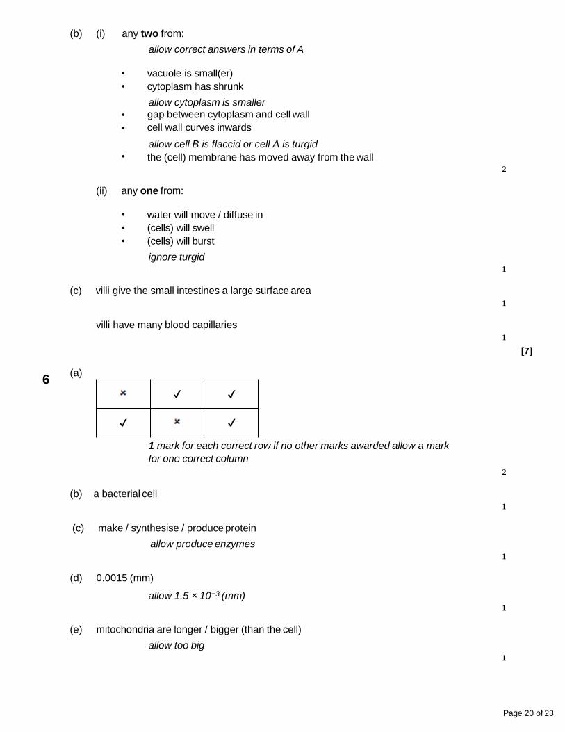
✔ ✔
✔ ✔
(a)
1 mark for each correct row if no other marks awarded allow a mark
for one correct column
Page 20 of 23
2
6
(b) a bacterial cell
1
(c) make / synthesise / produce protein
allow produce enzymes1
(d) 0.0015 (mm)
allow 1.5 × 10⁻³ (mm)1
(e) mitochondria are longer / bigger (than the cell)
allow too big1
Page 21 of 23
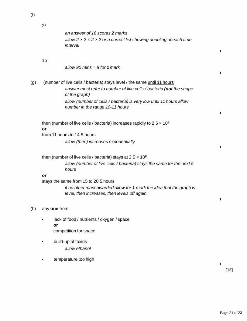
(f)
24
an answer of 16 scores 2 marks
allow 2 × 2 × 2 × 2 or a correct list showing doubling at each time
interval
1
16
allow 90 mins = 8 for 1 mark1
(g) (number of live cells / bacteria) stays level / the same until 11 hours
answer must refer to number of live cells / bacteria (not the shape
of the graph)
allow (number of cells / bacteria) is very low until 11 hours allow
number in the range 10-11 hours
1
then (number of live cells / bacteria) increases rapidly to 2.5 × 10⁸
or
from 11 hours to 14.5 hours
allow (then) increases exponentially1
then (number of live cells / bacteria) stays at 2.5 × 10⁸
allow (number of live cells / bacteria) stays the same for the next 5
hours
or
stays the same from 15 to 20.5 hours
if no other mark awarded allow for 1 mark the idea that the graph is
level, then increases, then levels off again
1
(h) any one from:
• lack of food / nutrients / oxygen / space
or
competition for space
• build-up of toxins
allow ethanol
• temperature too high1
[12]
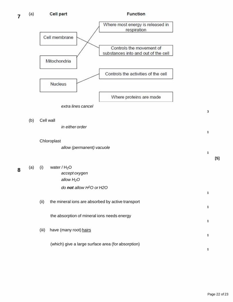
(a)
extra lines cancel
Page 22 of 23
3
7
(b) Cell wall
in either order1
Chloroplast
allow (permanent) vacuole1
[5]
(a) (i) water / H2O
accept oxygen
allow H2O
do not allow H2O or H2O1
(ii) the mineral ions are absorbed by active transport
1
the absorption of mineral ions needs energy
1
(iii) have (many root) hairs
1
(which) give a large surface area (for absorption)
1
8
Page 23 of 23
(b) carbon dioxide in
or
oxygen out
or
control water loss
accept gas exchange
ignore gases in and out
ignore gain / lose water
1
(c) (i) guard cells
1
(ii) (stomata are) closed
allow there is no gap / space1
(iii) plant will wilt / droop
ignore die1
[9]
Related Documents