antioxidants Review Fighting Oxidative Stress with Sulfur: Hydrogen Sulfide in the Renal and Cardiovascular Systems Joshua J. Scammahorn 1 , Isabel T. N. Nguyen 1 , Eelke M. Bos 2 , Harry Van Goor 3, * and Jaap A. Joles 1 Citation: Scammahorn, J.J.; Nguyen, I.T.N.; Bos, E.M.; Van Goor, H.; Joles, J.A. Fighting Oxidative Stress with Sulfur: Hydrogen Sulfide in the Renal and Cardiovascular Systems. Antioxidants 2021, 10, 373. https:// doi.org/10.3390/antiox10030373 Academic Editor: Ana Sofia Fernandes Received: 29 January 2021 Accepted: 25 February 2021 Published: 2 March 2021 Publisher’s Note: MDPI stays neutral with regard to jurisdictional claims in published maps and institutional affil- iations. Copyright: © 2021 by the authors. Licensee MDPI, Basel, Switzerland. This article is an open access article distributed under the terms and conditions of the Creative Commons Attribution (CC BY) license (https:// creativecommons.org/licenses/by/ 4.0/). 1 Department of Nephrology & Hypertension, University Medical Center Utrecht, 3508 GA Utrecht, The Netherlands; [email protected] (J.J.S.); [email protected] (I.T.N.N.); [email protected] (J.A.J.) 2 Department of Neurosurgery, Erasmus Medical Center Rotterdam, 3015 CN Rotterdam, The Netherlands; [email protected] 3 Department of Pathology and Medical Biology, University Medical Center Groningen and University of Groningen, 9713 GZ Groningen, The Netherlands * Correspondence: [email protected] Abstract: Hydrogen sulfide (H 2 S) is an essential gaseous signaling molecule. Research on its role in physiological and pathophysiological processes has greatly expanded. Endogenous enzymatic production through the transsulfuration and cysteine catabolism pathways can occur in the kidneys and blood vessels. Furthermore, non-enzymatic pathways are present throughout the body. In the renal and cardiovascular system, H 2 S plays an important role in maintaining the redox status at safe levels by promoting scavenging of reactive oxygen species (ROS). H 2 S also modifies cysteine residues on key signaling molecules such as keap1/Nrf2, NFκB, and HIF-1α, thereby promoting anti-oxidant mechanisms. Depletion of H 2 S is implicated in many age-related and cardiorenal diseases, all having oxidative stress as a major contributor. Current research suggests potential for H 2 S-based therapies, however, therapeutic interventions have been limited to studies in animal models. Beyond H 2 S use as direct treatment, it could improve procedures such as transplantation, stem cell therapy, and the safety and efficacy of drugs including NSAIDs and ACE inhibitors. All in all, H 2 S is a prime subject for further research with potential for clinical use. Keywords: hydrogen sulfide; reactive oxygen species; H 2 S donors; cardiorenal syndrome; thiosulfate 1. Introduction Hydrogen sulfide (H 2 S) was first described as a pungent toxic gas in “De Morbis Artificum Diatriba”, Bernardino Ramazzini’s treatise on worker’s diseases [1]. Today, we have come to better understand the toxic effect of H 2 S and have determined that this is most likely the result of cytochrome C oxidase (mitochondrial Complex IV), monoamine oxidase, -and/or Na + -K + -ATPase inhibition [1–3]. A brief overview of H 2 S toxicity is given in Table 1. In short, high concentrations disrupt the oxidative metabolism process of cells leading to impairment of organs most reliant on these processes [1]. During research on its toxicity, several groups reported the presence of H 2 S in tissues of healthy humans and laboratory animals [4–6]. Although the transsulfuration pathway was described in mammals more than fifty years ago, it was not until 1996 that the role of H 2 S as an endogenous gaseous signaling molecule was proposed [7,8]. Since then, research has expanded on the topic, revealing its role in many physiological processes including those of the renal and cardiovascular system [9,10]. In the body, the kidneys are believed to be the third largest producer of H 2 S, after the liver and gut [9]. All of the known pathways leading to the production of H 2 S have been described in the kidneys. Furthermore, renal homeostasis appears to be under control of H 2 S to some degree, with H 2 S levels contributing to the glomerular filtration rate (GFR), Na+ excretion, and K+ excretion [11]. Various effects mediated by H 2 S have been observed Antioxidants 2021, 10, 373. https://doi.org/10.3390/antiox10030373 https://www.mdpi.com/journal/antioxidants

Welcome message from author
This document is posted to help you gain knowledge. Please leave a comment to let me know what you think about it! Share it to your friends and learn new things together.
Transcript
antioxidants
Review
Fighting Oxidative Stress with Sulfur: Hydrogen Sulfide in theRenal and Cardiovascular Systems
Joshua J. Scammahorn 1, Isabel T. N. Nguyen 1, Eelke M. Bos 2 , Harry Van Goor 3,* and Jaap A. Joles 1
�����������������
Citation: Scammahorn, J.J.; Nguyen,
I.T.N.; Bos, E.M.; Van Goor, H.; Joles,
J.A. Fighting Oxidative Stress with
Sulfur: Hydrogen Sulfide in the Renal
and Cardiovascular Systems.
Antioxidants 2021, 10, 373. https://
doi.org/10.3390/antiox10030373
Academic Editor: Ana
Sofia Fernandes
Received: 29 January 2021
Accepted: 25 February 2021
Published: 2 March 2021
Publisher’s Note: MDPI stays neutral
with regard to jurisdictional claims in
published maps and institutional affil-
iations.
Copyright: © 2021 by the authors.
Licensee MDPI, Basel, Switzerland.
This article is an open access article
distributed under the terms and
conditions of the Creative Commons
Attribution (CC BY) license (https://
creativecommons.org/licenses/by/
4.0/).
1 Department of Nephrology & Hypertension, University Medical Center Utrecht,3508 GA Utrecht, The Netherlands; [email protected] (J.J.S.);[email protected] (I.T.N.N.); [email protected] (J.A.J.)
2 Department of Neurosurgery, Erasmus Medical Center Rotterdam, 3015 CN Rotterdam, The Netherlands;[email protected]
3 Department of Pathology and Medical Biology, University Medical Center Groningen and University ofGroningen, 9713 GZ Groningen, The Netherlands
* Correspondence: [email protected]
Abstract: Hydrogen sulfide (H2S) is an essential gaseous signaling molecule. Research on its rolein physiological and pathophysiological processes has greatly expanded. Endogenous enzymaticproduction through the transsulfuration and cysteine catabolism pathways can occur in the kidneysand blood vessels. Furthermore, non-enzymatic pathways are present throughout the body. In therenal and cardiovascular system, H2S plays an important role in maintaining the redox status at safelevels by promoting scavenging of reactive oxygen species (ROS). H2S also modifies cysteine residueson key signaling molecules such as keap1/Nrf2, NFκB, and HIF-1α, thereby promoting anti-oxidantmechanisms. Depletion of H2S is implicated in many age-related and cardiorenal diseases, all havingoxidative stress as a major contributor. Current research suggests potential for H2S-based therapies,however, therapeutic interventions have been limited to studies in animal models. Beyond H2S useas direct treatment, it could improve procedures such as transplantation, stem cell therapy, and thesafety and efficacy of drugs including NSAIDs and ACE inhibitors. All in all, H2S is a prime subjectfor further research with potential for clinical use.
Keywords: hydrogen sulfide; reactive oxygen species; H2S donors; cardiorenal syndrome; thiosulfate
1. Introduction
Hydrogen sulfide (H2S) was first described as a pungent toxic gas in “De MorbisArtificum Diatriba”, Bernardino Ramazzini’s treatise on worker’s diseases [1]. Today, wehave come to better understand the toxic effect of H2S and have determined that this ismost likely the result of cytochrome C oxidase (mitochondrial Complex IV), monoamineoxidase, -and/or Na+-K+-ATPase inhibition [1–3]. A brief overview of H2S toxicity isgiven in Table 1. In short, high concentrations disrupt the oxidative metabolism process ofcells leading to impairment of organs most reliant on these processes [1]. During researchon its toxicity, several groups reported the presence of H2S in tissues of healthy humansand laboratory animals [4–6]. Although the transsulfuration pathway was describedin mammals more than fifty years ago, it was not until 1996 that the role of H2S as anendogenous gaseous signaling molecule was proposed [7,8]. Since then, research hasexpanded on the topic, revealing its role in many physiological processes including thoseof the renal and cardiovascular system [9,10].
In the body, the kidneys are believed to be the third largest producer of H2S, after theliver and gut [9]. All of the known pathways leading to the production of H2S have beendescribed in the kidneys. Furthermore, renal homeostasis appears to be under control ofH2S to some degree, with H2S levels contributing to the glomerular filtration rate (GFR),Na+ excretion, and K+ excretion [11]. Various effects mediated by H2S have been observed
Antioxidants 2021, 10, 373. https://doi.org/10.3390/antiox10030373 https://www.mdpi.com/journal/antioxidants

Antioxidants 2021, 10, 373 2 of 20
with consequences in both the renal and cardiovascular systems including epigeneticregulation of apoptosis, immunoregulatory effects, cellular protein homeostasis, and itsrole as an oxygen sensor and/or inducing hypometabolism in cells [12]. These havebeen extensively reviewed elsewhere [9,12]. H2S has also been shown to have powerfulantioxidant properties. Indeed, it is part of the reactive species interactome, as defined byCortese-Krott et al. as the chemical interactions between reactive oxygen, nitrate and sulfurspecies (resp. ROS, RNS and RSS) and their downstream targets [13–15]. In general, H2Shas shown potential as a biomarker for disease; the reduction of H2S levels correlates withrenal/cardiovascular disease progression and general mortality [16,17].
Table 1. A brief overview of the toxic effects of hydrogen sulfide gas exposure.
Concentration 20–50 ppm 100–200 ppm 250–500 ppm 500+ ppm 1000+ ppm
EffectsKerato-
conjunctivitis,Airway agitation
Olfactory paralysis(smell disappears),
Eye and airwayagitation
becomes severe
Lung edema thatworsens with
longerexposure time
Serious eye damagewithin 30 min
Unconscious or deadwithin 8 h Amnesia
Immediatecollapse due to
respiratory failure
The complex interplay between the cardiovascular and renal system leads to patholo-gies affecting one having consequences for the other [18,19]}. This can lead to new diseasesdeveloping or worsening existing ones [20]. This has led to the classification systembased on the syndrome created by these interactions, known as the cardiorenal syndrome(CRS) [19,21]. A major component of this syndrome is related to blood pressure control, inwhich renal renin–angiotensin–aldosterone system (RAAS) activation, sympathetic ner-vous system, and the pump function of the heart can fall into a vicious circle [22]. However,this does not fully explain the CRS with chronic inflammation, persistent RAAS activa-tion, and ROS signaling being further implicated [20]. Notably, H2S has been shown toaffect various aspects of cardiorenal interactions [9,10]. Furthermore, H2S can be benefi-cial in the individual pathologies that fall under CRS: heart failure, cardiac hypertrophy,ischemia, chronic kidney disease (CKD), angiotensin related hypertension, and diabetesrelated renal pathology to name a few [23–25]. The antioxidant effects of H2S specificallyin renal-cardiovascular systems might hold the key to a better understanding or treatmentof CRS [11,26–28]. Considering the potential role of oxidative stress in this syndrome, it isimportant to further explore this in a dedicated review.
2. Endogenous Production of Hydrogen Sulfide
There are four known pathways resulting in the production of H2S in mammals, withthe kidneys showing notable activity in all three enzymatic ones [9,29]. Blood vessels alsoshow the activity of these enzymes [30]. Two enzymatic pathways, that of cystathionine-β-synthase (CBS) and cystathionine gamma-lyase (CSE), are grouped together underthe broader transsulfuration pathway, discussed in Section 2.1. A third enzymatic path-way exists primarily in the mitochondria, the cysteine catabolic pathway mediated by3-mercaptopyruvate sulfurtransferase (3-MST), and is expanded upon in Section 2.2. An-other non-enzymatic pathway leading to the production of H2S has been explored recentlyand is covered in Section 2.3. The presence of H2S throughout the body is thought to beregulated by the hypothalamic–pituitary axis (at least in mice) and diet [31,32].
2.1. Transsulfuration Pathway
The transsulfuration pathway was described in mammals during the mid-twentiethcentury, later rising to prominence due to the unravelling of H2S’s potential as a signalingmolecule [1]. The result of this pathway is the biosynthesis of L-cysteine from homocysteine,as shown in Figure 1, a process central in the metabolism of sulfur and regulation of cellularredox [33,34]. The enzymes CBS and CSE are essential to this pathway, being able toproduce H2S independently as well as in concert with each other. CBS, present in the

Antioxidants 2021, 10, 373 3 of 20
kidney, but less so in the heart, uses a combination of cysteine and homocysteine togenerate H2S [29]. It can also produce cystathionine from serine and homocysteine. CSE,notably active in the kidney and vasculature, but not the heart (in mice), takes cystationinecreated from CBS to produce cysteine, which it can also use to generate H2S [35,36]. Itis important to note that CSE expression is induced by endoplasmic reticulum stressand oxidative stress among other stimuli, whereas CBS is inhibited by the other gaseoussignaling molecules, NO, and carbon monoxide (CO). For more details on transsulfurationpathway and its regulation, see Sbodio et al. [33].
Figure 1. An overview of the transsulfuration pathway. The arrow, representing an enzyme, pointstoward the product of the reaction it catalyzes.
2.2. Cysteine Catabolism Pathway
Cysteine can be used to generate H2S via the cysteine catabolic pathway. D-cysteine isprocessed by D-amino acid oxidase (DOA) in the peroxisomes to produce 3-mercaptopyruvate(3MP), while L-cysteine and α-ketobutyrate produced from the transsulfuration path-way are turned into 3MP by cysteine aminotransferase (CAT) in the mitochondria, asshown in Figure 2 [37].
Figure 2. An overview of the cysteine catabolism pathway.
Antioxidants 2021, 10, 373 4 of 20
The enzyme 3-MST then comes into play, producing sulfides and polysulfides in-cluding H2S. In turn, H2S can reduce these various products. 3-MST is also capable ofconverting H2S into a hydrogen polysulfide. When 3MP is not present in sufficient concen-trations for the 3-MST activity, antioxidant cysteine and glutathione concentrations drop,suggesting that these are consumed [37].
2.3. Nonenzymatic Pathways
Recently, non-enzymatic production of H2S has also been described in blood andin vitro [35]. This process appears to require the presence of iron (Fe3+ form) and vitaminB6 in blood. Interestingly, the enzymatic pathway is most prominent in the liver and thekidney, while non-enzymatic production plays a greater role in other tissues [35]. Theoptimal substrate for this form of production appears to be cysteine, regardless of the D/L-isomer [35]. How this process relates to the redox aspect of H2S remains to be explored.Besides this novel pathway, various molecules found naturally in the body can donate H2Sincluding thiocysteine, thiosulfate, and polysulfides [38]. Such molecules therefore providenatural leads for therapeutic drugs.
3. Antioxidant Mechanisms of Hydrogen Sulfide
Cardiovascular and renal research on H2S in rodents indicate that it has the potentialto modulate oxidative stress at the tissue and organ level [34]. At the cellular level, H2Shas been shown to influence cellular redox via four mechanisms [39]. The first is thescavenging of ROS by induction of major antioxidants [27]. Second, cysteine residues inproteins can be modulated by H2S, resulting in persulfidation, which, in combination withthioredoxin, potentially protects proteins from oxidative stress [40,41]. Third, H2S plays arole in the mitochondria and oxidative respiration production of adenosine triphosphate(ATP) [42–44]. Finally, H2S can react with metals including iron in the heme of cytochromec oxidase [3]. Xie et al. have published an extensive review on the topic of H2S and cellularredox [27]. Beyond these interactions, it has become clear that there is an interplay withNO, another gaseous signaling molecule [45].
3.1. Reactive Oxygen Species Scavenging
Downstream of the transsulfuration pathway, cysteine also acts as an importantantioxidant and can be used to produce the major antioxidant glutathione [33]. Glutathioneis a thiol produced by combining L-glutamate and L-cysteine by glutamate cysteine ligaseand then combining the product (γ-glutamyl-L-cysteine) with L-glycine by glutathionesynthase. H2S, cysteine, and glutathione can all scavenge ROS by forming disulfide bondsfrom their –SH residue [33]. However, H2S’s role in direct scavenging is thought to belimited, as the concentrations present in vivo are too low for that to be its primary mode ofantioxidant activity. Thus, antioxidant effects related to CBS and CSE are not necessarilydirectly related to H2S production, but to other products resulting from the transsulfurationpathway. It is more likely that effects seen at the cellular level are a result of H2S’s signalingcapabilities. Indeed, the production of glutathione is regulated by known targets of H2Ssignaling including the Keap1/Nrf2 pathway.
3.2. Protein Modification
H2S has the ability to modify many proteins including Keap1, NFκb, and HIF-1α. Oneof the major antioxidant pathways is the Keap1/Nrf2 pathway, a simplified version ofwhich can be found in Figure 3. H2S has been shown to modulate Nrf2 through sulfurationof Keap1, leading to activation of Nrf2 [27]. In this way, H2S contributes to protectingthe cell from oxidative stress related injury [46]. Nrf2 in turn regulates the production ofmajor ROS scavengers such as glutathione and thioredoxin, as discussed previously [27,47].Keap1 also leads to transcription of various antioxidant enzymes such as superoxidedismutase, catalase, and glutathione peroxidase [27]. These aspects of Nrf2 signaling areimportant for preventing oxidative stress induced senescence [12]. H2S also activates NFκb,

Antioxidants 2021, 10, 373 5 of 20
a cornerstone of the inflammatory pathways activated by ROS signaling, as summarizedin Figure 3 [10,30]. NFκb leads to transcription of some of the same antioxidant enzymesthat are stimulated by Keap1 [27]. HIF-1α is a third important signaling molecule that ispotentially activated or downregulated by H2S [48,49]. The signaling pathway of HIF-1α isbriefly presented in Figure 3. The regulation is H2S-dose dependent, as lower doses appearto upregulate and stabilize HIF-1α while a higher dose downregulates and destabilizesit [48,50].
Figure 3. An overview of how H2S interacts with the Keap1/Nrf2, NFκb, and HIF-1α pathways. The conglomerate of threeH2S indicates supraphysiological levels of H2S.
3.3. Mitochondria and Respiratory Oxidation
The major cellular production of ROS is due to oxidative respiration and thereforelocated in the mitochondria. H2S is an essential molecule for mitochondria and regulatesthe amounts of ROS produced [43]. ROS are in turn important for regulating adaptation inorder to promote tissue survival, however, this comes at the cost of proper function whenROS induces oxidative stress [51]. There is a particularly interesting effect in tissues thatexpress the enzyme CSE and/or CBS. This is due to translocation of these enzymes to themitochondria in response to hypoxia and forced calcium release to the cytoplasm, at leastin vitro [43,52]. Translocation of CSE under the influence of calcium increases ATP produc-tion under normoxic and hypoxic conditions at the cost of cysteine. However, addition ofextra H2S through donors leads to decreased ATP production in normoxic conditions [43].Recently, normoxic perfusion of H2S donors in whole ex vivo porcine kidneys resulted inrenal oxygen consumption being reduced by over 60% with corresponding decreases inmitochondrial activity [53]. This would suggest that ROS production is reduced by H2Sthrough hypometabolism.
Despite the hypometabolism induced by H2S, ATP levels, renal function, and histolog-ical structure were unaffected, providing evidence that H2S can partially substitute oxygenin ATP production, thus reducing the amount of ROS generated by the mitochondria [53].In the mitochondria, H2S interacts with the heme group of cytochrome c and the metal co-factors of cytochrome c oxidase. Located in the intermembrane compartment, cytochromec transfers electrons between cytochrome c reductase (complex III) and cytochrome c oxi-dase (IV). H2S is able to donate electrons in the mitochondrial ATP production machinerythrough its interaction with cytochrome c as well as reducing complex IV directly withoutinteracting with complex III [54]. Beyond the implications for ROS production, cytochromec plays an important role in inducing apoptosis in which H2S intervenes.
Antioxidants 2021, 10, 373 6 of 20
4. Hydrogen Sulfide in Cardiovascular and Renal Physiology
Considering the interactions found between H2S and ROS, it is important to note therole of the two in maintaining cellular homeostasis. ROS is a necessary signaling moleculethat is maintained at an optimal concentration under physiological circumstances, with toolittle or too much being potentially problematic [55]. For an overview of the consequencesof reduced ROS, see the reviews by Sies et al. [51,55]. Increased ROS at oxidative stresslevels forms an essential part of our understanding of the biological role of H2S. Underphysiological circumstances in the cardiovascular system, H2S interacts with the balancebetween NO and ROS [56]. Renal H2S, on the other hand, seems to be under the influenceof higher enzyme concentrations [9,29]. In both cases, cell proliferation and functions areinfluenced by the degree of sulfuration versus oxidation of various proteins [9,29,56]. Inthis section, we focus on how these cellular and signaling mechanisms take place in thecardiovascular and renal systems and what effects result at the tissue and organ level underphysiological circumstances.
4.1. Cardiovascular
The production of ROS, in particular hydrogen peroxide (H2O2), in the heart is impor-tant for its ability to adapt to environmental stress [56]. In cardiomyocytes, stimulation ofthe alpha-adrenergic receptors by noradrenaline leads to the production of ROS throughNAPDH oxidase (NOX) [56]. ROS can then in turn oxidize cysteine residues in importantsignaling pathways such as NFκb and Nrf2. The cysteine residues are sulfurated by H2S,providing fine control over these activations considering that the effects of oxidation aredependent on which residues are oxidized [57]. NFκb, a pro-inflammatory pathway, is alsoactivated by angiotensin II (ANG II) and/or mechanical stretch via NOX and in turn ROScan regulate NOX activation through oxidation [30]. This, combined with the ability ofH2S to activate these pathways without the inflammatory effect, would suggest that therole of H2S is in part to limit ROS signaling to physiological levels.
At the tissue level, H2S protects against dysfunction through cellular senescenceand apoptosis while allowing for adaptation in the form of controlled inflammation,angiogenesis, and proliferation enacted by physiological levels of ROS [49,58]. This issupported by aging studies in rats where the levels of H2S were followed. Aged heartsin general are more prone to disease than younger hearts and tend to have lower levelsof H2S [59]. Furthermore, when looking solely at aged hearts, those with lower levels ofH2S are more prone to age related pathologies than those that have retained more H2S [60].One of the major driving mechanisms of these types of pathologies appears to be oxidativestress, the state in which ROS supersedes the safe adaptive range [12]. Going a step further,intervening with H2S treatment in hearts undergoing oxidative stress can restore redoxbalance [61].
When it comes to the vasculature, ROS are well established signaling molecules un-der physiological circumstances in the blood vessels, with different ROS having differentproperties in how they distribute in and out of the cell [55]. In the long-term, H2S preventsand reverses vascular remodeling through preventing smooth muscle proliferation andapoptosis [55]. H2S also interacts with NO for more rapid changes such as control of thevessel diameter, particularly when NO is depleted [62,63]. H2O2 and NO can cause vasodi-lation, whereas ROS other than H2O2 such as superoxide (O2
−) causes vasoconstriction [56].Overall, H2S appears to be a vasodilator [30,63]. However, the interactions are much morecomplex than their individual effects. Indeed, stimulation of the angiotensin receptor Ior endothelin (ET) receptor A leads to NADPH production of ROS and vasoconstrictionwhile ET receptor B causes vasodilation [62]. It may be possible that H2S regulation hasthe strongest effect on the dominant ROS being produced (be that O2
− or H2O2), resultingin an opposite effect on the vessels.

Antioxidants 2021, 10, 373 7 of 20
4.2. Renal
The kidneys are a major producer of H2S, as indicated by their expression of CBS, CSE,and 3-MST [9,11]. H2S causes similar effects to those of the cardiovascular system due tothe interactions with the same pathways to protect against inflammation and apoptosis byregulating ROS signaling [9,64]. Likewise, the defenses that H2S provides against oxidativestress are important for maintaining cellular function in the kidneys. Furthermore, renalH2S is also reduced due to the effects of aging in the kidneys [65]. One of the mechanismsbehind this effect is the modification of p21 regulated senescence [65]. On the matterof NO–ROS interactions, there is indication that H2S works in tandem with NO in thekidneys by upregulating endothelial NO synthase and thus stimulating NO production [9].However, there are also effects that are unique to the kidneys.
H2S is involved in renal homeostasis by increasing GFR and the excretion of Na+ andK+ through inhibition of the Na+–K+–2Cl--cotransporter and Na+–K+–ATPase [11]. Thisexplains part of H2S’s ability to regulate blood pressure, in combination with its role in theRAAS system through cyclic adenosine monophosphate (cAMP) regulated renin releaseand its aforementioned regulation of blood vessels [66–68]. However, research has shownthat H2S is metabolized in the presence of oxygen (O2) in renal tissue [69]. In this sense, itacts as a sensor for O2, becoming more active under hypoxic conditions and enhancingrenal blood flow to alleviate hypoxia in the kidneys [9,11]. Hypoxia also leads to systemicsignaling from the kidney to increase the number of red blood cells through erythropoietin(EPO) [70]. In other words, under physiological circumstances, there is a balance betweenthe gases H2S, O2, and NO that help maintain hemopoiesis and renal homeostasis [64,71].
5. Role in Cardiorenal Syndrome Pathologies
H2S production levels have been implicated as potential disease markers in variouspathologies [38]. Furthermore, reduced sulfate excretion has been shown to be a potentialbiomarker for renal and cardiovascular diseases as well as overall mortality in the generalpopulation [16]. When examining renal and cardiovascular pathologies in regard to H2Sresearch, there is a clear overlap with aging-associated pathologies, but also pathologiesrelated to cardiorenal syndrome (CRS) [12,20]. These include heart failure, kidney failure,ischemic events, cardiomyopathy, hypertension, and diabetes, an overview of which isgiven in Figure 4. While the definition of CRS has not been fully developed, the currentdefinitions provide a useful framework for an examination of H2S [20]. Following Roncoet al.’s classification of CRS, pathologies were grouped in the criteria of acute/chronicand the initial location of the syndrome: heart, kidney, or systemic [18]. By examining thechanges found in these diseases in the context of the previously discussed physiologicalbalance of H2S and other redox molecules, this section addresses the potential role of theantioxidant aspect of H2S.
Figure 4. A simplified overview of pathologies and events contributing to the cardiorenal syndrome.

Antioxidants 2021, 10, 373 8 of 20
5.1. Cardiac Cardiorenal Syndrome (CRS) Pathologies
A well-known effect of aging in humans is the switch from steady increase to a de-crease in diastolic pressure after passing middle age (as opposed to the continuous increaseof systolic pressure). This is attributed to increased arterial stiffness and hypertrophyof the heart with preserved end diastolic volume [72]. One of the major mechanismsbehind this and other afflictions of the heart is uncoupling of NOS, leading to productionof O2
−, where H2S has been shown to be restored in the experimental setting using ratmodels [59]. During aging, the level of H2S in heart tissue was reduced in rats [60]. Heartswith lower H2S levels have been shown to be more prone to disease as well as havingreduced functionality and experience uncoupling [59,73]. In other words, it is possible thatH2S’s antioxidant capabilities are a key aspect of the cardiac pathologies of aging. Notably,such pathologies tend to fall under the CRS.
Acute pathologies initiating in the heart related to CRS include myocardial infarction,heart transplantation, surgery, and acute heart failure from cardiomyopathy [20]. In general,these fall under ischemic events or maladaptation to the environment. In both cases, ROSsignaling and H2S have been shown to play a role. The role of ROS in pathology is oneof overproduction, in which the adaptive processes turn into damaging ones in the formof oxidative stress [55]. H2S mitigates the negative effects to a degree in acute disease,however, it can easily become depleted. The amount of H2S reserves is indicative of theseverity of the disease and damage, as shown by studies on aged rat hearts. This effect isalso observed within aging; when comparing aged hearts, the levels of H2S also predictedthe severity of the disease at the same (calendar) age [74].
Direct ROS scavenging is not the only potential path for H2S to mitigate the damage inacute cardiac problems. Inflammation plays a crucial role in the extent of damage that takesplace and is responsible for increased ROS levels. In turn, ROS modifies important signalingpathways such as NFκB and Keap1/Nrf2/, which are also targets for H2S modification,as previously discussed. H2S reduces inflammation, fibrosis, and hypertrophy inducedby these pathways, however, it can become rapidly depleted during prolonged or intenseoxidative stress [75,76]. Notably, H2S has been shown to play an essential role in autophagy,a major underlying mechanism of cardiac injury [77]. In cardiac autophagy, oxidative stressleads to the degradation of mitochondria, the primary source of that stress, as a form ofself-survival [78]. H2S has the ability to prevent autophagy in experimental settings, thusreducing the long-term effects of acute damage [78].
The chronic CRS pathology initiating in the heart is primarily chronic heart failure [20].Oxidative stress has been indicated as a major player in heart failure, with much researchfocused on the activation of matrix metallopeptidases (MMPs), which downregulates theproduction of H2S [54]. The importance of H2S in heart failure is demonstrated by thestudy of Koning et al. on ‘the fate of sulfate’ [25]. In this study, patients with chronic heartfailure were followed and their sulfate levels in blood and urine were measured. Patientsshowed higher plasma levels of sulfate and lower urinary excretion of sulfate compared tohealthy controls [22]. While this research shows that sulfate may be useful as a biomarker,it does beg the question of what the extra sulfate is indicative of, especially considering thathigher H2S in the tissues of rodent models suggests protection. It may be possible that thereduced sulfate excretion in the urine is indicative of renal malfunction in these patients,as low excretion is also correlated with certain forms of renal disease progression [17].Reduced sulfate excretion will clearly increase plasma levels. It is also possible that thesulfate found in the plasma might be in the form of polysulfides that are produced fromthe interactions with free radicals [79].
5.2. Renal CRS Pathologies
Acute renal initiators of CRS fall under the broad clinical observation of acute kidneyinjury (AKI), characterized by the loss of kidney function within one week [80]. AKI canbe further classified into groups based on the location of the underlying cause, namelyprerenal, renal (intrinsic), and postrenal. Interestingly, H2S has been found to play a role in

Antioxidants 2021, 10, 373 9 of 20
pathologies belonging to each of these categories. AKI has the potential to develop intoCKD, with oxidative stress, hypoxia, and fibrosis being implicated in the transition [64].Furthermore, the kidney signals its distress to the rest of the body and attempts to rectify thesituation by reducing vascular resistance through various mechanisms [81]. This activationcan have long-term consequences for the kidney, as it can lead to renal inflammation andfibrosis [81]. RAAS activation also leads to more workload on the heart due to increasedblood volume and vascular resistance, thus contributing to the development or exacerbationof CRS and related cardiac pathologies [81].
Prerenal AKI is characterized by a sharp decrease in blood flow to the kidneys and istherefore not an initiating event in CRS, but is rather secondary to problems of the heartand vasculature. The most common cause of AKI is prerenal caused by surgery, withcardiac surgery, namely cardiopulmonary bypass, having the highest associations [82,83].This type of ischemic event is similar to direct renal injury by transplantation, which resultsin an ischemic/reperfusion event [11,34]. Direct injury by transplantation is a potentialinitiator of CRS [20]. In both cases, the role of H2S as an oxygen sensor and regulator ofmetabolism is essential [9,84]. At the cellular level, reduced O2 leads to reduction in themetabolism of H2S, thereby increasing its levels. H2S then regulates the mitochondria andoxidative signaling to bring the cell into a lower energetic state [85]. In the case of therenal stem cells, located in the papilla and possibly in the tubules, this brings them into aquiescent state. This protects the tissues from cell depletion in the hypoxic environment,assuming the hypoxia is transient. However, the capacity of endogenous levels of H2S toperform this crucial ability is limited and crossing that limit leads to pathology.
Renal AKI also includes various conditions in which the kidney is directly affected [20]such as acute interstitial nephritis [86]. Acute interstitial nephritis is the result of an acuteautoimmune reaction or response to an infection (pyelonephritis), or, most commonly,a variety of medications [86,87]. Regardless of the instigator, the cause of disease isinflammation, which in turn can lead to fibrosis in the long-term [87]. Despite havingvarious potent targets for H2S, little research has been done on the topic. Chen et al.showed that sepsis induced AKI, a different major inflammatory type of injury, correspondswith H2S levels and that renal damage can be ameliorated by introducing more H2S [88].While there are to the best of our knowledge no publications on H2S related medication-instigated interstitial nephritis, H2S has been shown to improve the safety of nonsteroidalanti-inflammatory drugs (NSAIDs) in other organs [89,90]. NSAIDs are a known potentialinstigator of interstitial nephritis, while also increasing the chance of developing AKI,particularly in individuals with CKD [91]. On a tangent, acute nephrotoxicity caused byacetaminophen overdose depletes glutathione (and therefore H2S) in the kidneys, whilstsupplementation with H2S donors reduces inflammation and damage [92,93].
Acute tubular necrosis, another cause of intrinsic AKI, can be induced by cisplatin.H2S, in turn, has been shown to be protective in cisplatin-induced renal disease [94]. Recentresearch suggests that the protective effect is a result of SIRT3 modification, leading toattenuation of mitochondrial damage [95]. It is important to note that not all donors createthis effect, indeed, the slow releasing H2S donor GYY4137 worsens the renal damage bypromoting increased oxidative stress [96]. Furthermore, cisplatin is thought to downreg-ulate CSE, leading to reduced levels of H2S in renal cells, potentially the mechanism forits nephrotoxicity [94]. Other forms of tubular necrosis also result from ischemia and/orinflammatory processes, again suggesting a potential role for H2S signaling.
Postrenal AKI is caused by the blockage of the urinary tract, causing backflow and/orbuild-up of urine in the kidneys [97]. This blockage can be caused by a variety of problems,from kidney stones to bacterial infection [97]. The causes may not be directly related tothe redox control by H2S, however, H2S plays an important role in mitigating the damageresulting from this type of injury. Rats undergoing unilateral ureteral obstruction sufferfibrosis and loss of function that can be ameliorated by H2S treatment [98]. In particular,GYY4137 seems to mitigate fibrosis via the TGF-B1 mediated pathway, which is involvedin crosstalk with NFκB and inflammation [98]. Furthermore, H2S speeds up recovery time
Antioxidants 2021, 10, 373 10 of 20
in the rats that recover from the damage [98]. Should the acute obstruction become achronic one, then TGF-B1 and ANG II are upregulated, causing an epithelial–mesenchymaltransition in the kidney [99]. H2S mitigates this effect and has been shown to be protectivein ANG II related pathologies [23,99].
CKD is characterized by increased levels of oxidative stress and chronic hypoxia [64].Nrf2 plays an important role in regulating the oxidative stress under physiological circum-stances, however, in CKD, the activation seems to be insufficient [64]. This could be dueto the complex interactions that can cause or propagate CKD such as hyperglycemia andhypoxia [64]. In any case, the result is hypoxia, fibrosis, inflammation, oxidative stress,and/or anemia [50]. It is clear, however, that in CKD, H2S production capacity is reducedin the kidneys as well as the liver [100]. This is mirrored by homocysteine levels beingelevated while H2S levels are reduced in patients with CKD, suggesting that the transsul-furation pathway is disrupted [101]. H2S interactions with the HIF-1α pathway couldbe important, as HIF-1α protects against hypoxic injury initially and is downregulatedin CKD [50]. Prolonged exposure to HIF-1α activation, however, leads to fibrosis of thekidneys [50]. H2S’s dose dependent activation or downregulation could prove importantto potential treatment. This, together with H2S’s ability to alleviate inflammation, fibrosisoxidative stress, and anemia (through increased EPO synthesis) makes it a solid candidatefor future research on CKD. For an in-depth review of H2S and CKD, we refer to theextensive paper by Dugbartey [50].
5.3. Systemic CRS Pathologies
While not a primary disease of the heart or kidney, the various forms of diabetesmellitus (DM) play an important role in the development and severity of various CRSpathologies. ROS is induced and antioxidant pathways downregulated by advancedglycation end products resulting from hyperglycemia [102]. An example of one suchpathway is cAMP, which leads to increased ROS in DM type II and is also regulated byH2S [67,103]. At the glomerulus, blood is filtered and excretion products including waterand small molecules are transferred to Bowman’s capsule for further processing in thetubuli. Specialized cells called podocytes serve a crucial role in this filtration processand are damaged by hyperglycemia, resulting in reduced GFR. This damage and otherforms of diabetic nephropathy (DN) can be prevented by H2S [104]. Furthermore, thedamage caused by hyperglycemic conditions in diabetic kidney disease (DKD) in generalcan be relieved by H2S [96]. H2S production is reduced in hyperglycemic conditions dueto excessive MMP-9 activity, thus paving the way for oxidative stress to develop [11].However, H2S serves another role and increases insulin sensitivity and cellular glucoseuptake, thus potentially tackling the hyperglycemia at its source [105,106]. Altogether, H2Sshould be considered an important topic of research when it comes to the pathology andtreatment of diabetic CRS.
Hypertension is an important pathology due in part to its role as a risk factor forvarious other pathologies, some with significant morbidity and/or mortality rates [107]. Itcan be categorized as the more common primary hypertension with no directly attributablecause [108] or the rarer secondary hypertension, in which an underlying disorder suchas renal dysfunction is the culprit [23,107]. An important clinical distinction is resistanthypertension, defined as hypertension that does not respond to combination therapy ofthe four major antihypertensive drugs [107]. An estimated 9–18% of hypertensive casesare resistant and can be either essential or secondary, indicating a societal need for betterunderstanding and treatment options for these patients [107,109]. As discussed in theprevious section, blood pressure is in part controlled by redox signaling [63,110]. Higherlevels of ROS signaling predispose to hypertension and CSE knockout mice develop hy-pertension [9]. The few studies in humans on H2S in diabetes and transplantation thatalso included blood pressure measurements show H2S levels to be inversely associatedwith blood pressure [24]. The animal models in which H2S can effectively reduce bloodpressure are numerous including ANG II-induced hypertension [23,111], spontaneously

Antioxidants 2021, 10, 373 11 of 20
hypertensive rats [112], and L-NNA induced hypertension (inhibition of NO synthesis) [26].When existing antihypertensive drugs such as angiotensin-converting enzyme inhibitors(ACE-inhibitors) are modified by sulfhydrylation, they act as a donor for H2S [112]. In thecase of sulfhydrylated ACE-inhibitors, their safety is improved as well as having greaterpotential in treating hypertension than their standard counterparts through direct effects inthe vascular tissue [112]. Due to its various mechanisms of antioxidant activity and numer-ous effects related to blood pressure control, H2S could potentially serve as a new treatmentfor both kidney-induced secondary hypertension as well as essential hypertension.
Atherosclerosis is a common pathology of aging and comes with potentially debilitat-ing consequences, monitored clinically primarily through concentrations of low densitylipoprotein (LDL) and total cholesterol in the blood. Much like hypertension, treatment andprevention of this mostly asymptomatic process is essential for promoting cardiovascularhealth in the long-term. The endogenous production of H2S is perhaps a double-edgedsword in atherosclerosis, however, as shown in Figure 5 [30]. Endogenously producedH2S is connected to atherosclerosis as a CSE knockout shows accelerated atherosclerosisdevelopment, rescuable by H2S treatment [113]. Preexisting plaques, on the other hand,can develop micro-vessels through CSE/H2S mediated angiogenesis, which increases therisk of plaque ruptures [114]. Exogenous treatment has shown more promise on the wholethrough suggested mechanisms such as dilating the vessels to restore flow, interactionswith the Keap1/Nrf2 redox balance, protecting endothelial functionality from cell senes-cence, bypassing affected vessels through angiogenesis, protecting against (mitochondrial)DNA damage, and possibly foam cell formation via SIRT1 [115]. H2S is also importantin the liver’s metabolism of lipids, an essential part of atherosclerosis, and oxidized LDLlevels are inversely related to H2S [30,115]. While more research is needed in this areabefore its application in humans, tentative optimism can be had for the potential of H2S inpreventing atherosclerosis as well as treating early stages, but should be considered withmore caution for advanced disease.
Figure 5. H2S is helpful in early stages for the prevention of disease, however, it can be detrimental in later stages ofatherosclerosis. In the figure, LDL is low density lipoprotein, and VEGF is vascular endothelial growth factor.
6. Therapeutic Potential in Cardiovascular and Renal Disease
Considering the wide variety of diseases in which H2S has been implicated duringrecent years, it is not surprising that research has also started on exploiting its potentialas a therapeutic drug [116]. A major line of study are the therapeutic applications duringischemic/reperfusion events of both the heart and kidneys [34,84,85,117]. Notably, H2Streatment leads to amelioration of aging, sclerosis, and fibrosis in these organ systems.Furthermore, existing medications that interact with the renal-cardiovascular system inter-play such as NSAIDs and ACE inhibitors are being modified with sulfur to induce H2Sproduction. One such modification, ATB-346, has been shown to improve the safety of
Antioxidants 2021, 10, 373 12 of 20
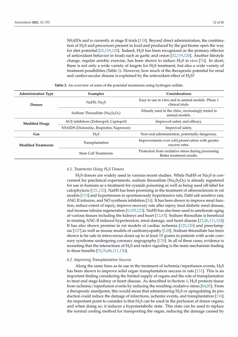
NSAIDs and is currently at stage II trials [118]. Beyond direct administration, the combina-tion of H2S and precursors present in food and produced by the gut biome open the wayfor diet potential [32,119,120]. Indeed, H2S has been recognized as the primary effectorof antioxidant behavior in foods such as garlic and onion [32,119,120]. Another lifestylechange, regular aerobic exercise, has been shown to induce H2S in vivo [76]. In short,there is not only a wide variety of targets for H2S treatment, but also a wide variety oftreatment possibilities (Table 2). However, how much of the therapeutic potential for renaland cardiovascular disease is explained by the antioxidant effect of H2S?
Table 2. An overview of some of the potential treatments using hydrogen sulfide.
Administration Type Examples Considerations
DonorsNaHS, Na2S Easy to use in vitro and in animal models. Phase 1
clinical trials.
Sodium Thiosulfate (Na2S2O3) Already used in the clinic, increasingly tested inanimal models.
Modified Drugs ACE-inhibitors (Zofenopril, Captopril) Improved safety and efficacy.
NSAIDS (Diclonefac, Ibuprofen, Naproxen) Improved safety.
Gas H2S Non-oral administration, potentially dangerous.
Modified TreatmentsTransplantation Improvements over cold preservation with greater
success rates.
Stem Cell Treatments Protection from oxidative stress during processing.Better treatment results.
6.1. Treatments Using H2S Donors
H2S donors are widely used in various recent studies. While NaHS or Na2S is con-venient for preclinical experiments, sodium thiosulfate (Na2S2O3) is already registeredfor use in humans as a treatment for cyanide poisoning as well as being used off-label forcalciphylaxis [121,122]. NaHS has been promising in the treatment of atherosclerosis in ratmodels [113] and hypertension in spontaneously hypertensive rats, Dahl salt sensitive rats,ANG II infusion, and NO synthesis inhibition [24]. It has been shown to improve renal func-tion, reduce extent of injury, improve recovery rate after injury, treat diabetic renal disease,and increase tubular regeneration [9,105,123]. NaHS has also been used to ameliorate agingof various tissues including the kidneys and heart [12,65]. Sodium thiosulfate is beneficialin treating ANG II induced hypertension, renal damage, and heart disease [23,26,111,124].It has also shown promise in rat models of cardiac ischemia [125,126] and preeclamp-sia [127] as well as mouse models of cardiomyopathy [128]. Sodium thiosulfate has beenshown to be safe in intravenous doses up to at least 15 grams in patients with acute coro-nary syndrome undergoing coronary angiography [129]. In all of these cases, evidence ismounting that the interactions of H2S and redox signaling is the main mechanism leadingto these benefits [74,76,88,111,130].
6.2. Improving Transplantation Success
Along the same lines as its use in the treatment of ischemia/reperfusion events, H2Shas been shown to improve solid organ transplantation success in rats [131]. This is animportant finding considering the limited supply of organs and the role of transplantationto treat end stage kidney or heart disease. As described in Section 4, H2S protects tissuefrom ischemic/reperfusion events by reducing the resulting oxidative stress [84,85]. Froma therapeutic standpoint, this would mean that administering H2S or upregulating its pro-duction could reduce the damage of infarctions, ischemic events, and transplantation [116].An important point to consider is that H2S can be used in the perfusion of donor organs,and when doing so, it induces a hypometabolic state. This state can be used to replacethe normal cooling method for transporting the organ, reducing the damage caused by
Antioxidants 2021, 10, 373 13 of 20
the cooling on top of its benefits regarding oxidative stress. Beyond the success rate of thetransplantation, it would be valuable if future research could investigate if H2S reduces theprerenal kidney injuries that come with transplantation of other organs, in particular hearttransplants, as these can lead to the development of CRS (see Section 5.2).
6.3. Diet and Exercise
Recently, H2S has been shown to be involved in the benefits exercise and diet canhave on cardiovascular health. In the case of exercise, it has been shown that long-termregular aerobic exercise is beneficial in attenuating age-related fibrosis of the heart. Re-cently, the mechanism of this attenuation was related to H2S levels [76]. In the case ofdiet, the benefits of the allium genus (onions), in particular allium sativum (garlic), hasbeen connected to H2S released from diallyl disulfide [50,58]. Diallyl sulfide has beenshown to have CSE-dependent benefits related to HIF-1a expression in cellular hypoxiaresponses [50,58]. Many of the benefits of these foods have been attributed to their an-tioxidant properties [32,119,120]. As H2S’s protective effects depend upon activation ofendogenous antioxidant capacity, it may surpass the disappointing results that directantioxidant treatment have shown thus far [132].
6.4. Regenerative Medicine
Stem cell treatment has shown promise in treating serious heart disease includingheart failure [133]. There are several proposed mechanisms of this benefit including differ-entiation, immunomodulatory factor, and H2S secretion [134]. However, its effectivenesscan vary greatly between individuals. Indeed, many clinical trials on stem cell treatmentfocus on using stem cells obtained from the patient, which can be of low quality consideringthe condition of these patients [133]. The quality of stem cells and their ability to differen-tiate requires a minimum level of ROS, however, high levels lead to oxidative stress thatresults in senescence or death of the stem cells [135]. This can be particularly challengingdue to simple matters that might take place in a treatment protocol such as exposure to air,resulting in an increase of ROS in readily available stem cell sources such as mesenchymalstem cells (MSCs) [134]. Considering the previously discussed ways that H2S antagonizesROS, it stands to reason that H2S co-therapy might create a synergistic effect by betterenabling stem cells to reach greater therapeutic potential while at the same time providingprotection on its own. Indeed initial research in bone indicates that H2S does indeed pre-serve MSC function [135]. Recently, Abdelmonem et al. found that preconditioning in vitroor co-delivery in vivo using NaHS both resulted in improved outcomes in treating rats withheart failure over MSC treatment alone [136]. Any combination of NaHS and/or MSCsresulted in no notable fibrosis. All combinations improved ejection fraction, fractionalshortening, and the left ventricle diameter in heart failure with the preconditioned MSCtreatment coming closest to healthy controls. Furthermore, preconditioned MSCs were ableto restore QRS duration and QT intervals to healthy control levels, whereas NaHS alone orin combination with MSCs did not [136]. While still in an early phase, combination therapyof stem cells and H2S has the potential to become a novel therapy.
7. Discussion
All in all, H2S research has thus far opened new avenues of research into importantdiseases of the cardiovascular and renal systems, many of which need more or bettertreatment options. A great deal of H2S’s potential lies in its antioxidant properties. H2S’sability to activate endogenous antioxidant production, modify key signaling proteins thatare also targeted by ROS, and regulate the metabolism of mitochondria make it a complexand interesting puzzle. This complexity may also underlie the benefits seen in the treatmentof rodent models of disease and the observations of H2S associations with different aspectsof various diseases in humans.
Although most of the research has been done in cell lines and in rodent models, we arereaching the point in which translation to the clinic is underway. The existing research in
Antioxidants 2021, 10, 373 14 of 20
humans has initially mostly been observational, exploring changes in sulfate under variouscircumstances. Currently a handful of drugs modified to be H2S donors are in varyingphases of clinical trials. While the focus of these trials is on improving the safety profile ofthe original drug, the safety data in humans may ease the way for trials aimed specificallyon the use of H2S donors as a treatment. Sodium thiosulfate is also in various clinical trials,for example, it has proven safe in phase II trials for treatment of acute coronary syndrome(ClinicalTrials.gov identifier NCT03017963) and is undergoing phase III trials. Hopefully,the results in animal models to directly treat pathologies such as those belonging to CRS aswell as indirectly by improving other treatment options such as transplantation and stemcell therapies can be replicated in humans.
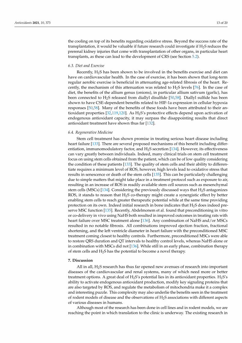
It is also important to realize that while H2S is reduced in cardiovascular and renalpathologies, H2S and the transsulfuration enzymes are increased in other pathologies suchas cancer and certain genetic neurological disorders [38]. When considering its use inhumans, one should be careful to take this into consideration, as it could very well haveimplications for use in some subpopulations. From the other perspective of treating thosepathologies with overproduction, care must also be taken to avoid making the heart andkidneys more vulnerable to oxidative stress related pathologies, exacerbating the alreadygrowing CRS problem. A diagram depicting dose-response relationships of H2S can befound in Figure 6. However, even if such problems exist, they are not insurmountable, andwe remain optimistic about H2S’s therapeutic potential.
Figure 6. A diagram depicting dose-response relationships of H2S concentrations. This review focused on the renal andcardiovascular systems, in which levels of H2S lower than physiological amounts can lead to disease. However, it isimportant to note that high levels are characteristic of cancer and can also lead to toxicity.
8. Conclusions
H2S is a gaseous signaling molecule that plays an important role in redox signaling.Research has exploded on its role in a broad spectrum of biological processes, both physio-logical and pathological. In the case of the renal and cardiovascular systems, H2S playsan important role in maintaining ROS signaling at safe levels by promoting scavengingof ROS as well as competitively modifying cysteine residues on key signaling molecules.As such, depletion of H2S is implicated in a variety of age-related pathologies as wellas pathologies that fall under CRS. A number of these pathologies are difficult to treatand require novel therapies. Current research suggests potential for H2S-based therapies,however, this has been limited primarily to studies in rodents. Fortunately, one donorof H2S, sodium thiosulfate, is already registered for use in humans, thus easing the wayfor translational studies. Furthermore, H2S shows potential for improving other forms of

Antioxidants 2021, 10, 373 15 of 20
treatment such as the safety of NSAIDs, transplantation success, and stem cell therapies.Considering all these points, H2S is a prime target for further research with potentially alarge clinical impact.
Author Contributions: Conceptualization, J.J.S., I.T.N.N., H.V.G. and J.A.J.; resources, J.J.S.; writing—original draft preparation, J.J.S.; writing—review and editing, J.J.S., I.T.N.N., E.M.B.; H.V.G. and J.A.J.;visualization, J.J.S.; supervision, H.V.G. and J.A.J.; project administration, H.V.G.; funding acquisition,H.V.G. and J.A.J. All authors have read and agreed to the published version of the manuscript.
Funding: This research was funded by a grant from the Netherlands CardioVascular ResearchInitiative: An initiative with support of the Dutch Heart Foundation (CVON2014-11 [RECONNECT]).This research was also supported by a grant from the Dutch Kidney Foundation (#17O16).
Acknowledgments: Figures 2, 5 and 6 were created using BioRender.com.
Conflicts of Interest: The authors declare no conflict of interest.
References1. Szabo, C. A timeline of hydrogen sulfide (H(2)S) research: From environmental toxin to biological mediator. Biochem. Pharm.
2018, 149, 5–19. [CrossRef] [PubMed]2. Guidotti, T.L. Hydrogen sulphide. Occup. Med. 1996, 46, 367–371. [CrossRef]3. Dorman, D.C.; Moulin, F.J.; McManus, B.E.; Mahle, K.C.; James, R.A.; Struve, M.F. Cytochrome oxidase inhibition induced by
acute hydrogen sulfide inhalation: Correlation with tissue sulfide concentrations in the rat brain, liver, lung, and nasal epithelium.Toxicol. Sci. 2002, 65, 18–25. [CrossRef] [PubMed]
4. Warenycia, M.W.; Goodwin, L.R.; Benishin, C.G.; Reiffenstein, R.J.; Francom, D.M.; Taylor, J.D.; Dieken, F.P. Acute hydrogensulfide poisoning. Demonstration of selective uptake of sulfide by the brainstem by measurement of brain sulfide levels. Biochem.Pharm. 1989, 38, 973–981. [CrossRef]
5. Goodwin, L.R.; Francom, D.; Dieken, F.P.; Taylor, J.D.; Warenycia, M.W.; Reiffenstein, R.J.; Dowling, G. Determination of sulfidein brain tissue by gas dialysis/ion chromatography: Postmortem studies and two case reports. J. Anal. Toxicol. 1989, 13, 105–109.[CrossRef] [PubMed]
6. Savage, J.C.; Gould, D.H. Determination of sulfide in brain tissue and rumen fluid by ion-interaction reversed-phase high-performance liquid chromatography. J. Chromatogr. 1990, 526, 540–545. [CrossRef]
7. Binkley, F.; Du Vigneaud, V. The Formation of Cysteine from Homocysteine and Serine by Liver Tissue of Rats. J. Biol. Chem. 1942,144, 507–511. [CrossRef]
8. Abe, K.; Kimura, H. The possible role of hydrogen sulfide as an endogenous neuromodulator. J. Neurosci. 1996, 16, 1066–1071.[CrossRef]
9. Cao, X.; Bian, J.S. The Role of Hydrogen Sulfide in Renal System. Front. Pharm. 2016, 7, 385. [CrossRef]10. Wu, D.; Hu, Q.; Zhu, D. An Update on Hydrogen Sulfide and Nitric Oxide Interactions in the Cardiovascular System. Oxidative
Med. Cell. Longev. 2018, 2018, 4579140. [CrossRef]11. Koning, A.M.; Frenay, A.R.; Leuvenink, H.G.; Van Goor, H. Hydrogen sulfide in renal physiology, disease and transplantation–the
smell of renal protection. Nitric Oxide 2015, 46, 37–49. [CrossRef] [PubMed]12. Perridon, B.W.; Leuvenink, H.G.; Hillebrands, J.L.; Van Goor, H.; Bos, E.M. The role of hydrogen sulfide in aging and age-related
pathologies. Aging (Albany Ny.) 2016, 8, 2264–2289. [CrossRef] [PubMed]13. Cortese-Krott, M.M.; Fernandez, B.O.; Kelm, M.; Butler, A.R.; Feelisch, M. On the chemical biology of the nitrite/sulfide
interaction. Nitric Oxide 2015, 46, 14–24. [CrossRef] [PubMed]14. Fukuto, J.M.; Ignarro, L.J.; Nagy, P.; Wink, D.A.; Kevil, C.G.; Feelisch, M.; Cortese-Krott, M.M.; Bianco, C.L.; Kumagai, Y.;
Hobbs, A.J.; et al. Biological hydropersulfides and related polysulfides—A new concept and perspective in redox biology. FEBSLett. 2018, 592, 2140–2152. [CrossRef]
15. Cortese-Krott, M.M.; Koning, A.; Kuhnle, G.G.C.; Nagy, P.; Bianco, C.L.; Pasch, A.; Wink, D.A.; Fukuto, J.M.; Jackson, A.A.; VanGoor, H.; et al. The Reactive Species Interactome: Evolutionary Emergence, Biological Significance, and Opportunities for RedoxMetabolomics and Personalized Medicine. Antioxid Redox Signal. 2017, 27, 684–712. [CrossRef] [PubMed]
16. Van den Born, J.C.; Frenay, A.S.; Koning, A.M.; Bachtler, M.; Riphagen, I.J.; Minovíc, I.; Feelisch, M.; Dekker, M.M.; Bulthuis, M.L.C.;Gansevoort, R.T.; et al. Urinary Excretion of Sulfur Metabolites and Risk of Cardiovascular Events and All-Cause Mortality in theGeneral Population. Antioxid Redox Signal. 2019, 30, 1999–2010. [CrossRef] [PubMed]
17. Van den Born, J.C.; Frenay, A.R.; Bakker, S.J.; Pasch, A.; Hillebrands, J.L.; Lambers Heerspink, H.J.; van Goor, H. High urinarysulfate concentration is associated with reduced risk of renal disease progression in type 2 diabetes. Nitric Oxide 2016, 55–56,18–24. [CrossRef]
18. Ronco, C.; Haapio, M.; House, A.A.; Anavekar, N.; Bellomo, R. Cardiorenal syndrome. J. Am. Coll. Cardiol. 2008, 52, 1527–1539.[CrossRef]
Antioxidants 2021, 10, 373 16 of 20
19. House, A.A.; Anand, I.; Bellomo, R.; Cruz, D.; Bobek, I.; Anker, S.D.; Aspromonte, N.; Bagshaw, S.; Berl, T.; Daliento, L.; et al.Definition and classification of Cardio-Renal Syndromes: Workgroup statements from the 7th ADQI Consensus Conference.Nephrol. Dial. Transpl. 2010, 25, 1416–1420. [CrossRef] [PubMed]
20. Braam, B.; Joles, J.A.; Danishwar, A.H.; Gaillard, C.A. Cardiorenal syndrome–current understanding and future perspectives. Nat.Rev. Nephrol. 2014, 10, 48–55. [CrossRef]
21. Uduman, J. Epidemiology of Cardiorenal Syndrome. Adv. Chronic Kidney Dis. 2018, 25, 391–399. [CrossRef] [PubMed]22. Ronco, C.; Bellasi, A.; Di Lullo, L. Cardiorenal Syndrome: An Overview. Adv. Chronic Kidney Dis. 2018, 25, 382–390. [CrossRef]
[PubMed]23. Snijder, P.M.; Frenay, A.R.; Koning, A.M.; Bachtler, M.; Pasch, A.; Kwakernaak, A.J.; Van den Berg, E.; Bos, E.M.; Hillebrands, J.L.;
Navis, G.; et al. Sodium thiosulfate attenuates angiotensin II-induced hypertension, proteinuria and renal damage. Nitric Oxide2014, 42, 87–98. [CrossRef] [PubMed]
24. Van Goor, H.; Van den Born, J.C.; Hillebrands, J.L.; Joles, J.A. Hydrogen sulfide in hypertension. Curr. Opin. Nephrol. Hypertens.2016, 25, 107–113. [CrossRef] [PubMed]
25. Koning, A.M.; Meijers, W.C.; Minovic, I.; Post, A.; Feelisch, M.; Pasch, A.; Leuvenink, H.G.; De Boer, R.A.; Bakker, S.J.; VanGoor, H. The fate of sulfate in chronic heart failure. Am. J. Physiol. Heart Circ. Physiol. 2017, 312, H415–H421. [CrossRef]
26. Nguyen, I.T.N.; Klooster, A.; Minnion, M.; Feelisch, M.; Verhaar, M.C.; Van Goor, H.; Joles, J.A. Sodium thiosulfate improves renalfunction and oxygenation in L-NNA-induced hypertension in rats. Kidney Int. 2020, 98, 366–377. [CrossRef] [PubMed]
27. Xie, Z.Z.; Liu, Y.; Bian, J.S. Hydrogen Sulfide and Cellular Redox Homeostasis. Oxidative Med. Cell. Longev. 2016, 2016, 6043038.[CrossRef] [PubMed]
28. Kalyanaraman, B. Teaching the basics of redox biology to medical and graduate students: Oxidants, antioxidants and diseasemechanisms. Redox Biol. 2013, 1, 244–257. [CrossRef]
29. Bao, L.; Vlcek, C.; Paces, V.; Kraus, J.P. Identification and tissue distribution of human cystathionine beta-synthase mRNAisoforms. Arch. Biochem. Biophys. 1998, 350, 95–103. [CrossRef]
30. Kanagy, N.L.; Szabo, C.; Papapetropoulos, A. Vascular biology of hydrogen sulfide. Am. J. Physiol. Cell Physiol. 2017, 312,C537–C549. [CrossRef]
31. Hine, C.; Kim, H.J.; Zhu, Y.; Harputlugil, E.; Longchamp, A.; Matos, M.S.; Ramadoss, P.; Bauerle, K.; Brace, L.; Asara, J.M.; et al.Hypothalamic-Pituitary Axis Regulates Hydrogen Sulfide Production. Cell Metab. 2017, 25, 1320–1333.e1325. [CrossRef]
32. Hine, C.; Zhu, Y.; Hollenberg, A.N.; Mitchell, J.R. Dietary and Endocrine Regulation of Endogenous Hydrogen Sulfide Production:Implications for Longevity. Antioxid Redox Signal. 2018, 28, 1483–1502. [CrossRef]
33. Sbodio, J.I.; Snyder, S.H.; Paul, B.D. Regulators of the transsulfuration pathway. Br. J. Pharm. 2019, 176, 583–593. [CrossRef]34. Bos, E.M.; Wang, R.; Snijder, P.M.; Boersema, M.; Damman, J.; Fu, M.; Moser, J.; Hillebrands, J.L.; Ploeg, R.J.; Yang, G.; et al.
Cystathionine γ-lyase protects against renal ischemia/reperfusion by modulating oxidative stress. J. Am. Soc. Nephrol. 2013, 24,759–770. [CrossRef]
35. Yang, J.; Minkler, P.; Grove, D.; Wang, R.; Willard, B.; Dweik, R.; Hine, C. Non-enzymatic hydrogen sulfide production fromcysteine in blood is catalyzed by iron and vitamin B(6). Commun. Biol. 2019, 2, 194. [CrossRef]
36. Xu, S.; Liu, Z.; Liu, P. Targeting hydrogen sulfide as a promising therapeutic strategy for atherosclerosis. Int. J. Cardiol. 2014, 172,313–317. [CrossRef] [PubMed]
37. Kimura, Y.; Koike, S.; Shibuya, N.; Lefer, D.; Ogasawara, Y.; Kimura, H. 3-Mercaptopyruvate sulfurtransferase produces potentialredox regulators cysteine- and glutathione-persulfide (Cys-SSH and GSSH) together with signaling molecules H(2)S(2), H(2)S(3)and H(2)S. Sci. Rep. 2017, 7, 10459. [CrossRef] [PubMed]
38. Cao, X.; Ding, L.; Xie, Z.Z.; Yang, Y.; Whiteman, M.; Moore, P.K.; Bian, J.S. A Review of Hydrogen Sulfide Synthesis, Metabolism,and Measurement: Is Modulation of Hydrogen Sulfide a Novel Therapeutic for Cancer? Antioxid Redox Signal. 2019, 31, 1–38.[CrossRef] [PubMed]
39. Pal, V.K.; Bandyopadhyay, P.; Singh, A. Hydrogen sulfide in physiology and pathogenesis of bacteria and viruses. Iubmb Life 2018,70, 393–410. [CrossRef]
40. Wedmann, R.; Onderka, C.; Wei, S.; Szijártó, I.A.; Miljkovic, J.L.; Mitrovic, A.; Lange, M.; Savitsky, S.; Yadav, P.K.; Torregrossa, R.;et al. Improved tag-switch method reveals that thioredoxin acts as depersulfidase and controls the intracellular levels of proteinpersulfidation. Chem. Sci. 2016, 7, 3414–3426. [CrossRef]
41. Palde, P.B.; Carroll, K.S. A universal entropy-driven mechanism for thioredoxin-target recognition. Proc. Natl. Acad. Sci. USA2015, 112, 7960–7965. [CrossRef]
42. Ahmad, A.; Olah, G.; Szczesny, B.; Wood, M.E.; Whiteman, M.; Szabo, C. AP39, A Mitochondrially Targeted Hydrogen SulfideDonor, Exerts Protective Effects in Renal Epithelial Cells Subjected to Oxidative Stress in Vitro and in Acute Renal Injury in Vivo.Shock 2016, 45, 88–97. [CrossRef]
43. Fu, M.; Zhang, W.; Wu, L.; Yang, G.; Li, H.; Wang, R. Hydrogen sulfide (H2S) metabolism in mitochondria and its regulatory rolein energy production. Proc. Natl. Acad. Sci. USA 2012, 109, 2943–2948. [CrossRef]
44. Strutyns’ka, N.A.; Semenykhina, O.M.; Chorna, S.V.; Vavilova, H.L.; Sahach, V.F. Hydrogen sulfide inhibits Ca(2+)-inducedmitochondrial permeability transition pore opening in adult and old rat heart. Fiziol. Zh. 2011, 57, 3–14. [CrossRef]
45. Bruce King, S. Potential biological chemistry of hydrogen sulfide (H2S) with the nitrogen oxides. Free Radic. Biol. Med. 2013, 55,1–7. [CrossRef]

Antioxidants 2021, 10, 373 17 of 20
46. Zhang, J.; Shi, C.; Wang, H.; Gao, C.; Chang, P.; Chen, X.; Shan, H.; Zhang, M.; Tao, L. Hydrogen sulfide protects against celldamage through modulation of PI3K/Akt/Nrf2 signaling. Int. J. Biochem. Cell Biol. 2019, 117, 105636. [CrossRef] [PubMed]
47. Amaral, J.H.; Rizzi, E.S.; Alves-Lopes, R.; Pinheiro, L.C.; Tostes, R.C.; Tanus-Santos, J.E. Antioxidant and antihypertensiveresponses to oral nitrite involves activation of the Nrf2 pathway. Free Radic. Biol. Med. 2019, 141, 261–268. [CrossRef]
48. Kai, S.; Tanaka, T.; Daijo, H.; Harada, H.; Kishimoto, S.; Suzuki, K.; Takabuchi, S.; Takenaga, K.; Fukuda, K.; Hirota, K.Hydrogen sulfide inhibits hypoxia- but not anoxia-induced hypoxia-inducible factor 1 activation in a von hippel-lindau- andmitochondria-dependent manner. Antioxid Redox Signal. 2012, 16, 203–216. [CrossRef]
49. Wang, M.; Yan, J.; Cao, X.; Hua, P.; Li, Z. Hydrogen sulfide modulates epithelial-mesenchymal transition and angiogenesis innon-small cell lung cancer via HIF-1α activation. Biochem. Pharm. 2020, 172, 113775. [CrossRef] [PubMed]
50. Dugbartey, G.J. The smell of renal protection against chronic kidney disease: Hydrogen sulfide offers a potential stinky remedy.Pharm. Rep. 2018, 70, 196–205. [CrossRef] [PubMed]
51. Sies, H.; Berndt, C.; Jones, D.P. Oxidative Stress. Annu. Rev. Biochem. 2017, 86, 715–748. [CrossRef]52. Zhu, H.; Blake, S.; Chan, K.T.; Pearson, R.B.; Kang, J. Cystathionine β-Synthase in Physiology and Cancer. BioMed Res. Int. 2018,
2018, 3205125. [CrossRef] [PubMed]53. Maassen, H.; Hendriks, K.D.W.; Venema, L.H.; Henning, R.H.; Hofker, S.H.; Van Goor, H.; Leuvenink, H.G.D.; Coester, A.M.
Hydrogen sulphide-induced hypometabolism in human-sized porcine kidneys. PLoS ONE 2019, 14, e0225152. [CrossRef][PubMed]
54. Vitvitsky, V.; Miljkovic, J.L.; Bostelaar, T.; Adhikari, B.; Yadav, P.K.; Steiger, A.K.; Torregrossa, R.; Pluth, M.D.; Whiteman, M.;Banerjee, R.; et al. Cytochrome c Reduction by H(2)S Potentiates Sulfide Signaling. ACS Chem. Biol. 2018, 13, 2300–2307.[CrossRef] [PubMed]
55. Sies, H.; Jones, D.P. Reactive oxygen species (ROS) as pleiotropic physiological signalling agents. Nat. Rev. Mol. Cell Biol. 2020, 21,363–383. [CrossRef]
56. Madamanchi, N.R.; Runge, M.S. Redox signaling in cardiovascular health and disease. Free Radic. Biol. Med. 2013, 61, 473–501.[CrossRef]
57. Foster, D.B.; Van Eyk, J.E.; Marbán, E.; O’Rourke, B. Redox signaling and protein phosphorylation in mitochondria: Progress andprospects. J. Bioenerg. Biomembr. 2009, 41, 159–168. [CrossRef]
58. Flannigan, K.L.; Agbor, T.A.; Motta, J.P.; Ferraz, J.G.; Wang, R.; Buret, A.G.; Wallace, J.L. Proresolution effects of hydrogen sulfideduring colitis are mediated through hypoxia-inducible factor-1α. Faseb. J. 2015, 29, 1591–1602. [CrossRef]
59. Drachuk, K.O.; Dorofeyeva, N.A.; Sagach, V.F. The role of hydrogen sulfide in diastolic function restoration during aging. Fiziol.Zh. 2016, 62, 9–18. [CrossRef]
60. Jin, S.; Pu, S.X.; Hou, C.L.; Ma, F.F.; Li, N.; Li, X.H.; Tan, B.; Tao, B.B.; Wang, M.J.; Zhu, Y.C. Cardiac H2S Generation Is Reduced inAgeing Diabetic Mice. Oxidative Med. Cell Longev. 2015, 2015, 758358. [CrossRef]
61. Mys, L.A.; Budko, A.Y.; Strutynska, N.A.; Sagach, V.F. Pyridoxal-5-phosphate restores hydrogen sulfide synthes and redox stateof heart and blood vessels tissue in old animals. Fiziol. Zh. 2017, 63, 3–9. [CrossRef] [PubMed]
62. Majzunova, M.; Dovinova, I.; Barancik, M.; Chan, J.Y. Redox signaling in pathophysiology of hypertension. J. BioMed. Sci. 2013,20, 69. [CrossRef] [PubMed]
63. Berenyiova, A.; Drobna, M.; Cebova, M.; Kristek, F.; Cacanyiova, S. Changes in the vasoactive effects of nitric oxide, hydrogensulfide and the structure of the rat thoracic aorta: The role of age and essential hypertension. J. Physiol. Pharm. 2018, 69. [CrossRef]
64. Honda, T.; Hirakawa, Y.; Nangaku, M. The role of oxidative stress and hypoxia in renal disease. Kidney Res. Clin. Pr. 2019, 38,414–426. [CrossRef] [PubMed]
65. Lee, H.J.; Feliers, D.; Barnes, J.L.; Oh, S.; Choudhury, G.G.; Diaz, V.; Galvan, V.; Strong, R.; Nelson, J.; Salmon, A.; et al. Hydrogensulfide ameliorates aging-associated changes in the kidney. Geroscience 2018, 40, 163–176. [CrossRef]
66. Kurtz, A. Control of renin synthesis and secretion. Am. J. Hypertens. 2012, 25, 839–847. [CrossRef] [PubMed]67. Cao, X.; Wu, Z.; Xiong, S.; Cao, L.; Sethi, G.; Bian, J.S. The role of hydrogen sulfide in cyclic nucleotide signaling. Biochem. Pharm.
2018, 149, 20–28. [CrossRef]68. Luo, R.; Hu, S.; Liu, Q.; Han, M.; Wang, F.; Qiu, M.; Li, S.; Li, X.; Yang, T.; Fu, X.; et al. Hydrogen sulfide upregulates renal AQP-2
protein expression and promotes urine concentration. Faseb. J. 2019, 33, 469–483. [CrossRef]69. Olson, K.R. Hydrogen sulfide as an oxygen sensor. Clin. Chem. Lab. Med. 2013, 51, 623–632. [CrossRef]70. Leigh, J.; Juriasingani, S.; Akbari, M.; Shao, P.; Saha, M.N.; Lobb, I.; Bachtler, M.; Fernandez, B.; Qian, Z.; Van Goor, H.; et al.
Endogenous H(2)S production deficiencies lead to impaired renal erythropoietin production. Can. Urol. Assoc. J. 2018, 13,E210–E219. [CrossRef]
71. Ratliff, B.B.; Abdulmahdi, W.; Pawar, R.; Wolin, M.S. Oxidant Mechanisms in Renal Injury and Disease. Antioxid Redox Signal.2016, 25, 119–146. [CrossRef]
72. Maksuti, E.; Westerhof, N.; Westerhof, B.E.; Broomé, M.; Stergiopulos, N. Contribution of the Arterial System and the Heart toBlood Pressure during Normal Aging—A Simulation Study. PLoS ONE 2016, 11, e0157493. [CrossRef]
73. Strutynska, N.A.; Kotsiuruba, A.V.; Budko, A.Y.; Mys, L.A.; Sagach, V.F. Mitochondrial dysfunction in the aging heart isaccompanied by constitutive no-synthases uncoupling on the background of oxidative and nitrosative stress. Fiziol. Zh. 2016, 62,3–11. [CrossRef]
Antioxidants 2021, 10, 373 18 of 20
74. Wei, C.; Zhao, Y.; Wang, L.; Peng, X.; Li, H.; Zhao, Y.; He, Y.; Shao, H.; Zhong, X.; Li, H.; et al. H2 S restores the cardioprotectionfrom ischemic post-conditioning in isolated aged rat hearts. Cell Biol. Int. 2015, 39, 1173–1176. [CrossRef]
75. Liang, M.; Jin, S.; Wu, D.D.; Wang, M.J.; Zhu, Y.C. Hydrogen sulfide improves glucose metabolism and prevents hypertrophy incardiomyocytes. Nitric Oxide 2015, 46, 114–122. [CrossRef]
76. Ma, N.; Liu, H.M.; Xia, T.; Liu, J.D.; Wang, X.Z. Chronic aerobic exercise training alleviates myocardial fibrosis in aged ratsthrough restoring bioavailability of hydrogen sulfide. Can. J. Physiol. Pharm. 2018, 96, 902–908. [CrossRef] [PubMed]
77. Zhang, Q.Y.; Jin, H.F.; Chen, S.; Chen, Q.H.; Tang, C.S.; Du, J.B.; Huang, Y.Q. Hydrogen Sulfide Regulating Myocardial Structureand Function by Targeting Cardiomyocyte Autophagy. Chin. Med. J. 2018, 131, 839–844. [CrossRef] [PubMed]
78. Wohlgemuth, S.E.; Calvani, R.; Marzetti, E. The interplay between autophagy and mitochondrial dysfunction in oxidativestress-induced cardiac aging and pathology. J. Mol. Cell. Cardiol. 2014, 71, 62–70. [CrossRef] [PubMed]
79. Iciek, M.; Bilska-Wilkosz, A.; Górny, M. Sulfane sulfur—New findings on an old topic. Acta Biochim. Pol. 2019, 66, 533–544.[CrossRef] [PubMed]
80. Ronco, C.; Bellomo, R.; Kellum, J.A. Acute kidney injury. Lancet 2019, 394, 1949–1964. [CrossRef]81. Dudoignon, E.; Dépret, F.; Legrand, M. Is the Renin-Angiotensin-Aldosterone System Good for the Kidney in Acute Settings?
Nephron 2019, 143, 179–183. [CrossRef] [PubMed]82. Meersch, M.; Schmidt, C.; Zarbock, A. Perioperative Acute Kidney Injury: An Under-Recognized Problem. Anesth Analg. 2017,
125, 1223–1232. [CrossRef] [PubMed]83. Thiele, R.H.; Isbell, J.M.; Rosner, M.H. AKI associated with cardiac surgery. Clin. J. Am. Soc. Nephrol. 2015, 10, 500–514. [CrossRef]
[PubMed]84. Azizi, F.; Seifi, B.; Kadkhodaee, M.; Ahghari, P. Administration of hydrogen sulfide protects ischemia reperfusion-induced acute
kidney injury by reducing the oxidative stress. Ir. J. Med. Sci. 2016, 185, 649–654. [CrossRef] [PubMed]85. Bos, E.M.; Leuvenink, H.G.; Snijder, P.M.; Kloosterhuis, N.J.; Hillebrands, J.L.; Leemans, J.C.; Florquin, S.; Van Goor, H. Hydrogen
sulfide-induced hypometabolism prevents renal ischemia/reperfusion injury. J. Am. Soc. Nephrol. 2009, 20, 1901–1905. [CrossRef]86. Caravaca-Fontán, F.; Fernández-Juárez, G.; Praga, M. Acute kidney injury in interstitial nephritis. Curr. Opin. Crit. Care 2019, 25,
558–564. [CrossRef]87. Clark, M.R.; Trotter, K.; Chang, A. The Pathogenesis and Therapeutic Implications of Tubulointerstitial Inflammation in Human
Lupus Nephritis. Semin. Nephrol. 2015, 35, 455–464. [CrossRef]88. Chen, Y.; Jin, S.; Teng, X.; Hu, Z.; Zhang, Z.; Qiu, X.; Tian, D.; Wu, Y. Hydrogen Sulfide Attenuates LPS-Induced Acute Kidney
Injury by Inhibiting Inflammation and Oxidative Stress. Oxid Med. Cell Longev. 2018, 2018, 6717212. [CrossRef]89. Wallace, J.L.; Vaughan, D.; Dicay, M.; MacNaughton, W.K.; De Nucci, G. Hydrogen Sulfide-Releasing Therapeutics: Translation to
the Clinic. Antioxid Redox Signal. 2018, 28, 1533–1540. [CrossRef]90. Van Dingenen, J.; Pieters, L.; Vral, A.; Lefebvre, R.A. The H2S-Releasing Naproxen Derivative ATB-346 and the Slow-Release H2S
Donor GYY4137 Reduce Intestinal Inflammation and Restore Transit in Postoperative Ileus. Front. Pharmacol. 2019, 10. [CrossRef]91. Zhang, X.; Donnan, P.T.; Bell, S.; Guthrie, B. Non-steroidal anti-inflammatory drug induced acute kidney injury in the community
dwelling general population and people with chronic kidney disease: Systematic review and meta-analysis. Bmc Nephrol. 2017,18, 256. [CrossRef]
92. Ozkaya, O.; Genc, G.; Bek, K.; Sullu, Y. A case of acetaminophen (paracetamol) causing renal failure without liver damage in achild and review of literature. Ren. Fail. 2010, 32, 1125–1127. [CrossRef]
93. Ozatik, F.Y.; Teksen, Y.; Kadioglu, E.; Ozatik, O.; Bayat, Z. Effects of hydrogen sulfide on acetaminophen-induced acute renaltoxicity in rats. Int. Urol. Nephrol. 2019, 51, 745–754. [CrossRef] [PubMed]
94. Cao, X.; Zhang, W.; Moore, P.K.; Bian, J. Protective Smell of Hydrogen Sulfide and Polysulfide in Cisplatin-Induced Nephrotoxicity.Int. J. Mol. Sci. 2019, 20, 313. [CrossRef]
95. Yuan, Y.; Zhu, L.; Li, L.; Liu, J.; Chen, Y.; Cheng, J.; Peng, T.; Lu, Y. S-Sulfhydration of SIRT3 by Hydrogen Sulfide AttenuatesMitochondrial Dysfunction in Cisplatin-Induced Acute Kidney Injury. Antioxid Redox Signal. 2019, 31, 1302–1319. [CrossRef]
96. Liu, M.; Jia, Z.; Sun, Y.; Zhang, A.; Yang, T. A H 2 S Donor GYY4137 Exacerbates Cisplatin-Induced Nephrotoxicity in Mice.Mediat. Inflamm. 2016, 2016, 8145785. [CrossRef] [PubMed]
97. Farrar, A. Acute Kidney Injury. Nurs. Clin. North Am. 2018, 53, 499–510. [CrossRef] [PubMed]98. Lin, S.; Lian, D.; Liu, W.; Haig, A.; Lobb, I.; Hijazi, A.; Razvi, H.; Burton, J.; Whiteman, M.; Sener, A. Daily therapy with a
slow-releasing H(2)S donor GYY4137 enables early functional recovery and ameliorates renal injury associated with urinaryobstruction. Nitric Oxide 2018, 76, 16–28. [CrossRef] [PubMed]
99. Lin, S.; Visram, F.; Liu, W.; Haig, A.; Jiang, J.; Mok, A.; Lian, D.; Wood, M.E.; Torregrossa, R.; Whiteman, M.; et al. GYY4137, aSlow-Releasing Hydrogen Sulfide Donor, Ameliorates Renal Damage Associated with Chronic Obstructive Uropathy. J. Urol.2016, 196, 1778–1787. [CrossRef] [PubMed]
100. Aminzadeh, M.A.; Vaziri, N.D. Downregulation of the renal and hepatic hydrogen sulfide (H2S)-producing enzymes and capacityin chronic kidney disease. Nephrol. Dial. Transpl. 2012, 27, 498–504. [CrossRef] [PubMed]
101. Karmin, O.; Siow, Y.L. Metabolic Imbalance of Homocysteine and Hydrogen Sulfide in Kidney Disease. Curr. Med. Chem. 2018,25, 367–377. [CrossRef]
102. Nowotny, K.; Jung, T.; Höhn, A.; Weber, D.; Grune, T. Advanced glycation end products and oxidative stress in type 2 diabetesmellitus. Biomolecules 2015, 5, 194–222. [CrossRef]
Antioxidants 2021, 10, 373 19 of 20
103. Isoni, C.A.; Borges, E.A.; Veloso, C.A.; Mattos, R.T.; Chaves, M.M.; Nogueira-Machado, J.A. cAMP activates the generation ofreactive oxygen species and inhibits the secretion of IL-6 in peripheral blood mononuclear cells from type 2 diabetic patients.Oxidative Med. Cell Longev. 2009, 2, 317–321. [CrossRef] [PubMed]
104. Liu, Y.; Zhao, H.; Qiang, Y.; Qian, G.; Lu, S.; Chen, J.; Wang, X.; Guan, Q.; Liu, Y.; Fu, Y. Effects of hydrogen sulfide on highglucose-induced glomerular podocyte injury in mice. Int. J. Clin. Exp. Pathol. 2015, 8, 6814–6820. [PubMed]
105. Xue, R.; Hao, D.D.; Sun, J.P.; Li, W.W.; Zhao, M.M.; Li, X.H.; Chen, Y.; Zhu, J.H.; Ding, Y.J.; Liu, J.; et al. Hydrogen sulfidetreatment promotes glucose uptake by increasing insulin receptor sensitivity and ameliorates kidney lesions in type 2 diabetes.Antioxid Redox Signal. 2013, 19, 5–23. [CrossRef] [PubMed]
106. Cai, J.; Shi, X.; Wang, H.; Fan, J.; Feng, Y.; Lin, X.; Yang, J.; Cui, Q.; Tang, C.; Xu, G.; et al. Cystathionine γ lyase-hydrogen sulfideincreases peroxisome proliferator-activated receptor γ activity by sulfhydration at C139 site thereby promoting glucose uptakeand lipid storage in adipocytes. Biochim. Biophys. Acta 2016, 1861, 419–429. [CrossRef] [PubMed]
107. Yaxley, J.P.; Thambar, S.V. Resistant hypertension: An approach to management in primary care. J. Fam. Med. Prim. Care 2015, 4,193–199. [CrossRef]
108. Van Der Sande, N.G.C.; Blankestijn, P.J.; Visseren, F.L.J.; Beeftink, M.M.; Voskuil, M.; Westerink, J.; Bots, M.L.; Spiering, W.Prevalence of potential modifiable factors of hypertension in patients with difficult-to-control hypertension. J. Hypertens. 2019, 37,398–405. [CrossRef] [PubMed]
109. Doroszko, A.; Janus, A.; Szahidewicz-Krupska, E.; Mazur, G.; Derkacz, A. Resistant Hypertension. Adv. Clin. Exp. Med. 2016, 25,173–183. [CrossRef] [PubMed]
110. Al-Magableh, M.R.; Kemp-Harper, B.K.; Hart, J.L. Hydrogen sulfide treatment reduces blood pressure and oxidative stress inangiotensin II-induced hypertensive mice. Hypertens. Res. 2015, 38, 13–20. [CrossRef] [PubMed]
111. Snijder, P.M.; Frenay, A.R.; De Boer, R.A.; Pasch, A.; Hillebrands, J.L.; Leuvenink, H.G.; Van Goor, H. Exogenous administration ofthiosulfate, a donor of hydrogen sulfide, attenuates angiotensin II-induced hypertensive heart disease in rats. Br. J. Pharm. 2015,172, 1494–1504. [CrossRef] [PubMed]
112. Bucci, M.; Vellecco, V.; Cantalupo, A.; Brancaleone, V.; Zhou, Z.; Evangelista, S.; Calderone, V.; Papapetropoulos, A.; Cirino, G.Hydrogen sulfide accounts for the peripheral vascular effects of zofenopril independently of ACE inhibition. Cardiovasc. Res.2014, 102, 138–147. [CrossRef] [PubMed]
113. Mani, S.; Li, H.; Untereiner, A.; Wu, L.; Yang, G.; Austin, R.C.; Dickhout, J.G.; Lhoták, Š.; Meng, Q.H.; Wang, R. Decreasedendogenous production of hydrogen sulfide accelerates atherosclerosis. Circulation 2013, 127, 2523–2534. [CrossRef] [PubMed]
114. Van den Born, J.C.; Mencke, R.; Conroy, S.; Zeebregts, C.J.; Van Goor, H.; Hillebrands, J.L. Cystathionine γ-lyase is expressed inhuman atherosclerotic plaque microvessels and is involved in micro-angiogenesis. Sci. Rep. 2016, 6, 34608. [CrossRef] [PubMed]
115. Wang, Z.J.; Wu, J.; Guo, W.; Zhu, Y.Z. Atherosclerosis and the Hydrogen Sulfide Signaling Pathway—Therapeutic Approaches toDisease Prevention. Cell Physiol. Biochem. 2017, 42, 859–875. [CrossRef] [PubMed]
116. Wen, Y.D.; Wang, H.; Zhu, Y.Z. The Drug Developments of Hydrogen Sulfide on Cardiovascular Disease. Oxidative Med. CellLongev. 2018, 2018, 4010395. [CrossRef]
117. Snijder, P.M.; De Boer, R.A.; Bos, E.M.; Van den Born, J.C.; Ruifrok, W.P.; Vreeswijk-Baudoin, I.; van Dijk, M.C.; Hillebrands, J.L.;Leuvenink, H.G.; Van Goor, H. Gaseous hydrogen sulfide protects against myocardial ischemia-reperfusion injury in micepartially independent from hypometabolism. PLoS ONE 2013, 8, e63291. [CrossRef] [PubMed]
118. Wallace, J.L.; Nagy, P.; Feener, T.; Allain, T.; Ditrói, T.; Vaughan, D.; Muscara, M.; De Nucci, G.; Buret, A. A3 A proof-of-concept,phase 2 clinical trial of the gi safety of a hydrogen sulfide-releasing anti-inflammatory drug (ATB-346). J. Can. AsSoc. Gastroenterol.2019, 2, 7–8. [CrossRef]
119. Capasso, A. Antioxidant action and therapeutic efficacy of Allium sativum L. Molecules 2013, 18, 690–700. [CrossRef]120. Kim, S.; Kim, D.B.; Jin, W.; Park, J.; Yoon, W.; Lee, Y.; Kim, S.; Lee, S.; Kim, S.; Lee, O.H.; et al. Comparative studies of bioactive
organosulphur compounds and antioxidant activities in garlic (Allium sativum L.), elephant garlic (Allium ampeloprasum L.) andonion (Allium cepa L.). Nat. Prod. Res. 2018, 32, 1193–1197. [CrossRef] [PubMed]
121. Parker-Cote, J.L.; Rizer, J.; Vakkalanka, J.P.; Rege, S.V.; Holstege, C.P. Challenges in the diagnosis of acute cyanide poisoning. Clin.Toxicol. 2018, 56, 609–617. [CrossRef]
122. Nigwekar, S.U.; Brunelli, S.M.; Meade, D.; Wang, W.; Hymes, J.; Lacson, E., Jr. Sodium thiosulfate therapy for calcific uremicarteriolopathy. Clin. J. Am. Soc. Nephrol. 2013, 8, 1162–1170. [CrossRef] [PubMed]
123. Sun, H.J.; Wu, Z.Y.; Cao, L.; Zhu, M.Y.; Liu, T.T.; Guo, L.; Lin, Y.; Nie, X.W.; Bian, J.S. Hydrogen Sulfide: Recent Progression andPerspectives for the Treatment of Diabetic Nephropathy. Molecules 2019, 24, 2857. [CrossRef] [PubMed]
124. Oosterhuis, N.R.; Frenay, A.R.; Wesseling, S.; Snijder, P.M.; Slaats, G.G.; Yazdani, S.; Fernandez, B.O.; Feelisch, M.; Giles, R.H.;Verhaar, M.C.; et al. DL-propargylglycine reduces blood pressure and renal injury but increases kidney weight in angiotensin-IIinfused rats. Nitric Oxide 2015, 49, 56–66. [CrossRef] [PubMed]
125. Kannan, S.; Boovarahan, S.R.; Rengaraju, J.; Prem, P.; Kurian, G.A. Attenuation of cardiac ischemia-reperfusion injury by sodiumthiosulfate is partially dependent on the effect of cystathione beta synthase in the myocardium. Cell Biochem. Biophys. 2019, 77,261–272. [CrossRef]
126. Ravindran, S.; Jahir Hussain, S.; Boovarahan, S.R.; Kurian, G.A. Sodium thiosulfate post-conditioning protects rat hearts againstischemia reperfusion injury via reduction of apoptosis and oxidative stress. Chem. Biol. Interact. 2017, 274, 24–34. [CrossRef]
Antioxidants 2021, 10, 373 20 of 20
127. Terstappen, F.; Clarke, S.M.; Joles, J.A.; Ross, C.A.; Garrett, M.R.; Minnion, M.; Feelisch, M.; Goor, H.V.; Sasser, J.M.; Lely, A.T.Sodium Thiosulfate in the Pregnant Dahl Salt-Sensitive Rat, a Model of Preeclampsia. Biomolecules 2020, 10, 302. [CrossRef]
128. Mizuta, Y.; Tokuda, K.; Guo, J.; Zhang, S.; Narahara, S.; Kawano, T.; Murata, M.; Yamaura, K.; Hoka, S.; Hashizume, M.; et al.Sodium thiosulfate prevents doxorubicin-induced DNA damage and apoptosis in cardiomyocytes in mice. Life Sci. 2020, 257,118074. [CrossRef]
129. De Koning, M.L.Y.; Assa, S.; Maagdenberg, C.G.; Van Veldhuisen, D.J.; Pasch, A.; Van Goor, H.; Lipsic, E.; Van der Harst, P. Safetyand Tolerability of Sodium Thiosulfate in Patients with an Acute Coronary Syndrome Undergoing Coronary Angiography: ADose-Escalation Safety Pilot Study (SAFE-ACS). J. Interv. Cardiol. 2020, 2020, 6014915. [CrossRef]
130. Li, H.; Wang, Y.; Wei, C.; Bai, S.; Zhao, Y.; Li, H.; Wu, B.; Wang, R.; Wu, L.; Xu, C. Mediation of exogenous hydrogen sulfidein recovery of ischemic post-conditioning-induced cardioprotection via down-regulating oxidative stress and up-regulatingPI3K/Akt/GSK-3β pathway in isolated aging rat hearts. Cell Biosci. 2015, 5, 11. [CrossRef]
131. Snijder, P.M.; Van den Berg, E.; Whiteman, M.; Bakker, S.J.; Leuvenink, H.G.; Van Goor, H. Emerging role of gasotransmitters inrenal transplantation. Am. J. Transpl. 2013, 13, 3067–3075. [CrossRef] [PubMed]
132. Bjelakovic, G.; Nikolova, D.; Gluud, C. Antioxidant supplements and mortality. Curr. Opin. Clin. Nutr Metab. Care 2014, 17, 40–44.[CrossRef] [PubMed]
133. Tehzeeb, J.; Manzoor, A.; Ahmed, M.M. Is Stem Cell Therapy an Answer to Heart Failure: A Literature Search. Cureus 2019, 11,e5959. [CrossRef] [PubMed]
134. Tan, D.Q.; Suda, T. Reactive Oxygen Species and Mitochondrial Homeostasis as Regulators of Stem Cell Fate and Function.Antioxid. Redox Signal. 2018, 29, 149–168. [CrossRef]
135. Liu, Y.; Yang, R.; Liu, X.; Zhou, Y.; Qu, C.; Kikuiri, T.; Wang, S.; Zandi, E.; Du, J.; Ambudkar, I.S.; et al. Hydrogen sulfide maintainsmesenchymal stem cell function and bone homeostasis via regulation of Ca(2+) channel sulfhydration. Cell Stem Cell 2014, 15,66–78. [CrossRef]
136. Abdelmonem, M.; Shahin, N.N.; Rashed, L.A.; Amin, H.A.A.; Shamaa, A.A.; Shaheen, A.A. Hydrogen sulfide enhances theeffectiveness of mesenchymal stem cell therapy in rats with heart failure: In vitro preconditioning versus in vivo co-delivery.BioMed. Pharm. 2019, 112, 108584. [CrossRef] [PubMed]
Related Documents