Fig. 16-1

Fig. 16-1. Building a Structural Model of DNA: Scientific Inquiry After most biologists became convinced that DNA was the genetic material, the challenge.
Dec 31, 2015
Welcome message from author
This document is posted to help you gain knowledge. Please leave a comment to let me know what you think about it! Share it to your friends and learn new things together.
Transcript
Fig. 16-1
Building a Structural Model of DNA: Scientific Inquiry
• After most biologists became convinced that DNA was the genetic material, the challenge was to determine how its structure accounts for its role
• Maurice Wilkins and Rosalind Franklin were using a technique called X-ray crystallography to study molecular structure
• Franklin produced a picture of the DNA molecule using this technique
Copyright © 2008 Pearson Education Inc., publishing as Pearson Benjamin Cummings

Fig. 16-6a
(a) Rosalind Franklin
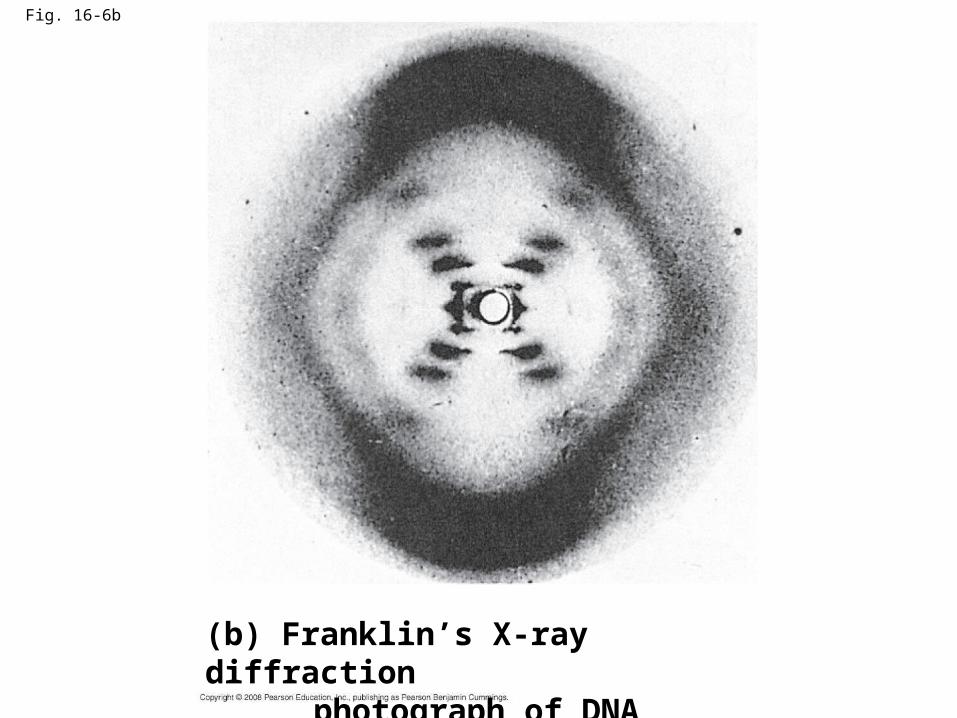
Fig. 16-6b
(b) Franklin’s X-ray diffraction photograph of DNA

• Franklin’s X-ray crystallographic images of DNA enabled Watson to deduce that DNA was helical
• The X-ray images also enabled Watson to deduce the width of the helix and the spacing of the nitrogenous bases
• The width suggested that the DNA molecule was made up of two strands, forming a double helix
Copyright © 2008 Pearson Education Inc., publishing as Pearson Benjamin Cummings
Animation: DNA Double HelixAnimation: DNA Double Helix
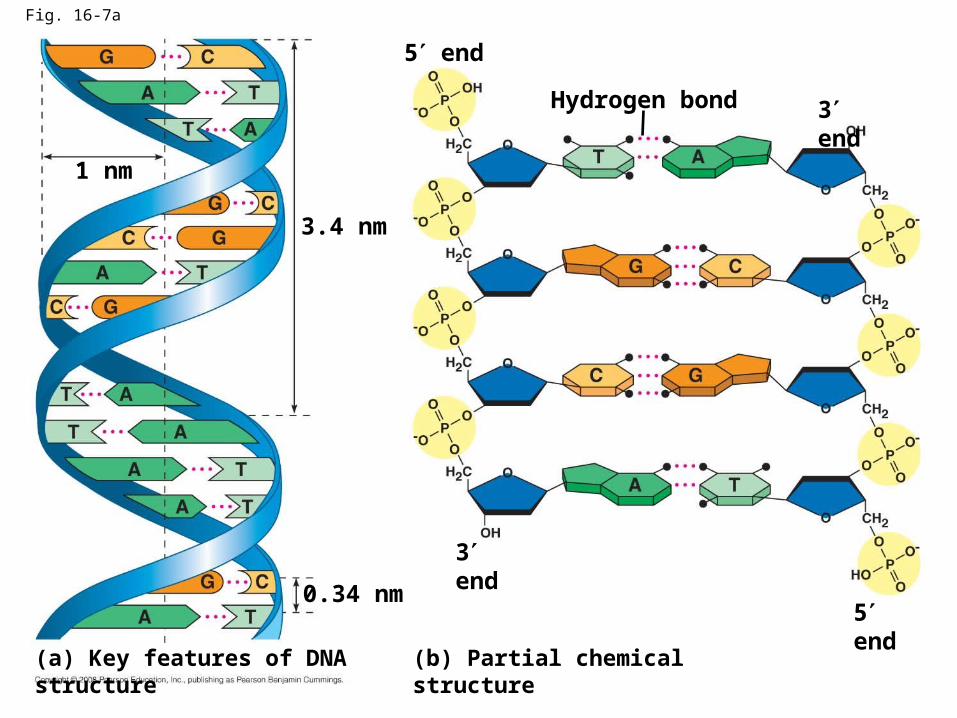
Fig. 16-7a
Hydrogen bond 3 end
5 end
3.4 nm
0.34 nm
3 end
5 end
(b) Partial chemical structure(a) Key features of DNA structure
1 nm

• Watson and Crick built models of a double helix to conform to the X-rays and chemistry of DNA
• Franklin had concluded that there were two antiparallel sugar-phosphate backbones, with the nitrogenous bases paired in the molecule’s interior
Copyright © 2008 Pearson Education Inc., publishing as Pearson Benjamin Cummings
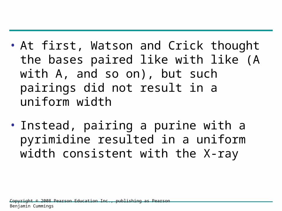
• At first, Watson and Crick thought the bases paired like with like (A with A, and so on), but such pairings did not result in a uniform width
• Instead, pairing a purine with a pyrimidine resulted in a uniform width consistent with the X-ray
Copyright © 2008 Pearson Education Inc., publishing as Pearson Benjamin Cummings
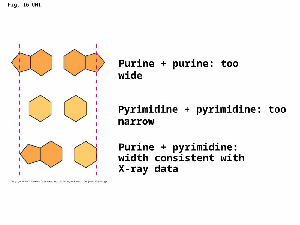
Fig. 16-UN1
Purine + purine: too wide
Pyrimidine + pyrimidine: too narrow
Purine + pyrimidine: width consistent with X-ray data
• Watson and Crick reasoned that the pairing was more specific, dictated by the base structures
• They determined that adenine (A) paired only with thymine (T), and guanine (G) paired only with cytosine (C)
• The Watson-Crick model explains Chargaff’s rules: in any organism the amount of A = T, and the amount of G = C
Copyright © 2008 Pearson Education Inc., publishing as Pearson Benjamin Cummings
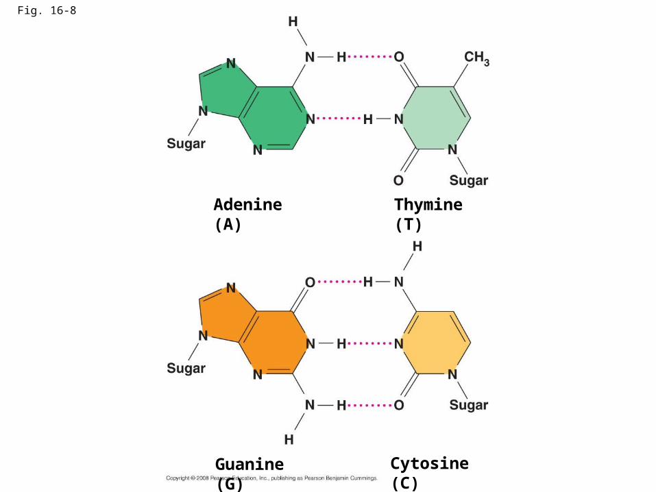
Fig. 16-8
Cytosine (C)
Adenine (A) Thymine (T)
Guanine (G)

Concept 16.2: Many proteins work together in DNA replication and repair
• The relationship between structure and function is manifest in the double helix
• Watson and Crick noted that the specific base pairing suggested a possible copying mechanism for genetic material
Copyright © 2008 Pearson Education Inc., publishing as Pearson Benjamin Cummings
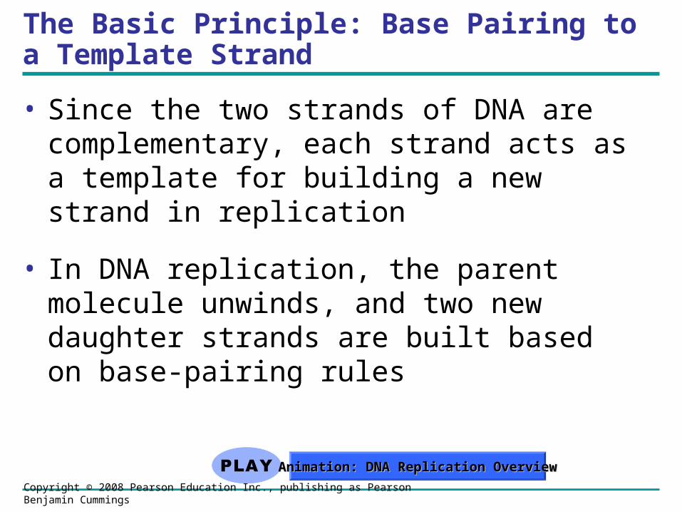
The Basic Principle: Base Pairing to a Template Strand
• Since the two strands of DNA are complementary, each strand acts as a template for building a new strand in replication
• In DNA replication, the parent molecule unwinds, and two new daughter strands are built based on base-pairing rules
Copyright © 2008 Pearson Education Inc., publishing as Pearson Benjamin Cummings
Animation: DNA Replication OverviewAnimation: DNA Replication Overview
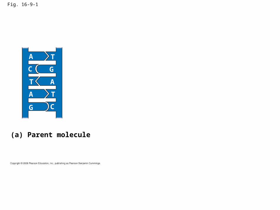
Fig. 16-9-1
A T
GC
T A
TA
G C
(a) Parent molecule

Fig. 16-9-2
A T
GC
T A
TA
G C
A T
GC
T A
TA
G C
(a) Parent molecule (b) Separation of strands
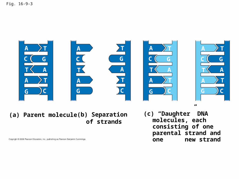
Fig. 16-9-3
A T
GC
T A
TA
G C
(a) Parent molecule
A T
GC
T A
TA
G C
(c) “Daughter” DNA molecules, each consisting of one parental strand and one new strand
(b) Separation of strands
A T
GC
T A
TA
G C
A T
GC
T A
TA
G C

DNA Replication: A Closer Look
• The copying of DNA is remarkable in its speed and accuracy
• More than a dozen enzymes and other proteins participate in DNA replication
Copyright © 2008 Pearson Education Inc., publishing as Pearson Benjamin Cummings

Getting Started
• Replication begins at special sites called origins of replication, where the two DNA strands are separated, opening up a replication “bubble”
• A eukaryotic chromosome may have hundreds or even thousands of origins of replication
• Replication proceeds in both directions from each origin, until the entire molecule is copied
Copyright © 2008 Pearson Education Inc., publishing as Pearson Benjamin Cummings
Animation: Origins of ReplicationAnimation: Origins of Replication

• At the end of each replication bubble is a replication fork, a Y-shaped region where new DNA strands are elongating
• Helicases are enzymes that untwist the double helix at the replication forks
• Single-strand binding protein binds to and stabilizes single-stranded DNA until it can be used as a template
• Topoisomerase corrects “overwinding” ahead of replication forks by breaking, swiveling, and rejoining DNA strands
Copyright © 2008 Pearson Education Inc., publishing as Pearson Benjamin Cummings
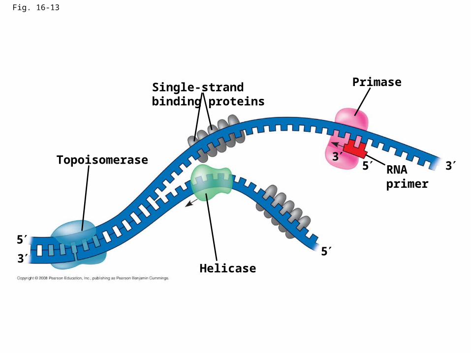
Fig. 16-13
Topoisomerase
Helicase
PrimaseSingle-strand binding proteins
RNA primer
55
5 3
3
3
• DNA polymerases cannot initiate synthesis of a polynucleotide; they can only add nucleotides to the 3 end
• The initial nucleotide strand is a short RNA primer
Copyright © 2008 Pearson Education Inc., publishing as Pearson Benjamin Cummings

• An enzyme called primase can start an RNA chain from scratch and adds RNA nucleotides one at a time using the parental DNA as a template
• The primer is short (5–10 nucleotides long), and the 3 end serves as the starting point for the new DNA strand
Copyright © 2008 Pearson Education Inc., publishing as Pearson Benjamin Cummings

Synthesizing a New DNA Strand
• Enzymes called DNA polymerases catalyze the elongation of new DNA at a replication fork
• Most DNA polymerases require a primer and a DNA template strand
• The rate of elongation is about 500 nucleotides per second in bacteria and 50 per second in human cells
Copyright © 2008 Pearson Education Inc., publishing as Pearson Benjamin Cummings
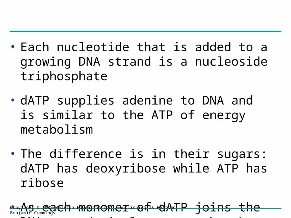
• Each nucleotide that is added to a growing DNA strand is a nucleoside triphosphate
• dATP supplies adenine to DNA and is similar to the ATP of energy metabolism
• The difference is in their sugars: dATP has deoxyribose while ATP has ribose
• As each monomer of dATP joins the DNA strand, it loses two phosphate groups as a molecule of pyrophosphate
Copyright © 2008 Pearson Education Inc., publishing as Pearson Benjamin Cummings
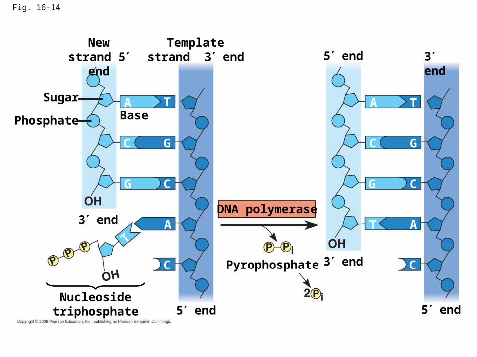
Fig. 16-14
A
C
T
G
G
G
GC
C C
C
C
A
A
AT
T
T
New strand 5 end
Template strand 3 end 5 end 3 end
3 end
5 end5 end
3 end
Base
Sugar
Phosphate
Nucleoside triphosphate
Pyrophosphate
DNA polymerase

Antiparallel Elongation
• The antiparallel structure of the double helix (two strands oriented in opposite directions) affects replication
• DNA polymerases add nucleotides only to the free 3end of a growing strand; therefore, a new DNA strand can elongate only in the 5to3direction
Copyright © 2008 Pearson Education Inc., publishing as Pearson Benjamin Cummings
• Along one template strand of DNA, the DNA polymerase synthesizes a leading strand continuously, moving toward the replication fork
Copyright © 2008 Pearson Education Inc., publishing as Pearson Benjamin Cummings
Animation: Leading StrandAnimation: Leading Strand
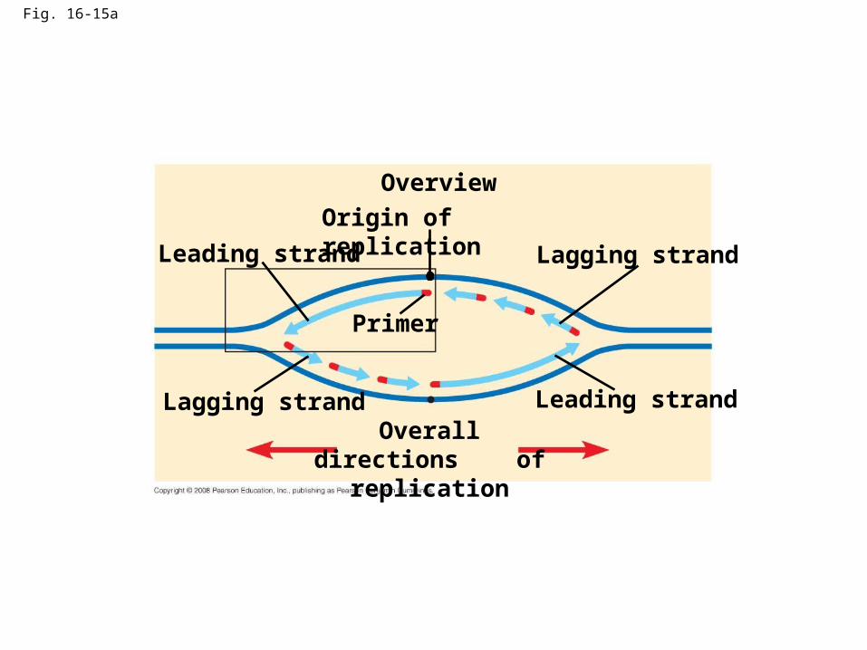
Fig. 16-15a
Overview
Leading strand
Leading strandLagging strand
Lagging strand
Origin of replication
Primer
Overall directions of replication

Fig. 16-15b
Origin of replication
RNA primer
“Sliding clamp”
DNA pol IIIParental DNA
3
5
5
5
5
5
5
3
3
3
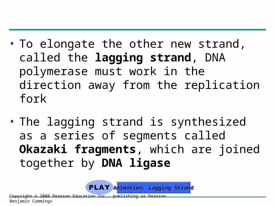
• To elongate the other new strand, called the lagging strand, DNA polymerase must work in the direction away from the replication fork
• The lagging strand is synthesized as a series of segments called Okazaki fragments, which are joined together by DNA ligase
Copyright © 2008 Pearson Education Inc., publishing as Pearson Benjamin Cummings
Animation: Lagging StrandAnimation: Lagging Strand
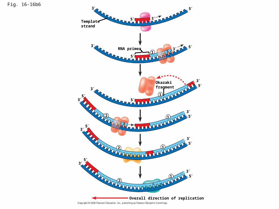
Fig. 16-16b6
Template strand
5
53
3
RNA primer 3 5
5
3
1
1
3
35
5
Okazaki fragment
12
3
3
5
5
12
3
3
5
5
12
5
5
3
3
Overall direction of replication

Table 16-1
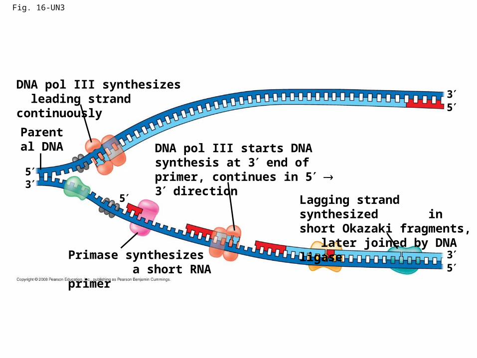
Fig. 16-UN3
DNA pol III synthesizes leading strand continuously
Parental DNA DNA pol III starts DNA
synthesis at 3 end of primer, continues in 5 3 direction
Lagging strand synthesized in short Okazaki fragments, later joined by DNA ligase
Primase synthesizes a short RNA primer
53
5
5
5
3
3
Related Documents

![FORCED EVOLUTION IN SILICO BY ARTIFICIAL ...are jumping genes, selfish DNA, transposons and retroelements [15, 27, 30, 31]. Many biologists speculate that Many biologists speculate](https://static.cupdf.com/doc/110x72/5ed6a1e0f8f40e7c16721bdf/forced-evolution-in-silico-by-artificial-are-jumping-genes-selfish-dna-transposons.jpg)