Fifty-gigahertz Microwave Exposure Effect of Radiations on Rat Brain Kavindra Kumar Kesari & J. Behari Received: 22 April 2008 / Accepted: 2 December 2008 / Published online: 17 December 2008 # Humana Press 2008 Abstract The object of this study is to investigate the effects of 50-GHz microwave radiation on the brain of Wistar rats. Male rats of the Wistar strain were used in the study. Animals of 60-day age were divided into two groups—group 1, sham-exposed, and group 2, experimental (microwave-exposed). The rats were housed in a temperature-controlled room (25 °C) with constant humidity (40–50%) and received food and water ad libitum. During exposure, rats were placed in Plexiglas cages with drilled ventilation holes and kept in an anechoic chamber. The animals were exposed for 2 h a day for 45 days continuously at a power level of 0.86 μW/cm 2 with nominal specific absorption rate 8.0×10 -4 w/kg. After the exposure period, the rats were killed and homogenized, and protein kinase C (PKC), DNA double-strand break, and antioxidant enzyme activity [superoxides dismutase (SOD), catalase, and glutathione peroxidase (GPx)] were estimated in the whole brain. Result shows that the chronic exposure to these radiations causes DNA double-strand break (head and tail length, intensity and tail migration) and a significant decrease in GPx and SOD activity (p =<0.05) in brain cells, whereas catalase activity shows significant increase in the exposed group of brain samples as compared with control (p =<0.001). In addition to these, PKC decreased significantly in whole brain and hippocampus (p <0.05). All data are expressed as mean ± standard deviation. We conclude that these radiations can have a significant effect on the whole brain. Keywords Glutathione peroxidase . Superoxidase . Catalase . Microwave radiation . Protein kinase C Introduction Microwaves may affect biological systems by increasing free radicals, which may enhance lipid peroxidation, and by changing the antioxidative activities of the brain cells, thus Appl Biochem Biotechnol (2009) 158:126–139 DOI 10.1007/s12010-008-8469-8 K. K. Kesari : J. Behari (*) Bioelectromagnetic Laboratory, School of Environmental Sciences, Jawaharlal Nehru University, New Delhi 110067, India e-mail: [email protected]

Welcome message from author
This document is posted to help you gain knowledge. Please leave a comment to let me know what you think about it! Share it to your friends and learn new things together.
Transcript
Fifty-gigahertz Microwave Exposure Effect of Radiationson Rat Brain
Kavindra Kumar Kesari & J. Behari
Received: 22 April 2008 /Accepted: 2 December 2008 /Published online: 17 December 2008# Humana Press 2008
Abstract The object of this study is to investigate the effects of 50-GHz microwaveradiation on the brain of Wistar rats. Male rats of the Wistar strain were used in the study.Animals of 60-day age were divided into two groups—group 1, sham-exposed, and group2, experimental (microwave-exposed). The rats were housed in a temperature-controlledroom (25 °C) with constant humidity (40–50%) and received food and water ad libitum.During exposure, rats were placed in Plexiglas cages with drilled ventilation holes and keptin an anechoic chamber. The animals were exposed for 2 h a day for 45 days continuouslyat a power level of 0.86 μW/cm2 with nominal specific absorption rate 8.0×10⁻⁴ w/kg.After the exposure period, the rats were killed and homogenized, and protein kinase C(PKC), DNA double-strand break, and antioxidant enzyme activity [superoxides dismutase(SOD), catalase, and glutathione peroxidase (GPx)] were estimated in the whole brain.Result shows that the chronic exposure to these radiations causes DNA double-strand break(head and tail length, intensity and tail migration) and a significant decrease in GPx andSOD activity (p=<0.05) in brain cells, whereas catalase activity shows significant increasein the exposed group of brain samples as compared with control (p=<0.001). In addition tothese, PKC decreased significantly in whole brain and hippocampus (p<0.05). All data areexpressed as mean ± standard deviation. We conclude that these radiations can have asignificant effect on the whole brain.
Keywords Glutathione peroxidase . Superoxidase . Catalase . Microwave radiation .
Protein kinase C
Introduction
Microwaves may affect biological systems by increasing free radicals, which may enhancelipid peroxidation, and by changing the antioxidative activities of the brain cells, thus
Appl Biochem Biotechnol (2009) 158:126–139DOI 10.1007/s12010-008-8469-8
K. K. Kesari : J. Behari (*)Bioelectromagnetic Laboratory, School of Environmental Sciences, Jawaharlal Nehru University,New Delhi 110067, Indiae-mail: [email protected]
leading to oxidative damage. The effect of microwave radiations on biological systems isprimarily identified as due to an increase in temperature, i.e., thermal [1], thoughnonthermal effects have also been identified [2, 3]. The present study was designed todetermine the effects of 50 GHz nonthermal microwave radiation on rat brain. Thisfrequency was chosen because 50 GHz has a very small penetration depth, which maycause an increase in the random molecular motion due to free-radical processes within thecell. So far, most of the investigations are confined to a frequency up to 10 GHz. Above-range frequencies will have limited penetration into the body, and their mode of interactionwith the system, including those at cellular and molecular levels, will be interesting toexamine. It will be of further interest to establish their commonality with after-exposureparameters at comparatively lower frequencies. The study also aims to spread over theelectromagnetic field (EMF) effect over a wider band of nonionizing electromagneticspectrum.
Protein kinase C (PKC) and antioxidant enzymes play an important role in functioningof the central nervous system. PKC plays a key role in a variety of pathologic states,including oncogenesis [4, 5]. This may affect the cellular responses to extracellular stimuliparticipated in cell differentiation and apoptosis [6]. PKC modulates ion conductance byphosphorylating membrane proteins such as channels, pumps, and ion exchange proteinsmobilization into the cytosol. This has been implicated in phosphorylation of severalneuronal proteins, which are thought to regulate neurotransmitter release and long-termpotentiation in memory formation [7]. The activation of this enzyme is thought to bebiochemically dependent on Ca2+. Tumor-promoting phorbol esters, such as 12,O-tetradecanoyl phorbol-13-acetate (TPA), have a structure very similar to diacylglyceroland activate PKC directly, both in vitro and in vivo [8]. TPA has a specific membranereceptor in the cell membrane [9, 10]. In order to stimulate cell proliferation in cells, growthfactor and PKC are needed to induce the signal pathways. Furthermore, tumor promotersTPA have a membrane receptor in the membrane of all cells. This receptor is considered tobe calcium phospholipid-dependent protein kinase (PKC). It is involved in the regulation ofa variety of cellular events, including modulation of receptor functions for major hormonesand certain enzymes such as adenylate cyclase and ornithine decarboxylase.
In the present investigation, PKC has been estimated in hippocampus, whole brain, andthe remaining portion. The hippocampus is understood to be responsible for learning andmemory. In addition to this, antioxidant enzyme estimation has also been performed. It iswell known that these enzymes play a major role in protecting the cells by removal of freeradicals, which are generated by microwave radiations. It has been established that theoverproduction of reactive oxygen species (ROS) comes through free radicals formationand may change the levels of superoxides dismutase (SOD), glutathione peroxidase (GPx),and catalase (CAT) activity in the whole brain. These parameters were undertaken todetermine the possible site of the EMF biointeraction with ROS. Very recently, Mahfouz etal. [11] reported that microwave/radiofrequency radiation may lead to oxidative stress dueto overproduction of ROS. Others workers, several from our laboratory, have shown thatthese radiations affect cholenergic systems, brain Na+–K+ ATPase activity [12, 13],growth-related enzymes [14], PKC activity [3], single-strand DNA breaks in brain cells [15,16], and reduced infertility in male rats [17].
In continuation, DNA double-strand breaks were observed and comet scoring wascarried out with tail and head-length migration and intensity. The first study of DNA strandbreak was observed by Lai and Singh [18] in rat brain cells at continuous and pulsed RFradiation. These field exposures induced the formation of DNA–protein and DNA–DNAcross-links in brain cells of rats [19], which could be the result of free-radical damage
Appl Biochem Biotechnol (2009) 158:126–139 127
involving iron cations [20, 21]. The results of Ivancsits et al. [22–25] indicate that theinteraction of these fields with DNA is quite complicated and apparently depends on manyfactors, such as the mode of exposure, the type of cells, and the intensity and duration ofexposure. Some researchers found conflicting results; some observed that there is no effectof radio frequency radiation exposure in DNA strand break of mammalian somatic cells[26, 27]. To support these data and to confirm their pathological implications, we havemeasured the activities of antioxidant enzymes, namely, SOD, CAT, and GPx and tumorpromoter PKC in rat brain cells.
Material and Methods
Material
The GPx (catalog No. 703102), CAT (catalog No. 707002), and SOD (CAT No. 706002)antioxidant enzyme kit was purchased from the Cayman Chemical Company, Ann Arbor,MI, USA. P32 radioactive labeled ATP was purchased from BRIT, Hyderabad, India. Therest of the chemicals were purchased from Thomas Baker Chemicals Limited, MarineDrive, Mumbai, India.
Animals Exposure
Male Wistar rats (60 days old and 190±20 g body weight) were obtained from the animalfacility of Jawaharlal Nehru University, New Delhi. They were divided into two groups, asham-exposed group (n=6) and an experimental group (n=6). In the animal exposure andsubsequent experimentation, a blind study was conducted, where the experimental andcontrol code was not disclosed before the data analysis. All animals were housed in an air-conditioned room, where the temperature was maintained at 25 °C with constant humidity(40–50%). The air circulation was constantly maintained to keep it in equilibrium with theroom temperature. The animals were provided with standard food pallets (prepared byBrook Bond India, Mumbai, India) and water ad libitum.
The protocol and study method was approved by the Institutional Animal EthicalCommittee and the Committee for Purpose of Control and Supervision of Experimental onAnimal.
Exposure Chamber
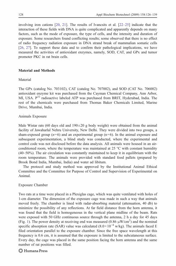
Two rats at a time were placed in a Plexiglas cage, which was quite ventilated with holes of1-cm diameter. The dimension of the exposure cage was made in such a way that animalsmoved freely. The chamber is lined with radar-absorbing material (attenuation, 40 db) tominimize the possibility of any reflections. At far field distance from the horn antenna, itwas found that the field is homogeneous in the vertical plane midline of the beam. Ratswere exposed with 50 GHz continuous source through the antenna, 2 h a day for 45 days(Fig. 1). The power density at receiving end was measured (0.86 μW/cm2) and the nominalspecific absorption rate (SAR) value was calculated (8.0×10⁻⁴ w/kg). The animals faced Efiled orientation parallel to the exposure chamber. Since the free space wavelength at thisfrequency is 0.6 cm, it is assumed that the exposure is limited to the subcutaneous surface.Every day, the cage was placed in the same position facing the horn antenna and the samenumber of rat positions was filled.
128 Appl Biochem Biotechnol (2009) 158:126–139
Sample Preparation and Tissue Homogenate
In the present investigations, enzyme assay was used to determine the enzyme activity(SOD, GPx, and CAT) in exposed Wistar rats. Immediately after exposure, animalswere killed by overdose of anesthesia and the brain was collected in ice-cold buffer.The brain was homogenized in cold buffer (50 mM Tris–HCl, pH 7.5, 5 mM EDTA,and 1 mM DTT per gram tissue) for GPx, 5–10 ml HEPES buffer for SOD, and(50 mM potassium phosphate, pH 7.0, containing 1 mM EDTA per gram tissue) forCAT. The sample was centrifuged at 10,000×g for 15 min at 4 °C. Supernatant wascollected and enzyme assay was performed. Different sets of animals with the same agegroup were taken for given parameters (DNA strand break, PKC, and antioxidantenzyme).
Estimation of GPx Activity
One hundred twenty microliters of assay buffer and 50 μl cosubstrate mixture were addedin nonenzymatic wells. One hundred microliters of assay buffer, 50 μl of cosubstratemixture, and 20 μl of diluted GPx were added in other wells as control samples, whereasthe same amount of assay buffer and cosubstrate including 20 μl of brain sample in place ofGPx were added in all the wells. Immediately, reaction was initiated by adding 20 μl ofcumene hydroperoxide to all the wells being used. Finally, the well plate was placed in amicroplate reader spectrophotometer, and the absorbances of the samples were taken at340 nm
Estimation of Superoxidase Activity
Twenty microliters of SOD standard was diluted with 1.98 ml of sample buffer. SODstandard wells were prepared by using 200 μl of the diluted radical detector and 10 μl ofdiluted standard. Sample wells were also prepared by adding 200 μl of the diluted radicaldetector and 10 μl of sample to the wells. The reaction was initiated by adding 20 μl ofdiluted xanthine oxidase to all the wells. The sample plate was kept in microplate readertemperature, and absorbance was taken at 450 nm.
Gunn Oscillator
Isolator
Variable Attenuator Horn
Anten
Animal Cage
Gunn Power Supply
Anaechoic material shield
Fig. 1 Schematic diagram of 50-GHz radiation source
Appl Biochem Biotechnol (2009) 158:126–139 129
Estimation of CAT Activity
One hundred microliters of assay buffer, 30 μl of methanol, and 20 μl of standard wereadded to wells, which contained 10 μl of formaldehyde and 9.99 ml of sample buffer andformaldehyde wells were prepared. Control wells were prepared by adding 100 μl ofdiluted assay buffer, 30 μl of methanol, and 20 μl of diluted CAT. Thirdly, the sample wellswere prepared by adding 100 μl of diluted assay buffer, 30 μl of methanol, and 20 μl oftissue samples. The reaction was initiated by adding 20 μl of diluted hydrogen peroxide toall the wells. Sample plate was incubated for 20 min at room temperature and 30 μl ofpotassium hydroxide was added to terminate the reaction. Thirty microliters of purpald(chromogen) was added to each well and thereafter incubated for 10 min at roomtemperature on a shaker. Ten microliters of potassium periodate was added to each well,incubated for 5 min at room temperature on shaker, and the absorbance of samples wastaken at 540 nm.
Calcium-dependent Protein Kinase (PKC) Assay
After 45 days of exposure, the animals were killed with rat cocktail anesthesia (Ketamine,Xylazine; IP) and then decapitated. Brain tissue was taken out from the cranial cavityimmediately and put into a deep freezer for a short while to become tissue hard. Thehippocampus was taken out and assays were performed in three sets as follows: (1)hippocampus, (2) whole brain minus hippocampus (remaining brain), (3) whole brain.
Each brain tissue was homogenized separately in 40 vol of ice-cold, 1-mM sodiumbicarbonate (pH 7.5). The homogenate was centrifuged at 600 g for 10 min at 4 °C. Thesupernatants were centrifuged at 20,000×g for 30 min at 4 °C. The pellet was pipetted withice-cold, 1-mM sodium bicarbonate and centrifuged at 20,000×g for 30 min at 4 °C. Thepellet was resuspended in incubation buffer (100 mM HEPES, 120 mM NaCl, 1.2 mMMgSO4, 2.5 mM KCl, 15 mM NaHCO3, 10 mM glucose, 1 mM EDTA, pH 7.4) andprotein concentration was measured by Lowry’s method [28]. Protein kinase activity wasassayed in a total volume of 0.5 ml incubation medium [50 mM HEPES, 10 mM MgCl2,0.5 mM CaCl2, and 0.2 mM EGTA (free calcium level of 0.1 mM), pH 7] with a totalprotein concentration of 100 μg. P32-labeled ATP (specific activity 3,000 Ci/mmol ATP)was added to initiate the reaction and then incubated at 25 °C. Fifty-microliter samples weretaken out and pipetted upon 3-mm filter discs (pretreated with 10% trichloroacetic acid,20 mM sodium pyrophosphate, and 10 mM EDTA). These filter discs were dropped into500 ml of the TCA mixture (10% trichloroacetic acid, 20 mM sodium pyrophosphate, and10 mM EDTA) and left overnight at 4 °C. Filters were washed once in 5% TCA, heated to90 °C for 15 min in 10% TCA. Furthermore, 5% TCA wash was extracted in hot ethanol/ether (3:1 v/v) before drying. Radioactivity was measured in a Hewlett Packard scintillationcounter
DNA Double-strand Breaks Estimation
In the present investigations, comet assay (also referred to as single-cell gel electrophoresis)is used to determine DNA damage (DNA strand break). Assay was performed according tothe technique of Singh [29]. Immediately after the exposure period, one rat at a time wasanesthetized by placing it in a glass jar containing cotton dipped in anesthetized ether.Animals were killed and brain was homogenized in phosphate buffer and a single-cellsuspension was made by using pipette. From the suspension, 10 μl of its suspension was
130 Appl Biochem Biotechnol (2009) 158:126–139
mixed with 0.2 ml, 0.7% agarose. Agarose was suspended in phosphate buffered salinewith 3:1 agarose higher resolution and kept at 37 °C to maintain physiological conditions[18]. The mixture was pipetted out and poured onto a fully frosted slide, immediatelycovered with coverglass (24×60 mm). These slides were kept in an ice-cold steel tray onice for 1 min to allow the agarose to gel. Again, a layer was made over the gel with 100 μlof agarose as before, after removing the coverglass [29, 30]. These slides were immersed inice-cold lysing solution with the addition of DNAase free proteinase K (0.5 mg/ml) andkept overnight at 4 °C. After lysing overnight, the slides were removed and placed in ahorizontal slab of an electrophoresis assembly. One liter of electrophoresis buffer wasgently poured into the assembly. After 20 min to allow for unwinding, electrophoresis wasstarted at 250 mA (12 V) for 30 min. The slides were removed from the electrophoresisapparatus and placed in coplin jar containing neutralizing buffer. After 30 min, the slideswere transferred to another jar of neutralizing solution. After one more change of 30 min,the slides were left vertical at room temperature to dry and stained with ethidium bromide(EtBr of 0.05 mg/ml) covered with a 24×60-mm coverglass. Microscopic slides wereprepared with each individual animal separately.
Images were taken at 100× magnification using a charge-coupled device camera GW525x(Genwac, Orangeburg, NY, USA) attached to Leica DMLB fluorescence microscope (Leica,Wetzlar, Germany) with an excitation filter of 490 nm, a 500-nm dichroic filter, and anemission filter of 515 nm. The images of double strand DNA break in brain cells wererecorded with fluorescence microscope.
Comet Scoring
Slides were assayed for double-strand DNA breaks. Twenty cells were selected from eachslide. Therefore, from each animal, 40 cells (two slides) were scored. Head and tail length(μm), intensity (%), and tail migration (μm) from the beginning of the nuclear area to thelast five pixels of DNA perpendicular to the direction of migration at the leading edge weremeasured. Tail and head length, migration, and intensity of individual cells were measured.The scoring of comet assay was done by using Comet assay IV 4.2 version software(Perceptive Instrument, Haverhill, UK). Data are presented as mean ± standard deviation.The difference between exposed and control groups was tested for significance byusing one-way analysis of variance (ANOVA). A difference at p<0.05 was consideredstatistically significant.
Results
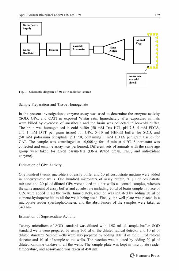
Antioxidant Enzymatic Activity in Brain Cells
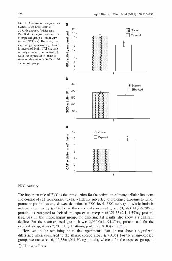
Compared with the control group (16.82±2.96), those exposed to the 50-GHz showeda significant decrease (12.66±0.87 nmol min−1 ml−1; p<0.001) in GPx activity (Fig. 2a).The exposed group also showed a significant decrease of SOD activity (169.09±15.34 U/ml; p<0.006) as compared to the control group (189.50±13.13) (Fig. 2b).However, the exposed group of animals showed a significant increase in CAT activity(9.81±1.60 nmol min−1 ml−1; p<0.001) as compared to the control group (6.86±0.76)(Fig. 2c). The results occulting significant changes occurring in the exposed group ofbrain cells in all antioxidants enzymatic activities of GPx SOD and CAT are shown inFig. 2a–c.
Appl Biochem Biotechnol (2009) 158:126–139 131
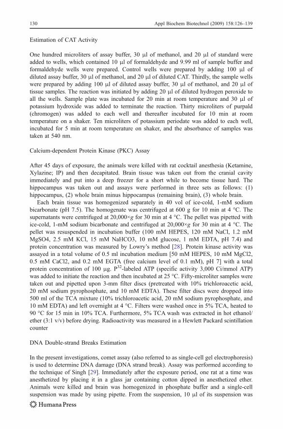
PKC Activity
The important role of PKC is the transduction for the activation of many cellular functionsand control of cell proliferation. Cells, which are subjected to prolonged exposure to tumorpromoter phorbol esters, showed depletion in PKC level. PKC activity in whole brain isreduced significantly (p<0.005) in the chronically exposed group (3,198.0±1,259.28/mgprotein), as compared to their sham exposed counterpart (6,321.33±2,141.55/mg protein)(Fig. 3a). In the hippocampus group, the experimental results also show a significantdecline. For the sham-exposed group, it was 3,990.0±1,494.27/mg protein, and for theexposed group, it was 2,703.0±1,213.46/mg protein (p<0.03) (Fig. 3b).
However, in the remaining brain, the experimental data do not show a significantdifference when compared to the sham-exposed group (p>0.05). For the sham-exposedgroup, we measured 6,455.33±4,061.20/mg protein, whereas for the exposed group, it
0
2
4
6
8
10
12
14
16
18
20
1
GP
x ac
tivi
ty n
mo
l/min
/ml Control
Exposed
*
0
50
100
150
200
250
1
SO
D a
ctiv
ity
U/m
l
Control
Exposed
*
0
2
4
6
8
10
12
1
CA
T a
ctiv
ity
nm
ol/m
in/m
l Control
Exposed *
a
b
c
Fig. 2 Antioxidant enzyme ac-tivities in rat brain cells in50 GHz exposed Wistar rats.Result shows significant decreasein exposed group of brain GPx(a) and SOD (b). However, theexposed group shows significant-ly increased brain CAT enzymeactivity compared to control (c).Data are expressed as mean ±standard deviation (SD). *p<0.05vs control group
132 Appl Biochem Biotechnol (2009) 158:126–139
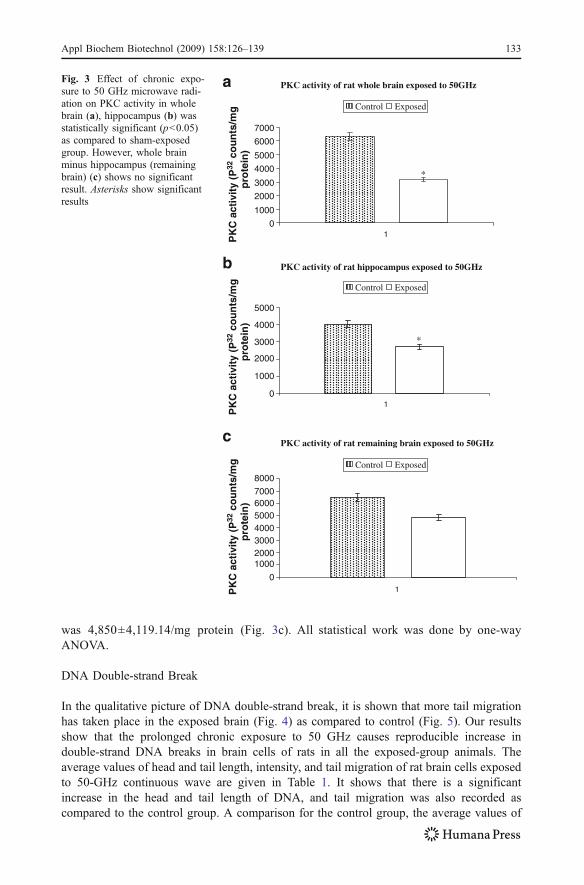
was 4,850±4,119.14/mg protein (Fig. 3c). All statistical work was done by one-wayANOVA.
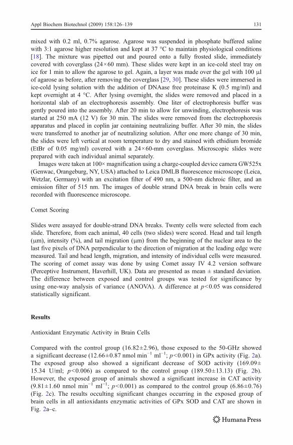
DNA Double-strand Break
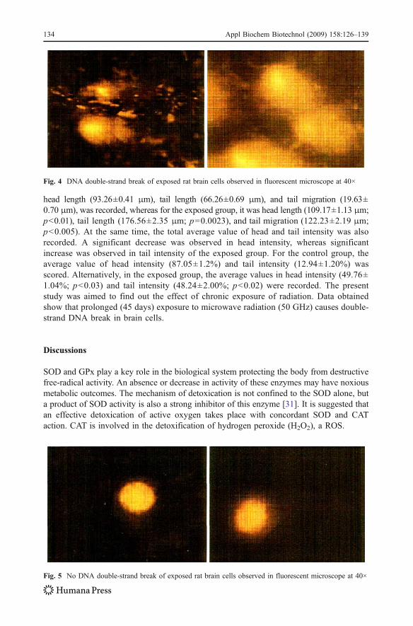
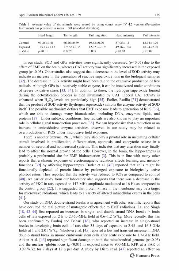
In the qualitative picture of DNA double-strand break, it is shown that more tail migrationhas taken place in the exposed brain (Fig. 4) as compared to control (Fig. 5). Our resultsshow that the prolonged chronic exposure to 50 GHz causes reproducible increase indouble-strand DNA breaks in brain cells of rats in all the exposed-group animals. Theaverage values of head and tail length, intensity, and tail migration of rat brain cells exposedto 50-GHz continuous wave are given in Table 1. It shows that there is a significantincrease in the head and tail length of DNA, and tail migration was also recorded ascompared to the control group. A comparison for the control group, the average values of
PKC activity of rat whole brain exposed to 50GHz
PKC activity of rat hippocampus exposed to 50GHz
PKC activity of rat remaining brain exposed to 50GHz
0
1000
2000
3000
4000
5000
6000
7000
0
1000200030004000500060007000
8000
0
1000
2000
3000
4000
5000
1PK
C a
ctiv
ity
(P32
co
un
ts/m
g
pro
tein
)P
KC
act
ivit
y (P
32 c
ou
nts
/mg
pro
tein
)P
KC
act
ivit
y (P
32 c
ou
nts
/mg
pro
tein
)
Control Exposed
1
1
*
*
Control Exposed
Control Exposed
a
b
c
Fig. 3 Effect of chronic expo-sure to 50 GHz microwave radi-ation on PKC activity in wholebrain (a), hippocampus (b) wasstatistically significant (p<0.05)as compared to sham-exposedgroup. However, whole brainminus hippocampus (remainingbrain) (c) shows no significantresult. Asterisks show significantresults
Appl Biochem Biotechnol (2009) 158:126–139 133
head length (93.26±0.41 μm), tail length (66.26±0.69 μm), and tail migration (19.63±0.70 μm), was recorded, whereas for the exposed group, it was head length (109.17±1.13 μm;p<0.01), tail length (176.56±2.35 μm; p=0.0023), and tail migration (122.23±2.19 μm;p<0.005). At the same time, the total average value of head and tail intensity was alsorecorded. A significant decrease was observed in head intensity, whereas significantincrease was observed in tail intensity of the exposed group. For the control group, theaverage value of head intensity (87.05±1.2%) and tail intensity (12.94±1.20%) wasscored. Alternatively, in the exposed group, the average values in head intensity (49.76±1.04%; p<0.03) and tail intensity (48.24±2.00%; p<0.02) were recorded. The presentstudy was aimed to find out the effect of chronic exposure of radiation. Data obtainedshow that prolonged (45 days) exposure to microwave radiation (50 GHz) causes double-strand DNA break in brain cells.
Discussions
SOD and GPx play a key role in the biological system protecting the body from destructivefree-radical activity. An absence or decrease in activity of these enzymes may have noxiousmetabolic outcomes. The mechanism of detoxication is not confined to the SOD alone, buta product of SOD activity is also a strong inhibitor of this enzyme [31]. It is suggested thatan effective detoxication of active oxygen takes place with concordant SOD and CATaction. CAT is involved in the detoxification of hydrogen peroxide (H2O2), a ROS.
Fig. 5 No DNA double-strand break of exposed rat brain cells observed in fluorescent microscope at 40×
Fig. 4 DNA double-strand break of exposed rat brain cells observed in fluorescent microscope at 40×
134 Appl Biochem Biotechnol (2009) 158:126–139
In our study, SOD and GPx activities were significantly decreased (p<0.05) due to theeffect of EMF on the brain, whereas CAT activity was significantly increased in the exposedgroup (p<0.05). Other studies also suggest that a decrease in the level of SOD activity mayindicate an increase in the generation of reactive superoxide ions in the biological samples[32]. The decrease in GPx activity might have been due to the excessive production of freeradicals. Although GPx is a relatively stable enzyme, it can be inactivated under conditionsof severe oxidative stress [33, 34]. In addition to these, the hydrogen superoxide formedduring the detoxification process is then illuminated by CAT. Indeed CAT activity isenhanced when H2O2 levels are particularly high [35]. Earlier, Rotilio [31] demonstratedthat the product of SOD activity (hydrogen superoxide) inhibits the enzyme activity of SODitself. The possible mechanism defines that EMF exposure leads to generation of ROS [36],which are able to damage many biomolecules, including DNA, enzymes, lipids, andproteins [37]. Under subtoxic conditions, free radicals are also known to play an importantrole in cellular signal transduction processes [38]. We can hypothesize that a reduction or anincrease in antioxidative enzyme activities observed in our study may be related tooverproduction of ROS under microwave field exposure.
There is another enzyme, PKC, which may also play a pivotal role in mediating cellularstimuli involved in proliferation, differentiation, apoptosis, and exocytotic release in anumber of neuronal and nonneuronal systems. This indicates that any alteration may finallylead to affect the normal growth of the cells. However, in the brain, the hippocampus isprobably a preferential site for EMF biointeraction [3]. This is in line with many otherreports that a chronic exposure of electromagnetic radiation affects learning and memoryfunctions [39] by affecting hippocampus. Butler et al. [40] reported that cells might befunctionally depleted of protein kinase by prolonged exposure to biologically activephorbol esters. They reported that the activity was reduced to 92% as compared to control[40]. An earlier study from our laboratory also suggests that there was a decrease in theactivity of PKC in rats exposed to 147-MHz amplitude-modulated at 16 Hz as compared tothe control group [22]. It is suggested that protein kinase in the membrane may be a targetfor microwave radiations, which leads to a variety of altered intracellular events in the cells[41].
Our study on DNA double-strand breaks is in agreement with other scientific reports thathave occulted the real picture of mutagenic effects due to EMF radiations. Lai and Singh[18, 42–44] first reported an increases in single- and double-strand DNA breaks in braincells of rats exposed for 2 h to 2,450-MHz field at 0.6–1.2 W/kg. More recently, this hasbeen confirmed by Paulraj and Behari [16], who reported an increase in single-strandbreaks in developing brain cells of rats after 35 days of exposure to 2.45- and 16.5-GHzfields at 1 and 2.01 W/kg. Nikolova et al. [45] reported a low and transient increase in DNAdouble-strand break in mouse embryonic stem cells after acute exposure to 1.7-GHz field.Aitken et al. [46] reported significant damage to both the mitochondrial genome (p<0.05)and the nuclear -globin locus (p<0.01) in exposed mice to 900-MHz RFR at a SAR of0.09 W/kg for 7 days at 12 h per day. A study by Diem et al. [47] reported an effect on
Table 1 Average value of six animals were scored by using comet assay IV 4.2 version (PerceptiveInstrument) has presented in mean ± SD (standard deviation).
Head length Tail length Tail migration Head intensity Tail intensity
Control 93.26±0.41 66.26±0.69 19.63±0.70 87.05±1.2 12.94±1.20Exposed 109.17±1.13 176.56±2.35 122.23±2.19 49.76±1.04 48.24±2.00p Value p<0.01 0.0023 0.005 p<0.03 p<0.02
Appl Biochem Biotechnol (2009) 158:126–139 135
exposed human fibroblasts and rat granulosa cells to mobile phone signal (1,800 MHz;SAR 1.2 or 2 W/kg; during 4, 16, and 24 h) and suggested that the exposure may induceDNA single- and double-strand breaks as measured by the comet assay.
Several reports suggested that DNA damage in cells could have an important implicationon human health because they are cumulative. Normally, DNA is capable of repairing itselfefficiently through a homeostatic mechanism, whereby cells maintain a delicate balancebetween spontaneous and induced DNA damage. DNA damage accumulates if such abalance is altered. Most cells have considerable ability to repair DNA single-strand breaks;for example, some cells can repair as many as 200,000 breaks in 1 h. However, DNAdouble-strand breaks, if not properly repaired, are known to lead to cell death or apoptosis.Indeed, we have observed an increase in apoptosis and decrease in sperm count [26, 48, 49]and DNA double-strand break in sperm and brain cells exposed to microwave RFR [17, 27,42, 44].
The correlation between oxidative damage to cells (DNA strand break, antioxidantenzyme, and PKC level changes) and ROS has been established in the present paper. ROSare free radicals, which may play a role in mechanisms of the biological effects induced byelectromagnetic radiation. It is suggested that the outcome of oxidative damage induced byEMFs will therefore depend on various factors, including the oxidative status of the cell,capability of endogenous antioxidation enzymes, and processes to counteract free radicalbuildup, availability of exogenous antioxidants, iron homeostasis (a balance of iron influx,storage, and use), the parameters of exposure (e.g., intensity and duration of exposure and,possibly, the wave shape), and whether the oxidative damage is cumulative. In aerobiccells, ROS are generated as a by-product of normal mitochondrial activity. If not properlycontrolled, ROS can cause severe damage to cellular macromolecules, especially DNA andantioxidative enzyme. DNA damage and alteration in enzyme activities are clear indicationsof tumor promotion.
Conclusion
We conclude that prolonged exposure to 50 GHz field radiation may decrease the level ofPKC, cause the DNA double-strand break, and also make the changes in antioxidantenzymes in neurological system of male rats due to free radicals formation. It also confirmsthat the possible site of action of such radiation is the hippocampus, the region responsiblefor control of learning and memory in the brain.
Acknowledgment Authors are thankful to Council for Scientific and Industrial Research (CSIR), NewDelhi, for financial assistance.
References
1. Stuchley, M. A. (1988). Biological effects of radiofrequency fields. In M. H. Repacholi (Ed.), Non-Ionizing Radiations, Physical characterization, Biological effects and Health Hazard Assessment.Proceeding for the International Non-Ionizing Radiation Workshop. Melbourne, pp. 197–217.
2. Kunjilwar, K. K., & Behari, J. (1993). Effect of amplitude-modulated radio frequency radiation oncholinergic system of developing rats. Brain Research, 601, 321–324. doi:10.1016/0006-8993(93)91729-C.
3. Paulraj, R., & Behari, J. (2004). Radiofrequency radiation effect on protein kinase C activity in ratsbrain. Mutation Research, 585, 127–131. doi:10.1016/S0027-5107(03)00113-1.
136 Appl Biochem Biotechnol (2009) 158:126–139
4. Harvey, C., & French, P. W. (2000). Effects on protein kinase C and gene expression in a human mastcell line, HMC-1, following microwave exposure. Cell Biology International, 23, 739–748. doi:10.1006/cbir.1999.0436.
5. Nishizuka, Y. (1986). Studies and perspectives of protein kinase C. Science, 233, 305–312. doi:10.1126/science.3014651.
6. Ohkusu, K., Isobe, K., Hidaka, H., & Nakashima, I. (1986). Elucidation of the protein kinase C-dependent apoptosis pathway in distinct of T lymphocytes in MRL-lpr/lpr mice. European Journal ofImmunology, 25, 3180–3186. doi:10.1002/eji.1830251129.
7. Suzuki, T. (1994). Protein kinase involved in the expression of long-term potentiation. The InternationalJournal of Biochemistry, 26, 735–744. doi:10.1016/0020-711X(94)90102-3.
8. Castagna, M., Takai, Y., Kaibuchi, K., Sano, K., Kikkawa, U., & Nishizuka, Y. (1982). Direct activationof calcium-activated, phospholipid-dependent protein kinase by tumorpromoting phorbol esters. TheJournal of Biological Chemistry, 78, 47–51.
9. Hunter, T., Ling, N., & Cooper, J. A. (1984). Protein kinase C phosphorylation of the EGF receptor at athreonine residue close to the cytoplasmic face of the plasma membrane. Nature, 311, 480–483.doi:10.1038/311480a0.
10. Niedel, J. E., Kuhn, L. J., & Vandenbark, G. R. (1983). Phorbol diester receptor copurifies with proteinkinase C. Proceedings of the National Academy of Sciences of the United States of America, 80, 36–40.doi:10.1073/pnas.80.1.36.
11. Mahfouz, R., Sharma, R., Lackner, J., Aziz, A., & Agarwal, A. (2008). Evaluation of chemiluminescenceand flow cytometry as tools in assessing production of hydrogen peroxide and superoxide anion inhuman spermatozoa. Fertility and Sterility, in press. doi:10.1016/j.fertnstert.2008.05.087.
12. Behari, J., Kunjilwar, K. K., & Pyne, S. (1998). Interaction of low level modulated RF radiation withNa+–K+–ATPase. Bioelectrochemistry and Bioenergetics, 47, 247–252. doi:10.1016/S0302-4598(98)00195-0.
13. Paulraj, R., Behari, J., & Rao, A. R. (1999). Effect of 112 MHz amplitude modulated radiation oncalcium ion efflux and ODC activity in chronically exposed rat brain. Indian Journal of Biochemistry &Biophysics, 36, 337–340.
14. Paulraj, R., & Behari, J. (2002). The effect of low level continuous 2.45 GHz wave on brain enzymes ofdeveloping rat brain. Electromagnetic Biology and Medicine, 21, 231–241. doi:10.1081/JBC-120015993.
15. Sarkar, S., Ali, S., & Behari, J. (1994). Effect of low power microwave on the mouse genome: a directDNA analysis. Mutation Research, 320, 141–147. doi:10.1016/0165-1218(94)90066-3.
16. Paulraj, R., & Behari, J. (2006). Single strand DNA breaks in rat brain cells exposed to microwaveradiation. Mutation Research, 596, 76–80. doi:10.1016/j.mrfmmm.2005.12.006.
17. Behari, J., & Kesari, K. K. (2006). Effects of microwave radiations on reproductive system of male rats.Embryo Talk, 1, 81–85.
18. Lai, H., & Singh, N. P. (1996). Double strand breaks in rats brain cells after acute exposure to radiofrequency electromagnetic radiation. International Journal of Radiation Biology, 69, 513–521.doi:10.1080/095530096145814.
19. Singh, N. P., & Lai, H. (1998). 60-Hz magnetic field exposure induces DNA crosslinks in rat brain cells.Mutation Research, 400, 313–320. doi:10.1016/S0027-5107(98)00017-7.
20. Altman, S. A., Zastawny, T. H., Randers-Eichhorn, L., Cacciuttolo, M. A., Akman, S. A., & Dizdaroglu,M. (1995). Formation of DNAprotein cross-links in cultured mammalian cells upon treatment with ironions. Free Radical Biology & Medicine, 19, 897–902. doi:10.1016/0891-5849(95)00095-F.
21. Lloyd, D. R., Phillips, D. W., & Carmichael, P. L. (1997). Generation of putative intrastrand cross-linksand strand breaks by transition metal ion-mediated oxygen radiacl ttack. Chemical Research inToxicology, 10, 393–400. doi:10.1021/tx960158q.
22. Ivancsits, S., Diem, E., Pilger, A., Rudiger, H. W., & Jahn, O. (2002). Induction of DNA strand breaksby intermittent exposure to extremely-low-frequency electromagnetic fields in human diploid fibroblasts.Mutation Research, 519, 1–13.
23. Ivancsits, S., Diem, E., Jahn, O., & Rudiger, H. W. (2003a). Intermittent extremely low frequencyelectromagnetic fields cause DNA damage in a dose-dependent way. International Archives ofOccupational and Environmental Health, 76, 431–436. doi:10.1007/s00420-003-0446-5.
24. Ivancsits, S., Diem, E., Jahn, O., & Rudiger, H. W. (2003b). Age-related effects on induction of DNAstrand breaks by intermittent exposure to electromagnetic fields. Mechanism of Ageing and Development,124, 847–850.
25. Suzuki, T. (1994). Protein kinase involved in the expression of long-term potentiation. The InternationalJournal of Biochemistry, 26, 735–744.
Appl Biochem Biotechnol (2009) 158:126–139 137
26. Sakuma, N., Komatsubara, Y., Takeda, H., Hirose, H., Sekijima, M., Nojima, T., & Miyakoshi, J. (2006).DNA strand breaks are not induced in human cells exposed to 2.1425 GHz band CW and W-CDMAmodulated radiofrequency fields allocated to mobile radio base stations. Bioelectromagnetics, 27, 51–57.doi:10.1002/bem.20179.
27. Vijayalaxmi, , & Obe, G. (2004). Controversial cytogenetic observations in mammalian somatic cellsexposed to radiofrequency radiation. Radiation Research, 162, 481–496.
28. Lowry, O. H., Resebrough, N. J., Farr, A. L., & Randall, R. J. (1951). Protein measurement with folin-phenol reagent. The Journal of Biological Chemistry, 193, 265–275.
29. Singh, N. (2003). In R. Blumenthal (Ed.), Apoptosis by DNA diffusion assay, methods in molecularmedicine (chemosensitivity). Totowas: Humana.
30. Tice, R. R., Agurell, E., Anderson, D., Burlinson, B., Hartmann, A., Kobayashi, H., et al. (2000). Singlecell gel/comet assay; guidelines for in vitro and in vivo genetic toxicology testing. Environmental andMolecular Mutagenesis, 35, 206–221. doi:10.1002/(SICI)1098-2280(2000)35:3<206::AID-EM8>3.0.CO;2-J.
31. Rotilio, G. (1972). Effect of hydrogen peroxide on dismutase and catalase activity in rat liver.Biochemichistry, 11, 2187–2189.
32. Alvarez, J. G., Touchstone, C. J., Blasco, L., & Storey, B. T. (1987). Spontaneous lipid peroxidation andproduction of hydrogen peroxide and superoxide in human spermatozoa. SOD as major enzymeprotectant against oxygen toxicity. Journal of Andrology, 8, 33–89.
33. Condell, R. A., & Tappel, A. L. (1993). Evidence for suitability of glutathione peroxidase as a protectiveenzyme: studies of oxidative damage, restoration and proteolysis. Archives of Biochemistry andBiophysics, 223, 407. doi:10.1016/0003-9861(83)90604-5.
34. Reiter, R. J. (1997). Melatonin aspects of exposure to low frequency electric and magnetic fields.Advances in electromagnetic fields in living systems (vol. 2, (pp. 1–27)). New York: Plenum.
35. Jones, D. P., Eklow, L., Thor, H., & Orrenius, S. (1981). Metabolism of hydrogen peroxide in isolatedhepatocytes: relative contributions of catalase and glutathione peroxidase in decomposition ofendogenously generated H2O2 Arch. Biochemistry & Biophysics, 210, 505–516. doi:10.1016/0003-9861(81)90215-0.
36. Chen, G., Upham, B. L., & Sun, W. (2000). Effect of electromagnetic field exposure on chemicallyinduced differentiation of friend erythroleukemia cells. Environmental Health Perspectives, 108, 967–972. doi:10.2307/3435056.
37. Brydon, L., Petit, L., Delagrange, P., Strosberg, A. D., & Jockers, R. (2000). Functional expression ofMT2 (Mel 1b) melatonin receptors in human PAZ6 adipocytes. Endocrinology, 142, 4264–4271.doi:10.1210/en.142.10.4264.
38. Suzuki, Y. J., Forman, H. J., & Sevanian, A. (1997). Oxidants as stimulators of signal transduction. FreeRadical Biology & Medicine, 22, 269–285. doi:10.1016/S0891-5849(96)00275-4.
39. Rodnight, R., Grower, H., Martinez-Millan, L., & DeSouza, D. (1982). Molecular aspects of neuraltransmission, learning and memory. In R. Caputto, & C. Ajmone-Marsan (Eds.), IBRO, vol. 10 (p. 125).New York: Raven.
40. Butler, A. P., Mar, P. K., McDonald, F. F., & Ramsay, R. L. (1991). Involvement of protein kinase C inthe regulation of ornithine decarboxylase mRNA by phorbol esters in rat hepatoma cells. ExperimentalCell Research, 194, 56–61. doi:10.1016/0014-4827(91)90129-I.
41. Byus, C. V., Kartun, K., Pieper, S. E., & Adey, W. R. (1988). Increased ornithine decarboxylase activityin cultured cells exposed to low energy modulated microwave fields and phorbol ester tumor promoters.Cancer Research, 48, 4222–4226.
42. Lai, H., & Singh, N. P. (1995). Acute low-intensity microwave exposure increases DNA single-strandbreaks in rat brain cells. Bioelectromagnetics, 16, 207–210. doi:10.1002/bem.2250160309.
43. Lai, H., & Singh, N. P. (2005). Interaction of microwaves and a temporally incoherent magnetic field onsingle and double DNA strand breaks in rat brain cells. Electromagnetic Biology and Medicine, 24, 23–29. doi:10.1081/JBC-200055046.
44. Lai, H., & Singh, N. P. (1997a). Acute exposure to a 60-Hz magnetic field increases DNA strand breaksin rat brain cells. Bioelectromagnetics, 18, 156–165. doi:10.1002/(SICI)1521-186X(1997)18:2<156::AID-BEM8>3.0.CO;2-1.
45. Nikolova, T., Czyz, J., Rolletschek, A., Blyszczuk, P., Fuchs, J., Jovtchev, G., et al. (2005).Electromagnetic fields affect transcript levels of apoptosis-related genes in embryonic stem cell-derivedneural progenitor cells. ASEB J, 19(12), 1686–1688.
46. Aitken, R. J., Bennetts, L. E., & Sawyer, D. (2005). Impact of radio frequency electromagnetic radiationon DNA integrity in the male germline. International Journal of Andrology, 28, 171–179. doi:10.1111/j.1365-2605.2005.00531.x.
138 Appl Biochem Biotechnol (2009) 158:126–139
47. Diem, E., Schwarz, C., Adlkofer, F., Jahn, O., & Rudiger, H. (200). Non-thermal DNA breakage bymobile-phone radiation. (1800). MHz) in human fibroblasts and in transformed GFSH-R17 rat granulosacells in vitro. Mutation Research, 583(2), 178–183.
48. Agarwal, A., Deepinder, F., & Sharma, R. K. (2007). Effect of cell phone usage on semen analysisin men attending infertility clinic: An observational study. Fertility and Sterility doi:10.1016/j.fertnstert.2007.01.166.
49. Agarwal, A., Deepinder, F., Sharma, R. K., Ranga, G., & Li, J. (2008). Effect of cell phone usage onsemen analysis in men attending infertility clinic: an observational study. Fertility and Sterility, 89(1),124–128. doi:10.1016/j.fertnstert.2007.01.166.
Appl Biochem Biotechnol (2009) 158:126–139 139
Related Documents