University of Nebraska Medical Center University of Nebraska Medical Center DigitalCommons@UNMC DigitalCommons@UNMC MD Theses Special Collections 5-1-1942 Fibrotic thickening of the ligamentum flavum Fibrotic thickening of the ligamentum flavum Wilbur W. Overmiller University of Nebraska Medical Center This manuscript is historical in nature and may not reflect current medical research and practice. Search PubMed for current research. Follow this and additional works at: https://digitalcommons.unmc.edu/mdtheses Part of the Medical Education Commons Recommended Citation Recommended Citation Overmiller, Wilbur W., "Fibrotic thickening of the ligamentum flavum" (1942). MD Theses. 941. https://digitalcommons.unmc.edu/mdtheses/941 This Thesis is brought to you for free and open access by the Special Collections at DigitalCommons@UNMC. It has been accepted for inclusion in MD Theses by an authorized administrator of DigitalCommons@UNMC. For more information, please contact [email protected].

Welcome message from author
This document is posted to help you gain knowledge. Please leave a comment to let me know what you think about it! Share it to your friends and learn new things together.
Transcript

University of Nebraska Medical Center University of Nebraska Medical Center
DigitalCommons@UNMC DigitalCommons@UNMC
MD Theses Special Collections
5-1-1942
Fibrotic thickening of the ligamentum flavum Fibrotic thickening of the ligamentum flavum
Wilbur W. Overmiller University of Nebraska Medical Center
This manuscript is historical in nature and may not reflect current medical research and
practice. Search PubMed for current research.
Follow this and additional works at: https://digitalcommons.unmc.edu/mdtheses
Part of the Medical Education Commons
Recommended Citation Recommended Citation Overmiller, Wilbur W., "Fibrotic thickening of the ligamentum flavum" (1942). MD Theses. 941. https://digitalcommons.unmc.edu/mdtheses/941
This Thesis is brought to you for free and open access by the Special Collections at DigitalCommons@UNMC. It has been accepted for inclusion in MD Theses by an authorized administrator of DigitalCommons@UNMC. For more information, please contact [email protected].
FIBROTIC TfilC!CENING
OF THE LIG.AMENTU1! FLAVUM
******
by
Wilbur c. Overmiller
Senior Thesis
******
Presented to the College or Medicine
University of Nebraska, Omaha
1942
··-· -<:---:.·:�-..: ·-.· - -. . ,;;.,
.-.
•. J
_t___,
TABLE OF CONTENTS
Introduction • • • • • . • • • • • • • 1
History • • • • • • • • • • • • • • • . 2
Incidence • • • • . • • • • • • . • • • 6
Anatoll\Y' • • • • • • • • • • • • • . • • 8
Pathology • • • • • • • • • • • • • • • 13
Etiology • • • • • • • • • • • • • • • • 20
Symptomatology • • • . . . . . . • 23
Findings • • • • • • • • • • • • • • • • 28
Differential Diagnosis • • • • • • • • • 38
Treatment
Prognosis
Summary
• • • • • • • • • • • • • • •
• • • • • • • • • • • • • • •
• • • • • • • • • • • • • • • •
39
41
42
Bibliograp}zy' • • • • • • • • • • • • • • 43
INTRODUCTIO:tf
The study of low back-ache with or without sciatica
has for many years aroused much interest, but it was not until
recently that attention has been focused on intraspinous lesions
of the intervertebral disk (with its nucleus pulposus) and of
the ligamentum fla.vur:i. In this country, although protrusion or
herniation of the intervertebra.l disk into the vertebral canal
is now being recognized quite often, little attention has yet
been paid to abnormalities of the ligamentum flavum and it has
been and is being recognized rarely. Fibrotic thickening,
hyperla.sia., or, as it is usually called hypertropll'J of the
ligamentum flavum, with compression of the cauda equina is a
condition that must be suspected in low back-ache and/or sciatica
not responding to ordinary forms of therapy, and its often co
incidental relationship to herniated intervertebra.l disk is one
that must be realized.
- 1 -
HISTORY
The earliest mention of pathology of the ligamentum flavum
was made by Elsberg (1) in 1913. A woman forty-nine years of age
had been thrown out of an automobile ten months previously. She
had had stiffness of the back since that time. Six weeks before the
operation she had pain in the distribution of the fourth lumbar root
on the left side, rapidly growing very severe. Old fracture and thick
ening of the arches of the fourth and fifth lumbar vertebrae was demon
strated by x-ray. Upon operation the swollen ligament presented in the
wound as soon as the spines and laminae had been removed. This mass
was about two cm. t�ick and the manner in which it was forced out show
ed that there must have been considerable pressure in the canal. The
mass was removed. Examination of the mass proved it to be a much
thickened ligarnentum flavum which had evidently been torn loose at the
time of the accident some ten months before. The patient made a com
plete recovery. Elsberg (2, J), in 1916 and 1941 mentioned briefly two
cases of enlarged ligamentum flavum.
In 1931, Towne and Reichert (4) reported two cases of en
largment of the ligamentum flavum without antecedent trauma. One pair
of the ligaments had blocked the circulation of the cerebral spinal
fluid. Upon the removal of the ligaments a marked indentation ot the
dura was found, which was taken to indicate long continued pressure
upon it.
An article by Puusepp (5) appeared in the Esthonian litera
- 2 -
ture in 1932, reporting three cases of enlarged ligamentum flavum
in two of which trauma might have been a Tactor.
Abbott (6) in 1936, reported the case of a woman who had,
in getting out of� car, tripped and fallen, lighting on her left
buttock and suffering a severe contusion. Upon operation an en
larged ligamentum flavum was found. Its removal was followed by
recovery.
In 1937, Spurling, Mayfield, and Rogers (7) reported seven
cases of enlarged ligamentum flavum.. The symptoms of the seven cases
were present from three .months to two years. In six of the cases
there had been one or more periods during which there had been a re
cession of symptoms. Each of the patien�s had been incapacitated for
at least three months prior to the operation. The chief complaint by
all was low back pain which radiated into one extremity in six cases
and into both extremities in one. There was complete sexual impotence
in three patients. Five patients noted sensory loss in the lower
lumbar and sacral segments. In only one was there urinary incontin
ence. N!uscle weakness in one leg was described by two patients and in
two others weakness in both legs was noticed. There was a postural
deformity in all patients. The achilles tendon reflex was abolished
or diminished in six of the seven cases. Trauma appeared to be the
etiological agent in each case.
In the same year, 1937, Brown (8) reported seven cases. Six
of these gave a history of trauma. The patient usually described a
snapping sensation in the spine followed by pain low in the back and
- 3 -
I
\
some time later varying from a few hours to several months, extending
into the thigh, leg and toot. This injury usually occurred during
the act or lifting in association with flexion and torsion ot' the spine.
Recovery followed operation in all seven ot' these cases.
Meredith and Lehman (9) in 1938, reported four cases or
enlarged ligamentum flavum. In one or these oases the location of
the lesion was unique inasmuch as it was in the thoracic region.
while all other cases reported have had lesions occurring in the lumbar
or lum.bosaoral region. These authors do not stress di reot trauma as a
cause or this condition ai!d ! state that iri · two or their oases there was
no history of trauma.
Naftaiger, Inman �d Saunders (10) in 1938, wrote a very
comprehensive article on both the intervertebral disk and the liga
m.entum t'lavum •. They showed�hat it is quite possible tor any S1f8lling
or the ligamentum tlavum, p,-rtioularly in the lumbar region, to cause
pressure on the nerve roots� These men were the first to stre•• the
pathology of the enlarged ligamentum flavum and its relation to trauma.
as an eti ologioal agent. They demonstrated that the enlargement was
due to a fibrosis or the totn ligaments.
In 1939, Spurling!and Bradford (11) reported thirteen cases . '
in seven of which there was , an antecedent hi story of trauma. In this
same year TiDllnes reported ome case in which th,re · appeared to be no
antecedent trauma. Morton.;also in 1939, reported three oases, all of I
which appeared to be the difeot result of trauma.
In 1939, Craig (l*) emphasized for the ti rst time the great
- 4 ...
.·,
·i11I
'1 "'.:!
'.j
- .]!
frequency with which both enlarged ligamentum flavum and herniated
disk are found in the same patient when he reported that in his last
one hundred and seventy-five oases of posterior protrusion of the
intervertebral disks. enlargement of the liga.mentum flavum
occurred in one hundred and fifty-five. Love (23) further emphasized
this when in reporting a consecutive series of one hundred and seventy
five oases of protruded disks, he made a note in the surgical cards of
one hundred and fifty-five oases that there was a definite abnormal
thickening or f1¥pertrop}w of the contiguous ligamentum flavum.
Horwitz (13) in 1939. reported the study of the spines in
seventy-five women cadavers. From his study he determined the normal
anatomical variations in the thickness of the ligamentum flavum.
Dockerty and Love (14) in 1940• studied ligamenta flava from autopsy
and operative specimens to further substantiate the work done by
Horwitz {13).
- 5 -
INCIDENCE
A survey of the literature seems to show that fibrotic
thickening of the ligamentum flavum is on the increase, but this is
probably not true. Tne condition is being recognized more frequently
and with rrreater certainty than before and thete has consequently been
an ever increasing number of cases reported. Thickening of the liga
mentum flavum is more common among persons doing hard manual labor
than it is among those persons who lead a sedentary life. The period
between 16 and 50 years of age is probably the most active time in an
individual's life span. This period of Great activity is reflected
in the age incidence of the occurrence of thickened ligamentum flavum.
The earliest case of ligamentum flavum thickening reported was in a
16 year old boy (11), while the oldest patient who has been operated
on for this condition was 59 (11). It is, however, in the fourth and
fifth decades of life that the condition occurs most frequently, and
statistics agree that men are affected much more freauently than
women (1, 2, 4, 5, 6, 7, 9, 17, 20, 21, 22, 11, 18, 19): of 53 cases
reported and summarized, 45 (85 per cent) occurred in men and 8 (15
per cent) occurred in women.
In determining the frequency with which fibrotic thickening
or the liga.�entum flavum occurs, it is interesting to site a survey of
the patients who entered the University Hospital with the complaint of
low back pain and "sciatica". Out of 40 such cases, 17 were operated.
Herniated intervertebral disk, alone was reported in 15 of these cases.
- 6 -
•
Herniated disk and thickening of the ligame�tum flavum was found
in one case. In only one case was a thickened ligamentum flavum
alone found. Love (25) however, reported a much higher incidence,
for in a consecutive series of 175 cases of protruded disk, he found
a definite tnickening of the contiguous ligamentum flavum in 155
uases. In their last 175 cases of posterior protrusion of the
interTertebral disks, Craig and Walsh (12) found 155 cases with thick�
ening of the ligamentum flavum, Brown (8) in emphasizing that this
is not an uncommon condition states, "I have operated on seven
patients during the last sefen months, in whom this condition caused
symptoms of root compression."
Finally, with the steadily rising incidence recorded in
vital statistics, it seems unquestionably true that many cases of
hypertrophy of the ligamentum flavum have been and are being over
looked (26).
-7-
·.-.1
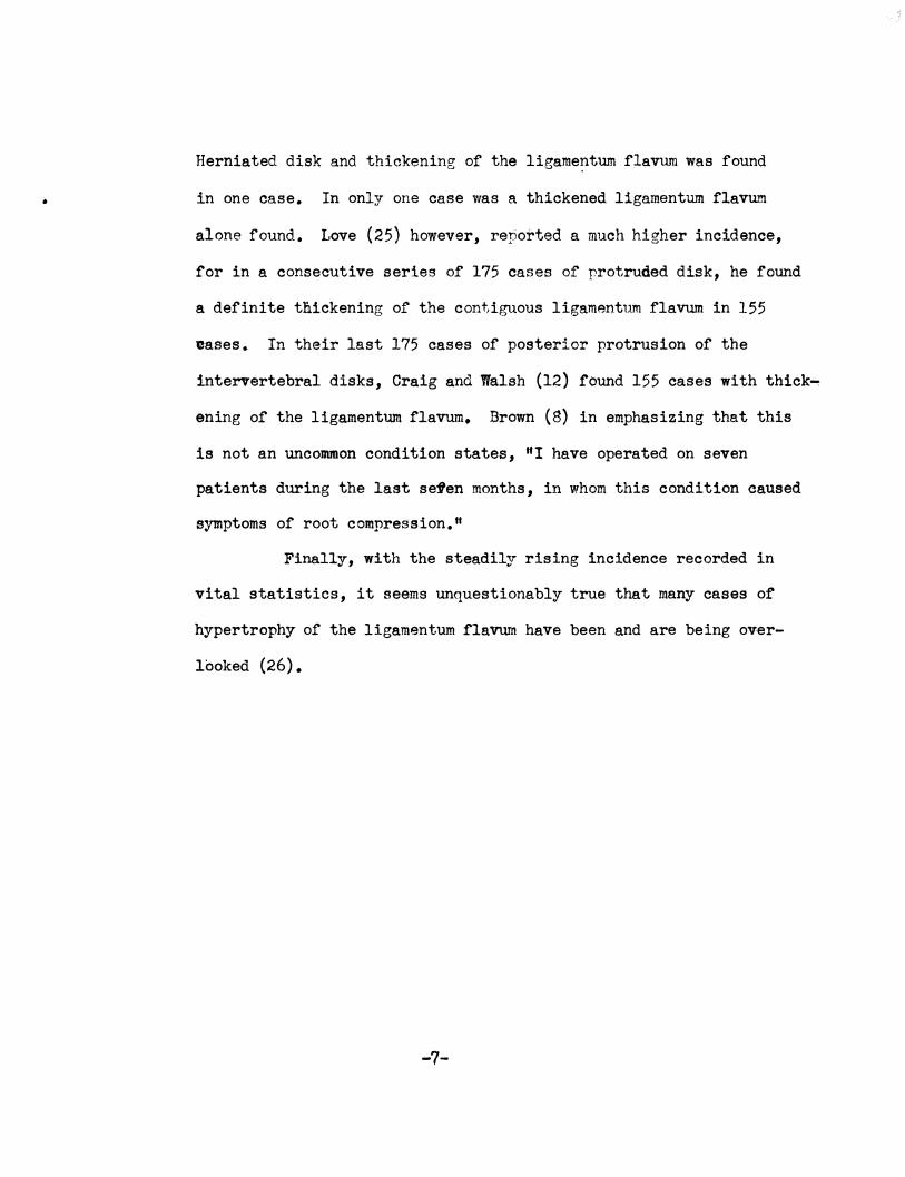
ANATOUY
The ligamenta flava are composed normally of yellow
elastic tissue and connect the laminae of adjacent vertebrae.
The normal ligaments are 2-3 mm. thick; (13, 14, 15) they are
the only predominantly elastic ligaments in the human body,
and, in virtue of their elasticity, can accommodate themselves
to the separation and approximation of the laminae in forward
and backward flexion of the vertebral column (15). Whereas
the ligamentum flavum is a continuous structure, it is con.,.
venient both for descriptive and clinical reasons to subdivide
it into two portions. The medial half, the broader and thick- 7 �er part of the ligament, is attached to the contiguous lamianae ...
and may with advantage be called the interlaminar portion. The
thinner lateral half is less wide and tapers off as it extends
laterally. It is attached predominantly to the articular
processes and is related to the interarticular joint and capsule.
This part will be referred to as the capsular portion. This
division into interlaminar and capsular portions is suggested
by some difference in direction of the fibers of the two parts.
The fibers of the interlaminar part are vertical in position,
whereas those of the capsular portion run obliquely downward and
laterally. (10).
Interiorly, the interlaminar portion is attached to the
upper border of the lamina below. On the bony specimen, this
- 8 -
attachment is indicated by a well defined eroove, the anterior
margin of which variously exhibits sharp, thin plaques or
spiculed of bone which extend up into the anterior face of
� ---·----
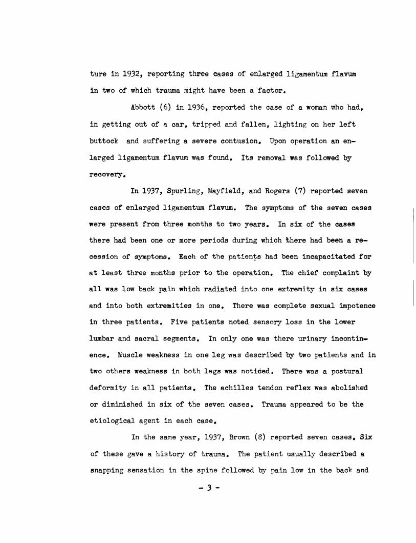
Fig 1. The ligamenta flava of the lumbar region • .Anterior Aspeot.
the corresponding ligament. These spicules are variable in size,
number and position and may perhaps be regarded as ossification
in the ligamentum flavum, but it has been noted that they are
attached to the lamina and are more in the nature of an ossific
extension from the bony attachment (10). Calcarious matter has
- 9 -
I
been observed in the ligamenturn flavum removed at operation,
and has been regarded by Meredith and Lehman (9) as calcium
deposited secondary to injury to the ligamentum. In the
opinion of Naffsiger et. al. (10) this calcareous matter is
nothing but ossific matter derived from those spicules which
must necessarily be fractured from their attachments when the
ligament is surgically removed.
The i�terlaminar portion is attached superiorly to a
well defined irregular and rough area, which occupies the
lower half of the lamina (10}. This area is separated from
the smooth upper half of the lamina by a sharp ridge. Character
istically, the ridge exhibits a small �ony spicule at the junc
tion of the attachments of the inte�laminar and capsular por
tions, but.the bony plaques found below have not been observed
at the urper attachment. Owing to this arrangement, the in
ferior half of each lamina is excluded anteriorly from the neural
canal and the upper smooth hal.r of the lamina alone is directly
related to the spinal dura. In the midline at the base of the
spinous process, the medial end of the interlaminar portion
blends with its fellow or the opposite side. Laterally, each
interlaminar portion is continuous with, and almost inseperable
· trom, the capsular moiety, except for the difference in direction
of the fibers of the two parts. The interlaminar portion is for
the most part a direct posterior relation or the spinal dura.
The capsular portion is attached below to a groove which
-lO -
···
extends along the periphery of the inferior articular process to
a point a little beyond the intervertebral foremen (10). The
ligament is attached. above. to the inferior border of the
pedicle and lies just below the groove for the spinal nerve
which is found on the inferior aspect of the structure. Laterally
the ligament. considerably attenuated. blends with the capsule of
the interarticular joint. some little distance lateral to the
intervertebral foremen. This portion of the ligament excludes
the joint from the neural cane.i and from the lower half of the
intervertebral foremen.
Walsh and Love in their writing upon protrusion ot the
intervertebral disks a'ttempt to explain the sites at which pro
j)rusion ot the disks occur by calling attention to the anatomical
shape of the spinal column. They classified one hundred cases
and found them to occur only at the points of greatest convexity
of concavity; namely. inthe fifth, sixth. or seventh cervical.
fifth. sixth, tenth or eleventh thoracic. or the third, fourth,
or fifth lumbar interspaoes ( 16). This same line of reasoning
can be followed in regard to i jury to the ligamen-tum. tlavum.
Craig and Walsh report that th y found enlargement of the liga
·mentum in 155 of the last 175 ases of posterior protrusion of
the intervertebral..'disks and o nolude that the enlargement in
these oases is most likely due J
to the trauma that produced the
protrusion of the disks (12).
Attar grouping together 53 oases reported by various
- ll -
\._;'
authors (1. 2. 4. s. s. 7. 9• 11. 11. 18. 19. 20. 21, 22). I
found that 46 of the enlarged ligamenta flava occured in the
lumbar region. 7 in the lumbosacral region and one in the thoracic
region of the spine. This predominance of lesions in the lumbar
concavity of the spine almost to the exclusion of the other
curvatures may be explained by the fact that it is this portion
of the spine that receives the greatest amount of trauma and
strain in body support and aoti vi t;y. Naffziger, et al, empr..asi ze
this fact when they estimate that the various force factors at
work (e.g •• in lifting a 50 lb. weight) are multiplied about ten
tilies and sometimes., especially in sudden effort, to an ewn great•
er degree. with a resultant pressure upon the lower lumbar disks ot
some 500 lbs. or more. Similarly. great strain is put on the
ligament& flava and the other vertebral ligaments (10).
-12 -
: -,
) .
..
PATHOLOGY
Descriptions of the enla�ged ligamentum flavum are some
what variable. The first account or pathological enlargement of
the ligamentum flavum was made by Elsberg (1). He described the
ligament as a firm whitish colored mass about 2 cm. thick bulging
into the spinal canal and compressing the fourth lumbar nerve root.
Meredith and Lehman describe one as a yellow mass that was fowid
lying over the dorsu.m of the dura. "It was 15 mm. in thickness.
It was cartilagenous in consistency and attached to the periosteum.
The tissue was bright yellow in color and was easily brushed away
.from the dura. When the mass was removed, a curved impression�dn·
the dura resulting from the overlying mass was evident" (9). An
enlarged ligament discovered by Naffziger, et al., was reported
by them as "considerably enlarged, rough, irregular and nodular.
It protruded .forward impinging on the first sacral nerve11 (10).
Dickson and Twort (15) reported in one case "The ligaments which
formed a single thickened transverse band were tough and pale
pinkish-red and had lost most or their normal yellow color. The
thickening extended on both_sides, partly encircling and compress
ing the immediate subjacent theca. The thickened and coalesced
ligaments were adherent to the subjacent dura, from which they w�re
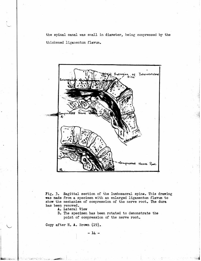
readily dissected." Flothow (26) reported finding an enlarged liga-.
mentum flavum which he described as extending down from under the
lumbar lamina to the sacral arch. In the same area he found that
- 13 -
i I
L�.
the spinal canal was small in, diameter, being compressed by the
thickened ligamentum flavum.
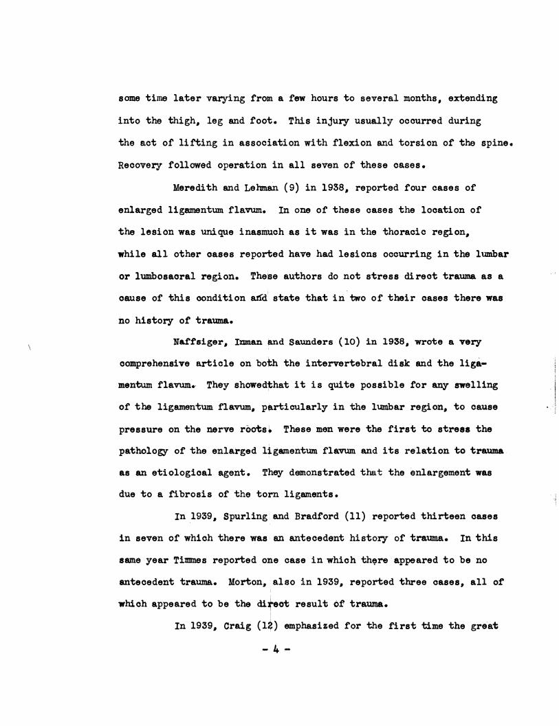
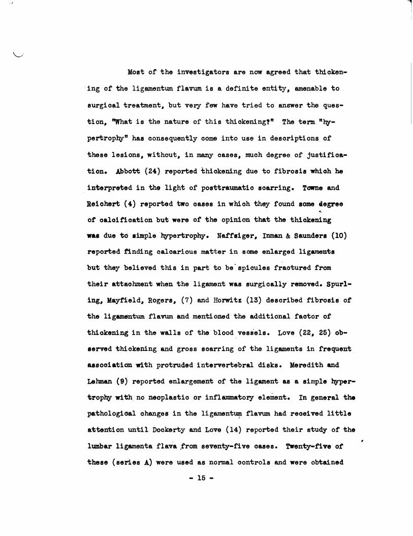
Fig. 3. Sagittal section of the lumbosacral spine. This drawing was made from a specimen with an enlarged ligamentum flavum to show the mechanism of compression of the nerve root. The dura has been removed •
.&.; Lateral View B. The specimen has been rotated to demonstrate the
point of compression pf the nerve root.
Copy after H. A. Brown (19}.,
- 14-
I i
I
,t
\,_,,i
Most of' the investigators are now agreed that thicken-
, ing ot the ligamentum. i'laVUJll is a dei'ini te entity• amenable to
surgical treatment. but very few have tried to answer the ques
tion. "What is the nature of' this thickening!" The term "l\Y
pertrophy" has consequently come into use in descriptions. of
these lesions. without. in many cases, much degree of' justi:f'ioa
tion. .Abbott (24) reported thiokemng due to fibrosis whioh he
. interpreted in the light of posttraumatio scarring. Towne and
Reichert (4) reported two oases in which they found some aegree ..
o� oaloifioation but were ot the opinion that the thiolcening
was due to aimple·l\Y'Pertrophy. N'af'taiger. Inman & Saunders (10)
reported �nding calcarious matter in sane enlarged ligaments
but they 'believed .this in part to be.spicules t�actured from
their attachment when the ligament was surgically removed. Spurl
ing.,
Mayti,ield• Rogers. (7) and Horwitz (13) described fibrosis ot
the ligamentum. flavum and mentioned the additional factor ot
thiolcening in the walls of the blood .vessels. Love (22.,
26) ob
ael"Ved thi:okening and gross scarring of the ligaments in frequent ..
aasooiatidn with protruded interTertebral disks. Meredith and
Lehman (9); reported enlargement or the ligament as a simple l\rper
trophy wi�. no neoplastic or inflammatory element. In general the
pathologi�l changes in the ligament� tlavum had received little
attention until Dockerty and LOTe (14) reported their study ot the
lumbar ligamenta £lava trom seventy-five oases. !rwenty-tive ot
these ( series A) were used as normal oontrols and were obtained
- 15 -
•
, I .\
at autopsy, and fifty, (series B) consisted of lumbar ligaments
removed at operation because of the syndrome of backache and
"sciatica". In series A., the ligaments were uniferrnly yellow
ish in color both on surface and on section. The average thick
ness of these ligaments was 2.8 mm. with extremes of 2 and 4 mm.
Horwitz (13) in his series of seventy-five cadavers reported an
average ligamental thickness of 3.7 mm. In series B, forty-five
of the fifty ligaments were not uniformly yellow in color but
presented whitish lines and bands which were more prominent on
sectional views. In ten of the cases this change was so marked
that little of the original normal yellowish color remained;
the ligaments consisted almost entirely of scar-like tissue.
The average thickness of the ligaments in this group was 5.1 mm.
with extremes of 2.5 and 9.5 mm.
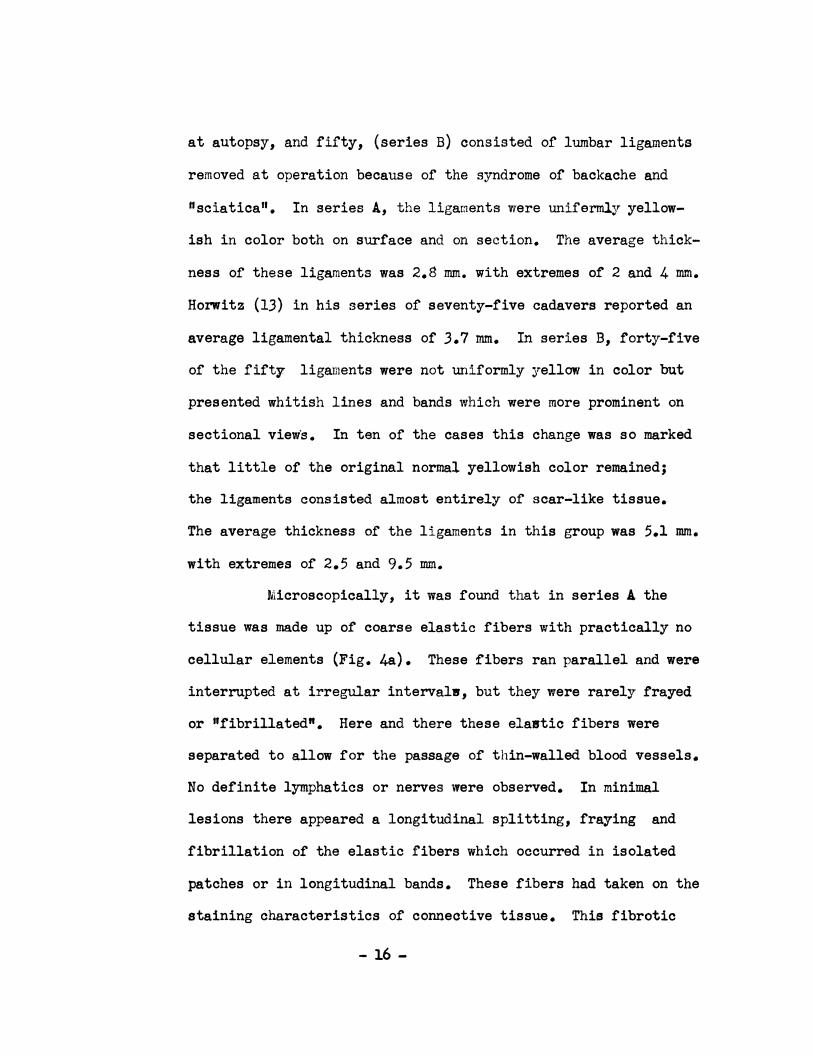
Microscopically, it was found that in series A the
tissue was made up of coarse elastic fibers with practically no
cellular elements (Fig. 4a). These fibers ran parallel and were
interrupted at irregular intervalw, but they were rarely frayed
or "fibrillated". Here and there these elastic fibers were
separated to allow for the passage of thin-walled blood vessels.
No definite lymphatics or nerves were observed. In minimal
lesions there appeared a longitudinal splitting, fraying and
fibrillation of the elastic fibers which occurred in isolated
patches or in longitudinal bands. These fibers had taken on the
staining characteristics of connective tissue. This fibrotic
- 16 -
I
change varied from minimal to almost complete replacement of the
normal elastic tissue of the ligament (Fig. 4b). Another ch&nge I
I
Fig. 4a. Normal lieamentum flavum: One may note slightly wb,vy
elastic tissue fibers; no fibrosis is rresent: stained with !
elastin stain.(xl90). I . I
4b. Grade l fibrosis in patchy distribution; on, may njote fragmentation and·hyalinization of elastic fibrils: stained �ith elastin stain (x45). After Dockerty and Love (14). I
i
!
I
Mchange varied from minimal to almost complete replacement ,r the I
normal elastic tissue of the ligament (Fig. 4b). It was b�cause i
of these findings that Dockerty and Love (14) suggested t�t the
term "hypertrophy� should be replaced by the more accurate
descriptive designation of "thickening with fibrosis." Another
change observed in series B, almost constantly was a hyal�ne
... 17 -
-1
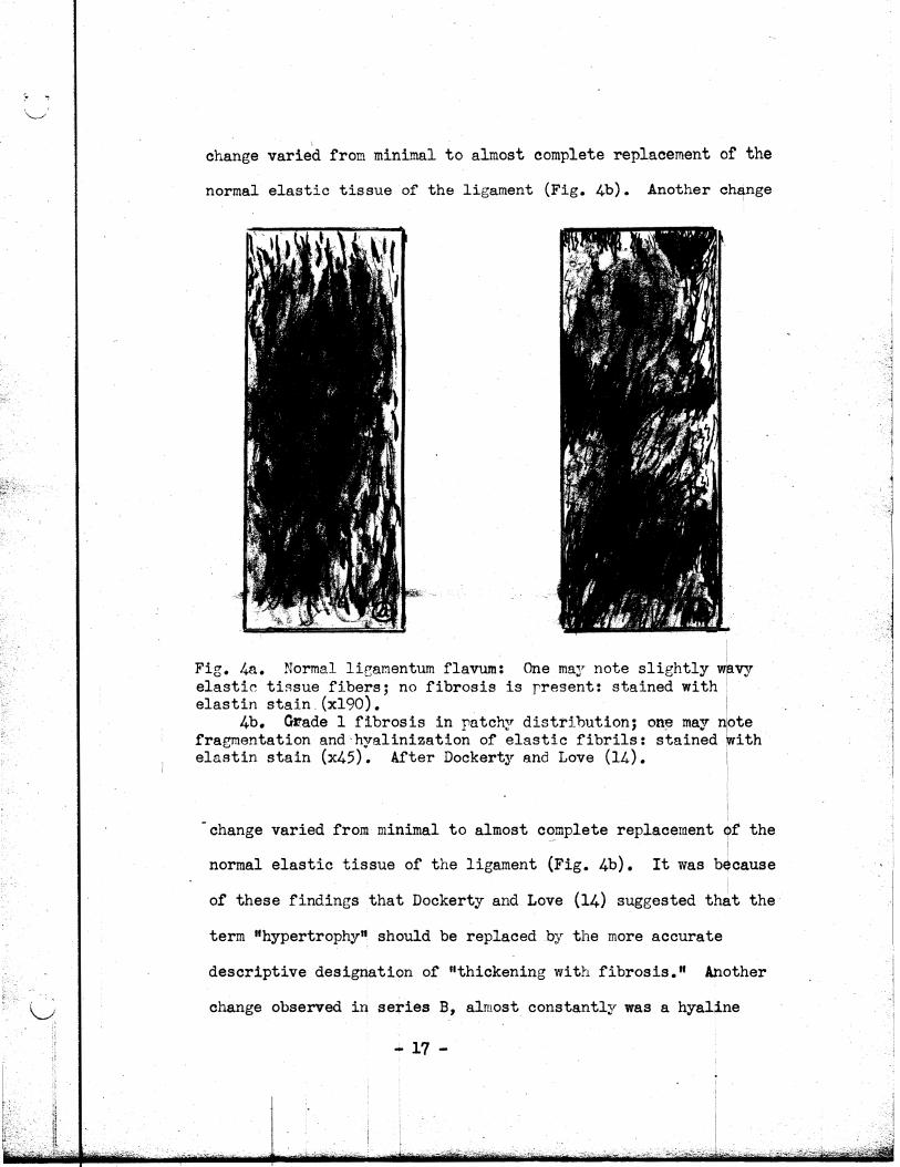
thiokening in the walls of the blood vessels as had been re-
portoo. proT10\u11y 'by otnor worlc@n ( 7, .l3), The lumina were
,.
Fig 5a. Marked fibrosis: Most of' the normal elastic tissue has been replaced by fibrous connective tissue.
5b. Blood vessel of' pathologic ligament. Not.a hyalinization of' wall and almost complete ooolusion of lumen; stained with elastin .stain (:r:200). After Dockerty and Love (14).
near�y always narrowed, in many instances to the point of . .
occlusion (Fig 5b.) •. In about a thi·rd of the ligaments in
series B the blood veS'Sels appeared to be increased in number
in addition to bei ng hyalinized. Calcification was noted in
three of the ligaments in the B series.
The questio� now arises, "Why do we have this
- 18 -
fibrotic replacement or the elastic fibe.rs of the ligaJ1Bntum
flavum?" The explanation.,
most widely accepted is that· of a
posttraumatic fibrosis (7, 9, 13., 14
., 15., 17, 18, 23, 24). The
elastic fibres of the ligamenta flava are normally under tension.
Torn from their attachments or ruptured, ·they contract and thick
en. - Tears heal by the formation of soar tissue,. and a ligament
in which such changes have taken place may be 9 mm. or more in
thickness (14) and is usually adherent to the dura. This
fibrotic hyperplasia may be unilateral or bilateral.,
the histo
logical changes being a replacement of the nonnal yel low elas'bio
tissue by collagenous white fibrous tissue in which there may be
oaloareous deposits (4., 14). The site of election tor the lesion
is, as has been previously stated.,
in the lower lumbar region.,
atteoting most commonly the ligaments between the fourth and
fi:rth lumbar vertebrae.
- 19 -
\ ..... /
ETIOLOGY
When considering the causative factors of enlargement
of the ligamentum flavum., the most constant agent by all odds
appearsto be trauma {l ., 9., 14., 15, 17., 18 ., 20., 24 ., 21). The
elastic fibres of the ligamenta flava are normally under tension
(15). When torn .from their attachments or ruptured., repair in
-.uoh specialized elastic tissue is completed by tissue of a
lower order• fibrous oonneotive tissue - and acute or ohroni,,o
trauma appears to oause the pathological thickening. '!'he trauma.,
however.,
mq be or vatioustn,es and result from a multitude of
causes. The most frequent trauma.., however., is produced by lift
ing in association with flexi_on and torsion of the spine., or
possibly a fall on the back or buttocks (17). In Elsberg•s
case (1) the thickening of the ligamentum flavum was incident
to an automobile accident in which the woman was thrown from
her oar-. Spurling (7) stated'.that it was most ccmnon in his
seven oases for the patient to give the history ot "while lift
ing a heavy object, I felt a snap and then a severe pain in the
lower part of m:, back. ffi I n a report of nine cases, Crosthwait
(27) ., reports the trauma as being due to several causes, includ
ing lwperflexion of the spine ., active o r passiveJ or slipping
while the spine is lwPerfle:xed; and lifting while in flexed or
strained position; diving., athletics ., automobile., railroad., and
obstetric accidents. In one of the cases reported at the Uni•
- 20 -
7 '
versity Hospital, the trauma, in an obstetrical patient re
sulted from delivery while the patient was up in stirrups.
This patient noticed no symptoms until about the tenth day
after delivery, when she began having low back pain and
found that she was unable to move her left leg.
After grouping together 53 cases reported by various
authors (1, 2, 4, 5, 6, 7, 9, 18, 11, 19, 20, 21, 22), I
found that there was a history of direct trauma in 14 cases,
a history of indirect trauma in 19 cases,and there was no
reported history of trauma in 18 of the cases. No mention
of the causative factor was made in 2 cases. In this series,
then, trau.'!18. was the etiological factor in 67 per cent of the
vases while there was no history of trauma in 35 per cent of
the cases. The f'act that there is no history of trauma in
this large a percentage of the cases possibly is due to the
fact that the trauma causing these lesions may be, and often
is of a relatively slight degree and the onset of sympta1s is
frequently not immediate so that the patient has forgotten or
has not particularly noticed the injury (18).
Skinner (20) believes that many of these ,J1t.t.if,m�,may
have a weak back to start with and that this may explain why
many cases occur either without trauma or with only a minor
insult.
Abbott (24) believes that trauma is the most plausible
etiological factor because of the association of a ruptured
- 21 -
I
I
. \
intervertebral disk as seen by Brown (8), Hampton (.28), and
Abbott (24), but a possible inflammatory process must be
borne in mind.
- 22 -
\,___,,,·
SYMPTOMATOLOGY
The outstanding subjective symptom which leads
the patient to consult his doctor in most cases is pain (1.
4. s. 7. 11, 17. 18, 19, 20. 21, 24). This pain is usually
low in the back with sciatic radiation. Love ( 29) terms this
root pain. He defines root pain as pain which begins within
or near the spinal cord and is projected peripherally to that
part of the body or extremity innervated by the nerve fibers
which leave the spinal cord through the spi nal nerve root em
erging at that level.
In order to emphasize the importance of pain• it may
be noted that it was the chief complaint in twelve out of thir
teen cases reported by Spurling and Bradford (11). end in all
seven cases reported by Spurling. Mayfield and Rogers (7).
Pain in the legs and back is usually unilateral, but
it is ·som;tirnes bilateral (9). The pain too is characteristic
ally intermittent (18). The case.of Mrs. M. R. reported at
the University Hospital had pain extending into her left lower
back region and down her left leg. Less severe pain extended
into her right leg and back. There were periods of remission
and exacerbation in her pain.
There is usually a low backache ( 16. 18) that char
acteristically becomes aggravated by sneezing, coughing. and
straining. especially at time of defecation. whereupon it
- 23- ·, _l.. l
·"--._,·
radiates over the thigh and the posterior or lateral portion
of the calf of the leg. The patients with thickened ligamentum
flavum center their complaint of pain in the lower spine while
protruding disk oases. center their complaint to pain in their
hip and leg (20).
The distribution of the pain along the course of
the sciatic nerve in such a large percentage of cases ex
plains why this condition is so o�en confused with the many
others that also produce or are associated with "soiatioa".
A history of onset of pain immediately following sane type
of trauma is valuable. especially since a tearing of the lig
a.mentum with eubeequent thickening may ooour in a patient who
has suffered sciatic pain from other causes prior to the in
jury. The onset of pain following ligamental trauma is most
often sudden but may be gradual. and there is commonly an
interval between the onset of the pain in the back and the pain
of sciatic distribution.
The wide variation, that is possible in the location
and the extent to which the thickened ligamentum flavum com
presses the cord or the nerve roots, makes it evident at once
that there is n•oessarily a great variability in the location.
radiation. degree. and constancy of the pain.
The lesion J118¥ be so extensive and so located as
to cause a paraplegia in some oases; however, in most in
stances there is no paralysis, but merely motor weakness
- 24 -
which is found usually to occur in the anterior tibial muscles (18).
l,ieredith and Lehman (9) reported one case of thickened ligamentum
flaVUI)l in which the only comrlaint was weakness of the legs of three
months' duration and in which there was no pain or sensory distrubance.
The sensory changes are usually quite variable or even
vague. These changes in sensation may be present over the buttocks,
perineum, posterior thigh, calf and foot (18). Bradford and Spurl
ing (11) reveal a useful differential point between herniated
nucleus pulposus and thickened ligamentum flavum in their statistical
summary of their patients. They found that in 60 per cent of their
cases of herniated nucleus pulposus there was hypesthesia or anes
thesia limited to the lateral aspect of the leg or foot or both. In
contrast to this, they found that the cases of hypertrophied liga
mentum flavum showed areas of hypesthesia elsewhere, but in only 11
per cent of the cases was the hypesthesia limited to these areas.
This wider spread of hypesthesia in the case of enlarged ligamentum
flavum may have been due to the fact that the ligaments when enlarged
involved by compression more nervous tissue. In studying sensory
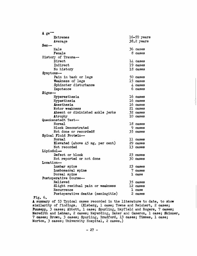
disturbances reported in 53 cases presented in the literature (1, 2,
4, 5, 6, 7, 9, 11, 17, 18, 19, 20, 21, 22) I found that hyperesthesia
was present in 16 cases, hypesthesia in 16 cases and anesthesia in 16
cases.
Patients with severe damage to the cauda equine. or .medul
lary conus have a loss or sphincter control. Dickson and Twort (15)
state that incontinence is rare, J;n patients with enlarged ligamentum
- 25 -
flavum, but that sexual impotence is common. The first of these state
ments is borne out by my survey of the literature, in which out of the
53 cases reported there was sphincter disturbance rep�rted in only 4
cases, but there were ohly 6 cases reported in which there was im
potance, a figure that does not support the second statement. (Fig.6)
-�-
AgeExtremes Average
Sex--Male Female
History of Trauma-Direct Indirect No history
Symptoms-Pain in back or legs Weakness of legs
.Sphincter disturbance Impotence
SignsHyperesthesia Hypesthesia Anesthesia Motor weakness Absent or diminished ankle jerks .ltrophy
Queckenstedt Test-Normal Block Demonstrated Not done or recorded8
Spinal Fluid Protein-Normal Elevated (above 45 mg. per cent) Not recorded
Lipiodol-Def'ect or block Not reported or not done
Location-Lumbar spine Lumbosacral spine Dorsal spine
Postoperative Course-Relieved
Fig. 6.
Slight residual pain or weakness Recurrence Postoperative deaths (meningitis)
16-59 years J8.2 years
36 cases 8 cases
14 cases 19 cases 18 cases
50 cases 15 cases 4 cases 6 cases
16 cases 16 cases 16 cases 21 cases
.32 cases 10 cases
18 cases 9 cases
35 cases
11 cases 29 cases 13 cases
2.3 cases 30 cases
2.3 cases 7 cases 1 case
35 cases 12 cases 1 case 2 cases
J. summary or 53 Typical cases recorded in the literature to date, to showsimilarity of findings. (Elsberg, l case; Towne and Reichert, 2 cases;Ptluaepp, 3. cases; Abbott, 1 case; Spurling, Mayfield and Rogers, 7 cases;Meredith and Lehman, 2 cases; Meyerding, Baker and Cameron, 1 case; Skinner,7 cases; Brown, 3 cases; Spurling, Bradford, 13 cases; Timmes, 1 case;.Morton, 3 cases; University Hospital, 2 cases.)
- 27 -
'I . .
FINDnms
When the patient is first seen, it is often noted that he
moves cautiously to prevent any jarring that would aggravate his
symptoms (30). A general restriction of the movements of the back -
is commonly seen (18). Often the�e is a flattening of the lumbar
concavity, the so called "poker spine", and a scoliosis in the same
region, with a tilting of the pelvis usually to the same side as the
lesion, although sometimes toward the opposite side (15). This
tilting or the pelvis, scoliosis and flattening of the lumbar con
cavity results from a protective spasm of the back muscles.
Dickson and Twort (15) believe that percussion and palpa
tion are of some value in diagnosing these cases. They have �.qµnd
that there may or may not be tenderness on pressure over the spinoua
processes of the lower lumbar vertebrae and tenderness of the sciatic
nerve to pressure in the buttock or thigh.
Muscle fibrillation is oc�asionally found. This along with
motor weakness, which is so frequently ·found, is an indication of
degenerative changes taking place in the muscle. The muscle in which
motor weakness is most <:>ften found is the. anterior tibial (18, 9).
The sensory findings are not in themselves characteristic or
thickening of the ligamentum flavum, but their presence in conjunction
with other findings is 6ften quite significant. The variability or the
sensory changes is quite remarkable for one may find paresthes1-,
hyperesthesia, hypesthesia or anesthesia. Any of these changes or
- 28 -
f.
combinations of them may be found involving the buttocks, perinetL�,
rosterior thigh, or calf and foot (18). In some instances of the p
patients with hypertrophied ligamentum flavum, the lesion may be
predicted by the widespread sisns of the sacral root compression (11).
The most constant objective findin� in these patients has
been reported to be an alteration in the Achilles-tendon reflex (18,
15). Brown (8) reported a diminished or absent ankle jerk in all
three of his cases. Bradford and Spurling (11) found a diminution or
absence of the ankle jerk in eight of the thirteen cases that they
reported. Out of the three cases reported by Morton (17), two were
found to have had an altered Achilles reflex. There is another ob
jective finding that is not so constantly found. There may or may
not be a limitation of flexion at the hip when the leg is straight
(18, 15).
Love and Walsh (16) attempted to determine the exact loca
tion of the pathological lesion in the spinal canal in a series of
one hundred cases by studying only the reflex changes, muscular weak
ness, and segmental sensory loss whenever present. They found that it
was impossible to accurately do so but concluded that such findings
did give excellent evidence as to the general region of the spinal
column in which the protrusion might be found.
Lumbar puncture should be carried out when neurological
examination suggests the presence of a lesion within the vertebral
canal or theca, or when the condition proves intractable to ordinary
treatment. In fibrotic thickening of the ligamentum flavum, the
- 29 -
:i j 1 ] "
i
total coagulable protein of the cerebrospinal fluid is commonly but
not invariably raised above the normal upper limits of 25 to 30 or
35 mg. per 100 cc., these normal fifures var7i1m with the age of the
patient (15, 11, 17, 18). An obstructive lesion in the vertebral
canal is characterist�cally associated with a rise in total protein
in the spinal fluid both above and below the lesion. If the lumbar
puncture is made at or below the fourth lumbar interspace (the common
site of obstruction in hyperplasia of the ligamentum flavum) a partial
or complete spinal block may be demonstrated. Spinal block, either
partial or complete is not a common finding in these cases, for out
of 53 typical cases reported in the literature, only 9 demonstrated
a positive Queckenstedt Test (1, 2, 4, 5, 6, 7, 9, 11, 17, 18, 20, 21,
22). Certainly, then, a negative result would not disprove the
presence of an enlarged ligament.
Dickson and Twort (15) often use an epidural injection of
saline as a diagnostic aid in doubtful cases. The injection of even
a few cubic centimeters of saline solution will, according to them,
cause an exacerbation of pain if there is already any degree of com
pression of the cauda equina.
Myelography is essential to diagnosis. There is at the
present time great controversy as to just what medium should be used
for visualizing pathology along the spinal canal. Iodized oil has
been favored in the past, and is still the most used medium, but
there l1&'. 0 inc�easing evidence in the literature or the value and ad-
. vantages . of air or oxygen.
- 30 -
�1
It is evident that, if no operation is to be performed for
the removal or the iodized oil, it would be unwise to inject it and
leave it there, even though evidence seems to indicate that it does
not produce an inflammatory reaction in every case (12, 15, .30, .31).
There is one feature that is of considerable importance, especially
in compensation cases. It is the presence of the opaque oil revealed
in every subsequent roentgeriogram which the patient or unscrupulous
attorneys and doctors may use to unfair advantage in obtaining ex
cessive compensation for the patient. This opaque oil wifLl be foµnd .,
,months or years later distributed along nerve roots alon�the spinal . J
canal and even up around the brain. Since this substance1 i� a foreign I ij.
body, often irritating to the cord, roots and meninges, �aotically
all neur�surgeons are agreed that lipiodol should not be :�njected un-. . I
less ;an exploratoey operation has been previously decided:j upon (12, 15, I
27, 30, 31). Even when a 1aminectomy is done and the duta is opened
and a large part of the lipiodol is removed, it is usuall� impossible :1
to remove it all (.30, 31). i
l The technique for examination of the spinal col+.mn with
. j
iodized oil has been well described by Dickson and Twort f15). Lipiodol, I
should never be injected in the presence of a suspected 1*1'1ammatory
lesion. The temperature of the oil must not be above tha� of the . I
body. It should not be used if it has become cloudy. Opfnions diffe; . 1
as to the quantity of iodised poppy-seed oil which should!be used. I
With the patient in the erect posture, some five cubic ce.timetere is ., II
required to fill the lower spinal the�a to the level of tte third
- 31 -
.'--./ .
lumbar vertebra. This amount is therefore required in ordor to be
sure of showine up any filling-defects due to protruded intervertebral
disks or thickened ligamenta flnva in the lower lumber region, if
diagnosis depends on straight films· and cannot be assisted by fluoros
copy. Anter6-posterior films with the patient prone, and lateral films,
are taken with the tilting-table at an angle of forty degrees. If the
fluoroscope can be employed along with a tilting-table, two cc. of
oil may be enough (15, 32) for this can be run up and down the lower
theca and films taken when the oil is seen to be at the site of any
notching or obstruction in the flow. The smaller amount is less likely
to be followed by urinary troubles and arachnoiditis. Irritation of
the roots of the cauda equina from the oil may sometimes be relieved
by draining it off by repeated lumbar punctures, but this is not always
effective. If an obstructive lesion is diagnosed and laminectomy per
formed the theca is opened at operation and the oil evacuated then.
Otherwise, if myelography proves negative, the oil should be drained
from the theca after sacral trephining.
There·is no infallible means of differentiating the cause of
a filline defect as viewed by x-ray picture or fluoroscopic examination
(27); It may be produced by hypertrophied ligamentum flavum, ruptured
disk, nucleus pulposus, cord tumor, or though rarely, a new growth in
bony tissue. According to Bee and Spurling (32) the commonest abnorm
ality found on myelography in cases of thickening of the ligamentum
flavum is a unilateral filling defect or notching of the column or oil.
This is because the thickening of the ligament is usually predominantly
- 32 -
,-,
!'-;··_
unilateral. A similar defect is the common finding in cases of pro-
trusion of an intervertebral disk. Theoretically, one should be able
to destinguish between a displaced nucleus and an hypertrophied liga
ment by lateral films. However, this is seldom the case, for nuclei
usually herniate lateralward beneath the nerve root, and the ligaments
frequently thicken more on one side than the other (32). Myelography
will not therefore always distinguish between these lesions, but
fortunately the same surgical approach is required for both (15). When
the thickening of the ligaments is bilateral a symmetrical hour glass
constr}ction of the dural sac and the enclosed column of oil is deJllon
strate�. A complete hold-up of the oil is still more rare, both in
protruded intervertebral disk and in hyperplasia of the ligamenta tlava
(.32).
Berens (.30) has summarized the important points in favor of
and ag{inst the use of iodized oil in diagnosis of deformities of the
spinallcanal.
FOR:
1. One can obtain clearer pictures when using oil.
2. The use of oil is more accurate on small lesions •
.3. Myelography with the use of oil is easy to perform..
The use of oil is not painful and requires no use ofanesthetic.
5. Oil as an opaque substance is useful in any part of thespinal canal.
- 33 -
AGAINST.
1. An operntion is necessary for the removal of the oil.
2. Oil that is not removed gives rise to an inflammatoryreaction.
3. It is impossible to remove all of the oil; subsequentroentgenoi:;rams always show its presence.
4. Occasionally oil is accidently placed extradurally whereit remains as a foreign substance.
5. It furnishes material for a malpractice suit.
Bosworth and Hare (33) believe that the value of lipiodol
more than balances its disadvantages inasmuch as it gives definite
proof of the diagnosis and permits the surgeon to find the lesion by
re�oval of only one vertebral lamina.
Craig and Walsh (12) have well described the technique for
examination of the spinal column by using air. They combine their
spinal punctures and spinograms. The proceedures are carried out in
the x-ray room with the patient lying on the fluoroscopic table. A
lumbar puncture is made in the usual manner, manometric readings are
obtained and 15 cc. of fluid is removed for study. The table is then
tilted to an angle of about 40 degrees with the patient's head down
(27) and the fractional introduction of air is carried out. Between
JO and 40 cc. of air is introduced, care beine taken that the lumbar
sac is emptied or fluid. Baker has suggested that shaking the pelvis
or jolting the hips helps dislodge any fluid that would obscure the
picture. Following the injection of air, stereoscopic anterior,
posterior and lateral films are made. The evidence of narrowing of
- 34 -
I ·I
the lumbar oanal, the protrusion of intervertebral disks or thicken
ing of the ligamentum flavum is not so convincing as in roentgeno
gra.ms taken after the injection of radiopaque oil but with practice
the reading of the films becomes much easier (12).
Air or oxygen have one great natural advantage over iodized
oil in that they are absorbed within a few hours and leave no after
effect. They are especially valuable when an attempt is being made
to diagnose a borderline oase, since air injections may be done
several times, if necessary to verify the presence and localization
of a pressure producing mass; and if it is eventually decided that
there is no existing deformity, there are no foreign bodies left in
�he patient such as remain when lipiodol is injected end none or only
part of it is removed (30).
Berens (30) emphasizes that one of the disadvantages of air
is that the roentgen plates are obtained by overexposure at rapid
speed, such as can be obtained by the use of rotating anode tube
apparatus at 300 milliamperes and one-half second exposure. Equipment
of this kind is not available for all patients and physicians who wish
to do this type of' diagnostic work. Another disadvantage of air, he
points out, is that it is somewhat painful and strong sedatives, or
some type of anesthetic such as intravenous pentathol sodium, is usually
required. In favor of' it, it may be stated the experience of' those
who have used air a great deal and have found that, it a poai ti ve diag
nosis is made with it, there is rarely a case where the pathology will
not be found at the time of' operation ( 30) • ...
- 35 -
The important points in favor of cth4 against the use of'
air or oxygen in diagnosis of deformities of' the spinal canal have
been swmnarized by Berens (30).
FOR AIR OR OXYGEN:
1. They will reveal all medium sized or large deformities of'the dural sac, such as would unquestionably require surgery.
2. There are no after-ef'f'eots from the use of' air or o:x;ygen.The head is kept lowe�ed about 30 degrees until the airabsorbs (three to four hours) (27).
. .
3. There is nothing to be revealed later by examination orroentgenogram if the case test is negative.
4 •. · There is no. matierial for malpractice suits.
5. One has the fluid to check for total protein.
6. This procedure may be performed several times if neoeasa.ry.
AGAINST USE OF AIR OR OXYGEN:
l. Their use requires powerful roentgen apparatus.
2. The v isualization is not always so ole•r·
3. Their use is not as accurate for small lesions as is the useof lipiodol.
4. Their use is painful and requires the use of a sedative oranesthetic.
5. They are m.ost useful.in lumbar, sacral and lower dorsal de-·formities.
6. Their use is more time consuming and more difficult to perform..
A new di agnostic proceedure, J!\Velosoopy, for determining the
nature of spinal canal pathology seems to have great practical possi-.
bilitiea. Pool (36) presents this as a means of visualising the oauda
equina through a splnal endoscope called J!\Velosoope. �eloscow is
usually performed under local anesthesia :with the patient in sitting .
.:. 36-
..;,_.
\__,'
posture - this gives rise to a hydrostatic distention of the araoh
noidal membrane - and is carried· out in muoh the same manner as an
ordinary lumbar puncture. Specimens of cerebrospinal fluid may be ,
collected, manometrio studies may be done and gas myelography may be
performed through the myelosoope. By use of the myelosoope the
presence of a thicke�ed ligamentum'flavum has been frequently de
tected by finding an :unusually large epidural space, with secondary
narrowing of the subarachnoid space (35). Pool has also recognized
this lesion in a man suffering from an acute unilateral "sciatica•
by the presence of greatly distended looped, root vessels obviously
due to compression of that nerve at a lower level. This prooeedure,
then, may in the f'uture afford the means of making a dii'f'erential
diagnosis between an operable and inoperable lesion of the lower
apinal cord and it may also senre aa a means of sparing many a patient
an exploratory laminect� or lipiodol injection.
- 'J7 -
DIFFERENTIAL DIAGNOSIS
Many conditions must be considered in the differential
diagnosis of thickening of the ligamenturn flavum.
The most important ones are probably an intraspinal
neoplasm and herniation of the intervertebral disk. These how
ever do not cause much concern since their treatment is the
same; namely, laminectomy and operative removal.
Some of the other common conditions which most closely
resemble thickened ligamentum flavum lesions are: low back pain
from any cause, lumbosacral strain, sacro-iliac disease, spondyl
itis and hypertrophic conditions involving the spine.
Some cases may resemble the clinical picture of syringo
myelia or of multiple sclerosis. Sciatic neuritis and fibrositis
may also be troublesome in making a differential diagnosis.
It is suggested by Love and Camp (29) that whenever any
. patient considered to be suffering from any of the above condi
tions has had sufficient conservative treatment and is not re
sponding favorable, the possibility of a protruded intervertebral
disk should be seriously considered as the cause of his disability.
Certainly it should be considered in any case of intractable and
recurrent sciatica.
\ I
'----'
TREATMENT
The modern treatment of fibrotic thickening of the
ligamentum flavum is very much the same as that used in the first
case reported by Elsberg in 1913 (1). However, since the use of
iodized oil, and air in localizing the lesion, has become an
accepted method of diagnosis, the number of laminae removed has
been decreased.
The essential factor in the treatment of thickening of
the ligamentum flavum is the wide lateral removal of the protruding
mass which is pressing on the spinal cord or nerve roots and pro
ducing symptoms of extradural compression. This mass is reached
by removing the lamina. As the lamina is removed, the ligament is
seen immediately beneath it, and if there is generalized enlarge
ment the constriction of the dural sac is often very marked. 1
wide lateral excision of the ligaments is made. Most often the
ligaments are not adherent to the dura, but they are sometimes quite
adherent (17). If they are adherent they must be carefully separated
from the dura and removed. A thorough examination should always be
made at operation in these cases to determine the presence of an
associated dislocation of an intervertebral disk as any undue promin
ence of the disk serves to dearease the size of the passage for the
nerve root, so that even a moderate enlargement of the ligament
would compress the root against it (17). If the dislocation is
sufficient to compress a nerve root it should or course be removed
and this is also true of a nucleus pulposus when found.
- 39
\, __ .• /
An important consideration in connection with the l�min
ectomy usually performed in these cases is whether the patient will
ultimately have a strong back. Many surgeons have advised spinal
fusion at the time of removal of the thickened lieamentum flavum
in order to insure the patient of e.s strong a back as possible.
Love, Adson, and Craig (34), however, feel that the spinal fusion
is not necessary. When not done, they maintain that no complications
arise. It is Skinner's (20) contention however, that these cases
have a weak back to begin with and that they can be benefited great
ly by a fusion at the time that the laminectomy is performed.
Love (34) keeps his patients in bed twelve days following
the operation. He allows them to leave the hospital on the four
teenth day if there are no postoperative complications. Skinner (20)
does not allow his patients, after they leave the hospital, to do
heaTY labor or engage in dangerous occupations where they_ are likely
to suffer an injury to their back, for a sixty day period. He follows
up his cases carefully in regard to the type of exercise they should
take, and watches their posture.
- 40 -
I
\, ___ ___,
PROGNOSIS
The results of operation for the removal of the thickened
ligamenta flava are in most oases very good. By far the majority
of patients are completely relieved of their pain and disability.
Usually the relief from pain is noticed immediately following
operation. Since most of the oases have been treated in the last
five years, the permanent results are still undetermined. However,
the results of Bradford a,nd Spurling (11) in their eight oases re
ported is typical of most clinics. In this series there were no
post-operative deaths. Nine of the patients have been completely
r•lieved of their symptoms while four ,have only slight residual
pain or weakness. All three of the patients reported by Brown (8)
gained complete relief' :from their symptoms following surgery. In
most oases the longer the symptoms have been present., the lees the
relief obtained by operation because of more severe damage to the
nervous system.
Reourrenoe o:f symptoms following operation is extremely
rare. There has been only one case reported in which there was a
reourrenoe. Meredith and Lehman (9) reported this case in which
there appeared to be an operative oure after a lamineotoll\Y and re
moval of a thi okened liga:mentum. fla.wm, but there was, 9 months
later, a complete return of disability and it waa found that the
patient had a similar lesion at a level one vertebra above the
original site.
- 4l -
SUMMARY
Enlargement of the ligamentum flavum is a definite an
atomical and pathological condition which is rapidly gaining in
importance.
The condition usually results from trauma which may be
either slight or severe.
The most common symptom is root pain. usually of
sciatic type. which oharaoteristioally undergoes remissions and
reourrenoes.
Diagnosis has been most successful af'ter instillation
of lipiodol into the subarachnoid space followed by roentgeno
gra.phio examination of.' the spinal canal. There is, at present
however. a swing toward the replacement of lipiodol by injeo-·
tions of air into the subaraohnoid space, thus avoiding the
hazard of.' a non absorbable and irritating substance within this
apace.
The treatment by lamineotomy with removal of the en
larged ligamentum flavum has been highly satisfactory.
This treatment is followed by complete or partial re
covery in nearly all oases and the risk to the patient is minimal.
- 42 -
. �· -• ... ·� --..: - ·;'!> r�
BIBLIOGRAPHY
1. Elsberg, Charles A.: Experiences in Spinal Surgery ObservationsUpon 60 Laminectomies for Spinal Disease, Surg. Gynecolo� and Obstetrics 16: 117, 1913.
2. Elsberg, Charles A., Diagnosis and Treatment of surgical Diseases of the Spinal Cord and Its Membranes, w. B. Saunders Co., Philadelphia, 231, 1916.
3. Elsberg, Charles A.: Dis�asea of the Spinal Cord, p. B. RoeberInc., New York� 473, 1941.
4. Towne, E. B. and Reichert, F. t., Compression of the Roots andCord by Thickened Ligamentum Flavum, .Annals of Surgery 941 327-336, September, 1931.
5. Puuaepp, L.: Compression of Cauda Equina Causing Tumor-LikeSymptoms. Recovery After Surgery, Folia Neuropath. Estonia 12: 38-48, 1932.
6. Abbott, W. D.: Compression of the Cauda Equina by the Ligamentum Flavum, J. A. M. A• 106: 2129-2130, June 20, 1936.
1. Spurling, R. G., Mayfield, F. H. and Rogers, J. B1 �rtropl\rof the Li gamentum Flavum as a Cause of Low Back Pain, ,J. A. M. A• 1091 928-933, September 18, 1937.
8. Brown, H. A•: Low Back Pain with Special Reference .to Dislocation of the Intervertebral Diak and }trpertropl\r of the Liga.mentum Flavum., West Journal of Surgery Gynecology and Obstetrics 45: 527•531, October 1937.
9. Meredith, J. I(. and Lehman, :s. P.: �ertropey- of the LigamentumFlavum. A R eport of Two Atypical Cases, Surgery 4 1 587-696, October 1938.
: 10. Na.f'f&iger, H. c., Imnan, v. and Saunders, J. B. de c. M.: Lesions. of the InterTertebral Disks and Ligamenta Flava, Clinical and .Anatomical Studies, Surgery Gynecology- and Obstetrics 66: 288-299, Feb ruary 1938.
11. Bradford., Keith F. and Spurling, G. R.: Intraspinal Causes ofLow Back Pain, Surgery Gyneoolo� and Obstetrios 691 446• 1939. ·
- 43 -
I i
12. Craig, w. M. and Walsh., M. N.: Diagnosis and Treat::nent of LowBack and Sciatic Pain Caused by Herniated Intervertebral Disk and Hypertrophied Ligamentum 1'�lavum., Minnesota Medicine 22: 511-517, August 1934.
13. Horwitz, Thomas: Lesions of the Intervertebral Disk and Ligamentum Flavum of the Lwnbar Vertebrae. .An .Anatomic study of 75 Human Cadavers, Surgery 6: 410, 1939.
14. Dockerty, M. B. and Love, J. G.: Thickening and Fibrosis (Socalled :ftrpertrophy) of the Ligamentum Flavum: A Pathologic Study of Fi�y Cases, Proceedings Staff Meetings of the Mayo Clinic 15: 161-166., March 13., 1940.
15. Dickson, w. E. c. a...�d Twort, R. J.: Thickened Ligamenta Flava- In Low Back-Ache and Sciatica, Lancet 1: 1113-1116, June
22, 1940.
16. Walsh, H. N. and Love, J. G.: Protruded Intervertebral Disk AsA Cause of Intractable Pain., Proceedings Staff Meetings of the Mayo Clinic 13: 203-205., March 30., 1938.
17. Morton, A. P.: Laminectomy for Low Back Pain, With Case Reports,u. s. Naval Medical Bulletin 37: 523-538 ., October, 1939.
18. Timmes., J. J.: :Ft{pertrophied Ligamentum Flavum (With CompleteBlock of Spinal Canal), u. s. Naval Medical Bulletin 37: 538-541, October, 1939.
19. Brown., H. A.: Enlargement of the Ligam.entum Flavum, A Cause ofLow-Back Pain with Sciatic Radiation, Journal of Bone and Joint Surgery 20: 325-338, April., 1938.
20. Skinner, H. L.: Ruptured Intervertebral Disk and J.zypertrophiedLigamentum Flavum Follow-Up study, Virginia Medical Monthly 67: 490-494, August, 1940.
21. Meyerding., H. w • ., Baker, G. s., Love, J. G. and Cameron, D. M.:Spondylolisthes is With Protrusion of Intervertebral Disk and Hypertrophied Ligamentum Flavum Associated with Multiple Loose Bodies of Right Shoulder Joint (Report of a Case) ., Proceedings staff Meetings of the Mayo Clinic 14: 801-806., December 20., 1939.
22. Love, J. G.: Intractable Low Back and Sciatic Pain Due to Protruded Intervertebral Disks; Diagnosis and Treatment.,
Minnesota Medicine 21: 832., December., 1938.
- 44 -
,.
'··
'
'\,_,.(
23. Love, J. Gratton.: Protruded Intervertebral Disks with a NoteRegarding lzy'pertrophy of Ligam.enta Flava, J. A. M. A. 113: 2029-2034, November 23, 1939.
24. Abbott, W. D.: lzy'pertrophy of the Ligamentum Flavum As aFactor in the Production of Low Back and Sciatic Pain, Journal Iowa Medical Society 28: 266-271, July 1938.
25. Love, J. G.: Protrusion of the Intervertebral Disk into theSpinal Canal, Proceedings Sta.ff Meetings of the Ma.yo Clinio 11: _ 529-534, August 19, 1936.
26. Flothow, P. G.: Nucleus Pulposus and lzy'pertrophy of the Ligamentum Flavum (Case Reports), Northwest Medicine 37: 14-ia, January 1938.
27. Crosthwair, w. L•: Injuries of the Spine with Special References to the Ligamentum Flavum, Southern Surgeon 9:872-877, December 1940. ·
28. Hampton, A. o. and Robinson, J. :M.: The RoentgenographicDemonstratioh of Rupture of the Intervertebral Disk· into the Spinal Canal After Injection of Lipiodol, American Journal Roentgenology 36: 782, 1936.
29. Love, J. G. 'and Camp, J. n.: Root Pain Resulting From Intraspinal Protrusion of the Intervertebral Disks; Diagnosis and Surgical Treatment, Journal of Bone and Joint Surgery 19: 776-804, 1937.
30. Berens, s. N., Lipiodol Versis Air as an Aid in Diagnosis orProtrusion of Intervertebral Disk (and }zy'pertroplv' ot Ligamentum Flavum), Northwest Medicine 39: 160-163, May 1940.
31. Scott, M. and Young, B. R�: Sciatic and Low-Back Pain Diagnostic Value of Air Myelography (Special Reference to Herniated Disk), Journal Medical Society, New Jersey 38: 24-26; January 1941.
32. Bell, J. c. and Spurling, R. G.: The Diagnosis or Lesions inthe Lower Spinal Canal, Radiology 31: 473-480, Ootober, 1938.
33. Ha.re, c. c. and Bosworth, D. M.: Herni$.tion or the NucleusPulposus and ijy'pertrophied Ligamenta Flava, New York State Journal of Medicine 39: 1739-1748, September 15, 1939.
- 45 -
1 . l
. ,'·
:j

t_,_;
34. Love, J. G. • Ads on., A. W. and Craig.,
W. M.: Chronic RecurringSciatic Pain Due to Protruded Intervertebral Disks
.,
Journal· Lancet 58: 479-481, November 1938.
35. Pool, J. Lawrence: Izy-eloscopy: Intraspinal Endoscopy., Surgery169-182, February 1942.
- 46 -

Related Documents