FHL2 expression in peritumoural fibroblasts correlates with lymphatic metastasis in sporadic but not in HNPCC-associated colon cancer Lucia Gullotti 1 , Jacqueline Czerwitzki 2 , Jutta Kirfel 1 , Peter Propping 3 , Nils Rahner 3 , Verena Steinke 3 , Philip Kahl 2 , Christoph Engel 4 , Roland Schu ¨le 5 , Reinhard Buettner 2 and Nicolaus Friedrichs 2 Four and a half LIM domain protein-2 (FHL2) is a component of the focal adhesion structures and has been suggested to have an important role in cancer progression. This study analyses the role of FHL2 in peritumoural fibroblasts of sporadic and hereditary non-polyposis colorectal cancer (HNPCC). Tissue specimens of 48 sporadic and 49 hereditary colon cancers, respectively, were stained immunohistochemically for FHL2, transforming growth factor (TGF)-b1 ligand and a-SMA. Myofibroblasts at the tumour invasion front co-expressed a-SMA and FHL2. Sporadic colon cancer but not HNPCC cases showed a correlation between TGF-b1 expression of the invading tumour cells and FHL2 staining of peritumoural myofibroblasts. Overexpression of FHL2 in peritumoural myofibroblasts correlated to lymphatic metastasis in sporadic colon cancer but not in HNPCC. In cultured mouse fibroblasts, TGF-b1 treatment induced myofibroblast differentiation, stimulated FHL2 protein expression and elevated number of migratory cells in transwell motility assays, suggesting that FHL2 is regulated downstream of TGF-b. Physical contact of colon cancer cells and myofibroblasts via FHL2-positive focal adhesions was detected in human colon carcinoma tissue and in co-culture assays using sporadic as well as HNPCC-derived tumour cell lines. Our data provide strong evidence for an important role of FHL2 in the progression of colon cancers. Tumour-secreted TGF-b1 stimulates FHL2 protein expression in peritumoural fibroblasts, probably facilitating the invasion of tumour glands into the surrounding tissue by enhanced myofibroblast migration and tight connection of fibroblasts to tumour cells via focal adhesions. These findings are absent in HNPCC-associated colon cancers in vivo and may contribute to a less invasive and more protruding tumour margin of microsatellite instable carcinomas. Laboratory Investigation (2011) 91, 1695–1705; doi:10.1038/labinvest.2011.109; published online 8 August 2011 KEYWORDS: FHL2; HNPCC; metastasis; peritumoural fibroblast; sporadic colon cancer; TGF-b1 Four and a half LIM domain protein-2 (FHL2) was first identified as a protein differentially expressed in human myoblasts and rhabdomyosarcoma cells, and thus named DRAL (downregulated in rhabdomyosarcoma LIM protein 1 ). FHL2 is a LIM-only protein with four complete and one N-terminal half LIM domains that mediate protein–protein interactions. 2 FHL2 associates with integrin receptors to form focal adhesion contacts and binds signal transducers such as b-catenin. 3–5 Triggered by lipid-induced signalling, such as sphingosine-1-phosphate, FHL2 translocates into the nucleus where it binds several transcription factors including serum response factor, AP1 and androgen receptor and functions as a coactivator or a corepressor to modulate gene expression. 6–8 We showed previously that FHL2 is strongly upregulated in mesenchymal cells of wounded skin, and demonstrated a function of FHL2 in myofibroblasts regulating their migra- tion and contraction during cutaneous wound healing. 9 Furthermore, FHL2 is critically involved in matrix assembly allowing migration of cells into the wound area. 10 Analogous to wound healing invasive carcinomas generate a specialised tumour stroma and deposit a matrix facilitating invasion into Received 26 January 2011; revised 22 April 2011; accepted 7 June 2011 1 Institute of Pathology, University of Bonn Medical School, Bonn, Germany; 2 Institute of Pathology, University of Cologne Medical School, Cologne, Germany; 3 Institute of Human Genetics, Biomedical Centre, University of Bonn, Bonn, Germany; 4 Institute of Medical Informatics, Statistics and Epidemiology, University of Leipzig, Leipzig, Germany and 5 Center for Clinical Research, University of Freiburg Medical School, Freiburg, Germany Correspondence: N Friedrichs, MD, Institute of Pathology, University of Cologne Medical School, Kerpener Strasse 62, Cologne 50924, Germany. E-mail: [email protected] Laboratory Investigation (2011) 91, 1695–1705 & 2011 USCAP, Inc All rights reserved 0023-6837/11 $32.00 www.laboratoryinvestigation.org | Laboratory Investigation | Volume 91 December 2011 1695

Welcome message from author
This document is posted to help you gain knowledge. Please leave a comment to let me know what you think about it! Share it to your friends and learn new things together.
Transcript

FHL2 expression in peritumoural fibroblasts correlateswith lymphatic metastasis in sporadic but not inHNPCC-associated colon cancerLucia Gullotti1, Jacqueline Czerwitzki2, Jutta Kirfel1, Peter Propping3, Nils Rahner3, Verena Steinke3, Philip Kahl2,Christoph Engel4, Roland Schule5, Reinhard Buettner2 and Nicolaus Friedrichs2
Four and a half LIM domain protein-2 (FHL2) is a component of the focal adhesion structures and has been suggested tohave an important role in cancer progression. This study analyses the role of FHL2 in peritumoural fibroblasts of sporadicand hereditary non-polyposis colorectal cancer (HNPCC). Tissue specimens of 48 sporadic and 49 hereditary coloncancers, respectively, were stained immunohistochemically for FHL2, transforming growth factor (TGF)-b1 ligand anda-SMA. Myofibroblasts at the tumour invasion front co-expressed a-SMA and FHL2. Sporadic colon cancer but not HNPCCcases showed a correlation between TGF-b1 expression of the invading tumour cells and FHL2 staining of peritumouralmyofibroblasts. Overexpression of FHL2 in peritumoural myofibroblasts correlated to lymphatic metastasis in sporadiccolon cancer but not in HNPCC. In cultured mouse fibroblasts, TGF-b1 treatment induced myofibroblast differentiation,stimulated FHL2 protein expression and elevated number of migratory cells in transwell motility assays, suggesting thatFHL2 is regulated downstream of TGF-b. Physical contact of colon cancer cells and myofibroblasts via FHL2-positivefocal adhesions was detected in human colon carcinoma tissue and in co-culture assays using sporadic as well asHNPCC-derived tumour cell lines. Our data provide strong evidence for an important role of FHL2 in the progression ofcolon cancers. Tumour-secreted TGF-b1 stimulates FHL2 protein expression in peritumoural fibroblasts, probablyfacilitating the invasion of tumour glands into the surrounding tissue by enhanced myofibroblast migration andtight connection of fibroblasts to tumour cells via focal adhesions. These findings are absent in HNPCC-associatedcolon cancers in vivo and may contribute to a less invasive and more protruding tumour margin of microsatelliteinstable carcinomas.Laboratory Investigation (2011) 91, 1695–1705; doi:10.1038/labinvest.2011.109; published online 8 August 2011
KEYWORDS: FHL2; HNPCC; metastasis; peritumoural fibroblast; sporadic colon cancer; TGF-b1
Four and a half LIM domain protein-2 (FHL2) was firstidentified as a protein differentially expressed in humanmyoblasts and rhabdomyosarcoma cells, and thus namedDRAL (downregulated in rhabdomyosarcoma LIM protein1).FHL2 is a LIM-only protein with four complete and oneN-terminal half LIM domains that mediate protein–proteininteractions.2 FHL2 associates with integrin receptors toform focal adhesion contacts and binds signal transducerssuch as b-catenin.3–5 Triggered by lipid-induced signalling,such as sphingosine-1-phosphate, FHL2 translocates into thenucleus where it binds several transcription factors including
serum response factor, AP1 and androgen receptor andfunctions as a coactivator or a corepressor to modulate geneexpression.6–8
We showed previously that FHL2 is strongly upregulatedin mesenchymal cells of wounded skin, and demonstrated afunction of FHL2 in myofibroblasts regulating their migra-tion and contraction during cutaneous wound healing.9
Furthermore, FHL2 is critically involved in matrix assemblyallowing migration of cells into the wound area.10 Analogousto wound healing invasive carcinomas generate a specialisedtumour stroma and deposit a matrix facilitating invasion into
Received 26 January 2011; revised 22 April 2011; accepted 7 June 2011
1Institute of Pathology, University of Bonn Medical School, Bonn, Germany; 2Institute of Pathology, University of Cologne Medical School, Cologne, Germany; 3Instituteof Human Genetics, Biomedical Centre, University of Bonn, Bonn, Germany; 4Institute of Medical Informatics, Statistics and Epidemiology, University of Leipzig, Leipzig,Germany and 5Center for Clinical Research, University of Freiburg Medical School, Freiburg, GermanyCorrespondence: N Friedrichs, MD, Institute of Pathology, University of Cologne Medical School, Kerpener Strasse 62, Cologne 50924, Germany.E-mail: [email protected]
Laboratory Investigation (2011) 91, 1695–1705
& 2011 USCAP, Inc All rights reserved 0023-6837/11 $32.00
www.laboratoryinvestigation.org | Laboratory Investigation | Volume 91 December 2011 1695

the surrounding tissue.11 This tissue remodelling is inducedby pro-metastatic factors, such as transforming growth factor(TGF)-b1, which is secreted by the tumour cells inducing theconversion of mesenchymal cells into myofibroblasts.12–15
A number of studies from our laboratory and other groupsprovide evidence that FHL2 is involved in cancer progres-sion.16–21 Elevated expression of FHL2 and interaction withfocal adhesion kinase (FAK) was found in human ovariancancers, indicating a role of FHL2 and FAK complex duringovarian tumour progression.17 We showed recently thatactivation of nuclear FHL2 signalling is linked to anaggressive biology and recurrence of prostate cancer.18 Wanget al21 showed that suppression of FHL2 in gastric and coloncancer cell lines induces cell differentiation and inhibitstumorigenesis. Analysing various colon cancer cell lines,Zhang et al19,20 showed that FHL2 expression and epithelial–mesenchymal transition (EMT) can be induced by TGF-b1.
This study aimed to determine the role of FHL2 inprogression of colorectal carcinoma focussing on stromalfibroblasts. As TGF-b1 is known as a signalling moleculepromoting invasion of colorectal cancer, we asked specificallywhether TGF-b1 signalling is able to modulate FHL2 expres-sion and determined, whether FHL2 expression patternsprovide prognostic information concerning aggressivebiology and metastasis. We specifically compared FHL2expression in sporadic and microsatellite-instable hereditary(hereditary non-polyposis colorectal cancer (HNPCC)) coloncancers, which are known to invade in a less aggressive andmore protruding-type manner22 and reveal less EMT.
MATERIALS AND METHODSPatient CollectiveParaffin-embedded tissue specimens of 97 patients wereexamined comprising 48 cases of sporadic colon cancer and49 cases of hereditary HNPCC patients with proven germ-line mutation in mismatch repair genes. Clinical data aresummarised in Table 1. Samples of sporadic colorectal cancercases were recruited from the archive of the Institute ofPathology (years 2002–2006), specimens of HNPCC patientswere taken from the archive of the German HNPCC Con-sortium (2003–2006).
Immunohistochemistry and ImmunofluorescenceStaining procedure for TGF-b1 ligand, FHL2 and a-SMAHaematoxylin–eosin staining was performed for routinepathological evaluation followed by staining and subsequentevaluation of TGF-b1 ligand, FHL2 and a-SMA. Immuno-histochemical a-SMA staining was carried out as describedbefore.23
The staining procedure for TGF-b1 ligand and FHL2 wascarried out as described briefly: tissue sections wereincubated overnight at 601C, deparaffinised for 2� 10 min inxylene, followed by 5 min in 100% ethanol. Then slides wereput for 5 min in 96% and subsequently in 70% ethanol andfor another 5 min in distilled water. For better antigen
retrieval, samples were boiled in a microwave oven in citratebuffer (pH 6.0). Then samples were cooled down for 20 min,washed in PBS for 5 min and incubated for 10 min in 10%normal pig serum. The samples were incubated over nightat 41C with rabbit anti-human FHL2 antibody (Schule Labo-ratory) used at a dilution of 1:250 or with rabbit anti-humanTGF-b1 ligand antibody (clone sc-146, Santa Cruz, SantaCruz, CA, USA) at a dilution of 1:50 for 1 h at room tem-perature. The samples were washed twice for 5 min in PBSand then incubated at room temperature for 30 min with thebiotinylated secondary antibody. Subsequently, a washing stepin PBS for 5 min and a peroxidase block with H2O2 for 10 minwas carried out. After washing in PBS for 5 min, streptavidinehorseradish peroxidase was applied for 30 min followed bywashing in PBS and subsequent visualisation with diamino-benzidine tetrahydrochloride (Sigma, Hamburg, Germany).
Table 1 Clinical data of 97 cases of sporadic andHNPCC-associated colon carcinomas
Sporadic colon cancer(n¼ 48)
HNPCC(n¼ 49)
Age (years)
o50 15 32
Z50 33 17
Age (mean) 59.45 47.12
Gender
Female 22 21
Male 26 28
Tumour size
pTx 5 3
pT1 0 2
pt2 4 7
pt3 28 29
pt4 11 8
Nodal status
pNx 4 5
pn0 15 26
pn1 13 10
pn2 16 8
Tumour grade
G1 2 2
G2 39 33
G3 7 14
Abbreviation: HNPCC, hereditary non-polyposis colorectal cancer.
FHL2 expression in colon cancer
L Gullotti et al
1696 Laboratory Investigation | Volume 91 December 2011 | www.laboratoryinvestigation.org

After a final washing step, slides were counterstained withMayer’s haematoxylin, dehydrated and mounted.
Immunofluorescence stainings using the antibodies men-tioned above were performed as described before.24
Evaluation of immunohistochemical staining intensities ofTGF-b1 ligand and FHL2Evaluation of immunohistochemical staining intensities wasrestricted to the carcinoma invasion front. TGF-b1 ligandexpression of invasive tumour epithelia or FHL2 expressionof peritumoural fibroblasts at the invasion front wereanalysed using a four-tier scoring system: staining intensitywas set to score value ‘0’ if no staining was present. Score‘1þ ’ was used if weak staining of cells was observed, score‘2þ ’ if moderate and score ‘3þ ’ if strong staining of cellswas detected. Overexpression of the respective marker wasdiagnosed if staining reaction was classified as ‘2þ ’ or ‘3þ ’.In cases with heterogeneous staining reactions, eg, because ofvarying tumour differentiation, the immunoreactive scorewas determined analysing the predominant area of tumourdifferentiation. Immunohistochemical scoring was carriedout by a trained pathologist (NF) who was blinded for theresults of follow-up data and data used for subsequent sta-tistical analyses.
Immunofluorescence MicroscopyImages were taken by using a Leica TCS SP2 AOBS spectralconfocal microscope (Leica, Solms, Germany) or a LeicaDMLB2 fluorescence microscope (Leica, Solms, Germany)equipped with the image analysis software ‘Diskus’ (Diskus,Konigswinter, Germany).
Western BlotSoluble protein lysates were extracted in 150 mM NaCl,10 mM Tris (pH 7.2), 0.1% SDS, 1% Triton X-100, 1%deoxycholate and 5 mM EDTA and centrifuged at 13 000 gfor 20 min at 41C. Protein lysates (15 mg) were denatured at901C for 10 min, run on 12% SDS-PAGE gels and electro-blotted to a PVDF membrane (Roti-PVDF, Roth GmbH,Karlsruhe, Germany) using standard protocols. After blockingin 5% non-fat dry milk/PBST for 2 h, the membranes wereincubated for 1 h with a mouse monoclonal anti-FHL2antibody25 (dilution 1:2000), a mouse monoclonal anti-a-SMA antibody (dilution 1:600; DAKO, Glostrup, Denmark),washed, incubated with horseradish peroxidase-conjugatedsecondary antibody (dilution 1:1000; Dako, Hamburg,Germany) and developed using ECL chemiluminescence(Amersham Biosciences, Little Chalfont, UK). As a control,blots were probed with an anti-a-tubulin antibody (dilution1:5000, Sigma, Hamburg, Germany).
Tissue CultureMurine embryonal fibroblasts were derived from 14.5-days-old embryos of C57BL/6 wild-type or FHL2-deficient mice asdescribed previously.9
Mouse fibroblasts were plated onto 15 cm dishes andgrown to 75% confluency in DMEM supplemented with 10%FCS. Then the cells were grown in low serum (DMEM sup-plemented with 0.5% FCS) for 48 h and treated with 2 ng/mlrecombinant human TGF-b1 (Tebu, Offenbach, Germany) inlow serum conditions for different time points (0, 24, 72 and96 h). The medium was changed every 24 h. For each timepoint, starved cells incubated with low serum were used ascontrol. All experiments were repeated at least three times.
Co-culture Assay of Fibroblasts and Tumour Cell LinesFibroblasts were seeded using DMEM supplemented with10% FBS followed by a 48 h interval of low serum conditions.Subsequently, TGF-b1 ligand (2 ng/ml) was added to fibro-blast culture for 48 h. In parallel, colon cell lines werecultured in low serum conditions (0.5% FBS) for 48 h.TGF-b1 ligand (2 ng/ml) was added to co-culture (25 000fibroblasts/ml and 25 000 tumour cells/ml) for 24 h followedby fixation of the cells and immunofluorescence staining(described below). For co-culture assays, microsatellite-stable(HT-29) and -instable colon carcinoma cell lines (HCT-116)were used.
Transwell Motility AssayTranswell motility assays were performed as describedbefore.26 In brief, fibroblasts were seeded using DMEM sup-plemented with 10% FBS followed by a 48 h interval of lowserum conditions. Subsequently, TGF-b1 ligand (2 ng/ml) wasadded to fibroblast culture for 72 h. In all, 100 000 cells inmedium containing 0.5% FBS were added into the upperchambers of transwells of 6.5 mm diameter and 8.0mm poresize (Corning Costar Corporation, Bodenheim, Germany).FBS concentration in the lower chamber was 2%. TGF-b1ligand (2 ng/ml) was added to the lower chamber. The disheswere incubated in a 371C incubator (5% CO2) for 6 h beforeremoving the medium from the top wells. Membrane fixationwas performed with 4% paraformaldehyde for 15 min. Afterremoval of the non-migrating cells from the upper chamberwith a cotton swab, the membranes were stained with Harris’haematoxylin for 15–20 min and washed. Membranes wereremoved from the plastic holders and mounted on glass slideswith Aquatex (Merck, Darmstadt, Germany). The migratedcells were counted in at least 10 fields of view (� 200 magni-fication). All assays were performed in triplicate.
In a second co-culture transwell motility assay, HCT116tumour cells were labelled with red QDot dye (Invitrogen,Darmstadt, Germany) and incubated with FHL2 wild-type orknockout (KO) fibroblasts labelled with green QDot dye. Toquantify the amount of migrated tumour cells, the areacovered by red fluorescence signals was calculated by imagingsoftware analysing 10 fields of view at � 200 magnification.
Statistical AnalysesFisher’s exact test or Mann–Whitney U-test was applied usingPrism software (Graph Pad Prism, La Jolla, CA, USA).
FHL2 expression in colon cancer
L Gullotti et al
www.laboratoryinvestigation.org | Laboratory Investigation | Volume 91 December 2011 1697

P-values p0.05 were considered significant (NSP40.05¼ notsignificant, *P¼ 0.05¼ significant, **P¼ 0.001 to o0.05¼very significant, ***Po0.001¼ extremely significant, Figure 6a).
In sporadic colon cancer collective, four cases were notincluded in statistical analyses, as nodal status was not known(pNx). In the analysis of TGF-b1 and FHL2 expression insporadic colon cancer three additional cases were excludedbecause of insufficient immunoreactivity for TGF-b1.
In HNPCC collective, five cases were not included in sta-tistical analyses, as nodal status was not known (pNx). Inthe analysis of TGF-b1 and FHL2 expression in HNPCC,five additional cases were excluded because of insufficientimmunoreactivity for TGF-b1.
RESULTSFHL2 Is Differentially Expressed in Non-neoplastic andNeoplastic Colon SpecimensIn stromal myofibroblasts of normal colon tissue, FHL2staining was nearly absent (Figure 1a, Supplementary Figures1A–C). Stromal cells adjacent to colon adenoma occasionallyshowed nuclear FHL2 expression (insert of Figure 1a).Stromal cells within carcinoma tissue virtually always showedintensive FHL2 staining (Figure 1b). In confocal images(Supplementary Figures 1D–F) and confocal 3D-recons-tructions (Supplementary Movies 2A–C), FHL2 protein wasdetected in the cytoplasm as well as in the nucleus ofmyofibroblasts. Stromal cells at the tumour invasion frontshowed highly variable FHL2 staining from negative(Figure 1b) to strong FHL2 expression (Figure 1c).
Pronounced accumulation of myofibroblasts at thetumour invasion front was observed in most cases ofsporadic colon cancer cases vanishing with growing distancefrom tumour glands (Figures 2a and c). In contrast, HNPCC-associated carcinomas showed dense lymphocytic infiltratesseparating tumour epithelia from the surrounding normaltissue (Figures 2b and d).
Expression of FHL2 correlated with lymphatic metastasis(Table 2a) in sporadic colon cancer (P¼ 0.02502) and,inversely, in HNPCC (P¼ 0.03204).
TGF-b1 Ligand Expression in Non-neoplastic andNeoplastic Colon SpecimensIn normal colon mucosa, TGF-b1 ligand was expressed in afaint, dot-like staining pattern located below the mucus plug
of the enterocytes (Supplementary Figure 3A). In colonadenomas, frequently a strong dot-like staining of tumourcells below the mucus plug (Supplementary Figure 3A) wasseen. In colon carcinoma, the expression of TGF-b1 ligand intumour cells was variable (Supplementary Figures 3B, C).
Figure 1 Differential expression of FHL2 in non-neoplastic and neoplastic
colon tissue. No significant FHL2 expression in stromal cells of normal colon
mucosa (a, right side). Few stromal cells adjacent to adenoma tissue
express FHL2 (a, insert, stromal cells marked with arrows). In the adjacent
carcinoma, a larger number of FHL2-positive stromal cells are present
(a, insert). (b) Intermediate grade colon carcinoma tissue surrounded by
dense lymphocytic infiltrates and absence of FHL2-immunopositive stromal
cells in the invasion front. (c) Low-grade colon carcinoma with intensive
FHL2 staining of stromal cells in the invasion front (scale bar in a: 500 mm,
scale bar in insert of a: 100mm, scale bars in b and c: 200 mm).
FHL2 expression in colon cancer
L Gullotti et al
1698 Laboratory Investigation | Volume 91 December 2011 | www.laboratoryinvestigation.org

Tumour stromal cells located between carcinoma cells occa-sionally showed a weak staining reaction.
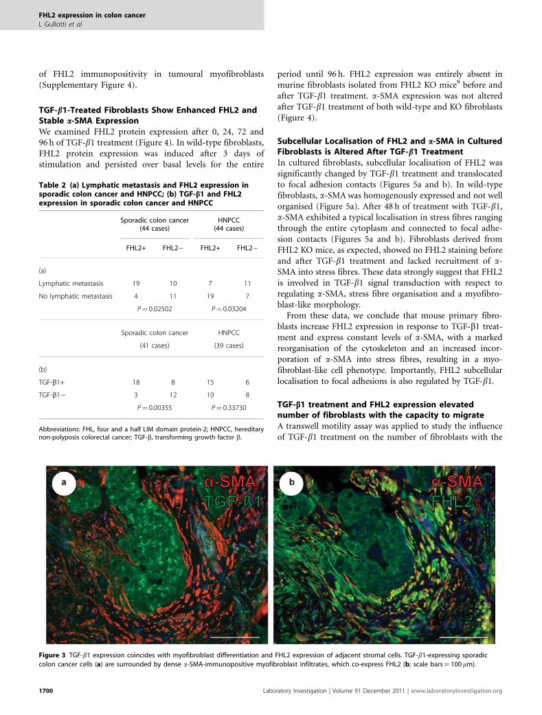
Expression of TGF-b1 ligand in tumour cells went alongwith intensive expression of FHL2 and a-SMA in adjacentstromal cells (Figure 3). A correlation of TGF-b1 ligand
expression in tumour cells with nuclear FHL2 expression ofadjacent stromal cells in the tumour invasion front (Table 2b)was detected in sporadic carcinoma cases (P¼ 0.00355) butnot in HNPCC (P¼ 0.33730). In contrast, colon carcinomas,which lacked TGF-b1 ligand expression showed absence
Figure 2 FHL2 protein is strongly expressed in peritumoural myofibroblasts. FHL2-positive myofibroblasts attached to invading tumour cells are frequently
found in sporadic colon cancer (a, c) but significantly less in HNPCC (b, d). FHL2þ /a-SMAþ myofibroblasts are marked with yellow arrows, tumour
cells are marked with red arrows. (c) Insert shows membranous expression of a-SMA and nuclear/cytoplasmic expression of FHL2 in a myofibroblast.
Note: dense DAPI-positive lymphocytic infiltrates in direct contact with HNPCC-associated carcinoma tissue in b and d, and strong membranous but nearly
absent nuclear FHL2 expression of (a-SMA-immunonegative) tumour cells in d (confocal images: scale bar in a and b: 150 mm, in c and d: 37.5 mm).
FHL2 expression in colon cancer
L Gullotti et al
www.laboratoryinvestigation.org | Laboratory Investigation | Volume 91 December 2011 1699
of FHL2 immunopositivity in tumoural myofibroblasts(Supplementary Figure 4).
TGF-b1-Treated Fibroblasts Show Enhanced FHL2 andStable a-SMA ExpressionWe examined FHL2 protein expression after 0, 24, 72 and96 h of TGF-b1 treatment (Figure 4). In wild-type fibroblasts,FHL2 protein expression was induced after 3 days ofstimulation and persisted over basal levels for the entire
period until 96 h. FHL2 expression was entirely absent inmurine fibroblasts isolated from FHL2 KO mice9 before andafter TGF-b1 treatment. a-SMA expression was not alteredafter TGF-b1 treatment of both wild-type and KO fibroblasts(Figure 4).
Subcellular Localisation of FHL2 and a-SMA in CulturedFibroblasts is Altered After TGF-b1 TreatmentIn cultured fibroblasts, subcellular localisation of FHL2 wassignificantly changed by TGF-b1 treatment and translocatedto focal adhesion contacts (Figures 5a and b). In wild-typefibroblasts, a-SMA was homogenously expressed and not wellorganised (Figure 5a). After 48 h of treatment with TGF-b1,a-SMA exhibited a typical localisation in stress fibres rangingthrough the entire cytoplasm and connected to focal adhe-sion contacts (Figures 5a and b). Fibroblasts derived fromFHL2 KO mice, as expected, showed no FHL2 staining beforeand after TGF-b1 treatment and lacked recruitment of a-SMA into stress fibres. These data strongly suggest that FHL2is involved in TGF-b1 signal transduction with respect toregulating a-SMA, stress fibre organisation and a myofibro-blast-like morphology.
From these data, we conclude that mouse primary fibro-blasts increase FHL2 expression in response to TGF-b1 treat-ment and express constant levels of a-SMA, with a markedreorganisation of the cytoskeleton and an increased incor-poration of a-SMA into stress fibres, resulting in a myo-fibroblast-like cell phenotype. Importantly, FHL2 subcellularlocalisation to focal adhesions is also regulated by TGF-b1.
TGF-b1 treatment and FHL2 expression elevatednumber of fibroblasts with the capacity to migrateA transwell motility assay was applied to study the influenceof TGF-b1 treatment on the number of fibroblasts with the
Figure 3 TGF-b1 expression coincides with myofibroblast differentiation and FHL2 expression of adjacent stromal cells. TGF-b1-expressing sporadic
colon cancer cells (a) are surrounded by dense a-SMA-immunopositive myofibroblast infiltrates, which co-express FHL2 (b; scale bars¼ 100 mm).
Table 2 (a) Lymphatic metastasis and FHL2 expression insporadic colon cancer and HNPCC; (b) TGF-b1 and FHL2expression in sporadic colon cancer and HNPCC
Sporadic colon cancer(44 cases)
HNPCC(44 cases)
FHL2+ FHL2� FHL2+ FHL2�
(a)
Lymphatic metastasis 19 10 7 11
No lymphatic metastasis 4 11 19 7
P¼ 0.02502 P¼ 0.03204
Sporadic colon cancer
(41 cases)
HNPCC
(39 cases)
(b)
TGF-b1+ 18 8 15 6
TGF-b1� 3 12 10 8
P¼ 0.00355 P¼ 0.33730
Abbreviations: FHL, four and a half LIM domain protein-2; HNPCC, hereditarynon-polyposis colorectal cancer; TGF-b, transforming growth factor b.
FHL2 expression in colon cancer
L Gullotti et al
1700 Laboratory Investigation | Volume 91 December 2011 | www.laboratoryinvestigation.org

capacity to migrate. Untreated FHL2 KO fibroblasts showedless migrating cells compared with untreated wild-type cells(P¼ 0.035, Figure 6a). A larger number of migrating cells wasseen in TGF-b1-treated wild-type fibroblasts comparedwith untreated wild-type fibroblasts (Po0.001, Figure 6a).TGF-b1 treated FHL2 KO fibroblasts showed a significantlylower number of migrating cells compared with TGF-b1treated wild-type cells (Po0.001, Figure 6a). TGF-b1-treatedKO fibroblasts showed a significantly higher number ofmigrating cells (Po0.001, Figure 6a) than untreated KOfibroblasts (Figure 6a).
In a co-culture transwell motility assay, HCT116 tumourcells were labelled with red dye and incubated with FHL2wild-type or KO fibroblasts labelled with green dye. Theco-incubation assay with wild-type fibroblasts showed moretumour cell signals than the assay co-incubated with KOfibroblasts (Supplementary Figure 5). Though, we oftenobserved a co-labelling of cells precluding a proper visualseparation of individual cells. This co-labelling of cells mightbe explained by the ability of fibroblasts to perform phago-cytosis27 or by overlap of cytoplasmic tails of two individualcells with different dye label.
Contact of Tumour Cells and Myofibroblasts In Vivo andIn VitroTo analyse the contact of FHL2-positive myofibroblasts andcolon cancer cells, paraffin-embedded human colon cancertissue was stained for FHL2 and a-SMA. A direct contact ofmyofibroblasts to tumour cells could be observed in sporadiccolon cancer in vivo (Figures 6b–d).
Furthermore, an in vitro co-culture assay was established usingsporadic (HT29) and HNPCC-derived (HCT116) colon cancercell lines. Immunofluorescence stainings revealed that murinemyofibroblasts were directly connected to tumour cells via FHL2-positive focal adhesion contacts in sporadic colon cancer cell linesas well as in HNPCC-derived cell lines (Figures 6e and f).
DISCUSSIONThis study analysed the role of the LIM-only protein FHL2in tumour progression of sporadic colon cancer and HNPCCfocussing on peritumoural myofibroblasts.
The role of FHL2 in tumorigenesis has been analysed invarious cancer type, including carcinomas of the ovary,17 thebreast,16 the prostate18 and the gastrointestinal tract.19,20
These studies mainly focussed on the role of FHL2 expressionin malignant tumour cells proper. However, in addition to itsexpression in tumour cells, FHL2 might serve as a marker forcharacterisation of myofibroblast-like stromal fibroblasts inthe tumour micromilieu. We previously determined animportant role of FHL2 in myofibroblasts during woundhealing.9 It has been shown that wounded tissue is compar-able in several aspects to the tumour stroma as both arecharacterised by activated, a-SMA-expressing myofibroblastsand deposition of provisional matrix and basal membranemolecules.11 As previous studies19,28 described an inductionof FHL2 expression by TGF-b1, this study considered bothmarkers in the systematic analysis of colorectal tumour tissueand tumour stroma in vitro and in vivo.
Previous studies reported on the relevance of TGF-b1-mediated tumour tissue remodelling for metastasis29 and alsoits specific role in progression of colon cancer.30 Therefore,we determined the expression patterns of FHL2, TGF-b1 anda-SMA, and their respective localisation in the micromilieuof sporadic and HNPCC-associated microsatellite-instablecolon cancer in vivo. Normal colon mucosa showed no sig-nificant FHL2 expression of stromal fibroblasts, a findingwhich was also described by a previous study.21 In contrast,carcinoma tissue displayed elevated expression of FHL2 instromal cells, which were identified as a-SMA-positivemyofibroblasts. Even in colon adenoma without signs ofinvasive growth myofibroblasts in direct contact to neoplasticepithelium revealed FHL2 expression indicating early pre-neoplastic stromal transformation. In 3D-reconstructionsof peritumoural myofibroblasts by confocal microscopy,FHL2 protein was found in the cytoplasm and in thenucleus of these stromal cells (Supplementary Movies 2A–C).This observation confirms previous studies, which found thatFHL2 serves as a signalling molecule and translocates intothe nucleus acting as a transcriptional coactivator andcorepressor.6,8,25,31
Importantly, according to our data stromal FHL2 over-expression and the presence of myofibroblast-like stromalcells provided a significant parameter for lymphatic meta-stasis in sporadic colon cancer. In contrast, strong stromalFHL2 expression of HNPCC-associated tumours did notshow prognostic relevance with regard to lymphatic spread,indicating that myofibroblastic remodelling of the tumourstroma may differ in these microsatellite-instable carcinomas.It is well known that HNPCC-associated colon cancers showa less invasive and more protruding, pushing-like tumourperiphery,22 a lesser degree of EMT and reveal a better stage-adjusted overall survival.
In this study, a positive correlation between TGF-b1expression of the tumour cells and FHL2 expression of thestromal cells was detected in sporadic colon cancer corres-ponding to observations of other studies.28 In contrast,
Figure 4 TGF-b1-treated fibroblasts show enhanced FHL2 and stable
a-SMA expression in western blot. Mouse fibroblasts were serum starved for
48 h and treated with control medium or medium containing TGF-b1 for
different time points as indicated. a-Tubulin served as loading control. Ko,
knockout cells; wt, wild-type cells.
FHL2 expression in colon cancer
L Gullotti et al
www.laboratoryinvestigation.org | Laboratory Investigation | Volume 91 December 2011 1701
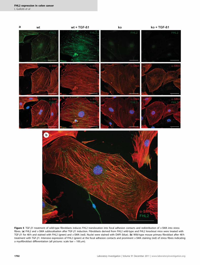
Figure 5 TGF-b1 treatment of wild-type fibroblasts induces FHL2 translocation into focal adhesion contacts and redistribution of a-SMA into stress
fibres. (a) FHL2 and a-SMA sublocalisation after TGF-b1 induction. Fibroblasts derived from FHL2 wild-type and FHL2 knockout mice were treated with
TGF-b1 for 48 h and stained with FHL2 (green) and a-SMA (red). Nuclei were stained with DAPI (blue). (b) Wild-type mouse primary fibroblast after 48 h
treatment with TGF-b1. Intensive expression of FHL2 (green) at the focal adhesion contacts and prominent a-SMA staining (red) of stress fibres indicating
a myofibroblast differentiation (all pictures: scale bar¼ 100 mm).
FHL2 expression in colon cancer
L Gullotti et al
1702 Laboratory Investigation | Volume 91 December 2011 | www.laboratoryinvestigation.org

significant correlation of TGF-b1 expression and stromalFHL2 staining was not found in HNPCC-associated cancers,indicating that TGF-�1 signalling from microsatellite-instablecolon cancer cells may be defective. A histological differencebetween sporadic and HNPCC-associated cancers is thepresence of dense lymphocytic infiltrates in the tumourmicromilieu.22 Schwitalle et al32,33 recently showed that
frameshift-induced neopeptides in HNPCC may induce thischaracteristic immune response. We therefore speculatethat TGF-b1-mediated interactions with myofibroblast-likestromal fibroblasts are altered in the presence of the immuneresponse in HNPCC.
In cell culture assays of this study, TGF-b1 inducedFHL2 protein expression in fibroblasts. After induction, a
Figure 6 TGF-b1 treatment and FHL2 expression elevate number of fibroblasts with the capacity to migrate (a) (***Po0.001, **P¼ 0.001 to o0.05).
In sporadic human colon carcinoma contact of a FHL2þ /a-SMAþ myofibroblast (yellow arrow) and a FHL2þ /a-SMA� tumour cell (red arrow) can be
observed (b, overlay; c, FHL2 staining; d, a-SMA staining with insert showing the myofibroblast in detail). In vitro, co-culture assays show direct contact of
fibroblasts (marked with yellow arrows) and tumour cells via focal adhesions in sporadic colon carcinoma cell line HT29 (marked with red arrows; e) but also
in HNPCC cell line HCT-116 (marked with red arrows; f; confocal pictures: images b–d: scale bar¼ 36mm, images e, f: scale bar¼ 50 mm).
FHL2 expression in colon cancer
L Gullotti et al
www.laboratoryinvestigation.org | Laboratory Investigation | Volume 91 December 2011 1703

myofibroblast-like differentiation was observed as describedpreviously.7,34 Myofibroblasts coexpressed a-SMA and FHL2with a characteristic expression pattern of FHL2 at focaladhesion contacts as also observed in wounded tissue.9 It istherefore likely that TGF-b1-induced, FHL2 overexpressingmyofibroblasts form a highly mobile tumour stromafavouring migration, invasive growth and dissemination ofcolon cancer cells. The leading role of myofibroblasts ininvasive growth of carcinomas has recently been described byGaggioli et al,35 who found that squamous cell carcinomacells move collectively within tracks in the extracellularmatrix behind the leading fibroblast remodelling the extra-cellular matrix. As TGF-b1 is chemotactic for fibroblasts,36
the in vitro setting described above may well be transferred toin vivo cancer development.
Our in vitro data showed an elevated number of FHL2wild-type fibroblasts with the capacity to migrate comparedwith FHL2 KO fibroblasts. This effect was also describedpreviously concerning the role of fibroblasts in wound heal-ing.9 These data point to an important role of FHL2 inmigration of fibroblasts in general. Furthermore, we showedthat treatment of fibroblasts with TGF-b1 went along with ahigher number of cells with the capacity to migrate, a findingwhich has also been reported in the literature before.15 Inco-culture assays of this study, a connection of tumour cellsto fibroblasts via focal adhesions was observed and detectedboth in sporadic and HNPCC-derived cell lines. As the denseperitumoural lymphocytic infiltrate typically found inHNPCC-associated cancers is not present in this co-cultureassay, we speculate that the tight physical interaction betweentumour cells and fibroblasts may be disturbed in vivo.
Taken together, the results of this study point to animportant role of FHL2 in tumour-induced tissue remodel-ling, invasion and lymphatic metastasis of sporadic colorectalcancer, probably mediated by TGF-b1-induced, FHL2-over-expressing myofibroblasts at the tumour invasion front. Onthe basis of our in vitro and in vivo findings, we postulate amodel that in sporadic colon cancer tumour-secreted TGF-b1induces FHL2 expression in peritumoural myofibroblastspromoting the migration of ‘leading’ tumour cells into thesurrounding stroma (Figure 7a). This phenomenon has been
Figure 6 Continued.
Figure 7 Model for interaction of FHL2-positive myofibroblasts (yellow)
and colon cancer cells (grey). In sporadic colon cancer tumour-secreted
TGF-�1 induces myofibroblast accumulation densely attached to tumour
tissue (a). In HNPCC lymphocytes (blue) separate myofibroblasts and
tumour cells (b).
FHL2 expression in colon cancer
L Gullotti et al
1704 Laboratory Investigation | Volume 91 December 2011 | www.laboratoryinvestigation.org

described previously for squamous cell carcinomas.35 Thesechanges in the tumour micromilieu are not present inHNPCC-associated carcinomas, and we speculate that theextensive lymphocytic immune response may alter theTGF-b1-mediated effects on stromal fibroblasts resulting in aless invasive growth pattern and less frequent lymphaticmetastasis (Figure 7b). As FHL2 overexpression of myo-fibroblasts in sporadic colon cancer was significantly linkedto lymphatic metastasis, we here propose FHL2 as a newprognostic marker indicating risk of lymphatic spread incolon cancers.
Supplementary Information accompanies the paper on the Laboratory
Investigation website (http://www.laboratoryinvestigation.org)
ACKNOWLEDGEMENTS
We thank Barbara Reddemann, Susanne Steiner and Christiane Esch for their
expert technical assistance. Lucia Gullotti was supported as a graduate
within the Graduiertenkolleg 804 of the Deutsche Forschungsgemeinschaft.
This work was supported by the BONFOR grant (BonFor-Program) to NF and
by a grant from the Deutsche Forschungsgemeinschaft to RB (GRK 804 and
SFB 832). This work was supported by the German Cancer Aid by a grant to
the German HNPCC Consortium (speaker: PP).
DISCLOSURE/CONFLICT OF INTEREST
The authors declare no conflict of interest.
1. Genini M, Schwalbe P, Scholl FA, et al. Subtractive cloning andcharacterization of DRAL, a novel LIM-domain protein down-regulatedin rhabdomyosarcoma. DNA Cell Biol 1997;16:433–442.
2. Bach I. The LIM domain: regulation by association. Mech Dev2000;91:5–17.
3. Martin B, Schneider R, Janetzky S, et al. The LIM-only protein FHL2interacts with beta-catenin and promotes differentiation of mousemyoblasts. J Cell Biol 2002;159:113–122.
4. Samson T, Smyth N, Janetzky S, et al. The LIM-only proteins FHL2 andFHL3 interact with alpha- and beta-subunits of the musclealpha7beta1 integrin receptor. J Biol Chem 2004;279:28641–28652.
5. Wixler V, Geerts D, Laplantine E, et al. The LIM-only protein DRAL/FHL2binds to the cytoplasmic domain of several alpha and beta integrinchains and is recruited to adhesion complexes. J Biol Chem2000;275:33669–33678.
6. Morlon A, Sassone-Corsi P. The LIM-only protein FHL2 is a serum-inducible transcriptional coactivator of AP-1. Proc Natl Acad Sci USA2003;100:3977–3982.
7. Mueller MM, Fusenig NE. Friends or foes—bipolar effects of thetumour stroma in cancer. Nat Rev Cancer 2004;4:839–849.
8. Philippar U, Schratt G, Dieterich C, et al. The SRF target gene Fhl2antagonizes RhoA/MAL-dependent activation of SRF. Mol Cell2004;16:867–880.
9. Wixler V, Hirner S, Muller JM, et al. Deficiency in the LIM-only proteinFhl2 impairs skin wound healing. J Cell Biol 2007;177:163–172.
10. Park J, Will C, Martin B, et al. Deficiency in the LIM-only protein FHL2impairs assembly of extracellular matrix proteins. FASEB J2008;22:2508–2520.
11. De Wever O, Mareel M. Role of tissue stroma in cancer cell invasion. JPathol 2003;200:429–447.
12. Hinz B. Formation and function of the myofibroblast during tissuerepair. J Invest Dermatol 2007;127:526–537.
13. Hinz B, Phan SH, Thannickal VJ, et al. The myofibroblast. One function,multiple origins. Am J Pathol 2007;170:1807–1816.
14. Ikushima H, Miyazono K. TGFbeta signalling: a complex web in cancerprogression. Nat Rev Cancer 2010;10:415–424.
15. Brenmoehl J, Miller SN, Hofmann C, et al. Transforming growth factor-beta 1 induces intestinal myofibroblast differentiation and modulatestheir migration. World J Gastroenterol 2009;15:1431–1442.
16. Gabriel B, Fischer DC, Orlowska-Volk M, et al. Expression of thetranscriptional coregulator FHL2 in human breast cancer: aclinicopathologic study. J Soc Gynecol Investig 2006;13:69–75.
17. Gabriel B, Mildenberger S, Weisser CW, et al. Focal adhesion kinaseinteracts with the transcriptional coactivator FHL2 and both areoverexpressed in epithelial ovarian cancer. Anticancer Res 2004;24:921–927.
18. Kahl P, Gullotti L, Heukamp LC, et al. Androgen receptor coactivatorslysine-specific histone demethylase 1 and four and a half LIM domainprotein 2 predict risk of prostate cancer recurrence. Cancer Res2006;66:11341–11347.
19. Zhang W, Jiang B, Guo Z, et al. Four-and-a-half LIM protein 2 promotesinvasive potential and epithelial-mesenchymal transition in coloncancer. Carcinogenesis 2010;31:1220–1229.
20. Zhang W, Wang J, Zou B, et al. Four and a half LIM protein 2 (FHL2)negatively regulates the transcription of E-cadherin throughinteraction with Snail1. Eur J Cancer 2010;47:121–130.
21. Wang J, Yang Y, Xia HH, et al. Suppression of FHL2 expression inducescell differentiation and inhibits gastric and colon carcinogenesis.Gastroenterology 2007;132:1066–1076.
22. Ruschoff J, Dietmaier W, Luttges J, et al. Poorly differentiated colonicadenocarcinoma, medullary type: clinical, phenotypic, and molecularcharacteristics. Am J Pathol 1997;150:1815–1825.
23. Friedrichs N, Jager R, Paggen E, et al. Distinct spatial expressionpatterns of AP-2alpha and AP-2gamma in non-neoplastic humanbreast and breast cancer. Mod Pathol 2005;18:431–438.
24. Friedrichs N, Steiner S, Buettner R, et al. Immunohistochemicalexpression patterns of AP2alpha and AP2gamma in the developingfetal human breast. Histopathology 2007;51:814–823.
25. Muller JM, Metzger E, Greschik H, et al. The transcriptional coactivatorFHL2 transmits Rho signals from the cell membrane into the nucleus.Embo j 2002;21:736–748.
26. Friedrichs N, Kuchler J, Endl E, et al. Insulin-like growth factor-1receptor acts as a growth regulator in synovial sarcoma. J Pathol2008;216:428–439.
27. Fadok VA, de Cathelineau A, Daleke DL, et al. Loss of phospholipidasymmetry and surface exposure of phosphatidylserine is required forphagocytosis of apoptotic cells by macrophages and fibroblasts. J BiolChem 2001;276:1071–1077.
28. Govoni KE, Amaar YG, Kramer A, et al. Regulation of insulin-like growthfactor binding protein-5, four and a half lim-2, and a disintegrin andmetalloprotease-9 expression in osteoblasts. Growth Horm IGF Res2006;16:49–56.
29. Prud0homme GJ. Pathobiology of transforming growth factor beta incancer, fibrosis and immunologic disease, and therapeutic considera-tions. Lab Invest 2007;87:1077–1091.
30. Tsushima H, Kawata S, Tamura S, et al. High levels of trans-forming growth factor beta 1 in patients with colorectal cancer:association with disease progression. Gastroenterology 1996;110:375–382.
31. Muller JM, Isele U, Metzger E, et al. FHL2, a novel tissue-specificcoactivator of the androgen receptor. EMBO J 2000;19:359–369.
32. Schwitalle Y, Kloor M, Eiermann S, et al. Immune response againstframeshift-induced neopeptides in HNPCC patients and healthyHNPCC mutation carriers. Gastroenterology 2008;134:988–997.
33. Schwitalle Y, Linnebacher M, Ripberger E, et al. Immunogenic peptidesgenerated by frameshift mutations in DNA mismatch repair-deficientcancer cells. Cancer Immun 2004;4:14.
34. Zeisberg M, Strutz F, Muller GA. Role of fibroblast activation ininducing interstitial fibrosis. J Nephrol 2000;13(Suppl 3):S111–S120.
35. Gaggioli C, Hooper S, Hidalgo-Carcedo C, et al. Fibroblast-led collectiveinvasion of carcinoma cells with differing roles for RhoGTPases inleading and following cells. Nat Cell Biol 2007;9:1392–1400.
36. Postlethwaite AE, Keski-Oja J, Moses HL, et al. Stimulation of thechemotactic migration of human fibroblasts by transforming growthfactor beta. J Exp Med 1987;165:251–256.
FHL2 expression in colon cancer
L Gullotti et al
www.laboratoryinvestigation.org | Laboratory Investigation | Volume 91 December 2011 1705
Related Documents