FGF23 Fails to Inhibit Uremic Parathyroid Glands Rocío Canalejo,* Antonio Canalejo, † Julio Manuel Martinez-Moreno,* M. Encarnacion Rodriguez-Ortiz,* Jose C. Estepa, ‡ Francisco Javier Mendoza, ‡ Juan Rafael Munoz-Castaneda,* Victoria Shalhoub, § Yolanda Almaden,* and Mariano Rodriguez* *Unidad de Investigacion, Servicio de Nefrologia, Red in ren, Instituto Maimo ´ nides de Invstigacio ´ n Biome ´ dica de Co ´ rdoba (I.M.I.B.I.C.), Hospital Universitario Reina Sofia, Departamento de Medicina, Cordoba, Spain; † Departamento de Biologia Ambiental y Salud Publica, Universidad de Huelva, Huelva, Spain; ‡ Departamento de Medicina y Cirugia Animal, Universidad de Cordoba, Cordoba, Spain; and § Department of Metabolic Disorders, Amgen, Inc., Thousand Oaks, California ABSTRACT Fibroblast growth factor 23 (FGF23) modulates mineral metabolism by promoting phosphaturia and decreasing the production of 1,25-dihydroxyvitamin D 3 . FGF23 decreases parathyroid hormone (PTH) mRNA and secretion, but despite a marked elevation in FGF23 in uremia, PTH production increases. Here, we investigated the effect of FGF23 on parathyroid function in normal and uremic hyperplastic parathyroid glands in rats. In normal parathyroid glands, FGF23 decreased PTH production, increased expression of both the parathyroid calcium-sensing receptor and the vitamin D receptor, and reduced cell proliferation. Furthermore, FGF23 induced phosphorylation of extracellular signal–regulated kinase 1/2, which mediates the action of FGF23. In contrast, in hyperplastic parathyroid glands, FGF23 did not reduce PTH production, did not affect expression of the calcium-sensing receptor or vitamin D receptor, and did not affect cell proliferation. In addition, FGF23 failed to activate the extracellular signal– regulated kinase 1/2–mitogen-activated protein kinase pathway in hyperplastic parathyroid glands. We observed very low expression of the FGF23 receptor 1 and the co-receptor Klotho in uremic hyperplastic parathyroid glands, which may explain the lack of response to FGF23 in this tissue. In conclusion, in hyperparathyroidism secondary to renal failure, the parathyroid cells resist the inhibitory effects of FGF23, perhaps as a result of the low expression of FGF23 receptor 1 and Klotho in this condition. J Am Soc Nephrol 21: 1125–1135, 2010. doi: 10.1681/ASN.2009040427 Fibroblast growth factor 23 (FGF23) is produced by bone cells and plays a fundamental role in the reg- ulation of mineral metabolism. FGF23 inhibits tubular resorption of phosphate and decreases 1 hydroxylase activity, which limits 1,25-dihy- droxyvitamin D 3 [1,25(OH) 2 D 3 ] production. Both phosphate excess and high 1,25(OH) 2 D 3 stimulate the production of FGF23. 1 FGF23 signals through a widely expressed receptor (FGFR) that becomes functional only in cells expressing the Klotho pro- tein. 2,3 Klotho, which is expressed in the parathy- roid cell, converts FGFR1(IIIc), a canonical recep- tor for various FGFs, into a specific receptor for FGF23. The tissue-specific unique biological activ- ity of FGF23 is likely to be regulated by the limited local distribution of Klotho. In renal failure, the de- crease in glomerular filtration causes phosphate re- tention, which stimulates the production of FGF23. This elevation in FGF23 levels should help to con- trol phosphate in patients with renal failure. 4 Received April 23, 2009. Accepted February 11, 2010. Published online ahead of print. Publication date available at www.jasn.org. Y.A. and M.R. share senior authorship. Correspondence: Dr. Yolanda Almade ´ n Pen ˜ a, Unidad de Inves- tigacion, Hospital Reina Sofı´a, Avenida Mene ´ ndez Pidal s/n, 14004 Co ´ rdoba, Spain. Phone: 34-957-010452; Fax: 34-957- 010452; E-mail: [email protected] Copyright © 2010 by the American Society of Nephrology BASIC RESEARCH www.jasn.org J Am Soc Nephrol 21: 1125–1135, 2010 ISSN : 1046-6673/2107-1125 1125

Welcome message from author
This document is posted to help you gain knowledge. Please leave a comment to let me know what you think about it! Share it to your friends and learn new things together.
Transcript
FGF23 Fails to Inhibit Uremic Parathyroid Glands
Rocío Canalejo,* Antonio Canalejo,† Julio Manuel Martinez-Moreno,*M. Encarnacion Rodriguez-Ortiz,* Jose C. Estepa,‡ Francisco Javier Mendoza,‡
Juan Rafael Munoz-Castaneda,* Victoria Shalhoub,§ Yolanda Almaden,* andMariano Rodriguez*
*Unidad de Investigacion, Servicio de Nefrologia, Red in ren, Instituto Maimonides de Invstigacion Biomedica deCordoba (I.M.I.B.I.C.), Hospital Universitario Reina Sofia, Departamento de Medicina, Cordoba, Spain;†Departamento de Biologia Ambiental y Salud Publica, Universidad de Huelva, Huelva, Spain; ‡Departamento deMedicina y Cirugia Animal, Universidad de Cordoba, Cordoba, Spain; and §Department of Metabolic Disorders,Amgen, Inc., Thousand Oaks, California
ABSTRACTFibroblast growth factor 23 (FGF23) modulates mineral metabolism by promoting phosphaturia anddecreasing the production of 1,25-dihydroxyvitamin D3. FGF23 decreases parathyroid hormone (PTH)mRNA and secretion, but despite a marked elevation in FGF23 in uremia, PTH production increases.Here, we investigated the effect of FGF23 on parathyroid function in normal and uremic hyperplasticparathyroid glands in rats. In normal parathyroid glands, FGF23 decreased PTH production, increasedexpression of both the parathyroid calcium-sensing receptor and the vitamin D receptor, and reducedcell proliferation. Furthermore, FGF23 induced phosphorylation of extracellular signal–regulated kinase1/2, which mediates the action of FGF23. In contrast, in hyperplastic parathyroid glands, FGF23 did notreduce PTH production, did not affect expression of the calcium-sensing receptor or vitamin D receptor,and did not affect cell proliferation. In addition, FGF23 failed to activate the extracellular signal–regulated kinase 1/2–mitogen-activated protein kinase pathway in hyperplastic parathyroid glands. Weobserved very low expression of the FGF23 receptor 1 and the co-receptor Klotho in uremic hyperplasticparathyroid glands, which may explain the lack of response to FGF23 in this tissue. In conclusion, inhyperparathyroidism secondary to renal failure, the parathyroid cells resist the inhibitory effects ofFGF23, perhaps as a result of the low expression of FGF23 receptor 1 and Klotho in this condition.
J Am Soc Nephrol 21: 1125–1135, 2010. doi: 10.1681/ASN.2009040427
Fibroblast growth factor 23 (FGF23) is produced bybone cells and plays a fundamental role in the reg-ulation of mineral metabolism. FGF23 inhibitstubular resorption of phosphate and decreases 1�hydroxylase activity, which limits 1,25-dihy-droxyvitamin D3 [1,25(OH)2D3] production. Bothphosphate excess and high 1,25(OH)2D3 stimulatethe production of FGF23.1 FGF23 signals through awidely expressed receptor (FGFR) that becomesfunctional only in cells expressing the Klotho pro-tein.2,3 Klotho, which is expressed in the parathy-roid cell, converts FGFR1(IIIc), a canonical recep-tor for various FGFs, into a specific receptor forFGF23. The tissue-specific unique biological activ-ity of FGF23 is likely to be regulated by the limited
local distribution of Klotho. In renal failure, the de-crease in glomerular filtration causes phosphate re-tention, which stimulates the production of FGF23.This elevation in FGF23 levels should help to con-trol phosphate in patients with renal failure.4
Received April 23, 2009. Accepted February 11, 2010.
Published online ahead of print. Publication date available atwww.jasn.org.
Y.A. and M.R. share senior authorship.
Correspondence: Dr. Yolanda Almaden Pena, Unidad de Inves-tigacion, Hospital Reina Sofıa, Avenida Menendez Pidal s/n,14004 Cordoba, Spain. Phone: �34-957-010452; Fax: �34-957-010452; E-mail: [email protected]
Copyright © 2010 by the American Society of Nephrology
BASIC RESEARCH www.jasn.org
J Am Soc Nephrol 21: 1125–1135, 2010 ISSN : 1046-6673/2107-1125 1125
Klotho and FGFR are abundantly expressed in parathyroidcells. Some studies5,6 showed that FGF23 decreases parathyroidhormone (PTH) secretion and PTH mRNA. In dialysis pa-tients, FGF23 levels can reach extremely high values4,7 but PTHis not reduced; in fact, the highest PTH values correspond topatients with a marked increase in FGF23 levels.8 Thus, it is notclear whether FGF23 is capable of reducing PTH production inuremia. Our hypothesis is that there may be a resistance to theaction of FGF23 in patients with uremia.
This study was designed to evaluate the effect of FGF23 onparathyroid function in normal and hyperplastic parathyroidglands. The study was performed in vivo and in vitro usingintact rat parathyroid glands from normal and uremic animalswith parathyroid hyperplasia.
RESULTS
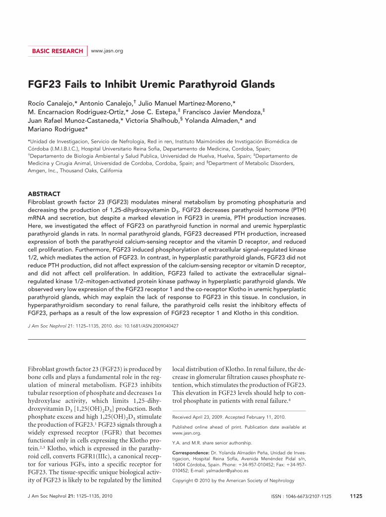
FGF23 Reduced PTH Secretion in Normal RatParathyroid GlandsA change in calcium (Ca) concentration in the medium fromnormal (1.25 mM) to low (0.8 mM) produced a two-fold increasein PTH secretion. Addition of FGF23 (200 nM; 5 � 103 ng/ml) tothe low-Ca medium produced a sustained reduction of PTH se-cretion. With a high Ca concentration in the medium, the addi-tion of FGF23 did not further reduce PTH secretion (Figure 1A).
The expression of PTH mRNA in high-Ca medium (1.5mM) was reduced as compared with low-Ca medium (0.8 mM;Figure 1B). The addition of FGF23 to the low-Ca mediummarkedly decreased PTH mRNA, whereas FGF23 did not fur-ther reduce PTH mRNA in high-Ca medium. Furthermore,both vitamin D receptor (VDR) and Ca-sensing receptor(CaR) mRNA were significantly increased in the high-Ca me-dium as compared with the low-Ca medium (Figure 1B); ad-dition of FGF23 to the low-Ca medium increased VDR andCaR mRNA to levels not different from those observed in thehigh-Ca concentration.
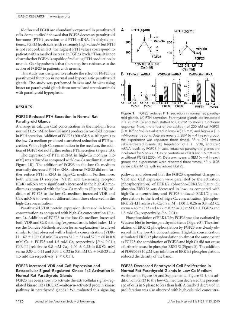
Parathyroid VDR protein expression decreased in low-Caconcentration as compared with high-Ca concentration (Fig-ure 2). Addition of FGF23 to the low-Ca medium increasedboth VDR and CaR staining (expressed as the label index [LI];see the Concise Methods section for an explanation) to a levelsimilar to that observed with a high-Ca concentration (VDR-LI: 167 � 10 in 0.8 mM Ca versus 510 � 51 and 520 � 60 in 0.8mM Ca � FGF23 and 1.5 mM Ca, respectively [P � 0.01];CaR-LI [relative to 0.8 mM Ca]: 1.00 � 0.23 in 0.8 Ca mMversus 3.03 � 0.41 and 3.34 � 0.32 in 0.8 mM Ca � FGF23 and1.5 mM Ca respectively [P � 0.01]).
FGF23 Increased VDR and CaR Expression andExtracellular Signal–Regulated Kinase 1/2 Activation inNormal Rat Parathyroid GlandsFGF23 has been shown to activate the extracellular signal–reg-ulated kinase 1/2 (ERK1/2)–mitogen-activated protein kinasepathway in parathyroid glands.5 We evaluated this signaling
pathway and observed that the FGF23-dependent changes inVDR and CaR expression were paralleled by the activation(phosphorylation) of ERK1/2 (phospho-ERK1/2; Figure 2);phospho-ERK1/2 was decreased in low- as compared withhigh-Ca concentration, and FGF23 induced ERK1/2 phos-phorylation to the level of high-Ca concentration (phospho-ERK1/2-LI [relative to Ca 0.8 mM]: 1.00 � 0.26 in 0.8 mM Caversus 4.45 � 0.23 and 4.27 � 0.27 in 0.8 mM Ca � FGF23 and1.5 mM Ca, respectively; P � 0.01).
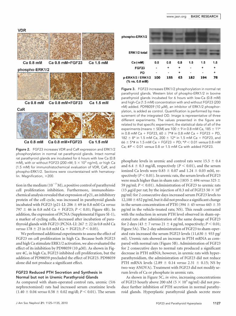
Phosphorylation of ERK1/2 by FGF23 was also evaluated byWestern blot in bovine parathyroid tissue (Figure 3). The stim-ulation of ERK1/2 phosphorylation by FGF23 was clearly ob-served in the low-Ca concentration. High-Ca concentrationstimulated ERK1/2 phosphorylation to almost the same extentas FGF23; the combination of FGF23 and high Ca did not causea further increase in phospho-ERK1/2 (Figure 3). The additionof PD98059 (10 �M), an inhibitor of ERK1/2 phosphorylation,reduced the density of the band.
FGF23 Decreased Parathyroid Cell Proliferation inNormal Rat Parathyroid Glands in Low-Ca MediumAs shown in Figure 4A and Supplemental Figure SI-1, the ad-dition of FGF23 to the low-Ca medium decreased the percent-age of cells in S phase to less than half. A marked decreased inproliferation was also observed with high calcitriol concentra-
Figure 1. FGF23 reduces PTH secretion in normal rat parathy-roid glands. (A) PTH secretion. Parathyroid glands are incubatedin 1.25 mM Ca and then shifted to 0.8 mM to show a functionalresponse. Next, the effect of the addition of 200 nM rat FGF23(5 � 103 ng/ml) is evaluated in low-Ca (0.8 mM) and high-Ca (1.5mM) concentrations. Data are means � SEM (n � 4 in each group;the experiment was repeated three times). #P � 0.01 versusvehicle-treated glands. (B) Regulation of PTH, VDR, and CaRmRNA levels by FGF23 in vitro. Intact rat parathyroid glands areincubated for 6 hours in Ca concentrations of 0.8 and 1.5 mM withor without FGF23 (200 nM). Data are means � SEM (n � 4 in eachgroup; the experiments were repeated three times). *P � 0.05versus 0.8 mM Ca with no added FGF23.
BASIC RESEARCH www.jasn.org
1126 Journal of the American Society of Nephrology J Am Soc Nephrol 21: 1125–1135, 2010
tion in the medium (10�7 M), a positive control of parathyroidcell proliferation inhibition. Furthermore, immunohisto-chemical analysis revealed that expression of p21, an inhibitoryprotein of the cell cycle, was increased in parathyroid glandsincubated with FGF23 (p21-LI: 206 � 69 in 0.8 mM Ca versus797 � 46 in 0.8 mM Ca � FGF23; P � 0.01; Figure 4B). Inaddition, the expression of PCNA (Supplemental Figure SI-1),a marker of cycling cells, decreased after incubation of para-thyroid glands with FGF23 (PCNA-LI: 267 � 22 in 0.8 mM Caversus 178 � 25 in 0.8 mM Ca � FGF23; P � 0.05).
We performed additional experiments to assess the effect ofFGF23 on cell proliferation in high Ca. Because both FGF23and high Ca stimulate ERK1/2 activation, we also evaluated theeffect of its inhibition by PD98059 (10 �M). As shown in Fig-ure 4C, in high Ca, FGF23 inhibited cell proliferation, but theaddition of PD98059 precluded the effect of FGF23. PD98059alone did not produce a significant effect.
FGF23 Reduced PTH Secretion and Synthesis inNormal but not in Uremic Parathyroid GlandsAs compared with sham-operated control rats, uremic (5/6nephrectomized) rats had increased serum creatinine levels(1.40 � 0.04 versus 0.58 � 0.02 mg/dl; P � 0.01). The serum
phosphate levels in uremic and control rats were 15.5 � 0.4and 6.4 � 0.3 mg/dl, respectively (P � 0.01), and the serumionized Ca levels were 0.83 � 0.07 and 1.24 � 0.03 mM, re-spectively (P � 0.01). In uremic rats, the serum levels of FGF23were much higher than in sham rats (1835 � 694 versus 325 �59 pg/ml; P � 0.01). Administration of FGF23 to uremic rats(15 �g/d per rat; by the injection of 0.3 ml of FGF23 50 � 106
pg/ml) for 2 consecutive days increased serum FGF23 levels to12,100 � 652 pg/ml, but it did not produce a significant changein the serum concentration of PTH (394 � 45 versus 445 � 35pg/ml in the vehicle-treated rats; NS). This result contrastedwith the reduction in serum PTH level observed in sham-op-erated rats after administration of the same dosage of FGF23for 2 days (43 � 7 versus 21 � 4 pg/ml, respectively; P � 0.01;Figure 5A). The 2-day administration of FGF23 to sham-oper-ated rats increased the serum FGF23 levels (11,630 � 933 pg/ml). Uremic rats showed an increase in PTH mRNA as com-pared with normal rats (Figure 5B). Administration of FGF23for 2 consecutive days to normal rats produced a significantdecrease in PTH mRNA; however, in uremic rats with hyper-parathyroidism, the administration of FGF23 did not reducePTH mRNA levels (2.89 � 0.14 versus 2.31 � 0.15; NS bytwo-way ANOVA). Treatment with FGF23 did not modify se-rum levels of Ca or phosphate in uremic rats.
As shown in Figure 5C, in vitro, increasing concentrationsof FGF23 hourly above 200 nM (5 � 103 ng/ml) did not pro-duce further inhibition of PTH secretion in normal parathy-roid glands. Hyperplastic parathyroid glands secrete more
Figure 2. FGF23 increases VDR and CaR expression and ERK1/2phosphorylation in normal rat parathyroid glands. Intact normalrat parathyroid glands are incubated for 6 hours with low Ca (0.8mM), with or without FGF23 (200 nM; 5 � 103 ng/ml), or high Ca(1.5 mM) for immunohistochemical evaluation of VDR, CaR, andphospho-ERK1/2. Sections were counterstained with hematoxy-lin. Magnification, �200.
Figure 3. FGF23 increases ERK1/2 phosphorylation in normal ratparathyroid glands. Western blot of phospho-ERK1/2 in bovineparathyroid glands incubated for 6 hours with low-Ca (0.8 mM)and high-Ca (1.5 mM) concentration with and without FGF23 (200nM) added. PD98059 (10 �M), an inhibitor of ERK1/2 phosphor-ylation, is added as control. Quantification is performed by mea-surement of the integrated OD. Image is representative of threedifferent experiments. The values presented in the figure arerelated to that specific experiment; the statistical data of all of theexperiments (means � SEM) are 100 � 9 in 0.8 mM Ca, 185 � 11*in 0.8 mM Ca � FGF23, 60 � 7*# in 0.8 mM Ca � FGF23 � PD,182 � 8* in 1.5 mM Ca, 200 � 12* in 1.5 mM Ca � FGF23, and66 � 5*# in 1.5 mM Ca � FGF23 � PD; *P � 0.01 versus 0.8 mMCa; #P � 0.01 versus 0.8 or 1.5 mM Ca with added FGF23.
BASIC RESEARCHwww.jasn.org
J Am Soc Nephrol 21: 1125–1135, 2010 FGF23 and Parathyroid Hyperplasia 1127
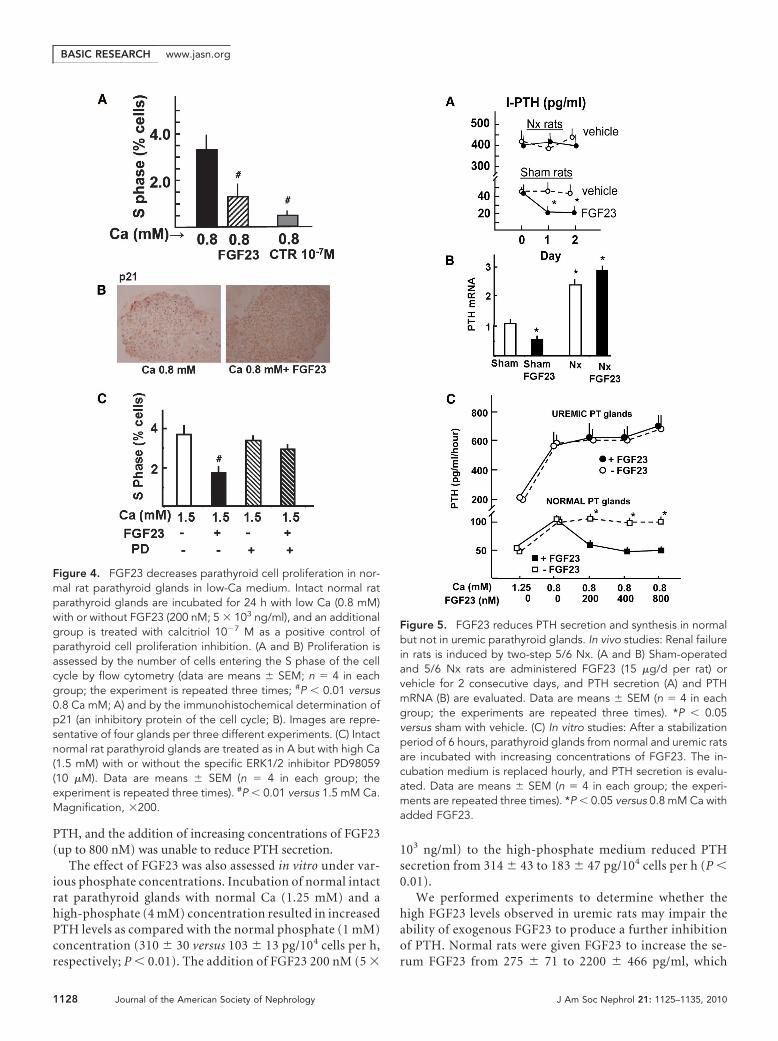
PTH, and the addition of increasing concentrations of FGF23(up to 800 nM) was unable to reduce PTH secretion.
The effect of FGF23 was also assessed in vitro under var-ious phosphate concentrations. Incubation of normal intactrat parathyroid glands with normal Ca (1.25 mM) and ahigh-phosphate (4 mM) concentration resulted in increasedPTH levels as compared with the normal phosphate (1 mM)concentration (310 � 30 versus 103 � 13 pg/104 cells per h,respectively; P � 0.01). The addition of FGF23 200 nM (5 �
103 ng/ml) to the high-phosphate medium reduced PTHsecretion from 314 � 43 to 183 � 47 pg/104 cells per h (P �0.01).
We performed experiments to determine whether thehigh FGF23 levels observed in uremic rats may impair theability of exogenous FGF23 to produce a further inhibitionof PTH. Normal rats were given FGF23 to increase the se-rum FGF23 from 275 � 71 to 2200 � 466 pg/ml, which
Figure 4. FGF23 decreases parathyroid cell proliferation in nor-mal rat parathyroid glands in low-Ca medium. Intact normal ratparathyroid glands are incubated for 24 h with low Ca (0.8 mM)with or without FGF23 (200 nM; 5 � 103 ng/ml), and an additionalgroup is treated with calcitriol 10�7 M as a positive control ofparathyroid cell proliferation inhibition. (A and B) Proliferation isassessed by the number of cells entering the S phase of the cellcycle by flow cytometry (data are means � SEM; n � 4 in eachgroup; the experiment is repeated three times; #P � 0.01 versus0.8 Ca mM; A) and by the immunohistochemical determination ofp21 (an inhibitory protein of the cell cycle; B). Images are repre-sentative of four glands per three different experiments. (C) Intactnormal rat parathyroid glands are treated as in A but with high Ca(1.5 mM) with or without the specific ERK1/2 inhibitor PD98059(10 �M). Data are means � SEM (n � 4 in each group; theexperiment is repeated three times). #P � 0.01 versus 1.5 mM Ca.Magnification, �200.
Figure 5. FGF23 reduces PTH secretion and synthesis in normalbut not in uremic parathyroid glands. In vivo studies: Renal failurein rats is induced by two-step 5/6 Nx. (A and B) Sham-operatedand 5/6 Nx rats are administered FGF23 (15 �g/d per rat) orvehicle for 2 consecutive days, and PTH secretion (A) and PTHmRNA (B) are evaluated. Data are means � SEM (n � 4 in eachgroup; the experiments are repeated three times). *P � 0.05versus sham with vehicle. (C) In vitro studies: After a stabilizationperiod of 6 hours, parathyroid glands from normal and uremic ratsare incubated with increasing concentrations of FGF23. The in-cubation medium is replaced hourly, and PTH secretion is evalu-ated. Data are means � SEM (n � 4 in each group; the experi-ments are repeated three times). *P � 0.05 versus 0.8 mM Ca withadded FGF23.
BASIC RESEARCH www.jasn.org
1128 Journal of the American Society of Nephrology J Am Soc Nephrol 21: 1125–1135, 2010
decreased the serum PTH levels from 43 � 7 to 24 � 5 pg/ml(P � 0.01). Additional administration of FGF23, which in-creased serum FGF23 to 10,780 � 1028 pg/ml, did not resultin a significant further reduction in PTH (SupplementalFigure SI-2).
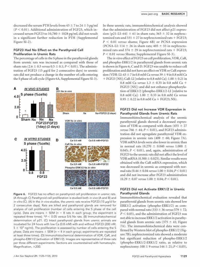
FGF23 Had No Effect on the Parathyroid CellProliferation in Uremic RatsThe percentage of cells in the S phase in the parathyroid glandsfrom uremic rats was increased as compared with those ofsham rats (2.6 � 0.5 versus 0.5 � 0.1; P � 0.01). The adminis-tration of FGF23 (15 �g/d for 2 consecutive days) to uremicrats did not produce a change in the number of cells enteringthe S phase of cell cycle (Figure 6A, Supplemental Figure SI-3).
In these uremic rats, immunohistochemical analysis showedthat the administration of FGF23 did not affect p21 expres-sion (p21-LI: 641 � 61 in sham rats; 365 � 32 in nephrec-tomized rats and 331 � 27 in nephrectomized rats � FGF23;P � 0.01 versus shams; Figure 6B) or PCNA expression(PCNA-LI: 114 � 26 in sham rats; 405 � 33 in nephrecto-mized rats and 374 � 29 in nephrectomized rats � FGF23;P � 0.01 versus Shams; Supplemental Figure SI-3).
The in vitro effect of FGF23 on cell proliferation, VDR, CaR,and phospho-ERK1/2 in parathyroid glands from uremic ratsis shown in Figure 6, C and D. FGF23 was unable to reduce cellproliferation and did not have an effect on VDR or CaR expres-sion (VDR-LI: 43 � 7 in 0.8 mM Ca versus 39 � 9 in 0.8 mM Ca� FGF23 [NS]; CaR-LI [relative to 0.8 mM Ca]: 1.00 � 0.21 in
0.8 mM Ca versus 1.3 � 0.35 in 0.8 mM Ca �FGF23 [NS]) and did not enhance phosphoryla-tion of ERK1/2 (phospho-ERK1/2-LI [relative to0.8 mM Ca]: 1.00 � 0.33 in 0.8 mM Ca versus0.95 � 0.22 in 0.8 mM Ca � FGF23; NS).
FGF23 Did not Increase VDR Expression inParathyroid Glands from Uremic RatsImmunohistochemical analysis of the uremicparathyroid glands showed a decreased expres-sion of VDR as compared with sham (455 � 27versus 766 � 44; P � 0.01), and FGF23 adminis-tration did not upregulate parathyroid VDR ex-pression in uremic rats (409 � 60; Figure 7A).VDR mRNA levels were also lower in uremic thanin normal rats (0.270 � 0.040 versus 1.000 �0.045; P � 0.01), and, again, administration ofFGF23 to the uremic rats did not affect the level ofVDR mRNA (0.300 � 0.025). Similar results wereobtained with the CaR mRNA expression, whichwas decreased in uremic as compared with nor-mal rats (0.44 � 0.04 versus 1.00 � 0.04; P � 0.01)and did not increase after FGF23 administration(0.29 � 0.07 versus 1.00 � 0.04; P � 0.01).
FGF23 Did not Activate ERK1/2 in UremicParathyroid GlandsImmunohistochemical evaluation revealed thatparathyroid glands from uremic rats showed lowERK1/2 activation (phospho-ERK1/2) as com-pared with normal rats (333 � 36 versus 579 � 72;P � 0.05), and the administration of FGF23 wasnot able to increase ERK1/2 activation in parathy-roid glands from uremic rats (375 � 41; Figure7A). The immunohistochemical data were con-firmed by Western blot of phospho-ERK1/2 (Fig-ure 7B); nephrectomized rats showed a moderatebut significant reduction of phospho-ERK1/2(phospho-ERK1/2-ERK1/2 ratio, as relative tonephrectomy: 100 � 9 versus 144 � 21; P � 0.05),
Figure 6. FGF23 has no effect on parathyroid cell proliferation in uremic rats.(A through C) Parathyroid cell proliferation is studied both in vivo (A and B) andin vitro (C). (A) In the in vivo studies, the uremic rats receive FGF23 (15 �g/d for2 consecutive days). Rats are killed and parathyroid glands are removed foranalysis of cell proliferation (number of cells entering the S phase of the cellcycle). Data are means � SEM (n � 8 rats in each group; the experiment isrepeated three times). *P � 0.05 versus 5/6 Nx rats. (B) Immunohistochemicaldetermination of p21. (C) Intact parathyroid glands from uremic animals areincubated for 24 hours with low Ca (0.8 mM) with and without FGF23 (200 nM;5 � 103 ng/ml). The proliferation is assessed by number of cells entering the Sphase. Data are means � SEM (n � 4 in each group; experiments are repeatedat least three times). (D) Immunohistochemical determination of VDR, CaR, andphospho-ERK1/2 (activation of ERK1/2). Images are representative of three ratsper three different experiments. Sections are counterstained with hematoxylin.Magnification, �200.
BASIC RESEARCHwww.jasn.org
J Am Soc Nephrol 21: 1125–1135, 2010 FGF23 and Parathyroid Hyperplasia 1129
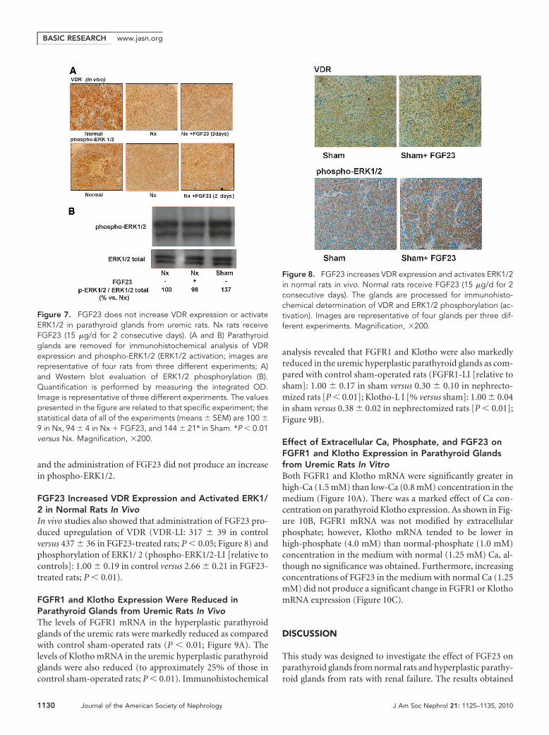
and the administration of FGF23 did not produce an increasein phospho-ERK1/2.
FGF23 Increased VDR Expression and Activated ERK1/2 in Normal Rats In VivoIn vivo studies also showed that administration of FGF23 pro-duced upregulation of VDR (VDR-LI: 317 � 39 in controlversus 437 � 36 in FGF23-treated rats; P � 0.05; Figure 8) andphosphorylation of ERK1/ 2 (phospho-ERK1/2-LI [relative tocontrols]: 1.00 � 0.19 in control versus 2.66 � 0.21 in FGF23-treated rats; P � 0.01).
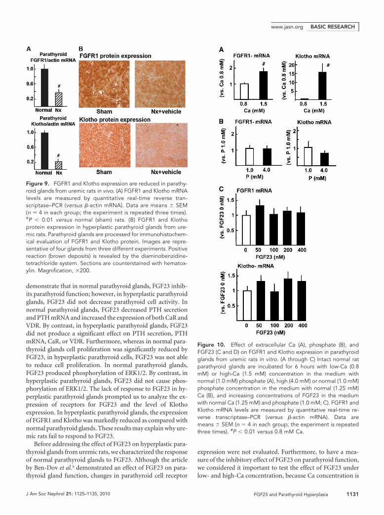
FGFR1 and Klotho Expression Were Reduced inParathyroid Glands from Uremic Rats In VivoThe levels of FGFR1 mRNA in the hyperplastic parathyroidglands of the uremic rats were markedly reduced as comparedwith control sham-operated rats (P � 0.01; Figure 9A). Thelevels of Klotho mRNA in the uremic hyperplastic parathyroidglands were also reduced (to approximately 25% of those incontrol sham-operated rats; P � 0.01). Immunohistochemical
analysis revealed that FGFR1 and Klotho were also markedlyreduced in the uremic hyperplastic parathyroid glands as com-pared with control sham-operated rats (FGFR1-LI [relative tosham]: 1.00 � 0.17 in sham versus 0.30 � 0.10 in nephrecto-mized rats [P � 0.01]; Klotho-L I [% versus sham]: 1.00 � 0.04in sham versus 0.38 � 0.02 in nephrectomized rats [P � 0.01];Figure 9B).
Effect of Extracellular Ca, Phosphate, and FGF23 onFGFR1 and Klotho Expression in Parathyroid Glandsfrom Uremic Rats In VitroBoth FGFR1 and Klotho mRNA were significantly greater inhigh-Ca (1.5 mM) than low-Ca (0.8 mM) concentration in themedium (Figure 10A). There was a marked effect of Ca con-centration on parathyroid Klotho expression. As shown in Fig-ure 10B, FGFR1 mRNA was not modified by extracellularphosphate; however, Klotho mRNA tended to be lower inhigh-phosphate (4.0 mM) than normal-phosphate (1.0 mM)concentration in the medium with normal (1.25 mM) Ca, al-though no significance was obtained. Furthermore, increasingconcentrations of FGF23 in the medium with normal Ca (1.25mM) did not produce a significant change in FGFR1 or KlothomRNA expression (Figure 10C).
DISCUSSION
This study was designed to investigate the effect of FGF23 onparathyroid glands from normal rats and hyperplastic parathy-roid glands from rats with renal failure. The results obtained
Figure 7. FGF23 does not increase VDR expression or activateERK1/2 in parathyroid glands from uremic rats. Nx rats receiveFGF23 (15 �g/d for 2 consecutive days). (A and B) Parathyroidglands are removed for immunohistochemical analysis of VDRexpression and phospho-ERK1/2 (ERK1/2 activation; images arerepresentative of four rats from three different experiments; A)and Western blot evaluation of ERK1/2 phosphorylation (B).Quantification is performed by measuring the integrated OD.Image is representative of three different experiments. The valuespresented in the figure are related to that specific experiment; thestatistical data of all of the experiments (means � SEM) are 100 �9 in Nx, 94 � 4 in Nx � FGF23, and 144 � 21* in Sham. *P � 0.01versus Nx. Magnification, �200.
Figure 8. FGF23 increases VDR expression and activates ERK1/2in normal rats in vivo. Normal rats receive FGF23 (15 �g/d for 2consecutive days). The glands are processed for immunohisto-chemical determination of VDR and ERK1/2 phosphorylation (ac-tivation). Images are representative of four glands per three dif-ferent experiments. Magnification, �200.
BASIC RESEARCH www.jasn.org
1130 Journal of the American Society of Nephrology J Am Soc Nephrol 21: 1125–1135, 2010
demonstrate that in normal parathyroid glands, FGF23 inhib-its parathyroid function; however, in hyperplastic parathyroidglands, FGF23 did not decrease parathyroid cell activity. Innormal parathyroid glands, FGF23 decreased PTH secretionand PTH mRNA and increased the expression of both CaR andVDR. By contrast, in hyperplastic parathyroid glands, FGF23did not produce a significant effect on PTH secretion, PTHmRNA, CaR, or VDR. Furthermore, whereas in normal para-thyroid glands cell proliferation was significantly reduced byFGF23, in hyperplastic parathyroid cells, FGF23 was not ableto reduce cell proliferation. In normal parathyroid glands,FGF23 produced phosphorylation of ERK1/2. By contrast, inhyperplastic parathyroid glands, FGF23 did not cause phos-phorylation of ERK1/2. The lack of response to FGF23 in hy-perplastic parathyroid glands prompted us to analyze the ex-pression of receptors for FGF23 and the level of Klothoexpression. In hyperplastic parathyroid glands, the expressionof FGFR1 and Klotho was markedly reduced as compared withnormal parathyroid glands. These results may explain why ure-mic rats fail to respond to FGF23.
Before addressing the effect of FGF23 on hyperplastic para-thyroid glands from uremic rats, we characterized the responseof normal parathyroid glands to FGF23. Although the articleby Ben-Dov et al.5 demonstrated an effect of FGF23 on para-thyroid gland function, changes in parathyroid cell receptor
expression were not evaluated. Furthermore, to have a mea-sure of the inhibitory effect of FGF23 on parathyroid function,we considered it important to test the effect of FGF23 underlow- and high-Ca concentration, because Ca concentration is
Figure 9. FGFR1 and Klotho expression are reduced in parathy-roid glands from uremic rats in vivo. (A) FGFR1 and Klotho mRNAlevels are measured by quantitative real-time reverse tran-scriptase–PCR (versus �-actin mRNA). Data are means � SEM(n � 4 in each group; the experiment is repeated three times).#P � 0.01 versus normal (sham) rats. (B) FGFR1 and Klothoprotein expression in hyperplastic parathyroid glands from ure-mic rats. Parathyroid glands are processed for immunohistochem-ical evaluation of FGFR1 and Klotho protein. Images are repre-sentative of four glands from three different experiments. Positivereaction (brown deposits) is revealed by the diaminobenzidine-tetrachloride system. Sections are counterstained with hematox-ylin. Magnification, �200.
Figure 10. Effect of extracellular Ca (A), phosphate (B), andFGF23 (C and D) on FGFR1 and Klotho expression in parathyroidglands from uremic rats in vitro. (A through C) Intact normal ratparathyroid glands are incubated for 6 hours with low-Ca (0.8mM) or high-Ca (1.5 mM) concentration in the medium withnormal (1.0 mM) phosphate (A), high (4.0 mM) or normal (1.0 mM)phosphate concentration in the medium with normal (1.25 mM)Ca (B), and increasing concentrations of FGF23 in the mediumwith normal Ca (1.25 mM) and phosphate (1.0 mM; C). FGFR1 andKlotho mRNA levels are measured by quantitative real-time re-verse transcriptase–PCR (versus �-actin mRNA). Data aremeans � SEM (n � 4 in each group; the experiment is repeatedthree times). #P � 0.01 versus 0.8 mM Ca.
BASIC RESEARCHwww.jasn.org
J Am Soc Nephrol 21: 1125–1135, 2010 FGF23 and Parathyroid Hyperplasia 1131
the main regulator of parathyroid function. Our experimentsdemonstrated that FGF23 decreased PTH secretion and PTHmRNA. These results confirm previous data reported by Ben-Dov et al.5 and Krajisnik et al.6 It is important to note that thedegree of inhibition of PTH secretion and PTH mRNA byFGF23 is similar to that observed by high Ca. In vitro experi-ments showed that in high-Ca concentration, FGF23 was un-able to reduce further the PTH secretion. Our interpretation isthat FGF23 is able to prevent an increase in PTH secretion andsynthesis when cells are being stimulated by hypocalcemia. Invivo, FGF23 reduced PTH secretion in normal rats with normalserum Ca, suggesting that high FGF23 levels inhibit parathy-roid function even with a normal serum Ca concentration.High phosphate has been shown to stimulate PTH productionand secretion9 –11; in vivo, an elevation of FGF23 induced byhigh phosphate may ameliorate the stimulation of PTH secre-tion by phosphate. It is intriguing that in our study, acute ad-ministration of exogenous FGF23 suppressed parathyroidgland function in normal rats, whereas transgenic mice over-expressing FGF23 developed secondary hyperparathyroidism;however, there are elements that can be affected by chronicelevation of FGF23 and that may contribute to secondary hy-perparathyroidism such as low 1,25(OH)2D3 and perhaps lowserum Ca. In uremic rats with parathyroid hyperplasia, evenvery high levels of FGF23 did not decrease PTH secretion orPTH mRNA either in vivo or in vitro. Thus. the lack of responseto exogenous FGF23 in nephrectomized rats demonstrates aresistance of the hyperplastic parathyroid gland to the action ofFGF23.
Uremic rats had high serum phosphate levels; we explored apotential effect of high serum phosphate concentration on theparathyroid response to FGF23. Thus, parathyroid glands wereincubated in high phosphate concentration. As expected, highphosphate stimulated PTH secretion, and FGF23 was effectivein reducing PTH secretion toward normal levels; high phos-phate does not interfere with the inhibitory action of FGF23 onthe parathyroid glands. These results suggest that the hyper-phosphatemia associated with renal failure was not the cause ofthe lack of parathyroid response to FGF23. Furthermore, invitro experiments showed that high phosphate did not decreaseFGFR1 or Klotho mRNA significantly.
Another significant effect of FGF23 was the upregulation ofCaR and VDR expression in normal parathyroid glands. Theactivation of these receptors results in the inhibition of para-thyroid cell activity; therefore, the upregulation of CaR andVDR may represent another way to reduce parathyroid func-tion. An upregulation of VDR and CaR was also observed inparathyroid glands incubated with a high Ca concentration;these results confirm previous published observations.12,13 Theaddition of FGF23 to high Ca did not further increase the ex-pression of parathyroid CaR and VDR. In uremic hyperplasticparathyroid glands, the expression of VDR and CaR is re-duced14 –16; the administration of FGF23 to rats with renal fail-ure failed to increase parathyroid ERK1/2 activity and did nothave an effect on the expression of VDR or CaR.
One important question addressed in this study waswhether FGF23 had an effect on parathyroid cell proliferation.In normal parathyroid glands, FGF23 decreased the number ofcells in S phase and enhanced expression of p21. Furthermore,ERK1/2 phosphorylation may mediate the reduction of cellproliferation induced by FGF23, because the specific inhibi-tion of ERK1/2 by PD98059 precluded the effect of FGF23. Bycontrast, in vivo and in vitro studies showed that FGF23 failedto suppress parathyroid cell proliferation in uremic hyperplas-tic parathyroid glands.
Ben-Dov et al.5 reported that FGFR1 and FGFR3 were co-expressed with Klotho in parathyroid glands. FGFR1 has beenshown to be the primary physiologic receptor for FGF23;FGFR1 is associated to a heterodimeric, high-affinity receptorfor FGF23.2,3 Liu et al.17 reported co-localization of FGFR1 andKlotho in the distal tubule, which suggests that this may be aneffector site of FGF23. In this study, we evaluated the expres-sion of both FGFR1 and Klotho. The inability of hyperplasticparathyroid glands to respond to FGF23 may be explained bythe fact that hyperplastic parathyroid cells from uremic ratshad very low expression of both FGFR1 and Klotho mRNA andthere was a clear decrease in FGFR1 protein level. Thus, de-creased expression of either FGFR1 or Klotho would have beenenough to obtain a resistance of uremic rats to the action ofFGF23. This is the first report showing a decrease in FGFR1and Klotho in hyperplastic parathyroid glands from uremicrats. A similar decrease in FGFR1 and Klotho was also observedin the remnant kidney (data not shown), supporting a previousobservation by others18; however, this study does not rule outthat the resistance to FGF23 in uremic hyperparathyroidismcould be in part due to the persistent elevation of FGF23.
Bjorklund et al.19 reported that Klotho mRNA expression isdecreased in primary parathyroid adenomas; they suggestedthat the high serum Ca may be causing the decrease in KlothomRNA expression. We show a decrease in Klotho expression insecondary parathyroid hyperplasia, which begs the question ofwhether increased parathyroid cell proliferation (hyperplasia)is associated with a decrease in Klotho expression. It should bestressed that the autonomous parathyroid proliferation andfunction in the primary hyperparathyroidism are differentfrom the uremic hyperparathyroidism. Primary and secondaryhyperparathyroidism both are characterized by parathyroidhyperplasia, but in primary hyperparathyroidism the serumCa is high and in secondary hyperparathyroidism the serum Cais low. Furthermore, Bjorklund et al.19 showed in vitro in bo-vine parathyroid cells in culture that high Ca in a dose-re-sponse manner decreased Klotho protein. Nevertheless, wefound that, in vitro, low Ca was associated with a decrease inKlotho mRNA expression; however, we did not perform ex-periments using Ca levels �1.5 mM.
In mice, Imura et al.20 found a molecular association ofKlotho and Na�, K�-ATPase; the lack of Klotho in a kl�/�
mouse impairs the secretion of PTH in response to low Ca,suggesting that Klotho may be required for Na�,K�-ATPase–dependent PTH release. In our study, we showed a decreased
BASIC RESEARCH www.jasn.org
1132 Journal of the American Society of Nephrology J Am Soc Nephrol 21: 1125–1135, 2010
expression of Klotho in hyperplastic parathyroid glands, whichwould prevent an inhibitory effect of FGF23 on PTH produc-tion. The relevance of Klotho in parathyroid cell function isillustrated by the marked effect of extracellular Ca on the reg-ulation of parathyroid Klotho expression.
Increased serum FGF23 levels in animal models of renalfailure and in patients with uremia has been mainly attributedto the accumulation of phosphate, which is known to stimulateFGF23 production by bone cells.4,7,8,21,22 Despite the progres-sive increase in FGF23 levels observed in renal failure, phos-phate accumulates, PTH secretion is increased, and parathy-roid proliferation persists. From our results, we deduce that thepoor expression of parathyroid FGFR and Klotho impairs theability of FGF23 to control mineral metabolism in renal failure.It suggests that the decrease in FGFR1-Klotho would facilitatethe phosphaturic activity of PTH.
Resistance of rat uremic hyperplastic parathyroid glands tothe action of FGF23 can be explained by the decrease in FGFRexpression. A number of factors could be responsible for theparathyroid resistance to FGF23 and the decrease in FGFR ex-pression. Changes in mineral metabolism or other metabolicchanges derived from the uremic status may cause a decrease inFGFR and klotho expression. Our study cannot rule out a di-rect effect of uremic toxins on FGFR and/or Klotho expression.High phosphate did not seem to affect the expression of para-thyroid FGFR and klotho in vitro; however, low Ca concentra-tion was associated with a decrease in both FGFR and klotho.In normal rats exposed to high levels of FGF23, a further in-crease in FGF23 did not result in an additional decrease inPTH; persistent elevation of FGF23 may be responsible for aparathyroid resistance to FGF23. Finally, the decrease in FGFRmay be the result of an increase in the rate of cell proliferation.
In clinical studies, high FGF23 levels have been shown topredict mortality in patients with renal failure23; therefore, ahigh FGF23 level may be a surrogate for phosphate accumula-tion with no capacity to reduce the phosphate load in patientswith renal failure. It is difficult to predict what the extent ofmineral metabolism abnormalities in rats with advanced renalfailure and ablated FGF23 production will be; most likely, theabnormal mineral metabolism is not affected by the presenceof FGF23, but, of course, this has to be demonstrated experi-mentally.
In conclusion, in normal rats, FGF23 reduces parathyroidcell function and parathyroid cells proliferation; however, inhyperplastic parathyroid glands from rats with renal failure,there is a reduction in receptors for FGF23 and in Klotho thatlikely makes the tissue resistant to the action of FGF23.
CONCISE METHODS
AnimalsMale Wistar rats (250 g) were purchased from the Animal Breeding
Facility of the University of Cordoba (Cordoba, Spain). Rats were
housed on a 12-hour/12-hour light/dark cycle and given ad libitum
access to food (Ca 0.6%, P 0.6%, 100 IU/100 g vitamin D). The ani-
mals were anesthetized with pentobarbital (50 mg/kg), and blood was
drained by aortic puncture; within 2 minutes, the parathyroid glands
were dissected free of the thyroid gland under a dissecting micro-
scope. Normal bovine parathyroid tissue sliced in small pieces (�1
mm3) were used when measurements required a large amount of
tissue protein. These glands were obtained from Covap Slaughter-
house (Pozoblanco, Cordoba, Spain). The experimental protocols
were reviewed and approved by the Ethics Committee for Animal
Research of the University of Cordoba, and all rats received humane
care in compliance with the Principles of Laboratory Animal Care
formulated by the National Society for Medical Research and the
Guide for the Care and Use of Laboratory Animals prepared by the
National Academy of Science.
In Vitro Parathyroid Tissue CultureParathyroid tissue culture was performed following the method de-
scribed by Almaden et al.9 Briefly, 10 intact rat parathyroid glands or
small (1 mm3) pieces of bovine parathyroid tissue were placed inside
a nylon basket in individual wells with constant shaking at 37°C in a
humid atmosphere. The incubation medium was buffered (7.4) and
contained (in mM) 125 NaCl, 5.9 KCl, 0.5 MgCl2, 1 Na pyruvate, 4
glutamine, 12 glucose, and 25 HEPES. Insulin 0.1 IU/ml, BSA 0.1%,
penicillin G 100 IU/ml, and streptomycin 100 �g/ml were added to
the medium. CaCl2 was added to medium to achieve the desired Ca
concentration. Ionized Ca concentration was measured using a selec-
tive electrode (634 Ca/pH analyzer; Ciba-Corning, Essex, UK). The
experiments were performed using normal (1 mM) phosphate
(NaH2PO4:Na2HPO4, 1:2 ratio) concentrations in the medium. In
specific experiments, the phosphate concentration was modified to 4
mM. After a stabilization period of 6 hours in 1.25 mM Ca, the glands
were transferred to various Ca concentrations with specific experi-
mental treatments. The incubation medium was replaced hourly, and
the medium removed was stored for biochemical determinations. In
other in vitro experiments, parathyroid glands were maintained in
culture for 6 or 24 hours in a Ca concentration of 0.8 or 1.5 mM with
or without the addition FGF23 or other compounds. Hyperplastic
glands for in vitro experiments were obtained from 5/6 nephrecto-
mized rats. Cell viability was assessed in mechanically dispersed cells
labeled with two fluorochrome dyes: Green fluordiacetate (Sigma)
and red propidium iodine (Sigma), which stain live and dead cells,
respectively. Stained cells were assessed by flow cytometry (FACScan;
Becton-Dickinson, San Jose, CA) using the software LYSIS II. Only
tissue samples with �90% viable cells were used in the experiments.
In Vivo StudiesRenal failure in rats was induced by two-step 5/6 nephrectomy (5/6
Nx) as follows. On day 1, renal mass ablation of two thirds of the left
kidney and on day 7 nephrectomy of the right kidney was performed.
Sham-operated rats underwent the same procedures without renal
resection. The experimental diet contained normal Ca (0.6%) and
high phosphate (1.2%).
On day 7, the rats were assigned to the experimental groups: 5/6
Nx � vehicle and 5/6 Nx � FGF23, which were administered FGF23
(15 �g/d per rat intraperitoneally [by the injection of 0.3 ml of FGF23
BASIC RESEARCHwww.jasn.org
J Am Soc Nephrol 21: 1125–1135, 2010 FGF23 and Parathyroid Hyperplasia 1133
50 � 106 pg/ml] for 2 consecutive days (FGF23 was administered on
days 8 and 9 after completion of 5/6 Nx). Sham rats fed a normal
phosphorus diet (0.6%) was used as a control. Blood samples were
obtained 24 hours after the last injection of FGF23. Rats were exsan-
guinated by aortic puncture, and parathyroid glands were removed
and processed immediately. Each experiment included eight rats in
each group. Experiments were repeated at least three times.
Serum ionized Ca levels was determined using a Ciba-Corning 634
ISE Ca2/pH Analyzer (Ciba-Corning). Intact rat PTH levels were
quantified using an Elisa Kit (Immutopics, San Clemente, CA).
Plasma creatinine and phosphate were measured by spectrophotom-
etry (Sigma Diagnostics, St. Louis, MO). FGF23 levels were deter-
mined using a rat FGF23 ELISA Kit (Kainos Laboratories, Tokyo,
Japan).
Determination of Cell Proliferation by Flow CytometryAll experiments designed to assess cell proliferation were made in
intact rat parathyroid glands. At the end of the experiment (in vivo or
in vitro), isolated cells were obtained from the glands under the in-
verted microscope using No. 5 Dumont tweezers-Biology Tips fol-
lowed by gentle pipetting in a 50-�l volume of PBS; immediately after,
the cells were processed for flow cytometric analysis. The cell cycle was
analyzed by the method described by Vindelov and Christensen.24 A
total of 10,000 clean cell nuclei inside the analysis region (the debris
was discarded) per sample were acquired by the flow cytometer and
analyzed using the CELLFIT software (Becton-Dickinson) with dou-
blet discrimination module to discriminate cell aggregates. The per-
centage of cells in the S phase was used as a marker of cell prolifera-
tion.
RNA AnalysisTotal RNA was extracted following a modification of Chomczynski
and Sacchi’s protocol.25 The RNA was dissolved in nuclease-free wa-
ter (Promega, Madison, WI) and heated for 10 minutes at 60°C. Total
RNA was quantified by spectrophotometry (ND-1000; Nanodrop
Technologies, Wilmington, DE). PTH, VDR, CaR, FGFR1, and
Klotho mRNA levels were determined versus �-actin mRNA by quan-
titative real-time reverse transcriptase–PCR (Light cycler; Roche Di-
agnostics, Basel, Switzerland). Amplifications versus actin were per-
formed using a reverse transcriptase–PCR kit (Qiagen Sybr Green).
The primers used for PCR are shown in Table 1.
ImmunohistochemistryParathyroid glands were fixed in 4% formalin and embedded in par-
affin. Three-micrometer sections were deparaffinized and incubated
in 0.3% H2O2 in methanol for 30 minutes. Next, sections were micro-
wave-treated in 0.01 mmol/L citrate buffer (pH 6) for 20 minutes.
Sections were blocked with goat serum 10% for 40 minutes, then
sections were incubated overnight at 4°C in a humidified chamber
with primary affinity-purified rabbit anti–phospho-ERK1/2 (T-202/
Y204) antibody (R&D Systems, Minneapolis, MN; goat anti-mouse
antibody, 1:100 dilution), rat anti-VDR mAb (Chemicon Int., Te-
mecula, CA; 1:100 dilution), mouse anti-FGFR1 mAb (GeneTex, Ir-
vine, CA; 1:200 dilution), mouse anti-p21waf1 mAb (Chemicon Int.;
1:50 dilution), mouse anti-PCNA (PC10) mAb (Santa Cruz Biotech-
nology, Santa Cruz, CA; 1:100 dilution), or rabbit anti-klotho anti-
body (Alpha Diagnostic, Int., San Antonio, TX; 1:50 dilution). After
rinsing, the sections were incubated for 40 minutes at room temper-
ature with peroxidase-labeled polymer conjugated to goat anti-
mouse/rabbit Igs and treated with 3,3� diaminobenzidine-tetrachlo-
ride chromogen solution for 10 to 20 minutes (En-Vision � System-
HRP [DAB]; DakoCytomation, Glostrup, Denmark; 1:100 dilution).
Every step was followed by three washes with PBS for 5 minutes.
Sections were counterstained with hematoxylin (Dako). Immunore-
activity was assessed using NIH image freeware 1.62. Distinct positive
staining was quantified in randomly selected areas on each specimen,
in a blind manner, over a minimum of five fields in more than three
sections. The number of cells expressing VDR, PCNA, and p21 was
determined by counting a minimum of 1000 nuclei per slide and
reported as the number of positive nuclei per 1000. The expression of
CaR, phospho-ERK1/2, Klotho, and FGR1 was measured by calculat-
ing the average OD per section of tissue by dividing the sum integrated
OD by the sum area (background staining was subtracted from this
value). Then, the staining was expressed relative to that of controls.
For avoiding the influence of nonspecific positive staining, plots of
�30 pixels were excluded. Values are expressed as the LI: Positive
nuclei per 1000 (VDR-LI, PCNA-LI, and p21-LI) or staining relative
to controls (CaR-LI, phospho-ERK1/2-LI, Klotho-LI, and FGFR1-
LI). Repeat counts showed variability of LIs �10%.
ChemicalsAll chemical products of culture medium were obtained from Sigma
(St. Louis, MO). PD98059 (ERK1/2–mitogen-activated protein ki-
nase inhibitor) was purchased from Calbiochem (San Diego, CA).
The rat FGF23 used in this work was obtained from the 293F cells and
was supplied by Amgen, Inc. (Thousand Oaks, CA).
Statistical AnalysisValues are expressed as means � SEM. The difference between means
for two different groups was determined by t test; the difference be-
tween means for three or more groups was assessed by ANOVA fol-
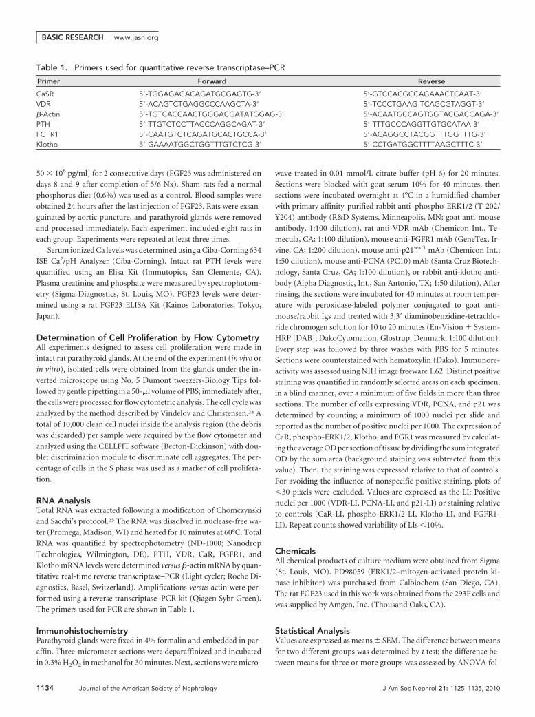
Table 1. Primers used for quantitative reverse transcriptase–PCR
Primer Forward Reverse
CaSR 5�-TGGAGAGACAGATGCGAGTG-3� 5�-GTCCACGCCAGAAACTCAAT-3�
VDR 5�-ACAGTCTGAGGCCCAAGCTA-3� 5�-TCCCTGAAG TCAGCGTAGGT-3�
�-Actin 5�-TGTCACCAACTGGGACGATATGGAG-3� 5�-ACAATGCCAGTGGTACGACCAGA-3�
PTH 5�-TTGTCTCCTTACCCAGGCAGAT-3� 5�-TTTGCCCAGGTTGTGCATAA-3�
FGFR1 5�-CAATGTCTCAGATGCACTGCCA-3� 5�-ACAGGCCTACGGTTTGGTTTG-3�
Klotho 5�-GAAAATGGCTGGTTTGTCTCG-3� 5�-CCTGATGGCTTTTAAGCTTTC-3�
BASIC RESEARCH www.jasn.org
1134 Journal of the American Society of Nephrology J Am Soc Nephrol 21: 1125–1135, 2010
lowed by post hoc Bonferroni correction analysis or two-way ANOVA
for multiple dependent variables. P � 0.05 was considered significant.
ACKNOWLEDGMENTS
This study was supported by Instituto Carlos III (FIS 07-0287, FIS
07/0315) and Junta de Andalucia (0025/07). Y.A. is a senior researcher
supported by the Fundacion Progreso y Salud, Consejeria de Salud
(Junta de Andalucia). We thank Juan A. Ballesteros from COVAP for
technical assistance.
DISCLOSURESV.S. is an employee and shareholder of Amgen; and M.R. receives research
funding from Amgen.
REFERENCES
1. Shimada T, Mizutani S, Muto T, Yoneya T, Hino R, Takeda S, TakeuchiY, Fujita T, Fukumoto S, Yamashita T: Cloning and characterization ofFGF23 as a causative factor of tumor-induced osteomalacia. Proc NatlAcad Sci U S A 98: 6500–6505, 2001
2. Kurosu H, Ogawa Y, Miyoshi M, Yamamoto M, Nandi A, Rosenblat KP,Baum MG, Schiavi S, Hu MC, Moe OW, Kuro-o M: Regulation offibroblast growth factor-23 signaling by Klotho. J Biol Chem 281:6120–6123, 2006
3. Urakawa I, Yamazaki Y, Shimada T, Iijima K, Hasegawa H, Okawa K,Fujita T, Fukumoto S, Yamashita T: Klotho converts canonical FGFreceptor into a specific receptor for FGF23. Nature 444: 770–774,2006
4. Gutierrez O, Isakova T, Rhee E, Shah A, Holmes J, Collerone G,Juppner H, Wolf M: Fibroblast growth factor-23 mitigates hyperphos-phatemia but accentuates calcitriol deficiency in chronic kidney dis-ease. J Am Soc Nephrol 16: 2205–2215, 2005
5. Ben-Dov IZ, Galitzer H, Lavi-Moshayoff V, Goet R, Kuro-o M, Moham-madi M, Sirkis R, Naveh-Many T, Silver J: The parathyroid is a targetorgan for FGF23 in rats. J Clin Invest 117: 4003–4008, 2007
6. Krajisnik T, Bjorklund P, Marsell R, Ljunggren O, Akerstrom G, JonssonKB, Westin G, Larsson TE: Fibroblast growth factor-23 regulates para-thyroid hormone and 1�-hydroxylase expression in cultured bovineparathyroid cells. J Endocrinol 195: 125–131, 2007
7. Larsson T, Nisbeth U, Ljunggren O, Juppner H, Jonsson KB: Circulat-ing concentration of FGF-23 increases as renal function declines inpatients with chronic kidney disease, but does not change in responseto variation in phosphate intake in healthy volunteers. Kidney Int 64:2272–2279, 2003
8. Nakanishi S, Kazama JJ, Nii-Kono T, Omori K, Yamashita T, FukumotoS, Gejyo F, Shigematsu T, Fukagawa M: Serum fibroblast growthfactor-23 levels predict the future refractory hyperparathyroidism indialysis patients Kidney Int 67: 1171–1178, 2005
9. Almaden Y, Canalejo A, Hernandez A, Ballesteros E, Garcia-Navarro S,Torres A, Rodriguez M: Direct effect of phosphorus on PTH secretionfrom whole rat parathyroid gland in vitro. J Bone Miner Res 11:970–976, 1996
10. Almaden Y, Hernandez A, Torregrosa V, Canalejo A, Sabate L, Fer-nandez-Cruz L, Campistol JM, Torres A, Rodriguez M: High phospho-rus directly stimulates parathyroid hormone secretion and synthesis by
human parathyroid tissue in vitro. J Am Soc Nephrol 9: 1845–1852,1998
11. Slatopolsky E, Finch J, Denda M, Ritter C, Zhong M, Dusso A, Mac-Donald P, Brown A: Phosphorus restriction prevents parathyroid glandgrowth: High phosphate directly stimulates PTH secretion in vitro.J Clin Invest 97: 2534–2540, 1996
12. Garfia B, Canadillas S, Canalejo A, Luque F, Siendones E, Quesada M,Almaden Y, Aguilera-Tejero E, Rodriguez M: Regulation of parathyroidvitamin D receptor expression by extracellular calcium. J Am SocNephrol 13: 2945–2952, 2002
13. Mendoza FJ, Lopez I, Canalejo R, Almaden Y, Martin D, Aguilera-Tejero E, Rodriguez M: Direct upregulation of parathyroid calcium-sensing receptor and vitamin D receptor by calcimimetics in uremicrats. Am J Physiol Renal Physiol 296: F605–F613, 2008
14. Merke J, Hugel U, Zlotkowsk A, Szabo A, Bommer J, Mall G, Ritz E:Diminished parathyroid 1,25(OH)2D3 receptors in experimental ure-mia. Kidney Int 32: 350–353, 1987
15. Szabo A, Ritz E, Schmidt-Gayk H, Reichel H: Abnormal expression andregulation of vitamin D receptor in experimental uremia. Nephron 73:619–628, 1996
16. Brown AJ, Ritter CS, Finch JL, Slatopolsky EA: Decreased calcium-sensing receptor expression in hyperplastic parathyroid glands ofuremic rats: role of dietary phosphate. Kidney Int 55: 1284–1292,1999
17. Liu S, Vierthaler L, Tang W, Zhou J, Quarles LD: FGFR3 and FGFR4 donot mediate renal effects of FGF23. J Am Soc Nephrol 19: 2342–2350,2008
18. Koh N, Fujimori T, Nishiguchi S, Tamori A, Shiomi S, Nakatani T,Sugimura K, Kishimoto T, Kinoshita S, Kuroki T, Nabeshima Y: Severelyreduced production of klotho in human chronic renal failure kidney.Biochem Biophys Res Commun 280: 1015–1020, 2001
19. Bjorklund P, Krajisnik T, Akerstrom G, Westin G, Larsson TE: Type Imembrane klotho expression is decreased and inversely correlated toserum calcium in primary hyperparathyroidism. J Clin EndocrinolMetab 93: 4152–4157, 2008
20. Imura A, Tsuji Y, Murata M, Maeda R, Kubota K, Iwano A, Obuse C,Togashi K, Tominaga M, Kita N, Tomiyama K, Iijima J, Nabeshima Y,Fujioka M, Asato R, Tanaka S, Kojima K, Ito J, Nozaki K, Hashimoto N,Ito T, Nishio T, Uchiyama T, Fujimori T, Nabeshima Y: Alpha-Klotho asa regulator of calcium homeostasis. Science 316: 1615–1618, 2007
21. Saito H, Maeda A, Ohtomo S, Hirata M, Kusano K, Kato S, Ogata E,Segawa H, Miyamoto K, Fukushima N: Circulating FGF-23 is regulatedby 1alpha, 25-dihydroxyvitamin D3 and phosphorus in vivo. J BiolChem 280: 2543–2549, 2005
22. Koiwa F, Kazama JJ, Tokumoto A, Onoda N, Kato H, Okada T,Nii-Kono T, Fukagawa M, Shigematsu T: Sevelamer hydrochloride andcalcium bicarbonate reduce serum fibroblast growth factor 23 levels indialysis patients. Ther Apher Dial 9: 336–339, 2005
23. Gutierrez OM, Mannstadt M, Isakova T, Rauh-Hain JA, Tamez H, ShahA, Smith K, Lee H, Thadhani R, Juppner H, Wolf M: Fibroblast growthfactor 23 and mortality among patients undergoing hemodialysis.N Engl J Med 359: 584–592, 2008
24. Vindelov L, Christensen IJ: Detergent and proteolytic enzyme-basedtechniques for nuclear isolation and DNA content analysis. In: Meth-ods in Cell Biology, Vol. 41, 2nd Ed., Flow Cytometry, edited byDarzynkiewicz Z, Robinson JP, Crissman HA, New York, AcademicPress, 1994, pp 219–229
25. Chomczynski P, Sacchi N: Single-step method of RNA isolation by acidguanidinium isothyocianate-phenol-chloroform extraction. Anal Bio-chem 162: 156–159, 1987
Supplemental information for this article is available online at http://www.jasn.org/.
BASIC RESEARCHwww.jasn.org
J Am Soc Nephrol 21: 1125–1135, 2010 FGF23 and Parathyroid Hyperplasia 1135
Related Documents


