REVIEW FGF signaling pathways in endochondral and intramembranous bone development and human genetic disease David M. Ornitz 1,3 and Pierre J. Marie 2 1 Department of Molecular Biology and Pharmacology, Washington University Medical School, St. Louis, Missouri 63110, USA; 2 Laboratory of Osteoblast Biology and Pathology, INSERM U349 affiliated CNRS, Hopital Lariboisiere, Paris cedex 10, France Over the last decade the identification of mutations in the receptors for fibroblast growth factors (FGFs) has de- fined essential roles for FGF signaling in both endochon- dral and intramembranous bone development. FGF sig- naling pathways are essential for the earliest stages of limb development and throughout skeletal develop- ment. In this review, we examine the role of FGF signal- ing in bone development and in human genetic diseases that affect bone development. We also explore what is presently known about how FGF signaling pathways in- teract with other major signaling pathways that regulate chondrogenesis and osteogenesis. Overview of skeletal development Skeletal elements are formed through two distinct de- velopmental processes. Endochondral ossification gives rise to long bones that comprise the appendicular skel- eton, facial bones, vertebrae, and the lateral medial clavicles. Intramembranous ossification gives rise to the flat bones that comprise the cranium and medial clavicles. Both types of ossification involve an initial condensation of mesenchyme and the eventual forma- tion of calcified bone. However, intramembranous bone formation accomplishes this directly, whereas endo- chondral ossification incorporates an intermediate step in which a cartilaginous template regulates the growth and patterning of the developing skeletal element. Development of endochondral bones initiates shortly after the formation of the limb bud with the condensa- tion of loose mesenchyme, marked by expression of type II collagen (Fig. 1A; Kosher et al. 1986; Nah et al. 1988). Condensing mesenchyme forms an anlage for the endo- chondral skeleton and can either branch or segment to form individual skeletal elements (Hall and Miyake 1992, 2000). Differentiation of condensing mesenchyme gives rise to a proliferating population of centrally local- ized type II collagen-expressing chondrocytes and more peripherally localized type I collagen-expressing peri- chondrial cells (Kosher et al. 1986). At this stage, chon- drocytes begin to elaborate a specialized extracellular matrix containing type II collagen. Midway between the ends of this elongated cartilaginous template, chondro- cytes exit the cell cycle, hypertrophy, and begin to syn- thesize type X collagen in place of type II collagen (Schmid and Linsenmayer 1985). To synthesize bone, a center of ossification forms in the mid-hypertrophic zone by neovascularization of the initially avascular cartilaginous template. The secretion and mineralization of a type I collagen-containing extra- cellular matrix is mediated by osteoblasts that are asso- ciated with the newly developed vasculature. As bones grow, this center of ossification propagates toward the two epiphyseal plates. The epiphyseal growth plate consists of well-demar- cated zones of cells that follow an elegant developmental program (Caplan and Pechak 1987; Hall and Miyake 1992; Olsen et al. 2000; Wagner and Karsenty 2001; Karsenty and Wagner 2002). Proximally (toward the end of a developing bone), a pool of chondrocytes (called the resting or reserve zone) supplies cells to a population of proliferating chondrocytes. Proliferating chondrocytes in turn differentiate to form a transient pool of prehyper- trophic and then a more long-lived pool of hypertrophic chondrocytes. At the distal end of the epiphyseal growth plate, hypertrophic chondrocytes die by apoptosis and are replaced by trabecular bone. In this manner, hyper- trophic chondrocytes provide a template for the forma- tion of trabecular bone. In addition to the cartilaginous and trabecular core, endochondral bone contains a hard outer shell of cortical bone (the bone collar), which surrounds the marrow cav- ity centrally and is contiguous with the perichondrium proximally (Caplan and Pechak 1987). The perichon- drium contains precursor pools of cells that give rise to osteoblasts that line the endosteal (inner) and periosteal (outer) surface of cortical bone. Osteoblasts differentiate into osteocytes, which become embedded within corti- cal bone. The center of ossification in the perichondrium 3 Corresponding author. E-MAIL [email protected]; FAX (314) 362-7058. Article and publication are at http://www.genesdev.org/cgi/doi/10.1101/ gad.990702. 1446 GENES & DEVELOPMENT 16:1446–1465 © 2002 by Cold Spring Harbor Laboratory Press ISSN 0890-9369/02 $5.00; www.genesdev.org

Welcome message from author
This document is posted to help you gain knowledge. Please leave a comment to let me know what you think about it! Share it to your friends and learn new things together.
Transcript
REVIEW
FGF signaling pathways in endochondraland intramembranous bone developmentand human genetic diseaseDavid M. Ornitz1,3 and Pierre J. Marie2
1Department of Molecular Biology and Pharmacology, Washington University Medical School, St. Louis, Missouri 63110, USA;2Laboratory of Osteoblast Biology and Pathology, INSERM U349 affiliated CNRS, Hopital Lariboisiere, Paris cedex 10, France
Over the last decade the identification of mutations inthe receptors for fibroblast growth factors (FGFs) has de-fined essential roles for FGF signaling in both endochon-dral and intramembranous bone development. FGF sig-naling pathways are essential for the earliest stages oflimb development and throughout skeletal develop-ment. In this review, we examine the role of FGF signal-ing in bone development and in human genetic diseasesthat affect bone development. We also explore what ispresently known about how FGF signaling pathways in-teract with other major signaling pathways that regulatechondrogenesis and osteogenesis.
Overview of skeletal development
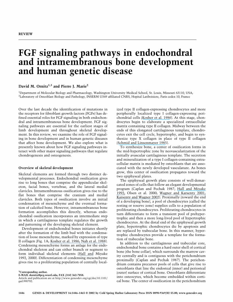
Skeletal elements are formed through two distinct de-velopmental processes. Endochondral ossification givesrise to long bones that comprise the appendicular skel-eton, facial bones, vertebrae, and the lateral medialclavicles. Intramembranous ossification gives rise to theflat bones that comprise the cranium and medialclavicles. Both types of ossification involve an initialcondensation of mesenchyme and the eventual forma-tion of calcified bone. However, intramembranous boneformation accomplishes this directly, whereas endo-chondral ossification incorporates an intermediate stepin which a cartilaginous template regulates the growthand patterning of the developing skeletal element.Development of endochondral bones initiates shortly
after the formation of the limb bud with the condensa-tion of loose mesenchyme, marked by expression of typeII collagen (Fig. 1A; Kosher et al. 1986; Nah et al. 1988).Condensing mesenchyme forms an anlage for the endo-chondral skeleton and can either branch or segment toform individual skeletal elements (Hall and Miyake1992, 2000). Differentiation of condensing mesenchymegives rise to a proliferating population of centrally local-
ized type II collagen-expressing chondrocytes and moreperipherally localized type I collagen-expressing peri-chondrial cells (Kosher et al. 1986). At this stage, chon-drocytes begin to elaborate a specialized extracellularmatrix containing type II collagen. Midway between theends of this elongated cartilaginous template, chondro-cytes exit the cell cycle, hypertrophy, and begin to syn-thesize type X collagen in place of type II collagen(Schmid and Linsenmayer 1985).To synthesize bone, a center of ossification forms in
the mid-hypertrophic zone by neovascularization of theinitially avascular cartilaginous template. The secretionand mineralization of a type I collagen-containing extra-cellular matrix is mediated by osteoblasts that are asso-ciated with the newly developed vasculature. As bonesgrow, this center of ossification propagates toward thetwo epiphyseal plates.The epiphyseal growth plate consists of well-demar-
cated zones of cells that follow an elegant developmentalprogram (Caplan and Pechak 1987; Hall and Miyake1992; Olsen et al. 2000; Wagner and Karsenty 2001;Karsenty and Wagner 2002). Proximally (toward the endof a developing bone), a pool of chondrocytes (called theresting or reserve zone) supplies cells to a population ofproliferating chondrocytes. Proliferating chondrocytes inturn differentiate to form a transient pool of prehyper-trophic and then a more long-lived pool of hypertrophicchondrocytes. At the distal end of the epiphyseal growthplate, hypertrophic chondrocytes die by apoptosis andare replaced by trabecular bone. In this manner, hyper-trophic chondrocytes provide a template for the forma-tion of trabecular bone.In addition to the cartilaginous and trabecular core,
endochondral bone contains a hard outer shell of corticalbone (the bone collar), which surrounds the marrow cav-ity centrally and is contiguous with the perichondriumproximally (Caplan and Pechak 1987). The perichon-drium contains precursor pools of cells that give rise toosteoblasts that line the endosteal (inner) and periosteal(outer) surface of cortical bone. Osteoblasts differentiateinto osteocytes, which become embedded within corti-cal bone. The center of ossification in the perichondrium
3Corresponding author.E-MAIL [email protected]; FAX (314) 362-7058.Article and publication are at http://www.genesdev.org/cgi/doi/10.1101/gad.990702.
1446 GENES & DEVELOPMENT 16:1446–1465 © 2002 by Cold Spring Harbor Laboratory Press ISSN 0890-9369/02 $5.00; www.genesdev.org
is aligned with the center of ossification at the chondro–osseous junction of the growth plate. A key feature of thegrowth of long bones is the coordination of chondrogen-esis and ossification in the epiphyseal growth plate andosteogenesis in the perichondrium/periosteum (Fig. 1A).Cranial vault development is a complex process in-
volving cells of neural crest origin (Couly et al. 1993) andparaxial mesoderm that contribute to intramembranousbones of the cranial vault and sutures (Noden 1992; Op-perman 2000). Intramembranous bone growth beginswith the condensation of mesenchymal cells (Fig. 1B).Ossification centers are formed by direct bone matrixdeposition-forming plates, which expand during develop-ment but do not fuse at the junction with other cranialbones (Hall andMiyake 2000). The junction between cal-varial bones is a functional structure called a suture,which is responsible for the maintenance of a separationbetween membranous bones and for regulating the ex-pansive growth of the skull. In the vicinity of the suture,a minority of osteogenic mesenchymal cells differentiate
into osteoprogenitor cells and then into osteoblasts thatexpress type I collagen, bone sialoprotein, and osteocal-cin, and synthesize bone matrix along the bone margins.Growth and differentiation at the suture is regulated byinteractions between the mesenchyme, the osteogenicfront, and the Dura mater, a tough, fibrous membraneforming the outer envelope of the brain and the innerlining of cranial bones and sutures. (see Fig. 3C below;Most et al. 1998; Opperman 2000; Morriss-Kay et al.2001). Because sutures are the major sites of intramem-branous bone growth during cranial vault development,the events occurring at the suture are essential for theregulation of intramembranous ossification (Cohen2000a).
FGFs and FGF receptors—a brief introduction
FGFs comprise a family of 22 genes encoding structur-ally related proteins (Ornitz and Itoh 2001). Six subfami-lies of FGFs, grouped by sequence similarities, tend to
Figure 1. Endochondral and intramembranous bone development. (A) Schematic representation of a developing endochondral bone.Endochondral skeletal development begins with the formation of a mesenchymal condensation, expressing type II collagen (blue).Centrally, cells differentiate into chondrocytes, which hypertrophy and express type X collagen (purple). Progression to the maturegrowth plate accompanies development of the perichondrium (yellow), vascular invasion, and the formation of a center of ossificationcontaining type I collagen-expressing osteoblasts (yellow). (B) Schematic representation of a developing intramembranous bone.Undifferentiated mesenchymal cells differentiate into osteoprogenitor cells expressing Cbfa1 (pink). Osteoprogenitor cells progress tomature osteoblasts that express Cbfa1 and type I collagen (yellow). These cells deposit and mineralize bone matrix. Osteoblasts eitherdie by apoptosis or are embedded in the matrix, becoming osteocytes.
FGF pathways in skeletal development
GENES & DEVELOPMENT 1447
share biochemical and functional properties and are ex-pressed in specific spatial and developmental patterns.Four distinct FGF receptor tyrosine kinase moleculesbind and are activated by most members of the FGF fam-ily. Alternative mRNA splicing produces FGF receptorswith unique ligand binding properties (Johnson andWilliams 1993; Ornitz et al. 1996). Alternative splicing ismostly tissue-specific, producing epithelial variants (bsplice forms) and mesenchymal variants (c splice forms;Miki et al. 1992; Orr-Urtreger et al. 1993; Chellaiah et al.1994; Naski and Ornitz 1998). FGF activity and specific-ity are further regulated by heparan sulfate oligosaccha-rides, in the form of heparan sulfate proteoglycans. Hep-arin/Heparan sulfate, FGF, and an FGF receptor (FGFR)associate to form a trimolecular complex (Rapraeger etal. 1991; Yayon et al. 1991; Ornitz et al. 1992). Heparanchains, themselves, have unique tissue-specific modifi-
cations that are required for, and may actually regulate,functional ligand–receptor interactions (Rapraeger 1995;Guimond and Turnbull 1999; Ornitz 2000; Allen et al.2001).
Mutations in FGF receptors in chondrodysplasiaand craniosynostosis syndromes
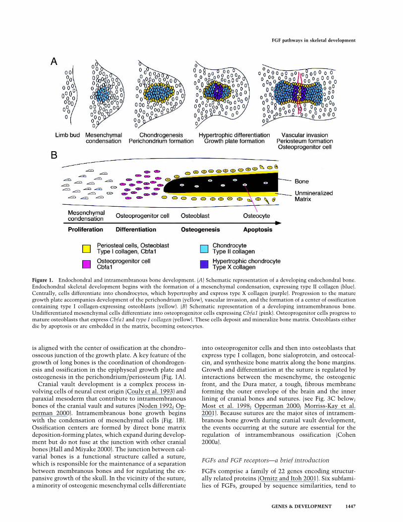
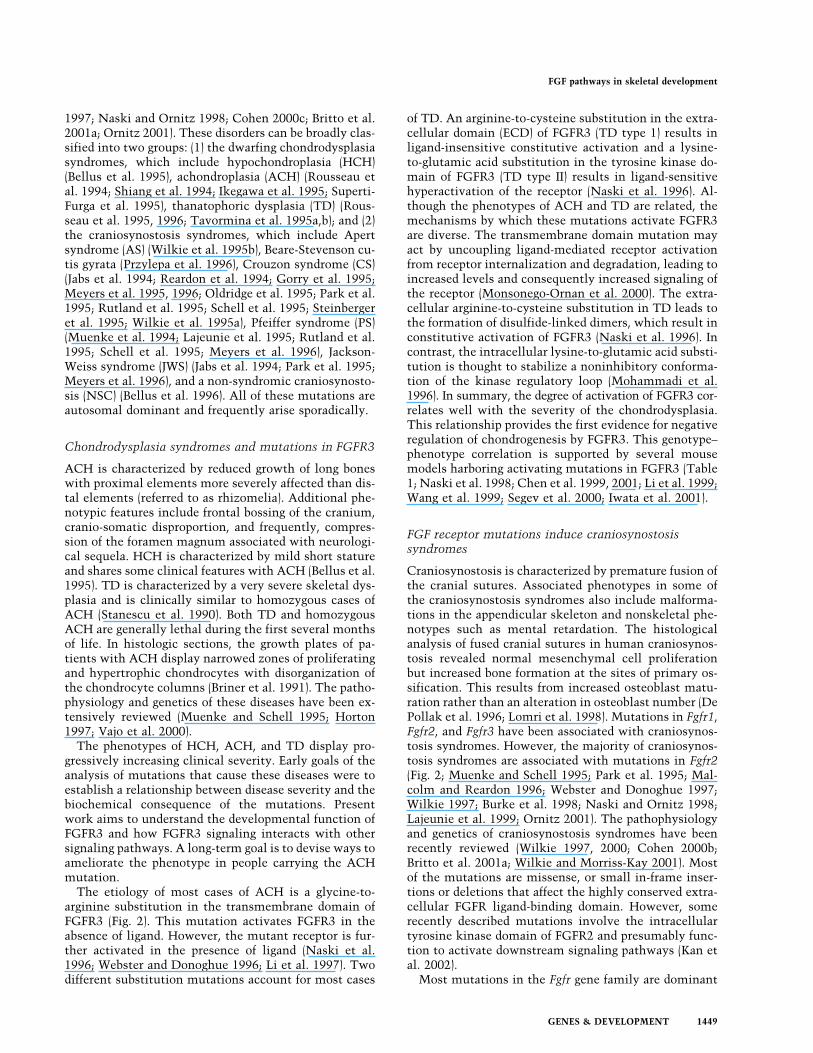
The importance of FGF signaling in skeletal develop-ment was first revealed with the discovery that a pointmutation in the transmembrane domain of FGFR3 is theetiology of Achondroplasia, the most common geneticform of dwarfism in humans (Fig. 2; Rousseau et al. 1994;Shiang et al. 1994). Since this discovery, the etiology ofmany other human skeletal dysplasias have been attrib-uted to specific mutations in the genes encoding FGFreceptors 1, 2, and 3 (Muenke and Schell 1995; Wilkie
Figure 2. FGF receptor mutations in human chondrodysplasia and craniosynostosis syndromes. (Left, green) Mutations in FGFreceptor 3. (ACH) Achondroplasia, (TD) thanatophoric dysplasia, (HCH) hypochondroplasia, (CDS) Crouzonodermoskeletal syndrome(Crouzon syndrome and acanthosis nigricans), (NSC) non-syndromic craniosynostosis. (Right, blue) Mutations in FGF receptor 2. (CS)Crouzon syndrome, (JWS) Jackson-Weiss syndrome, (PS) Pfeiffer syndrome, (AS) Apert syndrome, (BS) Beare-Stevenson cutis gyrata, (U)unclassified. (Right, red) (PS) A single mutation in FGF receptor 1 that causes Pfeiffer syndrome. The numbers represent the positionof the mutant amino acid in the human coding sequence. Amino acids are abbreviated using standard single-letter abbreviations. Datasource: Wilkie (1997), Naski and Ornitz (1998), and included references.
Ornitz and Marie
1448 GENES & DEVELOPMENT
1997; Naski and Ornitz 1998; Cohen 2000c; Britto et al.2001a; Ornitz 2001). These disorders can be broadly clas-sified into two groups: (1) the dwarfing chondrodysplasiasyndromes, which include hypochondroplasia (HCH)(Bellus et al. 1995), achondroplasia (ACH) (Rousseau etal. 1994; Shiang et al. 1994; Ikegawa et al. 1995; Superti-Furga et al. 1995), thanatophoric dysplasia (TD) (Rous-seau et al. 1995, 1996; Tavormina et al. 1995a,b); and (2)the craniosynostosis syndromes, which include Apertsyndrome (AS) (Wilkie et al. 1995b), Beare-Stevenson cu-tis gyrata (Przylepa et al. 1996), Crouzon syndrome (CS)(Jabs et al. 1994; Reardon et al. 1994; Gorry et al. 1995;Meyers et al. 1995, 1996; Oldridge et al. 1995; Park et al.1995; Rutland et al. 1995; Schell et al. 1995; Steinbergeret al. 1995; Wilkie et al. 1995a), Pfeiffer syndrome (PS)(Muenke et al. 1994; Lajeunie et al. 1995; Rutland et al.1995; Schell et al. 1995; Meyers et al. 1996), Jackson-Weiss syndrome (JWS) (Jabs et al. 1994; Park et al. 1995;Meyers et al. 1996), and a non-syndromic craniosynosto-sis (NSC) (Bellus et al. 1996). All of these mutations areautosomal dominant and frequently arise sporadically.
Chondrodysplasia syndromes and mutations in FGFR3
ACH is characterized by reduced growth of long boneswith proximal elements more severely affected than dis-tal elements (referred to as rhizomelia). Additional phe-notypic features include frontal bossing of the cranium,cranio-somatic disproportion, and frequently, compres-sion of the foramen magnum associated with neurologi-cal sequela. HCH is characterized by mild short statureand shares some clinical features with ACH (Bellus et al.1995). TD is characterized by a very severe skeletal dys-plasia and is clinically similar to homozygous cases ofACH (Stanescu et al. 1990). Both TD and homozygousACH are generally lethal during the first several monthsof life. In histologic sections, the growth plates of pa-tients with ACH display narrowed zones of proliferatingand hypertrophic chondrocytes with disorganization ofthe chondrocyte columns (Briner et al. 1991). The patho-physiology and genetics of these diseases have been ex-tensively reviewed (Muenke and Schell 1995; Horton1997; Vajo et al. 2000).The phenotypes of HCH, ACH, and TD display pro-
gressively increasing clinical severity. Early goals of theanalysis of mutations that cause these diseases were toestablish a relationship between disease severity and thebiochemical consequence of the mutations. Presentwork aims to understand the developmental function ofFGFR3 and how FGFR3 signaling interacts with othersignaling pathways. A long-term goal is to devise ways toameliorate the phenotype in people carrying the ACHmutation.The etiology of most cases of ACH is a glycine-to-
arginine substitution in the transmembrane domain ofFGFR3 (Fig. 2). This mutation activates FGFR3 in theabsence of ligand. However, the mutant receptor is fur-ther activated in the presence of ligand (Naski et al.1996; Webster and Donoghue 1996; Li et al. 1997). Twodifferent substitution mutations account for most cases
of TD. An arginine-to-cysteine substitution in the extra-cellular domain (ECD) of FGFR3 (TD type 1) results inligand-insensitive constitutive activation and a lysine-to-glutamic acid substitution in the tyrosine kinase do-main of FGFR3 (TD type II) results in ligand-sensitivehyperactivation of the receptor (Naski et al. 1996). Al-though the phenotypes of ACH and TD are related, themechanisms by which these mutations activate FGFR3are diverse. The transmembrane domain mutation mayact by uncoupling ligand-mediated receptor activationfrom receptor internalization and degradation, leading toincreased levels and consequently increased signaling ofthe receptor (Monsonego-Ornan et al. 2000). The extra-cellular arginine-to-cysteine substitution in TD leads tothe formation of disulfide-linked dimers, which result inconstitutive activation of FGFR3 (Naski et al. 1996). Incontrast, the intracellular lysine-to-glutamic acid substi-tution is thought to stabilize a noninhibitory conforma-tion of the kinase regulatory loop (Mohammadi et al.1996). In summary, the degree of activation of FGFR3 cor-relates well with the severity of the chondrodysplasia.This relationship provides the first evidence for negativeregulation of chondrogenesis by FGFR3. This genotype–phenotype correlation is supported by several mousemodels harboring activating mutations in FGFR3 (Table1; Naski et al. 1998; Chen et al. 1999, 2001; Li et al. 1999;Wang et al. 1999; Segev et al. 2000; Iwata et al. 2001).
FGF receptor mutations induce craniosynostosissyndromes
Craniosynostosis is characterized by premature fusion ofthe cranial sutures. Associated phenotypes in some ofthe craniosynostosis syndromes also include malforma-tions in the appendicular skeleton and nonskeletal phe-notypes such as mental retardation. The histologicalanalysis of fused cranial sutures in human craniosynos-tosis revealed normal mesenchymal cell proliferationbut increased bone formation at the sites of primary os-sification. This results from increased osteoblast matu-ration rather than an alteration in osteoblast number (DePollak et al. 1996; Lomri et al. 1998). Mutations in Fgfr1,Fgfr2, and Fgfr3 have been associated with craniosynos-tosis syndromes. However, the majority of craniosynos-tosis syndromes are associated with mutations in Fgfr2(Fig. 2; Muenke and Schell 1995; Park et al. 1995; Mal-colm and Reardon 1996; Webster and Donoghue 1997;Wilkie 1997; Burke et al. 1998; Naski and Ornitz 1998;Lajeunie et al. 1999; Ornitz 2001). The pathophysiologyand genetics of craniosynostosis syndromes have beenrecently reviewed (Wilkie 1997, 2000; Cohen 2000b;Britto et al. 2001a; Wilkie and Morriss-Kay 2001). Mostof the mutations are missense, or small in-frame inser-tions or deletions that affect the highly conserved extra-cellular FGFR ligand-binding domain. However, somerecently described mutations involve the intracellulartyrosine kinase domain of FGFR2 and presumably func-tion to activate downstream signaling pathways (Kan etal. 2002).Most mutations in the Fgfr gene family are dominant
FGF pathways in skeletal development
GENES & DEVELOPMENT 1449
and are thought to be gain-of-function mutations (Naskiand Ornitz 1998; Cohen 2000c; Ornitz 2001; Wilkie andMorriss-Kay 2001). Several of the mutations in Fgfr1 andFgfr2 in CS, PS, and JWS constitutively activate the re-ceptor by stabilizing intermolecular disulfide bonds,causing ligand-independent dimerization and signaling(Neilson and Friesel 1995; Wilkie et al. 1995a; Galvin etal. 1996; Robertson et al. 1998). Other mutations arethought to prolong the duration of receptor signaling oralter ligand-binding specificity (Anderson et al. 1998;Mansukhani et al. 2000; Plotnikov et al. 2000; Yu et al.2000; Ibrahimi et al. 2001). For example, one of the CSmutations in Fgfr2 results in ligand-independent activa-tion and dramatically decreased ligand binding (Mangas-arian et al. 1997). However, the mutant receptor in AShas lost ligand-binding specificity and thus can be acti-vated by inappropriate ligands (Plotnikov et al. 2000; Yuet al. 2000; Ibrahimi et al. 2001; Yu and Ornitz 2001). Amutation in FGFR3 has also been associated with aCrouzon-like phenotype and associated dermal thicken-ing and hyperpigmentation (Crouzon syndrome with ac-anthosis nigricans). This syndrome is now referred to asCrouzonodermoskeletal syndrome (CDS) (Cohen 2000c).The importance of FGF signaling in cranial bone de-
velopment is further supported by mice harboring acti-vating mutations in FGF signaling pathways (Table 1).Mice overexpressing FGF2 develop enlarged occipitalbones, which are formed in part by intramembranousossification (Coffin et al. 1995). Increased expression of
FGF3 and FGF4, induced by retroviral insertion, resultsin a phenotype that resembles CS (Carlton et al. 1998).Additionally, gain-of-function mutations in FGFR1 andFGFR2 in mice affect calvarial development. The P250Rmutation in FGFR1 (orthologous to the P252R substitu-tion associated with PS in humans) causes prematurefusion of calvarial sutures (Zhou et al. 2000). In addition,a dominant mutation that affects alternative splicing inFgfr2 causes coronal synostosis in mice (Hajihosseini etal. 2001; Yu and Ornitz 2001). In contrast, loss-of-func-tion mutations in Fgfrs have been less informative. Em-bryos lacking FGFR1 or FGFR2 die prior to skeletal de-velopment (Deng et al. 1994; Arman et al. 1999), andmice lacking FGFR3 do not have obvious defects in cal-varial bones (Colvin et al. 1996; Deng et al. 1996). How-ever, a secreted soluble FGFR2 ECD can inhibit FGF sig-naling and causes skull abnormalities in mice that re-semble those induced by Fgfr mutations in humans(Celli et al. 1998). Of the FGFs thus far targeted in mice,only mice lacking FGF18 have revealed a phenotype incalvarial development. Fgf18 null mice have delayed cal-varial ossification, indicating a requirement for FGF18 inintramembranous bone formation (Liu et al. 2002; Oh-bayashi et al. 2002).
FGF signaling in the developing limb bud
Fibroblast growth factors are involved in the earlieststages of limb development and in the formation of skel-
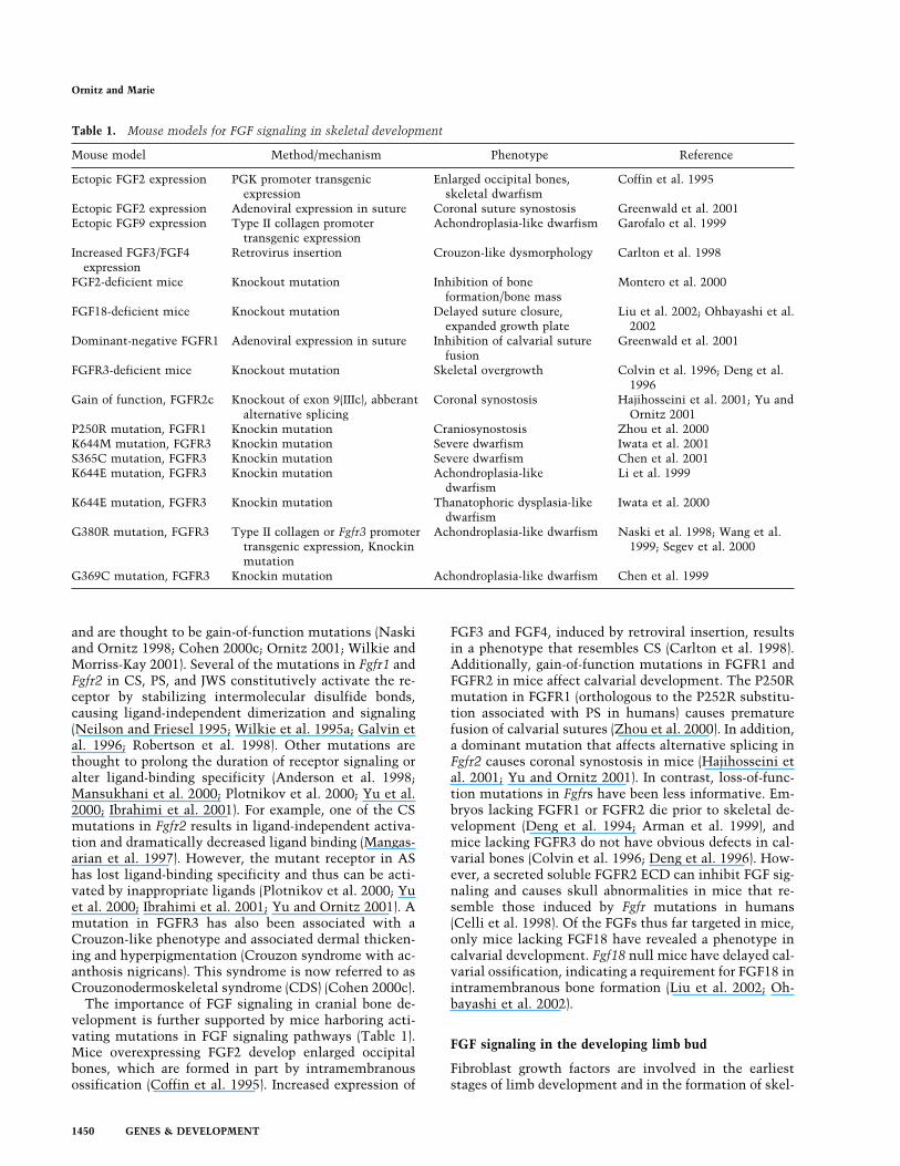
Table 1. Mouse models for FGF signaling in skeletal development
Mouse model Method/mechanism Phenotype Reference
Ectopic FGF2 expression PGK promoter transgenicexpression
Enlarged occipital bones,skeletal dwarfism
Coffin et al. 1995
Ectopic FGF2 expression Adenoviral expression in suture Coronal suture synostosis Greenwald et al. 2001Ectopic FGF9 expression Type II collagen promoter
transgenic expressionAchondroplasia-like dwarfism Garofalo et al. 1999
Increased FGF3/FGF4expression
Retrovirus insertion Crouzon-like dysmorphology Carlton et al. 1998
FGF2-deficient mice Knockout mutation Inhibition of boneformation/bone mass
Montero et al. 2000
FGF18-deficient mice Knockout mutation Delayed suture closure,expanded growth plate
Liu et al. 2002; Ohbayashi et al.2002
Dominant-negative FGFR1 Adenoviral expression in suture Inhibition of calvarial suturefusion
Greenwald et al. 2001
FGFR3-deficient mice Knockout mutation Skeletal overgrowth Colvin et al. 1996; Deng et al.1996
Gain of function, FGFR2c Knockout of exon 9(IIIc), abberantalternative splicing
Coronal synostosis Hajihosseini et al. 2001; Yu andOrnitz 2001
P250R mutation, FGFR1 Knockin mutation Craniosynostosis Zhou et al. 2000K644M mutation, FGFR3 Knockin mutation Severe dwarfism Iwata et al. 2001S365C mutation, FGFR3 Knockin mutation Severe dwarfism Chen et al. 2001K644E mutation, FGFR3 Knockin mutation Achondroplasia-like
dwarfismLi et al. 1999
K644E mutation, FGFR3 Knockin mutation Thanatophoric dysplasia-likedwarfism
Iwata et al. 2000
G380R mutation, FGFR3 Type II collagen or Fgfr3 promotertransgenic expression, Knockinmutation
Achondroplasia-like dwarfism Naski et al. 1998; Wang et al.1999; Segev et al. 2000
G369C mutation, FGFR3 Knockin mutation Achondroplasia-like dwarfism Chen et al. 1999
Ornitz and Marie
1450 GENES & DEVELOPMENT
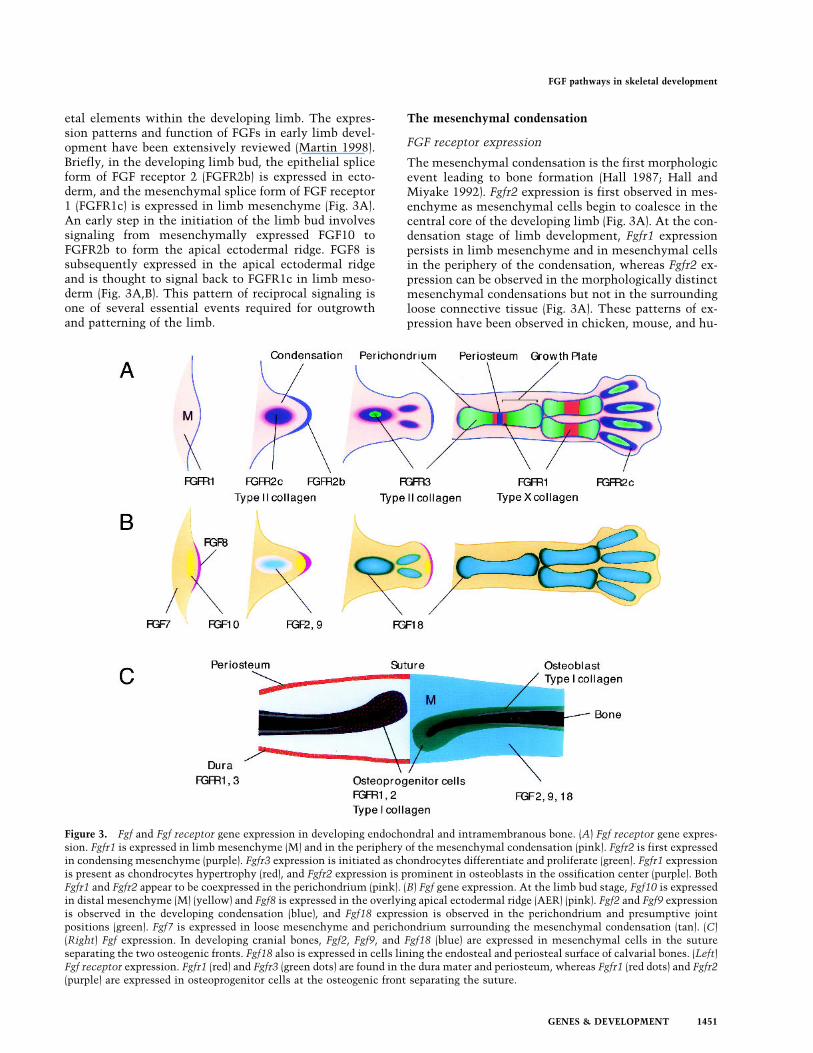
etal elements within the developing limb. The expres-sion patterns and function of FGFs in early limb devel-opment have been extensively reviewed (Martin 1998).Briefly, in the developing limb bud, the epithelial spliceform of FGF receptor 2 (FGFR2b) is expressed in ecto-derm, and the mesenchymal splice form of FGF receptor1 (FGFR1c) is expressed in limb mesenchyme (Fig. 3A).An early step in the initiation of the limb bud involvessignaling from mesenchymally expressed FGF10 toFGFR2b to form the apical ectodermal ridge. FGF8 issubsequently expressed in the apical ectodermal ridgeand is thought to signal back to FGFR1c in limb meso-derm (Fig. 3A,B). This pattern of reciprocal signaling isone of several essential events required for outgrowthand patterning of the limb.
The mesenchymal condensation
FGF receptor expression
The mesenchymal condensation is the first morphologicevent leading to bone formation (Hall 1987; Hall andMiyake 1992). Fgfr2 expression is first observed in mes-enchyme as mesenchymal cells begin to coalesce in thecentral core of the developing limb (Fig. 3A). At the con-densation stage of limb development, Fgfr1 expressionpersists in limb mesenchyme and in mesenchymal cellsin the periphery of the condensation, whereas Fgfr2 ex-pression can be observed in the morphologically distinctmesenchymal condensations but not in the surroundingloose connective tissue (Fig. 3A). These patterns of ex-pression have been observed in chicken, mouse, and hu-
Figure 3. Fgf and Fgf receptor gene expression in developing endochondral and intramembranous bone. (A) Fgf receptor gene expres-sion. Fgfr1 is expressed in limb mesenchyme (M) and in the periphery of the mesenchymal condensation (pink). Fgfr2 is first expressedin condensing mesenchyme (purple). Fgfr3 expression is initiated as chondrocytes differentiate and proliferate (green). Fgfr1 expressionis present as chondrocytes hypertrophy (red), and Fgfr2 expression is prominent in osteoblasts in the ossification center (purple). BothFgfr1 and Fgfr2 appear to be coexpressed in the perichondrium (pink). (B) Fgf gene expression. At the limb bud stage, Fgf10 is expressedin distal mesenchyme (M) (yellow) and Fgf8 is expressed in the overlying apical ectodermal ridge (AER) (pink). Fgf2 and Fgf9 expressionis observed in the developing condensation (blue), and Fgf18 expression is observed in the perichondrium and presumptive jointpositions (green). Fgf7 is expressed in loose mesenchyme and perichondrium surrounding the mesenchymal condensation (tan). (C)(Right) Fgf expression. In developing cranial bones, Fgf2, Fgf9, and Fgf18 (blue) are expressed in mesenchymal cells in the sutureseparating the two osteogenic fronts. Fgf18 also is expressed in cells lining the endosteal and periosteal surface of calvarial bones. (Left)Fgf receptor expression. Fgfr1 (red) and Fgfr3 (green dots) are found in the dura mater and periosteum, whereas Fgfr1 (red dots) and Fgfr2(purple) are expressed in osteoprogenitor cells at the osteogenic front separating the suture.
FGF pathways in skeletal development
GENES & DEVELOPMENT 1451
man limb development (Orr-Urtreger et al. 1991; Peterset al. 1992; Szebenyi et al. 1995; Delezoide et al. 1998).Later in skeletal development, Fgfr1 and Fgfr2 expressionpersists in the perichondrium and periosteum in patternsthat suggest expression in the osteoblast lineage(Delezoide et al. 1998). As chondrogenesis begins in thecenter of the condensation, Fgfr3 expression is first ob-served (Peters et al. 1993).
FGF signaling in condensing mesenchyme
The physiologic ligands that activate FGFRs in the mes-enchymal condensation have been difficult to identify.At the condensation stage, Fgf9 is expressed within con-densing mesenchyme (Fig. 3B; Colvin et al. 1999). How-ever, in the absence of FGF9 there are no apparent de-fects in skeletal development (Colvin et al. 2001a,b).Similarly, Fgf2, Fgf5, Fgf6, and Fgf7 are expressed in loosemesenchyme outside the condensation (Haub and Gold-farb 1991; deLapeyriere et al. 1993; Mason et al. 1994;Finch et al. 1995; Savage and Fallon 1995). However,mice lacking these FGFs have no apparent defects inskeletal development (Hebert et al. 1994; Guo et al.1996; Fiore et al. 1997). It is possible that a combinationof these and other FGFs may constitute the completeFGF signal to the developing condensation.The role of FGF signaling in condensing mesenchyme
is poorly understood. In primary chondrocytes and inundifferentiated mesenchymal cells, FGF signaling path-ways induce the expression of SOX9, an essential tran-scription factor for chondrocyte differentiation (Mu-rakami et al. 2000). Additionally, FGFR3 signaling mayenhance chondrocyte proliferation in the mesenchymalcondensation, even though it is well established that thisreceptor negatively regulates chondrogenesis during lateembryonic and postnatal development (Iwata et al. 2000,2001). Defects in signaling in the mesenchymal conden-sation could lead to skeletal abnormalities such as thebony syndactyly resulting from mutations in FGFR2 inApert syndrome. The Apert mutations result in loss ofligand binding specificity, allowing inappropriate activa-tion of FGFR2 in the mesenchymal condensation bymesenchymally expressed ligands such as FGF7 and in-appropriate activation of FGFR2b by ligands such asFGF2, FGF6, and FGF9 (Yu et al. 2000; Yu and Ornitz2001).
Endochondral ossification
Expression of FGFs and FGF receptorsin endochondral bone
Shortly after formation of a mesenchymal condensation,chondrogenesis ensues, and Fgfr3 expression is initiatedin chondrocytes in the differentiated core of the mesen-chymal condensation (Fig. 3A). At this stage, overlap inexpression may exist with Fgfr2. As the epiphysealgrowth plate is formed, Fgfr1 expression is initiated aschondrocytes further differentiate and hypertrophy. In-
terestingly, Fgfr1 and Fgfr3 have very distinct domains ofexpression with little overlap; Fgfr3 is expressed in pro-liferating chondrocytes, whereas Fgfr1 is expressed inprehypertrophic and hypertrophic chondrocytes (Peterset al. 1992, 1993; Deng et al. 1996). This juxtaposition ofFGFR1 and FGFR3 expression domains suggests uniquefunctions. Expression of Fgfr3 in the reserve and prolif-erating zone suggests a direct role for FGFR3 in regulat-ing chondrocyte proliferation and possibly differentia-tion (Peters et al. 1993; Delezoide et al. 1998; Naski et al.1998). In contrast, the expression of Fgfr1 in hypertro-phic chondrocytes (Peters et al. 1992; Delezoide et al.1998) suggests a role for FGFR1 in survival of the hyper-trophic chondrocyte, in regulating a feedback signal tocontrol the rate of differentiation, in regulating the pro-duction of the unique extracellular matrix products ofthese cells, or in signaling their eventual apoptoticdeath. Interestingly, examination of FGFR3 protein inrib cartilage shows the presence of large amounts ofcleaved FGFR3 ECD within the extracellular matrix ofhypertrophic chondrocytes, suggesting that proteolyticprocessing could regulate the activity of FGFR3 and thatthe FGFR3 ECD could bind ligand and thus modifyFGFR1 signaling in hypertrophic chondrocytes (Pandit etal. 2002).Several FGFs are expressed in developing endochon-
dral bone (Fig. 3B). Historically, FGF2 was the first FGFligand to be isolated from growth plate chondrocytes(Sullivan and Klagsbrun 1985). Subsequently, Fgf2 ex-pression has also been observed in periosteal cells and inosteoblasts (Hurley et al. 1994, 1999; Sabbieti et al.1999). Targeted deletion of FGF2 causes a relativelysubtle defect in osteoblastogenesis, leading to decreasedbone growth and bone density. However, no defects inchondrogenesis were observed (Montero et al. 2000). Fgf9is also expressed in chondrocytes (Colvin et al. 1999;Garofalo et al. 1999). However, the skeleton of Fgf9−/−
mice is apparently normal at birth when these mice dieof other causes (Colvin et al. 2001a,b).In the perichondrium, expression of Fgf7, Fgf8, Fgf17,
and Fgf18 has been observed (Mason et al. 1994; Finchet al. 1995; Xu et al. 1999; Liu et al. 2002; Ohbayashi etal. 2002), suggesting a possible paracrine signal to thegrowth plate. Recent genetic studies have identified adefect in chondrogenesis and osteogenesis in mice lack-ing FGF18 (Liu et al. 2002; Ohbayashi et al. 2002). Micelacking FGF7, FGF8, and FGF17 have apparently normalchondrogenesis, or in the case of FGF8 die prior to skel-etal development (Guo et al. 1996; Meyers et al. 1998; Xuet al. 2000). Issues of functional redundancy among theseand other FGFs will need to be addressed in the future.
Signaling pathways that regulate endochondralbone development
The growth plate is a developmental center that inte-grates many diverse signals to coordinate the complexpatterning and growth of the skeleton. Each growth platemust appropriately respond to positional cues and endo-
Ornitz and Marie
1452 GENES & DEVELOPMENT
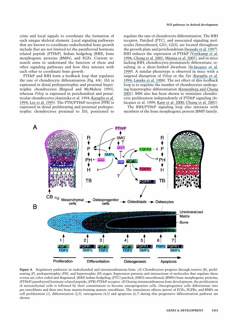
crine and local signals to coordinate the formation ofeach unique skeletal element. Local signaling pathwaysthat are known to coordinate endochondral bone growthinclude (but are not limited to) the parathyroid hormonerelated peptide (PTHrP), Indian hedgehog (IHH), bonemorphogenic proteins (BMPs), and FGFs. Current re-search aims to understand the function of these andother signaling pathways and how they interact witheach other to coordinate bone growth.PTHrP and IHH form a feedback loop that regulates
the rate of chondrocyte differentiation (Fig. 4A). Ihh isexpressed in distal prehypertrophic and proximal hyper-trophic chondrocytes (Bitgood and McMahon 1995),whereas Pthrp is expressed in perichondrial and periar-ticular chondrocytes (Amizuka et al. 1994; Karaplis et al.1994; Lee et al. 1995). The PTH/PTHrP receptor (PPR) isexpressed in distal proliferating and proximal prehyper-trophic chondrocytes proximal to Ihh, positioned to
regulate the rate of chondrocyte differentiation. The IHHreceptor, Patched (PTC), and associated signaling mol-ecules (Smoothened, Gli1, Gli3), are located throughoutthe growth plate and perichondrium (Iwasaki et al. 1997).IHH induces the expression of PTHrP (Vortkamp et al.1996; Chung et al. 2001; Minina et al. 2001), and in micelacking IHH, chondrocytes prematurely differentiate, re-sulting in a short-limbed dwarfism (St-Jacques et al.1999). A similar phenotype is observed in mice with atargeted disruption of Pthrp or the Ppr (Karaplis et al.1994; Lanske et al. 1996). The net effect of this feedbackloop is to regulate the number of chondrocytes undergo-ing hypertrophic differentiation (Kronenberg and Chung2001). IHH also has been shown to stimulate chondro-cyte proliferation independently of PTHrP signaling (St-Jacques et al. 1999; Karp et al. 2000; Chung et al. 2001).The IHH/PTHrP signaling loop also interacts with
members of the bone morphogenic protein (BMP) family.
Figure 4. Regulatory pathways in endochondral and intramembranous bone. (A) Chondrocytes progress through reserve (R), prolif-erating (P), prehypertrophic (PH), and hypertrophic (H) stages. Expression patterns and interactions of molecules that regulate theseevents are color coded and diagramed. (IHH) Indian hedgehog; (PTC) patched; (SMO) smoothened; (BMPs) bone morphogenic proteins;(PTHrP) parathyroid hormone related peptide; (PPR) PTHrP receptor. (B) During intramembranous bone development, the proliferationof mesenchymal cells is followed by their commitment to become osteoprogenitor cells. Osteoprogenitor cells differentiate intopre-osteoblasts and then into bone matrix-forming mature osteoblasts. The stimulatory effects (arrow) of FGFs, FGFRs, and BMPs oncell proliferation (1), differentiation (2,3), osteogenesis (4,5) and apoptosis (6,7) during this progressive differentiation pathway areshown.
FGF pathways in skeletal development
GENES & DEVELOPMENT 1453
Unlike PTHrP, BMP signaling promotes the proliferationof chondrocytes and delays maturation of differentiatedhypertrophic chondrocytes, resulting in an enlarged skel-eton (Fig. 4A; Duprez et al. 1996; Zou et al. 1997; Mininaet al. 2001). This effect of BMP occurs independently ofIHH and PTHrP (Minina et al. 2001). A further conse-quence of BMP signaling is to increase the domain of Ihhexpression (Kawakami et al. 1996; Pathi et al. 1999; Cap-devila and Belmonte 2001; Minina et al. 2001). IHH sig-naling also interacts with the BMP pathway by up-regu-lating expression of Bmp2 and Bmp4 in the growth plate(Pathi et al. 1999; Minina et al. 2001). The net effect ofthis signaling loop is to promote chondrogenesis.
FGF signaling pathways in the growth plate
Analysis of both gain-of-function and loss-of-functionmutations in FGFR3 at late gestational and postnatalstages of mouse development shows that the net conse-quence of signaling through this receptor is to limitchondrocyte proliferation and differentiation (Fig. 4A;Naski and Ornitz 1998; Ornitz 2001). This effect is me-diated in part by direct signaling in chondrocytes(Henderson et al. 2000; Rozenblatt-Rosen et al. 2002) andin part indirectly, by regulating the expression of theIHH/PTHrP/BMP signaling pathways. Mice harboring anactivating mutation in FGFR3 have decreased expressionof Ihh, Ptc, and Bmp4 (Naski et al. 1998; Li et al. 1999;Chen et al. 2001), whereas in mice lacking FGFR3, Ihh,Ptc, and Bmp4 expression are up-regulated (Naski et al.1998; data not shown). The overall function of FGFR3 isconsistent with a direct action of FGFR3 on proliferatingchondrocytes (see below) and an indirect consequence ofmodulating Hedgehog and BMP signaling (Fig. 4A).The ligands that signal to FGFR3 during endochondral
ossification have been elusive. However, recent datashow that growth plate histology of mice lacking FGF18is similar to that of mice lacking FGFR3. This includesthe up-regulation of Ihh and Ptc expression and in-creased chondrocyte proliferation. These similaritiesprovide strong evidence for a ligand–receptor relation-ship between FGF18 and FGFR3 (Liu et al. 2002; Ohbaya-shi et al. 2002). Furthermore, in vitro, FGF18 can acti-vate FGFR3c (Xu et al. 2000) and stimulate the prolifera-tion of cultured articular chondrocytes (Ellsworth et al.2002). FGF18 is most closely related to FGF8 and FGF17(Ornitz and Itoh 2001) and may not be the only FGFligand functioning in developing bone. There is someevidence for expression of FGF8 and FGF17 in develop-ing bone (Xu et al. 1999), suggesting that there may beadditional redundancy among these three ligands.Chondrogenesis and osteogenesis must be tightly co-
ordinated to correctly form and generate a biomechani-cally robust structure. Regulation of endochondral ossi-fication by FGFs, IHH, PTHrP, and BMPs involves sig-naling between different populations of chondrocytesand between chondrocytes and the perichondrium. Sev-eral studies have shown that the perichondrium elabo-rates a signal that negatively regulates both chondrocyteproliferation and differentiation (Long and Linsenmayer
1998; Haaijman et al. 1999; Alvarez et al. 2001). Theexpression pattern of Fgf18 and the phenotype of Fgf18null mice make FGF18 a good candidate for this signal.However, PTHrP and BMPs are also expressed in theperichondrium and may also contribute to this signal. Inthe reverse direction, IHH is the only factor identifiedthus far that signals from chondrocytes to the perichon-drium (Karaplis and Goltzman 2000; Chung et al. 2001).Mice lacking Fgf18 have a more severe phenotype than
mice lacking Fgfr3 (Liu et al. 2002). Unlike in Fgfr3−/−
mice, Fgf18−/− mice show delayed ossification in the longbone and decreased growth of cranial bones. These ob-servations suggest that FGF18 may signal to FGFR1 inhypertrophic chondrocytes and to FGFR1 or FGFR2 inthe perichondrium and periosteum. Like IHH and BMPs,FGF18 may also serve to coordinate the development ofthese closely associated compartments. A prediction ofthis model is that the expression of Fgf18 in the peri-chondrium must be tightly regulated by other factors.Interestingly, TGF-� requires the presence of the peri-chondrium to inhibit chondrocyte proliferation and dif-ferentiation (Alvarez et al. 2001). Therefore, TGF-�, aswell as IHH and BMPs, could act by regulating Fgf18expression in the perichondrium.
Response of the chondrocyte to FGF signaling
Inhibition of chondrocyte proliferation could result fromeither unique signaling properties of FGFR3 or a uniqueresponse of the proliferating chondrocyte to an FGFRsignal. FGFR1 and FGFR3 have different signaling prop-erties in some cell types in vitro (Wang et al. 1994; Lin etal. 1996; Naski et al. 1996). These differences may beattributable to differences in the strength of the tyrosinekinase signal but not to the specific signaling pathwayactivated (Raffioni et al. 1999). Consistent with this,both receptor kinase domains appear to have similar ac-tivities when expressed in proliferating chondrocytes invivo (Wang et al. 2001). This observation supports thehypothesis that the proliferating chondrocyte itself isuniquely responsive to an FGFR signal. Interestingly,during embryonic development, constitutive FGFR3 ac-tivation enhances chondrocyte proliferation (Iwata et al.2000, 2001). Taken together, these data suggest that theproliferating growth plate chondrocyte is a uniquely dif-ferentiated type of chondrocyte that acquires specific re-sponsiveness to FGF signals during late embryonic andpostnatal skeletal development.This unique response of the chondrocyte may result
from both indirect signals mediated by IHH and directsignals from both IHH and FGFR3. Expression of consti-tutively activating mutations in FGFR3 or treatmentof cells with FGF can induce nuclear translocation ofSTAT1 and STAT3 and induce the expression of the cellcycle inhibitor p21(WAF1/CIP1) (Su et al. 1997; Sahni etal. 1999; Hart et al. 2000). Furthermore, chondrocytesisolated from patients with Thanatophoric dysplasia,showed nuclear STAT1, increased Bax expression, de-creased Bcl2 expression, and an increase in the numberof apoptotic chondrocytes, suggesting that signaling
Ornitz and Marie
1454 GENES & DEVELOPMENT
through STAT1 may also regulate cell death (Legeai-Mallet et al. 1998).In vivo, the dwarfism phenotype observed in mice ex-
pressing activating mutations in FGFR3 correlated withthe activation of STAT proteins and up-regulation of cellcycle inhibitors (p16, p18, and p19; Chen et al. 1999; Li etal. 1999). Interestingly, in primary chondrocytes derivedfrommice lacking STAT1, FGF signaling failed to inducechondrocyte growth inhibition (Sahni et al. 1999). Addi-tionally, mating FGF2-expressing transgenic mice into aStat1 null background corrected the chondrodysplasiaphenotype characteristic of this transgenic line (Sahni etal. 2001). These data support a model in which STAT1mediates the growth inhibition by FGFR3.
FGF functions in intramembranous bone formation
FGF and FGFR signaling in intramembranousbone formation
The expression of FGFs and FGFRs is temporally andspatially regulated during craniofacial development.During intramembranous bone formation, Fgf2, Fgf4,and Fgf9 are expressed in sutural mesenchyme in earlycraniofacial skeletogenesis, suggesting that they may beinvolved in regulating calvarial osteogenesis (Kim et al.1998; Mehrara et al. 1998; Rice et al. 2000; Britto et al.2001b). Fgf18 (Liu et al. 2002; Ohbayashi et al. 2002) andFgf20 (Hajihosseini and Heath 2002) are also expressed indeveloping calvarial bones, and mice lacking Fgf18 havedefects in calvarial development (Liu et al. 2002; Oh-bayashi et al. 2002). Fgf18 is first detected in calvarialmesenchymal cells and later in development is ex-pressed in osteogenic mesenchyme and in differentiatedosteoblasts on the endosteal and periosteal surface of cra-nial bones (Fig. 3C; Liu et al. 2002; Ohbayashi et al.2002).Fgfr1 is expressed in the calvarial mesenchyme and
later in osteoblasts, whereas Fgfr2 is expressed at sites ofossification in differentiating osteoblasts (Hughes 1997;Britto et al. 1998, 2001b; Delezoide et al. 1998; Marie etal. 2000; Ohbayashi et al. 2002). Fgfr3 is not found incalvarial mesenchymal cells or periosteal cells but is de-tected at low levels in sutural osteogenic fronts at latestages of development (Delezoide et al. 1998; Molteni etal. 1999; Marie et al. 2000; Rice et al. 2000). However,mice lacking Fgfr3 do not have defects in calvarial de-velopment (Colvin et al. 1996; Deng et al. 1996). It istherefore likely that intramembranous bone formation iscontrolled primarily by FGFR1 and FGFR2.
Transcriptional mechanisms regulating calvarial boneformation and suture development
Several transcription factors control osteogenic differen-tiation at the level of the suture. The early commitmentof mesenchymal stem cells into osteoblasts requires ex-pression of CBFA1/RUNX2, a transcription factor thatregulates several osteoblast genes including type I colla-
gen, bone sialoprotein, osteopontin, and osteocalcin(Ducy et al. 1997). CBFA1 is essential for intramembra-nous bone formation, and haploinsufficiency at theCbfa1 locus leads to delayed membranous ossification inmice and humans (Komori et al. 1997; Lee et al. 1997;Mundlos et al. 1997). Other transcription factors are alsoimportant for intramembranous bone formation. MSX1and MSX2 are homeobox-containing transcription fac-tors that are associated with the differentiation of neuralcrest-derived intramembranous calvarial bones. Msx2 isexpressed in the mesenchyme and acts by inhibiting cal-varial osteoblast differentiation (Towler et al. 1994;Dodig et al. 1999). MSX2 overexpression in mice andmutations in Msx2 in humans induce precocious boneformation and craniosynostosis by increasing the num-ber of osteoprogenitor cells (Jabs et al. 1993; Liu et al.1995, 1999). In contrast, haploinsufficiency of Msx2 de-creases cell proliferation, delays suture closure, andcauses defects in skull ossification in both humans andmice (Dodig et al. 1999; Satokata et al. 2000; Wilkie et al.2000). Inactivation of DLX5, a transcription factor thatregulates osteocalcin expression (Newberry et al. 1998),induces delayed ossification of membranous bone inmice (Acampora et al. 1999). Haploinsufficiency ofTwist, a basic helix–loop–helix transcription factor, re-sults in premature cranial ossification (el Ghouzzi et al.1997; Howard et al. 1997). Twist is expressed in suturalmesenchyme and affects osteoblast gene expression andapoptosis in calvarial osteoblasts (Yousfi et al. 2001,2002). Conversely, delayed suture closure is associatedwith trisomy at the human Twist locus (Stankiewicz etal. 2001). It is likely that these factors interact to controlcranial suture ossification.FGF signaling affects the expression and activity of
several transcription factors that are required for cal-varial osteogenesis. In rat or mouse calvarial cells, FGF2activates osteocalcin transcription. This activity is in-hibited by the transcription factor MSX2 and is activatedby DLX5 (Newberry et al. 1996, 1998, 1999). Similarly,FGF4 can stimulate Msx1 gene expression and cell pro-liferation (Kim et al. 1998), and FGF2 can up-regulateTwist expression in mouse calvarial mesenchyme (Riceet al. 2000). Twist heterozygous mice show altered FGFRprotein expression (Rice et al. 2000), suggesting thatTWIST acts upstream of FGF signaling pathways. Addi-tionally, Twist could be a potential transcriptional regu-lator that mediates the negative effect of FGF2 on type Icollagen expression in calvarial cells (Fang et al. 2001).Thus, FGF/FGFR, MSX, and TWIST interact to controlcranial suture development in a coordinated manner.
Biological functions of FGFs in cranial bone formation
The biological effects of FGFs on osteogenic cells dependon their stage of differentiation. In vitro studies showthat FGF1, FGF2, FGF4, or FGF18 can potentiate growthof fetal or neonatal calvarial osteoblasts (Canalis et al.1988; Tang et al. 1996; Hurley et al. 2001; Shimoaka etal. 2001) but not mature osteoblasts (Debiais et al. 1998;Mansukhani et al. 2000). In vivo, FGF2 increases the
FGF pathways in skeletal development
GENES & DEVELOPMENT 1455
number of osteogenic cells and promotes calvarial osteo-genesis (Mundy et al. 1999). In contrast, calvarial osteo-genesis is decreased in mice lacking FGF2, or followinginhibition of endogenous FGF2 activity (Montero et al.2000; Moore et al. 2002). These data support a role forendogenous FGF2 as an autocrine mitogenic factor incalvaria.FGF2 production by calvarial osteoblasts is up-regu-
lated by FGF2 itself, and by parathyroid hormone, PGE2,and TGF-� (Hurley et al. 2001). Thus, the balance be-tween high and low levels of endogenous FGF2 may con-stitute a mechanism to control proliferation and ensurenormal cranial vault morphogenesis (Moore et al. 2002).In addition to FGF2, FGF18 appears to be important forcalvarial development. Recent analysis of Fgf18 null em-bryos revealed a transient decrease in the proliferation ofosteogenic mesenchymal cells associated with delayedsuture closure (Ohbayashi et al. 2002). Therefore, at leasttwo FGFs control osteogenic cell proliferation duringcalvarial development.FGFs also regulate calvarial cell differentiation. In
vitro, FGFs inhibit type I collagen expression and bonenodule formation of rat calvarial cells (Tang et al. 1996;Hurley et al. 2001). However, prolonged treatment withFGF2 increases calvarial osteoblast differentiation(Debiais et al. 1998). FGF2 promotes osteocalcin tran-scription in rodent and human calvarial osteoblasts(Schedlich et al. 1994; Boudreaux and Towler 1996; New-berry et al. 1996, 1998; Debiais et al. 1998). This effect,together with the reported increase in sodium-dependentphosphate transport (Suzuki et al. 2000), may contributeto the regulation of intramembranous calcification. Re-cent in vitro data indicate that FGF18 can act like FGF2to regulate calvarial osteoblast differentiation in vitro(Shimoaka et al. 2001). Consistent with this, decreasedterminal differentiation of osteoblasts was observed inFgf18 null mice, which may contribute to delayed sutureossification (Ohbayashi et al. 2002). Thus, FGF18 is re-quired for both the promotion of osteogenic mesenchy-mal cell proliferation and osteoblast differentiation indeveloping calvarial bones (Fig. 4B).In addition to regulating rates of proliferation and dif-
ferentiation, FGF signaling may also regulate cell deathin the cranial suture, a critical factor for determiningsuture patency. Perturbations in the number of apoptoticcells can lead to either premature or delayed suture clo-sure (Rice et al. 1999). The effects of FGF signaling onosteoblast apoptosis are dependent on the developmentalstage. FGF2 reduces apoptosis in cultured calvarial cells(Hill et al. 1997) but stimulates apoptosis in the devel-oping coronal suture (Mathijssen et al. 2001). In trans-genic mice overexpressing FGF2, apoptosis is restrictedto more differentiated calvarial osteoblasts at the osteo-genic front (Mansukhani et al. 2000). These observationsand studies on proliferation and differentiation showthat FGFs can control the balance between undifferenti-ated and differentiated osteogenic cells by increasing theproliferation of immature cells and by promoting the dif-ferentiation and apoptosis of more mature osteoblasts indeveloping calvaria (Fig. 4B).
FGFs may also affect calvarial osteogenic cells by regu-lating endogenous factors such as insulin-like growthfactors (IGFs) and their regulatory binding protein BP5(Canalis and Gabbitas 1995; Hurley et al. 1995), as wellas vascular endothelial growth factor (Saadeh et al. 2000),hepatocyte growth factor (Blanquaert et al. 1999), andTGF-� (Noda and Vogel 1989; Debiais et al. 1998). TGF-�2 and TGF-�3 are also secreted by the dura mater andregulate osteogenic suture cell proliferation and apopto-sis (Cohen 1997; Opperman 2000). Both TGF-�1 andFGF2 expression are up-regulated as cranial sutures fuse,suggesting a paracrine signal from dura to regulate sutureclosure (Most et al. 1998). Some BMPs are also expressedin the dura, sutural mesenchyme, and the osteogenicfront (Opperman 2000). BMP signaling promotes the dif-ferentiation and apoptosis of calvarial osteoblasts (Hay etal. 1999, 2001; Yamaguchi et al. 2000) and may thus actin concert with FGFs to control calvarial growth anddifferentiation during intramembranous bone develop-ment (Fig. 4B; Opperman 2000; Marie et al. 2002).Bone resorption by osteoclasts is required to maintain
the shape of craniofacial bones during development. Itis therefore significant that FGF2 can increase theformation of osteoclast-like cells (Hurley et al. 1998;Nakagawa et al. 1999) and activate mature osteoclasts(Kawaguchi et al. 2000) through FGFR1 (Chikazu et al.2000). FGF18 also can promote osteoclast formationthrough RANKL (receptor activator of NF�B ligand) sig-naling (Shimoaka et al. 2001). In addition, FGF2 in-creases the expression of metalloproteinases, collage-nases 1 and 3 (Varghese et al. 1995, 2000; Tang et al.1996; Newberry et al. 1997), tissue inhibitors of metal-loproteinases (TIMP) 1 and 3 (Varghese et al. 1995), andstromelysin-3 (which regulates collagenase activity incalvarial cells; Delaney and Canalis 1998). These mecha-nisms may control bone matrix degradation and remod-eling by FGFs during calvarial expansion.
FGF signaling pathways in intramembranousbone formation
Multiple FGF-induced signaling pathways are involvedin the control of the calvarial cell phenotype during in-tramembranous bone formation. FGFs activate ERK andp38 MAPK signaling pathways in osteoblasts (Hurley etal. 1996; Chaudhary and Avioli 1997, 2000; Kozawa et al.1999; Tokuda et al. 2000; Shimoaka et al. 2001). FGF2and FGF18 stimulate ERK2 phosphorylation, which pro-motes mitogenesis (Hurley et al. 1996; Chaudhary andAvioli 1997, 2000; Shimoaka et al. 2001) and down-regu-lates procollagen gene expression in calvarial osteoblasts(Chaudhary and Avioli 1997, 2000). The protein kinase C(PKC) pathway is involved in the control of sodium-de-pendent phosphate transport (Suzuki et al. 2000) and ex-pression of N-cadherin in calvarial osteoblasts (Debiaiset al. 2001). Mutations in Fgfr2 that cause Apert syn-drome induce constitutive activation of PKC in humancalvarial osteoblasts (Fragale et al. 1999; Lomri et al.2001). This signaling pathway is responsible for the in-creased differentiation and apoptosis in mutant osteo-
Ornitz and Marie
1456 GENES & DEVELOPMENT
blasts (Lemonnier et al. 2000, 2001a). Whether Fgfr mu-tations induce alterations in other pathways in calvarialosteoblasts is at present unknown.
Control of cranial suture closure by FGF signaling
Several experiments indicate that FGF signaling en-hances suture closure by regulating the progression ofundifferentiated cells toward mature osteoblasts. Im-plantation of FGF beads over a suture accelerates sutureclosure at late developmental stages (Iseki et al. 1997;Kim et al. 1998; Greenwald et al. 2001; Mathijssen et al.2001). However, at early developmental stages, FGF4 in-creases proliferation of undifferentiated mesenchymalcells and delays suture closure (Kim et al. 1998). FGFsignaling was suggested to shift the cell proliferation/differentiation balance toward differentiation by down-regulating Fgfr2 expression (Iseki et al. 1997; Kim et al.1998; Mehrara et al. 1998; Most et al. 1998; Johnson et al.2000). Accordingly, Fgfr2 expression is down-regulatedin osteoblast progenitor cells and osteoblasts at the os-teogenic front in fused sutures in Apert (Bresnick andSchendel 1998; Lemonnier et al. 2000) and Crouzon syn-drome patients (Bresnick and Schendel 1995). Negativeautoregulation of Fgfr2 also characterizes cranial devel-opment in Apert and Pfeiffer syndromes (Britto et al.2001b). In mice, the down-regulation of Fgfr2 induced byFGF2 application at the osteogenic front is associatedwith increased Fgfr1 expression, which may enhance os-teoblast differentiation (Iseki et al. 1999). Consistentwith this hypothesis, expression of a dominant-negativeFGFR1 gene inhibits suture fusion in rat calvaria (Green-wald et al. 2001). These data suggest that gradients ofFGFR1 and FGFR2 may regulate the balance betweenproliferation and differentiation of osteogenic cells in thecranial suture.Several cellular mechanisms may contribute to the
premature suture closure induced by gain-of-functionmutations in FGFRs. In nonosteoblastic cells, the ex-pression of FGFR2 harboring a Crouzon mutation resultsin increased cell proliferation (Galvin et al. 1996). In mu-rine calvarial cells, transfection of the Fgfr2 S252W (Ap-ert mutation) gene inhibits cell differentiation and in-creases proliferation (Mansukhani et al. 2000). In humanosteoblasts, Apert syndrome mutations do not increasecell proliferation (Lomri et al. 1998; Fragale et al. 1999;Lemonnier et al. 2000) but alternatively, increase theexpression of type 1 collagen, osteocalcin, and osteopon-tin, and enhance osteogenesis (Lomri et al. 1998; Lem-onnier et al. 2000). This premature osteogenic cell dif-ferentiation induced by Apert Fgfr2 mutations is associ-ated with increased N-cadherin expression and cell–celladhesion (Lemonnier et al. 2001b), which is reproducedby application of FGF2 (Debiais et al. 2001). The in-creased osteoblast differentiation and bone formation in-duced by activating mutations in Fgfr2 is consistent withthe phenotype of stenotic sutures in human nonsyn-dromic craniosynostosis (De Pollak et al. 1996; Shevde etal. 2001). The murine FGFR1 P250R mutation also in-creases expression of osteoblast differentiation genes
such as Cbfa1, bone sialoprotein, and osteocalcin (Zhouet al. 2000). Similarly, activation of FGFR3 by a G369Cmutation also enhances osteoblast differentiation duringendochondral bone formation (Chen et al. 1999). There-fore, most activating mutations in FGFRs appear to re-sult in premature differentiation of osteogenic cells inthe suture (Marie et al. 2000, 2002; Opperman 2000).Increased apoptosis is another feature of craniosynos-
toses that is enhanced by mutations in Fgfr2. In murineor human osteoblasts, activating FGFR2 C342Y (Crou-zon) or S252W (Apert) mutations promote apoptosis(Mansukhani et al. 2000; Lemonnier et al. 2001a). In hu-man mutant osteoblasts, this is mediated by increasedexpression of interleukin-1 and the pro-apoptotic pro-teins FAS and BAX (Lemonnier et al. 2001a). Thus,FGFR2 activation enhances apoptosis in calvarial osteo-blasts following acceleration of terminal osteoblastmaturation (Fig. 4B).
Conclusions and unresolved questions
Experimental and genetic evidence shows that FGFs andFGFRs are critical for the control of endochondral andintramembranous bone formation during development.FGF signals control the balance among skeletal cellgrowth, differentiation, and apoptosis. The identificationof signaling molecules that act both upstream and down-stream of FGFRs has led to a more comprehensive viewof the mechanisms that control skeletal developmentand has provided a molecular basis for understanding thepathogenesis of chondrodysplasia and craniosynostosissyndromes.Important unresolved issues include the identification
of the pathways that are activated by FGF/FGFR signal-ing at different stages of skeletal development and howthese pathways and molecules feed back to regulate FGFsignaling. Thus far only 2 of the 22 known FGF ligandshave been shown to be essential for skeletal develop-ment. The factors that regulate these FGFs and the iden-tity and function of additional members of the FGF fam-ily that function in skeletogenesis remain to be discov-ered. Additional factors that regulate FGF activity, suchas heparan sulfate proteoglycans that act as cofactors forFGF–FGFR interactions, may also have important rolesin regulating bone growth. Diseases such as Simpson-Golabi-Behmel syndrome, in which increased skeletalgrowth occurs as a consequence of loss-of-function mu-tations in the glypican-3 gene (DeBaun et al. 2001), couldact by modulating the activity of either stimulatory orinhibitory FGF signaling pathways.Several mouse models for chondrodysplasia and cra-
niosynostosis syndromes have been made. These miceappear to phenocopy many aspects of their human dis-ease counterparts. It will be important to exploit thesemouse models to assess not only the developmental bi-ology of the phenotype, but also genetic interactions andnovel genes affected by altered FGFR activity in bonetissue. Similarly, loss-of-function mutations in FGFR3have been informative, but analysis of loss-of-functionmutations in FGFR1 and FGFR2 in skeletal development
FGF pathways in skeletal development
GENES & DEVELOPMENT 1457
will require conditional gene targeting or possibly spliceform-specific mutations.
Acknowledgments
This work was supported by NIH grant HD39952, a gift fromthe Virginia Friedhofer Charitable Trust (D.M.O.), and byINSERM and theMinistère de la Recherche et de la Technologie(P.J.M.). We also thank D. Redmond for help with artwork.
References
Acampora, D., Merlo, G.R., Paleari, L., Zerega, B., Postiglione,M.P., Mantero, S., Bober, E., Barbieri, O., Simeone, A., andLevi, G. 1999. Craniofacial, vestibular and bone defects inmice lacking the Distal-less-related gene Dlx5. Develop-ment 126: 3795–3809.
Allen, B.L., Filla, M.S., and Rapraeger, A.C. 2001. Role of hepa-ran sulfate as a tissue-specific regulator of FGF-4 and FGFreceptor recognition. J. Cell Biol. 155: 845–858.
Alvarez, J., Horton, J., Sohn, P., and Serra, R. 2001. The peri-chondrium plays an important role in mediating the effectsof TGF-�1 on endochondral bone formation. Dev. Dyn.221: 311–321.
Amizuka, N., Warshawsky, H., Henderson, J.E., Goltzman, D.,and Karaplis, A.C. 1994. Parathyroid hormone-related pep-tide-depleted mice show abnormal epiphyseal cartilage de-velopment and altered endochondral bone formation. J. CellBiol. 126: 1611–1623.
Anderson, J., Burns, H.D., Enriquez-Harris, P., Wilkie, A.O.M.,and Heath, J.K. 1998. Apert syndrome mutations in fibro-blast growth factor receptor 2 exhibit increased affinity forFGF ligand. Hum. Mol. Genet. 7: 1475–1483.
Arman, E., Haffner-Krausz, R., Gorivodsky, M., and Lonai, P.1999. Fgfr2 is required for limb outgrowth and lung-branch-ing morphogenesis. Proc. Natl. Acad. Sci. 96: 11895–11899.
Bellus, G.A., McIntosh, I., Smith, E.A., Aylesworth, A.S., Kai-tila, I., Horton, W.A., Greenhaw, G.A., Hecht, J.T., and Fran-comano, C.A. 1995. A recurrent mutation in the tyrosinekinase domain of fibroblast growth factor receptor 3 causeshypochondroplasia. Nat. Genet. 10: 357–359.
Bellus, G.A., Gaudenz, K., Zackai, E.H., Clarke, L.A., Szabo, J.,Francomano, C.A., and Muenke, M. 1996. Identical muta-tions in three different fibroblast growth factor receptorgenes in autosomal dominant craniosynostosis syndromes.Nat. Genet. 14: 174–176.
Bitgood, M.J. and McMahon, A.P. 1995. Hedgehog and Bmpgenes are coexpressed at many diverse sites of cell–cell in-teraction in the mouse embryo. Dev. Biol. 172: 126–138.
Blanquaert, F., Delany, A.M., and Canalis, E. 1999. Fibroblastgrowth factor-2 induces hepatocyte growth factor/scatterfactor expression in osteoblasts. Endocrinology 140: 1069–1074.
Boudreaux, J.M. and Towler, D.A. 1996. Synergistic inductionof osteocalcin gene expression: Identification of a bipartiteelement conferring fibroblast growth factor 2 and cyclicAMP responsiveness in the rat osteocalcin promoter. J. Biol.Chem. 271: 7508–7515.
Bresnick, S. and Schendel, S. 1995. Crouzon’s disease correlateswith low fibroblastic growth factor receptor activity in ste-nosed cranial sutures. J. Craniofac. Surg. 6: 245–248.
———. 1998. Apert’s syndrome correlates with low fibroblastgrowth factor receptor activity in stenosed cranial sutures. J.Craniofac. Surg. 9: 92–95.
Briner, J., Giedion, A., and Spycher, M.A. 1991. Variation of
quantitative and qualitative changes of enchondral ossifica-tion in heterozygous achondroplasia. Pathol. Res. Pract.187: 271–278.
Britto, J.A., Chan, J.C., Evans, R.D., Hayward, R.D., Thorogood,P., and Jones, B.M. 1998. Fibroblast growth factor receptorsare expressed in craniosynostotic sutures. Plast. Reconstr.Surg. 101: 540–543.
Britto, J.A., Evans, R.D., Hayward, R.D., and Jones, B.M. 2001a.From genotype to phenotype: The differential expression ofFGF, FGFR, and TGF� genes characterizes human cranio-skeletal development and reflects clinical presentation inFGFR syndromes. Plast. Reconstr. Surg. 108: 2026–2046.
Britto, J.A., Moore, R.L., Evans, R.D., Hayward, R.D., and Jones,B.M. 2001b. Negative autoregulation of fibroblast growthfactor receptor 2 expression characterizing cranial develop-ment in cases of Apert (P253R mutation) and Pfeiffer (C278Fmutation) syndromes and suggesting a basis for differencesin their cranial phenotypes. J. Neurosurg. 95: 660–673.
Burke, D., Wilkes, D., Blundell, T.L., and Malcolm, S. 1998.Fibroblast growth factor receptors: Lessons from the genes.Trends Biochem. Sci. 23: 59–62.
Canalis, E. and Gabbitas, B. 1995. Skeletal growth factors regu-late the synthesis of insulin-like growth factor binding pro-tein-5 in bone cell cultures. J. Biol. Chem. 270: 10771–10776.
Canalis, E., Centrella, M., and McCarthy, T. 1988. Effects ofbasic fibroblast growth factor on bone formation in vitro. J.Clin. Invest. 81: 1572–1577.
Capdevila, J. and Belmonte, J.C. 2001. Patterning mechanismscontrolling vertebrate limb development. Annu. Rev. CellDev. Biol. 17: 87–132.
Caplan, A.I. and Pechak, D.G. 1987. The cellular and molecularembryology of bone formation. In Bone and mineral re-search (ed. W.A. Peck), pp. 117–183. Elsevier, New York.
Carlton, M.B.L., Colledge, W.H., and Evans, M.J. 1998. Crou-zon-like craniofacial dysmorphology in the mouse is causedby an insertional mutation at the Fgf3/Fgf4 locus. Dev. Dyn.212: 242–249.
Celli, G., LaRochelle, W.J., Mackem, S., Sharp, R., and Merlino,G. 1998. Soluble dominant-negative receptor uncovers es-sential roles for fibroblast growth factors in multi-organ in-duction and patterning. EMBO J. 17: 1642–1655.
Chaudhary, L.R. and Avioli, L.V. 1997. Activation of extracel-lular signal-regulated kinases 1 and 2 (ERK1 and ERK2) byFGF-2 and PDGF-BB in normal human osteoblastic and bonemarrow stromal cells: Differences in mobility and in-gel re-naturation of ERK1 in human, rat, and mouse osteoblasticcells. Biochem. Biophys. Res. Commun. 238: 134–139.
———. 2000. Extracellular-signal regulated kinase signalingpathway mediates downregulation of type I procollagen geneexpression by FGF-2, PDGF-BB, and okadaic acid in osteo-blastic cells. J. Cell. Biochem. 76: 354–359.
Chellaiah, A.T., McEwen, D.G., Werner, S., Xu, J., and Ornitz,D.M. 1994. Fibroblast growth factor receptor (FGFR) 3. Al-ternative splicing in immunoglobulin-like domain III createsa receptor highly specific for acidic FGF/FGF-1. J. Biol.Chem. 269: 11620–11627.
Chen, L., Adar, R., Yang, X., Monsonego, E.O., Li, C., Hauschka,P.V., Yayon, A., and Deng, C.X. 1999. Gly369Cys mutationin mouse FGFR3 causes achondroplasia by affecting bothchondrogenesis and osteogenesis. J. Clin. Invest. 104: 1517–1525.
Chen, L., Li, C., Qiao, W., Xu, X., and Deng, C. 2001. ASer(365) → Cys mutation of fibroblast growth factor receptor3 in mouse downregulates Ihh/PTHrP signals and causes se-vere achondroplasia. Hum. Mol. Genet. 10: 457–465.
Ornitz and Marie
1458 GENES & DEVELOPMENT
Chikazu, D., Hakeda, Y., Ogata, N., Nemoto, K., Itabashi, A.,Takato, T., Kumegawa, M., Nakamura, K., and Kawaguchi,H. 2000. Fibroblast growth factor (FGF)-2 directly stimulatesmature osteoclast function through activation of FGF recep-tor 1 and p42/p44 MAP kinase. J. Biol. Chem. 275: 31444–31450.
Chung, U.I., Schipani, E., McMahon, A.P., and Kronenberg,H.M. 2001. Indian hedgehog couples chondrogenesis to os-teogenesis in endochondral bone development. J. Clin. In-vest. 107: 295–304.
Coffin, J.D., Florkiewicz, R.Z., Neumann, J., Mort-Hopkins, T.,Dorn II, G.W., Lightfoot, P., German, R., Howles, P.N., Kier,A., O’Toole, B.A., et al. 1995. Abnormal bone growth andselective translational regulation in basic fibroblast growthfactor (FGF-2) transgenic mice.Mol. Biol. Cell 6: 1861–1873.
Cohen, Jr., M.M. 1997. Transforming growth factor � and fibro-blast growth factors and their receptors: Role in sutural bi-ology and craniosynostosis. J. Bone. Miner. Res.12: 322–330.
———. 2000a. Merging the old skeletal biology with the new. II.Molecular aspects of bone formation and bone growth. J.Craniofac. Genet. Dev. Biol. 20: 94–106.
———. 2000b. Apert syndrome. In Craniosynostosis, diagnosis,evaluation, and management (eds. M.M. Cohen, Jr. and R.E.MacLean), pp. 316–353. Oxford University Press, New York.
———. 2000c. Fibroblast growth factor receptor mutations. InCraniosynostosis, diagnosis, evaluation, and management(eds. M.M. Cohen, Jr. and R.E. MacLean), pp. 77–94. OxfordUniversity Press, New York.
Colvin, J.S., Bohne, B.A., Harding, G.W., McEwen, D.G., andOrnitz, D.M. 1996. Skeletal overgrowth and deafness in micelacking fibroblast growth factor receptor 3. Nat. Genet.12: 390–397.
Colvin, J.S., Feldman, B., Nadeau, J.H., Goldfarb, M., and Or-nitz, D.M. 1999. Genomic organization and embryonic ex-pression of the mouse fibroblast growth factor 9 gene. Dev.Dyn. 216: 72–88.
Colvin, J.S., Green, R.P., Schmahl, J., Capel, B., and Ornitz,D.M. 2001a. Male-to-female sex reversal in mice lacking fi-broblast growth factor 9. Cell 104: 875–889.
Colvin, J.S., White, A., Pratt, S.J., and Ornitz, D.M. 2001b. Lunghypoplasia and neonatal death in Fgf9-null mice identify thisgene as an essential regulator of lung mesenchyme. Devel-opment 128: 2095–2106.
Couly, G.F., Coltey, P.M., and Le Douarin, N.M. 1993. Thetriple origin of skull in higher vertebrates: A study in quail–chick chimeras. Development 117: 409–429.
DeBaun, M.R., Ess, J., and Saunders, S. 2001. Simpson GolabiBehmel syndrome: Progress toward understanding the mo-lecular basis for overgrowth, malformation, and cancer pre-disposition. Mol. Genet. Metab. 72: 279–286.
Debiais, F., Hott, M., Graulet, A.M., and Marie, P.J. 1998. Theeffects of fibroblast growth factor-2 on human neonatal cal-varia osteoblastic cells are differentiation stage specific. J.Bone. Miner. Res. 13: 645–654.
Debiais, F., Lemonnier, J., Hay, E., Delannoy, P., Caverzasio, J.,and Marie, P.J. 2001. Fibroblast growth factor-2 (FGF-2) in-creases N-cadherin expression through protein kinase C andSrc-kinase pathways in human calvaria osteoblasts. J. Cell.Biochem. 81: 68–81.
Delaney, A.M. and Canalis, E. 1998. Dual regulation of strome-lysin-3 by fibroblast growth factor-murine osteoblasts. J.Biol. Chem. 273: 16595–16600.
deLapeyriere, O., Ollendorff, V., Planche, J., Ott, M.O., Pizette,S., Coulier, F., and Birnbaum, D. 1993. Expression of the Fgf6gene is restricted to developing skeletal muscle in the mouseembryo. Development 118: 601–611.
Delezoide, A.L., Benoistlasselin, C., Legeaimallet, L., Lemerrer,M., Munnich, A., Vekemans, M., and Bonaventure, J. 1998.Spatio-temporal expression of Fgfr 1, 2 and 3 genes duringhuman embryo-fetal ossification. Mech. Dev. 77: 19–30.
Deng, C.X., Wynshaw-Boris, A., Shen, M.M., Daugherty, C.,Ornitz, D.M., and Leder, P. 1994. Murine FGFR-1 is requiredfor early postimplantation growth and axial organization.Genes & Dev. 8: 3045–3057.
Deng, C., Wynshaw-Boris, A., Zhou, F., Kuo, A., and Leder, P.1996. Fibroblast growth factor receptor 3 is a negative regu-lator of bone growth. Cell 84: 911–921.
De Pollack, C., Renierm, D., Hott, M., and Marie, P.J. 1996.Increased bone formation and osteoblastic cell phenotype inpremature cranial suture ossification (craniosynostosis). J.Bone. Miner. Res. 11: 401–407.
Dodig, M., Tadic, T., Kronenberg, M.S., Dacic, S., Liu, Y.H.,Maxson, R., Rowe, D.W., and Lichtler, A.C. 1999. EctopicMsx2 overexpression inhibits and Msx2 antisense stimu-lates calvarial osteoblast differentiation. Dev. Biol.209: 298–307.
Ducy, P., Zhang, R., Geoffroy, V., Ridall, A.L., and Karsenty, G.1997. Osf2/Cbfa1: A transcriptional activator of osteoblastdifferentiation. Cell 89: 747–754.
Duprez, D., Bell, E.J., Richardson, M.K., Archer, C.W., Wolpert,L., Brickell, P.M., and Francis-West, P.H. 1996. Overexpres-sion of BMP-2 and BMP-4 alters the size and shape of devel-oping skeletal elements in the chick limb. Mech. Dev.57: 145–157.
el Ghouzzi, V., Le Merrer, M., Perrin-Schmitt, F., Lajeunie, E.,Benit, P., Renier, D., Bourgeois, P., Bolcato-Bellemin, A.L.,Munnich, A., and Bonaventure, J. 1997. Mutations of theTWIST gene in the Saethre-Chotzen syndrome. Nat. Genet.15: 42–46.
Ellsworth, J.L., Berry, J., Bukowski, T., Claus, J., Feldhaus, A.,Holderman, S., Holdren, M.S., Lum, K.D., Moore, E.E., Ray-mond, F., et al. 2002. Fibroblast growth factor-18 is a trophicfactor for mature chondrocytes and their progenitors.Osteo-arthritis Cart. 10: 308–320.
Fang, M.A., Glackin, C.A., Sadhu, A., and McDougall, S. 2001.Transcriptional regulation of � 2(1) collagen gene expressionby fibroblast growth factor-2 in MC3T3-E1 osteoblast-likecells. J. Cell. Biochem. 80: 550–559.
Finch, P.W., Cunha, G.R., Rubin, J.S., Wong, J., and Ron, D.1995. Pattern of keratinocyte growth factor and keratinocytegrowth factor receptor expression during mouse fetal devel-opment suggests a role in mediating morphogenetic mesen-chymal–epithelial interactions. Dev. Dyn. 203: 223–240.
Fiore, F., Planche, J., Gibier, P., Sebille, A., Delapeyriere, O., andBirnbaum, D. 1997. Apparent normal phenotype of Fgf6−/−mice. Int. J. Devel. Biol. 41: 639–642.
Fragale, A., Tartaglia, M., Bernardini, S., Di Stasi, A.M., DiRocco, C., Velardi, F., Teti, A., Battaglia, P.A., and Migliac-cio, S. 1999. Decreased proliferation and altered differentia-tion in osteoblasts from genetically and clinically distinctcraniosynostotic disorders. Am. J. Pathol. 154: 1465–1477.
Galvin, B.D., Hart, K.C., Meyer, A.N., Webster, M.K., andDonoghue, D.J. 1996. Constitutive receptor activation byCrouzon syndrome mutations in fibroblast growth factor re-ceptor (FGFR)2 and FGFR2/Neu chimeras. Proc. Natl. Acad.Sci. 93: 7894–7899.
Garofalo, S., Kliger-Spatz, M., Cooke, J.L., Wolstin, O., Lun-strum, G.P., Moshkovitz, S.M., Horton, W.A., and Yayon, A.1999. Skeletal dysplasia and defective chondrocyte differen-tiation by targeted overexpression of fibroblast growth factor9 in transgenic mice. J. Bone. Miner. Res. 14: 1909–1915.
Gorry, M.C., Preston, R.A., White, G.J., Zhang, Y., Singhal,
FGF pathways in skeletal development
GENES & DEVELOPMENT 1459
V.K., Losken, H.W., Parker, M.G., Nwokoro, N.A., Post, J.C.,and Ehrlich, G.D. 1995. Crouzon syndrome: Mutations intwo spliceoforms of FGFR2 and a common point mutationshared with Jackson-Weiss syndrome. Hum. Mol. Genet.4: 1387–1390.
Greenwald, J.A., Mehrara, B.J., Spector, J.A., Warren, S.M., Fa-genholz, P.J., Smith, L.E., Bouletreau, P.J., Crisera, F.E.,Ueno, H., and Longaker, M.T. 2001. In vivo modulation ofFGF biological activity alters cranial suture fate. Am. J.Pathol. 158: 441–452.
Guimond, S.E. and Turnbull, J.E. 1999. Fibroblast growth factorreceptor signalling is dictated by specific heparan sulphatesaccharides. Curr. Biol. 9: 1343–1346.
Guo, L., Degenstein, L., and Fuchs, E. 1996. Keratinocytegrowth factor is required for hair development but not forwound healing. Genes & Dev. 10: 165–175.
Haaijman, A., Karperien, M., Lanske, B., Hendriks, J., Lowik,C.W., Bronckers, A.L., and Burger, E.H. 1999. Inhibition ofterminal chondrocyte differentiation by bone morphoge-netic protein 7 (OP-1) in vitro depends on the periarticularregion but is independent of parathyroid hormone-relatedpeptide. Bone 25: 397–404.
Hajihosseini, M.K. and Heath, J.K. 2002. Expression patterns offibroblast growth factors-18 and -20 in mouse embryos issuggestive of novel roles in calvarial and limb development.Mech. Dev. 113: 79–83.
Hajihosseini, M.K., Wilson, S., De Moerlooze, L., and Dickson,C. 2001. A splicing switch and gain-of-function mutation inFgfR2-IIIc hemizygotes causes Apert/Pfeiffer-syndrome-likephenotypes. Proc. Natl. Acad. Sci. 98: 3855–3860.
Hall, B.K. 1987. Earliest evidence of cartilage and bone devel-opment in embryonic life. Clin. Orthop. 225: 255–272.
Hall, B.K. and Miyake, T. 1992. The membranous skeleton: Therole of cell condensations in vertebrate skeletogenesis. Anat.Embryol. (Berl.) 186: 107–124.
———. 2000. All for one and one for all: Condensations and theinitiation of skeletal development. Bioessays 22: 138–147.
Hart, K.C., Robertson, S.C., Kanemitsu, M.Y., Meyer, A.N.,Tynan, J.A., and Donoghue, D.J. 2000. Transformation andStat activation by derivatives of FGFR1, FGFR3, and FGFR4.Oncogene 19: 3309–3320.
Haub, O. and Goldfarb, M. 1991. Expression of the fibroblastgrowth factor-5 gene in the mouse embryo. Development112: 397–406.
Hay, E., Hott, M., Graulet, A.M., Lomri, A., and Marie, P.J.1999. Effects of bone morphogenetic protein-2 on humanneonatal calvaria cell differentiation. J. Cell. Biochem.72: 81–93.
Hay, E., Lemonnier, J., Fromigue, O., and Marie, P.J. 2001. Bonemorphogenetic protein-2 promotes osteoblast apoptosisthrough a Smad-independent, protein kinase C-dependentsignaling pathway. J. Biol. Chem. 276: 29028–29036.
Hebert, J.M., Rosenquist, T., Gotz, J., and Martin, G.R. 1994.FGF5 as a regulator of the hair growth cycle: Evidence fromtargeted and spontaneous mutations. Cell 78: 1017–1025.
Henderson, J.E., Naski, M.C., Aarts, M.M., Wang, D., Cheng, L.,Goltzman, D., and Ornitz, D.M. 2000. Expression of FGFR3with the G380R achondroplasia mutation inhibits prolifera-tion and maturation of CFK2 chondrocytic cells. J. BoneMiner. Res. 15: 155–165.
Hill, P.A., Tumber, A., and Meikle, M.C. 1997. Multiple extra-cellular signals promote osteoblast survival and apoptosis.Endocrinology 138: 3849–3858.
Horton, W.A. 1997. Fibroblast growth factor receptor 3 and thehuman chondrodysplasias. Curr. Opin. Pediatr. 9: 437–442.
Howard, T.D., Paznekas, W.A., Green, E.D., Chiang, L.C., Ma,
N., Ortiz de Luna, R.I., Garcia Delgado, C., Gonzalez-Ramos,M., Kline, A.D., and Jabs, E.W. 1997. Mutations in TWIST, abasic helix-loop-helix transcription factor, in Saethre-Chot-zen syndrome. Nat. Genet. 15: 36–41.
Hughes, S.E. 1997. Differential expression of the fibroblastgrowth factor receptor (FGFR) multigene family in normalhuman adult tissues. J. Histochem. Cytochem. 45: 1005–1019.
Hurley, M.M., Abreu, C., Gronowicz, G., Kawaguchi, H., andLorenzo, J. 1994. Expression and regulation of basic fibro-blast growth factor mRNA levels in mouse osteoblasticMC3T3-E1 cells. J. Biol. Chem. 269: 9392–9396.
Hurley, M.M., Abreu, C., and Hakeda, Y. 1995. Basic fibroblastgrowth factor regulates IGF-I binding proteins in the clonalosteoblastic cell line MC3T3-E1. J. Bone. Miner. Res.10: 222–230.
Hurley, M.M., Marcello, K., Abreu, C., and Kessler, M. 1996.Signal transduction by basic fibroblast growth factor in ratosteoblastic Py1a cells. J. Bone. Miner. Res. 11: 1256–1263.
Hurley, M.M., Lee, S.K., Raisz, L.G., Bernecker, P., and Lorenzo,J. 1998. Basic fibroblast growth factor induces osteoclast for-mation in murine bone marrow cultures. Bone 22: 309–316.
Hurley, M.M., Tetradis, S., Huang, Y.F., Hock, J., Kream, B.E.,Raisz, L.G., and Sabbieti, M.G. 1999. Parathyroid hormoneregulates the expression of fibroblast growth factor-2 mRNAand fibroblast growth factor receptor mRNA in osteoblasticcells. J. Bone. Miner. Res. 14: 776–783.
Hurley, M.M., Marie, P.J., and Florkiewitz, R.Z. 2001. Fibro-blast growth factors and receptor families in bone. In Prin-ciples of bone biology (eds. J.P. Bilezikian et al.), pp. 825–851.Academic Press, London, UK.
Ibrahimi, O.A., Eliseenkova, A.V., Plotnikov, A.N., Yu, K., Or-nitz, D.M., and Mohammadi, M. 2001. Structural basis forfibroblast growth factor receptor 2 activation in Apert syn-drome. Proc. Natl. Acad. Sci. 5: 5.
Ikegawa, S., Fukushima, Y., Isomura, M., Takada, F., and Na-kamura, Y. 1995. Mutations of the fibroblast growth factorreceptor-3 gene in one familial and six sporadic cases ofachondroplasia in Japanese patients. Hum. Genet. 96: 309–311.
Iseki, S., Wilkie, A.O.M., Heath, J.K., Ishimaru, T., Eto, K., andMorrisskay, G.M. 1997. Fgfr2 and osteopontin domains inthe developing skull vault are mutually exclusive and can bealtered by locally applied FGF2. Development 124: 3375–3384.
Iseki, S., Wilkie, A.O., and Morriss-Kay, G.M. 1999. Fgfr1 andFgfr2 have distinct differentiation- and proliferation-relatedroles in the developing mouse skull vault. Development126: 5611–5620.
Iwasaki, M., Le, A.X., and Helms, J.A. 1997. Expression of In-dian hedgehog, bone morphogenetic protein 6 and gli duringskeletal morphogenesis. Mech. Dev. 69: 197–202.
Iwata, T., Chen, L., Li, C., Ovchinnikov, D.A., Behringer, R.R.,Francomano, C.A., and Deng, C.X. 2000. A neonatal lethalmutation in FGFR3 uncouples proliferation and differentia-tion of growth plate chondrocytes in embryos. Hum. Mol.Genet. 9: 1603–1613.
Iwata, T., Li, C.L., Deng, C.X., and Francomano, C.A. 2001.Highly activated Fgfr3 with the K644Mmutation causes pro-longed survival in severe dwarf mice. Hum. Mol. Genet.10: 1255–1264.
Jabs, E.W., Muller, U., Li, X., Ma, L., Luo, W., Haworth, I.S.,Klisak, I., Sparkes, R., Warman, M.L., Mulliken, J.B., et al.1993. A mutation in the homeodomain of the human MSX2gene in a family affected with autosomal dominant cranio-synostosis. Cell 75: 443–450.
Ornitz and Marie
1460 GENES & DEVELOPMENT
Jabs, E.W., Li, X., Scott, A.F., Meyers, G., Chen, W., Eccles, M.,Mao, J., Charnas, L.R., Jackson, C.E., and Jaye, M. 1994. Jack-son-Weiss and Crouzon syndromes are allelic with muta-tions in fibroblast growth factor receptor 2. Nat. Genet.8: 275–279.
Johnson, D.E. and Williams, L.T. 1993. Structural and func-tional diversity in the FGF receptor multigene family. Adv.Cancer Res. 60: 1–41.
Johnson, D., Iseki, S., Wilkie, A.O., and Morriss-Kay, G.M.2000. Expression patterns of Twist and Fgfr1, -2 and -3 in thedeveloping mouse coronal suture suggest a key role for Twistin suture initiation and biogenesis. Mech. Dev. 91: 341–345.
Kan, S.H., Elanko, N., Johnson, D., Cornejo-Roldan, L., Cook, J.,Reich, E.W., Tomkins, S., Verloes, A., Twigg, S.R., Rannan-Eliya, S., et al. 2002. Genomic screening of fibroblast growth-factor receptor 2 reveals a wide spectrum of mutations inpatients with syndromic craniosynostosis. Am. J. Hum.Genet. 70: 472–486.
Karaplis, A.C. and Goltzman, D. 2000. PTH and PTHrP effectson the skeleton. Rev. Endocr. Metab. Disord. 1: 331–341.
Karaplis, A.C., Luz, A., Glowacki, J., Bronson, R.T., Tybulewicz,V.L.J., Kronenbery, H.M., and Mulligan, R.C. 1994. Lethalskeletal dysplasia from targeted disruption of the parathy-roid hormone-related peptide gene. Genes & Dev. 8: 277–289.
Karp, S.J., Schipani, E., St-Jacques, B., Hunzelman, J., Kronen-berg, H., and McMahon, A.P. 2000. Indian hedgehog coordi-nates endochondral bone growth and morphogenesis viaparathyroid hormone related-protein-dependent and -inde-pendent pathways. Development 127: 543–548.
Karsenty, G. and Wagner, E.F. 2002. Reaching a genetic andmolecular understanding of skeletal development. Dev. Cell2: 389–406.
Kawaguchi, H., Chikazu, D., Nakamura, K., Kumegawa, M., andHakeda, Y. 2000. Direct and indirect actions of fibroblastgrowth factor 2 on osteoclastic bone resorption in cultures. J.Bone. Miner. Res. 15: 466–473.
Kawakami, Y., Ishikawa, T., Shimabara, M., Tanda, N., Eno-moto-Iwamoto, M., Iwamoto, M., Kuwana, T., Ueki, A.,Noji, S., and Nohno, T. 1996. BMP signaling during bonepattern determination in the developing limb. Development122: 3557–3566.
Kim, H.J., Rice, D.P., Kettunen, P.J., and Thesleff, I. 1998. FGF-,BMP- and Shh-mediated signalling pathways in the regula-tion of cranial suture morphogenesis and calvarial bone de-velopment. Development 125: 1241–1251.
Komori, T., Yagi, H., Nomura, S., Yamaguchi, A., Sasaki, K.,Deguchi, K., Shimizu, Y., Bronson, R.T., Gao, Y.H., Inada,M., et al. 1997. Targeted disruption of Cbfa1 results in acomplete lack of bone formation owing to maturational ar-rest of osteoblasts. Cell 89: 755–764.
Kosher, R.A., Kulyk, W.M., and Gay, S.W. 1986. Collagen geneexpression during limb cartilage differentiation. J. Cell Biol.102: 1151–1156.
Kozawa, O., Tokuda, H., Matsuno, H., and Uematsu, T. 1999.Involvement of p38 mitogen-activated protein kinase in ba-sic fibroblast growth factor-induced interleukin-6 synthesisin osteoblasts. J. Cell. Biochem. 74: 479–485.
Kronenberg, H.M. and Chung, U. 2001. The parathyroid hor-mone-related protein and Indian hedgehog feedback loop inthe growth plate. Novartis Found. Symp. 232: 144–157.
Lajeunie, E., Ma, H.W., Bonaventure, J., Munnich, A., andLeMerrer, M. 1995. FGFR2 mutations in Pfeiffer syndrome.Nat. Genet. 9: 108.
Lajeunie, E., Catala, M., and Renier, D. 1999. Craniosynostosis:From a clinical description to an understanding of bone for-
mation of the skull. Childs Nerv. Syst. 15: 676–680.Lanske, B., Karaplis, A.C., Lee, K., Luz, A., Vortkamp, A., Pirro,
A., Karperien, M., Defize, L.H.K., Ho, C., Mulligan, R.C., etal. 1996. PTH/PTHrP receptor in early development and In-dian hedgehog-regulated bone growth. Science 273: 663–666.
Lee, B., Thirunavukkarasu, K., Zhou, L., Pastore, L., Baldini, A.,Hecht, J., Geoffroy, V., Ducy, P., and Karsenty, G. 1997. Mis-sense mutations abolishing DNA binding of the osteoblast-specific transcription factor OSF2/CBFA1 in cleidocranialdysplasia. Nat. Genet. 16: 307–310.
Lee, K., Deeds, J.D., and Segre, G.V. 1995. Expression of para-throid hormone-related peptide and its receptor messengerribonucleic acids during fetal development of rats. Endocri-nology 136: 453–463.
Legeai-Mallet, L., Benoist-Lasselin, C., Delezoide, A.L., Mun-nich, A., and Bonaventure, J. 1998. Fibroblast growth factorreceptor 3 mutations promote apoptosis but do not alterchondrocyte proliferation in thanatophoric dysplasia. J. Biol.Chem. 273: 13007–13014.
Lemonnier, J., Delannoy, P., Hott, M., Lomri, A., Modrowski,D., and Marie, P.J. 2000. The Ser252Trp fibroblast growthfactor receptor-2 (FGFR-2) mutation induces PKC-indepen-dent downregulation of FGFR-2 associated with prematurecalvaria osteoblast differentiation. Exp. Cell Res. 256: 158–167.
Lemonnier, J., Hay, E., Delannoy, P., Fromigue, O., Lomri, A.,Modrowski, D., and Marie, P.J. 2001a. Increased osteoblastapoptosis in Apert craniosynostosis: Role of protein kinase Cand interleukin-1. Am. J. Pathol. 158: 1833–1842.
Lemonnier, J., Hay, E., Delannoy, P., Lomri, A., Modrowski, D.,Caverzasio, J., and Marie, P.J. 2001b. Role of N-cadherin andprotein kinase C in osteoblast gene activation induced by theS252W fibroblast growth factor receptor 2 mutation in Apertcraniosynostosis. J. Bone. Miner. Res. 16: 832–845.
Li, C., Chen, L., Iwata, T., Kitagawa, M., Fu, X.Y., and Deng,C.X. 1999. A Lys644Glu substitution in fibroblast growthfactor receptor 3 (FGFR3) causes dwarfism in mice by acti-vation of STATs and ink4 cell cycle inhibitors. Hum. Mol.Genet. 8: 35–44.
Li, Y., Mangasarian, K., Mansukhani, A., and Basilico, C. 1997.Activation of FGF receptors by mutations in the transmem-brane domain. Oncogene 14: 1397–1406.
Lin, H.Y., Xu, J.S., Ornitz, D.M., Halegoua, S., and Hayman, M.J.1996. The fibroblast growth factor receptor-1 is necessary forthe induction of neurite outgrowth in PC12 cells by aFGF. J.Neurosci. 16: 4579–4587.
Liu, Y.H., Kundu, R., Wu, L., Luo, W., Ignelzi, Jr., M.A., Snead,M.L., and Maxson, Jr., R.E. 1995. Premature suture closureand ectopic cranial bone in mice expressing Msx2 transgenesin the developing skull. Proc. Natl. Acad. Sci. 92: 6137–6141.
Liu, Y.H., Tang, Z., Kundu, R.K., Wu, L., Luo, W., Zhu, D.,Sangiorgi, F., Snead, M.L., and Maxson, R.E. 1999. Msx2gene dosage influences the number of proliferative osteo-genic cells in growth centers of the developing murine skull:A possible mechanism for MSX2-mediated craniosynostosisin humans. Dev. Biol. 205: 260–274.
Liu, Z., Xu, J., Colvin, J.S., and Ornitz, D.M. 2002. Coordinationof chondrogenesis and osteogenesis by fibroblast growth fac-tor 18. Genes & Dev. 16: 859–869.
Lomri, A., Lemonnier, J., Hott, M., de Parseval, N., Lajeunie, E.,Munnich, A., Renier, D., and Marie, P.J. 1998. Increased cal-varia cell differentiation and bone matrix formation inducedby fibroblast growth factor receptor 2 mutations in Apertsyndrome. J. Clin. Invest. 101: 1310–1317.
Lomri, A., Lemonnier, J., Delannoy, P., and Marie, P.J. 2001.
FGF pathways in skeletal development
GENES & DEVELOPMENT 1461
Increased expression of protein kinase C �, interleukin-1 �,and RhoA guanosine 5�-triphosphatase in osteoblasts ex-pressing the Ser252Trp fibroblast growth factor 2 Apert mu-tation: Identification by analysis of complementary DNAmicroarray. J. Bone. Miner. Res. 16: 705–712.
Long, F. and Linsenmayer, T.F. 1998. Regulation of growth re-gion cartilage proliferation and differentiation by perichon-drium. Development 125: 1067–1073.
Malcolm, S. and Reardon, W. 1996. Fibroblast growth factorreceptor-2 mutations in craniosynostosis. Ann. NY Acad.Sci. 785: 164–170.
Mangasarian, K., Li, Y., Mansukhani, A., and Basilico, C. 1997.Mutation associated with Crouzon syndrome causes ligand-independent dimerization and activation of FGF receptor-2.J. Cell. Physiol. 172: 117–125.
Mansukhani, A., Bellosta, P., Sahni, M., and Basilico, C. 2000.Signaling by fibroblast growth factors (FGF) and fibroblastgrowth factor receptor 2 (FGFR2)-activating mutationsblocks mineralization and induces apoptosis in osteoblasts.J. Cell Biol. 149: 1297–1308.
Marie, P.J., Lomri, A., Debiais, F., and Lemonnier, J. 2000. Fi-broblast growth factors and osteogenesis. In Skeletal growthfactors (ed. E. Canalis), pp. 179–196. Lippincott Williams andWilkins, Philadelphia.
Marie, P.J., Debiais, F., and Hay, E. 2002. Regulation of humancranial osteoblast phenotype by FGF-2, FGFR-2 and BMP-2signaling. Histol. Histopathol. 17: 877–885.
Martin, G.R. 1998. The roles of FGFs in the early developmentof vertebrate limbs. Genes & Dev. 12: 1571–1586.
Mason, I.J., Fuller-Pace, F., Smith, R., and Dickson, C. 1994.FGF-7 (keratinocyte growth factor) expression during mousedevelopment suggests roles in myogenesis, forebrain region-alisation and epithelial–mesenchymal interactions. Mech.Dev. 45: 15–30.
Mathijssen, I.M., van Leeuwen, H., Vermeij-Keers, C., andVaandrager, J.M. 2001. FGF-4 or FGF-2 administration in-duces apoptosis, collagen type I expression, and mineraliza-tion in the developing coronal suture. J. Craniofac. Surg.12: 399–400.
Mehrara, B.J., Mackool, R.J., McCarthy, J.G., Gittes, G.K., andLongaker, M.T. 1998. Immunolocalization of basic fibroblastgrowth factor and fibroblast growth factor receptor-1 andreceptor-2 in rat cranial sutures. Plast. Reconstr. Surg.102: 1805–1820.
Meyers, E.N., Lewandoski, M., and Martin, G.R. 1998. An Fgf8mutant allelic series generated by Cre- and Flp-mediated re-combination. Nat. Genet. 18: 136–141.
Meyers, G.A., Orlow, S.J., Munro, I.R., Przylepa, K.A., and Jabs,E.W. 1995. Fibroblast growth factor receptor 3 (FGFR3)transmembrane mutation in Crouzon syndrome with acan-thosis nigricans. Nat. Genet. 11: 462–464.
Meyers, G.A., Day, D., Goldberg, R., Daentl, D.L., Przylepa,K.A., Abrams, L.J., Graham, Jr., J.M., Feingold, M., Moe-schler, J.B., Rawnsley, E., et al. 1996. FGFR2 exon IIIa andIIIc mutations in Crouzon, Jackson-Weiss, and Pfeiffer syn-dromes: Evidence for missense changes, insertions, and adeletion due to alternative RNA splicing. Am. J. Hum.Genet. 58: 491–498.
Miki, T., Bottaro, D.P., Fleming, T.P., Smith, C.L., Burgess,W.H., Chan, A.M., and Aaronson, S.A. 1992. Determinationof ligand-binding specificity by alternative splicing: Two dis-tinct growth factor receptors encoded by a single gene. Proc.Natl. Acad. Sci. 89: 246–250.
Minina, E., Wenzel, H.M., Kreschel, C., Karp, S., Gaffield, W.,McMahon, A.P., and Vortkamp, A. 2001. BMP and Ihh/PTHrP signaling interact to coordinate chondrocyte prolif-
eration and differentiation. Development 128: 4523–4534.Mohammadi, M., Schlessinger, J., and Hubbard, S.R. 1996.
Structure of the FGF receptor tyrosine kinase domain revealsa novel autoinhibitory mechanism. Cell 86: 577–587.
Molteni, A., Modrowski, D., Hott, M., and Marie, P.J. 1999.Differential expression of fibroblast growth factor receptor-1, -2, and -3 and syndecan-1, -2, and -4 in neonatal rat man-dibular condyle and calvaria during osteogenic differentia-tion in vitro. Bone 24: 337–347.
Monsonego-Ornan, E., Adar, R., Feferman, T., Segev, O., andYayon, A. 2000. The transmembrane mutation G380R infibroblast growth factor receptor 3 uncouples ligand-medi-ated receptor activation from down-regulation. Mol. Cell.Biol. 20: 516–522.
Montero, A., Okada, Y., Tomita, M., Ito, M., Tsurukami, H.,Nakamura, T., Doetschman, T., Coffin, J.D., and Hurley,M.M. 2000. Disruption of the fibroblast growth factor-2 generesults in decreased bone mass and bone formation. J. Clin.Invest. 105: 1085–1093.
Moore, R., Ferretti, P., Copp, A., and Thorogood, P. 2002. Block-ing endogenous FGF-2 activity prevents cranial osteogen-esis. Dev. Biol. 243: 99–114.
Morriss-Kay, G.M., Iseki, S., and Johnson, D. 2001. Genetic con-trol of the cell proliferation–differentiation balance in thedeveloping skull vault: Roles of fibroblast growth factor re-ceptor signalling pathways. Novartis Found. Symp.232: 102–121.
Most, D., Levine, J.P., Chang, J., Sung, J., McCarthy, J.G., Schen-del, S.A., and Longaker, M.T. 1998. Studies in cranial suturebiology: Up-regulation of transforming growth factor-�1 andbasic fibroblast growth factor mRNA correlates with poste-rior frontal cranial suture fusion in the rat. Plast. Reconstr.Surg. 101: 1431–1440.
Muenke, M. and Schell, U. 1995. Fibroblast-growth-factor re-ceptor mutations in human skeletal disorders. Trends.Genet. 11: 308–313.
Muenke, M., Schell, U., Hehr, A., Robin, N.H., Losken, H.W.,Schinzel, A., Pulleyn, L.J., Rutland, P., Reardon, W., Mal-colm, S., et al. 1994. A common mutation in the fibroblastgrowth factor receptor 1 gene in Pfeiffer syndrome. Nat.Genet. 8: 269–274.
Mundlos, S., Otto, F., Mundlos, C., Mulliken, J.B., Aylsworth,A.S., Albright, S., Lindhout, D., Cole, W.G., Henn, W., Knoll,J.H., et al. 1997. Mutations involving the transcription factorCBFA1 cause cleidocranial dysplasia. Cell 89: 773–779.
Mundy, G., Garrett, R., Harris, S., Chan, J., Chen, D., Rossini,G., Boyce, B., Zhao, M., and Gutierrez, G. 1999. Stimulationof bone formation in vitro and in rodents by statins. Science286: 1946–1949.
Murakami, S., Kan, M., McKeehan, W.L., and de Crombrugghe,B. 2000. Up-regulation of the chondrogenic Sox9 gene byfibroblast growth factors is mediated by the mitogen-acti-vated protein kinase pathway. Proc. Natl. Acad. Sci.97: 1113–1118.
Nah, H.D., Rodgers, B.J., Kulyk, W.M., Kream, B.E., Kosher,R.A., and Upholt, W.B. 1988. In situ hybridization analysis ofthe expression of the type II collagen gene in the developingchicken limb bud. Coll. Relat. Res. 8: 277–294.
Nakagawa, N., Yasuda, H., Yano, K., Mochizuki, S., Kobayashi,N., Fujimoto, H., Shima, N., Morinaga, T., Chikazu, D.,Kawaguchi, H., et al. 1999. Basic fibroblast growth factorinduces osteoclast formation by reciprocally regulating theproduction of osteoclast differentiation factor and osteoclas-togenesis inhibitory factor in mouse osteoblastic cells. Bio-chem. Biophys. Res. Commun. 265: 158–163.
Naski, M.C. and Ornitz, D.M. 1998. FGF signaling in skeletal
Ornitz and Marie
1462 GENES & DEVELOPMENT
development. Front. Biosci. 3: D781–D794.Naski, M.C., Wang, Q., Xu, J., and Ornitz, D.M. 1996. Graded
activation of fibroblast growth factor receptor 3 by muta-tions causing achondroplasia and thanatophoric dysplasia.Nat. Genet. 13: 233–237.
Naski, M.C., Colvin, J.S., Coffin, J.D., and Ornitz, D.M. 1998.Repression of hedgehog signaling and BMP4 expression ingrowth plate cartilage by fibroblast growth factor receptor 3.Development 125: 4977–4988.
Neilson, K.M. and Friesel, R.E. 1995. Constitutive activation offibroblast growth factor receptor-2 by a point mutation as-sociated with Crouzon syndrome. J. Biol. Chem. 270: 26037–26040.
Newberry, E.P., Boudreaux, J.M., and Towler, D.A. 1996. Therat osteocalcin fibroblast growth factor (FGF)-responsive el-ement: An okadaic acid-sensitive, FGF-selective transcrip-tional response motif. Mol. Endocrinol. 10: 1029–1040.
Newberry, E.P., Willis, D., Latifi, T., Boudreaux, J.M., andTowler, D.A. 1997. Fibroblast growth factor receptor signal-ing activates the human interstitial collagenase promoter viathe bipartite Ets-AP1 element. Mol. Endocrinol. 11: 1129–1144.
Newberry, E.P., Latifi, T., and Towler, D.A. 1998. Reciprocalregulation of osteocalcin transcription by the homeodomainproteins Msx2 and Dlx5. Biochemistry 37: 16360–16368.
———. 1999. The RRM domain of MINT, a novel Msx2 bindingprotein, recognizes and regulates the rat osteocalcin pro-moter. Biochemistry 38: 10678–10690.
Noda, M. and Vogel, R. 1989. Fibroblast growth factor enhancestype � 1 transforming growth factor gene expression in os-teoblast-like cells. J. Cell Biol. 109: 2529–2535.
Noden, D.M. 1992. Vertebrate craniofacial development: Novelapproaches and new dilemmas. Curr. Opin. Genet. Dev.2: 576–581.
Ohbayashi, N., Shibayama, M., Kurotaki, Y., Imanishi, M., Fu-jimori, T., Itoh, N., and Takada, S. 2002. FGF18 is requiredfor normal cell proliferation and differentiation during os-teogenesis and chondrogenesis. Genes & Dev. 16: 870–879.
Oldridge, M., Wilkie, A.O., Slaney, S.F., Poole, M.D., Pulleyn,L.J., Rutland, P., Hockley, A.D., Wake, M.J., Goldin, J.H.,Winter, R.M., et al. 1995. Mutations in the third immuno-globulin domain of the fibroblast growth factor receptor-2gene in Crouzon syndrome.Hum. Mol. Genet. 4: 1077–1082.
Olsen, B.R., Reginato, A.M., and Wang, W. 2000. Bone develop-ment. Annu. Rev. Cell Dev. Biol. 16: 191–220.
Opperman, L.A. 2000. Cranial sutures as intramembranousbone growth sites. Dev. Dyn. 219: 472–485.
Ornitz, D.M. 2000. FGFs, heparan sulfate and FGFRs: Complexinteractions essential for development. Bioessays 22: 108–112.
———. 2001. Regulation of chondrocyte growth and differentia-tion by fibroblast growth factor receptor 3. Novartis Found.Symp. 232: 63–80; 272–282.
Ornitz, D.M. and Itoh, N. 2001. Fibroblast growth factors. Ge-nome Biol. 2: Reviews 3005.
Ornitz, D.M., Yayon, A., Flanagan, J.G., Svahn, C.M., Levi, E.,and Leder, P. 1992. Heparin is required for cell-free bindingof basic fibroblast growth factor to a soluble receptor and formitogenesis in whole cells. Mol. Cell. Biol. 12: 240–247.
Ornitz, D.M., Xu, J., Colvin, J.S., McEwen, D.G., MacArthur,C.A., Coulier, F., Gao, G., and Goldfarb, M. 1996. Receptorspecificity of the fibroblast growth factor family. J. Biol.Chem. 271: 15292–15297.
Orr-Urtreger, A., Givol, D., Yayon, A., Yarden, Y., and Lonai, P.1991. Developmental expression of two murine fibroblastgrowth factor receptors, flg and bek. Development
113: 1419–1434.Orr-Urtreger, A., Bedford, M.T., Burakova, T., Arman, E., Zim-
mer, Y., Yayon, A., Givol, D., and Lonai, P. 1993. Develop-mental localization of the splicing alternatives of fibroblastgrowth factor receptor-2 (FGFR2). Dev. Biol. 158: 475–486.
Pandit, S.G., Govindraj, P., Sasse, J., Neame, P.J., and Hassell,J.R. 2002. The fibroblast growth factor receptor, FGFR3,forms gradients of intact and degraded protein across thegrowth plate of developing bovine ribs. Biochem. J. 361: 231–241.
Park, W.J., Meyers, G.A., Li, X., Theda, C., Day, D., Orlow, S.J.,Jones, M.C., and Jabs, E.W. 1995. Novel FGFR2 mutations inCrouzon and Jackson-Weiss syndromes show allelic hetero-geneity and phenotypic variability. Hum. Mol. Genet.4: 1229–1233.
Pathi, S., Rutenberg, J.B., Johnson, R.L., and Vortkamp, A. 1999.Interaction of Ihh and BMP/Noggin signaling during carti-lage differentiation. Dev. Biol. 209: 239–253.
Peters, K.G., Werner, S., Chen, G., and Williams, L.T. 1992.Two FGF receptor genes are differentially expressed in epi-thelial and mesenchymal tissues during limb formation andorganogenesis in the mouse. Development 114: 233–243.
Peters, K., Ornitz, D.M., Werner, S., and Williams, L. 1993.Unique expression pattern of the FGF receptor 3 gene duringmouse organogenesis. Dev. Biol. 155: 423–430.
Plotnikov, A.N., Hubbard, S.R., Schlessinger, J., and Moham-madi, M. 2000. Crystal structures of two FGF–FGFR com-plexes reveal the determinants of ligand–receptor specificity.Cell 101: 413–424.
Przylepa, K.A., Paznekas, W., Zhang, M., Golabi, M., Bias, W.,Bamshad, M.J., Carey, J.C., Hall, B.D., Stevenson, R., Orlow,S., et al. 1996. Fibroblast growth factor receptor 2 mutationsin Beare-Stevenson cutis gyrata syndrome. Nat. Genet.13: 492–494.
Raffioni, S., Thomas, D., Foehr, E.D., Thompson, L.M., andBradshaw, R.A. 1999. Comparison of the intracellular signal-ing responses by three chimeric fibroblast growth factor re-ceptors in PC12 cells. Proc. Natl. Acad. Sci. 96: 7178–7183.
Rapraeger, A.C. 1995. In the clutches of proteoglycans: Howdoes heparan sulfate regulate FGF binding? Chem. Biol.2: 645–649.
Rapraeger, A.C., Krufka, A., and Olwin, B.B. 1991. Requirementof heparan sulfate for bFGF-mediated fibroblast growth andmyoblast differentiation. Science 252: 1705–1708.
Reardon, W., Winter, R.M., Rutland, P., Pulleyn, L.J., Jones,B.M., and Malcolm, S. 1994. Mutations in the fibroblastgrowth factor receptor 2 gene cause Crouzon syndrome.Nat.Genet. 8: 98–103.
Rice, D.P., Kim, H.J., and Thesleff, I. 1999. Apoptosis in murinecalvarial bone and suture development. Eur. J. Oral. Sci.107: 265–275.
Rice, D.P.C., Aberg, T., Chan, Y.S., Tang, Z.Q., Kettunen, P.J.,Pakarinen, L., Maxson, R.E., and Thesleff, I. 2000. Integra-tion of FGF and TWIST in calvarial bone and suture devel-opment. Development 127: 1845–1855.
Robertson, S.C., Meyer, A.N., Hart, K.C., Galvin, B.D., Webster,M.K., and Donoghue, D.J. 1998. Activating mutations inthe extracellular domain of the fibroblast growth factor re-ceptor 2 function by disruption of the disulfide bond in thethird immunoglobulin-like domain. Proc. Natl. Acad. Sci.95: 4567–4572.
Rousseau, F., Bonaventure, J., Legeal-Mallet, L., Pelet, A., Rozet,J.-M., Maroteaux, P., Le Merrer, M., and Munnich, A. 1994.Mutations in the gene encoding fibroblast growth factor re-ceptor-3 in achondroplasia. Nature 371: 252–254.
Rousseau, F., Saugier, P., Le Merrer, M., Munnich, A.,
FGF pathways in skeletal development
GENES & DEVELOPMENT 1463
Delezoide, A.L., Maroteaux, P., Bonaventure, J., Narcy, F.,and Sanak, M. 1995. Stop codon FGFR3 mutations in thana-tophoric dwarfism type 1. Nat. Genet. 10: 11–12.
Rousseau, F., el Ghouzzi, V., Delezoide, A.L., Legeai-Mallet, L.,Le Merrer, M., Munnich, A., and Bonaventure, J. 1996. Mis-sense FGFR3 mutations create cysteine residues in thanato-phoric dwarfism type I (TD1). Hum. Mol. Genet. 5: 509–512.
Rozenblatt-Rosen, O., Mosonego-Ornan, E., Sadot, E., Madar-Shapiro, L., Sheinin, Y., Ginsberg, D., and Yayon, A. 2002.Induction of chondrocyte growth arrest by FGF: Transcrip-tional and cytoskeletal alterations. J. Cell Sci. 115: 553–562.
Rutland, P., Pulleyn, L.J., Reardon, W., Baraitser, M., Hayward,R., Jones, B., Malcolm, S., Winter, R.M., Oldridge, M.,Slaney, S.F., et al. 1995. Identical mutations in the FGFR2gene cause both Pfeiffer and Crouzon syndrome phenotypes.Nat. Genet. 9: 173–176.
Saadeh, P.B., Mehrara, B.J., Steinbrech, D.S., Spector, J.A.,Greenwald, J.A., Chin, G.S., Ueno, H., Gittes, G.K., and Lon-gaker, M.T. 2000. Mechanisms of fibroblast growth factor-2modulation of vascular endothelial growth factor expressionby osteoblastic cells. Endocrinology 141: 2075–2083.
Sabbieti, M.G., Marchetti, L., Abreu, C., Montero, A., Hand,A.R., Raisz, L.G., and Hurley, M.M. 1999. Prostaglandinsregulate the expression of fibroblast growth factor-2 in bone.Endocrinology 140: 434–444.
Sahni, M., Ambrosetti, D.C., Mansukhani, A., Gertner, R.,Levy, D., and Basilico, C. 1999. FGF signaling inhibits chon-drocyte proliferation and regulates bone developmentthrough the STAT-1 pathway. Genes& Dev. 13: 1361–1366.
Sahni, M., Raz, R., Coffin, J.D., Levy, D., and Basilico, C. 2001.STAT1 mediates the increased apoptosis and reduced chon-drocyte proliferation in mice overexpressing FGF2. Develop-ment 128: 2119–2129.
Satokata, I., Ma, L., Ohshima, H., Bei, M., Woo, I., Nishizawa,K., Maeda, T., Takano, Y., Uchiyama, M., Heaney, S., et al.2000. Msx2 deficiency in mice causes pleiotropic defects inbone growth and ectodermal organ formation. Nat. Genet.24: 391–395.
Savage, M.P. and Fallon, J.F. 1995. FGF-2 mRNA and its anti-sense message are expressed in a developmentally specificmanner in the chick limb bud and mesonephros. Dev. Dyn.202: 343–353.
Schedlich, L.J., Flanagan, J.L., Crofts, L.A., Gillies, S.A., Gold-berg, D., Morrison, N.A., and Eisman, J.A. 1994. Transcrip-tional activation of the human osteocalcin gene by basicfibroblast growth factor. J. Bone Miner. Res. 9: 143–152.
Schell, U., Hehr, A., Feldman, G.J., Robin, N.H., Zackai, E.H.,de Die-Smulders, C., Viskochil, D.H., Stewart, J.M., Wolff,G., Ohashi, H., et al. 1995. Mutations in FGFR1 and FGFR2cause familial and sporadic Pfeiffer syndrome. Hum. Mol.Genet. 4: 323–328.
Schmid, T.M. and Linsenmayer, T.F. 1985. Immunohistochem-ical localization of short chain cartilage collagen (type X) inavian tissues. J. Cell Biol. 100: 598–605.
Segev, O., Chumakov, I., Nevo, Z., Givol, D., Madar-Shapiro, L.,Sheinin, Y., Weinreb, M., and Yayon, A. 2000. Restrainedchondrocyte proliferation and maturation with abnormalgrowth plate vascularization and ossification in humanFGFR-3(G380R) transgenic mice. Hum. Mol. Genet. 9: 249–258.
Shevde, N.K., Bendixen, A.C., Maruyama, M., Li, B.L., andBillmire, D.A. 2001. Enhanced activity of osteoblast differ-entiation factor (PEBP2�A2/CBFa1) in affected sutural osteo-blasts from patients with nonsyndromic craniosynostosis.Cleft Palate Craniofac. J. 38: 606–614.
Shiang, R., Thompson, L.M., Zhu, Y.-Z., Church, D.M., Fielder,
T.J., Bocian, M., Winokur, S.T., and Wasmuth, J.J. 1994. Mu-tations in the transmembrane domain of FGFR3 cause themost common genetic form of dwarfism, achondroplasia.Cell 78: 335–342.
Shimoaka, T., Ogasawara, T., Yonamine, A., Chikazu, D.,Kawano, H., Nakamura, K., Itoh, N., and Kawaguchi, H.2001. Regulation of osteoblast, chondrocyte, and osteoclastfunctions by fibroblast growth factor (FGF)-18 in comparisonwith FGF-2 and FGF-10. J. Biol. Chem. 11: 11.
Stanescu, R., Stanescu, V., and Maroteaux, P. 1990. Homozy-gous achondroplasia: Morphologic and biochemical study ofcartilage. Am. J. Med. Genet. 37: 412–421.
Stankiewicz, P., Thiele, H., Baldermann, C., Kruger, A., Gian-nakudis, I., Dorr, S., Werner, N., Kunz, J., Rappold, G.A., andHansmann, I. 2001. Phenotypic findings due to trisomy7p15.3-pter including the TWIST locus. Am. J. Med. Genet.103: 56–62.
Steinberger, D., Mulliken, J.B., and Muller, U. 1995. Predispo-sition for cysteine substitutions in the immunoglobulin-like chain of FGFR2 in Crouzon syndrome. Hum. Genet.96: 113–115.
St-Jacques, B., Hammerschmidt, M., and McMahon, A.P. 1999.Indian hedgehog signaling regulates proliferation and differ-entiation of chondrocytes and is essential for bone forma-tion. Genes & Dev. 13: 2072–2086.
Su, W.C.S., Kitagawa, M., Xue, N.R., Xie, B., Garofalo, S., Cho,J., Deng, C.X., Horton, W.A., and Fu, X.Y. 1997. Activationof Stat1 by mutant fibroblast growth-factor receptor inthanatophoric dysplasia type II dwarfism. Nature 386: 288–292.
Sullivan, R. and Klagsbrun, M. 1985. Purification of cartilage-derived growth factor by heparin affinity chromatography. J.Biol. Chem. 260: 2399–2403.
Superti-Furga, A., Eich, G., Bucher, H.U., Wisser, J., Giedion, A.,Gitzelmann, R., and Steinmann, B. 1995. A glycine 375-to-cysteine substitution in the transmembrane domain of thefibroblast growth factor receptor-3 in a newborn with achon-droplasia. Eur. J. Pediatr. 154: 215–219.
Suzuki, A., Palmer, G., Bonjour, J.P., and Caverzasio, J. 2000.Stimulation of sodium-dependent phosphate transport andsignaling mechanisms induced by basic fibroblast growthfactor in MC3T3-E1 osteoblast-like cells. J. Bone. Miner.Res. 15: 95–102.
Szebenyi, G., Savage, M.P., Olwin, B.B., and Fallon, J.F. 1995.Changes in the expression of fibroblast growth factor recep-tors mark distinct stages of chondrogenesis in vitro and dur-ing chick limb skeletal patterning. Dev. Dyn. 204: 446–456.
Tang, K.T., Capparelli, C., Stein, J.L., Stein, G.S., Lian, J.B.,Huber, A.C., Braverman, L.E., and DeVito, W.J. 1996. Acidicfibroblast growth factor inhibits osteoblast differentiation invitro: Altered expression of collagenase, cell growth-related,and mineralization-associated genes. J. Cell. Biochem.61: 152–166.
Tavormina, P.L., Rimoin, D.L., Cohn, D.H., Zhu, Y.Z., Shiang,R., and Wasmuth, J.J. 1995a. Another mutation that resultsin the substitution of an unpaired cysteine residue in theextracellular domain of FGFR3 in thanatophoric dysplasiatype I. Hum. Mol. Genet. 4: 2175–2177.
Tavormina, P.L., Shiang, R., Thompson, L.M., Zhu, Y., Wilkin,D.J., Lachman, R.S., Wilcox, W.R., Rimoin, D.L., Cohn,D.H., and Wasmuth, J.J. 1995b. Thanatophoric dysplasia(types I and II) caused by distinct mutations in fibroblastgrowth factor receptor 3. Nat. Genet. 9: 321–328.
Tokuda, H., Kozawa, O., and Uematsu, T. 2000. Basic fibroblastgrowth factor stimulates vascular endothelial growth factorrelease in osteoblasts: Divergent regulation by p42/p44 mi-
Ornitz and Marie
1464 GENES & DEVELOPMENT
togen-activated protein kinase and p38 mitogen-activatedprotein kinase. J. Bone. Miner. Res. 15: 2371–2379.
Towler, D.A., Rutledge, S.J., and Rodan, G.A. 1994. Msx-2/Hox8.1: A transcriptional regulator of the rat osteocalcin pro-moter. Mol. Endocrinol. 8: 1484–1493.
Vajo, Z., Francomano, C.A., and Wilkin, D.J. 2000. The molecu-lar and genetic basis of fibroblast growth factor receptor 3disorders: The achondroplasia family of skeletal dysplasias,Muenke craniosynostosis, and Crouzon syndrome with ac-anthosis nigricans. Endocr. Rev. 21: 23–39.
Varghese, S., Ramsby, M.L., Jeffrey, J.J., and Canalis, E. 1995.Basic fibroblast growth factor stimulates expression of inter-stitial collagenase and inhibitors of metalloproteinases in ratbone cells. Endocrinology 136: 2156–2162.
Varghese, S., Rydziel, S., and Canalis, E. 2000. Basic fibroblastgrowth factor stimulates collagenase-3 promoter activity inosteoblasts through an activator protein-1-binding site. En-docrinology 141: 2185–2191.
Vortkamp, A., Lee, K., Lanske, B., Segre, G.V., Kronenberg,H.M., and Tabin, C.J. 1996. Regulation of rate of cartilagedifferentiation by Indian hedgehog and PTH-related protein.Science 273: 613–622.
Wagner, E.F. and Karsenty, G. 2001. Genetic control of skeletaldevelopment. Curr. Opin. Genet. Dev. 11: 527–532.
Wang, J.K., Gao, G., and Goldfarb, M. 1994. Fibroblast growthfactor receptors have different signaling and mitogenic po-tentials. Mol. Cell. Biol. 14: 181–188.
Wang, Q., Green, R.P., Zhao, G., and Ornitz, D.M. 2001. Dif-ferential regulation of endochondral bone growth and jointdevelopment by FGFR1 and FGFR3 tyrosine kinase do-mains. Development 128: 3867–3876.
Wang, Y.C., Spatz, M.K., Kannan, K., Hayk, H., Avivi, A., Goriv-odsky, M., Pines, M., Yayon, A., Lonai, P., and Givol, D.1999. A mouse model for achondroplasia produced by tar-geting fibroblast growth factor receptor 3. Proc. Natl. Acad.Sci. 96: 4455–4460.
Webster, M.K. and Donoghue, D.J. 1996. Constitutive activa-tion of fibroblast growth factor receptor 3 by the transmem-brane domain point mutation found in achondroplasia.EMBO J. 15: 520–527.
———. 1997. FGFR activation in skeletal disorders: Too muchof a good thing. Trends Genet. 13: 178–182.
Wilkie, A.O. 1997. Craniosynostosis—genes and mechanisms.Hum. Mol. Genet. 6: 1647–1656.
———. 2000. Epidemiology and genetics of craniosynostosis.Am. J. Med. Genet. 90: 82–83.
Wilkie, A.O. and Morriss-Kay, G.M. 2001. Genetics of cranio-facial development and malformation. Nat. Rev. Genet.2: 458–468.
Wilkie, A.O.M., Morriss-Kay, G.M., Jones, E.Y., and Heath, J.K.1995a. Functions of fibroblast growth factors and their re-ceptors. Curr. Biol. 5: 500–507.
Wilkie, A.O.M., Slaney, S.F., Oldridge, M., Poole, M.D., Ash-worth, G.J., Hockley, A.D., Hayward, R.D., David, D.J., Pul-leyn, L.J., Rutland, P., et al. 1995b. Apert syndrome resultsfrom localized mutations of FGFR2 and is allelic with Crou-zon syndrome. Nat. Genet. 9: 165–172.
Wilkie, A.O., Tang, Z., Elanko, N., Walsh, S., Twigg, S.R.,Hurst, J.A., Wall, S.A., Chrzanowska, K.H., and Maxson, Jr.,R.E. 2000. Functional haploinsufficiency of the human ho-meobox gene MSX2 causes defects in skull ossification. Nat.Genet. 24: 387–390.
Xu, J., Lawshe, A., MacArthur, C.A., and Ornitz, D.M. 1999.Genomic structure, mapping, activity and expression of fi-broblast growth factor 17. Mech. Dev. 83: 165–178.
Xu, J.S., Liu, Z.H., and Ornitz, D.M. 2000. Temporal and spatial
gradients of Fgf8 and Fgf17 regulate proliferation and differ-entiation of midline cerebellar structures. Development127: 1833–1843.
Yamaguchi, A., Komori, T., and Suda, T. 2000. Regulation ofosteoblast differentiation mediated by bone morphogeneticproteins, hedgehogs, and Cbfa1. Endocr. Rev. 21: 393–411.
Yayon, A., Klagsbrun, M., Esko, J.D., Leder, P., and Ornitz, D.M.1991. Cell surface, heparin-like molecules are required forbinding of basic fibroblast growth factor to its high affinityreceptor. Cell 64: 841–848.
Yousfi, M., Lasmoles, F., Lomri, A., Delannoy, P., and Marie,P.J. 2001. Increased bone formation and decreased osteocal-cin expression induced by reduced Twist dosage in Saethre-Chotzen syndrome. J. Clin. Invest. 107: 1153–1161.
Yousfi, M., Lasmoles, F., El Ghouzzi, V., and Marie, P.J. 2002.Twist haploinsufficiency in Saethre-Chotzen syndrome in-duces calvarial osteoblast apoptosis due to increased TNF�
expression and caspase-2 activation. Hum. Mol. Genet.11: 359–369.
Yu, K. and Ornitz, D.M. 2001. Uncoupling fibroblast growthfactor receptor 2 ligand binding specificity leads to Apertsyndrome-like phenotypes. Proc. Natl. Acad. Sci. 98: 3641–3643.
Yu, K., Herr, A.B., Waksman, G., and Ornitz, D.M. 2000. Loss offibroblast growth factor receptor 2 ligand-binding specificityin Apert syndrome. Proc. Natl. Acad. Sci. 97: 14536–14541.
Zhou, Y.X., Xu, X., Chen, L., Li, C., Brodie, S.G., and Deng, C.X.2000. A Pro250Arg substitution in mouse Fgfr1 causes in-creased expression of Cbfa1 and premature fusion of cal-varial sutures. Hum. Mol. Genet. 9: 2001–2008.
Zou, H., Choe, K.M., Lu, Y., Massague, J., and Niswander, L.1997. BMP signaling and vertebrate limb development. ColdSpring Harb. Symp. Quant. Biol. 62: 269–272.
FGF pathways in skeletal development
GENES & DEVELOPMENT 1465
Related Documents