Ferroptosis: An Iron-Dependent Form of Nonapoptotic Cell Death Scott J. Dixon, 1 Kathryn M. Lemberg, 1 Michael R. Lamprecht, 3 Rachid Skouta, 1 Eleina M. Zaitsev, 1 Caroline E. Gleason, 1 Darpan N. Patel, 1 Andras J. Bauer, 1 Alexandra M. Cantley, 1 Wan Seok Yang, 1 Barclay Morrison III, 3 and Brent R. Stockwell 1,2,4, * 1 Department of Biological Sciences 2 Department of Chemistry 3 Department of Biomedical Engineering 4 Howard Hughes Medical Institute Columbia University, 550 West 120th Street, Northwest Corner Building, MC 4846, New York, NY 10027, USA *Correspondence: [email protected] DOI 10.1016/j.cell.2012.03.042 SUMMARY Nonapoptotic forms of cell death may facilitate the selective elimination of some tumor cells or be activated in specific pathological states. The onco- genic RAS-selective lethal small molecule erastin triggers a unique iron-dependent form of nonapop- totic cell death that we term ferroptosis. Ferroptosis is dependent upon intracellular iron, but not other metals, and is morphologically, biochemically, and genetically distinct from apoptosis, necrosis, and autophagy. We identify the small molecule ferrosta- tin-1 as a potent inhibitor of ferroptosis in cancer cells and glutamate-induced cell death in organotypic rat brain slices, suggesting similarities between these two processes. Indeed, erastin, like glutamate, inhibits cystine uptake by the cystine/glutamate antiporter (system x c ), creating a void in the antioxidant defenses of the cell and ultimately leading to iron- dependent, oxidative death. Thus, activation of fer- roptosis results in the nonapoptotic destruction of certain cancer cells, whereas inhibition of this process may protect organisms from neurodegeneration. INTRODUCTION Cell death is crucial for normal development, homeostasis, and the prevention of hyperproliferative diseases such as cancer (Fuchs and Steller, 2011; Thompson, 1995). It was once thought that almost all regulated cell death in mammalian cells resulted from the activation of caspase-dependent apoptosis (Fuchs and Steller, 2011; Thompson, 1995). More recently, this view has been challenged by the discovery of several regulated nonapoptotic cell death pathways activated in specific disease states, including poly(ADP-ribose) polymerase-1 (PARP-1) and apoptosis-inducing factor 1 (AIF1)- dependent parthanatos, caspase-1-dependent pyroptosis, and receptor-interacting protein kinase 1 (RIPK1)-dependent nec- roptosis (Bergsbaken et al., 2009; Christofferson and Yuan, 2010; Wang et al., 2009). We hypothesized that additional regulated forms of nonapoptotic cell death likely remain to be discovered that mediate cell death in other developmental or pathological circumstances. The RAS family of small GTPases (HRAS, NRAS, and KRAS) are mutated in 30% of all cancers (Vigil et al., 2010). Finding compounds that are selectively lethal to RAS mutant tumor cells is therefore a high priority. We previously identified two structur- ally unrelated small molecules, named erastin and RSL3, that were selectively lethal to oncogenic RAS mutant cell lines and that we refer to together as RAS-selective lethal (RSL) compounds (Dolma et al., 2003; Yang and Stockwell, 2008). Using affinity purification, we identified voltage-dependent anion channels 2 and 3 (VDAC2/3) as direct targets of erastin (Yagoda et al., 2007), but not RSL3. Short hairpin RNA (shRNA) and complementary DNA (cDNA) overexpression studies demon- strated that VDAC2 and VDAC3 are necessary, but not sufficient, for erastin-induced death (Yagoda et al., 2007), indicating that additional unknown targets are required for this process. The type of cell death activated by the RSLs has been enigmatic. Classic features of apoptosis, such as mitochondrial cytochrome c release, caspase activation, and chromatin fragmentation, are not observed in RSL-treated cells (Dolma et al., 2003; Yagoda et al., 2007; Yang and Stockwell, 2008). RSL-induced death is, however, associated with increased levels of intracellular reactive oxygen species (ROS) and is prevented by iron chelation or genetic inhibition of cellular iron uptake (Yagoda et al., 2007; Yang and Stockwell, 2008). In a recent systematic study of various mechanistically unique lethal compounds, the prevention of cell death by iron chelation was a rare phenomenon (Wolpaw et al., 2011), suggesting that few triggers can access iron-dependent lethal mechanisms. We have explored the hypothesis that RSLs such as erastin activate a lethal pathway that is different from apoptosis, necrosis, and other well-characterized types of regulated cell death. We find that erastin-induced death involves a unique constellation of morphological, biochemical, and genetic features, which led us to propose the name ferroptosis as a description for this phenotype. We identified a specific 1060 Cell 149, 1060–1072, May 25, 2012 ª2012 Elsevier Inc.

Welcome message from author
This document is posted to help you gain knowledge. Please leave a comment to let me know what you think about it! Share it to your friends and learn new things together.
Transcript

Ferroptosis: An Iron-DependentForm of Nonapoptotic Cell DeathScott J. Dixon,1 Kathryn M. Lemberg,1 Michael R. Lamprecht,3 Rachid Skouta,1 Eleina M. Zaitsev,1 Caroline E. Gleason,1
Darpan N. Patel,1 Andras J. Bauer,1 Alexandra M. Cantley,1 Wan Seok Yang,1 Barclay Morrison III,3
and Brent R. Stockwell1,2,4,*1Department of Biological Sciences2Department of Chemistry3Department of Biomedical Engineering4Howard Hughes Medical Institute
Columbia University, 550 West 120th Street, Northwest Corner Building, MC 4846, New York, NY 10027, USA
*Correspondence: [email protected] 10.1016/j.cell.2012.03.042
SUMMARY
Nonapoptotic forms of cell death may facilitatethe selective elimination of some tumor cells or beactivated in specific pathological states. The onco-genic RAS-selective lethal small molecule erastintriggers a unique iron-dependent form of nonapop-totic cell death that we term ferroptosis. Ferroptosisis dependent upon intracellular iron, but not othermetals, and is morphologically, biochemically, andgenetically distinct from apoptosis, necrosis, andautophagy. We identify the small molecule ferrosta-tin-1 as a potent inhibitor of ferroptosis in cancer cellsand glutamate-induced cell death in organotypic ratbrain slices, suggesting similarities between thesetwoprocesses. Indeed,erastin, likeglutamate, inhibitscystine uptake by the cystine/glutamate antiporter(system x�c ), creating a void in the antioxidantdefenses of the cell and ultimately leading to iron-dependent, oxidative death. Thus, activation of fer-roptosis results in the nonapoptotic destruction ofcertain cancer cells, whereas inhibition of this processmay protect organisms from neurodegeneration.
INTRODUCTION
Cell death is crucial for normal development, homeostasis,
and the prevention of hyperproliferative diseases such as
cancer (Fuchs and Steller, 2011; Thompson, 1995). It was
once thought that almost all regulated cell death in mammalian
cells resulted from the activation of caspase-dependent
apoptosis (Fuchs and Steller, 2011; Thompson, 1995). More
recently, this view has been challenged by the discovery
of several regulated nonapoptotic cell death pathways
activated in specific disease states, including poly(ADP-ribose)
polymerase-1 (PARP-1) and apoptosis-inducing factor 1 (AIF1)-
dependent parthanatos, caspase-1-dependent pyroptosis, and
receptor-interacting protein kinase 1 (RIPK1)-dependent nec-
1060 Cell 149, 1060–1072, May 25, 2012 ª2012 Elsevier Inc.
roptosis (Bergsbaken et al., 2009; Christofferson and Yuan,
2010; Wang et al., 2009). We hypothesized that additional
regulated forms of nonapoptotic cell death likely remain to be
discovered that mediate cell death in other developmental or
pathological circumstances.
The RAS family of small GTPases (HRAS, NRAS, and KRAS)
are mutated in �30% of all cancers (Vigil et al., 2010). Finding
compounds that are selectively lethal to RAS mutant tumor cells
is therefore a high priority. We previously identified two structur-
ally unrelated small molecules, named erastin and RSL3, that
were selectively lethal to oncogenic RAS mutant cell lines
and that we refer to together as RAS-selective lethal (RSL)
compounds (Dolma et al., 2003; Yang and Stockwell, 2008).
Using affinity purification, we identified voltage-dependent anion
channels 2 and 3 (VDAC2/3) as direct targets of erastin (Yagoda
et al., 2007), but not RSL3. Short hairpin RNA (shRNA) and
complementary DNA (cDNA) overexpression studies demon-
strated that VDAC2 and VDAC3 are necessary, but not sufficient,
for erastin-induced death (Yagoda et al., 2007), indicating that
additional unknown targets are required for this process.
The type of cell death activated by the RSLs has been
enigmatic. Classic features of apoptosis, such as mitochondrial
cytochrome c release, caspase activation, and chromatin
fragmentation, are not observed in RSL-treated cells (Dolma
et al., 2003; Yagoda et al., 2007; Yang and Stockwell, 2008).
RSL-induced death is, however, associated with increased
levels of intracellular reactive oxygen species (ROS) and is
prevented by iron chelation or genetic inhibition of cellular iron
uptake (Yagoda et al., 2007; Yang and Stockwell, 2008). In
a recent systematic study of various mechanistically unique
lethal compounds, the prevention of cell death by iron chelation
was a rare phenomenon (Wolpaw et al., 2011), suggesting that
few triggers can access iron-dependent lethal mechanisms.
We have explored the hypothesis that RSLs such as erastin
activate a lethal pathway that is different from apoptosis,
necrosis, and other well-characterized types of regulated cell
death. We find that erastin-induced death involves a unique
constellation of morphological, biochemical, and genetic
features, which led us to propose the name ferroptosis as
a description for this phenotype. We identified a specific

A
B
C
D
E F
Figure 1. Erastin-Induced Death Triggers the Accumulation of
Cytosolic ROS, Whose Production Can Be Inhibited by DFO
(A) Visualization of HT-1080 cell viability over time ±erastin (Era, 10 mM) and
deferoxamine (DFO, 100 mM).
(B and C) Cytosolic and lipid ROS production assessed over time (2, 4, and
6 hr) by flow cytometry using H2DCFDA and C11-BODIPY.
(D) Mitochondrial ROS assessed in HT-1080 cells treated for 6 hr with
erastin ±DFO, as above, or with rotenone (250 nM) ±DFO. In (A)–(D), repre-
sentative data from one of four experiments are shown.
(E) Erastin-induced death in 143B r0 and r+ cells.
(F) mtDNA-encoded transcript levels in r0 and r+ cells. Results in (E) and (F) are
mean ±SD from one of three representative experiments.
See Figure S1 for additional data showing that iron-dependent cell death
occurs independently of the mitochondrial electron transport chain.
small-molecule inhibitor of ferroptosis (ferrostatin-1) that
prevents ferroptosis in cancer cells, as well as glutamate-
induced cell death in postnatal rat brain slices. Our results
suggest an underlying similarity between diverse forms of
iron-dependent, nonapoptotic death and that the manipulation
of ferroptosis may be exploited to selectively destroy RAS
mutant tumor cells or to preserve neuronal cells exposed to
specific oxidative conditions.
RESULTS
Erastin Triggers Oxidative, Iron-Dependent Cell DeathRSL-induced cell death is a poorly characterized process
involving the accumulation of ROS derived from an unknown
source and the inhibition of cell death by iron chelation (Yagoda
et al., 2007; Yang and Stockwell, 2008). We observed that these
two processes were linked. Treatment of NRASmutant HT-1080
fibrosarcoma cells with the RSL molecule erastin (10 mM)
resulted in a time-dependent increase in cytosolic and lipid
ROS beginning at 2 hr, as assayed by flow cytometry using the
fluorescent probes H2DCFDA and C11-BODIPY, respectively
(Figures 1B and 1C). This increase in ROS preceded cell detach-
ment and overt death, which began at 6 hr (Figure 1A). ROS
accumulation and cell death were suppressed by cotreatment
with the iron chelator deferoxamine (DFO, 100 mM) (Figures
1A–1C), whereas incubation with three different exogenous
sources of iron, but not with other divalent transition metal ions
(Cu2+, Mn2+, Ni2+, and Co2+), potentiated erastin-induced death
(Figures S1A and S1B available online). As cell death occurred in
erastin-treated cells following a prolonged period of ROS accu-
mulation and was suppressed by antioxidants (see below), our
data suggest that the overwhelming, iron-dependent accumula-
tion of ROS is what kills these cells.
Because two erastin targets, VDAC2 and VDAC3, reside in the
mitochondria, we hypothesized that erastin-induced death
involved aberrant ROS production by the mitochondrial electron
transport chain (ETC). However, in erastin-treated (10 mM, 6 hr)
HT-1080 cells, we observed no increase in MitoSOX-sensitive
mitochondrial ROS production (Figure 1D, left). The ETC
complex I inhibitor rotenone (250 nM, 6 hr) enhanced MitoSOX-
sensitive ROS production but in a manner that was insensitive to
DFO (Figure 1D, right). Furthermore, KRAS mutant 143B osteo-
sarcoma cells incapable of ETC-dependent ROS formation,
due to the depletion of mitochondrial DNA (mtDNA)-encoded
transcripts (r0 cells), were as sensitive to erastin and RSL3 as
matched mtDNA-wild-type (r+) cells (Figures 1E, 1F, and S1C–
S1E). Thus, erastin-induced cell death in human cancer cells
involves DFO-sensitive ROS accumulation and can occur in cells
lacking a functional mitochondrial ETC.
Ferroptosis Is Distinct from Known Forms of Cell DeathWe examined whether ferroptosis shared morphological, bioen-
ergetic, or other similarities with apoptotic or necrotic death or
with autophagy. Using transmission electron microscopy, we
observed that HRAS mutant BJeLR-engineered tumor cells
treated with erastin exhibited none of the characteristic morpho-
logic features associated with staurosporine (STS)-induced
apoptosis (e.g., chromatin condensation and margination),
hydrogen peroxide (H2O2)-induced necrosis (e.g., cytoplasmic
and organelle swelling, plasma membrane rupture), or rapamy-
cin-induced autophagy (e.g., formation of double-membrane en-
closed vesicles) (Figure 2A). The lone distinctive morphological
feature of erastin-treated cells involved mitochondria that ap-
peared smaller than normal with increased membrane density,
consistent with our previous report (Yagoda et al., 2007) (Fig-
ure 2A). With respect to bioenergetics, we observed substantial
depletion of intracellular ATP in BJeLR andHT-1080 cells treated
Cell 149, 1060–1072, May 25, 2012 ª2012 Elsevier Inc. 1061

H2O
2DMSO Erastin Staurosporine Rapamycin
A
2µm 2µm 2µm 2µm2µm
500 nm 500 nm 500 nm 500 nm 500 nm
Cyclo
he
xim
ide
AL
LN
z−
VA
D-f
mk
E6
4d
3−
MA
Ch
loro
qu
ine
Bafilo
mycin
A
1N
ecro
sta
tin−
1C
yclo
sp
orin
e A
Tro
lox
DF
OU
01
26
Calu−1HT−1080BJeLR
0
0.8B C
Cell death inhibitors
Lethal molecules
-0.4
1.3
RS
L3
Era
stin
SA
HA
B−
La
pa
ch
on
e2
−M
eth
oxye
str
ad
iol
Ro
ten
on
eS
tau
rosp
orin
eD
oxo
rub
icin
Ph
en
yla
rsin
e o
xid
eTri
ch
osta
tin
AB
refe
ldin
−A
Borte
zom
ibH
ele
na
line
Taxol
Art
esunate
CHX (5 μM)
DFO (100 μM)CPX (5 μM)
Ebs (5 μM)
U0126 (5 μM)Tlx (100 μM)
H₂0
₂
ED
100
101
102
103
104
DCF fluorescence (FL1)
10 μ
M e
rast
in
-+100 μM DFO-
+10 μM U0126-
+50 μM Trolox-
+
Me
Me
DM
SO
H 2O 2
Era
stin
STS
Rap
0
1
2HT-1080BJeLR
No
rma
lize
dA
TP
/via
ble
ce
lls
Ce
ll line
Inh
ibito
rs
RS
L
Figure 2. Erastin-Induced Oxidative Death Is Iron Dependent
(A) Transmission electronmicroscopy of BJeLR cells treated with DMSO (10 hr), erastin (37 mM, 10 hr), staurosporine (STS, 0.75 mM, 8 hr), H2O2 (16mM, 1 hr), and
rapamycin (Rap, 100 nM, 24 hr). Single white arrowheads, shrunken mitochondria; paired white arrowheads, chromatin condensation; black arrowheads,
cytoplasmic and organelle swelling and plasma membrane rupture; black arrow, formation of double-membrane vesicles. A minimum of 10 cells per treatment
condition were examined.
(B) Normalized ATP levels in HT-1080 and BJeLR cells treated as in (A) with the indicated compounds. Representative data (mean ±SD) from one of three
independent experiments are shown.
(C) Modulatory profiling of known small-molecule cell death inhibitors in HT-1080, BJeLR, and Calu-1 cells treated with erastin (10 mM, 24 hr).
(D) Effect of inhibitors on H2DCFDA-sensitive ROS production in HT-1080 cells treated for 4 hr.
(E) Modulatory profiling of ciclopirox olamine (CPX), DFO, ebselen (Ebs), trolox (Tlx), U0126, and CHX on oxidative and nonoxidative lethal agents.
See Figure S2 for related data showing that iron-dependent cell death is independent of proapoptotic proteins.
with H2O2, but not erastin, STS, or rapamycin (Figure 2B), distin-
guishing ferroptosis from various forms of necrosis that involve
bioenergetic failure.
Using a variation of our recently reported modulatory profiling
strategy (Wolpaw et al., 2011), we tested the ability of 12 estab-
lished small-molecule cell death inhibitors to prevent ferroptosis
in HT-1080 cells, BJeLR cells, and KRAS mutant Calu-1 non-
small cell lung cancer cells. We computed the modulatory effect
(Me) for each inhibitor (tested in a 10 point, 4-fold dilution series)
1062 Cell 149, 1060–1072, May 25, 2012 ª2012 Elsevier Inc.
on the normalized viability of cells treated with a lethal dose of
erastin (Me < 0, death sensitization; Me = 0, no effect; Me > 0,
death rescue). The resulting values were clustered hierarchically
in an unsupervised fashion and displayed as a heat map. Using
this approach, we observed that erastin-induced death was
not consistently modulated by inhibitors of caspase, cathepsin,
or calpain proteases (z-VAD-fmk, E64d, or ALLN); RIPK1 (ne-
crostatin-1); cyclophilin D (cyclosporin A); or lysosomal func-
tion/autophagy (bafilomycin A1, 3-methyladenine, chloroquine),

CF
sh32
2-CS
sh44
0-ATP5G3
sh26
3-VDAC3
sh23
26-IREB2
sh97
8-TTC35
sh11
76-ACSF2
sh54
8-RPL8
STSThapsigarginRapamycinCamptothecinMG132RotenoneErastin
0
% R
escu
e
100
-50
E
0
80
% R
escu
e
sh26
3-VDAC3
sh44
0-ATP5G3
sh54
8-RPL8
sh11
76-ACSF2
sh97
8-TTC35
sh32
2-CS
sh23
26-IREB2
Calu−1 (1 μM)BJeLR (1 μM)SK−ES−1 (1 μM)143B (5 μM)U2OS (5 μM)CAKI−1 (40 μM)TC32 (5 μM)HT−1080 (5 μM)
A 1,087 genes(median 5 shRNA hairpins / gene)
HT-1080Calu-1
+STS +Erastin +Erastin
Candidate genes
Re-testing, qPCR
6 high-confidence genes
Correlation?
r = -0.01p = 0.46
sh-Control
sh26
3-VDAC3
sh47
0-CS
sh32
2-CS
sh57
0-ACSF2
sh17
76-ACSF2
sh10
99-IREB2
sh23
26-IREB2
sh30
9-TTC35
sh97
8-TTC35
sh58
72-ATP5G3
sh44
0-ATP5G3
sh58
2-RPL8
sh54
8-RPL8
0.0
0.5
1.0
Rel
ativ
em
RN
Ale
vel
B
Erastin (10 μM)
sh-Control
sh26
3-VDAC3
sh47
0-CS
sh32
2-CS
sh57
0-ACSF2
sh17
76-ACSF2
sh10
99-IREB2
sh23
26-IREB2
sh30
9-TTC35
sh97
8-TTC35
sh58
72-ATP5G3
sh44
0-ATP5G3
sh58
2-RPL8
sh54
8-RPL8
0
50
100
Viab
ility
(%of
cont
rol)
D
DM
SO5
μMEr
astin
HT-1080, 24 hr drug treatmentAOA+DMK
7 mMDMKDMSO
2 mMAOA
Acetyl-CoA Citrate Lipid
α-KG GlnGlucose
Pyruvate
AOADimethyl α-KG (DMK)
G
Figure 3. Erastin-Induced Ferroptosis
Exhibits a Unique Genetic Profile
(A) Outline of the shRNA screen and confirmation
pipeline.
(B and C) Six high-confidence genes required
for erastin-induced ferroptosis. (B) Viability of
HT-1080 cells infected with shRNAs for 72 hr and
treated with erastin (10 mM, 24 hr). (C) mRNA levels
for hairpins shown in (B) determined by using
RT-qPCR. Data in (B) and (C) are mean ±SD from
one of three experiments.
(D and E) Effect of shRNA-mediated silencing of
high-confidence genes by using the best hairpin
identified by mRNA silencing efficiency in (C) on cell
viability. (D) Viability of various cell lines treated with
a lethal dose of erastin (indicated in brackets) for
24 hr. (E) Viability of HT-1080 cells treated with
various death-inducing or cytostatic compounds.
For (D) and (E), % rescue was computed relative to
each shRNA alone +DMSO.
(F) Cartoon outline of glutamine (Gln) metabolism.
Red box indicates mitochondria.
(G) Images of HT-1080 cells treated with amino-
oxyacetic acid (AOA) ±dimethyl a-ketoglutarate
(DMK) ±erastin.
See also Figure S3.
which are compounds known to inhibit forms of apoptosis,
necrosis, and autophagic cell death (Figure 2C).
DFO, the antioxidant trolox, the MEK inhibitor U0126, and, to
a weaker extent, the protein synthesis inhibitor cycloheximide
(CHX), all rescued from erastin-induced death in HT-1080,
BJeLR, and Calu-1 cells (Figure 2C) (Yagoda et al., 2007). These
inhibitors were also effective at preventing erastin-induced fer-
roptosis in both wild-type and apoptosis-deficient Bax/Bak
double knockout (DKO) mouse embryonic fibroblasts (Figures
S2A and 2B), indicating that ferroptosis can be activated in
human-derived and mouse-derived cells and is independent of
the core apoptotic machinery regulated by Bax and Bak. DFO,
trolox, and U0126 all prevented the accumulation of H2
DCFDA-sensitive ROS in erastin-treated HT-1080 cells (Fig-
ure 2D), demonstrating that these inhibitors act to prevent death
upstream or at the level of ROS production. Because trolox,
U0126, and themembrane-permeable iron chelator 2,2-bipyridyl
could be added to HT-1080 cells up to 6 hr after erastin and still
confer substantial protection from death (Figure S2C), ferropto-
sis likely requires continuous iron-dependent ROS formation
over an extended period of time to trigger death.
Cell 149, 1060–10
Finally, in a modulatory profiling experi-
ment that tested the ability of DFO, trolox,
U0126, CHX, the membrane-permeable
iron chelator ciclopirox olamine (CPX),
and the glutathione peroxidase mimetic
ebselen (Ebs) to modulate the lethality of
erastin, RSL3, or 16 other mechanistically
distinct lethal compounds thought to kill
cells through various ROS-dependent
and ROS-independent mechanisms, we
observed that erastin and RSL3 formed
a distinct cluster that was separate from
all other inducers of cell death (Figure 2E). Together, these data
support the hypothesis that RSL-induced ferroptosis is distinct
from apoptosis, various forms of necrosis, and autophagy.
Ferroptosis Is Regulated by a Distinct Set of GenesTo explore the genetic basis of ferroptosis, we sought to identify
genes uniquely required for this process. We focused on the
potential role of the mitochondria, as this organelle displayed
an aberrant morphology in erastin-treated cells (Figure 2A). Mito-
chondrial gene function was perturbed using a custom arrayed-
shRNA library targeting 1,087 genes (median 5 hairpins/gene),
most of which (901, 88%) encode predicted mitochondrial
proteins (Pagliarini et al., 2008) (Figure 3A). Using this library,
we first compared the genetic suppressibility of erastin
(7.3 mM)-induced ferroptosis and STS (1 mM)-induced apoptosis
in Calu-1 cells (Figure 3A). Across all 5,817 informative hairpins,
we observed no significant correlation between those shRNAs
that rescued from erastin-induced ferroptosis and those that
rescued from STS-induced apoptosis (Spearman rank sum
test, r =�0.01,p=0.46), confirming thatdistinct genetic networks
govern erastin-induced ferroptosis and STS-induced apoptosis.
72, May 25, 2012 ª2012 Elsevier Inc. 1063

Next, we performed a second erastin resistance screen in
HT-1080 cells and, by using a rigorous confirmation pipeline,
identified six high-confidence genes supported by at least two
independent shRNAs per gene that are required for erastin-
induced ferroptosis in both HT-1080 and Calu-1 cells: ribosomal
protein L8 (RPL8), iron response element binding protein 2
(IREB2), ATP synthase F0 complex subunit C3 (ATP5G3), citrate
synthase (CS), tetratricopeptide repeat domain 35 (TTC35), and
acyl-CoA synthetase family member 2 (ACSF2) (Figures 3B and
3C). Consistent with the established CHX- and DFO-sensitive
nature of erastin-induced ferroptosis, RPL8 encodes a compo-
nent of the 60S ribosomal subunit presumably required for
translation, and IREB2 encodes a master regulator of iron
metabolism. We further validated these results, showing that
shRNA-mediated silencing of IREB2 and the IREB2 negative
regulator FBXL5 (Salahudeen et al., 2009; Vashisht et al., 2009)
resulted in reciprocal changes in the expression of the known
iron uptake, metabolism, and storage genes TFRC, ISCU,
FTH1, and FTL and in erastin sensitivity (Figures S3A–S3C).
These results provide confidence in the quality of the screening
and confirmation procedures.
To establish the generalizability of the results obtained in
HT-1080 andCalu-1 cells, we tested the effects of silencing these
genes in HT-1080, Calu-1, and six additional cell lines treated
with erastin. Silencing of these six high-confidence genes by
using the most effective hairpin for each gene, defined by
mRNA silencing levels in HT-1080 cells (Figure 3C), conferred
R20% rescue in 79% (38/48) of shRNA by cell line combinations
(Figure 3D). Thus, these genes appear to be broadly required for
erastin-induced ferroptosis. We next tested whether silencing of
these genes specifically attenuated erastin-induced ferroptosis
or more broadly modulated a variety of lethal effects. Silencing
of these six genes conferred protection against erastin-induced
ferroptosis (R40% rescue for 6/6 hairpins), but not cell death/
cytostasis induced by STS, rotenone, rapamycin, the protea-
some inhibitor MG132, the DNA-damaging agent camptothecin,
or the Ca2+-dependent ATPase inhibitor thapsigargin (R40%
rescue for 0/6 hairpins) (Figure 3E). Together, these data support
the hypothesis that a unique genetic network governs erastin-
induced ferroptosis compared to other forms of cell death.
Both ACSF2 and CS are implicated in the regulation of mito-
chondrial fatty acid metabolism (Mullen et al., 2011; Watkins
et al., 2007), and we wondered whether this process could
contribute to ferroptosis. In cancer cells, fatty acid synthesis is
in part dependent upon the metabolism of glutamine (Gln) to
a-ketoglutarate, a process that can be inhibited by the small-
molecule transaminase inhibitor aminooxyacetic acid (AOA)
(Wise et al., 2008) (Figure 3F). In cell culture media containing
both glucose and Gln, we found that AOA (2 mM) rescued both
HT-1080 and BJeLR cells from erastin-induced ferroptosis
(Figures 3F and S3D), mimicking the effects of silencing CS
and ACSF2. In AOA-treated HT-1080 cells, the lethality of erastin
was restored by coincubation with dimethyl a-ketoglutarate
(DMK), which provides the downstream metabolite whose
production from Gln is blocked by AOA (Wise et al., 2008)
(Figures 3F and 3G). Dichloroacetic acid (DCA), an unrelated
modulator of mitochondrial function not predicted to directly
affect Gln metabolism, had no effect on erastin-induced ferrop-
1064 Cell 149, 1060–1072, May 25, 2012 ª2012 Elsevier Inc.
tosis (Figure S3D). These results suggest that a Gln-, CS-, and
ACSF2-dependent lipid synthesis pathway could supply
a specific lipid precursor required for ferroptosis.
Identification of Ferrostatin-1 as a Small-MoleculeInhibitor of FerroptosisOne ultimate aim is to investigate the potential role of ferroptosis
in vivo, and we therefore sought to identify a potent and specific
drug-like small-molecule inhibitor of this process. To overcome
the inherent limitations of many individual small-molecule collec-
tions (Macarron et al., 2011), we assembled a custom screening
library of 9,517 small molecules derived from a starting pool of
3,372,615 commercially available compounds that were filtered
in silico on the basis of drug likeness, solubility, scaffold diver-
sity, and other parameters. Screening of this lead-optimized
compound (LOC) library and subsequent confirmation studies
identified a compound that we named ferrostatin-1 (Fer-1) as
the most potent inhibitor of erastin-induced ferroptosis in
HT-1080 cells (EC50 = 60 nM) (Figures 4A, S4A, and S4B). To
our knowledge, the activity for Fer-1 has not previously been
reported in any biological system. We performed a total
synthesis of Fer-1 (see Extended Experimental Procedures)
and used this material to confirm the activity of Fer-1 and
demonstrate that it specifically inhibited RSL-induced death,
but not cell death induced by other oxidative lethal compounds
and apoptosis-inducing agents (Figures 4B and 6A).
We next sought to define the Fer-1mechanism of action. Fer-1
did not inhibit extracellular signal-regulated kinase (ERK) phos-
phorylation or arrest the proliferation of HT-1080 cells, suggest-
ing that it does not inhibit the MEK/ERK pathway, chelate iron, or
inhibit protein synthesis (Figures 4C and 4D). Fer-1 did, however,
prevent erastin-induced accumulation of cytosolic and lipid ROS
(Figure 4E). Moreover, similar to the positive control antioxidant
trolox, Fer-1 readily oxidized the stable radical 2,2-diphenyl-
1-picrylhydrazyl (DPPH) under cell-free conditions, a test of
intrinsic antioxidant potential (Figure 4F). Substitution of the
primary aromatic amine for a nitro group (SRS8-24) or elimination
of the N-cyclohexyl moiety (CA-1) destroyed the antioxidant
capability of Fer-1 as well as its ability to prevent erastin
(10 mM)-induced death in HT-1080 cells (Figures 4F–4H). Thus,
both aromatic amines are required for Fer-1 to prevent RSL-
induced death, a function plausibly linked to its ability to scav-
enge free radicals.
Our results suggested that lipid ROS were crucial for erastin-
induced death. We therefore hypothesized that Fer-1 was a lipid
ROS scavenger, with the N-cyclohexyl moiety serving as a lipo-
philic anchor within biological membranes. Consistent with this
hypothesis, in a series of ten Fer-1 analogs in which the number
of carbons in the N-substituted cyclic moiety was systematically
varied, we observed a significant correlation between the
predicted lipophilicity (octanol-water partition coefficient, log P)
and the erastin-death-suppressing ability of each molecule
(Spearman R = �0.85, p = 0.002) (Figures 4I and S4C). Of
note, SRS8-72, a Fer-1 analog with N-cyclopropyl in place of
N-cyclohexyl, which was an order of magnitude less potent
than Fer-1 at preventing death, nonetheless retained equivalent
intrinsic antioxidant capability in the cell-free DPPH assay
(Figures 4F–4H and S4C). Thus, the N-cyclohexyl moiety likely
2 4 6-8
-7
-6
Partition coefficient (log P)
EC
50
[log
10
M]
(Fe
r-1
an
alo
g)
F
Ferrostatin-1
(M.W. 262.35)
A BNH2H
N
O
O
C
U0126 Fer-1
pErk
20 10 20 10
Total Erk
(μM)
ED
G
J K
DMSO
ErastinFer-1
Era+Fer-1
DCF (FL1)
% o
f M
ax
C11-BODIPY (FL1)
ROS production in HT-1080 cells
DM
SO
100
µM D
FO
5 µM
CHX
100
µM T
lx
5 µM
Fer
-10
2
4
6
8
Po
pu
latio
nd
ou
blin
gs
(48
ho
urs
)
10-8 10-6 10-40
50
100
DMSOFer-1
[Erastin] M
Via
bili
ty(%
ofc
on
tro
l)
10-9 10-7 10-5
DMSOFer-1
[RSL3] M
10-6 10-4 10-2
DMSOFer-1
10-9 10-7 10-5
DMSOFer-1
[Rotenone] M
10-10 10-8 10-6
DMSOFer-1
[STS] M
Spearman R = -0.85
P = 0.002
I
Fer-1
BHT
Tiron
TEMPO
Tlx
-2 0 2 4 6-8
-7
-6
-5
-4
-3
Partition coefficient (log P)
EC
50
[log
10
M]
(De
ath
inh
ibitio
n)
Cell-free
antioxidant activityH
SRS8-24
CA-1
SRS8-72
NH2HN
O
O
Fer-1
NH2
O
O
H
NHN
O
O
O ONH2H
N
O
O
Erastin death inhibition
DM
SO
Fer-1
BHTTrolox
TEMPOTiron
10-12 10-8 10-40
25
50
75
100
[Antioxidant] M
Via
bili
ty(%
ofc
on
tro
l)
Erastin death inhibition
SRS8-24
SRS8-72
Fer-1
CA-1
10-8 10-6 10-4
0
25
50
75
100
[Fer-1 analog] M
Via
bili
ty(%
ofc
on
tro
l)
DM
SO
Trolo
x
Fer-1
SRS8-
24
CA-1
SRS8-
720.0
0.5
1.0
Re
lativ
eD
PP
Ha
bs
at
51
7n
m
[H202] M
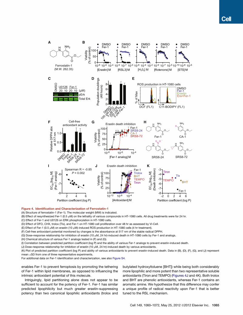
Figure 4. Identification and Characterization of Ferrostatin-1
(A) Structure of ferrostatin-1 (Fer-1). The molecular weight (MW) is indicated.
(B) Effect of resynthesized Fer-1 (0.5 mM) on the lethality of various compounds in HT-1080 cells. All drug treatments were for 24 hr.
(C) Effect of Fer-1 and U0126 on ERK phosphorylation in HT-1080 cells.
(D) Effect of DFO, CHX, trolox (Tlx), and Fer-1 on HT-1080 cell proliferation over 48 hr as assessed by Vi-Cell.
(E) Effect of Fer-1 (0.5 mM) on erastin (10 mM)-induced ROS production in HT-1080 cells (4 hr treatment).
(F) Cell-free antioxidant potential monitored by changes is the absorbance at 517 nm of the stable radical DPPH.
(G) Dose-response relationship for inhibition of erastin (10 mM, 24 hr)-induced death in HT-1080 cells by Fer-1 and analogs.
(H) Chemical structure of various Fer-1 analogs tested in (F) and (G).
(I) Correlation between predicted partition coefficient (log P) and the ability of various Fer-1 analogs to prevent erastin-induced death.
(J) Dose-response relationship for inhibition of erastin (10 mM, 24 hr)-induced death by various antioxidants.
(K) Plot of predicted partition coefficient (log P) and ability of various antioxidants to prevent erastin-induced death. Data in (B), (D), (F), (G), and (J) represent
mean ±SD from one of three representative experiments.
For additional data on Fer-1 identification and characterization, see also Figure S4.
enables Fer-1 to prevent ferroptosis by promoting the tethering
of Fer-1 within lipid membranes, as opposed to influencing the
intrinsic antioxidant potential of this molecule.
Intriguingly, lipid partitioning alone does not appear to be
sufficient to account for the potency of Fer-1. Fer-1 has similar
predicted lipophilicity but much greater erastin-suppressing
potency than two canonical lipophilic antioxidants (trolox and
butylated hydroxyltoluene [BHT]) while being both considerably
more lipophilic and more potent than two representative soluble
antioxidants (Tiron and TEMPO) (Figures 4J and 4K). Both trolox
and BHT are phenolic antioxidants, whereas Fer-1 contains an
aromatic amine. We hypothesize that this difference may confer
a unique profile of radical reactivity upon Fer-1 that is better
tuned to the RSL mechanism.
Cell 149, 1060–1072, May 25, 2012 ª2012 Elsevier Inc. 1065

C D E
5
Control Glutamate
Pre-injury
(brightfield)
Day 0
(PI stain)
Day +1
(PI stain)
A
B
CA1
Con
trol
5m
M G
luta
mat
e
Glut +
2µM
Fer
-1
Glut +
5µM
CPX
Glut +
10
µMM
K-8
010
25
50
% c
ell
de
ath
CA3
Con
trol
5m
M G
luta
mat
e
Glut +
2µM
Fer
-1
Glut +
5µM
CPX
Glut +
10
µMM
K-8
010
25
50
% c
ell
de
ath
Dentrate gyrus
Con
trol
mM
Gluta
mat
e
Glut +
2µM
Fer
-1
Glut +
5µM
CPX
Glut +
10
µMM
K-8
010
25
50
% c
ell
de
ath
Glutamate
+Fer-1
Glutamate
+CPX
Glutamate
+MK-801
Dissected Rat Brain Dissected Hippocampus Plated 400 μm Slices
Hippocampus
CA3
DG
CA1
******
******
******
******
******
*****
Figure 5. Effects of Fer-1 on Excitotoxic Cell Death in Organotypic Hippocampal Slice Cultures
(A) Cartoon outline of hippocampal slice procedure.
(B) Bright-field and fluorescent images of PI staining of treated hippocampal slices. Slices were treated with glutamate (5 mM, 3 hr) ±Fer-1 (2 mM), CPX (5 mM), or
MK-801 (10 mM). Representative images from 1 of 6 slices per condition are shown.
(C–E) Quantification of the effects depicted in (B). Data shown are mean ±SD. Data were analyzed using a two-way ANOVA (brain region x drug treatment)
followed by Bonferroni posttests.
**p < 0.01 and ***p < 0.001.
Fer-1 Prevents Glutamate-Induced NeurotoxicityExcitotoxic cell death that occurs in the nervous system in
epilepsy, stroke, and other trauma situations has been
described as an oxidative, iron-dependent process (Cheah
et al., 2006; Choi, 1988; Murphy et al., 1989). We hypothesized
that excitotoxic death could be related to erastin-induced ferrop-
tosis. We tested this hypothesis by using a rat organotypic
hippocampal slice culture (OHSC) model that closely resembles
the hippocampus in vivo by preserving the integrity of neuronal
connections, both inhibitory and excitatory, and their supporting
cells, including astrocytes and microglia (Lossi et al., 2009).
OHSCs have proven to be ideal complex preparations for lead-
compound identification and validation (Noraberg et al., 2005;
1066 Cell 149, 1060–1072, May 25, 2012 ª2012 Elsevier Inc.
Sundstrom et al., 2005), capable of predicting in vivo efficacy
(Cater et al., 2007; Morrison et al., 2002).
OHSCs were treated with a lethal excitotoxic stimulus (5 mM
L-glutamate, 3 hr) that mimics the consequences of stroke and
neurodegenerative disease (Morrison et al., 2002; Sundstrom
et al., 2005) (Figure 5A). These slices were coincubated with
glutamate and vehicle alone or with glutamate plus Fer-1
(2 mM), the iron chelator CPX (5 mM), or, as a positive control,
the N-methyl-D-aspartate (NMDA) receptor antagonist MK-801
(10 mM). We analyzed the effects of these compound treatments
on propidium iodide (PI) uptake as an indicator of cell death 24 hr
following the end of glutamate treatment in three defined regions
of the OHSCs: the dentate gyrus (DG), the CA1, and the CA3

fields of the hippocampus. A two-way analysis of variance
(ANOVA) suggested significant differences for both brain region
(F2,75 = 19.23, p < 0.0001) and compound treatment (F4,75 = 67.8,
p < 0.0001) factors. Focusing on the compound treatment effect,
Bonferroni posttests indicated that glutamate induced signifi-
cant cell death in all three regions of the brain and that this death
was attenuated significantly and to an almost identical extent by
cotreatment with Fer-1, CPX, or MK-801 (p < 0.001 for all inter-
actions except glutamate+MK-801 within the DG, p < 0.01)
(Figures 5B–5E). These results suggest that glutamate-induced
death in OHSCs and erastin-induced death in cancer cells share
in common a core lethal mechanism that can be inhibited by iron
chelation or Fer-1.
Erastin Inhibits System x�cCPX and Fer-1 suppressed erastin-induced death in cancer cells
and glutamate-induced toxicity in OHSCs, consistent with a
common iron- and ROS-dependent death execution mecha-
nism. We wondered whether any death-initiating mechanisms
could also be shared between these two processes.
Glutamate-induced death in brain cells can be initiated by
calcium influx through ionotropic glutamate receptors and
through competitive inhibition of cystine uptake by the Na+-inde-
pendent cystine/glutamate antiporter, system x�c (Choi, 1988;
Murphy et al., 1989). The calcium chelators BAPTA-AM,
Fura-2, and EGTA had no effect on erastin-induced death (Fig-
ure S5A) (Wolpaw et al., 2011), arguing against a role for Ca2+
influx in this process. However, we observed striking clustering
of erastin and sulfasalazine (SAS), a specific inhibitor of system
x�c (Gout et al., 2001), in a modulatory profile of 19 oxidative
and nonoxidative lethal molecules generated in HT-1080 cells
(Figure 6A). If blockade of system x�c -mediated cystine import
can trigger ferroptosis, then providing this metabolite to cells
through an alternative means should rescue from death. Indeed,
b-mercaptoethanol (b-ME), which can circumvent the inhibition
of system x�c by promoting cystine uptake through an alternative
pathway (Ishii et al., 1981), strongly inhibited cell death in
HT-1080 cells induced by erastin, SAS, and glutamate (Figures
6A and S5B). As predicted by these results, SAS, like erastin,
behaved as an RSL compound, albeit with considerably lower
potency than erastin (Figure S5C). This is nonetheless note-
worthy, as SAS is an FDA-approved drug not previously shown
to demonstrate such activity.
System x�c is a disulfide-linked heterodimer composed of
SLC7A11 (xCT) and SLC3A2 (4F2hc and CD98hc) (Sato et al.,
1999) (Figure 6B). Inhibition of system x�c can lead to a compen-
satory transcriptional upregulation of SLC7A11 (Lo et al., 2008).
Consistent with this, we observed substantial upregulation of
SLC7A11 in HT-1080 cells that were treated with erastin or
SAS, an effect that was suppressed by b-ME, but not DFO or
Fer-1 (Figure 6C). Further confirming the relevance of system
x�c to erastin-induced ferroptosis, siRNA-mediated silencing
of SLC7A11 with two independent siRNAs sensitized HT-1080
cells to erastin-induced death (Figures 6D and 6E), whereas
transfection of HT-1080 cells with a plasmid encoding DDK-
tagged SLC7A11 conferred protection from erastin- and SAS-
induced death (Figure S5D). Given these results, we directly
examined the uptake of [14C]-cystine into HT-1080 cells. Erastin
(10 mM), glutamate (50 mM), and SAS (1 mM) abolished the
Na+-independent uptake of [14C]-cystine, whereas RSL3 had
no effect on this process (Figures 6F and S5E).
How does erastin inhibit system x�c ? Analysis of affinity
purification data (Yagoda et al., 2007) identified SLC7A5
(LAT1, 4F2lc, and CD98lc) as the lone protein bound by an active
erastin affinity analog in lysates from bothHRAS-wild-type BJeH
and HRAS mutant BJeLR cells (Figure 6G). SLC7A5 (like
SLC7A11) is one of six light chains that bind SLC3A2 to form
amino acid transporters of differing substrate selectivity. The
SLC7A5/SLC3A2 complex (system L) transports large, neutral
amino acids (Kanai and Endou, 2003) (Figure 6B). In a profile
of 123 metabolites from human Jurkat T lymphocytes treated
with erastin (1 mM, 25 min) (Ramanathan and Schreiber, 2009),
highly significant decreases were observed in the levels of
system L substrates (Kanai and Endou, 2003), whereas the
levels of nonsystem L substrates were unchanged or increased
(Figure 6H). However, unlike inhibition of system x�c using
excess glutamate (12.5 mM), inhibition of system L using excess
D-phenylalanine (12.5 mM) (Kanai and Endou, 2003) did not
strongly sensitize to erastin (Figure 6I). Together, this suggests
that erastin inhibits system L-mediated amino acid uptake but
that this does not contribute directly to ferroptosis. Rather, era-
stin binding to SLC7A5 or the SLC7A5/SLC3A2 complex likely
interferes with cystine uptake by the SLC3A2/SLC7A11 complex
in trans.
NAPDHOxidases Provide One Source of Death-InducingROS in Erastin-Treated CellsBlocking system x�c inhibits cysteine-dependent glutathione
(GSH) synthesis and also inhibits the transplasma membrane
cysteine redox shuttle (Banjac et al., 2008; Ishii et al., 1981).
Both effects impair cellular antioxidant defenses, thereby facili-
tating toxic ROS accumulation. Having ruled out the mitochon-
drial ETC as a source of death-inducing ROS in erastin-treated
cells (Figures 1D–1F), we examined the role of the nicotinamide
adenine dinucleotide phosphate (NADPH) oxidase (NOX) family
of superoxide-producing enzymes (NOX1–5, DUOX1,2), which
are upregulated in several RAS mutant tumors (Kamata, 2009).
Erastin-induced ferroptosis was strongly suppressed in Calu-1
cells by the canonical NOX inhibitor diphenylene iodonium
(DPI), the NOX1/4-specific inhibitor GKT137831 (Laleu et al.,
2010), and an inhibitor of the NADPH-generating pentose phos-
phate pathway (PPP), 6-aminonicotinamde (6-AN) (Figures 7A
and 7B). Given that Calu-1 cells express NOX1 at much higher
levels thanNOX4 (Figure S6A), NOX1 is themost likely candidate
to mediate the observed NOX-dependent lethal effects in these
cells. Additionally, shRNA-mediated silencing of two PPP
enzymes, glucose-6-phosphate dehydrogenase (G6PD) and
phosphoglycerate dehydrogenase (PGD), also prevented
erastin-induced ferroptosis in Calu-1 cells to the same extent
as silencing of VDAC2 (Figures 7C and 7D). 6-AN also prevented
cell death as well as ROS production in BJeLR cells (Figures S6B
and 6C), suggesting an important role for this pathway in these
cell types. On the other hand, NOX and PPP inhibitors were
only partially effective at preventing erastin-induced ferroptosis
in HT-1080 cells (Figure 7B), indicating that other pathways, in
addition to the PPP/NOX pathway, can contribute to the onset
Cell 149, 1060–1072, May 25, 2012 ª2012 Elsevier Inc. 1067

B
G
RS
L3
Era
stin
SA
SR
ote
no
ne
2−
ME
B−
La
pa
ch
on
eD
oxo
rub
icin
CC
CP
PA
OTri
ch
oA
SA
HA
Bre
feld
in−
AS
TS
Borte
zom
ibTa
xo
lH
ele
na
line
Art
esu
na
te
PE
ITC
Fer−1 (0.5 μM)Trolox (100 μM)β−ME (50 μM)NAC (1 mM)CHX (5 μM)DFO (100 μM)
-0.4
1.3H20
2
Lethal molecules
C
Erastin (10 μM)SAS (1 mM)β-ME (50 μM)DFO (100 μM)Fer-1 (1 μM)
-+- - - - - - -+ + +- -+- +- +- +- - -- - -+ +- - - -+ - -- - - - -+ +- -- + -- - - - - - -+ +- - +
0
2
4
6
8
Re
lativ
eSL
C7A
11m
RN
Ale
ve
ls
A
D
Me
F
SLC7A11
Glu
Cys
SLC3A2
NAA
SLC7A5
System L
NAA
H
:
AA Fold-chng value Rank
Tyr 0.81 0.0005 1/123
Trp 0.77 0.001 2/123
Phe 0.82 0.002 6/123
Met 0.83 0.002 7/123
Ile/Leu 0.86 0.004 12/123
His 0.80 0.06 50/123
SAS,Glu
BJeH BJeLR
1 104
SLC7A5
I
10-7 10-6 10-5
0
25
50
75
100DMSO
D-Phe
L-Glu
Erastin (M)
Via
bili
ty(%
ofc
on
tro
l)
E
siCon
trol
siSLC
7A11
-2
siSLC
7A11
-30
25
50
75
100Erastin (10 µM)DMSO
Via
bili
ty(%
ofc
on
tro
l)
siCon
trol
siSLC
7A11
-2
siSLC
7A11
-30.0
0.5
1.0
Re
lativ
eSL
C7A
11m
RN
Ale
ve
ls
D-Phe
System xc
Metabolic profile: Jurkat T cells
(1 μM erastin, 25 minutes)
Non-system L substrates:
AA Fold-chng P value Rank
Ser 1.44 0.1 57/123
Thr 1.36 0.03 35/123
Asn 1.23 0.13 67/123
Ala 1.3 0.15 68/123
Arg 1.07 0.21 87/123
Erastin affinity purification
DM
SO
10 µM
Era
stin
1 m
M S
AS
50 m
M G
luta
mat
e0
25
50
75
100
[14C
]-cystin
eu
pta
ke
(%o
fco
ntro
l)
Figure 6. Erastin Inhibits the Activity of System x�c(A) Modulatory profile of HT-1080 cells treated with different lethal compounds and inhibitors.
(B) Cartoon depicting the composition and function of system L and system x�c . Cys, cystine; NAA, neutral amino acids.
(C) SLC7A11 mRNA levels in compound-treated (6 hr) HT-1080 cells determined by RT-qPCR.
(D and E) Effect of silencing SLC7A11 by using siRNA on erastin (10 mM, 8 hr) induced death (D) and mRNA levels (E) in HT-1080 cells.
(F) Normalized Na+-independent [14C]-cystine uptake by HT-1080 cells in response to various drugs. Data are represented as mean ±SD, n = 3.
(G) Identification of SLC7A5 as the lone target identified by erastin affinity purification in both BJeH and BJeLR cells.
(H) Metabolic profiling of system L and nonsystem L substrate amino acid levels in erastin-treated Jurkat cells.
(I) Effect of L-glutamic acid (L-Glu, 12.5 mM) and D-phenylalanine (D-Phe, 12.5 mM) on erastin-induced death in HT-1080 cells.
See also Figure S5.
of death in erastin-treated cells once the appropriate conditions
have been set by the inhibition of system x�c .
DISCUSSION
Ferroptotic death is morphologically, biochemically, and geneti-
cally distinct from apoptosis, various forms of necrosis, and
1068 Cell 149, 1060–1072, May 25, 2012 ª2012 Elsevier Inc.
autophagy. This process is characterized by the overwhelming,
iron-dependent accumulation of lethal lipid ROS (Figure 7E,
blue outline). Unlike other forms of apoptotic and nonapoptotic
death (Christofferson and Yuan, 2010; Jacobson and Raff,
1995), this requirement for ROS accumulation appears to be
universal. In at least some cells, NOX family enzymes make
important contributions to this process. Indeed, although we

Cys
NOX
Glutamate
Sulfasalazine
Ferroptosis
Lipid and
soluble ROS
RSL3?
DFO, CPX
Erastin
VDAC2,3
Metabolic
effect
TroloxFer-1
β-ME
System L
Amino
acid uptake
??
A
6-ANGlucose
PPP
ATP
NADPH
O₂ˉ•
NOX
GTK137831DPI
E
Lipids
B
DMSO
20 µM GKT0.5 µM DPI
200 µM 6-AN100 µM DFO
1.25 2.50 5.00Erastin (µM)
1.25 2.50 5.000
25
50
75
100
125
Erastin (µM)
Via
bili
ty(%
ofc
on
tro
l)
sh-G6PDsh-PGD
D
CCalu-1 cells
0
20
40
60
80
100
Via
bili
ty(%
ofc
on
tro
l)
sh-C
ontro
l
sh87
9-VDAC2
sh19
14-G
6PD
sh19
55-G
6PD
sh13
5-PGD
sh-C
ontro
l
sh87
9-VDAC2
sh19
14-G
6PD
sh19
55-G
6PD
sh13
5-PGD
0.0
0.5
1.0
Re
lativ
em
RN
Ale
ve
l
Gln
AOAsh-CS, sh-ACSF2
Calu-1 cells HT-1080 cells
System xc
Figure 7. Role of NOX in Erastin-Induced Death
(A) Outline of NOX pathway. Inhibitors are shown in green. PPP, pentose phosphate pathway.
(B) Effect of NOX pathway inhibitors on erastin-induced death in Calu-1 and HT-1080 cells. GKT, GKT137831.
(C and D) Effect of shRNA silencing of the PPP enzymes glucose-6-phosphate dehydrogenase (G6PD) and phosphogluconate dehydrogenase (PGD) on viability
of erastin (2.5 mM)-treated Calu-1 cells. Infection with shRNA targeting VDAC2 was used as a positive control. Relative mRNA levels in (D) were assessed by RT-
qPCR following shRNA knockdown. Data in (B), (C), and (D) represent mean ±SD.
(E) Model of ferroptosis pathway. The core ferroptotic lethal mechanism is highlighted in blue.
See Figure S6 for additional data supporting a role for the PPP/NOX pathway in erastin-induced cell death.
cannot exclude the possibility of a death-inducing protein or
protein complex activated downstream of ROS accumulation,
we posit that the executioners of death in cancer cells under-
going ferroptosis are these ROS themselves. An important
prediction of this model is that, under anoxic conditions, ferrop-
tosis will be inactive. However, even here, agents such as erastin
thatmay prevent uptake of essential amino acids by systemL are
likely to be toxic to cells.
Using an shRNA library targeting most known genes encoding
mitochondrial proteins (Pagliarini et al., 2008), we identified
specific roles for RPL8, IREB2, ATP5G3, TTC35, CS, and
ACSF2 in erastin-induced ferroptosis. A plausible hypothesis
to emerge from these data is that CS and ACSF2 are required
to synthesize a specific lipid precursor necessary for death (Fig-
ure 7E). Just as important, the high resolution of the arrayed
approach (1 hairpin/well, minimum 5 hairpins/gene) provides
confidence that the various mitochondrial genes not identified
in our screen, including many implicated in apoptotic and other
nonapoptotic death pathways (BID, BAK1, BAX, AIFM1, PPIF,
HTRA2, ENDOG, and PGAM5), are truly not required for era-
stin-induced ferroptosis. This screening collection will be a valu-
able resource for future studies of the role of the mitochondria in
cell physiology.
In cancer cells, inhibition of system x�c -mediated cystine
uptake by erastin, SAS, or glutamate may be sufficient to initiate
iron-dependent ferroptosis. Inhibition of system x�c is, however,
not necessary; RSL3 does not inhibit cystine uptake and yet trig-
gers an otherwise similar iron- and ROS-dependent ferroptototic
death program. Thus, RSL3 likely modulates the activity of
a target lying downstream of or in parallel to system x�c (Fig-
ure 7E). Importantly, this may enable RSL3 to activate ferroptosis
in cells or conditions in which cystine uptake via system x�c is
not limiting for survival. Lanperisone, another recently identified
oncogenic RAS-selective lethal small molecule that causes
Cell 149, 1060–1072, May 25, 2012 ª2012 Elsevier Inc. 1069

nonapoptotic, iron-dependent death in mouse Kras mutant
tumor cells (Shaw et al., 2011), may also inhibit the function of
system x�c or another target in the ferroptotic pathway. Other
compounds that behave as RSLs, such as PEITC, oncrasin,
and piperlongumine (Guo et al., 2008; Raj et al., 2011; Trachoo-
tham et al., 2006), trigger mitochondrial cytochrome c release,
caspase activation, and other features of apoptosis not
observed in cancer cells undergoing ferroptosis. Certain tumor
cells are highly resistant to apoptosis (Ni Chonghaile et al.,
2011). Thus, agents such as erastin, RSL3, and lanperisone
that can trigger nonapoptotic death may exhibit a unique spec-
trum of clinical activity.
In some brain cell populations, inhibition of system x�c by
glutamate triggers oxidative cell death dependent on iron and
lipid ROS as well as Ca2+ influx, mitochondrial damage, mito-
chondrial ROS production, and chromatin fragmentation (Li
et al., 1997; Murphy et al., 1989; Ratan et al., 1994; Tan et al.,
1998; Yonezawa et al., 1996). These latter events, beginning
with Ca2+ influx, are not required for RSL-induced ferroptosis
in cancer cells, perhaps because heightened activity of NOX or
other pro-oxidant enzymes or basally altered membrane lipid
composition is sufficient to promote death in the absence of
these additional features. Regardless, the oxidative death path-
ways triggered in cancer cells and brain cells by blockade of
cystine uptake both appear to access a core iron- and ROS-
dependent ferroptotic mechanism, accounting for the ability of
Fer-1 and CPX to attenuate death in both cases (Figure 7E).
The specific role of iron in ferroptosis remains unclear.
Ferroptosis cannot be explained by a simple increase in H2O2-
dependent, iron-catalyzed ROS production (i.e., Fenton chem-
istry), as H2O2-induced death is distinct from RSL-induced
ferroptosis (Figures 1 and 2). Rather, our results aremost consis-
tent with one or more iron-dependent enzymes functioning as
part of the core oxidative lethal mechanism. The void created
in the antioxidant defenses of the cell by the inhibition of cystine
uptake by erastin may be required to unleash the activity of these
enzymes. Thus, for better or worse, the aberrantly elevated
levels of iron that are observed in some cancer cells (Pinnix
et al., 2010) and pathological neuronal populations (Duce et al.,
2010; Lei et al., 2012) may predispose to ferroptotic death
in situations of cystine or cysteine limitation.
EXPERIMENTAL PROCEDURES
Analysis of Reactive Oxygen Species Production
The day before the experiment, 200,000 cells/well were seeded in 6-well
dishes (Corning). The day of the experiment, cells were treated with test
compounds for the indicated times; harvested by trypsinizaiton; resuspended
in 500 ml Hanks Balanced Salt Solution (HBSS, Gibco) containing H2DCFDA
(25 mM), C11-BODIPY(581/591) (2 mM), or MitoSOX (5 mM) (all from Molecular
Probes, Invitrogen); and incubated for 10 min at 37�C in a tissue culture incu-
bator. Cells were then resuspended in 500 ml of fresh HBSS, strained through
a 40 mM cell strainer (BD Falcon), and analyzed using a flow cytometer (FACS-
Calibur or Accuri C6, BD Biosciences) equipped with 488 nm laser for excita-
tion. Data were collected from the FL1 (H2DCFDA, C11-BODIPY) or FL2
channel (MitoSOX). A minimum of 10,000 cells were analyzed per condition.
Cancer Cell Viability Measurements
Cell viability was typically assessed in 384-well format by Alamar Blue (Invitro-
gen) fluorescence (ex/em 530/590) measured on a Victor3 plate reader (Perkin
1070 Cell 149, 1060–1072, May 25, 2012 ª2012 Elsevier Inc.
Elmer). In some experiments, Trypan blue dye exclusion counting was
performed by using an automated cell counter (ViCell, Beckman-Coulter).
Cell viability under test conditions is reported as a percentage relative to the
negative control treatment.
shRNA Screening
An arrayed collection of 6,528 shRNA hairpins derived from The RNAi Consor-
tium (TRC) collection targeting 1,087 genes, kindly provided by Vamsi Mootha
and Joshua Baughman (MIT), was screened in 384-well plate format (Corning)
in both Calu-1 and HT-1080 cells. shRNAs targeting GFP and RFP, randomly
distributed through each plate, served as negative controls. Four-hundred
cells/well were infected in duplicate for 48 hr with 2 ml shRNA-containing viral
supernatant, selected for 24 hr in puromycin (1.5 mg/ml), and then treated with
DMSO, erastin (7.3 mM), or STS (1 mM) for 24 hr. Cell viability was determined
by using Alamar Blue. For each hairpin within each treatment condition, a cell
death rescue score was computed as the ratio of the average viability of the
two replicates to the average viability of the within-plate negative controls.
These scores were used to compare the effects between compounds. To
identify genes required for ferroptosis, individual hairpins were scored as
hits if they displayed an average death suppressionR3 median average devi-
ations from the median within-plate or screen-wide negative control values.
Fifty-one candidate genes were identified with the same two (or more) unique
hairpins per gene called as hits in both the Calu-1 and HT-1080 screens. For
each candidate gene, confirmation studies using reverse-transcription quanti-
tative PCR (RT-qPCR) analysis of mRNA silencing were performed in HT-1080
cells by using freshly prepared virus as described in greater detail in the
Extended Experimental Procedures.
[14C]-Cystine Uptake Assay
Two-hundred thousand (200,000) HT-1080 cells/well were seeded overnight in
6-well dishes (Corning). The next day, cells were washed twice in prewarmed
Na+-free uptake buffer (137 mM choline chloride, 3 mM KCl, 1 mM CaCl2,
1 mM MgCl2, 5 mM D-glucose, 0.7 mM K2HPO4, and 10 mM HEPES [pH
7.4]) and then incubated for 10 min at 37�C in 1 ml of uptake buffer to deplete
cellular amino acids. At this point, in each well, the buffer was replaced with
600 ml uptake buffer containing compound and 0.12 mCi (80–110 mCi/mmol)
of L-[3,30-14C]-cystine (Perkin Elmer) and incubated for 3 min at 37�C. Cellswere then washed three times with ice-cold uptake buffer and lysed in
500 ml 0.1 M NaOH. To this lysate, 15 ml of scintillation fluid was added, and
radioactive counts per minute were obtained by using a scintillation counter.
All measurements were performed in triplicate for each condition.
Statistical Analyses
All statistical analyses were performed by using Prism 5.0c (GraphPad
Software).
SUPPLEMENTAL INFORMATION
Supplemental Information includes Extended Experimental Procedures
and six figures and can be found with this article online at doi:10.1016/
j.cell.2012.03.042.
ACKNOWLEDGMENTS
We thank Vamsi Mootha, Joshua Baughman, and David Root for sharing the
custom shRNA library; Kristy Brown and Elma Zaganjor for assistance with
electron microscopy; Darnelle Delva for help with qPCR; Rohitha SriRamarat-
nam for help with cell death assays; Eric Schon for providing the 143B cell
lines; Craig Thompson for providing Bax�/� Bak�/� MEFs; and Patrick Page
(GenKyoTex S.A.) for providing GKT137831. We thank David Clarke for
comments on the manuscript. Certain shRNA collections used in this work
were generated with the assistance of the Scientific Planning and Allocation
of Resources Committee (SPARC, to Vamsi Mootha). NSF grant
CHE-0840451 supported the purchase and operation of equipment used in
the chemical characterization of ferrostatin-1. M.R.L. was supported by
a fellowship from the National Science Foundation. K.M.L. was supported

by the Medical Scientist Training Program (Columbia University). S.J.D. was
supported by a postdoctoral fellowship from the Canadian Institutes of Health
Research. This research was supported by grants from the US National
Institutes of Health (5R01CA097061, 5R01GM085081, and R01CA161061),
the Arnold and Mabel Beckman Foundation, and NYSTAR. B.R.S. is an Early
Career Scientist of the Howard Hughes Medical Institute.
Received: January 17, 2012
Revised: March 9, 2012
Accepted: March 13, 2012
Published: May 24, 2012
REFERENCES
Banjac, A., Perisic, T., Sato, H., Seiler, A., Bannai, S., Weiss, N., Kolle, P.,
Tschoep, K., Issels, R.D., Daniel, P.T., et al. (2008). The cystine/cysteine cycle:
a redox cycle regulating susceptibility versus resistance to cell death.
Oncogene 27, 1618–1628.
Bergsbaken, T., Fink, S.L., and Cookson, B.T. (2009). Pyroptosis: host cell
death and inflammation. Nat. Rev. Microbiol. 7, 99–109.
Cater, H.L., Gitterman, D., Davis, S.M., Benham, C.D., Morrison, B., III, and
Sundstrom, L.E. (2007). Stretch-induced injury in organotypic hippocampal
slice cultures reproduces in vivo post-traumatic neurodegeneration: role of
glutamate receptors and voltage-dependent calcium channels. J. Neurochem.
101, 434–447.
Cheah, J.H., Kim, S.F., Hester, L.D., Clancy, K.W., Patterson, S.E., III, Papado-
poulos, V., and Snyder, S.H. (2006). NMDA receptor-nitric oxide transmission
mediates neuronal iron homeostasis via the GTPase Dexras1. Neuron 51,
431–440.
Choi, D.W. (1988). Glutamate neurotoxicity and diseases of the nervous
system. Neuron 1, 623–634.
Christofferson, D.E., and Yuan, J. (2010). Necroptosis as an alternative form of
programmed cell death. Curr. Opin. Cell Biol. 22, 263–268.
Dolma, S., Lessnick, S.L., Hahn, W.C., and Stockwell, B.R. (2003). Identifica-
tion of genotype-selective antitumor agents using synthetic lethal chemical
screening in engineered human tumor cells. Cancer Cell 3, 285–296.
Duce, J.A., Tsatsanis, A., Cater, M.A., James, S.A., Robb, E., Wikhe, K.,
Leong, S.L., Perez, K., Johanssen, T., Greenough, M.A., et al. (2010). Iron-
export ferroxidase activity of b-amyloid precursor protein is inhibited by zinc
in Alzheimer’s disease. Cell 142, 857–867.
Fuchs, Y., and Steller, H. (2011). Programmed cell death in animal develop-
ment and disease. Cell 147, 742–758.
Gout, P.W., Buckley, A.R., Simms, C.R., and Bruchovsky, N. (2001).
Sulfasalazine, a potent suppressor of lymphoma growth by inhibition of the
x(c)- cystine transporter: a new action for an old drug. Leukemia 15,
1633–1640.
Guo, W., Wu, S., Liu, J., and Fang, B. (2008). Identification of a small molecule
with synthetic lethality for K-ras and protein kinase C iota. Cancer Res. 68,
7403–7408.
Ishii, T., Bannai, S., and Sugita, Y. (1981). Mechanism of growth stimulation
of L1210 cells by 2-mercaptoethanol in vitro. Role of the mixed disulfide of
2-mercaptoethanol and cysteine. J. Biol. Chem. 256, 12387–12392.
Jacobson, M.D., and Raff, M.C. (1995). Programmed cell death and Bcl-2
protection in very low oxygen. Nature 374, 814–816.
Kamata, T. (2009). Roles of Nox1 and other Nox isoforms in cancer develop-
ment. Cancer Sci. 100, 1382–1388.
Kanai, Y., and Endou, H. (2003). Functional properties of multispecific amino
acid transporters and their implications to transporter-mediated toxicity.
J. Toxicol. Sci. 28, 1–17.
Laleu, B., Gaggini, F., Orchard, M., Fioraso-Cartier, L., Cagnon, L.,
Houngninou-Molango, S., Gradia, A., Duboux, G., Merlot, C., Heitz, F., et al.
(2010). First in class, potent, and orally bioavailable NADPH oxidase isoform
4 (Nox4) inhibitors for the treatment of idiopathic pulmonary fibrosis. J. Med.
Chem. 53, 7715–7730.
Lei, P., Ayton, S., Finkelstein, D.I., Spoerri, L., Ciccotosto, G.D., Wright, D.K.,
Wong, B.X., Adlard, P.A., Cherny, R.A., Lam, L.Q., et al. (2012). Tau deficiency
induces parkinsonism with dementia by impairing APP-mediated iron export.
Nat. Med. 18, 291–295.
Li, Y., Maher, P., and Schubert, D. (1997). A role for 12-lipoxygenase in nerve
cell death caused by glutathione depletion. Neuron 19, 453–463.
Lo, M., Ling, V., Wang, Y.Z., and Gout, P.W. (2008). The xc- cystine/glutamate
antiporter: a mediator of pancreatic cancer growth with a role in drug
resistance. Br. J. Cancer 99, 464–472.
Lossi, L., Alasia, S., Salio, C., and Merighi, A. (2009). Cell death and
proliferation in acute slices and organotypic cultures of mammalian CNS.
Prog. Neurobiol. 88, 221–245.
Macarron, R., Banks, M.N., Bojanic, D., Burns, D.J., Cirovic, D.A., Garyantes,
T., Green, D.V., Hertzberg, R.P., Janzen, W.P., Paslay, J.W., et al. (2011).
Impact of high-throughput screening in biomedical research. Nat. Rev. Drug
Discov. 10, 188–195.
Morrison, B., III, Pringle, A.K., McManus, T., Ellard, J., Bradley, M., Signorelli,
F., Iannotti, F., and Sundstrom, L.E. (2002). L-arginyl-3,4-spermidine is
neuroprotective in several in vitro models of neurodegeneration and in vivo
ischaemia without suppressing synaptic transmission. Br. J. Pharmacol.
137, 1255–1268.
Mullen, A.R., Wheaton, W.W., Jin, E.S., Chen, P.H., Sullivan, L.B., Cheng, T.,
Yang, Y., Linehan, W.M., Chandel, N.S., and Deberardinis, R.J. (2011). Reduc-
tive carboxylation supports growth in tumour cells with defective mitochon-
dria. Nature 481, 385–388.
Murphy, T.H., Miyamoto, M., Sastre, A., Schnaar, R.L., and Coyle, J.T. (1989).
Glutamate toxicity in a neuronal cell line involves inhibition of cystine transport
leading to oxidative stress. Neuron 2, 1547–1558.
Ni Chonghaile, T., Sarosiek, K.A., Vo, T.T., Ryan, J.A., Tammareddi, A., Moore,
Vdel.G., Deng, J., Anderson, K.C., Richardson, P., Tai, Y.T., et al. (2011).
Pretreatment mitochondrial priming correlates with clinical response to
cytotoxic chemotherapy. Science 334, 1129–1133.
Noraberg, J., Poulsen, F.R., Blaabjerg, M., Kristensen, B.W., Bonde, C.,
Montero, M., Meyer, M., Gramsbergen, J.B., and Zimmer, J. (2005). Organo-
typic hippocampal slice cultures for studies of brain damage, neuroprotection
and neurorepair. Curr. Drug Targets CNS Neurol. Disord. 4, 435–452.
Pagliarini, D.J., Calvo, S.E., Chang, B., Sheth, S.A., Vafai, S.B., Ong, S.E.,
Walford, G.A., Sugiana, C., Boneh, A., Chen, W.K., et al. (2008). A mitochon-
drial protein compendium elucidates complex I disease biology. Cell 134,
112–123.
Pinnix, Z.K., Miller, L.D., Wang, W., D’Agostino, R., Jr., Kute, T., Willingham,
M.C., Hatcher, H., Tesfay, L., Sui, G., Di, X., et al. (2010). Ferroportin and
iron regulation in breast cancer progression and prognosis. Sci. Transl. Med.
2, 43ra56.
Raj, L., Ide, T., Gurkar, A.U., Foley, M., Schenone, M., Li, X., Tolliday, N.J.,
Golub, T.R., Carr, S.A., Shamji, A.F., et al. (2011). Selective killing of cancer
cells by a small molecule targeting the stress response to ROS. Nature 475,
231–234.
Ramanathan, A., and Schreiber, S.L. (2009). Direct control of mitochondrial
function by mTOR. Proc. Natl. Acad. Sci. USA 106, 22229–22232.
Ratan, R.R., Murphy, T.H., and Baraban, J.M. (1994). Oxidative stress induces
apoptosis in embryonic cortical neurons. J. Neurochem. 62, 376–379.
Salahudeen, A.A., Thompson, J.W., Ruiz, J.C., Ma, H.W., Kinch, L.N., Li, Q.,
Grishin, N.V., and Bruick, R.K. (2009). An E3 ligase possessing an iron-respon-
sive hemerythrin domain is a regulator of iron homeostasis. Science 326,
722–726.
Sato, H., Tamba, M., Ishii, T., and Bannai, S. (1999). Cloning and expression of
a plasma membrane cystine/glutamate exchange transporter composed of
two distinct proteins. J. Biol. Chem. 274, 11455–11458.
Shaw, A.T., Winslow, M.M., Magendantz, M., Ouyang, C., Dowdle, J.,
Subramanian, A., Lewis, T.A., Maglathin, R.L., Tolliday, N., and Jacks, T.
(2011). Selective killing of K-ras mutant cancer cells by small molecule
inducers of oxidative stress. Proc. Natl. Acad. Sci. USA 108, 8773–8778.
Cell 149, 1060–1072, May 25, 2012 ª2012 Elsevier Inc. 1071

Sundstrom, L., Morrison, B., III, Bradley, M., and Pringle, A. (2005). Organo-
typic cultures as tools for functional screening in the CNS. Drug Discov. Today
10, 993–1000.
Tan, S., Sagara, Y., Liu, Y., Maher, P., and Schubert, D. (1998). The regulation
of reactive oxygen species production during programmed cell death. J. Cell
Biol. 141, 1423–1432.
Thompson, C.B. (1995). Apoptosis in the pathogenesis and treatment of
disease. Science 267, 1456–1462.
Trachootham, D., Zhou, Y., Zhang, H., Demizu, Y., Chen, Z., Pelicano, H.,
Chiao, P.J., Achanta, G., Arlinghaus, R.B., Liu, J., and Huang, P. (2006).
Selective killing of oncogenically transformed cells through a ROS-mediated
mechanism by beta-phenylethyl isothiocyanate. Cancer Cell 10, 241–252.
Vashisht, A.A., Zumbrennen, K.B., Huang, X., Powers, D.N., Durazo, A., Sun,
D., Bhaskaran, N., Persson, A., Uhlen, M., Sangfelt, O., et al. (2009). Control
of iron homeostasis by an iron-regulated ubiquitin ligase. Science 326,
718–721.
Vigil, D., Cherfils, J., Rossman, K.L., and Der, C.J. (2010). Ras superfamily
GEFs and GAPs: validated and tractable targets for cancer therapy?
Nat. Rev. Cancer 10, 842–857.
Wang, Y., Dawson, V.L., and Dawson, T.M. (2009). Poly(ADP-ribose) signals to
mitochondrial AIF: a key event in parthanatos. Exp. Neurol. 218, 193–202.
1072 Cell 149, 1060–1072, May 25, 2012 ª2012 Elsevier Inc.
Watkins, P.A., Maiguel, D., Jia, Z., and Pevsner, J. (2007). Evidence for 26
distinct acyl-coenzyme A synthetase genes in the human genome. J. Lipid
Res. 48, 2736–2750.
Wise, D.R., DeBerardinis, R.J., Mancuso, A., Sayed, N., Zhang, X.Y., Pfeiffer,
H.K., Nissim, I., Daikhin, E., Yudkoff, M., McMahon, S.B., and Thompson, C.B.
(2008). Myc regulates a transcriptional program that stimulates mitochondrial
glutaminolysis and leads to glutamine addiction. Proc. Natl. Acad. Sci. USA
105, 18782–18787.
Wolpaw, A.J., Shimada, K., Skouta, R., Welsch, M.E., Akavia, U.D., Pe’er, D.,
Shaik, F., Bulinski, J.C., and Stockwell, B.R. (2011). Modulatory profiling
identifies mechanisms of small molecule-induced cell death. Proc. Natl.
Acad. Sci. USA 108, E771–E780.
Yagoda, N., von Rechenberg, M., Zaganjor, E., Bauer, A.J., Yang, W.S.,
Fridman, D.J., Wolpaw, A.J., Smukste, I., Peltier, J.M., Boniface, J.J., et al.
(2007). RAS-RAF-MEK-dependent oxidative cell death involving voltage-
dependent anion channels. Nature 447, 864–868.
Yang, W.S., and Stockwell, B.R. (2008). Synthetic lethal screening identifies
compounds activating iron-dependent, nonapoptotic cell death in onco-
genic-RAS-harboring cancer cells. Chem. Biol. 15, 234–245.
Yonezawa, M., Back, S.A., Gan, X., Rosenberg, P.A., and Volpe, J.J. (1996).
Cystine deprivation induces oligodendroglial death: rescue by free radical
scavengers and by a diffusible glial factor. J. Neurochem. 67, 566–573.
Related Documents