J Physiol 588.9 (2010) pp 1635–1645 1635 Female mice target deleted for the neuromedin B receptor have partial resistance to diet-induced obesity Gabriela Silva Monteiro Paula 1 , Luana Lopes Souza 1 , Adriana Cabanelas 1 , Flavia Fonseca Bloise 1,2 , Val ´ eria Mello-Coelho 2 , Etsuko Wada 3 , Tania Maria Ortiga-Carvalho 1 , Karen Jesus Oliveira 1,4 and Carmen Cabanelas Pazos-Moura 1 1 Laborat´ orio de Endocrinologia Molecular, Instituto de Biof´ ısica Carlos Chagas Filho, Universidade Federal do Rio de Janeiro, Brazil 2 Laborat´ orio de Imunofisiologia, Instituto de Ciˆ encias Biom´ edicas, Universidade Federal do Rio de Janeiro, Brazil 3 Department of Degenerative Neurological Diseases, National Institute of Neuroscience, National Center of Neurology and Psychiatry, Tokyo, Japan and Core Research for Evolutional Science and Technology, Japan Science and Technology Agency, Saitama, Japan 4 Instituto Biom´ edico, Universidade Federal Fluminense, Rio de Janeiro, Brazil Previous studies have proposed a role for neuromedin B (NB), a bombesin-like peptide, in the control of body weight homeostasis. However, the nature of this role is unclear. The actions of NB are mediated preferentially by NB-preferring receptors (NBRs). Here we examined the consequences of targeted deletion of NBRs in female mice on body weight homeostasis in mice fed a normolipid diet (ND) or a high-fat diet (HFD) for 13 weeks. Body weight and food ingestion of neuromedin B receptor knockout (NBR-KO) mice fed a normolipid diet showed no difference in relation to wild-type (WT). However, the high-fat diet induced an 8.9- and 4.8-fold increase in body weight of WT and NBR-KO, respectively, compared to their controls maintained with a normolipid diet, even though the mice ingested the same amount of calories, regardless of genotype. Comparing mice fed the high-fat diet, NBR-KO mice accumulated approximately 45% less fat depot mass than WT, exhibited a lower percentage of fat in their carcasses (19.2 vs. 31.3%), and their adipocytes were less hypertrophied. Serum leptin and leptin mRNA in inguinal and perigonadal fat were lower in HFD NBR-KO than HFD WT, and serum adiponectin was similar among HFD groups and unaltered in comparison to ND-fed mice. HFD-fed WT mice developed glucose intolerance but not the HFD-fed NBR-KO mice, although they had similar glycaemia and insulinaemia. NBR-KO and WT mice on the normolipid diet showed no differences in any parameters, except for a trend to lower insulin levels. Therefore, disruption of the neuromedin B receptor pathway did not change body weight homeostasis in female mice fed a normolipid diet; however, it did result in partial resistance to diet-induced obesity. (Received 27 November 2009; accepted after revision 3 March 2010; first published online 8 March 2010) Corresponding author C. C. P. de Moura: Laborat´ orio de Endocrinologia Molecular, Instituto de Biof´ ısica Carlos Chagas Filho, Universidade Federal do Rio de Janeiro, CCS, bloco G, Ilha do Fund˜ ao, Rio de Janeiro, 21949-900, Brazil. Email: [email protected] Abbreviations GRP, gastrin-releasing peptide; GRPR, GRP receptor; HDL, high-density lipoprotein; HFD, high-fat diet; NB, neuromedinB; NBR-KO, NB receptor knockout; ND, normolipid diet; WAT, white adipose tissue; WT, wild-type. Introduction Neuromedin B (NB) and gastrin-releasing peptide (GRP), mammalian counterparts of the amphibian peptide bombesin, have a wide distribution in the central nervous system and peripheral tissues, especially in the gastrointestinal tract (Jensen et al . 2008). The administration of NB or GRP in mammals elicits several effects related to the control of end- ocrine and exocrine secretions, neurotransmission, cell growth and angiogenesis (Ohki-Hamazaki et al . 2005). Bombesin-related peptides act through G protein-coupled receptors and so far, three closely related receptors have been described in mammals, a NB-preferring receptor (NBR), a GRP-preferring receptor (GRPR), and an orphan receptor called bombesin-receptor subtype-3 (BRS-3), which has a very low affinity for both GRP and NB (Weber, 2009). Expression levels of NB-preferring receptor mRNA and binding studies have indicated the presence of high levels of receptors in the central nervous system, testis and C 2010 The Authors. Journal compilation C 2010 The Physiological Society DOI: 10.1113/jphysiol.2009.185322

Welcome message from author
This document is posted to help you gain knowledge. Please leave a comment to let me know what you think about it! Share it to your friends and learn new things together.
Transcript

J Physiol 588.9 (2010) pp 1635–1645 1635
Female mice target deleted for the neuromedin B receptorhave partial resistance to diet-induced obesity
Gabriela Silva Monteiro Paula1, Luana Lopes Souza1, Adriana Cabanelas1, Flavia Fonseca Bloise1,2,Valeria Mello-Coelho2, Etsuko Wada3, Tania Maria Ortiga-Carvalho1, Karen Jesus Oliveira1,4
and Carmen Cabanelas Pazos-Moura1
1Laboratorio de Endocrinologia Molecular, Instituto de Biofısica Carlos Chagas Filho, Universidade Federal do Rio de Janeiro, Brazil2Laboratorio de Imunofisiologia, Instituto de Ciencias Biomedicas, Universidade Federal do Rio de Janeiro, Brazil3Department of Degenerative Neurological Diseases, National Institute of Neuroscience, National Center of Neurology and Psychiatry, Tokyo, Japanand Core Research for Evolutional Science and Technology, Japan Science and Technology Agency, Saitama, Japan4Instituto Biomedico, Universidade Federal Fluminense, Rio de Janeiro, Brazil
Previous studies have proposed a role for neuromedin B (NB), a bombesin-like peptide, in thecontrol of body weight homeostasis. However, the nature of this role is unclear. The actionsof NB are mediated preferentially by NB-preferring receptors (NBRs). Here we examined theconsequences of targeted deletion of NBRs in female mice on body weight homeostasis in micefed a normolipid diet (ND) or a high-fat diet (HFD) for 13 weeks. Body weight and food ingestionof neuromedin B receptor knockout (NBR-KO) mice fed a normolipid diet showed no differencein relation to wild-type (WT). However, the high-fat diet induced an 8.9- and 4.8-fold increasein body weight of WT and NBR-KO, respectively, compared to their controls maintained witha normolipid diet, even though the mice ingested the same amount of calories, regardless ofgenotype. Comparing mice fed the high-fat diet, NBR-KO mice accumulated approximately 45%less fat depot mass than WT, exhibited a lower percentage of fat in their carcasses (19.2 vs. 31.3%),and their adipocytes were less hypertrophied. Serum leptin and leptin mRNA in inguinal andperigonadal fat were lower in HFD NBR-KO than HFD WT, and serum adiponectin was similaramong HFD groups and unaltered in comparison to ND-fed mice. HFD-fed WT mice developedglucose intolerance but not the HFD-fed NBR-KO mice, although they had similar glycaemiaand insulinaemia. NBR-KO and WT mice on the normolipid diet showed no differences in anyparameters, except for a trend to lower insulin levels. Therefore, disruption of the neuromedin Breceptor pathway did not change body weight homeostasis in female mice fed a normolipid diet;however, it did result in partial resistance to diet-induced obesity.
(Received 27 November 2009; accepted after revision 3 March 2010; first published online 8 March 2010)Corresponding author C. C. P. de Moura: Laboratorio de Endocrinologia Molecular, Instituto de Biofısica CarlosChagas Filho, Universidade Federal do Rio de Janeiro, CCS, bloco G, Ilha do Fundao, Rio de Janeiro, 21949-900, Brazil.Email: [email protected]
Abbreviations GRP, gastrin-releasing peptide; GRPR, GRP receptor; HDL, high-density lipoprotein; HFD, high-fatdiet; NB, neuromedin B; NBR-KO, NB receptor knockout; ND, normolipid diet; WAT, white adipose tissue; WT,wild-type.
Introduction
Neuromedin B (NB) and gastrin-releasing peptide (GRP),mammalian counterparts of the amphibian peptidebombesin, have a wide distribution in the centralnervous system and peripheral tissues, especiallyin the gastrointestinal tract (Jensen et al. 2008).The administration of NB or GRP in mammalselicits several effects related to the control of end-ocrine and exocrine secretions, neurotransmission, cell
growth and angiogenesis (Ohki-Hamazaki et al. 2005).Bombesin-related peptides act through G protein-coupledreceptors and so far, three closely related receptors havebeen described in mammals, a NB-preferring receptor(NBR), a GRP-preferring receptor (GRPR), and an orphanreceptor called bombesin-receptor subtype-3 (BRS-3),which has a very low affinity for both GRP and NB (Weber,2009). Expression levels of NB-preferring receptor mRNAand binding studies have indicated the presence of highlevels of receptors in the central nervous system, testis and
C© 2010 The Authors. Journal compilation C© 2010 The Physiological Society DOI: 10.1113/jphysiol.2009.185322

1636 G. S. M. Paula and others J Physiol 588.9
gastro-intestinal smooth muscle cells (Ohki-Hamazakiet al. 1997a). In the brain, the NB receptor was detectedin several regions including the arcuate nucleus and hypo-thalamus (Sano et al. 2004).
Previous studies have suggested the involvement ofbombesin-like peptides and related receptors in thecontrol of food intake and body weight. It has beenreported that bombesin, GRP or NB acute administrationinto rodents have satiety effects (Gibbs & Smith, 1988;Ladenheim et al. 1994; Gonzalez et al. 2008), but whetherthese are pharmacological or physiological effects remainsunclear, as well as the subtype of receptor involved.The lack of good specific antagonists had lead to thedevelopment of mice with targeted deletion of one of thesereceptors in order to better understand their physiologicalrole.
BRS-3 knockout mice are hyperphagic anddevelop obesity and glucose intolerance at adulthood(Ohki-Hamazaki et al. 1997b). GRPR knockout micealso developed higher body weight, although muchlater in life, at the age of 45 weeks old (Ladenheimet al. 2002). No alterations in body weight have beenreported for the neuromedin B receptor knockout mice(NBR-KO) (Oliveira et al. 2006, 2008), although, tothe best of our knowledge, no systematic study hasbeen performed to address the subject. In addition, theanorexic effect of acute administration of neuromedinB could not be demonstrated in NBR knockout mice orin their wild-type controls (Ohki-Hamazaki et al. 1999).Interestingly, both GRP and NB are potent stimulators ofthe electrical activity of proopiomelanocortic (POMC)neurons and neuropeptide Y neurons in isolated arcuatenucleus from mice (van del Pol et al. 2009), indicatingthat both anorexigenic and orexigenic pathways areactivated by bombesin-like peptides, and the final effectin vivo remains to be elucidated, especially regardingneuromedin B. Neuromedin B is also involved in thecentral control of the hypothalamic-pituitary-thyroidhormones axis (Pazos-Moura et al. 1996; Ortiga-Carvalhoet al. 1996, 1997, 2003; Oliveira et al. 2006, 2008), whichis importantly involved in energy homeostasis (Moura &Pazos-Moura, 2004). NBR-KO mice presented alterationsin defensive behaviour related to anxiety which wasassociated with increased levels of serotonin at the dorsalraphe nucleus (Yamada et al. 2002a,b,c, 2003; Yamanoet al. 2002). Since serotonin has a known anorexigeniceffect, it is feasible that neuromedin B may modulateanorexigenic pathways through regulation of serotoninneurons, although it has not been investigated. Recently,neuromedin B has been shown to be expressed insignificant amounts in mouse and human white adiposetissue, where its expression is under the negative controlof leptin (Hoggard et al. 2007). Since neuromedin Breceptors are also present in the adipose tissue (Yang et al.2003), a role for neuromedin B in energy homeostasis
has been proposed, although it remains unknown.Additionally, in humans, polymorphisms of the neuro-medin B gene, which were not functionally characterized,have been associated with increased susceptibility toobesity (Bouchard et al. 2004; Spalova et al. 2008).
Therefore, although much evidence suggests a role forneuromedin B in the control of body energy homeo-stasis, data are controversial, and the nature of this role iscompletely unclear. In this study, we sought to investigatewhether the disruption of the neuromedin B-specificpathway in mice targeted deleted for the neuromedin Breceptor would lead to an imbalance of energetic homeo-stasis in animals under a normolipid diet and whenchallenged with an excess calorie intake through theingestion of a high-fat diet.
Methods
Animals and experimental design
The present experiments comply with the policiesand regulations regarding ethical matters on animalexperimentation described in Drummond (Drummond,2009) and our protocol was approved by our InstitutionalCommittee on Animal Care and Use from Instituto deBiofısica Carlos Chagas Filho.
Animals were maintained under controlled temperature(24 ± 1◦C) and 12 h alternating darkness and artificiallight cycles (light on at 7 am). Female wild-type(WT) and neuromedin B receptor knockout (NBR-KO)mice, 12 animals each, were used in this experiment.Heterozygous NBR+/− mice generated as describedpreviously (Ohki-Hamazaki et al. 1999) were inter-bred to generate litters containing homozygous NBR−/−
(NBR-KO) and NBR+/+ (WT) progeny. To confirm thegenotype of the mice, genomic DNA was obtained fromtail samples and analysed by polymerase chain reaction(PCR) using specific primers as described previously(Ohki-Hamazaki et al. 1999).
After weaning (day 21) animals were placed in cagescontaining three animals with the same genotype withwater and standard chow (Bio-Tec, Brazil) ad libitum.Body weight was measured once a week from weaninguntil they were 13 weeks old. At this time, the standardchow was switched to one of two types of commercial diet:a normolipid diet (ND) or a high-fat diet (HFD) (Rhoster,Sao Paulo, Brazil). As indicated in Table 1, the compositionof ND followed the recommendations of The AmericanInstitute of Nutrition for maintenance of rodents’ bodyweight (AIN93M, Reeves et al. 1993) providing the samepercentage of nutrients and amount of calories, while HFDwas modified to achieve 62% of the total calories from fat.
The body weight of the animals and food intake percage were measured twice a week, and after 13 weekson the diets animals were killed by immersing in an
C© 2010 The Authors. Journal compilation C© 2010 The Physiological Society

J Physiol 588.9 Neuromedin B receptor knockout mice and resistance to obesity 1637
Table 1. Nutritional composition of diets: normolipid diet (ND)and high-fat diet (HFD), used for wild-type (WT) and neuromedinB receptor knockout (NBR-KO) mice
Ingredients (g) AIN-93M ND HFD
Cornstarch 465.692 432.87 204.11Casein 140 172.82 236.58Dextrinized cornstarch 155 155Sucrose 100 100 100Soybean oil 40 40 40Lard – – 320Cellulose 50 50 50Mineral mixa 35 35 35Vitamin mixb 10 10 10L-Cystine 1.8 1.8 1.8Choline bitartrate 2.5 2.5 2.5Butylhydroquinone 0.008 0.008 0.008Total (g) 1000 1000 1000Carbohydrate (%) 75.9 75 23Protein (%) 14.1 15 15Lipid (%) 10 10 62kcal g−1 3.6 3.6 5.2
aMineral mix (g kg−1): calcium, 357.0; phosphorus, 250.0;potassium, 74.6; sodium, 74.0; sulfur, 300; magnesium, 24.0; iron,5.21; copper, 0.3; manganese, 0.63; zinc, 1.65; chromium, 0.27;iodine, 0.01; selenium, 0.01; boron, 0.08; molybdenum, 0.01;silicon, 1.45; nickel, 0.03; lithium, 0.02; vanadium, 0.007 (AIN-93mineral mix; DYETS 210025; Dyets, Inc., Bethlehem, PA, USA).bVitamin mix (g kg−1 diet): thiamine HCl, 0.6; riboflavin, 0.6;pyridoxine HCl, 0.7; niacin, 3.0; calcium pantothenate, 1.60; folicacid, 0.20; biotin, 0.02; vitamin B12, 2.5; vitamin A palmitate, 0.80(500.00 IU g−1); vitamin E acetate, 15.0 (500 IU g−1); vitamin D3,0.25 (400.00 IU g−1); vitamin K1, 0.75 (AIN-93 vitamin mix; DYETS310025; Dyets, Inc.).
atmosphere of carbonic acid gas. After killing, trunkblood was collected and glycaemia was measured by anOptium Xceed meter (MediSense, UK), and serum wasobtained after centrifugation and frozen at −20◦C formeasurements of serum hormones and lipids. Inguinal,perigonadal and retroperitoneal white adipose tissueand brown adipose tissue were excised and weighed.Inguinal and perigonadal white adipose tissue samples(70–80 mg) were collected and frozen at −70◦C for RNAextraction, and other samples of the same tissues (50 mg)were maintained in buffered formalin (formaldehyde inphosphate-buffered saline) at 4◦C for histological analysis.Residual adipose tissue was returned to carcasses whichwere eviscerated, weighed and frozen at −20◦C forbody composition analysis. Liver samples (50 mg) werecollected, fixed in a 4% paraformaldehyde solution, keptin gradient sucrose solutions (10, 20 and 30%), washedin phosphate buffer, embedded using optimal cuttingtemperature compound (Tissue-Tek, CA, USA) and frozenat −70◦C for histological evaluation.
Glucose tolerance test
A glucose tolerance test was performed after the micehad been on the diets for 2 months. After a 12 hfast, mice received an intraperitoneal injection of 2 mg(g body weight)−1 D-(+)-glucose (Merck, Darmstadt,Germany) in phosphate-buffered saline. Blood glucoseconcentrations were measured via tail bleed before (0) and20, 40, 60, 90 and 120 min after injection. Blood glucosewas measured using an Optium Xceed meter.
Serum parameters
Serum leptin, adiponectin and insulin were determined byspecific rodent radioimmunoassay kits (Linco Research,MA, USA), in accordance with the recommendations ofthe manufacturer. The sensitivity and intra-assay variationwere 0.5 ng ml−1 and 5.3% for leptin, 0.78 ng ml−1 and2.6% for adiponectin, 0.1 ng ml−1 and 8.8% for insulin,respectively. All samples were measured within the sameassay.
Serum total cholesterol, high-density lipoprotein(HDL) cholesterol and triglycerides were measuredby colorimetric assays using commercial kits (AppliedBioSystems, CA, USA) and following the manufacturer’sinstructions.
Body composition analysis
Body composition (fat and protein masses) analysis wasdetermined by the carcass method, previously describedin detail (Rodrigues et al. 2009; Souza et al. 2009). Briefly,frozen eviscerated and weighed carcasses were autoclavedand homogenized in distilled water (1:1 w/v). Proteinswere extracted from 1 g of homogenates using potassiumhydroxide and total protein was quantified by the Bradfordmethod (Bradford, 1976). Three grams of homogenatewas used to determine fat mass gravimetrically. Sampleswere hydrolysed with potassium hydroxide and ethanol,and after the addition of sulfuric acid, total lipids wereextracted by three successive washes with petroleumether. The samples were dried at room temperature untilconstant weight was obtained. Both measurements wereexpressed as grams of protein or fat per 100 g carcass.
White adipose tissue histology and morphometry
Fixed samples of inguinal and perigonadal white adiposetissue (WAT) were dehydrated in ethanol and diaphanizedin xylol and then embedded in paraffin. Fat pads werecut into 5 μm sections at 250 μm intervals, mounted onslides and stained with haematoxylin and eosin. Imageswere captured at ×20 magnification and adipose cell areawas calculated with Image/J software (Image Processing
C© 2010 The Authors. Journal compilation C© 2010 The Physiological Society

1638 G. S. M. Paula and others J Physiol 588.9
and Analysis in Java, NIH) by manually tracing at least48 adipocytes in three sections for each tissue per mouse,and a total of four animals per group were evaluated.The total adipocyte number in WAT was determined bydividing the total WAT mass excised after the animals werekilled by the estimated mean adipocyte mass. The latterparameter was calculated by multiplying adipocyte density(0.948 mg ml−1, triolein) by mean adipocyte volume,which was determined from the mean value of adipocytediameter (from area) (Johmura et al. 2009).
Liver Oil Red O staining
Embedded tissues were cryosectioned at −22◦C into5 μm slices, adhered onto glass slides and kept at −20◦Covernight. Sections were washed in phosphate-bufferedsaline followed by propylene glycol and were stained with
Figure 1A, body weight of wild-type (WT, black triangles) and neuromedin Breceptor knockout (NBR-KO, grey triangles) mice fed standard chowfrom weaning at 3 weeks old to 13 weeks old. Values are expressed asmeans ± S.E.M. WT, n = 12; NBR-KO, n = 12. B, body weight of WTand NBR-KO mice fed a normolipid diet (WT ND, black triangles;NBR-KO ND, grey triangles) or high-fat diet (WT HFD, grey squares;NBR-KO HFD, black squares) over 13 weeks, starting when animalswere 13 weeks old. Values are expressed as means ± S.E.M. n = 6animals per group. # WT ND vs. WT HFD, † NBR-KO ND vs. NBR-KOHFD, ∗ WT HFD vs. NBR-KO HFD (P < 0.05).
Oil Red O. Stained sections were rinsed with propyleneglycol and distilled water followed by haematoxylinstaining and glycerol/phosphate buffer 1:1 covering.Images were captured at ×40 magnification and lipiddeposition was quantified by Image/J software using amodified protocol (Goodpaster et al. 2000). Briefly, 10arbitrary fields with the same area and approximately20 nuclei were analysed in 2 sections per animal. Thethreshold for the intensity of staining was adjusted in orderto pick up only the droplets of lipid, and the full range ofgreyscale imaging was from 0 (complete staining) to 255(no staining) arbitrary units. Data were expressed as thepercentage of the sum of Oil Red O stained area in relationto the total area of the image field.
White adipose tissue leptin mRNA analysis
Real-time PCR. Total inguinal and perigonadal whiteadipose tissue RNA was isolated from samples usingthe commercially available RNeasy lipid tissue mini kit(Qiagen, TX, USA). Total RNA was reverse transcribedusing 1 μg RNA and the Superscript III kit (Invitrogen,CA, USA). Leptin primer was designed as follows:forward: 5′-CATCTGCTGGCCTTCTCCAA-3′ reverse:5′-ATCCAGGCTCTCTGGCTTCTG-3′. 36B4 primer wasused as control as previously described (Machado et al.2009). Products were amplified on Applied Biosystems7500 Real-Time PCR System (Life Technologies Corp.,MD, USA) using SYBR Green PCR Master Mix (AppliedBioSystems, MD, USA). Cycle parameters were: 50◦C for2 min and 95◦C for 10 min, followed by 40 cycles at 95◦Cfor 15 s, 60◦C for 30 s, and 70◦C for 45 s. Product puritywas confirmed by agarose gel analysis. Changes in mRNAexpression were calculated from the cycle threshold, aftercorrecting for 36B4. Data are expressed as fold inductionover control group, which was set to 1.
Statistical analyses. Data are expressed as means ± S.E.M.and were evaluated using GraphPad Prism 5 (GraphPadPrism Software, Inc., CA, USA). Data were analysed bytwo-way ANOVA followed by a Bonferroni post test.Differences were considered to be significant at P ≤ 0.05.
Results
As depicted in Fig. 1A, NBR-KO mice and their wild-typecontrols fed standard chow exhibited similar body weightsfrom weaning at 3 weeks old until 13 weeks old. Also, theyingested a similar amount of chow (data not shown).
At that age, NBR-KO and WT were started on anormolipid diet (ND) or a high-fat diet (HFD) and werefed with these diets for 13 weeks. The body weights ofNBR-KO and WT mice fed the normolipid diet did notdiffer during the experiment which ended when they
C© 2010 The Authors. Journal compilation C© 2010 The Physiological Society
J Physiol 588.9 Neuromedin B receptor knockout mice and resistance to obesity 1639
were 26 weeks old (Fig. 1B). Therefore, deletion of theneuromedin B receptor did not interfere with growth andbody weight gain of mice fed a normolipid diet. However,the genotype interfered with the response to a high-fatdiet (Fig. 1B). WT mice fed a high-fat diet exhibitedsignificantly higher body weights than the WT animalsfed a normolipid diet (P < 0.05), starting at 4 weeks on thediets until the end of the experiment (54.6%). NBR-KOmice on the high-fat diet also gained more weight thanNBR-KO on the normolipid diet (19.7%); however, thedifference in body weight was lower than that of theWT groups, and became statistically significant only after10 weeks on the diets (P < 0.05). Therefore, for animalsfed a high-fat diet, the gain in body weight was significantlysmaller for NBR-KO than WT mice from 5 to 13 weekson the diets, when the cumulative body weight gain ofHFD-fed WT mice was 8.9-fold while that of the NBR-KOwas 4.8-fold compared to animals of the same genotypeon the normolipid diet (P < 0.05).
The total amounts of food intake (grams) and energyintake (calories) during the 13 weeks were not modifiedby the genotype, and NBR-KO and WT mice had similarenergy intakes, when fed either the normolipid diet orthe high-fat diet (Fig. 2A and B). The mass of whiteadipose tissue depots as well as the size of their adipocytesdid not differ between NBR-KO and WT mice fed thenormolipid diet. However, NBR-KO mice accumulatedless fat than wild-type in response to the high-fat diet(Fig. 3, P < 0.05). The relative mass (corrected by bodyweight) of inguinal, perigonadal and retroperitoneal fatdepots of HFD-fed NBR-KO mice was 44, 40 and 50%,respectively, lower than that of HFD-fed WT mice (Fig. 3A,D and G, P < 0.05). Also, the relative percentage of fatin the eviscerated carcasses of HFD-fed NBR-KO micewas lower than in HFD-fed WT mice (19.2% and 31.3%,respectively, Fig. 3H , P < 0.05), and no differences wereobserved in terms of protein content in their carcasses(Fig. 3I). The histological and morphometric analysis ofadipocytes of inguinal and perigonadal fat depots revealedthat the degree of hypertrophy caused by the high-fat dietwas lower in NBR-KO mice than in WT (Fig. 3C andF). Compared to animals fed the normolipid diet, the
mean areas of inguinal and perigonadal adipocytes wereincreased by 3- and 3.5-fold, respectively, in HFD-fedWT mice, while the increases were 2.5- and 2-fold,respectively, in the HFD-fed NBR-KO mice (Fig. 3B and E,P < 0.05). Therefore, for HFD-fed mice, the mean areas ofinguinal and perigonadal adipocytes were 41% and 46%,respectively, lower in NBR-KO than in WT (P < 0.05). Noinfluence of genotype or diet was observed in terms ofthe estimated number of adipocytes (WT ND: 51 ± 10,40 ± 7; NBR-KO ND: 79 ± 13, 45 ± 6; WT HFD: 50 ± 5,32 ± 4; NBR-KO HFD: 46 ± 11, 33 ± 4 (×104); inguinaland perigonadal, respectively), and presumably, hyper-plasia was not contributing to the increased adiposity ofmice fed the high-fat diet.
Brown adipose tissue mass relative to body weightwas not modified by diet or genotype (in mg (gbody weight)−1: WT ND, 0.104 ± 0.008; NBR-KO ND,0.108 ± 0.019; WT HFD, 0.139 ± 0.010; NBR-KO HFD,0.109 ± 0.007).
As expected, mice fed the high-fat diet exhibitedincreased serum leptin compared to mice fed thenormolipid diet; however, the increase was lower inNBR-KO (3.1-fold) mice than in WT (5.4-fold) (Fig. 4A,P < 0.05). Serum leptin was 2.1-fold higher in HFD-fedWT mice than in HFD-fed NBR-KO. No significantdifference related to genotype was observed in micefed the normolipid diet. The same pattern of responseto normolipid and high-fat diet occurred in terms ofleptin mRNA expression in inguinal and perigonadalwhite adipose tissues. Interestingly, the high-fat dietinduced a higher increment in leptin mRNA expressionin perigonadal WAT than in inguinal WAT, regardless ofgenotype. Compared to animals of the same genotypefed normolipid diet, HFD-fed WT mice exhibited a 6-and 3-fold increase in leptin mRNA expression in peri-gonadal and inguinal WAT, respectively (Fig. 4C andD), while NBR-KO mice presented no increase in leptinmRNA expression in inguinal and a small, not statisticallysignificant increase in leptin mRNA of perigonadal WAT(Fig. 4C and D).
Serum adiponectin was similar between groups(Fig. 4B), but correcting serum values per total white
Figure 2Total amount of food intake (A) and energy intake (B) of WT andNBR-KO mice fed a normolipid diet (ND) or a high-fat diet (HFD)for 13 weeks. Values are expressed as means ± S.E.M. n = 2cages with 3 mice per group. # WT ND vs. WT HFD, † NBR-KOND vs. NBR-KO HFD (P < 0.05).
C© 2010 The Authors. Journal compilation C© 2010 The Physiological Society

1640 G. S. M. Paula and others J Physiol 588.9
adipose tissue mass excised, animals fed the high-fat dietshowed lower values than those fed the normolipid diet;however, HFD-fed NBR-KO mice presented a 2.8-foldhigher relative adiponectin than HFD-fed WT mice(WT ND, 0.044 ± 0.007; NBR-KO ND, 0.055 ± 0.007;WT HFD, 0.010 ± 0.001; NBR-KO HFD, 0.028 ± 0.003 μgml−1 (g body weight)−1, P < 0.05).
Regarding serum lipids, total cholesterol, triglyceridesand HDL cholesterol exhibited no significant differencesamong all experimental groups, either comparing diets orgenotype (Table 2).
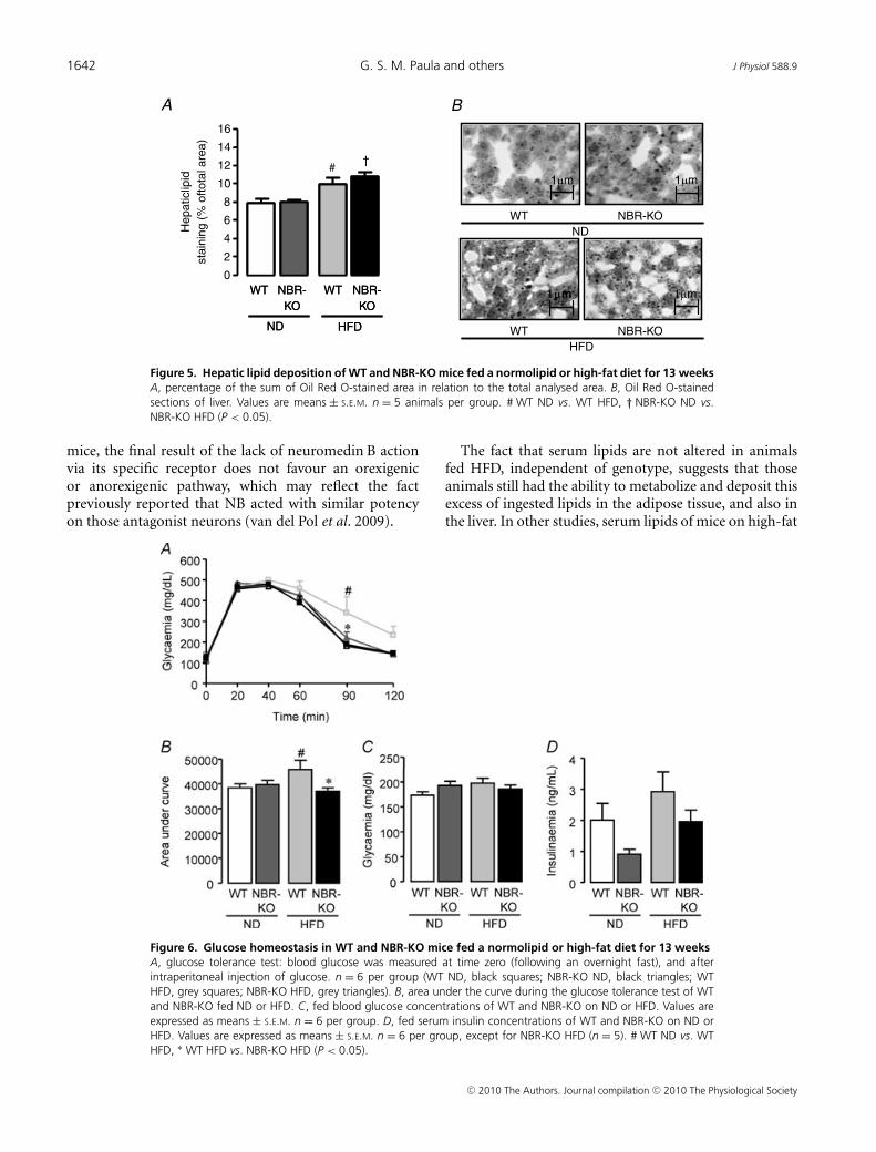
Oil Red O staining in liver samples (Fig. 5A and B)revealed that the percentage of area occupied by lipidstaining in liver from mice fed a high-fat diet was higherthan in mice fed the normolipid diet (P < 0.05) and nosignificant difference was observed between genotypes ineither diet.
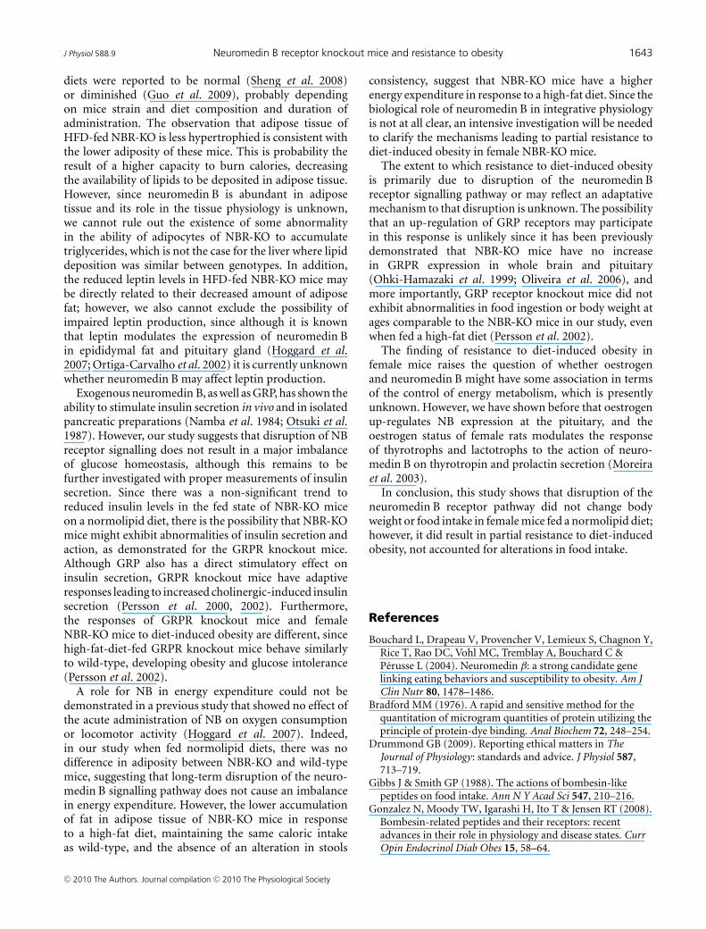
As depicted in Fig. 6A, NBR-KO and WT fed thenormolipid diet showed similar glucose tolerance curves.Fasting glycaemia (time zero) and all other time-pointsafter intraperitoneal injection of glucose were similar. Thehigh-fat diet caused an imbalance in glucose homeostasis
Figure 3. White adipose tissue morphology in WT and NBR-KO mice fed a normolipid or high-fat dietfor 13 weeksMass of inguinal (A), perigonadal (D) and retroperitoneal (G) white adipose tissue. Area of inguinal (B) andperigonadal (E) white adipocytes. Haematoxylin–eosin-stained histological sections of inguinal (C) and perigonadal(F) white adipose tissue. Percentage lipid (H) and protein (I) contents in eviscerated carcasses. Values are expressedas means ± S.E.M. n = 6 animals per group. # WT ND vs. WT HFD, † NBR-KO ND vs. NBR-KO HFD, ∗ WT HFD vs.NBR-KO HFD (P < 0.05).
C© 2010 The Authors. Journal compilation C© 2010 The Physiological Society

J Physiol 588.9 Neuromedin B receptor knockout mice and resistance to obesity 1641
Table 2. Serum lipids of wild-type mice (WT) and neuromedin B receptor knockoutmice (NBR-KO) fed a normolipid diet (ND) or high-fat diet (HFD) for 13 weeks
Serum lipids WT ND NBR-KO ND WT HFD NBR-KO HFD
Total cholesterol (mg dl−1) 129 ± 8 124 ± 4 147 ± 15 143 ± 8HDL cholesterol (mg dl−1) 74 ± 5 74 ± 4 81 ± 5 90 ± 7Triglycerides (mg dl−1) 89 ± 12 84 ± 8 69 ± 3 103 ± 18
n = 6 per group.
in WT mice, but not in NBR-KO mice, as revealed bythe glucose tolerance curves. HFD-fed WT mice exhibitedan 89% higher blood glucose than ND-fed WT mice at90 min after the intraperitoneal injection of glucose, andthe area under the curve of HFD-fed mice was increasedby 19% in comparison to ND-fed WT mice (Fig. 6B,P < 0.05). However, NBR-KO on a high-fat diet showed asimilar glucose tolerance curve to mice on the normolipiddiet. Glycaemia of animals, who were not fasted over-night, was similar among groups regardless of diet orgenotype (Fig. 6C). Serum insulin of the same animalsshowed important trends, which did not reach statisticallysignificant differences (Fig. 6D). Comparing mice fed thenormolipid diet, NBR-KO mice exhibited a 55% lowerserum insulin than WT. HFD-fed WT and NBR-KOanimals showed a 1.45-fold and 2.15-fold higher seruminsulin, respectively, than mice of the same genotype ona normolipid diet, and comparing mice fed the high-fatdiet, NBR-KO mice presented a 23% lower serum insulinthan WT.
Discussion
The main finding of the present study was that femaleneuromedin B receptor knockout mice developed apartial resistance to diet-induced obesity, suggesting theinvolvement of the NB–NBR pathway in the control ofbody energetic homeostasis.
Previous studies have demonstrated, in short-termfeeding tests, that neuromedin B has an acute anorexiceffect when administered centrally or systemically, as asingle dose, in rats (Gibbs & Smith, 1988), althoughit could not be reproduced in mice, either wild-typeor NBR-KO (Ohki-Hamazaki et al. 1999). Our resultsdo not support an important influence of NB andNBR on the control of long-term appetite in femalemice fed a normolipid diet or a high-fat diet, sincethey showed similar food intake. A previous studydemonstrated that NB stimulated the electrical activity ofboth proopiomelanocortic (POMC) and neuropeptide Yneurons of the arcuate nucleus (van del Pol et al. 2009).Our study suggests that, in vivo, at least in female
Figure 4. Adipocyte hormones in WT and NBR-KOmice fed a normolipid or high-fat diet for 13 weeksA, serum leptin, n = 6 per group, except for NBR-KO ND(n = 5). B, serum adiponectin, n = 6 per group, except forNBR-KO ND (n = 5). C, relative expression of inguinal whiteadipose tissue leptin mRNA (normalized by 36B4) to valuesof WT animals on normolipid diet (set as 1). Data wereobtained by quantitative PCR. Values are means ± S.E.M.n = 5 per group, except for WT ND (n = 4). D, relativeexpression of perigonadal white adipose tissue leptin mRNA(normalized by 36B4) to values of WT animals on anormolipid diet (set as 1). Values are means ± S.E.M. n = 6per group, except for WT HFD (n = 5). # WT ND vs. WTHFD, † NBR-KO ND vs. NBR-KO HFD, ∗ WT HFD vs. NBR-KOHFD (P < 0.05).
C© 2010 The Authors. Journal compilation C© 2010 The Physiological Society
1642 G. S. M. Paula and others J Physiol 588.9
1μm 1μm
1μm1μm 1μm
WT NBR-KO
ND
WT NBR-KO
HFD
A B
NBR-
KO
WT
ND HFD
NBR-
KO
WT
0
2
4
6
8
10
12
14
16
#†
Hepaticlip
id
sta
inin
g (
% o
ftota
l are
a)
NBR-
KO
WT
ND HFD
NBR-
KO
WT
Figure 5. Hepatic lipid deposition of WT and NBR-KO mice fed a normolipid or high-fat diet for 13 weeksA, percentage of the sum of Oil Red O-stained area in relation to the total analysed area. B, Oil Red O-stainedsections of liver. Values are means ± S.E.M. n = 5 animals per group. # WT ND vs. WT HFD, † NBR-KO ND vs.NBR-KO HFD (P < 0.05).
mice, the final result of the lack of neuromedin B actionvia its specific receptor does not favour an orexigenicor anorexigenic pathway, which may reflect the factpreviously reported that NB acted with similar potencyon those antagonist neurons (van del Pol et al. 2009).
The fact that serum lipids are not altered in animalsfed HFD, independent of genotype, suggests that thoseanimals still had the ability to metabolize and deposit thisexcess of ingested lipids in the adipose tissue, and also inthe liver. In other studies, serum lipids of mice on high-fat
Figure 6. Glucose homeostasis in WT and NBR-KO mice fed a normolipid or high-fat diet for 13 weeksA, glucose tolerance test: blood glucose was measured at time zero (following an overnight fast), and afterintraperitoneal injection of glucose. n = 6 per group (WT ND, black squares; NBR-KO ND, black triangles; WTHFD, grey squares; NBR-KO HFD, grey triangles). B, area under the curve during the glucose tolerance test of WTand NBR-KO fed ND or HFD. C, fed blood glucose concentrations of WT and NBR-KO on ND or HFD. Values areexpressed as means ± S.E.M. n = 6 per group. D, fed serum insulin concentrations of WT and NBR-KO on ND orHFD. Values are expressed as means ± S.E.M. n = 6 per group, except for NBR-KO HFD (n = 5). # WT ND vs. WTHFD, ∗ WT HFD vs. NBR-KO HFD (P < 0.05).
C© 2010 The Authors. Journal compilation C© 2010 The Physiological Society
J Physiol 588.9 Neuromedin B receptor knockout mice and resistance to obesity 1643
diets were reported to be normal (Sheng et al. 2008)or diminished (Guo et al. 2009), probably dependingon mice strain and diet composition and duration ofadministration. The observation that adipose tissue ofHFD-fed NBR-KO is less hypertrophied is consistent withthe lower adiposity of these mice. This is probability theresult of a higher capacity to burn calories, decreasingthe availability of lipids to be deposited in adipose tissue.However, since neuromedin B is abundant in adiposetissue and its role in the tissue physiology is unknown,we cannot rule out the existence of some abnormalityin the ability of adipocytes of NBR-KO to accumulatetriglycerides, which is not the case for the liver where lipiddeposition was similar between genotypes. In addition,the reduced leptin levels in HFD-fed NBR-KO mice maybe directly related to their decreased amount of adiposefat; however, we also cannot exclude the possibility ofimpaired leptin production, since although it is knownthat leptin modulates the expression of neuromedin Bin epididymal fat and pituitary gland (Hoggard et al.2007; Ortiga-Carvalho et al. 2002) it is currently unknownwhether neuromedin B may affect leptin production.
Exogenous neuromedin B, as well as GRP, has shown theability to stimulate insulin secretion in vivo and in isolatedpancreatic preparations (Namba et al. 1984; Otsuki et al.1987). However, our study suggests that disruption of NBreceptor signalling does not result in a major imbalanceof glucose homeostasis, although this remains to befurther investigated with proper measurements of insulinsecretion. Since there was a non-significant trend toreduced insulin levels in the fed state of NBR-KO miceon a normolipid diet, there is the possibility that NBR-KOmice might exhibit abnormalities of insulin secretion andaction, as demonstrated for the GRPR knockout mice.Although GRP also has a direct stimulatory effect oninsulin secretion, GRPR knockout mice have adaptiveresponses leading to increased cholinergic-induced insulinsecretion (Persson et al. 2000, 2002). Furthermore,the responses of GRPR knockout mice and femaleNBR-KO mice to diet-induced obesity are different, sincehigh-fat-diet-fed GRPR knockout mice behave similarlyto wild-type, developing obesity and glucose intolerance(Persson et al. 2002).
A role for NB in energy expenditure could not bedemonstrated in a previous study that showed no effect ofthe acute administration of NB on oxygen consumptionor locomotor activity (Hoggard et al. 2007). Indeed,in our study when fed normolipid diets, there was nodifference in adiposity between NBR-KO and wild-typemice, suggesting that long-term disruption of the neuro-medin B signalling pathway does not cause an imbalancein energy expenditure. However, the lower accumulationof fat in adipose tissue of NBR-KO mice in responseto a high-fat diet, maintaining the same caloric intakeas wild-type, and the absence of an alteration in stools
consistency, suggest that NBR-KO mice have a higherenergy expenditure in response to a high-fat diet. Since thebiological role of neuromedin B in integrative physiologyis not at all clear, an intensive investigation will be neededto clarify the mechanisms leading to partial resistance todiet-induced obesity in female NBR-KO mice.
The extent to which resistance to diet-induced obesityis primarily due to disruption of the neuromedin Breceptor signalling pathway or may reflect an adaptativemechanism to that disruption is unknown. The possibilitythat an up-regulation of GRP receptors may participatein this response is unlikely since it has been previouslydemonstrated that NBR-KO mice have no increasein GRPR expression in whole brain and pituitary(Ohki-Hamazaki et al. 1999; Oliveira et al. 2006), andmore importantly, GRP receptor knockout mice did notexhibit abnormalities in food ingestion or body weight atages comparable to the NBR-KO mice in our study, evenwhen fed a high-fat diet (Persson et al. 2002).
The finding of resistance to diet-induced obesity infemale mice raises the question of whether oestrogenand neuromedin B might have some association in termsof the control of energy metabolism, which is presentlyunknown. However, we have shown before that oestrogenup-regulates NB expression at the pituitary, and theoestrogen status of female rats modulates the responseof thyrotrophs and lactotrophs to the action of neuro-medin B on thyrotropin and prolactin secretion (Moreiraet al. 2003).
In conclusion, this study shows that disruption of theneuromedin B receptor pathway did not change bodyweight or food intake in female mice fed a normolipid diet;however, it did result in partial resistance to diet-inducedobesity, not accounted for alterations in food intake.
References
Bouchard L, Drapeau V, Provencher V, Lemieux S, Chagnon Y,Rice T, Rao DC, Vohl MC, Tremblay A, Bouchard C &Perusse L (2004). Neuromedin β: a strong candidate genelinking eating behaviors and susceptibility to obesity. Am JClin Nutr 80, 1478–1486.
Bradford MM (1976). A rapid and sensitive method for thequantitation of microgram quantities of protein utilizing theprinciple of protein-dye binding. Anal Biochem 72, 248–254.
Drummond GB (2009). Reporting ethical matters in TheJournal of Physiology: standards and advice. J Physiol 587,713–719.
Gibbs J & Smith GP (1988). The actions of bombesin-likepeptides on food intake. Ann N Y Acad Sci 547, 210–216.
Gonzalez N, Moody TW, Igarashi H, Ito T & Jensen RT (2008).Bombesin-related peptides and their receptors: recentadvances in their role in physiology and disease states. CurrOpin Endocrinol Diab Obes 15, 58–64.
C© 2010 The Authors. Journal compilation C© 2010 The Physiological Society
1644 G. S. M. Paula and others J Physiol 588.9
Goodpaster BH, Theriault R, Watkins SC & Kelley DE (2000).Intramuscular lipid content is increased in obesity anddecreased by weight loss. Metabolism 49, 467–472.
Guo J, Jou W, Gavrilova O & Hall KD (2009). Persistentdiet-induced obesity in male C57BL/6 mice resulting fromtemporary obesigenic diets. PloS One 4, e5370.
Hoggard N, Bashir S, Cruickshank M, Miller JD & SpeakmanJR (2007). Expression of neuromedin B in adipose tissue andits regulation by changes in energy balance. J Mol Endocrinol39, 199–210.
Jensen RT, Battey JF, Spindel ER & Benya RV (2008).International Union of Pharmacology. LXVIII. Mammalianbombesin receptors: nomenclature, distribution,pharmacology, signalling, and functions in normal anddisease states. Pharmacol Rev 60, 1–42.
Johmura Y, Watanabe K, Kishimoto K, Ueda T, Shimada S,Osada S, Nishizuka M & Imagawa M (2009). Fad24 causeshyperplasia in adipose tissue and improves glucosemetabolism. Biol Pharm Bull 32, 1656–1664.
Ladenheim EE, Hampton LL, Whitney AC, White WO, BatteyJF & Moran TH (2002). Disruptions in feeding and bodyweight control in gastrin-releasing peptide receptor deficientmice. J Endocrinol 174, 273–281.
Ladenheim EE, Taylor JE, Coy DH & Moran TH (1994).Blockade of feeding inhibition by neuromedin B using aselective receptor antagonist. Eur J Pharmacol271, 7–9.
Machado DS, Sabet A, Santiago LA, Sidhaye AR, ChiamoleraMI, Ortiga-Carvalho TM & Wondisford FE (2009). Athyroid hormone receptor mutation that dissociates thyroidhormone regulation of gene expression in vivo. Proc NatlAcad Sci U S A 106, 9441–9446.
Moreira RM, Curty FH, Lisboa PC, Amaral D, Ortiga-CarvalhoTM & Pazos-Moura CC (2003). Estrogen modulatesneuromedin B effects on thyrotropin and prolactin releasein vitro. Life Sci 72, 917–923.
Moura EG & Pazos-Moura CC (2004). Regulation ofthyrotropin synthesis and secretion. Arquivos Brasileiros deEndocrinologia e Metabologia 48, 40–52.
Namba M, Gathei MA, Adrian TE, Bacarese-Hamilton AJ,Mulderry PK & Bloom SR (1984). Effect of neuromedinB on gut hormone secretion in the rat. Biomed Res5, 229–234.
Ohki-Hamazaki H, Iwabuchi M & Maekawa F (2005).Development and function of bombesin-like peptides, andtheir receptors. Int J Dev Biol 49, 293–300.
Ohki-Hamazaki H, Sakai Y, Kamata K, Ogura H, Okuyama S,Watase K, Yamada K & Wada K (1999). Functionalproperties of two bombesin-like peptide receptors revealedby the analysis of mice lacking neuromedin B receptor. JNeurosci 19, 948–954.
Ohki-Hamazaki H, Wada E, Matsui K & Wada K (1997a).Cloning and expression of the neuromedin B receptor andthe third subtype of bombesin receptor genes in the mouse.Brain Res 762, 165–172a.
Ohki-Hamazaki H, Watase K, Yamamoto K, Ogura H, YamanoM, Yamada K, Maeno H, Imaki J, Kikuyama S, Wada E &Wada K (1997b). Mice lacking bombesin receptor subtype-3develop metabolic defects and obesity. Nature 390, 165–169.
Oliveira KJ, Cabanelas A, Veiga MA, Paula GS, Ortiga-CarvalhoTM, Wada E, Wada K & Pazos-Moura CC (2008). Impairedserum thyrotropin response to hypothyroidism in mice withdisruption of neuromedin B receptor. Regul Pept 146,213–217.
Oliveira KJ, Ortiga-Carvalho TM, Cabanelas A, Veiga MA, AokiK, Ohki-Hamazaki H, Wada K, Wada E & Pazos-Moura CC(2006). Disruption of neuromedin B receptor gene results indysregulation of the pituitary–thyroid axis. J Mol Endocrinol36, 73–80.
Ortiga-Carvalho TM, Curty FH, Nascimento-Saba CC, MouraEG, Polak J & Pazos-Moura CC (1997). Pituitaryneuromedin B content in experimental fasting and diabetesmellitus and correlation with thyrotropin secretion. MetabClin Exp 46, 149–153.
Ortiga-Carvalho TM, Oliveira KJ, Morales MM, Martins VP &Pazos-Moura CC (2003). Thyrotropin secretagogues reducerat pituitary neuromedin B, a local thyrotropin releaseinhibitor. Exp Biol Med 228, 1083–1088.
Ortiga-Carvalho TM, Oliveira KJ, Soares BA & Pazos-MouraCC (2002). The role of leptin in the regulation of TSHsecretion in the fed state: in vivo and in vitro studies.J Endocrinol 174, 121–125.
Ortiga-Carvalho TM, Polak J, McCann S & Pazos-Moura CC(1996). Effect of thyroid hormones on pituitary neuromedinB and possible interaction between thyroid hormones andneuromedin B on thyrotropin secretion. Regul Pept 67,47–53.
Otsuki M, Fujii M, Nakamura T, Tani S, Oka T, Yajima H &Baba S (1987). Effects of neuromedin B and neuromedin Con exocrine and endocrine rat pancreas. Am J PhysiolGastrointest Liver Physiol 252, G491–G498.
Pazos-Moura CC, Moura EG, Rettori V, Polak J & McCann S(1996). Role of neuromedin B in the in vitro thyrotropinrelease in response to thyrotropin-releasing hormone (TRH)from anterior pituitaries of eu-, hypo- and hyperthyroid rats.Proc Soc Exp Biol Med 211, 353–358.
Persson K, Gingerich RL, Nayak S, Wada K, Wada E & Ahren B(2000). Reduced GLP-1 and insulin responses and glucoseintolerance after gastric glucose in GRP receptor deletedmice. Am J Physiol Endocrinol Metab 279,E956–E962.
Persson K, Pacini G, Sundler F & Ahren B (2002). Islet functionphenotype in gastrin-releasing peptide receptorgene-deficient mice. Endocrinology 143, 3717–3726.
Reeves PG, Nielsen FH & Fahey GC Jr (1993). AIN-93 purifieddiets for laboratory rodents: final report of the AmericanInstitute of Nutrition ad hoc writing committee on thereformulation of the AIN-76A rodent diet. J Nutr 1223,1939–1951.
Rodrigues AL, Moura EG, Passos MCF, Dutra SCP & LisboaPC (2009). Postnatal early overnutrition changes the leptinsignalling pathway in the hypothalamic-pituitary-thyroidaxis of young and adult rats. J Physiol 587,2647–2661.
Sano H, Feighner SD, Hreniuk DL, Iwaasa H, Sailer AW, Pan J,Reitman ML, Kanatani A, Howard AD & Tan CP (2004).Characterization of the bombesin-like peptide receptorfamily in primates. Genomics 84, 139–146.
C© 2010 The Authors. Journal compilation C© 2010 The Physiological Society

J Physiol 588.9 Neuromedin B receptor knockout mice and resistance to obesity 1645
Sheng X, Zhang Y, Gong Z, Huang C & Zang YQ (2008).Improved insulin resistance and lipid metabolism bycinnamon extract through activation of peroxisomeproliferator-activated receptors. PPAR Res 2008, 581348.
Souza LL, Nunes MO, Paula GSM, Cordeiro A, Penha-Pinto V,Neto JFN, Oliveira KJ, Tavares do Carmo MG &Pazos-Moura CC (2009). Effects of dietary fish oil on thyroidhormone signaling in the liver. J Nutr Biochem;DOI: 10.1016/j.jnutbio.2009.07.008.
Spalova J, Zamrazilova H, Vcelak J, Vankova M, Lukasova P,Hill M, Hlavata K, Sramkova P, Fried M, Aldhoon B,Kunesova M, Bendlova B & Hainer V (2008). Neuromedinbeta: P73T polymorphism in overweight and obese subjects.Physiol Res 57 (Suppl. 1), S39–S48.
van den Pol AN, Yao Y, Fu LY, Foo K, Huang H, Coppari R,Lowell BB & Broberger C (2009). Neuromedin B andgastrin-releasing peptide excite arcuate nucleusneuropeptide Y neurons in a novel transgenic mouseexpressing strong Renilla green fluorescent protein in NPYneurons. J Neurosci 29, 4622–4639.
Weber HC (2009). Regulation and signalling of humanbombesin receptors and their biological effects. Curr OpinEndocrinol Diab Obes 16, 66–71.
Yamada K, Santo-Yamada Y & Wada K (2002c). Restraint stressimpaired maternal behaviour in female mice lacking theneuromedin B receptor (NMB-R) gene. Neurosci Lett 330,163–166.
Yamada K, Santo-Yamada Y, Wada E & Wada K (2002a). Roleof bombesin (BN)-like peptides/receptors in emotionalbehaviour by comparison of three strains of BN-like peptidereceptor knockout mice. Mol Psychiatry 7, 113–117.
Yamada K, Santo-Yamada Y & Wada K (2003). Stress-inducedimpairment of inhibitory avoidance learning in femaleneuromedin B receptor-deficient mice. Physiol Behav 78,303–309.
Yamada K, Wada E, Yamano M, Sun YJ, Ohara-Imaizumi M,Nagamatsu S & Wada K (2002b). Decreased marble buryingbehaviour in female mice lacking neuromedin-B receptor(NMB-R) implies the involvement of NMB/NMB-R in 5-HTneuron function. Brain Res 942, 71–78.
Yamano M, Ogura H, Okuyama S & Ohki-Hamazaki H (2002).Modulation of 5-HT system in mice with a targeteddisruption of neuromedin B receptor. J Neurosci Res 68,59–64.
Yang YS, Song HD, Li RY, Zhou LB, Zhu ZD, Hu RM, Han ZG& Chen JL (2003). The gene expression profiling of humanvisceral adipose tissue and its secretory functions. BiochemBiophys Res Commun 300, 839–846.
Author contributions
The conception and design of this study was conductedby G.S.M.P., K.J.O. and C.C.P.-M. with contributions fromV.M.-C. and T.M.O.-C. E.W. was responsible for the generationof neuromedin B receptor knockout mice. Experiments wereperformed by G.S.M.P., K.J.O., L.L.S., A.C. and F.F.B. All authorscontributed to the analysis and interpretation of data as wellas writing, revising and final approval of the version to bepublished. The study was conducted at the Instituto de BiofisicaCarlos Chagas Filho, Federal University of Rio de Janeiro, Brazil.
Acknowledgements
This research was supported by grants from Fundacao CarlosChagas Filho de Amparo a Pesquisa do Estado do Rio de Janeiro(FAPERJ), Conselho Nacional de Desenvolvimento Cientıfico eTecnologico (CNPq) and Coordenacao de Aperfeicoamento dePessoal de Nıvel Superior (CAPES). We also thank Uilian Calixtofor technical assistance.
C© 2010 The Authors. Journal compilation C© 2010 The Physiological Society
Related Documents


![CBI-DELETED COPY...CBI-deleted CBI-deleted guide RNA targets Cas9 to the intended site of action. Due to the design of the [ ] #4 CBI-deleted spacer used in the development of line](https://static.cupdf.com/doc/110x72/5f6824d95ae97d5e681729bf/cbi-deleted-copy-cbi-deleted-cbi-deleted-guide-rna-targets-cas9-to-the-intended.jpg)