Feline Leukemia Virus Immunity Induced by Whole Inactivated Virus Vaccination Andrea N. Torres a , Kevin P. O’Halloran a , Laurie J. Larson b , Ronald D. Schultz b , and Edward A. Hoover a,* a Department of Microbiology, Immunology, and Pathology, 1619 Campus Delivery, Colorado State University, Ft. Collins, CO 80523-1619, USA b Department of Pathobiological Sciences, 2015 Linden Dr., University of Wisconsin –Madison, Madison, WI 53706, USA Abstract A fraction of cats exposed to feline leukemia virus (FeLV) effectively contain virus and resist persistent antigenemia/viremia. Using real-time PCR (qPCR) to quantitate circulating viral DNA levels, previously we detected persistent FeLV DNA in blood cells of non-antigenemic cats considered to have resisted FeLV challenge. In addition, previously we used RNA qPCR to quantitate circulating viral RNA levels and determined that the vast majority of viral DNA is transcriptionally active, even in the absence of antigenemia. A single comparison of all USDA-licensed commercially available FeLV vaccines using these modern sensitive methods has not been reported. To determine whether FeLV vaccination would prevent nucleic acid persistence, we assayed circulating viral DNA, RNA, antigen, infectious virus, and virus neutralizing (VN) antibody in vaccinated and unvaccinated cats challenged with infectious FeLV. We identified challenged vaccinates with undetectable antigenemia and viremia concomitant with persistent FeLV DNA and/or RNA. Moreover, these studies demonstrated that two whole inactivated virus (WIV) adjuvanted FeLV vaccines (Fort Dodge Animal Health’s Fel-O-Vax Lv-K® and Schering-Plough Animal Health’s FEVAXYN FeLV®) provided effective protection against FeLV challenge. In nearly every recipient of these vaccines, neither viral DNA, RNA, antigen, nor infectious virus could be detected in blood after FeLV challenge. Interestingly, this effective viral containment occurred despite a weak to undetectable VN antibody response. The above findings reinforce the precept of FeLV infection as a unique model of effective retroviral immunity elicited by WIV vaccination, and as such holds valuable insights into retroviral immunoprevention and therapy. Keywords FeLV; vaccine; whole inactivated virus; immunity; diagnosis; pathogenesis *Corresponding author. Tel.: 970-491-7587; fax: 970-491-0523; [email protected] (E.A. Hoover). Conflict of interest statement None of the authors has a financial or personal relationship with other people or organizations that could inappropriately influence or bias the paper entitled “Feline Leukemia Virus Immunity Induced by Whole Inactivated Virus Vaccination”. Publisher's Disclaimer: This is a PDF file of an unedited manuscript that has been accepted for publication. As a service to our customers we are providing this early version of the manuscript. The manuscript will undergo copyediting, typesetting, and review of the resulting proof before it is published in its final citable form. Please note that during the production process errors may be discovered which could affect the content, and all legal disclaimers that apply to the journal pertain. NIH Public Access Author Manuscript Vet Immunol Immunopathol. Author manuscript; available in PMC 2011 March 15. Published in final edited form as: Vet Immunol Immunopathol. 2010 March 15; 134(1-2): 122. doi:10.1016/j.vetimm.2009.10.017. NIH-PA Author Manuscript NIH-PA Author Manuscript NIH-PA Author Manuscript

Welcome message from author
This document is posted to help you gain knowledge. Please leave a comment to let me know what you think about it! Share it to your friends and learn new things together.
Transcript

Feline Leukemia Virus Immunity Induced by Whole InactivatedVirus Vaccination
Andrea N. Torresa, Kevin P. O’Hallorana, Laurie J. Larsonb, Ronald D. Schultzb, and EdwardA. Hoovera,*a Department of Microbiology, Immunology, and Pathology, 1619 Campus Delivery, Colorado StateUniversity, Ft. Collins, CO 80523-1619, USAb Department of Pathobiological Sciences, 2015 Linden Dr., University of Wisconsin –Madison,Madison, WI 53706, USA
AbstractA fraction of cats exposed to feline leukemia virus (FeLV) effectively contain virus and resistpersistent antigenemia/viremia. Using real-time PCR (qPCR) to quantitate circulating viral DNAlevels, previously we detected persistent FeLV DNA in blood cells of non-antigenemic catsconsidered to have resisted FeLV challenge. In addition, previously we used RNA qPCR to quantitatecirculating viral RNA levels and determined that the vast majority of viral DNA is transcriptionallyactive, even in the absence of antigenemia. A single comparison of all USDA-licensed commerciallyavailable FeLV vaccines using these modern sensitive methods has not been reported. To determinewhether FeLV vaccination would prevent nucleic acid persistence, we assayed circulating viral DNA,RNA, antigen, infectious virus, and virus neutralizing (VN) antibody in vaccinated and unvaccinatedcats challenged with infectious FeLV. We identified challenged vaccinates with undetectableantigenemia and viremia concomitant with persistent FeLV DNA and/or RNA. Moreover, thesestudies demonstrated that two whole inactivated virus (WIV) adjuvanted FeLV vaccines (Fort DodgeAnimal Health’s Fel-O-Vax Lv-K® and Schering-Plough Animal Health’s FEVAXYN FeLV®)provided effective protection against FeLV challenge. In nearly every recipient of these vaccines,neither viral DNA, RNA, antigen, nor infectious virus could be detected in blood after FeLVchallenge. Interestingly, this effective viral containment occurred despite a weak to undetectable VNantibody response. The above findings reinforce the precept of FeLV infection as a unique model ofeffective retroviral immunity elicited by WIV vaccination, and as such holds valuable insights intoretroviral immunoprevention and therapy.
KeywordsFeLV; vaccine; whole inactivated virus; immunity; diagnosis; pathogenesis
*Corresponding author. Tel.: 970-491-7587; fax: 970-491-0523; [email protected] (E.A. Hoover).Conflict of interest statementNone of the authors has a financial or personal relationship with other people or organizations that could inappropriately influence orbias the paper entitled “Feline Leukemia Virus Immunity Induced by Whole Inactivated Virus Vaccination”.Publisher's Disclaimer: This is a PDF file of an unedited manuscript that has been accepted for publication. As a service to our customerswe are providing this early version of the manuscript. The manuscript will undergo copyediting, typesetting, and review of the resultingproof before it is published in its final citable form. Please note that during the production process errors may be discovered which couldaffect the content, and all legal disclaimers that apply to the journal pertain.
NIH Public AccessAuthor ManuscriptVet Immunol Immunopathol. Author manuscript; available in PMC 2011 March 15.
Published in final edited form as:Vet Immunol Immunopathol. 2010 March 15; 134(1-2): 122. doi:10.1016/j.vetimm.2009.10.017.
NIH
-PA Author Manuscript
NIH
-PA Author Manuscript
NIH
-PA Author Manuscript

1. IntroductionFeline leukemia virus (FeLV) was identified as a naturally occurring retroviral infection ofcats over 40 years ago (Jarrett et al., 1964; Kawakami et al., 1967; Rickard et al., 1969). Theprimary route of transmission of this gammaretrovirus is horizontally through saliva (Franciset al., 1977; Hardy et al., 1976; Hardy et al., 1973; Hoover et al., 1977a). The pathogenic effectsof FeLV infection are both cytoproliferative (e.g. lymphoma, myeloproliferative disorder) andcytosuppressive (e.g. immunodeficiency, myelosuppression) (Hoover and Mullins, 1991).
Historically, FeLV infection has represented a diametric paradigm of effective host responseleading to regressive infection vs. ineffective host response leading to progressive infectionand disease (Hoover et al., 1981). This model has been based on assays detecting either: (a)viremia by cell culture infectivity (VI) (de Noronha et al., 1977; Fischinger et al., 1974) or (b)intracellular antigenemia in leukocytes by immunofluorescent antibody (IFA) assay (Hardy etal., 1973; Hardy and Zuckerman, 1991a) or (c) extracellular antigenemia in plasma or serumby capture ELISA (Lutz et al., 1983a). Information obtained using these assays was used toestimate that in ~60% of young adult cats exposed to FeLV, neither p27 capsid antigen norinfectious virus were detectable in the blood after virus challenge (Hardy, 1980; Hardy et al.,1976; Hoover and Mullins, 1991; Rojko et al., 1979). In stark contrast, ~30% of exposedanimals developed persistent antigenemia and viremia. However, subsequent widespread useof the p27 capture ELISA, in combination with the IFA and VI assays, prompted theidentification of cats with discordant results (Hardy and Zuckerman, 1991b; Jarrett et al.,1982; Lutz et al., 1980b; Lutz et al., 1983b). In addition, several laboratories demonstrated thatit is possible to reactivate FeLV from some cats with regressive infections (Madewell andJarrett, 1983; Post and Warren, 1980; Rojko et al., 1982). These observations pointed to a morecomplex, less polar, view of FeLV:host relationships (Hoover and Mullins, 1991) and/orvarying limits in assay sensitivity.
We have recently applied quantitative real-time PCR (qPCR) to examine vaccinated andunvaccinated cats challenged oronasally with FeLV-A/61E and found covert FeLV DNA, inboth circulation and tissues, in the absence of detectable antigenemia (Torres et al., 2005).Investigators have shown that proviral integration occurs not only in cats with persistentantigenemia, but also in cats without detectable anitgenemia and with lower circulating proviralburdens (Cattori et al., 2006). Additionally, we have reported a near perfect agreement andstrong linear correlation between FeLV DNA and RNA in the blood of FeLV-challenged cats,inferring that a substantial fraction of the detected FeLV DNA was indeed integrated into thehost cell genome and initiated a transcriptionally active infection (Torres et al., 2008).Consequently, a spectrum of FeLV:host relationships have been identified, including cats withdetectable nucleic acids and undetectable antigenemia (latent infections) and cats with bothdetectable nucleic acids and antigenemia (active infections). These findings, and those ofcolleagues (Cattori et al., 2006; Flynn et al., 2002; Gomes-Keller et al., 2006a; Gomes-Kelleret al., 2006b; Hofmann-Lehmann et al., 2001; Hofmann-Lehmann et al., 2006; Tandon et al.,2005), demonstrated that DNA and RNA qPCR sensitivities are greater than p27 capsid antigencapture ELISA.
A singular feature of FeLV infection has been the development of effective vaccines providingprotection against virulent virus challenge. At the time this study was initiated, four FeLVvaccines were commercially available in the USA, each with varying formulations and efficacy[reviewed by Loar (Loar, 1993) and Sparkes (Sparkes, 1997)]. Despite the accumulation ofindividual vaccine trials, the differences in experimental designs have made comparisons ofvaccine efficacy virtually impossible. Moreover, most of these studies were performed beforethe advent of qPCR, thereby limiting the ability to detect evidence of viral infection. To ourknowledge, a single comparison of every USDA-licensed FeLV vaccine that is commercially
Torres et al. Page 2
Vet Immunol Immunopathol. Author manuscript; available in PMC 2011 March 15.
NIH
-PA Author Manuscript
NIH
-PA Author Manuscript
NIH
-PA Author Manuscript

available in the USA has not been reported using modern viral nucleic acid detection methods.In addition, our previous work identified several protected vaccinates without any evidence ofviral infection despite the use of DNA qPCR. Hofmann-Lehmann et. al. however, have notobserved this unusual level of protection (Hofmann-Lehmann et al., 2008; Hofmann-Lehmannet al., 2007; Hofmann-Lehmann et al., 2006).
The present study, therefore, had two purposes: (1) to compare all USDA-licensedcommercially available FeLV vaccines by determining whether they differed in ability toprotect against both active and latent viral infection using contemporary sensitive methods and(2) to determine whether a neutralizing humoral immune response was associated with highlyeffective viral containment. Accordingly, we examined virulent FeLV challenge outcomes incohorts of cats vaccinated with one of four commercially available vaccines and have assessedhost:virus relationships by criteria of viral DNA, RNA, p27 capsid antigen, infectious virus,and neutralizing antibody.
2. Materials and Methods2.1. Experimental animals
Forty specific-pathogen-free (SPF) cats were obtained from a commercial vendor (HarlanSprague Dawley, Inc., Mt. Horeb, WI). The cats were randomly apportioned up to 5 cats perenclosure and housed at Harlan Sprague Dawley during the immunization phase of theexperiment. Prior to virus challenge, they were transferred to Charmany Instructional Facilityat the University of Wisconsin-Madison School of Veterinary Medicine (Madison, WI). Forthe remainder of the study, the animals were housed in identical groupings as before inaccordance with the university animal care and use committee regulations.
2.2. ImmunizationFour groups of n=8 cats each received one of four commercially available vaccines accordingto the manufacturer’s specifications and one group (n=8) served as the unvaccinated control.Group A received the adjuvanted whole inactivated virus (WIV) vaccine Fel-O-Vax Lv-K®(Fort Dodge Animal Health, Overland Park, KS). Group B received FEVAXYN FeLV®(Schering-Plough Animal Health Corporation, Summit, NJ), also an adjuvanted WIV vaccine.Group C received the adjuvanted, inactivated mixed subunit vaccine LEUKOCELL 2® (PfizerAnimal Health, New York, NY). Group D received PROTEX®-FeLV (Intervet, Millsboro,DE). It was a non-adjuvanted WIV vaccine which is no longer commercially available. Thepriming vaccination was administered when the cats were 15 – 16 weeks of age. The boostingvaccination was administered three weeks later when the cats were 18 – 19 weeks of age.
2.3. Challenge virusFour months after receiving their boosting immunization, at 34 – 35 weeks of age, all cats werechallenged intraperitoneally with 200 μL of 5 × 10⁴ TCID50/mL FeLV-A/61E. This subgroupA virus strain is the highly replication competent, non-acutely pathogenic component of theFeLV-FAIDS complex (Donahue et al., 1988; Hoover et al., 1987; Mullins et al., 1986;Overbaugh et al., 1988). The cell-free infectious virus inoculum was prepared as supernatantfrom AH927 feline fibroblast cell cultures and determined to be equivalent to 1 CID100 (100%cat infective dose). Cats were observed twice daily for signs of illness after virus inoculation.
2.4. Sample collection and processingSample collections were performed on cats sedated with ketamine hydrochloride (11 mg/kg).Blood samples were collected immediately prior to challenge and every week thereafterthrough 8 weeks post-challenge (PC). Whole blood was shipped overnight on ice to Colorado
Torres et al. Page 3
Vet Immunol Immunopathol. Author manuscript; available in PMC 2011 March 15.
NIH
-PA Author Manuscript
NIH
-PA Author Manuscript
NIH
-PA Author Manuscript

State University (Ft. Collins, CO) where it was immediately processed upon arrival. Buffy coatcell pellets were stored at −70°C until analysis for FeLV DNA by qPCR. Plasma samples wereseparated into 1 mL aliquots and stored at −70°C until analysis for FeLV RNA by qPCR, FeLVp27 capsid antigen by capture ELISA, and infectious FeLV. Sera were stored at −70°C untilanalysis for FeLV neutralizing antibody.
2.5. Detection of FeLV DNA and RNA by qPCR assaysWeekly analysis of buffy coat cells for viral DNA and plasma for viral RNA was done byquantitative real-time PCR (qPCR) and reverse transcriptase qPCR, respectively. The DNAand RNA extractions, primer/probe set, and assay conditions were as described previously(Torres et al., 2005; Torres et al., 2008). The assays amplify exogenous and not endogenousFeLV sequences within the U3 region of FeLV-A/61E (Berry et al., 1988; Casey et al.,1981). The viral loads of the samples were determined by comparing the threshold cycle(CT) value of each nucleic acid sample with the standard curve of the co-amplified standardtemplate. End-point dilutions of the standard templates yielded lower detection limits of 5copies/DNA qPCR reaction and 10 copies/RNA qPCR reaction (1150 RNA copies/mLplasma).
2.6. Detection of FeLV by p27 capture ELISAFeLV p27 capsid antigen was detected in plasma weekly as previously described (Torres etal., 2008). In addition, sera were assessed by the commercial SNAP® FeLV antigen diagnostictest (IDEXX Laboratories, Inc., Westbrook, ME). In rare instances of incongruous results, theperceived most sensitive finding of the two assays was chosen.
2.7. Virus stockThe FeLV-A/61E stock used for the virus infectivity and virus neutralization assays consistedof supernatant collected from Crandell feline kidney (CrFK) cells chronically infected withFeLV-A/61E. The virus stock was strongly positive by the p27 capture ELISA and contained4.4 × 109 copies/mL by RNA qPCR. The 50% endpoint dilution of the virus stock was 2.5 ×10³ TCID50/mL.
2.8. Detection of infectious FeLVPlasma samples from cats inoculated with FeLV-A/61E 4 weeks previously were incubatedwith partially confluent AH927 cells and the presence of infectious FeLV (VI) was determinedby p27 capture ELISA of the cell culture supernatant. Specifically, AH927 cells were seededon a 24-well plate at a concentration of 1 × 10⁵ cells/well. Cells were grown in 1 mL of minimalessential medium containing Earle’s salts and L-glutamine (Gibco Products, Invitrogen Corp.,Carlsbad, CA) with the addition of 1% penicillin/streptomycin, 10% heat inactivated fetalbovine serum, and 4 μg/mL polybrene (Sigma-Aldrich Corp., St. Louis, MO). Following anovernight incubation at 37°C with 5% CO2, medium was removed from the subconfluent cellsand 400 μL of freshly thawed sample plasma was added to each well. After a 2 hour incubation,the plasma was replaced with 1 mL of fresh medium without supplementation of polybreneand the plates were placed at 37°C with 5% CO2. This was day 1. The cells were examineddaily for confluency and any cytopathic effects and were passaged (1:4 cell split) as necessary,approximately every 3 days. At each passage, the supernatants were assessed, in duplicate, forp27 by capture ELISA. The infectivity assay was stopped at day 21. The plasma sample wasconsidered positive for infectious FeLV if a mean ELISA absorbance value of ≥ 0.05 wasobtained (a threshold set above the negative control plus three times the standard deviation).Sham inoculated wells served as negative controls and an FeLV-A/61E virus stock as positivecontrols for the virus infectivity assay.
Torres et al. Page 4
Vet Immunol Immunopathol. Author manuscript; available in PMC 2011 March 15.
NIH
-PA Author Manuscript
NIH
-PA Author Manuscript
NIH
-PA Author Manuscript

2.9. Detection of FeLV neutralizing antibodiesVirus neutralizing (VN) antibodies were measured at 2 time-points; post-vaccination just priorto receiving the virus challenge and at 8 weeks PC. Briefly, AH927 cells were seeded on a 96-well plate at a concentration of 1 × 10⁴ cells/well and grown overnight in 100 μL of the samemedium and under the same conditions as that used in the VI assay. The following day (day1), sample sera were heat inactivated for 30 minutes at 56°C and 2-fold serial dilutions made(1:2 to 1:256). An equal volume (50 μL) of each serum dilution and 10 TCID50/mL FeLV-A/61E were incubated alone, in triplicate, for 1 hour at 37°C and 5% CO2 to allow antibodybinding. Medium was removed from the subconfluent AH927 cells and the serum/virusmixtures were transferred onto the cells. After a 2 hour incubation at 37°C, the serum/virusmixtures were removed, the cells washed 3 times, 200 μL fresh medium without polybreneadded, and the plates incubated at 37°C with 5% CO2. On day 4, approximately 50% of themedium was removed (100 μL) and the cells refed with 150 μL fresh medium. The assay wasstopped on day 7 and cell culture supernatants assessed, singly, for p27 by capture ELISA. Ifthe mean optical density value of each triplicate serum dilution was ≤ 0.05, the serum samplewas considered positive for VN antibody. The titer of antibody was taken as the reciprocal ofthe serum dilution. Naïve serum served as the negative control and FeLV regressor serumserved as the positive control for VN antibodies, respectively. In addition, sham inoculatedwells served as negative controls and a back-titration of the virus stock as positive controls forvirus infection, respectively.
2.10. Data analysisIn accord with USDA FeLV vaccine evaluation guidelines, a cat was considered FeLV-positive(actively infected) when 3 consecutive samples had positive antigen or virus results betweenweeks 3 to 8 PC (Shibley et al., 1991). The preventable fraction (PF) is used to express vaccineefficacy by accounting for the inherent resistance of some unvaccinated cats to developmentof persistent antigenemia/viremia after FeLV challenge (Pollack and Scarlett, 1990). The PFis calculated as
Undetectable results by both qPCR assays were corrected to a value of 1 and then all viral RNAand DNA levels were log transformed. Statistically significant differences in viral DNA andRNA levels between the FeLV:host categories and between the experimental groups weredetermined using repeated-measure analysis of variance (ANOVA) with the Tukey-Kramerpost-hoc test. The chi-square test was used to determine if VI assay results and the FeLV:hostcategories were independent. A statistically significant difference between groups wasconsidered to have occurred when a p value was < 0.05. Statistical tests were performed usingStatView® version 5.0.1 for Macintosh, copyright 1999 (Abacus Concepts, Inc., Berkeley,CA).
3. Results3.1. Host:virus relationships based on viral DNA and RNA levels, circulating p27, andinfectious virus
Virus challenge via the intraperitoneal route (vs. oronasal) produced animals representing thesame 4 previously described response categories (Torres et al., 2005). However, in thismanuscript we have reverted to numbered categories similar to those described by others inpreceding reports (Hoover and Mullins, 1991; Lutz et al., 1980a; Lutz et al., 1983b), since
Torres et al. Page 5
Vet Immunol Immunopathol. Author manuscript; available in PMC 2011 March 15.
NIH
-PA Author Manuscript
NIH
-PA Author Manuscript
NIH
-PA Author Manuscript

terminology becomes to a degree imprecise or confusing. In 15 cats neither viral DNA, viralRNA, antigenemia, nor viremia were ever detected in blood at any time-point (Fig. 1). Theseanimals previously described as having experienced abortive infection, were now simplyexpressed as a Category 1 response. Six cats were classified as having experienced regressiveinfection, here expressed as Category 2, on the basis of the continual absence of detectablecirculating infectious virus and antigen but the presence of detectable viral RNA and/or DNAin blood albeit in a transient low or persistent low mode (DNA median: 176 DNA copies/106
buffy coat cells; DNA range: undetectable to 5,675 DNA copies/106 buffy coat cells; RNAmedian: undetectable; RNA range: undetectable to 120,267 RNA copies/mL plasma). Fourcats classified as Category 3 (previously called latent infection) developed transientantigenemia and viral RNA burden (RNA median: 124 RNA copies/mL plasma; RNA range:undetectable to 255,238 RNA copies/mL plasma), yet retained a low to moderate circulatingviral DNA load (DNA median: 2,487 DNA copies/106 buffy coat cells; DNA range:undetectable to 57,252 DNA copies/106 buffy coat cells). In addition, viremia was not detectedeven when viral DNA and RNA levels were moderate to low. Lastly, 15 animals that developedpersistent antigenemia and persistent high circulating viral DNA and RNA levels (DNAmedian: 934,891 DNA copies/106 buffy coat cells; DNA range: 57 to 4,173,516 DNA copies/106 buffy coat cells; RNA median: 2,681,905 RNA copies/mL plasma; RNA range:undetectable to 161,904,762 RNA copies/mL plasma) were classified as Category 4 (akaprogressive infection). Viremia was detected only in 12 of 15 cats classified into Category 4.In no sample was infectious virus detected without detectable viral DNA and RNA. In oneanimal, viremia was detected yet antigenemia was not detected at that timepoint.
Statistically significant differences (p < 0.01) in viral DNA and RNA levels distinguished allFeLV categories (i.e. Categories 1 vs. 2; Categories 1 vs. 3; Categories 1 vs. 4; Categories 2vs. 4; and Categories 3 vs. 4) with one exception--Categories 2 vs. 3. Infectious virus was easilydetected in samples with high levels of viral DNA and RNA (≥105 DNA copies/106 buffy coatcells and ≥105 RNA copies/mL plasma) and this association was supported by chi squareanalysis (p < 0.0001). Low to moderate viral DNA and RNA levels (≥101 to <105 DNA copies/106 buffy coat cells and ≥101 to <105 RNA copies/mL plasma) were not demonstrablyassociated with plasma viral infectivity.
3.2. Vaccine efficacyThe assays above were applied to determine whether all USDA-licensed commerciallyavailable FeLV vaccines differed in ability to protect against both viral nucleic acid andantigenemia/viremia detection. The FeLV challenge induced adequate infection levels in theunvaccinated control group as seven of eight cats (88%) developed persistent antigenemia/viremia (Table 1). In addition, these seven unvaccinated cats developed persistent high viralDNA and RNA loads. However, neither viral DNA, RNA, antigenemia, nor viremia weredetected at any time PC in seven of eight cats vaccinated with Vaccine A and in six of eightcats vaccinated with Vaccine B (Fig. 2 and Fig. 3). By contrast, viral DNA and RNA weredetected in six of eight cats that received Vaccine C; three of which developed persistentantigenemia and detectable viremia. Lastly, all eight cats that received Vaccine D haddetectable viral DNA and RNA; five of which developed persistent antigenemia and four ofwhich had detectable viremia.
Statistically significant differences (DNA and RNA p < 0.01) were detected between VaccineA vs. unvaccinated controls and between Vaccine B vs. unvaccinated controls. Results forVaccine C and Vaccine D were not statistically different from the unvaccinated control group.For vaccines A and B the PF was100%.
Torres et al. Page 6
Vet Immunol Immunopathol. Author manuscript; available in PMC 2011 March 15.
NIH
-PA Author Manuscript
NIH
-PA Author Manuscript
NIH
-PA Author Manuscript

3.3. Virus neutralizing antibody responsesTo determine whether a neutralizing humoral immune response was associated with highlyeffective viral containment, virus neutralizing (VN) antibodies were measured post-vaccination just prior to receiving the virus challenge and at 8 weeks PC. FeLV vaccinationwas not distinguished by induction of substantial VN antibody titers (Fig. 3). Low VN antibodytiters were observed in almost every vaccinated animal irrespective of the FeLV:hostrelationship that developed PC. Indeed, in all vaccinated cats in which neither viral DNA, RNA,antigen, nor infectious virus were ever detected, minimal VN immune responses wereidentified. In total, strong (1:16 to 1:128) VN antibody responses were detected in only fourof 40 cats at 8 weeks PC. All of these animals had eliminated circulating viral RNA more thantwo weeks earlier, although in all four low viral DNA levels persisted in blood leukocytes.Thus, it appears that effective viral containment occurred despite a low to undetectable VNantibody response.
4. DiscussionDespite using a different route of challenge (intraperitoneal vs. oronasal), the present studyreinforced previous data demonstrating that the first whole inactivated virus (WIV) FeLVvaccine (Vaccine A) provided substantial protection against FeLV challenge (Torres et al.,2005). A second WIV adjuvanted FeLV vaccine (Vaccine B) also provided effective protectionagainst FeLV challenge. In nearly every recipient of the two WIV vaccines, neither viral DNA,RNA, antigen, nor infectious virus could be detected in blood. Thus, we found the preventablefraction for Vaccines A and B was 100%. These results are consistent with previously reportedpreventable fractions (PF) of 86 – 100% for Fel-O-Vax Lv-K® (Hoover et al., 1995, 1996;Legendre et al., 1991; Torres et al., 2005) and 90 – 100% for FEVAXYN FeLV® (Hines etal., 1991; Pedersen, 1993). In addition, the observed PF of 57% for Vaccine C was similar topreviously reported PF of 35 – 88% (Haffer et al., 1990; Harbour et al., 2002; Legendre et al.,1991; Pollock and Haffer, 1991; Tizard and Bass, 1991). In the present study WIV VaccinesA and B appeared effective in preventing not only active but also latent FeLV infectionfollowing challenge. While our inability to detect evidence of viral infection by any assayemployed at any point post exposure in these vaccinated cats is constrained by the limits ofassay sensitivity and frequency of sampling, these results nevertheless lend support to the tenetthat successful immunization against retroviral infection can be obtained by WIVimmunoprophylaxis.
It should be mentioned that other studies using FeLV qPCR to evaluate vaccine efficacy havenot identified this level of protection (Hofmann-Lehmann et al., 2008; Hofmann-Lehmann etal., 2007; Hofmann-Lehmann et al., 2006). Our viral DNA and RNA qPCR assays have slightlylower detection limits of 5 copies/DNA qPCR reaction and 10 copies/RNA qPCR reaction(1150 RNA copies/mL plasma), respectively, compared to that of Tandon et al. who reported1 copy/DNA qPCR reaction detected 38% of the time and 180 copies/RNA qPCR reaction(2250 copies RNA/mL) (Tandon et al., 2005). The FeLV-61E subgroup A virus isolate usedin the present study is a molecular clone of the highly replication competent, non-acutelypathogenic component of the FeLV-FAIDS complex (Donahue et al., 1988; Hoover et al.,1987; Mullins et al., 1986; Overbaugh et al., 1988) whereas Hofmann-Lehmann et al.(Hofmann-Lehmann et al., 2007; Hofmann-Lehmann et al., 2006) have used the moderatelypathogenic FeLV-A/Glasgow-1. However, the two isolates are subgroup A homologous. Thevirus challenge dosage used here was satisfactory according to USDA guidelines (Shibley etal., 1991) in that at least 60% of the controls developed persistent viremia--88% in the presentstudy and 70% in a previous study (Torres et al., 2005). In the studies of Hofmann-Lehmann(Hofmann-Lehmann et al., 2007; Hofmann-Lehmann et al., 2006) persistent viremia wasinduced in a comparable percentage (83 – 90%) of unvaccinated controls. However, the age
Torres et al. Page 7
Vet Immunol Immunopathol. Author manuscript; available in PMC 2011 March 15.
NIH
-PA Author Manuscript
NIH
-PA Author Manuscript
NIH
-PA Author Manuscript

(Hoover et al., 1976), outbred genetic background, and endogenous FeLV (enFeLV) repertoire(Tandon et al., 2008a; Tandon et al., 2008b; Tandon et al., 2007) of vaccinates at challengecould also explain this difference in any FeLV vaccine study.
We have shown in this study that the four putative FeLV:host response categories suggestedpreviously by DNA qPCR were reinforced with the addition of circulating viral RNA levels(Torres et al., 2005). These FeLV:host relationships became established by 8 weeks PC as withour previous findings. Fifteen cats were classified into Category 1 because they did not havedetectable infectious virus, antigen, viral DNA, or viral RNA in blood at any time-point. Inthis situation, these cats were indistinguishable from those never exposed to or never infectedby FeLV on the basis of viral DNA and RNA, p27 capture ELISA, and viral infectivity assayresults.
Since our inability to detect any indicator of virus infection did not reflect lack of exposure, itseems most plausible that vaccination primed the cats to resist infection. Statistically significantdifferences in the frequency of Category 1 animals were observed PC between cats receivingtwo of four vaccines vs. unvaccinated controls. Of the 15 cats in Category 1, seven receivedVaccine A, six received Vaccine B, and two received Vaccine C. Consequently, it appears thatCategory 1 animals had effective adaptive immune responses which abrogated infection andeliminated FeLV-infected cells within one week after viral exposure and that two of the fourvaccines used were able to effectively prime this successful response. It is probable, however,that virus could have been detected at time-points earlier than 1 week PC. Unfortunately, thisstudy was not designed to include such sampling times. It is also possible that virus could havebeen harbored in some tissue site and not revealed by blood assay, although in an earlier studywe found that in cats in which viral DNA was undetectable in blood cells, it was also notdetected in bone marrow, spleen, mesenteric lymph node, thymus, tonsil, or retropharyngeallymph node cells (Torres et al., 2005). In fact, we found circulating viral DNA was positivelycorrelated with tissue viral DNA, in accord with other studies which quantified viral burdensusing either the VI assay or semiquantitative PCR (Hoover et al., 1977a; Quackenbush et al.,1996a; Quackenbush et al., 1996b).
We previously demonstrated that viral DNA and RNA levels were highly correlated (Torreset al., 2008), thus in most instances, it appears that viral DNA initiates a transcriptionally activeinfection. It is assumed that measurement of extracellular (plasma) viral RNA indicates thepresence of intact virions, however, here we report that in most blood samples with low tomoderate viral DNA and RNA levels neither p27 nor infectious virus were detected. Thisfinding is in agreement with other investigators (Flynn et al., 2002; Hofmann-Lehmann et al.,2006; Tandon et al., 2005) and thus it is possible that only a very small percentage of virionsproduced are actually infectious as has been observed in HIV infections (Layne et al., 1992;Piatak et al., 1993). In addition, multiple other factors could have contributed to our inabilityto detect infectious virus in animals with low nucleic acid levels, these include: sampledeterioration, virus neutralizing antibody or some other inhibitory factor present in the assayedplasma, the use of cell-free plasma inocula, virus dilution through cell culture passage duringthe VI assay, and the sensitivity of p27 capture ELISA as the read-out (Dimitrov et al., 1993;Fischinger et al., 1976; Levy et al., 1975; Piatak et al., 1993; Sato et al., 1992; Wu et al.,2002). It seems most plausible to us that, given the high sensitivity of RNA qPCR, low levelsof infectious virus and antigen were produced but were below the limits of detection.Consequently, the term ‘latent’ could be relative when describing animals which retain viralgenome but do not produce detectable levels of virus particles.
Intermediate FeLV:host relationships such as Category 2 and 3 are potentially dynamic,making viral escape possible as was suggested by two cats with increased viral RNA levels at8 weeks PC. While viral reactivation is also possible we, and others, have observed that these
Torres et al. Page 8
Vet Immunol Immunopathol. Author manuscript; available in PMC 2011 March 15.
NIH
-PA Author Manuscript
NIH
-PA Author Manuscript
NIH
-PA Author Manuscript

relationships can be maintained for years and a recurrence of antigenemia is a rare event(Hofmann-Lehmann et al., 2007; Torres et al., 2005). Although FeLV transmission from non-antigenemic and intermittently antigenemic cats has been reported (Hayes et al., 1989; Pacittiet al., 1986), we can now appreciate how such animals with ‘localized’ or ‘sequestered’infections may have had sufficient virus, despite being undetectable, in milk or plasma totransmit to other cats. The possibility of transmission was not assessed in the discordant cats(detectable nucleic acids without detectable viremia or antigenemia) in the present study. Suchissues are pertinent to use of FeLV antigen-negative cats for blood donation, tissue transplants,and adoptions, as well as to the use of therapeutic immunosuppressive drugs in antigen-negativecats (Coronado et al., 2000; Gregory et al., 1991; Nemzek et al., 1994, 1996). Until the clinicalsignificance of nucleic acid-positive/antigen-negative, infectious virus-negative animals isunderstood, the diagnostic significance of applying FeLV qPCR in pet cats remains unclearand one should follow AAFP guidelines (Levy et al., 2008).
The virus neutralizing (VN) antibody titer produced following vaccination and PC was not areliable correlate of vaccine protection. VN antibody was usually not detectable followingvaccination and in only a few animals effecting successful viral containment were low levelVN antibody titers elicited. In animals with the highest VN antibody responses, viral DNAremained detectable in circulating cells. Previous FeLV vaccine studies have observed thatresistance to infection was attained either in the absence (Harbour et al., 2002; Hawks et al.,1991; Pedersen, 1993) or the occurrence of low and inconsistent (Haffer et al., 1990; Hardy etal., 1976; Hofmann-Lehmann et al., 2006; Hoover et al., 1976; Hoover et al., 1977b; Jarrett etal., 1977; Madewell and Jarrett, 1983; Pacitti et al., 1986; Schaller et al., 1977) VN antibodyresponses. It is possible, as reported by Hofmann-Lehmann et. al., that a VN antibody responseis not detectable at the 8 week PC sampling time-point, but instead becomes detectable severalweeks later (Hofmann-Lehmann et al., 2008; Hofmann-Lehmann et al., 2006). This lack ofassociation between humoral responses and effective viral containment in protected vaccinatessuggests a role for protective cell-mediated immune responses. Flynn et al. (Flynn et al.,2002; Flynn et al., 2000) have demonstrated an association between modest levels of FeLV-specific cytotoxic T lymphocyte activity in cats that resisted infection. Yet in summary, despitehighly effective WIV vaccination, the determinants of effective FeLV immunity remain farfrom understood.
In conclusion, we have used contemporary methodologies with increased sensitivity to studyFeLV host:virus relationships and vaccine-primed resistance. The results help explaininstances of discordant results using traditional assays and offer opportunity for further insightinto FeLV infection. In particular, further long-term studies are needed to determine the clinicalrelevance of persistent viral nucleic acids in non-antigenemic/non-viremic cats. Also meritedis additional focus on the FeLV infection dynamics in very early infection, when viruscontainment or lack thereof transpires. Finally, we presented evidence for immunity againstthis retroviral infection elicited by WIV vaccination. It would seem that further examinationof innate and cell-mediated immune responses is a requisite to unraveling the immunecorrelates of protection. The above findings reinforce the precept of FeLV infection as a modelof the early immune responses that determine effective vs. ineffective retroviral containment,offering insights into immunoprevention and therapy.
AcknowledgmentsThe project described was supported by Grant K08AI054194 from the National Institute of Allergy and InfectiousDisease. The content is solely the responsibility of the authors and does not necessarily represent the official views ofthe National Institute of Allergy and Infectious Diseases or the National Institutes of Health. This work was alsosupported by Gift Funds to the Department of Pathobiological Sciences, School of Veterinary Medicine, Universityof Wisconsin-Madison. These studies were conducted by A. Torres as partial fulfillment for a PhD degree at ColoradoState University.
Torres et al. Page 9
Vet Immunol Immunopathol. Author manuscript; available in PMC 2011 March 15.
NIH
-PA Author Manuscript
NIH
-PA Author Manuscript
NIH
-PA Author Manuscript

ReferencesBerry BT, Ghosh AK, Kumar DV, Spodick DA, Roy-Burman P. Structure and function of endogenous
feline leukemia virus long terminal repeats and adjoining regions. Journal of Virology 1988;62:3631–3641. [PubMed: 2843665]
Casey JW, Roach A, Mullins JI, Burck KB, Nicholson MO, Gardner MB, Davidson N. The U3 portionof feline leukemia virus DNA identifies horizontally acquired proviruses in leukemic cats. Proceedingsof the National Academy of Sciences of the United States of America 1981;78:7778–7782. [PubMed:6278488]
Cattori V, Tandon R, Pepin A, Lutz H, Hofmann-Lehmann R. Rapid detection of feline leukemia virusprovirus integration into feline genomic DNA. Molecular and Cellular Probes 2006;20:172–181.[PubMed: 16488115]
Coronado GS, Swenson CL, Martinez SA, Burkhardt KS, Arnoczky SP. Effects of a 98% solution ofglycerol or sterilization with ethylene oxide on FeLV in bone allografts and effects on boneincorporation of allografts in cats. American Journal of Veterinary Research 2000;61:665–671.[PubMed: 10850843]
de Noronha F, Poco A, Post JE, Rickard CG. Virus isolation test for feline leukemia virus. Journal of theNational Cancer Institute 1977;58:129–131. [PubMed: 189037]
Dimitrov DS, Willey RL, Sato H, Chang LJ, Blumenthal R, Martin MA. Quantitation of humanimmunodeficiency virus type 1 infection kinetics. Journal of Virology 1993;67:2182–2190. [PubMed:8445728]
Donahue PR, Hoover EA, Beltz GA, Riedel N, Hirsch VM, Overbaugh J, Mullins JI. Strong sequenceconservation among horizontally transmissible, minimally pathogenic feline leukemia viruses. Journalof Virology 1988;62:722–731. [PubMed: 2828667]
Fischinger PJ, Blevins CS, Nomura S. Simple, quantitative assay for both xenotropic murine leukemiaand ecotropic feline leukemia viruses. Journal of Virology 1974;14:177–179. [PubMed: 4365331]
Fischinger PJ, Ihle JN, Bolognesi DP, Schafer W. Inactivation of murine xenotropic oncornavirus bynormal mouse sera is not immunoglobulin-mediated. Virology 1976;71:346–351. [PubMed: 179206]
Flynn JN, Dunham SP, Watson V, Jarrett O. Longitudinal analysis of feline leukemia virus-specificcytotoxic T lymphocytes: correlation with recovery from infection. Journal of Virology2002;76:2306–2315. [PubMed: 11836409]
Flynn JN, Hanlon L, Jarrett O. Feline leukemia virus: protective immunity is mediated by virus-specificcytotoxic T lymphocytes. Immunology 2000;101:120–125. [PubMed: 11012762]
Francis DP, Essex M, Hardy WD Jr. Excretion of feline leukaemia virus by naturally infected pet cats.Nature 1977;269:252–254. [PubMed: 201852]
Gomes-Keller MA, Gonczi E, Tandon R, Riondato F, Hofmann-Lehmann R, Meli ML, Lutz H. Detectionof feline leukemia virus RNA in saliva from naturally infected cats and correlation of PCR resultswith those of current diagnostic methods. Journal of Clinical Microbiology 2006a;44:916–922.[PubMed: 16517876]
Gomes-Keller MA, Tandon R, Gonczi E, Meli ML, Hofmann-Lehmann R, Lutz H. Shedding of felineleukemia virus RNA in saliva is a consistent feature in viremic cats. Veterinary Microbiology 2006b;112:11–21. [PubMed: 16303261]
Gregory CR, Madewell BR, Griffey SM, Torten M. Feline leukemia virus-associated lymphosarcomafollowing renal transplantation in a cat. Transplantation 1991;52:1097–1099. [PubMed: 1661037]
Haffer KN, Koertje WD, Derr JT, Beckenhauer WH. Evaluation of immunosuppressive effect andefficacy of an improved-potency feline leukaemia vaccine. Vaccine 1990;8:12–16. [PubMed:2156386]
Harbour DA, Gunn-Moore DA, Gruffydd-Jones TJ, Caney SM, Bradshaw J, Jarrett O, Wiseman A.Protection against oronasal challenge with virulent feline leukaemia virus lasts for at least 12 monthsfollowing a primary course of immunisation with Leukocell 2 vaccine. Vaccine 2002;20:2866–2872.[PubMed: 12126896]
Hardy, WD, Jr. The virology, immunology and epidemiology of the feline leukemia virus. In: Hardy,WD., Jr; Essex, M.; McClelland, AJ., editors. Feline Leukemia Virus. Elsevier North Holland, Inc;New York: 1980. p. 33-78.
Torres et al. Page 10
Vet Immunol Immunopathol. Author manuscript; available in PMC 2011 March 15.
NIH
-PA Author Manuscript
NIH
-PA Author Manuscript
NIH
-PA Author Manuscript

Hardy WD Jr, Hess PW, MacEwen EG, McClelland AJ, Zuckerman EE, Essex M, Cotter SM, Jarrett O.Biology of feline leukemia virus in the natural environment. Cancer Research 1976;36:582–588.[PubMed: 175919]
Hardy WD Jr, Old LJ, Hess PW, Essex M, Cotter S. Horizontal transmission of feline leukaemia virus.Nature 1973;244:266–269. [PubMed: 4147636]
Hardy WD Jr, Zuckerman EE. Development of the immunofluorescent antibody test for detection offeline leukemia virus infection in cats. Journal of the American Veterinary Medical Association1991a;199:1327–1335. [PubMed: 1666076]
Hardy WD Jr, Zuckerman EE. Ten-year study comparing enzyme-linked immunosorbent assay with theimmunofluorescent antibody test for detection of feline leukemia virus infection in cats. Journal ofthe American Veterinary Medical Association 1991b;199:1365–1373. [PubMed: 1666084]
Hawks DM, Legendre AM, Rohrbach BW, Sebring R, Chavez L, Chu HJ, Acree WM. Antibody responseof kittens after vaccination followed by exposure to feline leukemia virus-infected cats. Journal ofthe American Veterinary Medical Association 1991;199:1463–1469. [PubMed: 1666104]
Hayes KA, Rojko JL, Tarr MJ, Polas PJ, Olsen RG, Mathes LE. Atypical localised viral expression in acat with feline leukaemia. Veterinary Record 1989;124:344–346. [PubMed: 2541530]
Hines DL, Cutting JA, Dietrich DL, Walsh JA. Evaluation of efficacy and safety of an inactivated virusvaccine against feline leukemia virus infection. Journal of the American Veterinary MedicalAssociation 1991;199:1428–1430. [PubMed: 1666097]
Hofmann-Lehmann R, Cattori V, Tandon R, Boretti FS, Meli ML, Riond B, Lutz H. How molecularmethods change our views of FeLV infection and vaccination. Veterinary Immunology andImmunopathology 2008;123:119–123. [PubMed: 18295346]
Hofmann-Lehmann R, Cattori V, Tandon R, Boretti FS, Meli ML, Riond B, Pepin AC, Willi B, OssentP, Lutz H. Vaccination against the feline leukaemia virus: outcome and response categories and long-term follow-up. Vaccine 2007;25:5531–5539. [PubMed: 17240486]
Hofmann-Lehmann R, Huder JB, Gruber S, Boretti F, Sigrist B, Lutz H. Feline leukaemia provirus loadduring the course of experimental infection and in naturally infected cats. Journal of General Virology2001;82:1589–1596. [PubMed: 11413369]
Hofmann-Lehmann R, Tandon R, Boretti FS, Meli ML, Willi B, Cattori V, Gomes-Keller MA, OssentP, Golder MC, Flynn JN, Lutz H. Reassessment of feline leukaemia virus (FeLV) vaccines with novelsensitive molecular assays. Vaccine 2006;24:1087–1094. [PubMed: 16198454]
Hoover EA, Mullins JI. Feline leukemia virus infection and diseases. Journal of the American VeterinaryMedical Association 1991;199:1287–1297. [PubMed: 1666070]
Hoover EA, Mullins JI, Chu HJ, Wasmoen TL. Development and testing of an inactivated feline leukemiavirus vaccine. Seminars in Veterinary Medicine & Surgery (Small Animal) 1995;10:238–243.[PubMed: 8820598]
Hoover EA, Mullins JI, Chu HJ, Wasmoen TL. Efficacy of an inactivated feline leukemia virus vaccine.AIDS Research & Human Retroviruses 1996;12:379–383. [PubMed: 8882314]
Hoover EA, Mullins JI, Quackenbush SL, Gasper PW. Experimental transmission and pathogenesis ofimmunodeficiency syndrome in cats. Blood 1987;70:1880–1892. [PubMed: 2823940]
Hoover EA, Olsen RG, Hardy WD Jr, Schaller JP, Mathes LE. Feline leukemia virus infection: age-related variation in response of cats to experimental infection. Journal of the National Cancer Institute1976;57:365–369. [PubMed: 187771]
Hoover EA, Olsen RG, Mathes LE, Schaller JP. Relationship between feline leukemia virus antigenexpression and viral infectivity in blood, bone marrow, and saliva of cats. Cancer Research 1977a;37:3707–3710. [PubMed: 198120]
Hoover, EA.; Rojko, JL.; Olsen, RG. Pathogenesis of feline leukemia virus infection. In: Olsen, RG.,editor. Feline Leukemia. CRC Press; Boca Raton, FL: 1981. p. 31-51.
Hoover EA, Schaller JP, Mathes LE, Olsen RG. Passive immunity to feline leukemia: evaluation ofimmunity from dams naturally infected and experimentally vaccinated. Infection and Immunity1977b;16:54–59. [PubMed: 194840]
Jarrett O, Golder MC, Weijer K. A comparison of three methods of feline leukaemia virus diagnosis.Veterinary Record 1982;110:325–328. [PubMed: 6281960]
Torres et al. Page 11
Vet Immunol Immunopathol. Author manuscript; available in PMC 2011 March 15.
NIH
-PA Author Manuscript
NIH
-PA Author Manuscript
NIH
-PA Author Manuscript

Jarrett O, Russell PH, Stewart MF. Protection of kittens from feline leukaemia virus infection bymaternally-derived antibody. Veterinary Record 1977;101:304–305. [PubMed: 199981]
Jarrett WFH, Martin WB, Crighton GW, Dalton RG, Stewart MF. Leukemia in the cat. Transmissionexperiments with leukemia (lymphosarcoma). Nature 1964;202:566–567. [PubMed: 14195053]
Kawakami TG, Theilen GH, Dungworth DL, Munn RJ, Beall SG. C-type viral particles in plasma of catswith feline leukemia. Science 1967;158:1049–1050. [PubMed: 6058344]
Layne SP, Merges MJ, Dembo M, Spouge JL, Conley SR, Moore JP, Raina JL, Renz H, Gelderblom HR,Nara PL. Factors underlying spontaneous inactivation and susceptibility to neutralization of humanimmunodeficiency virus. Virology 1992;189:695–714. [PubMed: 1386485]
Legendre AM, Hawks DM, Sebring R, Rohrbach B, Chavez L, Chu HJ, Acree WM. Comparison of theefficacy of three commercial feline leukemia virus vaccines in a natural challenge exposure. Journalof the American Veterinary Medical Association 1991;199:1456–1462. [PubMed: 1666103]
Levy J, Crawford C, Hartmann K, Hofmann-Lehmann R, Little S, Sundahl E, Thayer V. 2008 AmericanAssociation of Feline Practitioners’ feline retrovirus management guidelines. Journal of FelineMedicine and Surgery 2008;10:300–316. [PubMed: 18455463]
Levy JA, Ihle JN, Oleszko O, Barnes RD. Virus-specific neutralization by a soluble non-immunoglobulinfactor found naturally in normal mouse sera. Proceedings of the National Academy of Sciences ofthe United States of America 1975;72:5071–5075. [PubMed: 174092]
Loar AS. Feline Leukemia Virus: Immunization and Prevention. Veterinary Clinics of North America:Small Animal Practice 1993;23:193–211.
Lutz H, Pedersen NC, Durbin R, Theilen GH. Monoclonal antibodies to three epitopic regions of felineleukemia virus p27 and their use in enzyme-linked immunosorbent assay of p27. Journal ofImmunological Methods 1983a;56:209–220. [PubMed: 6186744]
Lutz H, Pedersen NC, Harris CW, Higgins J, Theilen GH. Detection of feline leukemia virus infection.Feline Practice 1980a;10:13–23.
Lutz, H.; Pedersen, NC.; Higgins, J.; Harris, CW.; Theilen, GH. Quantitation of p27 in the serum of catsduring natural infection with feline leukemia virus. In: Hardy, WD., Jr; Essex, M.; McClelland, AJ.,editors. Feline Leukemia Virus. Elsevier North Holland, Inc; New York: 1980b. p. 497-505.
Lutz H, Pedersen NC, Theilen GH. Course of feline leukemia virus infection and its detection by enzyme-linked immunosorbent assay and monoclonal antibodies. American Journal of Veterinary Research1983b;44:2054–2059. [PubMed: 6316819]
Madewell BR, Jarrett O. Recovery of feline leukemia virus from non-viraemic cats. Veterinary Record1983;112:339–342. [PubMed: 6304986]
Mullins JI, Chen CS, Hoover EA. Disease-specific and tissue-specific production of unintegrated felineleukaemia virus variant DNA in feline AIDS. Nature 1986;319:333–336. [PubMed: 3001539]
Nemzek JA, Arnoczky SP, Swenson CL. Retroviral transmission in connective tissue allotransplantation:an experimental study. Journal of Bone and Joint Surgery 1994;76A:1036–1041. [PubMed: 8027111]
Nemzek JA, Arnoczky SP, Swenson CL. Retroviral transmission in bone allotransplantation. ClinicalOrthopaedics and Related Research 1996;324:275–282. [PubMed: 8595768]
Overbaugh J, Donahue PR, Quackenbush SL, Hoover EA, Mullins JI. Molecular cloning of a felineleukemia virus that induces fatal immunodeficiency disease in cats. Science 1988;239:906–910.[PubMed: 2893454]
Pacitti AM, Jarrett O, Hay D. Transmission of feline leukemia virus in the milk of a non-viraemic cat.Veterinary Record 1986;118:381–384. [PubMed: 3012849]
Pedersen NC. Immunogenicity and efficacy of a commercial feline leukemia virus vaccine. Journal ofVeterinary Internal Medicine 1993;7:34–39. [PubMed: 8384261]
Piatak M Jr, Saag MS, Yang LC, Clark SJ, Kappes JC, Luk KC, Hahn BH, Shaw GM, Lifson JD. Highlevels of HIV-1 in plasma during all stages of infection determined by competitive PCR. Science1993;259:1749–1754. [PubMed: 8096089]
Pollack RVH, Scarlett JM. Randomized blind trial of a commercial FeLV vaccine. Journal of theAmerican Veterinary Medical Association 1990;196:611–616. [PubMed: 2154424]
Pollock VH, Haffer KN. Review of the first feline leukemia virus vaccine. Journal of the AmericanVeterinary Medical Association 1991;199:1406–1409. [PubMed: 1666092]
Torres et al. Page 12
Vet Immunol Immunopathol. Author manuscript; available in PMC 2011 March 15.
NIH
-PA Author Manuscript
NIH
-PA Author Manuscript
NIH
-PA Author Manuscript

Post, JE.; Warren, L. Reactivation of latent feline leukemia virus. In: Hardy, WD., Jr; Essex, M.;McClelland, AJ., editors. Feline Leukemia Virus. Elsevier North Holland, Inc; New York: 1980. p.151-155.
Quackenbush SL, Dean GA, Mullins JI, Hoover EA. Analysis of FeLV-FAIDS provirus burden andproductive infection in lymphocyte subsets in vivo. Virology 1996a;223:1–9. [PubMed: 8806534]
Quackenbush SL, Mullins JI, Hoover EA. Replication kinetics and cell tropism of an immunosuppressivefeline leukaemia virus. Journal of General Virology 1996b;77 (Pt 7):1411–1420. [PubMed: 8757981]
Rickard CG, Post JE, Noronha F, Barr LM. A transmissible virus-induced lymphocytic leukemia of thecat. Journal of the National Cancer Institute 1969;42:987–1014. [PubMed: 5793196]
Rojko JL, Hoover EA, Mathes LE, Olsen RG, Schaller JP. Pathogenesis of experimental feline leukemiavirus infection. Journal of the National Cancer Institute 1979;63:759–768. [PubMed: 224237]
Rojko JL, Hoover EA, Quackenbush SL, Olsen RG. Reactivation of latent feline leukaemia virusinfection. Nature 1982;298:385–388. [PubMed: 6283387]
Sato H, Orenstein J, Dimitrov D, Martin M. Cell-to-cell spread of HIV-1 occurs within minutes and maynot involve the participation of virus particles. Virology 1992;186:712–724. [PubMed: 1370739]
Schaller JP, Hoover EA, Olsen RG. Active and passive immunization of cats with inactivated felineoncornaviruses. Journal of the National Cancer Institute 1977;59:1441–1450. [PubMed: 198563]
Shibley GP, Tanner JE, Hanna SA. United States Department of Agriculture licensing requirements forfeline leukemia virus vaccines. Journal of the American Veterinary Medical Association1991;199:1402–1406. [PubMed: 1666091]
Sparkes AH. Feline leukemia virus: a review of immunity and vaccination. Journal of Small AnimalPractice 1997;38:187–194. [PubMed: 9179815]
Tandon R, Cattori V, Gomes-Keller MA, Meli ML, Golder MC, Lutz H, Hofmann-Lehmann R.Quantitation of feline leukaemia virus viral and proviral loads by TaqMan real-time polymerase chainreaction. Journal of Virological Methods 2005;130:124–132. [PubMed: 16054243]
Tandon R, Cattori V, Pepin AC, Riond B, Meli ML, McDonald M, Doherr MG, Lutz H, Hofmann-Lehmann R. Association between endogenous feline leukemia virus loads and exogenous felineleukemia virus infection in domestic cats. Virus Research 2008a;135:136–143. [PubMed: 18407367]
Tandon R, Cattori V, Willi B, Lutz H, Hofmann-Lehmann R. Quantification of endogenous andexogenous feline leukemia virus sequences by real-time PCR assays. Veterinary Immunology andImmunopathology 2008b;123:129–133. [PubMed: 18295344]
Tandon R, Cattori V, Willi B, Meli ML, Gomes-Keller MA, Lutz H, Hofmann-Lehmann R. Copy numberpolymorphism of endogenous feline leukemia virus-like sequences. Molecular and Cellular Probes2007;21:257–266. [PubMed: 17329079]
Tizard I, Bass EP. Evaluation of a killed, whole virion feline leukemia virus vaccine. Journal of theAmerican Veterinary Medical Association 1991;199:1410–1413. [PubMed: 1666093]
Torres AN, Mathiason CK, Hoover EA. Re-examination of feline leukemia virus: host relationships usingreal-time PCR. Virology 2005;332:272–283. [PubMed: 15661159]
Torres AN, O’Halloran KP, Larson LJ, Schultz RD, Hoover EA. Development and application of aquantitative real-time PCR assay to detect feline leukemia virus RNA. Journal of VeterinaryImmunology and Immunopathology 2008;123:81–89.
Wu T, Lee CG, Buckler-White A, Kozak CA. Genetic control of a mouse serum lipoprotein factor thatinactivates murine leukemia viruses: evaluation of apolipoprotein F as a candidate. Journal ofVirology 2002;76:2279–2286. [PubMed: 11836406]
Torres et al. Page 13
Vet Immunol Immunopathol. Author manuscript; available in PMC 2011 March 15.
NIH
-PA Author Manuscript
NIH
-PA Author Manuscript
NIH
-PA Author Manuscript

Fig. 1.Classification of host:virus relationships using circulating viral p27 antigen in conjunction withviral RNA and DNA levels. FeLV-A/61E-infected cats classified into Category 1 (squares)never had detectable p27, viral RNA (black symbols), or viral DNA (gray symbols) in blood(n = 15). Cats grouped into Category 2 (diamonds) did not have circulating p27 detected butviral RNA and/or DNA was detectable in blood, albeit at transient low or persistent low levels(n = 6). Cats considered to be in Category 3 (circles) developed a transient antigenemia andviral RNA burden, yet retained low to moderate viral DNA levels in blood (n = 4). Cats inCategory 4 (triangles) were persistently antigenemic and had persistent high circulating viralRNA and DNA levels (n = 15). Statistically significant differences (p < 0.01) in viral DNAand RNA levels were present between all FeLV categories (with the exception of Category 2vs. Category 3): Category 1 vs. Category 2, Category 1 vs. Category 3, Category 1 vs. Category4, Category 2 vs. Category 4, and Category 3 vs. Category 4. Mean ± SD are plotted.
Torres et al. Page 14
Vet Immunol Immunopathol. Author manuscript; available in PMC 2011 March 15.
NIH
-PA Author Manuscript
NIH
-PA Author Manuscript
NIH
-PA Author Manuscript

Fig. 2.Vaccine induced immunity against FeLV challenge. Seven of 8 cats vaccinated with VaccineA (light gray squares) and 6 of 8 cats vaccinated with Vaccine B (black Xs) had undetectableviral DNA (A) and RNA (B). By contrast, only 2 cats which received Vaccine C (blackdiamonds) had undetectable viral DNA and RNA. All 8 cats which received Vaccine D (opencircles) had detectable viral DNA and RNA PC. Seven of 8 unvaccinated control cats (darkgray triangles) had persistent high viral DNA and RNA levels. Statistically significantdifferences (DNA and RNA p<0.01) were detected between Vaccine A vs. unvaccinatedcontrols and between Vaccine B vs. unvaccinated controls. Results for Vaccine C and VaccineD were not statistically different from the unvaccinated control group.
Torres et al. Page 15
Vet Immunol Immunopathol. Author manuscript; available in PMC 2011 March 15.
NIH
-PA Author Manuscript
NIH
-PA Author Manuscript
NIH
-PA Author Manuscript
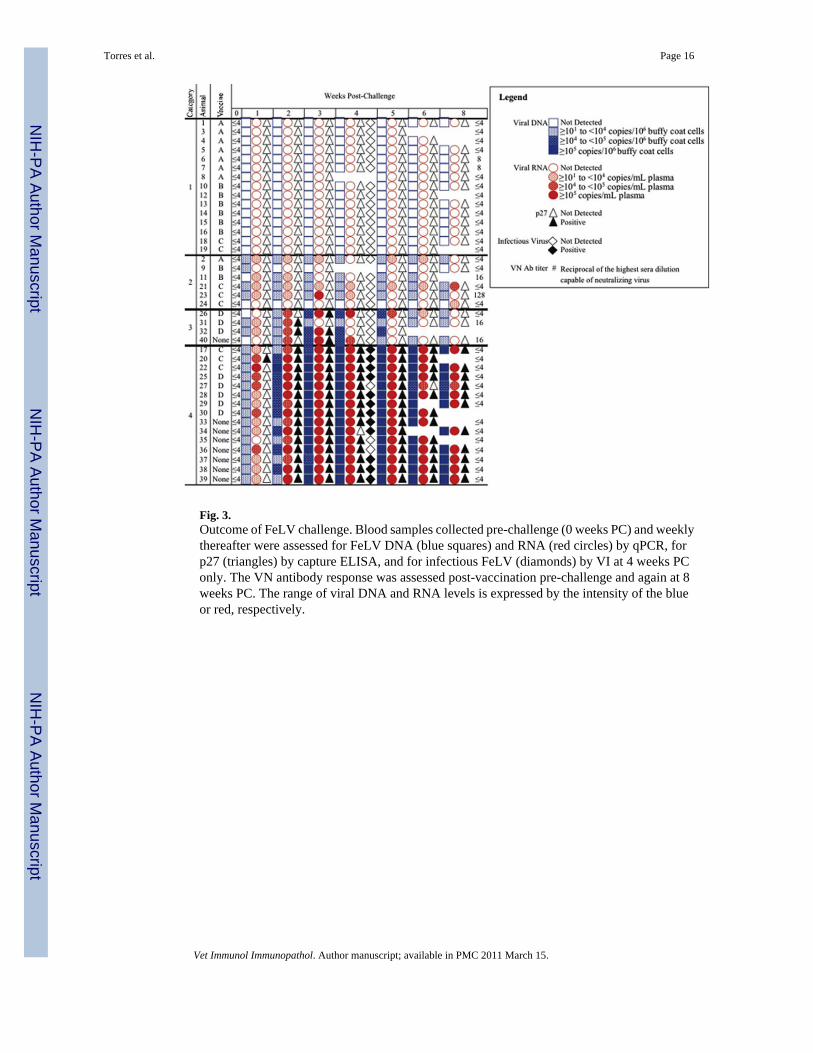
Fig. 3.Outcome of FeLV challenge. Blood samples collected pre-challenge (0 weeks PC) and weeklythereafter were assessed for FeLV DNA (blue squares) and RNA (red circles) by qPCR, forp27 (triangles) by capture ELISA, and for infectious FeLV (diamonds) by VI at 4 weeks PConly. The VN antibody response was assessed post-vaccination pre-challenge and again at 8weeks PC. The range of viral DNA and RNA levels is expressed by the intensity of the blueor red, respectively.
Torres et al. Page 16
Vet Immunol Immunopathol. Author manuscript; available in PMC 2011 March 15.
NIH
-PA Author Manuscript
NIH
-PA Author Manuscript
NIH
-PA Author Manuscript

NIH
-PA Author Manuscript
NIH
-PA Author Manuscript
NIH
-PA Author Manuscript
Torres et al. Page 17
Table 1
Efficacy of commercially available FeLV vaccines
Experimental group n Persistent antigenemia/viremia Preventable fraction (%)
Vaccine A 8 0 100
Vaccine B 8 0 100
Vaccine C 8 3 57
Vaccine D 8 5 29
Unvaccinated controls 8 7 not applicable
Vet Immunol Immunopathol. Author manuscript; available in PMC 2011 March 15.
Related Documents