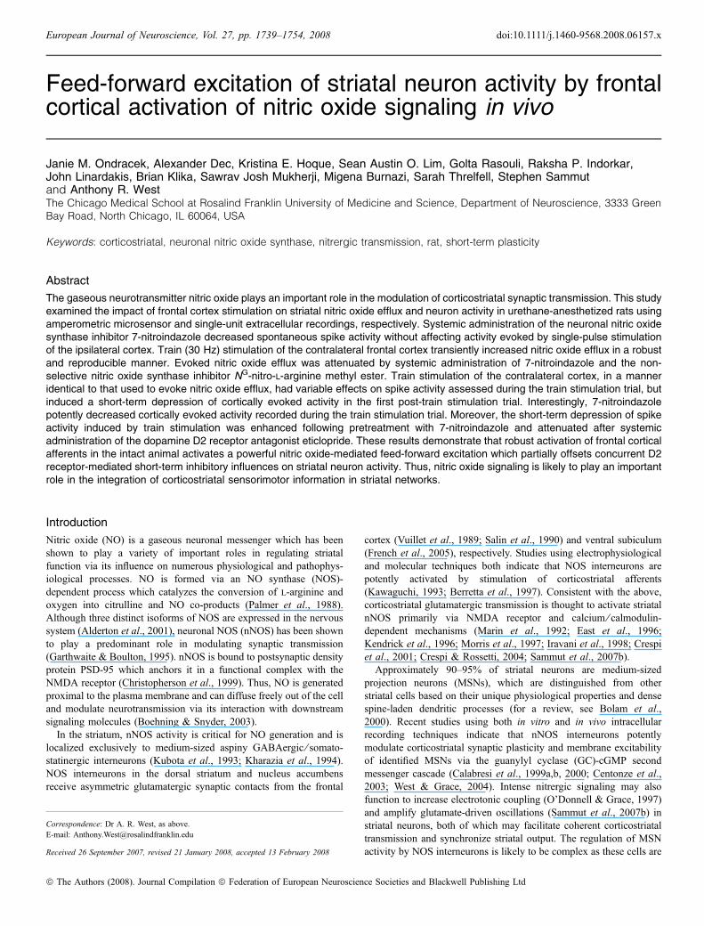
Feed-forward excitation of striatal neuron activity by frontal cortical activation of nitric oxide signaling in vivo Janie M. Ondracek, Alexander Dec, Kristina E. Hoque, Sean Austin O. Lim, Golta Rasouli, Raksha P. Indorkar, John Linardakis, Brian Klika, Sawrav Josh Mukherji, Migena Burnazi, Sarah Threlfell, Stephen Sammut and Anthony R. West The Chicago Medical School at Rosalind Franklin University of Medicine and Science, Department of Neuroscience, 3333 Green Bay Road, North Chicago, IL 60064, USA Keywords: corticostriatal, neuronal nitric oxide synthase, nitrergic transmission, rat, short-term plasticity Abstract The gaseous neurotransmitter nitric oxide plays an important role in the modulation of corticostriatal synaptic transmission. This study examined the impact of frontal cortex stimulation on striatal nitric oxide efflux and neuron activity in urethane-anesthetized rats using amperometric microsensor and single-unit extracellular recordings, respectively. Systemic administration of the neuronal nitric oxide synthase inhibitor 7-nitroindazole decreased spontaneous spike activity without affecting activity evoked by single-pulse stimulation of the ipsilateral cortex. Train (30 Hz) stimulation of the contralateral frontal cortex transiently increased nitric oxide efflux in a robust and reproducible manner. Evoked nitric oxide efflux was attenuated by systemic administration of 7-nitroindazole and the non- selective nitric oxide synthase inhibitor N G -nitro-l-arginine methyl ester. Train stimulation of the contralateral cortex, in a manner identical to that used to evoke nitric oxide efflux, had variable effects on spike activity assessed during the train stimulation trial, but induced a short-term depression of cortically evoked activity in the first post-train stimulation trial. Interestingly, 7-nitroindazole potently decreased cortically evoked activity recorded during the train stimulation trial. Moreover, the short-term depression of spike activity induced by train stimulation was enhanced following pretreatment with 7-nitroindazole and attenuated after systemic administration of the dopamine D2 receptor antagonist eticlopride. These results demonstrate that robust activation of frontal cortical afferents in the intact animal activates a powerful nitric oxide-mediated feed-forward excitation which partially offsets concurrent D2 receptor-mediated short-term inhibitory influences on striatal neuron activity. Thus, nitric oxide signaling is likely to play an important role in the integration of corticostriatal sensorimotor information in striatal networks. Introduction Nitric oxide (NO) is a gaseous neuronal messenger which has been shown to play a variety of important roles in regulating striatal function via its influence on numerous physiological and pathophys- iological processes. NO is formed via an NO synthase (NOS)- dependent process which catalyzes the conversion of l-arginine and oxygen into citrulline and NO co-products (Palmer et al., 1988). Although three distinct isoforms of NOS are expressed in the nervous system (Alderton et al., 2001), neuronal NOS (nNOS) has been shown to play a predominant role in modulating synaptic transmission (Garthwaite & Boulton, 1995). nNOS is bound to postsynaptic density protein PSD-95 which anchors it in a functional complex with the NMDA receptor (Christopherson et al., 1999). Thus, NO is generated proximal to the plasma membrane and can diffuse freely out of the cell and modulate neurotransmission via its interaction with downstream signaling molecules (Boehning & Snyder, 2003). In the striatum, nNOS activity is critical for NO generation and is localized exclusively to medium-sized aspiny GABAergic ⁄ somato- statinergic interneurons (Kubota et al., 1993; Kharazia et al., 1994). NOS interneurons in the dorsal striatum and nucleus accumbens receive asymmetric glutamatergic synaptic contacts from the frontal cortex (Vuillet et al., 1989; Salin et al., 1990) and ventral subiculum (French et al., 2005), respectively. Studies using electrophysiological and molecular techniques both indicate that NOS interneurons are potently activated by stimulation of corticostriatal afferents (Kawaguchi, 1993; Berretta et al., 1997). Consistent with the above, corticostriatal glutamatergic transmission is thought to activate striatal nNOS primarily via NMDA receptor and calcium ⁄ calmodulin- dependent mechanisms (Marin et al., 1992; East et al., 1996; Kendrick et al., 1996; Morris et al., 1997; Iravani et al., 1998; Crespi et al., 2001; Crespi & Rossetti, 2004; Sammut et al., 2007b). Approximately 90–95% of striatal neurons are medium-sized projection neurons (MSNs), which are distinguished from other striatal cells based on their unique physiological properties and dense spine-laden dendritic processes (for a review, see Bolam et al., 2000). Recent studies using both in vitro and in vivo intracellular recording techniques indicate that nNOS interneurons potently modulate corticostriatal synaptic plasticity and membrane excitability of identified MSNs via the guanylyl cyclase (GC)-cGMP second messenger cascade (Calabresi et al., 1999a,b, 2000; Centonze et al., 2003; West & Grace, 2004). Intense nitrergic signaling may also function to increase electrotonic coupling (O’Donnell & Grace, 1997) and amplify glutamate-driven oscillations (Sammut et al., 2007b) in striatal neurons, both of which may facilitate coherent corticostriatal transmission and synchronize striatal output. The regulation of MSN activity by NOS interneurons is likely to be complex as these cells are Correspondence: Dr A. R. West, as above. E-mail: [email protected] Received 26 September 2007, revised 21 January 2008, accepted 13 February 2008 European Journal of Neuroscience, Vol. 27, pp. 1739–1754, 2008 doi:10.1111/j.1460-9568.2008.06157.x ª The Authors (2008). Journal Compilation ª Federation of European Neuroscience Societies and Blackwell Publishing Ltd

Welcome message from author
This document is posted to help you gain knowledge. Please leave a comment to let me know what you think about it! Share it to your friends and learn new things together.
Transcript
Feed-forward excitation of striatal neuron activity by frontalcortical activation of nitric oxide signaling in vivo
Janie M. Ondracek, Alexander Dec, Kristina E. Hoque, Sean Austin O. Lim, Golta Rasouli, Raksha P. Indorkar,John Linardakis, Brian Klika, Sawrav Josh Mukherji, Migena Burnazi, Sarah Threlfell, Stephen Sammutand Anthony R. WestThe Chicago Medical School at Rosalind Franklin University of Medicine and Science, Department of Neuroscience, 3333 GreenBay Road, North Chicago, IL 60064, USA
Keywords: corticostriatal, neuronal nitric oxide synthase, nitrergic transmission, rat, short-term plasticity
Abstract
The gaseous neurotransmitter nitric oxide plays an important role in the modulation of corticostriatal synaptic transmission. This studyexamined the impact of frontal cortex stimulation on striatal nitric oxide efflux and neuron activity in urethane-anesthetized rats usingamperometric microsensor and single-unit extracellular recordings, respectively. Systemic administration of the neuronal nitric oxidesynthase inhibitor 7-nitroindazole decreased spontaneous spike activity without affecting activity evoked by single-pulse stimulationof the ipsilateral cortex. Train (30 Hz) stimulation of the contralateral frontal cortex transiently increased nitric oxide efflux in a robustand reproducible manner. Evoked nitric oxide efflux was attenuated by systemic administration of 7-nitroindazole and the non-selective nitric oxide synthase inhibitor NG-nitro-l-arginine methyl ester. Train stimulation of the contralateral cortex, in a manneridentical to that used to evoke nitric oxide efflux, had variable effects on spike activity assessed during the train stimulation trial, butinduced a short-term depression of cortically evoked activity in the first post-train stimulation trial. Interestingly, 7-nitroindazolepotently decreased cortically evoked activity recorded during the train stimulation trial. Moreover, the short-term depression of spikeactivity induced by train stimulation was enhanced following pretreatment with 7-nitroindazole and attenuated after systemicadministration of the dopamine D2 receptor antagonist eticlopride. These results demonstrate that robust activation of frontal corticalafferents in the intact animal activates a powerful nitric oxide-mediated feed-forward excitation which partially offsets concurrent D2receptor-mediated short-term inhibitory influences on striatal neuron activity. Thus, nitric oxide signaling is likely to play an importantrole in the integration of corticostriatal sensorimotor information in striatal networks.
Introduction
Nitric oxide (NO) is a gaseous neuronal messenger which has beenshown to play a variety of important roles in regulating striatalfunction via its influence on numerous physiological and pathophys-iological processes. NO is formed via an NO synthase (NOS)-dependent process which catalyzes the conversion of l-arginine andoxygen into citrulline and NO co-products (Palmer et al., 1988).Although three distinct isoforms of NOS are expressed in the nervoussystem (Alderton et al., 2001), neuronal NOS (nNOS) has been shownto play a predominant role in modulating synaptic transmission(Garthwaite & Boulton, 1995). nNOS is bound to postsynaptic densityprotein PSD-95 which anchors it in a functional complex with theNMDA receptor (Christopherson et al., 1999). Thus, NO is generatedproximal to the plasma membrane and can diffuse freely out of the celland modulate neurotransmission via its interaction with downstreamsignaling molecules (Boehning & Snyder, 2003).
In the striatum, nNOS activity is critical for NO generation and islocalized exclusively to medium-sized aspiny GABAergic ⁄ somato-statinergic interneurons (Kubota et al., 1993; Kharazia et al., 1994).NOS interneurons in the dorsal striatum and nucleus accumbensreceive asymmetric glutamatergic synaptic contacts from the frontal
cortex (Vuillet et al., 1989; Salin et al., 1990) and ventral subiculum(French et al., 2005), respectively. Studies using electrophysiologicaland molecular techniques both indicate that NOS interneurons arepotently activated by stimulation of corticostriatal afferents(Kawaguchi, 1993; Berretta et al., 1997). Consistent with the above,corticostriatal glutamatergic transmission is thought to activate striatalnNOS primarily via NMDA receptor and calcium ⁄ calmodulin-dependent mechanisms (Marin et al., 1992; East et al., 1996;Kendrick et al., 1996; Morris et al., 1997; Iravani et al., 1998; Crespiet al., 2001; Crespi & Rossetti, 2004; Sammut et al., 2007b).Approximately 90–95% of striatal neurons are medium-sized
projection neurons (MSNs), which are distinguished from otherstriatal cells based on their unique physiological properties and densespine-laden dendritic processes (for a review, see Bolam et al.,2000). Recent studies using both in vitro and in vivo intracellularrecording techniques indicate that nNOS interneurons potentlymodulate corticostriatal synaptic plasticity and membrane excitabilityof identified MSNs via the guanylyl cyclase (GC)-cGMP secondmessenger cascade (Calabresi et al., 1999a,b, 2000; Centonze et al.,2003; West & Grace, 2004). Intense nitrergic signaling may alsofunction to increase electrotonic coupling (O’Donnell & Grace, 1997)and amplify glutamate-driven oscillations (Sammut et al., 2007b) instriatal neurons, both of which may facilitate coherent corticostriataltransmission and synchronize striatal output. The regulation of MSNactivity by NOS interneurons is likely to be complex as these cells are
Correspondence: Dr A. R. West, as above.E-mail: [email protected]
Received 26 September 2007, revised 21 January 2008, accepted 13 February 2008
European Journal of Neuroscience, Vol. 27, pp. 1739–1754, 2008 doi:10.1111/j.1460-9568.2008.06157.x
ª The Authors (2008). Journal Compilation ª Federation of European Neuroscience Societies and Blackwell Publishing Ltd
capable of releasing neuropeptide Y, somatostatin and GABA ascotransmitters (Kawaguchi, 1997). Additionally, studies using extra-cellular recordings combined with local or systemic drug administra-tion have reported both excitatory (West et al., 2002; Liu et al., 2005a)and inhibitory (Sardo et al., 2002; Di Giovanni et al., 2003) effects ofNO generators on striatal neuron activity. Together with reportsindicating an important role for striatal NO–GC signaling in motorbehavior (for a review, see Del Bel et al., 2005), these studies suggestthat characterization of the complex signaling mechanisms utilized byNOS-containing interneurons will be essential for understandingnormal striatal function and pathophysiological conditions such asParkinson’s disease.The current study was undertaken to examine the impact of tonic
and phasic NO signaling on cortically evoked activity and short-termplasticity of putative striatal MSNs. Thus, electrical stimulationtechniques were used in conjunction with in vivo amperometricmicrosensor recordings of striatal NO efflux and single-unit extracel-lular recordings of striatal neuron activity.
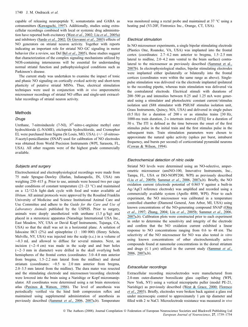
Methods
Drugs
Urethane, 7-nitroindazole (7-NI), NG-nitro-l-arginine methyl esterhydrochloride (L-NAME), eticlopride hydrochloride, and CremophorEL were purchased from Sigma (St Louis, MO, USA). (+ ⁄ –)S-nitroso-N-acetyl-penicillamine (SNAP) used for calibration of NO electrodeswas obtained from World Precision Instruments (WPI, Sarasota, FL,USA). All other reagents were of the highest grade commerciallyavailable.
Subjects and surgery
Electrochemical and electrophysiological recordings were made from75 male Sprague–Dawley (Harlan, Indianapolis, IN, USA) ratsweighing 250–415 g. Prior to use, animals were housed two per cageunder conditions of constant temperature (21–23 �C) and maintainedon a 12 ⁄ 12-h light–dark cycle with food and water available adlibitum. All animal protocols were approved by the Rosalind FranklinUniversity of Medicine and Science Institutional Animal Care andUse Committee and adhere to the Guide for the Care and Use ofLaboratory Animals published by the USPHS. Prior to surgery,animals were deeply anesthetized with urethane (1.5 g ⁄ kg) andplaced in a stereotaxic apparatus (Narashige International USA Inc.,East Meadow, NY, USA or David Kopf Instruments, Tujunga, CA,USA) so that the skull was set in a horizontal plane. A solution oflidocaine HCl (2%) and epinephrine (1 : 100 000) (Henry Schein,Melville, NY, USA) was injected into the scalp (s.c.) in a volume of�0.3 mL and allowed to diffuse for several minutes. Next, anincision (�2–4 cm) was made in the scalp and and burr holes(�2–3 mm in diameter) were drilled in the skull overlying bothhemispheres of the frontal cortex (coordinates: 3.0–4.0 mm anteriorfrom bregma, 1.5–2.2 mm lateral from the midline) and dorsalstriatum (coordinates: )0.5 to 2.0 mm anterior from bregma,2.0–3.5 mm lateral from the midline). The dura mater was resectedand the stimulating electrode and microsensor ⁄ recording electrodewere lowered into the brain using a Narishige or Kopf micromanip-ulator. All coordinates were determined using a rat brain stereotaxicatlas (Paxinos & Watson, 1986). The level of anesthesia wasperiodically verified via the hind limb compression reflex andmaintained using supplemental administration of anesthesia aspreviously described (Sammut et al., 2006, 2007a,b). Temperature
was monitored using a rectal probe and maintained at 37 �C using aheating pad (Vl-20F, Fintronics Inc., Orange, CT, USA).
Electrical stimulation
In NO microsensor experiments, a single bipolar stimulating electrode(Plastics One, Roanoke, VA, USA) was implanted into the frontalcortex (coordinates: 2.7–4.2 mm anterior to bregma, 1.5–2.5 mmlateral to midline, 2.0–4.2 mm ventral to the brain surface) contra-lateral to the microsensor as previously described (Sammut et al.,2007b). In electrophysiological studies, bipolar stimulating electrodeswere implanted either ipsilaterally or bilaterally into the frontalcortices (coordinates were within the same range as above). Single-pulse stimulation was delivered via the electrode implanted ipsilateralto the recording pipette, whereas train stimulation was delivered viathe contralateral electrode. Electrical stimuli with durations of200–500 ls and intensities between 0.25 and 1.25 mA were gener-ated using a stimulator and photoelectric constant current ⁄ stimulusisolation unit (S88 stimulator with PSIU6F stimulus isolation unit,Grass Instruments, Quincy, MA, USA) and delivered in single pulses(0.5 Hz) for a duration of 200 s or as stimulus trains [30 Hz,1000-ms train duration, 2-s intertrain interval (ITI)] for a duration of50 s. The ITI is defined as the time between the onset of the firststimulus pulse in the initial train and the first stimulus pulse in thesubsequent train. Train stimulation parameters were chosen toapproximate the natural spike activity (spikes per burst, intraburstfrequency, and bursts per second) of corticostriatal pyramidal neurons(Cowan & Wilson, 1994).
Electrochemical detection of nitric oxide
Striatal NO levels were determined using an NO-selective, amper-ometric microsensor (amiNO-100, Innovative Instruments, Inc.,Tampa, FL, USA or ISO-NOPF200, WPI) as previously described(Liu et al., 2005b; Sammut et al., 2006, 2007a,b). Briefly, the NOoxidation current (electrode potential of 0.865 V against a built-inAg ⁄ AgCl reference electrode) was amplified and recorded using acommercially available system (Apollo 4000, WPI). Prior to eachexperiment, the NO microsensor was calibrated in a temperaturecontrolled chamber (Diamond General, Ann Arbor, MI, USA) usingknown concentrations of the NO-generating compound SNAP (Ohtaet al., 1997; Zhang, 2004; Liu et al., 2005b; Sammut et al., 2006,2007a,b). Calibration plots were constructed prior to each experimentin order to determine the sensitivity and integrity of the electrodeand confirm that the NO oxidation current exhibited a linearresponse to NO concentrations ranging from 0.6 to 48 nm. Theselectivity of the NO microsensor for NO was also tested in vitrousing known concentrations of other electrochemically activecompounds found at nanomolar concentrations in the dorsal striatumand drugs (£ 1 lm) utilized in the current study (Sammut et al.,2006, 2007a,b).
Extracellular recordings
Extracellular recording microelectrodes were manufactured from2.0-mm outer-diameter borosilicate glass capillary tubing (WPI,New York, NY) using a vertical micropipette puller (model PE-21,Narishige) as previously described (West & Grace, 2000; Florescoet al., 2003). Briefly, electrodes were broken back against a glass rodunder microscopic control to approximately 1 lm tip diameter andfilled with 2 m NaCl. Microelectrode resistance was measured in vivo
1740 J. M. Ondracek et al.
ª The Authors (2008). Journal Compilation ª Federation of European Neuroscience Societies and Blackwell Publishing LtdEuropean Journal of Neuroscience, 27, 1739–1754
at 1000 Hz with a current of 10 nA using an FHC Xcell 3+microelectrode amplifier ⁄ impedance check (Frederick Haer Company(FHC), Bowdoinham, ME, USA) and ranged from 10 to 15 MW.Previous studies have shown that the majority of striatal neurons aresilent (Mallet et al., 2005). Thus, in order to isolate single units,extracellular microelectrodes were lowered incrementally through thedorsal striatum using a micromanipulator (MO-8, Narashige)while single-pulse electrical stimuli (0.5 Hz, 0.5 ms, 0.25–1.25 mA)were applied to the cortex (see above). Once a cell was detected, theposition of the recording electrode was adjusted to maximize the spikesignal to background noise ratio (‡ 4 : 1). In within-subjects studies,the impact of varying the intensity of electrical stimuli within a givenrange of current (0.4–0.8 mA) on evoked spike activity was assessedover 50 stimuli per trial. In between-subjects studies, stimulationcurrents were titrated to an intensity (280–1250 lA) which reliablyevoked spike activity approximately 50% of the time (e.g. �50 spikesin response to 100 stimulus pulses delivered at 0.5 Hz). Extracellularelectrode potentials were passed through a high-input impedanceamplifier ⁄ window discriminator (Fintronics WDR 420, or FHC Xcell3+ microelectrode amplifier) connected to a Grass AM-10 audiomonitor and displayed on a BK Precision 2120B dual-trace (30 mHz)oscilloscope. Single-unit activity was filtered (low-pass filter at 8 kHzand high-pass filter at 400 Hz), digitized (20 kHz) and recorded usingan Intel�-based microcomputer with a data acquisition board interface(DAP-5200a, Microstar Laboratories, Bellevue, WA, USA). Detailedanalysis of all spontaneous and evoked spikes was performed off-lineusing custom (Neuroscope) software applications (Brian Lowry,Pittsburgh, PA, USA).
Drug preparation ⁄ administration and experimental protocol
Urethane and eticlopride were dissolved in physiological saline(0.9%). 7-NI and L-NAME were dissolved in vehicle consisting of10% Cremophor EL in saline (0.9%) solution (Eblen et al., 1996;Sammut et al., 2006, 2007b). Effective doses of 7-NI (50 mg ⁄ kg,i.p.), L-NAME (100 mg ⁄ kg, i.p.) and eticlopride (100 lg ⁄ kg, i.p.)were derived from the range previously reported in the literature(Ohta et al., 1994; Eblen et al., 1996; Kalisch et al., 1996; Ayerset al., 1997; Adachi et al., 2000; Hirabayashi et al., 2001; Sammutet al., 2006, 2007a). In NO microsensor experiments, the responseto frontal cortical stimulation was assessed multiple times priorto drug or vehicle administration, with stimulations separated by15–20 min from each other in order to obtain at least twoconsecutive responses that did not vary by > 20% (Sammut et al.,2006, 2007a,b). The average of these measures was considered asthe baseline response to which responses following drug or vehicleadministration were compared. Previous time-course studies of theimpact of NOS inhibitors on NO synthesis in the striatum haveshown that systemic (i.p.) injections of 7-NI and L-NAME reducedNOS activity to �50–60% of control levels (Adachi et al., 2000).Moreover, the effects of 7-NI and L-NAME developed during thefirst 30–80 min post-injection and were sustained for > 180 minpost-injection (Adachi et al., 2000). Thus, in the current study, theimpact of vehicle, 7-NI and L-NAME on cortically evoked NOefflux was measured 20–120 min post-injection.
In single-unit recording studies, vehicle or 7-NI (50 mg ⁄ kg, i.p.)was administered using either a within- or a between-subjectsdesign. Eticlopride was administered using a within- subjectsdesign. In within-subjects studies using 7-NI, after isolating astriatal neuron and recording spontaneous (non-evoked) firingactivity (�2–5 min), the effects of separate trials (50 pulses each)
of single-pulse cortical stimulation (0.4, 0.6, and 0.8 mAstimulation intensities) were recorded prior to vehicle or drugadministration. Next, vehicle or 7-NI was administered andspontaneous firing and the effects of single-pulse electrical stimu-lation were reassessed approximately 20–30 min later as describedabove. In between-subjects studies using 7-NI, animals wereinjected with either vehicle (10% Cremophor EL in saline) or7-NI (in vehicle) at least 25 min prior to initiation of the recordingsession. After isolating a cell as described above, stimulationcurrents were adjusted to approximately 50% maximal respondingto stimulation delivered at 0.5 Hz. A pre-train stimulation baselinetrial consisting of 100 individual single-pulse stimulations deliveredover 200 s was then recorded. Once stable levels of single-pulse-evoked spiking were obtained, a series of train stimulations (25)were delivered to the contralateral cortex (30 Hz, 1000 ms trainduration, 2 s ITI for a duration of 50 s). In order to examine theimpact of train stimulation on spike probability during the trainstimulation trial, single-pulse stimulation was delivered concurrentlyto the ipsilateral cortex via a second stimulation channel which wastriggered by channel 1 (train) stimulation but delayed �500 msfrom the end of the stimulus train (see Figs 1 and 5). Immediatelyfollowing the train stimulation trial, three additional post-trainstimulation trials (200 s each) were performed in a manner whichwas identical to the pre-train stimulation trial (100 single-pulsetrials each, no train stimulation). Three post-train trials were chosenbased on the observation that train-induced changes in spikeprobability in control animals were usually observed to return toprestimulation levels during the second or third post-train stimula-tion trials, recorded approximately 200–600 s after completion ofthe train stimulation trial. Within-subjects studies using eticlopridefollowed this same experimental protocol, except the responsivenessof striatal neurons to the cortical train stimulation protocol wasre-tested at least 20 min after drug administration. As theseexperiments were designed to determine whether dopamine D2receptor activation mediates the inhibition of cortically evokedspike activity induced by cortical train stimulation, only neuronsexhibiting an inhibitory response were selected for further exper-imentation. Additionally, all striatal neurons included in the data setexhibited stable electrophysiological properties (see above) for aminimum of 20 min following drug administration and throughoutthe duration of the post-injection stimulation protocol.
Histology
After completion of each experiment, rats were deeply anesthetizedand perfused transcardially with ice-cold saline followed by 10%formalin in buffered phosphate (PB) (EMS, Hatfield, PA, USA).Brains were removed and post-fixed in 10% formalin ⁄ PB for at least3 days. Next, brains were immersed in PBS ⁄ sucrose solution (30%)and stored in a refrigerator until saturated. Brains were then sectionedinto 50-lm coronal slices, mounted and stained with Neutralred ⁄ Cresyl Violet (10 : 1) solution to enable histological assessmentof stimulating and recording electrode ⁄ NO microsensor sites (Sammutet al., 2007b).
Data analysis
In amperometric recording experiments, the NO oxidation current(pA) was allowed to stabilize for at least 150 s prior to stimulation.The NO oxidation current recorded (50 Hz sampling frequency)over the last 30 s of the pre-stimulation period was then averaged
Nitric oxide-mediated feed-forward excitation 1741
ª The Authors (2008). Journal Compilation ª Federation of European Neuroscience Societies and Blackwell Publishing LtdEuropean Journal of Neuroscience, 27, 1739–1754
using Apollo 4000 software applications (WPI) and subtracted fromthe mean NO oxidation current recorded during the last 30 s of thestimulation period (Liu et al., 2005b; Sammut et al., 2006,2007a,b). The mean stimulation-evoked concentration of NO (nm)is derived from in vitro calibration curves (Sammut et al., 2006,2007a,b). In single-unit recording studies, firing rate histogramswere constructed (1.0-ms bins of 2-min epochs) from data recordedprior to and following (20–30 min) 7-NI administration. Peri-stimulus time histograms (PSTHs) were constructed (1.0-ms bins)for each stimulus trial and spike probabilities were calculated bydividing the number of evoked action potentials (either 0 or 1 perpulse) by the number of stimuli delivered (Floresco et al., 2001).Single-unit and group data were summarized using populationPSTHs and ⁄ or spike probability plots as indicated. Excitatory (E)responses observed during train stimulation were operationallydefined as an increase in spike probability of > 2 SD above thepretrain stimulation mean. Inhibitory (I) responses observed duringtrain stimulation were operationally defined as a decrease in spikeprobability of > 2 SD below the pretrain stimulation mean.This ± 2 SD range was selected after studying the spike probabilitychanges of five single units recorded following vehicle injectionduring a 10–40-min post-injection period (see Tseng et al., 2000).Similar criteria were used in the assessment of stimulation-inducedchanges in evoked spike activity in a previous study (Jodo et al.,2005). The statistical significance of drug- and stimulation-inducedchanges in NO oxidation current and spike activity was determinedusing either a paired t-test ⁄ Wilcoxon signed rank test, Fisher exacttest or one- ⁄ two-way analysis of variance (anova) with repeatedmeasures (RM) as indicated (Sigma Stat, Jandel). Also, either aBonferroni or Tukey post-hoc test was used as indicated todetermine which group(s) contributed to overall differences seenwith anova. Data pertaining to NO efflux are graphically repre-sented as the mean ± SEM percentage of predrug ⁄ vehicle control.Data from single-unit recordings are graphically represented as themean ± SEM, spikes per bin, firing rate, spike probability, spikelatency and standard deviation of spike latency.
Results
Stimulating electrode, microsensor and recording electrodeplacements
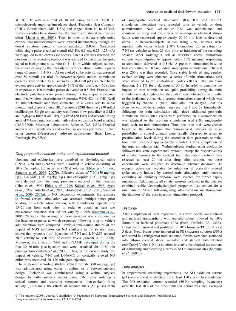
All identified stimulating electrode tips were confirmed to lie in thefrontal cortex between 2.7 and 4.2 mm anterior to bregma, 0.8 and2.5 mm lateral to the midline, and 2.0 and 5.5 mm ventral to thesurface of the skull (Paxinos & Watson, 1986; Fig. 1). Identifiedplacements for NO microsensors and extracellular recording elec-trodes implanted into the striatum were verified to lie between0.26 mm posterior and 1.6 mm anterior to bregma, 1.7 and 4.0 mmlateral to the midline, and 3.4 and 7.8 mm ventral to the dural surface(Paxinos & Watson, 1986; Fig. 1).
Modulation of spontaneous and cortically evoked spikeactivity by tonic NO
To assess the impact of endogenous NO tone on spontaneous andcortically evoked spike activity, striatal neurons were recordedbefore and after (�20–30 min) systemic administration of vehicle(10% Cremophor EL in 0.9% saline) or the selective nNOSinhibitor 7-NI (50 mg ⁄ kg, i.p.). It is estimated that 90–95% ofstriatal neurons are projection neurons (Wilson, 2004). Striatalneurons exhibiting spike characteristics resembling cholinergic(tonic or regular firing at a rate of �1–4 Hz) or fast-spikinginterneurons [respond to low-intensity cortical stimulation with ahigh-frequency train of short-duration (< 0.9 ms) action potentials]were not recorded in this study (Wilson et al., 1990; Kawaguchi,1993; Plenz & Kitai, 1998; Koos & Tepper, 1999; Mallet et al.,2005, 2006). Given the above, it is highly likely that the vastmajority of single units recorded in this study were MSNs. Inwithin-subjects studies, successful recordings of evoked activitybefore and after vehicle or 7-NI administration were obtained forn ¼ 5 and n ¼ 15 cells ⁄ rats, respectively. After isolating a singleunit using single-pulse stimulation of the ipsilateral cortex (seeMethods), 2–5 min of spontaneous firing activity was recorded
Fig. 1. Overview of stimulating and recording protocols. Stimulating electrodes were implanted bilaterally into the frontal cortex (left) as described in the Methodssection. In all electrophysiological studies, an extracellular recording electrode was implanted into the striatum (right) and single-unit activity evoked by single-pulsestimulation of the ipsilateral cortex was recorded prior to recording spontaneous firing activity. In studies involving NO microsensor measurements, NO effluxevoked via train stimulation of the contralateral motor cortex was recorded in the dorsal striatum. In studies examining the impact of train stimulation on corticallyevoked spike activity, train stimulation was delivered to the contralateral motor cortex, whereas single-pulse stimulation was delivered to the ipsilateral motor cortexto evoke single-unit spike activity. The probability of evoking spike activity during single-pulse stimulation of the ipsilateral cortex was recorded prior to, during andafter train stimulation of the contralateral cortex (diagrams are derived from the atlas of Paxinos & Watson, 1986).
1742 J. M. Ondracek et al.
ª The Authors (2008). Journal Compilation ª Federation of European Neuroscience Societies and Blackwell Publishing LtdEuropean Journal of Neuroscience, 27, 1739–1754
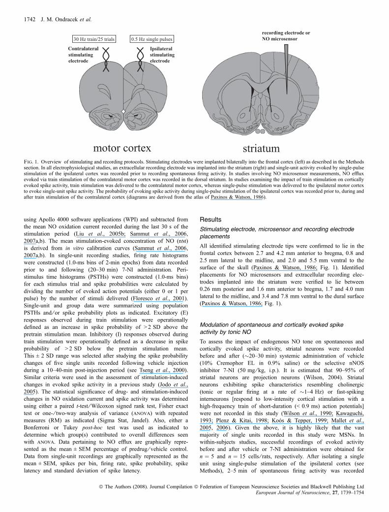
(Fig. 2). Vehicle administration (10–40 min) did not inducesignificant changes in spontaneous firing activity (data not shown).As shown in Fig. 2B and C, 7-NI administration significantlydepressed spontaneous firing activity (P < 0.05, Wilcoxon signed
rank test) without affecting the proportion of spontaneously activecells within the group (basal firing rates were collected for 14 totalcells ⁄ rats; pre-7-NI: seven spontaneously firing, seven quiescent;post-7-NI: three spontaneously firing, eleven quiescent; P > 0.05,Fisher exact test).To examine the potential effects of tonic NO on spike activity
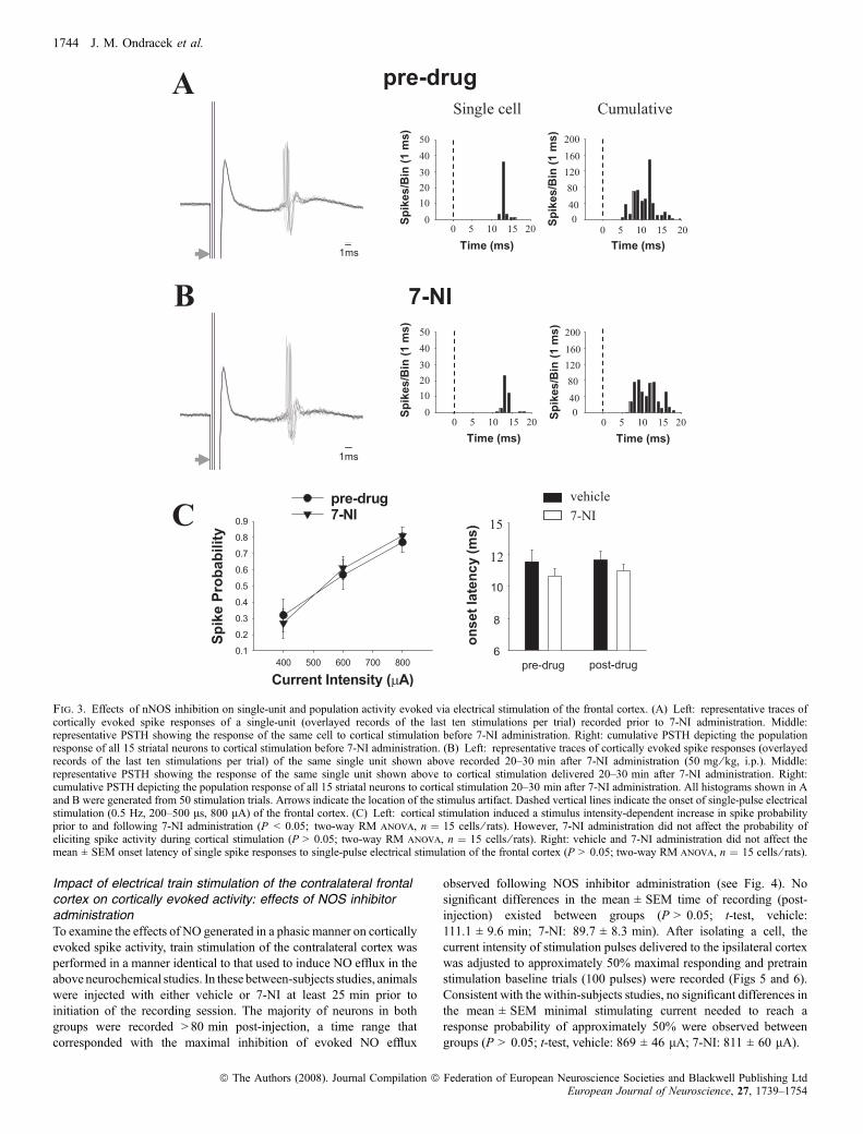
evoked by electrical stimulation of frontal cortical afferents, multiplestimulation trials consisting of 50 single pulses (0.5 Hz, 200–500 ls,400–800 lA) were delivered to the ipsilateral cortex in separate trialsboth before and 20–30 min after vehicle or 7-NI administration(Fig. 3A and B). Vehicle administration did not affect the mean ±SEM probability of evoking short-latency spike activity during singlepulse stimulation of the ipsilateral frontal cortex (P > 0.05; two-wayrepeated-measures (RM) anova with two factor repetition, data notshown). Short-latency spike activity evoked during repeated single-pulse stimulation of the ipsilateral frontal cortex was dependent on thelevel of current intensity, both prior to and following 7-NI adminis-tration (Fig. 3C; F2,28 ¼ 38.73, P < 0.001; two-way RM anova withtwo factor repetition and Tukey post-hoc test, P < 0.005 at all levels ofcurrent intensity). No significant differences in mean ± SEM spikeprobability or interactions between drug and current intensity wereobserved between measures taken prior to and following 7-NIadministration (Fig. 3C; P > 0.05; two-way RM anova with twofactor repetition). The average onset latency of evoked spikes andstandard deviation of spike latency was calculated from recordings ofunit activity during stimulation trials performed using the highesttested current intensity (800 lA), prior to and following vehicle or7-NI administration (20–30 min). Vehicle and 7-NI administration didnot affect the mean ± SEM onset latency of evoked spikes (Fig. 3C;P > 0.05; two-way RM anova with two factor repetition) or standarddeviation of spike latency (Fig. 3C, pre-vehicle: 1.29 ± 0.46 ms; post-vehicle: 1.40 ± 0.39 ms; pre-7-NI: 0.94 ± 0.19 ms; post-7-NI:1.21 ± 0.20 ms).
Impact of electrical train stimulation of the contralateralfrontal cortex on striatal NO efflux: effects of NOS inhibitoradministration
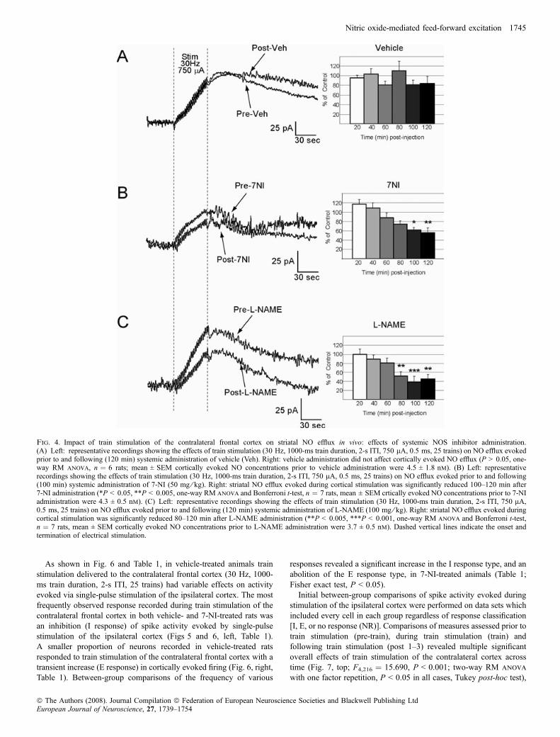
Afferents from the contralateral frontal cortex were activated usingelectrical stimuli delivered for 50 s as trains (25) of high-frequencystimulation (30 Hz, 1000-ms train duration, 2-s ITI, see Methods).The duration of the stimulation period and the stimulation pattern weredesigned to approximate the natural phasic firing activity of corticalpyramidal neurons observed in conscious behaving animals (Steriade,2001). To determine the potential role of NOS activation in the effectsof electrical train stimulation of the contralateral cortex on striatal NOefflux, train stimulation of the contralateral cortex was carried out asdescribed above (see Methods; Fig. 1) prior to and following (20–120 min) systemic administration of either vehicle or drug. Experi-ments were performed using L-NAME, a non-selective inhibitor of allknown NOS isoforms (100 mg ⁄ kg, i.p.), or the selective nNOSinhibitor 7-NI (50 mg ⁄ kg, i.p.). Vehicle administration did not affectNO efflux evoked by electrical train stimulation of the contralateralfrontal cortex (Fig. 4A; P > 0.05, one-way RM anova on measurestaken 20 min prior to and 20–120 min after vehicle injection). NOefflux observed during cortical train stimulation was attenuatedfollowing systemic administration of either 7-NI (Fig. 4;F6,29 ¼ 8.23, P < 0.001, one-way RM anova and Bonferroni t-test,P < 0.01 at 100–120 min post 7-NI) or L-NAME (Fig. 4C;F6,33 ¼ 8.10, P < 0.001, one-way RM anova and Bonferroni t-test,P < 0.005 at 80–120 min post L-NAME).
Fig. 2. Inhibition of nNOS activity decreases the spontaneous activity ofstriatal neurons isolated via electrical stimulation of the frontal cortex. (A) Top:representative trace of spontaneous spike activity of a striatal neuron recordedprior to systemic administration of 7-NI (50 mg ⁄ kg, i.p.). Bottom: magnifi-cation of the first spike shown in the above trace. (B) Representative firing ratehistograms showing the spontaneous activity (120 s) of a single striatal neuron(same cell as in A) before (top) and after (bottom) 7-NI administration. Asnoted above, the majority of neurons exhibiting spontaneous activity underbasal conditions were quiescent following 7-NI administration (4 ⁄ 7 spontane-ously active cells). Importantly, following 7-NI administration spike activitywas consistently evoked in all neurons by cortical stimulation.(C) 7-NI administration decreased the mean ± SEM firing rate of single unitspreviously isolated via electrical stimulation of the frontal cortex (*P < 0.05;Wilcoxon rank test, n ¼ 14 cells ⁄ rats).
Nitric oxide-mediated feed-forward excitation 1743
ª The Authors (2008). Journal Compilation ª Federation of European Neuroscience Societies and Blackwell Publishing LtdEuropean Journal of Neuroscience, 27, 1739–1754
Impact of electrical train stimulation of the contralateral frontalcortex on cortically evoked activity: effects of NOS inhibitoradministrationTo examine the effects of NO generated in a phasic manner on corticallyevoked spike activity, train stimulation of the contralateral cortex wasperformed in a manner identical to that used to induce NO efflux in theabove neurochemical studies. In these between-subjects studies, animalswere injected with either vehicle or 7-NI at least 25 min prior toinitiation of the recording session. The majority of neurons in bothgroups were recorded > 80 min post-injection, a time range thatcorresponded with the maximal inhibition of evoked NO efflux
observed following NOS inhibitor administration (see Fig. 4). Nosignificant differences in the mean ± SEM time of recording (post-injection) existed between groups (P > 0.05; t-test, vehicle:111.1 ± 9.6 min; 7-NI: 89.7 ± 8.3 min). After isolating a cell, thecurrent intensity of stimulation pulses delivered to the ipsilateral cortexwas adjusted to approximately 50% maximal responding and pretrainstimulation baseline trials (100 pulses) were recorded (Figs 5 and 6).Consistent with the within-subjects studies, no significant differences inthe mean ± SEM minimal stimulating current needed to reach aresponse probability of approximately 50% were observed betweengroups (P > 0.05; t-test, vehicle: 869 ± 46 lA; 7-NI: 811 ± 60 lA).
Fig. 3. Effects of nNOS inhibition on single-unit and population activity evoked via electrical stimulation of the frontal cortex. (A) Left: representative traces ofcortically evoked spike responses of a single-unit (overlayed records of the last ten stimulations per trial) recorded prior to 7-NI administration. Middle:representative PSTH showing the response of the same cell to cortical stimulation before 7-NI administration. Right: cumulative PSTH depicting the populationresponse of all 15 striatal neurons to cortical stimulation before 7-NI administration. (B) Left: representative traces of cortically evoked spike responses (overlayedrecords of the last ten stimulations per trial) of the same single unit shown above recorded 20–30 min after 7-NI administration (50 mg ⁄ kg, i.p.). Middle:representative PSTH showing the response of the same single unit shown above to cortical stimulation delivered 20–30 min after 7-NI administration. Right:cumulative PSTH depicting the population response of all 15 striatal neurons to cortical stimulation 20–30 min after 7-NI administration. All histograms shown in Aand B were generated from 50 stimulation trials. Arrows indicate the location of the stimulus artifact. Dashed vertical lines indicate the onset of single-pulse electricalstimulation (0.5 Hz, 200–500 ls, 800 lA) of the frontal cortex. (C) Left: cortical stimulation induced a stimulus intensity-dependent increase in spike probabilityprior to and following 7-NI administration (P < 0.05; two-way RM anova, n ¼ 15 cells ⁄ rats). However, 7-NI administration did not affect the probability ofeliciting spike activity during cortical stimulation (P > 0.05; two-way RM anova, n ¼ 15 cells ⁄ rats). Right: vehicle and 7-NI administration did not affect themean ± SEM onset latency of single spike responses to single-pulse electrical stimulation of the frontal cortex (P > 0.05; two-way RM anova, n ¼ 15 cells ⁄ rats).
1744 J. M. Ondracek et al.
ª The Authors (2008). Journal Compilation ª Federation of European Neuroscience Societies and Blackwell Publishing LtdEuropean Journal of Neuroscience, 27, 1739–1754
As shown in Fig. 6 and Table 1, in vehicle-treated animals trainstimulation delivered to the contralateral frontal cortex (30 Hz, 1000-ms train duration, 2-s ITI, 25 trains) had variable effects on activityevoked via single-pulse stimulation of the ipsilateral cortex. The mostfrequently observed response recorded during train stimulation of thecontralateral frontal cortex in both vehicle- and 7-NI-treated rats wasan inhibition (I response) of spike activity evoked by single-pulsestimulation of the ipsilateral cortex (Figs 5 and 6, left, Table 1).A smaller proportion of neurons recorded in vehicle-treated ratsresponded to train stimulation of the contralateral frontal cortex with atransient increase (E response) in cortically evoked firing (Fig. 6, right,Table 1). Between-group comparisons of the frequency of various
responses revealed a significant increase in the I response type, and anabolition of the E response type, in 7-NI-treated animals (Table 1;Fisher exact test, P < 0.05).Initial between-group comparisons of spike activity evoked during
stimulation of the ipsilateral cortex were performed on data sets whichincluded every cell in each group regardless of response classification[I, E, or no response (NR)]. Comparisons of measures assessed prior totrain stimulation (pre-train), during train stimulation (train) andfollowing train stimulation (post 1–3) revealed multiple significantoverall effects of train stimulation of the contralateral cortex acrosstime (Fig. 7, top; F4,216 ¼ 15.690, P < 0.001; two-way RM anova
with one factor repetition, P < 0.05 in all cases, Tukey post-hoc test),
Fig. 4. Impact of train stimulation of the contralateral frontal cortex on striatal NO efflux in vivo: effects of systemic NOS inhibitor administration.(A) Left: representative recordings showing the effects of train stimulation (30 Hz, 1000-ms train duration, 2-s ITI, 750 lA, 0.5 ms, 25 trains) on NO efflux evokedprior to and following (120 min) systemic administration of vehicle (Veh). Right: vehicle administration did not affect cortically evoked NO efflux (P > 0.05, one-way RM anova, n ¼ 6 rats; mean ± SEM cortically evoked NO concentrations prior to vehicle administration were 4.5 ± 1.8 nm). (B) Left: representativerecordings showing the effects of train stimulation (30 Hz, 1000-ms train duration, 2-s ITI, 750 lA, 0.5 ms, 25 trains) on NO efflux evoked prior to and following(100 min) systemic administration of 7-NI (50 mg ⁄ kg). Right: striatal NO efflux evoked during cortical stimulation was significantly reduced 100–120 min after7-NI administration (*P < 0.05, **P < 0.005, one-way RM anova and Bonferroni t-test, n ¼ 7 rats, mean ± SEM crtically evoked NO concentrations prior to 7-NIadministration were 4.3 ± 0.5 nm). (C) Left: representative recordings showing the effects of train stimulation (30 Hz, 1000-ms train duration, 2-s ITI, 750 lA,0.5 ms, 25 trains) on NO efflux evoked prior to and following (120 min) systemic administration of L-NAME (100 mg ⁄ kg). Right: striatal NO efflux evoked duringcortical stimulation was significantly reduced 80–120 min after L-NAME administration (**P < 0.005, ***P < 0.001, one-way RM anova and Bonferroni t-test,n ¼ 7 rats, mean ± SEM cortically evoked NO concentrations prior to L-NAME administration were 3.7 ± 0.5 nm). Dashed vertical lines indicate the onset andtermination of electrical stimulation.
Nitric oxide-mediated feed-forward excitation 1745
ª The Authors (2008). Journal Compilation ª Federation of European Neuroscience Societies and Blackwell Publishing LtdEuropean Journal of Neuroscience, 27, 1739–1754
as well as 7-NI treatment (Fig. 7, top; F1,216 ¼ 5.616, P < 0.05; two-way RM anova with one factor repetition, P < 0.05 in all cases,Tukey post-hoc test) on cortically evoked activity. The effect of 7-NItreatment also depended on time in relation to the train stimulationtrial. Thus, there was a significant interaction between 7-NI admin-istration and time post-train (F4,216 ¼ 4.211, P < 0.005; two-way RManova with one factor repetition). Interestingly, in vehicle-treated
animals train stimulation of the contralateral cortex induced a short-term depression of cortically evoked activity in the first post-trainstimulation trial (Fig. 7, top, P < 0.05) but did not alter subsequentpost-stimulation responses (Fig. 7, top, P > 0.05). Moreover, 7-NIadministration potently decreased cortically evoked activity duringtrain stimulation of the contralateral cortex (Fig. 7, top, P < 0.001)and in the first post-train stimulation trial (Fig. 7, top, P < 0.001), but
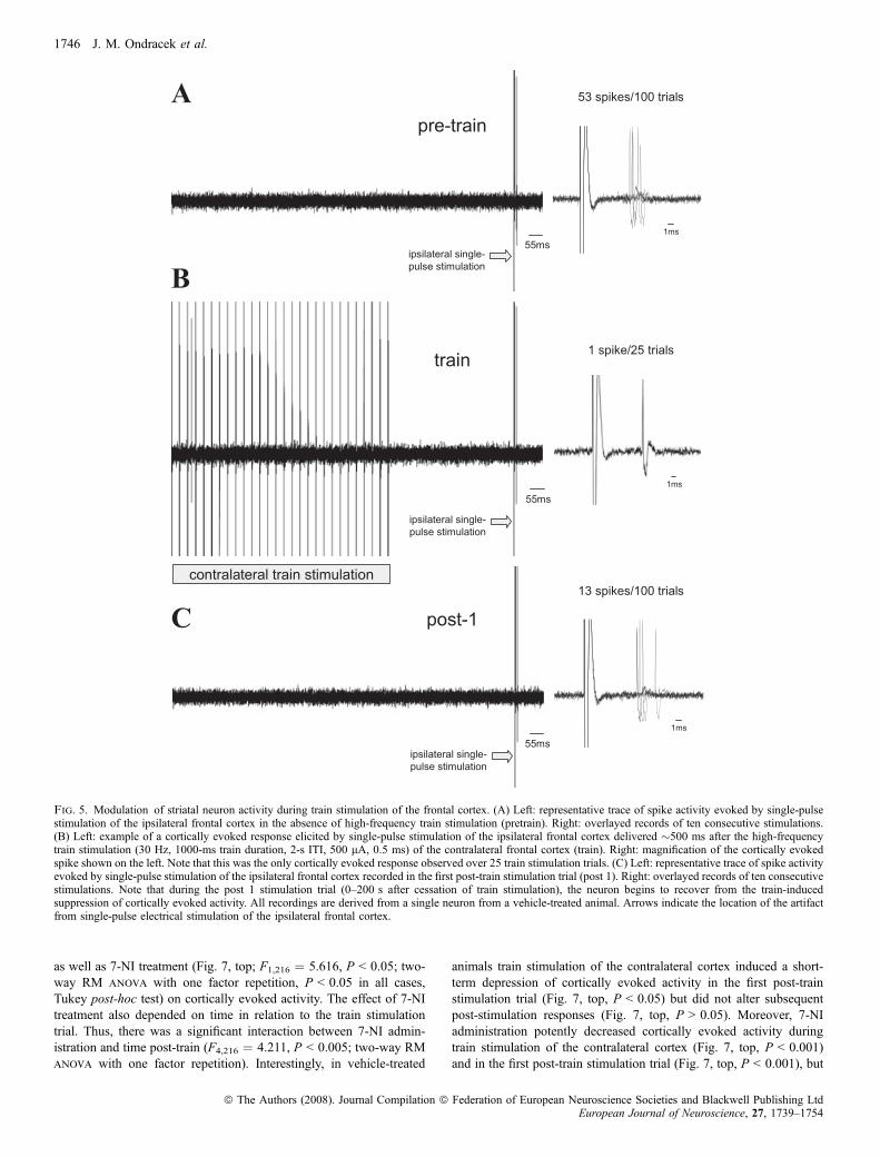
Fig. 5. Modulation of striatal neuron activity during train stimulation of the frontal cortex. (A) Left: representative trace of spike activity evoked by single-pulsestimulation of the ipsilateral frontal cortex in the absence of high-frequency train stimulation (pretrain). Right: overlayed records of ten consecutive stimulations.(B) Left: example of a cortically evoked response elicited by single-pulse stimulation of the ipsilateral frontal cortex delivered �500 ms after the high-frequencytrain stimulation (30 Hz, 1000-ms train duration, 2-s ITI, 500 lA, 0.5 ms) of the contralateral frontal cortex (train). Right: magnification of the cortically evokedspike shown on the left. Note that this was the only cortically evoked response observed over 25 train stimulation trials. (C) Left: representative trace of spike activityevoked by single-pulse stimulation of the ipsilateral frontal cortex recorded in the first post-train stimulation trial (post 1). Right: overlayed records of ten consecutivestimulations. Note that during the post 1 stimulation trial (0–200 s after cessation of train stimulation), the neuron begins to recover from the train-inducedsuppression of cortically evoked activity. All recordings are derived from a single neuron from a vehicle-treated animal. Arrows indicate the location of the artifactfrom single-pulse electrical stimulation of the ipsilateral frontal cortex.
1746 J. M. Ondracek et al.
ª The Authors (2008). Journal Compilation ª Federation of European Neuroscience Societies and Blackwell Publishing LtdEuropean Journal of Neuroscience, 27, 1739–1754
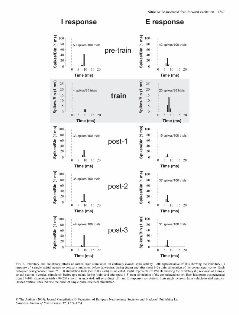
Fig. 6. Inhibitory and facilitatory effects of cortical train stimulation on cortically evoked spike activity. Left: representative PSTHs showing the inhibitory (I)response of a single striatal neuron to cortical stimulation before (pre-train), during (train) and after (post 1–3) train stimulation of the contralateral cortex. Eachhistogram was generated from 25–100 stimulation trials (50–200 s each) as indicated. Right: representative PSTHs showing the excitatory (E) response of a singlestriatal neuron to cortical stimulation before (pre-train), during (train) and after (post 1–3) train stimulation of the contralateral cortex. Each histogram was generatedfrom 25–100 stimulation trials (50–200 s each) as indicated. All recordings of I and E responses are derived from single neurons from vehicle-treated animals.Dashed vertical lines indicate the onset of single-pulse electrical stimulation.
Nitric oxide-mediated feed-forward excitation 1747
ª The Authors (2008). Journal Compilation ª Federation of European Neuroscience Societies and Blackwell Publishing LtdEuropean Journal of Neuroscience, 27, 1739–1754
did not alter subsequent post-stimulation responses. No significantdifferences in average onset latency of evoked spikes or standarddeviation of spike latency were observed between vehicle- and7-NI-treated groups (Fig. 7, middle, bottom; P > 0.05; two-way RManova with one factor repetition). Lastly, train stimulation wasobserved to decrease the standard deviation of spike latency ofcortically evoked spikes to a similar degree in both vehicle- and7-NI-treated animals (Fig. 7, bottom, F4,201 ¼ 2.936, P < 0.05;two-way RM anova with one factor repetition).To determine the impact of 7-NI administration on the magnitude of
inhibition observed during and immediately following train stimula-tion of the contralateral cortex, between-group comparisons of spikeactivity evoked via single-pulse stimulation of the ipsilateral cortexwere performed on neurons exhibiting an I response (Fig. 8).Comparisons of spike probability assessed prior to train stimulation
(pre-train), during train stimulation (train) and following trainstimulation (post 1–3) revealed multiple significant overall effects oftime post-train stimulation (Fig. 8, F4,117 ¼ 51.3, P < 0.001; two-wayRM anova with one factor repetition, P < 0.05 in all cases, Tukeypost-hoc test). As described above for the entire data set, the effect of7-NI treatment on neurons responding to train stimulation with an I
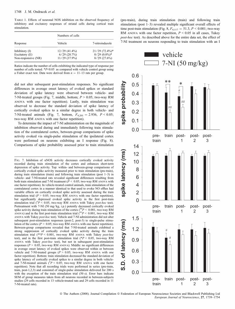
Table 1. Effects of neuronal NOS inhibition on the observed frequency ofinhibitory and excitatory responses of striatal cells during cortical trainstimulation.
Response
Numbers of cells
Vehicle 7-nitroindazole
Inhibitory (I) 12 ⁄ 29 (41.4%) 21 ⁄ 29 (72.4%)*Excitatory (E) 6 ⁄ 29 (20.7%) 0 ⁄ 29 (0.0%)*Non-responsive (NR) 11 ⁄ 29 (37.9%) 8 ⁄ 29 (27.6%)
Ratios indicate the number of cells exhibiting the indicated type of response pernumber of cells tested. *P<0.05 as compared with vehicle control group usinga Fisher exact test. Data were derived from n ¼ 11–13 rats per group.
Fig. 7. Inhibition of nNOS activity decreases cortically evoked activityrecorded during train stimulation of the cortex and enhances short-termdepression of spike activity. Top: within- and between-group comparisons ofcortically evoked spike activity measured prior to train stimulation (pre-train),during train stimulation (train) and following train stimulation (post 1–3) invehicle- and 7-NI-treated rats revealed significant differences resulting fromboth train stimulation and 7-NI treatment (P < 0.05; two-way RM anova withone factor repetition). In vehicle-treated control animals, train stimulation of thecontralateral cortex in a manner identical to that used to evoke NO efflux hadvariable effects on cortically evoked spike activity assessed during the trainstimulation trial (P > 0.05; two-way RM anova with one factor repetition),but significantly depressed evoked spike activity in the first post-trainstimulation trial (#P < 0.05, two-way RM anova with Tukey post-hoc test).Pretreatment with 7-NI (50 mg ⁄ kg, i.p.) potently decreased cortically evokedspike activity during train stimulation of the cortex (##P < 0.001, two-way RManova) and in the first post-train stimulation trial (##P < 0.001, two-way RManova with Tukey post-hoc test). Vehicle and 7-NI administration did not altersubsequent post-stimulation responses (post-2, post-3) to single-pulse stimu-lation of the cortex (P > 0.05; two-way RM anova with one factor repetition).Between-group comparisons revealed that 7-NI-treated animals exhibited astrong suppression of cortically evoked spike activity during the trainstimulation trial (**P < 0.001, two-way RM anova with Tukey post-hoctest), and in the first post-train stimulation trial (*P < 0.05, two-way RManova with Tukey post-hoc test), but not in subsequent post-stimulationresponses (P > 0.05, two-way RM anova). Middle: no significant differencesin average onset latency of evoked spikes were observed within or betweenvehicle and 7-NI-treated groups (P > 0.05; two-way RM anova with onefactor repetition). Bottom: train stimulation decreased the standard deviation ofspike latency of cortically evoked spikes to a similar degree in both vehicle-and 7-NI-treated animals (#P < 0.05; two-way RM anova with one factorrepetition). Note that all recording trials were performed in series (pre-train,train, post-1,2,3) and consisted of single-pulse stimulation delivered for 200 swith the exception of the train stimulation trial (50 s). Error bars indicateSEM of group measures taken from all neurons recorded in between-subjectsstudies (29 cells recorded in 13 vehicle-treated rats and 29 cells recorded in 117-NI-treated rats).
1748 J. M. Ondracek et al.
ª The Authors (2008). Journal Compilation ª Federation of European Neuroscience Societies and Blackwell Publishing LtdEuropean Journal of Neuroscience, 27, 1739–1754
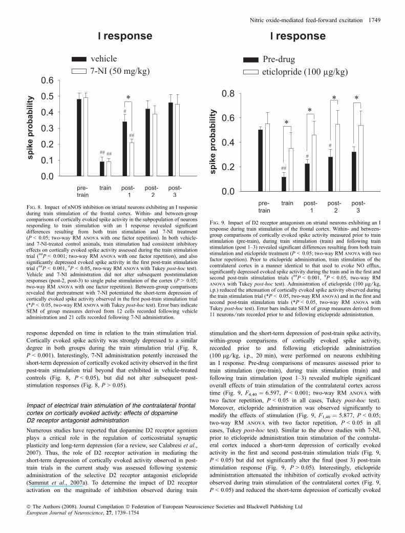
response depended on time in relation to the train stimulation trial.Cortically evoked spike activity was strongly depressed to a similardegree in both groups during the train stimulation trial (Fig. 8,P < 0.001). Interestingly, 7-NI administration potently increased theshort-term depression of cortically evoked activity observed in the firstpost-train stimulation trial beyond that exhibited in vehicle-treatedcontrols (Fig. 8, P < 0.05), but did not alter subsequent post-stimulation responses (Fig. 8, P > 0.05).
Impact of electrical train stimulation of the contralateral frontalcortex on cortically evoked activity: effects of dopamineD2 receptor antagonist administration
Numerous studies have reported that dopamine D2 receptor agonismplays a critical role in the regulation of corticostriatal synapticplasticity and long-term depression (for a review, see Calabresi et al.,2007). Thus, the role of D2 receptor activation in mediating theshort-term depression of cortically evoked activity observed in post-train trials in the current study was assessed following systemicadministration of the selective D2 receptor antagonist eticlopride(Sammut et al., 2007a). To determine the impact of D2 receptoractivation on the magnitude of inhibition observed during train
stimulation and the short-term depression of post-train spike activity,within-group comparisons of cortically evoked spike activity,recorded prior to and following eticlopride administration(100 lg ⁄ kg, i.p., 20 min), were performed on neurons exhibitingan I response. Pre-drug comparisons of measures assessed prior totrain stimulation (pre-train), during train stimulation (train) andfollowing train stimulation (post 1–3) revealed multiple significantoverall effects of train stimulation of the contralateral cortex acrosstime (Fig. 9, F4,40 ¼ 6.597, P < 0.001; two-way RM anova withtwo factor repetition, P < 0.05 in all cases, Tukey post-hoc test).Moreover, eticlopride administration was observed significantly tomodify the effects of stimulation (Fig. 9, F1,40 ¼ 5.877, P < 0.05;two-way RM anova with two factor repetition, P < 0.05 in allcases, Tukey post-hoc test). Similar to the above studies with 7-NI,prior to eticlopride administration train stimulation of the contralat-eral cortex induced a short-term depression of cortically evokedactivity in the first and second post-train stimulation trials (Fig. 9,P < 0.05) but did not significantly alter the final (post 3) post-trainstimulation response (Fig. 9, P > 0.05). Interestingly, eticloprideadministration attenuated the inhibition of cortically evoked activityobserved during train stimulation of the contralateral cortex (Fig. 9,P < 0.05) and reduced the short-term depression of cortically evoked
Fig. 9. Impact of D2 receptor antagonism on striatal neurons exhibiting an Iresponse during train stimulation of the frontal cortex. Within- and between-group comparisons of cortically evoked spike activity measured prior to trainstimulation (pre-train), during train stimulation (train) and following trainstimulation (post 1–3) revealed significant differences resulting from both trainstimulation and eticlopride treatment (P < 0.05; two-way RM anova with twofactor repetition). Prior to eticlopride administration, train stimulation of thecontralateral cortex in a manner identical to that used to evoke NO efflux,significantly depressed evoked spike activity during the train and in the first andsecond post-train stimulation trials (##P < 0.001, #P < 0.05, two-way RManova with Tukey post-hoc test). Administration of eticlopride (100 lg ⁄ kg,i.p.) reduced the attenuation of cortically evoked spike activity observed duringthe train stimulation trial (*P < 0.05, two-way RM anova) and in the first andsecond post-train stimulation trials (*P < 0.05, two-way RM anova withTukey post-hoc test). Error bars indicate SEM of group measures derived from11 neurons ⁄ rats recorded prior to and following eticlopride administration.
Fig. 8. Impact of nNOS inhibition on striatal neurons exhibiting an I responseduring train stimulation of the frontal cortex. Within- and between-groupcomparisons of cortically evoked spike activity in the subpopulation of neuronsresponding to train stimulation with an I response revealed significantdifferences resulting from both train stimulation and 7-NI treatment(P < 0.05; two-way RM anova with one factor repetition). In both vehicle-and 7-NI-treated control animals, train stimulation had consistent inhibitoryeffects on cortically evoked spike activity assessed during the train stimulationtrial (##P < 0.001; two-way RM anova with one factor repetition), and alsosignificantly depressed evoked spike activity in the first post-train stimulationtrial (##P < 0.001, #P < 0.05, two-way RM anova with Tukey post-hoc test).Vehicle and 7-NI administration did not alter subsequent poststimulationresponses (post-2, post-3) to single pulse stimulation of the cortex (P > 0.05;two-way RM anova with one factor repetition). Between-group comparisonsrevealed that pretreatment with 7-NI potentiated the short-term depression ofcortically evoked spike activity observed in the first post-train stimulation trial(*P < 0.05, two-way RM anova with Tukey post-hoc test). Error bars indicateSEM of group measures derived from 12 cells recorded following vehicleadministration and 21 cells recorded following 7-NI administration.
Nitric oxide-mediated feed-forward excitation 1749
ª The Authors (2008). Journal Compilation ª Federation of European Neuroscience Societies and Blackwell Publishing LtdEuropean Journal of Neuroscience, 27, 1739–1754
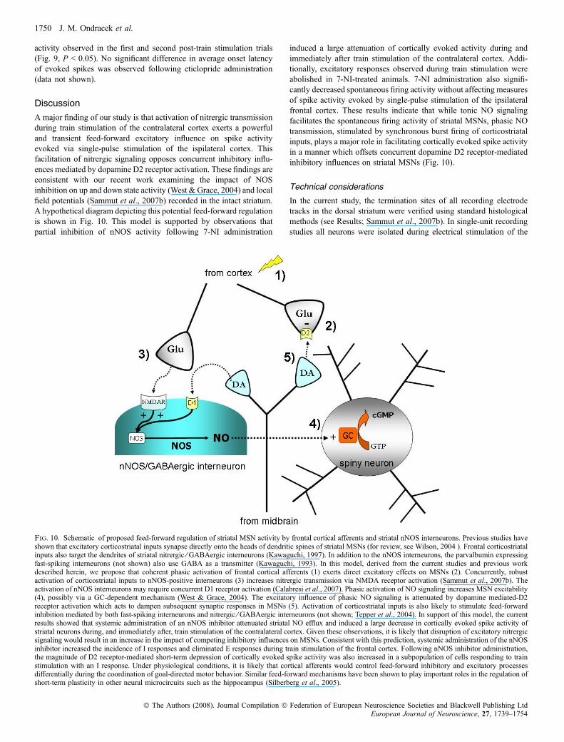
activity observed in the first and second post-train stimulation trials(Fig. 9, P < 0.05). No significant difference in average onset latencyof evoked spikes was observed following eticlopride administration(data not shown).
Discussion
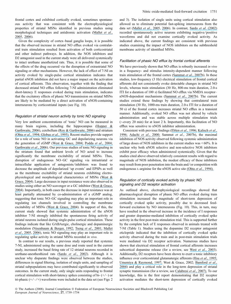
A major finding of our study is that activation of nitrergic transmissionduring train stimulation of the contralateral cortex exerts a powerfuland transient feed-forward excitatory influence on spike activityevoked via single-pulse stimulation of the ispilateral cortex. Thisfacilitation of nitrergic signaling opposes concurrent inhibitory influ-ences mediated by dopamine D2 receptor activation. These findings areconsistent with our recent work examining the impact of NOSinhibition on up and down state activity (West & Grace, 2004) and localfield potentials (Sammut et al., 2007b) recorded in the intact striatum.A hypothetical diagram depicting this potential feed-forward regulationis shown in Fig. 10. This model is supported by observations thatpartial inhibition of nNOS activity following 7-NI administration
induced a large attenuation of cortically evoked activity during andimmediately after train stimulation of the contralateral cortex. Addi-tionally, excitatory responses observed during train stimulation wereabolished in 7-NI-treated animals. 7-NI administration also signifi-cantly decreased spontaneous firing activity without affecting measuresof spike activity evoked by single-pulse stimulation of the ipsilateralfrontal cortex. These results indicate that while tonic NO signalingfacilitates the spontaneous firing activity of striatal MSNs, phasic NOtransmission, stimulated by synchronous burst firing of corticostriatalinputs, plays a major role in facilitating cortically evoked spike activityin a manner which offsets concurrent dopamine D2 receptor-mediatedinhibitory influences on striatal MSNs (Fig. 10).
Technical considerations
In the current study, the termination sites of all recording electrodetracks in the dorsal striatum were verified using standard histologicalmethods (see Results; Sammut et al., 2007b). In single-unit recordingstudies all neurons were isolated during electrical stimulation of the
Fig. 10. Schematic of proposed feed-forward regulation of striatal MSN activity by frontal cortical afferents and striatal nNOS interneurons. Previous studies haveshown that excitatory corticostriatal inputs synapse directly onto the heads of dendritic spines of striatal MSNs (for review, see Wilson, 2004 ). Frontal corticostriatalinputs also target the dendrites of striatal nitrergic ⁄ GABAergic interneurons (Kawaguchi, 1997). In addition to the nNOS interneurons, the parvalbumin expressingfast-spiking interneurons (not shown) also use GABA as a transmitter (Kawaguchi, 1993). In this model, derived from the current studies and previous workdescribed herein, we propose that coherent phasic activation of frontal cortical afferents (1) exerts direct excitatory effects on MSNs (2). Concurrently, robustactivation of corticostriatal inputs to nNOS-positive interneurons (3) increases nitrergic transmission via NMDA receptor activation (Sammut et al., 2007b). Theactivation of nNOS interneurons may require concurrent D1 receptor activation (Calabresi et al., 2007). Phasic activation of NO signaling increases MSN excitability(4), possibly via a GC-dependent mechanism (West & Grace, 2004). The excitatory influence of phasic NO signaling is attenuated by dopamine mediated-D2receptor activation which acts to dampen subsequent synaptic responses in MSNs (5). Activation of corticostriatal inputs is also likely to stimulate feed-forwardinhibition mediated by both fast-spiking interneurons and nitrergic ⁄ GABAergic interneurons (not shown; Tepper et al., 2004). In support of this model, the currentresults showed that systemic administration of an nNOS inhibitor attenuated striatal NO efflux and induced a large decrease in cortically evoked spike activity ofstriatal neurons during, and immediately after, train stimulation of the contralateral cortex. Given these observations, it is likely that disruption of excitatory nitrergicsignaling would result in an increase in the impact of competing inhibitory influences on MSNs. Consistent with this prediction, systemic administration of the nNOSinhibitor increased the incidence of I responses and eliminated E responses during train stimulation of the frontal cortex. Following nNOS inhibitor administration,the magnitude of D2 receptor-mediated short-term depression of cortically evoked spike activity was also increased in a subpopulation of cells responding to trainstimulation with an I response. Under physiological conditions, it is likely that cortical afferents would control feed-forward inhibitory and excitatory processesdifferentially during the coordination of goal-directed motor behavior. Similar feed-forward mechanisms have been shown to play important roles in the regulation ofshort-term plasticity in other neural microcircuits such as the hippocampus (Silberberg et al., 2005).
1750 J. M. Ondracek et al.
ª The Authors (2008). Journal Compilation ª Federation of European Neuroscience Societies and Blackwell Publishing LtdEuropean Journal of Neuroscience, 27, 1739–1754
frontal cortex and exhibited cortically evoked, sometimes spontane-ous activity that was consistent with the electrophysiologicalproperties of striatal MSNs identified in previous studies usingmorphological techniques and antidromic activation (Mallet et al.,2005, 2006).
Given the complexity of cortex–basal ganglia loops, it is possiblethat the observed increase in striatal NO efflux evoked via contralat-eral train stimulation resulted from activation of both corticostriataland other indirect pathways. In addition, the NOS inhibitors andD2 antagonist used in the current study were all delivered systemicallyto intact urethane anesthetized rats. Thus, it is possible that some ofthe effects of the drug occurred via the disruption of extrastriatal NOsignaling or other mechanisms. However, the lack of effect of 7-NI onactivity evoked by single-pulse cortical stimulation indicates thatpartial nNOS inhibition did not have a major impact on the activationof cortical afferents. This observation, together with the finding thatdecreased striatal NO efflux following 7-NI administration eliminatedshort-latency E responses evoked during train stimulation, indicatesthat the excitatory effects of phasic NO transmission on striatal MSNsare likely to be mediated by a direct activation of nNOS-containinginterneurons by corticostriatal inputs (see Fig. 10).
Regulation of striatal neuron activity by tonic NO signaling
Very low ambient concentrations of ‘tonic’ NO can be measured inmany brain regions, including the hippocampus (Hopper &Garthwaite, 2006), cerebellum (Roy & Garthwaite, 2006) and striatum(Ohta et al., 1994; Globus et al., 1995). Recent studies provide supportfor a role of tonic NO in activating GC and depolarizing neurons viathe generation of cGMP (West & Grace, 2004; Podda et al., 2004;Garthwaite et al., 2006). Our previous studies of tonic NO signaling inthe striatum found that ambient levels of NO acted to increasesignificantly the membrane excitability of striatal MSNs. Thus,disruption of endogenous NO–GC signaling via intrastriatal orintracellular application of antagonists ⁄ inhibitors was found todecrease the amplitude of depolarized ‘up events’ and EPSPs as wellas the membrane excitability of striatal neurons exhibiting electro-physiological and morphological characteristics of MSNs (West &Grace, 2004). Large decreases in input resistance were also observed instudies using either an NO scavenger or a GC inhibitor (West & Grace,2004). Importantly, in both cases the decrease in input resistance was atleast partially attenuated by co-administration of a cGMP analog,suggesting that tonic NO–GC signaling may play an important role inregulating ion channels involved in controlling the membraneexcitability of MSNs (West & Grace, 2004). In support of this, thecurrent study showed that systemic administration of the nNOSinhibitor 7-NI strongly inhibited the spontaneous firing activity ofstriatal neurons isolated during single-pulse cortical stimulation. Thesefindings indicate that like GABAergic, cholinergic and dopaminergicmodulation (Nisenbaum & Berger, 1992; Tseng et al., 2001; Malletet al., 2005, 2006), tonic NO signaling may play an important role inregulating spike activity in striatal MSNs recorded in vivo.
In contrast to our results, a previous study reported that systemic7-NI, administered using the same dose and route used in the currentstudy, increased the basal firing rate of 12 striatal neurons recorded inurethane-anesthesthized rats (Sardo et al., 2002). Although it isunclear why disparate findings were observed between the studies,differences in signal filtering, isolation of single units, and sampling ofstriatal subpopulations may have contributed significantly to observedoutcomes. In the current study, only single units responding to frontalcortical stimulation with short-latency spikes consisting of bi- (+ ⁄ –) ortri-phasic (+ ⁄ – ⁄ +) waveforms were included in the data set (see Figs 2
and 3). The isolation of single units using cortical stimulation alsoallowed us to eliminate potential fast-spiking interneurons from thedata set (Mallet et al., 2005, 2006). In contrast, Sardo et al. (2002)recorded spontaneously active neurons exhibiting negative ⁄ positivewaveforms and did not examine cortically evoked activity. Asindicated above, the current findings are consistent with previousstudies examining the impact of NOS inhibitors on the subthresholdmembrane activity of identified MSNs.
Facilitation of phasic NO efflux by frontal cortical afferents
We have previously shown that NO efflux is robustly increased in vivoin a frequency- and stimulus intensity-dependent manner followingtrain stimulation of the frontal cortex (Sammut et al., 2007b). In thesestudies, low-frequency (3 Hz) electrical stimulation of frontal corticalafferents did not consistently evoke detectable changes in striatal NOlevels, whereas train stimulation (30 Hz, 800-ms train duration, 2.0-sITI for a duration of 100 s) facilitated NO efflux via NMDA receptor–nNOS-dependent mechanisms (Sammut et al., 2007b). The currentstudies extend these findings by showing that contralateral trainstimulation (30 Hz, 1000-ms train duration, 2.0-s ITI for a duration of50 s) of the frontal cortex increases striatal NO efflux in a transientmanner. Additionally, evoked NO efflux was not altered by vehicleadministration and was stable across multiple stimulation trials(�every 20 min) for at least 2 h. Importantly, this facilitation of NOefflux was sensitive to nNOS inhibitor administration.Consistent with previous findings (Eblen et al., 1996; Kalisch et al.,
1996; Adachi et al., 2000; Sammut et al., 2007b), the maximalinhibition of NOS activity observed following systemic administrationof large doses of NOS inhibitors in the current studies was < 60%. It isunclear why both nNOS selective and non-selective NOS inhibitorsexhibit poor efficacy when administered systemically. Given that thestudies cited above observed relatively consistent results with regard tomagnitude of NOS inhibition, the modest efficacy of these inhibitorsmay result from poor penetration into the brain and ⁄ or competition withendogenous l-arginine for the nNOS active site (Ohta et al., 1994).
Regulation of cortically evoked activity by phasic NOsignaling and D2 receptor activation
As outlined above, electrophysiological recordings showed thatpharmacological disruption of phasic NO efflux evoked during trainstimulation increased the magnitude of short-term depression ofcortically evoked spike activity, possibly due to decreased feed-forward excitation by NO interneurons (Fig. 10). This, in turn, mayhave resulted in the observed increase in the incidence of I responsesand greater dopamine-mediated inhibition of cortically evoked spikeactivity in the first post-train stimulation trial. This is supported furtherby the complete lack of E responses observed in animals treated with7-NI (Table 1). Studies using the dopamine D2 receptor antagonisteticlopride indicated that the inhibition of cortically evoked spikeactivity observed during the train and in post-train stimulation trialswere mediated via D2 receptor activation. Numerous studies haveshown that electrical stimulation of frontal cortical afferents increasessubcortical dopamine release (for a review, see West et al., 2003).Additionally, D2 receptors have been shown to exert a tonic inhibitoryinfluence over corticostriatal glutamatergic afferents (Hsu et al., 1995;Umemiya & Raymond, 1997; West & Grace, 2002; Bamford et al.,2004) and play a critical role in the long-term depression of striatalsynaptic transmission (for a review, see Calabresi et al., 2007). To ourknowledge, this is the first report demonstrating that D2 receptoractivation mediates the short-term depression of cortically evoked
Nitric oxide-mediated feed-forward excitation 1751
ª The Authors (2008). Journal Compilation ª Federation of European Neuroscience Societies and Blackwell Publishing LtdEuropean Journal of Neuroscience, 27, 1739–1754
spike activity of striatal neurons in vivo. We have shown previouslythat activation of NO synthesis is also down-regulated via a dopamineD2 receptor-dependent mechanism (Sammut et al., 2007a). Thus, D2receptor activation may act in opposition to striatal NO signaling onseveral mechanistic levels.Multiple studies have used either reverse microdialysis or microi-
ontophoresis to examine the impact of locally applied NO generatorsor NOS inhibitors on evoked firing activity of striatal neurons (Westet al., 2002; Di Giovanni et al., 2003; Liu et al., 2005b). Phasicactivation of NO signaling via intrastriatal infusion of theNO-generating compound SNAP increased the firing rate and burstactivity of striatal neurons under basal conditions and during single-pulse electrical stimulation of the prefrontal cortex (West et al., 2002).Similar to these findings, microiontophoresis of the NO generatorsodium nitroprusside was shown to increase the basal firing rate of 51out of 66 striatal neurons and to potentiate the excitatory effects ofglutamate on a subpopulation of neurons (Liu et al., 2005a). This samestudy reported that microiontophoresis of the NOS inhibitor L-NAMEdecreased the excitatory effects of glutamate on a subpopulation ofneurons (Liu et al., 2005a). In contrast to the above studies,microiontophoresis of the superoxide and NO generator 3-morpholi-nosydnonimine (SIN-1) was shown to decrease glutamate-evokedfiring in 12 of 15 neurons, whereas L-NAME increased the excitatoryeffects of glutamate on striatal neurons (Di Giovanni et al., 2003).Given that SIN-1 generates superoxide concurrently with NO, whichtogether react to form peroxynitrite spontaneously (Hogg et al., 1992),it is possible that the inhibitory effects observed in the later study werenot related to NO generation, but resulted via the actions of anotherreactive oxygen species. It is unclear why these studies found oppositeeffects of L-NAME on glutamate-evoked firing. However, the studyby Liu and colleagues tested the effects of L-NAME on a significantlylarger sample of striatal neurons (28 cells) as compared with that ofDi Giovanni and colleagues (ten cells).The current findings are also consistent with our recent study of the
impact of train (30 Hz) stimulation of the frontal cortex on striatalfunction using dual NO microsensor and local field potentialrecordings (Sammut et al., 2007b). Although stimulation artifactsprohibited measurements of local fields during the train stimulationtrial, we were able to assess the impact of NOS inhibition on activityrecorded in a post-train trial (0–30 s) that is comparable with the post1 stimulation trial (0–200 s) described herein (Sammut et al., 2007b).The results of these studies demonstrated that systemic administrationof the NO synthase ⁄ GC inhibitor methylene blue simultaneouslyattenuated evoked NO efflux and the peak oscillation frequency(within the delta band) of local striatal field potentials recordedimmediately after cortical train stimulation (Sammut et al., 2007b).These findings are consistent with previous studies showing thatMSNs recorded during intrastriatal infusion of a NO scavengerexhibited smaller facilitatory synaptic responses to paired electricalstimuli delivered to the prefrontal cortex as compared with controlneurons (West & Grace, 2004). Taken together, these observationsindicate that glutamatergic transmission activated during corticostriatalcell burst firing may be amplified by NOS interneurons involved insynchronizing striatal network activity and spike generation of MSNsrecorded in intact striatal networks.In contrast to our findings, studies by Calabresi and colleagues
using brain slice preparations mainly predict an inhibitory action(long-term depression) of the NO–GC signaling pathway on thesynaptic activity of striatal MSNs (Calabresi et al., 1999a,b, 2000).It is possible that the apparent differences between these studiesperformed in vitro and our studies performed in the intact animal arerelated to differences in the individual network properties of neurons
within these preparations (Pare et al., 1998). Interestingly, stimulationprotocols that produce long-term depression in vitro have been shownto produce long-term potentiation of corticostriatal neurotransmissionin vivo (Charpier & Deniau, 1997), possibly due to the greater removalof the voltage-dependent magnesium block of NMDA receptors andhigher levels of basal excitability inherent to the intact animal. Lastly,long-term potentiation induced by tetanic stimulation of corticostriatalpathways is reported to be significantly reduced in eNOS– ⁄ – mice orfollowing treatment with a non-selective NOS inhibitor (Doreuleeet al., 2003). Thus, in addition to the preparation, the role played byNO in striatal synaptic plasticity may depend on the stimulationprotocol and the recruitment of different NOS isoforms.
Functional implications
The facilitation of corticostriatal throughput by phasic NO signalingmay represent a mechanism by which the impact of robust cortico-striatal signaling on MSNs is strengthened by feed-forward striatalintegration (Fig. 10). Similar interneuron-dependent feed-forwardexcitatory processes have been demonstrated in the hippocampus andspinal cord (for a review, see Silberberg et al., 2005) as well as thebasolateral amygdala (Woodruff et al., 2006). Support for this novelnetwork property of striatal NOS interneurons comes from studiesshowing that train stimulation of corticostriatal pathways facilitateselectrotonic coupling between MSNs in striatal slice preparationsvia an NO-dependent process (O’Donnell & Grace, 1997). AnNO-mediated synchronization of electrotonic coupling, together withfacilitation of glutamatergic transmission, may be important forpromoting and ⁄ or synchronizing up-state activity and spike dischargeof a network of functionally related MSNs. Consistent with theseobservations, studies described above (West & Grace, 2004) revealedthat inhibition of NOS and GC depressed the amplitude of sponta-neous up events known to be driven by glutamateric inputs(O’Donnell & Grace, 1995; Wilson & Kawaguchi, 1996). Our currentfindings, together with the above observations, suggest that adysfunction in striatal nitrergic neurotransmission could disrupt theoperation of the basal ganglia by altering corticostriatal informationprocessing and short-term plasticity in MSNs and result in inactivationof striatal output circuits. In support of this, a recent study utilizingquantitative EEG methods has shown that inhibition of neuronal NOSactivity has a major impact on electrical activity patterns withincorticostriatopallidal circuits, indicating that NO signaling plays animportant role in modulating efferent pathways to the globus pallidus(Ferraro et al., 2002). Future studies examining the impact of tonic andphasic NO transmission on identified striatonigral and striatopallidalneurons should help to clarify the neuromodulatory impact of thisimportant neural messenger on striatal function.
Acknowlegdements
We thank Drs Kuei Tseng and Grace (Beth) Stutzmann for their valuablecomments regarding this manuscript. This work was supported by the ChicagoMedical School, Parkinson’s Disease Foundation and United States PublicHealth grant NS 047452 (A.R.W.).
Abbreviations
7-NI, 7-nitroindazole; E, excitatory; FHC, Frederick Haer Company; GC,guanylyl cyclase; I, inhibitory; ITI, intertrain interval; MSNs, medium spinyneurons; L-NAME, NG-nitro-l-arginine methyl ester hydrochloride; NMDA,N-methyl-d-aspartame; nNOS, neuronal nitric oxide synthase; NO, nitric oxide;NOS, NO synthase; PBS, phosphate-buffered saline; PSTH, peri-stimulus timehistograms; SNAP, (+ ⁄ –)S-nitroso-N-acetyl-penicillamine; WPI, World Preci-sion Instruments.
1752 J. M. Ondracek et al.
ª The Authors (2008). Journal Compilation ª Federation of European Neuroscience Societies and Blackwell Publishing LtdEuropean Journal of Neuroscience, 27, 1739–1754
References
Adachi, N., Lei, B., Sountani, M. & Arai, T. (2000) Different roles of neuronaland endothelial nitric oxide synthases on ischemic nitric oxide production ingerbil striatum. Neurosci. Lett., 288, 151–154.
Alderton, W.K., Cooper, C.E. & Knowles, R.G. (2001) Nitric oxide synthases:structure, function and inhibition. Biochem. J., 357, 593–615.
Ayers, N.A., Kapas, L. & Krueger, J.M. (1997) The inhibitory effects ofN-omega-nitro-L-arginine methyl ester on nitric oxide synthase activity varyamong brain regions in vivo but not in vitro. Neurochem. Res., 22, 81–86.
Bamford, N.S., Zhang, H., Schmitz, Y., Wu, N.P., Cepeda, C., Levine, M.S.,Schmauss, C., Zakharenko, S.S., Zablow, L. & Sulzer, D. (2004) Hetero-synaptic dopamine neurotransmission selects sets of corticostriatal terminals.Neuron, 42, 653–663.
Berretta, S., Parthasarathy, H.B. & Graybiel, A.M. (1997) Local release ofGABAergic inhibition in the motor cortex induces immediate-early geneexpression in indirect pathway neurons of the striatum. J. Neurosci., 17,4752–4763.
Boehning, D. & Snyder, S.H. (2003) Novel neural modulators. Ann. Rev.Neurosci., 26, 105–131.
Bolam, J.P., Hanley, J.J., Booth, P.A. & Bevan, M.D. (2000) Synapticorganisation of the basal ganglia. J. Anat., 196, 527–542.
Calabresi, P., Centonze, D., Gubellini, P., Marfia, G.A. & Bernardi, G. (1999a)Glutamate-triggered events inducing corticostriatal long-term depression.J. Neurosci., 19, 6102–6110.
Calabresi, P., Gubellini, P., Centonze, D., Picconi, B., Bernardi, G., Chergui,K., Svenningsson, P., Fienberg, A.A. & Greengard, P. (2000) Dopamine andcAMP-regulated phosphoprotein 32 kDa controls both striatal long-termdepression and long-term potentiation, opposing forms of synaptic plasticity.J. Neurosci., 20, 8443–8451.
Calabresi, P., Gubellini, P., Centonze, D., Sancesario, G., Morello, M., Giorgi,M., Pisani, A. & Bernardi, G. (1999b) A critical role of the nitricoxide ⁄ cGMP pathway in corticostriatal long-term depression. J. Neurosci.,19, 2489–2499.
Calabresi, P., Picconi, B., Tozzi, A. & Di Filippo, M. (2007) Dopamine-mediated regulation of corticostriatal synaptic plasticity. Trends Neurosci.,30, 211–219.
Centonze, D., Grande, C., Usiello, A., Gubellini, P., Erbs, E., Martin, A.B.,Pisani, A., Tognazzi, N., Bernardi, G., Moratalla, R., Borrelli, E. &Calabresi, P. (2003) Receptor subtypes involved in the presynaptic andpostsynaptic actions of dopamine on striatal interneurons. J. Neurosci., 23,6245.
Charpier, S. & Deniau, J.M. (1997) In vivo activity-dependent plasticity atcortico-striatal connections: evidence for physiological long-term potentia-tion. Proc. Natl Acad. Sci. USA, 94, 7036–7040.
Christopherson, K.S., Hillier, B.J., Lim, W.A. & Bredt, D.S. (1999) PSD-95assembles a ternary complex with the N-methyl-D-aspartic acid receptor anda bivalent neuronal NO synthase PDZ. domain. J. Biol. Chem., 274, 27467–27473.
Cowan, R.L. & Wilson, C.J. (1994) Spontaneous firing patterns and axonalprojections of single corticostriatal neurons in the rat medial agranularcortex. J. Neurophysiol., 71, 17–32.
Crespi, F., Campagnola, M., Neudeck, A., McMillan, K., Rossetti, Z.,Pastorino, A., Garbin, U., Fratta-Pasini, A., Reggiani, A., Gaviraghi, G. &Cominacini, L. (2001) Can voltammetry measure nitrogen monoxide (NO)and ⁄ or nitrites? J. Neurosci. Methods, 109, 59–70.
Crespi, F. & Rossetti, Z.L. (2004) Pulse of nitric oxide release in response toactivation of N-methyl-D-aspartate receptors in the rat striatum: rapiddesensitization, inhibition by receptor antagonists, and potentiation byglycine. J. Pharmacol. Exp. Ther., 309, 462–468.
Del Bel, E.A., Guimaraues, F.S., Bermudez-Echeverry, M., Gomes, M.Z.,Schiaveto-de-Souza, A., Padovan-Neto, F.E., Tumas, V., Barion-Cavalcanti,A.P., Lazzarini, M., Nucci-da-Silva, L.P. & de Paula-Souza, D. (2005) Roleof nitric oxide on motor behavior. Cell. Mol. Neurobiol., 25, 371–392.
Di Giovanni, G., Ferraro, G., Sardo, P., Galati, S., Esposito, E. & La Grutta, V.(2003) Nitric oxide modulates striatal neuronal activity via solubleguanylyl cyclase: an in vivo microiontophoretic study in rats. Synapse,48, 100–107.
Doreulee, N., Sergeeva, O.A., Yanovsky, Y., Chepkova, A.N., Selbach, O.,Godecke, A., Schrader, J. & Helmut, L. (2003) Cortico-striatal synapticplasticity in endothelial nitric oxide synthase deficient mice. Brain Res., 964,159–163.
East, S.J., Parry-Jones, A. & Brotchie, J.M. (1996) Ionotropic glutamatereceptors and nitric oxide synthesis in the rat striatum. Neuroreport, 8,71–75.
Eblen, F., Loschmann, P.A., Wullner, U., Turski, L. & Klockgether, T. (1996)Effects of 7-nitroindazole, NG-nitro-L-arginine, and D-CPPene on harma-line-induced postural tremor, N-methyl-D-aspartate-induced seizures, andlisuride-induced rotations in rats with nigral 6-hydroxydopamine lesions.Eur. J. Pharmacol., 299, 9–16.
Ferraro, G., Sardo, P., Di Giovanni, G., Galati, S. & La Grutta, V. (2002)Nitric oxide and cortico-striato-pallidal motor circuitry: quantitative EEGanalysis of surface and depth recordings. Neurosci. Res. Commun., 30,121–133.
Floresco, S.B., Blaha, C.D., Yang, C.R. & Phillips, A.G. (2001) Modulation ofhippocampal and amygdalar-evoked activity of nucleus accumbens neuronsby dopamine. Cellular mechanisms of input selection. J. Neurosci., 21,2851–2860.
Floresco, S.B., West, A.R., Ash, B., Moore, H. & Grace, A.A. (2003) Afferentmodulation of dopamine neuron firing differentially regulates tonic andphasic dopamine transmission. Nat. Neurosci., 6, 968–973.
French, S.J., Ritson, G.P., Hidaka, S. & Totterdell, S. (2005) Nucleusaccumbens nitric oxide immunoreactive interneurons receive nitric oxide andventral subicular afferents in rats. Neuroscience, 135, 121–131.
Garthwaite, G., Bartus, K., Malcolm, D., Goodwin, D., Kollb-Sielecka, M.,Dooldeniya, C. & Garthwaite, J. (2006) Signaling from blood vessels to CNSaxons through nitric oxide. J. Neurosci., 26, 7730–7740.
Garthwaite, J. & Boulton, C.L. (1995) Nitric oxide signaling in the centralnervous system. Ann. Rev. Physiol., 57, 683–706.
Globus, M.Y., Prado, R. & Busto, R. (1995) Ischemia-induced changes inextracellular levels of striatal cyclic GMP: role of nitric oxide. Neuroreport,6, 1909–1912.
Hirabayashi, H., Takizawa, S., Fukuyama, N., Nakazawa, H. & Shinohara, Y.(2001) N-methyl-D-aspartate receptor antagonist reduces nitrotyrosineformation in caudate-putamen in rat focal cerebral ischemia-reperfusion.Neurosci. Lett., 299, 159–161.
Hogg, N., Darley-Usmar, V.M., Wilson, M.T. & Moncada, S. (1992)Production of hydroxyl radicals from the simultaneous generation ofsuperoxide and nitric oxide. Biochem. J., 281, 419–424.
Hopper, R.A. & Garthwaite, J. (2006) Tonic and phasic nitric oxide signals inhippocampal long-term potentiation. J. Neurosci., 26, 11513–11521.
Hsu, K.-S., Huang, C.-C., Yang, C.-H. & Gean, P.-W. (1995) Presynaptic D2
dopaminergic receptors mediate inhibition of excitatory synaptic transmis-sion in rat neostriatum. Brain Res., 690, 264–268.
Iravani, M.M., Millar, J. & Kruk, Z.L. (1998) Differential release of dopamineby nitric oxide in subregions of rat caudate putamen slices. J. Neurochem.,71, 1969–1977.
Jodo, E., Suzuki, Y., Katayama, T., Hoshino, K.Y., Takeuchi, S., Niwa, S. &Kayama, Y. (2005) Activation of medial prefrontal cortex by phencyclidineis mediated via a hippocampo-prefrontal pathway. Cereb. Cortex, 15,663–669.
Kalisch, B.E., Connop, B.P., Jhamandas, K., Beninger, R.J. & Boegman, R.J.(1996) Differential action of 7-nitro indazole on rat brain nitric oxidesynthase. Neurosci. Lett., 219, 75–78.
Kawaguchi, Y. (1993) Physiological, morphological, and histochemicalcharacterization of three classes of interneurons in rat neostriatum.J. Neurosci., 13, 4908–4923.
Kawaguchi, Y. (1997) Neostriatal cell subtypes and their functional roles.Neurosci. Res., 27, 1–8.
Kendrick, K.M., Guevara-Guzman, R., de la Riva, C., Christensen, J.,Ostergaard, K. & Emson, P.C. (1996) NMDA and kainate-evoked releaseof nitric oxide and classical transmitters in the rat striatum: in vivo evidencethat nitric oxide may play a neuroprotective role. Eur. J. Neurosci., 8, 2619–2634.
Kharazia, V.N., Schmidt, H.H. & Weinberg, R.J. (1994) Type I nitric oxidesynthase fully accounts for NADPH-diaphorase in rat striatum, but notcortex. Neuroscience, 62, 983–987.
Koos, T. & Tepper, J.M. (1999) Inhibitory control of neostriatal projectionneurons by GABAergic interneurons. Nat. Neurosci., 2, 467–472.
Kubota, Y., Mikawa, S. & Kawaguchi, Y. (1993) Neostriatal GABAergicinterneurones contain NOS, calretinin or parvalbumin. Neuroreport, 5, 205–208.
Liu, C.N., Liu, X., Gao, D. & Li, S. (2005a) Effects of SNP, GLU and GABAon the neuronal activity of striatum nucleus in rats. Pharmacol. Res., 51,547–551.
Liu, D., Sammut, S. & West, A.R. (2005b) Nitric oxide signaling modulates theresponsiveness of striatal medium spiny neurons to electrical stimulation ofthe substantia nigra. Striatal nitrergic signaling. In: Bolam, J.P., Ingham, C.A.& Magill, P.J. (Eds), The Basal Ganglia VIII. Springer Science and BusinessMedia, New York, pp. 503–512.
Nitric oxide-mediated feed-forward excitation 1753
ª The Authors (2008). Journal Compilation ª Federation of European Neuroscience Societies and Blackwell Publishing LtdEuropean Journal of Neuroscience, 27, 1739–1754
Mallet, N., Ballion, B., Le Moine, C. & Gonon, F. (2006) Cortical inputs andGABA interneurons imbalance projection neurons in the striatum ofparkinsonian rats. J. Neurosci., 26, 3875–3884.
Mallet, N., Le Moine, C., Charpier, S. & Gonon, F. (2005) Feedforwardinhibition of projection neurons by fast-spiking GABA interneurons in therat striatum in vivo. J. Neurosci., 25, 3857–3869.
Marin, P., Lafon-Cazal, M. & Bockaert, J. (1992) A nitric oxide synthaseactivity selectively stimulated by NMDA receptors depends on proteinkinase C activation in mouse striatal neurons. Eur. J. Neurosci., 4, 425–432.
Morris, B.J., Simpson, C.S., Mundell, S., Maceachern, K., Johnston, H.M. &Nolan, A.M. (1997) Dynamic changes in NADPH-diaphorase staining reflectactivity of nitric oxide synthase: evidence for a dopaminergic regulation ofstriatal nitric oxide release. Neuropharmacology, 36, 1589–1599.
Nisenbaum, E.S. & Berger, T.W. (1992) Functionally distinct subpopulations ofstriatal neurons are differentially regulated by GABAergic and dopaminergicinputs – I. In vivo analysis. Neuroscience, 48, 561–578.
O’Donnell, P. & Grace, A.A. (1995) Synaptic interactions among excitatoryafferents to nucleus accumbens neurons: Hippocampal gating of prefrontalcortical input. J. Neurosci., 15, 3622–3639.
O’Donnell, P. & Grace, A.A. (1997) Cortical afferents modulate striatal gapjunction permeability via nitric oxide. Neuroscience, 76, 1–5.
Ohta, K., Araki, N., Shibata, M., Hamada, J., Komatsumoto, S., Shimazu, K. &Fukuuchi, Y. (1994) A novel in vivo assay system for consecutivemeasurement of brain nitric oxide production combined with the microdi-alysis technique. Neurosci. Lett., 176, 165–168.
Ohta, K., Rosner, G. & Graf, R. (1997) Nitric oxide generation from sodiumnitroprusside and hydroxylamine in brain. Neuroreport, 8, 2229–2235.
Palmer, R.M., Ashton, D.S. & Moncada, S. (1988) Vascular endothelial cellssynthesize nitric oxide from L-arginine. Nature, 333, 664–666.
Pare, D., Shink, E., Gaudreau, H., Destexhe, A. & Lang, E.J. (1998) Impact ofspontaneous synaptic activity on the resting properties of cat neocorticalpyramidal neurons in vivo. J. Neurophysiol., 79, 1450–1460.
Paxinos, G. & Watson, C. (1986) The Rat Brain in Stereotaxic Coordinates.Academic Press, New York.
Plenz, D. & Kitai, S.T. (1998) Up and down states in striatal medium spinyneurons simultaneously recorded with spontaneous activity in fast-spikinginterneurons studied in cortex-striatum-substantia nigra organotypic cultures.J. Neurosci., 18, 266–283.
Podda, M.V., Marcocci, M.E., Oggiano, L., D’Ascenzo, M., Tolu, E., Palamara,A.T., Azzena, G.B. & Grassi, C. (2004) Nitric oxide increases thespontaneous firing rate of rat medial vestibular nucleus neurons in vitrovia a cyclic GMP-mediated PKG-independent mechanism. Eur. J. Neurosci.,20, 2124–2132.
Roy, B. & Garthwaite, J. (2006) Nitric oxide activation of guanylyl cyclase incells revisited. Proc. Natl Acad. Sci. USA, 103, 12185–12190.
Salin, P., Kerkerian-Le, G.L., Heidet, V., Epelbaum, J. & Nieoullon, A.(1990) Somatostatin-immunoreactive neurons in the rat striatum: effects ofcorticostriatal and nigrostriatal dopaminergic lesions. Brain Res., 521, 23–32.
Sammut, S., Bray, K.E. & West, A.R. (2007a) Dopamine D(2) receptor-dependent modulation of striatal NO synthase activity. Psychopharmacology(Berl.), 191, 793–803.
Sammut, S., Dec, A., Mitchell, D., Linardakis, J., Ortiguela, M. & West, A.R.(2006) Phasic dopaminergic transmission increases NO efflux in the ratdorsal striatum via a neuronal NOS and a dopamine D(1 ⁄ 5) receptor-dependent mechanism. Neuropsychopharmacology, 31, 493–505.
Sammut, S., Park, D.J. & West, A.R. (2007b) Frontal cortical afferents facilitatestriatal nitric oxide transmission in vivo via a NMDA receptor and neuronalNOS-dependent mechanism. J. Neurochem., doi:10.1111/j.1471-4159.2007.04811.x.
Sardo, P., Ferraro, G., Di Giovanni, G., Galati, S. & La Grutta, V. (2002)Inhibition of nitric oxide synthase influences the activity of striatal neuronsin the rat. Neurosci. Lett., 325, 179–182.
Silberberg, G., Grillner, S., LeBeau, F.E.N., Maex, R. & Markram, H. (2005)Synaptic pathways in neural microcircuits. Trends Neurosci., 28, 541–551.
Steriade, M. (2001) Impact of network activities on neuronal properties incorticothalamic systems. J. Neurophysiol., 86, 1–39.
Tepper, J.M., Koos, T. & Wilson, C.J. (2004) GABAergic microcircuits in theneostriatum. Trends Neurosci., 27, 662–669.
Tseng, K.Y., Kasanetz, F., Kargieman, L., Riquelme, L.A. & Murer, M.G.(2001) Cortical slow oscillatory activity is reflected in the membranepotential and spike trains of striatal neurons in rats with chronic nigrostriatallesions. J. Neurosci., 21, 6430–6439.
Tseng, K.Y., Riquelme, L.A., Belforte, J.E., Pazo, J.H. & Murer, M.G. (2000)Substantia nigra pars reticulata units in 6-hydroxydopamine-lesioned rats:responses to striatal D2 dopamine receptor stimulation and subthalamiclesions. Eur. J. Neurosci., 12, 247–256.
Umemiya, M. & Raymond, L.A. (1997) Dopaminergic modulation ofexcitatory postsynaptic currents in rat neostriatal neurons. J. Neurophys.,78, 1248–1255.
Vuillet, J., Kerkerian, L., Kachidian, P., Bosler, O. & Nieoullon, A. (1989)Ultrastructural correlates of functional relationships between nigral dopami-nergic or cortical afferent fibers and neuropeptide Y-containing neurons inthe rat striatum. Neurosci. Lett., 100, 99–104.
West, A.R., Floresco, S.B., Charara, A., Rosenkranz, J.A. & Grace, A.A.(2003) Electrophysiological interactions between striatal glutamatergic anddopaminergic systems. Ann. NY Acad. Sci., 1003, 53–74.
West, A.R., Galloway, M.P. & Grace, A.A. (2002) Regulation of striataldopamine neurotransmission by nitric oxide: effector pathways and signalingmechanisms. Synapse, 44, 227–245.
West, A.R. & Grace, A.A. (2000) Striatal nitric oxide signaling regulates theneuronal activity of midbrain dopamine neurons in vivo. J. Neurophysiol.,83, 1796–1808.
West, A.R. & Grace, A.A. (2002) Opposite influences of endogenous dopamineD1 and D2 receptor activation on activity states and electrophysiologicalproperties of striatal neurons: studies combining in vivo intracellularrecordings and reverse microdialysis. J. Neurosci., 22, 294–304.
West, A.R. & Grace, A.A. (2004) The nitric oxide-guanylyl cyclase signalingpathway modulates membrane activity states and electrophysiologicalproperties of striatal medium spiny neurons recorded in vivo. J. Neurosci.,24, 1924–1935.
Wilson, C.J. (2004) Basal ganglia. In: Shepherd, G. (Ed.), The SynapticOrganization of the Brain. Oxford University Press, Oxford, pp. 361–414.
Wilson, C.J., Chang, H.T. & Kitai, S.T. (1990) Firing patterns and synapticpotentials of identified giant aspiny interneurons in the rat neostriatum.J. Neurosci., 10, 508–519.
Wilson, C.J. & Kawaguchi, Y. (1996) The origins of two-state spontaneousmembrane potential fluctuations of neostriatal spiny neurons. J. Neurosci.,16, 2397–2410.
Woodruff, A.R., Monyer, H. & Sah, P. (2006) GABAergic excitation in thebasolateral amygdala. J. Neurosci., 26, 11881–11887.
Zhang, X. (2004) Real time and in vivo monitoring of nitric oxide byelectrochemical sensors-from dream to reality. Front. Biosci., 9, 3434–3446.
1754 J. M. Ondracek et al.
ª The Authors (2008). Journal Compilation ª Federation of European Neuroscience Societies and Blackwell Publishing LtdEuropean Journal of Neuroscience, 27, 1739–1754
Related Documents


![[18F]Fluorodopa PETshows striatal dopaminergic dysfunction ...](https://static.cupdf.com/doc/110x72/628e71a806be7c7a267428b6/18ffluorodopa-petshows-striatal-dopaminergic-dysfunction-.jpg)

![KONSEP TERBARU PENATALAKSANAAN PENYAKIT PARKINSON · pengurangan hilangnya striatal {[123]I}beta-CIT uptake, suatu petunjuk degenerasi neuron Glial-cell-line-derived neurotrophic](https://static.cupdf.com/doc/110x72/5e2791bf65e55267002b6183/konsep-terbaru-penatalaksanaan-penyakit-parkinson-pengurangan-hilangnya-striatal.jpg)