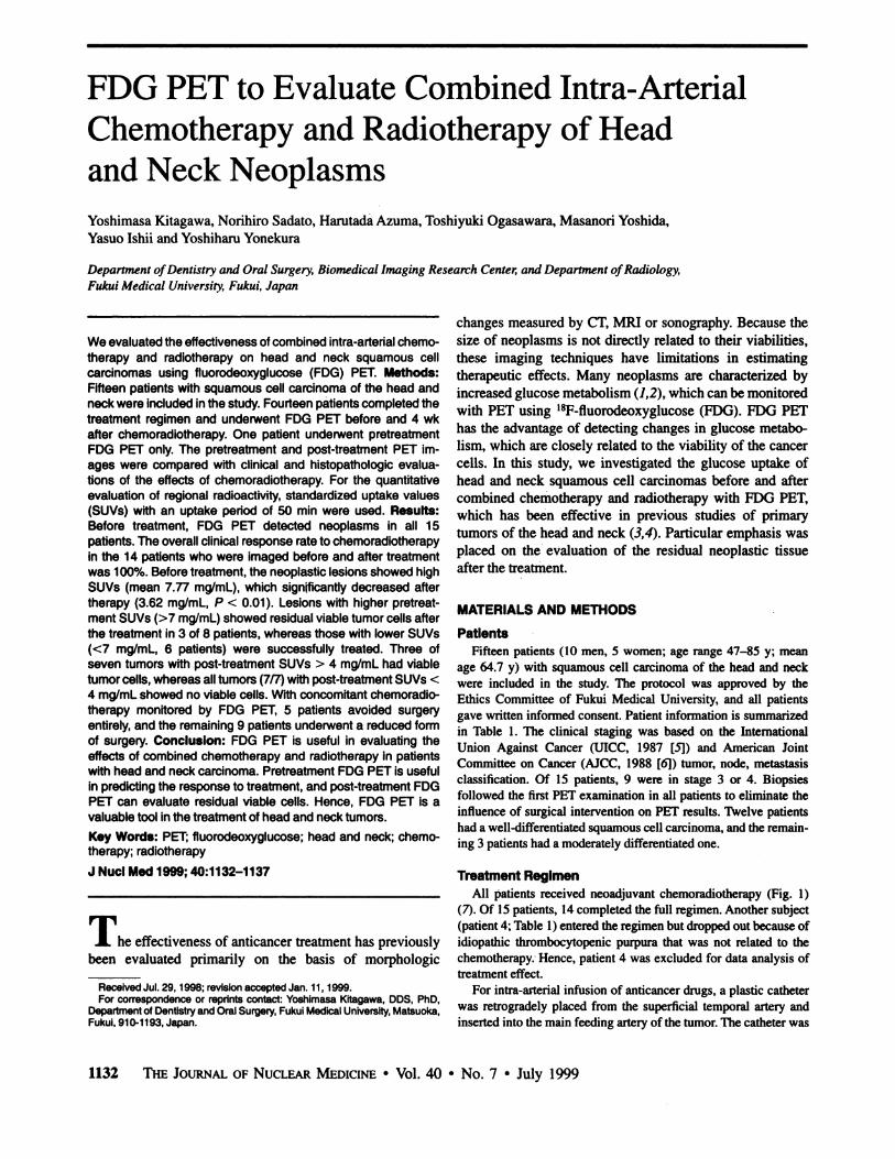
FDG PET to Evaluate Combined Intra-Arterial Chemotherapy and Radiotherapy of Head and Neck Neoplasms Yoshimasa Kitagawa, Norihiro Sadato, Harutada Azuma, Toshiyuki Ogasawara, Masanori Yoshida, Yasuo Ishii and Yoshiharu Yonekura Department of Dentistry and Oral Surgery, Biomedicai Imaging Research Center, and Department of Radiology, Fukui Medical University, Fukui, Japan We evaluated the effectiveness of combined ¡ntra-arterial chemo therapy and radiotherapy on head and neck squamous cell carcinomas using fluorodeoxyglucose (FDG) PET. Methods: Fifteen patients with squamous cell carcinoma of the head and neck were included in the study. Fourteen patients completed the treatment regimen and underwent FDG PET before and 4 wk after chemoradiotherapy. One patient underwent pretreatment FDG PET only. The pretreatment and post-treatment PET im ages were compared with clinical and histopathologic evalua tions of the effects of chemoradiotherapy. For the quantitative evaluation of regional radioactivity, standardized uptake values (SUVs) with an uptake period of 50 min were used. Results: Before treatment, FDG PET detected neoplasms in all 15 patients. The overall clinical response rate to chemoradiotherapy in the 14 patients who were imaged before and after treatment was 100%. Before treatment, the neoplastic lesions showed high SUVs (mean 7.77 mg/mL), which significantly decreased after therapy (3.62 mg/mL, P < 0.01). Lesions with higher pretreat ment SUVs (>7 mg/mL) showed residual viable tumor cells after the treatment in 3 of 8 patients, whereas those with lower SUVs (<7 mg/mL, 6 patients) were successfully treated. Three of seven tumors with post-treatment SUVs > 4 mg/mL had viable tumor cells, whereas all tumors (7/7) with post-treatment SUVs < 4 mg/mL showed no viable cells. With concomitant chemoradio therapy monitored by FDG PET, 5 patients avoided surgery entirely, and the remaining 9 patients underwent a reduced form of surgery. Conclusion: FDG PET is useful in evaluating the effects of combined chemotherapy and radiotherapy in patients with head and neck carcinoma. Pretreatment FDG PET is useful in predicting the response to treatment, and post-treatment FDG PET can evaluate residual viable cells. Hence, FDG PET is a valuable tool in the treatment of head and neck tumors. Key Words: PET; fluorodeoxyglucose;head and neck; chemo therapy; radiotherapy J NucÃ- Med 1999; 40:1132-1137 he effectiveness of anticancer treatment has previously been evaluated primarily on the basis of morphologic Received Jul. 29,1998; revision accepted Jan. 11,1999. For correspondence or reprints contact: Yoshimasa Kitagawa, DOS, PhD, Department of Dentistry and Oral Surgery, Fukui Medical University, Matsuoka, Fukui, 910-1193, Japan. changes measured by CT, MRI or sonography. Because the size of neoplasms is not directly related to their viabilities, these imaging techniques have limitations in estimating therapeutic effects. Many neoplasms are characterized by increased glucose metabolism (7,2), which can be monitored with PET using 18F-fluorodeoxyglucose (FDG). FDG PET has the advantage of detecting changes in glucose metabo lism, which are closely related to the viability of the cancer cells. In this study, we investigated the glucose uptake of head and neck squamous cell carcinomas before and after combined chemotherapy and radiotherapy with FDG PET, which has been effective in previous studies of primary tumors of the head and neck (3,4). Particular emphasis was placed on the evaluation of the residual neoplastic tissue after the treatment. MATERIALS AND METHODS Patients Fifteen patients (10 men, 5 women; age range 47-85 y; mean age 64.7 y) with squamous cell carcinoma of the head and neck were included in the study. The protocol was approved by the Ethics Committee of Fukui Medical University, and all patients gave written informed consent. Patient information is summarized in Table 1. The clinical staging was based on the International Union Against Cancer (UICC, 1987 [5]) and American Joint Committee on Cancer (AJCC, 1988 [6]) tumor, node, metastasis classification. Of 15 patients, 9 were in stage 3 or 4. Biopsies followed the first PET examination in all patients to eliminate the influence of surgical intervention on PET results. Twelve patients had a well-differentiated squamous cell carcinoma, and the remain ing 3 patients had a moderately differentiated one. Treatment Regimen All patients received neoadjuvant chemoradiotherapy (Fig. 1) (7). Of 15 patients, 14 completed the full regimen. Another subject (patient 4; Table 1) entered the regimen but dropped out because of ¡diopathic thrombocytopenic purpura that was not related to the chemotherapy. Hence, patient 4 was excluded for data analysis of treatment effect. For intra-arterial infusion of anticancer drugs, a plastic catheter was retrogradely placed from the superficial temporal artery and inserted into the main feeding artery of the tumor. The catheter was 1132 THEJOURNAL OFNUCLEAR MEDICINE • Vol. 40 • No. 7 • July 1999

Welcome message from author
This document is posted to help you gain knowledge. Please leave a comment to let me know what you think about it! Share it to your friends and learn new things together.
Transcript
FDG PET to Evaluate Combined Intra-ArterialChemotherapy and Radiotherapy of Headand Neck NeoplasmsYoshimasa Kitagawa, Norihiro Sadato, Harutada Azuma, Toshiyuki Ogasawara, Masanori Yoshida,Yasuo Ishii and Yoshiharu Yonekura
Department of Dentistry and Oral Surgery, Biomedicai Imaging Research Center, and Department of Radiology,Fukui Medical University, Fukui, Japan
We evaluated the effectiveness of combined ¡ntra-arterialchemo
therapy and radiotherapy on head and neck squamous cellcarcinomas using fluorodeoxyglucose (FDG) PET. Methods:Fifteen patients with squamous cell carcinoma of the head andneck were included in the study. Fourteen patients completed thetreatment regimen and underwent FDG PET before and 4 wkafter chemoradiotherapy. One patient underwent pretreatmentFDG PET only. The pretreatment and post-treatment PET images were compared with clinical and histopathologic evaluations of the effects of chemoradiotherapy. For the quantitativeevaluation of regional radioactivity, standardized uptake values(SUVs) with an uptake period of 50 min were used. Results:Before treatment, FDG PET detected neoplasms in all 15patients. The overall clinical response rate to chemoradiotherapyin the 14 patients who were imaged before and after treatmentwas 100%. Before treatment, the neoplastic lesions showed highSUVs (mean 7.77 mg/mL), which significantly decreased aftertherapy (3.62 mg/mL, P < 0.01). Lesions with higher pretreatment SUVs (>7 mg/mL) showed residual viable tumor cells afterthe treatment in 3 of 8 patients, whereas those with lower SUVs(<7 mg/mL, 6 patients) were successfully treated. Three ofseven tumors with post-treatment SUVs > 4 mg/mL had viabletumor cells, whereas all tumors (7/7) with post-treatment SUVs <4 mg/mL showed no viable cells. With concomitant chemoradiotherapy monitored by FDG PET, 5 patients avoided surgeryentirely, and the remaining 9 patients underwent a reduced formof surgery. Conclusion: FDG PET is useful in evaluating theeffects of combined chemotherapy and radiotherapy in patientswith head and neck carcinoma. Pretreatment FDG PET is usefulin predicting the response to treatment, and post-treatment FDGPET can evaluate residual viable cells. Hence, FDG PET is avaluable tool in the treatment of head and neck tumors.
Key Words: PET; fluorodeoxyglucose;head and neck; chemotherapy; radiotherapyJ NucÃMed 1999; 40:1132-1137
he effectiveness of anticancer treatment has previouslybeen evaluated primarily on the basis of morphologic
Received Jul. 29,1998; revision accepted Jan. 11,1999.For correspondence or reprints contact: Yoshimasa Kitagawa, DOS, PhD,
Department of Dentistry and Oral Surgery, Fukui Medical University, Matsuoka,Fukui, 910-1193, Japan.
changes measured by CT, MRI or sonography. Because thesize of neoplasms is not directly related to their viabilities,these imaging techniques have limitations in estimatingtherapeutic effects. Many neoplasms are characterized byincreased glucose metabolism (7,2), which can be monitoredwith PET using 18F-fluorodeoxyglucose (FDG). FDG PET
has the advantage of detecting changes in glucose metabolism, which are closely related to the viability of the cancercells. In this study, we investigated the glucose uptake ofhead and neck squamous cell carcinomas before and aftercombined chemotherapy and radiotherapy with FDG PET,which has been effective in previous studies of primarytumors of the head and neck (3,4). Particular emphasis wasplaced on the evaluation of the residual neoplastic tissueafter the treatment.
MATERIALS AND METHODS
PatientsFifteen patients (10 men, 5 women; age range 47-85 y; mean
age 64.7 y) with squamous cell carcinoma of the head and neckwere included in the study. The protocol was approved by theEthics Committee of Fukui Medical University, and all patientsgave written informed consent. Patient information is summarizedin Table 1. The clinical staging was based on the InternationalUnion Against Cancer (UICC, 1987 [5]) and American JointCommittee on Cancer (AJCC, 1988 [6]) tumor, node, metastasisclassification. Of 15 patients, 9 were in stage 3 or 4. Biopsiesfollowed the first PET examination in all patients to eliminate theinfluence of surgical intervention on PET results. Twelve patientshad a well-differentiated squamous cell carcinoma, and the remain
ing 3 patients had a moderately differentiated one.
Treatment RegimenAll patients received neoadjuvant chemoradiotherapy (Fig. 1)
(7). Of 15 patients, 14 completed the full regimen. Another subject(patient 4; Table 1) entered the regimen but dropped out because of¡diopathicthrombocytopenic purpura that was not related to thechemotherapy. Hence, patient 4 was excluded for data analysis oftreatment effect.
For intra-arterial infusion of anticancer drugs, a plastic catheter
was retrogradely placed from the superficial temporal artery andinserted into the main feeding artery of the tumor. The catheter was
1132 THEJOURNALOFNUCLEARMEDICINE•Vol. 40 •No. 7 •July 1999
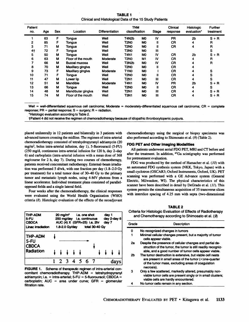
TABLE 1Clinical and Histological Data of the 15 Study Patients
Patientno.1234t56789101112131415Age638571725063667073714751664874SexFFMFMMMMFFMMMMMLocationTongueTongueTongueTongueTongueFloor
of themouthBuccalmucosaMaxillarygingivaMaxillarygingivaTongueLower
lipMandibleTongueMandibular
gingivaFloorof the mouthDifferentiationWellWellWellWellWellModerateWellWellModerateWellWellModerateWellWellWellTNM
classificationT4N2bT2NOT2NOT3NOT4N1T2NOT3N2bT2NOT1NOT2NOT2N1T4N1T2NOT2N1T3N1MOMOMOMOMOM1MOMOMOMOMOMOMOMOMOStageIVIIIIIIIIVIVIVIIIIIIIIIVIIIIIIIIClinicalresponsePRCRCRCRCRCRCRCRCRCRPRCRCRCRHistologieevaluation*2b442b4444442b444FurthertreatmentS
+RRSS
+RRSSSs
+ss
+s+RRRRR
Well = well-differentiated squamous cell carcinoma; Moderate = moderately-differentiated squamous cell carcinoma; CR = completeresponse; PR = partial response; S = surgery; R = radiation.
'Histologie evaluation according to Table 2.
tPatient 4 did not receive the regimen of chemoradiotherapy because of idiopathic thronbocytopenic purpura.
placed unilaterally in 12 patients and bilaterally in 3 patients withadvanced tumors crossing the midline. The regimen of intra-arterial
chemoradiotherapy consisted of tetrahydropyranyl adriamycin (20mg/m2, bolus intra-arterial infusion, day 1), 5-fluorouracil (5-FU)(250 mg/d, continuous intra-arterial infusion for 120 h, day 2-day6) and carboplatin (intra-arterial infusion with a mean dose of 368
mg/course for 2 h, day 7). During two courses of chemotherapy,patients received concomitant radiotherapy. External-beam irradiation was performed 5 d/wk, with one fraction per day (1.8-2.0 Gyper treatment) for a total tumor dose of 30-40 Gy to the primarytumor and metastatic lymph nodes, using 4-MV photons from alinear accelerator. Individual treatment plans consisted of parallel-
opposed fields and a single lateral fÃeld.Four weeks after the chemoradiotherapy, the clinical responses
were evaluated using the World Health Organization (WHO)criteria (8). Histologie evaluation of the effects of the neoadjuvant
THP-ADM5-FUCBDCALinac
irradiationTHP-ADM5-FUCBDCARadiation20
mg/m* i.a. oneshot250
mg/day i.a.continuousAUC(4)X(GFR+25) i.a. 2hrday
1day2-day6day
71.8-2.0 Gy/day total 30-40Gy\1M
M \\12345671 \\days
FIGURE 1. Schemaof therapeuticregimenof intra-arterialconcomitant chemoradiotherapy. THP-ADM = tetrahydropyranyladriamycin; i.a. = intra-arterial; 5-FU = 5-fluorouracil; CBDCA =carboplatin; AUC = area under curve; GFR = glomerularfiltration rate.
chemoradiotherapy using the surgical or biopsy specimens wasalso performed according to Shimosato et al. (9) (Table 2).
FOG PET and Other Imaging ModalitiesAll patients underwent serial FDG PET, MRI and CT before and
after the treatment. In addition, 67Ga scintigraphy was performed
for pretreatment evaluation.FDG was produced by the method of Harnacher et al. (10) with
an automated FDG synthesis system (NKK, Tokyo, Japan) with asmall cyclotron (OSCAR3; Oxford Instruments, Oxford, UK). PETscanning was performed with a GE Advance system (GeneralElectric, Milwaukee, WI). The physical characteristics of thisscanner have been described in detail by DeGrado et al. (U). Thissystem permits the simultaneous acquisition of 35 transverse sliceswith interslice spacing of 4.25 mm with septa (two-dimensional
TABLE 2Criteria for Histologie Evaluation of Effects of Radiotherapy
and Chemotherapy according to Shimosato et al. (9)
Grade Description
0 No recognized changes in tumors1 Minimal cellular changes present, but a majority of tumor
cells appear viable.2a Despite the presence of cellular changes and partial de
struction of the tumor, the tumor is still readily recognizable, and a good number of tumor cells appear viable.
2b The tumor destruction is extensive, but viable cell nestsare present in small areas of the tumor (<one-quarter
of the tumor mass, excluding areas of coagulationnecrosis).
3 Only a few scattered, markedly altered, presumably non-
viable tumor cells are present singly or in small clusters;viable cells are hardly encountered.
4 No tumor cells remain in any section.
CHEMORADIOTHERAPYEVALUATEDBYPET •Kitagawa et al. 1133
mode). Images were reconstructed to a full width at half maximumof 4.2 mm in both the transaxial and axial directions. The field ofview and pixel size of the reconstructed images were 256 and 2mm, respectively. Transmission scans were obtained for 10 minwith a standard pin source of 68Ge/68Ga for attenuation correction
of the emission images.Fourteen patients underwent FDG PET before and over 4 wk
after the chemoradiotherapy (mean 38 d after the treatment). Onepatient (patient 4) underwent pretreatment FDG PET only. Thepatients were administered 244-488 MBq (6.6-13.2 mCi) FDG in
a fasting state in the cubital vein over 10 s. In 6 patients, staticimages were obtained for 20 min, starting at 40 min postinjectionwithout arterial samplings. Of 15 patients, 9 also underwentdynamic scanning. Dynamic scans were obtained up to 60 min afterthe injection with arterial sampling. The mode of dynamic dataacquisition consisted of four 30-s frames, eight 60-s frames andfive 600-s frames. Plasma glucose levels were measured for all
patients. Static images were obtained by averaging the last twoframes of the dynamic data, from 40-60 min postinjection. From
the time of the injection, 2 mL of arterial blood was sampled every15 s in the first 2 min and then at 2, 3, 5, 7, 10, 15, 20, 30,45 and 60min after injection. Plasma radioactivity was measured with ascintillation counter against which the PET camera was cross-calibrated, using a cylindrical phantom filled with the I8F solution.
Data AnalysisFor the quantitative evaluation of regional radioactivity using
the static FDG PET images, regions of interest (round, 5 mm indiameter) were examined in the area of highest accumulatedradioactivity. These tissue radioactivities were corrected withinjected dose and patients' body weight to calculate the standard
ized uptake values (SUVs) using the following formula:
SUV =tissue radioactivity concentration (Bq/mL)
injected dose (Bq)/body weight (g)
For the 9 patients who underwent dynamic PET scanning,
graphic analysis of Patlak and Blasberg (12) and Patlak et al. (13)was performed to calculate the net influx constant of FDG (Ki) andthe glucose metabolic rate of the neoplasms on a pixel-by-pixel
basis, using the following formula:
C(t)
Ca(t) Ca(t)+ Vn,
where Ca(t) is the radioactivity in the plasma at time t, C(t) is theradioactivity at time t in each pixel and Vn is the initial distributionvolume of FDG in each pixel (¡2,13). To obtain the slope K¡,C(t)/Ca(t) was plotted against the integration of Ca(t) divided byCa(t). Data from 10-30 min were used to calculate K¡.To confirm
the feasibility of SUV as a measure of FDG accumulation,correlations of SUV and K¡were evaluated. Finally, we comparedthe pretreatment SUV and post-treatment SUV with the histologie
evaluation of residual tumor cells. To evaluate the treatment effect,the size of the tumor was determined by measuring the maximumarea in the single transaxial section of the contrast-enhanced MR
image.
Statistical AnalysisA paired Student / test (two-tailed) was used for comparisons of
before and after therapy values in each subject; an unpaired / testwas used for all other comparisons. A correlation between twovariables was analyzed with Pearson's coefficient of correlation.
RESULTS
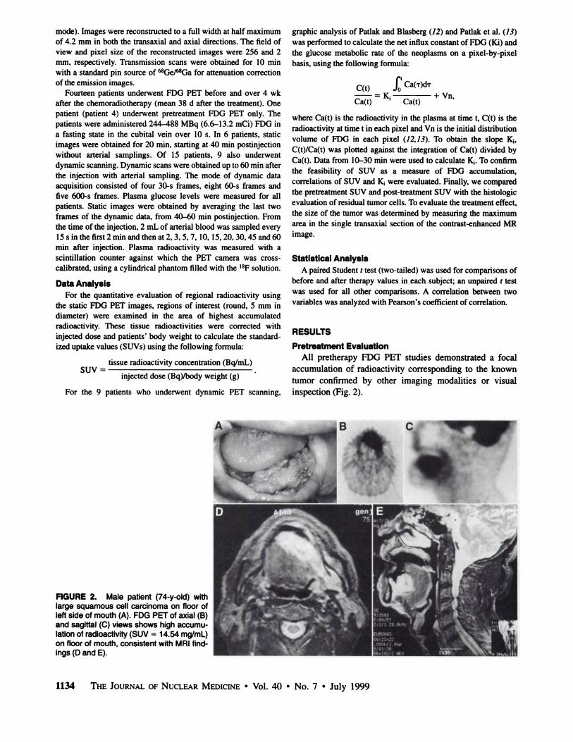
Pretreatment EvaluationAll pretherapy FDG PET studies demonstrated a focal
accumulation of radioactivity corresponding to the knowntumor confirmed by other imaging modalities or visualinspection (Fig. 2).
FIGURE 2. Male patient (74-y-old) withlarge squamous cell carcinoma on floor ofleft side of mouth (A). FDG PET of axial (B)and sagittal (C) views shows high accumulation of radioactivity (SUV = 14.54 mg/mL)on floor of mouth, consistent with MRI findings (D and E).
1134 THEJOURNALOFNUCLEARMEDICINE•Vol. 40 •No. 7 •July 1999
Response to TherapyEvaluation with Conventional Criteria. With the WHO
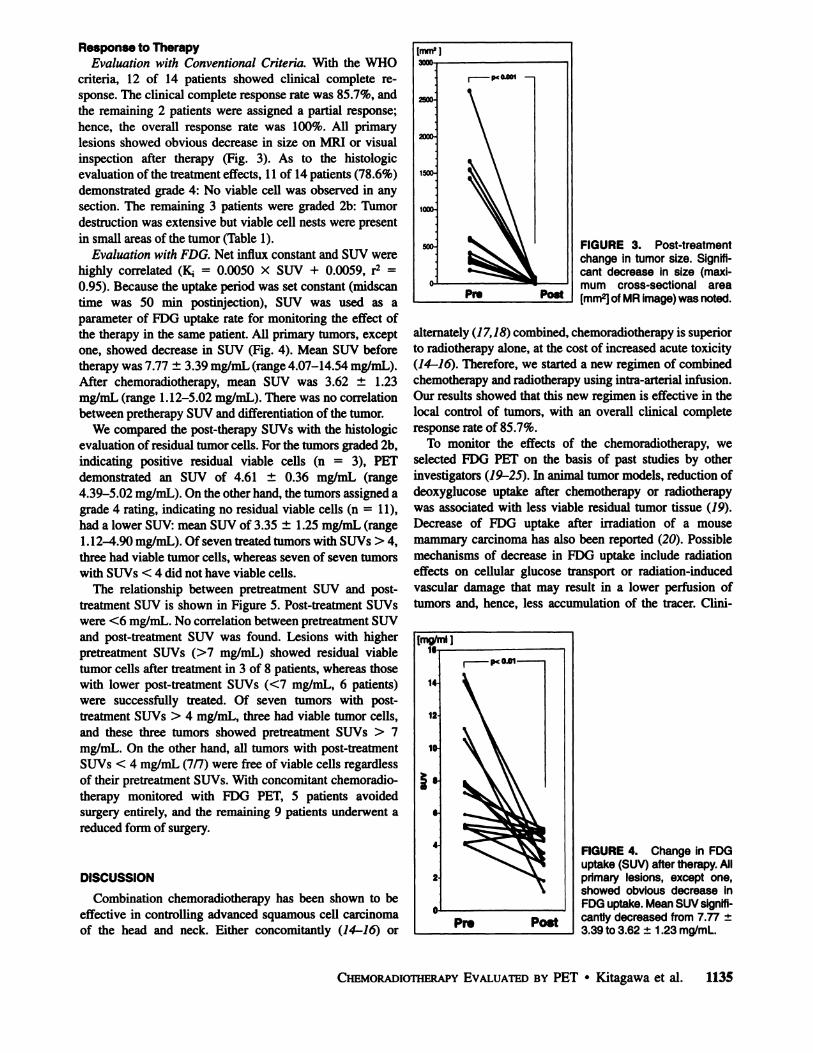
criteria, 12 of 14 patients showed clinical complete response. The clinical complete response rate was 85.7%, andthe remaining 2 patients were assigned a partial response;hence, the overall response rate was 100%. All primarylesions showed obvious decrease in size on MRI or visualinspection after therapy (Fig. 3). As to the histologieevaluation of the treatment effects, 11 of 14 patients (78.6%)demonstrated grade 4: No viable cell was observed in anysection. The remaining 3 patients were graded 2b: Tumordestruction was extensive but viable cell nests were presentin small areas of the tumor (Table 1).
Evaluation with FDG. Net influx constant and SUV werehighly correlated (K¡= 0.0050 X SUV + 0.0059, r2 =
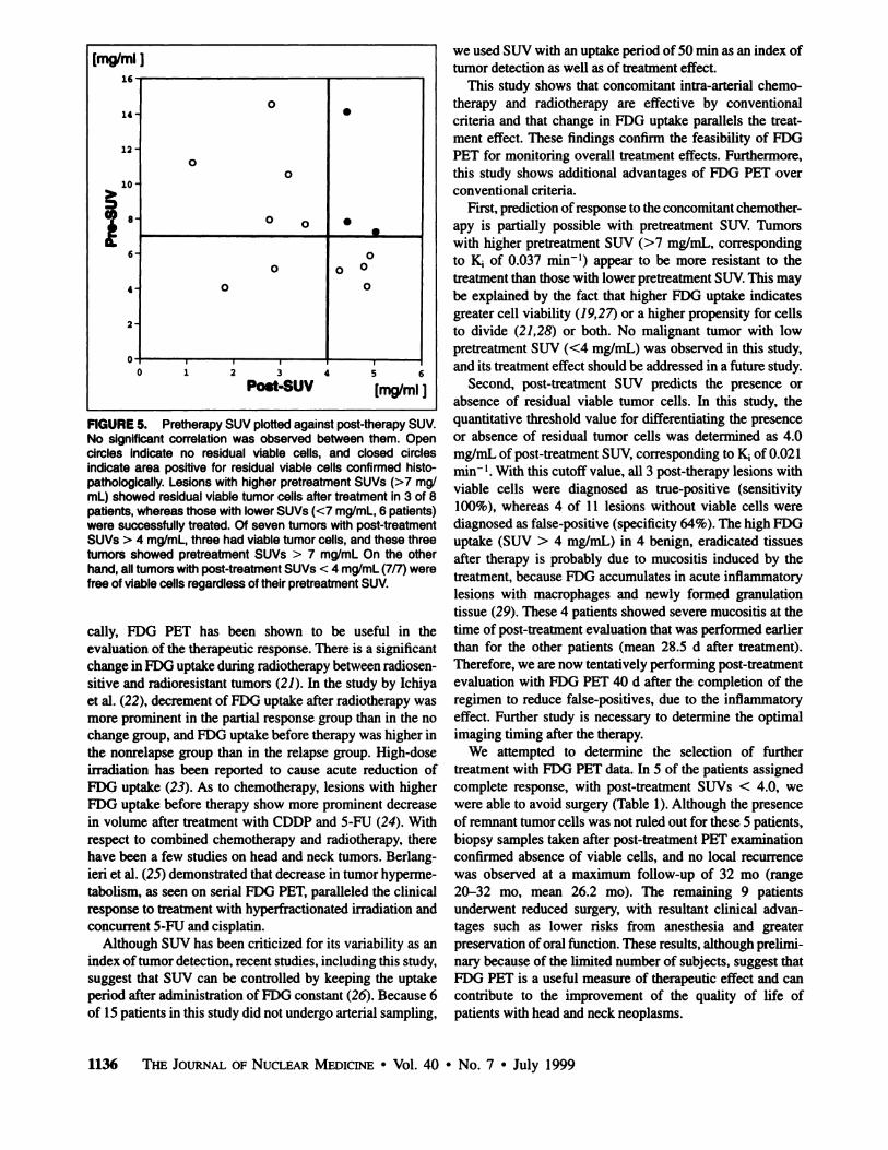
0.95). Because the uptake period was set constant (midscantime was 50 min postinjection), SUV was used as aparameter of FDG uptake rate for monitoring the effect ofthe therapy in the same patient. All primary tumors, exceptone, showed decrease in SUV (Fig. 4). Mean SUV beforetherapy was 7.77 ±3.39 mg/mL (range 4.07-14.54 mg/mL).
After chemoradiotherapy, mean SUV was 3.62 ± 1.23mg/mL (range 1.12-5.02 mg/mL). There was no correlation
between pretherapy SUV and differentiation of the tumor.We compared the post-therapy SUVs with the histologie
evaluation of residual tumor cells. For the tumors graded 2b,indicating positive residual viable cells (n = 3), PET
demonstrated an SUV of 4.61 ± 0.36 mg/mL (range4.39-5.02 mg/mL). On the other hand, the tumors assigned agrade 4 rating, indicating no residual viable cells (n = 11),
had a lower SUV: mean SUV of 3.35 ±1.25 mg/mL (range1.12-4.90 mg/mL). Of seven treated tumors with SUVs > 4,
three had viable tumor cells, whereas seven of seven tumorswith SUVs < 4 did not have viable cells.
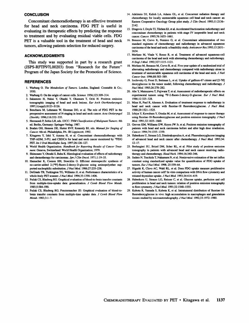
The relationship between pretreatment SUV and post-treatment SUV is shown in Figure 5. Post-treatment SUVs
were <6 mg/mL. No correlation between pretreatment SUVand post-treatment SUV was found. Lesions with higher
pretreatment SUVs (>7 mg/mL) showed residual viabletumor cells after treatment in 3 of 8 patients, whereas thosewith lower post-treatment SUVs (<7 mg/mL, 6 patients)were successfully treated. Of seven tumors with post-
treatment SUVs > 4 mg/mL, three had viable tumor cells,and these three tumors showed pretreatment SUVs > 7mg/mL. On the other hand, all tumors with post-treatment
SUVs < 4 mg/mL (7/7) were free of viable cells regardlessof their pretreatment SUVs. With concomitant chemoradiotherapy monitored with FDG PET, 5 patients avoidedsurgery entirely, and the remaining 9 patients underwent areduced form of surgery.
DISCUSSION
Combination chemoradiotherapy has been shown to beeffective in controlling advanced squamous cell carcinomaof the head and neck. Either concomitantly (14-16) or
[mm2]
Post
FIGURE 3. Post-treatmentchange ¡ntumor size. Significant decrease in size (maximum cross-sectional area[mm2]of MR image) was noted.
alternately (17,18) combined, chemoradiotherapy is superiorto radiotherapy alone, at the cost of increased acute toxicity(14-16). Therefore, we started a new regimen of combinedchemotherapy and radiotherapy using intra-arterial infusion.
Our results showed that this new regimen is effective in thelocal control of tumors, with an overall clinical completeresponse rate of 85.7%.
To monitor the effects of the chemoradiotherapy, weselected FDG PET on the basis of past studies by otherinvestigators (79-25). In animal tumor models, reduction of
deoxyglucose uptake after chemotherapy or radiotherapywas associated with less viable residual tumor tissue (79).Decrease of FDG uptake after irradiation of a mousemammary carcinoma has also been reported (20). Possiblemechanisms of decrease in FDG uptake include radiationeffects on cellular glucose transport or radiation-induced
vascular damage that may result in a lower perfusion oftumors and, hence, less accumulation of the tracer. Clini-
[mg/ml ]16
14
12
10
- p< 0.01
Pre Post
FIGURE 4. Change in FDGuptake (SUV) after therapy. Allprimary lesions, except one,showed obvious decrease inFDG uptake. Mean SUV significantly decreased from 7.77 ±3.39 to 3.62 ±1.23 mg/mL.
CHEMORADIOTHERAPYEVALUATEDBY PET •Kitagawa et al. 1135
[mg/ml]14-12-10-?
"6-4-2-ooo0 0oo*•00
°00123456Post-SUV
[mg/mi j
FIGURE 5. Pretherapy SUV plottedagainst post-therapySUV.No significant correlation was observed between them. Opencircles indicate no residual viable cells, and closed circlesindicate area positive for residual viable cells confirmed histo-pathologically. Lesions with higher pretreatment SUVs (>7 mg/mL) showed residual viable tumor cells after treatment in 3 of 8patients, whereas those with lower SUVs (<7 mg/mL, 6 patients)were successfully treated. Of seven tumors with post-treatmentSUVs > 4 mg/mL, three had viable tumor cells, and these threetumors showed pretreatment SUVs > 7 mg/mL On the otherhand, all tumors with post-treatment SUVs < 4 mg/mL (7/7) werefree of viable cells regardless of their pretreatment SUV.
cally, FDG PET has been shown to be useful in theevaluation of the therapeutic response. There is a significantchange in FDG uptake during radiotherapy between radiosensitive and radioresistant tumors (21). In the study by Ichiyaet al. (22), decrement of FDG uptake after radiotherapy wasmore prominent in the partial response group than in the nochange group, and FDG uptake before therapy was higher inthe nonrelapse group than in the relapse group. High-dose
irradiation has been reported to cause acute reduction ofFDG uptake (23). As to chemotherapy, lesions with higherFDG uptake before therapy show more prominent decreasein volume after treatment with CDDP and 5-FU (24). With
respect to combined chemotherapy and radiotherapy, therehave been a few studies on head and neck tumors. Berlang-ieri et al. (25) demonstrated that decrease in tumor hyperme-
tabolism, as seen on serial FDG PET, paralleled the clinicalresponse to treatment with hyperfractionated irradiation andconcurrent 5-FU and cisplatin.
Although SUV has been criticized for its variability as anindex of tumor detection, recent studies, including this study,suggest that SUV can be controlled by keeping the uptakeperiod after administration of FDG constant (26). Because 6of 15 patients in this study did not undergo arterial sampling,
we used SUV with an uptake period of 50 min as an index oftumor detection as well as of treatment effect.
This study shows that concomitant intra-arterial chemo
therapy and radiotherapy are effective by conventionalcriteria and that change in FDG uptake parallels the treatment effect. These findings confirm the feasibility of FDGPET for monitoring overall treatment effects. Furthermore,this study shows additional advantages of FDG PET overconventional criteria.
First, prediction of response to the concomitant chemotherapy is partially possible with pretreatment SUV. Tumorswith higher pretreatment SUV (>7 mg/mL, correspondingto K¡of 0.037 min"1) appear to be more resistant to the
treatment than those with lower pretreatment SUV. This maybe explained by the fact that higher FDG uptake indicatesgreater cell viability (19,27) or a higher propensity for cellsto divide (21,28) or both. No malignant tumor with lowpretreatment SUV (<4 mg/mL) was observed in this study,and its treatment effect should be addressed in a future study.
Second, post-treatment SUV predicts the presence or
absence of residual viable tumor cells. In this study, thequantitative threshold value for differentiating the presenceor absence of residual tumor cells was determined as 4.0mg/mL of post-treatment SUV, corresponding to K¡of 0.021min"1. With this cutoff value, all 3 post-therapy lesions with
viable cells were diagnosed as true-positive (sensitivity
100%), whereas 4 of 11 lesions without viable cells werediagnosed as false-positive (specificity 64%). The high FDG
uptake (SUV > 4 mg/mL) in 4 benign, eradicated tissuesafter therapy is probably due to mucositis induced by thetreatment, because FDG accumulates in acute inflammatorylesions with macrophages and newly formed granulationtissue (29). These 4 patients showed severe mucositis at thetime of post-treatment evaluation that was performed earlier
than for the other patients (mean 28.5 d after treatment).Therefore, we are now tentatively performing post-treatment
evaluation with FDG PET 40 d after the completion of theregimen to reduce false-positives, due to the inflammatory
effect. Further study is necessary to determine the optimalimaging timing after the therapy.
We attempted to determine the selection of furthertreatment with FDG PET data. In 5 of the patients assignedcomplete response, with post-treatment SUVs < 4.0, we
were able to avoid surgery (Table 1). Although the presenceof remnant tumor cells was not ruled out for these 5 patients,biopsy samples taken after post-treatment PET examination
confirmed absence of viable cells, and no local recurrencewas observed at a maximum follow-up of 32 mo (range20-32 mo, mean 26.2 mo). The remaining 9 patients
underwent reduced surgery, with resultant clinical advantages such as lower risks from anesthesia and greaterpreservation of oral function. These results, although preliminary because of the limited number of subjects, suggest thatFDG PET is a useful measure of therapeutic effect and cancontribute to the improvement of the quality of life ofpatients with head and neck neoplasms.
1136 THEJOURNALOFNUCLEARMEDICINE•Vol. 40 •No. 7 •July 1999
CONCLUSION
Concomitant chemoradiotherapy is an effective treatmentfor head and neck carcinoma. FDG PET is useful inevaluating its therapeutic effects by predicting the responseto treatment and by evaluating residual viable cells. FDGPET is a valuable tool in the treatment of head and necktumors, allowing patients selection for reduced surgery.
ACKNOWLEDGMENTS
This study was supported in part by a research grant(JSPS-RFTF97L00203) from "Research for the Future"
Program of the Japan Society for the Promotion of Science.
REFERENCES1. Warburg O. The Metabolism of Tumors. London, England: Constable & Co.;
1930.2. WarburgO. On the origin of cancer cells. Science. 1956;123:309-314.
3. Sakamoto H, Nakai Y, Ohashi Y, Okamura T, Ochi H. Positron emissiontomographic imaging of head and neck lesions. Ear Arch Otorhinolaryngol.1997;l(suppl):S123-S126.
4. Benchaou M, Lehmann W, Slosman DO, et al. The role of FDG PET in thepreoperative assessment of N-staging in head and neck cancer. Acta Otolaryngol(Stockh). 1996;! 16:332-335.
5. Hermanek P, Sobin LH, eds. UICC: TNM Classification of Malignant Tumors. 4thed. Berlin, Germany: Springer-Verlag; 1987.
6. Beahrs OH, Henson DE, Mutter RVP, Kennedy BJ, eds. Manual for Staging ofCancer. 4th ed. Philadelphia, PA: JB Lippincott; 1992.
7. Kitagawa Y, Ishii Y, Azuma H, et al. Concomitant chemoradiotherapy withTHP-ADM, 5-FU, and CBDCA for head and neck cancer monitored by I8FDGPET. Int J Oral Maxillofac Surg. 1997';26:126-127.
8. World Health Organization. Handbook for Reporting Results of Cancer Treatment. Geneva, Switzerland: World Health Organization; 1979.
9. Shimosato Y, Oboshi S, Baba K. Histológica! evaluation of effects of radiotherapyand chemotherapy for carcinomas. Jpn J Clin Oncol. 1971 ;1:19-35.
10. Harnacher K, Coenen HH, Stoecklin G. Efficient stereospecific synthesis ofno-carrier-added 2-[l8F]-fluoro-2-deoxy-D-glucose using aminopolyether sup
ported nucleophilic substitution. J NucÃMed. 1986;27:235-238.
11. DeGrado TR, Turkington TO, Williams JJ, et al. Performance characteristics of awhole-body PET scanner. J NucÃMed. 1994;35:1398-1406.
12. Patlak CS, Blasberg RG. Graphical evaluation of blood-to-brain transfer constantsfrom multiple-time-uptake data: generalization. J Cereb Blood Flow Metab.1985:5:584-590.
13. Patlak CS, Blasberg RG, Fenstermacher JD. Graphical evaluation of blood-to-brain transfer constants from multiple-time uptake data. J Cereb Blood FlowMetab. 1983;3:l-7.
14. Adelstein DJ, Kalish LA, Adams GL, et al. Concurrent radiation therapy andchemotherapy for locally unresectable squamous cell head and neck cancer: anEastern Cooperative Oncology Group pilot study. J Clin Oncol. 1993:11:2136-
2142.15. Dragovic J, Doyle TJ, Tilchen EJ, et al. Accelerated fractionation radiotherapy and
concomitant chemotherapy in patients with stage IV inoperable head and neckcancer. Cane« 1995:76:1655-1661.
16. Benasso M, Corvo R, Numico G, et al. Concomitant administration of twostandard regimens of chemotherapy and radiotherapy in advanced squamouscarcinoma of the head and neck : a feasibility study. Anticancer Res. 1995 ;15:2651 -
2654.17. Merlano M, Vitale V, Rosso R, et al. Treatment of advanced squamous-cell
carcinoma of the head and neck with alternating chemotherapy and radiotherapy.NEnglJMed. 1992:327:1115-1121.
18. Merlano M, Benasso M, Corvo R. et al. Five-year update of a randomized trial of
alternating radiotherapy and chemotherapy compared with radiotherapy alone intreatment of unresectable squamous cell carcinoma of the head and neck. J NatiCancer Inst. 1996;88:583-589.
19. losilevsky G, Front D, Betunan L, et al. Uptake of gallium-67 citrate and [2-3H]
deoxyglucose in the tumor model, following chemotherapy and radiotherapy. JNucÃMed. 1985:26:278-282.
20. Abe Y, Matsuzawa T, Fujiwara T, et al. Assessment of radiotherapeutic effects onexperimental tumors using l8F-2-fluoro-2-deoxy-D-glucose. Eur J NucÃMed.
1986:12:325-328.
21. Minn H, Paul R, Ahonen A. Evaluation of treatment response to radiotherapy inhead and neck cancer with fluorine-18 fluorodeoxyglucose. J NucÃMed.1988:29:1521-1525.
22. Ichiya Y, Kuwabara Y, Otsuka M, et al. Assessment of response to cancer therapyusing fluorine-18-fluorodeoxyglucose and positron emission tomography. J NucÃMed. 1991:32:1655-1660.
23. Greven KM, Williams DW, Keyes JW Jr, et al. Positron emission tomography ofpatients with head and neck carcinoma before and after high dose irradiation.Cancer. 1994:74:1355-1359.
24. Haberkorn U, Strauss LO, Dimitrakopoulou A, et al. Fluorodeoxyglucose imagingof advanced head and neck cancer after chemotherapy. J NucÃMed. 1993:34:12-17.
25. Berlangieri SU, Brizel DM, Scher RL, et al. Pilot study of positron emissiontomography in patients with advanced head and neck cancer receiving radiotherapy and chemotherapy. Head Neck. 1994:16:340-346.
26. Sadato N, Tsuchida T, Nakamura N, et al. Noninvasive estimation of the net influxconstant using standardized uptake value for quantification of FDG uptake oftumors. Eur J NucÃMed. 1998; 25:559-64.
27. Higashi K, Clavo AC, Wahl RL, et al. Does FDG uptake measure proliferativeactivity of human cancer cell? In vitro comparison with DNA flow cytometry andtritiated thymidine uptake. JNucl Med. 1993;34:414-^»19.
28. Haberkorn U, Strauss LG, Reisser C, et al. Glucose uptake, perfusion and cellproliferation in head and neck tumors: relation of positron emission tomographyto flow cytometry. J NucÃMed. 1991:32:1548-1555.
29. Kubota R, Yamada S, Kubota K, et al. Intratumoral distribution of fluorine-18-
fluorodeoxyglucose in vivo: high accumulation in macrophages and granulationtissues studied by rnicroautoradiography. J NucÃMed. 1992:33:1972-1980.
CHEMORADIOTHERAPYEVALUATEDBYPET •Kitagawa et al. 1137
Related Documents