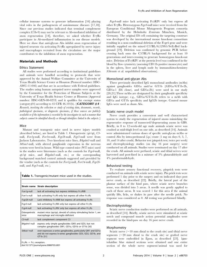
Fcc Receptor-Mediated Inflammation Inhibits Axon Regeneration Gang Zhang 1 , Nataliia Bogdanova 1 , Tong Gao 1 , Julia J. Song 1 , Mark S. Cragg 2 , Martin J. Glennie 2 , Kazim A. Sheikh 1 * 1 Department of Neurology, University of Texas Medical School at Houston, Houston, Texas, United States of America, 2 Antibody and Vaccine Group, Cancer Sciences Division, Faculty of Medicine, University of Southampton, Southampton, United Kingdom Abstract Anti-glycan/ganglioside antibodies are the most common immune effectors found in patients with Guillain-Barre ´ Syndrome, which is a peripheral autoimmune neuropathy. We previously reported that disease-relevant anti-glycan autoantibodies inhibited axon regeneration, which echo the clinical association of these antibodies and poor recovery in Guillain-Barre ´ Syndrome. However, the specific molecular and cellular elements involved in this antibody-mediated inhibition of axon regeneration are not previously defined. This study examined the role of Fcc receptors and macrophages in the antibody- mediated inhibition of axon regeneration. A well characterized antibody passive transfer sciatic nerve crush and transplant models were used to study the anti-ganglioside antibody-mediated inhibition of axon regeneration in wild type and various mutant and transgenic mice with altered expression of specific Fcc receptors and macrophage/microglia populations. Outcome measures included behavior, electrophysiology, morphometry, immunocytochemistry, quantitative real-time PCR, and western blotting. We demonstrate that the presence of autoantibodies, directed against neuronal/axonal cell surface gangliosides, in the injured mammalian peripheral nerves switch the proregenerative inflammatory environment to growth inhibitory milieu by engaging specific activating Fcc receptors on recruited monocyte-derived macrophages to cause severe inhibition of axon regeneration. Our data demonstrate that the antibody orchestrated Fcc receptor-mediated switch in inflammation is one mechanism underlying inhibition of axon regeneration. These findings have clinical implications for nerve repair and recovery in antibody-mediated immune neuropathies. Our results add to the complexity of axon regeneration in injured peripheral and central nervous systems as adverse effects of B cells and autoantibodies on neural injury and repair are increasingly recognized. Citation: Zhang G, Bogdanova N, Gao T, Song JJ, Cragg MS, et al. (2014) Fcc Receptor-Mediated Inflammation Inhibits Axon Regeneration. PLoS ONE 9(2): e88703. doi:10.1371/journal.pone.0088703 Editor: Michael Costigan, Boston Children’s Hospital and Harvard Medical School, United States of America Received September 26, 2013; Accepted January 10, 2014; Published February 11, 2014 Copyright: ß 2014 Zhang et al. This is an open-access article distributed under the terms of the Creative Commons Attribution License, which permits unrestricted use, distribution, and reproduction in any medium, provided the original author and source are credited. Funding: This study was supported by the National Institute of Neurological Disorders and Stroke (NIH/NINDS; grant R01 NS42888 and R01 NS54962) and GBS/ CIDP Foundation International. The funders had no role in study design, data collection and analysis, decision to publish, or preparation of the manuscript. Competing Interests: The authors have declared that no competing interests exist. * E-mail: [email protected] Introduction Axon regeneration is a response of injured nerve cells that is critical for the restoration of structure and function after peripheral or central nervous systems injuries; a response that is key to recovery from numerous neurological disorders. Depending on the pathophysiological situation, axon regeneration is often limited, resulting in poor recovery. Defining the molecular and cellular mechanisms that prevent regeneration of injured axons in various disease situations can provide key insights that may allow development of therapeutic approaches to enhance axon growth in neurological diseases. We present a novel mechanism involving adaptive and innate immune interactions to inhibit regeneration of injured axons with implications for a number of neuroimmuno- logical disorders. Guillain-Barre ´ syndrome (GBS) is an autoimmune disorder affecting the peripheral nervous system, which is the most common cause of acute flaccid paralysis worldwide. About 20% of GBS patients are left with significant disability. Poor recovery in GBS and other neurological disorders commonly reflect failure of axon regeneration and reinnervation of targets. Anti-ganglioside/ glycan antibodies (Abs) are strongly associated with the pathogen- esis of GBS [1,2]. Studies indicate that anti-gangliosides Abs in GBS patients are induced via molecular mimicry [1,3]. Several studies have suggested that GBS patients with anti-GD1a and/or GM1 Abs are more likely to recover slowly and have poor prognosis [4–13]. Understanding the mechanisms underlying failure of axonal regeneration is of critical importance to devise strategies to enhance nerve repair and recovery in GBS and other immune neurological conditions. In this context we previously examined the effects of anti-glycan Abs on peripheral nerve repair [14,15]. We found that passive transfer of specific patient-derived or experimental anti-glycan Abs severely inhibited axon regeneration after peripheral nervous system injury [14,15]. Overall, these observations support our hypothesis that inhibition of axon regeneration is one mechanism of poor recovery in GBS patients with anti-glycan Abs. However, the specific molecular and cellular elements of the inflammatory milieu involved in this Ab-mediated inhibition of axon regener- ation are not previously defined. In Ab-mediated inflammation, complement and/or Fcc recep- tors (FccRs) arms of innate immunity participate to produce injury. FccRs provide an important link between the humoral and PLOS ONE | www.plosone.org 1 February 2014 | Volume 9 | Issue 2 | e88703

Welcome message from author
This document is posted to help you gain knowledge. Please leave a comment to let me know what you think about it! Share it to your friends and learn new things together.
Transcript
Fcc Receptor-Mediated Inflammation Inhibits AxonRegenerationGang Zhang1, Nataliia Bogdanova1, Tong Gao1, Julia J. Song1, Mark S. Cragg2, Martin J. Glennie2,
Kazim A. Sheikh1*
1 Department of Neurology, University of Texas Medical School at Houston, Houston, Texas, United States of America, 2 Antibody and Vaccine Group, Cancer Sciences
Division, Faculty of Medicine, University of Southampton, Southampton, United Kingdom
Abstract
Anti-glycan/ganglioside antibodies are the most common immune effectors found in patients with Guillain-Barre Syndrome,which is a peripheral autoimmune neuropathy. We previously reported that disease-relevant anti-glycan autoantibodiesinhibited axon regeneration, which echo the clinical association of these antibodies and poor recovery in Guillain-BarreSyndrome. However, the specific molecular and cellular elements involved in this antibody-mediated inhibition of axonregeneration are not previously defined. This study examined the role of Fcc receptors and macrophages in the antibody-mediated inhibition of axon regeneration. A well characterized antibody passive transfer sciatic nerve crush and transplantmodels were used to study the anti-ganglioside antibody-mediated inhibition of axon regeneration in wild type and variousmutant and transgenic mice with altered expression of specific Fcc receptors and macrophage/microglia populations.Outcome measures included behavior, electrophysiology, morphometry, immunocytochemistry, quantitative real-time PCR,and western blotting. We demonstrate that the presence of autoantibodies, directed against neuronal/axonal cell surfacegangliosides, in the injured mammalian peripheral nerves switch the proregenerative inflammatory environment to growthinhibitory milieu by engaging specific activating Fcc receptors on recruited monocyte-derived macrophages to cause severeinhibition of axon regeneration. Our data demonstrate that the antibody orchestrated Fcc receptor-mediated switch ininflammation is one mechanism underlying inhibition of axon regeneration. These findings have clinical implications fornerve repair and recovery in antibody-mediated immune neuropathies. Our results add to the complexity of axonregeneration in injured peripheral and central nervous systems as adverse effects of B cells and autoantibodies on neuralinjury and repair are increasingly recognized.
Citation: Zhang G, Bogdanova N, Gao T, Song JJ, Cragg MS, et al. (2014) Fcc Receptor-Mediated Inflammation Inhibits Axon Regeneration. PLoS ONE 9(2):e88703. doi:10.1371/journal.pone.0088703
Editor: Michael Costigan, Boston Children’s Hospital and Harvard Medical School, United States of America
Received September 26, 2013; Accepted January 10, 2014; Published February 11, 2014
Copyright: � 2014 Zhang et al. This is an open-access article distributed under the terms of the Creative Commons Attribution License, which permitsunrestricted use, distribution, and reproduction in any medium, provided the original author and source are credited.
Funding: This study was supported by the National Institute of Neurological Disorders and Stroke (NIH/NINDS; grant R01 NS42888 and R01 NS54962) and GBS/CIDP Foundation International. The funders had no role in study design, data collection and analysis, decision to publish, or preparation of the manuscript.
Competing Interests: The authors have declared that no competing interests exist.
* E-mail: [email protected]
Introduction
Axon regeneration is a response of injured nerve cells that is
critical for the restoration of structure and function after
peripheral or central nervous systems injuries; a response that is
key to recovery from numerous neurological disorders. Depending
on the pathophysiological situation, axon regeneration is often
limited, resulting in poor recovery. Defining the molecular and
cellular mechanisms that prevent regeneration of injured axons in
various disease situations can provide key insights that may allow
development of therapeutic approaches to enhance axon growth
in neurological diseases. We present a novel mechanism involving
adaptive and innate immune interactions to inhibit regeneration of
injured axons with implications for a number of neuroimmuno-
logical disorders.
Guillain-Barre syndrome (GBS) is an autoimmune disorder
affecting the peripheral nervous system, which is the most
common cause of acute flaccid paralysis worldwide. About 20%
of GBS patients are left with significant disability. Poor recovery in
GBS and other neurological disorders commonly reflect failure of
axon regeneration and reinnervation of targets. Anti-ganglioside/
glycan antibodies (Abs) are strongly associated with the pathogen-
esis of GBS [1,2]. Studies indicate that anti-gangliosides Abs in
GBS patients are induced via molecular mimicry [1,3]. Several
studies have suggested that GBS patients with anti-GD1a and/or
GM1 Abs are more likely to recover slowly and have poor
prognosis [4–13]. Understanding the mechanisms underlying
failure of axonal regeneration is of critical importance to devise
strategies to enhance nerve repair and recovery in GBS and other
immune neurological conditions.
In this context we previously examined the effects of anti-glycan
Abs on peripheral nerve repair [14,15]. We found that passive
transfer of specific patient-derived or experimental anti-glycan Abs
severely inhibited axon regeneration after peripheral nervous
system injury [14,15]. Overall, these observations support our
hypothesis that inhibition of axon regeneration is one mechanism
of poor recovery in GBS patients with anti-glycan Abs. However,
the specific molecular and cellular elements of the inflammatory
milieu involved in this Ab-mediated inhibition of axon regener-
ation are not previously defined.
In Ab-mediated inflammation, complement and/or Fcc recep-
tors (FccRs) arms of innate immunity participate to produce
injury. FccRs provide an important link between the humoral and
PLOS ONE | www.plosone.org 1 February 2014 | Volume 9 | Issue 2 | e88703
cellular immune systems to generate inflammation [16] playing
vital roles in the pathogenesis of autoimmune diseases [17,18].
Since our previous studies indicated that terminal complement
complex (C5b-9) may not be relevant to Ab-mediated inhibition of
axon regeneration [14], therefore, we asked whether FccRs
participate in Ab-mediated inflammation in our disease models.
Here we show that anti-glycan Abs inhibit axon regeneration of
injured neurons via activating FccRs upregulated by nerve injury
and macrophages recruited from the circulation are the major
contributors to the inhibition of axon regeneration.
Materials and Methods
Ethics StatementAll studies were performed according to institutional guidelines
and animals were handled according to protocols that were
approved by the Animal Welfare Committee at the University of
Texas Health Science Center at Houston (Protocol number: HSC-
AWC-11-046) and that are in accordance with Federal guidelines.
The studies using human autopsied nerve samples were approved
by the Committee for the Protection of Human Subjects at the
University of Texas Health Science Center at Houston (Approval
number: HSC-GEN-08-0233) and it qualifies for exempt status
(category#4) according to 45 CFR 46.101(b). {CATEGORY #4 :
Research, involving the collection or study of existing data, documents, records,
pathological specimens, or diagnostic specimens, if these sources are publicly
available or if the information is recorded by the investigator in such a manner that
subjects cannot be identified directly or through identifiers linked to the subjects.}
MiceMutant and transgenic mice used in nerve injury models
(described below), are listed in Table 1. Osteopetrotic (op/op), C3-
null, Fcer1g-null, Fcer1a-null, Fcgr2b-null, Fcgr3-null were from
Jackson Laboratories. Two knockout mice lines, B4galnt1-null and
St8sia1-null, with altered ganglioside expression in the nervous
system were bred in house. Wild type control mice (WT mice) used
in the studies were littermates (such as the controls for Fcgr4-null,
op/op mice, and B4galnt1-null, etc.) or the corresponding
background matched control animals suggested and provided by
the vendor (such as the controls for Fcer1g-null, Fcer1a-null, Fcgr2b-
null, and Fcgr3-null, etc.).
Fcgr4-null mice lack activating FccRIV only but express all
other FccRs. Heterozygous Fcgr4-null mice were received from the
European Conditional Mouse Mutagenesis Program consortia
distributed by the Helmholtz Zentrum Munchen, Munich,
Germany. The original ES cell containing the targeting construct
was developed by the international mouse knockout consortium
resulting in a non-conditional deletion of the Fcgr4 gene and were
initially supplied on the mixed C57BL/6;129S5/SvEvBrd back-
ground [19]. Deletion was confirmed by genomic PCR before
crossing back onto the C57BL/6 background for at least 10
generations and inter-crossing to generate homozygous knock-out
mice. Deletion of FccRIV at the protein level was confirmed in the
blood by flow cytometry (assessing CD11b positive monocytes) and
in the spleen, liver and lymph node by immunohistochemistry
(Glennie et al. unpublished observations).
Monoclonal anti-glycan AbsThree previously described IgG monoclonal antibodies (mAbs)
against gangliosides GD1a and/or GT1b {GD1a/GT1b-2b,
GD1a-1 (E6 clone), and GD1a-2b} were used in our study
[20,21].These mAbs are designated by their ganglioside specificity
and IgG isotype; e.g., GD1a/GT1b-2b refers to a mAb with
GD1a and GT1b specificity and IgG2b isotype. Control mouse
IgGs were used as sham Abs.
Sciatic nerve crush modelNerve crush provides a convenient and well characterized
system to study the regeneration of injured axons mimicking the
regenerative response of transected/degenerating axons in GBS.
Briefly, in 8 to 12-week-old age male animals sciatic nerve was
crushed at mid-thigh level on one side, as described [14]. Animals
were administrated various doses of specific anti-glycan mAbs or
control Abs by intra-peritoneal (i.p.) route (1 mg on days 0, 3, 7,
10, and 14 after crush). Behavior test (pinprick test; twice per week)
and electrophysiology studies (on day 16 post surgery) were
conducted on all animals. Studies were terminated on day 17 after
the crush. All animals were perfused, sciatic and tibial nerves were
harvested and post-fixed in a mixture of 3% glutaraldehyde and
4% paraformaldehyde.
Behavioral testingTo evaluate sensory functional recovery, pinprick tests were
conducted on animals with sciatic nerve injury. Pin prick tests were
performed 1 day prior to the surgery and on indicated days post
nerve crush, as described [22]. Briefly, the lateral part of the
plantar surface of the hind paw, where sciatic nerve branches
sense, was divided into 5 areas. A needle was gently applied to
each of those areas. It was scored 1 for this area if the animal
quickly lifts, licks, or shakes its paw after the needle prick. No
response was considered as 0. All testing was performed blindly.
ElectrophysiologySciatic nerve conduction studies were performed on all animals,
as described [14]. Briefly, sciatic nerves were stimulated at sciatic
notch and compound muscle action potential amplitudes were
recorded in the hind-paw on day 16 post nerve crush.
MorphometrySciatic nerve (,10 mm distal to the crush site) and tibial nerve
segments (,20 mm distal to the crush site) or grafted nerve
segment were embedded in Epon, as described [23]. 1mm-
toluidine blue stained sections were obtained and one entire
section of the whole nerve segment/animal was used for
Table 1. Transgenic/mutant mice used in the studies.
Strain name Strain description
Fcer1g-null lack all activating but express inhibitory FccRIIB
Fcer1a-null lack activating FccRI only but express all other FccRs
Fcgr2b-null Lack inhibitory FccRIIB but express all activating FccRs
Fcgr3-null lack activating FccRIII only but express all other FccRs
Fcgr4-null lack activating FccRIV only but express all other FccRs
Osteopetroticmice
mutant mice (op/op), devoid of colony stimulating factor 1, aremacrophage and microglia deficient
C3-null lack complement component C3
B4galnt1-null express only simple gangliosides GM3 and GD3, but notcomplex gangliosides GM1, GD1a, GD1b or GT1b [50]
St8sia1-null over-expresses a-series gangliosides, particularly GM1 and GD1aand do not express b-series gangliosides GD3, GD2, GD1b, andGT1b [51]
FccRs = Fcc receptors.doi:10.1371/journal.pone.0088703.t001
FccRs and Inhibition of Axon Regeneration
PLOS ONE | www.plosone.org 2 February 2014 | Volume 9 | Issue 2 | e88703

quantification, as described [15,24]. All myelinated axons in a
single whole cross section of the nerve were counted at light level
(40X) by using a motorized stage and stereotactic imaging software
(Axiovision; Zeiss), as described [14,24]. This morphometric
avoids random sampling and includes all regenerating nerve
fibers. 12 nerves/group were used for each individual nerve crush
experiment and 6 nerves/group for each nerve grafting experi-
ment (also notated in the respective figure legends).
Nerve graft studyThe nerve graft studies were performed on age and gender
matched WT control and Fcer1g-null mice. The sciatic nerve grafts
(,2 cm long) collected from WT or Fcer1g-null mice were
implanted to the proximal stumps of transected sciatic nerves of
host animals (WT or Fcer1g-null mice) with 10-0 prolene sutures.
Different groups include: (1) WT nerve grafts in WT hosts; (2)
Fcer1g-null nerve grafts in WT hosts; (3) WT nerve grafts in Fcer1g-
null hosts; (4) Fcer1g-null nerve grafts in Fcer1g-null hosts. After the
surgery, all animals were administered 5 doses of GD1a/GT1b-2b
mAb (1mg, i.p.) The grafts were harvested 3 weeks after the
surgery and middle segment of each graft was processed and
embedded in Epon and morphometry was performed on 1-mm
cross sections.
Western blottingThe tissue lysates were extracted from intact and injured mouse
nerves at different time points after nerve crush, or human nerves
from controls and GBS patients. Protein concentration was
determined and 2 mg of total protein/lane were electrophoresed,
transferred to PVDF membranes, and probed with anti-Fcccommon chain [25] (US Biological) and anti-a-Tubulin or anti-b-
actin (loading/internal control) (Cell Signaling) Abs.
Immunocytochemistry (ICC)Injured or intact mouse sciatic nerves, or human nerves from
controls and GBS patients were fixed and cryoprotected.
Cryosections were double labeled for Fcc common chain (shared
by all activating FccRs) and CD68 (Macrophage marker; AbD
serotech), or glial fibrillary acidic protein (GFAP) (Schwann cell
marker; Santa Cruz Biotechnology), or Iba1 (microglial cell
marker; Abcam) with specific Abs. The sections were then
developed with specific fluorescently conjugated secondary Abs
and examined by fluorescent microscopy (Zeiss).
Human tissue harvesting and preparationHuman cauda equina, were obtained from autopsies of GBS
patients (various intervals after onset of GBS, i.e., acute and post-
acute phases) or patients without neuropathic disorders (controls).
The human tissues were then post-fixed in 4% paraformaldehyde
for 24–48 hours. Fixed tissues were cryoprotected and cryosec-
tioned, 10–15 mm cross sections were air-dried on glass slides for
immunocytochemistry studies.
Quantitative Real-time PCRAll primer sets were from Life Technologies: 18s, Hs99999901_s1;
FccRI, Mm00438874_m1; FccRII, Mm00438880_g1; FccRIII,
Mm00438883_m1; FccRIV, Mm00519988_m1. Total RNA was
extracted from injured or intact nerve tissues, according to
manufacturer’s instruction (Invitrogen). cDNA was generated
through reverse transcription from 0.2 mg of each RNA sample by
using High Capacity RNA-to-cDNA master mix (Life Technologies).
Real-time PCR was performed on ABI Step-One Plus (Applied
Biosystems) using TaqMan Fast Advanced Master Mix (Life
Technologies). 18s was used as normalization control. All PCR
were conducted in triplicate and repeated at least three times.
Ganglioside ELISA to determine kinetics of anti-glycanAbs in mice
Serum samples from WT and Mutant op/op mice collected at
various intervals after administration of anti-glycan mAbs were
used for ELISA, as described [20].
StatisticsAll numerical data are presented as mean 6 s.e.m. Differences
between groups were determined using Student’s t test or ANOVA
with corrections for multiple comparisons, p values , 0.05 were
considered statistically significant.
Results
Immune complex formation is required for the inhibitionof axon regeneration mediated by anti-glycan Abs
We examined the inhibitory effects of three GD1a-reactive
mAbs in the nerve crush model because some patients with GBS
and anti-GD1a Abs have poor recovery [4,6,9]. These mAbs were
tested in WT, B4galnt1-null (lack all complex gangliosides), and
St8sia1-null (lack b-series but overexpress a-series gangliosides
including GD1a) (Figure 1). Our results showed that GD1a/
GT1b-2b mAb induce significant inhibition of axon regeneration
in WT and St8sia1-null animals but not in B4galnt1-null mice that
did not express corresponding glycan antigens (Table 2), as
reported previously [14]. Two anti-GD1a mAbs did not inhibit
axon regeneration in WT or B4galnt1-null, whereas these mAbs
induced severe inhibition in St8sia1-null mice (Table 2). These
findings were consistent with our previous studies showing that
higher GD1a density present in St8sia1-null animals {building up
behind the biosynthetic block (Figure 1)} was necessary to induce
anti-GD1a Ab-mediated axonal injury in mice [26]. These results
emphasize the importance of immune complex formation in Ab-
mediated inhibition of axon regeneration.
FccRs are up-regulated in injured nerves of mice and GBSpatients
First, we examined the expression of FccRs in mouse nerve
crush model and compared it to GBS tissues. Quantitative PCR
studies showed highly significant upregulation (range 3–20 fold) of
different FccRs (activating and inhibitory) at mRNA level in
injured nerves compared to uninjured control nerves. mRNA of
FccRII and FccRIII had comparatively higher expression levels
throughout the study period compared to other FccRs (Figure 2A).
Upregulation of activating FccRs was confirmed at protein level
by ICC and immunoblotting on uninjured and injured nerve
segments. FccRs were undetectable in uninjured nerves and their
expression was significantly upregulated in injured nerves (Figures
2B and 2C). There are two macrophage populations, namely
resident and hematogenous monocyte-derived macrophages,
which respond and participate in degenerative and regenerative
responses after mammalian neural injury [27]. Reliable immuno-
logic and phenotypic markers that can differentiate between the
two macrophage populations are not readily available. However,
resident macrophages such as microglia usually act as an early
responder to the nerve injury, prior to the massive influx of
hematogenous macrophages [28] and temporal profiling allows
distinguishing these populations. The ICC studies showed that
activating FccRs were expressed by resident (Schwann and
microglial cells) and recruited (macrophages) glia in injured nerves
FccRs and Inhibition of Axon Regeneration
PLOS ONE | www.plosone.org 3 February 2014 | Volume 9 | Issue 2 | e88703

(Figure 2D). The upregulation of activating FccRs on resident glia
including microglia and Schwann cells was confirmed at time
points prior to the significant recruitment of circulating macro-
phages in injured nerves.
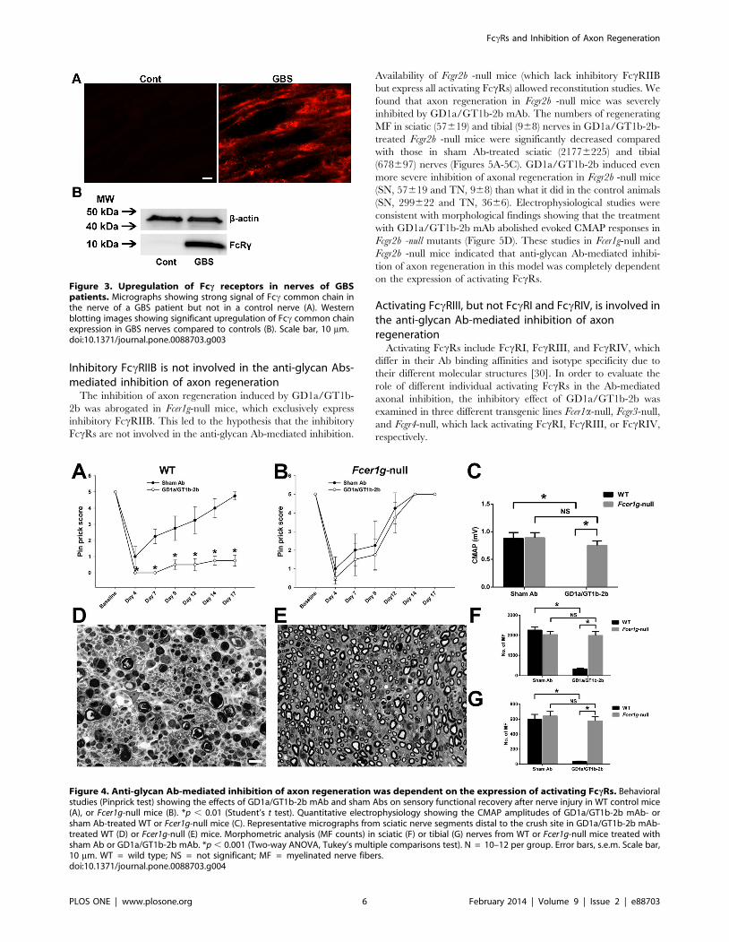
Examination of GBS nerves, obtained at different time points
after onset, showed that there was significant upregulation of Fcccommon chain in acute (data not shown) and post-acute phase of
GBS nerves, whereas control/uninjured human nerves did not
show staining for Fcc common chain (Figure 3A). Western blotting
studies confirmed ICC findings (Figure 3B). These results provide
evidence that activating FccRs are upregulated in GBS nerves and
injured nerves of experimental animals and are available to
participate in Ab-mediated inflammation.
Activating FccRs are required for the inhibition of axonregeneration mediated by anti-glycan Abs
We tested the inhibitory effects of GD1a/GT1b-2b in nerve
crush model on axon regeneration in Fcer1g-null mice, which lack
all activating FccRs and only express inhibitory FccRIIB [29].
Our results showed that Fcer1g-null mice were resistant to the
severe inhibitory effects seen in background-matched WT mice, as
assessed by behavioral, electrophysiological, and morphometric
measures. Behavioral studies (Pinprick test) showed that GD1a/
GT1b-2b mAb significantly reduced sensory functional recovery
after nerve injury in WT mice compared to WT animals treated
with sham Abs (Figure 4A), whereas the sensory functional
recovery was similar in Fcer1g-null mice treated with GD1a/
GT1b-2b and sham Abs (Figure 4B). Quantitative electrophysiol-
ogy (sciatic nerve conductions) indicated that GD1a/GT1b-2b
mAb adversely affected the motor nerve regeneration and target
(muscle) reinnervation in WT animals but not in Fcer1g-null mice
(Figure 4C). Morphological studies also demonstrated that the
GD1a/GT1b-2b mediated inhibition of axon regeneration found
in WT mice was reversed in Fcer1g-null mice (Figures 4D-4G).
There was significant decrease in regenerating myelinated nerve
fibers (MF) in GD1a/GT1b-2b-treated WT animals at sciatic (SN)
Figure 1. Scheme of biosynthetic pathways for major nervous system gangliosides. Blockades in ganglioside biosynthesis in knockoutmice are indicated by solid red lines. GM2/GD2-synthase knockout mice (B4galnt1-null) mice do not express any complex gangliosides, includingGM1, GD1a, GD1b or GT1b, and express only simple gangliosides GM3 and GD3. GD3-Synthase knockout mice (St8sia1-null), which lack the GD3synthase required for synthesis of b-series gangliosides, overexpress a-series gangliosides (highlighted), particularly GM1 and GD1a, and do notexpress b-series gangliosides GD3, GD2, GD1b, and GT1b.doi:10.1371/journal.pone.0088703.g001
Table 2. Anti-glycan mAbs and their inhibitory effects.
Anti-glycan mAbs Glycan specificity Inhibition of axon regeneration
WT mice {No. of MF} St8sia1-null mice {No. of MF} B4galnt1-null mice {No. of MF}
Control Ab None No {23516138 at SN;576662 at TN}
No {23796227 at SN;689672 at TN}
No {19246103 at SN; 529642 at TN}
GD1a/GT1b-2b GD1a/GT1b Yes {354650 (p , 0.05)at SN; 3465 at TN(p , 0.05)}
Yes {254619 (p , 0.05) at SN;2364 at TN (p , 0.05)}
No {1846645 (p . 0.05) at SN; 496614at TN (p . 0.05)}
GD1a-1(E6 clone) GD1a No {2121665(p . 0.05) at SN;535671 at TN (p .0.05)}
Yes {7926116 (p , 0.05) at SN;145641 at TN (p , 0.05)}
No {2023679 (p . 0.05) at SN; 523630at TN (p . 0.05)}
GD1a-2b GD1a No {2062652 (p .0.05)at SN; 566635 at TN (p.0.05)}
Yes {432666 (p , 0.05) at SN;4868 at TN (p , 0.05)}
No {19376101 (p . 0.05) at SN;536635 at TN (p . 0.05)}
The table contains the name of all anti-glycan mAbs used in the study, their glycan specificity, and their inhibitory effects on axon regeneration in WT and transgenicmice with altered ganglioside expression. Comparisons were made between control and anti-ganglioside Ab treated-animals, and the differences were determinedusing Student’s t-test. p , 0.05 was considered as significant. WT = wild type; MF = myelinated nerve fiber; SN = sciatic nerve; TN = tibial nerve; mAbs = monoclonalantibodies.doi:10.1371/journal.pone.0088703.t002
FccRs and Inhibition of Axon Regeneration
PLOS ONE | www.plosone.org 4 February 2014 | Volume 9 | Issue 2 | e88703

(336638) and tibial (TN) (3665) nerves compared with sham Ab-
treated WT mice in sciatic (22536152) and tibial (596669) nerves
(Figures 4D, 4F, and 4G). In contrast, no significant difference in
MF regeneration was found in Fcer1g-null mice treated with
GD1a/GT1b-2b (SN, 19956187 and TN, 577655) or sham Ab
(SN, 20326156 and TN, 642660) (Figures 4E, 4F, and 4G).
Figure 2. Upregulation of Fcc receptors expression in injured mouse nerves. Quantitative PCR showing the relative mRNA levels of FccRI,FccRII, FccRIII, and FccRIV in injured nerves at various time points after surgery (A). Single labeling immunocytochemistry studies showing theexpression of Fcc common chain (shared by all activating Fcc receptors) in the injured sciatic nerves (B). Western blotting images and quantitativedensitometry of Fcc common chain in the injured nerves (C). Double labeling immunocytochemistry studies showing the activating Fcc receptors byendogenous {microglia (Iba1 positive; 1 day after injury) and Schwann cells (GFAP positive; 4 days after injury} and recruited {macrophages (CD68positive); 14 days after injury} glia in the injured nerves (D). *p , 0.001 (One-way ANOVA, Bonferroni’s’ post-hoc test). Arrows showing the co-localization between activating Fcc receptors signal (Fcc common chain) and glia cell markers (GFAP and Iba1). N = 4–5 per group. Error bars, s.e.m.Scale bar, 10 mm.doi:10.1371/journal.pone.0088703.g002
FccRs and Inhibition of Axon Regeneration
PLOS ONE | www.plosone.org 5 February 2014 | Volume 9 | Issue 2 | e88703
Inhibitory FccRIIB is not involved in the anti-glycan Abs-mediated inhibition of axon regeneration
The inhibition of axon regeneration induced by GD1a/GT1b-
2b was abrogated in Fcer1g-null mice, which exclusively express
inhibitory FccRIIB. This led to the hypothesis that the inhibitory
FccRs are not involved in the anti-glycan Ab-mediated inhibition.
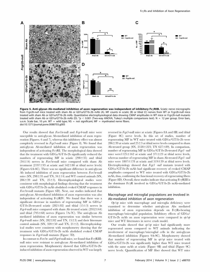
Availability of Fcgr2b -null mice (which lack inhibitory FccRIIB
but express all activating FccRs) allowed reconstitution studies. We
found that axon regeneration in Fcgr2b -null mice was severely
inhibited by GD1a/GT1b-2b mAb. The numbers of regenerating
MF in sciatic (57619) and tibial (968) nerves in GD1a/GT1b-2b-
treated Fcgr2b -null mice were significantly decreased compared
with those in sham Ab-treated sciatic (21776225) and tibial
(678697) nerves (Figures 5A-5C). GD1a/GT1b-2b induced even
more severe inhibition of axonal regeneration in Fcgr2b -null mice
(SN, 57619 and TN, 968) than what it did in the control animals
(SN, 299622 and TN, 3666). Electrophysiological studies were
consistent with morphological findings showing that the treatment
with GD1a/GT1b-2b mAb abolished evoked CMAP responses in
Fcgr2b -null mutants (Figure 5D). These studies in Fcer1g-null and
Fcgr2b -null mice indicated that anti-glycan Ab-mediated inhibi-
tion of axon regeneration in this model was completely dependent
on the expression of activating FccRs.
Activating FccRIII, but not FccRI and FccRIV, is involved inthe anti-glycan Ab-mediated inhibition of axonregeneration
Activating FccRs include FccRI, FccRIII, and FccRIV, which
differ in their Ab binding affinities and isotype specificity due to
their different molecular structures [30]. In order to evaluate the
role of different individual activating FccRs in the Ab-mediated
axonal inhibition, the inhibitory effect of GD1a/GT1b-2b was
examined in three different transgenic lines Fcer1a-null, Fcgr3-null,
and Fcgr4-null, which lack activating FccRI, FccRIII, or FccRIV,
respectively.
Figure 3. Upregulation of Fcc receptors in nerves of GBSpatients. Micrographs showing strong signal of Fcc common chain inthe nerve of a GBS patient but not in a control nerve (A). Westernblotting images showing significant upregulation of Fcc common chainexpression in GBS nerves compared to controls (B). Scale bar, 10 mm.doi:10.1371/journal.pone.0088703.g003
Figure 4. Anti-glycan Ab-mediated inhibition of axon regeneration was dependent on the expression of activating FccRs. Behavioralstudies (Pinprick test) showing the effects of GD1a/GT1b-2b mAb and sham Abs on sensory functional recovery after nerve injury in WT control mice(A), or Fcer1g-null mice (B). *p , 0.01 (Student’s t test). Quantitative electrophysiology showing the CMAP amplitudes of GD1a/GT1b-2b mAb- orsham Ab-treated WT or Fcer1g-null mice (C). Representative micrographs from sciatic nerve segments distal to the crush site in GD1a/GT1b-2b mAb-treated WT (D) or Fcer1g-null (E) mice. Morphometric analysis (MF counts) in sciatic (F) or tibial (G) nerves from WT or Fcer1g-null mice treated withsham Ab or GD1a/GT1b-2b mAb. *p , 0.001 (Two-way ANOVA, Tukey’s multiple comparisons test). N = 10–12 per group. Error bars, s.e.m. Scale bar,10 mm. WT = wild type; NS = not significant; MF = myelinated nerve fibers.doi:10.1371/journal.pone.0088703.g004
FccRs and Inhibition of Axon Regeneration
PLOS ONE | www.plosone.org 6 February 2014 | Volume 9 | Issue 2 | e88703
Our results showed that Fcer1a-null and Fcgr4-null mice were
susceptible to anti-glycan Ab-mediated inhibition of axon regen-
eration (Figures. 6 and 7), whereas this inhibitory effect was almost
completely reversed in Fcgr3-null mice (Figure 8). We found that
anti-glycan Ab-mediated inhibition of axon regeneration was
independent of activating FccRI. The morphological data showed
that the treatment with GD1a/GT1b-2b significantly reduced the
numbers of regenerating MF in sciatic (298635) and tibial
(34614) nerves in Fcer1a-null mice compared with sham Ab
treatment (2197693 at sciatic and 542680 at tibial nerve levels)
(Figures 6A-6C). There was no significant difference in anti-glycan
Ab induced inhibition of axon regeneration between Fcer1a-null
mice (SN, 298635 and TN, 34614) and WT control animals (SN,
286639 and TN, 2563). Electrophysiological studies were
consistent with morphological findings showing that the treatment
with GD1a/GT1b-2b mAb abolished evoked CMAP responses in
Fcer1a-null mutants (Figure 6D). Next, our studies indicated that
anti-glycan Ab-mediated inhibition of axon regeneration was also
independent of activating FccRIV. We found that there was a
significant decrease in numbers of regenerating MF in GD1a/
GT1b-2b-treated sciatic (282682) and tibial (1563) nerves in
Fcgr4-null mice compared with sham Ab treated sciatic (2952664)
and tibial (795668) nerves (Figures 7A-7C). The anti-glycan Ab
mediated inhibition of axon regeneration was similar between
Fcgr4-null mice (SN, 282682 and TN, 1563) and their littermate
control animals (SN, 320650 and TN, 2162). Electrophysiolog-
ical studies were consistent with morphometry showing that the
treatment with GD1a/GT1b-2b mAb abolished evoked CMAP
responses in Fcgr4-null mutants (Figure 7D).
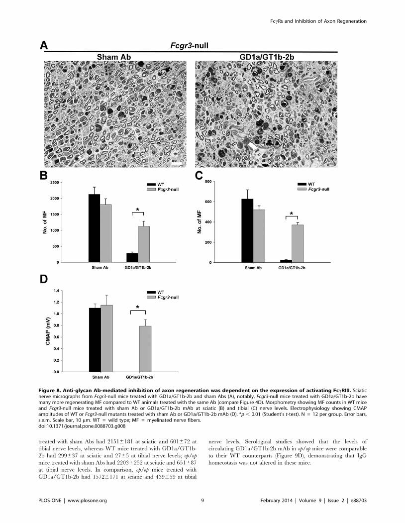
In contrast to Fcer1a- and Fcgr4-null mice, we found that FccRIII-
null mice were resistant to anti-glycan Ab-mediated inhibition of
axon regeneration. Morphometry showed that GD1a/GT1b-2b-
induced inhibition of axon regeneration observed in WT was largely
reversed in Fcgr3-null mice at sciatic (Figures 8A and 8B) and tibial
(Figure 8C) nerve levels. In this set of studies, number of
regenerating MF in WT mice treated with GD1a/GT1b-2b were
286639 at sciatic and 2563 at tibial nerve levels compared to sham
Ab-treated group (SN, 21286223; TN 627689). In comparison,
number of regenerating MF in GD1a/GT1b-2b-treated Fcgr3 -null
mice were11256162 at sciatic and 371623 at tibial nerve levels,
whereas number of regenerating MF in sham Ab-treated Fcgr3 -null
mice were 18076178 at sciatic and 519639 at tibial nerve levels.
Electrophysiology showed that Fcgr3 -null mutants treated with
GD1a/GT1b-2b mAb had significant recovery of evoked CMAP
amplitudes compared to WT mice treated with GD1a/GT1b-2b
mAb, thus, confirming the functional recovery of regenerating fibers
(Figure 8D). Overall, these studies indicate that activating FccRIII is
the dominant FccR involved in GD1a/GT1b-2b mAb-mediated
inhibition.
Macrophage and microglial populations are involved inAb-mediated inhibition of axon regeneration
Op/op mice with macrophage and microglia deficiency were
examined to determine whether anti-glycan Abs mediated
inhibition of axon regeneration depends on FccRs-bearing
macrophage/microglial population. Inhibitory effects of GD1a/
GT1b-2b mAb on axon regeneration were compared in op/op
mice and WT littermates in nerve crush model.
Our results showed that op/op mice had significantly more
regenerated axons compared to WT animals indicating the
involvement of macrophage/microglial cells in the anti-glycan
Ab-mediated inhibition (Figures 9A-9C). Morphometry showed
that number of regenerating MF in op/op mice treated with
GD1a/GT1b-2b was significantly higher than WT mice treated
with the same mAb at sciatic (Figure 9B) and tibial (Figure 9C)
nerve levels. Quantification of MF fiber showed that WT mice
Figure 5. Anti-glycan Ab-mediated inhibition of axon regeneration was independent of inhibitory FccRIIB. Sciatic nerve micrographsfrom Fcgr2b-null mice treated with sham Ab or GD1a/GT1b-2b mAb (A). MF counts in sciatic (B) or tibial (C) nerves from WT or Fcgr2b-null micetreated with sham Ab or GD1a/GT1b-2b mAb. Quantitative electrophysiological data showing CMAP amplitudes in WT mice or Fcgr2b-null mutantstreated with sham Ab or GD1a/GT1b-2b mAb (D). *p , 0.001 (Two-way ANOVA, Tukey’s multiple comparisons test). N = 12 per group. Error bars,s.e.m. Scale bar, 10 mm. WT = wild type; NS = not significant; MF = myelinated nerve fibers.doi:10.1371/journal.pone.0088703.g005
FccRs and Inhibition of Axon Regeneration
PLOS ONE | www.plosone.org 7 February 2014 | Volume 9 | Issue 2 | e88703

Figure 6. Anti-glycan Ab-mediated inhibition of axon regeneration was independent of activating FccRI. Sciatic nerve micrographsfrom Fcer1a-null mice treated with sham Ab or GD1a/GT1b-2b mAb (A). MF counts in sciatic (B) or tibial (C) nerves from WT or Fcer1a-null micetreated with sham Ab or GD1a/GT1b-2b mAb. Quantitative electrophysiological data showing CMAP amplitudes in WT or Fcer1a-null mice treatedwith sham Ab or GD1a/GT1b-2b mAb (D). *p , 0.001 (Two-way ANOVA, Tukey’s multiple comparisons test). N = 12 per group. Error bars, s.e.m. Scalebar, 10 mm. WT = wild type; NS = not significant; MF = myelinated nerve fibers.doi:10.1371/journal.pone.0088703.g006
Figure 7. Anti-glycan Ab-mediated inhibition of axon regeneration was independent of activating FccRIV. Sciatic nerve micrographsfrom Fcgr4-null mice treated with sham Ab or GD1a/GT1b-2b mAb (A). MF counts in sciatic (B) or tibial (C) nerves from WT or Fcgr4-null mice treatedwith sham Ab or GD1a/GT1b-2b mAb. Quantitative electrophysiological data showing CMAP amplitudes of WT or Fcgr4-null mice treated with shamAb or GD1a/GT1b-2b mAb (D). *p , 0.001 (Two-way ANOVA, Tukey’s multiple comparisons test). N = 12 per group. Error bars, s.e.m. Scale bar,10 mm. WT = wild type; NS = not significant; MF = myelinated nerve fibers.doi:10.1371/journal.pone.0088703.g007
FccRs and Inhibition of Axon Regeneration
PLOS ONE | www.plosone.org 8 February 2014 | Volume 9 | Issue 2 | e88703
treated with sham Abs had 21516181 at sciatic and 601672 at
tibial nerve levels, whereas WT mice treated with GD1a/GT1b-
2b had 299637 at sciatic and 2765 at tibial nerve levels; op/op
mice treated with sham Abs had 22036252 at sciatic and 651687
at tibial nerve levels. In comparison, op/op mice treated with
GD1a/GT1b-2b had 15726171 at sciatic and 439659 at tibial
nerve levels. Serological studies showed that the levels of
circulating GD1a/GT1b-2b mAb in op/op mice were comparable
to their WT counterparts (Figure 9D), demonstrating that IgG
homeostasis was not altered in these mice.
Figure 8. Anti-glycan Ab-mediated inhibition of axon regeneration was dependent on the expression of activating FccRIII. Sciaticnerve micrographs from Fcgr3-null mice treated with GD1a/GT1b-2b and sham Abs (A), notably, Fcgr3-null mice treated with GD1a/GT1b-2b havemany more regenerating MF compared to WT animals treated with the same Ab (compare Figure 4D). Morphometry showing MF counts in WT miceand Fcgr3-null mice treated with sham Ab or GD1a/GT1b-2b mAb at sciatic (B) and tibial (C) nerve levels. Electrophysiology showing CMAPamplitudes of WT or Fcgr3-null mutants treated with sham Ab or GD1a/GT1b-2b mAb (D). *p , 0.01 (Student’s t-test). N = 12 per group. Error bars,s.e.m. Scale bar, 10 mm. WT = wild type; MF = myelinated nerve fibers.doi:10.1371/journal.pone.0088703.g008
FccRs and Inhibition of Axon Regeneration
PLOS ONE | www.plosone.org 9 February 2014 | Volume 9 | Issue 2 | e88703

Recruited macrophages are the major contributor to theAb- and activating FccRs-mediated inhibition of axonregeneration
Our results showed that both endoneurial glia (Schwann and
microglial cells) and recruited macrophages express activating
FccRs (Figure 2D), therefore, we asked which FccRs-expressing
cells participate in producing an inflammatory inhibitory milieu in
the injured nerve.
A nerve grafting paradigm, in which nerve segments of donor
mice (WT or Fcer1g-null/ activating FccRs-null) were transplanted
into host animals (WT mice or Fcer1g-null), that allows determi-
nation of the contribution of circulating macrophages (recruited in
injured nerves from hosts) and resident endoneurial glial cells (in
grafted nerve segments from donors) in Ab-mediated inhibition of
axon regeneration was used to address this question. Previous
studies had established that grafted nerve segments retain donor
glia [31,32]. These chimeric animals were administered GD1a/
GT1b-2b Ab and axon regeneration was assessed in the grafted
nerve segments. We found that Fcer1g-null hosts implanted with
nerve grafts from Fcer1g-null donors were not susceptible to Ab-
mediated inhibition, whereas WT hosts implanted with WT nerve
grafts showed severe inhibition (Figures 9E and 9F). Notably, the
axon regeneration in WT or Fcer1g-null grafts implanted in Fcer1g-
null hosts was more pronounced compared to WT or Fcer1g-null
grafts implanted in WT hosts. Additionally, WT grafts implanted
in Fcer1g-null hosts had modest but significant reduction in number
of regenerating axons compared to Fcer1g-null grafts implanted in
Fcer1g-null hosts (Figures 9E and 9F). Morphometry showed the
numbers of MF in different chimeras is as follows: 1) Fcer1g-null
grafts in mutant Fcer1g-null hosts had 16096206 MF; 2) WT grafts
in Fcer1g-null hosts had 9966162 MF; 3) WT grafts in WT hosts
had 461 MF; and 4) Fcer1g-null grafts in WT hosts had 664 MF.
Figure 9. Activating FccRs-bearing macrophages were major contributors to inhibition of axon regeneration. Sciatic nerves showingregeneration of MF in op/op mice treated with sham or GD1a/GT1b-2b mAb (A). Morphometry data showing that number of regenerating MF in WTmice and op/op mice treated with sham Ab or GD1a/GT1b-2b at sciatic (B) and tibial (C) nerve levels. *p , 0.01 (Student’s t-test). N = 12 per group.Serology showing the levels of circulating GD1a/GT1b-2b mAb in op/op mice and WT animals (D). Micrographs (E) and morphometry (F) showing thatmutant graft transplanted in mutant host had the most regeneration, wild type graft transplanted in mutant host had some regenerating fibers, andwild type graft or mutant graft transplanted in wild type hosts had virtually no regenerating fibers. *p , 0.05; **p , 0.001 (Two-way ANOVA,Bonferroni’s post-hoc test). N = 6 per group. Error bars, s.e.m. Scale bar, 10 mm. WG = wild type grafts, WH = wild type hosts, MG = mutant (Fcer1g-null) grafts, and MH = mutant (Fcer1g-null) hosts. NS = not significant; MF = myelinated nerve fibers.doi:10.1371/journal.pone.0088703.g009
FccRs and Inhibition of Axon Regeneration
PLOS ONE | www.plosone.org 10 February 2014 | Volume 9 | Issue 2 | e88703
Overall, these studies showed that the macrophages recruited
from the circulation of the host animals were the dominant cell
type and endogenous nerve glia had minor contribution to the
activating FccRs-induced inflammation and Ab-mediated inhibi-
tion of axon regeneration.
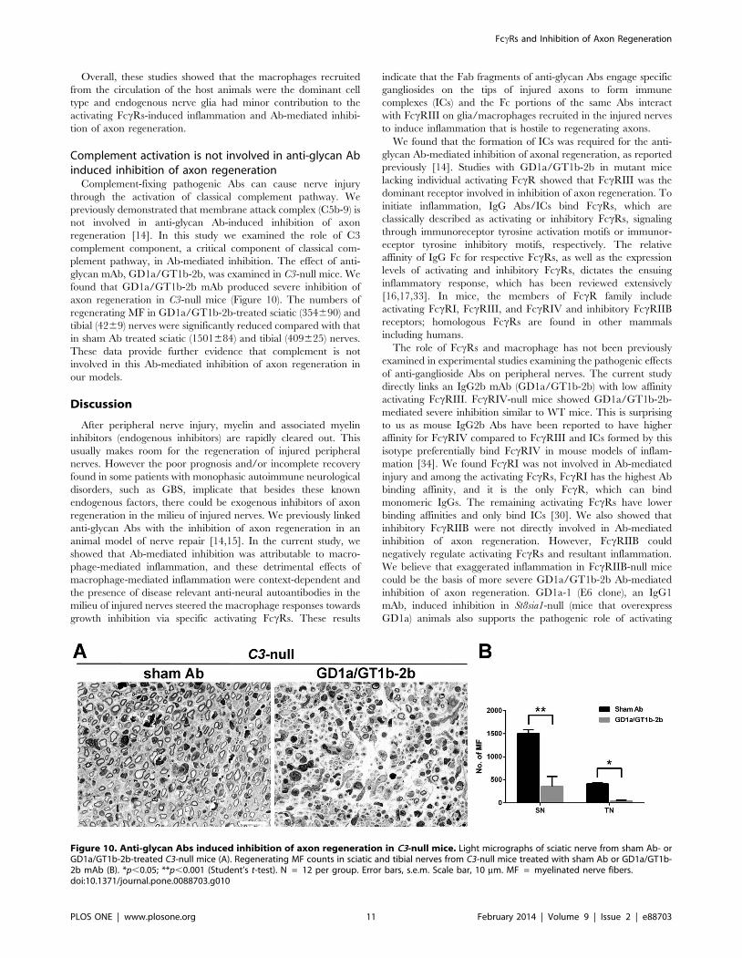
Complement activation is not involved in anti-glycan Abinduced inhibition of axon regeneration
Complement-fixing pathogenic Abs can cause nerve injury
through the activation of classical complement pathway. We
previously demonstrated that membrane attack complex (C5b-9) is
not involved in anti-glycan Ab-induced inhibition of axon
regeneration [14]. In this study we examined the role of C3
complement component, a critical component of classical com-
plement pathway, in Ab-mediated inhibition. The effect of anti-
glycan mAb, GD1a/GT1b-2b, was examined in C3-null mice. We
found that GD1a/GT1b-2b mAb produced severe inhibition of
axon regeneration in C3-null mice (Figure 10). The numbers of
regenerating MF in GD1a/GT1b-2b-treated sciatic (354690) and
tibial (4269) nerves were significantly reduced compared with that
in sham Ab treated sciatic (1501684) and tibial (409625) nerves.
These data provide further evidence that complement is not
involved in this Ab-mediated inhibition of axon regeneration in
our models.
Discussion
After peripheral nerve injury, myelin and associated myelin
inhibitors (endogenous inhibitors) are rapidly cleared out. This
usually makes room for the regeneration of injured peripheral
nerves. However the poor prognosis and/or incomplete recovery
found in some patients with monophasic autoimmune neurological
disorders, such as GBS, implicate that besides these known
endogenous factors, there could be exogenous inhibitors of axon
regeneration in the milieu of injured nerves. We previously linked
anti-glycan Abs with the inhibition of axon regeneration in an
animal model of nerve repair [14,15]. In the current study, we
showed that Ab-mediated inhibition was attributable to macro-
phage-mediated inflammation, and these detrimental effects of
macrophage-mediated inflammation were context-dependent and
the presence of disease relevant anti-neural autoantibodies in the
milieu of injured nerves steered the macrophage responses towards
growth inhibition via specific activating FccRs. These results
indicate that the Fab fragments of anti-glycan Abs engage specific
gangliosides on the tips of injured axons to form immune
complexes (ICs) and the Fc portions of the same Abs interact
with FccRIII on glia/macrophages recruited in the injured nerves
to induce inflammation that is hostile to regenerating axons.
We found that the formation of ICs was required for the anti-
glycan Ab-mediated inhibition of axonal regeneration, as reported
previously [14]. Studies with GD1a/GT1b-2b in mutant mice
lacking individual activating FccR showed that FccRIII was the
dominant receptor involved in inhibition of axon regeneration. To
initiate inflammation, IgG Abs/ICs bind FccRs, which are
classically described as activating or inhibitory FccRs, signaling
through immunoreceptor tyrosine activation motifs or immunor-
eceptor tyrosine inhibitory motifs, respectively. The relative
affinity of IgG Fc for respective FccRs, as well as the expression
levels of activating and inhibitory FccRs, dictates the ensuing
inflammatory response, which has been reviewed extensively
[16,17,33]. In mice, the members of FccR family include
activating FccRI, FccRIII, and FccRIV and inhibitory FccRIIB
receptors; homologous FccRs are found in other mammals
including humans.
The role of FccRs and macrophage has not been previously
examined in experimental studies examining the pathogenic effects
of anti-ganglioside Abs on peripheral nerves. The current study
directly links an IgG2b mAb (GD1a/GT1b-2b) with low affinity
activating FccRIII. FccRIV-null mice showed GD1a/GT1b-2b-
mediated severe inhibition similar to WT mice. This is surprising
to us as mouse IgG2b Abs have been reported to have higher
affinity for FccRIV compared to FccRIII and ICs formed by this
isotype preferentially bind FccRIV in mouse models of inflam-
mation [34]. We found FccRI was not involved in Ab-mediated
injury and among the activating FccRs, FccRI has the highest Ab
binding affinity, and it is the only FccR, which can bind
monomeric IgGs. The remaining activating FccRs have lower
binding affinities and only bind ICs [30]. We also showed that
inhibitory FccRIIB were not directly involved in Ab-mediated
inhibition of axon regeneration. However, FccRIIB could
negatively regulate activating FccRs and resultant inflammation.
We believe that exaggerated inflammation in FccRIIB-null mice
could be the basis of more severe GD1a/GT1b-2b Ab-mediated
inhibition of axon regeneration. GD1a-1 (E6 clone), an IgG1
mAb, induced inhibition in St8sia1-null (mice that overexpress
GD1a) animals also supports the pathogenic role of activating
Figure 10. Anti-glycan Abs induced inhibition of axon regeneration in C3-null mice. Light micrographs of sciatic nerve from sham Ab- orGD1a/GT1b-2b-treated C3-null mice (A). Regenerating MF counts in sciatic and tibial nerves from C3-null mice treated with sham Ab or GD1a/GT1b-2b mAb (B). *p,0.05; **p,0.001 (Student’s t-test). N = 12 per group. Error bars, s.e.m. Scale bar, 10 mm. MF = myelinated nerve fibers.doi:10.1371/journal.pone.0088703.g010
FccRs and Inhibition of Axon Regeneration
PLOS ONE | www.plosone.org 11 February 2014 | Volume 9 | Issue 2 | e88703
FccRIII in our model as IgG1 isotype is known to bind only
FccRIII among activating FccRs [30]. The molecular mechanisms
downstream of ICs binding to activating FccRs and leading to
proinflammatory response and associated inhibition of axon
regeneration in injured nerves are not ascertained in this study.
However, our previous morphological studies in this model favor
that proinflammatory mediators secreted by immune effector cells,
such as cytokines and chemokines, adversely influenced axon
regeneration because the ultrastructure of injured axonal tips was
consistent with dystrophic inhibited growth cones (reminiscent of
dystrophic bulbs originally described by Ramon y Cajal [35]) and
there was no attempt to phagocytose these aberrant structures
[14]. This pathological change can also be seen in GBS patients
with poor recovery [13].
Our studies demonstrated that monocyte-derived macrophages
recruited from the circulation were the dominant cells responsible
for the anti-glycan Ab-mediated inhibition of axon regeneration.
We found that FccRs are significantly upregulated in injured
nerves and protein level detection was only possible after injury,
consistent with previous reports [36,37]. Our data showed that the
elevated expression of FccRs was not restricted to macrophages
but also expressed by the resident glia (microglia and Schwann
cells) after peripheral nerve injury. Studies in op/op mice and nerve
grafting experiments in WT and Fcer1g-null mice determined that
recruited macrophages played dominant role in the Ab-mediated
inhibitory effect. Resident glia are also minor contributors to Ab-
mediated inhibition of axon regeneration was supported by the
studies showing WT nerve grafts in Fcer1g-null hosts had
significantly less regenerating fibers compared to Fcer1g-null nerve
grafts in Fcer1g-null hosts (Figures 9E and 9F). Our experimental
strategy does not allow to distinguish between the contributions of
Schwann cell and microglia to this ‘minor’ inhibitory component.
The present study confirmed that C3 complement component
was not involved in the anti-glycan Ab-mediated deleterious effects
on repair of injured nerve fibers. This finding in conjunction with
our previous results showing that C5 deficient animals are
susceptible to anti-glycan Ab-mediated inhibition of axon regen-
eration [14] indicate that complement-induced inflammation is
not directly involved in our animal model. We examined
complement arm of innate immunity because this has been
implicated in experimental models of anti-glycan Ab-mediated
injury to intact nerve fibers [38–41]. These studies demonstrate
the participation of classical pathway including activation of C3
complement component and terminal complement complex (C5b-
9). In addition to complement, whether or not FccRs are involved
in anti-glycan Ab-mediated injury to intact nerve fibers has not
been examined previously.
Our results provide one potential explanation for poor recovery
in GBS and indicate that immune effectors, such as anti-neural
Abs, exogenous to the nervous system, can not only injure intact
nervous system in acute phase of the disease but can also
substantially impair the neural repair during recovery phase. Most
of the previous work in the context of autoantibody-associated
neuroimmunological disorders has focused on identifying immune
mechanisms involved in injury to the intact nervous system,
whereas our results emphasize the pathogenic effects of anti-neural
Abs on nerve repair. Identification of activating FccRs as
mediators of Ab-induced inhibition of axon regeneration is
relevant to the clinical observations showing that certain
polymorphisms in activating FccR genes correlated with risk of
developing GBS, severity of disease, and prognosis [42,43]. Our
findings on GBS patient nerves provide evidence that activating
FccRs are upregulated on macrophages in acute and post-acute
phase and can potentially participate in Ab-mediated inhibition of
axon regeneration.
Collectively, we demonstrate that specific anti-glycan Abs
induced inhibition of axon regeneration by triggering neuroin-
flammation via engagement of specific activating FccRs on glial
cells and converted proregenerative environment of acutely
injured mammalian peripheral nerves to an inhibitory milieu for
axon regeneration. Our studies identify molecular and cellular
components of inflammatory cascade that adversely modulate
axon repair. These findings support the notion that macrophage-
mediated inflammation could be beneficial or harmful depending
upon the presence or absence of other cues in the environment of
injured nervous system. This inflammatory cascade of inhibition of
axon regeneration may be under recognized and could have wider
biological implications as naturally occurring anti-neural Abs and
those generated secondary to neural injury (including anti-glycan
Abs) are seen in a variety of settings [6,44–49] including chronic
immune neuropathies, multiple sclerosis, and traumatic spinal
cord and brain injuries in which axonal damage and failure of
axon regeneration are central to poor recovery.
Author Contributions
Conceived and designed the experiments: GZ KS. Performed the
experiments: GZ NB TG JJS. Analyzed the data: GZ TG KS. Contributed
reagents/materials/analysis tools: MSC MJG. Wrote the paper: GZ KS.
References
1. Willison HJ, Yuki N (2002) Peripheral neuropathies and anti-glycolipid
antibodies. Brain 125: 2591–2625.2. Hughes RA, Cornblath DR (2005) Guillain-Barre syndrome. Lancet 366: 1653–
1666.
3. Sheikh KA, Ho TW, Nachamkin I, Li CY, Cornblath DR, et al. (1998)Molecular mimicry in Guillain-Barre syndrome. Ann N Y Acad Sci 845:307–21:
307–321.4. Yuki N, Yamada M, Sato S, Ohama E, Kawase Y, et al. (1993) Association of
IgG anti-GD1a antibody with severe Guillain-Barre syndrome. Muscle Nerve
16: 642–647.5. Kuwabara S, Asahina M, Koga M, Mori M, Yuki N, et al. (1998) Two patterns
of clinical recovery in Guillain-Barre syndrome with IgG anti-GM1 antibody.Neurology 51: 1656–1660.
6. Press R, Mata S, Lolli F, Zhu J, Andersson T, et al. (2001) Temporal profile ofanti-ganglioside antibodies and their relation to clinical parameters and
treatment in Guillain-Barre syndrome. J Neurol Sci 190: 41–47.
7. Hadden RD, Karch H, Hartung HP, Zielasek J, Weissbrich B, et al. (2001)Preceding infections, immune factors, and outcome in Guillain-Barre syndrome.
Neurology 56: 758–765.8. Koga M, Yuki N, Hirata K, Morimatsu M, Mori M, et al. (2003) Anti-GM1
antibody IgG subclass: a clinical recovery predictor in Guillain-Barre syndrome.
Neurology 60: 1514–1518.
9. Carpo M, Pedotti R, Allaria S, Lolli F, Mata S, et al. (1999) Clinical presentation
and outcome of Guillain-Barre and related syndromes in relation to anti-ganglioside antibodies. J Neurol Sci 168: 78–84.
10. Kaida K, Morita D, Kanzaki M, Kamakura K, Motoyoshi K, et al. (2007) Anti-
ganglioside complex antibodies associated with severe disability in GBS. JNeuroimmunol 182: 212–218.
11. Kuwabara S, Yuki N, Koga M, Hattori T, Matsuura D, et al. (1998) IgG anti-GM1 antibody is associated with reversible conduction failure and axonal
degeneration in Guillain-Barre syndrome. Ann Neurol 44: 202–208.
12. Yuki N, Yoshino H, Sato S, Shinozawa K, Miyatake T (1992) Severe acuteaxonal form of Guillain-Barre syndrome associated with IgG anti-GD1a
antibodies. Muscle Nerve 15: 899–903.13. Sheikh KA, Zhang G (2010) An update on pathobiologic roles of anti-glycan
antibodies in Guillain-Barre syndrome. F1000 Biology Reports 2: 21.14. Lehmann HC, Lopez PHH, Zhang G, Ngyuen T, Zhang JY, et al. (2007)
Passive immunization with anti-ganglioside antibodies directly inhibits axon
regeneration in an animal model. J Neurosci 27: 27–34.15. Lopez PH, Zhang G, Zhang J, Lehmann HC, Griffin JW, et al. (2010) Passive
Transfer of IgG Anti-GM1 Antibodies Impairs Peripheral Nerve Repair. JNeurosci 30: 9533–9541.
16. Nimmerjahn F, Ravetch JV (2008) Fcgamma receptors as regulators of immune
responses. Nat Rev Immunol 8: 34–47.
FccRs and Inhibition of Axon Regeneration
PLOS ONE | www.plosone.org 12 February 2014 | Volume 9 | Issue 2 | e88703
17. Nimmerjahn F, Ravetch JV (2011) FcgammaRs in health and disease. Curr Top
Microbiol Immunol 350: 105–125.18. Su K, Wu J, Edberg JC, Li X, Ferguson P, et al. (2004) A promoter haplotype of
the immunoreceptor tyrosine-based inhibitory motif-bearing FcgammaRIIb
alters receptor expression and associates with autoimmunity. I. RegulatoryFCGR2B polymorphisms and their association with systemic lupus erythema-
tosus. J Immunol 172: 7186–7191.19. Collins FS, Rossant J, Wurst W (2007) A mouse for all reasons. Cell 128: 9–13.
20. Lunn MP, Johnson LA, Fromholt SE, Itonori S, Huang J, et al. (2000) High-
affinity anti-ganglioside IgG antibodies raised in complex ganglioside knockoutmice: reexamination of GD1a immunolocalization. J Neurochem 75: 404–412.
21. Lopez PH, Zhang G, Bianchet MA, Schnaar RL, Sheikh KA (2008) Structuralrequirements of anti-GD1a antibodies determine their target specificity. Brain
131: 1926–1939.22. Ma CH, Omura T, Cobos EJ, Latremoliere A, Ghasemlou N, et al. (2011)
Accelerating axonal growth promotes motor recovery after peripheral nerve
injury in mice. J Clin Invest 121: 4332–4347.23. Sheikh KA, Sun J, Lui Y, Kawai H, Crawford TO, et al. (1999) Mice lacking
complex gangliosides develop Wallerian degeneration and myelination defects.Proc Natl Acad Sci U S A 96: 7532–7537.
24. Zhang G, Lehmann HC, Bogdanova N, Gao T, Zhang J, et al. (2011)
Erythropoietin enhances nerve repair in anti-ganglioside antibody-mediatedmodels of immune neuropathy. PLoS One 6: e27067.
25. Letourneur O, Kennedy IC, Brini AT, Ortaldo JR, O’Shea JJ, et al. (1991)Characterization of the family of dimers associated with Fc receptors (Fc epsilon
RI and Fc gamma RIII). J Immunol 147: 2652–2656.26. Goodfellow JA, Bowes T, Sheikh K, Odaka M, Halstead SK, et al. (2005)
Overexpression of GD1a ganglioside sensitizes motor nerve terminals to anti-
GD1a antibody-mediated injury in a model of acute motor axonal neuropathy. JNeurosci 25: 1620–1628.
27. Vass K, Hickey WF, Schmidt RE, Lassmann H (1993) Bone marrow-derivedelements in the peripheral nervous system. An immunohistochemical and
ultrastructural investigation in chimeric rats. Lab Invest 69: 275–282.
28. Mueller M, Leonhard C, Wacker K, Ringelstein EB, Okabe M, et al. (2003)Macrophage response to peripheral nerve injury: the quantitative contribution of
resident and hematogenous macrophages. Lab Invest 83: 175–185.29. Takai T, Li M, Sylvestre D, Clynes R, Ravetch JV (1994) FcR gamma chain
deletion results in pleiotrophic effector cell defects. Cell 76: 519–529.30. Nimmerjahn F, Ravetch JV (2006) Fcgamma receptors: old friends and new
family members. Immunity 24: 19–28.
31. Berry M, Carlile J, Hunter A (1996) Peripheral nerve explants grafted into thevitreous body of the eye promote the regeneration of retinal ganglion cell axons
severed in the optic nerve. J Neurocytol 25: 147–170.32. Sahenk Z, Chen L, Mendell JR (1999) Effects of PMP22 duplication and
deletions on the axonal cytoskeleton. Ann Neurol 45: 16–24.
33. Nimmerjahn F, Ravetch JV (2007) Fc-receptors as regulators of immunity. AdvImmunol 96: 179–204.
34. Nimmerjahn F, Bruhns P, Horiuchi K, Ravetch JV (2005) FcgammaRIV: anovel FcR with distinct IgG subclass specificity. Immunity 23: 41–51.
35. Ramon y Cajal, S. (1928) Degeneration and regeneration of the nervous system
(R.M. May, Trans.). London: Oxford University Press.
36. Vedeler CA, Conti G, Bannerman P, Pleasure D (1992) Expression of genes
encoding receptors for IgG (FcRIII) and for C3b/C4b (Crry) in rat sciatic nerve
during development and Wallerian degeneration. J Neurosci Res 31: 654–661.
37. Nyland H, Matre R, Mork S (1980) Fc receptors of microglial lipophages in
multiple sclerosis. N Engl J Med 302: 120–121.
38. Halstead SK, Humphreys PD, Goodfellow JA, Wagner ER, Smith RA, et al.
(2005) Complement inhibition abrogates nerve terminal injury in Miller Fisher
syndrome. Ann Neurol 58: 203–210.
39. Halstead SK, Zitman FM, Humphreys PD, Greenshields K, Verschuuren JJ, et
al. (2008) Eculizumab prevents anti-ganglioside antibody-mediated neuropathy
in a murine model. Brain 131: 1197–1208.
40. Halstead SK, Humphreys PD, Zitman FM, Hamer J, Plomp JJ, et al. (2008) C5
inhibitor rEV576 protects against neural injury in an in vitro mouse model of
Miller Fisher syndrome. J Peripher Nerv Syst 13: 228–235.
41. Goodyear CS, O’Hanlon GM, Plomp JJ, Wagner ER, Morrison I, et al. (1999)
Monoclonal antibodies raised against Guillain-Barre syndrome-associated
Campylobacter jejuni lipopolysaccharides react with neuronal gangliosides and
paralyze muscle-nerve preparations. J Clin Invest 104: 697–708.
42. van Sorge NM, van der Pol WL, Jansen MD, Geleijns KP, Kalmijn S, et al.
(2005) Severity of Guillain-Barre syndrome is associated with Fc gamma
Receptor III polymorphisms. J Neuroimmunol 162: 157–164.
43. van der Pol WL, van den Berg LH, Scheepers RH, van der Bom JG, Van Doorn
PA, et al. (2000) IgG receptor IIa alleles determine susceptibility and severity of
Guillain-Barre syndrome. Neurology 54: 1661–1665.
44. Schwartz M, Sela BA, Eshhar N (1982) Antibodies to gangliosides and myelin
autoantigens are produced in mice following sciatic nerve injury. J Neurochem
38: 1192–1195.
45. Zhang Y, Popovich P (2011) Roles of autoantibodies in central nervous system
injury. Discov Med 11: 395–402.
46. Hayes KC, Hull TC, Delaney GA, Potter PJ, Sequeira KA, et al. (2002)
Elevated serum titers of proinflammatory cytokines and CNS autoantibodies in
patients with chronic spinal cord injury. J Neurotrauma 19: 753–761.
47. Ulvestad E, Williams K, Vedeler C, Antel J, Nyland H, et al. (1994) Reactive
microglia in multiple sclerosis lesions have an increased expression of receptors
for the Fc part of IgG. J Neurol Sci 121: 125–131.
48. Ankeny DP, Guan Z, Popovich PG (2009) B cells produce pathogenic antibodies
and impair recovery after spinal cord injury in mice. J Clin Invest 119: 2990–
2999.
49. Ankeny DP, Popovich PG (2010) B cells and autoantibodies: complex roles in
CNS injury. Trends Immunol 31: 332–338.
50. Liu Y, Wada R, Kawai H, Sango K, Deng C, et al. (1999) A genetic model of
substrate deprivation therapy for a glycosphingolipid storage disorder. J Clin
Invest 103: 497–505.
51. Kawai H, Allende ML, Wada R, Kono M, Sango K, et al. (2001) Mice
expressing only monosialoganglioside GM3 exhibit lethal audiogenic seizures. J
Biol Chem 276: 6885–6888.
FccRs and Inhibition of Axon Regeneration
PLOS ONE | www.plosone.org 13 February 2014 | Volume 9 | Issue 2 | e88703
Related Documents