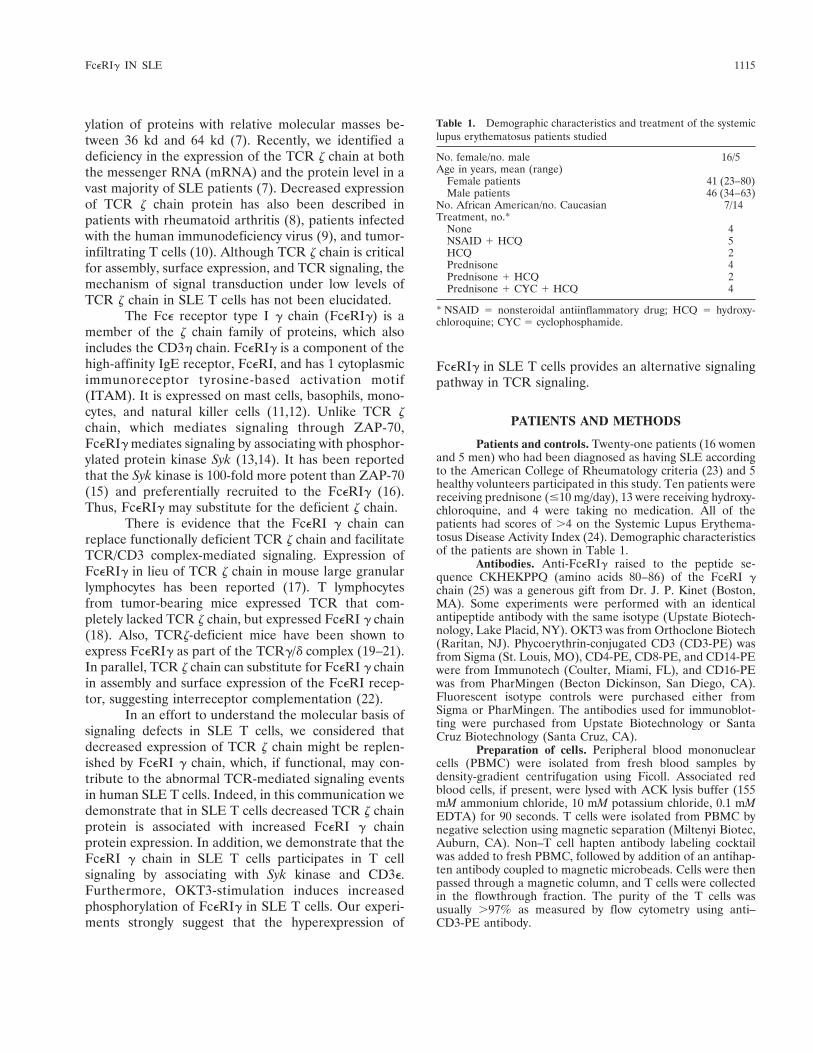
ARTHRITIS & RHEUMATISM Vol. 44, No. 5, May 2001, pp 1114–1121 © 2001, American College of Rheumatology Published by Wiley-Liss, Inc. Fce Receptor Type I g Chain Replaces the Deficient T Cell Receptor z Chain in T Cells of Patients With Systemic Lupus Erythematosus Edith J. Enyedy, 1 Madhusoodana P. Nambiar, 1 Stamatis-Nick C. Liossis, 1 Gregory Dennis, 2 Gary M. Kammer, 3 and George C. Tsokos 1 Objective. T cells from the majority of patients with systemic lupus erythematosus (SLE) express sig- nificantly lower levels of T cell receptor z chain, a critical signaling molecule. However, TCR/CD3 trigger- ing of SLE T cells shows increased phosphorylation of downstream signaling intermediates and increased [Ca 21 ] i response, suggesting the presence of alternative signaling mechanisms. We investigated whether Fce receptor type I g chain (FceRIg) could substitute for TCR z chain and contribute to T cell signaling in SLE. Methods. T cells were purified from the peri- pheral blood of 21 patients with SLE and 5 healthy volunteers. The expression of FceRIg was investigated using immunoblotting, reverse transcriptase– polymerase chain reaction, and flow cytometry methods. Involvement of the FceRIg in T cell signaling was studied by immunoprecipitation and/or immunoblotting after TCR/CD3 stimulation. Results. Western blotting and densitometric ana- lysis showed that the expression of FceRIg in SLE T cells was 4.3-fold higher than in normal T cells (P < 0.001). Flow cytometric analyses of T lymphocyte sub- sets revealed that the proportions of FceRIg1,CD31, FceRIg1,CD41, and FceRIg1, CD81 cells were sig- nificantly greater in SLE patients than in healthy controls (P < 0.001). Immunoprecipitation of SLE T cell lysates with an anti-FceRIg antibody showed that FceRIg associates with the tyrosine kinase Syk and the CD3e chain, suggesting that FceRIg is functionally involved in TCR signaling. Conclusion. These results demonstrate that the FceRI g chain is expressed at high levels in a large proportion of SLE T cells. The increased expression of FceRI g chain in SLE T cells may account in part for the aberrant antigen receptor–initiated signaling and con- tribute to the diverse cellular abnormalities found in this disease. Systemic lupus erythematosus (SLE) is a complex autoimmune disease characterized by the production of autoantibodies and involvement of multiple organs. At the pathogenetic level, the disease presents unpre- cedented complexity involving multiple genes, immuno- regulatory aberrations, and environmental and hor- monal factors. T lymphocytes from patients with SLE display numerous T cell aberrations that range from decreased cytotoxic cell function to increased helper activity and abnormal cytokine production (1,2). At the biochemical level, SLE T cells exhibit signaling defects that affect several interconnecting pathways (3,4). In SLE, the engagement of the T cell receptor (TCR) leads to a disease-specific increase in the free intracytoplasmic calcium concentration ([Ca 21 ] i ) (5). Besides fresh T cells, cultured SLE T cells (5) and autoantigen-specific T cell clones (6) also display increased TCR/CD3-triggered [Ca 21 ] i responses. Anti- CD3 antibodies also induce increased tyrosyl phosphor- The opinions and assertions contained herein are private views of the authors and are not to be construed as official or as reflecting the views of the Department of the Army or the Department of Defense. Dr. Kammer’s work was supported by NIH grant R01-AR- 39501. Dr. Tsokos’ work was supported by NIH grant R01-AI-42269. 1 Edith J. Enyedy, PhD, Madhusoodana P. Nambiar, PhD, Stamatis-Nick C. Liossis, MD, George C. Tsokos, MD, PhD: Walter Reed Army Institute of Research, Silver Spring, Maryland, and Uniformed Services University of the Health Sciences, Bethesda, Maryland; 2 Gregory Dennis, MD: Walter Reed Army Medical Center, Washington, DC; 3 Gary M. Kammer, MD: Wake Forest University School of Medicine, Winston-Salem, North Carolina. Address correspondence and reprint requests to George C. Tsokos, MD, PhD, Walter Reed Army Institute of Research, Building 503 Room 1A32, Robert Grant Avenue, Silver Spring, MD 20910- 7500. Submitted for publication September 22, 2000; accepted in revised form January 4, 2001. 1114

Welcome message from author
This document is posted to help you gain knowledge. Please leave a comment to let me know what you think about it! Share it to your friends and learn new things together.
Transcript
ARTHRITIS & RHEUMATISMVol. 44, No. 5, May 2001, pp 1114–1121© 2001, American College of RheumatologyPublished by Wiley-Liss, Inc.
Fce Receptor Type I g Chain Replaces theDeficient T Cell Receptor z Chain in T Cells of
Patients With Systemic Lupus Erythematosus
Edith J. Enyedy,1 Madhusoodana P. Nambiar,1 Stamatis-Nick C. Liossis,1 Gregory Dennis,2
Gary M. Kammer,3 and George C. Tsokos1
Objective. T cells from the majority of patientswith systemic lupus erythematosus (SLE) express sig-nificantly lower levels of T cell receptor z chain, acritical signaling molecule. However, TCR/CD3 trigger-ing of SLE T cells shows increased phosphorylation ofdownstream signaling intermediates and increased[Ca21]i response, suggesting the presence of alternativesignaling mechanisms. We investigated whether Fcereceptor type I g chain (FceRIg) could substitute forTCR z chain and contribute to T cell signaling in SLE.
Methods. T cells were purified from the peri-pheral blood of 21 patients with SLE and 5 healthyvolunteers. The expression of FceRIg was investigatedusing immunoblotting, reverse transcriptase–polymerase chain reaction, and flow cytometry methods.Involvement of the FceRIg in T cell signaling wasstudied by immunoprecipitation and/or immunoblottingafter TCR/CD3 stimulation.
Results. Western blotting and densitometric ana-lysis showed that the expression of FceRIg in SLE Tcells was 4.3-fold higher than in normal T cells (P <
0.001). Flow cytometric analyses of T lymphocyte sub-sets revealed that the proportions of FceRIg1,CD31,FceRIg1,CD41, and FceRIg1, CD81 cells were sig-nificantly greater in SLE patients than in healthycontrols (P < 0.001). Immunoprecipitation of SLE T celllysates with an anti-FceRIg antibody showed thatFceRIg associates with the tyrosine kinase Syk and theCD3e chain, suggesting that FceRIg is functionallyinvolved in TCR signaling.
Conclusion. These results demonstrate that theFceRI g chain is expressed at high levels in a largeproportion of SLE T cells. The increased expression ofFceRI g chain in SLE T cells may account in part for theaberrant antigen receptor–initiated signaling and con-tribute to the diverse cellular abnormalities found inthis disease.
Systemic lupus erythematosus (SLE) is a complexautoimmune disease characterized by the production ofautoantibodies and involvement of multiple organs. Atthe pathogenetic level, the disease presents unpre-cedented complexity involving multiple genes, immuno-regulatory aberrations, and environmental and hor-monal factors. T lymphocytes from patients with SLEdisplay numerous T cell aberrations that range fromdecreased cytotoxic cell function to increased helperactivity and abnormal cytokine production (1,2).
At the biochemical level, SLE T cells exhibitsignaling defects that affect several interconnectingpathways (3,4). In SLE, the engagement of the T cellreceptor (TCR) leads to a disease-specific increase inthe free intracytoplasmic calcium concentration([Ca21]i) (5). Besides fresh T cells, cultured SLE T cells(5) and autoantigen-specific T cell clones (6) also displayincreased TCR/CD3-triggered [Ca21]i responses. Anti-CD3 antibodies also induce increased tyrosyl phosphor-
The opinions and assertions contained herein are privateviews of the authors and are not to be construed as official or asreflecting the views of the Department of the Army or the Departmentof Defense.
Dr. Kammer’s work was supported by NIH grant R01-AR-39501. Dr. Tsokos’ work was supported by NIH grant R01-AI-42269.
1Edith J. Enyedy, PhD, Madhusoodana P. Nambiar, PhD,Stamatis-Nick C. Liossis, MD, George C. Tsokos, MD, PhD: WalterReed Army Institute of Research, Silver Spring, Maryland, andUniformed Services University of the Health Sciences, Bethesda,Maryland; 2Gregory Dennis, MD: Walter Reed Army Medical Center,Washington, DC; 3Gary M. Kammer, MD: Wake Forest UniversitySchool of Medicine, Winston-Salem, North Carolina.
Address correspondence and reprint requests to George C.Tsokos, MD, PhD, Walter Reed Army Institute of Research, Building503 Room 1A32, Robert Grant Avenue, Silver Spring, MD 20910-7500.
Submitted for publication September 22, 2000; accepted inrevised form January 4, 2001.
1114
ylation of proteins with relative molecular masses be-tween 36 kd and 64 kd (7). Recently, we identified adeficiency in the expression of the TCR z chain at boththe messenger RNA (mRNA) and the protein level in avast majority of SLE patients (7). Decreased expressionof TCR z chain protein has also been described inpatients with rheumatoid arthritis (8), patients infectedwith the human immunodeficiency virus (9), and tumor-infiltrating T cells (10). Although TCR z chain is criticalfor assembly, surface expression, and TCR signaling, themechanism of signal transduction under low levels ofTCR z chain in SLE T cells has not been elucidated.
The Fce receptor type I g chain (FceRIg) is amember of the z chain family of proteins, which alsoincludes the CD3h chain. FceRIg is a component of thehigh-affinity IgE receptor, FceRI, and has 1 cytoplasmicimmunoreceptor tyrosine-based activation motif(ITAM). It is expressed on mast cells, basophils, mono-cytes, and natural killer cells (11,12). Unlike TCR zchain, which mediates signaling through ZAP-70,FceRIg mediates signaling by associating with phosphor-ylated protein kinase Syk (13,14). It has been reportedthat the Syk kinase is 100-fold more potent than ZAP-70(15) and preferentially recruited to the FceRIg (16).Thus, FceRIg may substitute for the deficient z chain.
There is evidence that the FceRI g chain canreplace functionally deficient TCR z chain and facilitateTCR/CD3 complex-mediated signaling. Expression ofFceRIg in lieu of TCR z chain in mouse large granularlymphocytes has been reported (17). T lymphocytesfrom tumor-bearing mice expressed TCR that com-pletely lacked TCR z chain, but expressed FceRI g chain(18). Also, TCRz-deficient mice have been shown toexpress FceRIg as part of the TCRg/d complex (19–21).In parallel, TCR z chain can substitute for FceRI g chainin assembly and surface expression of the FceRI recep-tor, suggesting interreceptor complementation (22).
In an effort to understand the molecular basis ofsignaling defects in SLE T cells, we considered thatdecreased expression of TCR z chain might be replen-ished by FceRI g chain, which, if functional, may con-tribute to the abnormal TCR-mediated signaling eventsin human SLE T cells. Indeed, in this communication wedemonstrate that in SLE T cells decreased TCR z chainprotein is associated with increased FceRI g chainprotein expression. In addition, we demonstrate that theFceRI g chain in SLE T cells participates in T cellsignaling by associating with Syk kinase and CD3e.Furthermore, OKT3-stimulation induces increasedphosphorylation of FceRIg in SLE T cells. Our experi-ments strongly suggest that the hyperexpression of
FceRIg in SLE T cells provides an alternative signalingpathway in TCR signaling.
PATIENTS AND METHODS
Patients and controls. Twenty-one patients (16 womenand 5 men) who had been diagnosed as having SLE accordingto the American College of Rheumatology criteria (23) and 5healthy volunteers participated in this study. Ten patients werereceiving prednisone (#10 mg/day), 13 were receiving hydroxy-chloroquine, and 4 were taking no medication. All of thepatients had scores of .4 on the Systemic Lupus Erythema-tosus Disease Activity Index (24). Demographic characteristicsof the patients are shown in Table 1.
Antibodies. Anti-FceRIg raised to the peptide se-quence CKHEKPPQ (amino acids 80–86) of the FceRI gchain (25) was a generous gift from Dr. J. P. Kinet (Boston,MA). Some experiments were performed with an identicalantipeptide antibody with the same isotype (Upstate Biotech-nology, Lake Placid, NY). OKT3 was from Orthoclone Biotech(Raritan, NJ). Phycoerythrin-conjugated CD3 (CD3-PE) wasfrom Sigma (St. Louis, MO), CD4-PE, CD8-PE, and CD14-PEwere from Immunotech (Coulter, Miami, FL), and CD16-PEwas from PharMingen (Becton Dickinson, San Diego, CA).Fluorescent isotype controls were purchased either fromSigma or PharMingen. The antibodies used for immunoblot-ting were purchased from Upstate Biotechnology or SantaCruz Biotechnology (Santa Cruz, CA).
Preparation of cells. Peripheral blood mononuclearcells (PBMC) were isolated from fresh blood samples bydensity-gradient centrifugation using Ficoll. Associated redblood cells, if present, were lysed with ACK lysis buffer (155mM ammonium chloride, 10 mM potassium chloride, 0.1 mMEDTA) for 90 seconds. T cells were isolated from PBMC bynegative selection using magnetic separation (Miltenyi Biotec,Auburn, CA). Non–T cell hapten antibody labeling cocktailwas added to fresh PBMC, followed by addition of an antihap-ten antibody coupled to magnetic microbeads. Cells were thenpassed through a magnetic column, and T cells were collectedin the flowthrough fraction. The purity of the T cells wasusually .97% as measured by flow cytometry using anti–CD3-PE antibody.
Table 1. Demographic characteristics and treatment of the systemiclupus erythematosus patients studied
No. female/no. male 16/5Age in years, mean (range)
Female patients 41 (23–80)Male patients 46 (34–63)
No. African American/no. Caucasian 7/14Treatment, no.*
None 4NSAID 1 HCQ 5HCQ 2Prednisone 4Prednisone 1 HCQ 2Prednisone 1 CYC 1 HCQ 4
* NSAID 5 nonsteroidal antiinflammatory drug; HCQ 5 hydroxy-chloroquine; CYC 5 cyclophosphamide.
FceRIg IN SLE 1115

Sodium dodecyl sulfate–polyacrylamide gel electro-phoresis (SDS-PAGE) and immunoblotting. T cells were re-suspended in RPMI 1640, and 5 million T cells were lysed for60 minutes at 4°C in buffer containing 20 mM Tris HCl (pH7.4), 100 mM NaCl, 50 mM NaF, 1 mM sodium orthovanadate,5 mM EDTA, 10 mg/ml aprotinin, 10 mg/ml leupeptin, 1 mMphenylmethylsulfonyl fluoride, and 1% Triton X-100. Thelysates were centrifuged at 15,000 revolutions per minute for10 minutes at 4°C, and the supernatants were collected andstored at –80°C. The protein concentration was measuredusing Bio-Rad (Hercules, CA) protein assay reagent. Twelvemicrograms of protein was loaded in each well and separatedon a 16% Tris–glycine gel (Invitrogen, Carlsbad, CA) usingSDS-PAGE. Proteins were transferred to polyvinylidene diflu-oride (PVDF) membrane and blocked with 3% milk in Trisbuffered saline–Tween (0.1M Tris HCl, 0.15M NaCl, 0.5%Tween 20) followed by immunodetection with various antibod-ies (usual dilution: 1:1,000) and appropriate horseradish per-oxidase (HRP)–conjugated secondary antibodies. The ECL kit(Pharmacia Amersham Biotech, Piscataway, NJ) was used fordetection of the bands by chemiluminescence. The intensity ofthe bands in the autoradiogram was evaluated using Quanti-Scan 1.5, and was expressed as arbitrary densitometric units.
TCR/CD3-mediated activation and phosphotyrosineimmunoblotting. Five million T cells were activated with 10mg/ml OKT3 and 20 mg/ml goat anti-mouse antibody for 2.5minutes and 5 minutes, respectively, as described previously(7). Activation was stopped and the cells were lysed as above,electrophoresed, and transferred to PVDF membrane. Themembranes were blocked and immunoblotted with an anti-phosphotyrosine antibody (4G10) and developed as above.
Immunoprecipitation. Total cell lysate (100 mg pro-tein) was precleared with rabbit or mouse IgG and protein A/GPLUS agarose (Santa Cruz Biotechnology) for 30 minutes at4°C. After pelleting, supernatants were transferred to a newtube and incubated with 1 mg of the primary antibody followedby protein A/G PLUS agarose for 1 hour each at 4°C. Aftercentrifugation, the pellet was resuspended in 20 ml loadingbuffer containing 5% 2-mercaptoethanol. The samples wereboiled for 3 minutes and centrifuged. The supernatant wasused for SDS-PAGE followed by transfer to PVDF membraneand immunoblotted with appropriate antibodies as describedabove.
Flow cytometry. One hundred microliters of cell sus-pension (107 PBMC/ml) in staining buffer (phosphate bufferedsaline [PBS] containing 2% fetal bovine serum) was fixed with50 ml of 1.5% paraformaldehyde in PBS for 5 minutes at 4°C.After washing with staining buffer, cells were permeabilized atroom temperature with digitonin (0.5 mg/ml). Nonspecificbinding sites were blocked with MOPC or rabbit IgG for 10minutes. The cells were stained with the primary antibody for30 minutes at 4°C. After washing, cells were incubated withcorresponding fluorescence-labeled secondary antibody in thepresence of digitonin. Surface staining with various fluoresceinisothiocyanate (FITC)– and PE-labeled antibodies followedthe internal staining. In the end, cells were fixed with 1%paraformaldehyde and analyzed using a Becton DickinsonFACScan by using compensation for the FL2 channel andrecording 10,000 events per sample. Data were analyzed usingCellQuest software (Becton Dickinson, San Jose, CA).
Reverse transcriptase–polymerase chain reaction (RT-PCR) of Fc«RI g chain. Total RNA was isolated from 5 millionT cells, using an RNeasy mini kit according to the instructionsof the manufacturer (Qiagen, Santa Clarita, CA). Single-stranded complementary DNA (cDNA) was synthesized from1 mg total RNA, using an AMV reverse transcriptase–basedreverse transcription system (Promega, Madison, WI) andoligo-dT primer according to the instructions of the manufac-turer. The primers for human Fc«RIg were synthesized bySigma-Genosys (The Woodlands, TX) and are as follows:forward 59-CCT-GGG-AGA-GCC-TCA-GCT-CTG-CTA-TAT-C-39 (sense bp 79–106 according to the numberingdescribed by Kuester et al [26]), reverse 59-GAA-TAT-GAC-
Figure 1. Increased levels of Fce receptor type I g chain (FceRIg)protein in T cells of patients with systemic lupus erythematosus (SLE).A, Lysates were prepared from purified T cells from SLE patients andhealthy individuals as described in Patients and Methods. The proteinswere separated by sodium dodecyl sulfate–polyacrylamide gel elec-trophoresis, transferred, and the membranes were immunoblotted withanti-FceRIg, anti–T cell receptor z chain (anti-TCR z) (6B10.2), andanti-actin antibodies. B, For FceRIg and TCR z chain, the protein ratio ofthe optical densities of patient bands:mean value in controls run in thesame gel was calculated and plotted. SLE patients showed low levels ofTCR z chain and increased levels of FceRIg expression. Dashed linesindicate the level of FceRIg and TCR z chain protein in controls.
1116 ENYEDY ET AL

CGC-ATC-TAT-TGT-AAA-G-39 (antisense bp 311–287 [26]).The primers for b-actin, used as a control, were as follows:forward 59-CAT-GGG-TCA-GAA-GGA-TTC-CT-39, reverse59-AGC-TGG-TAG-CTC-TTC-TCC-A-39. Reverse transcrip-tion product was diluted 5-fold and used for PCR amplificationof Fc«RIg or b-actin control. The amplification was carriedout with a Biometra T-3 thermal cycler (Whatman Biometra,Gottingen, Germany), under the following conditions: initialdenaturation at 94°C for 4 minutes, followed by 33 cycles of94°C for 45 seconds, 67°C for 1 minute, 72°C for 1 minute, anda final extension at 72°C for 7 minutes. The PCR products (10ml) were electrophoresed on 1.5% SeaKem agarose gel (FMCBioProducts, Rockland, ME) and visualized with ethidiumbromide staining.
Statistical analysis. All data were analyzed by Stu-dent’s t-test (MINITAB for Windows, release 11.21; Minitab,State College, PA). Data are presented as the mean 6 SEM. Pvalues less than 0.05 were considered significant.
RESULTS
Increased expression of FceRIg protein in T cellsfrom patients with SLE. We (7) and others (27,28) haveshown that SLE T cells express significantly lower levelsof TCR z chain protein than do T cells from normal
controls or rheumatic disease controls. Yet, in SLE Tcells, engagement of the TCR leads to increased [Ca21]iresponses and protein tyrosine phosphorylation of cyto-solic substrates. Since FceRIg has 1 ITAM, which con-fers signaling properties for this protein and can replaceTCRz in certain cellular systems (21,29,30), we hypoth-esized that it might be up-regulated in SLE T cells. Toaddress this hypothesis, we immunoblotted T cell lysatesfrom 21 SLE patients and 5 healthy donors with apolyclonal anti-FceRIg antibody. As shown in Figure1A, SLE T cell lysates consistently revealed increasedband intensities with estimated Mr of 8–10 kd, corre-sponding to FceRIg. The same membranes werestripped and reprobed with an anti-actin antibody toconfirm equal protein loading among lanes (Figure 1A).The intensities of the bands were determined by densi-tometry and used to calculate the mean (6SEM) levelsof FceRIg protein expression in the samples from SLEpatients and controls. The average amount of FceRIgprotein in SLE was 1,500 6 260 units, compared with350 6 130 units in controls. Thus, on average, the levelof FceRIg in SLE T cells was 4.3-fold higher than in Tcells from healthy controls (P , 0.001 by unpairedt-test).
Consistentwithourprevious findings (7), immuno-blotting with N-terminal TCR z chain monoclonal anti-body revealed decreased levels of TCR z chain proteinin SLE T cells (Figure 1A). To determine whetherdecreased amounts of z chain protein correlate withincreased amounts of FceRI g chain protein, we firstnormalized the levels of each chain against the levels ofactin. The amounts of FceRI g chain or z chain for eachsubject were divided by the control amounts run in thesame gel, and the protein ratio was plotted. As shown inFigure 1B, in the majority of SLE patients (15 of 21), thedecrease in TCR z chain protein level was associatedwith an increase in FceRI g chain protein level. The datashow that there were 2 groups of patients: 1 expressinghigh levels of FceRIg and the other expressing modestlevels of FceRIg. Three of the SLE patients showedincreased levels of TCR z chain protein together withhigher FceRIg protein levels than in normal subjects. Inonly 1 SLE patient was the level of FceRIg protein lowerthan the average level in healthy controls, even thoughthis patient had a low level of TCR z chain protein.
We sought correlations between levels of FceRI gchain protein and treatment status, disease activity, andclinical manifestations, but no correlations were identi-fied. It should be noted that slightly more than half ofour patients were not being treated with prednisone atthe time of the study.
Figure 2. Increased levels of FceRIg mRNA in T cells of patients withSLE. Total RNA was isolated from 5 3 106 T lymphocytes, reversetranscribed, and Fc«RI g chain or b-actin was amplified with specificprimers as described in Patients and Methods. The polymerase chainreaction products (10 ml) were electrophoresed in 1.2% agarose gelsand visualized by ethidium bromide staining. Lane 1, 100-bp DNAladder molecular weight marker; lanes 2–7, SLE patient; lane 8,normal donor. The bands were quantitated by densitometry, and thefold increase is shown. All healthy controls showed very low amountsof FceRIg mRNA similar to the results for the control subject depictedin this figure. See Figure 1 for definitions.
FceRIg IN SLE 1117

Increased expression of FceRIg mRNA levels inT cells from patients with SLE. Next we investigatedwhether the increased levels of Fc«RI g chain in SLE Tcells were associated with increased levels of FceRIgmRNA. RNA was isolated from 5 million T cells, andcDNA was synthesized by reverse transcription usingAMV reverse transcriptase. FceRI g chain was PCRamplified from the first strand of cDNA, using specificprimers at an annealing temperature of 67°C as de-scribed in Patients and Methods. As shown in Figure 2,
agarose gel electrophoresis of the RT-PCR productsrevealed a major band with the expected size of 234 bpin all of the SLE patients. Densitometric analysis of thebands showed that in SLE T cells, the expression ofFceRIg mRNA was increased 8–28-fold compared withcontrols. Consistent with the increase in protein level,the increase in expression of Fc«RI g chain mRNA in Tcells from SLE patients compared with that in T cellsfrom healthy donors was significant.
Increased levels of FceRI g chain protein in Tcell subsets CD41 and CD81 from patients with SLE.We then investigated whether FceRI g chain protein wasincreased in both principal T cell subsets, i.e., CD41 andCD81. In addition, we wished to exclude the possibilitythat increased expression of FceRIg was due to thepresence of any non-T cells in the purified population ofT cells. Cells were permeabilized with digitonin, stainedinternally with an anti-FceRIg antibody, and detectedwith an FITC-conjugated anti-rabbit antibody. Doublestaining was performed with CD3-PE, CD4-PE, CD8-PE, CD14-PE, and CD16-PE antibodies. In studies withPBMC, gating on T cells (forward scatter versus sidescatter) was used so that .95% CD31 and only ,3%CD141 of the total number of cells would be inside thegate. In most cases, staining for CD141 inside the gatewas ,1%. The proportion of FceRIg1,CD161 stainedcells in the gated population was slightly higher in SLEsamples (mean 6 SEM, 10.5 6 2.3%) than in controlsamples (8.7 6 1.8%). When purified T cells were used,the FceRIg1,CD161 population was present in onlytrace amounts (,1%), in SLE as well as in normalsamples. For every antibody, an appropriate isotypecontrol was used for setting the axis before evaluatingthe distribution of the cells in the 4 quadrants.
As shown in Figure 3A, we consistently detectedincreased expression of FceRI g chain in the 2 principalsubsets in T cells of SLE patients. Analysis of the dataobtained from the quadrant statistics of double-stainedsamples revealed that the double-positive cell popula-tions of FceRIg1,CD31, FceRIg1,CD41, andFceRIg1CD81 were significantly higher (P , 0.001) inSLE samples than in samples from healthy controls(Figure 3B). These findings were consistent with theimmunoblotting results (Figure 1) and support the no-tion that the FceRI g chain protein level is indeedincreased in SLE T cells.
FceRIg associates with the protein kinase Sykand CDe in SLE T cells. In mast cells (31) FceRIgassociates with the protein tyrosine kinase Syk, whichapparently propagates the surface membrane–initiatedactivation signal. To determine whether the FceRI g
Figure 3. Increased amounts of intracytoplasmic FceRI g chain pro-tein in SLE T cells. Cells were permeabilized with digitonin (0.5mg/ml), and nonspecific binding sites were blocked with normal rabbitIgG and stained with anti-FceRIg antibody followed by fluoresceinisothiocyanate (FITC)–conjugated donkey anti-rabbit antibody as de-scribed in Patients and Methods. Surface staining with phycoerythrin-labeled CD3 (CD3-PE), CD4-PE, or CD8-PE was then performed.Gates were set using isotype controls. Normal rabbit IgG was used asisotype control for FceRIg, at the same concentration as the primaryantibody. A, Histograms of FceRIg staining of permeabilized T cellsfrom a representative SLE patient and a healthy control (dashedline 5 isotype; continuous line 5 FceRIg). B, Presence of FceRIg1
CD31, FceRIg1CD41, and FceRIg1CD81 subsets in SLE patientsand healthy controls (mean and SEM). See Figure 1 for otherdefinitions.
1118 ENYEDY ET AL

chain expressed in SLE T cells is functional and associ-ates with Syk we used an anti-FceRIg antibody toimmunoprecipitate lysates from SLE and normal T cells.Equal amounts of immunoprecipitates were separatedelectrophoretically and immunoblotted with the anti-FceRIg and anti-Syk antibodies. As shown in Figure 4,Syk was detected at high levels only in immunoprecipi-tates of SLE subjects but not of controls, suggesting thatFceRIg is associated with Syk in SLE T cells. Further, todetermine that FceRIg was a component of the TCR/CD3 complex, we immunoblotted the anti-FceRIg pre-cipitates with anti-CD3e antibody. Indeed, CDe wasdetected in all SLE immunoprecipitates, indicating thatFceRIg associates with CD3e of the TCR/CD3 complex(Figure 4). In normal samples, small amounts of CD3echain were detected in the anti-FceRIg precipitates.
Anti-CD3 antibody induces FceRIg phosphory-lation in SLE T cells. The first step in the signaltransduction after TCR/CD3 engagement is the tyrosinephosphorylation of TCR z chain followed by the phos-phorylation of a cellular substrate ZAP-70 or Syk. Wepredicted that the increased expression of FceRI g chainwould be associated with increased TCR/CD3-mediatedtyrosine phosphorylation of FceRIg in SLE T cells. Tcells were activated with 10 mg/ml OKT3 and 20 mg/mlgoat anti-mouse antibody for 2.5 minutes and 5 minutes.Blotting with HRP-conjugated anti-phosphotyrosineantibody showed tyrosine phosphorylation of the 8-kdand 10-kd bands corresponding to FceRIg and 21 and23-kd bands corresponding to the partial and totalphosphorylated forms of TCR z chain (Figure 5). Con-sistent with the deficient TCR z chain, in SLE, theintensity of the TCR z chain bands at 21 kd and 23 kd islower than in controls. Simultaneously, the phosphory-lation of the FceRIg is high in SLE while it is weak innormals. The membranes were stripped and reblotted
with anti-actin antibody to check for equal loading of thelanes. The optical density of the bands was quantitatedand the ratio of the intensities of the FceRIg bands toactin band is shown below each lane in Figure 5.Although there is variation among SLE patients, thephosphorylation of the 8 kd and 10 kd of FceRIg isincreased after activation in SLE T cell lysates, showingthat FceRIg is part of the TCR/CD3 complex (Figure 5).These data suggest that in SLE T cells, besides theconstitutive signaling process, there is additional signal-ing input through the FceRI g chain.
DISCUSSION
In the present study we focused on the mecha-nisms of signal transduction in TCR z chain–deficientSLE T cells. Although TCR z chain is important for theproper assembly, transfer and surface expression ofwhole TCR/CD3 complex, the expression of TCR zchain is not an absolute requirement. Mature singlepositive a/b cells appear in the periphery of z-knockoutmice (32). Recently, it was reported that mice lackingendogenous z chain transfected with the FceRIg genehave normal T cell development and function (20).These data imply that TCR z chain–deficient T cells canpropagate signal transduction cascade initiated by theTCR/CD3 complex. In these situations signaling can betransduced by using the ITAMs of other invariant chainsof CD3 complex or by other homologues of the z chain.
Figure 4. FceRIg from SLE T cells associates with the proteintyrosine kinase Syk and CD3e. Lysates from normal and SLE T cellswere precleared and subjected to immunoprecipitation with anti-FceRIg antibody. The immunoprecipitates were separated by SDS-PAGE, transferred to PVDF membrane, and immunoblotted withanti-FceRIg, anti-Syk (4D10), or anti-CD3e (M20) antibody.
Figure 5. Increased phosphorylation of FceRIg in activated T cellsfrom SLE patients. Five million T cells were activated for 2.5 and 5minutes with 10 mg/ml OKT3 and 20 mg/ml of goat anti-mouseantibody. Lysates were prepared and proteins were separated bySDS-PAGE on a 16% Tris-glycine gel. Proteins were transferred toPVDF membrane. Membranes were blocked with 5% BSA in TBSTand immunoblotted with HRP-conjugated anti-phosphotyrosine anti-body (4G10). The membranes were stripped and reblotted withanti-actin antibody to check for equal loading of the lanes. The opticaldensity of the bands was quantitated and the ratio of the intensities ofthe FceRIg bands to actin band is shown below each lane. Samplesfrom 3 different SLE patients are presented.
FceRIg IN SLE 1119
It has also been reported that Syk instead of ZAP-70 isalternatively and preferentially recruited to the subunitwhich may substitute for the missing z chain, ultimatelyleading to a difference in antigen signaling (20).
The important finding in this study is the in-creased level of expression of FceRIg in T cells fromSLE patients. Although the complete role of the FceRIgin SLE T cells is not known, the low levels of the TCR zchain in SLE T cells correlate with increased levels ofFceRIg, indirectly suggesting that FceRIg could possiblyreplace the deficient TCR z chain. It has been shownthat FceRIg could form functional heterodimers with ortotally replace the TCR z chain, allowing for surfaceexpression of the whole TCR (20). Since FceRI g chaincan associate with the TCR as either a homodimer orheterodimer with TCR z or h chain, it is possible that inSLE T cells the FceRI g chain could exist in either ofthese forms. Our preliminary data suggest that bothhomo- and heterodimers of FceRIg are present in SLET cells (E. Enyedy and G. Tsokos, unpublished results).
High-level expression of FceRIg was observed inboth CD41 and CD81 T cells of SLE patients. Theup-regulation of FceRIg is disease specific and indepen-dent of disease activity, clinical manifestations, andtreatment. The mRNA of FceRIg is also elevated in SLET cells, suggesting that the up-regulation is at the level oftranscription. FceRIg is transcriptionally regulated by abroad tissue-specific promoter which is active in all ofthe hematopoietic cells (33). Two positive cis-actingregulatory elements in T cells have been identified in thepromoter region (33) and may play a role in the regula-tion of FceRIg expression in SLE T cells.
Understanding the intricate relationship betweenthe down-regulation of the TCR z chain and overexpres-sion of the FceRI g chain may provide new insights intothe signaling abnormalities in SLE T cells (1). It hasbeen recently shown that partial phosphorylation of theTCR z chain ITAMs creates an inhibitory signalingenvironment that impedes the activation of T cells (34).Substitution of the TCR z chain by FceRIg could removethis inhibitory environment, resulting in TCR-inducedphosphorylation of proteins and intracellular calciumresponses in SLE T cells. Presently, it is not known howthe increased FceRIg can cause the hyperphosphoryla-tion and increased calcium response described in SLE Tcells. Previous studies reported in tumor-bearing miceshowed that the substitution of TCR z chain by FceRIgleads to low levels of tyrosyl phosphorylation and cal-cium response (18). It is conceivable that the signalintensity could vary based on the proportionate level ofFceRIg and TCR z chain in the TCR complex. In SLE T
cells we believe that, in addition to the proportion ofTCR z chain and FceRIg homo- or heterodimers in theTCR complex, other unidentified signaling abnormali-ties could also contribute to the signal transduction andthe net signal intensity could be higher in magnitude. Insupport of this we have identified significantly increasednumbers of mutations and splice variations of the TCRz chain in SLE T cells. Clearly, further studies arerequired to elucidate the detailed mechanisms of in-creased tyrosine phosphorylation and calcium responsesin SLE T cells.
FceRIg has a higher affinity toward Syk thanZAP-70 (35). Because Syk can phosphorylate tyrosylresidues of ITAMs independently of src-family kinasesand can become activated itself, it is less dependent onthe availability of the src-family kinases than ZAP-70(36). In turn, triggering of the TCR in SLE T cells inwhich the FceRI g chain replaces the TCR z chain mayaccelerate the kinetics of phosphorylation comparedwith normal T cells. Syk contains a 23 amino acidinsertion loop between the second SH2 domain and thecatalytic domain, compared with ZAP-70. Syk can phos-phorylate other Syk molecules, while ZAP-70 requiresLck or Fyn for phosphorylation of tyrosyl residues of theactivation loop. Furthermore, Syk is capable of phos-phorylating ITAMs in the absence of src kinases. Inaddition, the FceRI g chain has several Ser/Thr phos-phorylation sites preceding the ITAM, which couldpromote FceRIg-mediated signaling events. Taken to-gether, this evidence suggests that antigen receptorcomplex–driven signaling events in the absence of TCRz chain could be further facilitated by downstreamsignaling intermediates.
In summary, our results demonstrate elevatedexpression of FceRI g chain in SLE T cells. The pres-ence of increased levels of FceRI g chain enables thegeneration of TCR-mediated signaling that is differentfrom that of normal T cells.
REFERENCES
1. Tsokos GC, Liossis SN. Immune cell signaling defects in lupus:activation, anergy and death. Immunol Today 1999;20:119–24.
2. Tsokos GC. Overview of cellular immune function in systemiclupus erythematosus. In: Lahita RG, editor. Systemic lupus ery-thematosus. San Diego: Academic Press; 1999. p. 17–54.
3. Khan IU, Laxminarayana D, Kammer GM. A genetic mechanismunderlying deficient type I protein kinase A activity in systemiclupus erythematosus T lymphocytes. In: Kammer GM, Tsokos GC,editors. Lupus: molecular and cellular pathogenesis. Totowa (NJ):Humana Press; 1999. p. 231–56.
4. Scott JM, Richardson BC. Impaired DNA methylation in lupus Tcells. In: Kammer GM, Tsokos GC, editors. Lupus: molecular and
1120 ENYEDY ET AL
cellular pathogenesis. Totowa (NJ): Humana Press; 1999. p.278–98.
5. Vassilopoulos D, Kovacs B, Tsokos GC. TCR/CD3 complex-mediated signal transduction pathway in T cells and T cell linesfrom patients with systemic lupus erythematosus. J Immunol1995;155:2269–81.
6. Liossis SN, Hoffman RW, Tsokos GC. Abnormal early TCR/CD3-mediated signaling events of a snRNP-autoreactive lupus T cellclone. Clin Immunol Immunopathol 1998;88:305–10.
7. Liossis S-NC, Ding XZ, Dennis GJ, Tsokos GC. Altered pattern ofTCR/CD3-mediated protein-tyrosyl phosphorylation in T cellsfrom patients with systemic lupus erythematosus: deficient expres-sion of the T cell receptor zeta chain. J Clin Invest 1998;101:1448–57.
8. Maurice MM, Lankester AC, Bezemer AC, Geertsma MF, TakPP, Breedveld FC, et al. Defective TCR-mediated signaling insynovial T cells in rheumatoid arthritis. J Immunol 1997;159:2973–8.
9. Geertsma MF, Wengen-Stevenhagen A, van Dam EM, Risberg K,Kroon FP, Groeneveld PH, et al. Decreased expression of zmolecules by T lymphocytes is correlated with disease progressionin human immunodeficiency virus-infected persons. J Infect Dis1999;180:649–58.
10. Whiteside TL. Signaling defects in T lymphocytes of patients withmalignancy. Cancer Immunol Immunother 1999;48:346–52.
11. Capron M, Soussi GA, Morita M, Truong MJ, Prin L, Kinet JP, etal. Eosinophils: from low- to high-affinity immunoglobulin Ereceptors. Allergy 1995;50 Suppl 25:20–3.
12. Pan XQ, Darby C, Indik ZK, Schreiber AD. Activation of threeclasses of nonreceptor tyrosine kinases following Fc g receptorcrosslinking in human monocytes. Clin Immunol 1999;90:55–64.
13. Shiue L, Green J, Green OM, Karas JL, Morgenstern JP, RamMK, et al. Interaction of p72syk with the g and b subunits of thehigh-affinity receptor for immunoglobulin E, Fc e RI. Mol CellBiol 1995;15:272–81.
14. Shiue L, Zoller MJ, Brugge JS. Syk is activated by phosphoty-rosine-containing peptides representing the tyrosine-based activa-tion motifs of the high affinity receptor for IgE. J Biol Chem1995;270:10498–502.
15. Oliver JM, Burg D, Wilson BS, McLaughlin JL, Geahlen RL.Inhibition of mast cell Fc«RI-mediated signaling and effectorfunction by the Syk-selective inhibitor, piceatannol. J Biol Chem1994;269:29697–703.
16. Taylor N, Jahn T, Smith S, Lamkin T, Uribe L, Liu Y, et al.Differential activation of the tyrosine kinases ZAP-70 and Sykafter FcgRI stimulation. Blood 1997;89:388–96.
17. Koyasu S, d’Adamio L, Arulanandam AR, Abraham S, ClaytonLK, Reinherz EL. T cell receptor complexes containing Fc epsilonRI g homodimers in lieu of CD3 z and CD3 h components: a novelisoform expressed on large granular lymphocytes. J Exp Med1992;175:203–9.
18. Mizoguchi H, O’Shea JJ, Longo DL, Loeffler CM, McVicar DW,Ochoa AC. Alterations in signal transduction molecules in Tlymphocytes from tumor-bearing mice. Science 1992;258:1795–8.
19. Khattri R, Sperling AI, Qian D, Fitch FW, Shores EW, Love PE,et al. TCR-g d cells in CD3 z-deficient mice contain Fc epsilon RIgamma in the receptor complex but are specifically unresponsiveto antigen. J Immunol 1996;157:2320–7.
20. Shores E, Flamand V, Tran T, Grinberg A, Kinet J-P, Love PE.Fc«RIg can support T cell development and function in micelacking endogenous TCR z-chain. J Immunol 1997;159:222–30.
21. Qian D, Sperling AI, Lancki DW, Tatsumi Y, Barrett TA,Bluestone JA, et al. The g chain of the high-affinity receptor forIgE is a major functional subunit of the T-cell antigen receptorcomplex in g d T lymphocytes. Proc Natl Acad Sci U S A1993;90:11875–9.
22. Howard FD, Rodewald HR, Kinet JP, Reinherz EL. CD3 zsubunit can substitute for the g subunit of Fc e receptor type I inassembly and functional expression of the high-affinity IgE recep-tor: evidence for interreceptor complementation. Proc Natl AcadSci U S A 1990;87:7015–9.
23. Tan EM, Cohen AS, Fries JF, Masi AT, McShane DJ, RothfieldNF, et al. The 1982 revised criteria for the classification of systemiclupus erythematosus. Arthritis Rheum 1982;25:1271–7.
24. Bombardier C, Gladman DD, Urowitz MB, Caron D, Chang CH,and the Committee on Prognosis Studies in SLE. Derivation of theSLEDAI: a disease activity index for lupus patients. ArthritisRheum 1992;35:630–40.
25. Letourneur O, Kennedy IC, Brini AT, Ortaldo JR, O’Shea JJ,Kinet JP. Characterization of the family of dimers associated withFc receptors (Fc e RI and Fc g RIII). J Immunol 1991;147:2652–6.
26. Kuester H, Thompson H, Kinet JP. Characterization and expres-sion of the gene for the human receptor g subunit: definition of anew gene family. J Biol Chem 1990;265:6448–52.
27. Takeuchi T, Tsuzaka K, Pang M, Amano K, Koide J, Abe T. TCRz chain lacking exon 7 in two patients with systemic lupuserythematosus. Int Immunol 1998;10:911–21.
28. Brundula V, Rivas LJ, Blasini AM, Parıs M, Salazar S, StekmanIL, et al. Diminished levels of T cell receptor z chains in peripheralblood T lymphocytes from patients with systemic lupus erythem-atosus. Arthritis Rheum 1999;42:1908–16.
29. Orloff DG, Ra CS, Frank SJ, Klausner RD, Kinet JP. Family ofdisulphide-linked dimers containing the z and h chains of theT-cell receptor and the g chain of Fc receptors. Nature 1990;347:189–91.
30. Paolini R, Renard V, Vivier E, Ochiai K, Jouvin M-H, Malissen B,et al. Different roles for the Fc« RI g chain as a function of thereceptor context. J Exp Med 1995;181:247–55.
31. Chen T, Repetto B, Chizzonite R, Pullar C, Burghardt C, DharmE, et al. Interaction of phosphorylated Fc«RIg immunoglobulinreceptor tyrosine activation motif-based peptides with dual andsingle SH2 domains of p72syk: assessment of binding parametersand real time binding kinetics. J Biol Chem 1996;271:25308–15.
32. Shores EW, Ono M, Kawabe T, Sommers CL, Tran T, Lui K, et al.T cell development in mice lacking all T cell receptor z familymembers (z, h, and FceRIg). J Exp Med 1998;187:1093–101.
33. Brini AT, Lee GM, Kinet JP. Involvement of Alu sequences in thecell-specific regulation of transcription of the g chain of Fc and Tcell receptors. J Biol Chem 1993;268:1355–61.
34. Kersh EN, Kersh GJ, Allen PM. Partially phosphorylated T cellreceptor z molecules can inhibit T cell activation. J Exp Med1999;190:1627–36.
35. Kimura T, Sakamoto H, Appella E, Siraganian RP. Conforma-tional changes induced in the protein tyrosine kinase p72syk bytyrosine phosphorylation or by binding of phosphorylated immu-noreceptor tyrosine-based activation motif peptides. Mol Cell Biol1996;16:1471–8.
36. Turner I, Schweighoffer I, Colucci I, di Santo JP, Tybulewicz VL.Tyrosine kinase SYK: essential functions for immunoreceptorsignalling. Immunol Today 2000;21:148–54.
FceRIg IN SLE 1121
Related Documents