J Clin Pathol 1982;35:1380-1383 Fatty tumours of the uterus DERRICK J POUNDER From the Division of Tissue Pathology, Institute of Medical and Veterinary Science, Adelaide, South Australia SUMMARY Uterine fatty tumours (UFI) are uncommon and have received little attention in the English literature. They have aroused interest as a consequence of occasional diagnostic confusion with sarcomas and the continuing unresolved dispute as to their histogenesis. Three cases of UFT are described and the pathological features of note discussed. The viewpoint that these tumours are hamartomas/choristomas is rejected. UFT most probably represent tumour metaplasia within a leiomyoma. There is no uniform accepted nomenclature for such tumours and it is suggested that they be designated "uterine fatty tumours" and subdivided into "lipoma" and "mixed lipoma/ leiomyoma" (synonym lipoleiomyoma). Fatty tumours primary to the uterus are uncommon' and are almost invariably benign.2 Occasionally the clinical presentation3 or macroscopic appearance2 may mimic a sarcoma and create diagnostic con- fusion. There is no consensus as to the histogenesis or nomenclature of uterine fatty tumours (UFT).4 5 Three cases of UFT are presented and discussion centres on these areas of interest and contention. Material and methods All uterine fatty tumours (UFT) accessioned on the files of the Institute of Medical and Veterinary Science6 over a three-year period, January 1979 to January 1982, were retrieved. Amongst the approximately 54 000 surgical accessions during this period there were three cases of UFT. Nine, two and one paraffin blocks of tumour tissue were available from these three cases for review. Haematoxylin and eosin-stained and, where appropriate, Masson trichrome-stained sections from each paraffin block were studied. Results A summary of the relevant clinical history, clinical diagnosis and final diagnosis for the three cases studied is set out in the Table. The pathological diagnosis of a UFT was confirmed in each of the three cases and only the pathological findings relevant to this diagnosis will be described in detail. CASE 1 (FIGS. 1 AND 2) The resected specimen comprised a uterus with two Accepted for publication 2 June 1982 normal Fallopian tubes. There was a very large intra- mural uterine tumour mass which measured 200 mm in greatest diameter and weighed 2140 g. The tumour, which distorted the uterine cavity into an enlarged slit, was rounded and well circumscribed but not encapsulated. On cut surface the greater part of the tumour appeared yellow, lobulated and fatty. Within the tumour mass and abutting upon one margin was a hard, grey calcified wedge measuring 120 x 80 x 75 mm. Two typical spherical, circumscribed leio- myomas, each approximateiy 15 mm in diameter, were also identified within the myometrium. Microscopically, the fatty tumour comprised adult- type adipose tissue divided into lobules by thin connective tissue septae. There was no true tumour capsule but adjacent myometrium was compressed around the tumour to provide a false capsule. Irregularly dispersed amongst the adipocytes were single and small clusters of smooth muscle cells. In some areas of the tumour the smooth muscle component could be identified only after careful searching whilst in other areas groups of smooth muscle cells were readily apparent. Smooth muscle cells were present deep within the tumour as well as at its periphery. Nowhere did the smooth muscle component form the wide interlacing and whorled bands typically seen in a leiomyoma. The hard calcified wedge identified macroscopically comprised degenerate adipose tissue with an extensively hyalin- ised connective tissue stroma and heavy calcification of adipocytes. Similar smaller foci of hyaline sclerosis and dystrophic calcification of adipocytes were dis- posed irregularly throughout the tumour. The two small leiomyomas identified macroscopically showed some stromal hyalinisation but did not contain an adipose tissue component. 1380 copyright. on 14 April 2019 by guest. Protected by http://jcp.bmj.com/ J Clin Pathol: first published as 10.1136/jcp.35.12.1380 on 1 December 1982. Downloaded from

Welcome message from author
This document is posted to help you gain knowledge. Please leave a comment to let me know what you think about it! Share it to your friends and learn new things together.
Transcript

J Clin Pathol 1982;35:1380-1383
Fatty tumours of the uterus
DERRICK J POUNDER
From the Division of Tissue Pathology, Institute ofMedical and Veterinary Science, Adelaide, South Australia
SUMMARY Uterine fatty tumours (UFI) are uncommon and have received little attention in theEnglish literature. They have aroused interest as a consequence of occasional diagnostic confusionwith sarcomas and the continuing unresolved dispute as to their histogenesis. Three cases of UFTare described and the pathological features of note discussed. The viewpoint that these tumours arehamartomas/choristomas is rejected. UFT most probably represent tumour metaplasia within a
leiomyoma. There is no uniform accepted nomenclature for such tumours and it is suggested thatthey be designated "uterine fatty tumours" and subdivided into "lipoma" and "mixed lipoma/leiomyoma" (synonym lipoleiomyoma).
Fatty tumours primary to the uterus are uncommon'and are almost invariably benign.2 Occasionally theclinical presentation3 or macroscopic appearance2may mimic a sarcoma and create diagnostic con-fusion. There is no consensus as to the histogenesis ornomenclature of uterine fatty tumours (UFT).45Three cases of UFT are presented and discussioncentres on these areas of interest and contention.
Material and methods
All uterine fatty tumours (UFT) accessioned on thefiles of the Institute of Medical and VeterinaryScience6 over a three-year period, January 1979 toJanuary 1982, were retrieved. Amongst theapproximately 54 000 surgical accessions during thisperiod there were three cases of UFT. Nine, two andone paraffin blocks of tumour tissue were availablefrom these three cases for review. Haematoxylin andeosin-stained and, where appropriate, Massontrichrome-stained sections from each paraffin blockwere studied.
Results
A summary of the relevant clinical history, clinicaldiagnosis and final diagnosis for the three casesstudied is set out in the Table. The pathologicaldiagnosis of a UFT was confirmed in each of the threecases and only the pathological findings relevant tothis diagnosis will be described in detail.
CASE 1 (FIGS. 1 AND 2)The resected specimen comprised a uterus with two
Accepted for publication 2 June 1982
normal Fallopian tubes. There was a very large intra-mural uterine tumour mass which measured 200 mmin greatest diameter and weighed 2140 g. The tumour,which distorted the uterine cavity into an enlargedslit, was rounded and well circumscribed but notencapsulated. On cut surface the greater part of thetumour appeared yellow, lobulated and fatty. Withinthe tumour mass and abutting upon one margin was ahard, grey calcified wedge measuring 120 x 80 x 75mm. Two typical spherical, circumscribed leio-myomas, each approximateiy 15 mm in diameter,were also identified within the myometrium.
Microscopically, the fatty tumour comprised adult-type adipose tissue divided into lobules by thinconnective tissue septae. There was no true tumourcapsule but adjacent myometrium was compressedaround the tumour to provide a false capsule.Irregularly dispersed amongst the adipocytes weresingle and small clusters of smooth muscle cells. Insome areas of the tumour the smooth musclecomponent could be identified only after carefulsearching whilst in other areas groups of smoothmuscle cells were readily apparent. Smooth musclecells were present deep within the tumour as well asat its periphery. Nowhere did the smooth musclecomponent form the wide interlacing and whorledbands typically seen in a leiomyoma. The hardcalcified wedge identified macroscopically compriseddegenerate adipose tissue with an extensively hyalin-ised connective tissue stroma and heavy calcificationof adipocytes. Similar smaller foci of hyaline sclerosisand dystrophic calcification of adipocytes were dis-posed irregularly throughout the tumour. The twosmall leiomyomas identified macroscopically showedsome stromal hyalinisation but did not contain anadipose tissue component.
1380
copyright. on 14 A
pril 2019 by guest. Protected by
http://jcp.bmj.com
/J C
lin Pathol: first published as 10.1136/jcp.35.12.1380 on 1 D
ecember 1982. D
ownloaded from

Clinical details ofthree cases of uterine fatty tumour
Case Age (yr) Clinical history Clinical diagnosis Final diagnosis
1 83 Five years increasing weight and ? Ovarian cyst Uterine mixed lipoma/leiomyoma,abdominal girth; initially refused ? Uterine leiomyoma cervical dysplasia (moderate), uterinesurgery; oedema of right leg for one leiomyomas (2)week
2 80 Long-standing uterine prolapse Uterine prolapse Uterine prolapse, cervicalkeratinisation, uterine leiomyoma,uterine mixed lipoma/leiomyoma
3 73 Post-menopausal bleeding, large Ovarian tumour Mucinous cystadenocarcinoma (rightpelvic mass; curetting-hyperplastic ovary), hyperplastic endometrialpolyps polyp, uterine mixed lipoma/leiomyoma
4,- 1 ..
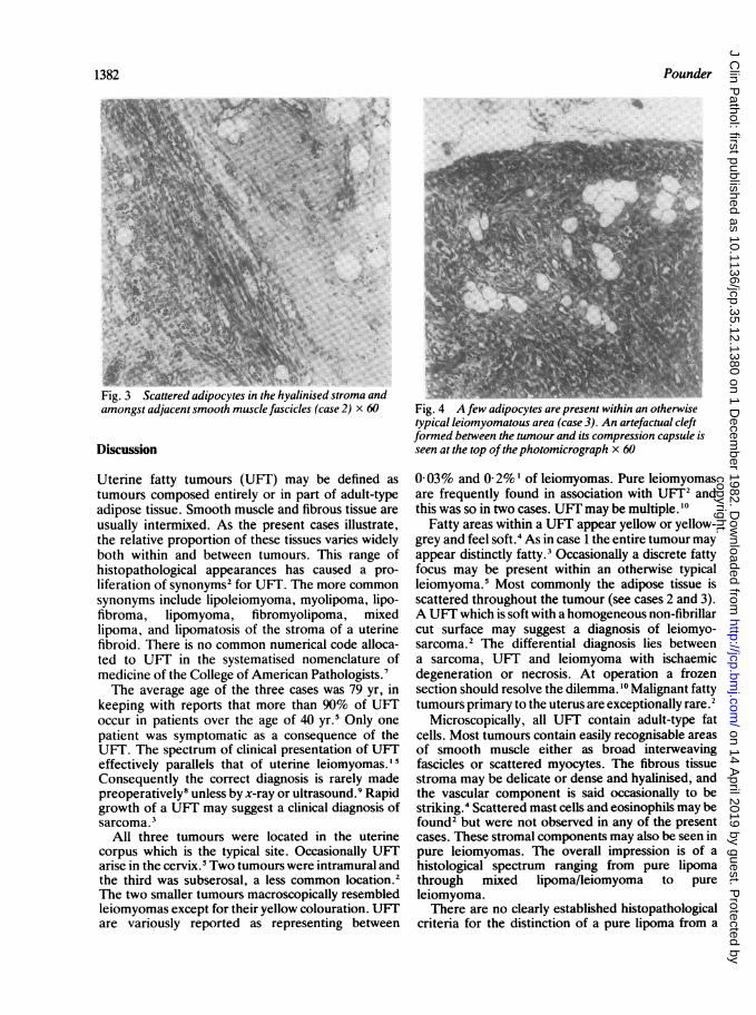
Fig. 1 An intra-mural mixed lipoma/eiomyoma (case 1)The atrophic endometrium is seen at the top ofthephotomicrograph. Note the lack ofbroad smooth musclefascicles within the tumour which is predominantly fatty.Masson's trichrome x 20
CASE 2 (FIG. 3)The simple hysterectomy specimen comprised auterus with parous cervix. A spherical, yellow andwhite, circumscribed nodule 25 mm in diameter wassited intramurally at the fundus. A second cir-cumscribed intramural nodule 8 mm in diameter waspresent in the lower uterine segment.
Microscopically the larger nodule was circum-scribed but not encapsulated and comprised anintimate admixture of adult-type adipose tissue,smooth muscle fascicles and a dense hyalinisedstroma. The adipose tissue component wasprominent and readily identified. The stromalhyalinisation and the quantity of blood vesselsresembled that seen in a typical leiomyoma. The
...-v _------
X,k _s__--W.
'... I
/~~~~~~~~~~~.1%,i I~
Fig. 2 Clusters ofsmooth muscle cells are scattered amongstthe adipocytes (case 1). Masson's trichrome x 60
smaller nodule in the lower uterine segment was aleiomyoma without any adipose tissue component.
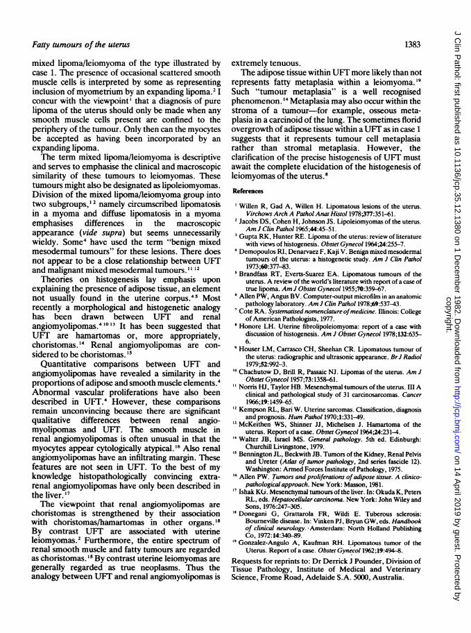
CASE 3 (FIG. 4)The surgical specimen was a total hysterectomy withbilateral salpingo-oophorectomy. At the uterinefundus was a subserosal, circumscribed, yellow andwhite nodule 20 mm in diameter. Microscopically,this fundal nodule resembled the UFT in case 2above. The smooth muscle component was moreprominent and approximately 10% of the tumour wasoccupied by broad whorling bands of smooth musclein a pattern typically seen in uterine leiomyomas. Thesmooth muscle component was primarily localised tothe periphery of the tumour whilst centrally there wasextensive hyalinisation. Adipocytes were sparselyscattered amongst the bands of smooth muscle butwere very prominent centrally.
1381Fatty tumours of the uterus
copyright. on 14 A
pril 2019 by guest. Protected by
http://jcp.bmj.com
/J C
lin Pathol: first published as 10.1136/jcp.35.12.1380 on 1 D
ecember 1982. D
ownloaded from
Pounder
JI 19. J 13utere UtvCtV it il &Vtyutina atr urt&tUn"&-V#'*-I`':0KA.T.' -W-wr_-II---:%- _ .
amongst adjacent smooth muscle fascicles (case 2) x 60 Fig. 4 A few adipocytes are present within an otherwisetypical leiomyomatous area (case 3). An artefactual cleftformed between the tumour and its compression capsule is
Discussion seen at the top ofthe photomicrograph x 60
Uterine fatty tumours (UFT) may be defined as
tumours composed entirely or in part of adult-typeadipose tissue. Smooth muscle and fibrous tissue are
usually intermixed. As the present cases illustrate,the relative proportion of these tissues varies widelyboth within and between tumours. This range ofhistopathological appearances has caused a pro-
liferation of synonyms2 for UFT. The more common
synonyms include lipoleiomyoma, myolipoma, lipo-fibroma, lipomyoma, fibromyolipoma, mixedlipoma, and lipomatosis of the stroma of a uterinefibroid. There is no common numerical code alloca-ted to UFT in the systematised nomenclature ofmedicine of the College of American Pathologists.'The average age of the three cases was 79 yr, in
keeping with reports that more than 90% of UFToccur in patients over the age of 40 yr.5 Only one
patient was symptomatic as a consequence of theUFT. The spectrum of clinical presentation of UFTeffectively parallels that of uterine leiomyomas.'5Consequently the correct diagnosis is rarely madepreoperatively8 unless by x-ray or ultrasound.9 Rapidgrowth of a UFT may suggest a clinical diagnosis ofsarcoma.3
All three tumours were located in the uterinecorpus which is the typical site. Occasionally UFTarise in the cervix.5 Two tumours were intramural andthe third was subserosal, a less common location.2The two smaller tumours macroscopically resembledleiomyomas except for their yellow colouration. UFTare variously reported as representing between
003% and 0 2%' of leiomyomas. Pure leiomyomasare frequently found in association with UF1T2 andthis was so in two cases. UFT may be multiple. 10
Fatty areas within a UFT appear yellow or yellow-grey and feel soft.4 As in case 1 the entire tumour mayappear distinctly fatty.3 Occasionally a discrete fattyfocus may be present within an otherwise typicalleiomyoma.5 Most commonly the adipose tissue isscattered throughout the tumour (see cases 2 and 3).A UFT which is soft with a homogeneous non-fibrillarcut surface may suggest a diagnosis of leiomyo-sarcoma.2 The differential diagnosis lies betweena sarcoma, UFT and leiomyoma with ischaemicdegeneration or necrosis. At operation a frozensection should resolve the dilemma. '0 Malignant fattytumours primary to the uterus are exceptionally rare.2
Microscopically, all UFT contain adult-type fatcells. Most tumours contain easily recognisable areasof smooth muscle either as broad interweavingfascicles or scattered myocytes. The fibrous tissuestroma may be delicate or dense and hyalinised, andthe vascular component is said occasionally to bestriking.4 Scattered mast cells and eosinophils may befound2 but were not observed in any of the presentcases. These stromal components may also be seen inpure leiomyomas. The overall impression is of ahistological spectrum ranging from pure lipomathrough mixed lipoma/leiomyoma to pureleiomyoma.There are no clearly established histopathological
criteria for the distinction of a pure lipoma from a
1382
copyright. on 14 A
pril 2019 by guest. Protected by
http://jcp.bmj.com
/J C
lin Pathol: first published as 10.1136/jcp.35.12.1380 on 1 D
ecember 1982. D
ownloaded from
Fatty tumours of the uterus
mixed lipoma/leiomyoma of the type illustrated bycase 1. The presence of occasional scattered smoothmuscle cells is interpreted by some as representinginclusion of myometrium by an expanding lipoma.2 Iconcur with the viewpoint' that a diagnosis of purelipoma of the uterus should only be made when anysmooth muscle cells present are confined to theperiphery of the tumour. Only then can the myocytesbe accepted as having been incorporated by anexpanding lipoma.The term mixed lipoma/leiomyoma is descriptive
and serves to emphasise the clinical and macroscopicsimilarity of these tumours to leiomyomas. Thesetumours might also be designated as lipoleiomyomas.Division of the mixed lipoma/leiomyoma group intotwo subgroups,' 2 namely circumscribed lipomatosisin a myoma and diffuse lipomatosis in a myomaemphasises differences in the macroscopicappearance (vide supra) but seems unnecessarilywieldy. Some4 have used the term "benign mixedmesodermal tumours" for these lesions. There doesnot appear to be a close relationship between UFTand malignant mixed mesodermal tumours." 12
Theories on histogenesis lay emphasis uponexplaining the presence of adipose tissue, an elementnot usually found in the uterine corpus.45 Mostrecently a morphological and histogenetic analogyhas been drawn between UFT and renalangiomyolipomas." '1' " It has been suggested thatUFT are hamartomas or, more appropriately,choristomas. '4 Renal angiomyolipomas are con-sidered to be choristomas. 15
Quantitative comparisons between UFT andangiomyolipomas have revealed a similarity in theproportions of adipose and smooth muscle elements.4Abnormal vascular proliferations have also beendescribed in UFT.4 However, these comparisonsremain unconvincing because there are significantqualitative differences between renal angio-myolipomas and UFT. The smooth muscle inrenal angiomyolipomas is often unusual in that themyocytes appear cytologically atypical.'6 Also renalangiomyolipomas have an infiltrating margin. Thesefeatures are not seen in UFT. To the best of myknowledge histopathologically convincing extra-renal angiomyolipomas have only been described inthe liver.'7The viewpoint that renal angiomyolipomas are
choristomas is strengthened by their associationwith choristomas/hamartomas in other organs.'8By contrast UFT are associated with uterineleiomyomas.2 Furthermore, the entire spectrum ofrenal smooth muscle and fatty tumours are regardedas choristomas. '5 By contrast uterine leiomyomas aregenerally regarded as true neoplasms. Thus theanalogy between UFT and renal angiomyolipomas is
extremely tenuous.The adipose tissue within UFT more likely than not
represents fatty metaplasia within a leiomyoma.'9Such "tumour metaplasia" is a well recognisedphenomenon. 14 Metaplasia may also occur within thestroma of a tumour-for example, osseous meta-plasia in a carcinoid of the lung. The sometimes floridovergrowth of adipose tissue within aUFT as in case 1suggests that it represents tumour cell metaplasiarather than stromal metaplasia. However, theclarification of the precise histogenesis of UFT mustawait the complete elucidation of the histogenesis ofleiomyomas of the uterus.8
References
'Willen R, Gad A, Willen H. Lipomatous lesions of the uterus.Virchows Arch A PatholAnat Histol 1978;377:351-61.
2 Jacobs DS, Cohen H, Johnson JS. Lipoleiomyomas of the uterus.Am J Clin Pathol 1965;44:45-51.
3Gupta RK, Hunter RE. Lipoma of the uterus: review of literaturewith views of histogenesis. Obstet Gynecol 1964;24:255-7.
4Demopoulos RI, Denarvaez F, Kaji V. Benign mixed mesodermaltumours of the uterus: a histogenetic study. Am J Clin Pathol1973;60:377-83.
Brandfass RT, Everts-Suarez EA. Lipomatous tumours of theuterus. A review of the world's literature with report of a case oftrue lipoma. Am J Obstet Gynecol 1955;70:359-67.
'Allen PW, Angus BV. Computer-output microfilm in an anatomicpathology laboratory. Am J Clin Pathol 1978;69:537-43.
Cote RA. Systematised nomenclature ofmedicine. Illinois: Collegeof American Pathologists, 1977.
8Honore LH. Uterine fibrolipoleiomyoma: report of a case withdiscussion of histogenesis. Am J Obstet Gynecol 1978;132:635-6.
9 Houser LM, Carrasco CH, Sheehan CR. Lipomatous tumour ofthe uterus: radiographic and ultrasonic appearance. BrJ Radiol1979;52:992-3.
'° Chachutow D, Brill R, Passaic NJ. Lipomas of the uterus. Am JObstet Gynecol 1957;73:1358-61.
"Norris HJ, Taylor HB. Mesenchymal tumours of the uterus. III Aclinical and pathological study of 31 carcinosarcomas. Cancer1966;19:1459-65.
2 Kempson RL, Bari W. Uterine sarcomas. Classification, diagnosisand prognosis. Hum Pathol 1970;1:331-49.
3 McKeithen WS, Shinner JJ, Michelsen J. Hamartoma of theuterus. Report of a case. Obstet Gynecol 1964;24:231-4.
' Walter JB, Israel MS. General pathology. 5th ed. Edinburgh:Churchill Livingstone, 1979.
"Bennington JL, Beckwith JB. Tumors of the Kidney, Renal Pelvisand Ureter (Atlas of tunor pathology, 2nd series fascicle 12).Washington: Armed Forces Institute of Pathology, 1975.
6 Allen PW. Tumors and proliferations ofadipose tissue. A clinico-pathological approach. New York: Masson, 1981.
Ishak KU. Mesenchymal tumours ofthe liver. In: Okuda K, PetersRL, eds. Hepatocellular carcinoma. New York: John Wiley andSons, 1976:247-305.
18 Donegani G, Grattarola FR, Wildi E. Tuberous sclerosis:Bournevllle disease. In: Vinken PJ, Bryun GW, eds. Handbookof clinical neurology. Amsterdam: North Holland PublishingCo, 1972:14:340-89.
"Gonzalez-Angulo A, Kaufman RH. Lipomatous tumor of theUterus. Report of a case. Obstet Gynecol 1962;19:494-8.
Requests for reprints to: Dr Derrick J Pounder, Division ofTissue Pathology, Institute of Medical and VeterinaryScience, Frome Road, Adelaide S.A. 5000, Australia.
1383
copyright. on 14 A
pril 2019 by guest. Protected by
http://jcp.bmj.com
/J C
lin Pathol: first published as 10.1136/jcp.35.12.1380 on 1 D
ecember 1982. D
ownloaded from
Related Documents