CHEMICAL COMPOSITION OF THE ESSENTIAL OILS OF FIVE FRUIT TREES AND NON-VOLATILE CONSTITUENTS OF Theobroma cacao L. POD-HUSK BY FATIMAH TEMITAYO ISHOLA B. Sc., M. Sc. (Ibadan) 116570 A Thesis in the Department of Chemistry, Submitted to the Faculty of Science in partial fulfillment of the requirements of the Degree of DOCTOR OF PHILOSOPHY of the UNIVERSITY OF IBADAN AUGUST, 2016

Welcome message from author
This document is posted to help you gain knowledge. Please leave a comment to let me know what you think about it! Share it to your friends and learn new things together.
Transcript

CHEMICAL COMPOSITION OF THE ESSENTIAL OILS OF
FIVE FRUIT TREES AND NON-VOLATILE CONSTITUENTS OF
Theobroma cacao L. POD-HUSK
BY
FATIMAH TEMITAYO ISHOLA B. Sc., M. Sc. (Ibadan)
116570
A Thesis in the Department of Chemistry,
Submitted to the Faculty of Science
in partial fulfillment of the requirements of the Degree of
DOCTOR OF PHILOSOPHY
of the
UNIVERSITY OF IBADAN
AUGUST, 2016
ii
ABSTRACT
Essential Oils (EOs) are volatile secondary metabolites characterised by a strong odour
and widely used for pharmacological and industrial applications. There is dearth of
information on chemical compositions and bioactivities of EOs of some fruit trees in
Nigeria. This study was therefore designed to extract and characterise the EOs from
selected fruit trees, screen the EOs for bioactivity as well as to isolate and characterise
non-volatile constituents from Theobroma cacao L. pod-husk due to its availability.
The plant samples (Carica papaya L., Theobroma cacao L., Persea americana M.,
Ananas comosus (L) Merr and Chrysophyllum albidum G. Don) were collected in
Ibadan, identified and authenticated at the Herbarium of Forest Research Institute of
Nigeria, Ibadan. Essential oils were extracted from the leaves, stem-barks, root-barks,
fruits, peels, pod-husk and seeds of the plants using hydro-distillation method and
analysed by Gas Chromatography (Flame Ionization Detector and Mass Spectrometry)
techniques. The antibacterial activity of the EOs at 20 µg/mL was assayed on two
Gram-positive and four Gram-negative bacteria using Microplate Alamar Blue Assay
measured in UV/Visible spectrophotometer. The antioxidant activity of the EOs at 20
µg/mL was determined by radical scavenging procedure while insecticidal activity was
evaluated by contact toxicity test using three grain pests. Pure compounds were
isolated from methanol extract of T. cacao pod-husk by chromatographic techniques.
The chemical structures of the compounds were elucidated using Infrared, Nuclear
Magnetic Resonance and Mass Spectroscopic techniques. Data were analysed using
descriptive statistics.
Twenty-seven EOs were obtained and their yield ranged from 0.1 to 1.2% (v/w). The
major components in P. americana EOs were β-caryophyllene (12.7%; leaf),
tetradecanal (31.8%; root-bark), globulol (25.4%; peel), (Z,Z)-4,15-octadecadien-1-ol
acetate (21.8%; seed), tetradecanal (24.9%; stem-bark) and p-xylene (40.5%; fruit).
Carica papaya EOs mainly comprised benzylisothiocyanate (89.1%; seed),
octadecanol (62.5%; root), octadecanol (71.1%; stem), m-xylene (35.1%; stem-bark),
heptadecanol (25.2%; fruit), phytol (21.8%; leaf), benzylisothiocyanate (71.5%; root-
bark) and 9-hexadecen-1-ol (16.9%; peel). P-xylene (62.4%; fruit), p-xylene (29.9%;
shoot) and tetradecanoic acid (8.6%; peel) dominated A. comosus EOs. The principal
constituents in T. cacao EOs were hexadecanoic acid (78.7%; leaf), o-xylene (53.3%;
iii
seed), ledol (33.6%; pod-husk) and β-bisabolol (17.3%; stem-bark). The dominant
compounds in C. albidum EOs were m-xylene (66.7%; seed), p-xylene (21.4%; seed-
bark), ethylhexadecanoate (19.9%; leaf), hexadecanoic acid (14.7%; stem-bark), m-
xylene (53.1%; root-bark) and hexadecanoic acid (14.7%; fruit-bark). The chemical
constituents for twenty-one of the EOs of the fruit plants were obtained for the first
time ever. Theobroma cacao leaf EO exhibited the highest inhibition against Gram-
negative bacteria at 78.6%, while P. americana fruit and peel EOs showed the highest
inhibition against Gram-positive bacteria at 69.9%. Persea americana fruit and seed
EOs displayed the highest and lowest radical scavenging activity at 42.1 and 1.2%,
respectively. The EOs showed activity between 0 to 20% in insecticidal assay. Column
chromatography of the methanol extract of T. cacao pod-husk yielded three known
triterpenes: 24-hydroxy-9,19-cycloanost-25-en-3-one, stigmast-5-en-3β-ol and ergosta-
5α,8α-epidioxy-6,22-dien-3β-ol.
The EOs have antibacterial and antioxidant properties which is indicative of their
potential as sources of pharmaceuticals.
Keywords: Fruit tree, Essential oils, Antibacterial activity, Triterpenes, Theobroma
cacao L.
Word count: 488

iv
DEDICATION
This research work is dedicated to:
MY LOVING
DAD
&
MUM
A MESSAGE TO MY PARENTS
You are one in a billion couples;
For Parents like you are hard to come by
If you don’t mind my saying it,
I owe everything in my life to you
And nothing to myself.
Your examples of hardwork, honesty,
Enthusiasm in others’ progress and kindness
Encourage and spur me on
To hope for the really worthwhile things in life.
Your love inspires me.
Your guidance and support saw me through
The most trying periods of my life
To be factual, the greatest blessing of my life
Is that I have you as Parents.
v
ACKNOWLEDGEMENTS
My deep and sincere gratitude goes to my supervisors; Professor O. Ekundayo and Dr.
Sherifat A. Aboaba for their thorough supervision, understanding and support that
propelled the success of this work.
I am very grateful to the Head of Department, Prof. A. A. Adesomoju and the entire
staff of Chemistry Department for the knowledge imparted to me and for allowing me
to use the facilities in the department during the course of my studies. I appreciate Dr.
I. O. Oladosu and Dr. O. Adewuyi for their assistance and advise at every stage of this
work.
With a deep sense of gratitude, I acknowledge the Third World Academy of Science
(TWAS) for the fellowship award which afforded me the opportunity to use
spectroscopic instruments in laboratories at the International Centre for Chemical and
Biological Sciences (ICCBS), University of Karachi, Karachi, Pakistan. Professor
Iqbal Choudhary (my host supervisor) is greatly acknowledged, for granting me bench
space in his laboratory for my doctoral research bench work. I also wish to
acknowledge Professors Atta-Ur-Rahman and Nezhun Gören, Drs. Attiyah-tul-Wahab,
Sammer Yousuf and Adhikari for their support during my research work in ICCBS.
I also appreciate the period spent working together with Rida, Sheeba Wajid, Farah
Ayaz, Hafsha, Seitimova, Mujeeb-ur-Rehman, Zehra, Saima, Mariam, Mr. Eltayeb,
Hira, Osas, Joseph, Farah Mukhtar, Mr. Seun and Habiba of ICCBS. I pray for
outstanding success in your research career. To my lab-mates in Organic Chemistry,
Ini Ante, Kemi Alade, Yahaya Shokunbi, Sola Akande, Dupe Akoro, Odunayo Odule
and Josiah Nkop, I appreciate the harmonious working relationship. The words of
encouragement by Mrs. Victoria Aderionokun are also appreciated.
My sincere appreciation goes to my parents, Mr. and Mrs. J. A. Ishola for building my
academic career right from the beginning up to this level. You are jewel of inestimable
value and I will cherish you all my life. To my sisters and brother; Mutma‘inah (my baby
mama), Rodiyah and Muhammad Addy, thanks for sharing in my moment of joy and
challenges, I love you all.
I am greatly indebted to my friend, brother and husband; Olamedey. Thank you for all
your support and understanding which made this dream come true. Your support,
vi
advice and love were sources of inspiration that catalysed the success of this research.
To our precious gift, Far‘ah, thank you for all the sacrifices which involve time I
denied you in order to complete this work. I love you.
I am most indebted to Almighty Allah for sparing my life to see the end of this
programme and for His favour on me in years past and years to come
To others that I did not mention, and have contributed one way or the other to the
success of this work, I want to say a big thank you.
Fatimah Temitayo ISHOLA
August, 2016
vii
CERTIFICATION
We certify that this work was carried out under our supervision by FATIMAH
TEMITAYO ISHOLA in the Department of Chemistry, Faculty of Science,
University of Ibadan, Ibadan, Nigeria.
……………………………………………………………….
Supervisor
Professor Olusegun Ekundayo
B.Sc. (Lagos), Ph.D. (London), FAS
Professor of Organic Chemistry
Department of Chemistry,
University of Ibadan,
Ibadan, Nigeria.
…………………………………………………………….......
Co-Supervisor
Dr. Sherifat A. Aboaba
B.Sc., M.Sc., Ph.D (Ibadan)
Department of Chemistry,
University of Ibadan,
Ibadan, Nigeria.
viii
TABLE OF CONTENTS
CONTENT PAGES
Title page i
Abstract ii
Dedication iv
Acknowledgment vi
Certification vii
Table of contents viii
List of Tables xii
List of Figures xiv
List of Schemes xvi
CHAPTER ONE: INTRODUCTION 1
1.1 Fruits as nutraceuticals 1
1.2 Justification of the research 2
1.3 Research objectives 5
CHAPTER TWO: LITERATURE REVIEW 6
2.1 Chemical Constituents of Plants 6
2.2 Plant Secondary Metabolites 6
2.2.1 Alkaloids 7
2.2.2 Phenolic Compounds 8
2.2.3 Terpenoids 8
2.2.3.1 Biosynthesis of terpenoids 13
2.2.3.2 Pharmacological relevance of terpenoids 15
2.2.3 Steroids 19
2.2.4 Saponins 19
2.3 Extraction of Secondary Metabolites 21
2.3.1 Cold Method 21
2.3.1.1 Percolation 21
2.3.1.2 Maceration 21
2.3.2 Hot Method 22
2.4 Essential Oils 23
2.4.1 Extraction of Essential Oils 26

ix
2.4.1.1 Distillation 26
2.4.1.2 Hydro or water distillation 27
2.4.1.3 Steam Distillation 27
2.4.1.4 Direct steam distillation 29
2.4.1.5 Hydro diffusion 29
2.4.1.6 Liquid Carbon Dioxide Extraction Method 29
2.4.1.7 Expression 30
2.4.1.8 Solvent Extraction 30
2.4.1.9 Florasols Extraction 31
2.4.2 Analysis of Essential Oils 31
2.4.3 Identification of Essential Oil Components 32
2.4.3.1 Retention Time 32
2.4.3.2 Retention Indices 32
2.5 Isolation of Secondary Metabolites by Chromatographic Techniques 33
2.5.1 Thin Layer Chromatography 33
2.5.2 Column Chromatography 34
2.5.3 Gas Chromatography 34
2.5.4 High Performance Liquid Chromatography 35
2.6 Spectroscopic Techniques 36
2.6.1 Ultraviolet (UV) – Visible Spectroscopy 36
2.6.2 Infra-red Spectroscopy 37
2.6.3 Nuclear Magnetic Resonance 37
2.6.3.1 1H NMR Spectroscopy 38
2.6.3.2 13
C NMR Spectroscopy 38
2.6.3.3 Distortionless Enhancement by Polarization Transfer
(DEPT) 38
2.6.3.4 IH-
1H Correlation Spectroscopy (COSY) 38
2.6.3.5 Heteronuclear Single Quantum Coherence (HSQC) 39
2.6.3.6 Heteronuclear Multiple Bond Correlation 39
2.6.3.7 Nuclear Overhauser Effect Spectroscopy (NOESY) 40
2.6.4 Mass Spectrometry 40
2.6.4.1 The Ionisation source 40
2.6.4.2 Electron Impact Ionisation 40
x
2.6.4.3 Electrospray Ionization (ESI) 41
2.6.4.4 Fast Atom Bombardment (FAB) 41
2.6.4.5 Chemical Ionisation 41
2.6.4.6 The Mass Analyzer 41
2.7 Biological Activities of Essential Oils 41
2.7.1 Insecticidal Activity 42
2.7.1.1 Common Stored Grain Pests 43
2.7.2 Antibacterial Activity 46
2.7.2.1 Properties of Selected Bacteria Species 48
2.7.3 Antioxidant Activity 49
2.8 Fruit Plant Samples 51
2.8.1 Persea americana Mill (Avocado Pear) 51
2.8.2 Carica papaya (Pawpaw) 57
2.8.3 Ananas comosus (Pineapple) 62
2.8.4 Theobroma cacao Linn (Cocoa) 67
2.8.5 Chrysophyllum albidium G. Don (African Star Apple) 73
CHAPTER THREE: MATERIALS AND METHOD 77
3.1 General Experimental Procedures 77
3.2 Plant Collection and Identification 77
3.3 Extraction of Plant Materials 80
3.3.1 Hydrodistillation 80
3.3.2 Extraction of Non-Volatile Components 80
3.4 Determination of Chemical Components of Volatile Extracts 80
3.4.1 Chromatographic Analyses of Essential Oil 80
3.4.2 Components identification 81
3.5 Determination of Chemical Components of Non-Volatile Extracts 81
3.5.1 Phytochemical Screening of Non-Volatile Extracts 81
3.6 Isolation of Compounds from Theobroma cacao Linn. Pod 84
3.6.1 Purification of 2TCHD-3 Using Chromatography 84
3.6.2 Characterisation of 2TCHD-3 Using Spectrometry 84
3.6.3 Purification of 72TCDE-1 Using Chromatography 85
3.6.4 Characterisation of 72TCDE-1 Using Spectrometry 85
3.6.5 Purification of 359TCDE-3 Using Chromatography 86

xi
3.6.6 Characterisation of 359TCDE-3 Using Spectrometry 86
3.7 Biological Activity of Essential Oils 89
3.7.1 Antibacterial screening 89
3.7.2 Antioxidant Activity: DPPH Radical Scavenging Activity 89
3.7.3 Insecticidal Activity 90
CHAPTER FOUR: RESULTS AND DISCUSSION 92
4.1 Essential Oils 92
4.1.1 Essential Oils Yield 92
4.1.2 Chemical Composition of Essential Oils 94
4.1.2.1 Persea americana 94
4.1.2.2 Carica papaya 106
4.1.2.3 Ananas comosus 119
4.1.2.4 Theobroma cacao 125
4.1.2.5 Chrysophyllum albidium 134
4.2 Non-Volatile Extracts 146
4.2.1 Percentage Yield of Non-Volatile Extract 146
4.2.2 Phytochemical Screening of Non-Volatile Extracts 148
4.3 Isolation of Compounds from T. cacao Linn Pod 150
4.3.1 Spectroscopic Analysis of Compound 2TCHD-3 150
4.3.2 Spectroscopic Analysis of 72TCDE-1 164
4.3.3 Spectroscopic Analysis of 359TCDE-3 178
4.4 Biological Activity of Essential Oils 192
4.4.1 Antibacterial Activity of Persea americana Essential Oils 192
4.4.2 Antibacterial Activity of Carica papaya Essential Oils 194
4.4.3 Antibacterial Activity of Ananas comosus Essential Oils 196
4.4.4 Antibacterial Activity of Theobroma cacao Essential Oils 198
4.4.5 Antibacterial Activity of Chrysophyllum albidum Essential Oils 200
4.4.6 Comparison of the Antibacterial Activity of the Essential Oils 202
4.4.7 Antioxidant Activity 203
4.4.8 Insecticidal Activity 210
CHAPTER FIVE: CONCLUSION 211
REFERENCES 213

xii
LIST OF TABLES
Tables Page
2.1 Examples of different classes of phenolic compounds 11
2.2 Examples of different classes of terpenoids 12
2.3 Pictures of Insects Used for the Study 45
2.4 Reported Phytochemicals of T. cacao Plant parts 70
3.1 Plant Parts and Code 78
3.2 Voucher Number of Selected Samples 79
4.1 Physicochemical Properties of Essential Oils 93
4.2 Essential Oil Components of Persea Americana Mill 96-99
4.3 Volatile Constituents of Carica papaya Plant Parts 108-110
4.4 Essential Oil Components of Ananas comosus Fruit, Peel and Shoot 120-121
4.5 Essential Oil Components of Theobroma cacao Linn Plant Parts 127-129
4.6 Essential Oil Components of Chrysophyllum albidum G. Don
Plant Parts 136-139
4.7 Percentage Yield of Non-Volatile Extracts 147
4.8 Phytochemicals of the Non-volatile Extracts of the Fruit Plant Parts 149
4.9 Infra Red values of 2TCHD-3 154
4.10 13
C and 1H NMR data of 2TCHD-3 and 24-hydroxy-25-cycloarten-3-one 163
4.11 Infra-Red values of Compound 72TCDE-1 168
4.12 13
C and 1H NMR data of 72TCDE-1 and Stigmast-5-en-3-ol 177
4.13 IR values of Compound 359TCDE-3 182
4.14 13
C and 1H NMR data of 359TCDE-3 and Ergosterol peroxide 191
4.15 Percentage Inhibition of Essential Oils of P. americana Mill Plant Parts 193
4.16 Percentage Inhibition of Essential Oils of C. papaya Plant Parts 195
4.17 Percentage Inhibition of Essential Oils of A. comosus Plant Parts 197
4.18 Percentage Inhibition of Essential Oils of T. cacao Linn Plant Parts 199
4.19 Percentage Inhibition of Essential Oils of C. albidum G Don Plant Parts 201
4.20 Percentage Radical Scavenging Activity of Essential Oils of P. americana
Plant Parts 205
4.18 Percentage Radical Scavenging Activity of Essential Oils of C. papaya
Plant Parts 206
xiii
4.19 Percentage Radical Scavenging Activity of Essential Oils of A. comosus
Plant Parts 207
4.20 Percentage Radical Scavenging Activity of Essential Oils of T. cacao Linn
Plant Parts 208
4.21 Percentage Radical Scavenging Activity of Essential Oils of C. albidum
G Don Plant Parts 209
xiv
LIST OF FIGURES
Figures Page
2.1 Clevenger-type apparatus for Hydrodistillation 28
2.2 Pictures of C. albidum Leaves, Seed and Fruit 56
2.3 Structures of Some Isolated Compounds in Cocoa 62
2.4 Pictures of T. cacao L. Tree and Pod Containing Seed 66
2.5 Pictures of P. americana Leaf, Fruit and Seed 71
2.6 Pictures of A. comosus Fruit 72
2.7 Picture of C. papaya Tree, Fruit and Seed 76
4.1 GC Chromatogram of Persea americana Leaf 100
4.2 GC Chromatogram of Persea americana Peel 101
4.3 GC Chromatogram of Persea americana Root Bark 102
4.4 GC Chromatogram of Persea americana Stem Bark 103
4.5 GC Chromatogram of Persea americana Fruit 104
4.6 GC Chromatogram of Persea americana Seed 105
4.7 GC/MS Chromatogram of Carica papaya Fruit 111
4.8 GC/MS Chromatogram of Carica papaya Seed 112
4.9 GC/MS Chromatogram of Carica papaya Leaf 113
4.10 GC/MS Chromatogram of Carica papaya Peel 114
4.11 GC/MS Chromatogram of Carica papaya Root 115
4.12 GC/MS Chromatogram of Carica papaya Root Bark 116
4.13 GC/MS Chromatogram of Carica papaya Stem Bark 117
4.14 GC/MS Chromatogram of Carica papaya Stem 118
4.15 GC/MS Chromatogram of Ananas comosus Fruit 122
4.16 GC/MS Chromatogram of Ananas comosus Peel 123
4.17 GC/MS Chromatogram of Ananas comosus Shoot 124
4.18 GCMS Chromatogram of Theobroma cacao Leaf 130
4.19 GCMS Chromatogram of Theobroma cacao Pod 131
4.20 GCMS Chromatogram of Theobroma cacao Leaf 132
4.21 GCMS Chromatogram of Theobroma cacao Stem Bark 133
4.22 GCMS Chromatogram of C. albidum Fruit Bark Oil 140
4.23 GCMS Chromatogram of C. albidum Stem Bark Oil 141
4.24 GCMS Chromatogram of C. abidum Root Bark Oil 142
xv
4.25 GCMS Chromatogram of C. abidum Leaf Oil 143
4.26 GCMS Chromatogram of C. abidum Seed Oil 144
4.27 GCMS Chromatogram of C. abidum Seed Bark Oil 145
4.28 Compound 2TCHD-3 (24-hydroxy-25-cycloarten-3-one) 152
4.29 IR Spectrum of Compound 2TCHD-3 153
4.30 EIMS Spectrum of Compound 2TCHD-3 155
4.31 1H NMR Spectrum of Compound 2TCHD-3 156
4.32 13
C NMR Spectrum of Compound 2TCHD-3 157
4.33 Dept 135 NMR Spectrum of Compound 2TCHD-3 158
4.34 Dept 90 NMR Spectrum of Compound 2TCHD-3 159
4.35 HSQC NMR Spectrum of Compound 2TCHD-3 160
4.36 HMBC NMR Spectrum of Compound 2TCHD-3 161
4.37 COSY Spectrum of Compound 2TCHD-3 162
4.38 Compound 72TCDE-1 (Stigmast-5-en-3-ol) 166
4.39 IR Spectrum of Compound 72TCDE-1 167
4.40 EIMS Spectrum of Compound 72TCDE-1 169
4.41 1H NMR Spectrum of Compound 72TCDE-1 170
4.42 13
C NMR Spectrum of Compound 72TCDE-1 171
4.43 Dept 135 Spectrum of Compound 72TCDE-1 172
4.44 Dept 90 Spectrum of Compound 72TCDE-1 173
4.45 HSQC Spectrum of Compound 72TCDE-1 174
4.46 HMBC Spectrum of Compound 72TCDE-1 175
4.47 COSY Spectrum of Compound 72TCDE-1 176
4.48 Compound 359TCDE-3 (Ergosterol peroxide) 180
4.49 IR Spectrum of Compound 359TCDE-3 181
4.50 EIMS Spectrum of Compound 359TCDE-3 183
4.51 1H NMR Spectrum of Compound 359TCDE-3 184
4.52 13
C NMR Spectrum of Compound 359TCDE-3 185
4.53 Dept 135 Spectrum of Compound 359TCDE-3 186
4.54 Dept 90 Spectrum of Compound 359TCDE-3 187
4.55 HSQC Spectrum of Compound 359TCDE-3 188
4.56 HMBC Spectrum of Compound 359TCDE-3 189
4.57 COSY Spectrum of Compound 359TCDE-3 190
xvi
LIST OF SCHEMES
Scheme Pages
2.1 The Shikimate pathway 9
2.2 The mevalonic acid pathway 14
3.1 Isolation Scheme for Non-Volatile Extract 83
3.2 Extraction and Fractionation Scheme of T. cacao Linn Pod 88
1
CHAPTER 1
INTRODUCTION
1.1 Fruits as Nutraceuticals
Nutraceuticals are substances that are regarded as food or part of food that provides
medical or health benefits, for the prevention and treatment of diseases (De Felice,
1995). They include a broad range of categories such as dietary supplements,
functional foods and herbal products (Radhika et al., 2011). The active compounds or
phytochemicals in plants, especially fruits, have been associated with numerous health
benefits (Lachance and Das, 2007) and are used as ingredients in many nutraceutical
and pharmaceutical products today. Radhika et al., (2011) listed some sources of active
ingredients from plants being used in manufacture of nutraceuticals.
Medicinal plants are of great importance to the health of individuals and communities
with great potentials for pharmaceutical and nutraceutical applications. The medicinal
value of these plants lies in some chemical substances that produce a definite
physiological action on the human body and these chemical substances are called
phytochemicals. These are non- nutritive chemicals that have protective or disease
preventive properties. There are at least fourteen classes of secondary metabolites
(phytochemicals) from fruits that exert biological activities and can potentially be used
to promote human health. These include alkaloids, amines, cyanogenic glycosides,
diterpenes, flavonoids, glucosinolates, monoterpenes, non-protein amino acids,
phenylpropanes, polyacetylenes, polyketides, sesquiterpenes, tetraterpenes, triterpenes,
saponins and steroids (Thompson and Thompson, 2010). Research by Mukherjee et al.
(2011) highlighted some chemical compounds from various parts of plants that exhibit
potential antioxidant activities, including madecossoside, asiaticoside, catechin,
epicatechin, 4-hydroxycinnamic acid, esculetin, curcumin, xanthorrhizol,
anthocyanins, diosgenin, gallic acid, ginsenoside, β-carotene and cyanidin-3-glucoside.
However, plant extracts can be toxic with
2
excessive lethal constituents such as aristolochic acids, pyrrolizidone alkaloids,
benzophenanthrine alkaloids, viscotoxins, saponins, diterpenes, cyanogenetic
glycosides and furanocoumarins (Bahorun et al., 2008). These compounds can affect
human health since nutraceutical products, unlike pharmaceutical products, are not as
well regulated and are commonly consumed without supervision or medical guidance.
On the other hand, phenolic compounds from a variety of fruits such as catechin,
anthocyanins, quercetin, kaempherol, resvasterol, curcuminoids, genistein, apigenin,
carotenoids, carnosic acid, caffeic acid and ferulic acid are known to possess
antioxidant activities and a sun-protective effect against UV light-induced damage
(Zaid et al., 2009).
Fruits are vital to humans. In fact, humans and many animals have become dependent
on fruits as a source of food. They account for a substantial fraction of the world's
agricultural output, and some (such as the apple and the pomegranate) have acquired
extensive cultural and symbolic meanings (Lewis, 2002). Generally, fruits are high in
fiber, water, vitamin C and sugars, although this latter varies widely from traces as in
lime, to 61% of the fresh weight of the date (Braide et al., 2012). Regular consumption
of fruit is associated with reduced risks of cancer, cardiovascular disease (especially
coronary heart disease), stroke, Alzheimer disease, cataracts and some of the
functional declines associated with aging (Lewis, 2002). All these potentials give fruits
the nutraceutical property.
1.2 Justification for the Research
Essential oils (EOs) or volatile oils are concentrated extracts characterized by a strong
odor obtained from plants by distillation or cold pressing used as medicines by
traditional healers. They represent a small fraction of a plant‘s composition but confer
the characteristic for which aromatic plants are used in the pharmaceutical,
nutraceutical, food and fragrance industries (Anitescu et al., 1997).
Essential oils are very interesting natural plant products and among other qualities they
possess various biological properties. The term ―biological‖ comprises all activities
that these mixtures of volatile compounds (mainly monoterpenoids and
sesquiterpenoids, benzenoids, phenylpropanoids) exert on humans, animals, and other
plants. On account of the complexity of these natural products, the toxicological or
3
biochemical testing of an EO will always be the sum of its constituents which either
act in a synergistic or in an antagonistic way with one another. Therefore, the chemical
composition of the EO is very important for the understanding of its biological
properties (Baser and Buchbauer, 2010).
The essential oils (volatile oils) from plants are known for their antisepticis,
bactericidal, virucidal and fungicidal, and medicinal properties and their fragrance,
which find uses in embalmment, preservation of foods and as antimicrobial,
insecticidal, analgesic, sedative, anti-inflammatory, spasmolytic and locally anaesthetic
remedies (Piccaglia et al., 1993; Shapiro et al., 1994; Xu et al., 2011). They have a
complex composition, containing from a few dozen to several hundred constituents,
especially hydrocarbons and oxygenated compounds which are responsible for the
characteristic odors and flavors (Pourmortazavi and Hajimirsadeghi, 2007). The
proportion of individual compounds in the oil composition is different from trace
levels to over 90% (δ-limonene in orange oil). The aroma of oils is the result of the
combination of the aromas of all components (Anitescu et al., 1997).
Fruits present an increasing economic importance in tropical regions, especially in the
field of eccentric juices‘ market. The flavor compositions of different medicinal plants
especially fruits have already been described for guava, banana, mango, melon,
papaya, passion fruit, pineapple, cupuaçu and bacuri (Maróstica and Pastore, 2007) and
also for other Brazilian fruits (Augusto et al., 2000; Franco and Shibamoto, 2000).
Numerous scientific investigations also point at consecutive rich eco-friendly sources
of immunostimulant, anticancer and antimicrobial properties, especially among fruits,
but only few of them involve waste parts of the fruits, i.e. seeds and peels. Many of the
fruits seeds and peels are thrown in the garbage or fed to livestock (Chanda et al.,
2010; El-Hawari and Rabeh, 2014). This present study aims to compile the information
available about the volatile compounds present in some Nigerian fruits in order to
diffuse the importance of such fruits and to promote researches in this field.
Pods are the outer layer of some fruits which is hard in texture and sometimes too
bitter or astringent to be eaten raw, as in the case of cocoa. They are usually discarded
when consuming fruits. They are also called pericarps or rinds that surround the seeds
(Azila and Azrina, 2012). The pericarp consists of three main parts, namely the epicarp

4
or exocarp, mesocarp and endocarp. The outermost part, the epicarp, is usually called
the skin or peel of the fruits. The middle layer, mesocarp, can be edible in some fruits
such as mango, or fibrous like in palm oil fruit. Finally, the endocarp encloses the
seeds. It occurs in various forms, such as the hard shell of coconuts or the soft shell of
cocoa (Bewley et al., 2006). In between the mesocarp and endocarp, there is also a part
called the aril or placenta of the seed that can be consumed. This part is usually white
in color and juicy as an attractant to animals in order for the plant to grow diversely
(Hion et al., 1985). Representing the outer part of the fruits, the pericarp comes in
various colors and changes during ripening depending on the types of fruits. For
example, cocoa pods when ripened turn yellow from either red maroon or green.
Although pods or pericarps are usually discarded when consuming the edible parts of
fruits, they contain some compounds that exhibit biological activities after extraction
and make them a source of pharmaceutical and nutraceutical products (Azila and
Azrina, 2012). Most fruit pods contain polyphenolic components that can promote
antioxidant effects on human health. Additionally, anti-inflammatory, antibacterial,
antifungal and chemopreventive effects are associated with these fruit pod extracts.
Besides polyphenolics, other compounds such as xanthones, carotenoids and saponins
also exhibit health effects and can be potential sources of nutraceutical and
pharmaceutical components (Azila and Azrina, 2012). Information on fruit pods or
pericarp of Garcinia mangostana, Ceratonia siliqua, Moringa oleifera, Acacia
nilotica, Sapindus rarak and Prosopis cineraria has been presented and discussed with
regard to their biological activity of the major compounds existing in them. The fruit
pods of other ethno-botanical plants have also been reviewed (Azila and Azrina, 2012).
Cocoa and cocoa products have received much attention due to their significant
polyphenol contents (Sabongi et al., 1998). Cocoa beans processing produce cocoa pod
husks and pulps. Cocoa pod husk‘s wastes have not been optimized by the majority of
cocoa farmers in Nigeria. They are sometimes used as animal feed, fertilizer, or
discarded. According to literature, cocoa pod husks contain pectin component which
are potential source of production of marketable natural pectin-derived emulsifier
(Yapo and Koffi, 2013). However, there is dearth of information on the secondary
metabolites present in the pod-husk waste.
5
Infectious diseases are leading cause of death worldwide. Natural products provide
unlimited opportunities for new drug leads because of the unmatched availability of
chemical diversity. Because of increasing threat of infectious diseases, the need of the
hour is to find natural agents with novel mechanism of action.
Extraction, identification and separation of the essential oil components of Nigerian
fruit trees is another way of enhancing their economic value and industrial application,
providing a cheaper and safer alternative source of raw material for industrial and other
useful purposes, as well as providing a safer and cheaper means of waste management
through transformation of fruit wastes to a source of industrial wealth.
1.3 Research Objectives
To extract and characterise the essential oils from five fruit trees using GC and
GC-MS for characterization.
To determine the antibacterial, insecticidal and antioxidant activity of the
extracted essential oils.
To extract and screen the fruit trees for phytochemicals
To isolate and characterise secondary metabolites from Theobroma cacao L.
pod

6
CHAPTER 2
LITERATURE REVIEW
2.1 Chemical Constituents of Plants
The chemical constituents of plants can basically be categorized into two viz; the
volatile and the non volatile. Volatile components in plants are typically classified into
four major categories: terpenoids, fatty acid derivatives, amino acid derivatives and
phenylpropanoid/benzenoid compounds. They are mostly found in the essential oils of
the plants. Non volatile components are flavonoids, sugars, alkaloids, tannins,
saponins, glycosides, steroids.
2.2 Plant Secondary Metabolites
Secondary metabolites are organic molecules that are not involved in the normal
growth and development of an organism. Primary metabolites on the other hand, such
as nucleic acids, amino acids, carbohydrate and fat have a key role in the survival of
the species (Harborne, 2001; Wink, 2004), playing an active function in the
photosynthesis and respiration, absence of secondary metabolites does not result in
immediate death, but rather in long-term impairment of the organism‘s survivability
(Roze et al., 2011), often playing an important role in plant defense (Anurag et al.,
2015). These roles include: protection against environmental stresses such as drought
and excessive light radiation; inhibiting herbivores and other pathogen attacks;
influence allellopathy and act as an attractant to pollinators (Hernandez et al., 2004;
Zobel et al., 1999; Schreiner, 2005). Several of these metabolites have therapeutic
properties and their concentration in the plant tissues is considered as the main factor
to evaluate the therapeutic value and quality of a given herb (Wills et al., 2000). They
contain numerous natural products with interesting pharmacology activities
(Verpoorte, 1998; Savithramma et al., 2011).
7
A simple classification of secondary metabolites based on their biosynthetic origin
includes three main groups: terpenes (such as plant volatiles, cardiac glycosides,
carotenoids and sterols), phenolics (such as phenolic acids, coumarins, lignans,
stilbenes, flavonoids, tannins and lignin) and nitrogen or sulphur containing
compounds (such as alkaloids and glucosinolates) (Croteau et al., 2000). A number of
traditional separation techniques with various solvent systems and spray reagents, have
been described as having the ability to separate and identify secondary metabolites.
Each plant family, genus, and species produces a characteristic mix of these chemicals,
and they can sometimes be used as taxonomic characters in classifying plants (Thrane,
2001).
2.2.1 Alkaloids
Alkaloids are basic compounds synthesized by living organisms containing one or
more heterocyclic nitrogen atoms, derived from amino acids (Kabera et al., 2014),
pharmacologically active (Aniszewski, 2007) and found in approximately 20% of the
species of vascular plants (Hegnauer et al., 1988). A huge variety of structural
formulas, coming from different biosynthetic pathways and presenting very diverse
pharmacological activities are characteristic of the group (Brielmann et al., 2006).
Most alkaloids are believed to function as defensive elements against predators,
especially mammals because of their general toxicity and deterrence capability
(Harborne, 1998; Hartmann et al., 1991). Many are toxic and can cause death, even in
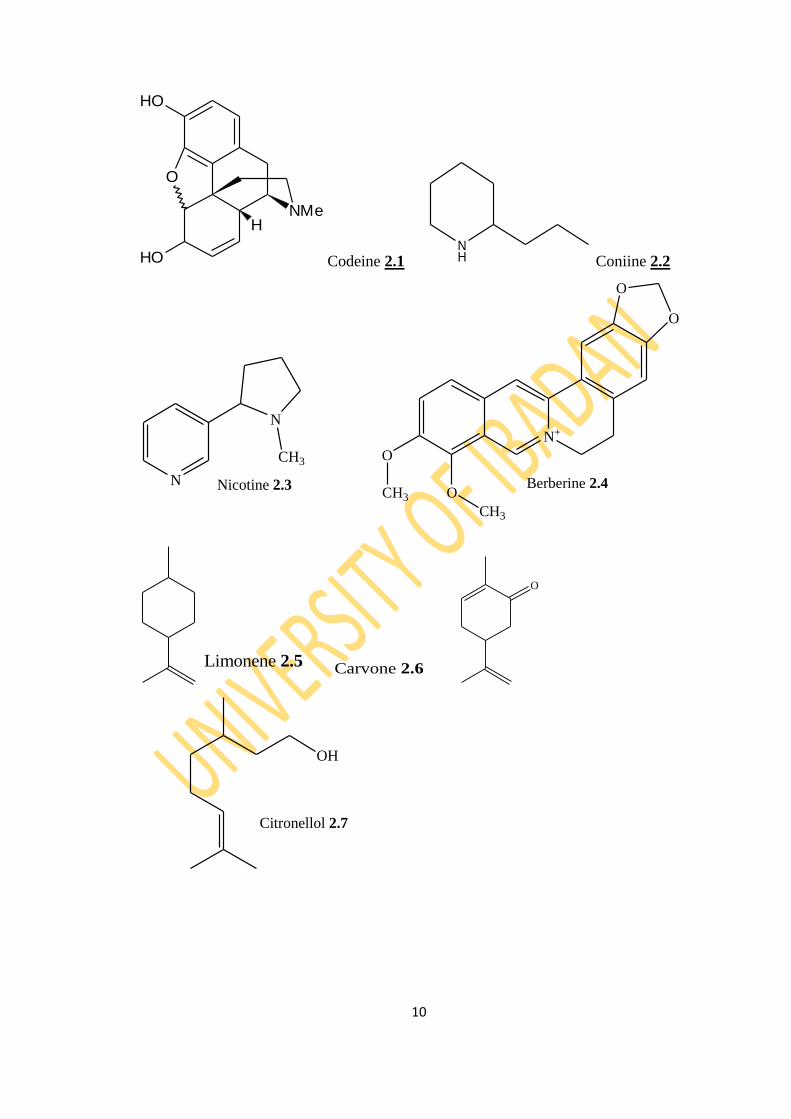
small quantities but have a long history in medication, such as codeine (2.1) as an
antidepressants (Simera et al., 2010; Smith et al., 2006; Vree et al., 2000). Some
interfere with components of the nervous system, especially the chemical transmitters;
others affect membrane transport, protein synthesis and miscellaneous enzyme
activities (Creelman and Mullet, 1997).
Alkaloids are usually colourless, crystalline, non- volatile solids which are insoluble in
water, but are soluble in organic solvents. Some alkaloids are liquids which are soluble
in water e.g. connine (2.2) and nicotine (2.3) and a few are coloured e.g. berberine
(2.4) is yellow (Finar, 1997). Besides carbon, hydrogen and nitrogen molecules of
alkaloids may contain sulphur and rarely chlorine or phosphorus (Lewis, 1998).
8
2.2.2 Phenolic Compounds
Phenolic compounds are one of the largest groups of secondary plants constituents
synthesized by fruits, vegetables, teas, cocoa and other plants that possess certain
health benefits. They usually possess an aromatic ring bearing one or more hydroxyl
groups and range from simple phenolic molecules to highly polymerised compounds
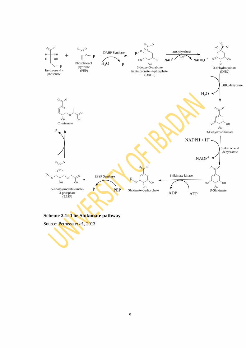
(Anurag et al., 2015). Their biosynthetic origin is through the shikimate pathway
(Scheme 2.1) (Finar, 2000; Petrussa et al., 2013). They are categorised into several
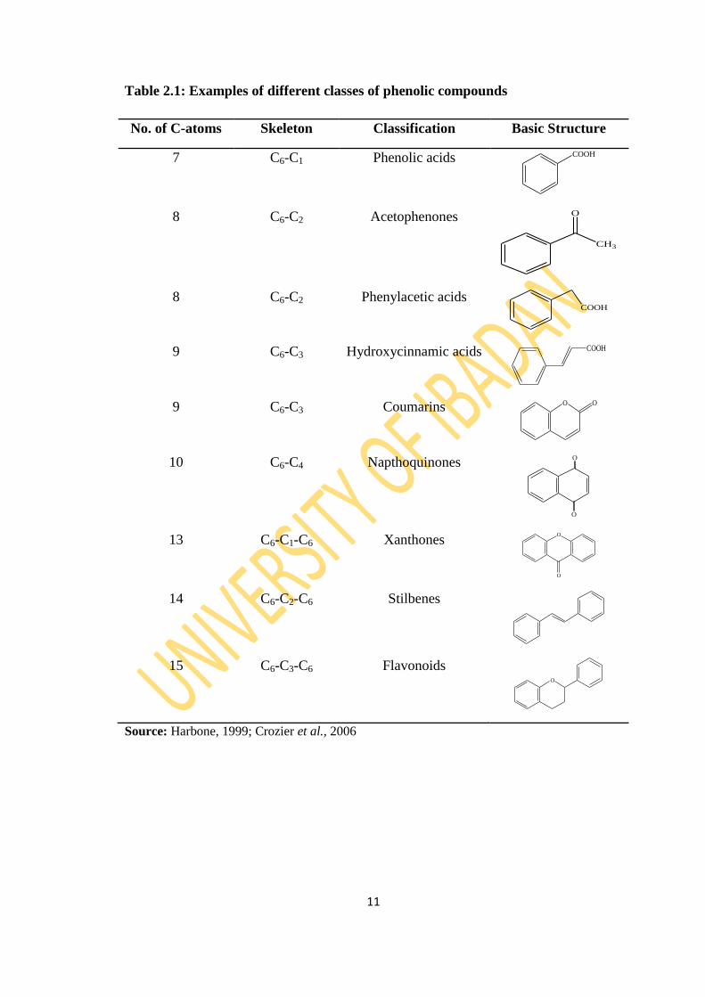
classes based on the number of carbon atoms in the basic carbon skeleton. Table 2.1
shows some examples of the different classes of phenols with their basic carbon
skeleton and the number of carbon atoms in the skeleton.
Phenolic compounds have antioxidant, anti-inflammatory, anti-carcinogenic and other
biological properties and may prevent oxidative stress (Park et al., 2001).
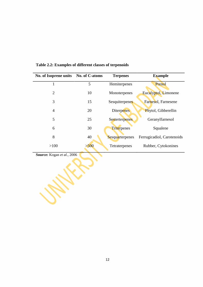
2.2.3 Terpenoids
Terpenoids are the largest and most diverse family of secondary metabolites, ranging
in structure from linear to polycyclic molecules and in size from the five-carbon
hemiterpenes to natural rubber, comprising thousands of isoprene units. A simple
unifying feature of all terpenoids is that they are derived from a simple process of
assembly of a C-5 unit, the isoprene unit C5H8 (Gershenzon and Dudareva, 2007).
During their formation, the isoprene units are linked in head and tail fashion. The
number of units incorporated into a particular terpene serves as a basis for their
classification as shown in Table 2.2. The classification of terpenoids ranges from
essential oil components, the volatile, mono and sesquiterpenes (C10 and C15), through
the less volatile diterpenes (C20) to the involatile triterpenoids and sterols (C30) and
carotenoids pigments (C40) (Wang et al., 2005). Each of these various classes of
terpenoids is significant in either plant growth metabolism or ecology (Harbone, 1984).
Terpenoids contribute to the scent of eucalyptus, the flavours of cinnamon, cloves, and
ginger, and the colour of yellow flowers.
9
H
O
H OH
H OH
O O
O
O-
+
O O-
OH
OHOH
OO
DAHP Synthase
3-deoxy-D-arabino-heptulosonate -7-phosphate
(DAHP)
DHQ Synthase
Co2+
NAD+ NADH,H+
O
O-
OH
OH
O
OH
3-dehydroquinate(DHQ)Erythrose -4 -
phosphate
H2O
OH
OH
O
O O-
OH
OH
OH
O O-
OH
OH
O
O O-
NADPH + H+
NADP+
3-Dehydroshikimate
Shikimic acid dehydratase
Phosphoenol pyruvate
(PEP)
ATPADP
Shikimate kinaseEPSP Synthase
5-Enolpyruvylshikimate-3-phosphate
(EPSP)
P
PP
P
DHQ dehydrase
O
OH
O
O O-
O
OH
Shikimate-3-phosphate
P
O
OH
O O-
O
OH
P
Chorismate
D-ShikimatePEPP
H2O P
Scheme 2.1: The Shikimate pathway
Source: Petrussa et al., 2013
10
O
NMe
HO
OH
H
Codeine 2.1 NH Coniine 2.2
N
N
CH3
Nicotine 2.3
N+
O
O
O
O
CH3
CH3
Berberine 2.4
Limonene 2.5
Carvone 2.6
O
OH
Citronellol 2.7
11
Table 2.1: Examples of different classes of phenolic compounds
No. of C-atoms Skeleton Classification Basic Structure
7 C6-C1 Phenolic acids COOH
8 C6-C2 Acetophenones
CH3
O
8 C6-C2 Phenylacetic acids COOH
9 C6-C3 Hydroxycinnamic acids COOH
9 C6-C3 Coumarins O O
10 C6-C4 Napthoquinones O
O
13 C6-C1-C6 Xanthones O
O
14 C6-C2-C6 Stilbenes
15 C6-C3-C6 Flavonoids O
Source: Harbone, 1999; Crozier et al., 2006
12
Table 2.2: Examples of different classes of terpenoids
No. of Isoprene units No. of C-atoms Terpenes Example
1 5 Hemiterpenes Prenol
2 10 Monoterpenes Eucalyptol, Limonene
3 15 Sesquiterpenes Farnesol, Farnesene
4 20 Diterpenes Phytol, Gibberellin
5 25 Sesterterpenes Geranylfarnesol
6 30 Triterpenes Squalene
8 40 Sesquarterpenes Ferrugicadiol, Carotenoids
>100 >500 Tetraterpenes Rubber, Cytokonines
Source: Kogan et al., 2006
13
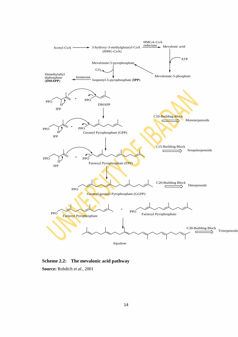
2.2.3.1 Biosynthesis of terpenoids
Terpenoids biogenetically originated through the condensation of the universal
phosphorylated derivative of hemiterpene, isopentenylpyrophosphate (IPP) CH2C
(CH3) CH2CH2OPP and dimethylallylpyrophosphate (DMAPP) (CH3)2C CH CH2OPP.
In biosynthesis, a molecule of isopentenylpyrophosphate (IPP) and
dimethylallylpyrophosphate, which are biosynthesized from three acetylcoenzyme A
moieties through mevalonic acid (MVA) via the mevalonate pathway (Kuzuama and
Seto, 2003), are linked together to give geranylpyrophosphate (GPP), which on
addition of another IPP unit forms farnesylpyrophospahte (FPP). GPP and FPP are
precursors of monoterpenes and sesquiterpenes respectively (Finar, 2000; Rohdich et
al., 2001; Paul, 2002). Thus, terpenoids biosynthesis is based on the isoprene
molecules CH2C (CH3) CHCH2, their carbon skeletons are built up from the union of
two or more of these C-5 units. The classification of terpenoids ranges from essential
oil components, the volatile, mono and sesquiterpenes (C10 and C15), through the less
volatile diterpenes (C20) to the involatile triterpenoids and sterols (C30) and carotenoids
pigments (C40). The biosynthesis of terpenoids is shown in Scheme 2.2.
14
Acetyl-CoA 3-hydroxy-3-methylglutaryl-CoA Mevalonic acid
ATP
Mevalonate-5-phosphate
(HMG-CoA)
Mevalonate-5-pyrophosphate
CO2
Isopentyl-5-pyrophosphate (IPP)Isomerase
Dimethylallyldiphosphate(DMAPP)
HMGA-CoAreductase
H
PPOPPO
PPO
IPP
DMAPP
HPPO
IPP
HPPO
IPP
Geranyl Pyrophosphate (GPP)
PPO
Farnesyl Pyrophosphate (FPP)
Monoterpenoids
Sesquiterpenoids
DiterpenoidsC20-Building Block
C15-Building Block
C10-Building Block
PPO
Geranyl-geranyl Pyrophosphate (GGPP)
PPOFarnesyl Pyrophosphate
PPOFarnesyl Pyrophosphate
TriterpenoidsC30-Building Block
Squalene
Scheme 2.2: The mevalonic acid pathway
Source: Rohdich et al., 2001
15
2.2.3.2 Pharmacological relevance of terpenoids
Terpenoids are used extensively for their aromatic qualities. Extensive biological
investigations have been carried out within the group and these studies have revealed a
broad spectrum of pharmacological and physiological properties (Maffei, 2010). They
play a role in traditional herbal remedies and are under investigation for antibacterial,
antineoplastic, and other pharmaceutical functions. Recent findings demonstrate that
certain nitrogenous terpene derivatives possess the potent anti-hypertensive activity
and may indicate a new era in medicine through the synthetic terpenoids path (Kabera
et al., 2014). The antimicrobial and insecticidal properties of other terpenoids have led
to their utilization as pesticides and fungicides in agriculture and horticulture (Kataev
et al., 2011; Böhme et al., 2014).
Monoterpenes have been shown to exert chemopreventive as well as chemotherapeutic
activities in mammary tumor models and thus may represent a new class of therapeutic
agents. Limonene (2.5) for example has been an interesting target molecule for
chemists and biologists (Duetz et al., 2001). Limonene is a well-established
chemopreventive and therapeutic agent against tumor cells (Kris-Etherton et al., 2002;
Crowell, 1999; Fabian, 2001). Carvone (2.6) has also been shown to prevent
chemically induced lung and forestomach carcinoma development (Wattenberg et al.,
1989). The mechanism of action of monoterpenes against tumor cells is the induction
of apoptosis and interference of the protein prenylation of key regulatory proteins
(Crowell, 1999; Fabian, 2001; Ariazi et al., 1999; Crowell et al., 1991). Acyclic
monoterpenes, citronellol (2.7), nerol (2.8) and geraniol (2.9), have been reported to
exhibit activity against Mycobacterium tuberculosis (Rajab et al., 1998). Eucalyptol
(2.10) is an ingredient in many brands of mouthwash and cough suppressant. It
controls airway mucus hypersecretion and asthma via anti-inflammatory cytokine
inhibition. Although eucalyptol is used as an insecticide and insect repellent, it is one
of many compounds that are attractive to males of various species of orchid bees, who
apparently gather the chemical to synthesize pheromones. The anti-inflammatory
activities of some medicinal plants result from the presence of one or more
sesquiterpene lactones. Artemisinin (2.11) is an important sesquiterpene lactone with
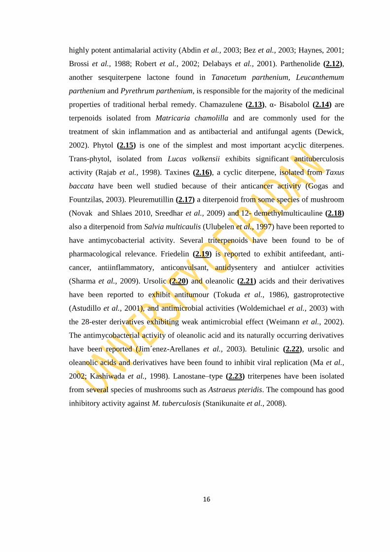
16
highly potent antimalarial activity (Abdin et al., 2003; Bez et al., 2003; Haynes, 2001;
Brossi et al., 1988; Robert et al., 2002; Delabays et al., 2001). Parthenolide (2.12),
another sesquiterpene lactone found in Tanacetum parthenium, Leucanthemum
parthenium and Pyrethrum parthenium, is responsible for the majority of the medicinal
properties of traditional herbal remedy. Chamazulene (2.13), α- Bisabolol (2.14) are
terpenoids isolated from Matricaria chamolilla and are commonly used for the
treatment of skin inflammation and as antibacterial and antifungal agents (Dewick,
2002). Phytol (2.15) is one of the simplest and most important acyclic diterpenes.
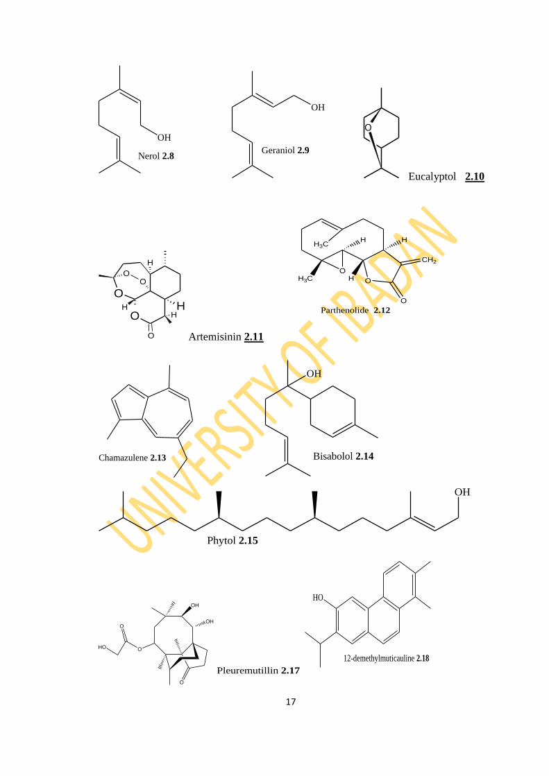
Trans-phytol, isolated from Lucas volkensii exhibits significant antituberculosis
activity (Rajab et al., 1998). Taxines (2.16), a cyclic diterpene, isolated from Taxus
baccata have been well studied because of their anticancer activity (Gogas and
Fountzilas, 2003). Pleuremutillin (2.17) a diterpenoid from some species of mushroom
(Novak and Shlaes 2010, Sreedhar et al., 2009) and 12- demethylmulticauline (2.18)
also a diterpenoid from Salvia multicaulis (Ulubelen et al., 1997) have been reported to
have antimycobacterial activity. Several triterpenoids have been found to be of
pharmacological relevance. Friedelin (2.19) is reported to exhibit antifeedant, anti-
cancer, antiinflammatory, anticonvulsant, antidysentery and antiulcer activities
(Sharma et al., 2009). Ursolic (2.20) and oleanolic (2.21) acids and their derivatives
have been reported to exhibit antitumour (Tokuda et al., 1986), gastroprotective
(Astudillo et al., 2001), and antimicrobial activities (Woldemichael et al., 2003) with
the 28-ester derivatives exhibiting weak antimicrobial effect (Weimann et al., 2002).
The antimycobacterial activity of oleanolic acid and its naturally occurring derivatives
have been reported (Jim´enez-Arellanes et al., 2003). Betulinic (2.22), ursolic and
oleanolic acids and derivatives have been found to inhibit viral replication (Ma et al.,
2002; Kashiwada et al., 1998). Lanostane–type (2.23) triterpenes have been isolated
from several species of mushrooms such as Astraeus pteridis. The compound has good
inhibitory activity against M. tuberculosis (Stanikunaite et al., 2008).
17
OH
Nerol 2.8
OH
Geraniol 2.9
O
Eucalyptol 2.10
OH
O
O
OO
H
H
H
Artemisinin 2.11
O
O
CH2
O
H3C H
H HH3C
Parthenolide 2.12
Chamazulene 2.13
OH
Bisabolol 2.14
OH
Phytol 2.15
OH
OH
OHO
O
O
Pleuremutillin 2.17
HO
12-demethylmuticauline 2.18
18
N
OH
O
O
OH
O O
O
O
O
HO
H
N
OH
O
O
H
OHO
O
OH
O
OH
Taxine A and B 2.16
O
Friedeline 2.19
O
OH
OH Ursolic acid 2.20
O
OH
OH Oleanolic Acid 2.21
O
OH
OHBetulinic Acid 2.22 Lanostane 2.23
19
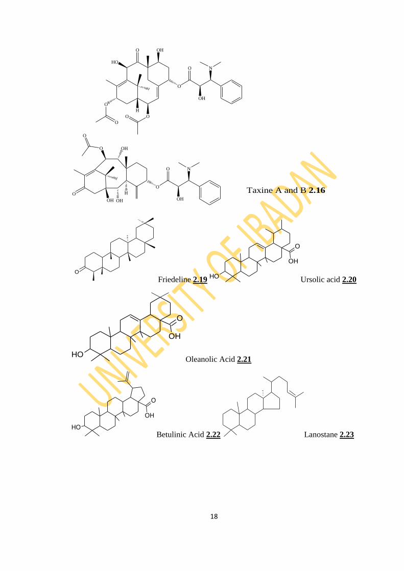
2.2.3 Steroids
Sterols are triterpenes which are based on the cyclopentane perhydrophenanthrene ring
system. They share the same biosynthetic origin with the terpenoids by acetate
pathway through the cyclization of squalene (Harborne, 1998; Finar, 2000). At one
time, sterols were mainly considered to be animal substances, but in recent years an
increasing number of such compounds have been detected in plant tissues. Plant
steroids are called ‗phytosterols‘ which include; β-sitosterols (2.24), stigmasterol
(2.25) and campesterol (2.26). Phytosterols occurred in higher plants as free and
simple glycosides.
The occurrence of ergosterol (2.27) is confined to lower plants like yeast and many
fungi. Fucosterol (2.28) which is the main steroid of many brown algae occurs mainly
in lower plants but also appears occasionally in higher plants. It has also been detected
in coconut (Harborne, 1998).
Pharmacologically, steroids have been shown to exhibit hormonal and anti-
inflammatory activities. They are used as contraceptives, androgenic and anabolic
agents. They also exhibit antifungal, antibacterial, antiviral and hypolipidemic
activities (Saeidnia et al., 2014).
2.2.4 Saponins
Saponins are plant glycosides of both triterpenes and sterols. They are surface-active
agents with soap-like properties and can be detected by their ability to cause foaming
and to haemolyse blood cells. Sapogenins are the aglycone of saponins and are
characterized by the presence of a spiroketal side chain. A characteristic property of
saponin is the haemolysis caused by an intravenous injection of their aqueous solutions
into animals; these solutions are comparatively harmless when taken orally (Finar,
1997).
The search in plants for saponins has been stimulated by the need for readily accessible
sources of sapogenins which can be converted in the laboratory to animal sterols of
therapeutic importance, like cortisone and contraceptive estrogen. Saponins are also of
economic interest because of their occasional toxicity to cattle, as found in saponins of
alfalfa or their sweet taste as found in glycyrrhizin of liquorice root (Ogundajo, 2014).
Several reports have shown that saponin exhibit a wide range of pharmacological
properties. Simoes-Pires et al. (2005) reported the use of saponins as vaccine adjuvants

20
in the treatment of the Herpes Simplex Virus (HSV), Human Immuno-deficiency Virus
(HIV), and influenza.
HO β-sitosterols 2.24
HO Stigmasterol 2.25
Campesterol 2.26
HO Ergosterol 2.27
HO Fucosterol 2.28
HO

21
2.3 Extraction of Secondary Metabolites
Many approaches can often be employed to extract secondary metabolites, although
water is used as an extractant in many traditional medicines. Organic solvents of
varying polarity are generally selected through modern methods of extraction to
exploit the solubility of various plant constituents. Obviously, wrong choice of solvent
and method will cause the entire processes to fail or the desired compounds from the
material may not be released from the matrix completely. Some extraction procedures
usually applied for the extraction of natural products from plants are:
2.3.1 Cold Method
2.3.1.1 Percolation
In percolation, the powdered plant materials are soaked initially in a solvent in a
percolator (a cylindrical or conical container with tap at the bottom). Additional
solvent is then added on top of the plant material and allowed to percolate slowly drop-
wise out of the bottom of the percolator. In this method, filtration of the extract is not
required because there is a filter at the outlet of the percolator. Percolation is adequate
for both initial and large scale extraction. The extent to which the material is ground
can influence the extract yield. Hence, fine powder, resins and plant materials that
swell excessively (e.g. those containing mucilage) can clog the percolator.
Furthermore, if the material is not homogeneously distributed in the container, the
solvent may not reach all the areas and the extraction will be incomplete. A
disadvantage of the technique is that large volumes of solvents are used and this can be
time consuming (Cannel, 1998).
2.3.1.2 Maceration
This method is simple and still widely used. The procedure involves soaking the
pulverized plant materials in a suitable solvent in a closed container at room
temperature. The technique is suitable for both initial and bulk extraction. Occasional
or constant stirring of the preparation (using mechanical shaker or mixers to guarantee
homogenous mixing) can increase the extraction yield. The extraction ultimately stops
when equilibrium is attained between the concentration of metabolites in the extract
and that of the plant material. After extraction, the residual plants material (marc) has
to be separated from the solvents. This involves a rough clarification by decanting,
which is usually followed by a filtration. To ensure exhaustive extraction, it is common
to carry out an initial maceration, followed by clarification and an addition of fresh
22
solvent to the residue. This can be performed periodically with all the filtrates pooled
together. It is a batch method of extraction (Harbone, 1998).
The major drawback of this technique is the fact that the process can be quite time
consuming, taking from a few hours up to weeks (Takahashi et al., 2001). Exhaustive
extraction can also consume large volumes of solvent and can lead to potential loss of
metabolite and, or plant materials. Furthermore some compounds may not be extracted
efficiently if the compounds of choice are poorly soluble at room temperature in the
solvent used. The major advantage of the method is those compounds that are thermo
labile are not affected.
2.3.2 Hot Method
Soxhlet extraction is the widely used hot method in the extraction of plant metabolites
because of its convenience. This method is adequate for both initial and bulk
extraction. The plant powder is packed in a thimble in an extraction chamber, which is
placed on top of a collecting flask. A suitable solvent is added to the flask, and the
flask is heated under reflux. When a certain level of condensed solvent has
accumulated in the thimble, it is siphoned into the flask beneath. As the solvent
saturates the plant material in the flask, it will solubilize the metabolite which is
emptied into the flask. Fresh solvent is re-condensed and the material extracted in the
thimble continuously. It is usually a continuous method of extraction (Harbone, 1984).
This makes Soxhlet extraction less time and solvent-consuming than maceration or
percolation. However, the main disadvantage of soxhlet extraction is that the extract is
constantly heated at the boiling point of the solvent used, and this can damage thermo
labile compounds and, or initiate the formation of artifacts (Zygmunt and Namiesnik,
2003).
In recent years, several faster and more automatic extraction techniques for solid
samples have been replacing conventional techniques. Among the modern techniques
are extraction by supercritical fluids extraction (SFE), pressurized liquids extraction
(PLE), Ultrasound–Assisted solvent extraction and microwave assisted extraction
MAE). These alternative techniques considerably reduce the consumption of solvents,
increase the speed of the extraction process, and simplify it (Ali et al., 2007). The
major drawback on these techniques in this part of the world is the lack of steady
power supply; hence the conventional techniques are still the methods of choice for
this project.
23
2.4 Essential Oil
Essential oils, volatile oils or simply the "oil" of the plant from which they were
extracted, such as ―oil of lemongrass‖ are hydrophobic liquids containing volatile
aroma compounds extracted from vegetal materials using various extraction techniques
(Yazdani et al., 2011). From the view point of practical applications, these materials
may be defined as odiferous bodies of an oily nature, obtained almost exclusively from
vegetable organs: flowers, leaves, barks, woods, roots, rhizomes, fruits, and seeds
(Burt, 2004; Celiktas et al., 2006; Skocibusic et al., 2006; Chalchat and Ozcan, 2008;
Hussain et al., 2008; Anwar et al., 2009a). These oils have strong aromatic
components that give a plant its distinctive odor, flavor, or scent (Koul et al., 2008).
Essential oils (EOs) are very interesting natural plant products and among other
qualities they possess various biological properties viz antibacterial, antifungal,
antioxidant and anti-carcinogenic properties (Tzortzakis, 2007) that these complex
mixtures of a large number of constituents (mainly terpenoids, benzenoids,
phenylpropanoids, etc.) in variable ratios (Van Zyl et al., 2006) exert on humans,
animals, and other plants.
Essential oils are secondary metabolism products in plants. Plants have essential oil
components and quality varying with geographical distribution, harvesting time,
growing conditions, and extraction method (Yang et al., 2005). These oils are typically
liquid at room temperature and are easily transform from a liquid to a gaseous state at
room temperature or a slightly higher temperature without decomposing (Koul et al.,
2008). Presently, essential oils are most often used in the food industry for flavoring,
the cosmetic industry for fragrances, and the pharmaceutical industry for their
functional properties. However, dozens of plant essential oils have been screened for
fumigant toxicity against a variety of insect pests primarily for agricultural and food
storage (Wang et al., 2006; Ayvaz et al., 2008; Benzi et al., 2009; Ebadollahi et al.,
2010).
Essential oils are very complex natural mixtures. The components include two groups
of distinct biosynthetical origin; the terpenoid group which is the main group and the
non-terpenoid group which may contain short-chain aliphatic substances, aromatic
substances, nitrogenated substances, and substances with sulphur (Croteau et al., 2000;
Bowels, 2003). In essential oils, the two terpenoid groups, the monoterpenes and
sesquiterpenes allow for a great variety of structures. Depending on the functional
24
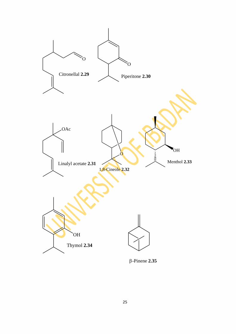
group attached they can be: aldehydes like citronellal (2.29), ketones such as
piperitone (2.30), esters for example linalyl acetate (2.31), oxides like 1,8-cineole
(2.32), alcohols such as menthol (2.33), phenols for example thymol (2.34) and
hydrocarbons like limonene (2.5) (Koul et al., 2008).
Essential oils exhibit a wide spectrum of pharmacological activities such as infection
control, wound healing, pain relief, anti-nausea, anti-inflammation and anti-anxiety
(Halcon, 2002; Kalemba and Kunicka, 2003). Traditional medicines containing
essential oils have been scientifically proven to be effective in treating various
ailments like malaria and others of microbial origin (Lopes et al., 1999; Nakatsu et al.,
2000; Goulart et al., 2004). The oils or some of their constituents are indeed effective
against a large variety of organisms including bacteria (Holley and Dhaval, 2005;
Basile et al., 2006; Schelz et al., 2006; Hu‘snu‘Can Baser et al., 2006), virus
(Duschatzky et al., 2005), fungi (Hammer et al., 2002; Velluti et al., 2003, 2004;
Serrano et al., 2005; Cavaleiro et al., 2006; Pawar and Thaker, 2006; Soylu et al.,
2006), protozoa (Monzote et al., 2006), parasites (Moon et al., 2006; Priestley et al.,
2006), larvae (Hierro et al., 2004; Pavela, 2005; Morais et al., 2006; Amer and
Mehlhorn, 2006a,b; Ravi Kiran et al., 2006), worms, insects (Bhatnagar et al., 1993;
Lamiri et al., 2001; Liu et al., 2006; Burfield and Reekie, 2005; Yang and Ma, 2005;
Sim et al., 2006; Kouninki et al., 2005; Park et al., 2006a,b; Chaiyasit et al., 2006;
Cheng et al., 2007) and molluscs (Lahlou and Berrada, 2001). The biological activities
of essential oils have been attributed to the composition or specific essential oil
constituent, for example: aldehydes in lemon grass (Cymbopogon citrates) have been
reported to have anti-inflammatory properties (Boukhatem et al., 2014). Ketones in
sweet fennel (Foeniculum vulgare) have been found to aid in wound healing and
dissolve mucus and fats (Nakatsu et al., 2000). Alcohols in tea tree (Melaleuca
alternifolia), true lavender (Lavandula angustifolia) and baboon wood (Virola
surinamensis) have anti-microbial and anti-malarial properties (Lopes et al., 1999;
Cowan, 1999). Esters in clarry sage (Salvia sclarea) have anti-cholinesterase
properties (Savalev et al., 2003). Phenols in thyme (Thymus vulgaris) and in clove
(Eugenia caryophyllata) have antimicrobial properties and can be used as food
preservatives (Nakatsu et al., 2000). Overall, essential oils are pertinent to
pharmaceutical, cosmetic and food research and are widely viewed as templates for
structure optimization programs with a goal to creating new drugs (Cragg et al., 1997).
25
O
Citronellal 2.29
O
Piperitone 2.30
OAc
Linalyl acetate 2.31
O
1,8-Cineole 2.32
OH
Menthol 2.33
OH
Thymol 2.34
-Pinene 2.35
26
2.4.1 Extraction of Essential Oils
Essential oils are extracted from different aromatic plants generally distributed in
Mediterranean and tropical countries across the world where they are highly regarded
as an important component of either native medicine, food or other products (Hussain
et al., 2009). These essential oils are accumulated in secretary cells, cavities, channels,
and epidermic cells of almost all plant organs such as flowers, buds, stems, leaves,
fruits, seeds and roots etc. (Burt, 2004; Chalchat and Ozcan, 2008; Hussain et al.,
2008; Anwar et al., 2009a) and can be extracted when plant organs are fresh, partially
dehydrated or dried (Ozcan, 2003; Asekun et al., 2007; Hussain et al., 2008).
The extraction of the essential oil depends mainly on the rate of diffusion of the oil
through the plant tissues to an exposed surface from where the oil can be removed by a
number of processes. There are different methods, depending upon the stability of the
oil, for the extraction of the oil from the plant materials. Steam distillation and
hydrodistillation are still in use today as the most important processes for obtaining
essential oils from the plants (Baker et al., 2000; Kulisic et al., 2004; Masango, 2004;
Sokovic and Van Griensven, 2006). Other methods employed for isolation of essential
oils include the use of liquid carbon dioxide or microwaves, low or high pressure
distillation employing boiling water or hot steam (Bousbia et al., 2009; Donelian et al.,
2009). The essential oils obtained by steam distillation or by expression are generally
preferred for food and pharmacological applications. Essential oil extraction is
different from the extraction of all the other secondary metabolites due to the volatility
of the compounds even at room temperature. The following methods are commonly
used:
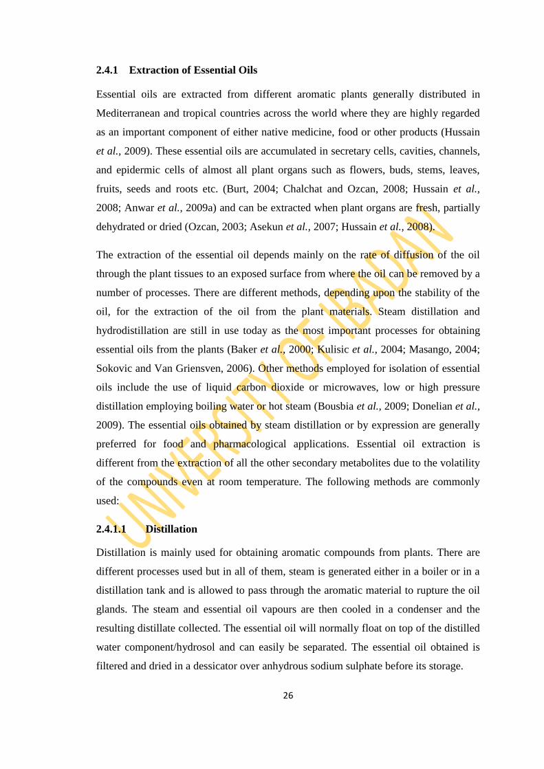
2.4.1.1 Distillation
Distillation is mainly used for obtaining aromatic compounds from plants. There are
different processes used but in all of them, steam is generated either in a boiler or in a
distillation tank and is allowed to pass through the aromatic material to rupture the oil
glands. The steam and essential oil vapours are then cooled in a condenser and the
resulting distillate collected. The essential oil will normally float on top of the distilled
water component/hydrosol and can easily be separated. The essential oil obtained is
filtered and dried in a dessicator over anhydrous sodium sulphate before its storage.

27
2.4.1.2 Hydro or water distillation
This is the simplest and usually cheapest distillation method. The plant material is
immersed in water and boiled. As the water is heated, the steam passes through the
plant material, vaporizing the volatile compounds. The vapours flow through a coil,
where they condense back to liquid, which is then collected in the receiving vessel.
In the laboratory hydro distillation is done using a Clevenger-type apparatus, shown in
Figure 2.1. The method is slow and hence time consuming. Furthermore, the prolonged
action of hot water can cause hydrolysis of some constituents of the essential oils such
as esters. It‘s also not a suitable method for large scale distillations and for distillation
of high saponin rich plant materials.
2.4.1.3 Steam Distillation
The method which is also referred to as wet steam distillation was developed to
overcome the drawbacks of hydro distillation. Direct contact of plant material with a
hot furnace bottom is thus avoided. The plant material is supported on a perforated grid
below which water is boiled. Steam rises through the plant material vaporizing the
essential oil with it and is condensed usually in a coil condenser by cooling water. The
method is suitable for distilling leafy materials but does not work well for woods, roots
and seeds. The distillation units are cheap, easy to operate and are extremely popular
with essential oil producers in developing countries. The method, however, is time
consuming, gives poor quality oil yields and oil separation is not complete.
28
Figure 2.1: Clevenger-type apparatus for Hydrodistillation

29
2.4.1.4 Direct steam distillation
The boiling point of most essential oil components exceeds that of water and generally
lies between 150- 300 oC. However, in the presence of steam they are volatilised at a
temperature close to 100 oC. The principle behind steam distillation is that two
immiscible liquids, when mixed, each exerts a vapour pressure, as if each liquid were
pure (Houghton and Raman, 1998). The total vapour pressure of the boiling mixture is
therefore equal to the sum of the partial pressures exerted by each of the individual
components of the mixture. When the total vapour pressure reaches atmospheric
pressure, the mixture starts to boil. The plant material is placed in a still and steam
prepared in a separate chamber is forced over it. The temperature of the steam is
carefully controlled so as not to burn the plant material or the essential oil. The rate of
distillation and yield of the oil are high and the oil obtained is of good quality.
However, partial loss of more polar constituents of the oil, due to their affinity for
water, may occur (Baker et al., 2000; Masango, 2004). The method is quite expensive
and only bigger producers can afford to own the distillation unit, hence it is much
popular for the isolation of essential oils on commercial scale (Masango, 2004).
2.4.1.5 Hydro diffusion
Hydro diffusion is a type of steam distillation where steam is fed in from the top onto
the botanical material. The process uses the principle of osmotic pressure to diffuse oil
from the oil glands. The system is connected and low pressure steam is passed into the
plant material from a boiler from the top. The oil and water are collected below the
condenser in a typical oil separator. The various components of the essential oils are
liberated based on their solubility in the boiling water rather than the order of their
boiling points (Srivastava, 1991). The main advantage of this method over steam
distillation is that less steam is used hence a shorter processing time and therefore
higher oil yield.
2.4.1.6 Liquid Carbon Dioxide Extraction Method
Extraction is carried out in a specially designed high-pressure soxhlet apparatus with
supercritical/liquid carbon dioxide (CO2) as the extracting solvent. The plant materials
are put into the extraction columns, which are under high pressure (55–58 bar) and the
liquid CO2 flows through the extraction columns until it is saturated with essential oil.
At the end of the extraction, the column is taken and the liquid CO2 is drained from it.
30
Extraction is carried out at a low temperature and this allows for maximum
preservation of all healthful substances in the extract like the aroma, taste, vitamins
and enzymes. The essential oils obtained by this method have been found to be
superior in quality and flavour as compared with the conventional steam distilled
essential oils (Wood et al., 2006). However, the method is expensive in terms of plant
and, in some cases, results in an unusual balance of extracted oil components.
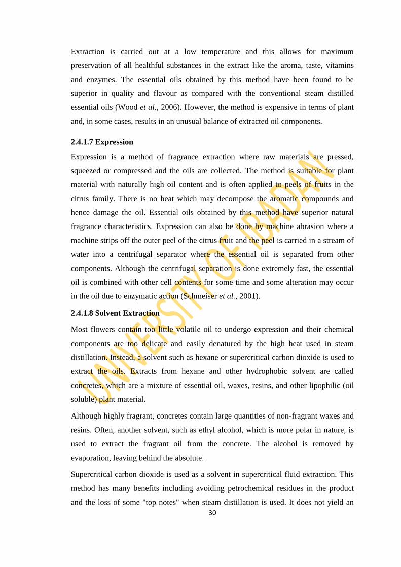
2.4.1.7 Expression
Expression is a method of fragrance extraction where raw materials are pressed,
squeezed or compressed and the oils are collected. The method is suitable for plant
material with naturally high oil content and is often applied to peels of fruits in the
citrus family. There is no heat which may decompose the aromatic compounds and
hence damage the oil. Essential oils obtained by this method have superior natural
fragrance characteristics. Expression can also be done by machine abrasion where a
machine strips off the outer peel of the citrus fruit and the peel is carried in a stream of
water into a centrifugal separator where the essential oil is separated from other
components. Although the centrifugal separation is done extremely fast, the essential
oil is combined with other cell contents for some time and some alteration may occur
in the oil due to enzymatic action (Schmeiser et al., 2001).
2.4.1.8 Solvent Extraction
Most flowers contain too little volatile oil to undergo expression and their chemical
components are too delicate and easily denatured by the high heat used in steam
distillation. Instead, a solvent such as hexane or supercritical carbon dioxide is used to
extract the oils. Extracts from hexane and other hydrophobic solvent are called
concretes, which are a mixture of essential oil, waxes, resins, and other lipophilic (oil
soluble) plant material.
Although highly fragrant, concretes contain large quantities of non-fragrant waxes and
resins. Often, another solvent, such as ethyl alcohol, which is more polar in nature, is
used to extract the fragrant oil from the concrete. The alcohol is removed by
evaporation, leaving behind the absolute.
Supercritical carbon dioxide is used as a solvent in supercritical fluid extraction. This
method has many benefits including avoiding petrochemical residues in the product
and the loss of some "top notes" when steam distillation is used. It does not yield an

31
absolute directly. The supercritical carbon dioxide will extract both the waxes and the
essential oils that make up the concrete. Subsequent processing with liquid carbon
dioxide, achieved in the same extractor by merely lowering the extraction temperature,
will separate the waxes from the essential oils. This lower temperature process
prevents the decomposition and denaturing of compounds. When the extraction is
complete, the pressure is reduced to ambient and the carbon dioxide reverts to a gas,
leaving no residue.
2.4.1.9 Florasols Extraction
This method of extraction uses a new family of benign non-CFC (Chlorinated
Fluorocarbons) gaseous solvents known as ―Florasol‖. Florasol is a refrigerant and it
was developed to replace Freon. Florasol is an ozone friendly product and it causes no
danger to the environment. The advantage is that the extraction of essential oils occurs
at or below room temperature so any degradation through temperature extremes does
not occur. The only thing that is extracted from the plants is the essential oils. The
essential oils are absolutely pure and contain no foreign substances at all. The oils are
refered to as phytols thus this method is also refered to as ―phytonic process‖
(Okwudiri, 2015).
2.4.2 Analysis of Essential Oils
The characterisation of essential oil on the basis of their chemical profiles is of great
importance due to their multiple applications in different fields of man‘s day to day
activities including pharmacy, perfumery, cosmetics, aromatherapy, and food and
beverages industry. The fact that essential oils are complex mixtures of biologically
active substances (Morris et al., 1979) proves a real challenge for determining their
accurate compositional data. The rapid advances in spectroscopic and chromatographic
techniques have totally changed the picture of chemical study of essential oils. Many
techniques like IR-spectroscopy, UV-spectroscopy, NMR spectroscopy and gas
chromatography have been used for studying the chemical profiles of volatile oils
(Bakkali et al., 2008; Hussain et al., 2008). The increasing importance of essential oils
in various domains of human activities has prompted an extensive need of reliable
methods for analyses of essential oils.
Literatures on the characterization of essential oils have revealed capillary gas
chromatography (GC) with flame ionisation detection (FID), are, in most cases, the

32
method of choice for quantitative determinations. Many researchers make use of mass
spectrometers (MS), coupled with GC, to determine the identities of components
(Salzer, 1977; Wilkins and Madsen, 1991; Daferera et al., 2000; Juliano et al., 2000;
Jerkovic et al., 2001; Delaquis et al., 2002; Hussain et al., 2008; Burt, 2004; Anwar et
al., 2009a,b). Gas chromatography has been proved to be an efficient method for the
characterization of essential oils (Bakkali et al., 2008; Anwar et al., 2009b). The
combination of gas chromatography and mass spectrometry (GC-MS) allows rapid and
reliable identification of essential oils components (Yadegarinia et al., 2006; Gulluce
et al., 2007; Anwar et al., 2009a). Time-of-flight mass spectrometric (TOF-MS)
detection has been increasingly used as a qualitative tool, for the detection of volatile
components (Adahchour et al., 2003). Capillary columns selected, in most cases, are
HP-5ms, DB-5 (cross-linked 5% diphenyl/95% dimethyl siloxane) or DB-1, also
known as SE-30, (polydimethyl siloxane) stationary phases. These more non-polar
stationary phases are often complimented by the use of a more polar stationary phase,
such as polyethylene glycol (Cavaleiro et al., 2004).
2.4.3 Identification of Essential Oil Components
2.4.3.1 Retention Time (tR)
Retention time is the time which elapses between sample injection and recording of the
peak maximum at constant operational conditions which include oven temperature,
carrier gas flow rate and sometimes sample size. Nature of the stationary phase,
column length and film thickness of the stationary phase are other factors affecting
retention time. Retention time of a solute varies with temperature and flow rate.
Maintaining constant conditions throughout an experiment are almost imposible,
therefore, it is not always possible to reproduce the retention time for a solute (Robert,
1993). Retention time is given by the following equation:
Where k = capacity factor; L = coumn length; u = true linear gas velocity in the
column (Sandra and Bicchi, 1987).
2.4.3.2 Retention Indices (RI)
33
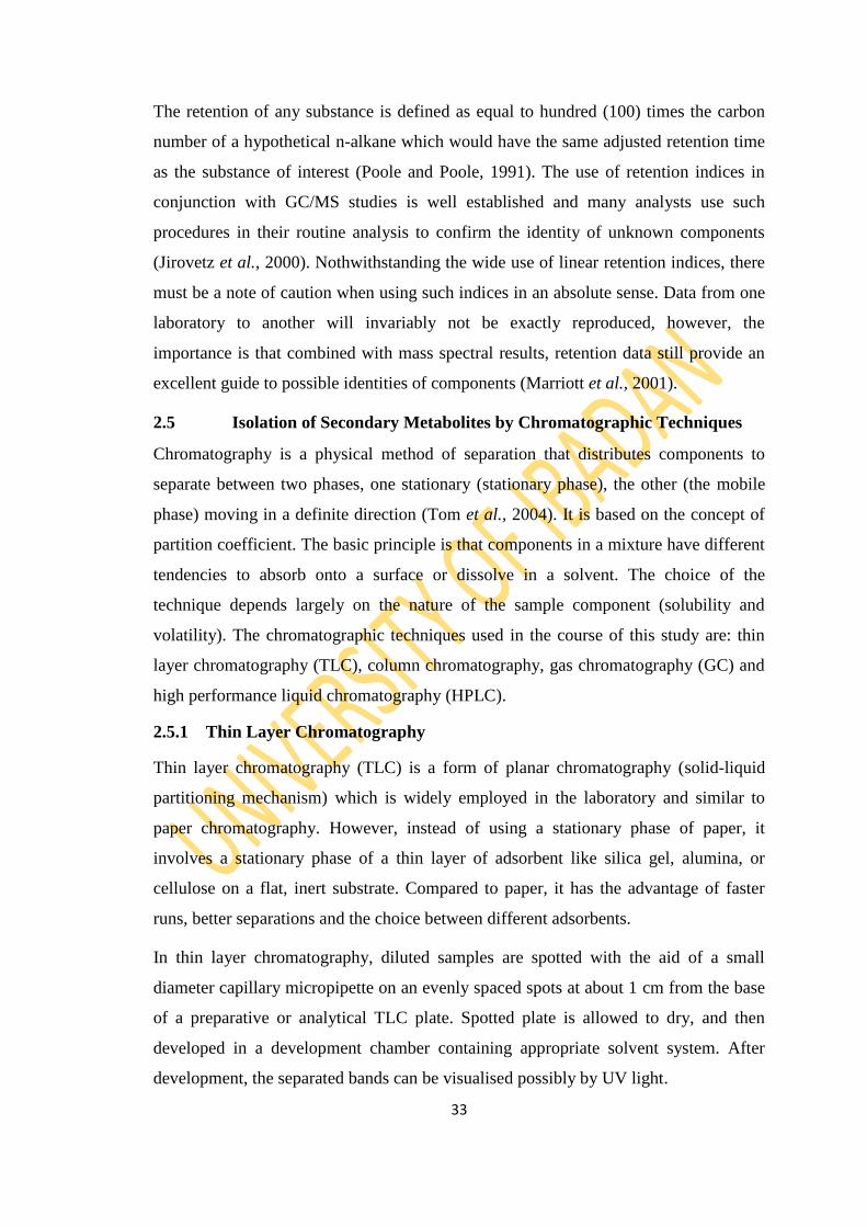
The retention of any substance is defined as equal to hundred (100) times the carbon
number of a hypothetical n-alkane which would have the same adjusted retention time
as the substance of interest (Poole and Poole, 1991). The use of retention indices in
conjunction with GC/MS studies is well established and many analysts use such
procedures in their routine analysis to confirm the identity of unknown components
(Jirovetz et al., 2000). Nothwithstanding the wide use of linear retention indices, there
must be a note of caution when using such indices in an absolute sense. Data from one
laboratory to another will invariably not be exactly reproduced, however, the
importance is that combined with mass spectral results, retention data still provide an
excellent guide to possible identities of components (Marriott et al., 2001).
2.5 Isolation of Secondary Metabolites by Chromatographic Techniques
Chromatography is a physical method of separation that distributes components to
separate between two phases, one stationary (stationary phase), the other (the mobile
phase) moving in a definite direction (Tom et al., 2004). It is based on the concept of
partition coefficient. The basic principle is that components in a mixture have different
tendencies to absorb onto a surface or dissolve in a solvent. The choice of the
technique depends largely on the nature of the sample component (solubility and
volatility). The chromatographic techniques used in the course of this study are: thin
layer chromatography (TLC), column chromatography, gas chromatography (GC) and
high performance liquid chromatography (HPLC).
2.5.1 Thin Layer Chromatography
Thin layer chromatography (TLC) is a form of planar chromatography (solid-liquid
partitioning mechanism) which is widely employed in the laboratory and similar to
paper chromatography. However, instead of using a stationary phase of paper, it
involves a stationary phase of a thin layer of adsorbent like silica gel, alumina, or
cellulose on a flat, inert substrate. Compared to paper, it has the advantage of faster
runs, better separations and the choice between different adsorbents.
In thin layer chromatography, diluted samples are spotted with the aid of a small
diameter capillary micropipette on an evenly spaced spots at about 1 cm from the base
of a preparative or analytical TLC plate. Spotted plate is allowed to dry, and then
developed in a development chamber containing appropriate solvent system. After
development, the separated bands can be visualised possibly by UV light.
34
Visualisation method can be a qualitative and quantitative technique used in the
location of separated component of a crude extract. The methods are tailored to
functional groups of metabolites. Metabolites with some level of unsaturation can be
located with iodine crystals to give either yellow or brown colouration. Method like
UV light for compounds that fluoresce is not destructive, but chemical methods
involving the use of reagents like silver halides for detecting alkyl halides, charring of
organic components with concentrated H2SO4 (tetraoxosulphate (VI) acid), 2,4-DPH
(2,4-Dinitrophenyl hydrazine) for carbonyl compounds, ferric chloride for phenolics,
ninhydrin for amino acids and p-dimethyl aminobenzaldehyde for amines are
destructive (Pavia et al., 2014).
High performance thin layer chromatography (HPTLC) is an improved version of thin
layer chromatography (Braithwaite and smith, 1999).
2.5.2 Column Chromatography
Column chromatography is a separation technique in which the stationary bed is within
a tube. It is an effective method of separating the components of crude extracts which
depends on: the length and diameter of the column, flow rate, nature of adsorbent and
mobile phase employed. The separating technique works on the net distribution of the
components of a mixture between the adsorbent and the mobile phase, owing to
selective adsorptivity and solubility (Pavia et al., 2014).
Pre-adsorbed material is loaded on a packed column, elution is achieved isocratically
or gradiently and the column is monitored using microscope slide TLC. Fractions are
collected, distilled and identified with respective Rf values. Isolates with the same Rf
values are combined and purity check is carried out. Differences in rates of movement
through the medium are calculated to different retention times of the sample (Ettre,
1993).
2.5.3 Gas Chromatography
Gas chromatography (GC) also known as gas-liquid chromatography (GLC) is one of
the most useful tools for separating and analysing organic compounds that can be
vaporised without decomposition. It resembles column chromatography in principle
with technique in which the mobile phase is a gas. The partitioning processes for
mixtures are carried out between an inert moving gas phase and a stationary liquid
phase, at a controllable temperature. The concentration of any compound in the gas

35
phase is a function of its vapour pressure; which is the basis of isolation (Pavia et al.,
2014).
Gas chromatography has a wide range of application. These include drugs and
consumer products analysis, environmental monitoring of air, water and legislation.
Gas chromatography-mass spectrometry (GC-MS) is advanced gas chromatography
technique which uses mass spectroscopic detectors. This allows sample mixtures
containing common organic analyte to be separated and identified using a single
bench-top instrument.
Gas Chromatography-Mass Spectrometry
Gas Chromatography-Mass Spectrometry (GC-MS) is a technique for the analysis and
quantitation of organic volatile and semi-volatile compounds. The advantage of the
coupling of a chromatographic device to a spectrometer is that complex mixtures can
be analyzed in detail by spectral interpretation of the separated individual components.
The coupling of a gas chromatograph with a mass spectrometer is the most often used
and a well established technique for the analysis of essential oils, due to the
development of easy-to-handle powerful systems concerning sensitivity, data
acquisition and processing, and above all their relatively low cost.
Majority of GC-MS applications utilize one-dimensional capillary GC with quadrupole
MS detection and electron ionization. Nevertheless, there are substantial numbers of
applications using different types of mass spectrometers and ionization techniques. The
proliferation of GC-MS applications is also a result of commercially available easy-to-
handle dedicated mass spectral libraries like NIST/EPA/NIH 2005; WILEY Registry
2006; MassFinder 2007; NIST 2011 and diverse printed versions such as Jennings and
Shibamoto, 1980; Joulain and Koenig, 1998 and Adams, 1989, 1995, 2007 inclusive of
retention indices providing identification of the separated compounds. However, this
type of identification has the potential of producing some unreliable results, if no
additional information is used, since some compounds, for example, the sesquiterpene
hydrocarbons α-cuprenene and β-himachalene, exhibit identical fragmentation pattern
and only very small differences of their retention index values. This example
demonstrates impressively that even a good library match and the additional use of
retention data may lead in some cases to questionable results, and therefore require
additional analytical data, for example, from NMR measurements.
36
2.5.4 High Performance Liquid Chromatography
High Performance Liquid Chromatography (HPLC) also known as High Pressure
Liquid Chromatography is a modification of the column chromatography with the aim
of achieving better isolations with tightly packed columns under pressure. Thus, the
solvent flow rate is increased and a better resolution is achieved. HPLC is operated at
ambient temperature and the compounds are therefore not subjected to thermal
rearrangement during the separation. HPLC is mainly used for some classes of
compounds which are non-volatile e.g. higher terpenoids, phenolics, alkaloids, lipids
and sugars (Harbone, 1998).
2.6 Spectroscopic Techniques
Pure isolates are often subjected to structural investigation so as to determine their
structures using combined spectral techniques of modern methods of spectroscopic
studies. Spectroscopic techniques all work on the principle of absorbance or emission
of energy. The methods have been established as an authentic as well as one of the
most significant techniques not only in solving structural problems but also in
analytical and preparative works (Brahmachari, 2009). The methods used in this study
include: Ultraviolet-Visible, Infra-Red, Nuclear Magnetic Resonance and Mass
Spectrometry.
2.6.1 Ultraviolet (UV) – Visible Spectroscopy
The presence of conjugated system in an organic compound can be determined by UV-
visible spectroscopy, since they will be absorbed in the UV-visible region (200-400
nm) and then experience a transition in the electronic level, thus, yielding
characteristic absorption bands. The transition (excitation) is usually from the ground
state to higher energy state. The energy of the ultraviolet radiation absorbed is equal to
the energy difference between the ground state and higher energy states (ΔE = hf).
Generally, the most favoured transition is from the highest occupied molecular orbital
(HOMO) to the lowest unoccupied molecular orbital (LUMO) (Pavia et al., 2001;
Kalsi, 2004).
UV employs the Beer-Lambert‘s law which explains that the greater the number of
molecule capable of absorbing light of a given wavelength, the greater the extent of
light absorption. Furthermore, the more effectively a molecule absorbs light of a given
37
wavelength, the greater the extent of light absorption. Beer-Lambert‘s law relates
absorption to the concentration of absorbing solute (log (IO/I) = Ɛcl). It also relates the
total absorption to optical path length (Ɛ = A/cl).
The effect of solvents and substitution cannot be overemphasised. This could however,
bring about a shift in absorption maximum (λmax) or intensity of absorption (Εmax). The
information obtained helps to deduce the presence of chromophoric systems: ethylenic
(isolated or conjugated), acetylenic unsaturations, carbonyls, acids, esters, nitro and
nitrile groups in the metabolite under investigation (Brahmachari, 2009).
2.6.2 Infra-red Spectroscopy
Infra red (IR) spectroscopy is a method of characterisation which gives sufficient
information on the structural pattern of organic compounds, in the region 4000- 400
cm-1
. As IR radiation is passed through a sample, specific wavelengths are absorbed
causing the chemical bonds in the material to undergo vibrations such as stretching,
contracting and bending (Tolstoy et al., 2003). Organic compounds containing
functional groups that absorb in the infra red region of the electromagnetic spectrum
are readily determined either in the neat or concentrated or diluted form. Factors like
hydrogen bonding, electronic effect, field effect, ring strain, atomic mass and
vibrational coupling often influence the relative absorption of organic functionalities
(Brahmachari, 2009).
2.6.3 Nuclear Magnetic Resonance
Nuclear Magnetic Resonance (NMR) Spectroscopy is a sophisticated technique often
employed in the study of molecules by means of interaction of nuclei of these
molecules, placed in a strong magnetic field, with radiofrequency electromagnetic
radiation. NMR can provide information about the structure, dynamics, reaction state
and chemical environment of molecules.
The principle behind NMR is that many nuclei have spin and all nuclei are electrically
charged. On application of an external magnetic field, an energy transfer is possible
between the base energy to a higher energy level. The energy transfer takes place at a
wavelength that corresponds to radio frequencies and when the spin returns to base
level, energy is emitted at the same frequency. The signal that matches this transfer is
measured in many ways and processed to yield an NMR spectrum for the nucleus

38
concerned (Pavia et al., 2014; Kalsi, 2004). Common NMR active nuclei are 1H,
13C,
15N,
19F,
31P and
29Si. One dimensional NMR involves acquiring data as a function of
one time variable while two dimensional NMR involves acquiring data as a function of
two time dependent variables.
Distortionless Enhancement by Polarisation Transfer (DEPT) and Attached Proton
Transfer (APT) are examples of a 1-D NMR (Das and Mahato, 1983; Mahato et al.,
1992). The three types of DEPT are: DEPT 45, 90 and 135. Two dimensional NMR
includes: J-Resolved e.g. COSY (Correlation Spectroscopy), TOCSY (Total
Correlation Spectroscopy) or NOESY (Nuclear Overhauser Effect Spectroscopy) and
Correlation experiments e.g. HETCOR (Heteronuclear Correlation Spectroscopy),
HSQC (Heteronuclear Single Quantum Correlation), HMQC (Heteronuclear Multiple
Quantum Correlation) or HMBC (Heteronuclear Multiple Bond Correlation). Two
dimensional NMR generally provides information about bonds connectivity (Williams
and King, 1990).
2.6.3.1 1H NMR Spectroscopy
Proton NMR spectroscopy essentially provides means of determining the structure of
an organic compound by measuring the magnetic moments of its hydrogen atoms. In
most natural products, hydrogen atoms are attached to different groups and the proton
NMR spectrum provides information about the number of hydrogen atoms in these
different environments.
2.6.3.2 13
C NMR Spectroscopy
13C experiment allows for the identification of carbon atoms in organic compounds. As
such 13
C NMR detects only 13
C isotope of carbon, whose abundance is only 1.1%,
because the main carbon isotope, 12
C is not detectable by NMR. 13
C NMR has a
number of complications that are not encountered in 1H NMR. (Caytan et al., 2007).
2.6.3.3 Distortionless Enhancement by Polarization Transfer (DEPT)
It is a very useful method for determining the presence of primary, secondary and
tertiary carbon atoms. The DEPT experiment differentiates between CH, CH2 and CH3
groups by variation of the selection angle parameter. DEPT 45 gives all carbons with
attached protons (regardless of number) in phase. DEPT 90 gives only CH groups, the
39
others being suppressed. DEPT 135 indicates CH and CH3 in positive mode while CH2
is in negative mode (Caytan et al., 2007).
2.6.3.4 IH-
1H Correlation Spectroscopy (COSY)
Homonuclear correlation spectroscopy shows correlation between protons that are
coupled to each other. It is the first and most popular two-dimension NMR which is
used to identify spins which are coupled to each other. In IH-
IH correlation
spectroscopy, coupling usually occur over two or three bonds (germinal or vicinal
coupling respectively) and its presence provides direct evidence of a bond within a
structure; it indicates connectivity. If the connectivity between all atoms in a structure
is known, the gross structure is, therefore, defined.
The two-dimensional spectrum that results from the COSY experiment shows the
frequencies for a single isotope, most commonly hydrogen (1H) along both axes.
COSY spectra shows two types of peaks namely the diagonal peak and cross peak.
Diagonal peaks have the same frequency coordinate on each axis and appear along the
diagonal of the plot; while cross peaks have different values for each frequency
coordinate and appear off the diagonal. Diagonal peaks correspond to the peaks in a
1D-NMR experiment, while the cross peaks indicate couplings between pairs of nuclei
(Keeler, 2010).
2.6.3.5 Heteronuclear Single Quantum Coherence (HSQC)
This is a C-H correlation experiment which uses proton detection of the 13
C signals.
HSQC detects correlations between nuclei of two different types which are separated
by one bond. This method gives one peak per pair of coupled nuclei, whose two
coordinates are the chemical shifts of the two coupled atoms. Heteronuclear multiple-
quantum correlation spectroscopy (HMQC) gives an identical spectrum as HSQC, but
using a different method. The two methods give similar quality results for small to
medium sized molecules, but HSQC is considered to be superior for larger molecules
(Schram and Bellama, 1988).
2.6.3.6 Heteronuclear Multiple Bond Correlation
Heteronuclear Multiple Bond Correlation (HMBC) is closely related to HMQC and
operates in essentially the same manner. In this case, however, the correlation is across
more than one bond that arises from so-called long-range couplings (J=2-15 Hz). Cross
40
peaks are between protons and carbons that are two or three bonds away (and
sometimes up to four or five bonds away in conjugated systems). Direct one bond
cross peaks are suppressed. This experiment is analogous to the proton–proton COSY
experiment in that it provides connectivity information over several bonds.
2.6.3.7 Nuclear Overhauser Effect Spectroscopy (NOESY)
These methods establish correlations between nuclei which are physically close to each
other regardless of whether there is a bond between them. In NOESY, the Nuclear
Overhauser cross relaxation between nuclear spins during the mixing period is used to
establish the correlations. The spectrum obtained is similar to COSY, with diagonal
peaks and cross peaks, however the cross peaks connect resonances from nuclei that
are spatially close rather than those that are through-bond coupled to each other. This
method is a very useful tool to study the conformation of molecules and for
determining the 3-dimensional structure of molecules (Lamber and Mazzola, 2002)
2.6.4 Mass Spectrometry
Mass Spectrometry is an analytic technique that utilizes the degree of deflection of
charged particles by a magnetic field to find the relative masses of molecular ions and
fragments. It is a powerful method because it provides a great deal of information and
can be conducted on tiny samples (Pavia et al., 2014; Kalsi, 2004). Therefore, mass
spectroscopy allows quantitation of atoms or molecules and provides structural
information by the identification of distinctive fragmentation patterns. The instrument
used in mass spectrometry analysis is mass spectrometer. The mass spectrometer
operation can be divided to three part namely, creation of gas-phase ions, separation of
the ions on their mass-to-charge ratio and measurement of the quantity of ions of each
mass-to-charge ratio. These three phases of operation are carried out by suitable
ionisation source, mass analysers and detector respectively.
2.6.4.1 The Ionisation source
Ionisation source converts gas phase sample molecules into ions. Examples include
Chemical Ionisation (CI), Electron Impact (EI), Electrospray Ionization (ESI), Fast
Atom Bombardment (FAB), Field Desorption/Field Ionisation (FD/FI), Matrix
41
Assisted Laser Desorption Ionisation (MALDI) and Thermospray Ionisation (TI)
(Kalsi, 2004).
2.6.4.2 Electron Impact Ionisation
This is obtained by passing a beam of electrons through a gas-phase sample and
collides with neutral analyte molecules (M) to produce a positively charged ion or a
fragment ion. This method is applicable to all volatile compounds and gives
reproducible mass spectra with fragmentation to provide structural information (Kalsi,
2004).
2.6.4.3 Electrospray Ionization (ESI)
Electrospray Ionisation is obtained by nebulizing solution under atmospheric pressure
and exposed to a high electrical field which creates a charge on the surface of the
droplet. The production of multiple charged ions makes electrospray extremely useful
for precise mass measurement (Kalsi, 2004).
2.6.4.4 Fast Atom Bombardment (FAB)
Fast Atom bombardment method of ionisation generates ion by using a high current of
bombarding particle to bombard the analyte which is in low volatile liquid matrix. This
is a soft ionisation technique and is suitable for analysis of low volatility species
(Kalsi, 2004).
2.6.4.5 Chemical Ionisation
Chemical ionisation method employed the ionisation of a reagent gas by electron
impact and then subsequently reacts with analyte molecules to produce analyte ions.
This method gives molecular weight information and reduced fragmentation in
comparison to EI (Kalsi, 2004).
2.6.4.6 The mass analyzer
Mass analyser sorts ions by their masses by applying electromagnetic fields. Examples
include quadrupoles, Time-of-Flight (TOF), magnetic sectors, fourier transform and
quadrupole ion traps (Kalsi, 2004).
2.7 Biological Activities of Essential Oils
42
The biological activities of essential oils are virtually the basis for which the
importance of the oils is determined. The bioactivity of the oils can be compared with
the activity of synthetically produced pharmacological preparations and investigated in
the same way ensuring that the factors that can affect the exactness of the activity are
put into consideration. The activity of the essential oils is related to composition,
functional groups and synergistic interactions between components (Dorman and
Deans, 2000).
2.7.1 Insecticidal Activity
Although stored grains can be destroyed by insects, fungi, and vertebrate pests, insect
pests are often the most important because of the favorable environmental conditions
that promote their development. Freedom from insect infestation and contamination
has become an important consideration in storage of grain and to maintain a high
quality product (Collins, 1998). Nearly one thousand species of insects have been
associated with stored products throughout the world, of which the majority belong to
Coleoptera (60%) and Lepidoptera (8-9%) (Schwartz and Burkholder, 1991; Kucerova
et al., 2003). Fumigants are mostly used against stored-grain insect pests, not only
because of their broad activity spectrum, but also because of their penetrating power
resulting in minimal or no residues on the treated products. Although effective
fumigants (e.g. methyl bromide and phosphine) are available, there is global concern
about their negative effects, such as ozone depletion, environmental pollution, toxicity
to non-target organisms, pest resistance, and pesticide residues (Hansen and Jensen,
2002; Benhalima et al., 2004; Bughio and Wilkins, 2004). Thus, there is an urgent
need to develop safe alternative fumigants for stored-grain pest management. Herbal
products are one potentially important source.
A large number of plant essential oils have been used against diverse insect pests since
they, unlike conventional pesticides, present no risk to humans and the environment
(Soares et al., 2008; Lima et al., 2009, 2011).
Sahaf et al. (2007) found a strong insecticidal activity of the Carum copticum C.B.
Clarke (Apiaceae) essential oil on Sitophilus oryzae (L.) (Curculionidae) and T.
castaneum. In another experiment, Chaubey (2008) studied the fumigant activity of
Anethum graveolens L. and Cuminum cyminum L. essential oils on Callosobruchus

43
chinensis (L.) (Bruchidae). Lopez et al. (2008) reported that Carum carvi L. and
Coriandrum sativum L. were toxic against Rhyzopertha dominica (F.) (Bostrichidae)
and S. oryzae.
Commonly used bioassay techniques for the determination of insecticidal activity are
Insecticide impregnated dust on grain (Champ, 1981), Direct spray on grain (Champ,
1981), Impregnated filter paper test (Tabassum et al., 1997) and Toxicity on various
surfaces (Atta-ur-Rahman et al., 2001)
2.7.1.1 Common Stored Grain Pests
Tribolium castaneum (Red Flour Beetle)
The red flour beetle, Tribolium castaneum (Herbst) (Coleoptera: Tenebrionidae), the
darkling beetles, can be a major pest in stored grains. This species has been found
associated with a wide range of commodities including grain, flour, peas, beans, cacao,
nuts, dried fruits, and spices, but milled grain products such as flour appear to be their
preferred food (Campbell and Runnion, 2003). It is a worldwide pest of stored
products, particularly food grains, and a model organism for ethological and food
safety research (Grünwald, 2013). It may cause an allergic response, but is not known
to spread disease or cause damage to structures or furniture. The United Nations, in a
recent post-harvest compendium, estimated that Tribolium castaneum and Tribolium
confusum, the confused flour beetle, are "the two most common secondary pests of all
plant commodities in store throughout the world (Sallam, 2008).
The red flour beetle is of Indo-Australian origin and less able to survive outdoors than
the closely related species Tribolium confusum. It has, as a consequence, a more
southern distribution, though both species are worldwide in heated environments. The
adult is long-lived, sometimes living more than three years. Although previously
regarded as a relatively sedentary insect, it has been shown in molecular and ecological
research to disperse considerable distances by flight (Ridley et al., 2011).
Female red flour beetles are polyandrous in mating behaviour in order to increase their
fertility assurance. Within a single copulation period, a single female will mate with
multiple different males. By mating with an increased number of males, female beetles
obtain a greater amount of sperm since many sexually active male red flour beetles are
non-virgins and may be sperm depleted. It is important to note that red flour beetles
44
engage in polyandry to obtain a greater amount of sperm from males, not to increase
the likelihood of finding genetically compatible sperm (Pai et al., 2005).
Rhyzopertha dominica (Lesser grain borer)
Rhyzopertha is a monotypic genus of beetles in the family Bostrichidae, the false
powderpost beetles. The sole species, Rhyzopertha dominica, is known commonly as
the lesser grain borer, American wheat weevil, Australian wheat weevil, and stored
grain borer. It is a beetle known nearly worldwide as a pest of stored cereal grains
(Granousky, 1997).
Callosobruchus analis (Pulse beetle)
Callosobruchus is a genus of beetles in the family Coleoptera: Chrysomelidae, the leaf
beetles. It is in the subfamily Bruchinae, the bean weevils (Tuda et al., 2006). Many
beetles in the genus are well known as economically important pests that infest stored
foodstuffs (Tuda et al., 2006). These beetles specialize on legumes of the tribe
Phaseoleae, which includes many types of beans used for food. Host plants include
mung bean (Vigna radiata), adzuki bean (V. angularis), rice bean (V. umbellata),
cowpea (Vigna unguiculata), Bambara groundnut (V. subterranea), pigeon pea
(Cajanus cajan), lablab (Lablab purpureus), and common bean (Phaseolus vulgaris)
(Tuda et al., 2006). They can also be found in peas, lentils, chickpeas, and peanuts
(Tuda et al., 2005). Most species in the genus are native to Asia. They can be found in
warm regions in the Old World. They occur in places outside of their native range as
introduced species. At least 11 species of legumes are natural hosts for these beetles,
including wild and domesticated plants. Some are considered pests because they
invade stores of legume foods, such as beans and lentils. They lay eggs on the seeds
and the larvae consume them as they develop. They emerge from the seeds as adults
(Tuda et al., 2005).
Callosobruchus spp. cause a potential loss in legume by feeding on the protein content
of the grain and their damage ranges from 12-30% in developing countries (Tsedeke,
1985; FAO, 1994). Field infestation by Callosobruchus spp. appears to be very
common (Mohan and Subbarao, 2000; Messina, 1984). This field infestation though
occurs at a very low level, acts as a potential source of initiation of population buildup
during post-harvest period in stores causing heavy losses (Khavilkar and Dalvi, 1984).

45
Table 2.3: Pictures of Insects Used for the Study
Insect Biological Name Common Name Picture
Tribolium castaneum
Red Flour Beetle
Rhyzopertha dominica
Lesser grain borer
Callosobruchus analis
Pulse beetle

46
2.7.2 Antibacterial Activity
Antibiotic resistance of organisms is a global threat (Zinn et al., 2004). There is a need
to search for new compounds (that are not penicillin based) that inhibit microbial
growth. Essential oils are known to exhibit antimicrobial properties that are lethal or
static to the growth of bacteria, fungi, or virus (Oka et al., 2000; Janssen et al., 1987).
They are used in the prevention and treatment of infections, with respect to their
preservative and antimicrobial properties, in food products, in cosmetics and as
disinfectants (Palevitch, 1994; Suppakul et al., 2003a,b). Seenivasan et al. (2006)
while working on the in vitro antibacterial activity of six plant essential oils observed
that cinnamon clove and lime oils inhibited antibacterial activity on both gram-positive
and gram-negative bacteria. The minimum inhibitory and bactericidal concentration
values were reported for the essential oils of Eucalyptus globulus as well as that of
Thymus algeriensis, suggests that these oils have the potential to be used as natural
agents in preservatives for food and pharmaceutical products (Abdenour et al., 2011).
The essential oil of Blumea megacephala is a newly discovered potential source of
natural antimicrobial compounds (Zhu et al., 2011). The study of Lalitha et al., (2011)
confirms that many essential oils as well as plant extracts possess in vitro antifungal
and antibacterial activity. The ability of plant essential oils to protect foods against
pathogenic and spoilage microorganisms has been reported (Lis-Balchin et al., 1998;
Rojas-Grau et al., 2007).
In general, the higher antimicrobial activity of essential oils is observed on gram-
positive bacteria than gram-negative bacteria (Kokoska et al., 2002; Okoh et al., 2010).
Lipophilic ends of lipoteichoic acids in cell membrane of gram positive bacteria may
facilitate the penetration of hydrophobic compounds of essential oils (Cox et al.,
2000). On the other hand, the resistance of gram-negative bacteria to essential oils is
associated with the protecting role of extrinsic membrane proteins or cell wall
lipopolysaccharides, which limits the diffusion rate of hydrophobic compounds
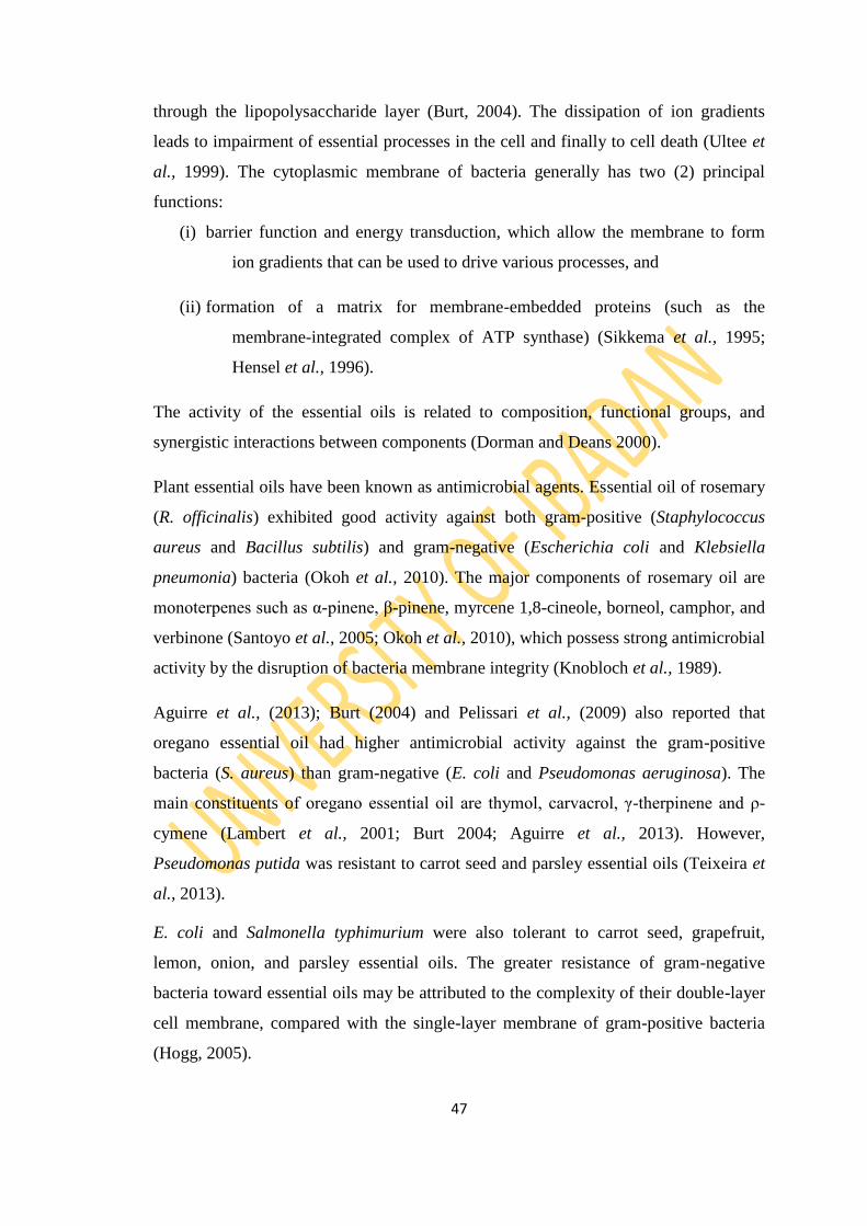
47
through the lipopolysaccharide layer (Burt, 2004). The dissipation of ion gradients
leads to impairment of essential processes in the cell and finally to cell death (Ultee et
al., 1999). The cytoplasmic membrane of bacteria generally has two (2) principal
functions:
(i) barrier function and energy transduction, which allow the membrane to form
ion gradients that can be used to drive various processes, and
(ii) formation of a matrix for membrane-embedded proteins (such as the
membrane-integrated complex of ATP synthase) (Sikkema et al., 1995;
Hensel et al., 1996).
The activity of the essential oils is related to composition, functional groups, and
synergistic interactions between components (Dorman and Deans 2000).
Plant essential oils have been known as antimicrobial agents. Essential oil of rosemary
(R. officinalis) exhibited good activity against both gram-positive (Staphylococcus
aureus and Bacillus subtilis) and gram-negative (Escherichia coli and Klebsiella
pneumonia) bacteria (Okoh et al., 2010). The major components of rosemary oil are
monoterpenes such as α-pinene, β-pinene, myrcene 1,8-cineole, borneol, camphor, and
verbinone (Santoyo et al., 2005; Okoh et al., 2010), which possess strong antimicrobial
activity by the disruption of bacteria membrane integrity (Knobloch et al., 1989).
Aguirre et al., (2013); Burt (2004) and Pelissari et al., (2009) also reported that
oregano essential oil had higher antimicrobial activity against the gram-positive
bacteria (S. aureus) than gram-negative (E. coli and Pseudomonas aeruginosa). The
main constituents of oregano essential oil are thymol, carvacrol, γ-therpinene and ρ-
cymene (Lambert et al., 2001; Burt 2004; Aguirre et al., 2013). However,
Pseudomonas putida was resistant to carrot seed and parsley essential oils (Teixeira et
al., 2013).
E. coli and Salmonella typhimurium were also tolerant to carrot seed, grapefruit,
lemon, onion, and parsley essential oils. The greater resistance of gram-negative
bacteria toward essential oils may be attributed to the complexity of their double-layer
cell membrane, compared with the single-layer membrane of gram-positive bacteria
(Hogg, 2005).
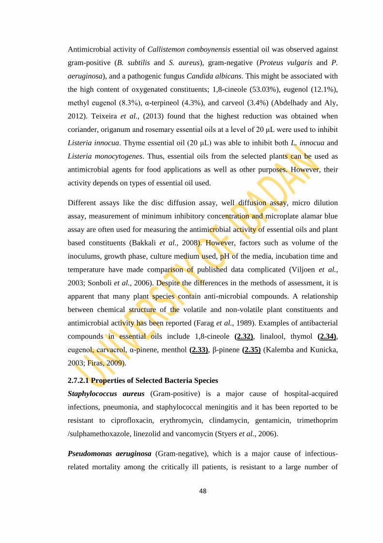
48
Antimicrobial activity of Callistemon comboynensis essential oil was observed against
gram-positive (B. subtilis and S. aureus), gram-negative (Proteus vulgaris and P.
aeruginosa), and a pathogenic fungus Candida albicans. This might be associated with
the high content of oxygenated constituents; 1,8-cineole (53.03%), eugenol (12.1%),
methyl eugenol (8.3%), α-terpineol (4.3%), and carveol (3.4%) (Abdelhady and Aly,
2012). Teixeira et al., (2013) found that the highest reduction was obtained when
coriander, origanum and rosemary essential oils at a level of 20 μL were used to inhibit
Listeria innocua. Thyme essential oil (20 μL) was able to inhibit both L. innocua and
Listeria monocytogenes. Thus, essential oils from the selected plants can be used as
antimicrobial agents for food applications as well as other purposes. However, their
activity depends on types of essential oil used.
Different assays like the disc diffusion assay, well diffusion assay, micro dilution
assay, measurement of minimum inhibitory concentration and microplate alamar blue
assay are often used for measuring the antimicrobial activity of essential oils and plant
based constituents (Bakkali et al., 2008). However, factors such as volume of the
inoculums, growth phase, culture medium used, pH of the media, incubation time and
temperature have made comparison of published data complicated (Viljoen et al.,
2003; Sonboli et al., 2006). Despite the differences in the methods of assessment, it is
apparent that many plant species contain anti-microbial compounds. A relationship
between chemical structure of the volatile and non-volatile plant constituents and
antimicrobial activity has been reported (Farag et al., 1989). Examples of antibacterial
compounds in essential oils include 1,8-cineole (2.32), linalool, thymol (2.34),
eugenol, carvacrol, α-pinene, menthol (2.33), β-pinene (2.35) (Kalemba and Kunicka,
2003; Firas, 2009).
2.7.2.1 Properties of Selected Bacteria Species
Staphylococcus aureus (Gram-positive) is a major cause of hospital-acquired
infections, pneumonia, and staphylococcal meningitis and it has been reported to be
resistant to ciprofloxacin, erythromycin, clindamycin, gentamicin, trimethoprim
/sulphamethoxazole, linezolid and vancomycin (Styers et al., 2006).
Pseudomonas aeruginosa (Gram-negative), which is a major cause of infectious-
related mortality among the critically ill patients, is resistant to a large number of

49
antibiotics (Bonfiglio et al., 1998; Gales et al., 2001) and hence carries the highest
case fatality rate of all Gram negative infections (Aliaga et al., 2002).
Escherichia coli (Gram-negative) also cause life threatening infections especially in
the hospital environment and has been found to be resistant to commonly used
antimicrobial agents like trimethoprim-sulfame-thoxazole (Amyes and May, 2007).
Shigella flexineri (Gram-negative) is a human intestinal pathogen, causing dysentery
by invading the epithelium of the colon and is responsible, worldwide, for an estimated
165 million episodes of shigellosis and 1.5 million deaths per year (Nanyonga, 2012).
Resistance of Shigella flexineri to antimicrobial agents has also been reported (Sack et
al., 1997).
Salmonella typhi (Gram-negative) is a genus of bacteria that cause typhoid fever
which is a major health problem especially in developing countries (Lin et al., 2000;
Otegbayo et al., 2003) and multidrug-resistant strains of Salmonella have been
encountered (Olowe et al., 2003).
Bacillus subtillis are Gram positive large rods that are widely spread in air soil and
water (Oyedeji, 2001). It causes food poisoning.
2.7.3 Antioxidant Activity
Human body consumes ample amounts of oxygen for the metabolism of biomolecules
in order to produce energy. Although oxygen is essential for life, but its metabolites, so
called reactive oxygen species (ROS), are very toxic and cause harm to cells. An
imbalance between ROS production and elimination leads to an ―oxidative stress‖.
Antioxidants are uniquely qualified to decrease the oxidative stress and neutralize ROS
before they damage the tissues. Hence a variety of antioxidants is required for
neutralization of free radicals to protect body from their adverse effects. An
antioxidant is thus a substance that, when present at a low concentration compared
with that of an oxidisable substrate, inhibits oxidation of the substrate (Halliwell and
Gutteridge, 2007).
Chemically, free radicals are molecules that are loosing an electron and this makes
them highly reactive as oxidants. In the act of desperately snatching an electron from
any other molecule, ROS exert oxidative damaging effects to molecules found in living
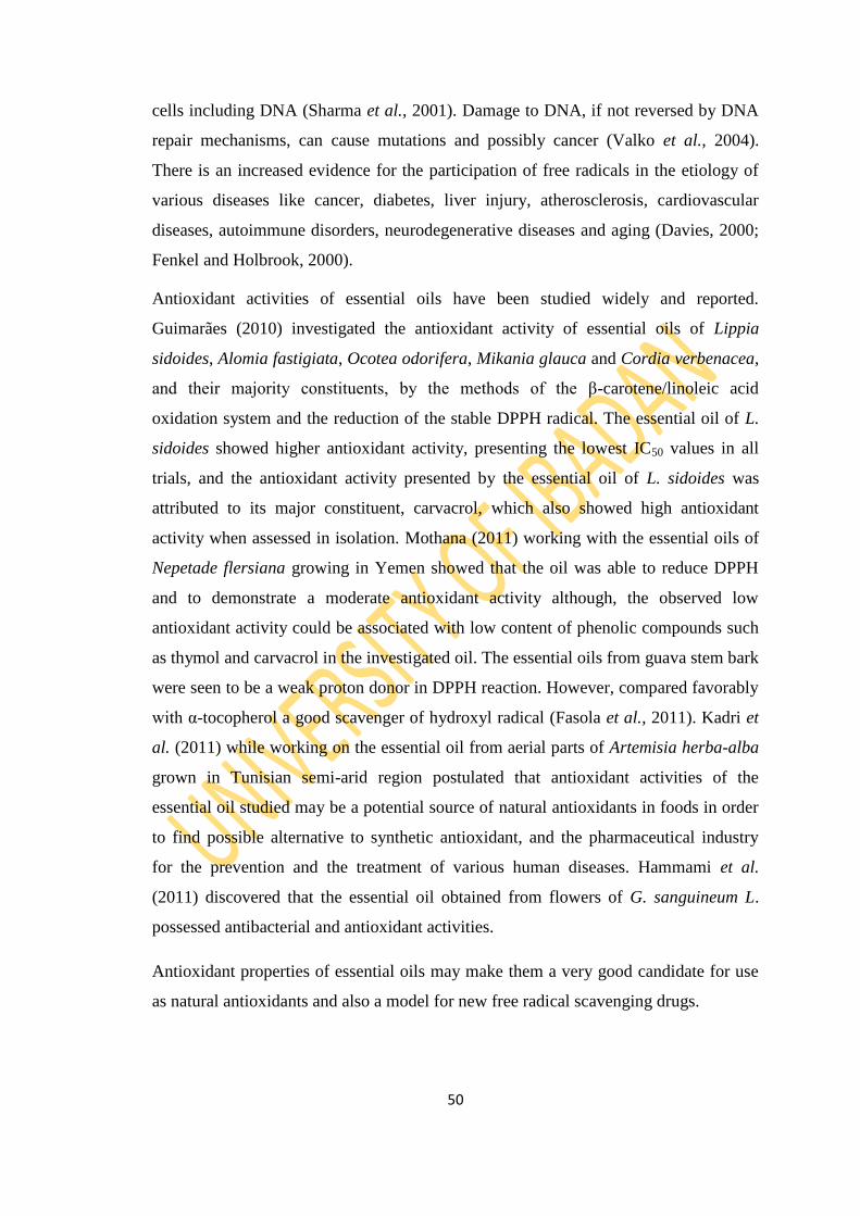
50
cells including DNA (Sharma et al., 2001). Damage to DNA, if not reversed by DNA
repair mechanisms, can cause mutations and possibly cancer (Valko et al., 2004).
There is an increased evidence for the participation of free radicals in the etiology of
various diseases like cancer, diabetes, liver injury, atherosclerosis, cardiovascular
diseases, autoimmune disorders, neurodegenerative diseases and aging (Davies, 2000;
Fenkel and Holbrook, 2000).
Antioxidant activities of essential oils have been studied widely and reported.
Guimarães (2010) investigated the antioxidant activity of essential oils of Lippia
sidoides, Alomia fastigiata, Ocotea odorifera, Mikania glauca and Cordia verbenacea,
and their majority constituents, by the methods of the β-carotene/linoleic acid
oxidation system and the reduction of the stable DPPH radical. The essential oil of L.
sidoides showed higher antioxidant activity, presenting the lowest IC50 values in all
trials, and the antioxidant activity presented by the essential oil of L. sidoides was
attributed to its major constituent, carvacrol, which also showed high antioxidant
activity when assessed in isolation. Mothana (2011) working with the essential oils of
Nepetade flersiana growing in Yemen showed that the oil was able to reduce DPPH
and to demonstrate a moderate antioxidant activity although, the observed low
antioxidant activity could be associated with low content of phenolic compounds such
as thymol and carvacrol in the investigated oil. The essential oils from guava stem bark
were seen to be a weak proton donor in DPPH reaction. However, compared favorably
with α-tocopherol a good scavenger of hydroxyl radical (Fasola et al., 2011). Kadri et
al. (2011) while working on the essential oil from aerial parts of Artemisia herba-alba
grown in Tunisian semi-arid region postulated that antioxidant activities of the
essential oil studied may be a potential source of natural antioxidants in foods in order
to find possible alternative to synthetic antioxidant, and the pharmaceutical industry
for the prevention and the treatment of various human diseases. Hammami et al.
(2011) discovered that the essential oil obtained from flowers of G. sanguineum L.
possessed antibacterial and antioxidant activities.
Antioxidant properties of essential oils may make them a very good candidate for use
as natural antioxidants and also a model for new free radical scavenging drugs.
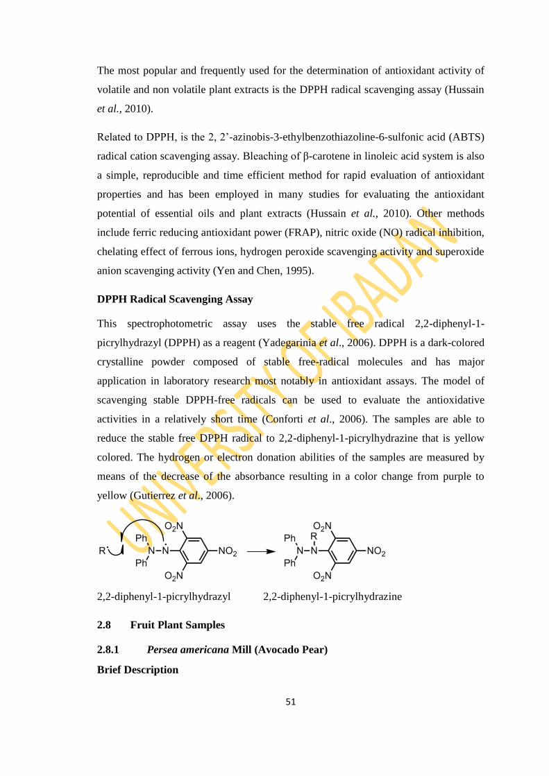
51
The most popular and frequently used for the determination of antioxidant activity of
volatile and non volatile plant extracts is the DPPH radical scavenging assay (Hussain
et al., 2010).
Related to DPPH, is the 2, 2‘-azinobis-3-ethylbenzothiazoline-6-sulfonic acid (ABTS)
radical cation scavenging assay. Bleaching of β-carotene in linoleic acid system is also
a simple, reproducible and time efficient method for rapid evaluation of antioxidant
properties and has been employed in many studies for evaluating the antioxidant
potential of essential oils and plant extracts (Hussain et al., 2010). Other methods
include ferric reducing antioxidant power (FRAP), nitric oxide (NO) radical inhibition,
chelating effect of ferrous ions, hydrogen peroxide scavenging activity and superoxide
anion scavenging activity (Yen and Chen, 1995).
DPPH Radical Scavenging Assay
This spectrophotometric assay uses the stable free radical 2,2-diphenyl-1-
picrylhydrazyl (DPPH) as a reagent (Yadegarinia et al., 2006). DPPH is a dark-colored
crystalline powder composed of stable free-radical molecules and has major
application in laboratory research most notably in antioxidant assays. The model of
scavenging stable DPPH-free radicals can be used to evaluate the antioxidative
activities in a relatively short time (Conforti et al., 2006). The samples are able to
reduce the stable free DPPH radical to 2,2-diphenyl-1-picrylhydrazine that is yellow
colored. The hydrogen or electron donation abilities of the samples are measured by
means of the decrease of the absorbance resulting in a color change from purple to
yellow (Gutierrez et al., 2006).
2,2-diphenyl-1-picrylhydrazyl 2,2-diphenyl-1-picrylhydrazine
2.8 Fruit Plant Samples
2.8.1 Persea americana Mill (Avocado Pear)
Brief Description
52
Pear is the name for about twenty (20) species of trees of a genus in the rose family,
and for their fruit. Persea americana Mill (Lauraceae) commonly called Avocado is
usually medium sized, erect and deciduous tree ranging from 15-20 m in height
(Ojewole et al., 2007) with trunks 30 cm (12 inches) or more in diameter. The leaves
are oval and simple and, unlike those of the apple, smooth and glossy. The white
flowers, which are borne in umbels, have five sepals, five petals, many stamens, and a
single pistil. The fruit is a pome, juicier than the apple, and varying from apple-shaped
to teardrop-shaped (Figure 2.2). Among different varieties, the thin skin varies in color
from light yellow and green through red and brown. The thick flesh varies in flavor
among different varieties. Pears are gathered from the trees before they are completely
ripe and are allowed to ripen in storage. Cold retards ripening, and heat speeds it. Most
pear varieties may be grown in either standard or dwarf sizes.
Ethnomedicinal Importance
Different parts of the plant are used in folk medicine for the treatment of several
ailments such as hypertension, diabetes and inflammation (Adeyemi et al., 2002; Lans,
2006; Bartholomew et al, 2007). Pears are eaten fresh and canned. The fruit contain
about 16% carbohydrate and negligible amounts of fat and protein. They are good
sources of the B-complex vitamins and also contain vitamin C; in addition, they
contain small amounts of phosphorus and iodine. Specifically the fruit is used as
vermifuge, for treatment of dysentery and as aphrodisiac (Bartholomew et al., 2007).
The leaves are used extensively in the treatment of hypertension (Gill, 1992; Lans
2006), sore throat, haemorrhage and inflammatory conditions (Bartholomew et al.,
2007). Some of the scientifically validated activities of the plant leaves include its
antihypertensive activity (Owolabi et al., 2005; Ojewole et al., 2007), anticonvulsant
effect (Ojewole and Amabeoku, 2006), analgesic and anti-inflammatory activities
(Adeyemi et al., 2002).
The leaves have been reported as an effective antitussive and antidiabetic, and for
relief of arthritis pain, by traditional medicine practitioners of the Ibibio tribe in South
Nigeria. The leaves have been reported to be effective anti-tussive, antidiabetic, and
anti-arthritic by traditional medicine practitioners of Ibibio tribe of Southern Nigeria.
Analgesic properties of the leaves have also been reported (Anita et al., 2005).
53
The seeds (crude or toasted) are employed in traditional Mexican medicine to treat skin
rashes, diarrhea, and dysentery caused by helminths and amoebas, for the cure of
infectious processes caused by fungi and bacteria, as well as for the treatment of
asthma, high blood pressure, and rheumatism (Aguilar and Aguilar, 1994; Argueta et
al., 1994; Adeyemi et al., 2002; Del-Refugio-Ramos et al., 2004; Osuna-Torres et al.,
2005; Moreno-Uribe, 2008; Anaka et al., 2009). The seeds of P. americana used alone
or mixed with other species, such as Psidium guajava, Mentha piperita or Ocimum
basilicum, are mainly employed for the treatment of diarrhea (Osuna-Torres et al.,
2005).
Previous Work
Results from previous investigation into the chemical composition of P. americana
leaf, fruit and seed showed that the investigated samples contain phytochemicals such
as phenols, saponins, tannins, steroids, alkaloids and flavonoids (Arukwe et al., 2012).
Proximate content revealed that the fruit of P. americana contains more of fat and
energy; seed had more of fat, protein and energy while the leaf had more of protein,
fibre, and ash (Arukwe et al., 2012).
P. americana has been reported to be effective against hepato-toxicity, inflammation,
cancer and hypertension (Adeyemi et al., 2002; Anaka et al., 2009; Imafidon and
Amaechina, 2010; Ojewole and Amabeoku, 2006).
The presence of fatty acids (linoleic, oleic, palmitic, stearic, linolenic, capric and
myristic acids), polyphenols (catechin, isocatechin, protocyanidin, flavonoids, tannins
and proanthocyanidin monomerics), saponins, glucosides (D-perseit, D-α-manoheptit,
D-monoheptulose, persiteol), sterols (β-sitosterol, campesterol, stigmasterol,
cholesterol), the amino acid carnitine and two glucosides of abscicic acid has been
reported for P. americana seeds (Nwaogu et al., 2008; Takenaga et al., 2008; Wang et
al., 2010). High concentrations of catechins, procyanidins and hydroxycinnamic acid
have recently been determined in 100% ethyl acetate (EtOAc), in 70% acetone and
70% methanol (MeOH) extracts obtained from P. americana peel and seeds, while the
pulp extract was rich in hydroxybenzoic acid, hydroxycinnamic acid and procyanidins
(Rodríguez-Carpena et al., 2011).
54
Interestingly, the hypolipemic effect of the MeOH extract obtained from P. americana
seeds has been demonstrated in male rats with induced hypercholesterolemia (Asaolu
et al., 2010; Imafidon and Amaechina, 2010). The same effect was described for the
aqueous extract, which also reduced blood pressure both in normal rats and those with
high blood pressure; in addition, it exerted a hypoglycemic effect on rats and rabbits
with diabetes (Okonta et al., 2007; Ogochukwu et al., 2009; Edem et al., 2009; Kofi et
al., 2009). The aqueous extract showed a median Lethal dose (LD50) = 10 g/kg in rats
when it was administered orally. Importantly, it did not alter the hematological
parameters nor the levels of Alanine amino transferase (ALT), Aspartate amino
transferase (AST), albumin, and creatinine in male and female rats were treated for 28
days (Ozolua et al., 2009).
The hexane and MeOH seed extracts of P. americana have been described to have a
Minimum inhibitory concentration (MIC) of <1.25 μg/ml against Candida sp.,
Cryptococcus neoformans and Malassezia pachydermatis. These extracts were also
active against Artemia salina, with Lethal concentration (LC50) values of 2.37 and
24.13 mg/mL, respectively. They were also active against Aedes aegypti larvae with
LC50 values of 16.7 and 8.9 mg/mL, respectively (Giffoni et al., 2009). On the other
hand, the MeOH extract from P. americana leaves inhibited completely the growth of
M. tuberculosis (MIC = 125 μg/mL) and H37Rv (MIC = 62.5 μg/mL); furthermore, the
hexane fraction inhibited the growth of both mycobacteria with MIC = 31.2 μg/mL
(Gomez-Flores et al., 2008). In addition, the EtOH extract was active against both
Gram positive and negative bacteria (with the exception of Staphylococcus epidermis
and Escherichia coli) with MIC of 500 μg/mL (Raymond-Chia and Dykes, 2011).
Regarding the bacterial activity of P. americana (var Hass and Fuerte), the acetone
seed extract exhibited moderate activity against Bacillus cereus, Staphylococcus
aureus and Listeria monocytogenes (Rodríguez-Carpena et al., 2011).
The trypanomicidal activity of the MeOH extract from P. americana seeds has been
also tested (Abe et al., 2005). It showed moderate activity when it was evaluated at the
concentration range of 250–500 μg/mL. In the case of the aqueous seed extract, it had
a slight anti-Giardia duodenalis (syn G. lamblia) activity, inducing 23% of mortality at
4 mg/mL (Ponce-Macotela et al., 1994). The antioxidant activity (AOA) of P.
americana seed, peel, pulp and leaves extract has been described by different methods

55
(Wang et al., 2010; Rodríguez-Carpena et al., 2011; Matsusaka et al., 2003; Yean-
Yean and Barlow, 2004; Asaolu et al., 2010a,b).
Jiménez-Arellanes et al. (2013) reported the antiprotozoal and antimycobacterial
activities of ethanol and chloroform extracts of P. americana.
Analgesic and anti-inflammatory properties of the leaves have been reported by
(Adeyemi et al., 2002).
Volatile constituents of avocado mesocarp were isolated by concurrent steam
distillation/solvent extraction in the Likens-Nickerson apparatus using pentane ether as
solvent. The extracts which resulted were concentrated in a Kuderna-Danish
concentrator and analysed using gas chromatography and linked gas chromatography-
mass spectroscopy (GC-MS) employing capillary columns of contrasting polarity.
Hydrocarbons (mainly sesquiterpenes) and alkanals were the predominant constituents
present. In the immediate extract of the avocado mesocarp, β-caryophyllene (60%) was
the main sesquiterpene, followed by α-humulene (5.9%), caryophyllene oxide (4.8%),
α-copaene (4.5%) and α-cubebene as the main hydrocarbons; alkanals were present,
but only in low concentrations (Sinyinda and Gramshaw, 1998).
In the extract prepared following storage (2 h) of the mesocarp at room temperature, β-
caryophylene (28.8%) was the main sesquiterpene, followed by α-copaene (10.7%), a
cadinene isomer (8.5%), α- and β-cubebene (7.7%) α-famesene (5.3%) and octane
(4.8%) as principal hydrocarbons; decenal (6.3%) and heptenal (3.2%) were the main
aldehydes (Sinyinda and Gramshaw, 1998).
Investigation of the essential oils composition of some Persea spices revealed β-
caryophyllene (43.9%) and valencene (16.0%) as the most abundant compounds in the
oil of P. americana leaf from Nigeria (Ogunbinu et al., 2007). Methyl chavicol (syn.,
estragole) (53.9%) is the major component in the oil of P. americana var. drymifolia
cv. Duke from Cuba (Pino et al., 2004). (E)-Avocadodienofuran (15.3%), (E)-
avocadenynofuran (13.2%), and β-caryophyllene (11.0%) were the major components
in the oil of P. indica Spreng (Pino and Rolando, 2006).
In another study, the oil of P. americana leaf revealed that (Z)-nerolidol, (E,E)-2,4-
decadienal, (E,E)-α-farnesene, β-caryophyllene, caryophyllene oxide, and α-copaene
were the major components (Pino et al., 2004).

56
Larijani et al., (2010) characterized thirty-six components in the oil of P. americana
leaf from Iran, accounting for 97.7% of the oil. The oil consists of 54.5%
monoterpenes, 0.6% oxygenated monoterpenoids, 37.7% sesquiterpenes, and 4.9%
oxygenated sesquiterpenoids with methyl eugenol (31.2%), β-caryophyllene (16.9%),
estragole (9.0%), δ-cadinene (4.8%), β-pinene (4.2%), and α-pinene (3.2%) as the
major components in the oil.
Figure 2.2: Pictures of P. americana Leaf, Fruit and Seed
57
2.8.2 Carica papaya (Pawpaw)
Brief Description
Carica papaya Linn belonging to family Caricaceae is commonly known as papaya or
pawpaw in English (Yogiraj et al., 2014). The papaya is a large, tree-like plant, with a
single stem growing from 1.8 to 10 m (6 to 33 ft) tall, with spirally arranged leaves
confined to the top of the trunk. The lower trunk is conspicuously scarred where leaves
and fruits are borne.
The leaves are large, 50–70 cm, palmately lobed or deeply incised with entire margins
and petioles of 1-3 feet in length. Stems are hollow, light green to tan brown in color
with diameter of 8 inches (Arvind et al., 2013). The flowers appear on the axils of the
leaves, maturing into large fruit, 15–45 cm long and 10–30 cm in diameter. Fruits are
borne axillary on the main stem, usually singly but sometimes in small clusters. The
fruit, which vary in shape from spherical to elongate and which may weigh from 0.5 up
to 20 lbs (9 kg), ripens when it feels soft and its skin has attained amber to orange hue
(Figure 2.3). Flesh is yellow-orange to salmon (pinkish-orange) at maturity (Yogiraj et
al., 2014). The edible portion surrounds the large central seed cavity. Individual fruits
mature in 5-9 months, depending on cultivator and temperature. Plants begin bearing
fruits in 6-12 months (Arvind et al., 2013). Unusually for large plants, the trees are
dioecious. The tree is usually unbranched, unless lopped.
Ethnomedicinal /Pharmacological Importance
C. papaya is eaten fresh as breakfast fruit or in salads or desserts. Papaya is also
exploited for its latex, which contains papain, a proteolytic (protein-digesting) enzyme
used in meat tenderizers.

58
Traditionally, the leaves have been used for treatment of a wide range of ailments. The
leaves are made into tea as a treatment for malaria in some parts of the world (Titanji
et al., 2008). Antimalarial and antiplasmodial activity has been noted in some
preparations of the plant, but the mechanism is not understood and not scientifical
proven. The young leaves of C. papaya are steamed and eaten like spinach in some
parts of Asia and in the treatment of jaundice, urinary complaints and gonorrhoea,
fever and dressing wounds (Krishna et al., 2008). Papaya leaf is dried and cured like a
cigar to be smoked by asthmatic persons. An infusion of fresh papaya leaves is used by
person to expel or destroy intestinal worms. Additional benefits of papaya leaves
include; as an acne medicine, appetite increase, menstrual pain ease, meat tenderizer
and nausea reliever (Arvind et al., 2013). The leaves of Carica papaya are used in
herbal medical practices in South East Nigeria to treat malaria and typhoid fever
(Igwe, 2015).
Ripe papaya fruit is used as laxative which assures of regular bowel movement. The
folic acid found in papayas is needed for the conversion of homocysteine into amino
acids such as cysteine or methionine. If unconverted, homocysteine can directly
damage blood vessel walls, is considered a significant risk factor for a heart attack or
stroke (Arvind et al., 2013).
Papaya seed is used as carminative, emmenagogue, vermifuge, abortifacient, counter
irritant, paste in the treatment of ringworm and psoriasis, antifertility agents in males.
The black seeds are edible and have a sharp, spicy taste. They are sometimes ground
and used as a substitute for black pepper (Yogiraj et al., 2014).
Papaya peel is often used in cosmetics as skin lightening agent. When peel is mixed
with honey and applied it can act as soothe and moisturizers the skin. The papaya
vinegar with lemon juice can be applied to the scalp for 20 minutes prior to
shampooing to fight dandruff. Adding papaya oil and vinegar to bathwater, along with
essential oils like lavender, orange and rosemary can be nourishing, refreshing and
relaxing, and can work as a pain reliever and muscle relaxant (Arvind et al., 2013).
Juice from papaya roots is used in some countries of Asia to ease urinary troubles. A
decoction formed by boiling the outer part of the roots of the papaya tree in the cure of
dyspepsia (Arvind et al., 2013). The roots are also said to cure piles and yaws.

59
C. papaya is also applied topically for the treatment of cuts, rashes, stings and burns.
C. papaya ointment is commonly made from fermented papaya flesh, and is applied as
a gel-like paste (Morton, 1987). Women in India, Bangladesh, Pakistan, Sri Lanka, and
other countries have long used green papaya as an herbal medicine for contraception
and abortion. Enslaved women in the West Indies were noted for consuming papaya to
prevent pregnancies and thus preventing their children from being born into slavery
(Morton, 1987). The latex is used locally as antiseptic. Infusion of the root is said to
remove urine concretions (Reed, 1979). Latex from the plant is used as dyspepsia cure
and can also be applied externally to burns and scalds (Reed, 1979). The flowers have
been used for jaundice. In Asia, the latex is smeared on the mouth of the uterus as
ecbolic. The root infusion is used for syphilis in Africa. Japanese believe that eating
papaya prevents rheumatism (Duke, 1984b). Dietary papaya does reduce urine acidity
in humans. The inner bark is used for sore teeth. The latex is used in psoriasis,
ringworm and it‘s prescribed for the removal of cancerous growths in Cuba (Duke,
1984b). It has been reported that the extract of unripe pawpaw possesses anti-sickling
and reversal of sickling properties (Oduola et al., 2006). The tea prepared with the
green papaya leaf, promotes digestion and aids in the treatment of ailments such as
chronic indigestion, overweight and obesity, arteriosclerosis, high blood pressure and
weakening of the heart (Mantok, 2005; Ayoola and Adeyeye, 2010).
Carica papaya contains an enzyme known as papain which is present in the bark,
leaves and fruit. The milky juice contains many biologically active compounds
including chymopapain and papain which is the ingredient that aids digestive system,
and again used in treatment of arthritis (Arvind et al., 2013).
Previous Work
Phytochemical analysis of methanol and aqueous extracts of Carica papaya aerial
parts proves the presence of phytocomponents as flavonoids, tannins, alkaloids,
carbohydrates and triterpenes (Khaled et al., 2013). Phytochemical analysis of Carica
papaya leaf extract revealed the presence of flavonoids (kaempferol and myricetin),
alkaloids (carpaine, pseudocarpaine, dehydrocarpaine I and II), phenolic compounds
(ferulic acid, caffeic acid, chlorogenic acid), the cynogenetic compounds
(benzylglucosinolate) glycosides, saponins, tannins and steroids (Anjum et al., 2013).
The leaf and fruit were reported to possess carotenoids namely β- carotene, lycopene,

60
anthraquinones glycoside, as compared to matured leaves and hence possess medicinal
properties like anti-inflammatory hypoglycaemic, anti-fertility, abortifacient,
hepatoprotective, wound healing, recently its antihypertensive and antitumor activities
have also been established.
Different properties of papaya such as antioxidant and free radical scavenging activity,
anticancer activity, anti-inflammatory activity, treatment for dengue fever, anti-
diabetic activity, wound healing activity and antifertility effects has been studied. Thus
Carica papaya acts as a multi faceted plant. It is also imperative to identify the
mechanism of the plant compounds and studying the active principle of the extract.
Thus, papaya should be included in diet as fruit salads, fruit juice, leaf extract,
decoction prepared through papaya leaves, etc. However, including papaya seeds in
any of the form should be avoided for young men and pregnant women, since it
possess antifertility effects that was demonstrated well in animal models (Natarjan et
al., 2014).
Studies of Dr. Sanath Hettige (Pigli and Runja, 2014), who conducted the research on
70 dengue fever patients, said papaya leaf juice helps increase white blood cells and
platelets, normalizes clotting, and repairs the liver. Carica papaya leaf extract was
found to increase the platelet count and also to decrease the clotting time in rats. The
study aims at determining the possible effects of papaya leaves in thrombocytopenia
occurring in dengue fever (Krishna and Thomas, 2014; Soobitha et al., 2013; Fenny et
al., 2012; Patil et al., 2013). Recent research on papaya leaf tea extract also
demonstrated cancer cell growth inhibition. It appears to boost the production of key
signaling molecules called Th1-type cytokines, which help regulate the immune
system.
Zunjar V investigated the microscopic evaluation of leaves of Carica papaya L. to
establish the salient diagnostic features for the leaf. The leaf shows abundant
sphaeraphides and rhomboidal calcium oxalate crystals. The leaves show no trichomes
and a continuous network of veins. Histochemical tests performed indicate the
presence of alkaloids and starch. Physiochemical parameters such as extractive values,
ash values and moisture content have also been studied for the leaf (Zunjar, 2011).
61
The antimicrobial activities of different solvent extracts of Carica papaya were tested
against the Gram-positive and Gram-negative bacterial strains and fungus by observing
the zone of inhibition (Baskaran et al., 2012). Papaya seeds have antibacterial
properties and are effective against E. coli, Salmonella and Staphylococcus infections.
Only the leaf extracts showed inhibitory effect against Candida albicans, whereas stem
and root extracts were ineffective. Among the leaf, stem and root extracts, the leaf
extract is found to exhibit more antimicrobial activity than the stem and root (Sumathi,
2014).
Khaled et al. (2013) research work deals with the evaluation of anti-HIV-1 effect of
Carica papaya aerial parts polar extracts and also the investigation of the chemical
content from the polar extracts of the plant. The methanol and aqueous extracts of
Carica papaya were tested for their anti-HIV-1 activity using the syncytia formation
assay. Methanol and aqueous extracts of Carica papaya aerial parts showed activity as
anti- HIV-1 agents, both of the extracts therapeutic index (TI) of 5.51 and 7.13
compared with the standard drug. The results have shown that Carica papaya
methanol and aqueous extracts have drug ability as anti- HIV-1 agents.
Achini et al. (2012) investigated that management of thrombocytopenia is by drugs
and blood products, both of which are costly. Conversely, Sri Lankan traditional
medicine use mature leaf concentrate of Carica papaya to treat this condition. This
claim was scientifically validated.
Igwe (2015) reported the chemical constituents of the extract of the leaves of C.
papaya with isopropanol as a choice of solvent. Gas Chromatography-Mass
Spectrometry (GC-MS) analysis revealed six compounds which were identified to be
hexahydro-1-aH-naphtho[1,8a-b]oxiren-2(3H)-one (2.17%), 3,7-dimethyloct-7-en-1-ol
(8.08%), 3-methyl-4- (phenylthio)- 2-enyl- 2,5-dihydrothiophene-1, 1-dioxide
(11.78%), cyclopentane undecanoic acid methyl ester (12.02%), 3,7,11,15-
tetramethyl-2-hexadecen-1-ol (37.78%) and 9-octadecenamide (28.18%). The extract
showed potent antimicrobial activity against Staphylococcus aureus, streptococcus
faecalis, Escherichia coli and Proteus mirabilis. The sensitivity of each test
microorganism to the extract was determined using the Disc Diffusion Technique.
Highest sensitivity was shown with S. aureus (14.33 mm at 100% concentration)
followed by E. coli (12.98 mm at 100% concentration) and P. mirabilis (12.37 mm at
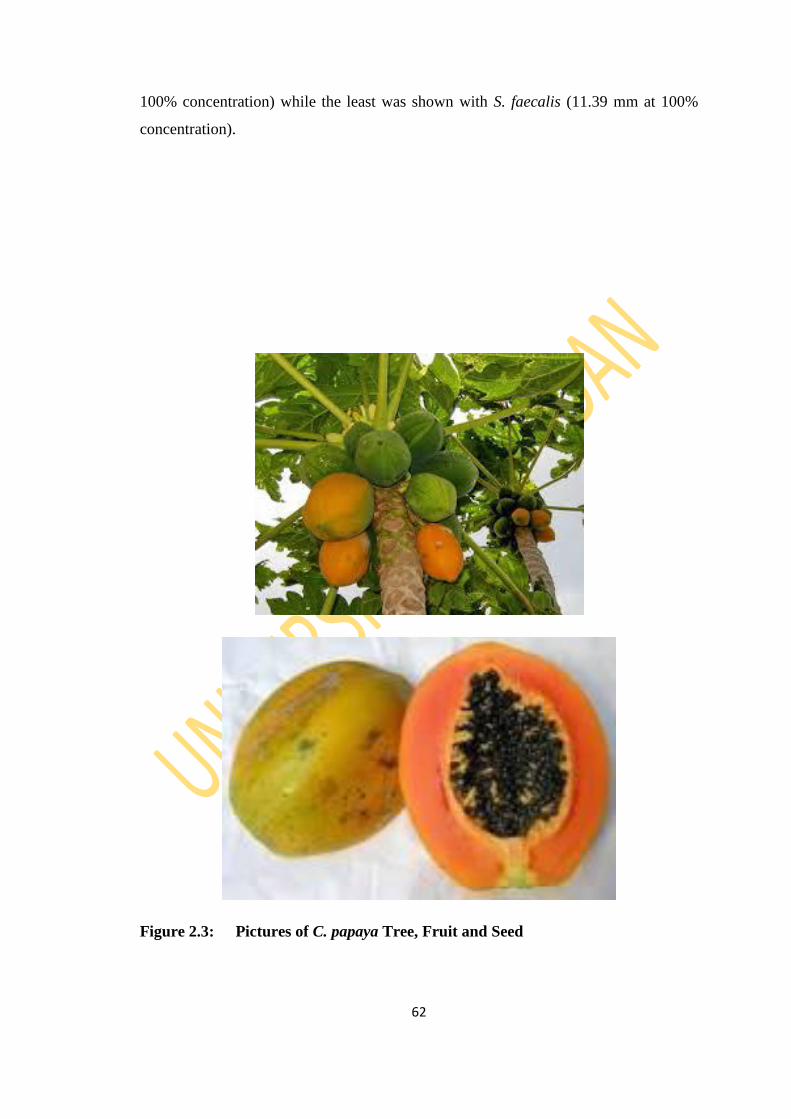
62
100% concentration) while the least was shown with S. faecalis (11.39 mm at 100%
concentration).
Figure 2.3: Pictures of C. papaya Tree, Fruit and Seed
63
2.8.3 Ananas comosus (Pineapple)
Brief Description
Pineapple with scientific name Ananas comosus (Figure 2.4) is a tropical plant with
edible multiple fruit consisting of coalesced berries, named for resemblance to the pine
cone, is the most economically important plant in the Bromeliaceae family (Coppens
d'Eeckenbrugge and Freddy, 2003).
The pineapple is an herbaceous perennial which grows to 1.0 to 1.5 meters (3.3 to 4.9
ft) tall, although sometimes it can be taller (Coppens d'Eeckenbrugge and Freddy,
2003). In appearance, the plant itself has a short, stocky stem with tough, waxy leaves.
When creating its fruit, it usually produces up to 200 flowers, although some large-
fruited cultivars can exceed this. Once it flowers, the individual fruits join together to
create what is commonly referred to as a pineapple. After the first fruit is produced,
side shoots (called 'suckers' by commercial growers) are produced in the leaf axils of
the main stem. Commercially, suckers that appear around the base are cultivated. It has
30 or more long, narrow, fleshy, trough-shaped leaves with sharp spines along the
margins that are 30 to 100 centimeters (1.0 to 3.3 ft) long, surrounding a thick stem. In
the first year of growth, the axis lengthens and thickens, bearing numerous leaves in
close spirals. After 12 to 20 months, the stem grows into a spike-like inflorescence up
to 15 cm long with over 100 spirally arranged, trimerous flowers, each subtended by a
bract (Davidson, 2008). Flower colors vary, depending on variety, from lavender,
through light purple to red.
The ovaries develop into berries which coalesce into a large, compact, multiple
accessory fruit. The fruit of a pineapple is arranged in two interlocking helices, eight in

64
one direction, thirteen in the other, each being a Fibonacci number (Jones and William,
2006).
Ethnomedicinal / Traditional Importance
Pineapple has been used as a medicinal plant in several native cultures. It may be
consumed fresh, canned, juiced, and are found in a wide array of food stuffs, dessert,
fruit salad, jam, yogurt, ice cream, candy, and as a complement to meat dishes. In
addition to consumption, in the Philippines the pineapple's leaves are used as the
source of a textile fiber called piña, and is employed as a component of wall paper and
furnishings, amongst other uses (Bartholomew et al., 2003; Davidson, 2008).
The whole plant is used to treat typhoid fever in Ijebu Ode Local Government Area in
Ogun State of Nigeria (Fadimu et al., 2014). Roasted unripe fruit juice is used by
different communities of Gohpur of Sonitpur district, Assam, India for strangury; a
condition caused by blockage or irritation at the base of the bladder, resulting in severe
pain and a strong desire to urinate (Saikia, 2006). The Garo tribal community of
Netrakona district in Bangladesh uses fruit juice for fever and leaf juice for
helminthiasis and jaundice (Rahmatullah et al., 2009). The root and fruit are either
eaten or applied topically as an anti-inflammatory, digestive and proteolytic agent
(Bhakta et al. 2012). It is traditionally used as an antiparasitic and anthelmintic agent
in the Tripura. This fruit can also be used to aid digestion. A root decoction is used to
treat diarrhoea. In some cultures, pineapple has become associated with the notion of
welcome, an association bespoken by the use of pineapple motifs as carved decorations
in woodworking. It can clear bronchial passages in those suffering with pneumonia and
bronchitis. The anti-inflammatory properties in this fruit help reduce the symptoms of
arthritis, and help reduce pain after surgery and sport injuries (Hossain et al., 2015).
Pineapple is currently being studied for its effectiveness in preventing heart disease.
Previous Work
Research reported in the December 2005 edition of "Medical Science Monitor" studied
the effect of pineapple and other fruit juices on plasma lipids. Researchers discovered
that rats that consumed pineapple juice over a three-hour period experienced a
decrease in lipoprotein particles, compounds that carry fat through the blood, and
increased metabolism, activities that lower cholesterol levels. The April 2011 "Journal
of Medicinal Food" revealed that the bromelain in pineapple juice slightly increased
65
the quantity of tendon cells after a crush injury to the Achilles heel in rat models; it
also decreased the levels of malondialdehyde, a compound that may cause mutations in
tissues. In doing so, pineapple juice contributed to healing in the early stages of an
injury (Bhakta et al., 2012).
The July-August 2009 issue of "Oxidative Medicine and Cellular Longevity" features a
study from Indian researchers, which correlates manganese consumption in pineapple
juice and other foods with increased sperm movement. It also protected sperm during
freezing for storage, which can raise your chances of conception (Bhakta et al., 2012;
Debnath et al., 2012).
More than 280 volatile compounds had been identified among the aroma volatiles of
pineapple, whereas only a few volatiles contribute to pineapple aroma (Tokitomo et
al., 2005). It has been reported that esters were the most abundant pineapple volatiles,
in particular, ethyl hexanoate and methyl hexanoate which have the highest
contribution to the pineapple aroma (Pickenhagen et al., 1981; Morais and Silva,
2011). Marta et al. (2010), Elss et al. (2005), Taivini et al. (2001), Umano et al.
(1992), Akioka et al. (2008) also reported that esters were the major volatile
compounds in pineapple volatile composition, however, He et al. (2007) reported that
hydrocarbons and esters were the main compounds, which could be explained by
differences in cultivars, growing conditions, and volatiles extraction methods. Such
differences could also justify why methyl butanoate and methyl 2-methylbutanoate
were not found in ‗Smooth Cayenne‘ pineapple, despite being the main components in
other studies (Wei et al., 2011).
On the other hand, Berger (1991) reported two minor hydrocarbon compounds, 1-
(E,Z)-3,5-undecatriene and 1-(E,Z,Z)-3,5,8-undecatetraene as the important
contributors to fresh-cut pineapple aroma due to their low odor threshold values. Also
the results of Takeoka et al. (1991) reported many sulfur-containing esters among
pineapple volatiles.
Wei et al. (2011) identified 44 volatile compounds when characterizing pineapple pulp
and core by headspace-solid phase microextraction (HS-SPME) and gas
chromatography-mass spectrometry (GC-MS) and revealed the presence of methyl
hexanoate, ethyl hexanoate, methyl 3-(methylthio) propanoate, methyl octanoate, ethyl
decanoate, α-terpineol, nonanal, and decanal.
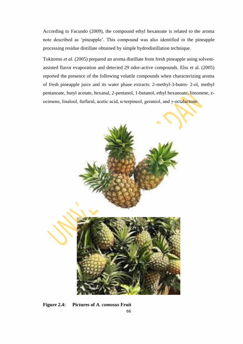
66
According to Facundo (2009), the compound ethyl hexanoate is related to the aroma
note described as ‗pineapple‘. This compound was also identified in the pineapple
processing residue distillate obtained by simple hydrodistillation technique.
Tokitomo et al. (2005) prepared an aroma distillate from fresh pineapple using solvent-
assisted flavor evaporation and detected 29 odor-active compounds. Elss et al. (2005)
reported the presence of the following volatile compounds when characterizing aroma
of fresh pineapple juice and its water phase extracts: 2-methyl-3-buten- 2-ol, methyl
pentanoate, butyl acetate, hexanal, 2-pentanol, 1-butanol, ethyl hexanoate, limonene, z-
ocimene, linalool, furfural, acetic acid, α-terpineol, geraniol, and γ-octalactone.
Figure 2.4: Pictures of A. comosus Fruit
67
2.8.4 Theobroma cacao Linn (Cocoa)
Brief Description
Theobroma cacao L. belongs to the genus of flowering plants in the mallow family,
Malvaceae, which is sometimes classified as a member of Sterculiaceae. It contains
roughly 20 species cultivated in the American, African, and Asian continents, and
many countries worldwide are involved in cocoa production, marketing, and
consumption (Almeida and Valle, 2007, 2009; Bertolde et al., 2014). Several species
of Theobroma produce edible seeds, notably cacao, cupuaçu, and mocambo. The seeds
usually inside the fruit; called a cocoa pod (ovoid) which is 15–30 cm long and 8–10
cm wide, ripening yellow to orange as seen in Figure 2.6, and weighs about 500 g
when ripe. The pod contains 20 to 60 seeds, usually called "beans", embedded in a
white pulp (Izuka and Mbagwu, 2013).
Ethnomedicinal importance/ Previous work
Cocoa has been extensively worked on with literature report on the various use to
alleviate fever, shortness of breath and heart conditions and manuscripts produced in
Europe and New Spain from the 16th to early 20th century revealed more than 150
medicinal uses for cocoa and/or chocolate (Dillinger et al., 2000, Cooper et al., 2008).
Cocoa bean has been reported to have anti-aging properties. The leaf and seeds
Theobroma cacao lowers blood sugar, fatigue, kidney malfunction and serve as anti-
ulcer and tumor. Cocoa bean was also reported by Sharma et al. (2012) to be good for
anxiolytic action. Cacao pigment, which is extracted from the husks, has shown anti-
HIV properties. The substance, consisting of polymerised flavonoids (e.g. catechins,
anthocyanidins and leukoanthycyanidin) inhibits the cytopathic effects of HIV in cell

68
culture. It appears to inhibit absorption of the virus rather than limiting its replication
once it is absorbed (Unten et al., 1991).
The stem bark of Theobroma cacao is boiled with water and mixed with hot pap as
baby food in the treatment of anaemia (Gbadamosi et al., 2012). Cocoa pod is a waste
product of cocoa seeds and have been found very useful industrially for making black
soaps which are highly medicinal for treating various ailment (Adewole et al., 2013).
This means that regular consumption of cocoa will reduce the occurrence of malaria
attack (Jayeola et al., 2011). Cocoa pod was also reported to be a good precursor for
active carbon (Adeyi, 2010).
The antioxidant capacity of cocoa bean and leaves has been investigated by Othman et
al. (2007) and Osman et al. (2004)
The plant was found to be rich in active metabolites like flavonoids, alkaloids, phenols,
saponin and tannins in the leaf, stem bark and seed as shown in Table 2.4
Fapohunda and Afolayan (2012) also reported the presence of phenols and tannins in
cocoa pod husk.
The analytical methodologies applied to isolate and purify cocoa bioactives involve
laborious pretreatment together with isolation and purification procedures (Hatano et
al., 2002; Ortega et al., 2008; Stark and Hofmann, 2005). Among these, isolation by
semi-preparative and preparative liquid chromatography (LC) with C18 reversed phase
(RP) offers high versatility to separate a wide range of nitrogenous and non-
nitrogenous bioactive compounds (Contreras et al., 2009; Rzeppa et al., 2011; Stark
and Hofmann, 2005).
The usual technique to analyze polyphenols from cocoa multicomponent extracts or a
specific isolated fraction is reversed phase high-pressure liquid chromatography (RP-
HPLC) with C8 (Srdjenovic et al., 2008), C12 (Pereira-Caro et al., 2013) and C18
(Andres-Lacueva et al., 2008; Calderón et al., 2009; Quiñones et al., 2011; Tomas-
Barberán et al., 2007) stationary phase. As solvent system, all of these authors used
linear gradients with acidified water (using formic or acetic acid) and acetonitrile or
methanol as organic solvent. This separation technique has been coupled to different
detectors for the qualitative and quantitative characterization of these compounds, such

69
as ultraviolet and diode-array detection (DAD) (Quiñones et al., 2011; Srdjenovic et
al., 2008), fluorescence (Payne et al., 2010; Pereira-Caro et al., 2013) and/or mass
spectrometry (MS) (Andres-Lacueva et al., 2008; Ortega et al., 2010; Pereira-Caro et
al., 2013).
Amongst the phytochemicals which are majorly polyphenols isolated from cocoa bean
include;
Flavonoids – Quercetin glucuronide, Quercetin hexose, Hexenyl xylopyranosyl
glucopyranoside, Quercetin arabinoside, Quercetin, Sucrose, Tri-O-methylsucrose,
Epicatechin (2.36), catechin (2.37), Procyanidin (2.38), (Epi)catechin glucopyranoside,
(Epi)gallocatechin, Catechin diglucopyranoside, Proanthocyanidin Arabinopyranosyl-
(epi)catechin, (Epi)catechin pentamer, (Epi)catechin dimer hexose, (Epi)catechin
methyl dimer, trans-Clovamide (N-[(2E)-3-(3.4-dihydroxyphenyl)-1-oxo-2-propen-1-
yl]-3-hydroxy-L-tyrosine), Deoxyclovamide (N-[(2E)-3-(3.4-Dihydroxyphenyl)-1-oxo-
2-propen-1-yl]-L-tyrosine) (Cádiz-Gurrea et al., 2014).
Alkaloids – Theobromine (2.39) (Cádiz-Gurrea et al., 2014), Caffeine (2.40) (Zheng et
al., 2002), Theophyline (2.41) (Abbe Maleyki and Amin, 2008).
Analysis of essential oils of the leaves of two cocoa clones by GC and GC-MS
revealed twenty-four and twenty compounds which were identified to be aldehydes,
ketones, alcohols and fatty acids (Chee et al., 2005).
To date, essential oils of other parts of the plant have not been investigated to the best
of my knowledge.

70
Table 2.4: Reported Phytochemicals of T. cacao Plant parts
Sample Sap Tannin Phenol Flav Alk Triter Ref
Leaf + + + + + + Zainal et al., 2014
Stem
bark
+ - + + Ogunmefun et al., 2013
+ + + + + Nwokonkwo and Okeke, 2014
Seed + + + + + + Izuka and Mbagwu, 2013

71
(-)-Epicatechin 2.36
N
NHN
N
O
O
CH3
CH3 Theobromine 2.39
(+)-Catechin 2.37
N
NN
N
O
O
CH3
CH3
CH3
Caffeine 2.40
N
NN
N
O
O
CH3
CH3
H
Theophyline 2.41
Procyanidin Dimer and Trimer in Cocoa 2.38
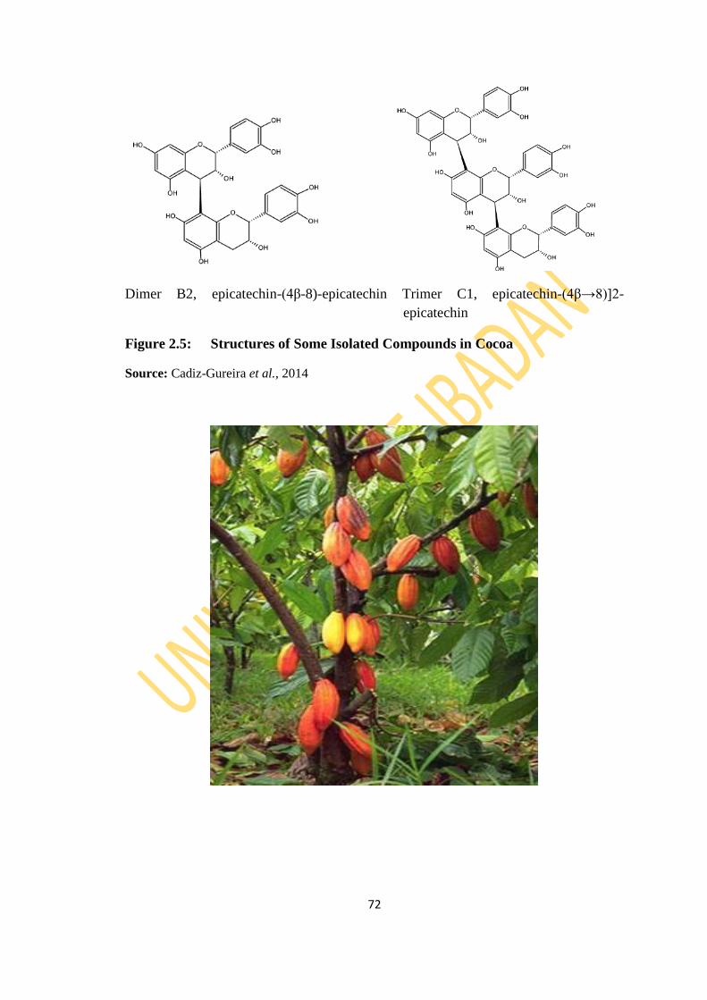
72
Dimer B2, epicatechin-(4β-8)-epicatechin Trimer C1, epicatechin-(4β→8)]2-
epicatechin
Figure 2.5: Structures of Some Isolated Compounds in Cocoa
Source: Cadiz-Gureira et al., 2014
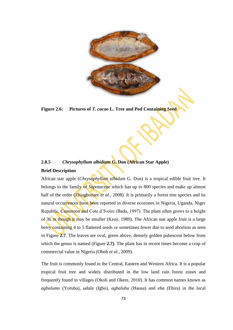
73
Figure 2.6: Pictures of T. cacao L. Tree and Pod Containing Seed
2.8.5 Chrysophyllum albidium G. Don (African Star Apple)
Brief Description
African star apple (Chrysophyllum albidum G. Don) is a tropical edible fruit tree. It
belongs to the family of Sapotaceae which has up to 800 species and make up almost
half of the order (Ehiagbonare et al., 2008). It is primarily a forest tree species and its
natural occurrences have been reported in diverse ecozones in Nigeria, Uganda, Niger
Republic, Cameroon and Cote d‘Ivoire (Bada, 1997). The plant often grows to a height
of 36 m though it may be smaller (Keay, 1989). The African star apple fruit is a large
berry containing 4 to 5 flattered seeds or sometimes fewer due to seed abortion as seen
in Figure 2.7. The leaves are oval, green above, densely golden pubescent below from
which the genus is named (Figure 2.7). The plant has in recent times become a crop of
commercial value in Nigeria (Oboh et al., 2009).
The fruit is commonly found in the Central, Eastern and Western Africa. It is a popular
tropical fruit tree and widely distributed in the low land rain forest zones and
frequently found in villages (Okoli and Okere, 2010). It has common names known as
agbalumo (Yoruba), udala (Igbo), agbaluba (Hausa) and eha (Ebira) in the local
74
languages in Nigeria (Amusa et al., 2003). C. albidum fruit is common in both urban
and rural centres especially during the months of December to April. The fruits are not
usually harvested from the trees, but left to drop naturally to the forest floor where they
are picked up (Amusa et al., 2003). It has sweet edible fruits and various ethnomedical
uses (Adebayo et al., 2011).
Ethnomedicinal /Traditional Use
Amusa et al. (2003) pointed out that across Nigeria, it is known by several local names
and is generally regarded as a plant with diverse ethnomedicinal uses. C. albidum is
widely used as an application to sprains, bruises and wounds in herbal medicine in
southern Nigeria. The seeds and roots extracts of C. albidium effectively arrested
bleeding from fresh wounds, inhibited microbial growth of known wound
contaminants and accelerates wound healing process (Okoli and Okere, 2010). The
people of south western Nigeria have been using C. albidum leaves for the
management of infections and ailments since prehistoric times (Duyilemi and Lawal,
2009). The roots and leaves of C. albidum have been widely used for medicinal
purposes (Adewusi, 1997). In addition, its seeds are a source of oil, which is used for
diverse purposes (Ugbogu and Akukwe, 2009). C. albidum is used in folklore in the
treatment of yellow fever, malaria, diarrhea, vaginal and dermatological infections
(Adebayo et al., 2011). The bark is used for the treatment of malaria and yellow fever
(Adebayo et al., 2011), while the leaf is used as an emollient and for the treatment of
skin eruption, stomach ache and diarrhea (Idowu et al., 2006) which are as a result of
infections and inflammatory reactions (Adisa, 2000; Idowu et al., 2006). The leaf
extract of C. albidum can help to thin the blood (antiplatelet effect) as well as regulate
the sugar level in blood sugar (Adebayo et al., 2010). The root bark has been known to
have antifertility effect on the male (Onyeka et al., 2012). Chrysophyllum albidum is
established to have haematinic potentials (Adewoye et al., 2012). The fruits also
contain 90% anacardic acid, which is used industrially in protecting wood and as
source of resin, while several other components of the tree including the roots and
leaves are used as a remedy for yellow fever and malaria (Duyilemi and Lawal, 2009).
The cotyledons from the seeds of C. albidum are used as ointments in the treatment of
vaginal and dermatological infections in Western Nigeria. The seeds are also used for
local games or discarded (Bada, 1997).
75
C. albidum is good for the treatment of fibroids as reported by Egunyomi and
Oladunjoye (2012). When freshly harvested the fleshy and juicy fruits have potentials
as an ingredient of soft drinks and can be fermented for wine or other alcohol
production (Ajewole and Adeyeye, 1991).
Previous Work
Phytochemical profile shows that African star apple leaves contain an array of
biologically active substances that include alkaloids, tannin, saponin, phenol and
flavonoid (Amusa et al., 2003; Okoli and Okere, 2010; Orijajogun et al. 2013; Kamba
and Hassan, 2011). African star apple fruits contain crude protein content of 8.75%,
carbohydrate content of 29.6% and moisture content of 42.1% as reported by Amusa et
al., 2003. The fleshy pulp of the fruits is eaten especially as snack and its fruit has been
found to have higher contents of ascorbic acid than oranges and guava (Amusa et al.,
2003). It was also reported as an excellent source of vitamins, irons, flavours to diets
(Adisa, 2000).
Its rich sources of natural antioxidants have been established to promote health by
acting against oxidative stress related diseases such as; diabetics, cancer and coronary
heart diseases (Burits and Bucar, 2002). In fact, the effect of DPPH free radical
scavenging activity on the fractions of petroleum ether, ethanol, butanol, ethyl acetate
and water extracts of the leaves was determined. The ethyl acetate fraction was
purified in column chromatography to obtain myricetin rhamnoside which also
exhibited an excellent radical scavenging activity compared with the standard or
positive control as studied by Adebayo et al. (2011).
Previous research on C. albidum include seed storage and its food value, physical
properties of the seed, use of the shell of seeds for the removal of metal ions and
antimicrobial effect of oil from its seeds against some local clinical bacteria isolates
(Amusa et al., 2003; Oyelade et al., 2005; Ugbogu and Akukwe, 2008; Oboh et al.,
2009). Eleagnine (2.42): an alkaloid isolated from the seed cotyledons has also been
examined for its antinociceptive, anti-inflammatory and antioxidant activities (Idowu
et al., 2006) and antiplatelet effect by Adebayo et al. (2010). The stem bark has
antimicrobial activity (Adewoye et al., 2011). The hydrogalacturonic acid (pectin)
content of the star apples is low (Alawuba et al., 1994).

76
In 2001, Moronkola identified eight (8) compounds accounting for 90.8% of total
components with esters (65.1%) constituting the most abundant class of compounds in
the essential oil composition of C. albidum fruit. A phthalate (dibutyl-1,2-
benzenedicarboxylate) was reported as the major compound. GC and GC-MS analysis
of the root essential oil had twenty-four (24) compounds. Monoterpenes (40.5%) and
sesquiterpenes (27.9%) were the dominant class of compounds with pinene (34%),
caryophyllene (12.8%), isocaryophyllene (8.5%) and 1,8-cineole (6.5%) as the major
compounds (Moronkola et al., 2006).
N
N
Eleaginine 2.42
H
H

77
Figure 2.7: Pictures of C. albidum Leaves, Seed and Fruit
78
CHAPTER 3
MATERIALS AND METHODS
3.1 General Experimental Procedures
All solvents were of analytical grade and used as supplied. Column chromatography
was performed on silica gel (Merck 70-230 mesh). Thin layer chromatography (TLC)
was performed on aluminium plates coated with silica gel (Merck 60 mesh). TLC
bands were visualized under ultraviolet light (at 254 nm and 365 nm) and by spraying
with cerric sulphate using gun spray. Solvents were removed under reduced pressure
using a Buchi rotary evaporator at pump pressure of 0.1 mmHg.
1H and
13C NMR were recorded at 600 MHz on Bruker Avance spectrophotometer at
300 K in deuterated solvents. Chemical shifts were expressed in parts per million
(ppm). Tetramethylsilane (TMS) was used as internal reference for 1H resonances and
were referred to the solvent peaks (chloroform-d; H 7.25 for residual CDCl3, and C
77.0). EI mass spectra were recorded on Varian MAT 312 double focusing
spectrophotometer while IR spectra were recorded in chloroform on a Perkin-Elmer
580 FTIR spectrophotometer. Melting points were determined on Yanaco
micromelting point apparatus and were uncorrected.
3.2 Plant Collection and Identification
Fresh plant parts of Persea americana, Carica papaya, Chrysophyllum albidum and
Theobroma cacao (Table 3.1) were collected from farms located at the outskirts of
Ibadan towards Ikire. Ananas comosus fruits were purchased from Oje Market, Ibadan.
Authentication of all the plant samples was carried out at Forest Research Institute of
Nigeria (FRIN). Voucher specimens were duly deposited in the FRIN herbarium and
voucher numbers assigned (Table 3.2).

79
Table 3.1: Plant Parts and Code
S/N Plant Sample Part used Code
1 Persea
americana
Mill.
Stem Bark PASB
Root Bark PARB
Leaf PAL
Peel PAP
Fruit PAF
Seed PASE
2 Carica papaya Root Bark CPRB
Root CPR
Stem Bark CPSB
Stem CPS
Leaf CPL
Fruit CPF
Seed CPSE
Peel CPP
3 Ananas
comosus
Shoot ACSH
Peel ACP
Fruit ACF
4 Theobroma
cacao Linn.
Stem Bark TCSB
Leaf TCL
Pod TCP
Seed TCSE
5 Chrysophyllum
albidum
Stem Bark CASB
Root Bark CARB
Leaf CAL
Seed CASE
Seed Bark CASeB
Fruit Bark CAFB

80
Table 3.2: Voucher Number of Selected Samples
S/N Plant Name Voucher Number
1 Persea americana Mill FHI 110501
2 Carica papaya Linn FHI110500
3 Ananas comosus (L) Merr FHI110495
4 Theobroma cacao Linn FHI110502
5 Chrysophyllum albidum G. Don FHI110499
81
3.3 Extraction of Plant Materials
3.3.1 Hydrodistillation
Fresh matured plant parts of the fruit samples were air-dried and 200–400 g of each
dried sample was subjected to hydrodistillation using an all-glass Clevenger-apparatus
designed by the British Pharmacoepoeia Specifications (1980) for 4 hours using a 5 L
quick fit round bottom flask. The oils were dried in desiccators containing anhydrous
sodium sulphate (Na2SO4) for 24 h and then stored in airtight vials in a refrigerator at 4
oC. The yields were calculated according to the weight of the plant material before
distillation.
3.3.2 Extraction of Non-Volatile Components
The air-dried, pulverized fruit plant parts were extracted with methanol by 72 hours
maceration method as outlined in Scheme 3.1. Maceration was done thrice for each
sample to ensure exhaustive extraction and maximum yield. The extracts were
decanted and double filtered using cotton wool and whatmann No. 1 filter paper. All
the filtered extracts were concentrated on rotary evaporator at 37 oC and dried in the
dessicator. The dried crude of each sample was weighed and the yields were calculated
according to the weight of the plant material before maceration.
3.4 Determination of Chemical Components of Volatile Extracts
3.4.1 Chromatographic Analyses of Essential Oil
Gas Chromatography (GC)
The oils were analyzed on an Agilent Model 7890A Gas Chromatography equipped
with a HP-5ms fused silica capillary column (30 m x 0.25 mm, film thickness 0.25
μm). Analytical conditions were: Oven temperature: 60 oC, with 2 minutes initial hold,
and then to 280 oC at 4
oC/min, with final hold time of 10 minutes; helium was used as
carrier gas at a flow rate of 1 mL/min. Retention indices were determined with
reference to a homologous series of normal alkanes analyzed under the same
conditions. Percentage composition of each constituent was calculated by integration
of the GC peak areas.
Gas Chromatography-Mass Spectrometry (GCMS)
GC-MS analyses were performed on an Agilent Model 7890A Gas Chromatography
interfaced to an Agilent 7000 GC/MS Triple Quad. The temperature program used for
82
the GC was the same as described above. The MS was operated in EI mode with
ionization voltage 70 eV and ion source temperature, 250 oC.
3.4.2 Components identification
The components of the essential oil were identified on the basis of their retention
indices. Identification confirmation was by comparison of their mass spectra with
published spectra (Adams, 2007; Joulain and Koenig, 1998) and those of reference
compounds from the Library of National Institute of Standard and Technology (NIST,
2011) database.
3.5 Determination of Chemical Components of Non-Volatile Extracts
3.5.1 Phytochemical Screening of Non-Volatile Extracts
Preliminary qualitative phytochemical screening was performed on the non volatile
extracts using standard procedures to identify chemical constituents.
Screening for alkaloids
The extract (0.5 g) was stirred in 5 mL of 1% dilute HCl on water bath and filtered
while hot. One mL of the filtrate was treated with a few drops of Dragendorff‘s
reagent. An orange brown precipitate was taken as evidence for the presence of
alkaloids in the extract (Evans, 2002).
Screening for flavonoids
Five millilitres of dilute ammonia solution was added to a portion of the plant extract
followed by addition of concentrated H2SO4. A yellow colouration observed in each
extract indicated the presence of flavonoids. The yellow colouration disappeared on
standing (Edeoga et al., 2005)
Screening for tannins
Two millilitre of the extract was added to few drops of 1% lead acetate. A yellowish
precipitate indicated the presence of tannins (Savithramma et al., 2011).
Screening for saponins
Five millilitres of extract was mixed with 20 mL of distilled water and then agitated for
15 minutes. Formation of foam indicated the presence of saponins (Kumar et al.,
2009).
83
Screening for glycosides
Five millilitres of the extracts were treated with 2 mL of glacial acetic acid containing
one drop of ferric chloride solution. Concentrated sulphuric acid was added, a reddish
brown colouration at the junction of the two layers and bluish green colour in the upper
layer indicated the presence of glycosides (Siddiqui and Ali, 1997).
Screening for Anthraquinones (Borntrager’s test)
Each plant extract (5 g) was shaken with 10 mL benzene, filtered 5 mL of 10%
ammonia solution added to the filtrate. The mixture was shaken and the presence of a
red colour in the ammoniacal (lower) phase indicated the presence of free hydroxyl
anthraquinones (Evans, 2002).
Screening for Anthocyanins
Two millilitres of aqueous extract was added to 2 mL of 2 N hydrochloric acid and
ammonia. The appearance of pink-red to blue-violet indicated the presence of
anthocyanins (Savithramma et al., 2011).
Screening for Reducing Sugar
The test extract/fraction (0.5 g) was boiled with 10 mL of distilled water and filtered.
Two millilitre of 1:1 v/v mixture of Fehling solutions A and B was added to the filtrate
and boiled in a water bath for about 3 minutes. Formation of a brick red precipitate
indicates the presence of free reducing sugars (Sofowora, 1993).
Salkowski test for steroidal nucleus
The chloroform filtrate (2 mL) of the test extract/fraction was transferred into a clean
dry test tube. Using a dropping pipette, 1 mL of conc. H2SO4 was then poured carefully
down the wall of the test tube to form two layers. A reddish- brown ring at the
interface of the two liquids indicates the presence of a steroidal ring.
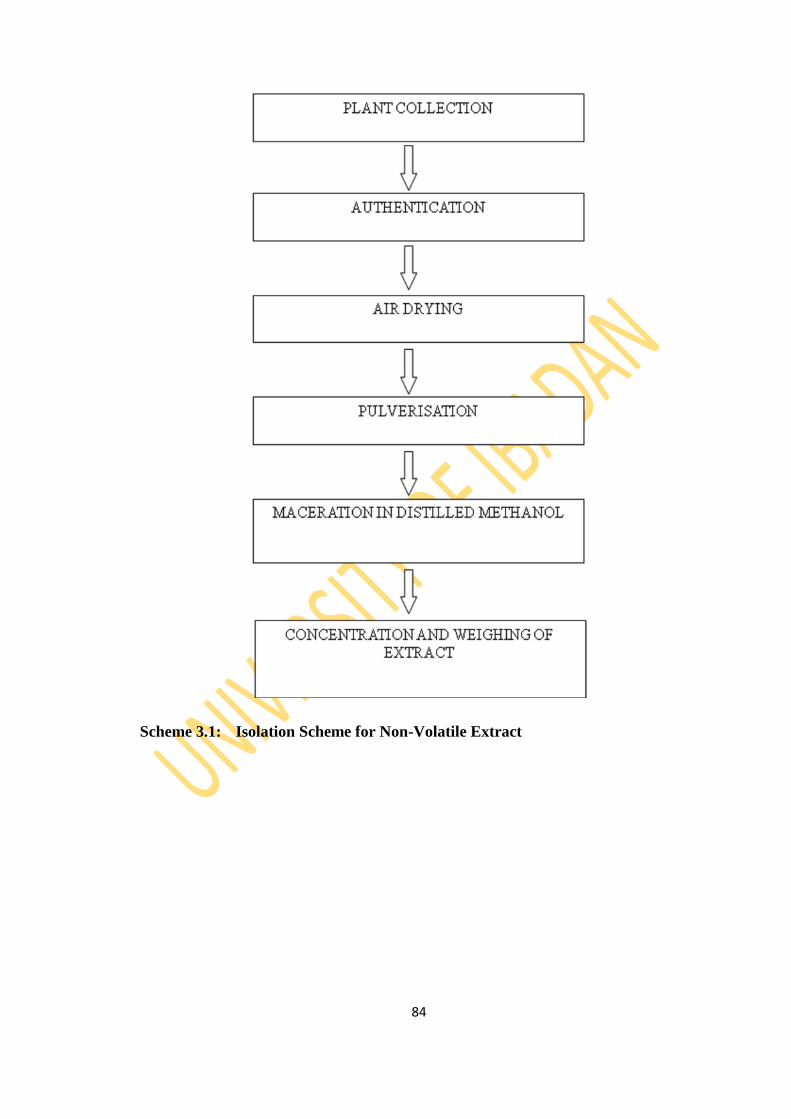
84
Scheme 3.1: Isolation Scheme for Non-Volatile Extract

85
3.6 Isolation of Compounds from Theobroma cacao Linn Pod
The methanol extract (25 g) of T. cacao L. pod was dissolved in methanol, pre-
adsorbed on silica gel (50 g) and mixed to obtain a homogenous solid mixture which
was allowed to air-dry in a fume cupboard. The mixture was then loaded on a glass
chromatography column packed with silica gel. The column was eluted with the
following mobile phase gradient in increasing order of polarity (Scheme 3.2): n-hexane
(100%, 3 L), n-hexane: CH2Cl2 [(9:1, 1 L); (4:1, 1 L); and (1:1, 1 L)], CH2Cl2 (100%, 1
L), CH2Cl2: ethyl acetate (1:1, 1 L), ethyl acetate (100%, 1 L), ethyl acetate: methanol
(1:1, 1 L), methanol (100%, 1 L), methanol: water (1:1, 1 L) and water (100%, 1 L). A
total of 260 fractions (50 mL portions) were collected and pooled to 30 sub-fractions
coded TCH-1, TCHD 1-3, TCD 1-2, TCDE 1-4, TCE-1, TCEM 1-8, TCM 1-4,
TCMW 1-6 and TCW-1 based on TLC pattern. Fractions were concentrated to dryness
using rotary evaporator and were transferred into weighed and labelled sample bottle.
TCHD-3 (75 mg) was further purified on HPLC (LC-980) using 2% n-hexane:
ethylacetate solvent system to yield white solid coded 2TCHD-3 (2.6 mg). Sub-
fractions TCDE-1 (300 mg) and TCDE-3 (500 mg) were however subjected to column
chromatography with 15% and 30% stepwise gradient of n-hexane: acetone solvent
system to produce white solids coded 72TCDE-1 (48.6 mg) and 359TCDE-3 (11 mg)
respectively.
3.6.1 Purification of 2TCHD-3 Using Chromatography
Hexane: DCM soluble fractions were pooled together to give three sub-fractions
(TCHD-1, TCHD-2 and TCHD-3) based on thin layer chromatography (TLC) on
precoated aluminium plates. TCHD-3 (75 mg) was purified on HPLC (LC-908)
equiped with refractive index indicator using 2% n-hexane: acetone solvent sysytem to
yield 2TCHD-3 (white solid). 2TCHD-3 was characterised using spectroscopic
techniques.
3.6.2 Characterisation of 2TCHD-3 Using Spectrometry
Appearance: white solid
Melting Point: 112-114 oC
Molecular formula: C30H48O2
Molecular Mass (EIMS) [M+-H2O]: 424.3903 g/mol.
Molecular Mass (calculated): 440.3642 g/mol.
86
Major EIMS m/z fragmentation peaks m/z (relative abundance): 424 (27) [M+-
H2O], 313 (16), 217 (12), 205 (17), 203 (12), 189 (13), 187 (13), 175 (30), 173 (27),
163 (24), 161 (32), 159 (22), 149 (42), 147 (55), 135 (45), 133 (52), 121 (63), 119
(54), 109 (71), 107 (69), 95 (87), 93 (69), 81 (71), 69 (100), 67 (67), 55 (90).
13C-NMR (600 MHz, CDCl3, δ ppm): 33.5 (t, C-1), 37.5 (t, C-2), 216.7 (s, C-3), 50.2 (s,
C-4), 48.4 (d, C-5), 21.5 (t, C-6), 28.1 (t, C-7), 47.9 (d, C-8), 21.1 (s, C-9), 26.0 (s, C-10),
26.7 (t, C-11), 33.4 (t, C-12), 45.3 (s, C-13), 48.7 (s, C-14) 35.5 (t, C-15), 25.85 (t, C-16),
52.1 (d, C-17), 18.1 (q, C-18), 29.6 (t, C-19), 35.7 (d, C-20), 18.3 (q, C-21), 33.2 (t, C-22),
32.7 (t, C-23), 67.6 (d, C-24), 144.3 (s, C-25), 114.3 (t, C-26), 17.0 (q, C-27), 22.2 (q, C-28),
20.8 (q, C-29), 19.3 (q, C-30).
The results are presented in Table 4.9.
1H-NMR (600 MHz, CDCl3, δ ppm) : 1.52/1.85 (2H, t, H-1a/b), 2.27/ 2.3 ( 2H, t, H-
2a/b), 1.69 (1H, t, H-5), 0.9/1.5 (2H, q, H-6a/b), 1.2 ( 2H, q, H-7), 1.56 (1H, t, H-8), 2.02
(1H, t, H-11 ), 1.5 (2H, t, H-12), 1.29/1.24 (2H, t, H-15a/b), 1.36 (2H, q, H-16 ), 1.58 (1H, q,
H-17), 0.96 (3H, s, H-18), 0.78/0.55 ( 2H, H-19a/b), 1.29 (1H, m, H-20), 0.87 (3H, d, H-21),
1.64 (2H, q, H-22 ), 1.63/1.82 (2H, q, H-23 ), 4.34 (1H, t, H-24), 4.87/4.98 (2H, s, H-26a/b),
1.78 (3H, s, H-27), 1.03 (3H, s, H-28), 1.08 (3H, s, H-29), 0.88 (3H, s, H-30).
The results are presented in Table 4.9.
FTIR spectrum (Vmax cm-1
, KBr): 2941 (C-H), 2869, 1706 (C=O(ketone)), 1458, 1375,
1110.9.
3.6.3 Purification of 72TCDE-1 Using Chromatography
DCM: Ethyl Acetate soluble fraction were pooled together to give four sub-fractions
(TCDE-1, TCDE-2, TCDE-3 and TCDE-4) based on thin layer chromatography (TLC)
on precoated aluminium plates. TCDE-1 (300 mg) was later subjected to column
chromatography on silica gel (70-230 mesh) eluted with 15% stepwise gradient of n-
hexane: acetone solvent sysytem. 150 fractions were collected in 20 mL portions.
Portion 72-79 (white solid) was labelled 72TCDE-1 and subjected to spectroscopic
analysis to characterise.
3.6.4 Characterisation of 72TCDE-1 Using Spectrometry
Appearance: white solid
Melting point: 110.4 - 112.9 oC
87
Molecular formula (HREIMS): C29H50O
Molecular Mass (HREIMS): 414.2 g/mol.
Molecular Mass (calculated): 414.7 g/mol.
Major EIMS m/z fragmentation peaks m/z (relative abundance): 414 (100) [M+],
412 (43), 396 (43), 381 (24), 329 (31), 303 (33), 273 (28), 231 (21), 213 (32), 163
(22), 159 (32), 144 (36), 134 (22), 118 (23), 108 (22), 95 (35), 69 (32), 55 (41).
13C-NMR (600 MHz, CDCl3, δ ppm): 37.22 (t, C-1), 31.64 (t, C-2), 71.79 (d, C-3),
42.28 (d, C-4), 140.7 (s, C-5), 121.7 (t, C-6), 31.87 (t, C-7), 31.63 (d, C-8), 50.1 (s, C-9),
36.48 (d, C-10), 21.79 (t, C-11), 39.6 (t, C-12), 42.95 (s, C-13), 56.73 (s, C-14) 26.0 (t, C-15),
28.23 (t, C-16), 55.8 (s, C-17), 36.1 (d, C-18), 19.38 (t, C-19), 34.44 (s, C-20), 26.0 (t, C-21),
45.79 (t, C-22), 23.02 (q, C-23), 12.2 (q, C-24), 29.1 (q, C-25), 21.05 (q, C-26), 19.8 (q, C-
27), 19.0 (q, C-28), 11.96 (q, C-29).
The results are presented in Table 4.10.
1H-NMR (600 MHz, CDCl3, δ ppm) : 1.04/1.82 (2H, t, H-1a/b), 1.43 ( 2H, q, H-2a/b),
3.5 (1H, m, H-3), 2.23/2.28 (2H, d, H-4), 5.32 (1H, t, H-6), 1.83 ( 2H, t, H-7), 1.69 (1H,
q, H-8), 0.9 (1H, q, H-9), 1.37 (2H, q, H-11), 1.14 (2H, t, H-12), 0.98 (1H, q, H-14), 1.15
(2H, q, H-15), 1.25/1.84 (2H, q, H-16 ), 1.08 (1H, q, H-17), 1.36 (1H, m, H-18), 0.98 ( 3H,
d, H-19), 1.48 (2H, q, H-20), 1.13 (2H, q, H-21), 1.52 (1H, m, H-22 ), 1.03 (2H, m, H-23 ),
0.64 (3H, t, H-24), 1.67 (1H, m, H-25), 0.82 (3H, d, H-26), 1.34, 3H, d, H-27), 0.89 (3H, s,
H-28), 0.87 (3H, s, H-29).
The results are presented in Table 4.10.
FTIR spectrum (Vmax cm-1
, KBr): 3346 (O-H), 2949 (C-H), 1676 (C=C).
3.6.5 Purification of 359TCDE-3 Using Chromatography
TCDE-3 (500 mg) was later subjected to column chromatography on silica gel (70-230
mesh) eluted with 30% stepwise gradient of n-hexane: acetone solvent sysytem. 450
fractions were collected in 20 mL portions. Portion 351-359 (white solid) was labelled
359TCDE-3 (11 mg) and subjected to spectroscopic analysis to characterise.
3.6.6 Characterisation of 359TCDE-3 Using Spectrometry
Appearance: white solid
Melting point: 144.7 – 146.5 oC
Molecular formula (HREIMS): C28H44O3

88
Molecular Mass (HREIMS): 428.99 g/mol.
Molecular Mass (calculated): 428.65 g/mol.
Major EIMS m/z fragmentation peaks m/z (relative abundance): 428 (2.2) [M+],
410 (14), 396 (100), 381 (24), 329 (31), 303 (33), 273 (28), 231 (21), 213 (32), 163
(22), 159 (32), 144 (36), 134 (22), 118 (23), 108 (22), 95 (35), 69 (32), 55 (41).
13C-NMR (600 MHz, CDCl3, δ ppm): 34.68 (t, C-1), 30.10 (t, C-2), 66.46 (d, C-3),
36.91 (d, C-4), 79.41 (s, C-5), 130.74 (t, C-6), 135.40 (t, C-7), 82.14 (d, C-8), 51.67 (s, C-
9), 36.96 (d, C-10), 20.62 (t, C-11), 39.33 (t, C-12), 44.55 (s, C-13), 51.08 (s, C-14) 23.39 (t,
C-15), 28.64 (t, C-16), 56.19 (s, C-17), 12.86 (d, C-18), 18.16 (t, C-19), 39.72 (s, C-20),
20.86 (t, C-21), 135.19 (t, C-22), 132.30 (q, C-23), 42.76 (q, C-24), 33.06 (q, C-25), 19.63
(q, C-26), 19.94 (q, C-27), 17.55 (q, C-28).
The results are presented in Table 4.11.
1H-NMR (600 MHz, CDCl3, δ ppm) : 1.94/1.68 (2H, t, H-1a/b), 1.82/1.51 ( 2H, q, H-
2a/b), 3.92 (1H, m, H-3), 2.09/1.91 (2H, d, H-4a/b), 6.48 (1H, d, H-6), 6.22 ( 2H, d, H-7),
1.55 (1H, q, H-9), 1.56/1.38 (2H, q, H-11a/b), 1.92/1.22 (2H, t, H-12a/b), 1.48 (1H, q, H-14),
1.31/1.49 (2H, q, H-15a/b), 1.33/1.74 (2H, q, H-16a/b ), 1.2 (1H, q, H-17), 0.79 (1H, m, H-
18), 0.87 ( 3H, d, H-19), 2.0 (2H, q, H-20), 0.98 (2H, q, H-21), 5.13 (1H, m, H-22 ), 5.18
(2H, m, H-23 ), 1.83 (3H, t, H-24), 1.45 (1H, m, H-25), 0.79 (3H, d, H-26), 0.81 (3H, d, H-
27), 0.91 (3H, s, H-28).
The results are presented in Table 4.11.
FTIR spectrum (Vmax cm-1
, KBr): 3346 (O-H), 2954/2875 (C-H), 1676 (C=C).
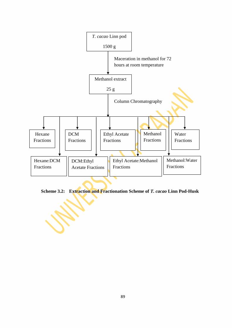
89
Scheme 3.2: Extraction and Fractionation Scheme of T. cacao Linn Pod-Husk
T. cacao Linn pod
1500 g
Methanol extract
25 g
Hexane
Fractions
DCM
Fractions
Ethyl Acetate
Fractions
Methanol
Fractions
Water
Fractions
Hexane:DCM
Fractions
DCM:Ethyl
Acetate Fractions
Ethyl Acetate:Methanol
Fractions
Methanol:Water
Fractions
Maceration in methanol for 72
hours at room temperature
Column Chromatography
90
3.7 Biological Activity of Essential Oils
3.7.1 Antibacterial screening
The essential oils were screened for antibacterial activities against six (6) standard
strains of laboratory stock bacteria representing Gram positive and Gram negative
(Staphylococcus aureus, Bacillus subtilis, Escherichia coli, Pseudomonas aeruginosa,
Shigella flexineri and Salmonella typhi). Microplate Alamar Blue Assay was used to
determine susceptibility or resistance of the essential oils to selected bacteria strains.
Organisms were grown in Mueller Hinton broth and inoculums were adjusted to 0.5
McFarland standard. Stock solutions of the essential oils were prepared in DMSO (1:1
concentration). Media was dispensed to all wells. Essential oils (20 µg/mL) were
added in the wells, control wells do not contain essential oil. The volume of 96-well
plate was made up to 200 µL. Finally 5x10⁶
cells were added in all wells including
both control and test. The plate was sealed with parafilm and incubated for 18 - 20
hours. Alamar Blue Dye was dispensed in each well and shaken at 80 RPM in a
shaking incubator for 2 – 3 hours. Plates were covered with foil in shaking incubator.
Change in color of Alamar Blue dye from blue to pink indicated the growth in bacterial
strains. Absorbance was recorded at 570 nm and 600 nm by the ELISA reader
(SpectraMax M2, Molecular Devices, CA, USA). Ampicillin was used as the reference
drug. The experiment was done in triplicate.
3.7.2 Antioxidant Activity: DPPH Radical Scavenging Activity
Radical scavenging activity was determined by a spectrophotometric method based on
the reduction of a methanol solution of DPPH using the reported method of
Yamaguchi et al. (1998). DPPH (Wako Chemicals USA, Inc.) solution in methanol
was prepared to make 0.3 mM. One milliliter of essential oil (20 µg/mL) was added to
1 mL of the 0.3 mM DPPH solution and shaken vigorously. The reaction is allowed to
progress for 30 min at 37 oC in the dark and absorbance is monitored by multiplate
reader, SpectraMax340, Molecular Devices, CA, USA at 517 nm. Upon reduction, the
color of the solution fades (Violet to pale yellow). Absolute methanol was used to zero
the spectrophotometer. N-acetylcysteine and gallic acid were used as the reference
compounds. The experiment was done in triplicate.
The activity was determined as a function of the % Radical Scavenging Activity which
was calculated using the formula;
91
% Radical Scavenging Activity =
Where: AC = Absorbance of the control
AS = Absorbance of the sample.
3.7.3 Insecticidal Activity
The insecticidal activity was conducted according to the impregnated filter paper
method also known as contact toxicity test described by Tabassum et al. (1997).
Materials
Test insects (Tribolium castaneum, Rhyzopertha dominica and Callosobruchus analis),
volatile organic solvent (methanol), standard insecticide (Permethrin), petri plates (9
cm diameter), micropipette (1000 µL), growth chamber, test sample, filter paper, glass
vials, brush.
Rearing Technique
The stored grain pests were reared in the laboratory under controlled conditions
(temperature and humidity) in plastic bottles containing sterile breeding media. Insects
of uniform age and size are used for the experiment.
1. Red flour beetle (Tribolium castaneum)
Rearing temperature: 30 oC
Relative humidity: 50 – 70%
Rearing media: Wheat flour
Life cycle: 22-25 days
2. Lesser grain borer (Rhyzopertha dominica)
Rearing temperature: 30 oC
Relative humidity: 50 – 70%
Rearing media: Wheat and gram seeds
Life cycle: 30 days
3. Pulse beetle (Callosobruchus analis)
Rearing temperature: 25-35 oC
Relative humidity: 50 – 70%
Rearing media: Mung seeds
Life cycle: 25-30 days
92
Preparation of Test Sample
Essential oil: 20 µg test sample + 1 mL methanol
Procedure
The filter papers were cut according to the size of glass petri plates (9 cm or 90 mm)
and put in the plates. Essential oils were loaded over the filter paper in the plates with
the help of micropipette. Ten healthy and active insects of same size and age of each
species were put in each plate (test and control) with the help of a clean brush. The
plates were incubated at 27 oC for 24 hours with 50% relative humidity in growth
chamber. The survival of the insects was assessed (count the number of survivals of
each species).
The Percentage Inhibition or Percentage Mortality was calculated using the formula
below:
Percentage Mortality = 100 – No. of insects alive in test x 100
No. of insects alive in control
Test Control
Positive control contained standard insecticide (Permethrin) at the concentration which
is effective against all test insects and test insects. Negative control contained volatile
solvent (methanol) and the test insects.

93
CHAPTER FOUR
RESULTS AND DISCUSSION
4.1 Essential Oils
The volatile extracts/essential oils (EOs) of the twenty-seven (27) samples were
obtained by the process of hydrodistillation using an all-glass Clevenger apparatus.
The physicochemical properties of the essential oils were determined and the EOs
were analysed by Gas Chromatography (Flame Ionization Detector and Mass
Spectrometry) techniques to determine their chemical constituents. The oils were also
investigated for their antibacterial, antioxidant and insecticidal activities.
4.1.1 Essential Oils Yield
The physicochemical properties of the essential oils are shown in Table 4.1. The
characteristic colours of the oils ranged from colourless to pale yellow with yields
ranging between 0.13 and 1.21% v/w. All the fruits have sweet aromatic and fruity
smell reported to be due to the presence of esters, aldehydes, alcohols, terpenes or their
derivatives, but the oils of the root bark and stem barks have irritating woody smell
while the leaves had a stong leafy odour. T. cacao seed had a malty odour.

94
Table 4.1: Physicochemical Properties of Essential Oils
S/N Plant Sample Part used % Extract Composition Colour Odour
1 Persea
americana
Stem Bark 0.8 Colourless Herbal
Root Bark 0.72 Colourless Herbal
Leaf 0.71 Colourless Leafy
Peel 0.51 Colourless Irritating
Fruit 0.23 Pale Yellow Leafy
Seed 0.47 Colourless Irritating
2 Carica papaya Root Bark 0.46 Colourless Woody
Root 0.26 Colourless Woody
Stem Bark 0.47 Colourless Woody
Stem 0.21 Colourless Woody
Leaf 0.89 Pale Yellow Leafy
Fruit 0.29 Pale Yellow Fruity
Seed 0.91 Colourless Sweet Aromatic
Peel 0.49 Pale Yellow Fruity
3 Ananas
comosus
Shoot 0.48 Colourless Leafy
Peel 0.71 Colourless Fruity
Fruit 0.62 Colourless Fruity
4 Theobroma
cacao
Stem Bark 1.29 Colourless Herbal
Leaf 1.03 Colourless Leafy
Pod 0.65 Colourless Irritating
Seed 1.15 Pale yellow Malty
5 Chrysophyllum
albidum
Stem Bark 0.86 Colourless Woody
Root Bark 1.21 Colourless Woody
Leaf 0.89 Pale Yellow Leafy
Seed 0.91 Colourless Sweet aromatic
Seed Bark 0.13 Colourless Herbal
Fruit Bark 0.95 Pale Yellow Fruity
95
4.1.2 Chemical Composition of Essential Oils
4.1.2.1 Persea americana M.
The chemical and percentage compositions of the essential oils of the plant parts of
Persea americana from the GC chromatograms (Figures 4.1-4.6) revealed a total of 13
to 48 constituents representing 83.09 to 96.58% composition (Table 4.2). The major
components in the fruit oil were p-xylene (40.51%), trans-nerolidol (16.17%),
heptacosane (12.96%), and 2,21-methylenebis tertbutyl-4-ethylphenol (5.48%). The
essential oil from the peel was dominated by globulol (25.43%), trans-nerolidol
(17.04%), β-elemene (13.62%) and hexadecanoic acid (13.42%). The prevalence of
(Z,Z)- 4,15-octadecadien-1-ol acetate (21.77%), β-elemene (15.54%) and p-xylene
(7.67%) were observed in the seed oil while leaf oil had β-caryophyllene (12.67%),
trans-phytol (11.81%), p-xylene (5.22%) and δ-cadinene (3.85%) as the major
compounds. Tetradecanal (24.99%), dodecanal (9.43%, tridecanal (7.32%) and β-
caryophyllene oxide (4.59%) were the major compounds in the stem bark oil while
tetradecanal (31.84%), dodecanal (13.17%) and tetradecanoic acid (7.25%) dominated
the root bark oil.
Furthermore, on analysis of the class of compounds present, the essential oils from the
peel and leaf were dominated by sesquiterpenes (68.37% and 40.79% respectively)
while the other oils had more non-terpenes (71.4% to 78.65%). Apocarotenes and
diterpenes were absent in all the oils except the leaf oil with 2.09% and 11.81%
composition, respectively. Triterpene (squalene- 0.55%) was present in only the seed
oil. Monoterpenes were present in all the oils except the peel oil. The percentage
compositions of the monoterpenes were low (0.11% to 11.08%).
The result obtained from this analysis compare favourably with respect to the
predominance of β-caryophyllene in the leaf oil as reported in previous research by
Ogunbinu et al. (2007). Larijani et al. (2010) however reported methyl eugenol as the
major compound while β-caryophyllene was the second predominant compound.
Sinyinda and Gramshaw (1998) also found β-caryophyllene to be the major constituent
in the mesocarp (fruit) essential oil.
The compositional pattern reveals the presence of β-elemene at an appreciable quantity
in the peel and seed oil, trans-nerolidol was relatively dominant in the fruit and peel
96
oil. The stem bark and root bark oils were dominated by aldehydes (tetradecanal,
dodecanal and tridecanal). Phytol was found to be present in only the leaf oil and the
percentage composition was relatively high (11.81%) making it the second dominant
compound. It is an acyclic diterpene alcohol that can be used as a precursor for the
manufacture of synthetic forms of vitamin E and vitamin K 1 (Daines et al., 2003;
Igwe, 2014). Phytol is used in the fragrance industry and in cosmetics, shampoos, toilet
soaps, household cleaners and detergents (McGinty, 2010; Igwe, 2014). Application of
phytol also include infection fighting and natural alternativetherapies for hypertension
and cancer (Daines et al., 2003; McGinty, 2010; Igwe, 2014). Phytol has been reported
to have anti-mycobacterial activity against Mycobacterium tuberculosis (Rajab, 1998;
Daines et al., 2003; Igwe, 2014). The high quantity of phytol in the leaf of P.
americana suggests that the plant might be used in the treatment of tuberculosis.
This study represents the first comprehensive characterization of the volatile
constituents of the essential oil of all the plant parts of Persea americana Mill grown
in Nigeria.
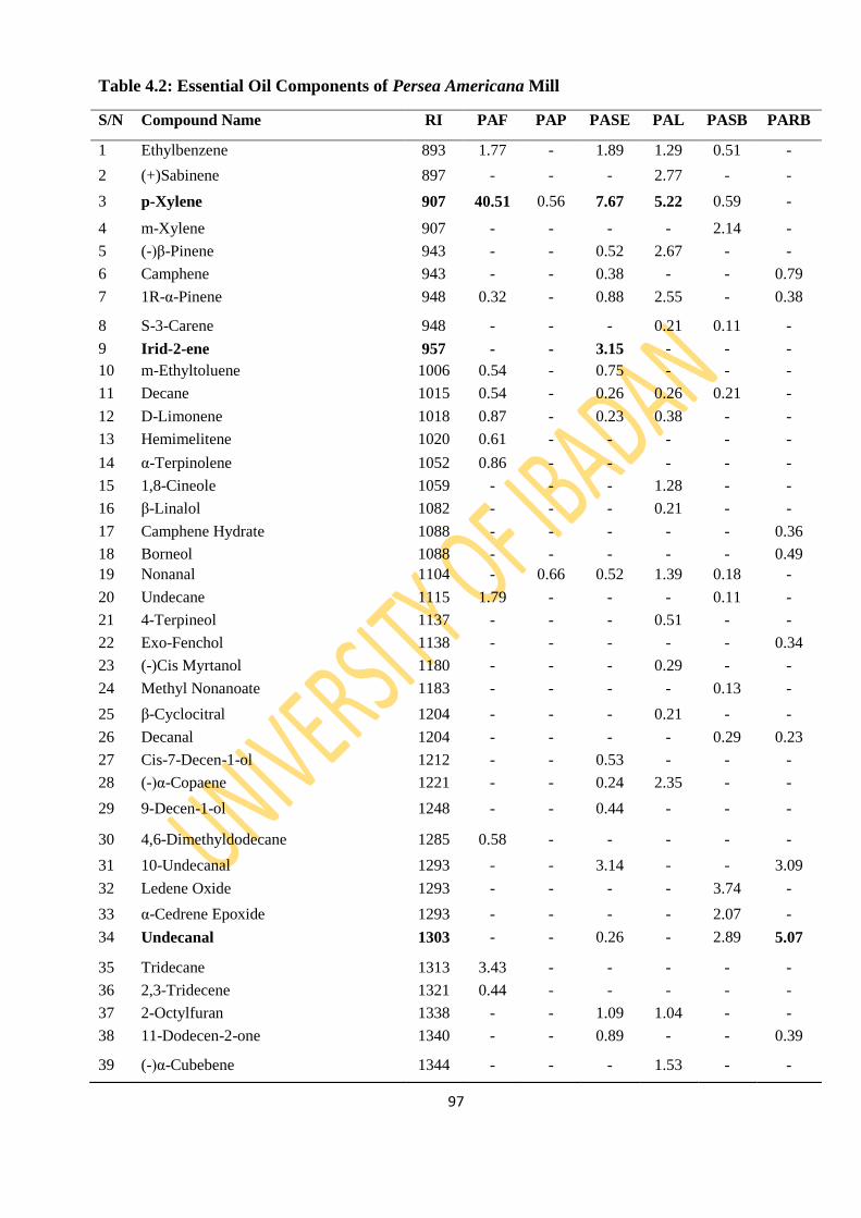
97
Table 4.2: Essential Oil Components of Persea Americana Mill
S/N Compound Name RI PAF PAP PASE PAL PASB PARB
1 Ethylbenzene 893 1.77 - 1.89 1.29 0.51 -
2 (+)Sabinene 897 - - - 2.77 - -
3 p-Xylene 907 40.51 0.56 7.67 5.22 0.59 -
4 m-Xylene 907 - - - - 2.14 -
5 (-)β-Pinene 943 - - 0.52 2.67 - -
6 Camphene 943 - - 0.38 - - 0.79
7 1R-α-Pinene 948 0.32 - 0.88 2.55 - 0.38
8 S-3-Carene 948 - - - 0.21 0.11 -
9 Irid-2-ene 957 - - 3.15 - - -
10 m-Ethyltoluene 1006 0.54 - 0.75 - - -
11 Decane 1015 0.54 - 0.26 0.26 0.21 -
12 D-Limonene 1018 0.87 - 0.23 0.38 - -
13 Hemimelitene 1020 0.61 - - - - -
14 α-Terpinolene 1052 0.86 - - - - -
15 1,8-Cineole 1059 - - - 1.28 - -
16 β-Linalol 1082 - - - 0.21 - -
17 Camphene Hydrate 1088 - - - - - 0.36
18 Borneol 1088 - - - - - 0.49
19 Nonanal 1104 - 0.66 0.52 1.39 0.18 -
20 Undecane 1115 1.79 - - - 0.11 -
21 4-Terpineol 1137 - - - 0.51 - -
22 Exo-Fenchol 1138 - - - - - 0.34
23 (-)Cis Myrtanol 1180 - - - 0.29 - -
24 Methyl Nonanoate 1183 - - - - 0.13 -
25 β-Cyclocitral 1204 - - - 0.21 - -
26 Decanal 1204 - - - - 0.29 0.23
27 Cis-7-Decen-1-ol 1212 - - 0.53 - - -
28 (-)α-Copaene 1221 - - 0.24 2.35 - -
29 9-Decen-1-ol 1248 - - 0.44 - - -
30 4,6-Dimethyldodecane 1285 0.58 - - - - -
31 10-Undecanal 1293 - - 3.14 - - 3.09
32 Ledene Oxide 1293 - - - - 3.74 -
33 α-Cedrene Epoxide 1293 - - - - 2.07 -
34 Undecanal 1303 - - 0.26 - 2.89 5.07
35 Tridecane 1313 3.43 - - - - -
36 2,3-Tridecene 1321 0.44 - - - - -
37 2-Octylfuran 1338 - - 1.09 1.04 - -
38 11-Dodecen-2-one 1340 - - 0.89 - - 0.39
39 (-)α-Cubebene 1344 - - - 1.53 - -
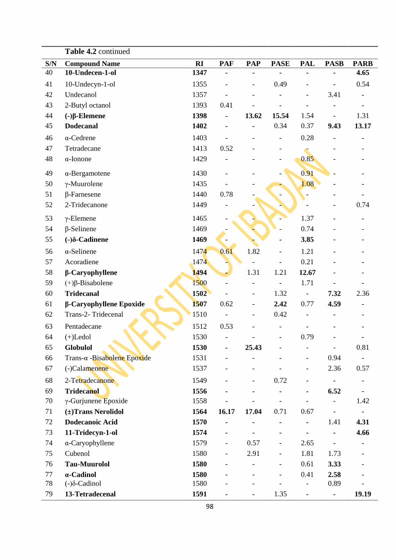
98
Table 4.2 continued
S/N Compound Name RI PAF PAP PASE PAL PASB PARB
40 10-Undecen-1-ol 1347 - - - - - 4.65
41 10-Undecyn-1-ol 1355 - - 0.49 - - 0.54
42 Undecanol 1357 - - - - 3.41 -
43 2-Butyl octanol 1393 0.41 - - - - -
44 (-)β-Elemene 1398 - 13.62 15.54 1.54 - 1.31
45 Dodecanal 1402 - - 0.34 0.37 9.43 13.17
46 α-Cedrene 1403 - - - 0.28 - -
47 Tetradecane 1413 0.52 - - - - -
48 α-Ionone 1429 - - - 0.85 - -
49 α-Bergamotene 1430 - - - 0.91 - -
50 γ-Muurolene 1435 - - - 1.08 - -
51 β-Farnesene 1440 0.78 - - - - -
52 2-Tridecanone 1449 - - - - - 0.74
53 γ-Elemene 1465 - - - 1.37 - -
54 β-Selinene 1469 - - - 0.74 - -
55 (-)δ-Cadinene 1469 - - - 3.85 - -
56 α-Selinene 1474 0.61 1.82 - 1.21 - -
57 Acoradiene 1474 - - - 0.21 - -
58 β-Caryophyllene 1494 - 1.31 1.21 12.67 - -
59 (+)β-Bisabolene 1500 - - - 1.71 - -
60 Tridecanal 1502 - - 1.32 - 7.32 2.36
61 β-Caryophyllene Epoxide 1507 0.62 - 2.42 0.77 4.59 -
62 Trans-2- Tridecenal 1510 - - 0.42 - - -
63 Pentadecane 1512 0.53 - - - - -
64 (+)Ledol 1530 - - - 0.79 - -
65 Globulol 1530 - 25.43 - - - 0.81
66 Trans-α -Bisabolene Epoxide 1531 - - - - 0.94 -
67 (-)Calamenene 1537 - - - - 2.36 0.57
68 2-Tetradecanone 1549 - - 0.72 - - -
69 Tridecanol 1556 - - - - 6.52 -
70 γ-Gurjunene Epoxide 1558 - - - - - 1.42
71 (±)Trans Nerolidol 1564 16.17 17.04 0.71 0.67 - -
72 Dodecanoic Acid 1570 - - - - 1.41 4.31
73 11-Tridecyn-1-ol 1574 - - - - - 4.66
74 α-Caryophyllene 1579 - 0.57 - 2.65 - -
75 Cubenol 1580 - 2.91 - 1.81 1.73 -
76 Tau-Muurolol 1580 - - - 0.61 3.33 -
77 α-Cadinol 1580 - - - 0.41 2.58 -
78 (-)δ-Cadinol 1580 - - - - 0.89 -
79 13-Tetradecenal 1591 - - 1.35 - - 19.19
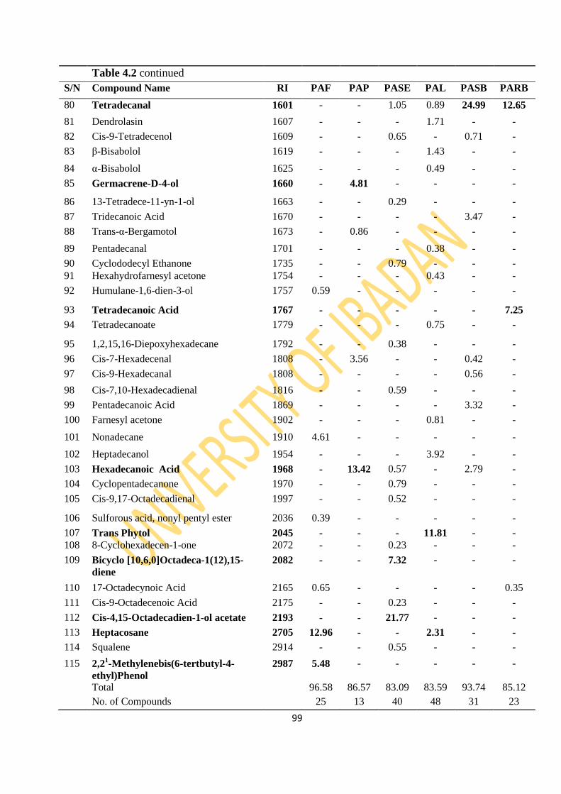
99
Table 4.2 continued
S/N Compound Name RI PAF PAP PASE PAL PASB PARB
80 Tetradecanal 1601 - - 1.05 0.89 24.99 12.65
81 Dendrolasin 1607 - - - 1.71 - -
82 Cis-9-Tetradecenol 1609 - - 0.65 - 0.71 -
83 β-Bisabolol 1619 - - - 1.43 - -
84 α-Bisabolol 1625 - - - 0.49 - -
85 Germacrene-D-4-ol 1660 - 4.81 - - - -
86 13-Tetradece-11-yn-1-ol 1663 - - 0.29 - - -
87 Tridecanoic Acid 1670 - - - - 3.47 -
88 Trans-α-Bergamotol 1673 - 0.86 - - - -
89 Pentadecanal 1701 - - - 0.38 - -
90 Cyclododecyl Ethanone 1735 - - 0.79 - - -
91 Hexahydrofarnesyl acetone 1754 - - - 0.43 - -
92 Humulane-1,6-dien-3-ol 1757 0.59 - - - - -
93 Tetradecanoic Acid 1767 - - - - - 7.25
94 Tetradecanoate 1779 - - - 0.75 - -
95 1,2,15,16-Diepoxyhexadecane 1792 - - 0.38 - - -
96 Cis-7-Hexadecenal 1808 - 3.56 - - 0.42 -
97 Cis-9-Hexadecanal 1808 - - - - 0.56 -
98 Cis-7,10-Hexadecadienal 1816 - - 0.59 - - -
99 Pentadecanoic Acid 1869 - - - - 3.32 -
100 Farnesyl acetone 1902 - - - 0.81 - -
101 Nonadecane 1910 4.61 - - - - -
102 Heptadecanol 1954 - - - 3.92 - -
103 Hexadecanoic Acid 1968 - 13.42 0.57 - 2.79 -
104 Cyclopentadecanone 1970 - - 0.79 - - -
105 Cis-9,17-Octadecadienal 1997 - - 0.52 - - -
106 Sulforous acid, nonyl pentyl ester 2036 0.39 - - - - -
107 Trans Phytol 2045 - - - 11.81 - -
108 8-Cyclohexadecen-1-one 2072 - - 0.23 - - -
109 Bicyclo [10,6,0]Octadeca-1(12),15-
diene
2082 - - 7.32 - - -
110 17-Octadecynoic Acid 2165 0.65 - - - - 0.35
111 Cis-9-Octadecenoic Acid 2175 - - 0.23 - - -
112 Cis-4,15-Octadecadien-1-ol acetate 2193 - - 21.77 - - -
113 Heptacosane 2705 12.96 - - 2.31 - -
114 Squalene 2914 - - 0.55 - - -
115 2,21-Methylenebis(6-tertbutyl-4-
ethyl)Phenol
2987 5.48 - - - - -
Total 96.58 86.57 83.09 83.59 93.74 85.12
No. of Compounds 25 13 40 48 31 23
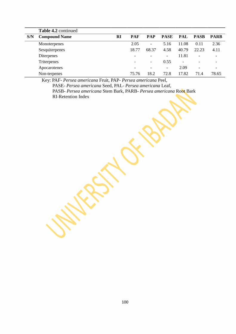
100
Key: PAF- Persea americana Fruit, PAP- Persea americana Peel,
PASE- Persea americana Seed, PAL- Persea americana Leaf,
PASB- Persea americana Stem Bark, PARB- Persea americana Root Bark
RI-Retention Index
Table 4.2 continued
S/N Compound Name RI PAF PAP PASE PAL PASB PARB
Monoterpenes 2.05 - 5.16 11.08 0.11 2.36
Sesquiterpenes 18.77 68.37 4.58 40.79 22.23 4.11
Diterpenes - - - 11.81 - -
Triterpenes - - 0.55 - - -
Apocarotenes - - - 2.09 - -
Non-terpenes 75.76 18.2 72.8 17.82 71.4 78.65

101
Figure 4.1: GC Chromatogram of Persea americana Leaf
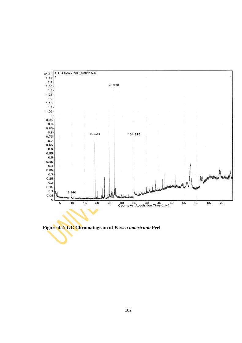
102
Figure 4.2: GC Chromatogram of Persea americana Peel
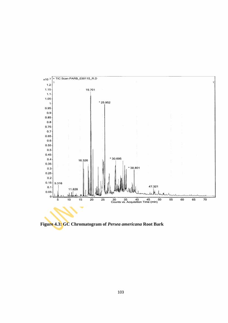
103
Figure 4.3: GC Chromatogram of Persea americana Root Bark
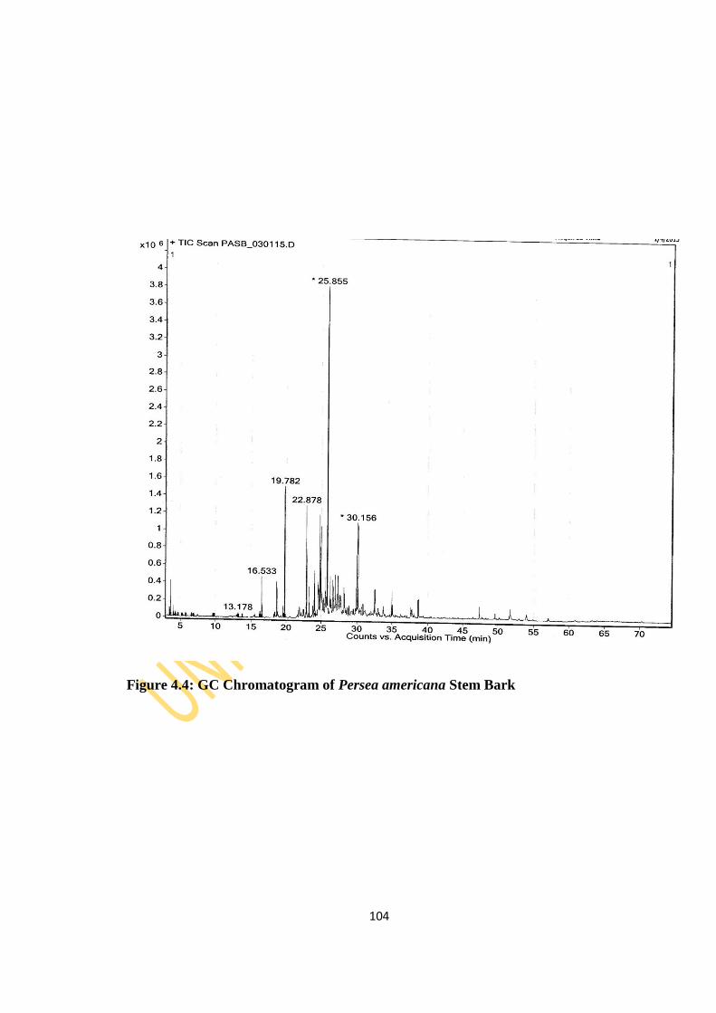
104
Figure 4.4: GC Chromatogram of Persea americana Stem Bark

105
Figure 4.5: GC Chromatogram of Persea americana Fruit
106
Figure 4.6: GC Chromatogram of Persea americana Seed
4.1.2.2 Carica papaya
The GC (Figures 4.7-4.14) and GCMS analyses of essential oils from Carica papaya
fruit, leaf, peel, root bark, root, stem bark, stem and seed afforded 46, 40, 23, 26, 10,
16, 11 and 9 compounds identified, representing 94.32%, 90.19%, 72.03%, 94.67%,
97.06%, 98.11%, 93.01% and 99.58% of essential oil composition, respectively (Table
4.3). The dominant constituents in the fruit oil are heptadecanol (25.17%), phytol

107
(10.64%) and hexadecanoic acid (8.44%). The leaf oil has phytol (21.83%), farnesyl
acetone (10.69%) and heptacosane (10.59%) as its major compounds while 9-
hexadecen-1-ol (16.97%), m-xylene (14.5%), heptacosane (7.58%) and squalene
(6.57%) were predominant amongst the identified compounds in the peel oil.
Benzylisothiocyanate (71.54%) and octadecanol (62.54%) were the major constituents
in the essential oil of the root bark and the root respectively. m-xylene (35.11%),
heptacosane (22.01%), p-xylene (12.46%) and ethylbenzene (10.48%) dominate the
compounds identified in the essential oil from the stem bark while the stem oil was
dominated by octadecanol (71.13%), hexadecanol (6.31%) and heptacosane (6.23%).
The seed oil however has benzylisothiocyanate (89.12%) as the major constituent.
Analyses of the chemical constituents based on the class of compounds revealed the
presence of monoterpenes, though in low quantity, in all the oils except the leaf, stem
and seed oils. The fruit and leaf oil have 10.64% and 22.16% diterpenes, respectively
while triterpenes were present in all the oils except root and seed oils. The oils had
high percentage of non-terpene compounds within the range of 54.65% to 99.43%
except for the leaf oil with 26.41%. Four, five and one apocarotene compounds
representing 15.75%, 27.8% and 1.24% were identified in the fruit, leaf and peel oil,
respectively.
In an earlier study on the volatile constituents of Carica papaya leaf of Nigerian
origin, six compounds were identified with phytol (37.78%), 9-octadecenamide
(28.18%), cyclopentaneundecanoic acid methyl ester (12.02%) and 3-methyl-4-
(phenylthio)-2-enyl-2,5-dihydrothiophene-1,1-dioxide (11.78%) as the major
constituents (Igwe, 2015). However, in the present study, more constituents were
identified. Hexahydrofarnesyl acetone (7.77%), geranyl acetone (5.96%), squalene
(3.62%) and benzylisothiocyanate (3.03%) were identified in appreciable quantity in
the leaf oil of this study but were not reported in the earlier study by Igwe (2015)
although phytol was found to be the major and only common compound in both
studies. The presence of benzylisothiocyanate which is an organosulphur compound
(OSC) in the oils of the fruit, leaf, root bark, root and seed is a probable indication that
the oils will have anticarcinogenic property. Organosulphur compounds (OSCs)
prevent or slow down the carcinogenic process induced by a variety of chemical
carcinogens. OSCs offer protection against cancer (Igwe, 2015). These include
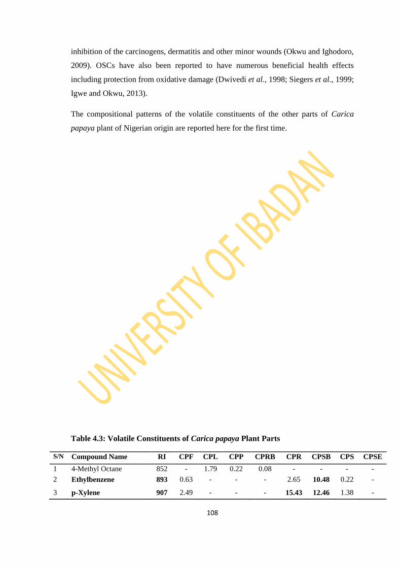
108
inhibition of the carcinogens, dermatitis and other minor wounds (Okwu and Ighodoro,
2009). OSCs have also been reported to have numerous beneficial health effects
including protection from oxidative damage (Dwivedi et al., 1998; Siegers et al., 1999;
Igwe and Okwu, 2013).
The compositional patterns of the volatile constituents of the other parts of Carica
papaya plant of Nigerian origin are reported here for the first time.
Table 4.3: Volatile Constituents of Carica papaya Plant Parts
S/N Compound Name RI CPF CPL CPP CPRB CPR CPSB CPS CPSE
1 4-Methyl Octane 852 - 1.79 0.22 0.08 - - - -
2 Ethylbenzene 893 0.63 - - - 2.65 10.48 0.22 -
3 p-Xylene 907 2.49 - - - 15.43 12.46 1.38 -
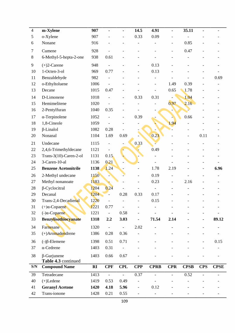
109
4 m-Xylene 907 - - 14.5 4.91 - 35.11 - -
5 o-Xylene 907 - - 0.33 0.09 - - - -
6 Nonane 916 - - - - - 0.85 - -
7 Cumene 928 - - - - - 0.47 - -
8 6-Methyl-5-hepta-2-one 938 0.61 - - - - - - -
9 (+)2-Carene 948 - - - 0.13 - - - -
10 1-Octen-3-ol 969 0.77 - - 0.13 - - - -
11 Benzaldehyde 982 - - - - - - - 0.69
12 o-Ethyltoluene 1006 - - - - 1.49 0.39 - -
13 Decane 1015 0.47 - - - 0.65 1.78 - -
14 D-Limonene 1018 - - 0.33 0.31 - 1.04 - -
15 Hemimelitene 1020 - - - - 0.97 2.16 - -
16 2-Pentylfuran 1040 0.35 - - - - - - -
17 α-Terpinolene 1052 - - 0.39 - - 0.66 - -
18 1,8-Cineole 1059 - - - - 1.94 - - -
19 β-Linalol 1082 0.28 - - - - - - -
20 Nonanal 1104 1.69 0.69 - 0.23 - - 0.11 -
21 Undecane 1115 - - 0.33 - - - - -
22 2,4,6-Trimethyldecane 1121 - - - 0.49 - - - -
23 Trans-3(10)-Caren-2-ol 1131 0.15 - - - - - - -
24 3-Caren-10-al 1136 0.21 - - - - - - -
25 Benzene Acetonitrile 1138 1.24 - - 1.78 2.19 - - 6.96
26 2-Methyl undecane 1150 - - - 0.19 - - - -
27 Methyl nonanoate 1183 - - - 0.23 - 2.16 - -
28 β-Cyclocitral 1204 0.24 - - - - - - -
29 Decanal 1204 - 0.28 0.33 0.17 - - - -
30 Trans-2,4-Decadienal 1220 - - - 0.15 - - - -
31 (+)α-Copaene 1221 0.77 - - - - - - -
32 (-)α-Copaene 1221 - 0.58 - - - - - -
33 Benzylisothiocyanate 1318 2.2 3.03 - 71.54 2.14 - - 89.12
34 Farnesane 1320 - - 2.02 - - - - -
35 (+)Aromadendrene 1386 0.28 0.36 - - - - - -
36 (-)β-Elemene 1398 0.51 0.71 - - - - - 0.15
37 α-Cedrene 1403 0.31 - - - - - - -
38 β-Gurjunene
Table 4.3 continued
1403 0.66 0.67 - - - - - -
S/N Compound Name RI CPF CPL CPP CPRB CPR CPSB CPS CPSE
39 Tetradecane 1413 - - 0.37 - - 0.52 - -
40 (+)Ledene 1419 0.53 0.49 - - - - - -
41 Geranyl Acetone 1420 4.18 5.96 - 0.12 - - - -
42 Trans-ionone 1428 0.21 0.55 - - - - - -
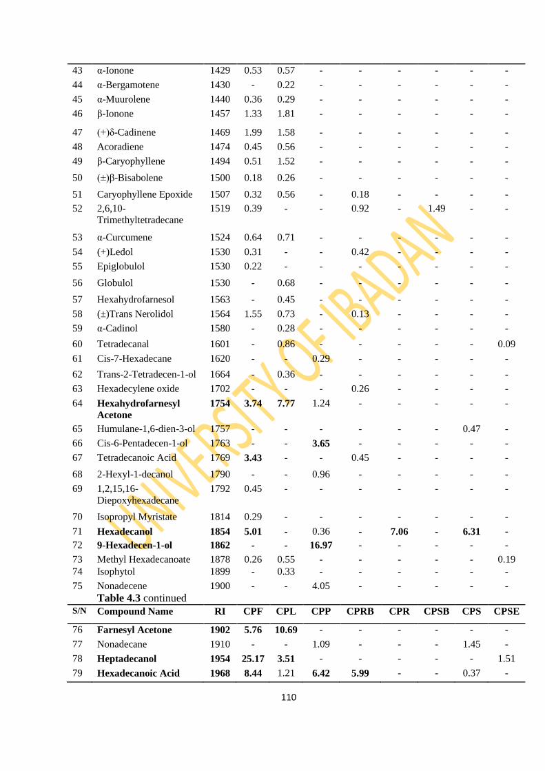
110
43 α-Ionone 1429 0.53 0.57 - - - - - -
44 α-Bergamotene 1430 - 0.22 - - - - - -
45 α-Muurolene 1440 0.36 0.29 - - - - - -
46 β-Ionone 1457 1.33 1.81 - - - - - -
47 (+)δ-Cadinene 1469 1.99 1.58 - - - - - -
48 Acoradiene 1474 0.45 0.56 - - - - - -
49 β-Caryophyllene 1494 0.51 1.52 - - - - - -
50 (±)β-Bisabolene 1500 0.18 0.26 - - - - - -
51 Caryophyllene Epoxide 1507 0.32 0.56 - 0.18 - - - -
52 2,6,10-
Trimethyltetradecane
1519 0.39 - - 0.92 - 1.49 - -
53 α-Curcumene 1524 0.64 0.71 - - - - - -
54 (+)Ledol 1530 0.31 - - 0.42 - - - -
55 Epiglobulol 1530 0.22 - - - - - - -
56 Globulol 1530 - 0.68 - - - - - -
57 Hexahydrofarnesol 1563 - 0.45 - - - - - -
58 (±)Trans Nerolidol 1564 1.55 0.73 - 0.13 - - - -
59 α-Cadinol 1580 - 0.28 - - - - - -
60 Tetradecanal 1601 - 0.86 - - - - - 0.09
61 Cis-7-Hexadecane 1620 - - 0.29 - - - - -
62 Trans-2-Tetradecen-1-ol 1664 - 0.36 - - - - - -
63 Hexadecylene oxide 1702 - - - 0.26 - - - -
64 Hexahydrofarnesyl
Acetone
1754 3.74 7.77 1.24 - - - - -
65 Humulane-1,6-dien-3-ol 1757 - - - - - - 0.47 -
66 Cis-6-Pentadecen-1-ol 1763 - - 3.65 - - - - -
67 Tetradecanoic Acid 1769 3.43 - - 0.45 - - - -
68 2-Hexyl-1-decanol 1790 - - 0.96 - - - - -
69 1,2,15,16-
Diepoxyhexadecane
1792 0.45 - - - - - - -
70 Isopropyl Myristate 1814 0.29 - - - - - - -
71 Hexadecanol 1854 5.01 - 0.36 - 7.06 - 6.31 -
72 9-Hexadecen-1-ol 1862 - - 16.97 - - - - -
73 Methyl Hexadecanoate 1878 0.26 0.55 - - - - - 0.19
74 Isophytol 1899 - 0.33 - - - - - -
75 Nonadecene
Table 4.3 continued
1900 - - 4.05 - - - - -
S/N Compound Name RI CPF CPL CPP CPRB CPR CPSB CPS CPSE
76 Farnesyl Acetone 1902 5.76 10.69 - - - - - -
77 Nonadecane 1910 - - 1.09 - - - 1.45 -
78 Heptadecanol 1954 25.17 3.51 - - - - - 1.51
79 Hexadecanoic Acid 1968 8.44 1.21 6.42 5.99 - - 0.37 -

111
80 γ-Palmito acetone 1980 - - - 0.63 - - - -
81 Trans Phytol 2045 10.64 21.83 - - - - - -
82 Octadecanol 2053 - - - - 62.54 - 71.13 -
83 Cis-9,12-Octadecadien-
1-ol
2069 - - 3.25 - - - - -
84 Methyl trans-9-
octadecanoate
2085 - - - - - - - 0.51
85 Ethyl-9-octadecanoate 2185 - - - - - - - 0.36
86 Eicosanol 2252 - - - 2.17 - - - -
87 Docosanol 2451 - 0.28 - - - - - -
88 Heptacosane 2705 0.76 10.59 7.58 2.19 - 22.01 6.23 -
89 α-Glyceryl Linolenate 2705 - 0.64 - - - - - -
90 Squalene 2914 2.81 3.62 6.57 0.78 - 1.28 3.11 -
91 2,21-Methylene bis [6(1,1-
dimethyl-4-ethyl]Phenol
2987 - 1.06 - - - 5.25 2.23 -
92 Heptatriacotanol 3942 - 1.56 0.78 - - - -
Total 94.32 90.19 72.03 94.67 97.06 98.11 93.01 99.58
No. of Compounds 46 40 23 26 10 16 11 9
Monoterpenes 0.88 - 0.72 0.44 2.91 2.17 - -
Sesquiterpenes 9.59 10.2 1.56 0.73 - - 0.47 0.15
Diterpenes 10.64 10.64 22.16 - - - - -
Triterpenes 2.81 3.62 6.57 0.78 - 1.28 3.11 -
Apocarotenes 15.75 27.8 1.24 0.12 - - - -
Non-terpenes 54.65 26.41 61.94 92.6 94.15 94.66 89.43 99.43
Key: CPF- Carica papaya Fruit, CPL- Carica papaya Leaf, CPP- Carica papaya Peel,
CPRB- Carica papaya Root Bark, CPR- Carica papaya Root,
CPSB- Carica papaya Stem Bark, CPS- Carica papaya Stem, CPSE- Carica papaya
Seed RI-Retention Index

112
Figure 4.7: GC Chromatogram of Carica papaya Fruit
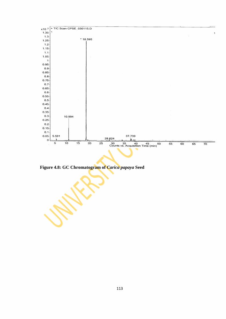
113
Figure 4.8: GC Chromatogram of Carica papaya Seed

114
Figure 4.9: GC Chromatogram of Carica papaya Leaf

115
Figure 4.10: GC Chromatogram of Carica papaya Peel

116
Figure 4.11: GC Chromatogram of Carica papaya Root
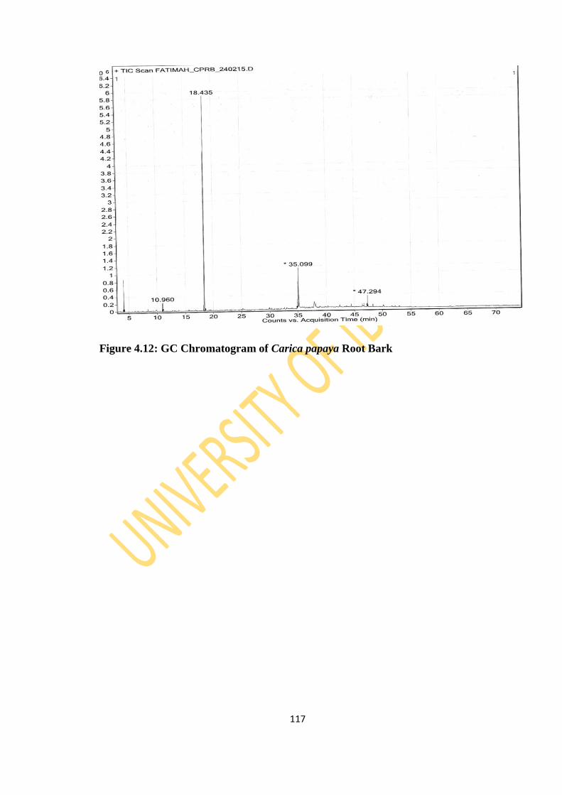
117
Figure 4.12: GC Chromatogram of Carica papaya Root Bark
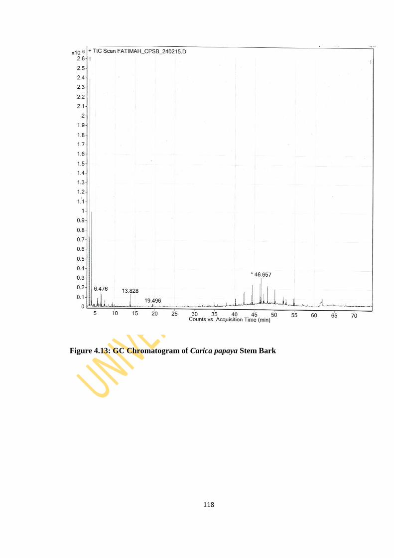
118
Figure 4.13: GC Chromatogram of Carica papaya Stem Bark
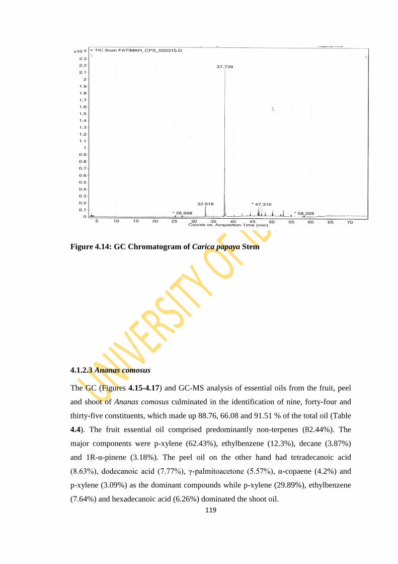
119
Figure 4.14: GC Chromatogram of Carica papaya Stem
4.1.2.3 Ananas comosus
The GC (Figures 4.15-4.17) and GC-MS analysis of essential oils from the fruit, peel
and shoot of Ananas comosus culminated in the identification of nine, forty-four and
thirty-five constituents, which made up 88.76, 66.08 and 91.51 % of the total oil (Table
4.4). The fruit essential oil comprised predominantly non-terpenes (82.44%). The
major components were p-xylene (62.43%), ethylbenzene (12.3%), decane (3.87%)
and 1R-α-pinene (3.18%). The peel oil on the other hand had tetradecanoic acid
(8.63%), dodecanoic acid (7.77%), γ-palmitoacetone (5.57%), α-copaene (4.2%) and
p-xylene (3.09%) as the dominant compounds while p-xylene (29.89%), ethylbenzene
(7.64%) and hexadecanoic acid (6.26%) dominated the shoot oil.
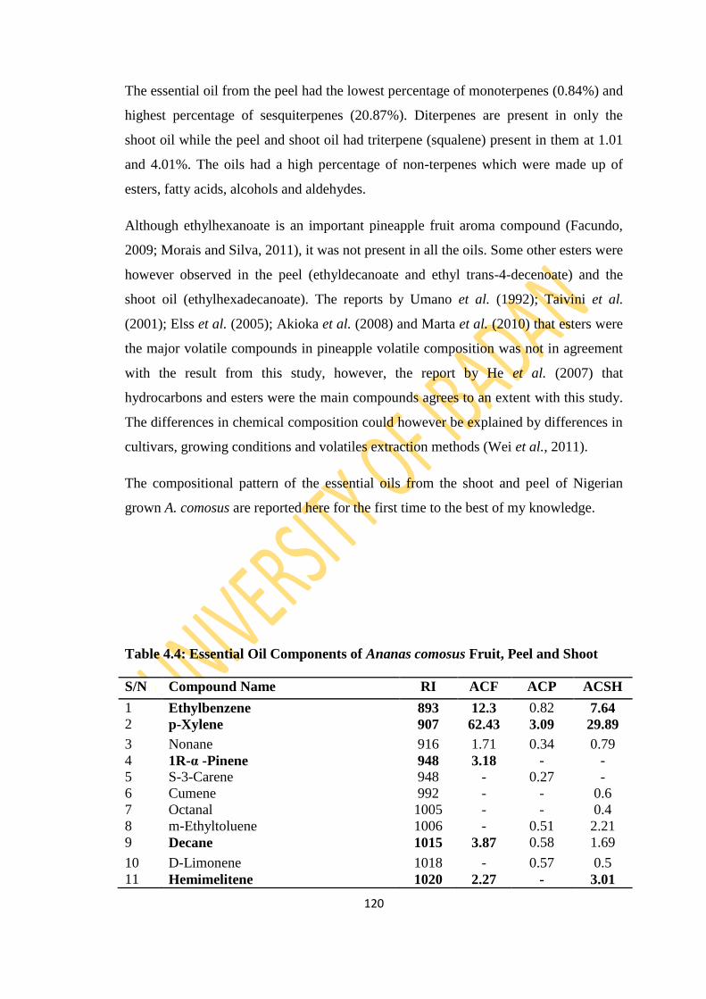
120
The essential oil from the peel had the lowest percentage of monoterpenes (0.84%) and
highest percentage of sesquiterpenes (20.87%). Diterpenes are present in only the
shoot oil while the peel and shoot oil had triterpene (squalene) present in them at 1.01
and 4.01%. The oils had a high percentage of non-terpenes which were made up of
esters, fatty acids, alcohols and aldehydes.
Although ethylhexanoate is an important pineapple fruit aroma compound (Facundo,
2009; Morais and Silva, 2011), it was not present in all the oils. Some other esters were
however observed in the peel (ethyldecanoate and ethyl trans-4-decenoate) and the
shoot oil (ethylhexadecanoate). The reports by Umano et al. (1992); Taivini et al.
(2001); Elss et al. (2005); Akioka et al. (2008) and Marta et al. (2010) that esters were
the major volatile compounds in pineapple volatile composition was not in agreement
with the result from this study, however, the report by He et al. (2007) that
hydrocarbons and esters were the main compounds agrees to an extent with this study.
The differences in chemical composition could however be explained by differences in
cultivars, growing conditions and volatiles extraction methods (Wei et al., 2011).
The compositional pattern of the essential oils from the shoot and peel of Nigerian
grown A. comosus are reported here for the first time to the best of my knowledge.
Table 4.4: Essential Oil Components of Ananas comosus Fruit, Peel and Shoot
S/N Compound Name RI ACF ACP ACSH
1 Ethylbenzene 893 12.3 0.82 7.64
2 p-Xylene 907 62.43 3.09 29.89
3 Nonane 916 1.71 0.34 0.79
4 1R-α -Pinene 948 3.18 - -
5 S-3-Carene 948 - 0.27 -
6 Cumene 992 - - 0.6
7 Octanal 1005 - - 0.4
8 m-Ethyltoluene 1006 - 0.51 2.21
9 Decane 1015 3.87 0.58 1.69
10 D-Limonene 1018 - 0.57 0.5
11 Hemimelitene 1020 2.27 - 3.01
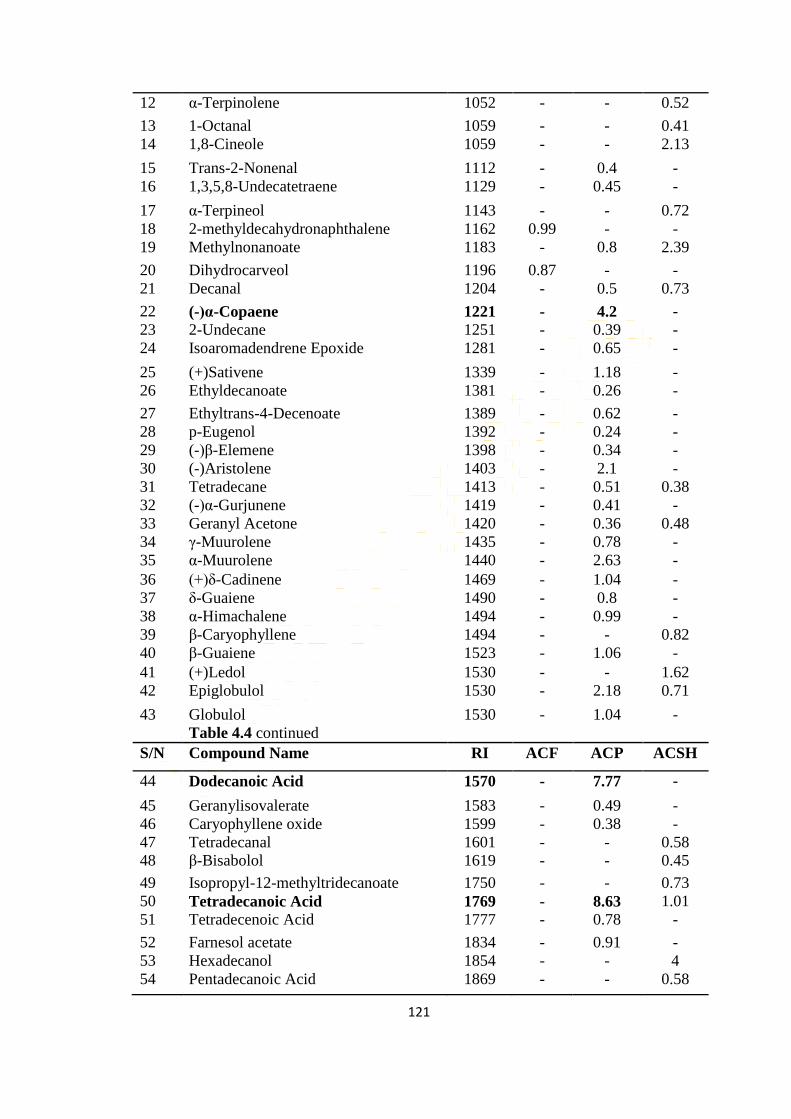
121
12 α-Terpinolene 1052 - - 0.52
13 1-Octanal 1059 - - 0.41
14 1,8-Cineole 1059 - - 2.13
15 Trans-2-Nonenal 1112 - 0.4 -
16 1,3,5,8-Undecatetraene 1129 - 0.45 -
17 α-Terpineol 1143 - - 0.72
18 2-methyldecahydronaphthalene 1162 0.99 - -
19 Methylnonanoate 1183 - 0.8 2.39
20 Dihydrocarveol 1196 0.87 - -
21 Decanal 1204 - 0.5 0.73
22 (-)α-Copaene 1221 - 4.2 -
23 2-Undecane 1251 - 0.39 -
24 Isoaromadendrene Epoxide 1281 - 0.65 -
25 (+)Sativene 1339 - 1.18 -
26 Ethyldecanoate 1381 - 0.26 -
27 Ethyltrans-4-Decenoate 1389 - 0.62 -
28 p-Eugenol 1392 - 0.24 -
29 (-)β-Elemene 1398 - 0.34 -
30 (-)Aristolene 1403 - 2.1 -
31 Tetradecane 1413 - 0.51 0.38
32 (-)α-Gurjunene 1419 - 0.41 -
33 Geranyl Acetone 1420 - 0.36 0.48
34 γ-Muurolene 1435 - 0.78 -
35 α-Muurolene 1440 - 2.63 -
36 (+)δ-Cadinene 1469 - 1.04 -
37 δ-Guaiene 1490 - 0.8 -
38 α-Himachalene 1494 - 0.99 -
39 β-Caryophyllene 1494 - - 0.82
40 β-Guaiene 1523 - 1.06 -
41 (+)Ledol 1530 - - 1.62
42 Epiglobulol 1530 - 2.18 0.71
43 Globulol
Table 4.4 continued
1530 - 1.04 -
S/N Compound Name RI ACF ACP ACSH
44 Dodecanoic Acid 1570 - 7.77 -
45 Geranylisovalerate 1583 - 0.49 -
46 Caryophyllene oxide 1599 - 0.38 -
47 Tetradecanal 1601 - - 0.58
48 β-Bisabolol 1619 - - 0.45
49 Isopropyl-12-methyltridecanoate 1750 - - 0.73
50 Tetradecanoic Acid 1769 - 8.63 1.01
51 Tetradecenoic Acid 1777 - 0.78 -
52 Farnesol acetate 1834 - 0.91 -
53 Hexadecanol 1854 - - 4
54 Pentadecanoic Acid 1869 - - 0.58
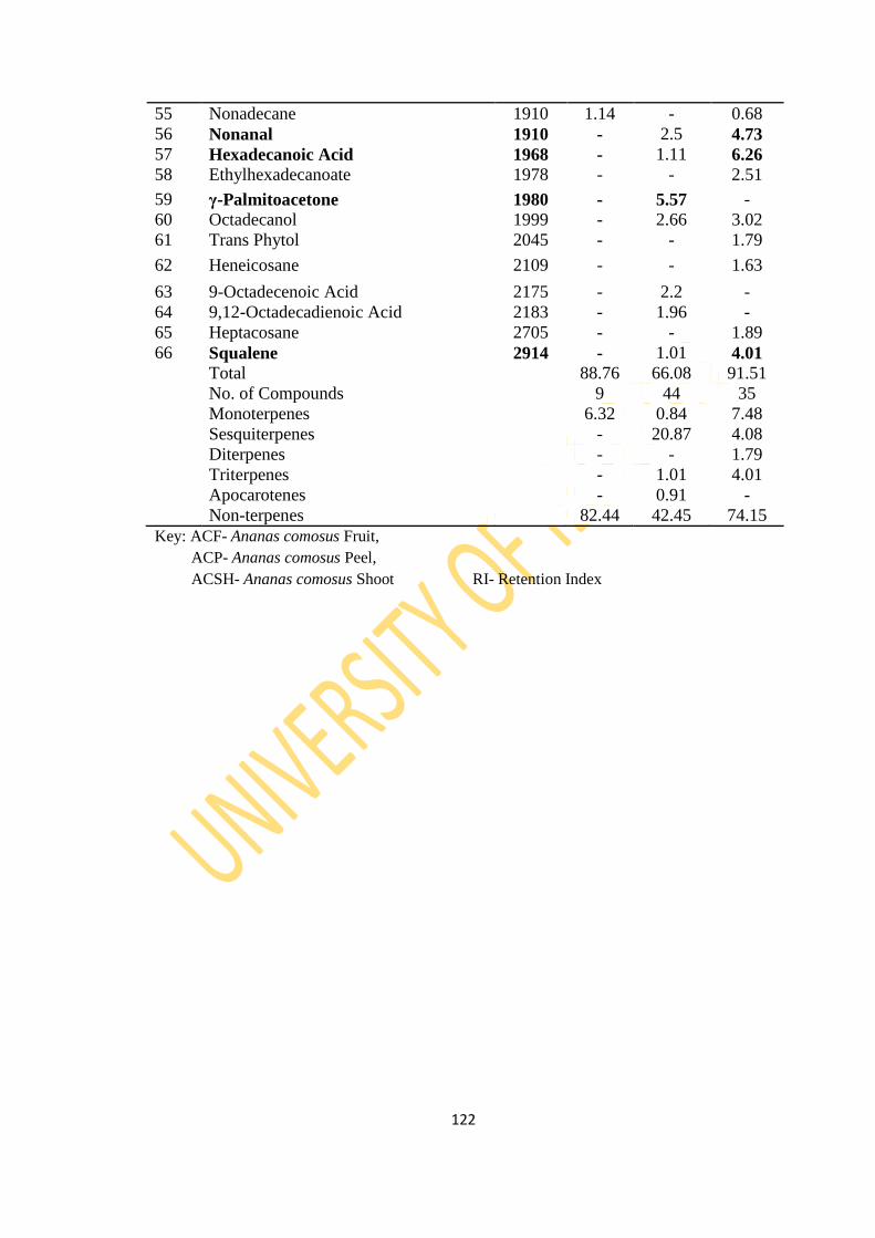
122
55 Nonadecane 1910 1.14 - 0.68
56 Nonanal 1910 - 2.5 4.73
57 Hexadecanoic Acid 1968 - 1.11 6.26
58 Ethylhexadecanoate 1978 - - 2.51
59 γ-Palmitoacetone 1980 - 5.57 -
60 Octadecanol 1999 - 2.66 3.02
61 Trans Phytol 2045 - - 1.79
62 Heneicosane 2109 - - 1.63
63 9-Octadecenoic Acid 2175 - 2.2 -
64 9,12-Octadecadienoic Acid 2183 - 1.96 -
65 Heptacosane 2705 - - 1.89
66 Squalene 2914 - 1.01 4.01
Total 88.76 66.08 91.51
No. of Compounds 9 44 35
Monoterpenes 6.32 0.84 7.48
Sesquiterpenes - 20.87 4.08
Diterpenes - - 1.79
Triterpenes - 1.01 4.01
Apocarotenes - 0.91 -
Non-terpenes 82.44 42.45 74.15
Key: ACF- Ananas comosus Fruit,
ACP- Ananas comosus Peel,
ACSH- Ananas comosus Shoot RI- Retention Index
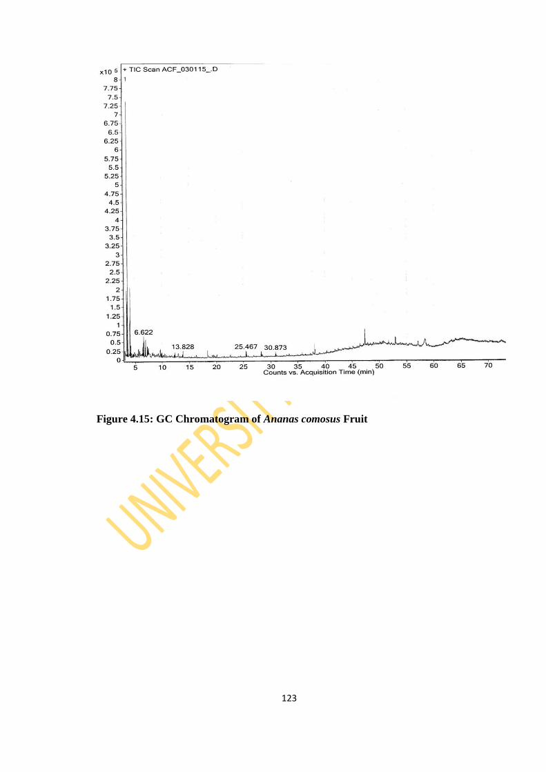
123
Figure 4.15: GC Chromatogram of Ananas comosus Fruit
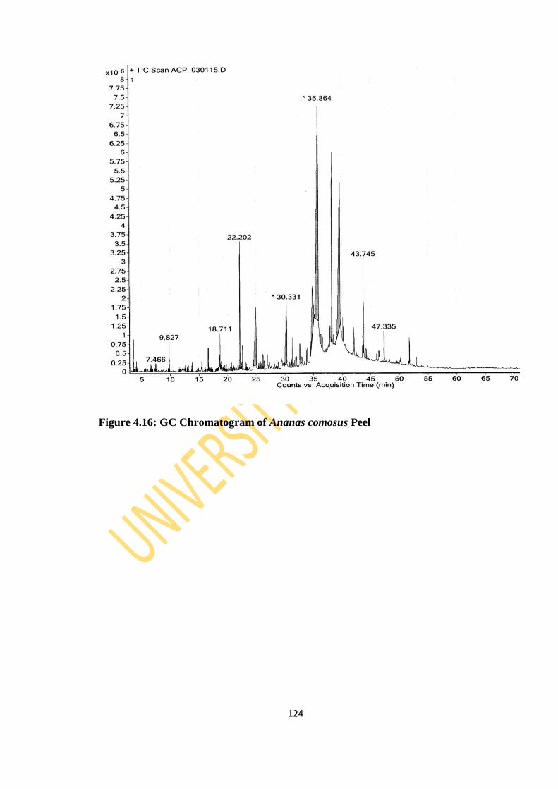
124
Figure 4.16: GC Chromatogram of Ananas comosus Peel
125
Figure 4.17: GC Chromatogram of Ananas comosus Shoot
4.1.2.4 Theobroma cacao
The GC (Figure 4.18-4.21) and GCMS analyses of the colourless essential oil
extracted from the hydrodistillation of the leaf, stem bark, pod and seed of Theobroma
cacao revealed a total of 30, 52, 28 and 19 identified constituents representing 99.8%,
99.2%, 88.43% and 94.45% of the total oils respectively (Table 4.5). The stem bark
126
essential oil had 30 sesquiterpenes which made up 68% of the total identified
constituents, the peel and leaf oil had 10 and 25 sesquiterpenes representing 40.32%
and 15.74% respectively. The seed had low percentage of both monoterpene (3.42%)
and sesquiterpenes (4.03%). Diterpenes and triterpenes were present in only the peel
oil while apocarotenes were observed in the peel and seed oil only.
The major constituents identified in the leaf oil were hexadecanoic acid (78.69%),
octadecanoic acid (4.87%) and epiglobulol (3.93%) while β-caryophyllene (1.16%)
and β-bisabolol (0.94%) were amongst the minor compounds. The stem bark oil was
dominated by β-bisabolol (17.31%), m-xylene (12.45%), β-bisabolene (6.38%), β-
caryophyllene (5.16%), β-sesquiphellandrene (4.61%) and α-bisabolol (4.16%) while
ledol (33.62%), α-terpineol (10.12%), heptadecanol (8.98%), farnesyl acetone (5.41%),
tetradecanal (4.79%) and 1,8-cineole (4.04%) were the dominant compounds in the
pod oil. The seed oil however had o-xylene (53.26%), 3,5-dimethyl octane (5.74%)
and hexahydrofarnesyl acetone (4.12%) as its major compounds while nonadecane
(4.05%), selin-7(11)-en-4α-ol (4.03%) and nonadecanol (3.57%) were also present in
significant quantity.
The GC-MS analysis of T. cacao pod oil by the process of soxhlet extraction by
Adewole et al. (2013) revealed fifteen (15) compounds constituting fatty acids and
other organic compounds. However, terpenoids dominated the components in this
study and hexadecanoic acid was the only fatty acid present. The difference in
composition could be as a result of differences in the extraction method and
geographical location of the plant. Although more fatty acids were identified in the
essential oils from the leaf and stem bark, only the leaf essential oil had fatty acid as
the major compound.
Frauendorfer and Schieberle (2006) reported thirty-five (35) odor-active constituents in
cocoa powder based on molecular sensory correlations. Some of the compounds
include methylpropanal (malty odor), 2- and 3- methylbutanal (malty odor) and
phenylacetaldehyde (honey-like). Although these compounds were not present in the
essential oils, the pod oil contains other aldehydes and hexadecanal was present in the
seed oil.
127
The comprehensive study of the essential oil components of Theobroma cacao L. leaf,
stem bark, pod and seed are been reported for the first time.
Table 4.5: Essential Oil Components of Theobroma cacao Linn Plant Parts
S/N Compound Name RI TCL TCSB TCP TCSE
1 Toluene 749 0.29 - - -
2 3,5-Dimethyloctane 887 - - - 5.74
3 Ethylbenzene 893 - 3.63 - -

128
4 o-Xylene 907 - - - 53.26
5 m-Xylene 907 - 12.45 1.66 -
6 p-Xylene 907 - 4.57 - -
7 Nonane 916 - 0.11 - -
8 m-Pyrrole 920 - 0.41 - -
9 Cumene 928 - 0.12 - -
10 1-Methyl-6-(1-
methylethylidine)bicycle[3,1,0]Hexane
956 - 0.17 - -
11 m-Ethyl toluene 1006 - 0.95 -
12 Decane 1015 - 0.43 - 0.76
13 D-Limonene 1018 - 0.28 - 1.42
14 Hemimeltene 1020 - 0.18 - -
15 β-Cymene 1042 - - - 0.43
16 α-Terpinolene 1052 - - - 1.57
17 1,8-Cineole 1059 0.15 0.35 4.04 -
18 Nonanal 1104 - 0.24 0.49 -
19 Undecane 1115 - - - 0.69
20 α-Terpineol 1143 - - 10.12 -
21 2,3,5,8-Tetramethyldecane 1156 - - - 0.85
22 Methyl nonanoate 1183 - 0.35 - -
23 Decanal 1204 - - 0.46 -
24 (-)α-Copaene 1221 0.31 1.09 - -
25 2-Dodecene 1222 - - - 0.79
26 2-Undecanone 1251 - - 0.72 -
27 Undecanal 1303 - - 0.37 -
28 Tridecane 1313 - - - 1.89
29 Benzene isothiocyanate 1318 - 0.34 - -
30 2-Methylundecanal 1338 - - 1.31 -
31 Aromadendrene 1386 - - 0.51 -
32 (-)β-Elemene 1398 - 0.25 - -
33 α-Cedrene 1403 0.05 1.63 - -
34 (+)α-Longipinene 1403 0.79 - - -
35 α-Patchoulene 1403 0.19 3.31 - -
36 Geranyl acetone 1420 - - 1.81 -
37 (+)Epi-β-Santalene 1425 - 0.38 - -
38 α-Bergamotene
Table 4.5 continued 1430 0.23 4.19 - -
S/N Compound Name RI TCL TCSB TCP TCSE
39 β-Farnesene 1440 0.14 0.34 - -
40 α-Amorphene 1440 - 0.21 - -
41 β-Sesquiphellandrene 1446 0.25 4.61 - -
42 2-Tridecanone 1449 - - 0.42 -
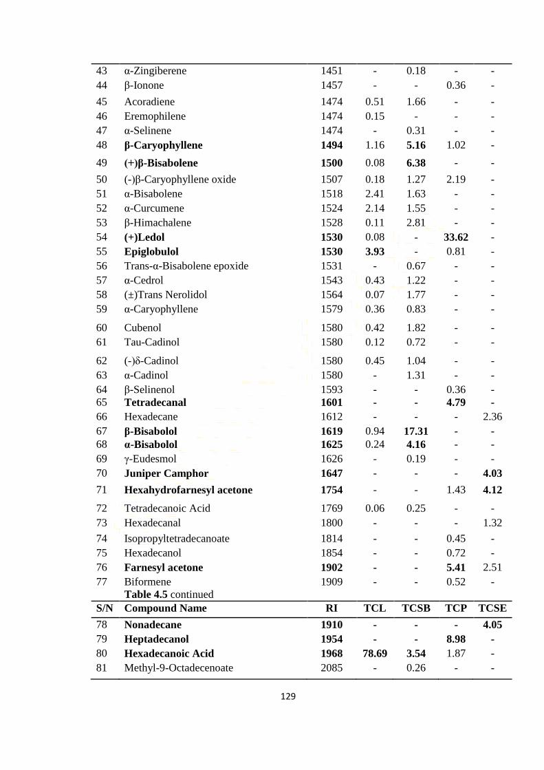
129
43 α-Zingiberene 1451 - 0.18 - -
44 β-Ionone 1457 - - 0.36 -
45 Acoradiene 1474 0.51 1.66 - -
46 Eremophilene 1474 0.15 - - -
47 α-Selinene 1474 - 0.31 - -
48 β-Caryophyllene 1494 1.16 5.16 1.02 -
49 (+)β-Bisabolene 1500 0.08 6.38 - -
50 (-)β-Caryophyllene oxide 1507 0.18 1.27 2.19 -
51 α-Bisabolene 1518 2.41 1.63 - -
52 α-Curcumene 1524 2.14 1.55 - -
53 β-Himachalene 1528 0.11 2.81 - -
54 (+)Ledol 1530 0.08 - 33.62 -
55 Epiglobulol 1530 3.93 - 0.81 -
56 Trans-α-Bisabolene epoxide 1531 - 0.67 - -
57 α-Cedrol 1543 0.43 1.22 - -
58 (±)Trans Nerolidol 1564 0.07 1.77 - -
59 α-Caryophyllene 1579 0.36 0.83 - -
60 Cubenol 1580 0.42 1.82 - -
61 Tau-Cadinol 1580 0.12 0.72 - -
62 (-)δ-Cadinol 1580 0.45 1.04 - -
63 α-Cadinol 1580 - 1.31 - -
64 β-Selinenol 1593 - - 0.36 -
65 Tetradecanal 1601 - - 4.79 -
66 Hexadecane 1612 - - - 2.36
67 β-Bisabolol 1619 0.94 17.31 - -
68 α-Bisabolol 1625 0.24 4.16 - -
69 γ-Eudesmol 1626 - 0.19 - -
70 Juniper Camphor 1647 - - - 4.03
71 Hexahydrofarnesyl acetone 1754 - - 1.43 4.12
72 Tetradecanoic Acid 1769 0.06 0.25 - -
73 Hexadecanal 1800 - - - 1.32
74 Isopropyltetradecanoate 1814 - - 0.45 -
75 Hexadecanol 1854 - - 0.72 -
76 Farnesyl acetone 1902 - - 5.41 2.51
77 Biformene
Table 4.5 continued
1909 - - 0.52 -
S/N Compound Name RI TCL TCSB TCP TCSE
78 Nonadecane 1910 - - - 4.05
79 Heptadecanol 1954 - - 8.98 -
80 Hexadecanoic Acid 1968 78.69 3.54 1.87 -
81 Methyl-9-Octadecenoate 2085 - 0.26 - -
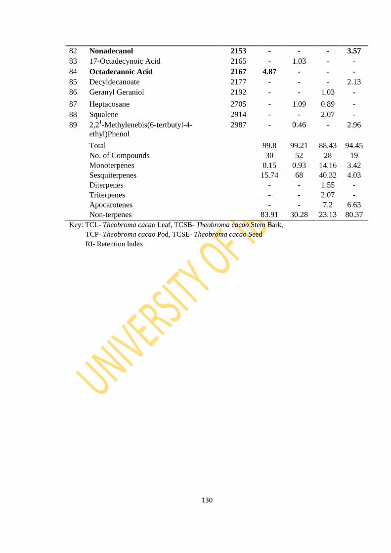
130
82 Nonadecanol 2153 - - - 3.57
83 17-Octadecynoic Acid 2165 - 1.03 - -
84 Octadecanoic Acid 2167 4.87 - - -
85 Decyldecanoate 2177 - - - 2.13
86 Geranyl Geraniol 2192 - - 1.03 -
87 Heptacosane 2705 - 1.09 0.89 -
88 Squalene 2914 - - 2.07 -
89 2,21-Methylenebis(6-tertbutyl-4-
ethyl)Phenol
2987 - 0.46 - 2.96
Total 99.8 99.21 88.43 94.45
No. of Compounds 30 52 28 19
Monoterpenes 0.15 0.93 14.16 3.42
Sesquiterpenes 15.74 68 40.32 4.03
Diterpenes - - 1.55 -
Triterpenes - - 2.07 -
Apocarotenes - - 7.2 6.63
Non-terpenes 83.91 30.28 23.13 80.37
Key: TCL- Theobroma cacao Leaf, TCSB- Theobroma cacao Stem Bark,
TCP- Theobroma cacao Pod, TCSE- Theobroma cacao Seed
RI- Retention Index
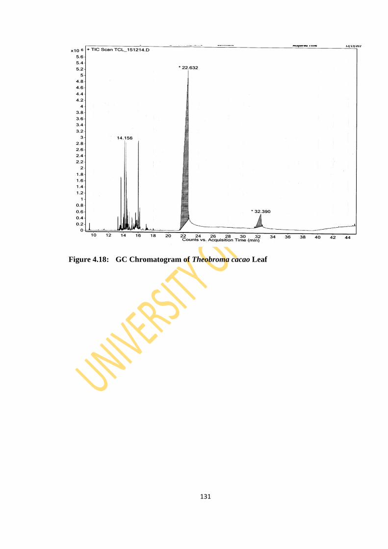
131
Figure 4.18: GC Chromatogram of Theobroma cacao Leaf
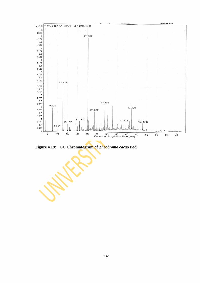
132
Figure 4.19: GC Chromatogram of Theobroma cacao Pod
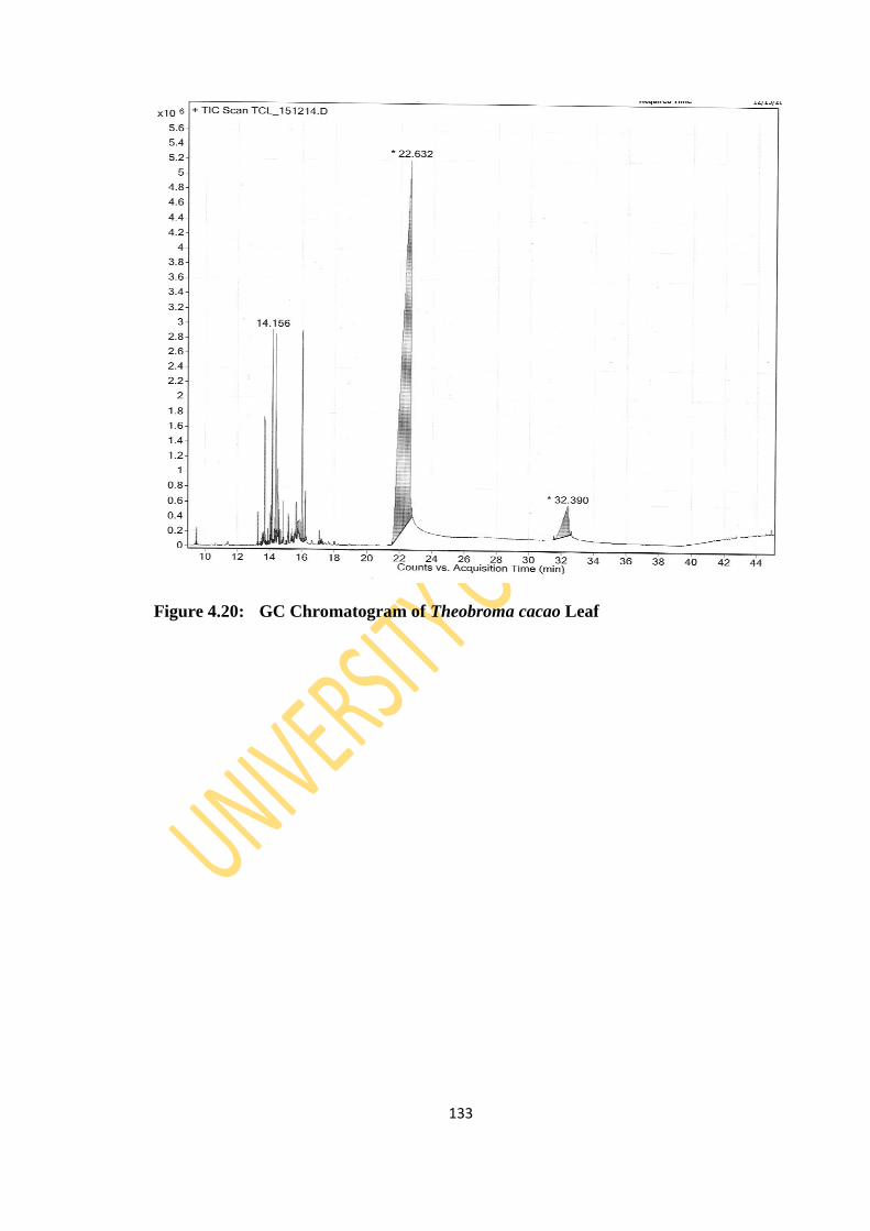
133
Figure 4.20: GC Chromatogram of Theobroma cacao Leaf

134
Figure 4.21: GC Chromatogram of Theobroma cacao Stem Bark
4.1.2.5 Chrysophyllum albidium G. Don
The analyses of the essential oils from the fruit bark, root bark, stem bark, seed bark,
leaf and seed of Chrysophyllum albidium by GC (Figure 4.23-4.27) and GCMS
showed the presence of 65, 33, 45, 21, 25 and 18 compounds constituting 79.49%,
100%, 90.81%, 98.43%, 96.62% and 98.37% of each of the total oils, respectively
(Table 4.6). Monoterpenes and sesquiterpenes were present in all the oils. The root
bark oil had the highest percentage of monoterpene (8.5%) while the fruit bark oil had
the least (1.19%). The leaf oil had the highest percentage of sesquiterpenes (75.67%)
and the seed bark oil had the lowest quantity (1.3%). Triterpenes were observed in
only the fruit bark and stem bark oils while Apocarotenes were detected in all the oils
135
except the seed oil. The root bark, seed bark and seed oils had 66.9%, 94.24% and
91.15% non-terpene compounds, respectively. All other samples had less than 35% of
their components as non-terpenes.
The major compounds in the fruit bark oil were hexadecanoic acid (12.73%), selin-
7(11)-en-4α-ol (11.42%), β-elemene (6.74%) and β-bisabolol (3.44%) while the root
bark oil was dominated by m-xylene (53.11%), ethylbenzene (9.41%), octadecanol
(5.91%) and β-elemene (4.74%). Hexadecanoic acid (14.69%), β-elemene (12.71%),
selin-7(11)-en-4α-ol (6.32%) and β-bisabolol (5.52%) were the dominant compounds
in the stem bark oil while the seed bark oil was dominated by p-xylene (21.38%),
ethylhexadecanoate (19.94%), ethyl-9,12-octadecadienoate (16.91%) and heptacosane
(10.51%). α-farnesene (38.11%), β-elemene (7.81%), p-xylene (5.15%) and α-selinene
(5.11%) were the major constituents in the leaf oil but m-xylene (66.72%) and
undecane (7.16%) dominated the seed oil.
Moronkola et al. (2006) reported twenty-four (24) compounds from the GC and
GC/MS analysis of the root essential oil of C. albidum G. Don with monoterpenes
(40.5%) and sesquiterpenes (27.9%) as the dominant class of compounds with pinene
(34%), caryophyllene (12.8%), isocaryophyllene (8.5%) and 1,8-cineole (6.5%) as the
major compounds. However, the report did not agree with the result of this study. This
could be attributed to differences in geographical locations of the plant, sensitivity of
instrument used for analysis and moisture content of plant as at time of sampling. Non-
terpenes (66.9%) and sesquiterpenes (24.15%) were found to be dominant with m-
xylene and β-elemene as the major non-terpene and sesquiterpene compounds,
respectively. β-pinene was present in the oil in low quantity. Earlier report on the
essential oil of the fruit by Moronkola (2001) presented eight (8) compounds
accounting for 90.8% of total components with esters (65.1%) constituting the most
abundant class of compounds. A phthalate (dibutyl-1,2-benzenedicarboxylate) was the
major compound. The fruit bark analysed in this study however contains more
sesquiterpenes (44.56%). The oils were found to contain esters like methylnonanoate,
ethylhexadecanoate, ethyl-9-octadecanoate and ethyloctadecenoate which are probably
responsible for the sweet, fruity smell of the extracts from the seed bark. The
compositional pattern of the essential oils from all the other plant parts of C. albidum
is here reported for the first time.
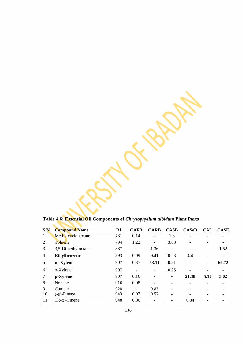
136
Table 4.6: Essential Oil Components of Chrysophyllum albidum Plant Parts
S/N Compound Name RI CAFB CARB CASB CASeB CAL CASE
1 Methylcyclohexane 781 0.14 - 1.3 - - -
2 Toluene 794 1.22 - 3.08 - - -
3 3,5-Dimethyloctane 887 - 1.36 - - - 1.52
4 Ethylbenzene 893 0.09 9.41 0.23 4.4 - -
5 m-Xylene 907 0.37 53.11 0.81 - - 66.72
6 o-Xylene 907 - - 0.25 - - -
7 p-Xylene 907 0.16 - - 21.38 5.15 3.02
8 Nonane 916 0.08 - - - - -
9 Cumene 928 - 0.83 - - - -
10 (-)β-Pinene 943 0.07 0.52 - - - -
11 1R-α –Pinene 948 0.06 - - 0.34 - -
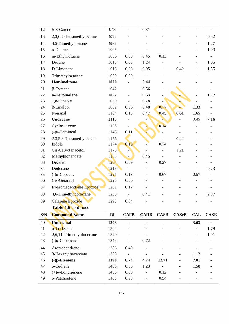
137
12 S-3-Carene 948 - 0.31 - - - -
13 2,3,6,7-Tetramethyloctane 958 - - - - - 0.82
14 4,5-Dimethylnonane 986 - - - - - 1.27
15 α-Decene 1005 - - - - - 1.09
16 m-EthylToluene 1006 0.09 0.45 0.13 - - -
17 Decane 1015 0.08 1.24 - - - 1.05
18 D-Limonene 1018 0.03 0.95 - 0.42 - 1.55
19 Trimethylbenzene 1020 0.09 - - - - -
20 Hemimelitene 1020 - 3.44 - - - -
21 β-Cymene 1042 - 0.56 - - - -
22 α-Terpinolene 1052 - 0.63 - - - 1.77
23 1,8-Cineole 1059 - 0.78 - - - -
24 β-Linalool 1082 0.56 0.48 0.77 - 1.33 -
25 Nonanal 1104 0.15 0.47 0.45 0.61 1.65 -
26 Undecane 1115 - - - - 0.45 7.16
27 Cyclosativene 1125 - - 0.14 - - -
28 (-)α-Terpineol 1143 0.11 - - - - -
29 2,3,5,8-Tetramethyldecane 1156 - - - 0.42 - -
30 Indole 1174 0.18 - 0.74 - - -
31 Cis-Carvotanacetol 1175 - - - 1.21 - -
32 Methylnonanoate 1183 - 0.45 - - - -
33 Decanal 1204 0.09 - 0.27 - - -
34 Dodecane 1215 - - - - - 0.73
35 (-)α-Copaene 1221 0.13 - 0.67 - 0.57 -
36 Cis-Geraniol 1228 0.06 - - - - -
37 Isoaromadendrene Epoxide 1281 0.17 - - - - -
38 4,6-Dimethyldodecane 1285 - 0.41 - - - 2.87
39 Calarene Epoxide 1293 0.04 - - - - -
Table 4.6 continued
S/N Compound Name RI CAFB CARB CASB CASeB CAL CASE
40 Undecanal 1303 - - - - 3.63 -
41 α-Tridecene 1304 - - - - - 1.79
42 2,6,11-Trimethyldodecane 1320 - - - - - 1.01
43 (-)α-Cubebene 1344 - 0.72 - - - -
44 Aromadendrene 1386 0.49 - - - - -
45 3-Hexenylhexanoate 1389 - - - - 1.12 -
46 (-)β-Elemene 1398 6.74 4.74 12.71 - 7.81 -
47 α-Cedrene 1403 0.83 1.23 - - 1.58 -
48 (+)α-Longipinene 1403 0.09 - 0.12 - - -
49 α-Patchoulene 1403 0.38 - 0.54 - - -

138
50 α-Gurjunene 1419 0.58 - 1.38 - 1.02 -
51 Geranyl acetone 1420 0.12 - 0.24 - - -
52 α-Ionone 1429 - - - - 0.69 -
53 β-Bergamotene 1430 0.06 - - - - -
54 α-Bergamotene 1430 - 2.01 0.19 - 0.92 -
55 γ-Cadinene 1435 0.05 - - - - -
56 Cyclohexylhexanoate 1445 - - - - 0.68 -
57 β-Sesquiphellandrene 1446 0.38 - 0.51 - - -
58 α-Farnesene 1458 - - - - 38.11 0.88
59 β-Selinene 1469 3.32 1.15 4.15 - - -
60 (+)δ-Cadinene 1469 - - 0.32 - - -
61 α-Selinene 1474 2.11 2.13 3.01 - 5.11 3.02
62 Acoradiene 1474 - 0.43 0.69 - - -
63 β-Caryophyllene 1494 1.69 2.93 4.91 0.37 8.54 -
64 α-Himachalene 1494 0.41 - 0.51 - - -
65 (±)β-Bisabolene 1500 1.45 2.67 1.42 - - -
66 β-Caryophyllene oxide 1507 1.99 - 0.85 - 2.14 -
67 Cis-α-Bisabolene 1518 - 0.37 - - - -
68 2,6,10-Trimethyltetradecane 1519 - - - 0.86 - -
69 α-Curcumene 1524 2.56 0.75 3.48 - - -
70 β-Himachalene 1528 - 0.71 0.13 - 0.63 -
71 Palustrol 1530 0.29 - - - - -
72 Epiglobulol 1530 1.24 - 0.87 - - -
73 Globulol 1530 0.52 - 0.36 - 1.31 -
74 (+)Ledol 1530 - - - - 1.56 -
75 Veridiflorol 1530 - - - 0.45 - -
76 α-Bisabolene oxide 1531 0.23 - - - - -
77 Isomethyl-α-ionol 1532 0.12 - - - - -
78 (-)Calamenene 1537 0.48 - - - - -
Table 4.6 continued
S/N Compound Name RI CAFB CARB CASB CASeB CAL CASE
79 Limonen-6-ol Pivalate 1560 0.11 - - - - -
80 Hexahydrofarnesol 1563 - - 0.39 - - -
81 Trans Nerolidol 1564 0.21 - - - 2.35 -
82 Dodecanoic Acid 1570 1.83 - 1.79 - - -
83 γ-Elemene 1570 - - - 0.48 - -
84 α-Caryophyllene 1579 1.15 0.62 2.13 - 3.11 -
85 Cubenol 1580 0.78 - 0.76 - - -
86 Tau-Cadinol 1580 0.5 - 0.47 - - -
87 Dendrolasin 1607 - - - - 0.91 -
88 β-Bisabolol 1619 3.44 2.79 5.52 - - -
89 α-Bisabolol 1625 0.54 - 1.32 - - -

139
90 Juniper Camphor 1647 11.42 0.9 6.32 - - -
91 8-Heptadecene 1719 - - - - 2.71 -
92 Hexahydrofarnesylacetone 1754 0.52 0.45 0.35 0.92 3.54 -
93 Tetradecanoic Acid 1769 4.06 - 2.17 - - -
94 Isopropyltetradecanoate 1814 - - - 3.08 - -
95 5-Octadecene 1818 - - - - - 0.68
96 Hexadecanol 1854 1.23 - 1.91 - - -
97 Pentadecanoic Acid 1869 1.81 - 1.27 - - -
98 Nonadecane 1910 - - - 2.21 - -
99 Heptadecanol 1954 0.72 - - - - -
100 Hexadecanoic Acid 1968 12.73 - 14.09 - - -
101 9-Hexadecenoic Acid 1976 1.6 - - - - -
102 Ethylhexadecanoate 1978 - - - 19.94 - 1.42
103 Octadecanol 2053 - - 5.91 - - -
104 Heneicosane 2109 - - - 3.01 - -
105 Ethyloctadecanoate 2177 - - - 0.51 - -
106 Cis,cis-9,12-Octadecadienoic
Acid
2183 1.12 - - - - -
107 Ethyl-9-Octadecenoate 2185 - - - 5.21 - -
108 Ethyl-9,12-
Octadecadienoate
2193 - - - 16.91 - -
109 Heptacosane 2705 - - - 10.51 - -
110 α-Amyrin 2873 1.05 - - - - -
111 Squalene 2914 1.44 - 1.18 - - -
112 Lupenyl Acetate 2987 2.94 - - - - -
113 2,2-Methylene bis [6-(1,1-
dimethylethyl)4-
ethyl]Phenol
2987 - - - 5.19 - -
114 Heptatriacotanol 3942 0.19 - - - - -
Total 79.49 100 90.81 98.43 96.62 98.37
No. of Compounds 65 33 45 19 23 17
Table 4.6 continued
S/N Compound Name RI CAFB CARB CASB CASeB CAL CASE
Monoterpenes 1.19 8.5 1.65 1.97 1.33 3.32
Sesquiterpenes 44.56 24.15 53.58 1.3 75.67 3.9
Diterpenes - - - - - -
Triterpenes 5.43 - 1.18 - - -
Apocarotenes 0.64 0.45 0.74 0.92 4.23 -
Non-terpenes 27.67 66.9 33.66 94.24 15.39 91.15
Key: CAFB- Chrysophyllum albidum Fruit Bark, CARB- Chrysophyllum albidum Root Bark,
CASB- Chrysophyllum albidum Stem Bark, CASeB- Chrysophyllum albidum Seed Bark,
CAL- Chrysophyllum albidum Leaf, CASE- Chrysophyllum albidum Seed
RI- Retenton Index

140

141
Figure 4.22: GC Chromatogram of C. albidum Fruit Bark Oil

142
Figure 4.23: GC Chromatogram of C. albidum Stem Bark Oil
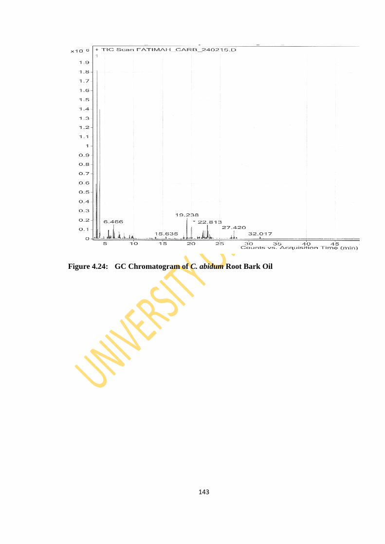
143
Figure 4.24: GC Chromatogram of C. abidum Root Bark Oil
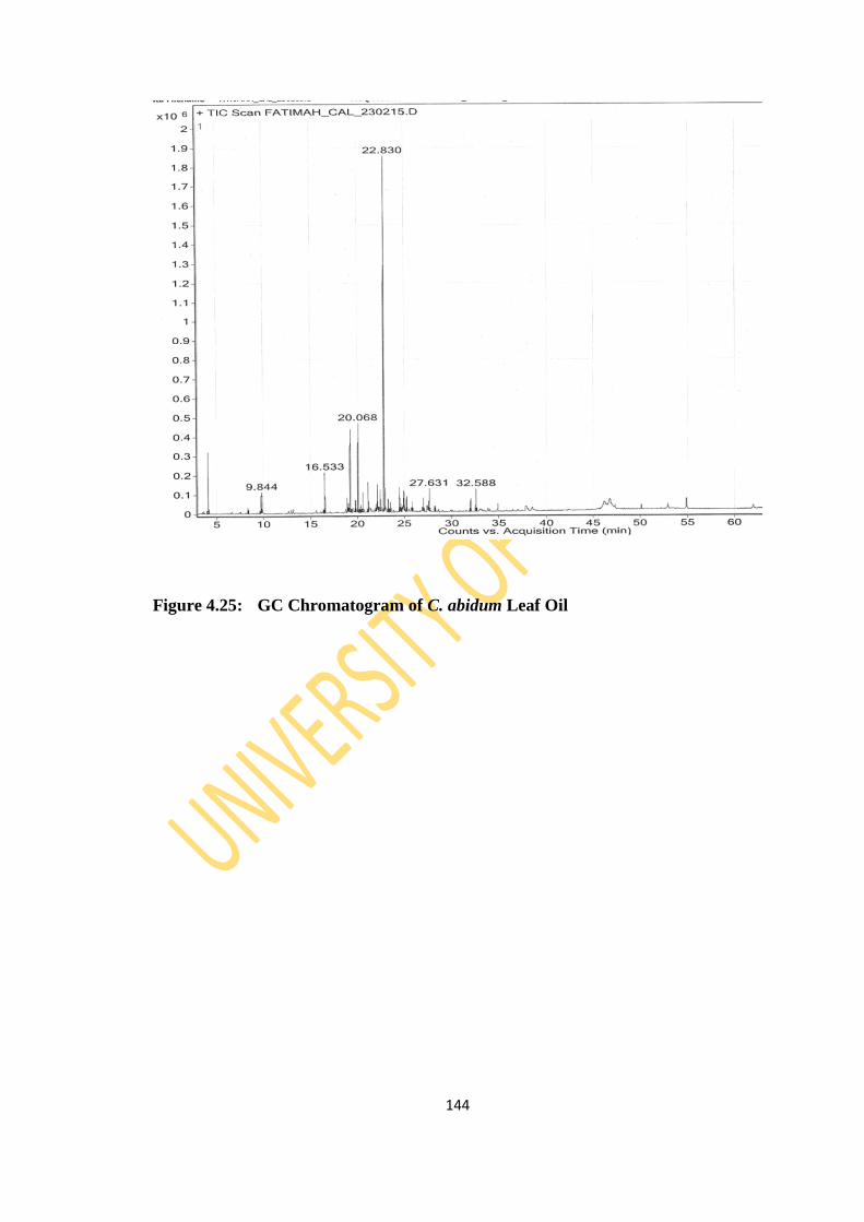
144
Figure 4.25: GC Chromatogram of C. abidum Leaf Oil
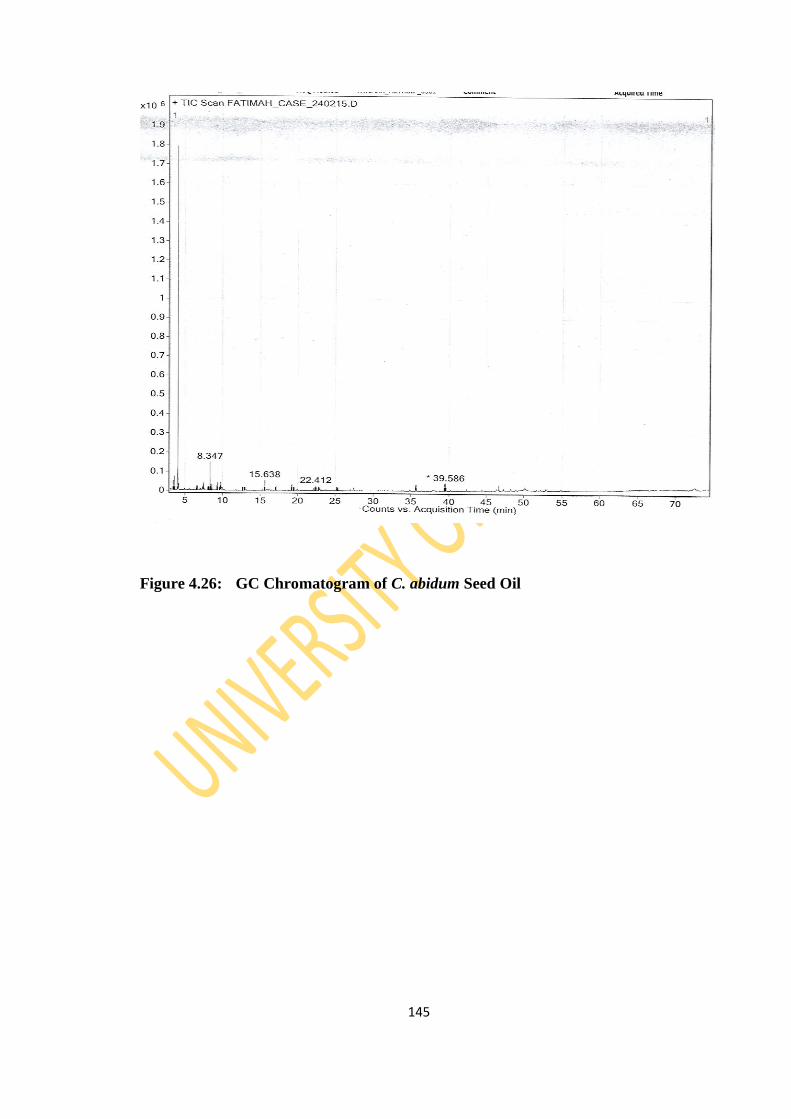
145
Figure 4.26: GC Chromatogram of C. abidum Seed Oil
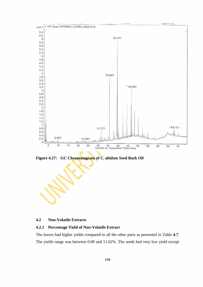
146
Figure 4.27: GC Chromatogram of C. abidum Seed Bark Oil
4.2 Non-Volatile Extracts
4.2.1 Percentage Yield of Non-Volatile Extract
The leaves had higher yields compared to all the other parts as presented in Table 4.7.
The yields range was between 0.08 and 11.62%. The seeds had very low yield except
147
for T. cacao seed. All the leaf extracts had greenish colour while the stem and root
barks had brownish colours except for the stem and root bark of C. papaya.
Table 4.7: Percentage Yield of Non-Volatile Extracts
S/N Plant Sample Part used Extract Yield (%w/w)
1 Persea americana Stem Bark 7.19

148
Root Bark 9.25
Leaf 11.62
Peel 5.78
Seed 3.41
2 Carica papaya Root Bark 2.42
Stem Bark 2.23
Seed 0.73
Leaf 5.61
Peel 4.01
3 Ananas comosus Shoot 2.85
Peel 4.02
4 Theobroma cacao Linn. Stem Bark 5.13
Leaf 5.82
Pod 1.49
Seed 6.23
5 Chrysophyllum albidium Stem Bark 2.6
Root Bark 2.88
Leaf 11.43
Seed 2.91
Seed Bark 0.08
Fruit Bark 4.12
4.2.2 Phytochemical Screening of Non-Volatile Extracts
All the 22 samples screened for secondary metabolites showed the presence of
alkaloids, saponins and flavonoids with varying observation of anthocyanins, steroids,
149
glycosides and anthraquinones (Table 4.8). Reducing sugar was not detected in all
extracts of the fruit plants indicating that the reducing sugar in fruits are more
concentrated in the juicy part of the fruit and not the other parts of the tree.
This result is in agreement with earlier reports on the leaves of C. albidum G. Don
(Amusa et al., 2003; Okoli and Okere, 2010; Orijajogun et al. 2013; Kamba and
Hassan, 2011) with respect to the presence of alkaloids, tannin, saponin, phenol and
flavonoid. The results obtained showed that P. americana Mill contain phytochemicals
such as saponins, tannins, steroids, alkaloids and flavonoids corresponding to earlier
report by Arukwe et al. (2012). T. cacao Linn was found to be rich in saponins,
alkaloids, flavonoids and tannins. This is in accordance with previous study by Zainal
et al., 2014, Ogunmefun et al., 2013, Izuka and Mbangwu, 2013 and Nwokonkwo and
Okeke, 2014). Phytochemical analysis of C. papaya revealed the detection of
flavonoids, alkaloids, steroids and tannins in the methanol extract. This corresponds to
earler report by Khaled et al. 2013) and Anjum et al. (2013). The groups of secondary
metabolites in each of the extracts are known to have a variety of biological activities
which could be responsible for the ethnomedicinal use of the fruit plants. The
preliminary phytochemical analysis (Table 4.8) of the extracts of A. comosus shoot and
peel are reported for the first time to the best of my knowledge.
Table 4.8: Phytochemicals of the Non-volatile Extracts of the Fruit Tree Parts
Code Alk Antho Tan Gly Sap Ste Flav Anthra Red. Sug
PASB + + ND + + + + + ND
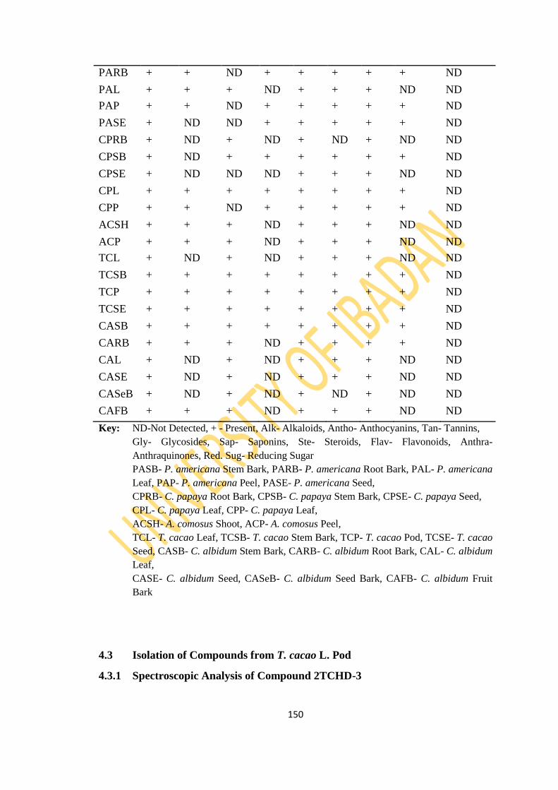
150
PARB + + ND + + + + + ND
PAL + + + ND + + + ND ND
PAP + + ND + + + + + ND
PASE + ND ND + + + + + ND
CPRB + ND + ND + ND + ND ND
CPSB + ND + + + + + + ND
CPSE + ND ND ND + + + ND ND
CPL + + + + + + + + ND
CPP + + ND + + + + + ND
ACSH + + + ND + + + ND ND
ACP + + + ND + + + ND ND
TCL + ND + ND + + + ND ND
TCSB + + + + + + + + ND
TCP + + + + + + + + ND
TCSE + + + + + + + + ND
CASB + + + + + + + + ND
CARB + + + ND + + + + ND
CAL + ND + ND + + + ND ND
CASE + ND + ND + + + ND ND
CASeB + ND + ND + ND + ND ND
CAFB + + + ND + + + ND ND
Key: ND-Not Detected, + - Present, Alk- Alkaloids, Antho- Anthocyanins, Tan- Tannins,
Gly- Glycosides, Sap- Saponins, Ste- Steroids, Flav- Flavonoids, Anthra-
Anthraquinones, Red. Sug- Reducing Sugar
PASB- P. americana Stem Bark, PARB- P. americana Root Bark, PAL- P. americana
Leaf, PAP- P. americana Peel, PASE- P. americana Seed,
CPRB- C. papaya Root Bark, CPSB- C. papaya Stem Bark, CPSE- C. papaya Seed,
CPL- C. papaya Leaf, CPP- C. papaya Leaf,
ACSH- A. comosus Shoot, ACP- A. comosus Peel,
TCL- T. cacao Leaf, TCSB- T. cacao Stem Bark, TCP- T. cacao Pod, TCSE- T. cacao
Seed, CASB- C. albidum Stem Bark, CARB- C. albidum Root Bark, CAL- C. albidum
Leaf,
CASE- C. albidum Seed, CASeB- C. albidum Seed Bark, CAFB- C. albidum Fruit
Bark
4.3 Isolation of Compounds from T. cacao L. Pod
4.3.1 Spectroscopic Analysis of Compound 2TCHD-3

151
Compound 2TCHD-3 (2.6 mg) (Figure 4.28) appeared as white solid with a melting
point of 112-114 oC. The IR absorption bands (Figure 4.29) show the presence of
ketone (ѴC=O stretching 1706 cm-1
), olefin (ѴC=C stretching 1654) and aliphatic
hydrocarbons (ѴCH stretching 2941 and 2869 cm-1
) as seen on Table 4.9. The low
resolution EI-MS (Figure 4.30) showed m/z 424.4 (M+-H2O) corresponding to a
molecular formular C30H46O. The mass spectrum showed fragment ions at m/z 313 [M
- C8H13O (side chain)]+ and 175 [M - C17H27O2 (side chain + ring A)]
+.
The 1H NMR (Figure 4.31) spectrum showed five methyl singlets (δH 0.89, 0.98, 1.03,
1.08, 1.78), a secondary methyl group at δH 0.87 (3H, d, J= 5.6 Hz), terminal
methylene protons at δH 4.87 (1H, br, s) and 4.98 (1H, br, s) and two doublet protons
for a cyclopropyl CH2 group at δH 0.55 (1H, d, J= 4.3 Hz) and 0.78 (1H, d, J= 4.3 Hz),
indicating a cycloartane skeleton. 1H NMR data reveals the presence of an OH group
on C-24 δH 3.47 and an oxymethine δH 4.34 (1H, t, J= 6.6 Hz). This oxymethine proton
was assigned at C-24 due to its chemical shift and coupling pattern as well as HMBC
correlation to C-25, C-26, and C-27. The signals at δC 216.7, δC 144.3 and δC 114.3
were due to the presence of quaternary carbon with an oxo group at C-3 and olefinic
carbons at C-25 and C-26 respectively (Figure 4.32). The DEPT 90 (Figure 4.33)
DEPT 135 (Figure 4.34), HSQC (Figure 4.35), HMBC (Figure 4.36), and COSY
(Figure 4.37) showed thirty carbon signal including six methyls, twelve methylenes,
five methine and seven quaternary carbons. The HMBC spectrum (Figure 4.36)
showed long range correlations from H-22, H-26, and H-27 to C-24; H-19 to C-8, C-9,
C-11 and C-12; and between H-30 to C-8, C-13, C-14 and C-15. On the other hand, in
the HMBC spectrum, the signals of H-28 and H-29 correlated with that of the oxo
group (δC-3 216.7), indicating that the oxo group was located at C-3 and which caused
the H-19 signals of the cyclopropane ring to appear downfield to δH 0.76 (d, J= 4.3
Hz) and 0.55 (d, J= 4.3 Hz), respectively.
Hence, the structure of 2TCHD-3 was assigned as 24-hydroxy-25-cycloarten-3-one
with spectroscopic data (Figure 4.28) that was consistent to reported literature values
(Chiu et al., 2008) as shown on Table 4.10.
Cycloartanes have been reported to have cytotoxic and leishmanicidal activities
(Lavoie et al., 2013; Choudhary et al., 2008). Zahid et al. (2007) reported that

152
cycloartanes exhibited acetylcholinesterase inhibition which implies their possible
application in the treatment of Alzheimer‘s disease.
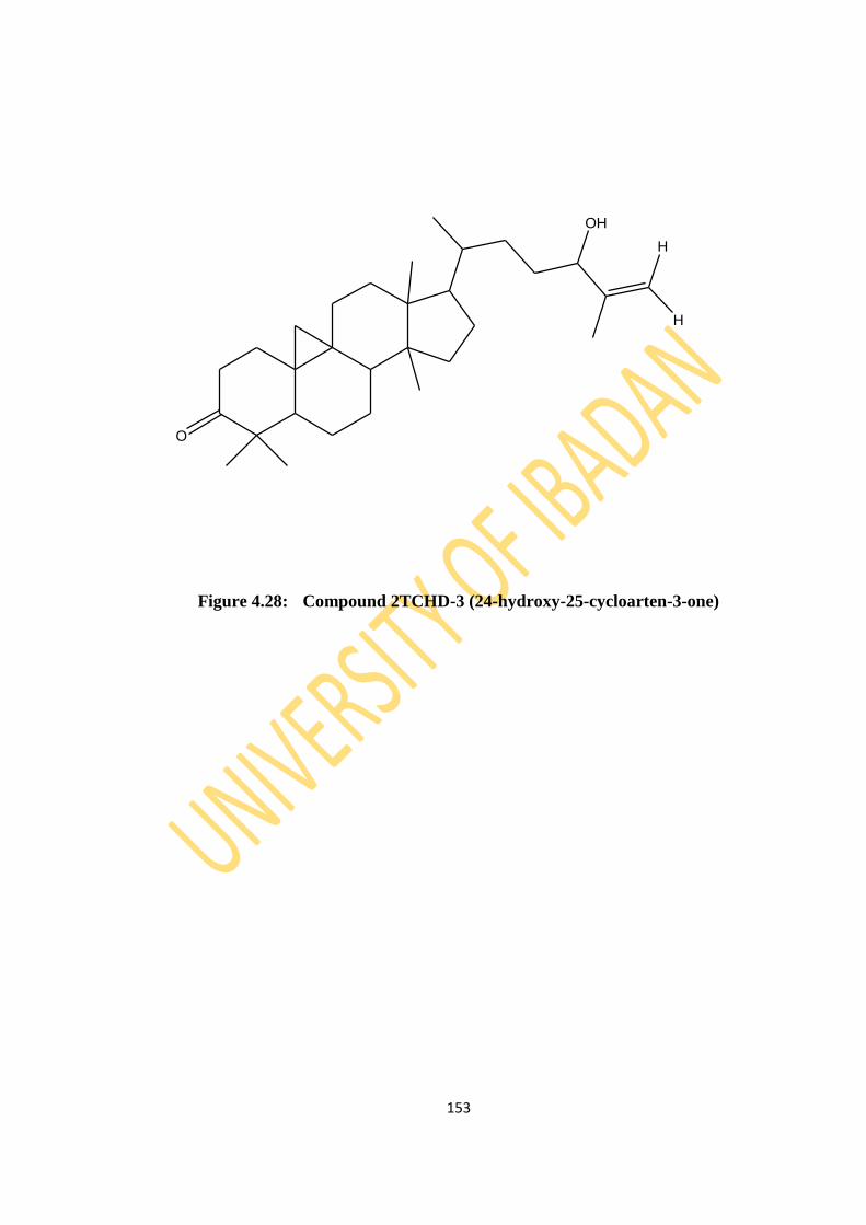
153
Figure 4.28: Compound 2TCHD-3 (24-hydroxy-25-cycloarten-3-one)
O
H
H
OH

154
Figure 4.29: IR Spectrum of Compound 2TCHD-3
Table 4.9: Infra Red values of 2TCHD-3
Functional Group IR values (Ѵ/cm-1
)
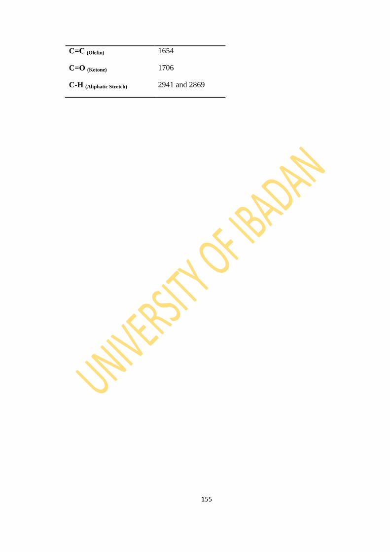
155
C=C (Olefin) 1654
C=O (Ketone) 1706
C-H (Aliphatic Stretch) 2941 and 2869

156
Figure 4.30: EIMS Spectrum of Compound 2TCHD-3

157
Figure 4.31: 1H NMR Spectrum of Compound 2TCHD-3
O
H
H
OH
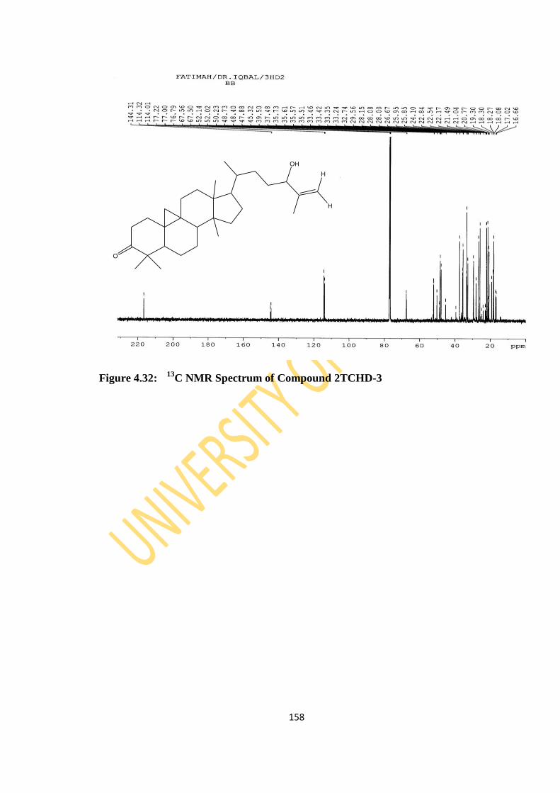
158
Figure 4.32: 13
C NMR Spectrum of Compound 2TCHD-3
O
H
H
OH
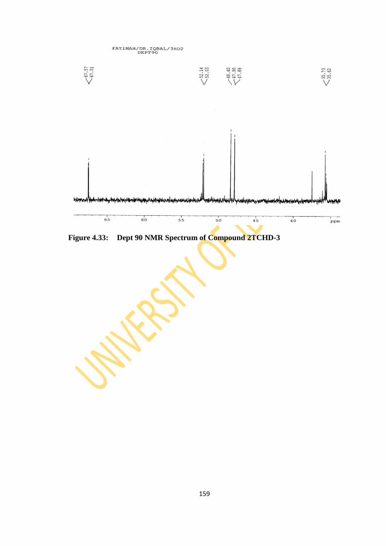
159
Figure 4.33: Dept 90 NMR Spectrum of Compound 2TCHD-3
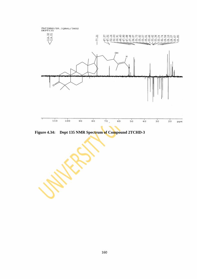
160
Figure 4.34: Dept 135 NMR Spectrum of Compound 2TCHD-3
O
H
H
OH
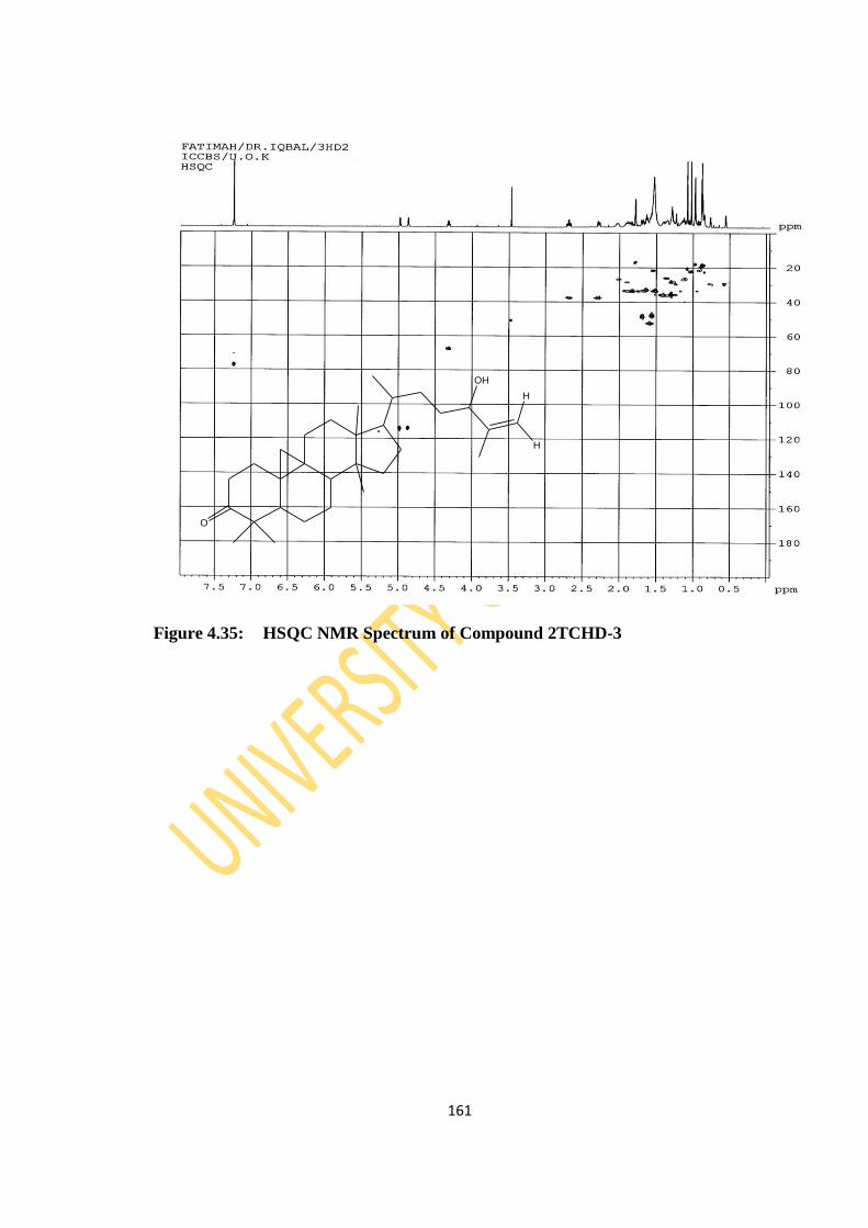
161
Figure 4.35: HSQC NMR Spectrum of Compound 2TCHD-3
O
H
H
OH

162
Figure 4.36: HMBC NMR Spectrum of Compound 2TCHD-3
OH
O
H
H

163
Figure 4.37: COSY Spectrum of Compound 2TCHD-3
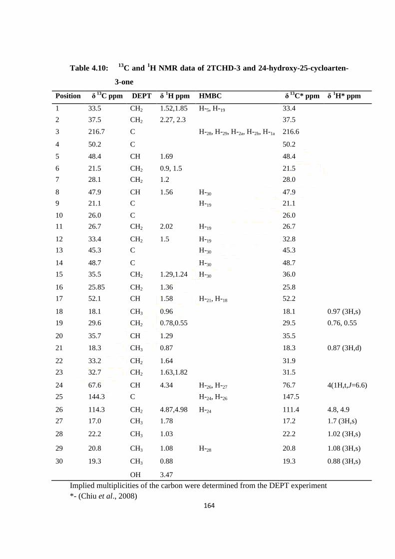
164
Table 4.10: 13
C and 1H NMR data of 2TCHD-3 and 24-hydroxy-25-cycloarten-
3-one
Position δ
13C ppm DEPT δ
1H ppm HMBC
δ
13C* ppm δ
1H* ppm
1 33.5 CH2 1.52,1.85 H-5, H-19 33.4
2 37.5 CH2 2.27, 2.3 37.5
3 216.7 C H-28, H-29, H-2a, H-2b, H-1a 216.6
4 50.2 C 50.2
5 48.4 CH 1.69 48.4
6 21.5 CH2 0.9, 1.5 21.5
7 28.1 CH2 1.2 28.0
8 47.9 CH 1.56 H-30 47.9
9 21.1 C H-19 21.1
10 26.0 C 26.0
11 26.7 CH2 2.02 H-19 26.7
12 33.4 CH2 1.5 H-19 32.8
13 45.3 C H-30 45.3
14 48.7 C H-30 48.7
15 35.5 CH2 1.29,1.24 H-30 36.0
16 25.85 CH2 1.36 25.8
17 52.1 CH 1.58 H-21, H-18 52.2
18 18.1 CH3 0.96 18.1 0.97 (3H,s)
19 29.6 CH2 0.78,0.55 29.5 0.76, 0.55
20 35.7 CH 1.29 35.5
21 18.3 CH3 0.87 18.3 0.87 (3H,d)
22 33.2 CH2 1.64 31.9
23 32.7 CH2 1.63,1.82 31.5
24 67.6 CH 4.34 H-26, H-27 76.7 4(1H,t,J=6.6)
25 144.3 C H-24, H-26 147.5
26 114.3 CH2 4.87,4.98 H-24 111.4 4.8, 4.9
27 17.0 CH3 1.78 17.2 1.7 (3H,s)
28 22.2 CH3 1.03 22.2 1.02 (3H,s)
29 20.8 CH3 1.08 H-28 20.8 1.08 (3H,s)
30 19.3 CH3 0.88 19.3 0.88 (3H,s)
OH 3.47
Implied multiplicities of the carbon were determined from the DEPT experiment
*- (Chiu et al., 2008)

165
4.3.2 Spectroscopic Analysis of 72TCDE-1
Compound 72TCDE-1 (48.6 mg) with the structure in Figure 4.38 appeared as a white
amorphous solid with a melting point of 110.4-112.9 oC. The IR absorption bands
(Figure 4.39) show the presence of hydroxyl (ѴOH stretching 3346 cm-1
), aliphatic
hydrocarbons (ѴCH stretching 2949 and 2869 cm-1
), 1676 cm‐1 (ѴC=C absorption peak);
other absorption peaks includes 1460 cm‐1 (ѴCH2), 1049 cm‐1 (cycloalkane) and (ѴCO
stretching 1340 cm-1
) as presented on Table 4.11. The molecular formula C29H50O
(414.2) was confirmed by EIMS fragment ions 369.2 (M-45) or loss of HO+=CH‐CH3
(Figure 4.40). Ion peak at m/z 273 is due to the formation of carbocation by β-bond
cleavage of side chain leading to the loss of C10H23 that corresponds to M‐143. The
dehydration of fragment at m/z 273 would yield m/z 255, which on successive
dealkylation would yield ions at m/z 187, 173, 159, 144, 132, 106.9, 69, 54.9, 42.9.
The signals of 1H NMR spectra showed the presence of six methyl signals that
appeared as two methyl singlets at δ 0.64 and 1.01; three methyl doublets that appeared
at δ 0.81, 0.83 and 0.93; and a methyl triplet at δ 0.84 (Figure 4.41). The 1H NMR
signals at δH 3.53 (m) and δH 5.32 (t) revealed the presence of oxymethine and olefinic
methine protons at C-3 and C-6 respectively. The signals at δC 71.79, δC 121.7 and δC
140.7 were due to the presence of oxymethine carbon at C-3 and olefinic carbons at C-
5 and C-6 (Figure 4.42).
DEPT 90 (Figure 4.45) and DEPT 135 (Figure 4.45) exhibited six methyls, eleven
methylenes and ten methine carbons.
The HSQC spectrum (Figure 4.45) showed that methine protons at δH 3.53 (1H, m, H-
3) and δH 5.32 (1H, t, H-6) were bonded to carbons at δC 71.79 (C-3) and δC 121.7 (C-6)
respectively. HMBC spectrum (Figure 4.46) reveals the correlation of methine proton;
H-6 with C-4, C-7 and C-8 while oxymethine carbon; C-3 was correlated to protons H-
1, H-2 and H-4. There was correlation between the methane carbon; C-17 and H-18,
H-19, H-22, H-16 and H-15. The COSY spectrum (Figure 4.47) revealed the
correlation of oxymethine proton at C-3 with methylene protons at C-2 and C-4 while
olefinic methine proton at C-6 was correlated with methylene protons at C-7. Thus, the
structure of 72TCDE-1 was assigned as Stigmast-5-en-3-ol (β-sitosterol) with
166
spectroscopic data (Figure 4.38) that was consistent to the reported literature values
(Habib et al., 2007; Jamal et al., 2009; Patra et al., 2010; Chaturvedula and Prakash,
2012) as seen on Table 4.12 and was further supported by the key COSY and HMBC
correlations as shown in Figures 4.46 and 4.47.
Stigmast-5-en-3-ol as a steroid, is an important class of bioorganic molecules,
widespread in plants. This compound has a long history of consumption as food or
pharmaceutical products, and generally recognized as safe without undesirable side
effects (Saeidnia et al., 2014). It is usually used for heart disease,
hypercholesterolemia, modulating the immune system, prevention of cancer, as well as
for rheumatoid arthritis, tuberculosis, cervical cancer, hair loss and benign prostatic
hyperplasia. Furthermore, its diverse biological and pharmacological activities (anti-
inflamatory, anticancer, angiogenic, immunomodulatory, antihelminthic, antioxidant,
neuroprotection and antidiabetic) have been reported (Prieto et al., 2006; Loizou et al.,
2010; Chai et al., 2008; Villasenor et al., 2002; Baskar et al., 2012; Shi et al., 2013;
Radika et al., 2013).

167
Figure 4.38: Compound 72TCDE-1 (Stigmast-5-en-3β-ol)
HO
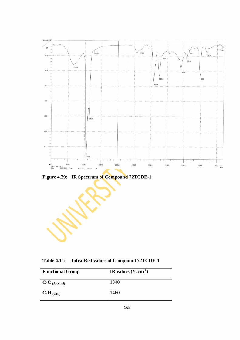
168
Figure 4.39: IR Spectrum of Compound 72TCDE-1
Table 4.11: Infra-Red values of Compound 72TCDE-1
Functional Group IR values (Ѵ/cm-1
)
C-C (Alcohol) 1340
C-H (CH2) 1460
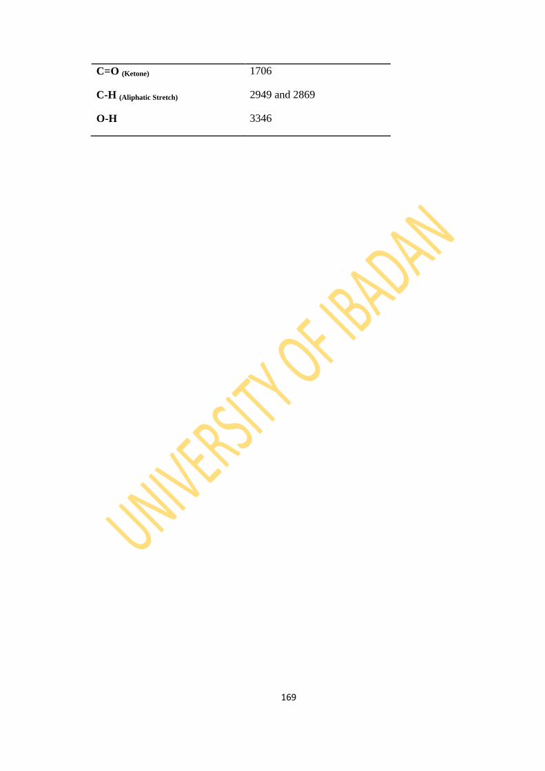
169
C=O (Ketone) 1706
C-H (Aliphatic Stretch) 2949 and 2869
O-H 3346

170
Figure 4.40: EIMS Spectrum of Compound 72TCDE-1

171
Figure 4.41: 1H NMR Spectrum of Compound 72TCDE-1
HO
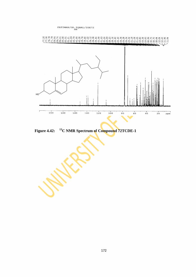
172
Figure 4.42: 13
C NMR Spectrum of Compound 72TCDE-1
HO
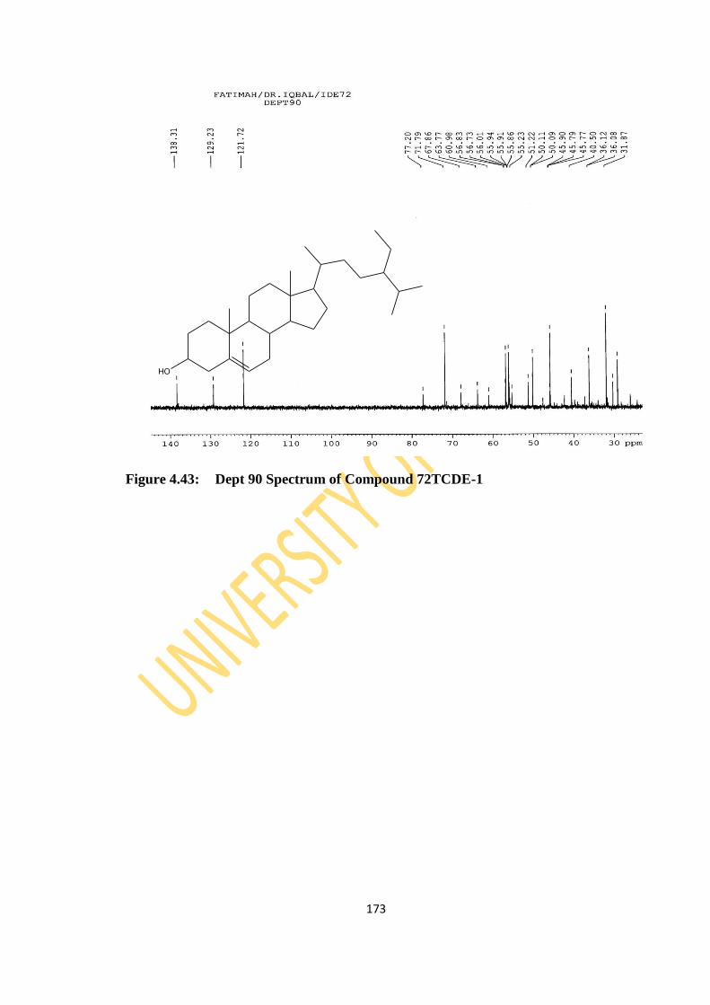
173
Figure 4.43: Dept 90 Spectrum of Compound 72TCDE-1
HO
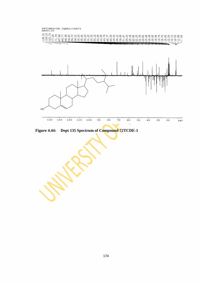
174
Figure 4.44: Dept 135 Spectrum of Compound 72TCDE-1
HO

175
Figure 4.45: HSQC Spectrum of Compound 72TCDE-1
HO
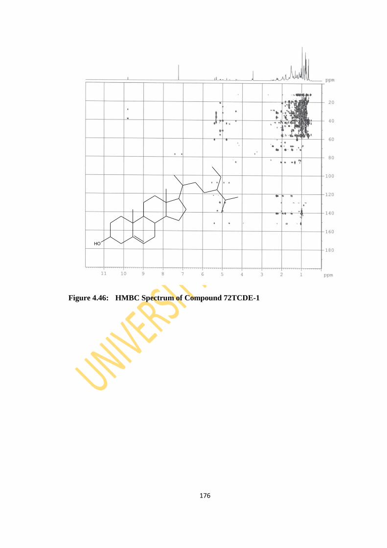
176
Figure 4.46: HMBC Spectrum of Compound 72TCDE-1
HO
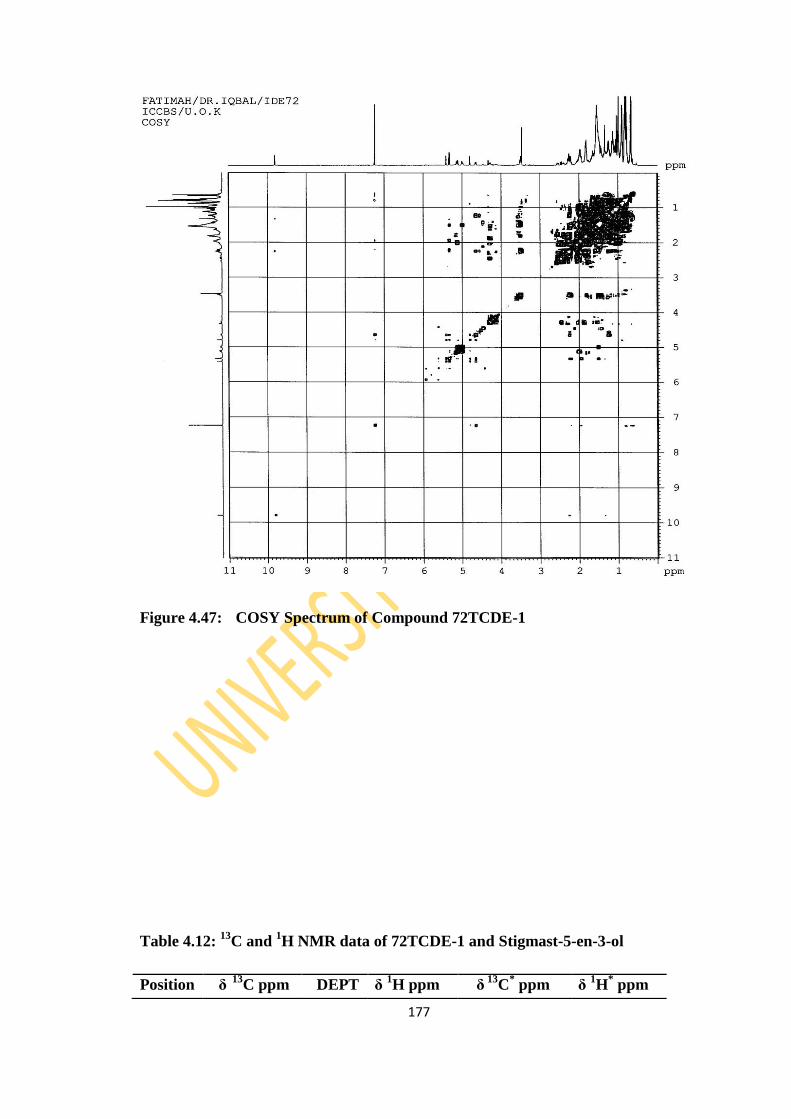
177
Figure 4.47: COSY Spectrum of Compound 72TCDE-1
Table 4.12: 13
C and 1H NMR data of 72TCDE-1 and Stigmast-5-en-3-ol
Position δ
13C ppm DEPT δ
1H ppm
δ
13C
* ppm δ
1H
* ppm

178
Implied multiplicities of the carbon were determined from the DEPT experiment
*- (Chaturvedula and Prakash, 2012)
1 37.22 CH2 1.04, 1.82 37.5 1.47
2 31.64 CH2 1.43 31.9 1.56
3 71.79 CH 3.5 72.0 3.52
4 42.28 CH2 2.23,2.28 42.5 2.28
5 140.7 C 140.9
6 121.7 CH 5.32 121.9 5.36
7 31.89 CH2 1.83 32.1 2.03
8 31.87 CH 1.69 32.1 1.67
9 50.1 CH 0.9 50.3 1.48
10 36.48 C 36.7
11 21.05 CH2 1.37 21.3 1.52
12 39.6 CH2 1.14 39.9 1.49
13 42.95 C 42.6
14 56.73 CH 0.98 56.9 1.5
15 26.0 CH2 1.14 26.3 1.6
16 28.23 CH2 1.25, 1.84 28.5 1.84
17 55.8 CH 1.08 56.3 1.49
18 36.1 CH 1.36 36.3 1.64
19 19.38 CH3 0.98 19.2 1.02
20 34.44 CH2 1.48 34.2 0.88
21 25.38 CH2 1.13 26.3 1.04
22 45.79 CH 1.52 46.1 1.5
23 23.02 CH2 1.03 23.3 1.04
24 12.2 CH3 0.64 12.2 0.68
25 29.1 CH 1.67 29.4 1.65
26 21.05 CH3 0.98, 0.82 20.1 0.83
27 19.8 CH3 1.34 19.6 0.94
28 19.0 CH3 0.89 19.0 0.85
29 11.96 CH3 0.87 12.0 0.88
OH 3.42
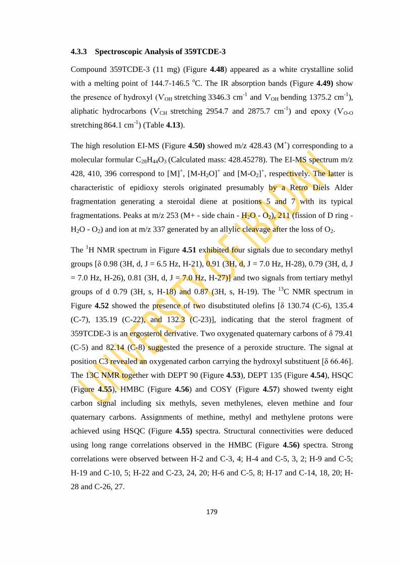
179
4.3.3 Spectroscopic Analysis of 359TCDE-3
Compound 359TCDE-3 (11 mg) (Figure 4.48) appeared as a white crystalline solid
with a melting point of 144.7-146.5 oC. The IR absorption bands (Figure 4.49) show
the presence of hydroxyl (ѴOH stretching 3346.3 cm-1
and ѴOH bending 1375.2 cm-1
),
aliphatic hydrocarbons (ѴCH stretching 2954.7 and 2875.7 cm-1
) and epoxy (ѴO-O
stretching 864.1 cm-1
) (Table 4.13).
The high resolution EI-MS (Figure 4.50) showed m/z 428.43 (M+) corresponding to a
molecular formular C28H44O3 (Calculated mass: 428.45278). The EI-MS spectrum m/z
428, 410, 396 correspond to [M]+, [M-H2O]
+ and [M-O2]
+, respectively. The latter is
characteristic of epidioxy sterols originated presumably by a Retro Diels Alder
fragmentation generating a steroidal diene at positions 5 and 7 with its typical
fragmentations. Peaks at m/z 253 (M+ - side chain - H2O - O2), 211 (fission of D ring -
H2O - O2) and ion at m/z 337 generated by an allylic cleavage after the loss of O2.
The 1H NMR spectrum in Figure 4.51 exhibited four signals due to secondary methyl
groups [δ 0.98 (3H, d, J = 6.5 Hz, H-21), 0.91 (3H, d, J = 7.0 Hz, H-28), 0.79 (3H, d, J
= 7.0 Hz, H-26), 0.81 (3H, d, J = 7.0 Hz, H-27)] and two signals from tertiary methyl
groups of d 0.79 (3H, s, H-18) and 0.87 (3H, s, H-19). The 13
C NMR spectrum in
Figure 4.52 showed the presence of two disubstituted olefins [δ 130.74 (C-6), 135.4
(C-7), 135.19 (C-22), and 132.3 (C-23)], indicating that the sterol fragment of
359TCDE-3 is an ergosterol derivative. Two oxygenated quaternary carbons of δ 79.41
(C-5) and 82.14 (C-8) suggested the presence of a peroxide structure. The signal at
position C3 revealed an oxygenated carbon carrying the hydroxyl substituent [δ 66.46].
The 13C NMR together with DEPT 90 (Figure 4.53), DEPT 135 (Figure 4.54), HSQC
(Figure 4.55), HMBC (Figure 4.56) and COSY (Figure 4.57) showed twenty eight
carbon signal including six methyls, seven methylenes, eleven methine and four
quaternary carbons. Assignments of methine, methyl and methylene protons were
achieved using HSQC (Figure 4.55) spectra. Structural connectivities were deduced
using long range correlations observed in the HMBC (Figure 4.56) spectra. Strong
correlations were observed between H-2 and C-3, 4; H-4 and C-5, 3, 2; H-9 and C-5;
H-19 and C-10, 5; H-22 and C-23, 24, 20; H-6 and C-5, 8; H-17 and C-14, 18, 20; H-
28 and C-26, 27.
180
Down field signals at δ 6.48, δ 6.22 (AB quartet, J = 8, 2H, H-6, H-7) in the 1H NMR
spectrum revealed the presence of disubstitued double bond which were correlated
with carbon signals of δ 135.39 (C-6), δ 130.68 (C-7) in HMBC spectrum.
Based on this spectroscopic data and by comparison with literature on Table 4.14
(Krzyczkowski et al., 2009; Yue et al., 2001; Cateni et al., 2007), it was found that
359TCDE-3 corresponds to Ergosta-5α,8α-epidioxy-6,22-dien-3β-ol, more known as
ergosterol peroxide and been reported for the first time in T. cacao.
Ergosterol peroxide has been reported to have strong antitumor, cytotoxic and
anticomplementary activities (Shin et al., 2001).
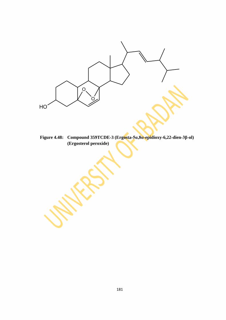
181
Figure 4.48: Compound 359TCDE-3 (Ergosta-5α,8α-epidioxy-6,22-dien-3β-ol)
(Ergosterol peroxide)
HO
O
O

182
Figure 4.49: IR Spectrum of Compound 359TCDE-3

183
Table 4.13: IR values of Compound 359TCDE-3
Functional Group IR values (Ѵ/cm-1
)
O-O (Epoxy) 864
C-H (Aliphatic Stretching) 2941 and 2869
O-H (Stretching) 3346
(Bending) 1375

184
Figure 4.50: EIMS Spectrum of Compound 359TCDE-3
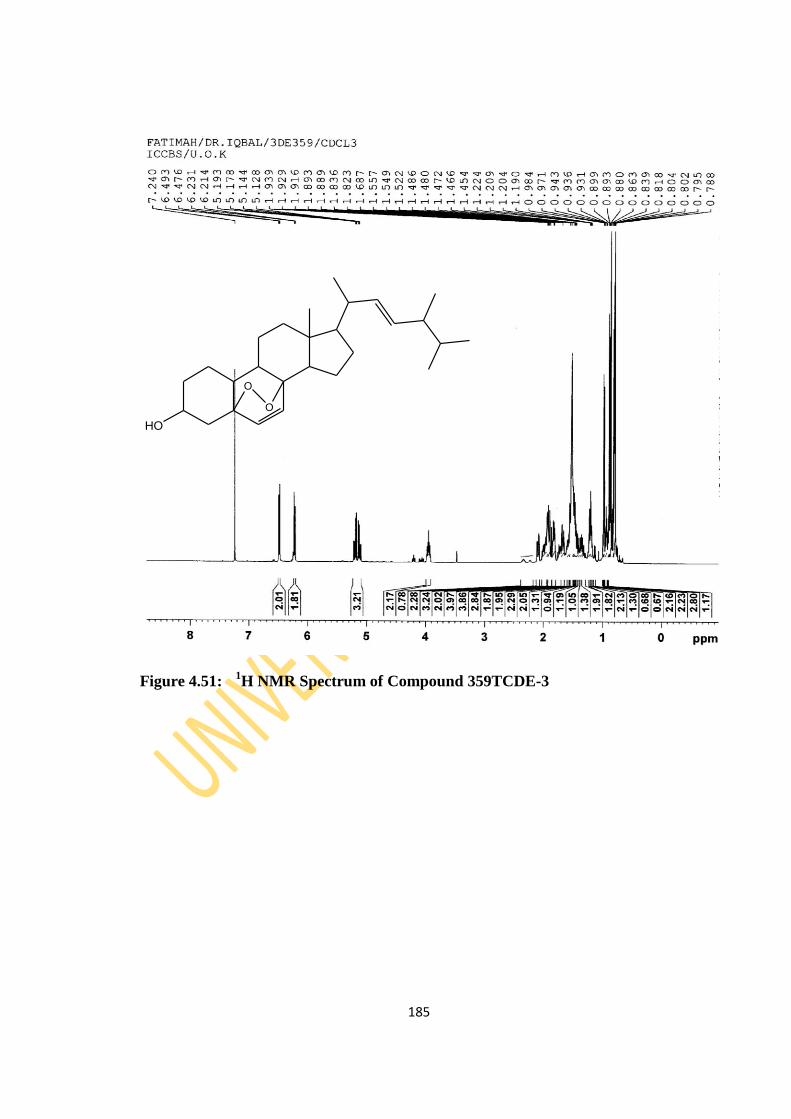
185
Figure 4.51: 1H NMR Spectrum of Compound 359TCDE-3
HO
O
O

186
Figure 4.52: 13
C NMR Spectrum of Compound 359TCDE-3
HO
O
O
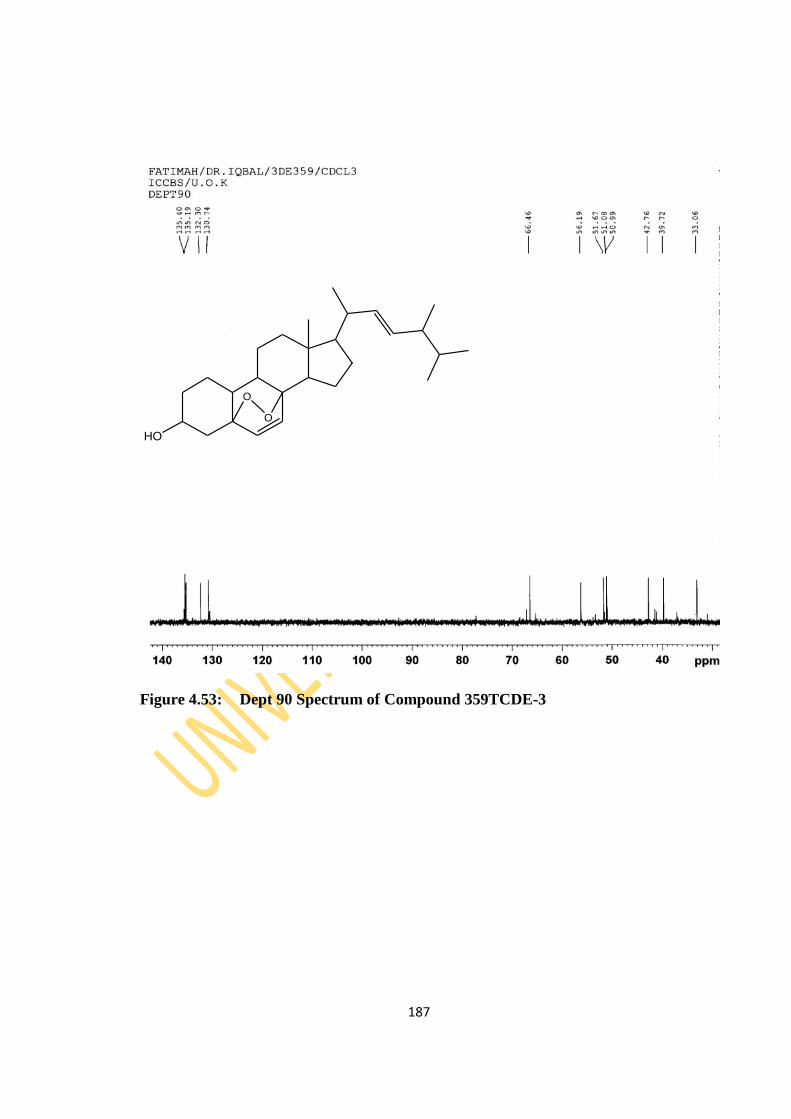
187
Figure 4.53: Dept 90 Spectrum of Compound 359TCDE-3
HO
O
O
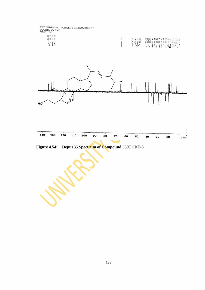
188
Figure 4.54: Dept 135 Spectrum of Compound 359TCDE-3
HO
O
O
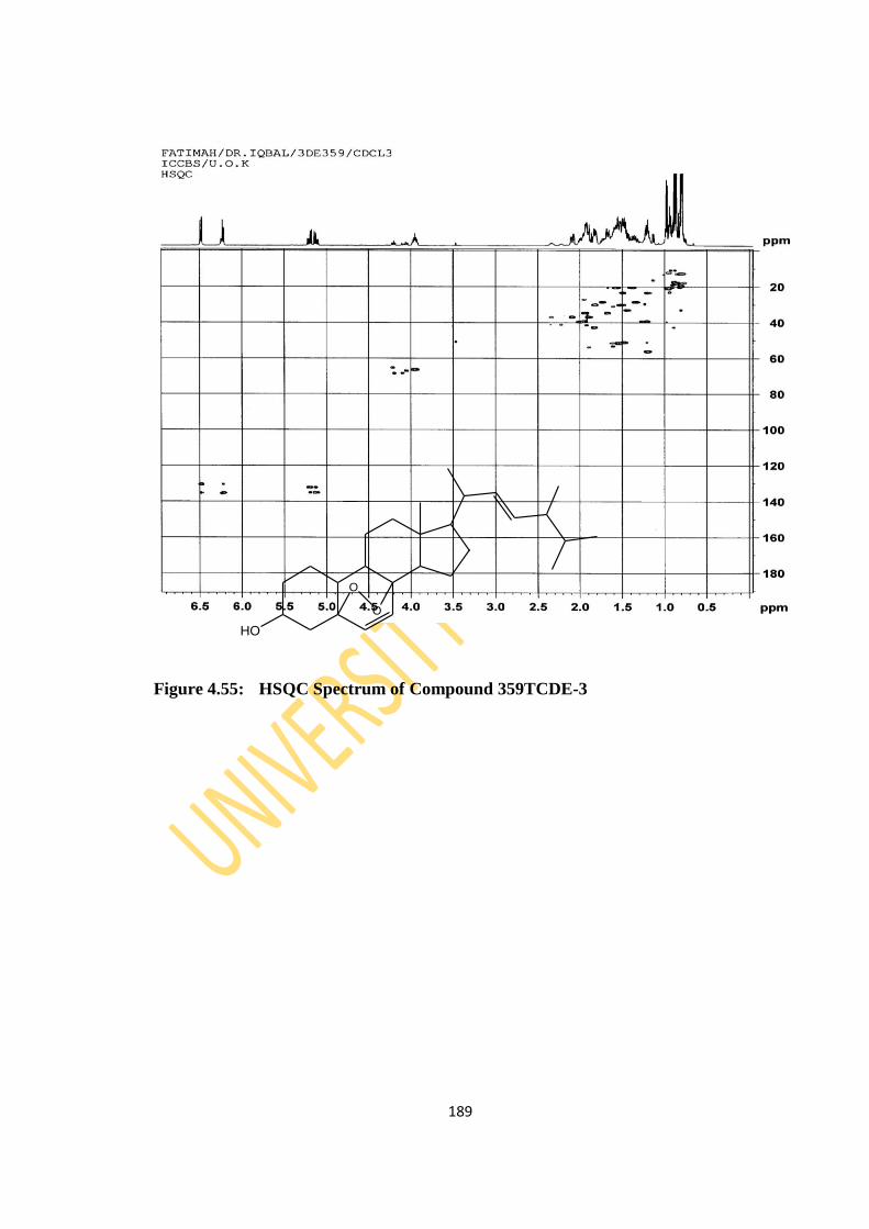
189
Figure 4.55: HSQC Spectrum of Compound 359TCDE-3
HO
O
O
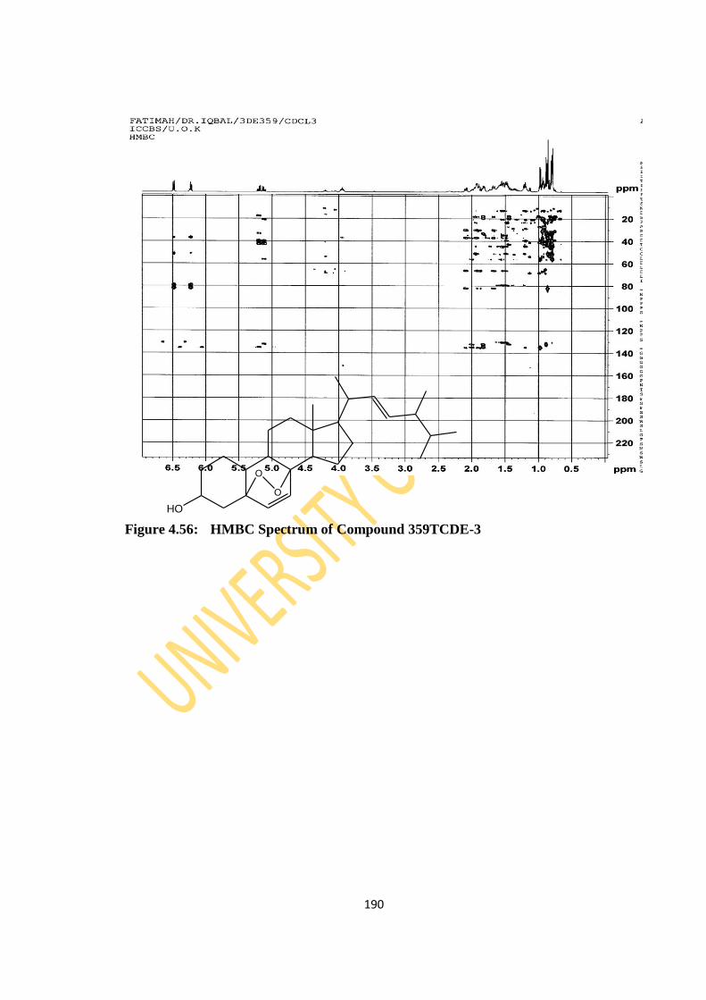
190
Figure 4.56: HMBC Spectrum of Compound 359TCDE-3
HO
O
O
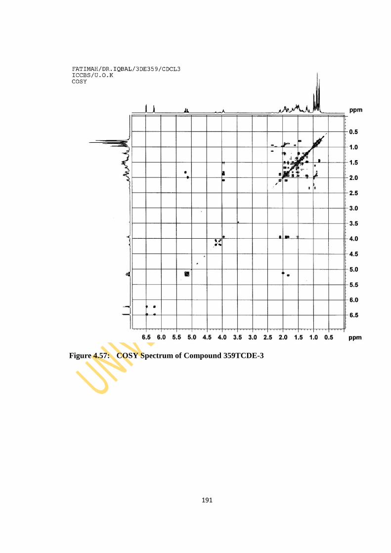
191
Figure 4.57: COSY Spectrum of Compound 359TCDE-3
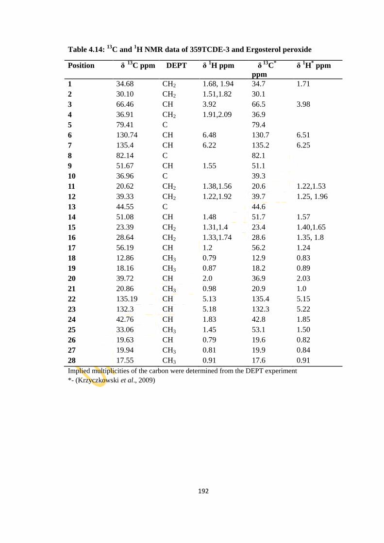
192
Table 4.14: 13
C and 1H NMR data of 359TCDE-3 and Ergosterol peroxide
Position δ
13C ppm DEPT δ
1H ppm
δ
13C
*
ppm
δ 1H
* ppm
1 34.68 CH2 1.68, 1.94 34.7 1.71
2 30.10 CH2 1.51,1.82 30.1
3 66.46 CH 3.92 66.5 3.98
4 36.91 CH2 1.91,2.09 36.9
5 79.41 C 79.4
6 130.74 CH 6.48 130.7 6.51
7 135.4 CH 6.22 135.2 6.25
8 82.14 C 82.1
9 51.67 CH 1.55 51.1
10 36.96 C 39.3
11 20.62 CH2 1.38,1.56 20.6 1.22,1.53
12 39.33 CH2 1.22,1.92 39.7 1.25, 1.96
13 44.55 C 44.6
14 51.08 CH 1.48 51.7 1.57
15 23.39 CH2 1.31,1.4 23.4 1.40,1.65
16 28.64 CH2 1.33,1.74 28.6 1.35, 1.8
17 56.19 CH 1.2 56.2 1.24
18 12.86 CH3 0.79 12.9 0.83
19 18.16 CH3 0.87 18.2 0.89
20 39.72 CH 2.0 36.9 2.03
21 20.86 CH3 0.98 20.9 1.0
22 135.19 CH 5.13 135.4 5.15
23 132.3 CH 5.18 132.3 5.22
24 42.76 CH 1.83 42.8 1.85
25 33.06 CH3 1.45 53.1 1.50
26 19.63 CH 0.79 19.6 0.82
27 19.94 CH3 0.81 19.9 0.84
28 17.55 CH3 0.91 17.6 0.91
Implied multiplicities of the carbon were determined from the DEPT experiment
*- (Krzyczkowski et al., 2009)
193
4.4 Biological Activity of Essential Oils
In a bid to determine the possible therapeutic value of the analysed 27 (twenty–seven)
essential oils, the antibacterial, antioxidant and insecticidal activities were evaluated.
The antibacterial assay was carried out on selected Gram-positive bacteria
(Staphylococcus aureus and Bacillus subtilis) and Gram-negative bacteria (Escherichia
coli, Pseudomonas aeruginosa, Shigella flexineri and Salmonella typhi) known as
causative agents for various infectious diseases using Alamar Blue Assay. The
antioxidant assay was evaluated by the method of DPPH radical scavenging activity
while the insecticidal activity was done by contact toxicity test.
4.4.1 Antibacterial Activity of Persea americana Essential Oil
The six (6) essential oils of Persea americana were not active against the
Pseudomonas aeruginosa strain while all were active against Staphylococcus aureus
strain with percentage inhibition of 69.93%, 69.06%, 19.84%, 14.49%, 28.88% and
10.52% for the fruit, peel, seed, leaf, stem bark and root bark oils respectively. The
fruit and peel oils had 11.46% and 19.05% inhibition against Esherichia coli strain but
were not active against Shigella flexenari strain. The leaf and root bark oils however
had 22.04% and 14.65% inhibition against Bacillus subtilis strain (Table 4.15). Only
the essential oil from the seed showed activity against Salmonella typhi strain
(16.78%).

194
Table 4.15: Percentage Inhibition of Essential Oils of P. americana Mill Plant
Parts
Key: Drug- Ampicillin, PAF- P. americana Fruit, PAP- P. americana Peel,
PASE- P. americana Seed, PAL- P. americana Leaf,
PASB- P. americana Stem Bark, PARB- P. americana Root Bark
Name of Bacteria Percent (%) Inhibition
Drug PAF PAP PASE PAL PASB PARB
Escherichia coli 72 11.46 19.05 0 0 0 0
Bacillus subtilis 76 0 0 0 22.04 0 14.65
Shigella flexenari 65 0 0 20.87 24.68 16.35 17.07
Staphylococcus aureus 79 69.93 69.06 19.84 14.49 28.88 10.52
Pseudomonas aeruginosa 80 0 0 0 0 0 0
Salmonella typhi 70 0 0 16.78 0 0 0

195
4.4.2 Antibacterial Activity of Carica papaya Essential Oil
The eight (8) essential oils of Carica papaya were not active against the Pseudomonas
aeruginosa strain while only the root bark essential oil was active against Esherichia
coli strain (75.08%) and only the essential oil from the seed showed activity against
Salmonella typhi strain (27.58%). The leaf oil was inactive against all the tested
bacteria strains. The fruit, peel, root, stem bark and stem essential oils showed
moderate percentage inhibition (13.82%-30.69%) against Bacillus subtilis and Shigella
flexenari but the essential oils of the root bark, root, stem bark and stem had 67.36%,
14.73%, 13.47% and 12.10% inhibition respectively against Staphylococcus aureus
strain while the seed oil showed 35.24% inhibition against Bacillus subtilis strain
(Table 4.16).
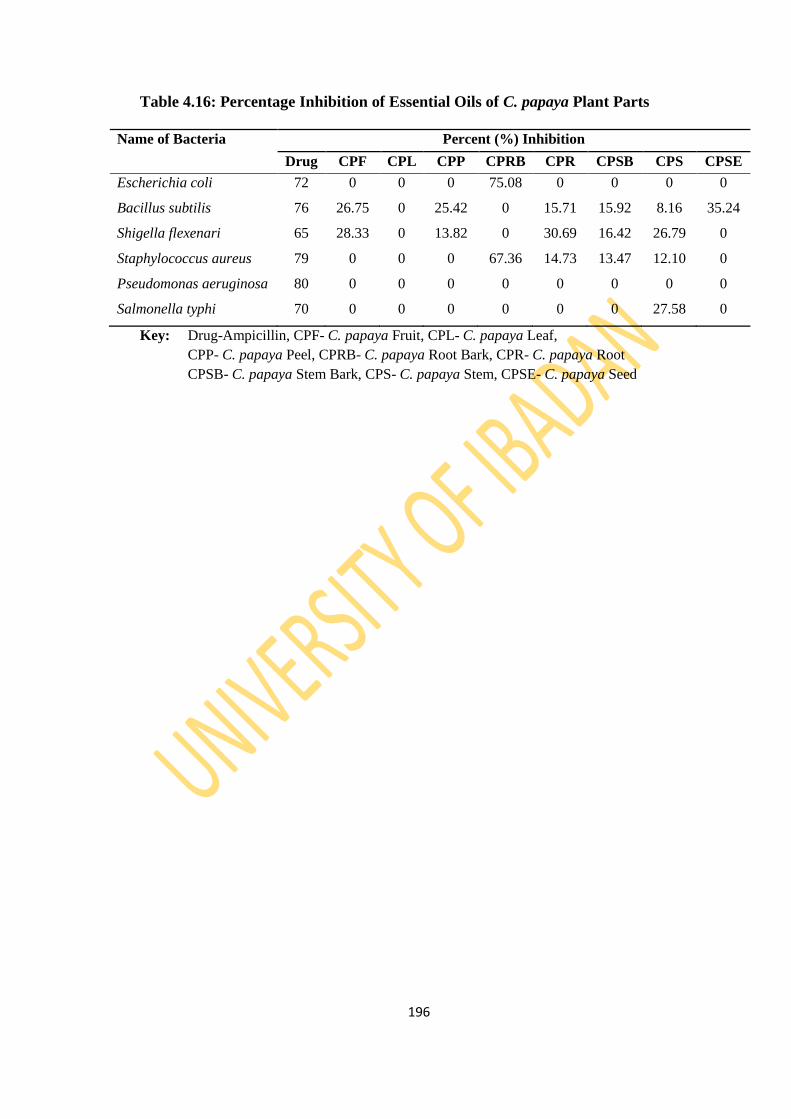
196
Table 4.16: Percentage Inhibition of Essential Oils of C. papaya Plant Parts
Name of Bacteria Percent (%) Inhibition
Drug CPF CPL CPP CPRB CPR CPSB CPS CPSE
Escherichia coli 72 0 0 0 75.08 0 0 0 0
Bacillus subtilis 76 26.75 0 25.42 0 15.71 15.92 8.16 35.24
Shigella flexenari 65 28.33 0 13.82 0 30.69 16.42 26.79 0
Staphylococcus aureus 79 0 0 0 67.36 14.73 13.47 12.10 0
Pseudomonas aeruginosa 80 0 0 0 0 0 0 0 0
Salmonella typhi 70 0 0 0 0 0 0 27.58 0
Key: Drug-Ampicillin, CPF- C. papaya Fruit, CPL- C. papaya Leaf,
CPP- C. papaya Peel, CPRB- C. papaya Root Bark, CPR- C. papaya Root
CPSB- C. papaya Stem Bark, CPS- C. papaya Stem, CPSE- C. papaya Seed

197
4.4.3 Antibacterial Activity of Ananas comosus Essential Oil
The essential oils (3) of Ananas comosus were also screened against the selected
bacteria and the study revealed Esherichia coli, Pseudomonas aeruginosa and
Salmonella typhi strains were resistant to all the oils. Shigella flexenari was susceptible
to all the oils; fruit, peel and shoot with 39.21%, 20.56% and 22.74% inhibiton
respectively. The peel and shoot oils were also active against Staphylococcus aureus
strain with 14.97% and 11.49% inhibition while Bacillus subtilis strain was susceptible
to only the fruit oil with 7.36% inhibition (Table 4.17).

198
Table 4.17: Percentage Inhibition of Essential Oils of A. comosus Plant Parts
Name of Bacteria Percent (%) Inhibition
Drug ACF ACP ACSH
Escherichia coli 72 0 0 0
Bacillus subtilis 76 7.36 0 0
Shigella flexenari 65 39.21 20.56 22.74
Staphylococcus aureus 79 0 14.97 11.49
Pseudomonas aeruginosa 80 0 0 0
Salmonella typhi 70 0 0 0
Key: Drug- Ampicillin, ACF- A. comosus Fruit, ACP- A. comosus Peel,
ACSH- A. comosus Shoot
199
4.4.4 Antibacterial Activity of Theobroma cacao Essential Oil
The essential oils (4) of Theobroma cacao L. were also screened against the selected
bacteria and the study revealed the non active nature of the essential oil from the seed
against all the test bacteria strains while the leaf oil showed a high percentage
inhibition (78.59%) against Escherichia coli strain (Table 4.18). Bacillus subtilis,
Shigella flexenari, Pseudomonas aeruginosa and Salmonella typhi strains were
resistant to all the oils. Staphylococcus aureus strain was however susceptible to the
stem bark and pod oil at 37.68% and 70.12% inhibition respectively. The stem bark
and pod oils were also active against Esherichia coli strain with 28.95% and 31.73%
inhibition.

200
Table 4.18: Percentage Inhibition of Essential Oils of T. cacao Linn Plant Parts
Name of Bacteria Percent (%) Inhibition
Drug TCL TCSB TCP TCSE
Escherichia coli 72 78.59 28.95 31.73 0
Bacillus subtilis 76 0 0 0 0
Shigella flexenari 65 0 0 0 0
Staphylococcus aureus 79 0 37.68 70.12 0
Pseudomonas aeruginosa 80 0 0 0 0
Salmonella typhi 70 0 0 0 0
Key: Drug- Ampicillin, TCL- T. cacao Leaf, TCSB- T. cacao Stem Bark,
TCP- T. cacao Pod, TCSE- T. cacao Seed
201
4.4.5 Antibacterial Activity of Chrysophyllum albidum Essential Oil
The essential oils (6) of Chrysophyllum albidum were also screened against the
selected bacteria and the study revealed the non active nature of the essential oil from
the seed against all the test bacteria strains except Staphylococcus aureus with 70.59%.
Escherichia coli strain was resistant to all the EOs from the different parts of this fruit
tree but Shigella flexenari was susceptible to all the volatile oils except the seed EO.
Pseudomonas aeruginosa and Salmonella typhi strains were resistant to all the oils
except the seed bark (10.19%) and root bark (9.79%) oils respectively (Table 4.19).
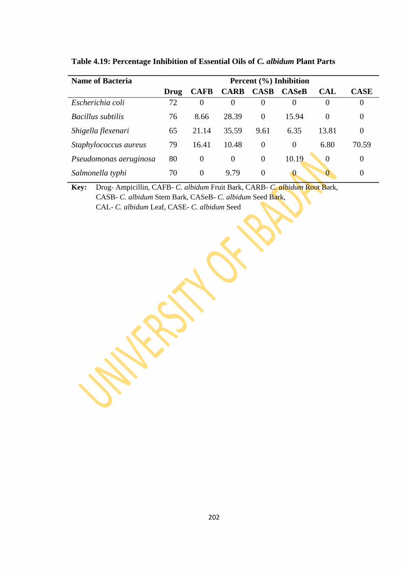
202
Table 4.19: Percentage Inhibition of Essential Oils of C. albidum Plant Parts
Name of Bacteria Percent (%) Inhibition
Drug CAFB CARB CASB CASeB CAL CASE
Escherichia coli 72 0 0 0 0 0 0
Bacillus subtilis 76 8.66 28.39 0 15.94 0 0
Shigella flexenari 65 21.14 35.59 9.61 6.35 13.81 0
Staphylococcus aureus 79 16.41 10.48 0 0 6.80 70.59
Pseudomonas aeruginosa 80 0 0 0 10.19 0 0
Salmonella typhi 70 0 9.79 0 0 0 0
Key: Drug- Ampicillin, CAFB- C. albidum Fruit Bark, CARB- C. albidum Root Bark,
CASB- C. albidum Stem Bark, CASeB- C. albidum Seed Bark,
CAL- C. albidum Leaf, CASE- C. albidum Seed
203
4.4.6 Comparison of the Antibacterial Activity of the Essential Oils
Researchers have reported that Gram positive bacteria are more susceptible to essential
oils than Gram negative bacteria (Mann et al., 2000) but in this study the essential oils
did not show any preferential activity against the Gram positive and Gram negative
bacteria used for the study. The oils, however, showed poor antibacterial activity
against P. aeruginosa and S. typhi (both Gram negative bacteria) with percentage
inhibition less than 30%. P. aeruginosa has been reported to be a well known
antibiotic resistant Gram-negative bacterium which is generally less sensitive to the
actions of plants essential oils (Boussaada et al., 2008) but was sensitive to the
essential oil of only C. albidum seed bark with 10.19 % inhibition while S. typhi was
susceptible to only C. albidum root bark, C. papaya stem and P. americana seed
volatile oils with 9.79%, 27.58% and 16.78% inhibition respectively. Of all the
essential oils investigated for antibacterial activity against E. coli, C. papaya root bark
and T. cacao leaf showed the most favourable efficacy with 75.08% and 78.59%
inhibition respectively as against 72% inhibition of the drug. T. cacao stem bark, pod
and P. americana fruit, peel oils showed relatively lower percentage inhibition
(28.95%, 31.73%, 11.46% and 19.05% respectively) against E. coli. Twelve (12),
seventeen (17) and eighteen (18) of the total twenty-seven (27) oils showed activity
against B. subtilis, S. flexinari and S. aureus respectively. The percentage inhibition of
the oils against B. subtilis was however low in comparison with the drug. Amongst the
eighteen (18) oils that were active against S. aureus, it was observed that the essential
oils of C. albidum seed, C. papaya root bark, T. cacao pod, P. americana fruit and peel
had relatively high percentage inhibition at 70.59%, 67.36%, 70.12%, 69.93% and
69.06% respectively when compared with the drug with 79% inhibition. All the
seventeen (17) oils active against S. flexenari had below 30% inhibition as against the
standard drug‘s inhibition at 65%.
The notable antimicrobial variations between the oils may be attributed to the fact that
the biological activity of an essential oil is linked to its chemical composition and at
times to the major chemical constituents (Lawrence, 2000; Cimanga et al., 2002). The
major compounds in the oils might have been responsible for the antibacterial activity
of the essential oils. However, it has been observed that the antibacterial effects of
whole essential oils are stronger than their major components when tested individually
(Lataou and Tantaoui-Elarak, 1994) and this suggests contribution of other
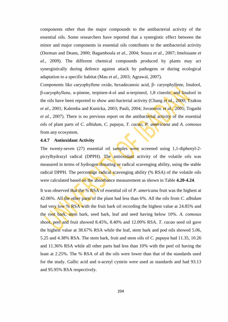
204
components other than the major compounds to the antibacterial activity of the
essential oils. Some researchers have reported that a synergistic effect between the
minor and major components in essential oils contributes to the antibacterial activity
(Dorman and Deans, 2000; Bagamboula et al., 2004; Souza et al., 2007; Imelouane et
al., 2009). The different chemical compounds produced by plants may act
synergistically during defence against attack by pathogens or during ecological
adaptation to a specific habitat (Mau et al., 2003; Agrawal, 2007).
Components like caryophyllene oxide, hexadecanoic acid, β- caryophyllene, linalool,
β-caryophyllene, α-pinene, terpinen-4-ol and α-terpineol, 1,8 cineole, and linalool in
the oils have been reported to show anti-bacterial activity (Chang et al., 2000; Tzakou
et al., 2001; Kalemba and Kunicka, 2003; Pauli, 2004; Jovanovic et al., 2005; Togashi
et al., 2007). There is no previous report on the antibacterial activity of the essential
oils of plant parts of C. albidum, C. papaya, T. cacao, P. americana and A. comosus
from any ecosystem.
4.4.7 Antioxidant Activity
The twenty-seven (27) essential oil samples were screened using 1,1-diphenyl-2-
picrylhydrazyl radical (DPPH). The antioxidant activity of the volatile oils was
measured in terms of hydrogen donating or radical scavenging ability, using the stable
radical DPPH. The percentage radical scavenging ability (% RSA) of the volatile oils
were calculated based on the absorbance measurement as shown in Table 4.20-4.24.
It was observed that the % RSA of essential oil of P. americana fruit was the highest at
42.06%. All the other parts of the plant had less than 6%. All the oils from C. albidum
had very low % RSA with the fruit bark oil recording the highest value at 24.85% and
the root bark, stem bark, seed bark, leaf and seed having below 10%. A. comosus
shoot, peel and fruit showed 8.45%, 8.40% and 12.09% RSA. T. cacao seed oil gave
the highest value at 38.67% RSA while the leaf, stem bark and pod oils showed 5.06,
5.25 and 4.38% RSA. The stem bark, fruit and stem oils of C. papaya had 11.35, 10.26
and 11.36% RSA while all other parts had less than 10% with the peel oil having the
least at 2.25%. The % RSA of all the oils were lower than that of the standards used
for the study. Gallic acid and n-acetyl cystein were used as standards and had 93.13
and 95.95% RSA respectively.
205
The observed low % RSA of the essential oils can be explained by the fact that the oils
are not capable of donating hydrogen atom and the low solubility provided by the oils
in the reaction medium of the assay because this test utilizes methanol or ethanol as
solvent as explained by a report by Mata et al. (2007). Viuda-Martos et al. (2009) also
cited these factors as the main limitation of this assay for measuring antioxidant
activity of lipophilic samples like many essential oils. Despite the essential oils tested
in this study not showing significant antioxidant activity, many essential oils have
shown antioxidant potential. As an example there is the research conducted by
Guimarães (2010), who investigated the antioxidant activity of essential oils of Lippia
sidoides, Alomia fastigiata, Ocotea odorifera, Mikania glauca and Cordia verbenacea,
and their major constituents, by the methods of the β-carotene/linoleic acid oxidation
system, the formation of thiobarbituric acid reactive species (TBARS) and the
reduction of the stable DPPH radical, and found that the essential oil of L. sidoides
showed higher antioxidant activity, presenting the lowest IC50 values in all trials, and
the antioxidant activity presented by the essential oil of L. sidoides was attributed to its
major constituent carvacrol, which also showed high antioxidant activity when
assessed in isolation (Andrade et al., 2013).
The phenolic content in plants has been reported to be responsible for the antioxidant
activity of some plants (Othman et al., 2007). Phenolic compounds like thymol and
carvacrol found in some plant essential oils have been reported to have antioxidant
activity (Miguel et al., 2004). Also essential oils rich in monoterpene hydrocarbons
have been reported to have high antioxidant activity (Tepe et al., 2005). Ruberto and
Baratta (2000) investigated the antioxidant activity of 98 pure essential oil
components, which represent the main classes of typical compounds of essential oils
and found out that sesquiterpene hydrocarbons exerted a low, if any, antioxidant effect.
The analysis of the essential oil components in this study revealed that the oils were
mainly dominated by sesquiterpenes and non-terpenes. The poor antioxidant activity of
these essential oils, probably, is due to their lack of phenolic compounds and low
concentrations of monoterpene hydrocarbons. However, it has been observed that
correlation of the antioxidant activities of essential oils and their chemical
compositions is often very complicated (Miguel, 2010). Essential oil constituents
acting individually or synergistically may contribute to the antioxidant activity of the
oil (Tiwari, 2001).
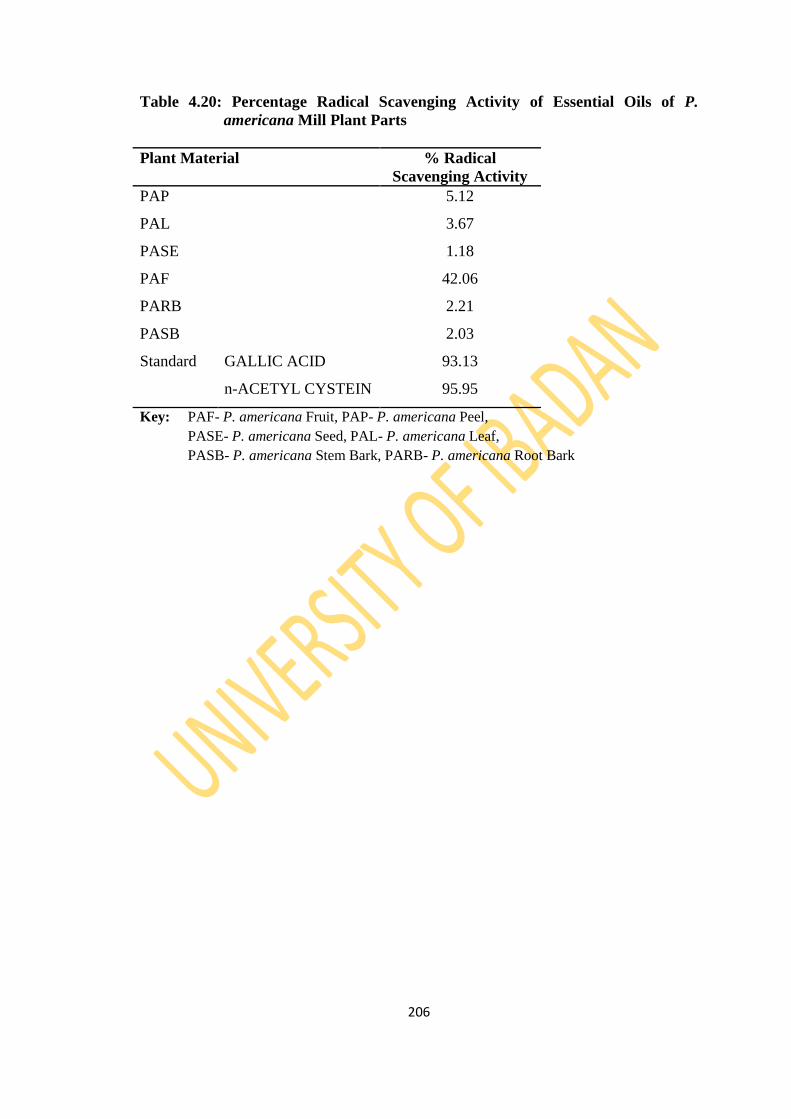
206
Table 4.20: Percentage Radical Scavenging Activity of Essential Oils of P.
americana Mill Plant Parts
Plant Material % Radical
Scavenging Activity
PAP 5.12
PAL 3.67
PASE 1.18
PAF 42.06
PARB 2.21
PASB 2.03
Standard GALLIC ACID 93.13
n-ACETYL CYSTEIN 95.95
Key: PAF- P. americana Fruit, PAP- P. americana Peel,
PASE- P. americana Seed, PAL- P. americana Leaf,
PASB- P. americana Stem Bark, PARB- P. americana Root Bark
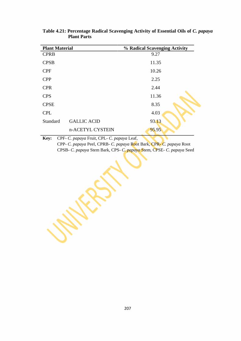
207
Table 4.21: Percentage Radical Scavenging Activity of Essential Oils of C. papaya
Plant Parts
Plant Material % Radical Scavenging Activity
CPRB 9.27
CPSB 11.35
CPF 10.26
CPP 2.25
CPR 2.44
CPS 11.36
CPSE 8.35
CPL 4.03
Standard GALLIC ACID 93.13
n-ACETYL CYSTEIN 95.95
Key: CPF- C. papaya Fruit, CPL- C. papaya Leaf,
CPP- C. papaya Peel, CPRB- C. papaya Root Bark, CPR- C. papaya Root
CPSB- C. papaya Stem Bark, CPS- C. papaya Stem, CPSE- C. papaya Seed
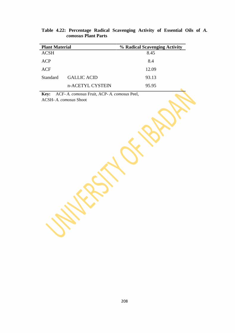
208
Table 4.22: Percentage Radical Scavenging Activity of Essential Oils of A.
comosus Plant Parts
Plant Material % Radical Scavenging Activity
ACSH 8.45
ACP 8.4
ACF 12.09
Standard GALLIC ACID 93.13
n-ACETYL CYSTEIN 95.95
Key: ACF- A. comosus Fruit, ACP- A. comosus Peel,
ACSH- A. comosus Shoot
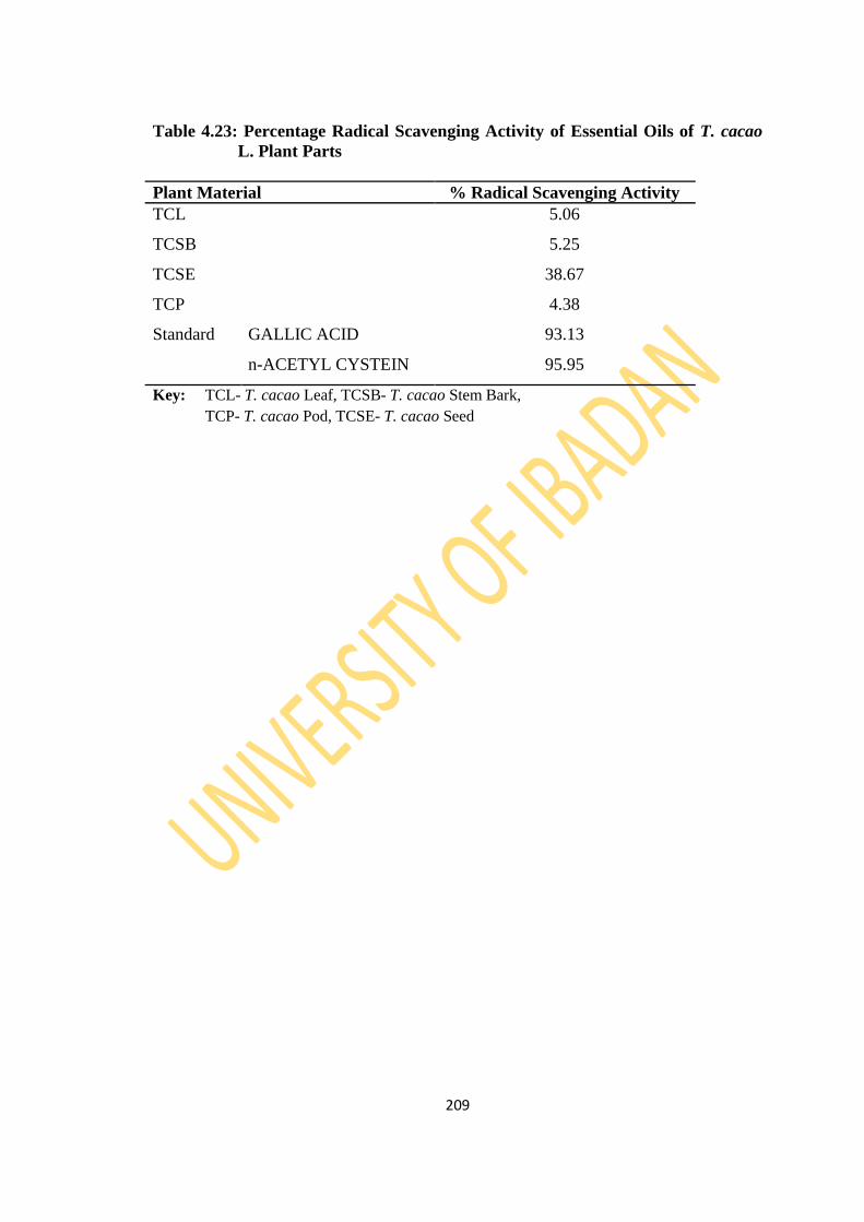
209
Table 4.23: Percentage Radical Scavenging Activity of Essential Oils of T. cacao
L. Plant Parts
Plant Material % Radical Scavenging Activity
TCL 5.06
TCSB 5.25
TCSE 38.67
TCP 4.38
Standard GALLIC ACID 93.13
n-ACETYL CYSTEIN 95.95
Key: TCL- T. cacao Leaf, TCSB- T. cacao Stem Bark,
TCP- T. cacao Pod, TCSE- T. cacao Seed
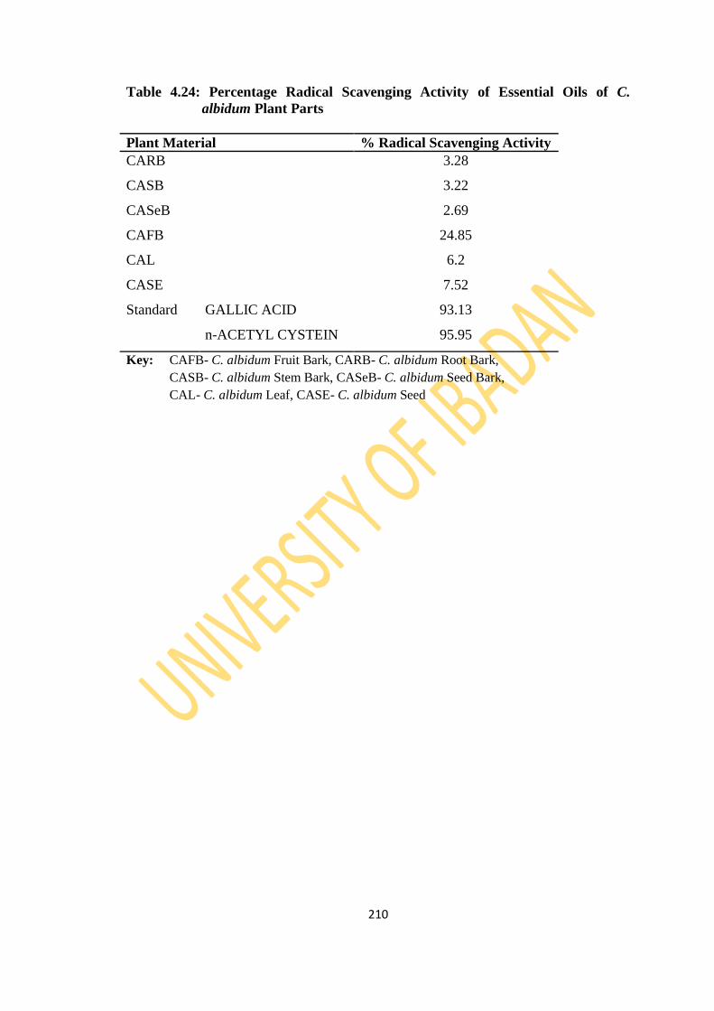
210
Table 4.24: Percentage Radical Scavenging Activity of Essential Oils of C.
albidum Plant Parts
Plant Material % Radical Scavenging Activity
CARB 3.28
CASB 3.22
CASeB 2.69
CAFB 24.85
CAL 6.2
CASE 7.52
Standard GALLIC ACID 93.13
n-ACETYL CYSTEIN 95.95
Key: CAFB- C. albidum Fruit Bark, CARB- C. albidum Root Bark,
CASB- C. albidum Stem Bark, CASeB- C. albidum Seed Bark,
CAL- C. albidum Leaf, CASE- C. albidum Seed

211
4.4.8 Insecticidal Activity
All the oils showed no toxicity (0% mortality) against the insects except for
Chrysophyllum albidium stem bark, Theobroma cacao Linn. leaf and Ananas comosus
peel with 20% mortality against Rhyzopertha dominica and Ananas comosus peel
exhibited 20% mortality of Callosbruchus analis. The insects were observed to be
resistant to the oils used for this study based on the impregnated filter paper method
used which is a form of contact toxicity. In contact toxicity stomach poisoning occurs
while the insects feed on the whole grains. The weevils have to pick up the lethal dose
of treatment from the essential oil to cause toxicity.
Previous studies have shown that the toxicity of essential oils obtained from aromatic
plants against storage pests is related to the oil‘s main components (Lee, 2003). The
insecticidal constituents of many plant extracts and essential oils are mainly
monoterpenoids (Regnault-Roger and Hamraoui, 1995; Ahn et al., 1998; Isman, 2000;
Asgar, 2011). Monoterpenoids are typically volatile and rather lipophilic compounds
that can penetrate into insects rapidly and interfere with their physiological functions
(Lee et al., 2002). Due to their high volatility, they are fumigant and gaseous and
might be of importance for stored-product insects (Ahn et al. 1998). Various
monoterpenes like 1,8-cineole, linalool, α-pinene, terpinen-4-ol, and α-terpinene have
been reported to show contact and fumigation toxicity to stored product pests
(Papachristos et al., 2004; Stamapoulos et al., 2007). Therefore, the resistance of the
essential oils studied for insecticidal activity may be related to the non-dominance of
monoterpenes in the identified components in the oils.
The synergistic action between major and minor components of essential oils could
also be responsible for the repellent action of the oils to the insects. Plant essential oils
are mixtures of different major and minor components and their biological activity is
generally determined by their major components or synergism/antagonism among
different components (Shukla et al., 2012; Tapondjou et al., 2005).
212
CHAPTER 5
CONCLUSION
The essential oils of the fruit plants revealed diverse compounds which can be
classified as hydrocarbons, monoterpenes, sesquiterpenes, diterpenes, triterpenes and
non-terpenes. The non-terpenes were made up of esters, aldehydes, alcohols, fatty
acids and their derivatives. The variations and similarities in the yield and
compositional pattern of the essential oils may depend on factors such as plant parts
utilized, storage, the plant species, drying method and the drying period. The detailed
compositional pattern of the essential oils from all the plant parts of the selected fruits
is being reported for the first time.
The bioassays carried out on the essential oils showed different properties with respect
to the three activities of study. The antibacterial assay gave low inhibition of most of
the essential oils against the test organisms. The observed antibacterial activity
revealed the essential oils that can be of importance in the combat against bacteria
pathogens and good sources of medicinally useful antibacterial drugs. However, in
vivo studies and clinical trials would be needed to justify and further evaluate the
potential of these essential oils as reliable antibacterial agents. Also, more detailed
studies of the mechanism of actions of these oils will be of great help in utilizing their
full potential in pharmaceutical, cosmetics and aromatherapy industries.
The essential oils had a relatively weak inhibition of DPPH activity. There was also
non-significant insecticidal activity of the oils to the insects used for the study. The
bioactivity pattern of the oils could be attributed to the interaction between compounds
in the oils which usually leads to antagonistic, additive or synergestic effects. All the
samples screened for secondary metabolites showed the presence of alkaloids,
saponins and flavonoids with varying observation of anthocyanins, steroids, glycosides
and anthraquinones. Reducing sugar was not observed in all the samples.
212
The isolation and characterization of the triterpenes; cycloartane (24-hydroxy-9,19-
cycloanost-25-en-3-one) and a steroid (Stigmast-5-en-3β-ol) from Theobroma cacao
Linn pod is being reported for the first time. The pharmacokinetic, structure activity
relationship and toxicological profile of these isolated bioactive compounds could be
carried out to explore the possibility of translating them to prescription drugs for
orthodox therapies.
213
REFERENCES
Abbe Maleyki, M. J. and Amin, I. 2008. Polyphenols in Cocoa and Cocoa Products: Is
There a Link between Antioxidant Properties and Health? Review. Molecules
13:2190-2219.
Abdelhady, M. I. and Aly, H. A. H. 2012. Antioxidant antimicrobial activities of
Callistemon comboynensis essential oil. Free Radical Antioxidants 2:37–41.
Abdenour, A., Susana L., Mohammed B., Amin L., Carmen R., Antonio H., Rafael P.
and Pilar C. 2011. Chemical composition and antimicrobial activity of essential
oils of Thymus algeriensis, Eucalyptus globulus and Rosmarinus officinalis
from Morocco. Journal of the Science of Food and Agriculture. 91.14:2643–
2651.
Abdin, M. Z., Israr, M., Rehman, R. U. and Jain, S. K. 2003. Artemisinin, a novel
antimalarial drug: Biochemical and molecular approaches for enhanced
production. Planta Medica 69.4:289-299.
Abe, F., Nagafuji, S., Okawa, M., Kinjo, J., Akahane, H., Ogura, T., Martínez-Alfaro,
M. A. and Reyes-Chilpa, R. 2005. Trypanocidal constituents in plants:
Evaluation of some Mexican plants for their trypanocidal activity and active
constituents in the seed of Persea americana. Biological Pharmacy Bulletin
28.7:1314–1317.
Achini, G., Ratnasooriya, W. D., Jayakody, J. R. A. C., Charmain, F. and Charmini, K.
2012. Thrombocytosis and Anti-inflammatory properties and toxicological
evaluation of Carica papaya mature leaf concentrate in a murine model, Online
International Journal of Medicinal Plant Research 1.2:21-30.
Adahchour, M., Van Stee, L. L. P., Beens, J., Vreuls, R. J. J., Batenburg, M. A. and
Brinkman, U. A. T. 2003. Comprehensive two-dimensional gas
chromatography with time-of-flight spectrometric detection for the trace
analysis of flavour compounds in food. Journal of Chromatography A
1019:157-172.
Adams, R. P. 1989. Identification of Essential Oils by Ion Trap Mass Spectroscopy.
Academic Press, San Diego.
Adams, R. P. 1995. Identification of Essential Oil Components by Gas
Chromatography/Mass Spectroscopy. Carol Stream: Allured Publishing
Corporation, Carol Stream, Illionois.
214
Adams, R. P. 2007. Identification of Essential Oil Components by Gas
Chromatography/ Mass Spectrometry, 4th Edition, Allured Publication
Corporation, Carol Stream, Illinois.
Adebayo, A. H., Abolaji, A. O., Kela, R., Ayepola, O. O., Olorunfemi, T. B. and
Taiwo O. S. 2011. Antioxidant activities of the leaves of Chrysophyllum
albidum G. Pakistan Journal of Pharmaceutical Science 24.4:545-551.
Adebayo, A. H., Abolaji, A. O., Opata, T. K. and Adegbenro, I. K. 2010. Effects of
ethanolic leaf extract of Chrysophyllum albidum G. on biochemical and
hematological parameters of albino Wistar rats. African Journal of
Biotechnology 9.14:2145-2150.
Adewole, E., Ajiboye, B., Ojo, B., Ogunmodede, O. T. and Oso, O. 2013.
Characterization of cocoa (Theobroma cocoa) pod. International Journal of
Scientific and Engineering Research 4.1:1-5.
Adewoye E. O, Salami A. T, Emikpe B. O. 2012. Effect of Methanolic Extract of
Chrysophyllum albidum Bark on Hematological Parameters in Mice with
Experimental Hemorrhagic Anemia. African Journal of Biomedical Research
15:2.
Adewoye, E. O., Salami, A. T., Lawal, T. O. and Adeniyi, B. A. 2011. The
Antimicrobial and Kill kinetics of Chrysophyllum albidum Stem Bark Extracts.
European Journal of Scientific Research 56.3:434-444.
Adewusi, H. A. 1997. The African star apple (Chrysophyllum albidum) Indigenous
Knowledge from Ibadan, Southwestern Nigeria. In: Proceedings of a National
Workshop on the Potentials of the star apple in Nigeria. (Eds.) Denton, O. A.,
Ladipo, D. O., Adetoro, M. A. And Sarumi, M. B. p 25-33.
Adeyemi, O. O., Okpo, S. O. and Ogunti, O. O. 2002. Analgesic and anti-
inflammatory effects of the aqueous extract of leaves of Persea americana Mill
(Lauraceae) Fitoterapia 73:375-380.
Adeyi, O. 2010. Proximate composition of some agricultural wastes in Nigeria and
their potential use in activated carbon production. Journal of Applied Science
Environmental Management 14.1:55–58.
Adisa, S. A. 2000. Vitamin C, Protein and Mineral content of African Star Apple (C.
albidum) In: Proceeding of the 18th Annual Conference of National Institute of

215
Science and Technology (Eds.) Garba, S. A. Ijagbone, I. F., Iyagba, A. O.,
Iyamu, A. O., Kilani, A. S. and Ufaruna, N. p 141-146.
Agrawal, A. A. 2007. Macroevolution of plant defense strategies. Trends in Ecological
Evolution 22:103-109.
Aguilar, A. and Aguilar, A. 1994. Herbario Medicinal del IMSS. 1st edition: Editorial
Redact, México p111-112.
Aguirre, A., Borneo, R. and Le´on, A. E. 2013. Antimicrobial, mechanical and barrier
properties of triticale protein films incorporated with oregano essential oil.
Food Bioscience 1:2–9.
Ahmed, M. R. 2011. Chemical Composition, Antimicrobial and Antioxidant Activities
of the Essential Oil of Nepetade flersiana Growing in Yemen. Records of
Natural Product 6.2: 189-195.
Ahn, Y. I., Lee, S. B., Lee, H. S. and Kim, G. H. 1998. Insecticidal and acaricidal
activity of caravacrol and thujaplicine derived from Thujopsis dolabrata var.
hondai sawdust. Journal of Chemical Ecology 24.1:1–90.
Ajewole, K. and Adeyeye, A. 1991. Seed oil of white star apple (Chrysophyllum
albidum); Physiochemical characteristics and fatty acid composition. Jornal of
science, food and Agriculture 54:313-315.
Akioka, T. 2008. Volatile components of philippine pineapple. Koryo 237:87-92.
Alawuba, O. C. G., Nwanekezi, E. C and Mkpolulu, C. C. M. 1994. Characterization
of pectic Substances from Selected Tropical Fruits. Journal of Food Science
and Technology India 31:159-161.
Ali, L., Miguel, P., Jamal, B. and Carmelo, G. B. 2007. Investigation on phenolic
compounds stability during microwave-assisted extraction. Journal of
Chromatography A 1140:24-29.
Aliaga, L., Mediavilla, J. D. and Cobo, F. 2002. A clinical index predicting mortality
with Pseudomonas aeruginosa bacteraemia. Journal of Medical Microbiology
51:615-619.
Almeida A. A. F. and Valle R. R. 2007. Ecophysiology of the cacao tree. Brazillian
Journal of Plant Physiology 19:425-448.
Almeida A. A. F. and Valle R. R. 2009. Cacao: Ecophysiology of Growth and
Production. In: Ecophysiology of Tropical Tree Crops (DaMatta F, ed.). Nova
Science Publishers, Inc., Hauppauge, p 37-70.

216
Amer, A. and Mehlhorn, H., 2006a. Larvicidal effects of various essential oils against
Aedes, Anopheles, and Culex larvae (Diptera, Culicidae). Parasitology
Research 99:466–472.
Amer, A. and Mehlhorn, H., 2006b. Repellency effect of forty-one essential oils
against Aedes, Anopheles, and Culex mosquitoes. Parasitology Research
99:478–490.
Amusa, N. A., Ashaye, O. A. and Oladapo, M. O. 2003. Biodeterioration of African
star apple (Chrysophyllum albidum) in storage and the effect on its food value.
African Journal of Biotechnology 2:56-59.
Amyes, S. and May, G. 2007. Enterococci and streptococci. International Journal of
Antimicrobiology Agents 3:43–52.
Anaka, O. N., Ozolua, R. I., and Okpo, S. O. 2009. Effect of the aqueous seed extract
of Persea americana Mill (Lauraceae) on the blood pressure of Sprague
Dawley rats. African Journal of PharmacoPharmacology 3.10:485-490.
Andrade, M. A., Cardoso, M., Andrade, J., Silva, L. F., Teixeira, M. L., Valério
Resende, J. M., Figueiredo, A. C. and Barroso, J. G. 2013. Chemical
Composition and Antioxidant Activity of Essential Oils from Cinnamodendron
dinisii Schwacke and Siparuna guianensis Aublet. Antioxidants 2:384-397.
Andres-Lacueva, C., Monagas, M., Khan, N., Izquterdo-Pulido, M., Urpi-Sarda, M.,
Permanyer, J. and Lamuela-Raventós, R. M. 2008. Flavanol and flavonol
contents of cocoa powder products: Influence of the manufacturing process.
Journal of Agricultural and Food Chemistry 56.9:3111–3117.
Aniszewski, T. 2007. Alkaloids—Secrets of Life Alkaloids Chemistry, Biological
Significance Application and Ecological Role. 1st Edition, Elsevier,
Amsterdam.
Anita, B. S., Okokon, J. E. and Okon, P. A. 2005. Hypoglycemic activity of aqueous
leaf extract of Persea americana Mill. Indian Journal of Pharmacology
37.5:325-326.
Anitescu, G., Doneanu, C. and Radulescu, V. 1997. Isolation of coriander oil:
Comparison between steam distillation and supercritical CO2 extraction.
Flavour and Fragrance Journal 12:173-176.
217
Anjum, V., Ansari, S. H., Naquvi, K. J., Arora, P. and Ahmad, A. 2013. Development
of quality standards of Carica papaya Linn Leaves. Scholar Research Library
5.2:370-376.
Anurag, K., Irchhaiya, R., Anumalik, Y., Gupta N., Swadesh K., Gupta, N., Kumar, S.,
Yadav, V., Prakash A. and Gurjar, H. 2015. Metabolites in Plants and its
Classification. World Journal of Pharmacy and Pharmaceutical Sciences
4.1:287-305.
Anwar, F., Ali, M., Hussain, A. I. and Shahid, M. 2009a. Antioxidant and
antimicrobial activities of essential oils and extracts of fennel (Foeniculum
vulgare Mill.) seeds from Pakistan. Flavour and Fragrance Journal 24:170-
176.
Anwar, F., Hussain, A. I., Sherazi, S. T. H. and Bhanger, M. I. 2009b. Changes in
composition and antioxidant and antimicrobial activities of essential oil of
fennel (Foeniculum vulgare Mill.) fruit at different stages of maturity. Journal
of Herbs, Spices and Medicinals Plants 15:1-16.
Argueta, A., Cano, L. and Rodarte, M. 1994. Atlas de las Plantas de la Medicina
Tradicional, Vols. 2, 3. 1st edition. D.F: Editorial Instituto Nacional
Indigenista. Mexicana, México.
Ariazi, E. A., Satomi, Y. and Ellis, M. J. 1999. Activation of the transforming growth
factor beta signaling pathway and induction of cytosis and apoptosis in
mammary carcinomas treated with the anticancer agent perillyl alcoho. Cancer
Research 59.8:1917-1928.
Arukwe, U., Amadi, B. A., Duru, M. K. C., Agomuo, E. N., Adindu, E. A., Odika, P.
C., Lele, K. C., Egejuru, L. and Anudike, J. 2012. Chemical composition of
Persea americana leaf, fruit and seed. International Journal of Research and
Reviews in Applied Science 11.2:346-349.
Arvind, G., Bhowmik, D., Duraivel, S. and Harish, G. 2013. Traditional and
medicinal uses of Carica papaya. Journal of Medicinal Plant Studies 1.1:2320-
3862.
Asaolu, M. F., Asaolu, S. S., Fakunle, J. B., Emman-Okon, B. O., Ajayi, E. O. and
Togun, R. A. 2010a. Evaluation of in vitro antioxidant activities of methanol
extracts of Persea americana and Cnidosculus aconitifolius. Pakistan Journal
of Nutrition 9:1074–1077

218
Asaolu, M. F., Asaolu, S. S., Oyeyemi, A. O. and Aluko, B. T. 2010b. Hypolipemic
effects of methanolic extract of Persea americana seeds in
hypercholesterolemic rats. Journal of Medical Sciences 1.4:126–128.
Asekun, O. T., Grierson, D. S. and Afolayan, A. J. 2007. Effects of drying methods on
the quality and quantity of the essential oil of Mentha longifolia L. sunsp.
Capensis. Food Chemistry 101:995-998.
Asgar, E. 2011. Iranian Plant Essential Oils as Sources of Natural Insecticide Agents.
Bio Chemistry 5:266-290.
Astudillo, L., Rodriguez J. A. and Schmeda-Hirschmann, G. 2001. Gastroprotective
activity of oleanolic acid derivatives on experimentally induced gastric lesions
in rats and mice. Journal of Pharmacy and Pharmacology 54:583-588.
Atta-ur-Rahman, Choudhary, M. I. and William, J. T. 2001 Bioassay techniques for
drug development. Harward academic Publisher, Cambridge, Masachusetts. p
67-68.
Augusto, F., Valente, A. L. P., Tada, E. S. and Rivellino, R. S. 2000. Screening of
Brazilian fruit aromas using solid-phase microextraction–gas chromatography–
mass spectrometry. Journal of Chromatography A, 873:117−127.
Ayoola, P. B and Adeyeye, A. 2010. Phytochemical and nutrient evaluation of Carica
papaya (Pawpaw) leaves. International Journal of Research and Reviews in
Applied Science 5.3:325-328.
Ayvaz, A., Albayrak, S. and Karaborklu, S. 2008. Gamma radiation sensitivity of the
eggs, larvae and pupae of Indian meal moth Plodia interpunctella (Hübner)
(Lepidoptera: Pyralidae). Pest Management Science 64:505-512.
Azila, A. K. and Azrina, A. 2012. Fruit Pod Extracts as a Source of Nutraceuticals and
Pharmaceuticals. Molecules 17:11931-11946.
Bada, S. O. 1997. Preliminary information on the ecology of Chrysophyllum albidum
G.Don in West and Central Africa. Proceedings of National Workshop on the
potentials of Star Apple in Nigeria, p16-25.
Bagamboula, C. F., Uyttendaele, M. and Debevere, J. 2004. Inhibitory effect of thyme
and basil essential oils, carvacrol, thymol, estragol, linalool and p-cymene
towards Shigella sonnei and S. flexneri. Food Microbiology 21:33-42.
Bahorun, T., Necgheen, V. S. and Aruoma, O. I. 2008. Botanical drugs, nutraceuticals
and functional foods: The context of Africa. In Nutraceutical and Functional

219
Food Regulations in the United States and around the World; Bagchi, D., Ed.;
Academic Press: New York, United States of America, p 341–348.
Baker, G. R., Lowe, R. F. and Southwell, I. A. 2000. Comparison of oil recovered
from tea tree by ethanol extraction and steam distillation. Journal of
Agricultural Food Chemistry 48:4041-4043.
Bakkali, F., Averbeck, S., Averbeck, D. and Idaomar, M. 2008. Biological effects of
essential oils-A review. Food Chemistry and Toxicology 46:446-475.
Bartholomew, D. P., Paul, R. E. and Rohrbach, K. G. 2003. The Pineapple: Botany,
Production and Uses, CABI Publishing, Wallingford, Oxfordshire, England. p
21.
Bartholomew, I. C. B., Odetola, A. A. and Agomo, P. U. 2007. Hypoglycaemic and
hypocholesterolemic potential of Persea Americana leaf extracts. Journal of
Medicinal Food, 10:356-360.
Basile, A., Senatore, F., Gargano, R., Sorbo, S., Del Pezzo, M., Lavitola, A., Ritieni,
A., Bruno, M., Spatuzzi, D., Rigano, D. and Vuotto, M. L., 2006. Antibacterial
and antioxidant activities in Sideritis italica (Miller) Greuter et Burdet essential
oils. Journal of Ethnopharmacology 107: 240–248.
Baskar, A. A., Numair, K. S., Paulraj, M. G., Alsaif, M. A., Muamar, M. and
Ignacimuthu S. 2012. β-Sitosterol prevents lipid peroxidation and improves
antioxidant status and histoarchitecture in rats with 1,2-dimethylhydrazine-
induced colon cancer. Journal of Medicine and Food 15:335-343.
Baskaran, C., Ratha Bai, V., Velu, S. and Kubendiran, K. 2012. The efficacy of Carica
papaya leaf extract on some bacterial and fungal strain by well diffusion
method. Asian Pacific Journal of Tropical Disease 347-349.
Baser, K. H. C. and Buchbauer, G. 2010. Handbook of Essential Oils Science,
Technology and Applications. CRC Press, Boca Raton, Florida.
Benhalima, H., Chaudhry, M. Q., Mills, K. A. and Price, N. R. 2004. Phosphine
resistance in stored-product insects collected from various grain storage
facilities in Morocco. Journal of Stored Products Research 40:241-249.
Benzi, V., Stefanazzi, N. and Ferrero, A. A. 2009. Biological activity of essential oils
from leaves and fruits of pepper tree (Schinus molle L.) to control rice weevil
(Sitophilus oryzae L.). Chilean. Journal Agricultural Research 69:154-159.

220
Bertolde, F. Z., Almeida, A. F., Silva, F. A. C., Oliveira, T. M. and Pirovani C. P.
2014. Efficient method of protein extraction from Theobroma cacao L. roots
for two-dimensional gel electrophoresis and mass spectrometry analyses.
Genetic Molecule Research 13.3:5036-5047.
Bewley, J. D., Black, M. and Halmer, P. 2006. The Encyclopedia of Seeds: Science,
Technology and Uses; CAB International: Cambridge, Masachusetts, United
States of America, p 55–57.
Bez, G., Kalita, B., Sarmah, P., Barua, N. C. and Dutta, D. K. 2003. Recent
developments with 1,2,4-trioxane-type artemisinin analogues. Current Organic
Chemistry 7.12:1231-1255.
Bhakta, T., Debnath, P., Dey. P. and Chanda, A. 2012. A Survey on Pineapple and its
medicinal value. Scholars Academic Journal of Pharmacy 1.1:24-29.
Bhatnagar, M., Kapur, K. K., Jalees, S. and Sharma, S. K., 1993. Laboratory
evaluation of insecticidal properties of Ocimum basilicum L. and Ocimum
sanctum L. plants, essential oils and their major constituents against vector
mosquito species. Journal of Entomology Research 17: 21–26.
Böhme, K. Velázquez, J. B. and Calo-Mata, P. 2014. ―Antibacterial, Antiviral and
Antifungal Activity of Essential Oils: Mechanisms and Applications.‖ In
Antimicrobial Compounds. Springer, Berlin. p 51-81.
Bonfiglio, G., Laksai, Y., Franchino, L., Amicosante, G. and Nicoletti, G. 1998.
Mechanism of β-lactam resistance amongst Pseudomonas aeruginosa isolated
in an Italian survey. Journal of Antimicrobiology Chemotherapy 42:697-702.
Boukhatem, M. N., Ferhat, M. A., Kameli, A., Saidi, F. and Kebir, H. T. 2014.
Lemon grass (Cymbopogon citratus) essential oil as a potent anti-inflammatory
and antifungal drugs. Libyan Journal of Medicine 9: 10-14.
Bousbia, N., Vian, M. A., Ferhat, M. A., Petitcolas, E., Meklati, B. Y. and Chemat, F.
2009. Comparison of two isolation methods for essential oil from rosemary
leaves: Hydrodistillation and microwave hydrodiffusion and gravity. Food
Chemistry 355-362.
Boussaada, O., Ammar, S., Saidana, D., Chriaa, J., Chraif, I., Daami, M., Helal, A. N.
and Mighri, Z. 2008. Chemical composition and antimicrobial activity of
volatile components from capitula and aerial parts of Rhaponticum acaule DC
growing wild in Tunisia. Microbiology Research 163: 87-95.
221
Bowles, E. J. 2003. Chemistry of Aromatherapeutic Oils. Allen & Unwin, Crows Nest,
New South Wales.
Brahmachari, G. 2009. Bioactive Natural Products: Chemistry and Biology. Alpha
Science International Limited. Oxford, United Kingdom. p734.
Braide, W., Oranusi, S. U. and Otali, C. C. 2012. Nutritional, antinutritional, minerals
and vitamin compositions of fourteen brands of fruit juice sold in onitsha main
market. FS Journal of Research Basic and Applied Science 1.3: 4-6.
Braithwaite, A. and Smith. F. J. 1999. Chromatographic method. Seden. Kluwar.
Academic Publishers. p 235-270.
Brielmann, H. R., Setzer, W. N., Kaufman, P. B., Kirakosyan, A. and Cseke, L. J.
2006. Phytochemicals: The Chemical Components of Plants, In: Natural
Products from Plants (2nd ed), Cseke L. J., Kirakosyan A., Kaufman P. B.,
Warber S. L., Dule J. A. and Brielmann H. R., , CRC Press, Boca Raton,
Florida. p 1-50
British Pharmacopoeia Specification. 1980. H. M. Stationary Office.
Brossi, A., Venugopalan, B. and Gerpe, L. D. 1988. Arteether, a new antimalarial
drug: synthesis and antimalarial properties. Journal of Medical Chemistry
31.3:645-650.
Bughio, F. M., and Wilkins, R. M. 2004. Influence of malathion resistance status on
survival and growth of Tribolium castaneum (Coleoptera: Tenebrionidae),
when fed on flour from insect resistant and susceptible grain rice cultivars.
Journal of Stored Products Research 40:65-75.
Burfield, T. and Reekie, S. L., 2005. Mosquitoes, malaria and essential oils.
International Journal of Aromatherapy 15:30–41.
Burits, M. F. and Bucar, F. 2002. Antioxidant activity of Chrysophyllum albidum
essential oil. Phytotherapy Research 14:323-328.
Burt, S. 2004. Essential oils: their antimicrobial properties and potential application in
foods-A review. International Journal of Food Microbiology 94:223-253.
Cádiz-Gurrea, M. L., Lozano-Sanchez, J., Contreras-Gámez, M., Legeai-Mallet, L.,
Fernández-Arroyo, S. and Segura-Carretero, A. 2014. Isolation, comprehensive
characterization and antioxidant activities of Theobroma cacao extract. Journal
of Functional Foods 10:485–498.
222
Campbell, J. F. and Runnion, C. 2003. Patch exploitation by female red flour beetles,
Tribolium castaneum. Journal of Insect Science 3.20:1-8.
Cannell, R. J. P. 1998. How to approach the isolation of a natural product. In Cannell,
R.J.P. (ed.): Methods in biotechnology. Natural products isolation. Humana
Press, Totowa, New Jersey, United States of America p 1-51.
Cateni, F., Doljak, B., Zacchigna, M., Anderluh, M., Piltaver, A. and Scialino, G.
2007. New biologically active epidioxysterols from Stereum hirsutum.
Bioorganic and Medicinal Chemistry Letters 17:6330–6334.
Cavaleiro, C., Pinto, E., Goncalves, M. J. and Salgueiro, L., 2006. Antifungal activity
of Juniperus essential oils against dermatophyte, Aspergillus and Candida
strains. Journal of Applied Microbiology 100:1333–1338.
Cavaleiro, C., Salgueiro, L. R., Miguel, M. G. and Proenca da Cunha, A. 2004.
Analysis by gas chromatography-mass spectrometry of the volatile components
of Teucrium lusitanicum and Teucrium algarbiensis. Journal of
Chromatography 1033:187-190.
Caytan, Elsa, Remauld, Gerald, S., Tenailleau, Eve, Akoka, Serge 2007. ‗Precise and
accurate quantitative 13
C NMR with reduced experimental time‘ Talanta
71.3:1016-1021.
Celiktas, O. Y., Kocabas, E. E. H., Bedir, E., Sukan, F. V., Ozek, T. and Baser, K. H.
C. 2006. Antimicrobial activities of methanol extracts and essential oils of
Rosmarinus officinalis, depending on location and seasonal variations. Food
Chemistry 100:553-559.
Chai, J. W., Kuppusamy, U. R. and Kanthimathi, M. S. 2008. Beta-sitosterol induces
apoptosis in MCF-7 cells. Malaysian Journal of Biochemical and Molecular
Biology 16:28-30.
Chaiyasit, D., Choochote, W., Rattanachanpichai, E., Chaithong, U., Chaiwong, P.,
Jitpakdi, A., Tippawangkosol, P., Riyong, D. and Pitasawat, B., 2006. Essential
oils as potential adulticides against two populations of Aedes aegypti, the
laboratory and natural field strains, in Chiang Mai province, northern Thailand.
Parasitology Research 99:715– 721.
Chalchat, J. C. and Ozcan, M. M. 2008. Comparative essential oil composition of
flowers, leaves and stems of basil (Ocimum basilicum L.) used as herb. Food
Chemistry 110:501-503.
223
Champ, B. R. 1981. Methods for detecting Pesticide resistance in storage pests.
Proceedings on Australlian Development Assessment. Course on preservation
of stored cereals, p 691-696.
Chanda, S., Baravalia, Y., Kaneria, M. and Rakholiya, K. 2010. Fruit and vegetable
peels – strong natural source of antimicrobics. Current Research, Technology
and Education Topics in Applied Microbiology and Microbial Biotechnology
A. Méndez-Vilas (Ed). Formatex research centre, Spain. p 444-450.
Chang, S. T., Chen, P. F. and Chang, S. C. 2000. Antibacterial activity of essential oils
and extracts from Taiwania (Taiwaniacryptomerioides Hayata). O J Chinese
33:119-125.
Chaturvedula, V. S. P. and Prakash, I. 2012. Isolation of Stigmasterol and β-Sitosterol
from the dichloromethane extract of Rubus suavissimus. International Current
Pharmaceutical Journal 1.9:239-242.
Chaubey, M. K. 2008. Fumigant toxicity of essential oils from some common spices
against Pulse beetle, Callosobruchus chinensis (Coleoptera: Bruchidae).
Journal of Oleo Science 57:171-179.
Chee, S. Y. K., Abdul Malek S. N. and Ramli N. 2005. Essential Oils in the Leaves of
Cocoa (Theobroma cacao L) Clone UIT1 And NA33. Journal of Essential Oil
Research 17:212-224.
Cheng, S. S., Chang, H. T., Wu, C. L. and Chang, S. T. 2007. Anti-termitic activities
of essential oils from coniferous trees against Coptotermes formosanus.
Bioresource Technology 8:456–459.
Chiu, H., Wu, J., Tung, Y., Lee, T., Chien, S. and Kuo, Y. 2008. Triterpenoids and
Aromatics from Derris laxiflora. Journal of Natural Products 71.11:1829-
1832.
Choudhary, M. I., Jan, S., Abbaskhan, A., Musharraf, S. G., Samreen, Sattar, S. A. and
Atta-ur-Rahman. 2008. Cycloartane Triterpenoids from Astragalus bicuspis.
Journal of Natural Products 71.9:1557–1560.
Cimanga, K., Kambu, K., Tona, L., Apers, S., De Bruyne, T., Hermans, N., Totté, J.,
Pieters, L. and Vlietinck, A. J. 2002. Correlation between chemical
composition and antibacterial activity of essential oils of some aromatic
medicinal plants growing in the Democratic Republic of Congo. Journal of
Ethnopharmacology 79:213-220.

224
Collins, P. J. 1998. Resistance to grain protectants and fumigants in insect pests of
stored products in Australia. Presented in Australian Postharvest Technical
conference. pp 55-57.
Conforti, F., Statti, G., Uzunov, D. and Menichini, F. 2006. Comparative chemical
composition and antioxidant activities of wild and cultivated Laurus nobilis L.
leaves and Foeniculum vulgare subsp. Piperitum (Ucria) coutinho seeds.
Biology and Pharmacy Bulletin 29.10:2056–2064.
Contreras, M. D. M., Carrón, R., Montero, M. J., Ramos, M. and Recio, I. 2009. Novel
casein-derived peptides with antihypertensive activity. International Dairy
Journal 19.10:566–573.
Cooper, K. A., Donovan, J. L., Waterhouse, A. L. and Williamson, G. 2008. Cocoa
and health: a decade of research. British Journal of Nutrition 99:1-11.
Coppens d'Eeckenbrugge, G. and Freddy, L. 2003. "Chapter 2: Morphology, Anatomy,
and Taxonomy". In D.P Bartholomew, R.E. Paull, and K.G. Rohrbach. The
Pineapple: Botany, Production, and Uses. CABI Publishing, Wallingford,
Oxfordshire, England. p 21.
Cowan, M. M. 1999. Plant products as antimicrobial agents. Clinical Microbiology
Review. 12:564-582.
Cox, S. D., Mann, C. M., Markham, J. L., Bell, H. C., Gustafson, J. E., Warmington, J.
R. and Wyllie, S. G., 2000. The mode of antimicrobial action of essential oil of
Melaleuca alternifolia (tea tree oil). Journal of Applied Microbiology 88:170–
175.
Cragg, M. G., Newman, D. J. and Snader, K. M. 1997. Natural products in drug
discovery and development. Journal of Natural Products 60: 52-60.
Creelman, R. A. and Mullet, J. E. 1997. Biosynthesis and action of jasmonates in
plants. Annual Review of Plant Physiology and Plant Molecular Biology 48:
355-381.
Croteau, R., Kutchan, T. M. and Lewis, N. G. 2000. Natural Products (Secondary
Metabolites). In Buchanan B., Gruissem W. and Jones R., (Eds.) Biochemistry
and Molecular Biology of Plants. American Society of Plant Physiology p
1250-1318.

225
Crowell, P. L. 1999. Prevention and therapy of cancer by detary monoterpenes.
Journal of Nutrition 129.3:775-778.
Crowell, P. L., Chang, R. R., Ren, Z., Elson, C. E. and Gould, M. N. 1991. Selective
inhibition of isoprenylation of 21-26-Kda proteins by the anticarcinogen D-
limonene and its metabolites. Journal of Biological Chemistry 266.26:17,679-
17,685.
Crozier, A., Clifford, M. N. and Ashihara, H. (eds) 2006. Plant secondary metabolites,
occurrence, structure and role in the human diet. Blackwell Publishing Ltd,
Oxford.
Daferera, D. J., Ziogas, B. N. and Polissiou, M. G. 2000. GC-MS analysis of essential
oils from some Greek aromatic plants and their fungitoxicity on Penicillium
digitatum. Journal of Agricultural and Food Chemistry 48:2576–2581.
Daines, A. M., Payne, R. J., Humphries, M. E. and Abell, A. D. 2003. The Synthesis
of Naturally Occurring Vitamin K and Vitamin K Analogues. Current Organic
Chemistry 7:1625-1634.
Das, M. C. and Mahato, S. B. 1983. A review of Triterpenoids. Phytochemistry
22:1071-1095.
Davidson, A. 2008. The Penguin Companion to Food. Penguin Books. City of
Westminster, London.
Davies, K. J. 2000. Oxidative stress, antioxidant defenses and damage removal, repair
and replacement systems. International Union of Biochemistry and Molecular
Biology Life. 50:279–289.
De Felice, L. S. 1995. The nutraceutical revolution, its impact on food industry.
Trends Food Science Technology 6:59–61.
Debnath, P., Dey, P., Chanda, A. and Bhakta, T. 2012. A Survey on Pineapple and its
medicinal value. Scholars Academic Journal of Pharmacy 1.1.
Del Refugio R. M., Jerz, G., Villanueva, S., López-Dellamary, F., Waibel, R. and
Winterhalter, P. 2004. Two glucosylated abscicic acid derivates from avocado
seeds (Persea americana Mill. Lauraceae cv. Hass). Phytochemistry 65.7:955-
962.
Delabays, N., Simonnet, X. and Gaudin, M. 2001. The genetics of artemisinin content
in Artemisia annua L. and the breeding of high yielding cultivars. Current
Medicinal Chemistry 8.15:1795-1801.

226
Delaquis, P. J., Stanich, K., Girard, B. and Mazza, G. 2002. Antimicrobial activity of
individual and mixed fractions of dill, cilantro, coriander and eucalyptus
essential oils. International Journal of Food Microbiology 74:101–109.
Dewick, P. M. 2002. The mevalonate and deoxycelulose phosphate pathways:
terpenoids and steroids. In: Dewick, P. M. (ed), Medicinal Natural Products,
John Wiley & Sons, Limited, New York.
Dillinger, T. L., Barriga, P., Escarcega, S., Jimenez, M., Lowe, D. S. and Grivetti, L.
E. 2000. Food of the Gods: Cure for humanity? A cultural history of the
medicinal and ritual use of chocolate. Journal of Nutrition 130:2057S-2072S.
Donelian, A., Carlson, L. H. C., Lopes, T. J. and Machado, R. A. F. 2009. Comparison
of extraction of patchouli (Pogostemon cablin) essential oil with supercritical
CO2 and by steam distillation. The Journal of Supercritical Fluids 48:15-20.
Dorman, H. J. D. and Deans, S. G. 2000. Antimicrobial agents from plants:
antibacterial activity of plant volatile oils. Journal Applied Microbiology
88:308–316.
Duetz, W. A., Fjallman, A. H. M., Ren, S. Y., Jourdat, C. and Witholt, B. 2001.
Biotransformation of D-limonene to (+) trans-carveol by toluene-grown
Rhodococcus opacus PWD4 cells. Applied Environmental Microbiology
67.6:2829-2832.
Duke, J. A. 1984b. Borderline herbs. CRC Press. Boca Raton, Florida.
Duschatzky, C. B., Possetto, M. L., Talarico, L. B., Garcia, C. C., Michis, F., Almeida,
N. V., De Lampasona, M. P., Schuff, C. and Damonte, E. B., 2005. Evaluation
of chemical and antiviral properties of essential oils from South American
plants. Antiviral Chemistry and Chemotherapy 16:247–251.
Duyilemi, O. P. and Lawal, I. O. 2009. Antibacterial activity and phytochemical
screening of Chrysophyllum albidum leaves. Asian Journal of Food and Agro-
Industry Special Issue:S75-S79.
Dwivedi, C., John, L. M., Schmidt, D. S. and Encjineer, F. N. 1998. Effects of oil-
soluble organosulphur compounds from garlic on doxorubicin-induced lipid
peroxidation. Anti-cancer Drugs 9:291-294.
Ebadollahi, A., Safaralizadeh, M. H. and Pourmirza, A. A. 2010. Fumigant toxicity of
Lavandula stoechas L. oil against three insect pests attacking stored products.
Journal of Plant Protection Research 50:56-60.
227
Edem, D., Ekanem, I. and Ebong, P. 2009. Effect of aqueous extracts of alligator pear
seed (Persea americana Mill.) on blood glucose and histopathology of
pancreas in alloxan-induced diabetic rats. Pakistan Journal of Pharmaceutical
Science 22.3:272–276.
Edeoga, H. O., Okwu, D. E. and Mbaebie, B. O. 2005. Phytochemical constituents
of some Nigerian medicinal plants. African Journal of Biotechnology 47:685-
688.
Egunyomi, A. S. and Oladunjoye, S. 2012. Studies on the chemical composition and
Nutritive value of the fruit of African star apple. African Journal of
Agricultural Research 7.31:4256-42588.
Ehiagbonare, J. E., Onyibe, H. I. and Okoegwale, E. E. 2008. Studies on the isolation
of normal and abnormal seedlings of Chrysophyllum albidum: A step towards
sustainable management of the taxon in the 21st century. Science Research
Essay 3.12:567-570.
El-Hawary, S. S. and Rabeh, M. A. 2014. Mangifera indica peels: A common waste
product with impressive immunostimulant, anticancer and antimicrobial
potency. Journal of Natural Sciences Research 4.3:102-115.
Elss, S., Preston, C., Hertzig, C., Heckel, F., Richling, E. and Schreier, P. 2005. Aroma
profiles of pineapple fruit (Ananas comosus [L.] Merr.) and pineapple products.
LWT-Food Science and Technology 38:263-274.
Ettre, L. S. 1993. "Nomenclature for chromatography (IUPAC Recommendations
1993)". Pure and Applied Chemistry 65:4.
Evans, W. C., Trease G. E. and Evans, D. 2002. Trease and Evans Pharmacognosy.
15th edition. Edinburgh, Saunders: Pp 249 and 454.
Fabian, C. J. 2001. Breast cancer chemoprevention: beyond tamoxifen. Breast Cancer
Research 3.2:99-103.
Facundo, H. V. V. 2009. Mudanças no perfil sensorial e de voláteis do suco de abacaxi
concentrado durante o processamento. Dissertação (Mestrado em Tecnologia
de Alimentos)-Faculdade de Engenharia de Alimentos, Universidade Federal
do Ceará, Fortaleza. p 82.
Fadimu, Y. O., Mohammed, I. and Zurmi, S. R. 2014. Ethnomedicinal survey of anti-
typhoid plants in Ijebu Ode Local Government Area of Ogun State, Nigeria.
International Journal of Science Nature 5.2:332-336.

228
FAO (Food Agricultural Organisation), 1994. Grain storage techniques, evaluation and
trends in developing countries. FAO Agricultural Services Bulletin, Rome,
Italy. p 109.
Fapohunda, S. O. and Afolayan, A. 2012. Fermentation of Cocoa Beans and
Antimicrobial Potentials of the pod Husk Phytochemicals. Journal of Phys.
Pharmacy Advanced 2.3: 158-164.
Farag, R. S., Daw, Z. Y., Hewedi, F. M., El-Baroty, G. S. A. 1989. Antimicrobial
activity of some Egyptian spice essential oils. Journal of Food Protection
52:665-667.
Fasola, T. R., Oloyede, G. K. and Aponjolosun, B. S. 2011. Chemical composition,
toxicity and antioxidant activities of essential oils of stem bark of nigerian
species of guava (Psidium guajava Linn). EXCLI Journal 10:34-43.
Fenkel, T. and Holbrook, N. J. 2000. Oxidants, oxidative stress and the biology of
aging. Nature. 408:240–247.
Fenny, Y., Endang, H. and Jusuf, K. 2012. The effect of Carica papaya leaves extract
capsules on platelets count and haematocrit level in dengue fever patient.
International Journal of Medicinal and Aromatic Plants 2.4:573-578.
Finar, I. L. 1997. Organic Chemistry Vol. 2: stereochemistry and chemistry of natural
product, Longman Group Ltd, England. p 517-518, 345-346, 793, 604.
Finar, I. L. 2000. Organic Chemistry, Vol. II Stereochemistry and chemistry of natural
product. 5th edition, Longman Publisher, England.
Firas, A. A. 2009. Isolation and identification of antimicrobial compound from Mentha
longifolia L. leaves grown wild in Iraq. Annual Clinical Microbiology and
Antimicrobiology 8:20.
Franco, M. R. B. and Shibamoto, T. 2000. Volatile composition of some Brazilian
fruits: Umbu-caja (Spondias citherea), Camu-camu (Myrciaria dubia), Araça-
boi (Eugenia stipitata), and Cupuaçu (Theobroma grandiflorum). Journal of
Agricultural and Food Chemistry, 48:1263−1265.
Frauendorfer, F. and Schieberle, P. 2006. Identification of the Key Aroma Compounds
in Cocoa Powder Based on Molecular Sensory Correlations. Journal of
Agricultural and Food Chemistry 54:5521-5529.
229
Gales, A. C., Jones. R. N., Turnidge, J., Rennie, R. and Ramphal, R. 2001.
Characterization of Pseudomonas aeruginosa isolates: occurrence rates,
antimicrobial susceptibility patterns, and molecular typing in the global
SENTRY. Clinical Infected Disease 32:146-155.
Gbadamosi, I. T., Moody, J. O. and Yekini, A. O. 2012. Nutritional Composition of
Ten Ethnobotanicals Used for the Treatment of Anaemia in Southwest Nigeria.
European Journal of Medicinal Plants 2.2:140-150.
Gershenzon, J. and Dudareva, N. 2007. The function of terpene natural products in the
natural world. Natural Chemistry and Biology 3:408–414.
Giffoni, L. J., Salles, E. H., Aguiar, R, Nogueira, R. S., Costa, J. J., Medeiros, S. L.,
De Morais, S. and Gadelha, M. F. 2009. Chemical composition, toxicity and
larvicity and antifungal activities of Persea americana (avocado) seed extracts.
Revista da Sociedade Brasileira de Medicina Tropical 42:110–113.
Gill, L. S. 1992. Ethnomedical Uses of Plants in Nigeria. Uniben Press, Benin,
Nigeria.
Gogas, H. and Fountzilas, G. 2003. The role of taxanes as a component of neoadjuvant
chemotherapy for breast cancer. Annual Oncology 14.5:667-674.
Gomez-Flores, R., Arzate-Quintana, C., Quintanilla-Licea, R., Tamez-Guerra, P.,
Tamez-Guerra, R., Monreal-Cuevas, E. and Rodríguez-Padilla, C. 2008.
Antimicrobial activity of Persea americana Mill (Lauraceae) (Avocado) and
Gymnosperma glutinosum (Spreng.) Less (Asteraceae) leaf extracts and
activities fractions against Mycobacterium tuberculosis. American-European
Journal of Scientific Research 3.2:188–194.
Goulart, H. R., Kimura, A. E., Peres, J. V., Conto, S. A., Aquino Duarte, A. F. and
Katzin, A. M. 2004. Terpenoids arrest parasite development and inhibit
biosynthesis of isoprenoids in Plasmodium falciparum. Antimicrobial Agents in
Chemotherapy 48:2502-2509.
Granousky, T. A. 1997. Stored Product Pests. In: Handbook of Pest Control, 8th
Edition. Hedges, S. A. a. D. Moreland. (eds.) Mallis Handbook and Technical
Training Company. Cleveland, Ohio.
Grünwald, S., Iris, V. A., Ana-Maria, G., Ludmila, B., Michael, B. and Uwe, W. 2013.
"The Red Flour Beetle Tribolium castaneum as a Model to Monitor Food

230
Safety and Functionality". Advance Biochemical Engineering Biotechnology
135:111–122.
Guimarães, L. G. L. 2010. Óleos Essenciais de Lippia sidoides Cham., Alomia
fastigiata (Gardner) Benth, Ocotea odorifera (Vell.) Rohwer, Mikania glauca
Mart. e Cordia verbenacea D.C.: Identificação e Quantificação Química,
Caracterização das Estruturas Secretoras, Atividades Antioxidante
Antibacteriana. Ph.D. Thesis, Universidade Federal de Lavras, Lavras, Brazil.
Gulluce, M., Shain, F., Sokmen, M., Ozer, H., Daferera, D., Sokmen, A., Polissiou,
M., Adiguzel, A. and Ozcan H. 2007. Antimicrobial and antioxidant properties
of the essential oils and methanol extract from Mentha longifolia L. spp.
longifolia. Food Chemistry 103:1449-1456.
Gutierrez, R. M., Luna, H. and Garrido, S. 2006. Antioxidant activity of Tagetes
erecta essential oil. Journal of Chilean Chemical Society 51.2:883–886.
Habib, M. R., Nikkon, F., Rahman, M., Haque, M. E. and Karim, M. R. 2007.
Isolation of Stigmasterol and β-Sitosterol from Methanolic Extract of Root
Bark of Calotropis gigantean (Linn). Pakistan Journal of Biological Sciences
10: 4174-4176.
Halcon, L. L. 2002. Aromatherapy: Therapeutic application of essential oils.
Minnesota Medical Association 85:42-46.
Halliwell, B. H. and Gutteridge, J. M. C. 2007. Free Radicals in Biology and
Medicine, 4th edn. Oxford University Press, Oxford, United States of America.
Hammami, I., Triki, M. and Rebai, A. 2011. Chemical compositions, antibacterial and
antioxidant activities of essential oil and various extracts of Geranium
sanguineum L. flowers. Arch. Applied Science Research 3.3:135-144.
Hammer, K. A., Carson, C. F. and Riley, T. V. 2002. In vitro activity of Melaleuca
alternifolia (tea tree) oil against dermatophytes and other filamentous fungi.
Journal of Antimicrobiology and Chemotherapy 50:195–199.
Hansen, L. S. and Jensen K. M. V. 2002. Effect of temperature on parasitism and host-
feeding of Trichogramma turkestanica (Hymenoptera: Trichogrammatidae) on
Ephestia kuehniella (Lepidoptera: Pyralidae). Journal of Economic
Entomology 95:50-56.
Harborne, J. B. 1998. Phytochemical Methods: A Guide to Modern Techniques of
Plant Analysis. Springer, London.
231
Harborne, J. B. 1999. Phytochemical dictionary: In Baxter, H. and Moss, G. P. (Eds.).
Crozier. A., Clifford M, N. and Ashihara H. 2006. Handbook of bioactive
compounds from plants (2nd ed.). London: Taylor & Francis. Plant Secondary
Metabolites: Occurrence, Structure and Role in the Human Diet by Blackwell
Publishing Ltd, Hoboken, New Jersey, United States of America p 2.
Harborne, J. B. 2001. Twenty-five years of chemical ecology. Natural Products
Reports 18:361–379.
Hartmann, T. 1991. Alkaloids. In herbivores; their interaction with secondary plant
metabolites, Vol. I, The chemical participants, 2nd ed., G. A. Rosenthal and
Berenbaum M. R., eds Academic press, San Diego. p 33-85.
Hatano, T., Miyatake, H., Natsume, M., Osakabe, N., Takizawa, T., Ito, H. and
Yoshida, T. 2002. Proanthocyanidin glycosides and related polyphenols from
cacao liquor and their antioxidant effects. Phytochemistry 59.7:749–758.
Haynes, R. K. 2001. Artemisinin and derivatives: the future for malaria treatment.
Current Opinion on Infections and Disease 14.6:719-726.
He, Y. D., Wei, C. B., Li, S. P., Li, R. M. and Sun, G. M. 2007. Analysis of aroma
component of pineapple with gas chromatography/mass spectrometry. Fujian
Analytical Test 16:1-4.
Hegnauer, R. 1988. Biochemistry, distribution and taxonomic relevance of higher
plant alkaloids. Phytochemistry 27:2423-2427.
Hensel, M., Achmus, H., Deckers-Hebestreit, G. and Altendorf, K. 1996. The ATP
synthase of Streptomyces lividans: characterization and purification of the
F1F0 complex. Biochimica Biophysica Acta 1274:101–108.
Hernandez, I., Alegre, L. and Munne-Bosch, S. 2004. Drought-Induced changes in
flavonoids and other low molecular weight antioxidants in Cistus clusii grown
under Mediterranean field conditions. Tree Physiology. 24:1303-1311.
Hierro, I., Valero, A., Pe´rez, P., Gonzalez, P., Cabo, M. M., Montilla, M. P. and
Navarro, M. C., 2004. Action of different monoterpenic compounds against
Anisakis simplex s.I. L3 larvae. Phytomedicine 11:77–82.
Hion, A. G., Duplantier, J. M., Quris, R., Feel, F., Sourd, C., Decoux, J. P., Dubost, G.,
Emmons, L., Erard, C. and Hecketsweiler, P. 1985. Fruit characters as a basis
of fruit choice and sees dispersal in a topical forest vertebrate. Oecologia
(Berlin) 65:324–327.
232
Hogg, S. 2005. Essential microbiology. John Wiley & Sons, Chichester, West Sussex,
England.
Holley, R. A. and Dhaval, P. 2005. Improvement in shelf-life and safety of perishable
foods by plant essential oils and smoke antimicrobials. Food Microbioliogy
22:273–292.
Hossain, M. F., Akhtar, S. and Anwar, M. 2015. Nutritional Value and Medicinal
Benefits of Pineapple. International Journal of Nutrition and Food Sciences
4.1: 84-88.
Houghton, P. J. and Raman, A. 1998. Laboratory handbook for the fractionation of
natural extracts. 1st edition. Chapman and Hall, London.
Hu¨snu Can Baser, K., Demirci, B., Iscan, G., Hashimoto, T., Demirci, F., Noma, Y.
and Asakawa, Y. 2006. The essential oil constituents and antimicrobial activity
of Anthemis aciphylla BOISS. var. discoidea BOISS. Chemical Pharmacy
Bulletin (Tokyo) 54:222–225.
Hussain, A. I., Anwar F., Bhatti, I. A. and Iqbal, T. 2009. Characterization and
biological activities of essential oils of some species of lamiaceae. Doctoral
thesis. University of agriculture, Faisalabad, Pakistan. p 15-32.
Hussain, A. I., Anwar, F., Shahid, M., Ashraf, M. and Przybylski R. 2010. Chemical
composition, antioxidant and antimicrobial activities of essential oil of
spearmint (Mentha spicata L.) from Pakistan. Journal of Essential Oil
Research 22:78-84.
Hussain, A. I., Anwar, F., Sherazi, S. T. H. and Przybylski, R. 2008. Chemical
composition. Antioxidant and antimicrobial activities of basil (Ocimum
basilicum) essential oils depends on seasonal variations. Food Chemistry
108:986-995.
Idowu, T. O., Iwalewa, E. O., Aderogba, M. A., Akinpelu, B. A. and Ogundaini, A. O.
2006. Antinociceptive, Anti-inflammatory and Antioxidant activities of
Eleagnine: An alkaloid isolated from seed cotyledon of Chrysophyllum
albidum. Journal of Biological Science 6:1029–1034.
Igwe, O. U and Abii, T. 2014. Characterization of bioactive sesquiterpenes, Organic
acids and their derivatives from the leaves of Psidium guajava Linn.
International Research Journal of Pure and Applied Chemistry 4.4:456-467.
233
Igwe, O. U. 2014. Chromatographic and spectrometric characterization of bioactive
compounds from the leaves of Hyptis lanceolata Poir. International Journal of
Chemical and Pharmaceutical Sciences 2.1:547-553.
Igwe, O. U. 2015 chemical constituents of the leaf essential oil of carica papaya from
south east Nigeria and its antimicrobial activity. International Journal Of
Research In Pharmacy And Chemistry 5.1: 77-83.
Igwe, O. U. and Okwu, D. E. 2013. GC-MS evaluation of bioactive compounds and
antibacterial activity of the oil fraction from the stem bark of Brachystegia
eurycoma Harms. International Journal of Chemical Sciences 11.1:357-371.
Imafidon, K. E. and Amaechina, F. C. 2010. Effects of aqueous seed extract of Persea
americana Mill. (avocado) on blood pressure and lipid profile in hypertensive
rats. Advanced Biology Research 4.2:116–121.
Imelouane, B. H., Amhamdi, J. P., Wathelet, M., Ankit, K. and Khedid El Bachiri, A.
2009. Chemical composition of the essential oil of thyme (Thymus vulgaris)
from Eastern Morocco. International Journal of Agricultural Biology 11:205-
208.
Isman, M. B. 2000. Plant essential oils for pest and disease management. Crop
Protection 19:603–608.
Izuka, C. M and Mbagwu, F. N. 2013. Phytochemical Screening On The Seed Of
Theobroma cacao L. (Sterculiaceae). Global Research Journal of Science
2.2:127-135.
Jamal, A. K., Yaacob, W. A. and Din, L. B. 2009. A Chemical Study on Phyllanthus
columnaris. European Journal of Scientific Research 28:76-81.
Janssen, A. M., Sheffer, J and Baerheim-Svendsen, A. 1987. Antimicrobial activity of
essential oils Planta Med. 53:395- 398.
Jayeola, C. O., Oluwadun, A., Olubamiwa, O., Effedua, H. and Kale, O. E. 2011. Anti-
malarial activity of cocoa powder in mice. African Journal of Biochemistry
Research 5.11:328-332.
Jennings, W. and Shibamoto, T. 1980. Qualitative Analysis of Flavor and Fragrance
Volatiles by Glass Capillary Gas Chromatography. Academic Press, New
York.
234
Jerkovic, I., Mastelic, J. and Milos, M. 2001. The impact of both the season of
collection and drying on the volatile constituents of Origanum vulgare L. ssp.
hirtum grown wild in Croatia. International Journal of Food Science and
Technology 36:649–654.
Jim´enez-Arellanes, A. M., Meckes, M., Ram´ırez, R., Torres, J. and Luna-Herrera, J.
2003. Activity against multidrug-resistant Myocbacteriun tuberculosis in
Mexican plants used to treat respiratory diseases. Phytotherapy Research
17.8:903–908.
Jirovetz, L., Puschmann, C., Stojanova, A., Metodiev, S. and Buchbauer, G. 2000.
Analysis of the essential oil volatiles of Douglas fir (Pseudotsuga menzlesll).
Flavour and Fragrance Journal 15:434-441.
Jones, J. and Wilson, W. 2006. Science. An Incomplete Education, Ballantine Books,
New York, United States. p 544.
Joulain, D. and Koenig, W. A. 1998. The Atlas of Spectral Data of Sesquiterpene
Hydrocarbons. E. B. Verlag, Hamburg, Germany.
Jovanovic, T., Kitic, D., Palic, R., Stojanovtc, G. and Ristic, M. 2005. Chemical
composition and antimicrobial activity of the essential oil of Actnos arvensis
(Lam.) Dandy from Serbia. Flavor Fragrance Journal 20:288-290.
Juliano, C., Mattana, A. and Usai, M. 2000. Composition and in vitro antimicrobial
activity of the essential oil of Thymus herba-barona Loisel growing wild in
Sardinia. Journal of Essential Oil Research 12:516–522.
Kabera, J. N., Semana, E., Mussa, A. R. and He, X. 2014. Plant Secondary
Metabolites: Biosynthesis, Classification, Function and Pharmacological
Properties Journal of Pharmacy and Pharmacology 2:377-392.
Kadri, A., Ines, B. C., Zied, Z., Ahmed, B., Néji, G., Mohamed D. and Radhouane, G.
2011. Chemical constituents and antioxidant activity of the essential oil from
aerial parts of Artemisia herba-alba grown in Tunisian semi-arid region.
African Journal of Biotechnology 10.15:2923-2929.
Kalemba, D. and Kunicka, A. 2003. Antibacterial and antifungal properties of essential
oils. Current Medicinal Chemistry 10:813–829.
Kalsi, P. S. 2004. Spectroscopy of organic compounds. 6th edition. New Age
International Limited, New Delhi.
235
Kamba, A. S. and Hassan, L. G. 2011. Phytochemical screening and Antimicrobial
activities of African star Apple (Chrysophyllum albidum) leaves, stem against
some pathogenic micro organism. International Journal of Pharmaceutical
Frontier Research 1.2:119-129.
Kashiwada, Y., Wang, H. K., Nagao, T., Kitanaka, S., Yasuda, I., Fujioka, T.,
Yamagishi, T., Cosentino, L. M., Kozuka, M., Okabe, H., Ikeshiro, Y., Hu, C.
Q., Yeh, E. and Lee, K. H. 1998. Anti-AIDS agents, Anti-HIV activity of
oleanolic acid, pomolic acid, and structurally related triterpenoids. Journal of
Natural Products 61:1090-1095.
Kataev, V. E., Strobykina, I. Yu., Andreeva, O. V., Garifullin, B. F., Sharipova, R. R.,
Mironov, V. F., and Chestnova, R.V. 2011. ―Synthesis and Antituberculosis
Activity of Derivatives of Stevia Rebaudiana Glycoside Steviolbioside and
Diterpenoid Isosteviol Containing Hydrazone, Hydrazide, and Pyridinoyl
Moieties.‖ Russian Journal of Bioorganic Chemistry 37.4:483-491.
Keay, R. W. J. 1989. Trees of Nigeria. A revised version of Nigerian trees (Vol. 1 and
2) (Eds) Keay, R. W. J., Onoche C. F. A. and Stanfield, D. P. Clarendon Press,
Oxford. p 476.
Keeler, J. 2010. Understanding NMR Spectroscopy. 2nd edition: Wiley, London.
Khaled, R., Meng-Ting, L., Lin, T. Z. and Yong-Tang, Z. 2013. Phytochemical
screening of the polar extracts of Carica papaya Linn and the evaluation of her
Anti-HIV-1 activity. Journal of Applied Industrial Science 1.3:49-53.
Khavilkar, S. V. and Dalvi, C. S. 1984. Carry over the pulse beetle Callosobruchus
maculatus (F) infestation from field and its control. Bulletin of Grain
Technology, 22:54-61.
Knobloch, K., Pauli, P., Iber, B., Weigand, H. and Weis, N. 1989. Antibacterial and
antifungal properties of essential oil components. Journal of Essential Oil
Research 1:603–608.
Koffi, N., Kouakou, E. A. and Dodiomon, S. 2009. Effect of aqueous extract of Persea
americana seeds on the glycemia of diabetic rabbits. European Journal of
Science Research 26.3:376–385.
Kogan, S. B., Kaliya, M., and Froumin, N. 2006. ―Liquid Phase Isomerization of
Isoprenol into Prenol in Hydrogen Environment.‖ Applied Catalysis A:
General 297.2:231-236.

236
Kokoska, L., Polesny, Z., Rada, V., Nepovim, A. and Vanek, T. 2002. Screening of
some Siberian medicinal plants for antimicrobial activity. Journal of
Ethnopharmacology 82:51–53.
Koul, O., Walia, S. and Dhaliwal, G. S. 2008. Essential oils as green pesticides:
potential and constraints. Biopesticides International 4:63-84.
Kouninki, H., Haubruge, E., Noudjou, F. E., Lognay, G., Malaisse, F., Ngassoum, M.
B., Goudoum, A., Mapongmetsem, P. M., Ngamo, L. S. and Hance, T. 2005.
Potential use of essential oils from Cameroon applied as fumigant or contact
insecticides against Sitophilus zeamais Motsch. (Coleoptera: Curculionidae).
Community Agriculture and Applied Biology Science 70:787–792.
Kris-Etherton, P. M., Hecker, K. D. and Bonanome, A. 2002. Bioactive compounds in
foods: their role in the prevention of cardiovascular disease and cancer.
American Journal of Medicine 113(Suppl 9B):71-88.
Krishna, K. L., Paridhavi, M. and Jagruti, A. P. 2008. Review on nutritional, medicinal
and pharmacological properties of Papaya (Carica papaya Linn). Natural
Product Radiance 7.4:364-373.
Krishna, P. V. and Thomas, F. 2014. Formulation, nutrient and microbial analysis of
papaya leaves and guava incorporated RTS beverage, International Journal of
Current Microbiology and Applied Science 3.5:233-236.
Krzyczkowski, W., Malinowska, E, Suchocki, P, Kleps, J, Olejnik, M. and Herold, F.
2009. Isolation and quantitative determination of ergosterol peroxide in various
edible mushroom species. Food Chemistry 113:351–355.
Kucerova, Z., Aulicky, R. and Stejskal, V. 2003. Accumulation of pest-arthropods in
grain residues found in an empty store. Journal of Plant Diseases and
Protection 110:499-504.
Kulisic, T., Radonic, A., katalinic, V. and Milos, M. 2004. Use of different methods
for the testing activity of oregano essential oil. Food Chemistry 85:633-640.
Kumar, A., Rajput, G., Dhatwalia V. K. and Srivastav, G. 2009. Phytocontent
screening of mucuna seeds and exploit in opposition topathogenic microbes.
Journal of Biological Environmental Science 3:71-76.
Kuzuama, T. and Seto, H. 2003. Diversity of the biosynthesis of the isoprene units.
Report 20:171-183.

237
Lachance, P. A. and Das, Y. T. 2007. Nutraceuticals. Compr. Medicinal Chemistry
2.1:449–461.
Lahlou, M. and Berrada, R. 2001. Potential of essential oils in schistosomiasis control
in Morocco. International Journal of Aromatherapy 11:87–96.
Lalitha, V., Kiran, B. and Raveesha, K. 2011. Antifungal and antibacterial potentiality
of six essential oils extracted from plant source. International Journal of
Engineering Science and Technology (IJEST) 3.4:3029-3038.
Lambert, J. B. and Mazzola, E. P. 2002. Nuclear Magnetic Resonance: An introduction
to principles, application and experimental methods. Prentice Hall, Upper
Saddle River, New Jersey. 114, 143-148, 155pp.
Lambert, R. J. W., Skandamis, P. N., Coote, P., Nychas, G. J. E. 2001. A study of the
minimum inhibitory concentration and mode of action of oregano essential oil,
thymol and carvacrol. Journal of Applied Microbiology 91:453–462.
Lamiri, A., Lahloui, S., Benjilali, B. and Berrada, M. 2001. Insecticidal effects of
essential oils against Hessian fly, Mayetiola destructor (Say). Field Crops
Research 71:9–15.
Lans, A. L. 2006. Ehtnomedicines used in Trinidad and Tobago for urinary problems
and diabetes mellitus. Journal of ethnobiology and Ethnomedicine 2:45.
Larijani, K., Rustaiyan, A., Azar, P. A., Fereshteh N. and Taban, S. 2010. Composition
of essential oil of leaves of Persea americana cultivated in Iran. Chemistry of
Natural Compounds 46.3:
Lataoui, N. and Tantaoui-Elaraki, A. 1994. Individual and combined antibacterial
activity of the main components of three thyme essentials oils. Rivista Italiana
EPPOS. 13:13-19.
Lavoie, S., Gauthier, C., Legault, J., Mercier, S., Mshvildadze, V. and Pichette, A.
2013. Lanostane- and cycloartane-type triterpenoids from Abies balsamea
oleoresin. Beilstein Journal of Organic Chemistry 9:1333–1339.
Lawrence, B. M. 2000. Essential oils: from agriculture to chemistry. International
Journal of Aromatherapy 10:82–98.
Lee, B. H. 2003. The potential of 1, 8-cineole as a fumigant for stored wheat. In E.J.
Wright, M. C. Webb and E. Highley, (eds.). Stored Grain in Australia. 2003.
Proceeding on Australlian Postharvest Technique Conference Canberra, 25-27

238
June 2003, CSIRO Stored Grain Research Laboratory, Canberra, Australia. p
230-234.
Lee, S., Peterson, C. J. and Coats, J. R. 2002. Fumigation toxicity of monoterpenoids
to several stored product insects. Journal of Stored Product Research 39:77–
85.
Lewis, R. A. 1998. Lewis' dictionary of toxicology. CRC Press, Boca Raton, p.51
Lewis, R. A. 2002. CRC Dictionary of Agricultural Sciences. CRC Press. Boca Raton,
Florida, United States of America.
Lima, R. K., Cardoso, M. G., Moraes, J. C., Carvalho, S. M., Rodrigues, V. G.,
Guimarães, L. G. L. 2011. Chemical composition and fumigant effect of
essentialoil of Lippia sidoides Cham. And monoterpenes against Tenebrio
molitor (L.) (Coleoptera: Tenebrionidae). Ciência e Agrotecnologia, Lavras,
35.4:664-671.
Lima, R. K., Cardoso, M. G., Moraes, J. C., Melo, B. A., Rodrigues, V. G. and
Guimarães, P. L. 2009. Insecticidal activity of long-pepper essential oil (Piper
hispidinervum C. DC.) on fall armyworm Spodoptera frugiperda (J. E. Smith,
1797) (Lepidoptera: Noctuidae). Acta Amaz. 39: 377–382.
Lin, F. Y. C., Ho, V. A., Bay, P. V., Thuy, N. T. T., Bryla, D., Thanh, T. C. and Ha, B.
K. 2000. The epidemiology of typhoid fever in the Dong Thap Province,
Mekong Delta region of Vietnam. American Journal of Tropical Medical
Hygiene 62:644-648.
Lis-Balchin, M., Deans, S. G. and Eaglesham, E. 1998. Relationship between
bioactivity and chemical composition of commercial essential oils. Flavour
Fragrance Journal 13:98–104.
Liu, C. H., Mishra, A. K., Tan, R. X., Tang, C., Yang, H. and Shen, Y. F. 2006.
Repellent and insecticidal activities of essential oils from Artemisia princeps
and Cinnamomum camphora and their effect on seed germination of wheat and
broad bean. Bioresource Technology 97:1669–1673.
Loizou, S., Lekakis, I., Chrousos, G. P. and Moutsatsou, P. 2010. Beta-sitosterol
exhibits anti-inflammatory activity in human aortic endothelial cells. Molecule
Nutrition and Food Research 54:551-558.
239
Lopes, N. P., Kato, M. J., Andrade, E. H., Maia, J. G., Yoshida, M., Planchart, A. R.
and Katzin, A. M. 1999. Antimalarial use of the volatile oil from the leaves of
Virola surinamensis. Journal of Ethnopharmacology 67:313-319.
Lopez, M. D., Jordan, M. J. and Pascual-Villalobos, M. J. 2008. Toxic compounds in
essential oils of coriander, caraway and basil active against stored rice pests.
Journal of Stored Products Research 44:273-278.
Ma, L. Y, Wei, F, Ma S. C, and Lin R. C, 2002. Two new chromone glycosides from
Selaginella uncinata. Chinese chemical Letters 138:748-751.
Maffei, M. 2010. ―Sites of Synthesis, Biochemistry and Functional Role of Plant
Volatiles.‖ South African Journal of Botany 76.4:612-631.
Mahato, S. B., Nandy, A. K. and Roy, G. 1992. Triterpenoids. Phytochemistry
31:2199-2249.
Mann, C. M., Cox, S. D. and Markham, J. L. 2000. The outer membrane of
Pseudomonas aeruginosa NCTC 6749 contributes to its tolerance to the
essential oil of Melaleuca alternifolia (tea tree oil). Letters of Applied
Microbiology 30:294–297.
Mantok, C. 2005. Multiple Usage of Green Papaya in Healing at Tao Garden. Tao
Garden Health spa & Resort. Thailand. www.tao-garden.com.
Maróstica, M. R. Jr. and Pastore, G. M. 2007. Tropical fruit flavor (Chapter 8). In R.
G. Berger (Ed.), Flavours and fragrances: Chemistry, bioprocessing and
sustainability. Springer, Berlin p 189−201.
Marriott, P. J., Shelliea, R. and Cornwell, C. 2001. Gas Chromatographic
Technologies for the analysis of essential oils. Journal of Chromatography A.
936:1-22.
Marta, M. C., María Alejandra, R. G. and Olga, M. B. 2010. Aroma Profile and
Volatiles Odor Activity long Gold Cultivar Pineapple Flesh. Journal of Food
Science 75:S506-S512.
Masango, P. 2004. Cleaner production of essential oil by steam distillation. Journal of
Cleaner Production 13:833-839.
MassFinder, 2007. MassFinder Software, Version 3.7. Dr. Hochmuth Scientific
Consulting. Hamburg, Germany.
240
Mata, A. T., Proença, C., Ferreira, A. R., Serralheiro, M. L. M., Nogueira, J. M. F.,
Araújo, M. E. M. 2007. Antioxidant and antiacetylcholinesterase activities of
five plants used as portuguese food spices. Food Chemistry 103:778–786.
Matsusaka, Y., Kawabata, J. and Takanori, K. 2003. Antioxidative constituents in
avocado (Persea americana Mill) seeds. Journal of Japanese Society of Food
Science and Technology 50.11:550–552.
Mau, J. L., Lai, E. Y. C., Wang, N. P., Chen, C. C., Chang, C. H. and Chyau, C. C.
2003. Composition and antioxidant activity of the essential oil from Curcuma
zedoaria. Food Chemistry 82:583-591.
McGinty, D. 2010. Fragrance Material Review on Phytol. Food Chemistry Toxicology
48:559-563.
Messina, F. J. 1984. Influence of cowpea and pod maturity on the oviposition choices
and larval survival of a bruchid beetle Callosobruchus maculatus. Entomologia
Experimentalis et Applicata 35:241-248.
Miguel, G., Simoes, M., Figueiredo, A. C., Barroso, J. G., Pedro, L. G. and Carvalho,
L. 2004. Composition and antioxidant activities of the essential oils of Thymus
caespititius, Thymus camphoratus and Thymus mastichina. Food Chemistry
86:183–188.
Miguel, M. G. 2010. Antioxidant and Anti-Inflammatory Activities of Essential Oils:
A Short Review. Molecule 15:9252-9287.
Mohan, S. and Subbarao, P. V. 2000. First second of Callosobruchus maculatus
infesting blackgram and greengram under field conditions of Tamil Nadu.
Bulletin of Grain Technology 33.1:73-74.
Monzote, L., Montalvo, A. M., Almanonni, S., Scull, R., Miranda, M. and Abreu, J.
2006. Activity of the essential oil from Chenopodium ambrosioides grown in
Cuba against Leishmania amazonensis. Chemotherapy 52:130–136.
Moon, T., Wilkinson, J. M. and Cavanagh, H. M. 2006. Antiparasitic activity of two
Lavandula essential oils against Giardia duodenalis, Trichomonas vaginalis
and Hexamita inflata. Parasitology Research 99:722–728.
Morais, M. M. and Silva, M. A. 2011. Retenção de aroma na secagem em atmosferas
normal e modificada: desenvolvimento do sistema de estudo. Ciência e
Tecnologia de Alimentos 31.2:295-302.

241
Morais, S. M., Cavalcanti, E. S., Bertini, L. M., Oliveira, C. L., Rodrigues, J. R. and
Cardoso, J. H. 2006. Larvicidal activity of essential oils from Brazilian Croton
species against Aedes aegypti L. Journal of American Mosquito Control
Association 22:161–164.
Moreno-Uribe, V. 2008. Herbolaria y tradición en la región de Xico. 1st edition.
Diseño Editorial, Veracruz, Mexico.
Moronkola D. O. 2001. Essential Oil from the Fruits of Six Nigerian Plants and
Syntheses of Fragrant Terpenoids. A Ph.D Thesis in the Department of
Chemistry, University of Ibadan, Nigeria p12-14, 92-98.
Moronkola, D. O., Oyedeji, O. A., Ekundayo, O. and Ogunbinu, O. A. 2006.
Composition of the root essential oil of Chrysophyllum albidum G.Don. Bowen
Journal of Agriculture 3.1:53-58.
Morris, J. A., Khettry, A. and Seitz, W. 1979. Antimicrobial activity of aroma
chemicals and Essential oils. Journal of the American Oil Chemists' Society
56:595-603.
Morton, J. F. 1987. Papaya In: Fruits of warm climates p336-346.
Mukherjee, P. K., Maity, N., Nema, N. K. and Sarkar, B. K. 2011. Bioactive
compounds from natural resources against skin aging. Phytomedicine 19:64–
73.
Nakatsu, T., Lupo, A., Chinn, J. and Kang, R. 2000. Biological activity of essential
oils and their constituents. Study of Natural Product Chemistry 21:571-631.
Nanyonga S. K. 2012. Chemical Composition And Biological Potential Of The
Volatile And Non Volatile Constituents Of Tarchonanthus camphoratus And
Tarchonanthus trilobus Var Galpinni Of Kwazulu–Natal Province. A Ph.D
thesis submitted to the Department of Chemistry, University of Zululand, Kwa-
Dlangezwa, South Africa. xxxvi+277 pp.
Natarjan, S., Theivanai and Vidhya, R. M. 2014. Potential medical properties of
Carica papaya Linn, International Journal of Pharmaceutical Science 6.2:168-
173.
NIST. 2011. Chemistry Web Book. Data from NIST Standard Reference Database 69.
NIST/EPA/NIH Mass Spectral Library 2005, Version: NIST 05. Mass Spectrometry
Data Center. Gaithersburg: National Institute of Standard and Technology.
242
Novak, R. and Shlaes, D. M. 2010. The Pleuremutilin antibiotics: a new class for
human use. Current opinion in investigational drugs 11.2:182-191.
Nwaogu, L. A., Alisi, C. S. and Ojiako, O. A. 2008. Studies on the nutritional and
phytochemical properties of Persea americana seed. Bio-Research 6.1:320-
322.
Nwokonkwo D. C. and Okeke, G. N. 2014. The Chemical Constituents and Biological
Activities of Stem Bark Extract of Theobroma Cacao. Global Journal of
Science Frontier Research: E Interdiciplinary 14.4
Oboh, I. O., Aluyor, E. O. and Audu, T. O. K. 2009. Use of Chrysophyllum albidum
for the removal of metal ions from aqueous solution. Scientific Research and
Essay 4.6:632-635.
Oduola, T., Adeniyi, F. A. A., Ogunyemi, E. O., Bello, I. S. and Idowu, T. O. 2006.
Antisickling agent in an extract of unripe pawpaw (Carica papaya): Is it real?
African Journal of Biotechnology 5.20:1947-1949.
Ogochukwu, N. A., Ozolua, R. I. and Okpo, S. O. 2009. Effect of the aqueous seed
extract of Persea americana Mill (Lauraceae) on the blood pressure of
Sprague Dawley rats. African Journal of Pharmaceutical Pharmacology
3.10:485–490.
Ogunbinu, A. O., Ogunwande, I. A., Flamini, G. and Cioni, P. L. 2007. Volatile
compounds of Persea americana Mill from Nigeria. Journal of Essential Oil
Bearing Plants 10:133–138.
Ogundajo, A. L. 2014. Isolation and structural elucidation of bioactive constituents of
coffea brevipes hiern and pseudoceratina purpurea carter. A Ph.D Thesis in
the Department of Chemistry, University of Ibadan. xviii+244.
Ogunmefun, O. T., Fasola, T. R., Saba, A. B. and Oridupa, O. A. 2013. The
ethnobotanical, phytochemical and mineral analyses of Phragmanthera incana
(Klotzsch), A species of mistletoe growing on three plant hosts in South-
Western Nigeria. International Journal of Biomedical Science 9.1:33-40.
Ojewole, J. A. and Amabeoku, G. J. 2006. Anticonvulsant effect of Persea americana
Mill (Lauraceae) (Avocado) leaf aqueous extract in mice. Phytotherapy
Research, 20.8:696-700,
Ojewole, J. A., Kamadyaapa, D. R., Gondwe, M. M., Moodley, K. and Musabayane,
C. T. 2007. Cardiovascular effects of Persea Americana Mill (Lauraceae)
243
(avocado) aqueous leaf extract in experimental animals. Cardiovascular
Journal of Africa 18.2:69-76.
Oka, Y., Nacar, S. and Putievsky, E. 2000. Nematicidal activity of essentials oils and
their components againts the root-knot nematode. Phytopathology 90.7:710-
715.
Okoh, O. O., Sadimenko, A. P. and Afolayan, A. J. 2010. Comparative evaluation of
the antibacterial activities of the essential oils of Rosmarinus officinalis L.
obtained by hdrodistillation and solvent free microwave extraction methods.
Food Chemistry 120:308–312.
Okoli, B. J. and Okere, O. S. 2010. Antimicrobial Activity of the Phytochemical
Constituents of Chrysophyllum albidum G.Don_Holl. (African Star apple)
Plant. Journal of Research in National Development 8:1.
Okonta, M., Okonta, L. and Aguwa, C. N. 2007. Blood glucose lowering activities of
seeds of Persea americana on alloxan induced diabetic rats. Nigerian Journal
of Natural Product Medicine 11:26–28.
Okwu, D. E. and Ighodoro, B. U. 2009. GC-MS evaluation of bioactive compounds
and antibacterial activity of the oil fraction from the leaves of Alstonia boonei
De wild. Der Pharma Chemica 2.1:261-272.
Okwudiri, I. G. 2015. The Calming Effect of Oil on Water and its Conceptual
Applications in the Treatment of Various Health Challenges. International
Journal of Science and Research 4.1: 1565-1569.
Olowe, O. A., Olayemi, A. B., Eniola, K. I. T. and Adeyeba, A. O. 2003. Aetiological
agents of diarrhea in children under 5 years of age in Osogbo. African Journal
Clinical Experiment Microbiology 4:62-66.
Onawunmi, G. O., Yisak, W. A. B. and Ogunlana, E. O. 1984. Antibacterial
constituents of the essential oil of Cymbopogon citrates (DC) STAPF. Journal
of Ethnopharmacology 12: 279-286.
Onyeka, C. A., Aligwekwe, A. U., Olawuyi, T. S., Nwakama, E. A., Kalu, E. C. and
Oyeyemi, A. W. 2012. Antifertility Effects of Ethanolic Root Bark Extract of
Chrysophyllum albidum in Male Albino Rats. International Journal of Applied
Research in Natural Products 5.1:12-17.
Orijajogun, O. J., Olajide, O. O., Fatokun, A. O., Orishadipe, A. T. and Batari, M. L.
2013. The Preliminary Chemical Constituents and Free Radical Scavanging
244
Activities of The Exocarp of The Fruit Extract of African Star Apple
(Chrysophyllum albidum G. Don). International Journal of Research in
Pharmacy and Science 3.3:72-80.
Ortega, N., Romero, M. P., Macià, A., Reguant, J., Anglès, N., Morelló, J. R. and
Motilva, M. J. 2010. Comparative study of UPLC-MS/MS and HPLC-MS/MS
to determine procyanidins and alkaloids in cocoa samples. Journal of Food
Composition and Analysis 23.3:298–305.
Ortega, N., Romero, M. P., MacIà, A., Reguant, J., Anglès, N., Morelló, J. R. and
Motilva, M. J. 2008. Obtention and characterization of phenolic extracts from
different cocoa sources. Journal of Agricultural and Food Chemistry
56.20:9621–9627.
Osman, H., Nasarudin, R. and Lee, S. L. 2004. Extracts of cocoa (Theobroma cacao
L.) leaves and their antioxidation potential. Food Chemistry 86:41-46.
Osuna-Torres, L., Tapia-Pérez, M. E. and Aguilar-Contreras, A. 2005. Plantas
medicinales de la medicina tradicional mexicana para tratar afecciones
gastrointestinales. 1st edition. España: Editorial Universidad de Barcelona.
Otegbayo, J. A., Daramola, O. O., Onyegbutulem, H. C., Balogun, W. F. and
Oguntoye, O. O. 2003. Retrospective analysis of typhoid fever in a tropical
tertiary health facility. Tropical Gastroenterology 23:9-12.
Othman, A., Ismail, A., Abdul Ghani, N. and Adenan I. 2007. Antioxidant capacity
and phenolic content of cocoa beans. Food Chemistry 100:1523–1530.
Owolabi, M. A., Jaja, S. I. and Coker, H. A. 2005. Vasorelaxant action of aqueous
extract of the leaves of Persea americana on isolated thoracic rat aorta.
Fitoterapia, 76.6:567-573.
Oyedeji, A. O. 2001. Essential oil composition and antimicrobial activities of some
medicinal plants in Nigeria. A Ph.D thesis submitted to the Department of
Chemistry, University of Ibadan, Nigeria. xxv+262pp.
Oyelade, O. J., Odugbenro, P. O., Abioye, A. O. and Raji, N. L. 2005. Some physical
properties of African star apple (Chrysophyllum albidum) seeds. Journal of
Food Engineering 67:435-440.
Ozcan, M. 2003. Antioxidant activities of rosemary, sage, and sumac extracts and their
combinations on the stability of natural peanut oil. Journal of Medicinal Food
6:267-270.
245
Ozolua, R. I., Anaka, O. N., Okpo, S. O. and Idogun, S. E. 2009. Acute and sub-acute
toxicological assessment of the aqueous seed extract of Persea americana Mill
(Lauraceae) in rats. African Journal of Traditional Complementary Alternative
Medicine 6.4:573–578.
Pai, A., Bennett, L. and Yan, G. 2005. "Female multiple mating for fertility assurance
in red flour beetles (Tribolium castaneum)". Canadian Journal of Zoology
83.7:913–919.
Palevitch, D. (Ed) 1994. Non-conventional uses of volatile oils and their constituents
in Agriculture In: Proceedings of the 4th symposium on the economy of
medicinal and aromatic plants. Nyons p 26-40.
Papachristos, D. P., Karamanoli, K. I., Stamopoulos, D. C. and Spiroudi, U. M. 2004.
The relationship between the chemical composition of three essential oils and
their insecticidal activity against Acanthoscelides obtectus (Say). Pest
Management Science 60:514-520.
Park, E. S., Moon, W. S., Song, M. J., Kim, M. N., Chung, K. H. and Yoon, J. S. 2001.
―Antimicrobial Activity of Phenol and Benzoic Acid Derivatives.‖
International biodeterioration and biodegradation 47.4:209-214.
Park, I. K., Choi, K. S., Kim, D. H., Choi, I. H., Kim, L. S., Bak, W. C., Choi, J. W.
and Shin, S. C. 2006a. Fumigant activity of plant essential oils and components
from horseradish (Armoracia rusticana), anise (Pimpinella anisum) and garlic
(Allium sativum) oils against Lycoriella ingenua (Diptera: Sciaridae). Pesticide
Management Science 62:723–728.
Park, I. K., Kim, L. S., Choi, I. H., Lee, Y. S., Shin, S. C. 2006b. Fumigant activity of
plant essential oils and components from Schizonepeta tenuifolia against
Lycoriella ingenua (Diptera: Sciaridae). Journal of Economic Entomology
99:1717–1721.
Patil, S., Shetty, S., Bhide, R. and Narayanan, S. 2013. Evaluation of platelet
augmentation activity of Carica papaya leaf aqueous extract in rats. Journal of
Pharmacognocy and Phytochemistry 1:227-232.
Patra, A., Jha, S., Murthy, P. N. and Manik, S. A. 2010. Isolation and characterization
of stigmast-5-en-3β-ol (β-sitosterol) from the leaves of Hygrophila spinosa T.
Anders. International Journal of Pharma Science and Research 1.2:95-100.
246
Paul, M. D. 2002. Medicinal Natural Products: A biosynthetic approach 2nd edition.
John Wiley & Sons. New York.
Pauli, S. H. 2004. Specific selection of essential oil compounds for treatment of
children‘s infection diseases. Pharmacy 1:1–30.
Pavela, R. 2005. Insecticidal activity of some essential oils against larvae of
Spodoptera littoralis. Fitoterapia 76:691–696.
Pavia, D. L., Lampman, G. M., Kriz, G. S. and Vyvyan, J. A. 2001. Introduction to
Organic Laboratory Techniques. Third edition. Brooks/Cole Cengage
Learning, Thompson. p 125-240.
Pavia, D. L., Lampman, G. M., Kriz, G. S. and Vyvyan, J. A. 2014. Introduction to
Spectroscopy. 4th Edition. Brooks/Cole Cengage Learning, Thompson. p 614-
625.
Pawar, V. C. and Thaker, V. S. 2006. In vitro efficacy of 75 essential oils against
Aspergillus niger. Mycoses 49: 316–323.
Payne, M. J., Hurst, W. J., Miller, K. B., Rank, C. and Stuart, D. A. 2010. Impact of
fermentation, drying, roasting, and Dutch processing on epicatechin and
catechin content of cacao beans and cocoa ingredients. Journal of Agricultural
and Food Chemistry 58.19:10518–10527.
Pelissari, F. M., Grossmann, M. V. E., Yamashita, F. and Pineda, E. A. G. 2009.
Antimicrobial, mechanical, and barrier properties of cassava starch-chitosan
films incorporated with oregano essential oil. Journal of Agricultural Food
Chemistry 57:7499–7504.
Pereira-Caro, G., Borges, G., Nagai, C., Jackson, M. C., Yokota, T., Crozier, A. and
Ashihara, H. 2013. Profiles of phenolic compounds and purine alkaloids during
the development of seeds of Theobroma cacao cv. Trinitario. Journal of
Agricultural and Food Chemistry, 61.2:427–434.
Petrussa, E., Braidot, E., Zancani, M. Peresson, C., Bertolini, A., Patui, S. and Vanello,
A. 2013. Plant flavonoids-Biosynthesis, transport and involvment in stress
response. International Journal of Molecular Sciences 14:14950-14973.
Piccaglia, R., Marotti, M., Giovanelli, E., Deans S. G. and Eaglesham, E. 1993.
Antibacterial and antioxidant properties of Mediterranean aromatic plants.
Industrial Crops Production 2:7–50
247
Pickenhagen, W., Velluz, A., Passerat, J. P. 1981. Estimation of 2,5-dimethyl-4-
hydroxy-3 (2H)-furanone (furaneol) in cultivated and wild strawberries,
pineapples and mangoes. Journal of Science Food and Agriculture 32:1132-
1134.
Pigli, R. K. and Runja, C. 2014. Medicinal plants used in dengue treatment.
International Journal of Chemical Natural Science 2.1:70-76.
Pino, J. A. Marbot, R. and Martí, M. P. 2006. Leaf Oil of Persea americana Mill. var.
drymifolia cv. Duke Grown in Cuba. Journal of Essential Oil Research
18.4:440--442.
Pino, J. A., Fernandas, P., Marbot, R., Rosado, A. and Fontinha, S. S. 2004. Leaf Oils
of Helichrysum melaleucum Rchb. ex Holl., Oenanthe divaricata (R. Br.)
Mabb. and Persea indica (L.) Spreng. from MadeiraJournal of Essential Oil
Research, 16.5:487-489.
Ponce-Macotela, M., Navarro-Alegría, I., Martínez-Gordillo, M. N. and Álvarez-
Chacón, R. 1994. Efecto antigiardiásico in vitro de 14 extractos de plantas.
Revista de Investigaćion Clinica 46:343–347.
Poole, C. F. and Poole, S. W. 1991. Chromatography today. Elsevier Science
publishers B. V. Sara Burgerhartstraat 25, Amsterdam, Netherlands. 1:2-176.
Pourmortazavi, S. M. and Hajimirsadeghi, S. S. 2007. Supercritical fluid extraction in
plant essential and volatile oil analysis. Journal of chromatography A
1163.1:2-24
Priestley, C. M., Burgess, I. F. and Williamson, F. M. 2006. Lethality of essential oil
constituents towards the human louse, Pediculus humanus, and its eggs.
Fitoterapia 77:303–309.
Prieto, J. M., Recio, M. C. and Giner, R. M. 2006. Anti-inflammatory activity of β-
sitosterol in a model of oxazolone induced contact-delayed-type
hypersensitivity. Bol Latinoam Caribe Plant Medical Aromaterapy 5:57-62.
Quiñones, M., Sánchez, D., Muguerza, B., Miguel, M. and Aleixandre, A. 2011.
Mechanisms for antihypertensive effect of CocoanOX, a polyphenol-rich cocoa
powder, in spontaneously hypertensive rats. Food Research International
44.5:1203–1208.

248
Radhika, P. R., Singh, R. B. M. and Sivakumar, T. 2011. Nutraceuticals: an area of
tremendous scope. International Journal of Research in Ayurveda and
Pharmacy 2:400–415.
Radika, M. K., Viswanathan, P. and Anuradha, C. V. 2013. Nitric oxide mediates the
insulin sensitizing effects of β-sitosterol in high fat diet-fed rats. Nitric Oxide
32:43-53.
Rahmatullah, M., Mukti, I. J., Haque, A. K. M. F., Mollik, M. A. H., Parvin, K., Jahan,
R., Chowdhury, M. H. and Rahman, T. 2009. An ethno botanical survey and
pharmacological evaluation of medicinal plants used by the Garo tribal
community living in Netrakona district, Bangladesh. Advanced Natural
Applied Science 3.3:402-418.
Rajab, M. S. 1998. Antimycobacterial Activity of (E)-Phytol and Derivatives: A
Preliminary Structure-activity Study. Planta Medica 64.1:2-4.
Rajab, M. S., Cantrell, C., Franzblau, S. G. and Fischer, N. H. 1998.
Antimycobacterial activity of (E)-phyto and derivatives-a preliminary
structure-activity study. Planta Medica 64.1:2-4.
Ravi Kiran, S., Bhavani, K., Sita Devi, P., Rajeswara Rao, B. R. and Janardhan Reddy,
K. 2006. Composition and larvicidal activity of leaves and stem essential oils
of Chloroxylon swietenia DC against Aedes aegypti and Anopheles stephensi.
Bioresource Technology 97:2481– 2484.
Raymond Chia T. W. and Dykes, G. A. 2011. Antimicrobial activity of crude epicarp
and seed extracts from mature avocado fruit (Persea americana) of three
cultivars. Pharmaceutical Biology 48.7:753–756.
Reed, C. F. 1979. Information summaries on 1000 economic plants. Typescripts
submitted to the United States Department of Agriculture.
Regnault-Roger, C. and Hamraoui, A. 1995. Fumigant toxic activity and reproductive
inhibition induced by monoterpenes on Aeanthoseelides obteetus (Say)
(Coleoptera), a bruchid of kidney bean (Phaseolus vulgaris L.). Journal of
Stored Products Research 31.4:291–299.
Ridley, A. W., Hereward, J. P., Daglish, G. J., Raghu, S., Collins, P. J. and Walter, G.
H. 2011. The spatiotemporal dynamics of Tribolium castaneum (Herbst): adult
flight and gene flow. Molecular Ecology 20:1635-1646.
249
Robert, A., Dechy-Cabaret, O., Cazelles, J. and Meunier, B. 2002. From Mechanistic
studies on artemisinin derivatives to new modular antmalarial drugs. Accounts
of Chemistry Research. 35.3:167-174.
Robert, J. P. 1993. Gas chromatography in analytical toxicology: Principles and
practice In: Gas-Chromatography (Edited by Bangh, P. J.). Oxford University
Press, Oxford, New York p190.
Rodríguez-Carpena, J. G., Morcuende, D., Andrade, M. J., Kylli, P., Estévez, M. 2011.
Avocado (Persea americana Mill.) phenolics, in vitro antioxidant and
antimicrobial activities, and inhibition of lipid and protein oxidation in porcine
patties. Journal of Agriculture Food Chemistry 59.10:5625–5635.
Rohdich, F., Eisenreich, W., Wingsintaweekul, J., Hecht, S., Schuhr, C.A. and Bacher
A. 2001. Biosynthesis of terpenoids. 2C-Methyl-D-erythritol-2,4-
cyclodiphosphate synthase (IspF) from Plasmodium falciparium. European
Journal of Biochemistry. 268.11:3190-3197.
Rojas-Gra¨u, M. A., Avena-Bustillos, R. J., Olsen, C., Friedman, M., Henika, P. R.,
Mart´ın-Belloso, O., Pan, Z. and McHugh, T. H. 2007. Effects of plant
essential oils and oil compounds on mechanical, barrier and antimicrobial
properties of alginate-apple puree edible films. Journal of Food Engineering
81:634–641.
Roze, L. V., Chanda, A. and Linz, J. E. 2011. Compartmentalization and molecular
traffic in secondary metabolism: a new undestanding of established cellular
processes. Fungal Genetics and Biology 48:35–48.
Ruberto, G. and Baratta, M. T. 2000. Antioxidant activity of selected essential oil
components in two lipid model systems. Food Chemistry 69:167–174.
Rzeppa, S., Von Bargen, C., Bittner, K., and Humpf, H. 2011. Analysis of flavan-3-ols
and procyanidins in food samples by Reversed Phase High-Performance Liquid
Chromatography coupled to Electrospray Ionization Tandem Mass
Spectrometry (RP-HPLC-ESI-MS/MS). Journal of Agricultural and Food
Chemistry 59.19:10594–10603.
Sabongi, C., Osakabe, N., Natsume, M., Takizawa, T., Gomi, S. and Osawa, T. 1998.
Antioxidative Polyphenols Isolated from Theobroma cacao. Journal of
Agriculture Food Chemistry 46.2:454-457.
250
Sack R. B., Rahman M., Yunus M. 1997. Antimicrobial resistance in organisms
causing diarrheal disease. Clinical Infections and Disease 24:102-105.
Saeidnia, S., Manayi, A., Gohari, A. R. and Abdollahi, M. 2014. The Story of Beta-
sitosterol- A Review European Journal of Medicinal Plants 4.5:590-609.
Sahaf, B. Z., Moharramipour, S. and Meshkatalsadat M. H. 2007. Chemical
constituents and fumigant toxicity of essential oil from Carum copticum
against two stored product beetles. Insect Science 14:213-218.
Saikia, B. 2006. Ethno medicinal plants from Gohpur of Sonitpur district, Assam.
Indian Journal of Traditional Knowledge 5.4:529-530.
Sallam, M. N. 2008. "Insect damage: damage on post-harvest". In compendium 2. on
post-harvest operations.
Salzer, U. J. 1977. The analysis of essential oils and extracts (oleoresins) from
seasonings- a critical review. Critical Reviews in Food Science and Nutrition
9.4:345–373.
Sandra, P. and Bicchi, C. 1987. Cappillary Gas Chromatography in Essential Oil
Analysis. Second edition. Huethig, New York. p15-21.
Santoyo, S., Cavero, S., Jaime, L., Ibanez, E., Senorans, F. J. and Reglero, G. 2005.
Chemical composition and antimicrobial activity of Rosmarinus officinalis L.
essential oil obtained via supercritical fluid extraction. Journal of Food
Protection 68:790–795.
Savelev, S., Okello, E. and Perry, N. S. 2003. Synergistic and antagonistic interactions
of anticholinesterase terpenoids in Salvia lavandulaefolia essential oil.
Pharmacological Biochemstry Behaviour 75:661-668.
Savithramma, N., Linga Rao, M. and Suhrulatha, D. 2011. ―Screening of Medicinal
Plants for Secondary Metabolites.‖ Middle-East Journal of Science Research
8:579-584.
Schelz, Z., Molnar, J. and Hohmann, J. 2006. Antimicrobial and antiplasmid activities
of essential oils. Fitoterapia 77:279–285.
Schmeiser H. H., Gminski R. and Mersch-Sundermann V. 2001. Evaluation of health
risks caused by musk ketone. International Journal of Hygienic Environmental
Health 203:293-299.
Schram, J. and Bellama, J. M. 1988. Two Dimensional NMR Spectroscopy. Wiley,
New York p 256.

251
Schreiner, M. 2005. Vegetable crop management strategies to increase the quantity of
phytochemicals. European Journal of Nutrition 44:85-94.
Schwartz, B. E., and Burkholder W. E. 1991. Development of the granary weevil
(Coleoptera: Curculionidae) on barley, corn, oats, rice, and wheat. Journal of
Economic Entomology 84:1047-1052.
Seenivasan, P., Manickkam, J. and Savarimuthu, I. 2006. "In vitro antibacterial
activity of some plant essential oils". BMC Complementary Alternative
Medicine 6:39.
Serrano, M., Martinez-Romero, D., Castillo, S., Guille´n, F. and Valero, D. 2005. The
use of natural antifungal compounds improves the beneficial effect of MAP in
sweet cherry storage. Innovative Food Science Emerging Techology 6:115–
123.
Shapiro, S., Meier, A. and Guggenheim, B. 1994. The antimicrobial activity of
essential oils and essential oil components towards oral bacteria. Oral
Microbiology and Immunology 9:202-208.
Sharma, R. A., Singh, B., Singh, D. and Chandrawat, P. 2009. Ethnomedicinal,
pharmacological properties and chemistry of some medicinal plants of
Boraginaceae in India. Journal of Medicinal Plants Research 3.13: 1153-1175.
Sharma, P. C., Yelne, M. B. and Dennis, T. J. 2001. Database on medicinal plants
used in Ayurveda, Delhi. Documentation and Publication Division, Central
Council for Research in Ayurveda and Siddha 3:404.
Sharma, P., Jha, A. B., Dubey, R. S. and Pessarakli, M. 2012. Reactive oxygen
species, oxidative damage, and antioxidative defence mechanism in plants
under stressful conditions. Journal of Botany 1:1-26..
Shi, C., Wu, F., Zhu, X. and Xu, J. 2013. Incorporation of β-sitosterol into the
membrane increases resistance to oxidative stress and lipid peroxidation via
estrogen receptor-mediated PI3K/GSK3β signaling. Biochimica Biophysica
Acta 1830:2538-2544.
Shin, Y., Tamai, Y. and Terazawa, M. 2001. Chemical Constituents of Inonotus
obliquus IV. - Triterpene and Steroids from Cultured Mycelia. Eurasian
Journal for Forest Research 2:27-30.
Shukla, R., Singh, P. and Prakash, B. 2012. Antifungal, aflatoxin inhibition and
antioxidant activity of Callistemon lanceolatus (Sm.) Sweet essential oil and its
252
major component 1,8- cineole against fungal isolates from chickpea seeds.
Food Control 25:27-33.
Siddiqui, A. A. and Ali, M. 1997. Practical pharmaceutical chemistry. Ist Edition. CBS
publishers and distributors, New Delhi p126-131.
Siegers, C. P., Robke, A. and Pentz, R. 1999. Effects of garlic preparations on
superoxide production by phorbol ester activated granulocytes. Phytomedicine
6:13-26.
Sikkema, J., de Bont, J. A. M. and Poolman, B. 1995. Mechanisms of membrane
toxicity of hydrocarbons. Microbiology Review 59:201–222.
Sim, M. J., Choi, D. R. and Ahn, Y. J. 2006. Vapor phase toxicity of plant essential
oils to Cadra cautella (Lepidoptera: Pyralidae). Journal of Economic
Entomology 99:593–598.
Simera, M., Poliacek, J., and Jakus, J. 2010. ―Central Antitussive Effect of Codeine in
the Anesthetized Rabbit.‖ European Journal of Medical Research 15:184-188
Simões-Pires, C. A., Queiroz, E. F., Henriques, A. T., and Hostettmann, K. 2005.
Isolation and online identification of antioxidant compounds from three
Baccharis species by HPLC-UV-MS/MS with post-column derivatisation.
Phytochemical Analysis 16.5:307-314.
Sinyinda, S. and Gramshaw, J. W. 1998. Volatiles of avocado fruit. Food Chemistry
62.4:483-487.
Skocibusic, M., Bezic, N. and Dunkic, V. 2006. Phytochemical composition and
antimicrobial activity of the essential oils from Satureja subspicata Vis.
Growing in Croatia. Food Chemistry 96:20-28.
Smith, J., Owen, E., Earis, J. and Woodcock, A. 2006. ―Effect of Codeine on Objective
Measurement of Cough in Chronic Obstructive Pulmonary Disease.‖ Journal
of Allergy and Clinical Immunology 117.4:831-835.
Sofowora, A. 1993. Medicinal Plants and Traditional Medicine in Africa. 2nd Edition.
Spectrum Books Limited, Ibadan, Nigeria, p1-53.
Sokovic, M. and Van Griensven, L. J. L. D. 2006. Antimicrobial activity of essential
oils and their components against the three major pathogens of the cultivated
button mushroom, Agaricus bisporus. European Journal Plant Pathology
116:211-224.
253
Sonboli, A., Babakhani, B. and Mehrabian, A. R. 2006. Antimicrobial activity of six
constituents of essential oil from Salvia. Zeitschrift fur Naturforschung C.
Journal of Bioscience 61:160-164.
Soobitha, S., Tan, C. C., Kee, C. C., Thayan, R. and, Mok, B. T. 2013. Carica papaya
leaves juice significantly accelerates the rate of increase in platelet count
among patients with dengue fever and dengue haemorrhagic fever, Evi Bas
Complimentary Alternative Medicine 148-151.
Souza, E. L., Stamford, T. L. M., Lima, E. O. and Trajano, V. N. 2007. Effectiveness
of Origanum vulgare L. essential oil to inhibit the growth of food spoiling
yeasts. Food Control 18:409-413.
Soylu, E. M., Soylu, S. and Kurt, S. 2006. Antimicrobial activity of the essential oils
of various plants against tomato late blight disease agent Phytophthora
infestans. Mycopathologia 161:119–128.
Srdjenovic, B., Djordjevic-Milic, V., Grujic, N., Injac, R. and Lepojevic, Z. 2008.
Simultaneous HPLC determination of caffeine, theobromine, and theophylline
in food, drinks, and herbal products. Journal of Chromatographic Science
46.2:144–149.
Sreedhar, K., Collins, C. M., Hartley, A. J., Bailey, A. M. and Foster, G. D., 2009.
Establishing molecular for genetic manipulation of the Pleuremutilin –
producing fungus Clitopilus passeckerianus. Applied Environmental
Microbiology 75.22:7196-7204.
Srivastava, S. B. 1991. Perfume, flavor and essential oil industries, 6th Edition,
published by Small-scale Industries Research Institute, Delhi, India p 113–119.
Stamapoulos, D. C., Damos, P. and Karagianidou, G. 2007. Bioactivity of five
monoterpenoid vapours to Tribolium confusum (du Val) (Coleoptera:
Tenebrionidae). Journal of Stored Product Research 43:571-577.
Stanikunaite, R., Radwan, M., Trappe, J. M., Fronczek, F. and Ross, S. A. 2008.
Lanostane-type triterpene from the mushroom Astraeus pteridis with
antituberculosis activity. Journal of Natural products 71:2077-2079.
Stark, T. and Hofmann, T. 2005. Isolation, structure determination, synthesis, and
sensory activity of N-phenylpropenoyl-L-amino acids from cocoa (Theobroma
cacao). Journal of Agricultural and Food Chemistry 53.13:5419– 5428.
254
Styers, D., Sheehan, D. J., Hogan, P. and Sahm, D. F. 2006. Laboratory-based
surveillance of current antimicrobial resistance patterns and trends among
Staphylococcus aureus: Annual Clinical Microbiology and Antimicrobiology
5:2.
Sumathi, R. 2014. Phytochemical analysis and in-vitro antimicrobial activity of
aqueous and solvent extracts of Carica papaya against clinical pathogens.
International Journal of Advanced Research in Biological Science 1.1:73-77.
Suppakul, P., Miltz, J., Sonneveld, K and Bigger, S. W. 2003b. Antimicrobial
properties of basil and its possible application in food packaging. Journal of
Agricultural Food Chemistry 51:3197-3207.
Suppakul, P., Miltz, J., Sonneveld, K. and Bigger, S. W. 2003a. Active packaging
technologies with an emphasis on antimicrobial packaging and its application.
Journal of Food Science 68:408-420.
Tabassum, R., Naqvi, S. N. H., Azmi, M. A., Nurulain, S. M. and Khan, M. F. 1997.
Residual effect of a neem fraction, nimolicine and an insect growth regulator,
dimilin, against stored grain pest Callosobruchus analis. Proceeding on
Pakistan Congress Zoology 17:165-170.
Taivini, T., Angelina, C. L., Christine, S. and Francis, C. 2001. Volatile compounds in
fresh pulp of pineapple(ananas comosus [L.] Merr) from French Polynesia.
Journal of Essential Oil Research 13:314-318.
Takahashi, H., Hirata, S., Minami, H., and Fukuyama, Y. 2001. Triterpene and
flavanone glycoside from Rhododendron simsii. Phytochemistry. 56:875-879.
Takenaga, F., Matsuyama, K., Abe, S., Torii, Y. and Itoh, S. 2008. Lipid and fatty acid
composition of mesocarp and seed of avocado fruits harvested at northern
range in Japan. Journal of Oleo Science, 57.11:591-597.
Takeoka, G. R., Buttery, R. G., Teranishi, R., Flath, R. A. and Guentert, M. 1991.
Identification of additional pineapple volatiles. Journal of Agriculture Food
and Chemistry 39:1848-1851.
Tapondjou, A. L., Adler, C., Fontem, D. A., Bouda, H. and Reichmuth, C. 2005.
Bioactivities of cymol and essential oils of Cupressus sempervirens and
Eucalyptus saligna against Sitophilus Zeamais Motschulsky and Tribolium
confusum du val. Journal of Stored Product Research 41:91-102.

255
Teixeira, B., Marques, A., Ramos, C., Neng, N. R., Nogueira, J. M. F., Saraiva, J. A.
and Nunes, M. L. 2013. Chemical composition and antibacterial and
antioxidant properties of commercial essential oils. Industrial Crops
Production 43:587–595.
Tepe, B., Daferera, D., Sokmen, A., Sokmen, M. and Polissiou, M. 2005.
Antimicrobialand antioxidant activities of the essential oil and various extracts
of Salvia tomentosa Miller (Lamiaceae). Food Chemistry 90:333-340.
Thompson, M. D. and Thompson, H. J. 2010. Botanical diversity in vegetable and fruit
intake: Potential health benefits. In Bioactice Foods in Promoting Health
Fruits and Vegetables; Academic Press: San Francisco, California, United
States of America, p 3–17.
Thrane, U. 2001. ―Development in the Taxonomy of Fusarium Species Based on
Secondary Metabolites.‖ In Fusarium: Paul E. Nelson memorial symposium,
edited by B. A. Summerell. APS Press, St. Paul, Minnesota. p 29-49.
Titanji, V. P., Zofou, D. and Ngemenya, M. N. 2008. The antimalarial potential of
medicinal plants used for the treatment of malaria in Cameroonian folk
medicine. African Journal of Traditional, Complementary and Alternative
Medicines 5.3:302- 321.
Tiwari, A. K. 2001. Imbalance in antioxidant defence and human diseases; approach of
natural antioxidant therapy. Current Science 8:1179-1187.
Togashi, N., Shiraishi, A., Nishizaka, M., Matsuoka, K., Endo, K. and Hamashima, H.
2007. Antibacterial activity of long‐chain fatty alcohols against Staphylococcus
aureus. Molecules. 12:139‐148.
Tokitomo, Y., Steinhaus, M., Bütner, A. and Schieberle, P. 2005. Odor-active
constituents in fresh pineapple (Ananas comosus [L.] Merr.) by quantitative
and sensory evaluation. Bioscience Biotechnology Biochemistry, 69:1323-1330.
Tokuda, H., Ohigashi, H. Koshimizu, K. and Ito, Y. 1986. Inhibitory effects of ursolic
and oleanolic acid on skin tumor promotion by 12-O-tetradecanoylphorbol-13-
acetate. Cancer Letters 33.3:279-285.
Tolstoy, E., Chernyshova, I. V. and Skryshevsky, V. A. 2003. Handbook of Infra-red
spectroscopy of Ultra thin films. John Wiley and sons Incorporated, Canada.
256
Tom, K. 2004. Quality control analytical methods: High performance liquid
chromatography. International Journal of Pharmaceutical Compounding
8.3:223-227.
Tomas-Barberan, F. A., Cienfuegos-Jovellanos, E., Marín, A., Muguerza, B., Gil-
Izquierdo, A., Cerda, B., Zafrilla, P., Morillas, J., Mulero, J., Ibarra, A.,
Pasamar, M. A., Ramón, D. and Espín, J. C. 2007. A new process to develop a
cocoa powder with higher flavonoid monomer content and enhanced
bioavailability in healthy humans. Journal of Agricultural and Food Chemistry
55.10:3926–3935.
Tsedeke, A. 1985. Evaluation of some insecticides for the control of the bean bruchids
in stored haricot bean. Ethiopian Journal of Agricultural Sciences 7:112-117.
Tuda, M., Chou, L. Y., Niyomdham, C., Buranapanichpan, S. and Tateishi, Y. 2005.
Ecological factors associated with pest status in Callosobruchus (Coleoptera:
Bruchidae): high host specificity of non-pests to Cajaninae (Fabaceae). Journal
of Stored Products Research 41:31– 45.
Tuda, M., Rönn, J., Buranapanichpan, S., Wasano N. and Arnqvist, G. 2006.
Evolutionary diversification of the bean beetle genus Callosobruchus
(Coleoptera: Bruchidae): traits associated with stored-product pest status.
Molecular Ecology 15:3541–3551.
Tzakou, O., Pitarokili, D., Chinou, I. B. and Harvala, C. 2001. Composition and
antimicrobial activity of essential oil of Salvia ringens. Planta Medica 67:81-
83.
Tzortzakis, N. G. and Economakis, C. D. 2007. Antifungal activity of lemongrass
(Cympopogon citratus L.) essential oil against key postharvest pathogens.
Innovative Food Science Emerging Technology 8:253-258.
Ugbogu, O. C. and Akukwe, A. R. 2009. The antimicrobial effect of oils from
Pentaclethra macrophylla Bent, Chrysophyllum albidum G.Don and Persea
gratissima Gaerth F on some local clinical bacteria isolates. African Journal of
Biotechnology 8.2:285-287.
Ultee, A., Kets, W. and Smid, E. 1999. Mechanisms of action of carvacrol on the food-
borne pathogen Bacillus cereus. Appled Environmental Microbiology 65:4606–
4610.
257
Ulubelen, A., Topcu, G. and Johansson, C. B. 1997. Norditerpenoids and diterpenoids
from Salvia maulticaulis with antituberculous activity. Journal of Natural
Products 60.12:1275-1280.
Umano, K., Hagi, Y., Nakahara, K., Shoji, A. and Shibamoto, T. 1992. Volatile
constituents of green and ripened pineapple. Journal of Agricultural Food
Chemistry 40:599-603.
Unten, S., Ushijima, H., Shimizu, H., Tsuchie, H., Kitamura, T., Moritome, N. and
Sakagami, H. 1991. Effect of cacao husk extract on human immunodefficiency
virus infection. Letters in Applied Microbiology 14:251-254.
Valko, M., Izakovic, M., Mazur, M., Rhodes, C. J. and Telser, J. 2004. "Role of
oxygen radicals in DNA damage and cancer incidence". Molecular Cell
Biochemistry 266:37-40.
Van Zyl, R. L., Seatlholo, S. T. and van Vuuren, S. F. 2006. The biological activities
of 20 nature identical essential oil constituents. Journal of Essential Oil
Research 18:129-133.
Velluti, A., Sanchis, V., Ramos, A. J., Egido, J. and Marin, S. 2003. Inhibitory effect
of cinnamon, clove, lemongrass, oregano and palmarose essential oils on
growth and fumonisin B1 production by Fusarium proliferatum in maize grain.
International Journal of Food Microbiology 89:145–154.
Velluti, A., Sanchis, V., Ramos, A. J., Turon, C. and Marin, S. 2004. Impact of
essential oils on growth rate, zearalenone and deoxynivalenol production by
Fusarium graminearum under different temperature and water activity
conditions in maize grain. Journal of Applied Microbiology 96:716– 724.
Verpoorte, R. 1998, ―Exploration of Nature‘s Chemodiversity: The Role of Secondary
Metabolites as Leads in Drug Development.‖ Drug Discovery Today 3.5:232-
238.
Viljoen, A., Vuuren, S. V., Ernst, E., Klepser, M., Demirci, B. and van Wyk, B. 2003.
Osmitopsis asteriscoides (Asteraceae): the antimicrobial and essential oil
composition of a Cape-Dutch remedy. Journal of Ethnopharmacology 88:137-
143.
Villasenor, I. M., Angelada, J., Canlas, A. P. and Echegoyen, D. 2002. Bioactivity
studies on betasitosterol and its glucoside. Phytotherapy Research 16:417-421.
258
Viuda-Martos, M., Navajas, Y. R., Zapata, E. S., Fernández-López, J., Pérez-Álvarez,
J. A. 2009. Antioxidant activity of essential oils of five spice plants widely
used in a Mediterranean diet. Flavour Fragrance Journal 25:13–19.
Vree, T., Van Dongen, R., and KoopmanKimenai, P. 2000. ―Codeine Analgesia is Due
to Codeine-6-glucuronide, not Morphine.‖ International Journal of Clinical
Practice 54.6:395.
Wang, G., Tang, W. and Bidigare, R. R. In Ed: Zhang L. and Demain, A. L. 2005.
Natural Products Drug Discovery and Therapeutic Medicine Terpenoids As
Therapeutic Drugs As Pharmaceutical Agents. Humana Press, New Jersey,
United States of America.
Wang, J., Zhu, F., Zhou, X. M., Niu, C. Y. and Lei, C. L. 2006. Repellent and
fumigant activity of essential oils from Artemisia vulgaris to Tribolium
castaneum (Herbst) (Coleoptera: Tenebrionidae). Journal of Stored Products
Research 42:339-347.
Wang, W., Bostic, T. R. and Gu, L. 2010. Antioxidant capacities, procyanidins and
pigments in avocados of different strains and cultivars. Food Chemistry
122.4:1193–1198.
Wattenberg, L. W., Sparnins, V. L. and Barany, G. 1989. Inhibition of N-
nitrosodiethylamine carcinogenesis in mice by naturally occurring organosulfur
compounds and monoterpenes. Cancer Research 49.10:2689-2692.
Wei, C., Liu, S., Liu, Y., Ling-Ling, L., Yang, W. and Sun, G. 2011. Aroma
Compounds from Different Pineapple Parts. Molecules 16:5104-5112.
Weimann, C., Goransson, U., Pongprayoon-Claeson, P., Claeson, L., Bohlin, H.,
Rimpler and Heinrich, M. 2002. Spasmolytic effects of Baccharis conferta and
some of its constituents. The Journal of Pharmacy and Pharmacology 54.1:99–
104.
WILEY Registry. 2006. Wiley Registry of Mass Spectral Data, 8th edition. Wiley,
New York
Wilkins, C. and Madsen, J. O. 1991. Oregano headspace constituents. Zeitschrift fur
Lebensmittel-Untersuchung und Forschung 192:214–219.
Williams, K. R. and King, R. W. 1990. Showcasing 2D NMR spectroscopy in an
undergraduate setting, Journal of Chemical Education 67.5:A125.

259
Wills, R. B. H., Bone, K. and Morgan, M. 2000. Herbal products: active constituents,
mode of action and quality control. Nutrition Research Review 13:47-77.
Wink, M. 2004. Evolution of toxins and antinutritional factors in plants with special
emphasis on Leguminosae. In Poisonous Plants and Related Toxins, CABI
publishing. Wallingford, Oxfordshire, England. p 1–25.
Woldemichael, G. M., Singh, M. P., Maiese, W. M. and Timmermann, B. N. Z. 2003.
Constituents of antibacterial extract of Caesalpinia paraguariensis Burk.
Zeitschrift Fur Naturforschung 58.1-3:70-75.
Wood JA., Bernards WW., Charpentier PA. 2006. Extractio of ginsenosides from
North American ginseng using modified supercritical carbondioxide. J
Supercritical Fluids. 39: 40-47.
Xu, L., Zhan, X., Zeng, Z., Chen, R., Li, H., Xie, T. and Wang, S. 2011. Recent
advances on supercritical fluid extraction of essential oils. African Journal of
Pharmacy and Pharmacology 5.9:1196-1211.
Yadegarinia, D., Gachkar, L., Rezaei, M. B., Taghizadeh, M., Astaneh, S. A. and
Rasooli, I. 2006. Biochemical activities of Iranian Mentha piperita L. and
Mentha communis L., essential oils. Phytochemistry 67:1249-1255.
Yamaguchi, T., Takamura, H., Matoba, T. and Terao, J. 1998. Bioscience
Biotechnology Biochemistry 62:1201-1204.
Yang, P. and Ma, Y., 2005. Repellent effect of plant essential oils against Aedes
albopictus. Journal of Vector Ecology 30:231–234.
Yang, P., Ma, Y. and Zheng, S. 2005. Adulticidal activity of five essential oils against
Culex pipiens quinquefasciatus. Journal of Pesticide Science 30: 84-89.
Yapo, B. M. and Koffi, K. L. 2013. Extraction and Characterization of Gelling and
Emulsifying Pectin Fractions from Cacao Pod Husk. Journal of Food and
Nutrition Research 1.4:46-51.
Yazdani, D., Tan, Y. H., Zainal Abidin, M. A. and Jaganath, I. B. 2011. A review on
bioactive compounds isolated from plants against plant pathogenic fungi.
Journal of Medicinal Plants Research 5.30:6584-6589.
Yean-Yean, S. and Barlow, P. J. 2004. Antioxidant activity and phenolic content of
selected fruit seeds. Food Chemistry 88.3:411–417.
Yen, G. and Chen, H. 1995. Antioxidant activity of various tea extract in relation to
their anti mutagenicity. Journal of Agricultural Food Chemistry 43:7-32.

260
Yogiraj, V., Goyal, P. K., Chauhan C. S., Goyal, A. and Vyas B. 2014. Carica papaya
Linn: An Overview. International Journal of Herbal Medicine 2.5:1-8
Yue, J. M., Chen, S. N., Lin, Z. W. and Sun, H. D. 2001. Sterols from the fungus
Lactarium volemus. Phytochemistry 56:801–806.
Zahid, S., Ata, Athar. and Samarasekera, R. 2007. New Cycloartane-type
Triterpenoids from Artocarpus nobilis. Z. Naturforsch. 62b:280–284.
Zaid, M. A., Afaq, F., Syed, D. N. and Mukhtar, H. 2009. Botanical antioxidants for
protection against damage from sunlight. In Nutritional Cosmetics: Beauty
from Within, 1st edition. Aaron, T., Blair, R. M., Eds.; William Andrew
Incorporation, Oxford, United Kingdom, p 159–183
Zainal, B., Abdah, M. A., Taufiq-Yap, Y. H., Roslida, A. H. and Rosmin, K. 2014.
Anticancer Agents from Non-Edible Parts of Theobroma cacao. Natural
Products Chemistry and Research 2.4:1-8.
Zheng, X. Q., Ye, C. X., Kato, M., Crozier, A., Ashihara, H. 2002. Theacrine (1,3,7,9-
tetramethyluric acid) synthesis in leaves of a Chinese tea, kucha (Camellia
assamica var. kucha). Phytochemistry 60:129–134.
Zhu, L., Tian, Y., Yang, L. and Jiang, J. 2011. Chemical composition and
antimicrobial activities of Essential oil of Blumea megacephala. EXCLI
Journal 10:62-68
Zinn, C. S., Henrik, W., Vibeke, T. R. and Sarisa, 2004. An International Multicenter
Study of Antimicrobial Resistance and Typing of Hospital Staphylococcus
aureus Isolates from 21 Laboratories in 19 Countries or States. Microbial Drug
Resistance 10.2:160-168.
Zobel, A. M., Clarke, P. A. and Lynch, J. M. 1999. Production of phenolics in
response to UV irradiation and heavy metals in seedlings of Acer species. In:
Narwal, S.S., ed. Recent advances in allelopathy, CRC Press, Boca Raton,
Florida. p 1-12.
Zunjar, V. 2011. Pharmacognostic physiochemical and phytochemical studies Carica
papaya Linn leaves. Pharmacognosy Journal 3:214-217.
Zygmunt, J. B. and Namiesnik, J. 2003. Preparation of samples of plant material for
chromatographic analysis. Journal of Chromatographic Science 41:109–116.
Related Documents