Proceedings of the 2013 International Symposium on Insect Vectors and Insect-Borne Diseases 17 Fastidious Prokaryotes and Plant Health Chung-Jan Chang 1, 2 , Hsien-Tzung Shih 3 , Chiou-Chu Su 4 , and Fuh-Jyh Jan 2, 5 1 Department of Plant Pathology, University of Georgia, Griffin, GA, USA 2 Department of Plant Pathology, National Chung Hsing University, Taichung 402, Taiwan 3 Applied Zoology Division, Taiwan Agricultural Research Institute, Council of Agriculture, Taichung 413, Taiwan 4 Pesticide Application Division, Taiwan Agricultural Chemicals and Toxic Substances Research Institute, Council of Agriculture, Taichung 413, Taiwan * To be published in Plant Pathology Bulletin 22: xxx-xxx (2013) 5 Corresponding author, E-mail: [email protected]; Fax: +886-4-22854145 ABSTRACT The prokaryotes are almost everywhere or we can phrase like this “prokaryotes are wherever there is life”. They were the earliest organisms on earth. Today, they still dominant the biosphere for the following two facts: 1) their collective biomass outweighs all eukaryotes combined at least tenfold, and 2) more prokaryotes inhabit a handful of fertile soil or the mouth or skin of a human than the total number of people who have ever lived. They thrive in habitats that are too cold, too hot, too salty, too acidic, or too alkaline for any eukaryote because they display diverse adaptations that allow them to inhabit many environments and they have great genetic diversity. Phytopathogenic fastidious prokaryotes are plant pathogens that either resist to grow in any available bacterial culture media or require specific or enriched media to grow. They include Xylella fastidiosa, Leifsonia xyli subsp. xyli, L. xyli subsp. cynodontis and Clavibacter michiganensis subsp. sepedonicus and C. michiganensis subsp. michiganensis that reside in xylem and spiroplasmas, phytoplasmas and Candidatus Liberibacter spp. that reside in phloem. X. fastidiosa is the causal agent of more than 19 diseases; among them Pierce’s disease of grape and citrus variegated chlorosis are two major maladies that cause serious economic loss on wine and citrus juice industry. L. xyli subsp. xyli, and L. xyli subsp. cynodontis are associated with ratoon stunting disease of sugarcane and Bermuda grass stunting respectively and C. michiganensis subsp. sepedonicus with bacterial ring rot in potato and C. michiganensis subsp.

Welcome message from author
This document is posted to help you gain knowledge. Please leave a comment to let me know what you think about it! Share it to your friends and learn new things together.
Transcript
Proceedings of the 2013 International Symposium on Insect Vectors and Insect-Borne Diseases
17
Fastidious Prokaryotes and Plant Health
Chung-Jan Chang 1, 2, Hsien-Tzung Shih 3, Chiou-Chu Su 4, and Fuh-Jyh Jan 2, 5 1 Department of Plant Pathology, University of Georgia, Griffin, GA, USA 2 Department of Plant Pathology, National Chung Hsing University, Taichung 402,
Taiwan 3 Applied Zoology Division, Taiwan Agricultural Research Institute, Council of Agriculture,
Taichung 413, Taiwan 4 Pesticide Application Division, Taiwan Agricultural Chemicals and Toxic Substances
Research Institute, Council of Agriculture, Taichung 413, Taiwan * To be published in Plant Pathology Bulletin 22: xxx-xxx (2013) 5 Corresponding author, E-mail: [email protected]; Fax: +886-4-22854145
ABSTRACT The prokaryotes are almost everywhere or we can phrase like this “prokaryotes are
wherever there is life”. They were the earliest organisms on earth. Today, they still dominant the biosphere for the following two facts: 1) their collective biomass outweighs all eukaryotes combined at least tenfold, and 2) more prokaryotes inhabit a handful of fertile soil or the mouth or skin of a human than the total number of people who have ever lived. They thrive in habitats that are too cold, too hot, too salty, too acidic, or too alkaline for any eukaryote because they display diverse adaptations that allow them to inhabit many environments and they have great genetic diversity. Phytopathogenic fastidious prokaryotes are plant pathogens that either resist to grow in any available bacterial culture media or require specific or enriched media to grow. They include Xylella fastidiosa, Leifsonia xyli subsp. xyli, L. xyli subsp. cynodontis and Clavibacter michiganensis subsp. sepedonicus and C. michiganensis subsp. michiganensis that reside in xylem and spiroplasmas, phytoplasmas and Candidatus Liberibacter spp. that reside in phloem. X. fastidiosa is the causal agent of more than 19 diseases; among them Pierce’s disease of grape and citrus variegated chlorosis are two major maladies that cause serious economic loss on wine and citrus juice industry. L. xyli subsp. xyli, and L. xyli subsp. cynodontis are associated with ratoon stunting disease of sugarcane and Bermuda grass stunting respectively and C. michiganensis subsp. sepedonicus with bacterial ring rot in potato and C. michiganensis subsp.
Fastidious Prokaryotes and Plant Health
18
michiganensis with bacterial tomato canker. Spiroplasmas are the causal agents of citrus stubborn, corn stunt and periwinkle diseases. Phytoplasmas are associated with more than 500 diseases worldwide. Ca. Liberibacter spp., are the causal agents of citrus Huanglongbing or citrus greening, zebra chip disease of potato and others. Pierce’s disease is the limiting factor for the establishment of wine industry for the entire southeastern United States from Texas to the Carolinas along the gulf coast of Mexico. Recent introduction of the glassy-winged sharpshooter leafhoppers in California has threatened the winery industry of California. The successful isolation of X. fastidiosa from the tissues with citrus variegated chlorosis (CVC) symptoms followed by the identification of the major insect vectors provided crucial information for citrus growers and citrus juice industry to deal with the CVC crisis in Brazil. The successful isolation of X. fastidiosa from blueberry tissues with leaf scorch symptoms followed by the identification of the susceptibility/resistance of various blueberry cultivars provided significant information for the blueberry industry which has recently become the number one fruit commodity in Georgia. The biological characteristics of the three phloem-limited prokaryotes, namely spiroplasmas, phytoplasmas and Ca. Liberibacter spp., and the diseases they induce and their vectors will be discussed. Most plant pathogenic prokaryotes do not require an active insect vector to spread them from plants to plants, while X. fastidiosa, Ralstonia syzygii, Ca. Liberibacter spp., phytoplasmas, and spiroplasmas do. To date among all known vectors, the single most successful insects vectoring the diseases belong to the Order of Hemiptera. Keywords: fastidious prokaryotes, Xylella fastidiosa, Ca. Liberibacter spp., spiroplasmas,
phytoplasmas, Huanglongbing, Hemiptera, glassy-winged sharpshooter, Pierce’s disease of grape, citrus variegated chlorosis, bacterial leaf scorch of blueberry
INTRODUCTION
In the Kingdom Prokaryotae, there are two domains, Archaea and Bacteria which differ in structure, physiology, and biochemistry(33). Archaea like bacteria but are thought to be more closely related to eukaryotes than to bacteria. The prokaryotes are almost everywhere or we can phrase like this “prokaryotes are wherever there is life”. They were the earliest organisms on earth. Today, they still dominant the biosphere for the following two facts: 1) their collective biomass outweighs all eukaryotes combined at least tenfold, and 2) more prokaryotes inhabit a handful of fertile soil or the mouth
Proceedings of the 2013 International Symposium on Insect Vectors and Insect-Borne Diseases
19
or skin of a human than the total number of people who have ever lived. They thrive in habitats that are too cold, too hot, too salty, too acidic, or too alkaline for any eukaryote because they display diverse adaptations that allow them to inhabit many environments and they have great genetic diversity. [http://www.course-notes.org/Biology/Outlines/Chapter_27_Prokaryotes].
The prokaryotes are small and most are unicellular with the cell sizes ranging from 1 µm to 10 µm, but they can vary in size from 0.2µm to 750µm. Being so small, they have both harmful and beneficial impacts on humans and plants. Human life is only possible due to the action of prokaryotic microbes, both those in the environment and those species that call us home. Internally, they help us digest our food, produce crucial nutrients for us, protect us from pathogenic microbes, and help train our immune systems to function correctly. However on the harmful side, though pathogenic prokaryotes represent only a small fraction of prokaryotes species, yet they cause about half of human diseases. For example, there are between 2 and 3 million people a year die of the lung disease tuberculosis, caused by the bacillus Mycobacterium tuberculosis.
The prokaryotes that cause plant diseases belong in the Bacteria Domain. In the Division Gracilicutes, the Gram-negative bacteria, under the Class Proteobacteria, prokaryotes that cause plant diseases belong in three known Families and one unnamed Family. In Family Enterobacteriaceae, there are four Genera: Erwinia, Pantoea, Serratia, and Sphingomonas. In Family Pseudomonadaceae, there are seven Genera: Acidovorax, Pseudomonas, Ralstonia, Rhizobacter, Rhizomons, Xanthomonas, and Xylophilus. In Family Rhizobiaceae, there are two Genera: Agrobacterium and Rhizobium. In a still unnamed Family, there are two Genera: Xylella and Candidatus Liberibacter. In the Division Firmicutes, the Gram-positive bacteria, under the Class Firmibacteria, there are two Genera: Bacillus and Clostridium whereas under the Class Thallobacteria, there are six Genera: Arthrobacter, Clavibacter, Curtobacterium, Leifsonia, Rhodococcus, and Streptomyces. In the Division Tenericutes, under the Class Mollicutes, prokaryotes that cause plant diseases belong in two Families. In the Family Spiroplasmataceae, there is one Genus, Spiroplasma and in the Family Acholeplasmataceae, there is one Genus, Candidatus Phytoplasma (1).
Fastidious prokaryotes are those that either resist to grow in any available media, such as phytoplasmas, Ca. Liberibacter spp., and Ca. Phlomobacter fragariae or those that require specific and enriched media, such as spiroplasmas, X. fastidiosa, Leifsonia xyli subsp. xyli, L. xyli subsp. cynodontis and Clavibacter michiganensis subsp.
Fastidious Prokaryotes and Plant Health
20
sepedonicus. Based on the inhabitant, X. fastidiosa, Leifsonia spp., and C. michiganensis subsp. sepedonicus are xylem-inhabiting while spiroplasmas, phytoplasmas, Ca. Liberibacter spp., and Ca. Phlomobacter fragariae are phloem- inhabiting prokaryotes.
Xylem-limited bacterial plant pathogens
According to Wells et al. (32), X. fastidiosa possesses the following characteristics: predominately single, straight rods with a cell size ranges from 0.25-0.35 μm in width and 0.9-3.5 μm in length; two types of colonies: convex to pulvinate smooth opalescent with entire margins or umbonate rough with finely undulated margins; Gram-negative, nonmotile, aflagellate, oxidase negative, catalase positive, and strict aerobic; nonfermentative, nonhalophilic, nonpigmented; and require a specific and enriched medium such as CS20, PD2, PW, or BCYE for growth. The optimal temperature for growth is 26-28 ℃, whereas the optimal pH is 6.5-6.9. The habitat is the xylem of plant tissue. The G+C content of the DNA is 51.0 to 52.5 mol% determined by thermal denaturation or 52.0 to 53.1 mol% determined by bouyant density.
Ever since Wells et al. (32) named then xylem-limited bacterium as X. fastidiosa in 1987, X. fastidiosa has been reclassified into five subspecies according to their differences in genetic makeup, host range, physiology, and biochemistry. They are X. fastidiosa subsp. fastidiosa for strains of grape, almond, alfalfa, and maple, X. fastidiosa subsp. multiplex for strains of peach, plum, almond, elm, sycamore, and pigeon grape, X. fastidiosa subsp. pauca for strains of citrus (25), X. fastidiosa subsp. sandyi for strains of oleander, daylily, jacaranda, and magnolia (26), and X. fastidiosa subsp. tashke for strains of Chitalpa tashkentensis, a common ornamental landscape plant (23). However, the last two subspecies have not been officially recognized by the researchers in the community of systematic bacteriology.
X. fastidiosa requires specific and enriched media to grow as compared to other bacteria (8). There are seven complex components that are used in the listed four media: soy peptone (Scott Laboratories), Bacto tryptone (Difco), phytone peptone (BBL), trypticase peptone (BBL), soytone (Difco) or phytone (BBL), and yeast extract; either one or two complex components for each medium; two iron sources for the medium either hemin chloride (Sigma) or soluble ferric pyrophosphate; four inorganic salts: ammonium phosphate, potassium phosphate (monobasic or dibasic) or magnesium sulfate; three amino acids and two Krebs cycle intermediates: citrate or succinate; and three detoxifying components: potato starch (J. T. Baker), activated charcoal (Norit
Proceedings of the 2013 International Symposium on Insect Vectors and Insect-Borne Diseases
21
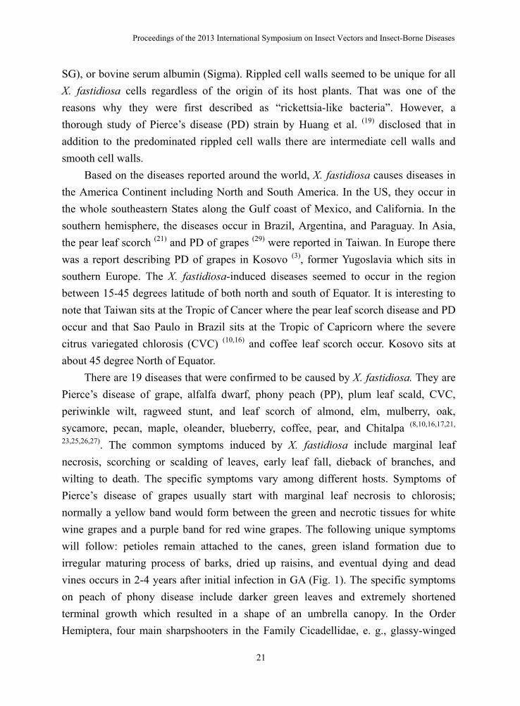
SG), or bovine serum albumin (Sigma). Rippled cell walls seemed to be unique for all X. fastidiosa cells regardless of the origin of its host plants. That was one of the reasons why they were first described as “rickettsia-like bacteria”. However, a thorough study of Pierce’s disease (PD) strain by Huang et al. (19) disclosed that in addition to the predominated rippled cell walls there are intermediate cell walls and smooth cell walls.
Based on the diseases reported around the world, X. fastidiosa causes diseases in the America Continent including North and South America. In the US, they occur in the whole southeastern States along the Gulf coast of Mexico, and California. In the southern hemisphere, the diseases occur in Brazil, Argentina, and Paraguay. In Asia, the pear leaf scorch (21) and PD of grapes (29) were reported in Taiwan. In Europe there was a report describing PD of grapes in Kosovo (3), former Yugoslavia which sits in southern Europe. The X. fastidiosa-induced diseases seemed to occur in the region between 15-45 degrees latitude of both north and south of Equator. It is interesting to note that Taiwan sits at the Tropic of Cancer where the pear leaf scorch disease and PD occur and that Sao Paulo in Brazil sits at the Tropic of Capricorn where the severe citrus variegated chlorosis (CVC) (10,16) and coffee leaf scorch occur. Kosovo sits at about 45 degree North of Equator.
There are 19 diseases that were confirmed to be caused by X. fastidiosa. They are Pierce’s disease of grape, alfalfa dwarf, phony peach (PP), plum leaf scald, CVC, periwinkle wilt, ragweed stunt, and leaf scorch of almond, elm, mulberry, oak, sycamore, pecan, maple, oleander, blueberry, coffee, pear, and Chitalpa (8,10,16,17,21,
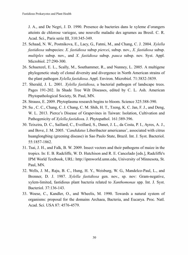
23,25,26,27). The common symptoms induced by X. fastidiosa include marginal leaf necrosis, scorching or scalding of leaves, early leaf fall, dieback of branches, and wilting to death. The specific symptoms vary among different hosts. Symptoms of Pierce’s disease of grapes usually start with marginal leaf necrosis to chlorosis; normally a yellow band would form between the green and necrotic tissues for white wine grapes and a purple band for red wine grapes. The following unique symptoms will follow: petioles remain attached to the canes, green island formation due to irregular maturing process of barks, dried up raisins, and eventual dying and dead vines occurs in 2-4 years after initial infection in GA (Fig. 1). The specific symptoms on peach of phony disease include darker green leaves and extremely shortened terminal growth which resulted in a shape of an umbrella canopy. In the Order Hemiptera, four main sharpshooters in the Family Cicadellidae, e. g., glassy-winged
Fastidious Prokaryotes and Plant Health
22
sharpshooter (GWSS), blue-green sharpshooter, red-headed sharpshooter, and green sharpshooter were the important vectors for PD X. fastidiosa and GWSS vectoring phony peach and plum leaf scald diseases as well.
CVC was first observed in 1987 on sweet orange trees in the southwestern part of Minas Gerais, Brazil. Since then, the disease has been observed in the neighboring State of San Paulo and other citrus producing states (10). Rossetti et al. (24) were the first to show by electron microscopy that a xylem-limited bacterium, probably a strain of X. fastidiosa, was present in all symptomatic leaves and fruits tested but not in similar tissues from symptomless trees.
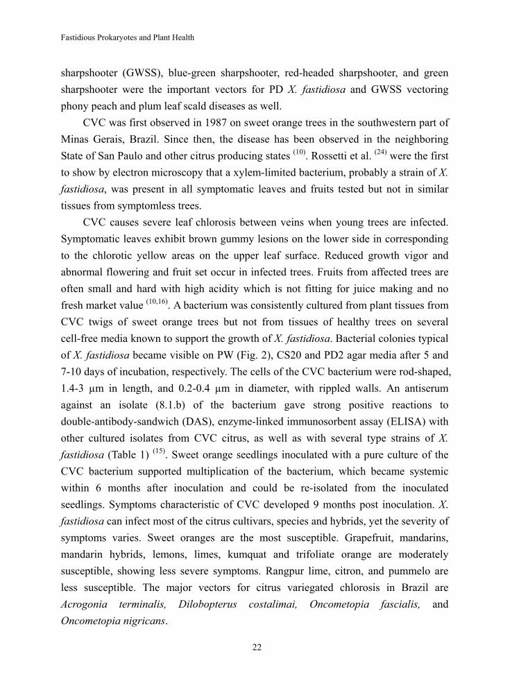
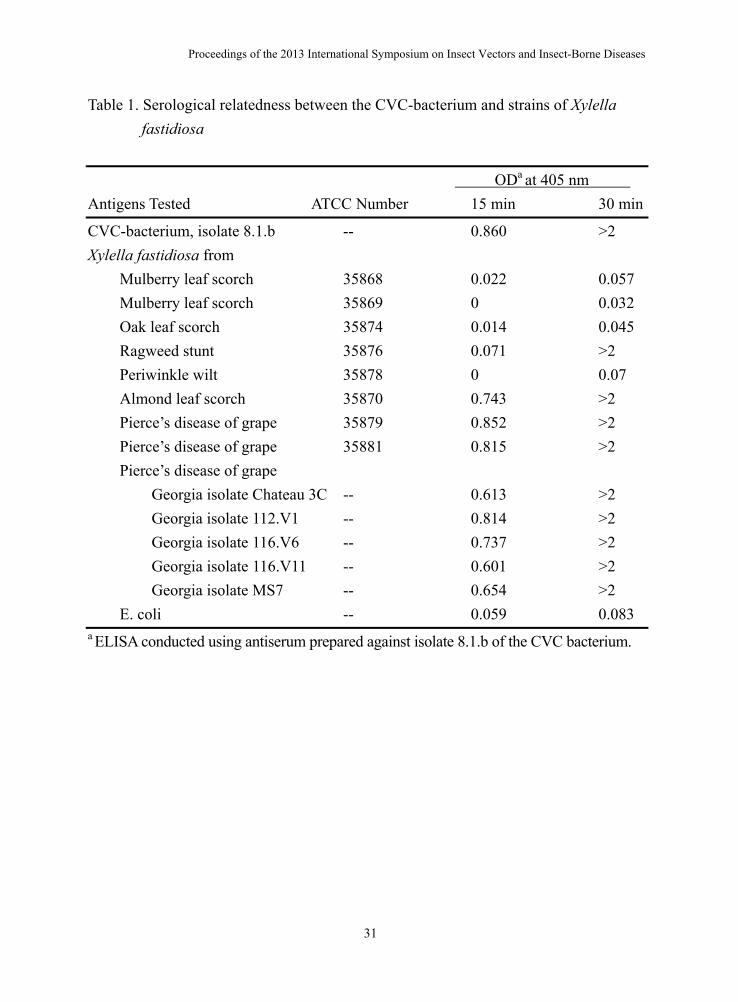
CVC causes severe leaf chlorosis between veins when young trees are infected. Symptomatic leaves exhibit brown gummy lesions on the lower side in corresponding to the chlorotic yellow areas on the upper leaf surface. Reduced growth vigor and abnormal flowering and fruit set occur in infected trees. Fruits from affected trees are often small and hard with high acidity which is not fitting for juice making and no fresh market value (10,16). A bacterium was consistently cultured from plant tissues from CVC twigs of sweet orange trees but not from tissues of healthy trees on several cell-free media known to support the growth of X. fastidiosa. Bacterial colonies typical of X. fastidiosa became visible on PW (Fig. 2), CS20 and PD2 agar media after 5 and 7-10 days of incubation, respectively. The cells of the CVC bacterium were rod-shaped, 1.4-3 µm in length, and 0.2-0.4 µm in diameter, with rippled walls. An antiserum against an isolate (8.1.b) of the bacterium gave strong positive reactions to double-antibody-sandwich (DAS), enzyme-linked immunosorbent assay (ELISA) with other cultured isolates from CVC citrus, as well as with several type strains of X. fastidiosa (Table 1) (15). Sweet orange seedlings inoculated with a pure culture of the CVC bacterium supported multiplication of the bacterium, which became systemic within 6 months after inoculation and could be re-isolated from the inoculated seedlings. Symptoms characteristic of CVC developed 9 months post inoculation. X. fastidiosa can infect most of the citrus cultivars, species and hybrids, yet the severity of symptoms varies. Sweet oranges are the most susceptible. Grapefruit, mandarins, mandarin hybrids, lemons, limes, kumquat and trifoliate orange are moderately susceptible, showing less severe symptoms. Rangpur lime, citron, and pummelo are less susceptible. The major vectors for citrus variegated chlorosis in Brazil are Acrogonia terminalis, Dilobopterus costalimai, Oncometopia fascialis, and Oncometopia nigricans.
Proceedings of the 2013 International Symposium on Insect Vectors and Insect-Borne Diseases
23
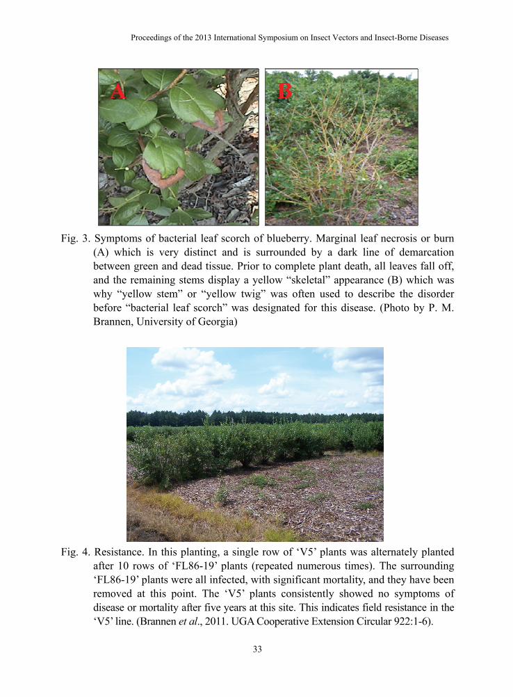
Relative to total sales, blueberries are the number one fruit commodity in the state of Georgia, surpassing even peaches. Production is concentrated in the southern coastal flatwoods. Rabbiteye blueberry (Vaccinium virgatum Aiton), a native species, has long been the predominant blueberry species cultivated in Georgia. More recently, however, growers have increased the production of the southern highbush cultivars (V. corymbosum interspecific hybrids) as a result of a very favorable market window. Growers and scientists started to observe a new disorder affecting the southern highbush selection FL 86-19 in the Georgia blueberry production region. An initial symptom was marginal leaf scorch (burn) of the older leaves which is very distinct and is surrounded by a dark line of demarcation between green and dead tissue (Fig. 3A), similar to that observed with extreme drought or fertilizer salt burn. New developing shoots were usually abnormally thin with a reduced number of flower buds. Leaf drop eventually occurred with young twigs or stems of the southern highbush selection FL 86-19 developing a yellow, “skeleton-like” appearance (Fig. 3B) which was why “yellow stem” or “yellow twig” was often used to describe the disorder. At this stage, the root system still appeared healthy, except for the possible loss of fine new roots. Whole plants or individual canes showed symptoms. The plant eventually died after leaf drop, typically during the second year of observation (11).
This prompted the enzyme-linked immunosorbent assay (ELISA) tests and isolations of X. fastidiosa. A single diseased blueberry bush of the selection FL 89-16 was excavated from a blueberry farm in South Georgia on 2 Feb. 2006. The bush was subsequently stored under cold room conditions (5 °C)-in a plastic trash bag to prevent moisture loss—until attempted detection of X. fastidiosa using direct isolation and ELISA tests (Agdia, Inc., Elkhart, IN). From this initial plant, two leaf and two root tissue samples were collected for isolation and ELISA testing on 2 Mar. 2006. The diseased blueberry bush was then moved to a greenhouse and planted in a 30.5-cm diameter pot. This original diseased plant was used to monitor the survival of the bacterium and symptom development on new growth after being stored for 48 d at 5 °C. ELISA results indicated all four tissues tested positive for the bacterial pathogen, X. fastidiosa, whereas only the two root tissues provided positive isolations. One leaf and one root tissue sample were later collected from each of five additional diseased plants for isolation and ELISA testing. Both isolation and ELISA testing methods obtained positive results. Cultures were multiplied to inoculate seedlings of three cultivars: ‘Southern Belle’ (eight plants), ‘Premier’ (six), and ‘Powderblue’ (six) on 23 May 2006
Fastidious Prokaryotes and Plant Health
24
and one selection, FL 86-19 (eight), on 31 May 2006. Two FL 86-19 plants started to show symptoms of marginal necrosis 54 days postinoculation , whereas one plant each of ‘Southern Belle’ and ‘Powderblue’ started to show symptoms of marginal necrosis 63 days postinoculation and ‘Premier’ stayed symptomless. All eight culture-inoculated FL 86-19 plants (100%) showed symptoms 72 days postinoculation, but no symptoms were observed on the control plants. One hundred twenty-six days postinoculation, two ‘Powderblue’ and four ‘Southern Belle’ plants showed mild symptoms, whereas all ‘Premier’ plants were asymptomatic. Positive reisolations of the bacteria from the inoculated symptomatic plants, not from asymptomatic plants, fulfilled Koch's postulates, which confirmed X. fastidiosa was the causal bacterium of the new blueberry disorder, the bacterial leaf scorch of blueberry.
This original blueberry bush provided valuable information on the survivability of the X. fastidiosa blueberry strain. The bacterium was able to survive at 5 °C for 48 d when the bush was kept in a plastic bag before being planted in a large pot and kept in the greenhouse. On 10 July 2006, tissues from this bush were collected for isolation and ELISA and the results were positive for both methods. The blueberry industry-particularly growers-in the southeastern United States will find this information especially important, because the research suggests that the bacteria is able to survive in the aboveground tissues through the south Georgia winter because it is unlikely for the temperature to remain at 5 °C 24 h a day for a consecutive 48 d in the winter. Furthermore, the source of inoculum for transmission would likely be available year-round (11).
By 3 months after initial inoculation, all eight X. fastidiosa-injected FL 86-19 plants showed symptoms, whereas all four PW medium-only-injected plants remained asymptomatic. For the other three cultivars, only two of six ‘Powderblue’ and four of eight ‘Southern Belle’ showed mild symptoms, whereas zero of six ‘Premier’ plants were symptomatic even at 4 months postinoculation. Both ELISA and direct isolations confirmed the presence of X. fastidiosa in symptomatic plants. Yellow stems or twigs were a strong symptomatic indicator of X. fastidiosa infection. There seemed to be a different degree of susceptibility among the three cultivars and one selection with selection FL 86-19 clearly being the most susceptible consistent with what had been observed in the field (11).
Further studies indicated that there is varietal resistance in some southern highbush blueberries. The FL 86-16 variety is particularly susceptible to infection. When compared with other southern highbush or rabbiteye varieties, the “V5” variety
Proceedings of the 2013 International Symposium on Insect Vectors and Insect-Borne Diseases
25
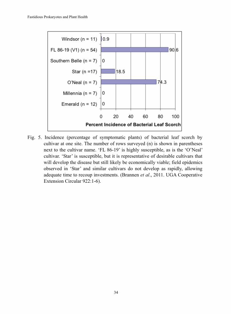
showed resistance to the bacterium (Fig. 4). This is encouraging, since it indicates that breeding can be utilized to develop varieties that are highly resistant to Xylella. Likewise, surveys have shown that there are other varieties that either do not develop symptoms or that slow epidemic spread of the disease (Fig. 5). ‘FL 86-19’ is highly susceptible, as is the ‘O’Neal’ cultivar. ‘Star’ is susceptible, but it is representative of desirable cultivars that will develop the disease but still likely be economically viable; field epidemics observed in ‘Star’ and similar cultivars do not develop as rapidly, allowing adequate time to recoup investments (6).
Insect vectors for the blueberry bacterial leaf scorch disease are under investigation in Georgia and the glassy-winged sharpshooter leafhopper, Homalodisca vitripennis (formerly H. coagulata), is likely an important suspect. Phloem-limited plant pathogenic prokaryotes
In Mollicutes, the cell wall-less and phloem-limited prokaryotes, there are two major plant pathogens: spiroplasmas and phytoplasmas. Spiroplasmas are cells with helical forms during log phase growth. Most spriroplasmas are cultivable in enriched media that contain supplemented sterols and other ingredients (9). They are facultative anaerobic or microaerophilic. They are associated with three plant diseases: citrus stubborn and horseradish brittle root disease by Spiroplasma citri, corn stunt disease by S. kunkelii, and periwinkle disease by S. phoeniceum. Phytoplasmas have been associated with more than 500 plant diseases worldwide (22) ever since the historical discovery by Doi et al. (14) of then referred as mycoplasma-like organisms (MLO) found in the pholem elements of plants infected with mulberry dwarf, potato witches’-broom, aster yellows, or paulownia witches’-broom. Phytoplasmas are still noncultivable even though they have been classified into 30 group-subgroups and four undetermined entities based on the 16S rDNA RFLP grouping (http: //plantpathology. ba. ars.usda.gov/pclass/pclass_taxonomy.html).
There were unintentional fans of phytoplasmas for as early as 1000 years ago in Song Dynasty and as recent as nowadays. When phytoplasmas infect peonies, the plants produced flowers not in the typical red or yellow colors, but in a delicate green we call virescence. The green flower was considered so attractive and valuable about 1000 years ago in China that the Song Dynasty’s imperial court received a special annual tribute composed of the blossoms. More recently, phytoplasmas have helped brighten winter holidays by transforming otherwise lanky poinsettias, with their eye-catching red
Fastidious Prokaryotes and Plant Health
26
leaves, into bushy ornamentals for their fans, lovers of Christmas decorations (28). But most effects of phytoplasmas on plants are not that appealing. They were
actually naively quite destructive and malicious. For example, they wither grapes in Europe and Australia; dwarf corn growth in South America; devastate pears and apples in the United States and Europe; destroy peanuts, sesame, and soybean in Asia; and sicken elms, coconuts, asters, and hydrangeas on multiple continents. Just one 2001 phytoplasma outbreak in apple trees caused a loss of about 25 million Euros in Germany and about 100 million Euros in Italy (28).
For all the destruction they inflict, you might expect that lots of big agricultural companies and respected academic labs have garnered ample amount of information about them. Well to the contrary, to this day, the inability to grow these bacteria outside plants or insects hinders efforts to get a handle on their biology and genomes despite the fact that plant pathologists had spent over half a century thinking that phytoplasmas were viruses.
Walnut witches’-broom disease was reported by Chang et al. (7) after the MLO particles were observed in the sieve cells of the symptomatic tissues collected from Griffin, GA. Abnormal proliferation of numerous small shoots with lighter green color which resembled the shape of a broom became evident in mid-July. The insect vector for this disease is still unknown even though DNA fragments were isolated and cloned from diseased walnut and later DNA probes were developed to monitor the seasonal occurrence of walnut witches'-broom MLO (12, 13). There are other economically important phytoplasma diseases, such as lethal yellowing of coconuts in Jamaica and lime witches’-broom in Oman and many others in Taiwan.
Recent finding of secreted AY-WB protein 11 (SAP11) by Bai et al (2) and secreted protein TENGU by Hoshi et al (18) seemed to suggest that phytoplasmas are finally giving up some of their secrets of how they used the proteins to modify the activity of plant genes that participate in the disease development. Some scientists are already contemplating whether they can create plants with only the positive attributes of an infection. Perhaps adding a single phytoplasma gene to a plant’s DNA could create bushy poinsettias or green peonies that don’t carry the annoying pathogens (28).
Spiroplasma citri causes stubborn disease on citrus and brittle root disease on horseradish. S. citri is transmitted in a propagative, circulative manner by several leafhoppers including Circulifer tenellus and Scaphytopius nitridus in citrus-growing regions of California and Arizona and C. haematoceps (syn. Neoaliturus haematoceps)
Proceedings of the 2013 International Symposium on Insect Vectors and Insect-Borne Diseases
27
in the Mediterranean region. The pathogen multiplies in the vector but no transovarial transmission occurs. Spatial and temporal analysis of CSD incidence indicate only primary spread occurring and no or very limited secondary spread (citrus to citrus). C. tenellus and N. haematoceps have a wide host range which includes many natural hosts of S. citri but citrus is a non host of these leafhoppers. Citrus becomes infected when inoculative Circulifer vectors feed temporarily on citrus during migratory flights (5).
Spiroplasma kunkelii causes characteristic small chlorotic stripes at the leaf bases of new leaves 25-30 days after initial inoculation. The chlorotic stripes fused together and extended toward the leaf tips with green spots and stripes exhibited on a chlorotic background. The infected plants are stunted due to much shorter internodes and a proliferation of secondary shoots in leaf axils; thus it is named corn stunt disease. Corn stunt disease is transmitted by Dalbulus maidis (DeLong and Wolcott) and D. elimatus (Ball) in nature whereas it can be transmitted experimentally by Graminella nigrifrons (Forbes), G. sonora (Ball), Stirellus bicolor (Van Duzee), Exitianus exitiosus (Uhler), and Euscelidius variegatus (Kirsch.) (31).
The other phloem-limited bacteria are the causal agent of Huanglongbing (HLB) and other diseases, Ca. Liberibacter spp. Striking symptoms of “yellow shoots” were often seen in sweet orange of young and high density orchard (1,000 trees per hectare). Two most characteristic symptoms of HLB are leaves with blotchy mottle and fruits with small size and color inversion (4). HLB are transmitted by psyllid vectors. In Asia, Southeast Asia, and Oceania, Diaphorina citri is the vector, Ca. L. asiaticus is the HLB agent, and both are heat tolerant (Asian form of HLB). In Africa and Madagascar island, Trioza erytreae is the vector, Ca. L. africanus is the HLB agent, and both are heat-sensitive (African form of HLB) (4). Another HLB agent, Ca. L. americanus, was found in 2004 in Sao Paulo State, Brazil (30) and 2005 in Florida, USA (20) and its vector is D. citri.
The phloem-limited plant pathogenic prokaryotes, phytoplasmas, spiroplasmas, and the pathogens (Cadidatus Liberibacter spp.) of Huanglongbing (HLB), are transmitted to plants by phloem-feeding insects belonging to the Order of Hemiptera.
LITERATURE CITED 1. Agrios, G. N. 2005. Plant Pathology, Fifth Edi. Elsevier Academic Press. Pages
639-642. 2. Bai, X., Chorrea, V. R., Toruno, T. Y., Ammar, E. D., Kimoun, S., and Hogenhout, S.
Fastidious Prokaryotes and Plant Health
28
A. 2009. AY-WB phytoplasma secretes a protein that targets plant cell nuclei. Mol. Plant Microb. Interact. 22:18-30.
3. Berisha, B., Chen, Y. D. Zhang, G. Y., Xu, B. Y., and Chen, T. A. 1998. Isolation of Pierce’s disease bacteria from grapevines in Europe. Euro. J. Plant Pathol. 104:427-433.
4. Bove, J. M. 2006. Huanglongbing: a destructive, newly-emerging century-old disease of citrus. J. Plant Pathol. 88:7-37.
5. Bove, J. M., Renaudin, J., Saillard, C., Foissac, X., and Garnier, M. 2003. Spiroplasma citri, a plant pathogenic mollicute: relationships with its two hosts, the plant and the leafhopper vector. Ann. Rev. Phytopath. 41:483-500.
6. Brannen, P. M., Krewer, G., Roland, B., Horton, D., and Chang, C. J. 2011. Bacterial leaf scorch of blueberry. CAES Publications, UGA Cooperative Extension Circular 922:1-6.
7. Chang, C. J., Impson, L., and Cunfer, B. 1986. Walnut witches'-broom disease in Georgia. Phytopathology 76:1139 (abstr).
8. Chang, C. J., and Walker, J. T. 1988. Bacterial leaf scorch of northern red oak: isolation, cultivation, and pathogenicity of a xylem-limited bacterium. Plant Dis. 72:730-733.
9. Chang, C. J. 1989. Nutrition and cultivation of spiroplasmas. In "The Mycoplasmas, Vol. 5" (R. F. Whitcomb and J. G. Tully, eds.), pp. 201-241. Academic Press, New York.
10. Chang, C. J., Garnier, M., Zreik, L., Rossetti, V., and Bove, J. M. 1993. Culture and serological detection of the xylem-limited bacterium causing citrus variegated chlorosis and its identification as a strain of Xylella fastidiosa. Curr. Microbiol. 27:137-142.
11. Chang, C. J., Donaldson, R., Brannen, P. M., Krewer, G., and Boland, B. 2009. Bacterial leaf scorch, a new blueberry disease caused by Xylella fastidiosa. HortScience 44:413-417.
12. Chen, J., Chang, C. J., Jarret, R. L., and Gawel, N. 1992. Isolation and cloning of DNA fragments from a mycoplasma-like organism associated with walnut witches'-broom disease. Phytopathology 82:306-309.
13. Chen, J., Chang, C. J., and Jarret, R. L. 1992. DNA probes as molecular markers to monitor the seasonal occurrence of walnut witches'-broom mycoplasmalike organism. Plant Dis. 76:1116-1119.
14. Doi, Y., Teranaka, M., Yora, K., and Asuyama, H. 1967. Mycoplasma or PLT group-like microorganisms found in the pholem elements of plants infected with
Proceedings of the 2013 International Symposium on Insect Vectors and Insect-Borne Diseases
29
mulberry dwarf, potato witches’-broom, aster yellows, or paulownia witches’-broom. Ann. Phytopathol. Soc. Jpn. 33:259-266.
15. Garnier, M., Chang, C. J., Zreik, L., Rossetti, V., and Bove, J. M. 1993. Citrus variegated chlorosis: serological detection of Xylella fastidiosa, the bacterium associated with the disease. In: Proc. 12th Conf. IOCV, IOCV, Riverside.
16. Hartung, J. S., Beretta, J., Brlansky, R. H., Spisso, J., and Lee, R. F. 1994. Citrus variegated chlorosis bacterium: axenic culture, pathogenicity, and serological relationships with other strains of Xylella fastidiosa. Phytopathology 84:591-597.
17. Hernandez-Martinez, R., de la Cerda, K. A., Costa, H. S., Cooksey, D. A., and Wong, F. P. 2007. Phylogenetic relationships of Xylella fastidiosa strains isolated from ornamentals in southern California. Phytopathology 97:857-864.
18. Hoshi, A., Oshima, K., Kakizawa, S., Ishii, Y., Ozeki, J., Hashimoto, M., Komatsu, K., Kagiwada, S., Yamaji, Y., and Namba, S. 2009. A unique virulence factor for proliferation and dwarfism in plants identified from a phytopathogenic bacterium. Proc. Natl. Acad. Sci. USA 106 (15):6416-6421.
19. Huang, P. Y., Milholland, R. D., and Daykin, M. E. 1986. Structural and morphological changes associated with the Pierce's disease bacterium in bunch and muscadine grape tissues. Phytopathology 76:1232-1238.
20. Irey, M. S., Gast, T., and Gottwald, T. R. 2006. Comparison of visual assessment and polymerase chain reaction assay testing to estimate the incidence of the hunaglongbing pathogen in commercial Florida citrus. Proc. Fla. State Hort. Soc. 119:89-93.
21. Leu, L. S., and Su, C. C. 1993. Isolation, cultivation, and pathogenicity of Xylella fastidiosa, the causal bacterium of pear leaf scorch disease in Taiwan. Plant Dis. 77: 642-646.
22. McCoy, R. E., Caudwell, A., Chang, C. J., Chen, T. A., Chiykowski, L. N., Cousin, M. T., Dale, J. L., de Leeuw, G. T. N., Golino, D. A., Hackett, K. J., Kirkpatrick, B. C., Marwitz, R., Petzold, H., Sinha, R. C., Suguira, M., Whitcomb, R. F., Yang, I. L., Zhu, B. M., and Seemuller, E. 1989. Plant diseases associated with mycoplasma-like organisms. In "The Mycoplasmas, Vol. 5" (R. F. Whitcomb and J. G. Tully, eds.), pp. 545-640. Academic Press, New York.
23. Randall, J. J., Goldberg, N. P., Kemp, J. D., Radionenko, M., French, J. M., Olsen, M. W., and Hanson, S. F. 2009. Genetic analysis of a novel Xylella fastidiosa subspecies found in the Southwestern United States. Appl. Environ. Microbiol. 75:5631-5638.
24. Rossetti, V., Garnier, M., Bove, J. M., Beretta, M. J. G., Teixeira, A. R. R., Quaggio,
Fastidious Prokaryotes and Plant Health
30
J. A., and De Negri, J. D. 1990. Presence de bacteries dans le xyleme d’orangers atteints de chlorose variegee, une nouvelle maladie des agrumes au Bresil. C. R. Acad. Sci., Paris serie III, 310:345-349.
25. Schaad, N. W., Postnikova, E., Lacy, G., Fatmi, M., and Chang, C. J. 2004. Xylella fastidiosa subspecies: X. fastidiosa subsp piercei, subsp. nov., X. fastidiosa subsp. multiplex subsp. nov., and X. fastidiosa subsp. pauca subsp. nov. Syst. Appl. Microbiol. 27:290-300.
26. Schuenzel, E. L., Scally, M., Southammer, R., and Nunney, L. 2005. A multigene phylogenetic study of clonal diversity and divergence in North American strains of the plant pathogen Xylella fastidiosa. Appl. Environ. Microbiol. 71:3832-3839.
27. Sherald, J. L. 2001. Xylella fastidiosa, a bacterial pathogen of landscape trees. Pages 191-202. In Shade Tree Wilt Diseases, edited by C. L. Ash. American Phytopathological Society, St. Paul, MN.
28. Strauss, E. 2009. Phytoplasma research begins to bloom. Science 325:388-390. 29. Su , C. C., Chang, C. J. Chang, C. M. Shih, H. T., Tzeng, K. C. Jan, F. J., and Deng,
W. L. 2013. Pierce’s Disease of Grapevines in Taiwan: Isolation, Cultivation and Pathogenicity of Xylella fastidiosa. J. Phytopathol. 161:389-396.
30. Teixeira, D. C., Saillard, C., Eveillard, S., Danet, J. L., da Costa, P. I., Ayres, A. J., and Bove, J. M. 2005. ‘Candidatus Liberibacter americanus’, associated with citrus huanglongbing (greening disease) in Sao Paulo State, Brazil. Int. J. Syst. Bacteriol. 55:1857-1862.
31. Tsai, J. H., and Falk, B. W. 2009. Insect vectors and their pathogens of maize in the tropics. In: E. B. Radcliffe, W. D. Hutchison and R. E. Cancelado [eds.], Radcliffe's IPM World Textbook, URL: http://ipmworld.umn.edu, University of Minnesota, St. Paul, MN.
32. Wells, J. M., Raju, B. C., Hung, H. Y., Weisburg, W. G., Mandelco-Paul, L., and Brenner, D. J. 1987. Xylella fastidiosa gen. nov., sp. nov: Gram-negative, xylem-limited, fastidious plant bacteria related to Xanthomonas spp. Int. J. Syst. Bacteriol. 37:136-143.
33. Woese, C., Kandler, O., and Wheelis, M. 1990. Towards a natural system of organisms: proposal for the domains Archaea, Bacteria, and Eucarya. Proc. Natl. Acad. Sci. USA 87: 4576-4579.
Proceedings of the 2013 International Symposium on Insect Vectors and Insect-Borne Diseases
31
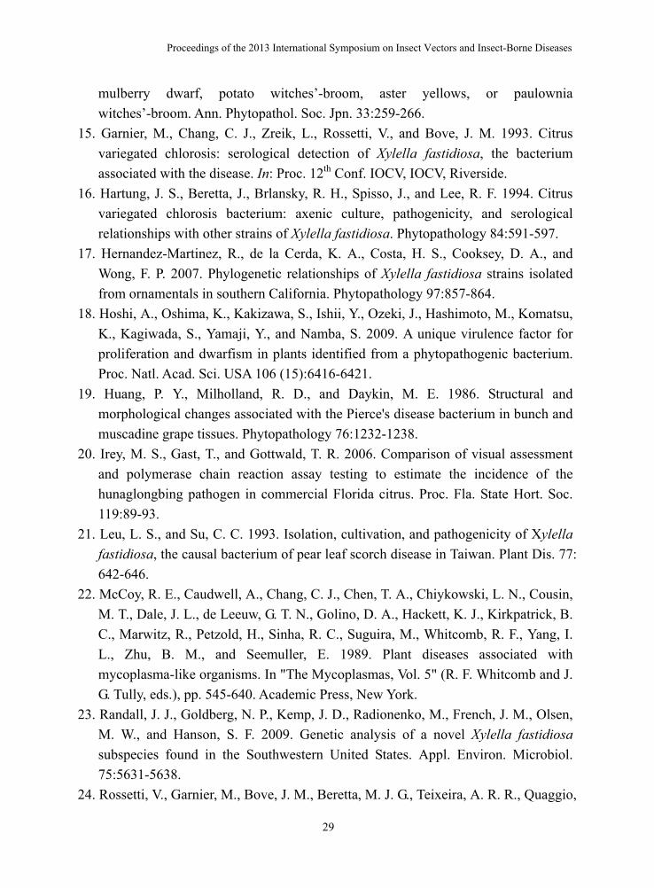
Table 1. Serological relatedness between the CVC-bacterium and strains of Xylella fastidiosa
ODa at 405 nm Antigens Tested ATCC Number 15 min 30 min
CVC-bacterium, isolate 8.1.b -- 0.860 >2 Xylella fastidiosa from Mulberry leaf scorch 35868 0.022 0.057 Mulberry leaf scorch 35869 0 0.032 Oak leaf scorch 35874 0.014 0.045 Ragweed stunt 35876 0.071 >2 Periwinkle wilt 35878 0 0.07 Almond leaf scorch 35870 0.743 >2 Pierce’s disease of grape 35879 0.852 >2 Pierce’s disease of grape 35881 0.815 >2 Pierce’s disease of grape Georgia isolate Chateau 3C -- 0.613 >2 Georgia isolate 112.V1 -- 0.814 >2 Georgia isolate 116.V6 -- 0.737 >2 Georgia isolate 116.V11 -- 0.601 >2 Georgia isolate MS7 -- 0.654 >2 E. coli -- 0.059 0.083 a ELISA conducted using antiserum prepared against isolate 8.1.b of the CVC bacterium.
Fastidious Prokaryotes and Plant Health
32
Fig. 1. Symptoms of Pierce’s disease of grapes: A close-up view of marginal leaf
necrosis (A), petioles remained attached to the canes after leaves fall (B), green island (C) formed due to irregular maturing process of barks, and dried up raisins (D), and eventual dying and dead vines in 2-4 years after initial infection in GA, USA. (Photo by Chung-Jan Chang)
Fig. 2. Colonies of the CVC-bacterium obtained from a drop of tissue homogenate on
PW 9 days after inoculation ( x 16). (photo by Chung-Jan Chang).
Proceedings of the 2013 International Symposium on Insect Vectors and Insect-Borne Diseases
33
Fig. 3. Symptoms of bacterial leaf scorch of blueberry. Marginal leaf necrosis or burn
(A) which is very distinct and is surrounded by a dark line of demarcation between green and dead tissue. Prior to complete plant death, all leaves fall off, and the remaining stems display a yellow “skeletal” appearance (B) which was why “yellow stem” or “yellow twig” was often used to describe the disorder before “bacterial leaf scorch” was designated for this disease. (Photo by P. M. Brannen, University of Georgia)
Fig. 4. Resistance. In this planting, a single row of ‘V5’ plants was alternately planted
after 10 rows of ‘FL86-19’ plants (repeated numerous times). The surrounding ‘FL86-19’ plants were all infected, with significant mortality, and they have been removed at this point. The ‘V5’ plants consistently showed no symptoms of disease or mortality after five years at this site. This indicates field resistance in the ‘V5’ line. (Brannen et al., 2011. UGA Cooperative Extension Circular 922:1-6).
Fastidious Prokaryotes and Plant Health
34
Fig. 5. Incidence (percentage of symptomatic plants) of bacterial leaf scorch by
cultivar at one site. The number of rows surveyed (n) is shown in parentheses next to the cultivar name. ‘FL 86-19’ is highly susceptible, as is the ‘O’Neal’ cultivar. ‘Star’ is susceptible, but it is representative of desirable cultivars that will develop the disease but still likely be economically viable; field epidemics observed in ‘Star’ and similar cultivars do not develop as rapidly, allowing adequate time to recoup investments. (Brannen et al., 2011. UGA Cooperative Extension Circular 922:1-6).
Related Documents