Research Familiarity with a vocal category biases the compartmental expression of Arc/Arg3.1 in core auditory cortex Tamara N. Ivanova, 1 Christina Gross, 2,3,5 Rudolph C. Mappus, 1 Yong Jun Kwon, 1,4 Gary J. Bassell, 2,3 and Robert C. Liu 1,3 1 Department of Biology, Emory University, Atlanta, Georgia 30322, USA; 2 Department of Cell Biology, Emory University School of Medicine, Atlanta, Georgia 30322, USA; 3 Center for Translational Social Neuroscience, Emory University, Atlanta, Georgia 30322, USA; 4 Graduate Program in Neuroscience, Laney Graduate School, Emory University, Atlanta, Georgia 30322, USA Learning to recognize a stimulus category requires experience with its many natural variations. However, the mechanisms that allow a category’s sensorineural representation to be updated after experiencing new exemplars are not well under- stood, particularly at the molecular level. Here we investigate how a natural vocal category induces expression in the audi- tory system of a key synaptic plasticity effector immediate early gene, Arc/Arg3.1, which is required for memory consolidation. We use the ultrasonic communication system between mouse pups and adult females to study whether prior familiarity with pup vocalizations alters how Arc is engaged in the core auditory cortex after playback of novel exem- plars from the pup vocal category. A computerized, 3D surface-assisted cellular compartmental analysis, validated against manual cell counts, demonstrates significant changes in the recruitment of neurons expressing Arc in pup-experienced animals (mothers and virgin females “cocaring” for pups) compared with pup-inexperienced animals (pup-naı ¨ve virgins), especially when listening to more familiar, natural calls compared to less familiar but similarly recognized tonal model calls. Our data support the hypothesis that the kinetics of Arc induction to refine cortical representations of sensory cate- gories is sensitive to the familiarity of the sensory experience. The ability to recognize novel exemplars of behaviorally relevant sensory categories is critical to navigating our environment and communicating with others. A sensory system must presumably assess whether a stimulus matches the collection of features that statistically define a recognized category. At the same time, varia- tion present in new exemplars must themselves be incorporated into the sensory system’s representation of that category. The neu- ral mechanisms that underlie such categorization at the electro- physiological level are being elucidated in both the visual and auditory modalities (DiCarlo et al. 2012; Bizley and Cohen 2013; Shepard et al. 2015), but the molecular mechanisms that support the learning and memory of sensory categories remain poorly understood. One promising molecular coordinator of activity-dependent changes in synaptic efficacy, which has been suggested to underlie learning and memory, is the effector immediate early gene (IEG) Arc/Arg3.1 (Arc),a “master regulator” of synaptic plasticity during information processing (Shepherd and Bear 2011). Arc is expressed in hippocampal neurons that then undergo synaptic plasticity as a result of a behavioral experience (Guzowski et al. 1999; Vazdarja- nova and Guzowski 2004; Fletcher et al. 2006). It can also be in- duced in sensory systems after sensory experiences (Tagawa et al. 2005; Carpenter-Hyland et al. 2010; Ivanova et al. 2011; Morin et al. 2011). Moreover, the levels of Arc expression correlate with hippocampus-dependent learning (Guzowski et al. 2001), plays es- sential roles in homeostatic plasticity and the long-term consolida- tion of memories (Gao et al. 2010). Recent studies suggest this may stem from its apparent preferential “inverse tagging” in activated neurons of the subset of inactive synapses, which then undergo long-term depression by endocytosis of AMPA receptors (Waung et al. 2008; Okuno et al. 2012). Arc’s action explains how repeated experiences with the same visual stimulus leads to a progressively smaller population of visual cortical neurons that is more robustly activated by that stimulus (Wang et al. 2006), thereby helping to refine the stimulus specificity of sensory neurons. In the real world though, the exact same experience or stimulus is not usually en- countered. Instead, natural stimuli are more likely to fall into the same behaviorally relevant stimulus category, but differ in their physical detail from exemplar to exemplar. Given Arc’s role in re- fining synaptic connectivity for a neuron’s stimulus specificity, how might it be deployed to support plasticity for stimulus gener- alization to novel exemplars of a familiar stimulus category? We address this question in the context of natural auditory categorization of behaviorally relevant communication sounds in a mouse model. Maternal females recognize the ultrasonic vocali- zations (USV) of displaced mouse pups as behaviorally important, and will seek out and retrieve the lost pup to the nest. On the other hand, nonmaternal, virgin females do not show a behavioral pref- erence for pup calls over neutral sounds, until they have had 5 Present address: Division of Neurology, Cincinnati Children’s Hospital Medical Center, Cincinnati, OH 45229, USA. Corresponding author: [email protected] # 2017 Ivanova et al. This article is distributed exclusively by Cold Spring Harbor Laboratory Press for the first 12 months after the full-issue publication date (see http://learnmem.cshlp.org/site/misc/terms.xhtml). After 12 months, it is available under a Creative Commons License (Attribution-NonCommercial 4.0 International), as described at http://creativecommons.org/licenses/by-nc/ 4.0/. Article is online at http://www.learnmem.org/cgi/doi/10.1101/lm.046086. 117. 24:612–621; Published by Cold Spring Harbor Laboratory Press ISSN 1549-5485/17; www.learnmem.org 612 Learning & Memory Cold Spring Harbor Laboratory Press on December 5, 2021 - Published by learnmem.cshlp.org Downloaded from

Welcome message from author
This document is posted to help you gain knowledge. Please leave a comment to let me know what you think about it! Share it to your friends and learn new things together.
Transcript
Research
Familiarity with a vocal category biases thecompartmental expression of Arc/Arg3.1in core auditory cortex
Tamara N. Ivanova,1 Christina Gross,2,3,5 Rudolph C. Mappus,1 Yong Jun Kwon,1,4
Gary J. Bassell,2,3 and Robert C. Liu1,31Department of Biology, Emory University, Atlanta, Georgia 30322, USA; 2Department of Cell Biology, Emory University School ofMedicine, Atlanta, Georgia 30322, USA; 3Center for Translational Social Neuroscience, Emory University, Atlanta, Georgia 30322, USA;4Graduate Program in Neuroscience, Laney Graduate School, Emory University, Atlanta, Georgia 30322, USA
Learning to recognize a stimulus category requires experience with its many natural variations. However, the mechanisms
that allow a category’s sensorineural representation to be updated after experiencing new exemplars are not well under-
stood, particularly at the molecular level. Here we investigate how a natural vocal category induces expression in the audi-
tory system of a key synaptic plasticity effector immediate early gene, Arc/Arg3.1, which is required for memory
consolidation. We use the ultrasonic communication system between mouse pups and adult females to study whether
prior familiarity with pup vocalizations alters how Arc is engaged in the core auditory cortex after playback of novel exem-
plars from the pup vocal category. A computerized, 3D surface-assisted cellular compartmental analysis, validated against
manual cell counts, demonstrates significant changes in the recruitment of neurons expressing Arc in pup-experienced
animals (mothers and virgin females “cocaring” for pups) compared with pup-inexperienced animals (pup-naı̈ve virgins),especially when listening to more familiar, natural calls compared to less familiar but similarly recognized tonal model
calls. Our data support the hypothesis that the kinetics of Arc induction to refine cortical representations of sensory cate-
gories is sensitive to the familiarity of the sensory experience.
The ability to recognize novel exemplars of behaviorally relevantsensory categories is critical to navigating our environment andcommunicating with others. A sensory system must presumablyassess whether a stimulus matches the collection of features thatstatistically define a recognized category. At the same time, varia-tion present in new exemplars must themselves be incorporatedinto the sensory system’s representation of that category. The neu-ral mechanisms that underlie such categorization at the electro-physiological level are being elucidated in both the visual andauditory modalities (DiCarlo et al. 2012; Bizley and Cohen 2013;Shepard et al. 2015), but the molecular mechanisms that supportthe learning and memory of sensory categories remain poorlyunderstood.
One promising molecular coordinator of activity-dependentchanges in synaptic efficacy, which has been suggested to underlielearning and memory, is the effector immediate early gene (IEG)Arc/Arg3.1 (Arc), a “master regulator” of synaptic plasticity duringinformation processing (Shepherd and Bear 2011). Arc is expressedin hippocampal neurons that then undergo synaptic plasticity as aresult of a behavioral experience (Guzowski et al. 1999; Vazdarja-nova and Guzowski 2004; Fletcher et al. 2006). It can also be in-duced in sensory systems after sensory experiences (Tagawa et al.2005; Carpenter-Hyland et al. 2010; Ivanova et al. 2011; Morinet al. 2011). Moreover, the levels of Arc expression correlate withhippocampus-dependent learning (Guzowski et al. 2001), plays es-
sential roles in homeostatic plasticity and the long-term consolida-tion of memories (Gao et al. 2010). Recent studies suggest this maystem from its apparent preferential “inverse tagging” in activatedneurons of the subset of inactive synapses, which then undergolong-term depression by endocytosis of AMPA receptors (Waunget al. 2008; Okuno et al. 2012). Arc’s action explains how repeatedexperiences with the same visual stimulus leads to a progressivelysmaller population of visual cortical neurons that is more robustlyactivated by that stimulus (Wang et al. 2006), thereby helping torefine the stimulus specificity of sensory neurons. In the real worldthough, the exact same experience or stimulus is not usually en-countered. Instead, natural stimuli are more likely to fall into thesame behaviorally relevant stimulus category, but differ in theirphysical detail from exemplar to exemplar. Given Arc’s role in re-fining synaptic connectivity for a neuron’s stimulus specificity,how might it be deployed to support plasticity for stimulus gener-alization to novel exemplars of a familiar stimulus category?
We address this question in the context of natural auditorycategorization of behaviorally relevant communication sounds ina mouse model. Maternal females recognize the ultrasonic vocali-zations (USV) of displaced mouse pups as behaviorally important,and will seek out and retrieve the lost pup to the nest. On the otherhand, nonmaternal, virgin females do not show a behavioral pref-erence for pup calls over neutral sounds, until they have had
5Present address: Division of Neurology, Cincinnati Children’sHospital Medical Center, Cincinnati, OH 45229, USA.Corresponding author: [email protected]
# 2017 Ivanova et al. This article is distributed exclusively by Cold SpringHarbor Laboratory Press for the first 12 months after the full-issue publicationdate (see http://learnmem.cshlp.org/site/misc/terms.xhtml). After 12 months,it is available under a Creative Commons License (Attribution-NonCommercial4.0 International), as described at http://creativecommons.org/licenses/by-nc/4.0/.
Article is online at http://www.learnmem.org/cgi/doi/10.1101/lm.046086.117.
24:612–621; Published by Cold Spring Harbor Laboratory PressISSN 1549-5485/17; www.learnmem.org
612 Learning & Memory
Cold Spring Harbor Laboratory Press on December 5, 2021 - Published by learnmem.cshlp.orgDownloaded from
experience “cocaring” for pups—suggesting that the behavioralrelevance of pup USVs is acquired (Ehret et al. 1987; Lin et al.2013). These whistle-like calls are naturally variable along variousacoustic dimensions, such as frequency, duration, frequency mod-ulation, and bandwidth (Liu et al. 2003; Grimsley et al. 2011), butnevertheless form a distinct acoustic category that can be categor-ically perceived by maternal mice (Ehret and Haack 1981; Ehret1992). Importantly, excitatory plasticity in a subset of putative py-ramidal neurons in the core auditory cortex of maternal femalesenhances the spiking response to favor the combination of acous-tic features that are statistically predictive of the pup USV category,an effect not observed in nonmaternal virgins (Shepard et al.2015). Hence, for maternal but not nonmaternal mice, pup USVsform a familiar, behaviorally relevant sound category that core au-ditory cortical activity “learns” to systematically differentiate fromother sound categories.
The mounting electrophysiological evidence for plasticity inauditory cortex in this vocalization recognition paradigm providesa basis for studying the molecular mechanisms involved (Liu andSchreiner 2007; Galindo-Leon et al. 2009; Cohen et al. 2011; Linet al. 2013; Marlin et al. 2015). In particular, it opens a unique op-portunity to investigate in a natural context how Arc is expressedin auditory cortex when new sounds (models of pup USVs) eitherdo (maternal mice) or do not (nonmaternal mice) conform to a fa-miliar, behaviorally relevant category. We assessed this using a cellcompartmental analysis of Arc gene transcription by fluorescent insitu hybridization and 3D surface reconstruction (Guzowski et al.1999; Ivanova et al. 2011).We have previously used thesemethodsto demonstrate that prior history with a pure tone can shift howthat tone induces compartmental Arc expression in core auditorycortical neurons, such that, soon after stimulation, Arc is foundmore often in just the perinuclear cytoplasmic (perinuc) compart-
ment in mice that previously heard the exact same tone comparedwith those that had not.
To further test and extend our interpretation of this stimulus“familiarity” effect in compartmental Arc expression, we now askin a completely different auditory paradigm—the maternal pupUSV recognition model—whether the same bias for perinuc-onlyexpression can be induced by novel exemplars containing acousticfeatures congruent with a natural sound category that is either fa-miliar or not familiar to the animal. Our data suggest that themoresimilar the combination of acoustic features in the exemplars is to afamiliar, behaviorally relevant category, the earlier a bias forperinuc-only Arc expression is observed in core auditory corticalneurons.
Results
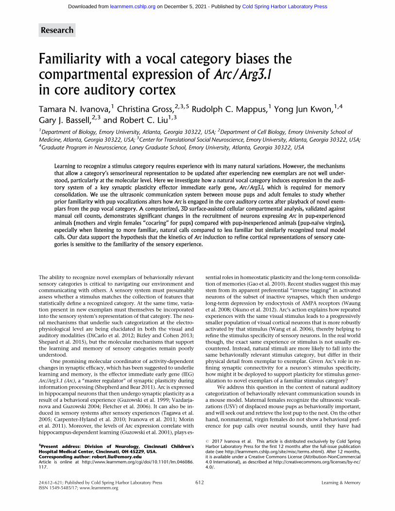
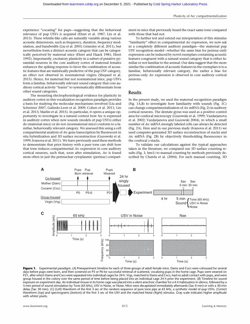
In the present study, we used the maternal recognition paradigm(Fig. 1A,B) to investigate how familiarity with sounds (Fig. 1C)can change compartmentalization ofArcmRNA (Fig. 2) in auditorycortical neurons. The dentate gyrus was used as a positive controlarea for confocal microscopy (Guzowski et al. 1999; Vazdarjanovaet al. 2002; Vazdarjanova and Guzowski 2004), in which a smallnumber of Arc mRNA strongly labeled cells can always be detected(Fig. 2A). Here and in our previous study (Ivanova et al. 2011) weused computer-generated 3D surface reconstruction of nuclei andArc mRNA (Fig. 2B) by objectively thresholding fluorescence inthe confocal z-stacks.
To validate our calculations against the typical approachestaken in the literature, we compared our 3D surface counting re-sults (Fig. 3, Ims1) to manual counting by methods previously de-scribed by Chawla et al. (2004). For such manual counting, 3D
5 minSound
4 hr Tone (65 kHz)USV in NoiseNoise
Sac0 min
Sac30 min
Virgin (Vrg)Group-housed
Mother (Dam)Co-housed
Cocarer (Coc)
PupsBorn
P0 P5-6 P21
A BPupretrieval
PupsWeaned
24 hrsingly
housed
Experiment
{
Freq
uenc
y (k
Hz)
Am
plitu
de (a
rb)
40
60
80
Time (s)
Time (s) Time (s)
65 kHz USV in Noise Nse
0 1 2 3 0 1 2 3
0 1 2 30 1 2 3 0 1 2 3
100
C
Figure 1. Experimental paradigm. (A) Preexperiment timeline for each of three groups of adult female mice. Dams and Cocs were cohoused for severaldays before pups were born, and then screened on P5 or P6 for successful retrieval of scattered, vocalizing pups in the home cage. Pups were weaned onP21, after which Dams and Cocs were separated into individual cages for 24 h. Vrgs, matched to Dams and Cocs, had no adult contact with pups, and weregroup housed in the colony over the same period of time before being placed into an individual cage 24 h prior the experiment. (B) Timeline for soundexposure on experiment day. An individual mouse in its home cage was placed into a silent anechoic chamber for a 4-h habituation in silence, followed by a5-min period of sound stimulation by Tone (65 kHz), USV in Noise, or Noise. Mice were decapitated immediately afterwards (Sac 0 min) or with a 30-mindelay (Sac 30 min). (C) (Left) Waveform of the first 3 sec of the random sequence of pure tone pips at 65 kHz, a synthetic model of pup USVs. (Center)Waveform (top) and spectrograms (bottom) of the first 3 sec of the USV and the matched Noise (Right) stimulus. Gray scale indicates higher amplitudewith whiter pixels.
Plasticity of Arc compartmentalization
www.learnmem.org 613 Learning & Memory
Cold Spring Harbor Laboratory Press on December 5, 2021 - Published by learnmem.cshlp.orgDownloaded from

surfaces were not generated, and instead we manually countedDAPI nuclei and looked for areas ofArc fluorescence on consecutiveplanes of each z-stack (13 z-stacks; 777 cells; 8 animals). Thesemanual counts (M) of Arc-positive (Foci-only, Perinuc-only, andBoth) cells were compared with those determined by our3D-assisted protocol (seeMaterials andMethods, Fig. 3A). For a nu-cleus to be designated as having Arc foci by the manual countingmethod, it had to exhibit Arc fluorescence in at least three consec-utive planes, while cells with Arc signal spread around the nucleuswere designated as having perinuclear cytoplasmic expression.Additionally, we compared our 3D-calculations for the samez-stacks using two different thresholds, with Ims2’s threshold forArc signal fluorescence increased by 10%.
Even though there were not largemean differences in the per-centages of cells counted by the two different methods (Fig. 3A),there were subtle but systematic differences apparent from pairedcomparisons. In particular, relative to the manual counts, smallpoints of Arc fluorescence within nuclei tended to be slightly over-counted as foci of Arc by the 3D reconstruction (∼1%, Fig. 3B,Foci-only, Ims1-M). This could be explained by the intensitythreshold used for generating the 3D surfaces, since an increaseof 10% in the threshold decreased the foci-only counts and elimi-nated the discrepancy between the 3D versus manual countingmethods (Fig. 3B, Foci-only, Ims2-M). However, changing thisthreshold undercounted (albeit not significantly) neurons with
Both foci and perinuclear cytoplasmic expression (Fig. 3B, Both,Ims2-Ims1, and Ims2-M), resulting in an overall significant under-count for Arc-positive neurons (Fig. 3B, Arc-positive, Ims2-Ims1,and Ims2-M). No effect on Perinuc-only counts was found forsuch a change in threshold, consistent with those neurons’ expres-sion primarily being determined by large regions of Arc fluores-cence. These results imply that there are tradeoffs in choosingthresholds for what is considered nonbackground expression.Importantly, the use of the computer-generated 3D surfaces makesthese tradeoffs objective and explicit rather than subjective, andshould enhance reproducibility of Arc counting. In any case, forall analyses presented here, cell count percentages based on 3D sur-faces are compared across groups treated equally, minimizing theeffect that any systemic bias would have in altering ourconclusions.
Moving beyondmethodological validation,we turnednext tomeasuring expression differences dependent on familiarity withthe acoustic features of pup USVs. A key acoustic feature character-izing the pup USV category is call frequency (Liu et al. 2003), a pa-rameter that maternal mice can use as a basis for their categoricalperception of USVs (Ehret and Haack 1982). We therefore startedby asking whether playback of pure tones at 65 kHz (Fig. 1C,left), the approximate frequencymost commonly found in naturalCBA/CaJ mouse pup calls (Liu et al. 2003), would induce Arc ex-pression differently in core auditory cortical neurons of Dams ver-sus Vrgs. The stimulus consisted of randomly generated sequenceof 65 kHz pure tone-pip bouts, with temporal properties (pip dura-tion, intercall interval, interbout interval, and number of calls in about) spanning their ranges found in natural USV bouts (Fig. 1B,center). For both animal groups (Dam andVrg), this particular syn-thetic tonal stimulus had never been encountered previously,though for the Dam, the tone frequency’s closeness to naturalUSVs could affect its perception as pup-like (Ehret & Haack1982). After animals were given a 4-h period of silence in an an-echoic chamber (Fig. 1B), they were either held in silence for an ad-ditional 30 min (Silence group), or were played a 5-min, 65 kHzbout of tone stimuli before being sacrificed immediately (0 min),or after a delay in silence (30 min).
Silence itself was associated with ∼16% Arc-positive expres-sion (Foci-only + Both + Perinuc-only) in auditory cortex in bothDams and Vrgs, indicating a relatively high basal level of Arc ex-pression in cortical layers III–VI (Fig. 4A, left bars). Tonal 65 kHzstimulation led to a main effect of time in increasing the percent-age of total Arc-positive neurons (time: F(2,113) = 69.53, P = 2.1 ×10⁻²⁰, 2 × 3 ANOVA). However, there was nomain effect of animalgroup and no interaction between time and animal group (animal:F(1,113) = 0.07, P = 0.80; time × animal: F(2,113) = 0.31, P = 0.73, 2×3ANOVA). By the 30-min post-stimulus time point, Arc mRNA wasexpressed in nearly 60% of core auditory cortical neurons irrespec-tive of animal group (Fig. 4A, right bars), affirming sound-inducedexpression of Arc. Significant post hoc comparisons are shown inFigure 4A.
For cells expressingArc only as Foci in the nucleus (Fig. 4B), 65kHz tone stimulation led to a main effect of time but not animalgroup, and only a trending interaction (time: F(2,113) = 4.85, P =0.0096; animal: F(1,113) = 2.19, P = 0.14; time × animal: F(2,113) =2.86, P = 0.06, 2×3 ANOVA). At the 0-min time point (Fig. 4B, mid-dle bars), the soundhad elevated Foci-only expression significantlyin Vrgs (P = 0.0138, Tukey) and was trending in Dams (P = 0.071,Tukey). Intriguingly, the Foci-only expression in Vrgs increasedby 30-min time point compared with silence level (P = 0.0185,Tukey), but no such increase was observed in Dams (P = 1.0,Tukey).
Next, the percentage of neurons with Arc expressed only inthe perinuclear cytoplasm following 65 kHz stimulation showeda significant main effect of time, animal group, and interaction
Peri-nuc
Imaris 3D z-stack Reconstruction
Consecutive sections of example z-stack B
A Arc mRNA distribution in DG
Overlay Nuclei Arc-positive cells
Figure 2. Neuron classification based on cellular compartmental expres-sion of Arc. (A) The typical ArcmRNA distribution in the dentate gyrus (DG)at 30 min after 5 min sound stimulation, providing a positive control brainarea for confocal microscopy. Blue represents DAPI staining of nuclei;green represents Arc/Arg3.1 mRNA expression. (B) (Left column) 3Dsurface reconstructions by IMARIS. Blue represents DAPI staining ofnuclei; green represents Arc/Arg3.1 mRNA expression. Arc-positiveneurons were classified as “Foci-only” (red arrow) if they contained intra-nuclear fluorescent surfaces only (top); “Perinuc-only” if they containedperinuclear fluorescent surfaces (yellow arrow) only (bottom); and“Both” if they had intranuclear and perinuclear surfaces overlappingDAPI surface (middle). (Middle column) Confocal z-stack (63× magnifica-tion) of the corresponding tissue section (20 µm; ∼30–34 optical sections)subjected to FISH. (Right three columns) Sequential sections of the z-stackshowing “Foci-only” (top, sections 13–12–11), “Perinuc-only” (bottom,sections 18–17–16), and “Both” (middle, sections 15–14–13).
Plasticity of Arc compartmentalization
www.learnmem.org 614 Learning & Memory
Cold Spring Harbor Laboratory Press on December 5, 2021 - Published by learnmem.cshlp.orgDownloaded from

between time and animal group (Fig. 4C; time: F(2,113) = 67.64, P =2.1 × 10⁻²⁰; animal: F(1,113) = 5.61, P = 0.0195; time × animal:F(2,113) = 7.69, P = 0.0007, 2×3 ANOVA). At 0min immediately afterthe stimulation, Perinuc-only expression was low, and did not sig-nificantly differ between the groups (Vrg 0 min: 3.9 ± 2.2%, Dam 0min: 3.3 ± 2.5%, P = 1.0, Tukey). By 30-min though, the percentageof Perinuc-only neurons rose in both animal groups, but Dams hada higher percentage compared with Vrgs (Dam: 30.9 ± 2.5%, Vrg:17.6 ± 2.2%, P = 2.7 × 10⁻⁴, Tukey). Hence, even though totalArc-positive expression did not differ be-tween animal groups at any time point(Fig. 4A), the cell compartmental compo-sition of this total expression did (Fig. 4B,C). Under the assumption that 65 kHztones sound sufficiently like pup USVsto mothers (Ehret and Haack 1982), thisdifference in expression is consistentwith the interpretation that a larger pop-ulation of neurons with Perinuc-onlyArc expression provides a molecular traceofwhether newexemplars soundmore fa-miliar (Ivanova et al. 2011).
However, the fact that the expres-sion difference was observed at the30-min but not at 0-min time point raisessome questions about the above interpre-tation, since we previously observed anincrease in Perinuc-only expression0-min after rehearing a (slightly longer)10-min familiar stimulus (Ivanova et al.2011). Therefore, as an independenttest, our next experiment exposed ani-mals to 5 min of either prerecorded pupUSVs (Fig. 1B, center), or to the control,microphone noise (Nse) from the record-
ings (Fig. 1B, right), and examined Arc ex-pression immediately afterwards. Naturalpup USVs should sound more familiar toand be recognized by mothers but notbynaïve virgins, even if the specific exem-plars are novel.
Overall Arc-positive expression wasslightly higher for the USV comparedwith Nse stimulation (Fig. 5A; stimulus:F(1,77) = 7.51, P = 0.0076; animal: F(1,77) =1.18, P = 0.28; stimulus × animal: F(1,77)= 4.56, P = 0.036, 2×2ANOVA). This resultwas carried mainly by a large percentageof Arc-positive neurons in Dams hearingUSV (Fig. 5A), though multiple-comparison corrected post hoc testing in-dicated this was not significantly higherthan for Vrgs hearing USV (P = 0.11,Tukey’s). At this 0-min time point afterplayback, there was again no significantdifference in Foci-only Arc expression(Fig. 5B), either between animal groupsor across stimuli (stimulus: F(1,77) = 0.67,P = 0.41, animal: F(1,77) = 0.48, P = 0.49,stimulus × animal: F(1,77) = 2.25, P = 0.14,2×2 ANOVA).
Critically though, significant differ-ences were found while comparingPerinuc-only expression (Fig. 5C). An in-teraction between stimulus and animalgroup (stimulus × animal: F(1,77) = 5, P =
0.028, 2×2 ANOVA) and main effects of each (stimulus: F(1,77) =5.57, P = 0.021; animal: F(1,77) = 5.84, P = 0.018, 2×2 ANOVA) wereentirely due to a much higher percentage of Perinuc-only express-ing neurons in Dams hearing USV compared with all other groups(Fig. 5C; Dam-USV: 11.0 ± 1.4%; versus Vrg-Nse, 4.0 ± 1.5%, P =0.0051; versus Vrg-USV, 4.2 ± 1.5%, P = 0.0077; versus Dam-Nse,4.3 ± 1.5%,P = 0.0076,Tukey’s). Importantly, the fact that theback-ground recording of Nse stimulus elicited no significant differencesbetween Vrgs and Dams for any of our Arc measures indicates that
A
0
10
20
30
40
50
60
70
Foci-only
Perinuc-only
Both Total
Ims1M
11.4
3
10.1
5
Perinuc-onlyBoth
-3-2-10123
Arc-positive Foci-only
Ims1-M Ims2-MIms2-Ims1
B
Ims1-M Ims2-MIms2-Ims1
Ims1-M Ims2-MIms2-Ims1 Ims1-M Ims2-MIms2-Ims1
Diff
eren
ce in
%
-3-2
-10123
Diff
eren
ce in
%
% c
ells
with
Arc
mR
NA
Image analysis
* ** *
Figure 3. Comparative analysis of 3D-IMARIS and manual count of Arc expression in auditory cortex.(A) Comparison of 3D surface-assisted cell compartmental counts (Ims1, black bar) to manual counts(M, white bar). Percentage reflects the fraction of cells in a z-stack classified as having Arc mRNA inthe specified compartment(s) (13 Z-stacks, 777 cells). No significant differences, except marginallyfor Foci-only cells (no multiple comparison correction). Ims1 versus M paired t-test: Foci-only, P =0.045; Perinuc-only, P = 0.659; Both, P = 0.54; Total, P = 0.59. (B) Paired comparisons of compartmentalcell counts. Comparisons of Ims1, M, and Ims2 (10% higher fluorescence threshold for 3D Imariscounts) suggest that increasing the fluorescence threshold for 3D surfaces reduces the number ofFoci-only neurons closer to manual counts, but reduces the overall number of Arc-positive neuronsbecause fewer neurons are classified as having Both Foci and Perinuc expression. Ims2 vs Ims1:Foci-only, P = 0.003; Perinuc-only, 0.76; Both, P = 0.42; Total, P = 0.023. Ims2 versus M: Foci-only, P =0.95; Perinuc-only, 0.56; Both, P = 0.27; Total, P = 0.05. Asterisks (*) indicate significant paired t-tests(P < 0.05). Error bars represent standard error.
Vrg Dam Vrg Dam Vrg Dam0
20
40
60
80
100
% c
ells
with
Arc
mR
NA Silence 0 min 30min
Arc-positive
t **
**
Vrg Dam Vrg Dam Vrg Dam0
20
40
60
80
100Silence 0 min 30min
Foci-only
t
* ** *
*
Vrg Dam Vrg Dam Vrg Dam0
20
40
60
80
100Silence 0 min 30min
Perinuc-only
t,a,i
*
*
B CA
Figure 4. Arc mRNA induction in core auditory cortex 0 min or 30 min after playback of 5 min of 65kHz pure tone sequences. Neurons in layers III–VI of core auditory cortex were classified as Arc-positive(A), Foci-only (B), and Perinuc-only (C ) for Vrgs (white bar, 23 z-stack images) and Dams (black bar, 22z-stacks) at 0-min after stimulation, and for Vrgs (18 z-stacks) and Dams (18 z-stacks) at 30-min afterstimulation. First two left bars in each panel shows Arc-labeled cells after 4.5 h silence. Of note, 65kHz pure tone induces greater Foci-only expression in Dams and Vrgs immediately after playbacktone, without any differences between groups. At 30 min, the percentage of Foci neurons in Dams(10.3 ± 1.6%) becomes comparable to the silence level (9.56 ± 0.8%), but the percentage of Foci inVrgs remains larger than in Dams (Vrg 19.2 ± 3.4%, P = 0.026). Tone models of pup USVs inducegreater Perinuc-only expression in Dams versus Vrgs, but only at 30 min, without any differences inoverall Arc-positive expression. Significance in the two-way ANOVA is indicated for main effects oftime (t), animal group (a), or interactions between the two (i). Asterisks (*) indicate significant multiplecomparison-corrected post hoc tests (P < 0.05). Error bars represent standard error.
Plasticity of Arc compartmentalization
www.learnmem.org 615 Learning & Memory
Cold Spring Harbor Laboratory Press on December 5, 2021 - Published by learnmem.cshlp.orgDownloaded from

the animal differences observed for Perinuc-only expression elicit-ed by the USV stimulus must be due to the presence of naturalUSVs in the recording. Since thiswas found as quickly as 5min afterthe onset of sound stimulation, our data are consistent with thepossibility that the time course of Arc mRNA expression and/ordetectability in core auditory cortex is accelerated when acoustical-ly realistic, familiar stimuli are heard.
Alternatively, one might argue that the differences observedbetween Dams and Vrgs might simply be due to dissimilarities intheir history of reproductive hormones, even though at the post-weaning time point of sound playback here, both Dams and Vrgswould be cycling normally. It may also be that pup USVs are farmore salient to Dams than to Vrgs, irrespective to whether theyare familiar or not. To address these possibilities within our naturalparadigm, we next investigated a third animal group of Cocs, thatis, virgin cocarers. These females gain the same duration of pupcare and USV exposure as Dams, but do not experience the hor-mones of pregnancy, parturition, and lactation to the same degree(Fig. 1A). Furthermore, pupUSVs apparently have a diminished de-gree of behavioral salience for Cocs compared with Dams afterpups are weaned (Ehret and Koch 1989; Lin et al. 2013). Hence,Cocs and Dams should be similar in terms of their level of acousticfamiliarity with pup USVs through their common pup care experi-ence, but differ in their hormonal trajectories and degree of pupUSV salience post-weaning, which for post-weaning Cocs aremore similar to Vrgs.
To determine whether Cocs are more similar to Dams or Vrgsin their pattern ofArc expression after hearing pupUSVs, weplayedthe natural calls to animals from all three groups and sacrificedthem after a 30-min delay (Guzowski et al. 2001; Khodadad et al.2015). As with the 65 kHz tones,ArcmRNAwas expressed in nearly60% of core auditory cortical neurons irrespective of an animalgroup, and there were no significant differences (Fig. 6A; F(2,75) =0.99, P = 0.38, 1×3 ANOVA). However, at the compartmental level,animal group significantly modulated both the percentage ofFoci-only (Fig. 6B; F(2,75) = 4.48, P = 0.01, 1×3 ANOVA) andPerinuc-only (Fig. 6C; F(2,75) = 5.89, P = 0.0042, 1 × 3 ANOVA) ex-pression. The percentage of Foci-only cells in Dams (10.5 ± 1.4%)was similar with their level for silence, and was not differentfrom that seen 30 min after 65 kHz playback (Fig. 4B, 10.3 ±1.6%). In contrast, Vrgs (16.1 ± 1.3%) had the highest Foci-only
percentage of all animal groups, which was significantly largerthan for Dams (P = 0.005 Fisher’s), but not than Coc (13.8 ±1.3%, P = 0.26, Fisher’s).
Furthermore, after a 30-min delayVrgs (21.9 ± 1.8%) had a sig-nificantly lower percentage of Perinuc-only expressing neuronscompared with Dams (30.1 ± 1.6%; P = 0.0011, Fisher’s) and Cocs(26.9 ± 1.7%, P = 0.046, Fisher’s). Hence the pattern of compart-mental Arc expression in Cocs appeared to more closely alignwith that of Dams, suggesting that neither a maternal hormonaltrajectory nor sustained salience of pup USVs are needed to beable to see a bias toward greater Perinuc-only Arc expression (sum-marized in Fig. 6, bottom). This result therefore supports the hy-pothesis that compartmental Arc expression in core auditorycortical neurons can provide a molecular trace of the experience-dependent familiarity of a sound category, evenwhen the exact ex-emplars heard are novel.
Finally, pooling sound-stimulated data across animal groups,we found that a sound as short as 5 min induces changes in thetranscription of Arc mRNA in the auditory cortex, as shown by av-eraging compartmental expression across all stimuli (Fig. 7A; time:F(2,314) = 114.46,P = 3.3 × 10−45,1 × 3ANOVA).Thesechangesare infact observed for each of the stimuli (Fig 7B): Tone (time: F(2,131) =84.17, P = 3.1 × 10⁻²⁴, 1 × 3 ANOVA), Noise (time: F(2,112) = 122.61,P = 6.2 × 10⁻²⁹, 1× 3 ANOVA), and USVs (time: F(2,167) = 123.14, P =1.4 × 10⁻³⁴, 1 × 3 ANOVA). These results suggest that brief soundexposures in general recruit a continuing cascade of new ArcmRNA (reflected in the combined Both + Foci-only category) in au-ditory cortex extending beyond the typical ∼10 min window afterexperience to which hippocampal CA1 Arc expression is usuallyconfined. Apparently, the time course of Arc expression in this
Vrg Dam Vrg Dam0
20
40
60
80
100
% c
ells
with
Arc
mR
NA Nse USV
Arc-positive
s,i
*
Vrg Dam Vrg Dam0
20
40
60
80
100Nse USV
Foci-only
Vrg Dam Vrg Dam0
20
40
60
80
100Nse USV
s,a,i
*
B CAPerinuc-only
Figure 5. ArcmRNA expression in core auditory cortex 0 min after play-back of 5 min of Nse or USV. Neurons in layers III–VI of core auditory cortexwere classified as Arc-positive (A), Foci-only (B), and Perinuc-only (C) forVrgs (20 z-stack images) and Dams (20 z-stacks) exposed to Nse, andfor Vrgs (19 z-stacks) and Dams (22 z-stacks) listening to USV. Naturalpup USV induces greater Perinuc-only expression in Dams versus Vrgs,which cannot be explained by a response to microphone noise in the re-cording. Also after USV stimulation Dams had significantly higher level ofArc-positive cells versus Nse, and versus Vrgs after Nse. Significance intwo-way ANOVA is indicated for main effects of stimulus (s), animalgroup (a), or interactions between the two (i). Asterisks (*) indicate signifi-cant multiple comparison-corrected post hoc tests (P < 0.05). Error barsrepresent standard error.
Vrg Coc Dam0
20
40
60
80
100
% c
ells
with
Arc
mR
NA
Arc-positive
ns
Vrg Coc Dam0
20
40
60
80
100Foci-only
*
Vrg Coc Dam0
20
40
60
80
100
**
Perinuc-only
Foci-only
Arc−
Vrg
Both
Foci-only
Arc−
Coc
Both
Perinuc-only
Foci-only
Arc−
Dam
Both
Perinuc-only
B CA
D
Perinuc-only
Figure 6. Pup care experience and not simply maternal hormonalhistory and call salience biases Perinuc-only expression of Arc mRNA incore auditory cortex 30 min after playback of 5 min of USV. Neurons inlayers III–VI of core auditory cortex were classified as Arc-positive (A),Foci-only (B), and Perinuc-only (C) for Vrgs (25 z-stack images), Cocs(26 z-stacks), and Dams (28 z-stacks). Coc mice exhibit a pattern of com-partmental Arc expression that is not significantly different fromDams, butis significantly different from Vrgs. (D) Recruitment of Arc-transcribingneurons is accelerated by hearing exemplars of a familiar stimulus catego-ry. Sacrificing Vrgs 30-min after playback of USV leads to a steady rate ofrecruitment of new neurons transcribing Arc, as seen in the roughlyequal proportion of neurons classified as Perinuc-only (earliest response,black), Both (intermediate response, dark gray) and Foci-only (mostrecent response, light gray). Prior experience with vocalizing pups biasesthe recruitment toward earlier transcription, so that a larger proportionof neurons are classified as Perinuc-only (black) in Cocs and Dams.Asterisks (*) indicate significant multiple comparison-corrected post hoctests (P < 0.05). Error bars represent standard error.
Plasticity of Arc compartmentalization
www.learnmem.org 616 Learning & Memory
Cold Spring Harbor Laboratory Press on December 5, 2021 - Published by learnmem.cshlp.orgDownloaded from

sensory cortical network can be different from that seen in thehippocampus.
Discussion
Neural plasticity to encode behaviorally relevant stimuli is a well-establishedmechanism for sensory learning andmemory, yet stud-ies of this have generally overlooked the question of how variablestimuli within a learned category engage plasticity mechanisms. Abalance has to be found between plasticity to incorporate stimulusfeatures of new exemplars, and stability to maintain previouslylearned representations. This could be achieved if plasticity mech-anisms themselves were sensitive to the history of prior experiencewith similar stimuli, even when those stimuli are not identical tothose experienced earlier. Here we have shown that the intracellu-lar compartmentalization of a key synaptic plasticity effector im-mediate early gene, Arc/Arg3.1, in core auditory cortex exhibitssuch flexibility even if animals hear new examples of familiar nat-ural vocal category.
An important methodological point is that our compartmen-tal analysis utilized manually supervised, computer-generated 3Dsurfaces (Fogarty et al. 2013; Lee et al. 2015) to classify cells basedon the location where Arc mRNA is expressed, which differs fromprior approaches (Chawla et al. 2004; Ramirez-Amaya et al.2005). Our analysis is based on z-stack confocal imaging of DAPInuclear and Arc fluorescence staining, as with all compartmentalanalysis methods. However, our image analysis software then al-lows the generation (guided by manual verification) of artificial3D surfaces for both, using parameters tailored to approximatethe real fluorescence seen in the original z-stacks. These artificialsurfaces greatly simplify a less biased definition of the nuclearedge, especially in cases where this edge is not well delineated inall layers of the original z-stack. The ability to rotate each nuclearsurface to verify the relative location of its Arc fluorescence also en-hances confidence in the compartmental classification. Moreover,even though we, like others (Chawla et al. 2004; Ramirez-Amayaet al. 2005), cannot assign Arc fluorescence far from the nucleusas belonging to a specific neuron without the use of a marker forcell membranes, the use of 3D surfaces at least provides a system-atic way to capture perinuclear Arc expression from the overlapof Arc and nuclear surfaces. The importance of such an objectivemethod is apparent from the fact that increasing/decreasing the
quantitative fluorescence threshold for3D surface generation can bias (∼1% dis-crepancy) the compartmental percentag-es toward equivalent/higher Foci-onlyand simultaneously lower/equivalentBoth expression relative to manualcounts (Fig. 3B). While every methodhas its advantages and disadvantages,the key point here is that our comparisonsacross animal groups were analyzed bythe same methods in a blind fashion.
In our study, we used the ultrasoniccommunication system between mousepups and adult femalemice to testwheth-er prior experience with vocalizing pupsalters the pattern of Arc expression elicit-ed by both synthetic and natural exem-plars of the pup USV category. In noneof our experiments was the “total” per-centage of Arc-positive neurons in coreauditory cortex sensitive to prior experi-ence hearing and/or interacting withmouse pups. Critically though, the com-
partmental expression of Arc was. When listening to novel exem-plars of natural calls, mothers show a more rapid and sustainedincrease in the population of neurons with Perinuc-only expres-sion compared with pup-naïve virgins (Figs 5C, 6C), which couldarise from Arc mRNA being generated either earlier or with muchgreater magnitude. When listening to pure tone models of pupUSVs, which are still salient to mothers (Ehret and Haack 1982)though acoustically less similar to the natural pupUSVs they heardduring pup rearing, a higher Perinuc-only percentagewas again ob-served, albeit after a longer delay (Fig. 4C). Hence, compartmentalArc expression is sensitive to prior experience with the “category”of pup USVs, extending our previous result based on replaying“identical” familiar stimuli (Ivanova et al. 2011).
As a further test of whether this category familiarity, and notjust a mother’s hormonal history and/or the immediate salience ofthe USVs, biases the Perinuc-only expression, we also played backnatural USVs to cocaring females after pups had been weaned.Cocarers also showed a higher Perinuc-only population thannaïve virgins (Fig. 5C). Altogether, our results are consistent withthe interpretation that prior experience hearing exemplars of asound category helps a larger population of core auditory corticalneurons to accumulate Arc mRNA in just the perinuclear cytoplas-mic compartment once new category exemplars are heard. Wespeculate that the engagement of this molecular mechanism forplasticity is therefore itself plastic and sensitive to experience.
Our results fit within an emerging conceptual framework forunderstanding Arc’s function in modifying synaptic connectivityafter experience, and extend this framework by considering newsensory experiences that are congruent with but not identical toprevious experiences. Completely novel stimuli can trigger a tem-porally extended program of Arc transcription within the nuclei ofsensory cortical and hippocampal neurons, typically peaking 15–30 min after a brief experience, depending on the type of sensoryexperience and brain area (Guzowski et al. 2001; Kelly andDeadwyler 2002, 2003; Ramirez-Amaya et al. 2013; Khodadadet al. 2015; Nakayama et al. 2015). In core auditory cortex, this isseen within 5 min of the onset of an unfamiliar stimulus categorythrough the ∼30% ofArc-positive neurons in Vrgs listening to puretones (Fig. 4A, middle white bar), Vrgs and Dams listening to noise(Fig. 5A, left white and black bars, respectively), or Vrgs listening topup USVs (Fig. 5A, right white bar). According to the current un-derstanding of Arc kinetics (Vazdarjanova et al. 2002), initiallytranscribed Arc mRNA is fully transported from the nucleus into
Silence 0min 30min0
20
40
60
80
100
% c
ells
with
Arc
mR
NA
*
*
Foci-onlyBothPerinuc-only
Silence 0min 30min0
20
40
60
80
100Tone
*
*
Silence 0min 30min
Nse
*
*
Silence 0min 30min
USV
*
*
All StimuliBA
Figure 7. (A) The overall effect of 5-min sound playback on the ArcmRNA expression. (A) Sound play-back, regardless of the specific sound, or one’s familiarity with it (i.e., pooling Dam, Vrg, Coc) leads tothe recruitment of new neurons transcribing Arc versus silence (51 Z-stack images) immediately afterplayback (0-min, 128 Z-stack images). Up to ∼40% of all neurons become Arc-positive, most of themclassified as Foci-only (new Arc transcription, light gray). Thirty minutes after playback up to ∼60% ofall neurons are Arc-positive (138 Z-stack images), most of them are classified as Perinuc-only (black),and Both (dark gray); the proportion of cells with only new Arc transcription (Foci-only) returned tosilence level. (B) Time-dependent expression for each specific stimulus, regardless of familiarity.Asterisks (*) indicate significant multiple comparison-corrected post hoc tests in the total Arc-positive ex-pression (P < 0.05). Error bars represent standard error.
Plasticity of Arc compartmentalization
www.learnmem.org 617 Learning & Memory
Cold Spring Harbor Laboratory Press on December 5, 2021 - Published by learnmem.cshlp.orgDownloaded from

the perinuclear cytoplasm by ∼30 min later, and is being shuttledinto the dendrites or degraded (Das et al. 2003). Since cells with ArcmRNA only in the dendrites or disconnected from the nuclearmembrane were not counted by our methods, an initial 27%(35%) Foci-positive (Foci-only + Both) cell population in Vrgsseen for tone (pup USV) stimulation at 0min became a populationof only 17% (22%) of Perinuc-only cells by 30 min. This ∼37% re-duction for both novel stimulus categories is consistent with de-cays that have previously been reported for this time point,suggesting that initial auditory cortical Arc expression in Vrgs fornovel stimuli follows thewell-characterized kinetics seen in hippo-campus CA1 (Vazdarjanova et al. 2002).
Importantly though, in light of this self-consistent result fornovel stimuli in Vrgs, the decays for natural pup and tonal modelUSVs in Dams, the experimental group with prior familiarity withpup USVs, are noticeably slower than in Vrgs. For 65-kHz tonemodels (Fig. 4), Dams initially had 25% Foci-positive (Foci-only+ Both) cells at 0 min, and a statistically indistinguishable 31%Perinuc-only cells by 30 min (P = 0.23, t-test). For pup USVs (Figs.5C, 6C), an initial 39% Foci-positive population at 0-min decayedto 30%at 30-min, a significant 23%decrease (P < 0.005, t-test). Thissmaller stimulus-dependent decay in mothers relative to virgins(23% versus 37%) was a surprise—one that we did not initially hy-pothesize—but shows how Arc expression unfolds across the neu-ral network based on prior experience (Abraham 2008). Hence, notonly is the initial expression of Arc across the core auditory corticalpopulation apparently faster, but it seems to decay more slowlywhen a sound category is somewhat familiar.
Furthermore, our data from the 30-min time point also showthat sound-induced expression of Arc in naïve mice (Vrg and Coc)continues for longer than inDamswhile listening to the tones (Fig.4B, right bars) or USVs (Fig. 6B). Since silence itself produces nomore than 10% Foci-only and ∼3% Perinuc-only neurons in audi-tory cortex (Fig. 7), with no difference between animal groups,comparable to our earlier study (Ivanova et al. 2011), we suggestthat these neurons with newly transcribed ArcmRNA 30 min aftersound presentation are likely responding (late) to the sound, andnot simply exhibiting baseline transcription.
Why there may be a temporally extended window for recruit-ing more neurons to transcribe Arc is not entirely clear, but it mayreflect neuronal diversity in different brain areas in the time con-stants for the molecular cascades that lead up to Arc transcription(Saha et al. 2011; Ramirez-Amaya et al. 2013). That diversitymight,for example, arise from time constants that effectively vary withthe strength of initial neuronal depolarization by stimuli, whichpresumably depends on how strongly a neuron is connected with-in the neural network ultimately driven by the stimulus (Shenget al. 1990). Alternatively, permissive neuromodulatory inputsthat are temporally diffuse (Gu 2002) or delayed recurrent networkactivity (Yasuda et al. 2007) may gradually increase the populationof neurons newly transcribing Arc. In this light, we recently foundthat muscimol injected into auditory cortex immediately after anauditory fear conditioning session impairs the consolidation ofthe acoustic cue, suggesting that nonstimulus locked auditory cor-tical neural firing after a sound experience is necessary for learning(Banerjee et al. 2017). Furthermore, the possibility for late tran-scription was also observed in the dentate gyrus (but not theCA1) of rats, where Arc transcription was sustained for hours fol-lowing spatial exploration (Ramirez-Amaya et al. 2013). At the30-min time point in core auditory cortex, the timeline of neuro-nal recruitment can be inferred from the compartmental expres-sion (Fig. 2; Zelikowsky et al. 2014). The neurons expressing just“Perinuc-only” Arc must have been recruited earliest during thestimulation, with those showing “Both” nuclear and perinuclearcytoplasmic Arc and those showing “Foci-only” Arc mRNA beingrecruited progressively later, respectively. For a completely novel
stimulus category, such as Vrgs listening to USVs, recruitment ap-pears to be relatively steady over time based on the approximatelyequal proportions of the Arc-positive neural population in thesedifferent classes (Fig. 6, bottom,Vrg). Interestingly, recruitment ap-pears capped at ∼60% of neurons in core auditory cortex, perhapsdue to baseline, intrinsic connectivity, since Arc-positive expres-sion appears generally limited to this irrespective of the stimulus(Fig. 4A, right bars; Fig. 6A).
The function of this Arc is presumed to help consolidate theauditory memory traces triggered by the sound so as to better reac-tivate relevant neuronswhen the same sound is encountered in thefuture. Through its role in weakening less active synapses (i.e.,those that were not well-driven by the stimuli) in an activated neu-ron (Okuno et al. 2012), and consolidating memories associatedwith potentiated synapses (Guzowski et al. 2000; Messaoudi et al.2007; Korb et al. 2013; Ramirez-Amaya et al. 2013), Arc is hypoth-esized to improve the signal to noise in the ability of stimulus-dependent synaptic inputs to drive a neuron (Morin et al. 2015).At the same time, it may be paring away connectivity in neuronsthat are much less driven by the stimulus, consistent with a rolefor Arc in stimulus selectivity in the visual cortex (Wang et al.2006).
These functions of Arc would ultimately alter how the popu-lation of neurons subsequently responds electrophysiologically tothe presentation of the identical stimulus. Once such a “familiar”stimulus is encountered, many of the neurons that previously ex-pressed Arc mRNA would now be better driven by the specificsound, and we speculate that there might be some nuclear history,perhaps in the form of epigenetic marks, that facilitate Arc tran-scription more rapidly. This could explain the early accumulationof Perinuc-only neurons in animals that heard sounds that weremore familiar from prior experience (i.e., Dams and Cocs listeningto USVs, Fig. 6, bottom, respectively). Alternatively or in addition,neurons from animals with prior experience might have more effi-cient processing or export of preexisting and/or newly induced ArcmRNA (Johnson et al. 2009). Furthermore, the degree to whichnew exemplars of the same familiar category of sound engage anoverlapping set of synapses that were previously potentiated bythat sound categorywould determine howmuchof a familiarity ef-fect would be observed in the Perinuc-only population. This couldexplain the late 30 min, but not 0 min, familiarity effect seen inDams (compared with Vrgs) listening to the tonal models of pupUSVs.
Our results support the view that the continual engagementof a plasticity gene such as Arc, even when a stimulus category isfamiliar, helps incorporate new information about a stimulus inthe behavioral contexts it is encountered (Miyashita et al. 2009;Morin et al. 2015), much like the process of memory reconsolida-tion (Dudai 2012; Bozon et al. 2003). This could be an essentialmechanism by which the cortex builds up a more robust represen-tation of stimulus categories from different experiences with thatcategory, allowing hippocampal mechanisms of pattern recogni-tion to better recall those events, even when the stimuli are incom-plete or degraded (Rolls 2016). Other paradigms have also foundthat familiar stimuli or environments still engage Arc expression,sometimes even more so than novel contexts (Miyashita et al.2009; Ivanova et al. 2011; Morin et al. 2011). In our studieshere, we presume that the Arc-positive expression in Dams listen-ing to USVs was as high as in Vrgs because of the novel environ-ment in which the sounds were played back (anechoic chamber,without actual pups). An interesting question to investigate inthe future would be whether some repeated number of identicaltrials might lead to a saturation of what new information can begained in an experience (habituation), leading eventually to a re-duced percentage of neurons expressing Arc. This might perhapsexplain why baseline home cage expression is generally low, and
Plasticity of Arc compartmentalization
www.learnmem.org 618 Learning & Memory
Cold Spring Harbor Laboratory Press on December 5, 2021 - Published by learnmem.cshlp.orgDownloaded from
why a well-learned tone elicits less Arc expression (Carpenter-Hyland et al. 2010). In any case, irrespective of why the total num-ber of neurons transcriptionally activated remains high in familiarconditions, our results provide new evidence at the population lev-el that the expression of ArcmRNA is nevertheless altered betweeninitial and later experiences. Therefore, this work underlines thepossibility that Arc mRNA may play a role in the process of incor-porating new stimulus information about already familiar sounds,and enabling the subtle refinement of existing sensorineuralrepresentations.
Materials and Methods
Experimental designAll procedures were approved by the Emory Institutional AnimalCare and Use Committee. Experiments were performed on CBA/CaJ female mice (15–16 wk old), which were housed at least twoper cage in a colony under reversed 14-h light/10-h dark cycle.Animals had access to food andwater ad libitum,with experimentsconducted during their dark cycle.
Figure 1A illustrates our experimental timeline for each ofthree groups of adult female mice (3–4 animals per group).Primiparous Mothers (Dam), which were initially paired at theage of 9 wk with a male for mating and then separated into anew cage at least 10 d before pups were born, had at least 21 d ofcaring for their pups experience. Cocarers (Coc), whichwere addedto a littermate Mother’s home cage on the same day Mother wasseparated from the male, had the same full-term experience of car-ing for pups, but without physiological changes associated withpregnancy, parturition, and lactation. All Mothers and Cocarerswere screened to check for successful retrieval of scattered, vocaliz-ing pups in the home cage over a 10-min period on postnatal dayP5 or P6. Pups were weaned when they reached P21, after whichMothers and Cocarers were separated into individual cages for24 h. Naïve virgins (Vrg), which were adult female mice (age-matched to Dam and Coc) with neither mating experience norphysiological changes associated with pregnancy, parturition,and lactation, had no adult contact with pups. Virgin mice weregroup housed in the colony and placed into an individual homecage 24 h before experiments.
On a given experiment day, an individual mouse in its homecage was placed into a silent anechoic chamber (44′′ × 27′′ × 24′′,W ×D ×H inner dimensions, Acoustic Systems) for 4-h for habitu-ation, followed by a 5-min test period of sound stimulation (Fig.1B). Mice were decapitated immediately or with 30-min delay aftersound stimulation, and brains were processed for in situ hybridiza-tion experiments targeting the immediate early gene Arc.
Acoustic stimulationSounds were generated by a TDT (Tucker Davis Technologies) RX6digital signal processor at a sampling rate of 233 kilosamples/sec-ond, attenuated by a PA5 programmable attenuator (TuckerDavis Technologies), and played through an EMIT speaker(Infinity, EMIT). Acoustic stimuli included 5-min bouts of ultra-sonic pure tones, prerecorded ultrasonic pup vocalizations (USV)and broadband intrinsic microphone recording noise (Nse). Puretones (Fig. 1B, left) consisted of dynamic bouts of a random num-ber of 65 kHz tone pips (mean of 4, range 2–6), with random dura-tions (60 ± 24 msec, mean ± standard deviation) and inter-toneintervals (206 ± 49 msec) within a bout, and random interbout in-tervals (755 ± 146 msec between the end of one bout and the startof the next). This provided a tonal model of mouse pup calls with afrequency matching with the most commonly found in theseUSVs (Liu et al. 2003). Natural pup USVs (Fig. 1B, center) were pre-viously recorded from P6-7 pups (Liu et al. 2003) using a ¼′′ cali-brated microphone (Bruel and Kjaer). The 5-min recording wasselected so that the mean frequency of detected pup calls in thesound file was close to 65 kHz. The recording was high-pass filteredabove 25 kHz. And third (Fig. 1B, right), 5-min ofmicrophone ther-mal prerecorded noise (wide-bandwidth, arising from the intrinsic
noise floor of the microphone) was used as a control sound for theUSV recording. Pup calls were removed from the recording, andthose segments were replaced with random quiet backgroundnoise segments from the remaining recording. The resulting soundfile was high-pass filtered above 25 kHz and scaled by the same fac-tor as the original USV to match its background noise.
Tissue processing and fluorescent in situ
hybridization (FISH)Immediately after decapitation, brains were rapidly removed, cov-ered with OCT media (VWR International), and frozen promptlyin liquid nitrogen. Frozen brains were stored at −80°C prior to cry-osectioning. Serial 20 µm coronal sections cut by cryostat (Leica)were captured on Super-frost slides (Fisher Scientific), and storedat −80°C until fluorescent in situ hybridization (FISH). FISH wasperformed on frozen slide-mounted brain sections followingprotocols previously described in detail elsewhere (Guzowski et al.1999; Muddashetty et al. 2007; Ivanova et al. 2011). Briefly,Digoxigenin-labeled Arc antisense and sense riboprobes (NCBI ac-cession number NM_018790.2, nucleotides 273–1369) were pre-pared using a commercial kit (Roche Molecular Biochemicals).After riboprobe hybridization (16 h, 56°C), slides were treatedwith RNaseA (10mg/mL) and 3%hydrogen peroxide, and followedby incubation in block buffer containing anti-digoxigenin-POD,Fab fragments (Roche Diadnostics). Arc probes were detected withTSA-Direct Cyanine-3 fluorescence amplification kit (TSA AmpKit, PerkinElmer). Nuclei were counterstained with 4′,6-diami-dino-2-phenylindole (DAPI, Electron Microscopy Sciences).
Confocal microscopy and cell countingImage acquisition was performed using a Zeiss LSM 510 (Carl ZeissJena GmbH) or in the Olympus FV1000 (FluoViewFV1000 SpectralConfocal Laser ScanningMicroscope, Olympus Corporation of theAmericas) confocal microscope. Coronal sections were matched toa standard mouse atlas (Paxinos and Franklin 2001) to identify theanatomically labeled primary auditory cortex (Au1); sections fellbetween −2.30 and −2.80 mm relative to Bregma. The regions ofauditory cortex and cortical layers in the coronal slices were iden-tified based on nuclear DAPI staining with a 10× objective(Anderson et al. 2009). Then, using a 63× oil objective, confocalz-stacks composed of 0.5 µm thick optical sections were collectedthrough the regions of interest. All images were saved in a 12 bitTIFF format at 512 × 512 or 1024 × 1024 pixels. The dentate gyruswas used as a positive control area for confocal microscopy, inwhich a small number of Arc mRNA strongly labeled cells canalways be detected. By 30 min, Arc mRNA was located predomi-nantly within the perinuclear cytoplasm, as expected based on pri-or literature from the hippocampus (Guzowski et al. 1999;Vazdarjanova et al. 2002; Vazdarjanova and Guzowski 2004).The Figure 2A shows a typical Arc mRNA distribution in DG at 30min delay after 5 min sound stimulation.
Our analysis focused on cortical layers corresponding to thethalamorecipient layers III–IV and infragranular layers V–VI in au-ditory cortex (Cruikshank et al. 2002; Winer et al. 2005). For eachlayer (layer III–IV and V–VI), usually two nonoverlapping z-stacksat a given cortical depth were imaged per slide. A confocal z-stackconsisted of ∼30–34 optical sections, spaced 0.5 µm apart.Typically 12–24 z-stacks were collected per experimental group.ArcmRNAfluorescencewas analyzed in images of sections incubat-ed with sense or anti-sense riboprobes. Sense-riboprobe images,which usually showed very weak intensity, were used to confirmthe specificity of the probe to bind ArcmRNA. A 3D image volumewas reconstructed from sequential z-sections and analyses wereperformed with Imaris software (Bitplane Scientific Software),which generated artificial surfaces around both DAPI-stained nu-clei and fluorescently labeled Arc mRNA that allowed for a less bi-ased estimate of the physical extent of these signals. Artificialsurfaces were created by thresholding absolute intensity andsmoothing, and were visually compared with the original z-stack.The software’s nucleus diameter setting was initially determinedby using the line tool to measure the diameter of a random subset
Plasticity of Arc compartmentalization
www.learnmem.org 619 Learning & Memory
Cold Spring Harbor Laboratory Press on December 5, 2021 - Published by learnmem.cshlp.orgDownloaded from

of DAPI-nuclei found in the confocal stack, and using the interac-tive DAPI-channel software histogram to select a threshold to in-clude as many nuclei as possible while excluding background.The segmentation of nuclear objects was performed by “Seed” al-gorithm, with seed point diameter set to this minimum diameter(typically∼5 µm), and the result was manually verified by visuallycomparing against the original z-stack. In addition, we manuallysplit large clusters ofmerged surfaces that actually represent closelyspaced distinct nuclei. Nuclei that were cut off at the edges of thez-stack were removed, and only whole nuclei were analyzed. Wealso manually removed presumed glial cells, which had muchsmaller nuclei, stained strongly with DAPI, and did not expressArc (Chawla et al. 2004). The average neuron-like 3D-DAPI surfacesin each image analyzed was 59.7 ± 1.67 (mean ± standard error),and at 20.9 ± 0.8 images per animal group, we had on average1248.9 ± 68.5 nuclei per animal group. All cells were classified aspositive or negative for Arc-signal by making nuclei translucent(the transparency of a DAPI-channel is altered by adjusting theopacity to 56%), and rotating the object 360° horizontally and180° vertically to change the viewing angle.
For ArcmRNA analysis, the green channel (Alexa 488) was se-lected as the source, and artificial surfaces were created by thresh-olding absolute intensity. In order for a cell to be classified asArc-positive, we required that an Arc-3D surface be in contactwith its DAPI-surface. Neurons distributed across the mouse audi-tory cortex showed a characteristic intranuclear signal, which con-sisted of one or two discrete foci of intense fluorescence. Cells thatcontained two clear intranuclear Arc surfaces, which had to have around, smooth shape, and size of at least ∼2.5 µm diameter, werecounted and classified as “Foci.” However if only one Arc-3D sur-face was detected, but had a smooth shape and size of around∼4.0-4.5 µm, we compared the 3D surface to the original z-stackto validate such an image before including that nucleus into ourArc-positive counts. In addition to cells with intranuclear foci,we observed cells with two other distinct staining profiles: cellswith Arc mRNA perinuclear cytoplasmic staining only (Fig. 1A),and those with both intranuclear and perinuclear cytoplasmicstaining. Cells that only exhibited diffuse, predominantly perinu-clear (e.g., ∼70% of volume was outside the DAPI surface), Arcstaining were classified as having Arc-perinuclear-cytoplasmic ex-pression (Perinuc-only). Double-labeled cells that contained peri-nuclear and intranuclear staining of Arc were classified as “Both”.Each of the three compartmental patterns were calculated as per-centage of the overall DAPI-stained cells for each image. The sumof the percentages of cells expressing Arc mRNA (Foci-only, Both,Perinuc-only) totaled the percentage of Arc-positive neurons.Note that “Foci-only” and “Perinuc-only” should be distinguishedfrom “Foci-positive” and “Perinuc-positive” cells, which were thesum of cells with “Foci-only” and “Perinuc-only,” respectively,with “Both” expression. Data from layers III–IV and V–VI werecombined, and averaged across the cortical layers III-VI for eachgroup animals.
Comparison of 3D surface-assisted quantification of Arcexpression to manual countingTovalidate our 3D surface-assisted countingmethods against exist-ing manual counting methods, we separately counted cells in asubset of images using a reconstructed sequential confocal z-stack(Fig. 1B, top; Chawla et al. 2004). The manual image analysis wasperformed using the Slice View application in the IMARIS software.We used this application to view individual z-sections, and navi-gate to any position (XY, XZ, and YZ) within each 3D stack.Z-stacks were then evaluated for Arc-positive (Foci-only, Both, orPerinuc-only) staining. The thresholds for foci size and intensity,and thresholds for perinuclear cytoplasmic distribution were setand equal to the threshold we used for 3D IMARIS. For a cell tobe designated as having a “foci” of Arc expression in the nucleus,a minimum intensity threshold had to be present in consecutiveplanes; in most cases, this was at least three planes. For perinuclearlabeling, cells with theArc signal spread around the nucleus over atleast three planes, were designated as having “perinuclear cytoplas-mic” expression.
StatisticsData were analyzed either by one-way or two-way ANOVA inMATLAB (Mathworks). In the case of two-way ANOVAs, post hoctests were carried out by Tukey’s honestly significant difference(HSD) to control for multiple comparisons. Differences were con-sidered significant at P < 0.05.
AcknowledgmentsWe thank Dr. Edgar Galido-Leon, Dr. Frank G. Lin, and AdamWillats for fruitful discussions. We thank the staff of EmoryUniversity Integrated Cellular Imaging Microscopy Core and espe-cially Neil Anthony for his help in the analysis of 3D-images. Wethank Xiaoyu Xu for help in the classification of Arc-positive cells.Thisworkwas supported in part byNIDCDR01DC008343 (R.C.L.),NIH training grant 5R90DA033462 (Y.J.K.) and the EmoryUniversity Integrated Cellular Imaging Microscopy Core of theEmory Neuroscience NINDS Core Facilities grant, P30NS055077.
ReferencesAbraham WC. 2008. Metaplasticity: tuning synapses and networks for
plasticity. Nat Rev Neurosci 9: 387–399.Anderson LA, Christianson GB, Linden JF. 2009. Mouse auditory cortex
differs from visual and somatosensory cortices in the laminardistribution of cytochrome oxidase and acetylcholinesterase. Brain Res1252: 130–142.
Banerjee SB, Gutzeit VA, Baman J, Aoued HS, Doshi NK, Liu RC, Ressler KJ.2017. Perineuronal nets in the adult sensory cortex are necessary for fearlearning. Neuron 95: 169–179.e3.
Bizley JK, Cohen YE. 2013. The what, where and how of auditory-objectperception. Nat Rev Neurosci 14: 693–707.
Bozon B, Davis S, Laroche S. 2003. A requirement for the immediate earlygene zif268 in reconsolidation of recognition memory after retrieval.Neuron 40: 695–701.
Carpenter-Hyland EP, Plummer TK, Vazdarjanova A, Blake DT. 2010. Arcexpression and neuroplasticity in primary auditory cortex during initiallearning are inversely related to neural activity. Proc Natl Acad Sci 107:14828–14832.
Chawla MK, Lin G, Olson K, Vazdarjanova A, Burke SN, McNaughton BL,Worley PF, Guzowski JF, Roysam B, Barnes CA. 2004. 3D-catFISH: asystem for automated quantitative three-dimensional compartmentalanalysis of temporal gene transcription activity imaged by fluorescencein situ hybridization. J Neurosci Methods 139: 13–24.
Cohen L, RothschildG,Mizrahi A. 2011.Multisensory integration of naturalodors and sounds in the auditory cortex. Neuron 72: 357–369.
Cruikshank SJ, Rose HJ, Metherate R. 2002. Auditory thalamocorticalsynaptic transmission in vitro. J Neurophysiol 87: 361–384.
Das B, Butler JS, Sherman F. 2003. Degradation of normal mRNA in thenucleus of Saccharomyces cerevisiae. Mol Cell Biol 23: 5502–5515.
DiCarlo JJ, Zoccolan D, Rust NC. 2012. How does the brain solve visualobject recognition? Neuron 73: 415–434.
Dudai Y. 2012. The restless engram: consolidations never end. Annu RevNeurosci 35: 227–247.
Ehret G. 1992. Categorical perception of mouse-pup ultrasounds in thetemporal domain. Anim Behav 43: 409–416.
Ehret G, Haack B. 1981. Categorical perception of mouse pup ultrasound bylactating females. Naturwissenschaften 68: 208–209.
Ehret G, Haack B. 1982. Ultrasound recognition in housemice: key-stimulusconfiguration and recognition mechanism. J Comp Physiol [A] 148:245–251.
Ehret G, Koch M. 1989. Ultrasound-induced parental. Behavior in housemice is controlled by female sex-hormones and parental experience.Ethology 80: 81–93.
Ehret G, Koch M, Haack B, Markl H. 1987. Sex and parental experiencedetermine the onset of an instinctive behavior in mice.Naturwissenschaften 74: 47.
Fletcher BR, Calhoun ME, Rapp PR, Shapiro ML. 2006. Fornix lesionsdecouple the induction of hippocampal arc transcription from behaviorbut not plasticity. J Neurosci 26: 1507–1515.
FogartyMJ, Hammond LA, Kanjhan R, BellinghamMC, Noakes PG. 2013. Amethod for the three-dimensional reconstruction of Neurobiotin-filledneurons and the location of their synaptic inputs. Front Neural Circuits 7:153.
Galindo-Leon EE, Lin FG, Liu RC. 2009. Inhibitory plasticity in a lateralband improves cortical detection of natural vocalizations. Neuron 62:705–716.
Plasticity of Arc compartmentalization
www.learnmem.org 620 Learning & Memory
Cold Spring Harbor Laboratory Press on December 5, 2021 - Published by learnmem.cshlp.orgDownloaded from

Gao M, Sossa K, Song L, Errington L, Cummings L, Hwang H, Kuhl D,Worley P, Lee HK. 2010. A specific requirement of Arc/Arg3.1 for visualexperience-induced homeostatic synaptic plasticity in mouse primaryvisual cortex. J Neurosci 30: 7168–7178.
Grimsley JMS, Monaghan JJM, Wenstrup JJ. 2011. Development of socialvocalizations in mice. PLoS One 6: e17460.
Gu Q. 2002. Neuromodulatory transmitter systems in the cortex and theirrole in cortical plasticity. Neuroscience 111: 815–835.
Guzowski JF, McNaughton BL, Barnes CA, Worley PF. 1999.Environment-specific expression of the immediate-early gene Arc inhippocampal neuronal ensembles. Nat Neurosci 2: 1120–1124.
Guzowski JF, Lyford GL, Stevenson GD, Houston FP, McGaugh JL,Worley PF, Barnes CA. 2000. Inhibition of activity-dependent arcprotein expression in the rat hippocampus impairs the maintenance oflong-term potentiation and the consolidation of long-term memory. JNeurosci 20: 3993–4001.
Guzowski JF, Setlow B, Wagner EK, McGaugh JL. 2001.Experience-dependent gene expression in the rat hippocampus afterspatial learning: a comparison of the immediate-early genes Arc, c-fos,and zif268. J Neurosci 21: 5089–5098.
Ivanova T, Matthews A, Gross C, Mappus RC, Gollnick C, Swanson A,Bassell GJ, Liu RC. 2011. Arc/Arg3.1 mRNA expression reveals asub-cellular trace of prior sound exposure in adult primary auditorycortex. Neuroscience 181: 117–126.
Johnson SA, Cubberley G, Bentley DL. 2009. Cotranscriptional recruitmentof the mRNA export factor Yra1 by direct interaction with the 3′ endprocessing factor Pcf11. Mol Cell 33: 215–226.
Kelly MP, Deadwyler SA. 2002. Acquisition of a novel behavior induceshigher levels of Arc mRNA than does overtrained performance.Neuroscience 110: 617–626.
Kelly MP, Deadwyler SA. 2003. Experience-dependent regulation of theimmediate-early gene arc differs across brain regions. J Neurosci 23:6443–6451.
Khodadad A, Adelson PD, Lifshitz J, Thomas TC. 2015. The time course ofactivity-regulated cytoskeletal (ARC) gene and protein expression in thewhisker-barrel circuit using two paradigms of whisker stimulation.BehavBrain Res 284: 249–256.
Korb E,WilkinsonCL, Delgado RN, LoveroKL, Finkbeiner S. 2013. Arc in thenucleus regulates PML-dependent GluA1 transcription and homeostaticplasticity. Nat Neurosci 16: 874–883.
Lee HJ, Haberman RP, Roquet RF, Monfils MH. 2015. Extinction andretrieval+extinction of conditioned fear differentially activate medialprefrontal cortex and amygdala in rats. Front Behav Neurosci 9: 369.
Lin FG, Galindo-Leon EE, Ivanova TN, Mappus RC, Liu RC. 2013. A role formaternal physiological state in preserving auditory cortical plasticity forsalient infant calls. Neuroscience 247: 102–116.
Liu RC, Schreiner CE. 2007. Auditory cortical detection and discriminationcorrelates with communicative significance. PLoS Biol 5: e173.
Liu RC, Miller KD, Merzenich MM, Schreiner CE. 2003. Acoustic variabilityand distinguishability among mouse ultrasound vocalizations. J AcoustSoc Am 114: 3412–3422.
Marlin BJ, Mitre M, D’Amour JA, Chao MV, Froemke RC. 2015. Oxytocinenables maternal behaviour by balancing cortical inhibition. Nature520: 499–504.
Messaoudi E, Kanhema T, Soulé J, Tiron A, Dagyte G, da Silva B,Bramham CR. 2007. Sustained Arc/Arg3.1 synthesis controls long-termpotentiation consolidation through regulation of local actinpolymerization in the dentate gyrus in vivo. J Neurosci 27: 10445–10455.
Miyashita T, Kubik S, Haghighi N, Steward O, Guzowski JF. 2009. Rapidactivation of plasticity-associated gene transcription in hippocampalneurons provides a mechanism for encoding of one-trial experience. JNeurosci 29: 898–906.
Morin JP, Quiroz C, Mendoza-Viveros L, Ramirez-Amaya V,Bermudez-Rattoni F. 2011. Familiar taste induces higher dendritic levelsof activity-regulated cytoskeleton-associated protein in the insularcortex than a novel one. Learn Mem 18: 610–616.
Morin JP, Guzman-Ramos K, Bermudez-Rattoni F. 2015. New insights onretrieval-induced and ongoing memory consolidation: lessons from arc.Neural Plast 2015: 184083.
Muddashetty RS, Kelic S, Gross C, Xu M, Bassell GJ. 2007. Dysregulatedmetabotropic glutamate receptor-dependent translation of AMPAreceptor and postsynaptic density-95 mRNAs at synapses in a mousemodel of fragile X syndrome. J Neurosci 27: 5338–5348.
Nakayama D, Iwata H, Teshirogi C, Ikegaya Y, Matsuki N, Nomura H. 2015.Long-delayed expression of the immediate early gene Arc/Arg3.1 refinesneuronal circuits to perpetuate fear memory. J Neurosci 35: 819–830.
Okuno H, Akashi K, Ishii Y, Yagishita-Kyo N, Suzuki K, Nonaka M,Kawashima T, Fujii H, Takemoto-Kimura S, Abe M, et al. 2012. Inversesynaptic tagging of inactive synapses via dynamic interaction of Arc/Arg3.1 with CaMKIIβ. Cell 149: 886–898.
Paxinos G, Franklin KBJ. 2001. The mouse brain in stereotaxic coordinates, 2nded. Academic, San Diego.
Ramirez-AmayaV, VazdarjanovaA,Mikhael D, Rosi S,Worley PF, Barnes CA.2005. Spatial exploration-induced arc mRNA and protein expression:evidence for selective, network-specific reactivation. J Neurosci 25:1761–1768.
Ramirez-Amaya V, Angulo-Perkins A, Chawla MK, Barnes CA, Rosi S. 2013.Sustained transcription of the immediate early gene Arc in the dentategyrus after spatial exploration. J Neurosci 33: 1631–1639.
Rolls ET. 2016. Pattern separation, completion, and categorisation in thehippocampus and neocortex. Neurobiol Learn Mem 129: 4–28.
Saha RN, Wissink EM, Bailey ER, Zhao M, Fargo DC, Hwang JY, Daigle KR,Fenn JD, Adelman K, Dudek SM. 2011. Rapid activity-inducedtranscription of Arc and other IEGs relies on poised RNA polymerase II.Nat Neurosci 14: 848–856.
ShengM,McFaddenG, GreenbergME. 1990.Membrane depolarization andcalcium induce c-fos transcription via phosphorylation of transcriptionfactor CREB. Neuron 4: 571–582.
Shepard KN, Lin FG, Zhao CL, Chong KK, Liu RC. 2015. Behavioralrelevance helps untangle natural vocal categories in a specific subset ofcore auditory cortical pyramidal neurons. J Neurosci 35: 2636–2645.
Shepherd JD, BearMF. 2011. New views ofArc, amaster regulator of synapticplasticity. J Neurosci 14: 279–284.
Tagawa Y, Kanold PO, Majdan M, Shatz CJ. 2005. Multiple periods offunctional ocular dominance plasticity in mouse visual cortex. NatNeurosci 8: 380–388.
Vazdarjanova A, Guzowski JF. 2004. Differences in hippocampal neuronalpopulation responses to modifications of an environmental context:evidence for distinct, yet complementary, functions of CA3 and CA1ensembles. J Neurosci 24: 6489–6496.
Vazdarjanova A,McNaughton BL, Barnes CA,Worley PF,Guzowski JF. 2002.Experience-dependent coincident expression of the effectorimmediate-early genes arc and Homer 1a in hippocampal andneocortical neuronal networks. J Neurosci 22: 10067–10071.
Wang KH, Majewska A, Schummers J, Farley B, Hu C, Sur M, Tonegawa S.2006. In vivo two-photon imaging reveals a role of arc in enhancingorientation specificity in visual cortex. Cell 126: 389–402.
Waung MW, Pfeiffer BE, Nosyreva ED, Ronesi JA, Huber KM. 2008. Rapidtranslation of Arc/Arg3.1 selectively mediates mGluR-dependent LTDthrough persistent increases in AMPAR endocytosis rate. Neuron 59:84–97.
Winer JA, Miller LM, Lee CC, Schreiner CE. 2005. Auditory thalamocorticaltransformation: structure and function. Trends Neurosci 28: 255–263.
Yasuda M, Fukuchi M, Tabuchi A, Kawahara M, Tsuneki H, Azuma Y,Chiba Y, Tsuda M. 2007. Robust stimulation of TrkB induces delayedincreases in BDNF and Arc mRNA expressions in cultured rat corticalneurons via distinct mechanisms. J Neurochem 103: 626–636.
Zelikowsky M, Hersman S, Chawla MK, Barnes CA, Fanselow MS. 2014.Neuronal ensembles in amygdala, hippocampus, and prefrontal cortextrack differential components of contextual fear. J Neurosci 34:8462–8466.
Received June 23, 2017; accepted in revised form September 1, 2017.
Plasticity of Arc compartmentalization
www.learnmem.org 621 Learning & Memory
Cold Spring Harbor Laboratory Press on December 5, 2021 - Published by learnmem.cshlp.orgDownloaded from

10.1101/lm.046086.117Access the most recent version at doi: 24:2017, Learn. Mem.
Tamara N. Ivanova, Christina Gross, Rudolph C. Mappus, et al.
in core auditory cortexArc/Arg3.1expression of Familiarity with a vocal category biases the compartmental
References
http://learnmem.cshlp.org/content/24/12/612.full.html#ref-list-1
This article cites 59 articles, 18 of which can be accessed free at:
License
Commons Creative
.http://creativecommons.org/licenses/by-nc/4.0/described at a Creative Commons License (Attribution-NonCommercial 4.0 International), as
). After 12 months, it is available underhttp://learnmem.cshlp.org/site/misc/terms.xhtmlfirst 12 months after the full-issue publication date (see This article is distributed exclusively by Cold Spring Harbor Laboratory Press for the
ServiceEmail Alerting
click here.top right corner of the article or
Receive free email alerts when new articles cite this article - sign up in the box at the
© 2017 Ivanova et al.; Published by Cold Spring Harbor Laboratory Press
Cold Spring Harbor Laboratory Press on December 5, 2021 - Published by learnmem.cshlp.orgDownloaded from
Related Documents


