Familial ALS-superoxide dismutases associate with mitochondria and shift their redox potentials Alberto Ferri* †‡ , Mauro Cozzolino †‡ , Claudia Crosio †§ , Monica Nencini † , Arianna Casciati † , Edith Butler Gralla ¶ , Giuseppe Rotilio , Joan Selverstone Valentine ¶ **, and Maria Teresa Carri ` ††† *Institute of Neuroscience, Department of Psychobiology and Psychopharmacology, Consiglio Nazionale delle Ricerche, 00100 Rome, Italy; † Laboratory of Neurochemistry, Fondazione Santa Lucia, Istituto di Ricovero e Cura a Carattere Scientifico, 00179 Rome, Italy; § Department of Physiological, Biochemical, and Cell Sciences, University of Sassari, 07100 Sassari, Italy; ¶ Department of Chemistry and Biochemistry, University of California, Los Angeles, CA 90095-1569; and Department of Biology, Universita ` di Roma ‘‘Tor Vergata,’’ 00133 Rome, Italy Contributed by Joan Selverstone Valentine, July 17, 2006 Recent studies suggest that the toxicity of familial amyotrophic lateral sclerosis mutant Cu, Zn superoxide dismutase (SOD1) arises from its selective recruitment to mitochondria. Here we demon- strate that each of 12 different familial ALS-mutant SOD1s with widely differing biophysical properties are associated with mito- chondria of motoneuronal cells to a much greater extent than wild-type SOD1, and that this effect may depend on the oxidation of Cys residues. We demonstrate further that mutant SOD1 pro- teins associated with the mitochondria tend to form cross-linked oligomers and that their presence causes a shift in the redox state of these organelles and results in impairment of respiratory com- plexes. The observation that such a diverse set of mutant SOD1 proteins behave so similarly in mitochondria of motoneuronal cells and so differently from wild-type SOD1 suggests that this behavior may explain the toxicity of ALS-mutant SOD1 proteins, which causes motor neurons to die. motor neuron neurodegeneration amyotrophic lateral sclerosis I n the familial form of ALS (fALS), which is linked to mutations in the Cu, Zn superoxide dismutase (SOD1) gene, it is generally considered that the pathological phenotype is because of the acquisition by the mutant SOD1 protein of new properties that transform it from a highly stable, dimeric anti- oxidant enzyme into a protein with a propensity to form toxic aggregates and cause oxidative damage to neuronal tissue (1). More than 100 different ALS-causing mutations in the SOD1 gene have been reported to date, and the properties of many of the fALS-linked mutant SOD1 proteins (mutSOD1s) have been studied (2–8) in a concerted effort to identify properties com- mon to the mutant proteins but not shared by wild-type SOD1 (wtSOD1), which might explain their toxicity. Instead of simi- larities, however, biochemical and biophysical studies of the mutSOD1s have revealed a wide range of differences; in fact, some of the mutSOD1s have been found to be very similar to wtSOD1 in all of the properties that have been measured (1). For example, mutSOD1s with substitutions at or near the metal- binding region have altered metal-binding properties and a tendency to crystallize in filamentous structures, but other mutSOD1s with substitutions remote from the metal-binding region are very similar to wtSOD1 in their crystal structures (9–11). Likewise, the global stabilities of some of the metal ion-free mutSOD1 apoproteins are severely compromised, but others have apoproteins that are nearly identical to or even more stable than apo-wtSOD1 (7). Therefore, other abnormal prop- erties of the mutant proteins must be sought to explain the toxicity of all of the mutSOD1s. We reasoned that the common properties shared by the ALS-mutSOD1 proteins and not by wtSOD1 protein might only become apparent when the mutant proteins were studied within the cellular context and also, possibly, only within motoneuronal cells. We have therefore built a collection of inducible cell lines, derived from a mouse motoneuronal line (neuroblastoma spinal cord-34; NSC-34) (12), which expresses a wide panel of mutSOD1s under control of the inducible Tet-On promoter. Our studies, reported here, indicate that each of those cell lines expressing a diverse range of mutSOD1s contain a significantly higher proportion of the cellular SOD1 protein associated with mitochondria and that the redox potential in that compartment has become significantly more oxidizing, relative to the cell lines expressing wtSOD1. These properties, which are held in com- mon by all of the diverse mutSOD1s studied, may be the cause of their toxicity in ALS. Results MutSOD1s Show Diverse Properties with Respect to Superoxide Dismutase Activity and Propensities to Monomerize. The 12 mutSOD1s that were chosen for the study are ones whose biophysical properties have been found to be highly diverse. Seven of the mutations are located at or near the metal-binding region: three with amino acid substitutions at either Cu (H46R and H48Q) or Zn (H80R) ligands, three (S134N, D124V, and D125H) with substitutions in the electrostatic loop, and one (G85R) that is known to lower substantially the metal-binding affinity. Five mutations remote from the metal-binding region were also chosen: one (A4V) with a substitution at the dimer interface, three (G37R, D90A, and G93A) in the strands, and one (E133) in the loop connecting two strands. Each of these latter mutSOD1s in the metallated state has properties very similar to wtSOD1; however, the stabilities of their apoproteins differ widely. wtSOD1 and mutSOD1s show different immuno- reactivities against commercial antibodies both in denaturing and reducing conditions (13) and nonreducing conditions (A.F., M.C., and M.T.C., unpublished work). Therefore, for this study, we made C-terminus myc-tagged (Fig. 1) and untagged versions (Fig. 6, which is published as supporting information on the PNAS web site) of the wtSOD1 and most of the mutants; where it was possible to assay both, the two sets of proteins showed similar behavior. In addition, the wild type was different from the mutSOD1s in both cases, encouraging us to believe that the myc tag is not influencing the observed behavior. Under the conditions used in this study, i.e., inducible expres- sion for 48 h, the mutSOD1s did not induce a marked death Conflict of interest statement: No conflicts declared. Freely available online through the PNAS open access option. Abbreviations: fALS, familial ALS; GSH, reduced glutathione; GSSG, oxidized glutathione; malPEG, maleimide-polyethylene glycol; NSC, neuroblastoma x spinal cord; SOD1, Cu, Zn superoxide dismutase; mutSOD1, mutant SOD1; wtSOD1, wild-type SOD1. ‡ A.F. and M.C. contributed equally to this work. **To whom correspondence may be addressed at: Department of Chemistry and Biochem- istry, University of California, Los Angeles, CA 90095-1569. E-mail: joansvalentine@ ucla.edu. †† To whom correspondence may be addressed at: Dipartimento di Biologia, Universita ` di Roma ‘‘Tor Vergata,’’ Via della Ricerca Scientifica, 00133 Rome, Italy. E-mail: carri@ Bio.uniroma2.it. © 2006 by The National Academy of Sciences of the USA 13860 –13865 PNAS September 12, 2006 vol. 103 no. 37 www.pnas.orgcgidoi10.1073pnas.0605814103

Welcome message from author
This document is posted to help you gain knowledge. Please leave a comment to let me know what you think about it! Share it to your friends and learn new things together.
Transcript
Familial ALS-superoxide dismutases associate withmitochondria and shift their redox potentialsAlberto Ferri*†‡, Mauro Cozzolino†‡, Claudia Crosio†§, Monica Nencini†, Arianna Casciati†, Edith Butler Gralla¶,Giuseppe Rotilio�, Joan Selverstone Valentine¶**, and Maria Teresa Carri†�††
*Institute of Neuroscience, Department of Psychobiology and Psychopharmacology, Consiglio Nazionale delle Ricerche, 00100 Rome, Italy; †Laboratory ofNeurochemistry, Fondazione Santa Lucia, Istituto di Ricovero e Cura a Carattere Scientifico, 00179 Rome, Italy; §Department of Physiological, Biochemical,and Cell Sciences, University of Sassari, 07100 Sassari, Italy; ¶Department of Chemistry and Biochemistry, University of California, Los Angeles, CA90095-1569; and �Department of Biology, Universita di Roma ‘‘Tor Vergata,’’ 00133 Rome, Italy
Contributed by Joan Selverstone Valentine, July 17, 2006
Recent studies suggest that the toxicity of familial amyotrophiclateral sclerosis mutant Cu, Zn superoxide dismutase (SOD1) arisesfrom its selective recruitment to mitochondria. Here we demon-strate that each of 12 different familial ALS-mutant SOD1s withwidely differing biophysical properties are associated with mito-chondria of motoneuronal cells to a much greater extent thanwild-type SOD1, and that this effect may depend on the oxidationof Cys residues. We demonstrate further that mutant SOD1 pro-teins associated with the mitochondria tend to form cross-linkedoligomers and that their presence causes a shift in the redox stateof these organelles and results in impairment of respiratory com-plexes. The observation that such a diverse set of mutant SOD1proteins behave so similarly in mitochondria of motoneuronal cellsand so differently from wild-type SOD1 suggests that this behaviormay explain the toxicity of ALS-mutant SOD1 proteins, whichcauses motor neurons to die.
motor neuron � neurodegeneration � amyotrophic lateral sclerosis
In the familial form of ALS (fALS), which is linked tomutations in the Cu, Zn superoxide dismutase (SOD1) gene,
it is generally considered that the pathological phenotype isbecause of the acquisition by the mutant SOD1 protein of newproperties that transform it from a highly stable, dimeric anti-oxidant enzyme into a protein with a propensity to form toxicaggregates and cause oxidative damage to neuronal tissue (1).More than 100 different ALS-causing mutations in the SOD1gene have been reported to date, and the properties of many ofthe fALS-linked mutant SOD1 proteins (mutSOD1s) have beenstudied (2–8) in a concerted effort to identify properties com-mon to the mutant proteins but not shared by wild-type SOD1(wtSOD1), which might explain their toxicity. Instead of simi-larities, however, biochemical and biophysical studies of themutSOD1s have revealed a wide range of differences; in fact,some of the mutSOD1s have been found to be very similar towtSOD1 in all of the properties that have been measured (1). Forexample, mutSOD1s with substitutions at or near the metal-binding region have altered metal-binding properties and atendency to crystallize in filamentous structures, but othermutSOD1s with substitutions remote from the metal-bindingregion are very similar to wtSOD1 in their crystal structures(9–11). Likewise, the global stabilities of some of the metalion-free mutSOD1 apoproteins are severely compromised, butothers have apoproteins that are nearly identical to or even morestable than apo-wtSOD1 (7). Therefore, other abnormal prop-erties of the mutant proteins must be sought to explain thetoxicity of all of the mutSOD1s.
We reasoned that the common properties shared by theALS-mutSOD1 proteins and not by wtSOD1 protein might onlybecome apparent when the mutant proteins were studied withinthe cellular context and also, possibly, only within motoneuronalcells. We have therefore built a collection of inducible cell lines,derived from a mouse motoneuronal line (neuroblastoma �
spinal cord-34; NSC-34) (12), which expresses a wide panel ofmutSOD1s under control of the inducible Tet-On promoter. Ourstudies, reported here, indicate that each of those cell linesexpressing a diverse range of mutSOD1s contain a significantlyhigher proportion of the cellular SOD1 protein associated withmitochondria and that the redox potential in that compartmenthas become significantly more oxidizing, relative to the cell linesexpressing wtSOD1. These properties, which are held in com-mon by all of the diverse mutSOD1s studied, may be the causeof their toxicity in ALS.
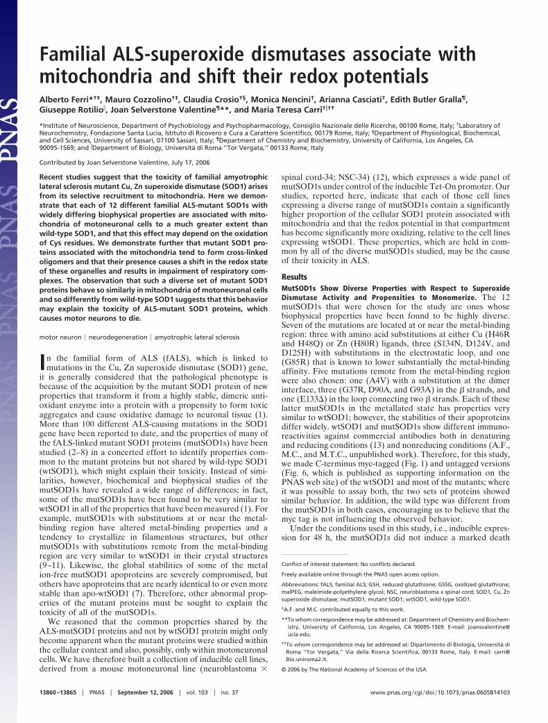
ResultsMutSOD1s Show Diverse Properties with Respect to SuperoxideDismutase Activity and Propensities to Monomerize. The 12mutSOD1s that were chosen for the study are ones whosebiophysical properties have been found to be highly diverse.Seven of the mutations are located at or near the metal-bindingregion: three with amino acid substitutions at either Cu (H46Rand H48Q) or Zn (H80R) ligands, three (S134N, D124V, andD125H) with substitutions in the electrostatic loop, and one(G85R) that is known to lower substantially the metal-bindingaffinity. Five mutations remote from the metal-binding regionwere also chosen: one (A4V) with a substitution at the dimerinterface, three (G37R, D90A, and G93A) in the � strands, andone (E133�) in the loop connecting two � strands. Each of theselatter mutSOD1s in the metallated state has properties verysimilar to wtSOD1; however, the stabilities of their apoproteinsdiffer widely. wtSOD1 and mutSOD1s show different immuno-reactivities against commercial antibodies both in denaturingand reducing conditions (13) and nonreducing conditions (A.F.,M.C., and M.T.C., unpublished work). Therefore, for this study,we made C-terminus myc-tagged (Fig. 1) and untagged versions(Fig. 6, which is published as supporting information on thePNAS web site) of the wtSOD1 and most of the mutants; whereit was possible to assay both, the two sets of proteins showedsimilar behavior. In addition, the wild type was different fromthe mutSOD1s in both cases, encouraging us to believe that themyc tag is not influencing the observed behavior.
Under the conditions used in this study, i.e., inducible expres-sion for 48 h, the mutSOD1s did not induce a marked death
Conflict of interest statement: No conflicts declared.
Freely available online through the PNAS open access option.
Abbreviations: fALS, familial ALS; GSH, reduced glutathione; GSSG, oxidized glutathione;malPEG, maleimide-polyethylene glycol; NSC, neuroblastoma x spinal cord; SOD1, Cu, Znsuperoxide dismutase; mutSOD1, mutant SOD1; wtSOD1, wild-type SOD1.
‡A.F. and M.C. contributed equally to this work.
**To whom correspondence may be addressed at: Department of Chemistry and Biochem-istry, University of California, Los Angeles, CA 90095-1569. E-mail: [email protected].
††To whom correspondence may be addressed at: Dipartimento di Biologia, Universita diRoma ‘‘Tor Vergata,’’ Via della Ricerca Scientifica, 00133 Rome, Italy. E-mail: [email protected].
© 2006 by The National Academy of Sciences of the USA
13860–13865 � PNAS � September 12, 2006 � vol. 103 � no. 37 www.pnas.org�cgi�doi�10.1073�pnas.0605814103
phenotype (data not shown), an increase in the cell level ofoxidative stress (as determined by reaction with dihydro-ethidium; see Fig. 7, which is published as supporting informa-tion on the PNAS web site), or an imbalance in the total ratioreduced glutathione�oxidized glutathione (GSH�GSSG) (Table1, which is published as supporting information on the PNASweb site). In total cell extracts, the mutSOD1s displayed a widerange of superoxide dismutase activities and propensities tomonomerize, as had been seen in previous studies (8): Some ofthem exhibited behavior similar to that of wtSOD1 and retainednear-to-full SOD activity and the typical two-banded pattern inthe activity gel assay (14), whereas some readily lost SOD activityin nondenaturing conditions (Fig. 1 A). Interestingly, a fractionof the native A4V mutSOD1 was observed to be present as aSOD-active monomer. The other mutSOD1s showed a markedtendency to form monomers in mild denaturing, nonreducingconditions (0.1% SDS), but they differed widely in this propertyalso, with some very prone to monomerization and others (e.g.,G93A) acting very much like the wild-type protein (Fig. 1B).Similarly diverse results were obtained when the accessibility ofcysteine residues was assayed by modification of total cellextracts with polyethyleneglycol maleimide (malPEG). The ac-cessibility of the cysteines to malPEG modification was found tobe the same as that of wtSOD1 for some of the mutSOD1s (A4V,G37R, D90A, and G93A), whereas others (G85R, H46R, H48Q,H80R, S134N, D125H, and D124V) were found to be morereactive (Fig. 8, which is published as supporting information onthe PNAS web site).
MutSOD1s Accumulated in the Mitochondrial Fraction of NSC-34 CellsAre Partially Oxidized and Thus Less Accessible to Modification bymalPEG. As had been reported both in vitro and in vivo in the brainand spinal cord of transgenic mice and in human spinal cord(15–20), we found that some of the SOD1 protein, wild type ormutant, was associated with the mitochondria, but we found
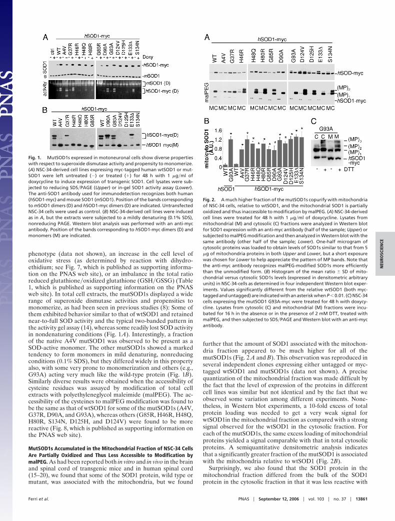
further that the amount of SOD1 associated with the mitochon-dria fraction appeared to be much higher for all of themutSOD1s (Fig. 2 A and B). This observation was reproduced inseveral independent clones expressing either untagged or myc-tagged wtSOD1 and mutSOD1s (data not shown). A precisequantization of the mitochondrial fraction was made difficult bythe fact that the level of expression of the proteins in differentcell lines was similar but not identical and by the fact that weobserved some variation among different experiments. None-theless, in Western blot experiments, a 10-fold excess of totalprotein loading was needed to get a very weak signal forwtSOD1in the mitochondrial fraction as compared with a strongsignal observed for the wtSOD1 in the cytosolic fraction. Foreach of the mutSOD1s, the same excess loading of mitochondrialproteins yielded a signal comparable with that in total cytosolicproteins. A semiquantitative densitometric analysis indicatesthat a significantly greater fraction of the mutSOD1 is associatedwith the mitochondria relative to wtSOD1 (Fig. 2B).
Surprisingly, we also found that the SOD1 protein in themitochondrial fraction differed from the bulk of the SOD1protein in the cytosolic fraction in that it was less reactive with
Fig. 1. MutSOD1s expressed in motoneuronal cells show diverse propertieswith respect to superoxide dismutase activity and propensity to monomerize.(A) NSC-34-derived cell lines expressing myc-tagged human wtSOD1 or mut-SOD1 were left untreated (�) or treated (�) for 48 h with 1 �g�ml ofdoxycycline to induce expression of transgenic SOD1. Cell lysates were sub-jected to reducing SDS�PAGE (Upper) or in-gel SOD1 activity assay (Lower).The anti-SOD1 antibody used for immunodetection recognizes both human(hSOD1-myc) and mouse SOD1 (mSOD1). Position of the bands correspondingto mSOD1 dimers (D) and hSOD1-myc dimers (D) are indicated. UntransfectedNSC-34 cells were used as control. (B) NSC-34-derived cell lines were inducedas in A, but the extracts were subjected to a mildly denaturing (0.1% SDS),nonreducing PAGE. Western blot analysis was performed with an anti-mycantibody. Position of the bands corresponding to hSOD1-myc dimers (D) andmonomers (M) are indicated.
Fig. 2. A much higher fraction of the mutSOD1s copurify with mitochondriaof NSC-34 cells, relative to wtSOD1, and the mitochondrial SOD1 is partiallyoxidized and thus inaccessible to modification by malPEG. (A) NSC-34-derivedcell lines were treated for 48 h with 1 �g�ml of doxycycline. Lysates frommitochondrial (M) and cytosolic (C) fractions were analyzed in Western blotfor SOD1 expression with an anti-myc antibody (half of the sample; Upper) orsubjected to malPEG modification and then analyzed in Western blot with thesame antibody (other half of the sample; Lower). One-half microgram ofcytosolic proteins was loaded to obtain levels of SOD1s similar to that from 5�g of mitochondria proteins in both Upper and Lower, but a short exposurewas chosen for Lower to help appreciate the pattern of MP bands. Note thatthe anti-myc antibody recognizes malPEG-modified SOD1s more efficientlythan the unmodified form. (B) Histogram of the mean ratio � SD of mito-chondrial versus cytosolic SOD1s levels (expressed in densitometric arbitraryunits) in NSC-34-cells as determined in four independent Western blot exper-iments. Values significantly different from the relative wtSOD1 (both myc-tagged and untagged) are indicated with an asterisk when P � 0.01. (C) NSC-34cells expressing the mutSOD1 G93A-myc were treated for 48 h with doxycy-cline. Lysates from cytosolic (C) and mitochondrial (M) fractions were incu-bated for 16 h in the absence or in the presence of 2 mM DTT, treated withmalPEG, and then subjected to SDS�PAGE and Western blot with an anti-mycantibody.
Ferri et al. PNAS � September 12, 2006 � vol. 103 � no. 37 � 13861
NEU
ROSC
IEN
CE
malPEG. Quantization of the amount of malPEG-modifiableSOD1 was made difficult by the fact that malPEG modificationproduced both size shifts in the SOD1 protein and, at times,enhanced the intensity of the immunoreactive bands on immu-noblots (Fig. 2 A Lower). Nonetheless, the intensity of the bandindicated as (MP)1, corresponding to SOD1 modified with onemalPEG, was usually less intense in protein associated withmitochondria than in the corresponding cytosolic sample (Fig.2A Lower and Fig. 9,which is published as supporting informa-tion on the PNAS web site). Treatment of both cytosolic andmitochondrial fractions with DTT before malPEG treatmentsignificantly increased the reactivity of SOD1 to this reagent(Fig. 2C), suggesting the presence of oxidized cysteines in SOD1in these preparations.
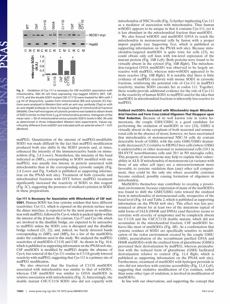
Cys-111 Is Necessary for Association with Mitochondria of C6F mut-SOD1. Human SOD1 has four cysteine residues that have differentreactivities: Cys-111, which is exposed on the protein surface nearthe dimer interface, is expected to be the most prone to modifica-tion with malPEG, followed by Cys-6, which is packed tightly withinthe interior of the � barrel. By contrast, Cys-57 and Cys-146, whichare involved in the disulfide bridge, are expected to be unreactiveto malPEG unless the SOD1 core is disrupted and the disulfidebridge reduced (21, 22), and, indeed, we barely detected bandscorresponding to (MP)3 and (MP)4 for a few of the mutSOD1sunder the conditions used in this study. We analyzed the malPEGreactivities of mutSOD1s C111S and C6F. As shown in Fig. 10A,which is published as supporting information on the PNAS web site,C6F mutSOD1 is modified by malPEG despite the absence ofCys-6, but removal of the Cys-111 residue in C111S greatly loweredreactivity with malPEG, suggesting that Cys-111 is a primary site ofmalPEG modification.
We also observed that the fraction of C111S mutSOD1associated with mitochondria was similar to that of wtSOD1,whereas C6F mutSOD1 was similar to G93A mutSOD1 inrelative association with mitochondria (Fig. 3). Interestingly, thedouble mutant C6F�C111S SOD1 also did not copurify with
mitochondria of NSC34 cells (Fig. 3) further implicating Cys-111as a mediator of association with mitochondria. Thus humanwtSOD1 appears to be unique in that it contains Cys-111, yet itis less abundant in the mitochondrial fraction than mutSOD1.
We also forced wtSOD1 and mutSOD1 G93A to reach themitochondria in motoneuronal cells by fusion with a properimport peptide (see Supporting Text, which is published assupporting information on the PNAS web site). Because mito-chondria-targeted mutSOD1 is quite toxic for cells (23), wecould obtain only cell lines with low-level expression of themutant protein (Fig. 10B Left). Both proteins were found to bevirtually absent in the cytosol (Fig. 10B Right). The mitochon-dria-targeted G93A mutSOD1 was observed to be largely un-reactive with malPEG, whereas mito-wtSOD1 appeared to bemore reactive (Fig. 10B Right). It is notable that there is littleevidence of malPEG reactivity with mouse SOD1 in cytosolicfractions, reinforcing the potential role of Cys-111 in malPEGreactivity; murine SOD1 encodes Ser at codon 111. Together,these results provide additional evidence for the role of Cys-111in the reactivity of human SOD1 to malPEG and for the idea thatmutSOD1 in mitochondrial fractions is inherently less reactive tomalPEG.
Oxidized mutSOD1s Associated with Mitochondria Impair Mitochon-drial Function and Form Cross-Linked Oligomers That Disappear uponThiol Reduction. Because of its well known role in redox ho-meostasis, the couple GSH�GSSG is a good candidate forperforming the oxidation of mutSOD1 in vivo (24). GSSG isvirtually absent in the cytoplasm of both neuronal and nonneu-ronal cells in the absence of stress; however, we have ascertainedthat mitochondria of motoneuronal NSC-34 cells do containdetectable levels of GSSG, with the ratio GSH�GSSG dramat-ically decreased (5:1) relative to HEPG2 liver cells (where GSSGis undetectable) or other neuronal or nonneuronal cells (10:1 inSH-SY5Y neuroblastoma cells and 20:1 in N9 microglia cells).This property of motoneurons may help to explain their vulner-ability in ALS: If mitochondria of motoneurons (at variance withthose of any other cell type) are a microenvironment morefavorable to cysteine oxidation than (any) other cell compart-ment, they could be the only site where accessible cysteinesbecome oxidized, possibly causing formation of oligomers inmitochondria.
MutSOD1s themselves may contribute to creating a prooxi-dant environment, because expression of many of the mutSOD1swas found to shift the GSH�GSSG ratio toward the oxidizedform in mitochondria of motoneuronal cells, irrespective of thebasal level (Fig. 4A and Table 2, which is published as supportinginformation on the PNAS web site). This effect was less pro-nounced or absent for at least two of the mutations typical ofmild forms of fALS (H46R and D90A) (and therefore seems tocorrelate with severity of symptoms) and be completely absentfor C111S and the C6F�C111S double mutant, which did notaccumulate in the mitochondrial fraction; C6F mutSOD1 be-haves like most of mutSOD1s (Fig. 4B). As a confirmation thatcysteine residues of SOD1 are specifically sensitive to modifi-cation of the redox environment created by the couple GSH�GSSG, preincubation of the recombinant purified G93A andH46R mutSOD1s with the oxidized form of glutathione (GSSG)prevented their derivatization by malPEG, whereas preincuba-tion with the reduced form of glutathione (GSH) facilitatedderivatization relative to control (Fig. 11 A Right, which ispublished as supporting information on the PNAS web site).Furthermore, treatment of mutSOD1 with hydrogen peroxide invitro did not interfere with cysteine accessibility (Fig. 11 A Left),suggesting that oxidative modification of Cys residues, ratherthan some other type of oxidation, is involved in modification ofmutSOD1s.
In line with our observations, and supporting the concept that
Fig. 3. Oxidation of Cys-111 is necessary for C6F mutSOD1 association withmitochondria. NSC-34 cell lines expressing myc-tagged hSOD1s (WT, C6F,C111S, and the double SOD1 mutant C6F�C111S) were treated for 48 h with 1�g�ml of doxycycline. Lysates from mitochondrial (M) and cytosolic (C) frac-tions were analyzed in Western blot with an anti-myc antibody (Top) or withan anti-Hsp60 antibody to check for equal loading of mitochondrial fractions(Middle). One-half microgram of cytosolic proteins was loaded to obtain levelsof SOD1s similar to that from 5 �g of mitochondria proteins. Histogram of themean ratio � SD of mitochondrial versus cytosolic SOD1s levels in NSC-34-cellsas determined in three independent Western blot experiments. Values sig-nificantly different from wtSOD1 are indicated with an asterisk when P � 0.01(Bottom).
13862 � www.pnas.org�cgi�doi�10.1073�pnas.0605814103 Ferri et al.
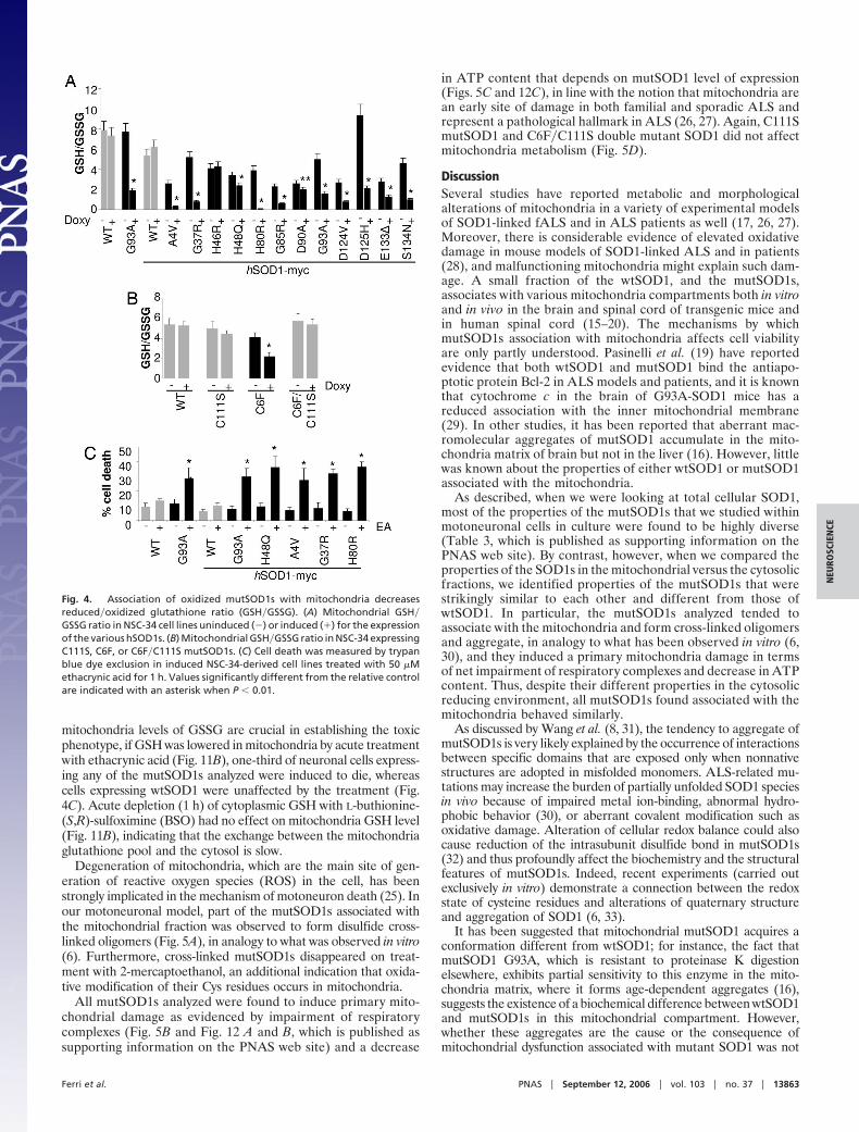
mitochondria levels of GSSG are crucial in establishing the toxicphenotype, if GSH was lowered in mitochondria by acute treatmentwith ethacrynic acid (Fig. 11B), one-third of neuronal cells express-ing any of the mutSOD1s analyzed were induced to die, whereascells expressing wtSOD1 were unaffected by the treatment (Fig.4C). Acute depletion (1 h) of cytoplasmic GSH with L-buthionine-(S,R)-sulfoximine (BSO) had no effect on mitochondria GSH level(Fig. 11B), indicating that the exchange between the mitochondriaglutathione pool and the cytosol is slow.
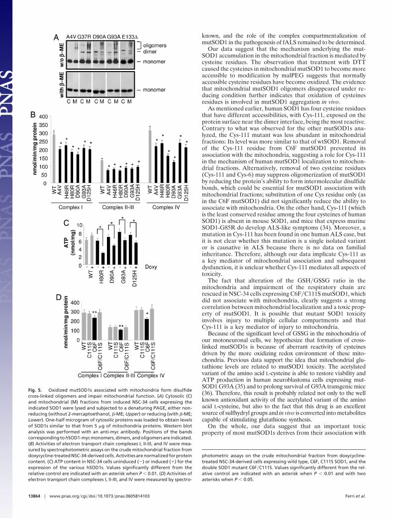
Degeneration of mitochondria, which are the main site of gen-eration of reactive oxygen species (ROS) in the cell, has beenstrongly implicated in the mechanism of motoneuron death (25). Inour motoneuronal model, part of the mutSOD1s associated withthe mitochondrial fraction was observed to form disulfide cross-linked oligomers (Fig. 5A), in analogy to what was observed in vitro(6). Furthermore, cross-linked mutSOD1s disappeared on treat-ment with 2-mercaptoethanol, an additional indication that oxida-tive modification of their Cys residues occurs in mitochondria.
All mutSOD1s analyzed were found to induce primary mito-chondrial damage as evidenced by impairment of respiratorycomplexes (Fig. 5B and Fig. 12 A and B, which is published assupporting information on the PNAS web site) and a decrease
in ATP content that depends on mutSOD1 level of expression(Figs. 5C and 12C), in line with the notion that mitochondria arean early site of damage in both familial and sporadic ALS andrepresent a pathological hallmark in ALS (26, 27). Again, C111SmutSOD1 and C6F�C111S double mutant SOD1 did not affectmitochondria metabolism (Fig. 5D).
DiscussionSeveral studies have reported metabolic and morphologicalalterations of mitochondria in a variety of experimental modelsof SOD1-linked fALS and in ALS patients as well (17, 26, 27).Moreover, there is considerable evidence of elevated oxidativedamage in mouse models of SOD1-linked ALS and in patients(28), and malfunctioning mitochondria might explain such dam-age. A small fraction of the wtSOD1, and the mutSOD1s,associates with various mitochondria compartments both in vitroand in vivo in the brain and spinal cord of transgenic mice andin human spinal cord (15–20). The mechanisms by whichmutSOD1s association with mitochondria affects cell viabilityare only partly understood. Pasinelli et al. (19) have reportedevidence that both wtSOD1 and mutSOD1 bind the antiapo-ptotic protein Bcl-2 in ALS models and patients, and it is knownthat cytochrome c in the brain of G93A-SOD1 mice has areduced association with the inner mitochondrial membrane(29). In other studies, it has been reported that aberrant mac-romolecular aggregates of mutSOD1 accumulate in the mito-chondria matrix of brain but not in the liver (16). However, littlewas known about the properties of either wtSOD1 or mutSOD1associated with the mitochondria.
As described, when we were looking at total cellular SOD1,most of the properties of the mutSOD1s that we studied withinmotoneuronal cells in culture were found to be highly diverse(Table 3, which is published as supporting information on thePNAS web site). By contrast, however, when we compared theproperties of the SOD1s in the mitochondrial versus the cytosolicfractions, we identified properties of the mutSOD1s that werestrikingly similar to each other and different from those ofwtSOD1. In particular, the mutSOD1s analyzed tended toassociate with the mitochondria and form cross-linked oligomersand aggregate, in analogy to what has been observed in vitro (6,30), and they induced a primary mitochondria damage in termsof net impairment of respiratory complexes and decrease in ATPcontent. Thus, despite their different properties in the cytosolicreducing environment, all mutSOD1s found associated with themitochondria behaved similarly.
As discussed by Wang et al. (8, 31), the tendency to aggregate ofmutSOD1s is very likely explained by the occurrence of interactionsbetween specific domains that are exposed only when nonnativestructures are adopted in misfolded monomers. ALS-related mu-tations may increase the burden of partially unfolded SOD1 speciesin vivo because of impaired metal ion-binding, abnormal hydro-phobic behavior (30), or aberrant covalent modification such asoxidative damage. Alteration of cellular redox balance could alsocause reduction of the intrasubunit disulfide bond in mutSOD1s(32) and thus profoundly affect the biochemistry and the structuralfeatures of mutSOD1s. Indeed, recent experiments (carried outexclusively in vitro) demonstrate a connection between the redoxstate of cysteine residues and alterations of quaternary structureand aggregation of SOD1 (6, 33).
It has been suggested that mitochondrial mutSOD1 acquires aconformation different from wtSOD1; for instance, the fact thatmutSOD1 G93A, which is resistant to proteinase K digestionelsewhere, exhibits partial sensitivity to this enzyme in the mito-chondria matrix, where it forms age-dependent aggregates (16),suggests the existence of a biochemical difference between wtSOD1and mutSOD1s in this mitochondrial compartment. However,whether these aggregates are the cause or the consequence ofmitochondrial dysfunction associated with mutant SOD1 was not
Fig. 4. Association of oxidized mutSOD1s with mitochondria decreasesreduced�oxidized glutathione ratio (GSH�GSSG). (A) Mitochondrial GSH�GSSG ratio in NSC-34 cell lines uninduced (�) or induced (�) for the expressionof the various hSOD1s. (B) Mitochondrial GSH�GSSG ratio in NSC-34 expressingC111S, C6F, or C6F�C111S mutSOD1s. (C) Cell death was measured by trypanblue dye exclusion in induced NSC-34-derived cell lines treated with 50 �Methacrynic acid for 1 h. Values significantly different from the relative controlare indicated with an asterisk when P � 0.01.
Ferri et al. PNAS � September 12, 2006 � vol. 103 � no. 37 � 13863
NEU
ROSC
IEN
CE
known, and the role of the complex compartmentalization ofmutSOD1 in the pathogenesis of fALS remained to be determined.
Our data suggest that the mechanism underlying the mut-SOD1 accumulation in the mitochondrial fraction is mediated bycysteine residues. The observation that treatment with DTTcaused the cysteines in mitochondrial mutSOD1 to become moreaccessible to modification by malPEG suggests that normallyaccessible cysteine residues have become oxidized. The evidencethat mitochondrial mutSOD1 oligomers disappeared under re-ducing condition further indicates that oxidation of cysteinesresidues is involved in mutSOD1 aggregation in vivo.
As mentioned earlier, human SOD1 has four cysteine residuesthat have different accessibilities, with Cys-111, exposed on theprotein surface near the dimer interface, being the most reactive.Contrary to what was observed for the other mutSOD1s ana-lyzed, the Cys-111 mutant was less abundant in mitochondrialfractions: Its level was more similar to that of wtSOD1. Removalof the Cys-111 residue from C6F mutSOD1 prevented itsassociation with the mitochondria, suggesting a role for Cys-111in the mechanism of human mutSOD1 localization to mitochon-drial fractions. Alternatively, removal of two cysteine residues(Cys-111 and Cys-6) may suppress oligomerization of mutSOD1by reducing the protein’s ability to form intermolecular disulfidebonds, which could be essential for mutSOD1 association withmitochondrial fractions; substitution of one Cys residue only (asin the C6F mutSOD1) did not significantly reduce the ability toassociate with mitochondria. On the other hand, Cys-111 (whichis the least conserved residue among the four cysteines of humanSOD1) is absent in mouse SOD1, and mice that express murineSOD1-G85R do develop ALS-like symptoms (34). Moreover, amutation in Cys-111 has been found in one human ALS case, butit is not clear whether this mutation is a single isolated variantor is causative in ALS because there is no data on familialinheritance. Therefore, although our data implicate Cys-111 asa key mediator of mitochondrial association and subsequentdysfunction, it is unclear whether Cys-111 mediates all aspects oftoxicity.
The fact that alteration of the GSH�GSSG ratio in themitochondria and impairment of the respiratory chain arerescued in NSC-34 cells expressing C6F�C111S mutSOD1, whichdid not associate with mitochondria, clearly suggests a strongcorrelation between mitochondrial localization and a toxic prop-erty of mutSOD1. It is possible that mutant SOD1 toxicityinvolves injury to multiple cellular compartments and thatCys-111 is a key mediator of injury to mitochondria.
Because of the significant level of GSSG in the mitochondria ofour motoneuronal cells, we hypothesize that formation of cross-linked mutSOD1s is because of aberrant reactivity of cysteinesdriven by the more oxidizing redox environment of these mito-chondria. Previous data support the idea that mitochondrial glu-tathione levels are related to mutSOD1 toxicity. The acetylatedvariant of the amino acid L-cysteine is able to restore viability andATP production in human neuroblastoma cells expressing mut-SOD1 G93A (35) and to prolong survival of G93A transgenic mice(36). Therefore, this result is probably related not only to the wellknown antioxidant activity of the acetylated variant of the aminoacid L-cysteine, but also to the fact that this drug is an excellentsource of sulfhydryl groups and in vivo is converted into metabolitescapable of stimulating glutathione synthesis.
On the whole, our data suggest that an important toxicproperty of most mutSOD1s derives from their association with
Fig. 5. Oxidized mutSOD1s associated with mitochondria form disulfidecross-linked oligomers and impair mitochondrial function. (A) Cytosolic (C)and mitochondrial (M) fractions from induced NSC-34 cells expressing theindicated SOD1 were lysed and subjected to a denaturing PAGE, either non-reducing (without 2-mercaptoethanol, �-ME; Upper) or reducing (with �-ME;Lower). One-half microgram of cytosolic proteins was loaded to obtain levelsof SOD1s similar to that from 5 �g of mitochondria proteins. Western blotanalysis was performed with an anti-myc antibody. Positions of the bandscorresponding to hSOD1-myc monomers, dimers, and oligomers are indicated.(B) Activities of electron transport chain complexes I, II-III, and IV were mea-sured by spectrophotometric assays on the crude mitochondrial fraction fromdoxycycline-treated NSC-34-derived cells. Activities are normalized for proteincontent. (C) ATP content in NSC-34 cells uninduced (�) or induced (�) for theexpression of the various hSOD1s. Values significantly different from therelative control are indicated with an asterisk when P � 0.01. (D) Activities ofelectron transport chain complexes I, II-III, and IV were measured by spectro-
photometric assays on the crude mitochondrial fraction from doxycycline-treated NSC-34-derived cells expressing wild type, C6F, C111S SOD1, and thedouble SOD1 mutant C6F�C111S. Values significantly different from the rel-ative control are indicated with an asterisk when P � 0.01 and with twoasterisks when P � 0.05.
13864 � www.pnas.org�cgi�doi�10.1073�pnas.0605814103 Ferri et al.
the mitochondria, and that this association is because of gluta-thione-mediated modification of cysteine residues, causingmutSOD1s to accumulate in a oxidized, aggregate state. Further,the presence of mutSOD1s leads to impairment of the respira-tory chain and a shift in the mitochondrial redox balance(GSH�GSSG ratio) toward more oxidizing. Understanding themechanism of association of mutSOD1 with mitochondriashould help to develop methods to prevent this phenomenon todecrease toxicity in patients.
Materials and MethodsDetailed experimental procedures are provided in SupportingText.
Cell Lines. Mouse motoneuronal cell line NSC-34 (a gift of N. R.Cashman, University of Toronto, Toronto, ON, Canada; see ref.12) is a hybrid NSC cell line that is considered the best stablemotoneuronal cell line model system available (see SupportingText). This line was stably transfected with the pTet-ON plasmid(Clontech, Palo Alto, CA) coding for the reverse tetracyclinecontrolled transactivator (rtTA), by using lipofectamine reagent(Invitrogen, Paisley, U.K.) according to the manufacturer’sguideline. Single clones were isolated after 3 weeks of selectionwith 400 �g�ml G418 (Gibco, Paisley, U.K.) and screenedaccording to their ability to control the expression of an induc-ible reporter gene (pTRE-luc, Clontech). The line designedNSC-34-ON7, which displays a very low level of basal expressionand high inducibility, was used to construct inducible cell linesexpressing the cDNAs encoding human wtSOD1 or 1 of the 12human fALS-typical mutSOD1s inserted in the pTRE2 orpTRE2–5myc plasmids. To allow the selection, we cotransfectedthe plasmid pTK-Hygro (Clontech). Multiple clones for eachconstruction, isolated after selection with 400 �g�ml Hygromi-cin-B (Invitrogen), were analyzed by Western blot, and cloneswith similar level of expression were chosen. Stable clonesexpressing mitoSOD1 (wild type or G93A) were obtained bytransfection of NSC-34 cells with pCMV-mito-hSOD1 wt�G93A
plasmids and selection with 400 �g�ml Hygromicin-B. Condi-tions for growth and induction of cell cultures are reported inSupporting Text.
Subcellular Fractionation and Mitochondria Purification. Mitochon-dria were isolated from NSC-34-derived cell lines as reported byref. 15 with some modification (see Supporting Text).
Detection of Accessible Cysteines in SOD1. Cytosolic and mitochon-drial fractions were resuspended in mitochondrial buffer containing0.1% SDS. malPEG (molecular mass of 5 kDa; NEKTAR, SanCarlos, CA) was then added to a final concentration of 3 mM forcovalent modification of accessible cysteines for 1 h at 25°C. Theaddition of malPEG to accessible cysteines increases the subunitmass of SOD1 by �5 kDa per modification. SDS�PAGE loadingbuffer containing 5% 2-mercaptoethanol was then added. Sampleswere boiled at 100°C for 3 min, separated by denaturing SDS�PAGE, and subject to Western blot analysis as described.
Determination of Glutathione Content. Determination of intracel-lular glutathione was performed in HPLC as described (37).
Statistical Analysis. The results are presented as means � SD ofn � 3 independent experiments. Statistical evaluation wasconducted by a simple Student’s t test, and values significantlydifferent from the relative control are indicated with an asteriskwhen P � 0.01 and with two asterisks when P � 0.05.
Note. Deng et al. (38) have published a paper that supports a role fordisulfide-linked SOD1 multimerization in mitochondria of mouse mod-els of ALS.
We thank Carla Russo, Laura Biondini, and Ilaria Amori for invaluablesupport in the construction of NSC-34-derived cell lines and LiliaCalabrese and Andrea Battistoni for critical reading of the manuscript.This work was supported by Telethon Grant GGP030066 (to M.T.C.),Ministero della Salute Ricerca Finalizzata Malattie Neurodegenerativeand Progetto Finalizzato ‘‘Approcci neuroprotettivi nel danno da dep-rivazione energetica’’ (M.T.C.), and National Institutes of Health GrantsDK46828 and GM28222 (to J.S.V.).
1. Valentine JS, Doucette PA, Potter SZ (2005) Annu Rev Biochem 74:563–593.2. Hayward LJ, Rodriguez JA, Kim JW, Tiwari A, Goto JJ, Cabelli DE, Valentine
JS, Brown RH, Jr (2002) J Biol Chem 277:15923–15931.3. Rodriguez JA, Valentine JS, Eggers DK, Roe JA, Tiwari A, Brown RH, Jr,
Hayward LJ (2002) J Biol Chem 277:15932–15937.4. Lindberg MJ, Tibell L, Oliveberg M (2002) Proc Natl Acad Sci USA 99:16607–16612.5. Lindberg MJ, Bystrom R, Boknas N, Andersen PM, Oliveberg M (2005) Proc
Natl Acad Sci USA 102:9754–9759.6. Furukawa Y, O’Halloran TV (2005) J Biol Chem 280:17266–17274.7. Rodriguez JA, Shaw BF, Durazo A, Sohn SH, Doucette PA, Nersissian AM,
Faull KF, Eggers DK, Tiwari A, Hayward LJ, Valentine JS (2005) Proc NatlAcad Sci USA 102:10516–10521.
8. Wang J, Slunt H, Gonzales V, Fromholt D, Coonfield M, Copeland NG,Jenkins NA, Borchelt DR (2003) Hum Mol Genet 12:2753–2764.
9. Elam JS, Taylor AB, Strange R, Antonyuk S, Doucette PA, Rodriguez JA,Hasnain SS, Hayward LJ, Valentine JS, Yeates TO, Hart PJ (2003) Nat StructBiol 10:461–467.
10. Hough MA, Grossmann JG, Antonyuk SV, Strange RW, Doucette PA,Rodriguez JA, Whitson LJ, Hart PJ, Hayward LJ, Valentine JS, Hasnain SS(2004) Proc Natl Acad Sci USA 101:5976–5981.
11. Antonyuk S, Elam JS, Hough MA, Strange RW, Doucette PA, Rodriguez JA,Hayward LJ, Valentine JS, Hart PJ, Hasnain SS (2005) Protein Sci 14:1201–1213.
12. Cashman NR, Durham HD, Blusztajn JK, Oda K, Tabira T, Shaw IT, DahrougeS, Antel JP (1992) Dev Dyn 194:209–221.
13. Fujiwara N, Miyamoto Y, Ogasahara K, Takahashi M, Ikegami T, TakamiyaR, Suzuki K, Taniguchi N (2005) J Biol Chem 280:5061–5070.
14. Steinkuhler C, Sapora O, Carri MT, Nagel W, Marcocci L, Ciriolo MR, WeserU, Rotilio G (1991) J Biol Chem 266:24580–24587.
15. Okado-Matsumoto A, Fridovich I (2001) J Biol Chem 276:38388–38393.16. Vijayvergiya C, Beal MF, Buck J, Manfredi G (2005) J Neurosci 25:2463–2470.17. Mattiazzi M, D’Aurelio M, Gajewski CD, Martushova K, Kiaei M, Beal MF,
Manfredi G (2002) J Biol Chem 277:29626–29633.18. Higgins CM, Jung C, Ding H, Xu Z (2002) J Neurosci 22:RC215.
19. Pasinelli P, Belford ME, Lennon N, Bacskai BJ, Hyman BT, Trotti D, BrownRH, Jr (2004) Neuron 43:19–30.
20. Liu J, Lillo C, Jonsson PA, Vande Velde C, Ward CM, Miller TM, SubramaniamJR, Rothstein JD, Marklund S, Andersen PM, et al. (2004) Neuron 43:5–17.
21. Tiwari A, Hayward LJ (2003) J Biol Chem 278:5984–5992.22. Arnesano F, Banci L, Bertini I, Martinelli M, Furukawa Y, O’Halloran TV
(2004) J Biol Chem 279:47998–48003.23. Takeuchi H, Kobayashi Y, Ishigaki S, Doyu M, Sobue G (2002) J Biol Chem
277:50966–50972.24. Schafer FQ, Buettner GR (2001) Free Radical Biol Med 30:1191–1212.25. Andersen JK (2004) Nat Med 10 (Suppl):S18–25.26. Bendotti C, Carrı MT (2004) Trends Mol Med 10:393–400.27. Xu Z, Jung C, Higgins C, Levine J, Kong J (2004) J Bioenerg Biomembr 36:395–399.28. Valentine JS (2002) Free Radical Biol Med 33:1314–1320.29. Kirkinezos IG, Barman SR, Hernandez D, Oca-Cossio J, Arias LJ, Perez-
Pinzon MA, Bradley WG, Moraes CT (2005) J Neurosci 25:164–172.30. Rakhit R, Crow JP, Lepock JR, Kondejewski LH, Cashman NR, Chakrabartty
A (2004) J Biol Chem 279:15499–15504.31. Wang J, Xu G, Borchelt DR (2006) J Neurochem 96:1277–1288.32. Jonsson PA, Graffmo KS, Andersen PM, Brannstrom T, Lindberg M, Olive-
berg M, Marklund SL (2006) Brain 129:451–464.33. Doucette PA, Whitson LJ, Cao X, Schirf V, Demeler B, Valentine JS, Hansen
JC, Hart PJ (2004) J Biol Chem 279:54558–54566.34. Ripps ME, Huntley GW, Hof PR, Morrison JH, Gordon JW (1995) Proc Natl
Acad Sci USA 92:689–693.35. Beretta S, Sala G, Mattavelli L, Ceresa C, Casciati A, Ferri A, Carri MT,
Ferrarese C (2003) Neurobiol Dis 13:213–221.36. Andreassen OA, Dedeoglu A, Klivenyi P, Beal MF, Bush AI (2000) Neuro-
Report 11:2491–2493.37. Ciriolo MR, Palamara AT, Incerpi S, Lafavia E, Bue MC, De Vito P, Garaci
E, Rotilio G (1997) J Biol Chem 272:2700–2708.38. Deng HX, Shi Y, Furukawa Y, Zhai H, Fu R, Liu E, Gorrie GH, Khan MS,
Hung WY, Bigio EH, et al. (2006) Proc Natl Acad Sci USA 103:7142–7147.
Ferri et al. PNAS � September 12, 2006 � vol. 103 � no. 37 � 13865
NEU
ROSC
IEN
CE
Related Documents