Failure to Precondition Pathological Human Myocardium Sudip Ghosh, FRCS,* Nicholas B. Standen, PHD,² Manuel Galin ˜anes, MD, PHD* Leicester, United Kingdom OBJECTIVES We investigated the effects of ischemic preconditioning (PC) on diabetic and failing human myocardium and the role of mitochondrial K ATP channels on the response in these diseased tissues. BACKGROUND There is conflicting evidence to suggest that PC is a healthy heart phenomenon. METHODS Right atrial appendages were obtained from seven different groups of patients: nondiabetics, diet-controlled diabetics, noninsulin-dependent diabetics (NIDD) receiving K ATP channel blockers, insulin-dependent diabetics (IDD), and patients with left ventricular ejection fraction (LVEF) .50%, LVEF between 30% and 50% and LVEF ,30%. After stabilization, the muscle slices were randomized into five experimental groups (n 5 6/group): 1) aerobic control—incubated in oxygenated buffer for 210 min, 2) ischemia alone—90 min ischemia followed by 120 min reoxygenation, 3) preconditioning by 5 min ischemia/5 min reoxygen- ation before 90 min ischemia/120 min reoxygenation, 4) diazoxide (Mito K ATP opener, 0.1 mm)—for 10 min before the 90 min ischemia/120 min reoxygenation and 5) gliben- clamide (10 mm)—10 min exposure prior to PC (only in the diabetic patient groups). Creatine kinase leakage into the medium (CK, U/g wet wt) and MTT reduction (OD/mg wet wt), an index of cell viability, were assessed at the end of the experiment. RESULTS Ischemia caused similar injury in both normal and diseased tissue. Preconditioning prevented the effects of ischemia in all groups except NIDD, IDD and poor cardiac function (,30%). In the diazoxide-treated groups, protection was mimicked in all groups except the NIDD and IDD groups. Interestingly, glybenclamide abolished protection in nondiabetic and diet- controlled NIDD groups and did not affect NIDD groups receiving K ATP channel blockers or IDD groups. CONCLUSIONS These results show that failure to precondition the diabetic heart is due to dysfunction of the mitochondrial K ATP channels and that the mechanism of failure in the diabetic heart lies in elements of the signal transduction pathway different from the mitochondrial K ATP channels. (J Am Coll Cardiol 2001;37:711– 8) © 2001 by the American College of Cardiology Ischemic preconditioning (PC) falls within a spectrum of adaptive responses to ischemia and represents the ability of the myocardium to adapt to sublethal ischemic stress in the short term so that it is more resistant to a subsequent, potentially injurious period of ischemia. Preconditioning consists of two phases of protection: an early or first window of protection (#2 h) and a delayed or second window of protection ($24 h). The underlying mechanism of PC has been extensively investigated; however, the basis of such cardioprotection is not fully elucidated. The most favored hypothesis for the first window of PC suggests that a variety of endogenous ligands such as adenosine, bradykinin, cat- echolamines and opoids activate receptors linked to protein kinase C (PKC) to initiate an intracellular signal transduc- tion pathway. Protein kinase C may activate a tyrosine kinase, which in turn activates mitogen-activated protein or c-Jun-N-terminal kinases (JNK) kinases leading to phos- phorylation of K ATP channels (1). There is convincing evidence in the literature that K ATP channels are involved in the protection of ischemic PC in the human myocardium (2,3), although the exact place of these channels in the signal transduction pathway is still unclear (4 – 6). Recently we have shown that the mitochondrial, not the sarcolem- mal, K ATP channel is responsible for this powerful protec- tive mechanism in the human myocardium (7). Within the enormous amount of research describing the cellular basis of the PC response, relatively few studies relevant to coronary artery disease in humans have focused on the effect of PC in hearts with concurrent abnormalities. More importantly, even amongst those studies, the conclu- sions have been conflicting. Clinical studies identify a number of conditions that increase mortality from myocar- dial infarction; these include heart failure, diabetes, hyper- tension, aging and hypercholesterolemia (8,9). It is plausible that these conditions interfere with the biochemical path- ways underlying the PC response. Cardiovascular disease associated with diabetes mellitus is a major cause of death in patients with diabetes (10). In the vast majority of animal studies, diabetic hearts demonstrate a reduced tolerance to anoxia, hypoxia or ischemia (11–13), but studies that have investigated the effect of precondition- ing on diabetic hearts have yielded confusing data. Tosaki et al. (14) have shown in the streptozotocin-induced diabetic rat heart that PC does not confer cardiac protection. Their results were opposed to those of Liu et al. (15), who had From the *Division of Cardiac Surgery, Department of Surgery and the ²Depart- ment of Cell Physiology and Pharmacology, University Hospitals Leicester, Glenfield Campus, Leicester, United Kingdom. Presented at the 72nd Annual AHA Scientific Congress, November 7–10, 1999, Atlanta, Georgia. This study was supported in part by a grant from the University of Leicester. Manuscript received June 6, 2000; revised manuscript received September 28, 2000, accepted November 3, 2000. Journal of the American College of Cardiology Vol. 37, No. 3, 2001 © 2001 by the American College of Cardiology ISSN 0735-1097/01/$20.00 Published by Elsevier Science Inc. PII S0735-1097(00)01161-X

Welcome message from author
This document is posted to help you gain knowledge. Please leave a comment to let me know what you think about it! Share it to your friends and learn new things together.
Transcript

Failure to PreconditionPathological Human MyocardiumSudip Ghosh, FRCS,* Nicholas B. Standen, PHD,† Manuel Galinanes, MD, PHD*Leicester, United Kingdom
OBJECTIVES We investigated the effects of ischemic preconditioning (PC) on diabetic and failing humanmyocardium and the role of mitochondrial KATP channels on the response in these diseasedtissues.
BACKGROUND There is conflicting evidence to suggest that PC is a healthy heart phenomenon.METHODS Right atrial appendages were obtained from seven different groups of patients: nondiabetics,
diet-controlled diabetics, noninsulin-dependent diabetics (NIDD) receiving KATP channelblockers, insulin-dependent diabetics (IDD), and patients with left ventricular ejectionfraction (LVEF) .50%, LVEF between 30% and 50% and LVEF ,30%. After stabilization,the muscle slices were randomized into five experimental groups (n 5 6/group): 1) aerobiccontrol—incubated in oxygenated buffer for 210 min, 2) ischemia alone—90 min ischemiafollowed by 120 min reoxygenation, 3) preconditioning by 5 min ischemia/5 min reoxygen-ation before 90 min ischemia/120 min reoxygenation, 4) diazoxide (Mito KATP opener,0.1 mm)—for 10 min before the 90 min ischemia/120 min reoxygenation and 5) gliben-clamide (10 mm)—10 min exposure prior to PC (only in the diabetic patient groups).Creatine kinase leakage into the medium (CK, U/g wet wt) and MTT reduction (OD/mgwet wt), an index of cell viability, were assessed at the end of the experiment.
RESULTS Ischemia caused similar injury in both normal and diseased tissue. Preconditioning preventedthe effects of ischemia in all groups except NIDD, IDD and poor cardiac function (,30%).In the diazoxide-treated groups, protection was mimicked in all groups except the NIDD andIDD groups. Interestingly, glybenclamide abolished protection in nondiabetic and diet-controlled NIDD groups and did not affect NIDD groups receiving KATP channel blockersor IDD groups.
CONCLUSIONS These results show that failure to precondition the diabetic heart is due to dysfunction of themitochondrial KATP channels and that the mechanism of failure in the diabetic heart lies inelements of the signal transduction pathway different from the mitochondrial KATP channels.(J Am Coll Cardiol 2001;37:711–8) © 2001 by the American College of Cardiology
Ischemic preconditioning (PC) falls within a spectrum ofadaptive responses to ischemia and represents the ability ofthe myocardium to adapt to sublethal ischemic stress in theshort term so that it is more resistant to a subsequent,potentially injurious period of ischemia. Preconditioningconsists of two phases of protection: an early or first windowof protection (#2 h) and a delayed or second window ofprotection ($24 h). The underlying mechanism of PC hasbeen extensively investigated; however, the basis of suchcardioprotection is not fully elucidated. The most favoredhypothesis for the first window of PC suggests that a varietyof endogenous ligands such as adenosine, bradykinin, cat-echolamines and opoids activate receptors linked to proteinkinase C (PKC) to initiate an intracellular signal transduc-tion pathway. Protein kinase C may activate a tyrosinekinase, which in turn activates mitogen-activated protein orc-Jun-N-terminal kinases (JNK) kinases leading to phos-phorylation of KATP channels (1). There is convincingevidence in the literature that KATP channels are involved in
the protection of ischemic PC in the human myocardium(2,3), although the exact place of these channels in thesignal transduction pathway is still unclear (4–6). Recentlywe have shown that the mitochondrial, not the sarcolem-mal, KATP channel is responsible for this powerful protec-tive mechanism in the human myocardium (7).
Within the enormous amount of research describing thecellular basis of the PC response, relatively few studiesrelevant to coronary artery disease in humans have focusedon the effect of PC in hearts with concurrent abnormalities.More importantly, even amongst those studies, the conclu-sions have been conflicting. Clinical studies identify anumber of conditions that increase mortality from myocar-dial infarction; these include heart failure, diabetes, hyper-tension, aging and hypercholesterolemia (8,9). It is plausiblethat these conditions interfere with the biochemical path-ways underlying the PC response.
Cardiovascular disease associated with diabetes mellitus isa major cause of death in patients with diabetes (10). In thevast majority of animal studies, diabetic hearts demonstratea reduced tolerance to anoxia, hypoxia or ischemia (11–13),but studies that have investigated the effect of precondition-ing on diabetic hearts have yielded confusing data. Tosaki etal. (14) have shown in the streptozotocin-induced diabeticrat heart that PC does not confer cardiac protection. Theirresults were opposed to those of Liu et al. (15), who had
From the *Division of Cardiac Surgery, Department of Surgery and the †Depart-ment of Cell Physiology and Pharmacology, University Hospitals Leicester, GlenfieldCampus, Leicester, United Kingdom. Presented at the 72nd Annual AHA ScientificCongress, November 7–10, 1999, Atlanta, Georgia. This study was supported in partby a grant from the University of Leicester.
Manuscript received June 6, 2000; revised manuscript received September 28, 2000,accepted November 3, 2000.
Journal of the American College of Cardiology Vol. 37, No. 3, 2001© 2001 by the American College of Cardiology ISSN 0735-1097/01/$20.00Published by Elsevier Science Inc. PII S0735-1097(00)01161-X

earlier shown, also in the rat heart, that myocardial infarc-tion is reduced in diabetes and that PC further increases theprotection of these hearts. There are very few studies inhuman diabetic tissue. Cleveland et al. (16) used a func-tional isolated atrial trabeculae model and showed that PCdid not confer any protection of the myocardium on patientstaking long-term oral hypoglycemic agents. They hypothe-sized that long-term inhibition of KATP channels with theseagents may be responsible for the excess cardiovascularmortality associated with diabetes.
Heart failure is common in all forms of heart disease.Mechanical dysfunction of the failing heart is due to manyfactors, including neurohormonal disturbance, accumulationof extracellular matrix, alteration of excitation-contractioncoupling and maladaptation of myocardial energetics (17).Very few studies have investigated the effects of the PCresponse in the failing myocardium in light of alterations inthe cellular metabolic and biochemical pathways associatedwith heart failure. Cleveland et al. (3) showed in isolatedventricular trabeculae from patients requiring heart trans-plantation that PC conferred protection. However, morerecently Dekker et al. (18) have studied perfused papillarymuscles from rabbits in which cardiac failure has beeninduced by a combination of pressure and volume overload-
ing. The end points used to assess responses to ischemia,namely the time to onset of a rise in the intracellularconcentration of Ca21 (cellular uncoupling) and of ischemiccontracture, were delayed by PC in normal myocardium butwere exaggerated by PC in the failing myocardium.
The aims of the present study were 1) to investigate theeffects of PC on the diabetic and failing human myocar-dium, and 2) to investigate the role of mitochondrial KATPchannels in the responses of these pathological conditions.These studies were carried out in an in vitro model ofhuman right atrial myocardium of simulated ischemia andreoxygenation.
MATERIALS AND METHODS
Experimental preparation. The right atrial appendage ofpatients undergoing elective coronary artery surgery oraortic valve replacement was obtained. Patients were ex-cluded if they had enlarged right atriums, atrial arrhythmiasor right ventricular failure, or were taking opioid analgesia.Patient characteristics are detailed in Table 1. Local ethicalcommittee approval was obtained for the harvesting tech-nique, and the investigation conforms to the principlesoutlined in the Declaration of Helsinki. The specimenswere collected in oxygenated HEPES buffered solution at 4°to 5°C and immediately sectioned and prepared for study.Briefly, the appendage was mounted onto a ground glassplate with the epicardial surface face down and was thensliced with surgical skin graft blades (Shwann-Morton,Sheffield, United Kingdom) to a thickness of between 300and 500 mm. The specimen and the slide were kept moistthroughout the procedure. Then 30 to 50 mg of musclewere transferred to conical flasks (25 ml Erlenmeyer flasks,Duran, Astell Scientific, Kent, United Kingdom) containing10 ml of oxygenated buffered solution. Following this, theflasks were placed in a shaking water bath maintained at
Abbreviations and AcronymsCK 5 creatine kinaseDCD 5 diet-controlled diabetesIDD 5 insulin-dependent diabetesKATP 5 ATP-dependent potassium channelsLVEF 5 left ventricular ejection fractionMTT 5 3-[4,5-dimethylthiazol-2-yl]-2,5
diphenyltetrazolium bromideNIDD 5 noninsulin-dependent diabetesPC 5 preconditioningPKC 5 protein kinase C
Table 1. Patient Characteristics
Nondiabetics DCD NIDD IDD LVEF >50% LVEF 5 30%–50% LVEF <30%
Number 6 6 6 6 6 6 6Age (yrs 6 SEM) 68.1 6 3.2 65.1 6 1.9 59.4 6 5.4 70.2 6 2.8 58.7 6 9.8 66.1 6 2.8 69.4 6 5.9Gender (male/female) 4/2 3/3 5/1 4/2 3/3 2/4 1/5Hypertension (%) 50 33 50 67 33 33 50Previous MI (%) 0 33 33 33 0 0 83Medication
Glibenclamide 3Gliclazide 2Tolbutamide 1Nitrates 6 6 5 5 6 6 5Ca21 antagonists 6 6 6 6 5 5 6Beta-blockers 5 6 3 2 6 6 3KATP opener 0 0 0 0 0 0 0
Mean LA size (cm 6 SEM) ,4 ,4 ,4 ,4 2.8 6 0.4 3.2 6 0.36 4.2 6 0.19Mean RA size (cm 6 SEM) ,4 ,4 ,4 ,4 ,4 3.4 6 0.14 3.7 6 0.24Mean PA pressures (mm Hg 6 SEM) ND ND ND ND ND 24.1 6 5.4 36.8 6 11.1LVEF (% 6 SEM) 58.1 6 12.4 60.2 6 5.4 55.8 6 3.9 52.1 6 6.1 63.2 6 1.6 35.2 6 3.1 23.1 6 5.1
DCD 5 diet-controlled diabetics; IDD 5 insulin-dependent diabetics; LA 5 left atrial; LVEF 5 left ventricular ejection fraction; MI 5 myocardial infarction; ND 5 notdetermined; NIDD 5 noninsulin-dependent diabetics; PA 5 pulmonary artery; RA 5 right atrial.
712 Ghosh et al. JACC Vol. 37, No. 3, 2001Preconditioning in Diseased Human Myocardium March 1, 2001:711–8

37°C. The oxygenation of the incubation medium wasmaintained by a continuous flow of 95% O2/5% CO2 gasmixture to obtain a pO2 between 25 and 30 kPa and a pCO2between 6.0 and 6.5 kPa. The pO2, pCO2 and pH in theincubation medium were monitored by intermittent analy-ses of the effluent by using an automated blood gas analyzer(Model 855 Blood Gas System, Chiron Diagnostics, Hal-stead, Essex, United Kingdom) and the pH was keptbetween 7.36 and 7.45. For the induction of simulatedischemia, the medium was bubbled with 95% N2/5% CO2(pH 6.8 to 7.0) and D-glucose was removed and the pO2was maintained at 0 kPa. In this preparation, tissue injuryand viability were assessed but the atrium was not paced andforce development was not measured.Solutions. The incubation medium was prepared dailywith deionized distilled water and contained: NaCl (118mmol/l), KCl (4.8 mmol/l), NaHCO3 (27.2 mmol/l), KH2PO4 (1 mmol/l), MgCl2 (1.2 mmol/l), CaCl2 (1.25 mmol/l), D-glucose (10 mmol/l) and HEPES (20 mmol/l). Dur-ing simulated ischemia, the substrate D-glucose was re-moved and replaced with 2-deoxy glucose (10 mmol/l) tomaintain a constant osmolarity. The KATP channel blockerglibenclamide was made up to a concentration of 10 mmolin 100 ml K-H solution and the KATP opener diazoxide wasdissolved in dimethylsulfoxide immediately before beingadded into the experimental solutions. All reagents wereobtained from Sigma Chemical Co. (Poole, Dorset, UnitedKingdom).
Experimental protocols. After the atrium was sectioned,the preparations were allowed to stabilize for 30 min andthen randomly allocated to various protocols. In all studiessimulated ischemia was induced for 90 min followed by120 min of reoxygenation. The drugs were applied to thesections for 10 min after the initial 30 min of stabilizationand then removed before ischemia. Two studies wereperformed:
STUDY 1. To investigate whether diabetes influences theprotective effect of PC, atrial specimens were collected fromfour groups of patients: nondiabetics, diet-controlled dia-betics (DCD), noninsulin-dependent diabetics (NIDD) onlong-term oral suplhonylureas and insulin-dependent dia-betics (IDD). Preparations from each group were thenrandomly allocated to various protocols (n 5 6/group)shown in Figure 1.
STUDY 2. To investigate the effect of cardiac function on theprotection induced by PC, atrial specimens were collectedfrom three groups of patients: with normal left ventricularejection fraction (LVEF .50%); with moderately impairedfunction (LVEF 30% to 50%) and with severely impairedfunction (LVEF ,30%). Preparations from each groupwere then randomly allocated to various protocols (n 56/group) shown in Figure 2.
In both these studies, PC was induced by a single cycle of5 min ischemia/5 min reoxygenation, a protocol that wehave demonstrated provides maximal protection in this
Figure 1. Experimental protocols for study 1. All groups were equilibrated for 30 min in aerobic conditions (37°C). Then the right atrial slices wererandomly assigned to one of the study groups (n 5 6/group): Aerobic Control 5 aerobically incubated for the entire experimental time. Ischemia Alone 5subjected to 90 min simulated ischemia followed by 120 min reoxygenation. PC 5 preconditioning with 5 min ischemia followed by 5 min reoxygenationbefore undergoing 90 min ischemia. Diazoxide 5 incubation in 0.1 mm of the drug for 10 min before exposure to 90 min ischemia. Glibenclamide 5pretreatment in 10 mm of the drug for 10 min before preconditioning was applied.
Figure 2. Experimental protocols for study 2. All groups were equilibrated for 30 min in aerobic conditions (37°C). Following this, the right atrial sliceswere randomly assigned to one of the following study groups (n 5 6/group): Aerobic Control 5 aerobically incubated for the entire experimental timecourse. Ischemia Alone 5 subjected to 90 min simulated ischemia followed by 120 min reoxygenation. PC 5 preconditioning with 5 min ischemia followedby 5 min reoxygenation before undergoing 90 min ischemia. Diazoxide 5 incubation in 0.1 mM of the drug for 10 min before exposure to 90 min ischemia.
713JACC Vol. 37, No. 3, 2001 Ghosh et al.March 1, 2001:711–8 Preconditioning in Diseased Human Myocardium
model (19). Preliminary studies (data not shown) haddemonstrated that increasing the number of cycles of 5 minischemia/5 min reoxygenation from one to three does notelicit protection beyond that obtained with a single cycle. Inthe groups receiving diazoxide and glibenclamide, the drugswere used at a concentration of 0.1 mm and 10 mm,respectively. We have shown that these drug concentrationsare the minimum effective concentration required to elicit aresponse and to block PC (7).Assessment of tissue injury and viability. At the end ofeach experimental protocol, tissue injury was determined bymeasuring the leakage of creatine kinase (CK) into theincubation medium and tissue viability by the reduction of3-[4,5-dimethylthiazol-2-yl]-2,5 diphenyltetrazolium bro-mide (MTT) to blue formazan product.
CK LEAKAGE. The activity of CK leakage into the mediumduring the reperfusion period was assayed by a kineticultraviolet method based on the formation of NAD (SigmaCatalogue No. 1340-K) at 37°C and the results expressed asU/g wet wt.
MTT REDUCTION. At the end of the experimental time, thetissue was loaded into a Falcon conical tube (15 ml, BectonDickinson Labware, Cowley, United Kingdom) and 2 ml ofphosphate buffer solution (0.05 m) containing MTT (1.25mg/ml, 3 mm at final concentration) was added, incubatedfor 30 min at 37°C and then homogenized in 2 ml dimethylsulfoxide (Homogenizer Ultra-Turrax T25, dispersing toolG8, IKA-Labortechnic, Staufen, Germany) at 9,500 rpmfor 1 min. The homogenate was then centrifuged at 1,000 gfor 10 min and 0.2 ml of the supernatant was dispensed intoa 98-well flat-bottom microtiter plate (Nunc Brand Prod-ucts, Roskilde, Denmark). After this, the absorbance wasmeasured on a plate reader (Benchmark, Bio-Rad Labora-tories, Hercules, California) at 550 nm and the resultsexpressed as OD/mg wet wt.Statistical analysis. All data are presented as mean 6SEM. All values were compared by two-way analysis ofvariance with application of a post hoc Bonferroni’s test.Statistical significance was assumed at the p , 0.05 level.
RESULTS
All samples entering the studies completed the appliedexperimental protocol and were included in the analysis.Effect of PC in diabetes (Study 1). Figure 3A shows thatCK leakage was increased to a similar degree by ischemiaalone in all study groups. It also shows that PC completelyreversed the effect of ischemia in the nondiabetics and DCDgroups, so that mean CK leakage values were similar tothose in the aerobic control group, but did not have aprotective effect in NIDD and IDDM groups. Interestingly,diazoxide, a specific mito KATP channel opener, mimickedthe protection afforded by PC in nondiabetics and DCDgroups but failed to protect the NIDD and IDD groups. Asexpected, glibenclamide, a nonspecific KATP channel
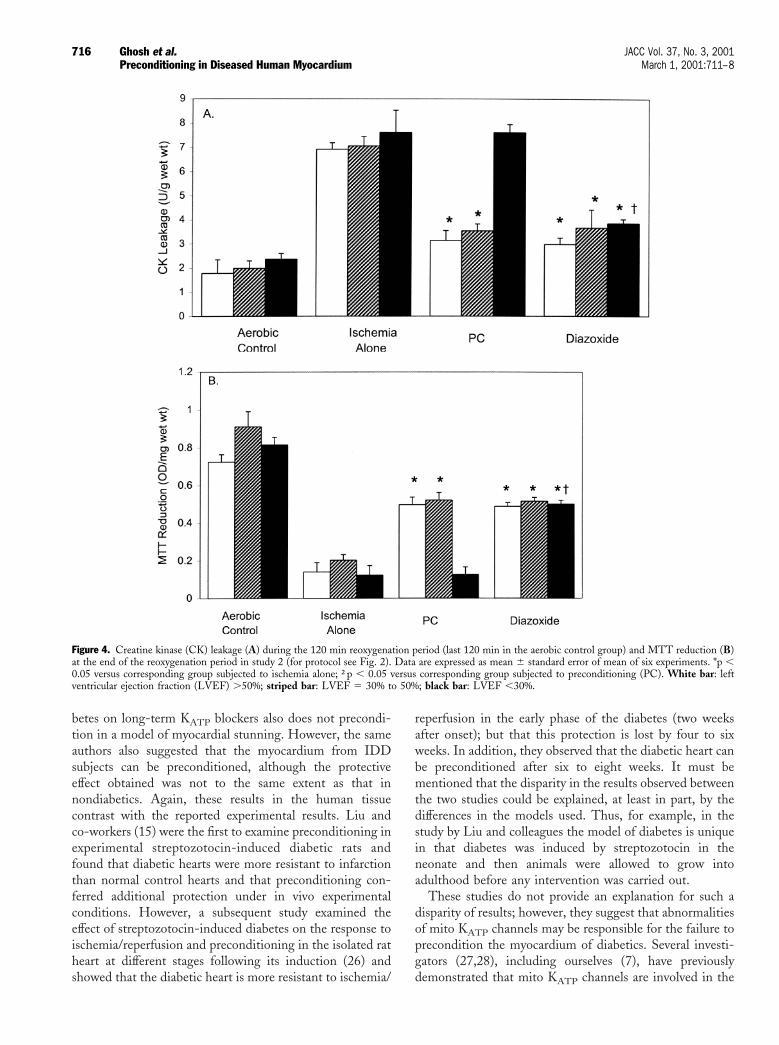
blocker, abolished the protection of PC in nondiabetics andin DCD and had no effect in the NIDD and IDD groups.The MTT results shown in Figure 3B are a mirror image ofthe CK leakage results with PC and diazoxide exhibiting asimilar protection in the nondiabetic and DCD groups andno protection in the NIDD and IDD groups. Overall, theresults suggest that changes in mito KATP channels inpatients with NIDD and IDD are the most likely cause forthe failure to precondition the myocardium. However, thepossibility that alterations in signal transduction pathwayscould also contribute cannot be completely excluded.Effect of PC in patients with contractile cardiac dysfunc-tion (Study 2). Figure 4A also shows that ischemia aloneresulted in a significant increase in CK leakage similar in allstudy groups regardless of the severity of cardiac dysfunc-tion. Importantly, PC was significantly protective; decreaseof CK leakage was observed in the groups with LVEF$30% but not in the group with LVEF ,30%. As observedin study 1, diazoxide mimicked the effect of PC on CKleakage in the groups with LVEF $30%. However, incontrast with the absence of protection by PC in the groupwith LVEF ,30%, diazoxide reduced CK leakage in theLVEF ,30% group to an extent similar to that seen inLVEF $30% groups. The results on MTT reductionshown in Figure 4B were again a mirror image of the CKleakage, suggesting that the cause for the absence ofprotection by PC in the LVEF ,30% group is probably dueto alterations in some element(s) of the signal transductionpathway that exclude the mito KATP channels.
It should be noted that in these two studies the numberof patients in each study group was too small for analysis ofthe possible influence of different clinical characteristics onthe effects of ischemia and preconditioning.
DISCUSSION
The major findings of this study are that myocardium frompatients with diabetes and poor cardiac function is notprotected from ischemic PC although the injury induced byischemia/reperfusion is not exacerbated in these conditions.Furthermore, the demonstration that activation of mitoKATP channels in myocardium from hearts with poorcardiac function mimics the protection induced by precon-ditioning in the myocardium from hearts with good con-tractility, but not from patients with diabetes, suggests thatthe failure to precondition in these conditions is due toalterations of different elements of the signal transductionpathway. Cardiovascular mortality is increased in patientswith heart failure (20,21) and diabetes (10) and thereforeany myocardial adaptation to ischemia would decreasemortality associated with these diseases. The results of ourstudies may have important clinical implications and anumber of points warrant further discussion.Preconditioning and diabetes. Insulin regulates the bal-ance of energy substrates available to the heart and alsoregulates metabolism and myocardial perfusion via actions
714 Ghosh et al. JACC Vol. 37, No. 3, 2001Preconditioning in Diseased Human Myocardium March 1, 2001:711–8

on various intracellular regulatory proteins and messengersystems (22). It is therefore conceivable that diabetes mayaffect ischemic injury. However, our results show that thediabetic human myocardium is not more sensitive to lethalischemic injury than the nondiabetic myocardium under theconditions of the present study. Because epidemiologicalstudies have clearly shown that patients with either IDD orNIDD are more prone to develop myocardial infarction andpostinfarction complications (10,23), it may be argued thatthe cause of cardiac complications in diabetics is not thetolerance of the heart to ischemia. The literature is incon-sistent with respect to how susceptible the hearts of diabeticanimals are to injury from ischemia/reperfusion, andwhereas some studies have observed a greater susceptibility
to ischemia/reperfusion injury (15), others have reported nosignificant effect (24,25). The divergent results may beexplained by experimental differences, but if our results inthe human atrial myocardium can be confirmed in ventric-ular myocardium then our attention to reduce cardiaccomplications in diabetes should be centered in the contextof blood components and the vasculature rather than in theown myocardium.
Our studies have demonstrated that PC affords protec-tion of the myocardium from patients with DCD but thatthis is lost when patients are on long-term hypoglycemics orbecome insulin dependent. These results are in agreementwith those reported by Cleveland et al. (16). These inves-tigators showed that myocardium from patients with dia-
Figure 3. Creatine kinase (CK) leakage (A) during the 120 min reoxygenation period (last 120 min in the aerobic control group) and MTT reduction (B)at the end of the reoxygenation period in study 1 (for protocol see Fig. 1). Data are expressed as mean 6 standard error of mean of six experiments. *p ,0.05 versus corresponding group subjected to ischemia alone. White bar 5 nondiabetics; black bar 5 diet-controlled diabetes; striped bar 5noninsulin-dependent diabetes; hatched bar 5 insulin-dependent diabetes; PC 5 preconditioning.
715JACC Vol. 37, No. 3, 2001 Ghosh et al.March 1, 2001:711–8 Preconditioning in Diseased Human Myocardium
betes on long-term KATP blockers also does not precondi-tion in a model of myocardial stunning. However, the sameauthors also suggested that the myocardium from IDDsubjects can be preconditioned, although the protectiveeffect obtained was not to the same extent as that innondiabetics. Again, these results in the human tissuecontrast with the reported experimental results. Liu andco-workers (15) were the first to examine preconditioning inexperimental streptozotocin-induced diabetic rats andfound that diabetic hearts were more resistant to infarctionthan normal control hearts and that preconditioning con-ferred additional protection under in vivo experimentalconditions. However, a subsequent study examined theeffect of streptozotocin-induced diabetes on the response toischemia/reperfusion and preconditioning in the isolated ratheart at different stages following its induction (26) andshowed that the diabetic heart is more resistant to ischemia/
reperfusion in the early phase of the diabetes (two weeksafter onset); but that this protection is lost by four to sixweeks. In addition, they observed that the diabetic heart canbe preconditioned after six to eight weeks. It must bementioned that the disparity in the results observed betweenthe two studies could be explained, at least in part, by thedifferences in the models used. Thus, for example, in thestudy by Liu and colleagues the model of diabetes is uniquein that diabetes was induced by streptozotocin in theneonate and then animals were allowed to grow intoadulthood before any intervention was carried out.
These studies do not provide an explanation for such adisparity of results; however, they suggest that abnormalitiesof mito KATP channels may be responsible for the failure toprecondition the myocardium of diabetics. Several investi-gators (27,28), including ourselves (7), have previouslydemonstrated that mito KATP channels are involved in the
Figure 4. Creatine kinase (CK) leakage (A) during the 120 min reoxygenation period (last 120 min in the aerobic control group) and MTT reduction (B)at the end of the reoxygenation period in study 2 (for protocol see Fig. 2). Data are expressed as mean 6 standard error of mean of six experiments. *p ,0.05 versus corresponding group subjected to ischemia alone; †p , 0.05 versus corresponding group subjected to preconditioning (PC). White bar: leftventricular ejection fraction (LVEF) .50%; striped bar: LVEF 5 30% to 50%; black bar: LVEF ,30%.
716 Ghosh et al. JACC Vol. 37, No. 3, 2001Preconditioning in Diseased Human Myocardium March 1, 2001:711–8
protection of ischemic PC. The findings by Smith et al. (29)that KATP channels are altered, with a greater outwardsingle-channel current in the ventricular myocardium ofdiabetic rats, further support this thesis. Clearly, furtherresearch is needed to fully elucidate how the alteration ofthis channel contributes to the failure to precondition themyocardium of diabetics.Preconditioning and the failing heart. Left ventricularhypertrophy and LV chamber dilation are among thecompensatory mechanisms of the failing heart. There isexperimental evidence that the hypertrophied myocardiumis at greater risk from ischemia/reperfusion injury, and it isgenerally believed that the failing heart is less tolerant tosuch injury. Our results, however, have shown for the firsttime that the effects of ischemia/reoxygenation are similar inthe failing and nonfailing myocardium. In addition, we havealso demonstrated for the first time that the myocardiumfrom hearts exhibiting a LVEF ,30% cannot be precondi-tioned. Our results contrast with those of Cleveland et al.(3) showing that the isolated ventricular trabeculae obtainedfrom patients undergoing cardiac transplantation can bepreconditioned. Again, the explanation for this differencecannot be found in the reported experimental studies, andwhereas some investigators have shown protection of thefailing heart by PC (3), others have observed no effect (30)or even further tissue damage (18). The diversity of resultsis not entirely surprising because of the lack of uniformity ofexperimental design and the degree of heart failure we haveshown in this study.
The protection observed with diazoxide in the myocar-dium from hearts with LVEF ,30% was commensuratewith the protection induced by PC in the myocardium fromnonfailing hearts. This supports the thesis that the failure toprecondition the failing human heart is not due to analteration in the response of the mitochondrial KATP chan-nel but is caused by abnormalities in other elements of thepreconditioning signaling pathway. Considerable evidenceindicates that PKC is intimately involved in ischemic PC(31), and the failure to precondition the failing heart may bedue to the chronic activation of PKC observed in thiscondition (32). There are several PKC isoforms, some ofwhich have been involved with PC, and in future studies thetype of isoforms and their expression in the myocardium ofthe failing heart should be investigated. Indeed, if specificPKC isoforms are proved to be responsible for the failure toprecondition, then their manipulation could become atherapeutic intervention to reduce myocardial injury inischemia/reperfusion of the failing heart.Possible limitations of the study and clinical implica-tions. A potential limitation of our study was the use ofatrial tissue as opposed to ventricular myocardium, andtherefore any extrapolation must be conducted with caution;however, Yellon and colleagues have suggested that PCexerts identical protection in atrial and ventricular myocar-dium (2). The present study also used atrial tissue tocharacterize the effects of ischemia and reperfusion in the
failing and diabetic human myocardium. However, atrialand ventricular myocardium possess characteristics of theirown that may influence susceptibility to ischemia/reperfusion injury, and as a consequence results from onemay not be applicable to the other. Thus the reporteddifferences in the distribution of potassium channels(33,34), which contribute to the characteristic differencesbetween atrial and ventricular action potentials, may deter-mine a different response to ischemia/reperfusion. Un-doubtedly, KATP channels are present in both atrium andventricle (33), although their density in both tissues isunknown. It must also be mentioned that the preparation issuperfused (“simulated ischemia”) as opposed to beingarterially perfused, and simulated ischemia is achieved byremoval of oxygen and blocking glycolytic ATP productionwith 2-deoxyglucose. This results in metabolic conditionswithin the myocardium that may be different from thosethat occur in the myocardium during clinical ischemia.
Another limitation might be that right atrial appendageswere obtained from patients subjected to various medicaltreatments (e.g., nitrates, beta-blockers, calcium antago-nists), which may have influenced ischemia/reperfusioninjury and the protection induced by PC. However, itshould be emphasized that all medication was stopped theday before surgery when specimens were taken for the study,and that significant effect of the medication was unlikelybecause all preparations responded to ischemia/reperfusionwith a similar degree of injury. The preparation used in thisstudy was not electrically stimulated (i.e., nonbeating) andtherefore one should be cautious when extrapolating to thein vivo situation.Conclusions. Preconditioning is a potent protective inter-vention whose use has been advocated in clinical situationssuch as angioplasty and cardiac surgery. The results of ourstudies have obvious clinical implications in that PC cannotbe beneficial to patients with NIDD or IDD and those withcardiac failure. The results also show that in the failing hearta degree of protection similar to that seen with PC can beobtained by the administration of a selective opener of mitoKATP channels, an intervention that is not effective indiabetics.
AcknowledgmentsWe thank Professor David Jones for his help with statisticalanalysis of the data.
Reprint requests and correspondence: Professor Manuel Gali-nanes, Division of Cardiac Surgery, Department of Surgery,University Hospitals Leicester, Glenfield Campus, Leicester LE39QP, United Kingdom. E-mail: [email protected].
REFERENCES
1. Carrol R, Yellon DM. Myocardial adaptation to ischemia—thepreconditioning phenomenon. Int J Cardiol 1999;68:S93–S101.
2. Speechly-Dick ME, Grover GJ, Yellon DM. Does ischemic precon-
717JACC Vol. 37, No. 3, 2001 Ghosh et al.March 1, 2001:711–8 Preconditioning in Diseased Human Myocardium
ditioning in the human involve PKC and the ATP-dependent K1
channel? Circ Res 1995;77:1030–5.3. Cleveland JC Jr., Wollmering MM, Meldrum DR, et al. Ischemic
preconditioning in human and rat ventricle. Am J Physiol 1996;271:H1786–94.
4. Pain TS, Cohen MV, Downey JM. The mitochondrial KATP channelmay be a trigger rather than the end-effector of preconditioning’santi-infarct effect (abstr.). Circulation 1999;100:I342.
5. Forbes RA, Steenburgen C, Murphy E. The protective effect ofdiazoxide is blocked by anti-oxidants (abstr.). Circulation 1999;100:I342.
6. Wang Y, Hirai K, Ashraf M. Activation of mitochondrial KATPchannel for cardiac protection against ischemic injury is dependent onPKC activity. Circ Res 1999;85:731–41.
7. Ghosh S, Standen NB, Galinanes M. Evidence for mitochondrialKATP channels as effectors of human myocardial preconditioning.Cardiovasc Res 2000;45:934–40.
8. Kannell WB, McGee DL. Diabetes and cardiovascular risk factors.Circulation 1979;59:8–13.
9. Roberts WC. Preventing and arresting coronary atherosclerosis. AmHeart J 1995;130:580–600.
10. Kannell WB. Role of diabetes in cardiac disease: conclusion frompopulation studies. In: Zonaraich S, editor. Diabetes and the Heart.Springfield, Illinois: Thomas Publishers, 1978:97–112.
11. Hearse DJ, Steward DA, Chain EB. Diabetes and the survival andrecovery of the anoxic myocardium. J Mol Cell Cardiol 1975;7:397–415.
12. Feuvray D, Idell-Wenger JR, Neely JR. Effects of ischemia on ratmyocardial function and metabolism in diabetes. Circ Res 1979;44:322–9.
13. Tani M, Neely JR. Hearts from diabetic rats are more resistant toin-vitro ischemia: possible role of altered Ca21 metabolism. Circ Res1998;62:931–40.
14. Tosaki A, Pali T, Droy-Lefaix M-T. Effects of ginkgo biloba extractand preconditioning on diabetic rat myocardium. Diabetologia 1996;39:1255–62.
15. Liu Y, Thornton JD, Cohen MV, et al. Streptozotocin-inducednon-insulin-dependent diabetes protects hearts from infarction. Cir-culation 1993;88:1273–8.
16. Cleveland JC Jr., Meldrum DR, Cain BS, et al. Oral sulphonylureahypoglycaemic agents prevent ischemic preconditioning in humanmyocardium. Two paradoxes revisited. Circulation 1997;96:29–32.
17. Scheuer J. Metabolic factors in myocardial failure. Circulation 1993;87VII54–57.
18. Dekker LRC, Rademaker H, Vermenlen JT, et al. Cellular uncouplingduring ischemia in hypertrophied and failing rabbit ventricular myo-cardium. Effects of preconditioning. Circulation 1998;97:1724–30.
19. Ghosh S, Standen NB, Galinanes M. Preconditioning the humanmyocardium by simulated ischemia: studies on the early and delayedprotection. Cardiovasc Res 2000;45:350–9.
20. Franciosa JA, Wilen M, Ziesche S, Cohn JN. Survival in men withsevere chronic left ventricular failure due to either coronary heartdisease or idiopathic dilated cardiomyopathy. Am. J Cardiol 1983;51:832–6.
21. Kjekshus J. Arrythmias and mortality in congestive cardiac failure.Am J Cardiol 1990;65:42I–48I.
22. Stanley WC, Lopaschuk GD, McCormack JG. Regulation of energysubstrate metabolism in the diabetic heart. Cardiovasc Res 1997;34:25–33.
23. Stone PH, Muller JE, Hartwell T. The effect of diabetes mellitus onprognosis and serial left ventricular function after acute myocardialinfarction: contribution of both coronary disease and diastolic leftventricular dysfunction to adverse prognosis. The MILIS StudyGroup. J Am Coll Cardiol 1989;14:49–57.
24. Paulson DJ. The diabetic heart is more sensitive to ischemic injury.Cardiovasc Res 1997;34:104–12.
25. Feuvray D, Lopaschuk GD. Controversies on the sensitivity of thediabetic heart to ischemic injury: the sensitivity of the diabetic heart toischemic injury is decreased. Cardiovasc Res 1997;34:113–20.
26. Tosaki A, Engelman DT, Engelman RM, Das DK. The evolution ofdiabetic response to ischemia/reperfusion and preconditioning inisolated working rat hearts. Cardiovasc Res 1996;31:526–36.
27. Liu Y, Sato T, O’Rourke B, Marban E. Mitochondrial ATP-dependent potassium channels: novel effectors of cardioprotection?Circulation 1998;97:2463–9.
28. Garlid K, Paucek P, Yarov-Yarovoy V, et al. Cardioprotective effect ofdiazoxide and its interaction with mitochondrial ATP-sensitive K1channels: a possible mechanism of cardioprotection. Circ Res 1997;81:1072–82.
29. Smith JM, Wahler GM. ATP-sensitive potassium channels are alteredin ventricular myocytes from diabetic rats. Mol Cell Biochem 1996;158:43–51.
30. Moolman JA, Genade S, Tromp E, Opie LH, Lochner A. Ischemicpreconditioning does not protect hypertrophied myocardium againstischemia. S Afr Med J 1997;87 Suppl 3:C151–6.
31. Cohen MV, Downey JM. Preconditioning during ischemia: basicmechanism and potential clinical applications. Cardiol Rev 1995;3:137–49.
32. Bowling N, Walsh RA, Song G, Estridge T, et al. Increased PKCactivity and expression of calcium sensitive isoforms in the failinghuman heart. Circulation 1999;99:384–91.
33. Heidbuchel H, Vereecke J, Carmeliet E. Three different potassiumchannels in human atrium: contribution to the basal potassiumconductance. Circ Res 1990;66:1277–86.
34. Amos GJ, Wettwer E, Metsger F, Li Q, Himmel HM. Differencesbetween outward currents of human atrial and subepicardial ventricularmyocytes. J Physiol 1996;491:31–50.
718 Ghosh et al. JACC Vol. 37, No. 3, 2001Preconditioning in Diseased Human Myocardium March 1, 2001:711–8
Related Documents