FACT Prevents the Accumulation of Free Histones Evicted from Transcribed Chromatin and a Subsequent Cell Cycle Delay in G1 Macarena Morillo-Huesca 1. , Douglas Maya 1. , Mari Cruz Mun ˜ oz-Centeno 1"* , Rakesh Kumar Singh 2 , Vincent Oreal 3 , Gajjalaiahvari Ugander Reddy 2 , Dun Liang 2 , Vincent Ge ´li 3 , Akash Gunjan 2 , Sebastia ´n Cha ´ vez 1"* 1 Departamento de Gene ´ tica, Universidad de Sevilla, Seville, Spain, 2 Department of Biomedical Sciences, College of Medicine, Florida State University, Tallahassee, Florida, United States of America, 3 Laboratoire d’Instabilite ´ Ge ´ne ´tique et Cance ´ rogene `se, Institut de Biologie Struturale et Microbiologie, Centre National de la Recherche Scientifique, Marseille, France Abstract The FACT complex participates in chromatin assembly and disassembly during transcription elongation. The yeast mutants affected in the SPT16 gene, which encodes one of the FACT subunits, alter the expression of G1 cyclins and exhibit defects in the G1/S transition. Here we show that the dysfunction of chromatin reassembly factors, like FACT or Spt6, down- regulates the expression of the gene encoding the cyclin that modulates the G1 length (CLN3) in START by specifically triggering the repression of its promoter. The G1 delay undergone by spt16 mutants is not mediated by the DNA–damage checkpoint, although the mutation of RAD53, which is otherwise involved in histone degradation, enhances the cell-cycle defects of spt16-197. We reveal how FACT dysfunction triggers an accumulation of free histones evicted from transcribed chromatin. This accumulation is enhanced in a rad53 background and leads to a delay in G1. Consistently, we show that the overexpression of histones in wild-type cells down-regulates CLN3 in START and causes a delay in G1. Our work shows that chromatin reassembly factors are essential players in controlling the free histones potentially released from transcribed chromatin and describes a new cell cycle phenomenon that allows cells to respond to excess histones before starting DNA replication. Citation: Morillo-Huesca M, Maya D, Mun ˜ oz-Centeno MC, Singh RK, Oreal V, et al. (2010) FACT Prevents the Accumulation of Free Histones Evicted from Transcribed Chromatin and a Subsequent Cell Cycle Delay in G1. PLoS Genet 6(5): e1000964. doi:10.1371/journal.pgen.1000964 Editor: Sue Biggins, Fred Hutchinson Cancer Research Center, United States of America Received August 21, 2009; Accepted April 20, 2010; Published May 20, 2010 Copyright: ß 2010 Morillo-Huesca et al. This is an open-access article distributed under the terms of the Creative Commons Attribution License, which permits unrestricted use, distribution, and reproduction in any medium, provided the original author and source are credited. Funding: This work has been supported by the Spanish Ministry of Education and Science (grants BMC2003-07072-C03-01 and BFU2007-67575-C03-02/BMC to SC and fellowship to MM-H) and by the Andalusian Government (BIO-271 and fellowship to DM). Work in VG’s laboratory is supported by the ‘‘Ligue Nationale contre le Cancer’’ (e ´quipe labe ´lise ´e and fellowship to VO). The research work done in AG’s laboratory is supported by a Bankhead-Coley Cancer Research Program grant (07BN-02) from the Florida Department of Health, and partly by an NIH grant (R15GM079678-01). The funders had no role in study design, data collection and analysis, decision to publish, or preparation of the manuscript. Competing Interests: The authors have declared that no competing interests exist. * E-mail: [email protected] (SC); [email protected] (MCM-C) . These authors contributed equally to this work. " These authors were joint senior authors of this work. Introduction The FACT complex plays an important role by allowing RNA polymerase II (Pol II) to transcribe through chromatin (reviewed by [1,2]), the only factor known to date that is able to stimulate Pol II-dependent transcription elongation through chromatin in a highly purified system [3,4]. Yeast FACT is required in vivo to transcribe genes with highly positioned nucleosomes at the 59 end of the transcribed region [5], and several lines of evidence of other organisms also support that FACT plays an important role in transcription elongation in vivo [6–10]. In spite of its role in elongation, several in vivo and in vitro approaches indicate an additional role of yFACT in establishing transcription initiation complexes by promoting TBP binding to core promoters in a TFIIA-dependent manner [11,12]). Finally and in addition to its role in transcription, FACT also plays an important function during DNA replication [13–15]. In humans, the FACT complex is composed of two proteins, p140 and SSRP1, which are highly homologous to the essential yeast proteins Spt16/Cdc68/Ssf1 (hereafter referred to as Spt16) and Pob3, respectively [16]. SPT16 had been previously identified as both a CDC gene [17], and also as a recessive suppressor of the deletion of SWI4, a transcription factor required for the high-level expression of the G1 cyclin genes, CLN1 and CLN2 [18]. Besides, Spt16 had also been described as a protein involved in transcription since several spt16 alleles suppress the transcriptional effects of Ty insertions in yeast (Spt- phenotype) [19]. yFACT has been reported to interact physically or genetically with other factors related to histone modifications and chromatin remodeling, like the Paf complex, the ATP-dependent chromatin factor Chd1 and the NuA3 histone acetyltransferase complex [11,20–22]. A reciprocal regulation of the FACT function by H2B ubiquitination has also been described [23]. In agreement with these findings, yFACT and the HMG-box protein Nhp6 have PLoS Genetics | www.plosgenetics.org 1 May 2010 | Volume 6 | Issue 5 | e1000964

Welcome message from author
This document is posted to help you gain knowledge. Please leave a comment to let me know what you think about it! Share it to your friends and learn new things together.
Transcript
FACT Prevents the Accumulation of Free HistonesEvicted from Transcribed Chromatin and a SubsequentCell Cycle Delay in G1Macarena Morillo-Huesca1., Douglas Maya1., Mari Cruz Munoz-Centeno1"*, Rakesh Kumar Singh2,
Vincent Oreal3, Gajjalaiahvari Ugander Reddy2, Dun Liang2, Vincent Geli3, Akash Gunjan2, Sebastian
Chavez1"*
1 Departamento de Genetica, Universidad de Sevilla, Seville, Spain, 2 Department of Biomedical Sciences, College of Medicine, Florida State University, Tallahassee, Florida,
United States of America, 3 Laboratoire d’Instabilite Genetique et Cancerogenese, Institut de Biologie Struturale et Microbiologie, Centre National de la Recherche
Scientifique, Marseille, France
Abstract
The FACT complex participates in chromatin assembly and disassembly during transcription elongation. The yeast mutantsaffected in the SPT16 gene, which encodes one of the FACT subunits, alter the expression of G1 cyclins and exhibit defectsin the G1/S transition. Here we show that the dysfunction of chromatin reassembly factors, like FACT or Spt6, down-regulates the expression of the gene encoding the cyclin that modulates the G1 length (CLN3) in START by specificallytriggering the repression of its promoter. The G1 delay undergone by spt16 mutants is not mediated by the DNA–damagecheckpoint, although the mutation of RAD53, which is otherwise involved in histone degradation, enhances the cell-cycledefects of spt16-197. We reveal how FACT dysfunction triggers an accumulation of free histones evicted from transcribedchromatin. This accumulation is enhanced in a rad53 background and leads to a delay in G1. Consistently, we show that theoverexpression of histones in wild-type cells down-regulates CLN3 in START and causes a delay in G1. Our work shows thatchromatin reassembly factors are essential players in controlling the free histones potentially released from transcribedchromatin and describes a new cell cycle phenomenon that allows cells to respond to excess histones before starting DNAreplication.
Citation: Morillo-Huesca M, Maya D, Munoz-Centeno MC, Singh RK, Oreal V, et al. (2010) FACT Prevents the Accumulation of Free Histones Evicted fromTranscribed Chromatin and a Subsequent Cell Cycle Delay in G1. PLoS Genet 6(5): e1000964. doi:10.1371/journal.pgen.1000964
Editor: Sue Biggins, Fred Hutchinson Cancer Research Center, United States of America
Received August 21, 2009; Accepted April 20, 2010; Published May 20, 2010
Copyright: � 2010 Morillo-Huesca et al. This is an open-access article distributed under the terms of the Creative Commons Attribution License, which permitsunrestricted use, distribution, and reproduction in any medium, provided the original author and source are credited.
Funding: This work has been supported by the Spanish Ministry of Education and Science (grants BMC2003-07072-C03-01 and BFU2007-67575-C03-02/BMC toSC and fellowship to MM-H) and by the Andalusian Government (BIO-271 and fellowship to DM). Work in VG’s laboratory is supported by the ‘‘Ligue Nationalecontre le Cancer’’ (equipe labelisee and fellowship to VO). The research work done in AG’s laboratory is supported by a Bankhead-Coley Cancer Research Programgrant (07BN-02) from the Florida Department of Health, and partly by an NIH grant (R15GM079678-01). The funders had no role in study design, data collectionand analysis, decision to publish, or preparation of the manuscript.
Competing Interests: The authors have declared that no competing interests exist.
* E-mail: [email protected] (SC); [email protected] (MCM-C)
. These authors contributed equally to this work.
" These authors were joint senior authors of this work.
Introduction
The FACT complex plays an important role by allowing RNA
polymerase II (Pol II) to transcribe through chromatin (reviewed
by [1,2]), the only factor known to date that is able to stimulate Pol
II-dependent transcription elongation through chromatin in a
highly purified system [3,4]. Yeast FACT is required in vivo to
transcribe genes with highly positioned nucleosomes at the 59 end
of the transcribed region [5], and several lines of evidence of other
organisms also support that FACT plays an important role in
transcription elongation in vivo [6–10]. In spite of its role in
elongation, several in vivo and in vitro approaches indicate an
additional role of yFACT in establishing transcription initiation
complexes by promoting TBP binding to core promoters in a
TFIIA-dependent manner [11,12]). Finally and in addition to its
role in transcription, FACT also plays an important function
during DNA replication [13–15].
In humans, the FACT complex is composed of two proteins,
p140 and SSRP1, which are highly homologous to the essential
yeast proteins Spt16/Cdc68/Ssf1 (hereafter referred to as Spt16)
and Pob3, respectively [16]. SPT16 had been previously identified
as both a CDC gene [17], and also as a recessive suppressor of the
deletion of SWI4, a transcription factor required for the high-level
expression of the G1 cyclin genes, CLN1 and CLN2 [18]. Besides,
Spt16 had also been described as a protein involved in
transcription since several spt16 alleles suppress the transcriptional
effects of Ty insertions in yeast (Spt- phenotype) [19].
yFACT has been reported to interact physically or genetically
with other factors related to histone modifications and chromatin
remodeling, like the Paf complex, the ATP-dependent chromatin
factor Chd1 and the NuA3 histone acetyltransferase complex
[11,20–22]. A reciprocal regulation of the FACT function by H2B
ubiquitination has also been described [23]. In agreement with
these findings, yFACT and the HMG-box protein Nhp6 have
PLoS Genetics | www.plosgenetics.org 1 May 2010 | Volume 6 | Issue 5 | e1000964
been shown to form a heterodimer capable of binding nucleo-
somes [24] and of reorganizing them in vitro [25,26]. Both the
yFACT subunits are able to bind H3/H4 tetramers and H2A/
H2B dimers, sometimes in a functionally redundant manner
[27,28]. These interactions are thought to allow FACT to
destabilize nucleosomes during transcription [26,29].
Some spt16 alleles are synthetically lethal with mutations
affecting chromatin assembly [30]. Moreover, they lead to the
activation of cryptic transcription initiation sites within coding
regions, indicating that FACT, together with other factors like
Spt6, also plays a role in maintaining the integrity of the chromatin
structure during transcription [9,31–34].
Several spt16 mutants show defects while progressing through
START, the main regulatory event in the G1 phase of the cell
cycle [17,35]. At a non-permissive temperature, the G1 phenotype
of these spt16 mutants has been accounted for by the drastic
reduction in the expression of CLN1, CLN2 and CLN3, the genes
encoding the three G1 cyclins [35,36]. CLN1 and CLN2 are able to
self-regulate their expression by a positive feed-back mechanism
[37], but the regulation of G1 length requires the activation of the
cyclin-dependent kinase Cdc28 (Cdk1) by Cln3 [38–41]. Cln3-
associated Cdk1 binds SBF (Swi4-Swi6) to the CLN1 and CLN2
promoters where it phosphorylates the negative regulator of
START, Whi5 [42]. This phosphorylation promotes its release
from SBF and leads to the activation of the CLN1 and CLN2
promoters [43,44]. SBF-dependent recruitment of FACT plays an
important role in this activation, which promotes the G1/S
transition [45]. Notably, the kinase activity of Cln1,2-Cdk1
triggers the degradation of the cyclin-dependent kinase inhibitor
Sic1 which no longer inhibits the S phase-promoting complex
Clb5,6-Cdc28 [46,47].
Another key regulatory process during the G1/S transition is
the induction of histone genes, which allows the coupling of bulk
histone synthesis to ongoing DNA replication. In proliferating
cells, the synthesis of the vast majority of histones occurs during
the S-phase of the cell cycle. The tight cell cycle regulation of the
histone genes results from their transcriptional repression in phases
G1 and G2, their transcriptional activation just before the S-phase
and the post-transcriptional regulation of their mRNAs. During
the S-phase, histone genes can also respond to changes; for
instance, the accumulation of histones in response to the genotoxic
agents interfering with DNA replication induces their repression
(reviewed by [48]).
In recent years, a novel mechanism in budding yeast preventing
the accumulation of free histones and which is superimposed upon
the regulation of histone gene transcription and mRNA stability
has been described [49]. This mechanism involves the use of the
DNA damage checkpoint protein kinase Rad53 as part of a
surveillance process that not only monitors the accumulation of
excess histones, but also induces their degradation. This
degradation is controlled by phosphorylation and is carried out
by the proteosome in an ubiquitylation-mediated manner [50].
Excess histones are thought to be generated at the end of the
normal S-phase or in response to an abrupt decrease of DNA
synthesis following DNA damage. This mechanism is dependent
on neither the checkpoint kinases Mec1 and Tel1 nor other DNA-
damage checkpoint proteins (reviewed in [51]).
Some aspects of the G1 phenotype of the spt16 mutants remain
unknown. It is not clear whether FACT plays a direct role in the
regulation of CLN3. Alternatively, the decreased expression of
CLN3 might be a physiological response to FACT inactivation. We
investigated this question and found that the inactivation of FACT
down-regulates the CLN3 promoter in START. This phenomenon
coincides with the accumulation of free histones and is enhanced
by the mutation of the free histones controller, RAD53. We also
found that the forced entry of FACT-deficient cells into the S-
phase lowers their viability. Finally, we discovered that the
overexpression of histones in the wild-type cells decreases CLN3
transcription in START and leads to a delay in G1. We propose
that the accumulation of free histones triggers the down-regulation
of CLN3, thereby contributing to control the excess histones before
starting DNA replication. We further propose that the main
potential source of free histones is transcribed chromatin and that
chromatin reassembly factors play an essential protective role in
this respect.
Results
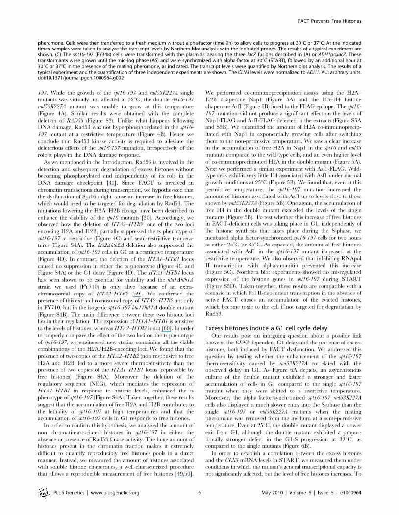
FACT inactivation causes a down-regulation of the CLN3promoter in START
The arrest of yeast cells in G1 after Spt16 inactivation has been
suggested to be a possible direct consequence of a very strict
requirement of Spt16 for CLN1, CLN2 and CLN3 transcription
[35]. FACT has been shown to participate directly in the
activation of the CLN1 and CLN2 promoters after START [45].
In order to explore the effect of FACT inactivation on the previous
step, we quantified the mRNA levels of CLN3 in alpha factor-
synchronized spt16-197 cells at a non-permissive temperature (see
Materials and Methods for the ranges of permissive and non-
permissive temperatures ranges). We treated cells for two hours
with the pheromone at 30uC, then for one a further hour at either
30uC or 35uC in the continuous presence of alpha-factor. Next we
released the cells from the arrest and analyzed CLN3 mRNA by
Northern blotting (Figure 1). In agreement with previously
published results based on asynchronous cells [35], the CLN3
mRNA levels in the pheromone-treated spt16-197 cells at 35uCwere clearly lower than in the wild-type cells (see time 0 in
Figure 1B). When cultures were released from alpha-factor, the
wild-type cells progressed into the S-phase at any temperature,
whereas spt16-197 cells entered the S-phase only at 30uC, with
most spt16-197 cells remaining in G1 at the restrictive temperature
(see times 10, 20 and 45 min in Figure 1A). The CLN3 mRNA
levels in the spt16-197cells released from alpha-factor at 37uC
Author Summary
Lengthy genomic DNA is packed in a highly organizednucleoprotein structure called chromatin, whose basicsubunit is the nucleosome which is formed by DNAwrapped around an octamer of proteins called histones.Nucleosomes need to be disassembled to allow DNAtranscription by RNA polymerases. An essential factor forthe disassembly/reassembly process during DNA transcrip-tion is the FACT complex. We investigated a phenotype ofyeast FACT mutants, a delay in a specific step of the cellcycle division process immediately prior to starting DNAreplication. The dysfunction caused by the FACT mutationcauses a downregulation of a gene, CLN3, which controlsthe length of that specific step of the cell cycle. FACTdysfunction also increases the level of the free histonesreleased from chromatin during transcription, and thephenotype of the Spt16 mutant is enhanced by a secondmutation affecting a protein that regulates DNA repair andexcess histone degradation. Moreover, we show that theoverexpression of histones causes a cell cycle delay beforeDNA replication in wild-type cells. Our results point out aso-far unknown connection between chromatin dynamicsand the regulation of the cell cycle.
FACT Prevents Free Histones
PLoS Genetics | www.plosgenetics.org 2 May 2010 | Volume 6 | Issue 5 | e1000964

FACT Prevents Free Histones
PLoS Genetics | www.plosgenetics.org 3 May 2010 | Volume 6 | Issue 5 | e1000964

remained very low. In contrast, the mRNA levels of ADH1, a
constitutive non-cycling gene, were only partially affected by Spt16
inactivation (Figure 1B), which is in agreement with the limited
effect of spt16-197 at restrictive temperatures in a broad set of non-
cyclin genes [35,36]. The occupancy of CLN3 and ADH1 by the
RNA polymerase II (RNApol II) paralleled their mRNA levels. As
Figure 1C depicts, the amount of RNApol II bound to the CLN3
transcribed region at 35uC was significantly lower in spt16-197
(52%) than in the wild type, whereas the variation of RNApol II
binding to ADH1 was slighter.
The fact that the CLN3 expression markedly reduced in
response to Spt16 inactivation might be due to either a direct
effect of FACT dysfunction on CLN3 transcription or a yet
unidentified signaling pathway targeting the CLN3 promoter in
response to FACT inactivation. In order to distinguish between
these possibilities, we first analyzed the response of a reporter fused
to the CLN3 promoter upon Spt16 inactivation in alpha-factor-
synchronized cells. To construct this fusion, we combined the
entire intergenic region between the neighboring CLN3 and CYC3
coding regions and the coding region of E. coli lacZ (Figure 2A). We
chose lacZ since we had previously showed that the transcription
elongation of this reporter gene is not sensitive to FACT
dysfunction [5]. As shown in Figure 2B, lacZ mRNA was hardly
detectable when the synchronized spt16-197 cells were incubated
at the restrictive temperature, indicating that the CLN3pr::lacZ
reporter (fusion 1) behaved similarly to the endogenous CLN3
mRNA. In contrast, the mRNA levels of an ADH1pr::lacZ
transcriptional fusion did not lower under the same conditions.
This result indicates that the promoter region of CLN3 mediates
the drop in its mRNA levels after FACT inactivation.
We further constructed additional CLN3pr::lacZ fusions con-
taining different segments of the CLN3-CYC3 intergenic region
(Figure 2A). No difference in copy number among the reporter
plasmids was detected by quantitative PCR (Figure S1). We
measured the lacZ mRNA levels expressed by these fusions in the
spt16-197 cells synchronized in START. The expression patterns
of the complete CLN3pr::lacZ (fusion 1) and ADH1pr::lacZ were in
agreement with the experiments described above (Figure 2C).
Fusion 2, which contains the entire intergenic region, except the
CLN3 59UTR and the CYC3 termination region (2999, 2339)
(Figure 2A), retained sensitivity to Spt16 inactivation as it dropped
as the lacZ mRNA levels did after Spt16 inactivation (Figure 2C).
A significant influence of mRNA instability in this decrease was
ruled out because two completely different mRNAs (lacZ and
CLN3) responded similarly to FACT inactivation when they were
driven by the same promoter (CLN3pr). In contrast, when lacZ
mRNA was driven by the ADH1 promoter, it did not show any
significant variation in spt16-197 at the restrictive temperature
(Figure 2C). In short, spt16-197 does not influence the expression
of CLN3 at a post-initiation level. However, Spt16 may also play a
role in transcription initiation by facilitating TBP binding to the
core promoters. We constructed CLN3pr::lacZ fusion number 3
(2472, 2389), which contains the minimal core promoter of
CLN3 (Figure 2A). In spite of the weak expression of this fusion
(five fold lower than fusion number 2 in asynchronous cultures), its
mRNA level was not significantly influenced by Spt16 inactivation
(Figure 2C). These results, in addition to the absence of FACT
binding to the CLN3 promoter during START (David Stillman,
personal communication), indicate that the effect of Spt16
inactivation on the expression of CLN3 in START is not due to
the specific involvement of FACT in the transcription of this gene
in this particular step of the cell cycle. Instead, our results suggest
the existence of a control mechanism mediated by the promoter of
CLN3 which regulates the G1/S transition in response to FACT
dysfunction. Such a mechanism might protect the cell from the
deleterious effect of entering the S-phase under these conditions.
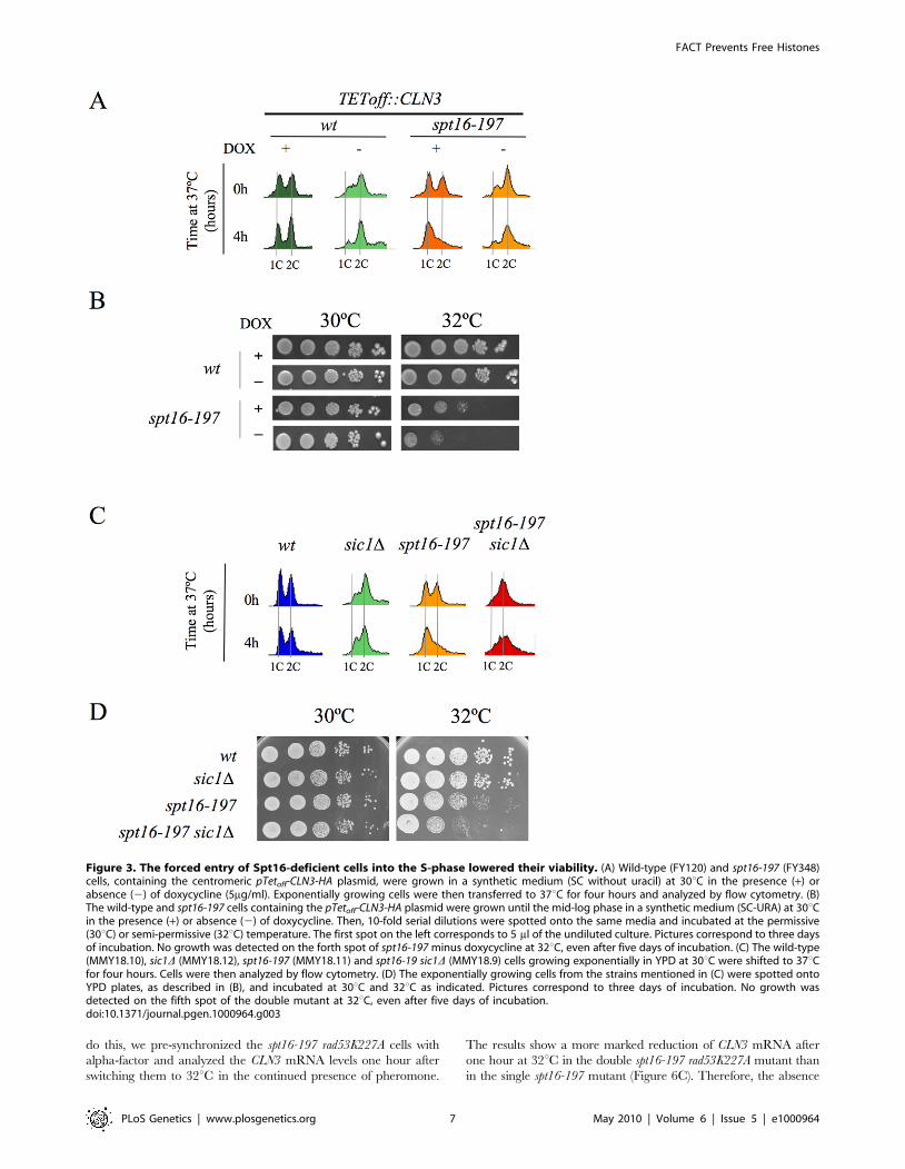
We tested this hypothesis by forcing the entry of spt16-197 cells
into the S-phase. First, we went about this by overexpressing CLN3
with a Tetoff::CLN3 construct (negatively regulated by doxycycline)
which suppressed the accumulation of spt16-197 cells in G1 at a
restrictive temperature (Figure 3A). We observed a negative effect
of the overexpression of CLN3 on the viability of spt16-197 cells at
a semi-permissive temperature (Figure 3B). Similar results were
obtained in those cells lacking Sic1, the inhibitor of the Cdc28-Clb
complexes that negatively regulates entry into the S-phase
(Figure 3C and 3D). These data reveal that the forced progression
of spt16-197 cells into the S-phase at semi-permissive temperatures
is deleterious; therefore, the G1 delay triggered by the inactivation
of FACT is cell-protective.
The lethality associated with FACT dysfunction relates tothe accumulation of the free histones evicted duringtranscription
The association between impairment of transcription elongation
and genome instability is well established [52–54]. Since FACT
plays a role during elongation, we wondered whether the G1 delay
induced by FACT inactivation could be due to the action of the
canonical DNA-damage checkpoint. To test this hypothesis, we
investigated the possible implication of Rad9, a component of the
DNA-damage sensor machinery operating in G1 [55,56]. We
performed a FACS analysis with strains carrying the spt16-197 and
rad9D mutations. The profile of the spt16-197 rad9D double mutant
after four hours at a restrictive temperature was almost identical to
that of the single spt16-197 mutant (Figure S2A), indicating that
the G1 arrest produced by FACT inactivation is proficient in the
absence of Rad9. Accordingly, no genetic interaction between
spt16-197 and rad9D was detected when we analyzed the
thermosensitivity of the double mutant (Figure S2B). We further
tested whether spt16-197 exhibited any aggravation of its ts
phenotype in the absence of Mec1, the key kinase of the DNA-
damage checkpoint pathway [57]. As with rad9D, the deletion of
MEC1 in spt16-197 did not affect its thermosensitive (ts) phenotype
(Figure 4A).
It was intriguing to note that of all the DNA-damage checkpoint
genes we tested, only the mutation of RAD53, which encodes
another essential protein kinase for the DNA-damage checkpoint
[58], led to a clear genetic interaction with spt16-197. Indeed the
rad53K227A allele, which abolishes most Rad53 kinase activity,
dramatically enhanced the thermosensitive phenotype of spt16-
Figure 1. CLN3 expression is down-regulated after Spt16 inactivation. Wild-type (FY120) and spt16-197 (FY348) cells grown asynchronously (AS)were synchronized at START by treatment with alpha-factor for two hours at 30uC (ST), followed by one additional one hour at 30uC or 35uC in thepresence of the mating pheromone. Cells were then released from the arrest at time 0 at either 30uC or 35uC by washing out the alpha-factor. Sampleswere taken at different time points to analyze the DNA content by flow cytometry and the proportion of unbudded cells by microscopy (A), and toquantify the mRNA levels of the indicated genes by Northern blot analysis (B). Transcripts levels were represented as arbitrary units (AU) afternormalization with 18S rRNA. The results of a typical experiment and the quantification of three independent experiments are shown. (C) Relative RNApolII binding to the transcribed region of CLN3 and ADH1 in spt16-197. Cells were synchronized with alpha-factor for two hours at 30uC, followed by oneadditional one hour at 30uC or 35uC in the presence of the mating pheromone. The 8WG16 antibody, recognizing the CTD repeats of Rpb1, was used.doi:10.1371/journal.pgen.1000964.g001
FACT Prevents Free Histones
PLoS Genetics | www.plosgenetics.org 4 May 2010 | Volume 6 | Issue 5 | e1000964
Figure 2. The CLN3 promoter mediated the down-regulation of its mRNA levels after Spt16 inactivation. (A) Diagram of the plasmid-borne lacZ fusions, encoding E. coli ß-galactosidase, that were generated by introducing different fragments of the CLN3 promoter region.Constructions were numbered 1 to 3. The positions of the early cell cycle boxes (ECB) and the TATA box are indicated. An ADH1 promoter-lacZ fusionwas used as a constitutively expressed control. (B) The mRNA levels of CLN3pr::lacZ and ADH1pr::lacZ fusion. The spt16-197 (FY348) strain, transformedwith the plasmids bearing lacZ fusion number 1 (CLN3pr::lacZ) or ADH1pr::lacZ, was grown until the mid-log phase (AS), and was then synchronized atSTART (ST) by alpha-factor treatment for 2h at 30uC, followed by one additional hour of incubation at 30uC or 37uC in the presence of the mating
FACT Prevents Free Histones
PLoS Genetics | www.plosgenetics.org 5 May 2010 | Volume 6 | Issue 5 | e1000964
197. While the growth of the spt16-197 and rad53K227A single
mutants was virtually not affected at 32uC, the double spt16-197
rad53K227A mutant was unable to grow at this temperature
(Figure 4A). Similar results were obtained with the complete
deletion of RAD53 (Figure S3). Unlike what happens following
DNA damage, Rad53 was not hyperphosphorylated in the spt16-
197 mutant at a restrictive temperature (Figure 4B). Hence we
conclude that Rad53 kinase activity is required to alleviate the
deleterious effects of the spt16-197 mutation, irrespectively of the
role it plays in the DNA damage response.
As we mentioned in the Introduction, Rad53 is involved in the
detection and subsequent degradation of excess histones without
becoming phosphorylated and independently of its role in the
DNA damage checkpoint [49]. Since FACT is involved in
chromatin transactions during transcription, we hypothesized that
the dysfunction of Spt16 might cause an increase in free histones,
which would need to be targeted for degradation by Rad53. The
mutations lowering the H2A–H2B dosage have been described to
enhance the viability of the spt16 mutants [30]. Accordingly, we
observed how the deletion of HTA2–HTB2, one of the two loci
encoding H2A and H2B, partially suppressed the ts phenotype of
spt16-197 at restrictive (Figure 4C) and semi-restrictive tempera-
tures (Figure S4A). The hta2Dhtb2D deletion also suppressed the
accumulation of spt16-197 cells in G1 at a restrictive temperature
(Figure 4D). In contrast, the deletion of the HTA1–HTB1 locus
caused no suppression in either the ts phenotype (Figure 4C and
Figure S4A) or the G1 delay (Figure 4D). The HTA1–HTB1 locus
has been shown to be essential for viability and the hta1Dhtb1Dstrain we used (FY710) is only alive because of an extra-
chromosomal copy of HTA2–HTB2 [59]. We confirmed the
presence of this extra-chromosomal copy of HTA2–HTB2 not only
in FY710, but in the isogenic spt16-197 hta1/htb1D double mutant
(Figure S4B). The main difference between these two histone loci
lies in their regulation. The expression of HTA1–HTB1 is sensitive
to the levels of histones, whereas HTA2–HTB2 is not [60]. In order
to properly compare the effect of the two loci on the ts phenotype
of spt16-197, we engineered new strains containing all the viable
combinations of the H2A/H2B-encoding loci. We found that the
presence of two copies of the HTA2–HTB2 (non responsive to free
H2A and H2B) led to a more severe thermosensitivity than the
presence of two copies of the HTA1–HTB1 locus (repressible by
free histones) (Figure S4A). Moreover the deletion of the
regulatory sequence (NEG), which mediates the repression of
HTA1–HTB1 in response to histone levels, enhanced the ts
phenotype of spt16-197 (Figure S4A). Taken together, these results
suggest that the accumulation of free H2A and H2B contributes to
the lethality of spt16-197 at high temperatures and that the
accumulation of spt16-197 cells in G1 responds to free histones.
In order to confirm this hypothesis, we analyzed the amount of
non chromatin-associated histones in spt16-197 in either the
absence or presence of Rad53 kinase activity. The huge amount of
histones present in the chromatin fraction makes it extremely
difficult to quantify reproducibly free histones pools in a direct
manner. Instead, we measured the amount of histones associated
with soluble histone chaperones, a well-characterized procedure
that allows a reproducible measurement of free histones [49,50].
We performed co-immunoprecipitation assays using the H2A–
H2B chaperone Nap1 (Figure 5A) and the H3–H4 histone
chaperone Asf1 (Figure 5B) fused to the FLAG epitope. The spt16-
197 mutation did not produce a significant effect on the levels of
Nap1-FLAG and Asf1-FLAG detected in the extracts (Figure S5A
and S5B). We quantified the amount of H2A co-immunoprecip-
itated with Nap1 in exponentially growing cells after switching
them to the non-permissive temperature. We saw a clear increase
in the accumulation of free H2A in Nap1 in the spt16 and rad53
mutants compared to the wild-type cells, and an even higher level
of co-immunoprecipitated H2A in the double mutant (Figure 5A).
Next we performed a similar experiment with Asf1-FLAG. Wild-
type cells exhibit very little H4 associated with Asf1 under normal
growth conditions at 25uC (Figure 5B). We found that, even at this
permissive temperature, the spt16-197 mutation increased the
amount of histones associated with Asf1 up to levels close to those
shown by rad53K227A (Figure 5B). One again, the accumulation of
free H4 in the double mutant exceeded the levels of the single
mutants (Figure 5B). To test whether this increase of free histones
in FACT-deficient cells was taking place in G1, independently of
the histone synthesis that takes place during the S-phase, we
incubated alpha factor-synchronized spt16-197 cells for two hours
at either 25uC or 35uC. As expected, the amount of free histones
associated with Asf1 in the spt16-197 mutant increased at the
restrictive temperature. We also observed that inhibiting RNApol
II transcription with alpha-amanitin prevented this increase
(Figure 5C). Northern blot experiments showed no misregulated
expression of the histone genes in spt16-197 during START
(Figure S5D). Taken together, these results are compatible with a
scenario in which Pol II-dependent transcription in the absence of
active FACT causes an accumulation of the evicted histones,
which become toxic to the cell if not targeted for degradation by
Rad53.
Excess histones induce a G1 cell cycle delayOur results pose an intriguing question about a possible link
between the CLN3-dependent G1 delay and the presence of excess
histones, both induced by FACT dysfunction. We addressed this
question by testing whether the enhancement of the spt16-197
thermosensitivity caused by rad53K227A correlated with the
observed delay in G1. As Figure 6A depicts, an asynchronous
culture of the double mutant exhibited a stronger and faster
accumulation of cells in G1 compared to the single spt16-197
mutant when they were shifted to a restrictive temperature.
Moreover, the alpha-factor-synchronized spt16-197 rad53K227A
cells also displayed a much slower entry into the S-phase than the
single spt16-197 or rad53K227A mutants when the mating
pheromone was removed from the medium at a semi-permissive
temperature. Even at 25uC, the double mutant displayed a slower
exit from G1, although the double mutant exhibited a propor-
tionally stronger defect in the G1-S progression at 32uC, as
compared to the single mutants (Figure 6B).
In order to establish a correlation between the excess histones
and the CLN3 mRNA levels in START, we measured them under
conditions in which the mutant’s general transcriptional capacity is
not significantly affected, but the level of free histones increases. To
pheromone. Cells were then transferred to a fresh medium without alpha-factor (time 0h) to allow cells to progress at 30uC or 37uC. At the indicatedtimes, samples were taken to analyze the transcript levels by Northern blot analysis with the indicated probes. The results of a typical experiment areshown. (C) The spt16-197 (FY348) cells were transformed with the plasmids bearing the three lacZ fusions described in (A) or ADH1pr::lacZ. Thesetransformants were grown until the mid-log phase (AS) and were synchronized with alpha-factor at 30uC (START), followed by an additional hour at30uC or 37uC in the presence of the mating pheromone, as indicated. The transcript levels were quantified by Northern blot analysis. The results of atypical experiment and the quantification of three independent experiments are shown. The CLN3 levels were normalized to ADH1. AU: arbitrary units.doi:10.1371/journal.pgen.1000964.g002
FACT Prevents Free Histones
PLoS Genetics | www.plosgenetics.org 6 May 2010 | Volume 6 | Issue 5 | e1000964
do this, we pre-synchronized the spt16-197 rad53K227A cells with
alpha-factor and analyzed the CLN3 mRNA levels one hour after
switching them to 32uC in the continued presence of pheromone.
The results show a more marked reduction of CLN3 mRNA after
one hour at 32uC in the double spt16-197 rad53K227A mutant than
in the single spt16-197 mutant (Figure 6C). Therefore, the absence
Figure 3. The forced entry of Spt16-deficient cells into the S-phase lowered their viability. (A) Wild-type (FY120) and spt16-197 (FY348)cells, containing the centromeric pTetoff-CLN3-HA plasmid, were grown in a synthetic medium (SC without uracil) at 30uC in the presence (+) orabsence (2) of doxycycline (5mg/ml). Exponentially growing cells were then transferred to 37uC for four hours and analyzed by flow cytometry. (B)The wild-type and spt16-197 cells containing the pTetoff-CLN3-HA plasmid were grown until the mid-log phase in a synthetic medium (SC-URA) at 30uCin the presence (+) or absence (2) of doxycycline. Then, 10-fold serial dilutions were spotted onto the same media and incubated at the permissive(30uC) or semi-permissive (32uC) temperature. The first spot on the left corresponds to 5 ml of the undiluted culture. Pictures correspond to three daysof incubation. No growth was detected on the forth spot of spt16-197 minus doxycycline at 32uC, even after five days of incubation. (C) The wild-type(MMY18.10), sic1D (MMY18.12), spt16-197 (MMY18.11) and spt16-19 sic1D (MMY18.9) cells growing exponentially in YPD at 30uC were shifted to 37uCfor four hours. Cells were then analyzed by flow cytometry. (D) The exponentially growing cells from the strains mentioned in (C) were spotted ontoYPD plates, as described in (B), and incubated at 30uC and 32uC as indicated. Pictures correspond to three days of incubation. No growth wasdetected on the fifth spot of the double mutant at 32uC, even after five days of incubation.doi:10.1371/journal.pgen.1000964.g003
FACT Prevents Free Histones
PLoS Genetics | www.plosgenetics.org 7 May 2010 | Volume 6 | Issue 5 | e1000964

Figure 4. Genetic interactions connect FACT dysfunction to free histones. (A) rad53K227A enhances the thermosensitivity of spt16-197,irrespectively of the DNA damage checkpoint. Strains VO1-1, VO1-2, VO1-3, VO1-5, VO1-6, and VO1-8 were grown in YPD medium at 25uC. 10-foldserial dilutions were plated on YPD plates and incubated for 3 days at the indicated temperatures. (B) FACT dysfunction did not induce thehyperphosphorylation of Rad53p. The indicated strains (FY120, FY348, VO3, and VO7) were grown in YPD at either 25uC or 37uC for two hours. TheTCA-treated protein extracts were analyzed by Western blot analysis with the goat anti-Rad53 polyclonal antibody. The hyperphosphorylation ofRad53 (shown by an *) was evidenced in the wild-type cells treated with 0.02% MMS. (C) hta2/htb2D partially suppressed spt16-197. Strains (FY120,
FACT Prevents Free Histones
PLoS Genetics | www.plosgenetics.org 8 May 2010 | Volume 6 | Issue 5 | e1000964
of Rad53 kinase activity enhances the down-regulation of CLN3
produced by the dysfunction of the spt16-197 allele.
If our hypothesis that transcriptionally evicted histones trigger
the G1 delay of spt16-197 is true, we should then expect the other
mutants affected in chromatin reassembly to also exhibit similar
cell cycle defects. In addition to FACT, another important factor
that participates in chromatin reassembly during transcription is
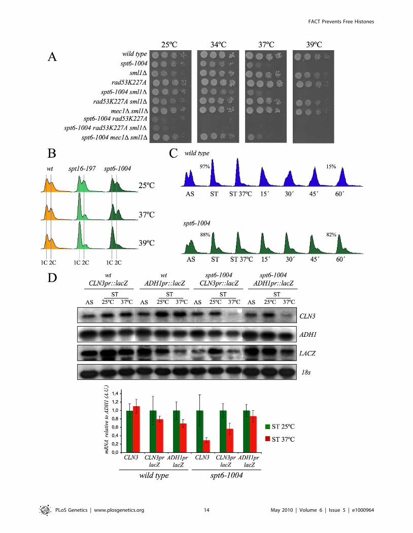
Spt6 [61]. We found that the viability of cells bearing the mutant
allele spt6-1004 clearly lowered in the absence of Rad53 kinase
activity, but remained unchanged in the absence of Mec1
(Figure 7A). We also found that spt6-1004 cells clearly accumu-
lated in G1 when shifted to a restrictive temperature in both
asynchronous and alpha factor-synchronized cells (Figure 7B and
7C). As in the spt16-197 cells, the CLN3 mRNA levels, or the lacZ
mRNA levels when driven by the CLN3 promoter, decreased when
alpha factor-synchronized spt6-1004 cells were shifted to a
restrictive temperature (Figure 7D).
Given these results, we thought it would be interesting to
determine whether the overexpression of histones in G1 induces a
cell cycle delay in wild-type cells. We found that an additional
copy of the HTA1–HTB1 locus produced a very slight delay in cell
cycle progression when alpha factor-synchronized cells were
released from the pheromone. In contrast, a copy of the same
locus lacking the regulatory sequence that mediates its repression
in response to histone levels (DNEG) led to a clear accumulation of
cells in G1 (asynchronous culture) and a more marked delay in the
entry of synchronized cells into the S-phase (Figure 8A).
Accordingly, we detected a more significant decrease of the
CLN3 mRNAs in START, in those cells bearing the deregulated
HTA1–HTB1 copy (DNEG) than in those transformed with the
intact allele (Figure 8B). Since all the cells in this experiment were
held in START by the presence of the mating pheromone, the
drop in CLN3 mRNA could not be an indirect consequence of free
histones inhibiting cell cycle progression. We conclude, therefore,
that excess histones downregulate CLN3 in the wild-type cells
during G1 and subsequently delay their progression through the
G1/S transition.
Discussion
The primary objective of our study was to understand the
defects in the G1/S progression exhibited by the spt16-197
mutant. The genetic and molecular evidence described in the
Results section indicates that the dysfunction of Spt16 down-
regulates the expression of CLN3 in START by triggering a
regulatory mechanism that specifically represses the CLN3
promoter. Our results also reveal that the G1 delay undergone
by the spt16 mutant is not mediated by the DNA-damage
checkpoint, although the rad53K227A mutation enhances both
the thermosensitivity of spt16-197 and its G1 phenotype. This
result, in combination with the lack of phosphorylation of Rad53
after Spt16 inactivation, indicates that excess histones are involved
in this phenomenon. We confirm that this hypothesis is true by
showing that the Spt16 dysfunction produces an accumulation of
free histones associated with histone chaperones in G1 (Figure 5C),
and that excess histones induce a delay in the otherwise wild-type
cells during the G1/S transition concomitantly with CLN3
downregulation.
Chromatin as a potential source of free histonesIt is well known that the accumulation of non-nucleosomal
histones in the cell is toxic [62] and that this toxicity is normally
avoided by regulating the histone gene expression at both the
transcriptional and posttranscriptional levels [60]. Obviously,
these mechanisms are incapable of controlling excess histones
when they originate by eviction from chromatin due to a
dysfunction in chromatin reassembly during transcription. We
demonstrate herein that Spt16 inactivation results in the
accumulation of non-chromatin bound histones in G1. The only
way for the cell to avoid the toxic effects of these excess histones is
to degrade them in a process that is mediated by Rad53 [49]. Our
results show that the absence of Rad53 kinase activity lowers the
viability of the spt16-197 mutant and suggest that one of the roles
of the Rad53-dependent histone degradation mechanism is the
elimination of those histones evicted by the transcriptional activity
that are not reassembled into chromatin. Beyond the S-phase,
transcribed chromatin is probably the main source of free histones
in yeast cells, presumably due to minor imbalances between
histone supply and demand during chromatin reassembly. The
general similarities between histone trafficking during all the
chromatin transactions suggests that DNA repair or DNA
replication might also result in the excess of free histones when
chromatin assembly is dysfunctional [63].
Our results suggest that FACT, in addition to avoiding initiation
from cryptic promoters [9,31], is a protective factor against the
toxic risk represented by evicted histones. A recent publication
reports how Spt16 promotes the redeposition of the original H3
and H4 histones evicted by elongating Pol II [34]. Our results
agree with this conclusion since we have detected a clear
accumulation of free H3 and H4 in Spt16-deficient cells.
However, we have also noted an increase in the free H2A pool.
This result and the genetic interactions between spt16-197 and the
H2A–H2B loci also indicate that Spt16 plays a role in preventing
the accumulation of evicted H2A and H2B. In fact, a high H2A–
H2B/H3–H4 gene ratio impairs the spt16-11 growth, whereas a
low H2A–H2B/H3–H4 gene ratio improves it [30], suggesting
that the accumulation of free H2A–H2B caused by Spt16
dysfunction may be more relevant for the phenotypes observed.
It has been recently demonstrated that FACT promotes the
transition between the canonical nucleosome configuration and a
looser, more dynamic structure that involves changes in the
interaction of the four core histones with the DNA [26]. According
to this view, FACT would be essential for the maintenance of this
altered configuration during transcription elongation by limiting
the amount of the four histones joining the free pools. One
prediction of this model is that those histone mutations which
destabilize this alternative nucleosomal configuration would
promote free histone accumulation. Some H4 mutations affecting
H3–H4 tetramer/H2A–H2B dimer interactions show delayed
G1/S transition and reduced CLN3 expression levels [64].
Accordingly, we detected a negative synthetic interaction between
one of these mutations (hhf1-36, bearing the H4-Y72G mutation)
and rad53K227A (Figure S6).
The protective role against evicted histones is probably not an
exclusive function of FACT, but is also a function of the other
factors that cooperate during chromatin reassembly, like Spt6, for
which we show some evidence. It is likely that Asf1, Nap1, the Hir
FY348, FY710, DMY10, DMY11, and DMY12) were grown in YPD medium at 25uC. 10-fold serial dilutions of the indicated strain were plated on YPDplates and incubated for three days at the indicated temperatures. (D) Cells of strains FY120 (WT), FY348 (spt16-197), FY710 (hta1D–htb1D), DMY10(hta2D–htb2D), DMY11 (spt16-197 hta1D–htb1D), and DMY12 (spt16-197 hta2D–htb2D), exponentially growing in YPD at 25uC, were shifted to 37uC forfour hours or kept at 25uC. Samples were taken to analyze the DNA content by flow cytometry. Numbers indicate the proportion of G1 cells.doi:10.1371/journal.pgen.1000964.g004
FACT Prevents Free Histones
PLoS Genetics | www.plosgenetics.org 9 May 2010 | Volume 6 | Issue 5 | e1000964
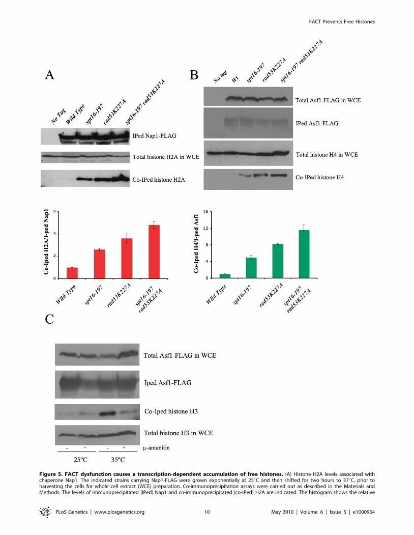
Figure 5. FACT dysfunction causes a transcription-dependent accumulation of free histones. (A) Histone H2A levels associated withchaperone Nap1. The indicated strains carrying Nap1-FLAG were grown exponentially at 25uC and then shifted for two hours to 37uC, prior toharvesting the cells for whole cell extract (WCE) preparation. Co-immunoprecipitation assays were carried out as described in the Materials andMethods. The levels of immunoprecipitated (IPed) Nap1 and co-immunoprecipitated (co-IPed) H2A are indicated. The histogram shows the relative
FACT Prevents Free Histones
PLoS Genetics | www.plosgenetics.org 10 May 2010 | Volume 6 | Issue 5 | e1000964
complex or Nsr1 also protect against evicted histones. Asf1 has
been seen to act as an intermediary in the parental histone
disassembly/reassembly during replication as it associates with the
MCM proteins at the replication fork [65]. An analogous situation
might exist during transcription whereby Asf1 or the HIR
complex, both of which have been shown to be capable of
chromatin assembly outside the S-phase [66,67], may act as an
intermediary in the histone eviction and reassembly process in
conjunction with FACT. Nap1 promotes nucleosome assembly by
eliminating nonnucleosomal H2A- and H2B-DNA interactions
and it prevents aberrant transcription by avoiding excessive H2A–
H2B binding to DNA [68].
In this study we also show how the overexpression of H2A–H2B
results in G1 delays in the wild-type cells. Similar effects, but to a
lesser extent, were obtained by overproducing H3–H4 (data not
shown). These results demonstrate that a histone-mediated G1
delay can be obtained in a background with no possible indirect
effect mediated by either the role of FACT in the expression of the
G1-S regulators, or the function of Rad53 in the control of early
replication.
CLN3-dependent G1 delay in response to free histonesOur results show that the G1-delay caused by Spt16
dysfunction correlates with a down-regulation of the transcrip-
tional activity of the CLN3 promoter. Cln3 shortage can only
explain a transient G1-delay as cells can enter the S-phase after a
prolonged G1 in the absence of Cln3. Therefore it is likely that the
permanent G1 arrest shown by spt16-197 cells at high temper-
atures (over 37uC) is caused by a combination of the CLN3-
mediated transient delay and additional defects on the expression
of other G1/S regulators caused by Spt16 inactivation. For
instance, the MluI cell-cycle boxes (MCB)-mediated activation of
SWI4 and SWI6 is also negatively affected by FACT dysfunction
[18]. The CLN1 and CLN2 promoters are two other clear targets
of Spt16 inactivation, since FACT binds them during G1 and
participates in their activation after START [45]. However, the
marked reduction in the CLN3 expression following FACT
inactivation cannot be caused by a direct involvement of FACT
in the transcriptional activity of the CLN3 promoter during G1
because it does not bind this promoter during this phase of the cell
cycle (David Stillman, personal communication). We show herein
that a CLN3pr::lacZ fusion lacking any sequence in common with
CLN3 mRNA is also responsive to the inactivation of Spt16 or
Spt6 under conditions in which a constitutively expressed
ADH1pr::lacZ is not. Furthermore, rad53K227A enhances the
level of CLN3 down-regulation caused by spt16-197. Finally, the
overexpression of H2A–H2B in an otherwise wild-type strain
decreases the CLN3 expression. Taken together, these results
support the existence of a mechanism which down-regulates the
CLN3 promoter during G1 in response to the accumulation of free
histones caused by either defects in cotranscriptional chromatin
reassembly or histone genes deregulation.
CLN3 mediates several regulatory pathways by coupling the cell-
cycle progression in G1 to physiological conditions, including
nitrogen deprivation [69], daughter cell G1-delay [70,71] and
changes in glucose levels [72]. In the last case, the up-regulation of
the CLN3 promoter is due to the binding of the Azf1
transcriptional activator. Another transcription factor shown to
regulate the CLN3 promoter is the Mcm1-containing early cell-
cycle box (ECB) binding complex [73]. Homeodomain repressors,
Yox1 and Yhp1, restrict the ECB-dependent activation of the
CLN3 transcription to the M/G1 phase [74]. The deletion of
AZF1, YOX1 or YHP1 does not alter the accumulation of cells in
G1, as indicated by spt16-197 at the restrictive temperature (data
not shown). Moreover, the binding of Mcm1 to the CLN3
promoter, measured by chromatin immunoprecipitation, was not
affected by Spt16 inactivation (data not shown). We conclude that
the transcriptional regulation of CLN3 in response to the
accumulation of free histones is not mediated by any of the
known transcription factors operating on the CLN3 promoter. It is
even conceivable that the promoter itself acts as a sensor of free
histone concentration and is repressed in response to the excess
histones.
In mammalian cells, histone overexpression slows down entry
into and progression through the S-phase [65]. Interestingly,
depletion of human Spt16 leads to the repression of the H1, H2A
and H2B genes [75], which could be the result of the accumulation
of the free histones in human cells after FACT dysfunction. Given
the analogy between the G1-S regulators in yeast (Cln3-SBF-Whi5-
Rpd3) and mammals (CyclinD1-E2F-Rb-HDAC1) [42,45], the
functional link between the accumulation of free histones and the
regulation of the G1-S transition may be evolutionarily conserved.
Chromatin repairThe free histones evicted by the transcriptional activity of cells
can potentially associate non-specifically with DNA via electro-
static interactions, and may give rise to aberrant chromatin
structures which can be considered a form of chromatin damage.
As with DNA damage, which enhances the risk of genome
instability, excess free histones may have serious implications for
the normal progression of DNA replication since the toxicity of
free histones is maximal in the S-phase [49]. Consistently with this
hypothesis, the experimental conditions under which the spt16-
induced G1 delay is overcome (CLN3 overexpression, SIC1
deletion) involve an overall decrease in cell viability. The G1
delay should allow cells to reduce the free histone levels through
the Rad53-mediated histone degradation pathway before entering
the S-phase. The persistence of excess histones, as in the spt16-197
rad53K227A double mutant, would lead to severe replication
dysfunctions. Accordingly, chromatin repair would be the combina-
tion of DNA repair, chromatin reassembly and excess histone
degradation. In this sense, it is interesting to note that Rad53,
which participates in both the DNA damage checkpoint and the
excess histone degradation pathway, may act as a super-integrator
accumulation of H2A on Nap1 compared to the wild-type cells (the data have been normalized to the amount of Nap1 actually IPed). The total levelsof H2A in WCE are shown to demonstrate that roughly equal amounts of WCE were used for the immunoprecipitation reactions. (B) The histone H4levels associated with Asf1 in the spt16-197 cells. The indicated strains carrying FLAG-tagged Asf1 were grown in YPD media at 25uC, while theexponentially growing cells were harvested for WCE preparation. The co-immunoprecipitation assays for Asf1-H4 were carried out as indicated in theMaterials and Methods. The total histone H4 and the total Asf1-FLAG levels in the WCE are shown to demonstrate that roughly equal amounts of WCEwere used for the immunoprecipitation reactions. The histogram displays the relative accumulation of H4 on Asf1 if compared to the wild-type cells(the data have been normalized to the amount of Asf1 actually IPed). (C) The accumulation of the free histones in G1 upon FACT dysfunction isdependent on transcription. The spt16-197 cells (FY348) carrying FLAG-tagged Asf1 were grown in YPD at 25uC, alpha factor-treated for two hours at25uC, divided into four equal aliquots, and finally incubated with or without alpha-amanitin for two hours at 25uC or 35uC in the continued presenceof the pheromone. Following this, cells were harvested and processed as described for (A). The Asf1-FLAG and histone H3 levels in WCE were shownto demonstrate that roughly equal amounts of WCE were used for the immunoprecipitation reactions.doi:10.1371/journal.pgen.1000964.g005
FACT Prevents Free Histones
PLoS Genetics | www.plosgenetics.org 11 May 2010 | Volume 6 | Issue 5 | e1000964
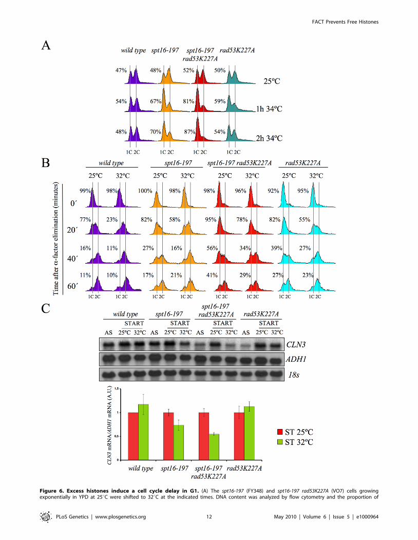
Figure 6. Excess histones induce a cell cycle delay in G1. (A) The spt16-197 (FY348) and spt16-197 rad53K227A (VO7) cells growingexponentially in YPD at 25uC were shifted to 32uC at the indicated times. DNA content was analyzed by flow cytometry and the proportion of
FACT Prevents Free Histones
PLoS Genetics | www.plosgenetics.org 12 May 2010 | Volume 6 | Issue 5 | e1000964

of chromatin repair functions. This new concept could serve as a
convenient framework to gain a better understanding of global
genomic defects.
Materials and Methods
For further details, see Text S1.
Yeast strains and general proceduresAll the yeast strains used in this study were derived from the
S288C genetic background, unless otherwise indicated, and are
listed in Table S1. In our background, temperatures over 33uCwere restrictive for spt16-197 growth, whereas temperatures below
31uC were permissive. All the experiments including spt16-197
mutants were performed at several temperatures. For each
experiment shown in the Results section, we chose the maximal
restrictive temperature at which specific reproducible results were
obtained. Standard procedures were followed for cell culturing,
synchronization at START and flow cytometry [76,77].
Northern blot analysesThe Northern blot analyses were performed as previously
described [5]. Six micrograms of total RNA prepared from yeast
cells underwent electrophoresis on formaldehyde-agarose gels
transferred to Hybond–N filters and UV crosslinked prior to
hybridization at 65uC in 0.5M sodium phosphate buffer pH7 7%
SDS with a [32P]dCTP-labeled DNA probe. Quantification of the
mRNA levels was performed in a phosphorimager (FLA-3000,
FujiFilm); the data are provided in arbitrary units. All the values
were normalized in relation to the amount of 25S rDNA detected
by hybridization with a 32P-oligolabeled 589 bp 25S rRNA
internal fragment obtained by PCR and by using the 19-mer
oligonucleotides TTGGAGAGGGCAACTTTGG and CAG-
GATCGGTCGATTGTGC. For the mRNA histone analysis,
the whole coding regions of HTA1 and HHT1 were used as probes.
Chromatin immunoprecipitationPol II ChIPs were performed as in [78], using the 8WG16
monoclonal antibody. Amplicons for Q-PCR quantification
extended from +110 to +193 for CLN3, and from +6 to +95 for
ADH1, in relation to the transcription start sites.
Rad53p phosphorylation assayYeast cultures were grown at 25uC to OD = 1. Cultures were
kept at 25uC, or shifted at 37uC and incubated for two hours.
Protein extracts for the Western blot analyses were prepared from
trichloroacetic acid (TCA)-treated yeast cells. Protein extracts were
resolved on a 7.7% SDS-PAGE (35:0.2 acrylamide/bis-acrylam-
ide). Immunoblots were done with the goat anti-Rad53 polyclonal
antibody from Santa Cruz Biotechnology.
Detection of non chromatin-bound ‘‘free’’ histonesassociated with the histone chaperones Nap1 and Asf1
For the determination of histones associated with Nap1 and
Asf1 in Figure 5A and 5B, one-liter cultures of the indicated
strains carrying Nap1-FLAG [79] or Asf1-FLAG were grown
exponentially in YPD media at 25uC. Cells were then harvested
as such at a density of 2.5610E7 cells/ml for the Asf1-FLAG
experiment shown in Figure 5B. For the Nap1-FLAG experi-
ment shown in Figure 5A, cells were grown at 25uC until they
reached a density of 1.5610E7 cells/ml at which point they
were switched to the restrictive temperature of 37uC for two
hours prior to harvesting the cells at a density of 2.5610E7
cells/ml. Whole cell extracts (WCEs) were prepared as
previously described [49], and FLAG-tagged Nap1 (pRS316-
Flag-yNap1) or Asf1-FLAG was immunoprecipitated using
FLAG M2 agarose (Sigma). The immunoprecipitated material
was resolved on precast 4–12% polyacrylamide gradient gels in
MES buffer (BioRad), and were processed for Western blotting
as previously described [49]. FLAG M2 antibodies (Sigma) were
used to detect Nap1-FLAG and Asf1-FLAG, while histone H4
and H2B were detected using the previously described
polyclonal antibodies [49]. Histone H2A was detected using
an H2A antibody from Millipore (Cat. # 07-146).
The influence of transcription on free histone accumulation
shown in Figure 5C was tested as follows. One liter of overnight
culture of the spt16-197 (FY348) cells carrying FLAG-tagged Asf1
was grown in YPD at 25uC. Once the cells had reached a density
of 1.5610E7 cells/ml, they were treated with alpha-factor for two
hours at 25uC. Cells were treated with additional amounts of
alpha-factor, divided into four equal aliquots, and were treated
with or without 200mg/ml alpha-amanitin for 2 hours at 25uC or
35uC in the continued presence of alpha-factor. Afterward, cells
were harvested and processed as described before, except histone
H3, which was detected using a polyclonal antibody directed
against the C-terminus of histone H3, as previously described [49].
The inhibition of Pol II by alpha-amanitin was controlled by
monitoring the mRNA levels of ACT1 in relation to ribosomal
RNA levels (that are unaffected at the alpha-amanitin concentra-
tion used) using quantitative RT-PCR (Figure S5C).
Supporting Information
Figure S1 Quantification of the relative copy number of the
plasmids described in Figure 2. The indicated plasmids were
detected by quantitative PCR as described in Text S1. The ratio
between the amplicon localized in the Amp gene of the plasmid
and another amplicon localized in the chromosomal GAL1 genes is
shown. 1.0 corresponds to the empty vector.
Found at: doi:10.1371/journal.pgen.1000964.s001 (0.18 MB PDF)
Figure S2 The G1 delay provoked by Spt16 inactivation was not
prevented by the deletion of RAD9. (A) Wild-type (MMY20.4),
rad9D (MMY20.1), spt16-197 (MMY20.2) and spt16-19 rad9D(MMY20.3) cells growing exponentially in YPD at 30uC were
shifted to 37uC for four hours. Cells were then analyzed by flow
cytometry. (B) Wild-type and mutant cells exponentially growing
in YPD at 30uC were spotted onto YPD plates and incubated at
30uC, 32uC and 33uC, as indicated.
Found at: doi:10.1371/journal.pgen.1000964.s002 (0.19 MB
PDF)
unbudded cells was quantified by microscopy. (B) The wild-type (FY120), spt16-197 (FY348), spt16-197 rad53K227A (VO7) and rad53-K227A (V03) cellswere synchronized at START by a treatment with alpha-factor for two hours at 25uC, followed by an additional one hour at 25uC or 32uC in thepresence of mating pheromone. Cells were then released from the G1-arrest at time 0 at either 25uC or 32uC by washing out the alpha-factor.Samples were taken at the different time points to analyze the DNA content by flow cytometry and to measure the proportion of unbudded cells bymicroscopy. (C) Those samples from asynchronous cultures and from the time 0 of B were taken to analyze the CLN3 mRNA levels by Northern blot.The results of a significant experiment and the average quantification of three independent experiments are shown. The CLN3 mRNA levels werenormalized to ADH1.doi:10.1371/journal.pgen.1000964.g006
FACT Prevents Free Histones
PLoS Genetics | www.plosgenetics.org 13 May 2010 | Volume 6 | Issue 5 | e1000964
FACT Prevents Free Histones
PLoS Genetics | www.plosgenetics.org 14 May 2010 | Volume 6 | Issue 5 | e1000964

Figure S3 rad53D enhances the thermosensitivity of spt16-197
independently of the DNA damage checkpoint. Cells were grown
in YPD medium at 25uC. 10-fold serial dilutions of the indicated
strain were plated on YPD plates (or YPD+0.02% methyl methane
sulfonate, MMS) and incubated for three days at the indicated
temperatures.
Figure 7. Spt6 dysfunction provokes G1/S defects and lowers cell viability in combination with rad53K227A. (A) Strains FY120, FY2180,VO1, VO3, DMY5, VO4, VO2, DMY6, DMY7, and DMY8 were grown in YPD at 25uC, spotted onto YPD plates and incubated for three days at theindicated temperature. (B) The wild-type (FY120), spt16-197 (FY348), and spt6-1004 (FY2180) cells exponentially growing in YPD at 25uC, were shiftedfor two hours at the indicated temperatures. Samples were then taken to analyze the DNA content by flow cytometry. (C) The wild-type (FY120) andspt6-1004 (FY2180) cells were synchronized at START by alpha-factor treatment for 2h at 25uC, followed by an additional one hour at 37uC in thepresence of the mating pheromone. Cells were then released from the G1-arrest at time 0 at 37uC by washing out the alpha-factor. Samples weretaken at the different time points to analyze the DNA content by flow cytometry and to measure the proportion of unbudded cells by microscopy. (D)The mRNA levels of CLN3 and CLN3pr::lacZ in spt6-1004. The wild-type (FY120) and spt6-1004 (FY2180) cells were transformed with the plasmidsbearing the CLN3pr::lacZ fusion number 1 (see Figure 2A) or ADH1pr::lacZ. These transformants were grown until the mid-log phase (AS) and werealpha factor-synchronyzed at 25uC (START), followed by one additional hour at 37uC in the continued presence of pheromone. The RNA samples weretaken to analyze the transcript levels. mRNAs were quantified by Northern blot analysis. The results of a typical experiment and the averagequantification of three independent experiments are shown. The CLN3 mRNA levels were normalized to ADH1, and the values of each strain at 37uCwere represented in relation to 25uC.doi:10.1371/journal.pgen.1000964.g007
Figure 8. The overexpression of histones in the wild-type cells induces G1 delay. (A) Wild-type cells were transformed with pRS316 (emptyvector) or with the analogous centromeric plasmids expressing either HTA1–HTB1 or a mutant version of this locus lacking the sequence thatmediates its transcriptional repression in response to the free histones (HTA1–HTB1DNEG). The transformants were grown exponentially in a selectivemedium (AS), synchronized at START by treatment with alpha-factor for two hours (ST) and released from the arrest by washing out the alpha-factor.Samples were then taken at the different time points to analyze the DNA content by flow cytometry and the proportion of unbudded cells bymicroscopy. (B) Aliquots from the synchronized cells used in (A) were taken to analyze the CLN3 mRNA levels in START by Northern blot. The results ofa typical experiment and the average quantification of three independent experiments are shown. The CLN3 mRNA levels have been normalized toADH1.doi:10.1371/journal.pgen.1000964.g008
FACT Prevents Free Histones
PLoS Genetics | www.plosgenetics.org 15 May 2010 | Volume 6 | Issue 5 | e1000964
Found at: doi:10.1371/journal.pgen.1000964.s003 (0.17 MB
PDF)
Figure S4 hta2/htb2D partially suppresses the thermosensitivity
of spt16-197. (A) Strains FY120, FY348, FY710, DMY10,
DMY11, and DMY12 were transformed with pRS316 (empty
vector), pRS316-HTA1-HTB1 or pRS316-HTA1-HTB1DNEG.
Transformants were grown in SC-Ura medium at 25uC. 10-fold
serial dilutions of the indicated strain were plated on SC-Ura
plates and incubated for three days at the indicated temperatures.
The chromosomal, extrachromosal or plasmidic copies of HTA1–
HTB1 and HTA2–HTB2 presented in each transformant are
indicated. Red squares indicate the results of those strains
containing two copies of H2A/H2B-encoding loci. (B) An
extrachromosomal amplification of the HTA2–HTB2 locus was
detected by PCR in FY710 (hta1/htb1D) and DMY11 (spt16-197
hta1/htb1D), following the protocol described in (Libuda and
Winston, 2006).
Found at: doi:10.1371/journal.pgen.1000964.s004 (1.22 MB
PDF)
Figure S5 spt16-197 does not affect the levels of histone
chaperones Nap1 and Asf1 or alter histone gene regulation. (A)
Nap1 levels were unaffected in spt16-197. Nap1-FLAG was
detected in WCEs of 4 independent generated wild type and
spt16-197 strains by Western blotting using an anti-FLAG
antibody. A non-specific band was used as the loading control.
(B) Asf1 levels were unaffected in spt16-197. Asf1-FLAG was
detected in WCE by Western blot with an anti-FLAG antibody.
H2A was used as the loading control. (C) Alpha–amanitin
inhibited RNA pol II transcription in alpha-factor-synchronized
spt16-197 cells. The mRNA levels of the constitutively expressed
ACT1 gene were measured by quantitative RT-PCR in the FY348
cells grown exponentially at 25uC and synchronized with alpha
factor for four hours. The data was normalized to the levels of
ribosomal RNA which is not affected by the concentration of
alpha–amanitin used. (D) Repression of histones genes in G1 was
not abolished by spt16-197. Wild-type (FY120) and spt16-197
(FY348) cells grown asynchronously (AS) were synchronized at
START (ST) by treatment with alpha-factor for two hours at 25uC(ST), followed by an additional one hour at 25uC or 35uC in the
presence of the mating pheromone. Cells were then released from
the arrest at time 0 at either 25uC or 35uC by washing out the
alpha-factor. Samples were taken at the indicated time points to
analyze mRNA levels by Northern blot. H2A indicate the signal
corresponding to HTA1 and HTA2, and H3 indicate the signal of
HHT1 and HHT2.
Found at: doi:10.1371/journal.pgen.1000964.s005 (0.24 MB
PDF)
Figure S6 hhf1-36 and rad53K227A exhibit a negative synthetic
interaction. Strains MSY623, DMY15, MSY781, and DMY16
exponentially grown in YPD at 30uC were spotted onto YPD
plates and incubated for three days at 30uC and 37uC, as
indicated.
Found at: doi:10.1371/journal.pgen.1000964.s006 (0.16 MB
PDF)
Table S1 The yeast strains used in this work.
Found at: doi:10.1371/journal.pgen.1000964.s007 (0.03 MB
DOC)
Text S1 Supplementary material and methods and supplemen-
tary references.
Found at: doi:10.1371/journal.pgen.1000964.s008 (0.03 MB
DOC)
Acknowledgments
We thank David Stillman for sharing unpublished data; Sandra Gavalda,
Marie-Helene Miquel Kabbaj, Oscar Cabrera, and Sara Jackson for their
experimental help; and D. Bentley, J. Correa-Bordes, J.L. Crespo, E. Garı,
E. de Nadal, F. Posas, J. Tyler, and A. Verreault for their helpful
comments. We also thank M. Aldea, T. Formosa, H. Matsumoto, K.
Nagata, M. A. Osley, M. M. Smith, and F. Winston for generous gifts of
strains and plasmids.
Author Contributions
Conceived and designed the experiments: MMH DM MCMC RKS VG
AG SC. Performed the experiments: MMH DM MCMC VO GUR DL.
Analyzed the data: MMH DM MCMC RKS VO GUR DL VG AG SC.
Wrote the paper: MCMC RKS VG AG SC.
References
1. Reinberg D, Sims RJ, 3rd (2006) de FACTo nucleosome dynamics. J Biol Chem
281: 23297–23301.
2. Formosa T (2008) FACT and the reorganized nucleosome. Mol Biosyst 4:
1085–1093.
3. Orphanides G, LeRoy G, Chang CH, Luse DS, Reinberg D (1998) FACT, a
factor that facilitates transcript elongation through nucleosomes. Cell 92:
105–116.
4. Pavri R, Zhu B, Li G, Trojer P, Mandal S, et al. (2006) Histone H2B
monoubiquitination functions cooperatively with FACT to regulate elongation
by RNA polymerase II. Cell 125: 703–717.
5. Jimeno-Gonzalez S, Gomez-Herreros F, Alepuz PM, Chavez S (2006) A gene-
specific requirement for FACT during transcription is related to the chromatin
organization of the transcribed region. Mol Cell Biol 26: 8710–8721.
6. Biswas D, Dutta-Biswas R, Mitra D, Shibata Y, Strahl BD, et al. (2006)
Opposing roles for Set2 and yFACT in regulating TBP binding at promoters.
Embo J 25: 4479–4489.
7. Formosa T (2003) Changing the DNA landscape: putting a SPN on chromatin.
Curr Top Microbiol Immunol 274: 171–201.
8. Lindstrom DL, Squazzo SL, Muster N, Burckin TA, Wachter KC, et al. (2003)
Dual roles for Spt5 in pre-mRNA processing and transcription elongation
revealed by identification of Spt5-associated proteins. Mol Cell Biol 23:
1368–1378.
9. Mason PB, Struhl K (2003) The FACT complex travels with elongating RNA
polymerase II and is important for the fidelity of transcriptional initiation in vivo.
Mol Cell Biol 23: 8323–8333.
10. Saunders A, Werner J, Andrulis ED, Nakayama T, Hirose S, et al. (2003)
Tracking FACT and the RNA polymerase II elongation complex through
chromatin in vivo. Science 301: 1094–1096.
11. Biswas D, Dutta-Biswas R, Stillman DJ (2007) Chd1 and yFACT act in
opposition in regulating transcription. Mol Cell Biol 27: 6279–6287.
12. Biswas D, Yu Y, Prall M, Formosa T, Stillman DJ (2005) The Yeast FACT
Complex Has a Role in Transcriptional Initiation. Mol Cell Biol 25:
5812–5822.
13. O’Donnell AF, Brewster NK, Kurniawan J, Minard LV, Johnston GC, et al.
(2004) Domain organization of the yeast histone chaperone FACT: the
conserved N-terminal domain of FACT subunit Spt16 mediates recovery from
replication stress. Nucleic Acids Res 32: 5894–5906.
14. Schlesinger MB, Formosa T (2000) POB3 is required for both transcription
and replication in the yeast Saccharomyces cerevisiae. Genetics 155:
1593–1606.
15. VanDemark AP, Blanksma M, Ferris E, Heroux A, Hill CP, et al. (2006) The
structure of the yFACT Pob3-M domain, its interaction with the DNA
replication factor RPA, and a potential role in nucleosome deposition. Mol Cell
22: 363–374.
16. Orphanides G, Wu WH, Lane WS, Hampsey M, Reinberg D (1999) The
chromatin-specific transcription elongation factor FACT comprises human
SPT16 and SSRP1 proteins. Nature 400: 284–288.
17. Prendergast JA, Murray LE, Rowley A, Carruthers DR, Singer RA, et al. (1990)
Size selection identifies new genes that regulate Saccharomyces cerevisiae cell
proliferation. Genetics 124: 81–90.
18. Lycan D, Mikesell G, Bunger M, Breeden L (1994) Differential effects of Cdc68
on cell cycle-regulated promoters in Saccharomyces cerevisiae. Mol Cell Biol 14:
7455–7465.
19. Malone EA, Clark CD, Chiang A, Winston F (1991) Mutations in SPT16/
CDC68 suppress cis- and trans-acting mutations that affect promoter function in
Saccharomyces cerevisiae. Mol Cell Biol 11: 5710–5717.
FACT Prevents Free Histones
PLoS Genetics | www.plosgenetics.org 16 May 2010 | Volume 6 | Issue 5 | e1000964
20. John S, Howe L, Tafrov ST, Grant PA, Sternglanz R, et al. (2000) The
something about silencing protein, Sas3, is the catalytic subunit of NuA3, a
yTAF(II)30-containing HAT complex that interacts with the Spt16 subunit of
the yeast CP (Cdc68/Pob3)-FACT complex. Genes Dev 14: 1196–1208.
21. Krogan NJ, Kim M, Ahn SH, Zhong G, Kobor MS, et al. (2002) RNA
polymerase II elongation factors of Saccharomyces cerevisiae: a targeted
proteomics approach. Mol Cell Biol 22: 6979–6992.
22. Squazzo SL, Costa PJ, Lindstrom DL, Kumer KE, Simic R, et al. (2002) The
Paf1 complex physically and functionally associates with transcription elongation
factors in vivo. Embo J 21: 1764–1774.
23. Fleming AB, Kao CF, Hillyer C, Pikaart M, Osley MA (2008) H2B
ubiquitylation plays a role in nucleosome dynamics during transcription
elongation. Mol Cell 31: 57–66.
24. Formosa T, Eriksson P, Wittmeyer J, Ginn J, Yu Y, et al. (2001) Spt16-Pob3 and
the HMG protein Nhp6 combine to form the nucleosome-binding factor SPN.
Embo J 20: 3506–3517.
25. Rhoades AR, Ruone S, Formosa T (2004) Structural features of nucleosomes
reorganized by yeast FACT and its HMG box component, Nhp6. Mol Cell Biol
24: 3907–3917.
26. Xin H, Takahata S, Blanksma M, McCullough L, Stillman DJ, et al. (2009)
yFACT Induces Global Accesibility of Nucleosomal DNA without H2A–H2B
Displacement. Molecular Cell 35: 365–376.
27. Stuwe T, Hothorn M, Lejeune E, Rybin V, Bortfeld M, et al. (2008) The FACT
Spt16 ‘‘peptidase’’ domain is a histone H3–H4 binding module. Proc Natl Acad
Sci U S A 105: 8884–8889.
28. VanDemark AP, Xin H, McCullough L, Rawlins R, Bentley S, et al. (2008)
Structural and functional analysis of the Spt16p N-terminal domain reveals
overlapping roles of yFACT subunits. J Biol Chem 283: 5058–5068.
29. Belotserkovskaya R, Saunders A, Lis JT, Reinberg D (2004) Transcription
through chromatin: understanding a complex FACT. Biochim Biophys Acta
1677: 87–99.
30. Formosa T, Ruone S, Adams MD, Olsen AE, Eriksson P, et al. (2002) Defects in
SPT16 or POB3 (yFACT) in Saccharomyces cerevisiae cause dependence on the
Hir/Hpc pathway: polymerase passage may degrade chromatin structure.
Genetics 162: 1557–1571.
31. Kaplan CD, Laprade L, Winston F (2003) Transcription elongation factors
repress transcription initiation from cryptic sites. Science 301: 1096–1099.
32. Cheung V, Chua G, Batada NN, Landry CR, Michnick SW, et al. (2008)
Chromatin- and transcription-related factors repress transcription from within
coding regions throughout the Saccharomyces cerevisiae genome. PLoS Biol 6:
e277. doi:10.1371/journal.pbio.0060277.
33. Vanti M, Gallastegui E, Respaldiza I, Rodriguez-Gil A, Gomez-Herreros F,
et al. (2009) Yeast genetic analysis reveals the involvement of chromatin
reassembly factors in repressing HIV-1 basal transcription. PLoS Genet 5:
e1000339. doi:10.1371/journal.pgen.1000339.
34. Jamai A, Puglisi A, Strubin M (2009) Histone Chaperone Spt16 Promotes
Redeposition of the Original H3–H4 Histones Evicted by Elongating RNA
Polymerase. Molecular Cell 35: 377–383.
35. Rowley A, Singer RA, Johnston GC (1991) CDC68, a yeast gene that affects
regulation of cell proliferation and transcription, encodes a protein with a highly
acidic carboxyl terminus. Mol Cell Biol 11: 5718–5726.
36. Xu Q, Johnston GC, Singer RA (1993) The Saccharomyces cerevisiae Cdc68
transcription activator is antagonized by San1, a protein implicated in
transcriptional silencing. Mol Cell Biol 13: 7553–7565.
37. Skotheim JM, Di Talia S, Siggia ED, Cross FR (2008) Positive feedback of G1
cyclins ensures coherent cell cycle entry. Nature 454: 291–296.
38. Dirick L, Bohm T, Nasmyth K (1995) Roles and regulation of Cln-Cdc28
kinases at the start of the cell cycle of Saccharomyces cerevisiae. Embo J 14:
4803–4813.
39. Koch C, Schleiffer A, Ammerer G, Nasmyth K (1996) Switching transcription
on and off during the yeast cell cycle: Cln/Cdc28 kinases activate bound
transcription factor SBF (Swi4/Swi6) at start, whereas Clb/Cdc28 kinases
displace it from the promoter in G2. Genes Dev 10: 129–141.
40. Stuart D, Wittenberg C (1995) CLN3, not positive feedback, determines the
timing of CLN2 transcription in cycling cells. Genes Dev 9: 2780–2794.
41. Tyers M, Tokiwa G, Futcher B (1993) Comparison of the Saccharomyces
cerevisiae G1 cyclins: Cln3 may be an upstream activator of Cln1, Cln2 and
other cyclins. Embo J 12: 1955–1968.
42. Wang H, Carey LB, Cai Y, Wijnen H, Futcher B (2009) Recruitment of Cln3
cyclin to promoters controls cell cycle entry via histone deacetylase and other
targets. PLoS Biol 7: e1000189. doi:10.1371/journal.pbio.1000189.
43. Costanzo M, Nishikawa JL, Tang X, Millman JS, Schub O, et al. (2004) CDK
activity antagonizes Whi5, an inhibitor of G1/S transcription in yeast. Cell 117:
899–913.
44. de Bruin RA, McDonald WH, Kalashnikova TI, Yates J, 3rd, Wittenberg C
(2004) Cln3 activates G1-specific transcription via phosphorylation of the SBF
bound repressor Whi5. Cell 117: 887–898.
45. Takahata S, Yu Y, Stillman DJ (2009) The E2F functional analogue SBF recruits
the Rpd3(L) HDAC, via Whi5 and Stb1, and the FACT chromatin reorganizer,
to yeast G1 cyclin promoters. Embo J 28: 3378–3389.
46. Schneider BL, Yang QH, Futcher AB (1996) Linkage of replication to start by
the Cdk inhibitor Sic1. Science 272: 560–562.
47. Schwob E, Bohm T, Mendenhall MD, Nasmyth K (1994) The B-type cyclin
kinase inhibitor p40SIC1 controls the G1 to S transition in S. cerevisiae. Cell 79:
233–244.
48. Gunjan A, Paik J, Verreault A (2005) Regulation of histone synthesis and
nucleosome assembly. Biochimie 87: 625–635.
49. Gunjan A, Verreault A (2003) A Rad53 kinase-dependent surveillance
mechanism that regulates histone protein levels in S. cerevisiae. Cell 115:
537–549.
50. Singh RK, Kabbaj MH, Paik J, Gunjan A (2009) Histone levels are regulated by
phosphorylation and ubiquitylation-dependent proteolysis. Nat Cell Biol 11:
925–933.
51. Singh RK, Paik J, Gunjan A (2009) Generation and management of excess
histones during the cell cycle. Front Biosci 14: 3145–3158.
52. Chavez S, Aguilera A (1997) The yeast HPR1 gene has a functional role in
transcriptional elongation that uncovers a novel source of genome instability.
Genes Dev 11: 3459–3470.
53. Luna R, Jimeno S, Marin M, Huertas P, Garcia-Rubio M, et al. (2005)
Interdependence between transcription and mRNP processing and export, and
its impact on genetic stability. Mol Cell 18: 711–722.
54. Nourani A, Robert F, Winston F (2006) Evidence that Spt2/Sin1, an HMG-like
factor, plays roles in transcription elongation, chromatin structure, and genome
stability in Saccharomyces cerevisiae. Mol Cell Biol 26: 1496–1509.
55. Gerald JN, Benjamin JM, Kron SJ (2002) Robust G1 checkpoint arrest in
budding yeast: dependence on DNA damage signaling and repair. J Cell Sci 115:
1749–1757.
56. Toh GW, Lowndes NF (2003) Role of the Saccharomyces cerevisiae Rad9
protein in sensing and responding to DNA damage. Biochem Soc Trans 31:
242–246.
57. Pellicioli A, Foiani M (2005) Signal transduction: how rad53 kinase is activated.
Curr Biol 15: R769–771.
58. Allen JB, Zhou Z, Siede W, Friedberg EC, Elledge SJ (1994) The SAD1/
RAD53 protein kinase controls multiple checkpoints and DNA damage-induced
transcription in yeast. Genes Dev 8: 2401–2415.
59. Libuda DE, Winston F (2006) Amplification of histone genes by circular
chromosome formation in Saccharomyces cerevisiae. Nature 443: 1003–1007.
60. Osley MA (1991) The regulation of histone synthesis in the cell cycle. Annu Rev
Biochem 60: 827–861.
61. Bortvin A, Winston F (1996) Evidence that Spt6p controls chromatin structure
by a direct interaction with histones. Science 272: 1473–1476.
62. Meeks-Wagner D, Hartwell LH (1986) Normal stoichiometry of histone dimer
sets is necessary for high fidelity of mitotic chromosome transmission. Cell 44:
43–52.
63. De Koning L, Corpet A, Haber JE, Almouzni G (2007) Histone chaperones: an
escort network regulating histone traffic. Nat Struct Mol Biol 14: 997–1007.
64. Santisteban MS, Arents G, Moudrianakis EN, Smith MM (1997) Histone
octamer function in vivo: mutations in the dimer-tetramer interfaces disrupt
both gene activation and repression. Embo J 16: 2493–2506.
65. Groth A, Corpet A, Cook AJ, Roche D, Bartek J, et al. (2007) Regulation of
replication fork progression through histone supply and demand. Science 318:
1928–1931.
66. Green EM, Antczak AJ, Bailey AO, Franco AA, Wu KJ, et al. (2005)
Replication-independent histone deposition by the HIR complex and Asf1. Curr
Biol 15: 2044–2049.
67. Schwabish MA, Struhl K (2006) Asf1 mediates histone eviction and deposition
during elongation by RNA polymerase II. Mol Cell 22: 415–422.
68. Andrews AJ, Chen X, Zevin A, Stargell LA, Luger K (2010) The histone
chaperone Nap1 promotes nucleosome assembly by eliminating nonnucleosomal
histone DNA interactions. Mol Cell 26: 834–842.
69. Gallego C, Gari E, Colomina N, Herrero E, Aldea M (1997) The Cln3 cyclin is
down-regulated by translational repression and degradation during the G1 arrest
caused by nitrogen deprivation in budding yeast. Embo J 16: 7196–7206.
70. Laabs TL, Markwardt DD, Slattery MG, Newcomb LL, Stillman DJ, et al.
(2003) ACE2 is required for daughter cell-specific G1 delay in Saccharomyces
cerevisiae. Proc Natl Acad Sci U S A 100: 10275–10280.
71. Di Talia S, Wang H, Skotheim JM, Rosebrock AP, Futcher B, et al. (2009)
Daughter-specific transcription factors regulate cell size control in budding yeast.
PLoS Biol 7: e1000221. doi:10.1371/journal.pbio.1000221.
72. Newcomb LL, Hall DD, Heideman W (2002) AZF1 is a glucose-dependent
positive regulator of CLN3 transcription in Saccharomyces cerevisiae. Mol Cell
Biol 22: 1607–1614.
73. Mai B, Miles S, Breeden LL (2002) Characterization of the ECB binding
complex responsible for the M/G(1)-specific transcription of CLN3 and SWI4.
Mol Cell Biol 22: 430–441.
74. Pramila T, Miles S, GuhaThakurta D, Jemiolo D, Breeden LL (2002) Conserved
homeodomain proteins interact with MADS box protein Mcm1 to restrict ECB-
dependent transcription to the M/G1 phase of the cell cycle. Genes Dev 16:
3034–3045.
75. Li Y, Zeng SX, Landais I, Lu H (2007) Human SSRP1 has Spt16-dependent
and -independent roles in gene transcription. J Biol Chem 282: 6936–6945.
76. Rose MD, Winston F, Hieter P (1990) Methods in Yeast Genetics: A Laboratory
Course Manual. Cold Spring HarborNY: Cold Sprong Harbor Laboratory
Press.
FACT Prevents Free Histones
PLoS Genetics | www.plosgenetics.org 17 May 2010 | Volume 6 | Issue 5 | e1000964
77. Winey M, Goetsch L, Baum P, Byers B (1991) MPS1 and MPS2: novel yeast
genes defining distinct steps of spindle pole body duplication. J Cell Biol 114:745–754.
78. Pelechano V, Jimeno-Gonzalez S, Rodriguez-Gil A, Garcia-Martinez J, Perez-
Ortin JE, et al. (2009) Regulon-specific control of transcription elongation
across the yeast genome. PLoS Genet 5: e1000614. doi:10.1371/journal.
pgen.1000614.79. Miyaji-Yamaguchi M, Kato K, Nakano R, Akashi T, Kikuchi A, et al. (2003)
Involvement of nucleocytoplasmic shuttling of yeast Nap1 in mitotic progression.
Mol Cell Biol 23: 6672–6684.
FACT Prevents Free Histones
PLoS Genetics | www.plosgenetics.org 18 May 2010 | Volume 6 | Issue 5 | e1000964
Related Documents


