JOURNAL OF BACTERIOLOGY, Nov. 2009, p. 6525–6538 Vol. 191, No. 21 0021-9193/09/$12.00 doi:10.1128/JB.00652-09 Copyright © 2009, American Society for Microbiology. All Rights Reserved. Facilitated Oligomerization of Mycobacterial GroEL: Evidence for Phosphorylation-Mediated Oligomerization † C. M. Santosh Kumar, 1 Garima Khare, 3 C. V. Srikanth, 1 § Anil K. Tyagi, 3 Abhijit A. Sardesai, 2 * and Shekhar C. Mande 1 * Laboratory of Structural Biology 1 and Laboratory of Bacterial Genetics, 2 Centre for DNA Fingerprinting and Diagnostics, Hyderabad 500001, India, and Department of Biochemistry, University of Delhi South Campus, New Delhi 110021, India 3 Received 18 May 2009/Accepted 21 August 2009 The distinctive feature of the GroES-GroEL chaperonin system in mediating protein folding lies in its ability to exist in a tetradecameric state, form a central cavity, and encapsulate the substrate via the GroES lid. However, recombinant GroELs of Mycobacterium tuberculosis are unable to act as effective molecular chaper- ones when expressed in Escherichia coli. We demonstrate here that the inability of M. tuberculosis GroEL1 to act as a functional chaperone in E. coli can be alleviated by facilitated oligomerization. The results of directed evolution involving random DNA shuffling of the genes encoding M. tuberculosis GroEL homologues followed by selection for functional entities suggested that the loss of chaperoning ability of the recombinant mycobac- terial GroEL1 and GroEL2 in E. coli might be due to their inability to form canonical tetradecamers. This was confirmed by the results of domain-swapping experiments that generated M. tuberculosis-E. coli chimeras bearing mutually exchanged equatorial domains, which revealed that E. coli GroEL loses its chaperonin activity due to alteration of its oligomerization capabilities and vice versa for M. tuberculosis GroEL1. Fur- thermore, studying the oligomerization status of native GroEL1 from cell lysates of M. tuberculosis revealed that it exists in multiple oligomeric forms, including single-ring and double-ring variants. Immunochemical and mass spectrometric studies of the native M. tuberculosis GroEL1 revealed that the tetradecameric form is phosphorylated on serine-393, while the heptameric form is not, indicating that the switch between the single- and double-ring variants is mediated by phosphorylation. GroEL, an essential chaperonin, is known to form a ring- shaped structure for sequestering substrate proteins from the crowded cellular milieu and is responsible for the occurrence of various cellular processes, such as de novo folding, trans- port, and macromolecular assembly, within a biologically rel- evant time scale (7, 26, 48, 53). In Escherichia coli, GroEL, along with its cofactor GroES, assists the folding of about 10 to 30% of cytosolic proteins, among which some are known to be essential for cell viability (15, 26, 27, 31). GroEL was originally identified as the host factor responsible for phage and T4 capsid protein assembly and was subsequently shown to be essential for cell viability (17, 20). E. coli groEL is found in an operonic arrangement with groES (groESL), and its expression is regulated by multiple promoter elements. GroEL function has been shown to be a complex interplay between its interaction with and encapsulation of substrate proteins, with concomitant conformational changes induced by ATP binding, hydrolysis, and GroES binding (24, 56, 62). E. coli GroEL exists as a homotetradecamer forming two isolo- gous rings of seven identical subunits each. Crystallographic analyses have delineated the three-domain architecture of GroEL monomers and the GroES-GroEL interactions (4, 63). The central region of the GroEL polypeptide, spanning amino acid residues 191 to 376, constitutes the GroES and substrate polypeptide-binding apical domain. The equatorial ATPase domain spanning two extremities of the GroEL polypeptide, that is, residues 6 to 133 and 409 to 523, is responsible for the ATPase activity and the bulk of intersubunit interactions. The hinge-forming intermediate domain, spanning two regions on the polypeptide, namely, residues 134 to 190 and 377 to 408, connects the said two domains in the tertiary structure. The conformational changes resulting from ATP binding and hy- drolysis at the equatorial domain are coupled to those occur- ring at the apical domain via this hinge region (4, 63). The usual size limit for the substrate proteins, as shown by both in vitro and in vivo studies, is around 57 kDa, although the cis cavity is reported to theoretically accommodate larger pro- teins, on the order of 104 kDa (10, 27, 35, 46). Productive in vivo folding of the proteins larger than the usual size limit, such as the 86-kDa maltose binding protein fusion and 82-kDa mitochondrial aconitase, has also been reported (9, 29). Since such large substrates are difficult to accommodate in the cen- tral cavity, it has been suggested that their productive folding might occur outside the cis cavity. These studies therefore indicate that the substrate recognition patterns of GroEL may be more diverse than initially thought. Recent genome annotation studies of various bacteria have revealed that a few bacterial genomes possess multiple copies of groEL genes (2, 18, 30). The Mycobacterium tuberculosis genome bears two copies of groEL genes (groELs). One of * Corresponding author. Mailing address: Centre for DNA Finger- printing and Diagnostics, Bldg. 7, Gruhakalpa, 5-4-399/B Nampally, Hyderabad 500001, India. Phone: 91-40-24749401. Fax: 91-40- 24785447. E-mail for Shekhar C. Mande: [email protected]. E-mail for Abhijit A. Sardesai: [email protected]. Correspondent for reprint requests: Shekhar C. Mande. † Supplemental material for this article may be found at http://jb .asm.org/. § Present address: Mucosal Immunology Laboratory, Massachusetts General Hospital, Charlestown, MA, 02129-4404. Published ahead of print on 28 August 2009. 6525 by on October 13, 2009 jb.asm.org Downloaded from

Welcome message from author
This document is posted to help you gain knowledge. Please leave a comment to let me know what you think about it! Share it to your friends and learn new things together.
Transcript
JOURNAL OF BACTERIOLOGY, Nov. 2009, p. 6525–6538 Vol. 191, No. 210021-9193/09/$12.00 doi:10.1128/JB.00652-09Copyright © 2009, American Society for Microbiology. All Rights Reserved.
Facilitated Oligomerization of Mycobacterial GroEL: Evidence forPhosphorylation-Mediated Oligomerization�†
C. M. Santosh Kumar,1 Garima Khare,3 C. V. Srikanth,1§ Anil K. Tyagi,3Abhijit A. Sardesai,2* and Shekhar C. Mande1*
Laboratory of Structural Biology1 and Laboratory of Bacterial Genetics,2 Centre for DNA Fingerprinting and Diagnostics,Hyderabad 500001, India, and Department of Biochemistry, University of Delhi South Campus, New Delhi 110021, India3
Received 18 May 2009/Accepted 21 August 2009
The distinctive feature of the GroES-GroEL chaperonin system in mediating protein folding lies in its abilityto exist in a tetradecameric state, form a central cavity, and encapsulate the substrate via the GroES lid.However, recombinant GroELs of Mycobacterium tuberculosis are unable to act as effective molecular chaper-ones when expressed in Escherichia coli. We demonstrate here that the inability of M. tuberculosis GroEL1 toact as a functional chaperone in E. coli can be alleviated by facilitated oligomerization. The results of directedevolution involving random DNA shuffling of the genes encoding M. tuberculosis GroEL homologues followedby selection for functional entities suggested that the loss of chaperoning ability of the recombinant mycobac-terial GroEL1 and GroEL2 in E. coli might be due to their inability to form canonical tetradecamers. This wasconfirmed by the results of domain-swapping experiments that generated M. tuberculosis-E. coli chimerasbearing mutually exchanged equatorial domains, which revealed that E. coli GroEL loses its chaperoninactivity due to alteration of its oligomerization capabilities and vice versa for M. tuberculosis GroEL1. Fur-thermore, studying the oligomerization status of native GroEL1 from cell lysates of M. tuberculosis revealed thatit exists in multiple oligomeric forms, including single-ring and double-ring variants. Immunochemical andmass spectrometric studies of the native M. tuberculosis GroEL1 revealed that the tetradecameric form isphosphorylated on serine-393, while the heptameric form is not, indicating that the switch between the single-and double-ring variants is mediated by phosphorylation.
GroEL, an essential chaperonin, is known to form a ring-shaped structure for sequestering substrate proteins from thecrowded cellular milieu and is responsible for the occurrenceof various cellular processes, such as de novo folding, trans-port, and macromolecular assembly, within a biologically rel-evant time scale (7, 26, 48, 53). In Escherichia coli, GroEL,along with its cofactor GroES, assists the folding of about 10 to30% of cytosolic proteins, among which some are known to beessential for cell viability (15, 26, 27, 31). GroEL was originallyidentified as the host factor responsible for phage � and T4capsid protein assembly and was subsequently shown to beessential for cell viability (17, 20). E. coli groEL is found in anoperonic arrangement with groES (groESL), and its expressionis regulated by multiple promoter elements.
GroEL function has been shown to be a complex interplaybetween its interaction with and encapsulation of substrateproteins, with concomitant conformational changes induced byATP binding, hydrolysis, and GroES binding (24, 56, 62). E.coli GroEL exists as a homotetradecamer forming two isolo-gous rings of seven identical subunits each. Crystallographic
analyses have delineated the three-domain architecture ofGroEL monomers and the GroES-GroEL interactions (4, 63).The central region of the GroEL polypeptide, spanning aminoacid residues 191 to 376, constitutes the GroES and substratepolypeptide-binding apical domain. The equatorial ATPasedomain spanning two extremities of the GroEL polypeptide,that is, residues 6 to 133 and 409 to 523, is responsible for theATPase activity and the bulk of intersubunit interactions. Thehinge-forming intermediate domain, spanning two regions onthe polypeptide, namely, residues 134 to 190 and 377 to 408,connects the said two domains in the tertiary structure. Theconformational changes resulting from ATP binding and hy-drolysis at the equatorial domain are coupled to those occur-ring at the apical domain via this hinge region (4, 63).
The usual size limit for the substrate proteins, as shown byboth in vitro and in vivo studies, is around 57 kDa, although thecis cavity is reported to theoretically accommodate larger pro-teins, on the order of 104 kDa (10, 27, 35, 46). Productive invivo folding of the proteins larger than the usual size limit, suchas the 86-kDa maltose binding protein fusion and 82-kDamitochondrial aconitase, has also been reported (9, 29). Sincesuch large substrates are difficult to accommodate in the cen-tral cavity, it has been suggested that their productive foldingmight occur outside the cis cavity. These studies thereforeindicate that the substrate recognition patterns of GroEL maybe more diverse than initially thought.
Recent genome annotation studies of various bacteria haverevealed that a few bacterial genomes possess multiple copiesof groEL genes (2, 18, 30). The Mycobacterium tuberculosisgenome bears two copies of groEL genes (groELs). One of
* Corresponding author. Mailing address: Centre for DNA Finger-printing and Diagnostics, Bldg. 7, Gruhakalpa, 5-4-399/B Nampally,Hyderabad 500001, India. Phone: 91-40-24749401. Fax: 91-40-24785447. E-mail for Shekhar C. Mande: [email protected]. E-mailfor Abhijit A. Sardesai: [email protected]. Correspondent for reprintrequests: Shekhar C. Mande.
† Supplemental material for this article may be found at http://jb.asm.org/.
§ Present address: Mucosal Immunology Laboratory, MassachusettsGeneral Hospital, Charlestown, MA, 02129-4404.
� Published ahead of print on 28 August 2009.
6525
by on October 13, 2009
jb.asm.org
Dow
nloaded from
these, groEL1, is arranged in an operon, with the cognatecochaperonin groES being the first gene, while the secondcopy, groEL2, exists separately on the genome (13). Recombi-nant mycobacterial GroELs were shown to possess biochemi-cal features that deviated significantly from the trademarkproperties of E. coli GroEL. The most striking feature of M.tuberculosis GroELs, however, was their oligomeric state,where contrary to expectations, in vitro they did not form thecanonical tetradecameric assembly when purified from E. coli.The proteins instead existed as lower oligomers (dimers) irre-spective of the presence or absence of cofactors, such as thecognate GroES or ATP (40, 41). Furthermore, they displayedweak ATPase activities and GroES independence in prevent-ing aggregation of the denatured polypeptides.
Evolutionary studies of M. tuberculosis groEL sequenceshave suggested rapid evolution of the groEL1 gene, yet withoutturning these into pseudogenes (21). The other hypothesissuggests that M. tuberculosis, being an organism that growsslowly, might require GroEL function that does not utilizeATP rapidly but, rather, with a slow turnover rate. Alternately,additional mechanisms might exist in M. tuberculosis whichcould mediate regulated oligomerization of M. tuberculosischaperonins. Such regulation might help in the controlled uti-lization of ATP in nutrient-deprived M. tuberculosis, as ob-served for other chaperones, such as small heat shock proteins(23).
In the present study, we have exploited the unusual oligo-meric status of the recombinant M. tuberculosis GroELs tostudy the significance of oligomer formation for GroEL’s func-tion as a molecular chaperone. Furthermore, we have exploredthe possibility of the existence of regulated oligomerization fornative M. tuberculosis GroELs in their natural setting. We firstshow that M. tuberculosis groEL genes are not capable of com-plementing a conditional allele of E. coli groEL, namely,groEL44. The results of phenotypic and biochemical analysesof GroEL variants obtained by gene shuffling and domainswapping suggest that the impaired chaperoning ability of re-combinant M. tuberculosis GroELs is a consequence of theirinability to form higher-order oligomers in E. coli and thatoligomerization is the prelude to the formation of an activeGroEL chaperonin. Further, by immunochemical and massspectrometric (MS) analysis of native mycobacterial GroELs,we show that M. tuberculosis GroEL1 exists in multiple oligo-meric forms, viz., monomeric, dimeric, heptameric (singlering), and tetradecameric (double ring) forms, and that theswitch between single-ring and double-ring variants is operatedby phosphorylation on a serine residue. These observationssuggest that the determinants of oligomerization for M. tuber-culosis GroEL1 are distinct from those of its E. coli counter-part and that it does oligomerize in M. tuberculosis (its nativeenvironment), whereas it loses its oligomerization capabilitywhen expressed in E. coli. It could thus be possible that M.tuberculosis GroEL1 requires a certain native M. tuberculosisprotein, probably a eukaryotic-like Ser-Thr protein kinase, tooligomerize properly, though the precise reason cannot bediscerned by these observations.
MATERIALS AND METHODS
Materials, bacterial strains, and growth conditions. The molecular biologyprocedures employed in this study were performed according to the standard
protocols (47). All chemicals, enzymes, and antibiotics were purchased fromSigma, Inc. Antibodies IT3 and IT56 were procured via an NIH-NIAID TBVaccine Testing and Research Materials contract awarded to Colorado StateUniversity, CO; anti-Cpn60.1Mtb (Cpn60.1 of M. tuberculosis) was a kind giftfrom A. R. M. Coates; and the phosphoaminoacyl-specific antibodies antiphos-phoserine polyclonal antibody and phosphothreonine monoclonal antibody werepurchased from Assay Designs and Rockland Immunochemicals, respectively. E.coli was cultured in standard LB broth supplemented as appropriate. E. coli SV2is a derivative of the E. coli K-12 strain B178 (galE groESL�) which bears atemperature-sensitive allele of groEL, namely, groEL44. E. coli LG6 and E. coliMGM100 are derivatives of MG1655 wherein the chromosomal groESL operonis placed downstream from the lactose- or isopropyl-�-D-thiogalactopyranoside(IPTG)-inducible Plac and L-arabinose-inducible PBAD promoters, respectively(26, 36, 54). These strains were kind gifts from Alan Fersht, Arthur Horwich, andMillicent Masters, respectively. Coliphages �cIB2 and T4GT7 were sourced fromlaboratory stocks. M. tuberculosis H37Rv was propagated on Middlebrook 7H11medium and cultured in Middlebrook 7H9 liquid medium supplemented witholeic acid-albumin-dextrose-catalase and Tween 80, as appropriate (BD Bio-sciences). For the studies of the effects of temperature shift, a fraction of the M.tuberculosis culture at mid-log phase was subjected to heat shock for 3 to 6 h at42°C. Lysates from the resulting cultures were prepared according to the stan-dard protocols (44). The plasmids and oligonucleotide primers used in this studyare listed in Tables S1 and S2, respectively, in the supplemental material. Theopen reading frames (ORFs) encoding M. tuberculosis GroES, GroEL1, andGroEL2 and E. coli GroEL/S were cloned under the arabinose-inducible PBAD
promoter (22). M. tuberculosis groEL1 was amplified from an M. tuberculosisgenomic DNA library, a kind gift from Stewart Cole (13), using primers SCM7Fand SCM5R and cloned into the NdeI and SmaI sites of pBAD24N to obtainplasmid pSCM1604. M. tuberculosis groEL2 was sourced from the plasmidpSCM1000 upon digestion with restriction endonucleases XbaI and HindIII, andthe resulting fragment was cloned into pBAD18 that was digested with the sameenzymes to generate plasmid pSCM1605 in which the groEL2 ORF is precededby an optimally placed vector-borne ribosome binding site. E. coli groEL wasamplified from the genomic DNA of strain MG1655 using primers SCM4F andSCM3R and cloned into the NcoI and HindIII sites of pBAD24 to generateplasmid pSCM1608. Furthermore, two additional plasmids, pSCM1602 andpSCM1603, were constructed in which groEL1 and groEL2, respectively, wereplaced into pBAD24 downstream from and in an operonic arrangement with M.tuberculosis groES.
In vivo assay for the GroEL function of recombinant M. tuberculosis GroELs.Stationary-phase cultures of E. coli strains SV2 and LG6 containing plasmidspSCM1601, pSCM1602, pSCM1603, and pBAD24 were serially diluted. Fivemicroliters of each serially diluted culture of E. coli SV2 were spotted onto LBagar plates supplemented with either 0.2% D-glucose (to repress the PBAD pro-moter) or 0.2% L-arabinose (to induce the expression of cloned groEL genes).The cultures of E. coli LG6 were spotted onto LB agar supplemented with 0.2%D-glucose (to repress the PBAD promoter), 0.2% L-arabinose (to induce theexpression of cloned groEL genes), or 1 mM IPTG (to induce the chromosomalcopy of the E. coli groESL operon). The plates bearing SV2 cultures wereincubated at permissive (30°C) and restrictive (42°C) temperatures, and thosebearing LG6 cultures were incubated at 30°C.
Generation of functional GroEL ORFs from M. tuberculosis GroEL genes viagene shuffling. Since an ORF encoding an active version of GroEL is expectedto complement the temperature-sensitive phenotype associated with the groEL44allele, it allows facile genetic selection for the isolation of M. tuberculosis GroELvariants with increased chaperone activity. Toward this end, amplified M. tuber-culosis groEL1 and groEL2 ORFs were used for multigene DNA shuffling, fol-lowing the reported method (51, 58). Briefly, PCR products were subjected tolimited DNase I digestion and fragments of 50 to 150 base pairs were recoveredfrom agarose gels, which were further used as a template for a primerlessassembly PCR. The assembled PCR product was used for the next round of PCR,with E. coli groEL-specific primers, according to the method of Zhao and Arnold(66). For the final round of PCR, E. coli groEL-specific primers were used, takinginto consideration the presence of the 13-residue repeat (GGM)4M at the car-boxyl terminus, alteration in the size and chemical nature of which is thought toaffect GroEL’s function in folding a few of the substrate polypeptides (16, 52).The product obtained after the final PCR was digested and cloned under thePBAD promoter, and the library of plasmids was recovered in E. coli strain DH5�.A pooled plasmid preparation from this library was used to transform E. colistrain SV2, and direct selection for recovery of plasmids encoding active versionsof groEL was employed by plating the transformation mixture onto LB–ampicil-lin–L-arabinose plates at the restrictive temperature of 42°C. Nine plasmids(pSCM1622 to pSCM1637) were chosen for further study (see Table S1 in the
6526 KUMAR ET AL. J. BACTERIOL.
by on October 13, 2009
jb.asm.org
Dow
nloaded from
supplemental material). A detailed protocol followed for gene shuffling is pre-sented in the supplemental material.
Domain-swapping experiments. Two chimeric M. tuberculosis-E. coli hybridORFs were generated, namely, groELMEF and groELMER, by employing overlapextension PCR (60) wherein the equatorial domains of M. tuberculosis GroEL1and E. coli GroEL were mutually exchanged (see Fig. 4A). The resulting plas-mids, bearing groELMEF and groELMER under the PBAD promoter, were desig-nated pSCM1609 and pSCM1611, respectively. Phenotypic analysis of the clonedgenes was performed in E. coli strains SV2 and MGM100. Detailed methods arepresented in the supplemental material.
In vivo assay for bacteriophage morphogenesis. Cultures of E. coli SV2 bear-ing plasmids pSCM1604, pSCM1605, pSCM1608, pSCM1609, pSCM1611, andpBAD24 obtained after culturing in 0.4% D-maltose with the appropriate anti-biotic were overlaid in soft agar on the surface of LB plates containing either0.2% D-glucose or 0.2% L-arabinose. The plaque-forming abilities of bacterio-phages �c1B2 and T4GT7 were assessed by spotting 5 �l each of 100-fold dilutedsuspensions of bacteriophage stocks. The plates were incubated at 30°C.
ATPase activity assays. The ATPase activity of the purified GroELs wasquantified with a colorimetric assay performed as described previously (25, 41,57). Briefly, 50 �l of the reaction buffer containing 50 mM Tris-HCl (pH 8.0), 10mM KCl, 10 mM MgCl2, and 2.5 �M of each GroEL was incubated with 1 mMATP at 37°C for 20 min. Enzymatic reactions were terminated by the addition of200 �l of an acidic solution of malachite green. The amount of inorganic phos-phate liberated was measured at 655 nm using a Nanodrop ND-1000. Controlreactions without ATP and GroEL were performed. A standard curve withmonobasic potassium phosphate was generated concurrently with each experi-ment.
Prevention of the aggregation of citrate synthase by chaperonins. Pig heartcitrate synthase aggregation was performed as reported previously (5). Briefly,0.15 �g/ml citrate synthase was incubated at 43°C in the presence or absence ofequimolar oligomer ratios of different GroEL variants in 40 mM HEPES–KOHbuffer (pH 7.5). The ability of the said chaperones to prevent the aggregation ofcitrate synthase was monitored for 20 min on a Hitachi F-4000 spectrofluorim-eter with emission and excitation wavelengths set at 465 nm and correspondingband passes set at 3.0 nm. The temperature of the sample was maintained witha Julabo circulating water bath and was monitored by using a Physitemp type Tmicrocouple.
Chaperonin-assisted refolding of chemically denatured citrate synthase. Theprotocol for denaturing, refolding, and assaying the activity of pig heart mito-chondrial citrate synthase was followed essentially as reported previously (41,50). Briefly, citrate synthase at 15 �M was denatured in denaturation buffercontaining 100 mM Tris-HCl (pH 8.0), 6 M guanidine-HCl, and 1 mM dithio-threitol for 30 min at 25°C. The denatured citrate synthase was diluted 100-foldinto the refolding buffer containing 20 mM potassium phosphate buffer (pH 7.5),10 mM MgCl2, 2 mM ATP, 1 mM oxaloacetic acid, and 1 �M GroEL variant inthe presence or absence of 2 �M E. coli GroES, and the activity of the recoveredcitrate synthase was assayed after 60 min. The enzyme activity was assayed as theconsumption of acetyl-coenzyme A (CoA) revealed by the decrease in absorptionat 233 nm for 90 s. Refolding reactions in the absence of the chaperonins andwith the 1.5 nM native citrate synthase were set as controls.
Separation of multiple oligomeric forms of M. tuberculosis GroEL1 by gelfiltration. Cell lysates were prepared from normally grown (at 37°C) and heat-shocked (at 42°C) M. tuberculosis cultures that were normalized for opticaldensity. Two to 3 mg total protein of the cell lysates from both the cultures wereresolved by gel filtration chromatography on a Superdex S200 16/60 (GE Bio-sciences, Inc.), and the resulting fractions were loaded on 10% sodium dodecylsulfate–polyacrylamide gel electrophoresis (SDS-PAGE) gels and probed for thepresence of M. tuberculosis GroEL1 by immunoblotting, using anti-Cpn60.1Mtb
(rabbit polyclonal) antibody at a 1:10,000 dilution. Fractions bearing individualoligomeric forms of M. tuberculosis GroEL1, as determined by comparison to themolecular weight standards that were also resolved by gel filtration on SuperdexS200 16/60, were pooled, and an equal proportion of each pool was resolved on10% SDS–PAGE gels and probed by immunoblotting using anti-Cpn60.1Mtb forquantitative determination of the level of each oligomeric form.
Determination of phosphorylation by immunoblotting. Using antibodies spe-cific to M. tuberculosis GroEL1 and GroEL2, these proteins were immunopre-cipitated from M. tuberculosis cell lysates using standard protocols (3). Briefly, 50to 100 �g total protein of cell lysates from the normally grown and heat-shockedcultures were mixed separately with antibodies anti-Cpn60.1Mtb and IT56 atequal titers and were incubated at 4°C overnight with constant mixing. Fortymicroliters of protein A-coupled Sepharose beads (GE Biosciences) equilibratedin buffer P (50 mM Tris [pH 8.0], 100 mM NaCl, 1 mM EDTA, 1 mM phenyl-methylsulfonyl fluoride, and 0.5% NP-40) was added to each tube and was
incubated for 3 h with constant mixing. The protein A-coupled Sepharose beads,which were bound by the antibody-GroEL complex, were collected by centrifu-gation, and the pellet was washed three times in buffer P to remove the unboundcell proteins. Equal proportions of the protein-bound beads were resolved on10% SDS–PAGE gels to estimate the levels of GroELs in the M. tuberculosis celllysates. The immunoprecipitated samples were probed further by immunoblot-ting for the presence of phosphorylation using antibodies specific to phos-phoseryl and phosphothreonyl residues at 1:1,000 dilutions. Once it was con-firmed that the phosphorylation was on the seryl residue, equal amounts ofindividual oligomeric forms of GroEL1 were probed for the presence of phos-phorylation using antiphosphoserine antibody as described above. To ensureequal loading, the transferred proteins were visualized by staining with Ponsuered, followed by immunoblotting with the antiphosphoseryl antibody.
Determination of the site of phosphorylation on M. tuberculosis GroEL1 byMS. Different oligomeric forms of recombinant M. tuberculosis GroEL1 wereresolved on 10% SDS–PAGE gels, and the Coomassie-stained bands corre-sponding to GroEL1 were sliced out as polyacrylamide gel plugs. These weresubjected to in-gel digestion using trypsin gold (Promega), followed by a matrix-assisted laser desorption ionization–time of flight (MALDI-TOF) study for pep-tide mass fingerprinting and protein sequencing using an Ultraflex MALDI-tandem TOF (TOF-TOF) instrument (Bruker Daltonics). The peptides whichhosted a seryl residue were subjected to tandem MS (MS-MS) using an ion trapmass spectrometer (Agilent) to determine the presence of phosphorylation. TheMALDI-TOF and MS-MS data were analyzed by using the MASCOT searchengine or Spectrum Mill software. In these experiments, purified recombinantGroEL1 was included as a control for peptide identification. The MS studieswere carried out in collaboration with The Centre for Genomic Applications(TCGA), New Delhi, India.
RESULTS
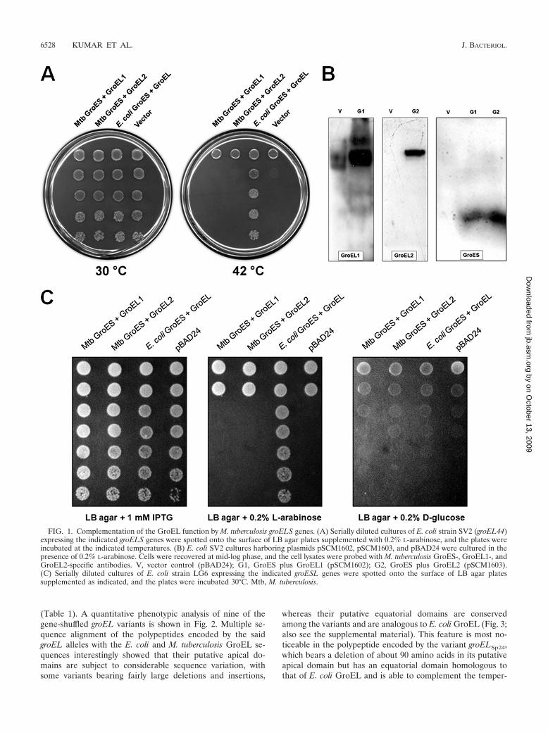
Mycobacterial GroELs do not complement the groEL44 al-lele. Owing to their unusual behavior in vitro, we testedwhether the two M. tuberculosis groEL ORFs could comple-ment the loss of GroEL function in E. coli. This was tested intwo E. coli strains, SV2, which harbors a temperature-sensitivegroEL44 allele, and LG6, in which the expression of the chro-mosomal groESL operon is under the control of the Plac pro-moter. The expression of M. tuberculosis groEL1 and groEL2from the PBAD promoter in SV2 did not lead to rescue of itstemperature-sensitive phenotype even when the M. tuberculo-sis groELs were coexpressed with their cognate groES (Fig.1A). For these studies, a plasmid bearing E. coli groESL underthe control of the PBAD promoter, which complemented thetemperature-sensitive phenotype of the groEL44 allele, wasalso employed. Immunoblots with GroEL1- and GroEL2-spe-cific antibodies established that the proteins were expressedand, hence, that the lack of complementation was not due tothe lack of expression (Fig. 1B).
Likewise, M. tuberculosis GroELs could not support thegrowth of E. coli LG6 (Fig. 1C). The inability of M. tuberculosisgroELs to complement the loss of GroEL function in E. coli isconsistent with the weakened in vitro chaperonin activityshown by our earlier studies of M. tuberculosis GroELs (41).
The apical domain of GroEL can tolerate considerable vari-ation. In order to investigate the molecular features which leadto the differences in E. coli and M. tuberculosis GroELs, wegenerated a pool of groEL variants via DNA shuffling, startingwith ORFs encoding M. tuberculosis GroEL1 and GroEL2 astemplate DNA. ORFs capable of encoding active versions ofGroEL were selected in E. coli SV2. The mutants thus gener-ated were chimeras of M. tuberculosis groEL1 and groEL2 andE. coli groEL. Sequence analysis of the clones and the com-parison of G�C contents show that the gene-shuffled groELvariants are in fact derived from M. tuberculosis groELs
VOL. 191, 2009 MULTIPLE FORMS OF MYCOBACTERIAL GroEL1 6527
by on October 13, 2009
jb.asm.org
Dow
nloaded from
(Table 1). A quantitative phenotypic analysis of nine of thegene-shuffled groEL variants is shown in Fig. 2. Multiple se-quence alignment of the polypeptides encoded by the saidgroEL alleles with the E. coli and M. tuberculosis GroEL se-quences interestingly showed that their putative apical do-mains are subject to considerable sequence variation, withsome variants bearing fairly large deletions and insertions,
whereas their putative equatorial domains are conservedamong the variants and are analogous to E. coli GroEL (Fig. 3;also see the supplemental material). This feature is most no-ticeable in the polypeptide encoded by the variant groELSp24,which bears a deletion of about 90 amino acids in its putativeapical domain but has an equatorial domain homologous tothat of E. coli GroEL and is able to complement the temper-
FIG. 1. Complementation of the GroEL function by M. tuberculosis groELS genes. (A) Serially diluted cultures of E. coli strain SV2 (groEL44)expressing the indicated groELS genes were spotted onto the surface of LB agar plates supplemented with 0.2% L-arabinose, and the plates wereincubated at the indicated temperatures. (B) E. coli SV2 cultures harboring plasmids pSCM1602, pSCM1603, and pBAD24 were cultured in thepresence of 0.2% L-arabinose. Cells were recovered at mid-log phase, and the cell lysates were probed with M. tuberculosis GroES-, GroEL1-, andGroEL2-specific antibodies. V, vector control (pBAD24); G1, GroES plus GroEL1 (pSCM1602); G2, GroES plus GroEL2 (pSCM1603).(C) Serially diluted cultures of E. coli strain LG6 expressing the indicated groESL genes were spotted onto the surface of LB agar platessupplemented as indicated, and the plates were incubated 30°C. Mtb, M. tuberculosis.
6528 KUMAR ET AL. J. BACTERIOL.
by on October 13, 2009
jb.asm.org
Dow
nloaded from
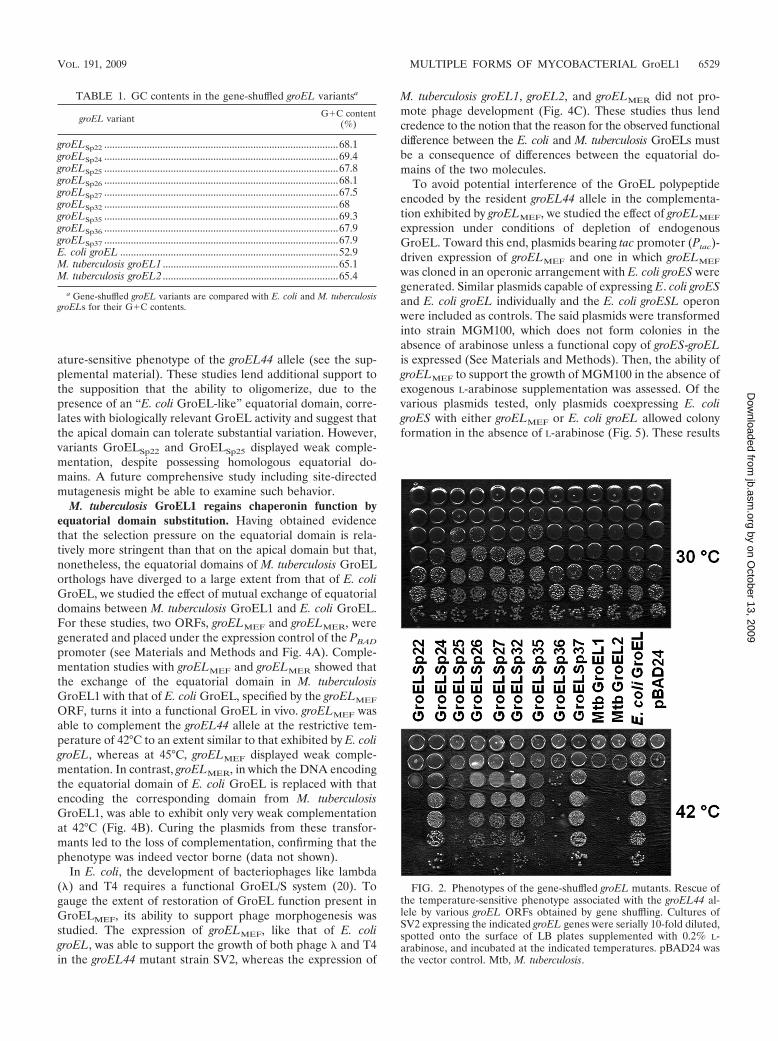
ature-sensitive phenotype of the groEL44 allele (see the sup-plemental material). These studies lend additional support tothe supposition that the ability to oligomerize, due to thepresence of an “E. coli GroEL-like” equatorial domain, corre-lates with biologically relevant GroEL activity and suggest thatthe apical domain can tolerate substantial variation. However,variants GroELSp22 and GroELSp25 displayed weak comple-mentation, despite possessing homologous equatorial do-mains. A future comprehensive study including site-directedmutagenesis might be able to examine such behavior.
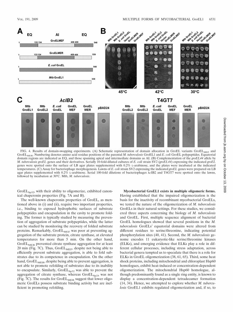
M. tuberculosis GroEL1 regains chaperonin function byequatorial domain substitution. Having obtained evidencethat the selection pressure on the equatorial domain is rela-tively more stringent than that on the apical domain but that,nonetheless, the equatorial domains of M. tuberculosis GroELorthologs have diverged to a large extent from that of E. coliGroEL, we studied the effect of mutual exchange of equatorialdomains between M. tuberculosis GroEL1 and E. coli GroEL.For these studies, two ORFs, groELMEF and groELMER, weregenerated and placed under the expression control of the PBAD
promoter (see Materials and Methods and Fig. 4A). Comple-mentation studies with groELMEF and groELMER showed thatthe exchange of the equatorial domain in M. tuberculosisGroEL1 with that of E. coli GroEL, specified by the groELMEF
ORF, turns it into a functional GroEL in vivo. groELMEF wasable to complement the groEL44 allele at the restrictive tem-perature of 42°C to an extent similar to that exhibited by E. coligroEL, whereas at 45°C, groELMEF displayed weak comple-mentation. In contrast, groELMER, in which the DNA encodingthe equatorial domain of E. coli GroEL is replaced with thatencoding the corresponding domain from M. tuberculosisGroEL1, was able to exhibit only very weak complementationat 42°C (Fig. 4B). Curing the plasmids from these transfor-mants led to the loss of complementation, confirming that thephenotype was indeed vector borne (data not shown).
In E. coli, the development of bacteriophages like lambda(�) and T4 requires a functional GroEL/S system (20). Togauge the extent of restoration of GroEL function present inGroELMEF, its ability to support phage morphogenesis wasstudied. The expression of groELMEF, like that of E. coligroEL, was able to support the growth of both phage � and T4in the groEL44 mutant strain SV2, whereas the expression of
M. tuberculosis groEL1, groEL2, and groELMER did not pro-mote phage development (Fig. 4C). These studies thus lendcredence to the notion that the reason for the observed functionaldifference between the E. coli and M. tuberculosis GroELs mustbe a consequence of differences between the equatorial do-mains of the two molecules.
To avoid potential interference of the GroEL polypeptideencoded by the resident groEL44 allele in the complementa-tion exhibited by groELMEF, we studied the effect of groELMEF
expression under conditions of depletion of endogenousGroEL. Toward this end, plasmids bearing tac promoter (Ptac)-driven expression of groELMEF and one in which groELMEF
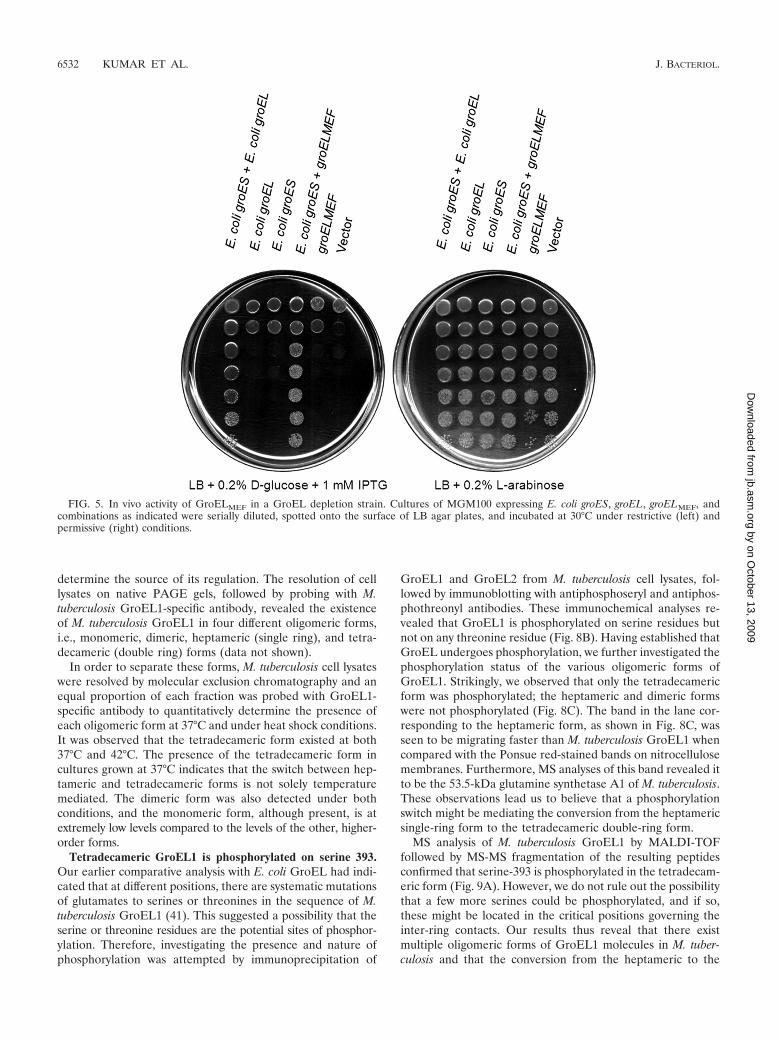
was cloned in an operonic arrangement with E. coli groES weregenerated. Similar plasmids capable of expressing E. coli groESand E. coli groEL individually and the E. coli groESL operonwere included as controls. The said plasmids were transformedinto strain MGM100, which does not form colonies in theabsence of arabinose unless a functional copy of groES-groELis expressed (See Materials and Methods). Then, the ability ofgroELMEF to support the growth of MGM100 in the absence ofexogenous L-arabinose supplementation was assessed. Of thevarious plasmids tested, only plasmids coexpressing E. coligroES with either groELMEF or E. coli groEL allowed colonyformation in the absence of L-arabinose (Fig. 5). These results
FIG. 2. Phenotypes of the gene-shuffled groEL mutants. Rescue ofthe temperature-sensitive phenotype associated with the groEL44 al-lele by various groEL ORFs obtained by gene shuffling. Cultures ofSV2 expressing the indicated groEL genes were serially 10-fold diluted,spotted onto the surface of LB plates supplemented with 0.2% L-arabinose, and incubated at the indicated temperatures. pBAD24 wasthe vector control. Mtb, M. tuberculosis.
TABLE 1. GC contents in the gene-shuffled groEL variantsa
groEL variant G�C content(%)
groELSp22 ........................................................................................68.1groELSp24 ........................................................................................69.4groELSp25 ........................................................................................67.8groELSp26 ........................................................................................68.1groELSp27 ........................................................................................67.5groELSp32 ........................................................................................68groELSp35 ........................................................................................69.3groELSp36 ........................................................................................67.9groELSp37 ........................................................................................67.9E. coli groEL ..................................................................................52.9M. tuberculosis groEL1 ..................................................................65.1M. tuberculosis groEL2 ..................................................................65.4
a Gene-shuffled groEL variants are compared with E. coli and M. tuberculosisgroELs for their G�C contents.
VOL. 191, 2009 MULTIPLE FORMS OF MYCOBACTERIAL GroEL1 6529
by on October 13, 2009
jb.asm.org
Dow
nloaded from
clearly show that GroELMEF can substitute for E. coli GroELin vivo and requires E. coli GroES for its activity, which is notinfluenced by the resident GroEL of strain SV2.
The equatorial domain of GroELMEF is responsible for it at-taining a higher oligomeric state. Having shown that GroELMEF
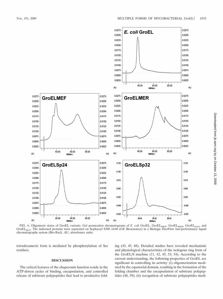
is functional in vivo, we wished to test whether the ability ofGroELMEF to complement the defect in the groEL44 allele isa consequence of it existing in higher-order oligomeric formand thus being able to form a cavity for encapsulation, featuresthat are lacking in the parental M. tuberculosis GroEL1 (40).Since the polypeptides encoded by the gene-shuffled variantsof groEL, groELSp24, and groELSp32 bear considerable varia-tion (Fig. 3; also see Fig. S1 in the supplemental material),similar attributes of the encoded polypeptides were studied.Gel filtration studies with Sephacryl S300 showed that GroELMEF
was capable of existing in a higher oligomeric state, similar tothat seen for E. coli GroEL, whereas GroELMER displayed alower oligomeric character (Fig. 6), a property reminiscent ofthat seen for M. tuberculosis GroEL1 (59). One noticeableaspect of the oligomeric properties of GroELMEF is that itexisted in equilibrium between higher and lower oligomericstates, with the higher oligomeric state being the predominantspecies, which presumably explains its somewhat weakenedability to substitute for E. coli GroEL in vivo at 45°C (Fig. 4B).
Since the higher oligomeric state of GroELMEF displays gelfiltration characteristics similar to those of E. coli GroEL, it isreasonable to presume that the said state corresponds to atetradecameric assembly. We have also examined the oligo-meric states of GroELSp24 and GroELSp32, variants that beardeletions in the putative intermediate apical domain boundary.Both the variants displayed tendencies to exist in a higheroligomeric state, and their gel filtration profiles were consistentwith their predicted molecular weights.
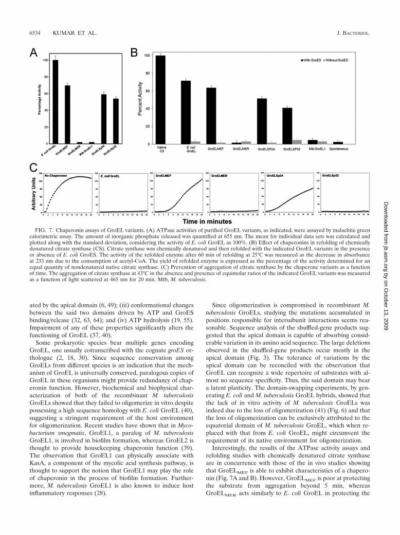
The biochemical properties of purified GroEL variants cor-relate with their in vivo activities. GroEL-assisted folding ofsubstrate proteins typically involves (i) binding of the polypep-tides by virtue of exposed hydrophobic interactions; (ii) se-questration of polypeptides into the cavity, thereby preventingirreversible aggregation; (iii) ATP hydrolysis; and (iv) GroES-dependent refolding of substrate polypeptides. Since the gelfiltration results revealed that GroELMEF, GroELSp24, andGroELSp32 are oligomeric, we studied their biochemical fea-tures in hydrolyzing ATP and refolding the model substrate,citrate synthase. The ability of GroELMEF to exist in a higheroligomeric state correlated with its ability, in comparison tothat displayed by GroELMER, to refold chemically denaturedsubstrate protein (Fig. 7A and B). Similarly, GroELSp24 and
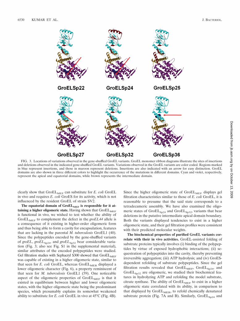
FIG. 3. Locations of variations observed in the gene-shuffled GroEL variants. GroEL monomer ribbon diagrams illustrate the sites of insertionsand deletions observed in the indicated gene-shuffled GroEL variants. Variations observed in the GroEL variants are color coded. Regions markedin blue represent insertions, and those in maroon represent deletions. Insertions are also indicated with an arrow for easy distinction. GroELdomains are also shown in three different colors to highlight the occurrence of the mutations in different domains. Cyan and violet, respectively,represent the apical and equatorial domains, while brown represents the intermediate domain.
6530 KUMAR ET AL. J. BACTERIOL.
by on October 13, 2009
jb.asm.org
Dow
nloaded from
GroELSp32, with their ability to oligomerize, exhibited canon-ical chaperonin properties (Fig. 7A and B).
The well-known chaperonin properties of GroEL, as men-tioned above in (i) and (ii), require two important properties,i.e., binding to exposed hydrophobic surfaces of substratepolypeptides and encapsulation in the cavity to promote fold-ing. The former is typically studied by measuring the preven-tion of aggregation of substrate polypeptides, while the lattercan be studied by monitoring the recovery of folded substrateproteins. Remarkably, GroELMEF was poor at preventing ag-gregation of the substrate protein, citrate synthase, at elevatedtemperatures for more than 5 min. On the other hand,GroELMER prevented citrate synthase aggregation for at least20 min (Fig. 7C). Thus, GroELMEF, despite not being able toefficiently prevent substrate aggregation, is able to fold sub-strates due to its competence in encapsulation. On the otherhand, GroELMER, despite being able to prevent aggregation, isnot able to promote refolding of substrates due to its inabilityto encapsulate. Similarly, GroELSp32 was able to prevent theaggregation of citrate synthase, whereas GroELSp24 was not(Fig. 7C). The results for GroELMER suggest that lower oligo-meric GroELs possess substrate binding activity but are inef-ficient in promoting refolding.
Mycobacterial GroEL1 exists in multiple oligomeric forms.Having established that the impaired oligomerization is thebasis for the inactivity of recombinant mycobacterial GroELs,we tested the nature of the oligomerization of M. tuberculosisGroELs in their natural settings. For these studies, we consid-ered three aspects concerning the biology of M. tuberculosisand GroEL. First, multiple sequence alignment of bacterialGroEL homologues showed that several positions in the M.tuberculosis GroELs’ equatorial domains were altered fromdifferent residues to serine/threonine, indicating potentialphosphorylation sites (40, 41). Second, the M. tuberculosis ge-nome encodes 11 eukaryotic-like serine/threonine kinases(ELKs), and emerging evidence that ELKs play a role in dif-ferent cellular processes, including stress adaptation, acrossbacterial genera tempted us to speculate that there is a role forELKs in GroEL oligomerization (38, 61, 65). Third, some heatshock proteins, including mitochondrial and chloroplast Hsp60homologues, exhibit heat-induced or concentration-dependentoligomerization. The mitochondrial Hsp60 homologue, al-though predominantly found as a single ring entity, is known todisplay a concentration-dependent tetradecamer formation(14, 34). Hence, we attempted to explore whether M. tubercu-losis GroEL1 exhibits regulated oligomerization and, if so, to
FIG. 4. Results of domain-swapping experiments. (A) Schematic representation of domain allocation in GroEL variants GroELMEF andGroELMER. Numbering denotes amino acid residue positions of the parental M. tuberculosis GroEL1 and E. coli GroEL polypeptides. Equatorialdomain regions are indicated as EQ, and those spanning apical and intermediate domains as AI. (B) Complementation of the groEL44 allele byM. tuberculosis groEL genes and their derivatives. Serially 10-fold-diluted cultures of E. coli strain SV2 (groEL44) expressing the indicated groELgenes were spotted onto the surface of LB agar plates supplemented with 0.2% L-arabinose, and the plates were incubated at the indicatedtemperatures. (C) Assay for bacteriophage morphogenesis. Lawns of E. coli strain SV2 expressing the indicated groEL genes were prepared on LBagar plates supplemented with 0.2% L-arabinose. Serial 100-fold dilutions of bacteriophages �cIB2 and T4GT7 were spotted onto the lawns,followed by incubation at 30°C. Mtb, M. tuberculosis.
VOL. 191, 2009 MULTIPLE FORMS OF MYCOBACTERIAL GroEL1 6531
by on October 13, 2009
jb.asm.org
Dow
nloaded from
determine the source of its regulation. The resolution of celllysates on native PAGE gels, followed by probing with M.tuberculosis GroEL1-specific antibody, revealed the existenceof M. tuberculosis GroEL1 in four different oligomeric forms,i.e., monomeric, dimeric, heptameric (single ring), and tetra-decameric (double ring) forms (data not shown).
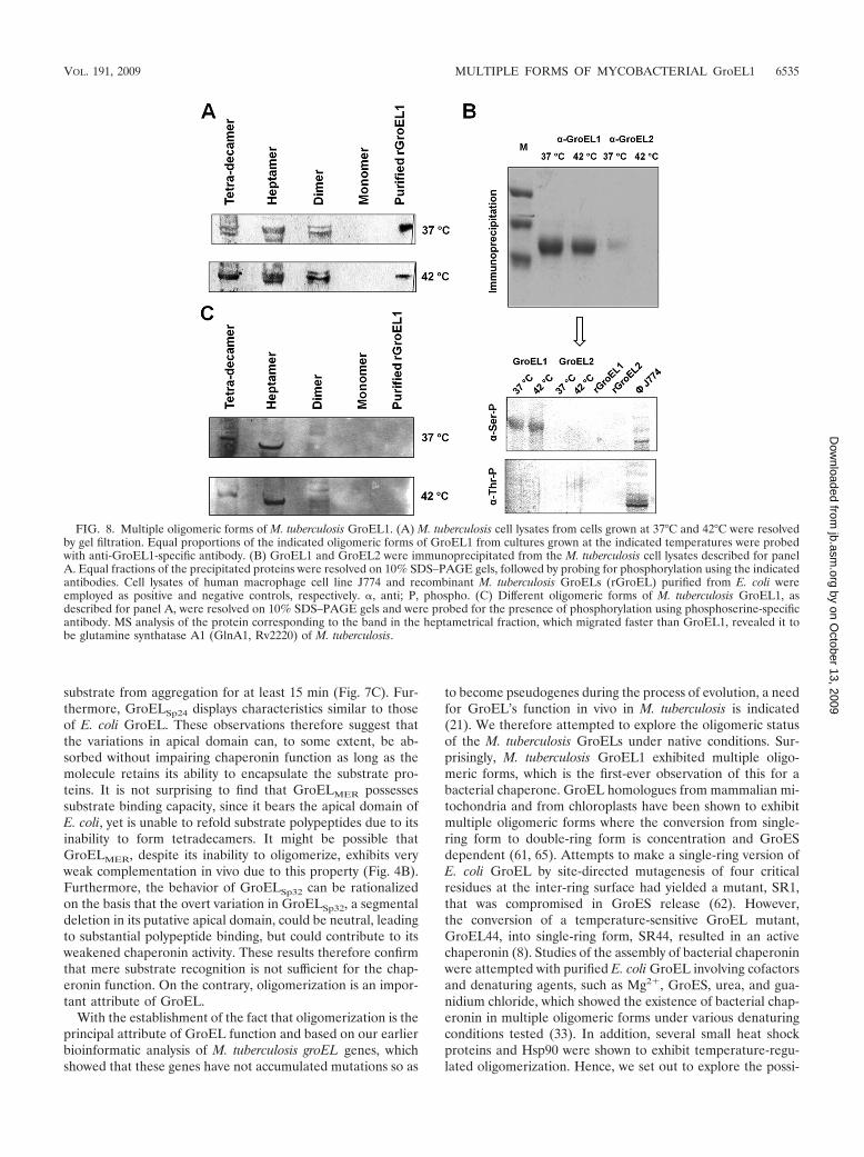
In order to separate these forms, M. tuberculosis cell lysateswere resolved by molecular exclusion chromatography and anequal proportion of each fraction was probed with GroEL1-specific antibody to quantitatively determine the presence ofeach oligomeric form at 37°C and under heat shock conditions.It was observed that the tetradecameric form existed at both37°C and 42°C. The presence of the tetradecameric form incultures grown at 37°C indicates that the switch between hep-tameric and tetradecameric forms is not solely temperaturemediated. The dimeric form was also detected under bothconditions, and the monomeric form, although present, is atextremely low levels compared to the levels of the other, higher-order forms.
Tetradecameric GroEL1 is phosphorylated on serine 393.Our earlier comparative analysis with E. coli GroEL had indi-cated that at different positions, there are systematic mutationsof glutamates to serines or threonines in the sequence of M.tuberculosis GroEL1 (41). This suggested a possibility that theserine or threonine residues are the potential sites of phosphor-ylation. Therefore, investigating the presence and nature ofphosphorylation was attempted by immunoprecipitation of
GroEL1 and GroEL2 from M. tuberculosis cell lysates, fol-lowed by immunoblotting with antiphosphoseryl and antiphos-phothreonyl antibodies. These immunochemical analyses re-vealed that GroEL1 is phosphorylated on serine residues butnot on any threonine residue (Fig. 8B). Having established thatGroEL undergoes phosphorylation, we further investigated thephosphorylation status of the various oligomeric forms ofGroEL1. Strikingly, we observed that only the tetradecamericform was phosphorylated; the heptameric and dimeric formswere not phosphorylated (Fig. 8C). The band in the lane cor-responding to the heptameric form, as shown in Fig. 8C, wasseen to be migrating faster than M. tuberculosis GroEL1 whencompared with the Ponsue red-stained bands on nitrocellulosemembranes. Furthermore, MS analyses of this band revealed itto be the 53.5-kDa glutamine synthetase A1 of M. tuberculosis.These observations lead us to believe that a phosphorylationswitch might be mediating the conversion from the heptamericsingle-ring form to the tetradecameric double-ring form.
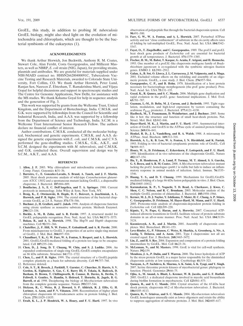
MS analysis of M. tuberculosis GroEL1 by MALDI-TOFfollowed by MS-MS fragmentation of the resulting peptidesconfirmed that serine-393 is phosphorylated in the tetradecam-eric form (Fig. 9A). However, we do not rule out the possibilitythat a few more serines could be phosphorylated, and if so,these might be located in the critical positions governing theinter-ring contacts. Our results thus reveal that there existmultiple oligomeric forms of GroEL1 molecules in M. tuber-culosis and that the conversion from the heptameric to the
FIG. 5. In vivo activity of GroELMEF in a GroEL depletion strain. Cultures of MGM100 expressing E. coli groES, groEL, groELMEF, andcombinations as indicated were serially diluted, spotted onto the surface of LB agar plates, and incubated at 30°C under restrictive (left) andpermissive (right) conditions.
6532 KUMAR ET AL. J. BACTERIOL.
by on October 13, 2009
jb.asm.org
Dow
nloaded from
tetradecameric form is mediated by phosphorylation of Serresidues.
DISCUSSION
The critical features of the chaperonin function reside in theATP-driven cycles of binding, encapsulation, and controlledrelease of substrate polypeptides that lead to productive fold-
ing (43, 45, 48). Detailed studies have revealed mechanisticand physiological characteristics of the isologous ring form ofthe GroEL/S machine (11, 42, 45, 53, 54). According to thecurrent understanding, the following properties of GroEL aresignificant in controlling its activity: (i) oligomerization medi-ated by the equatorial domain, resulting in the formation of thefolding chamber and the encapsulation of substrate polypep-tides (48, 59); (ii) recognition of substrate polypeptides medi-
FIG. 6. Oligomeric states of GroEL variants. Gel permeation chromatograms of E. coli GroEL, GroELMEF, GroELMER, GroELSp24, andGroELSp32. The indicated proteins were separated on Sephacryl S300 16/60 (GE Biosciences) in a Biologic DuoFlow fast-performance liquidchromatography system (Bio-Rad). AU, absorbance units.
VOL. 191, 2009 MULTIPLE FORMS OF MYCOBACTERIAL GroEL1 6533
by on October 13, 2009
jb.asm.org
Dow
nloaded from
ated by the apical domain (6, 49); (iii) conformational changesbetween the said two domains driven by ATP and GroESbinding/release (32, 63, 64); and (iv) ATP hydrolysis (19, 55).Impairment of any of these properties significantly alters thefunctioning of GroEL (37, 40).
Some prokaryotic species bear multiple genes encodingGroEL, one usually cotranscribed with the cognate groES or-thologue (2, 18, 30). Since sequence conservation amongGroELs from different species is an indication that the mech-anism of GroEL is universally conserved, paralogous copies ofGroEL in these organisms might provide redundancy of chap-eronin function. However, biochemical and biophysical char-acterization of both of the recombinant M. tuberculosisGroELs showed that they failed to oligomerize in vitro despitepossessing a high sequence homology with E. coli GroEL (40),suggesting a stringent requirement of the host environmentfor oligomerization. Recent studies have shown that in Myco-bacterium smegmatis, GroEL1, a paralog of M. tuberculosisGroEL1, is involved in biofilm formation, whereas GroEL2 isthought to provide housekeeping chaperonin function (39).The observation that GroEL1 can physically associate withKasA, a component of the mycolic acid synthesis pathway, isthought to support the notion that GroEL1 may play the roleof chaperonin in the process of biofilm formation. Further-more, M. tuberculosis GroEL1 is also known to induce hostinflammatory responses (28).
Since oligomerization is compromised in recombinant M.tuberculosis GroELs, studying the mutations accumulated inpositions responsible for intersubunit interactions seems rea-sonable. Sequence analysis of the shuffled-gene products sug-gested that the apical domain is capable of absorbing consid-erable variation in its amino acid sequence. The large deletionsobserved in the shuffled-gene products occur mostly in theapical domain (Fig. 3). The tolerance of variations by theapical domain can be reconciled with the observation thatGroEL can recognize a wide repertoire of substrates with al-most no sequence specificity. Thus, the said domain may beara latent plasticity. The domain-swapping experiments, by gen-erating E. coli and M. tuberculosis GroEL hybrids, showed thatthe lack of in vitro activity of M. tuberculosis GroELs wasindeed due to the loss of oligomerization (41) (Fig. 6) and thatthe loss of oligomerization can be exclusively attributed to theequatorial domain of M. tuberculosis GroEL, which when re-placed with that from E. coli GroEL, might circumvent therequirement of its native environment for oligomerization.
Interestingly, the results of the ATPase activity assays andrefolding studies with chemically denatured citrate synthaseare in concurrence with those of the in vivo studies showingthat GroELMEF is able to exhibit characteristics of a chapero-nin (Fig. 7A and B). However, GroELMEF is poor at protectingthe substrate from aggregation beyond 5 min, whereasGroELMER acts similarly to E. coli GroEL in protecting the
FIG. 7. Chaperonin assays of GroEL variants. (A) ATPase activities of purified GroEL variants, as indicated, were assayed by malachite greencalorimetric assay. The amount of inorganic phosphate released was quantified at 655 nm. The mean for individual data sets was calculated andplotted along with the standard deviation, considering the activity of E. coli GroEL as 100%. (B) Effect of chaperonins in refolding of chemicallydenatured citrate synthase (CS). Citrate synthase was chemically denatured and then refolded with the indicated GroEL variants in the presenceor absence of E. coli GroES. The activity of the refolded enzyme after 60 min of refolding at 25°C was measured as the decrease in absorbanceat 233 nm due to the consumption of acetyl-CoA. The yield of refolded enzyme is expressed as the percentage of the activity determined for anequal quantity of nondenatured native citrate synthase. (C) Prevention of aggregation of citrate synthase by the chaperone variants as a functionof time. The aggregation of citrate synthase at 43°C in the absence and presence of equimolar ratios of the indicated GroEL variants was measuredas a function of light scattered at 465 nm for 20 min. Mtb, M. tuberculosis.
6534 KUMAR ET AL. J. BACTERIOL.
by on October 13, 2009
jb.asm.org
Dow
nloaded from
substrate from aggregation for at least 15 min (Fig. 7C). Fur-thermore, GroELSp24 displays characteristics similar to thoseof E. coli GroEL. These observations therefore suggest thatthe variations in apical domain can, to some extent, be ab-sorbed without impairing chaperonin function as long as themolecule retains its ability to encapsulate the substrate pro-teins. It is not surprising to find that GroELMER possessessubstrate binding capacity, since it bears the apical domain ofE. coli, yet is unable to refold substrate polypeptides due to itsinability to form tetradecamers. It might be possible thatGroELMER, despite its inability to oligomerize, exhibits veryweak complementation in vivo due to this property (Fig. 4B).Furthermore, the behavior of GroELSp32 can be rationalizedon the basis that the overt variation in GroELSp32, a segmentaldeletion in its putative apical domain, could be neutral, leadingto substantial polypeptide binding, but could contribute to itsweakened chaperonin activity. These results therefore confirmthat mere substrate recognition is not sufficient for the chap-eronin function. On the contrary, oligomerization is an impor-tant attribute of GroEL.
With the establishment of the fact that oligomerization is theprincipal attribute of GroEL function and based on our earlierbioinformatic analysis of M. tuberculosis groEL genes, whichshowed that these genes have not accumulated mutations so as
to become pseudogenes during the process of evolution, a needfor GroEL’s function in vivo in M. tuberculosis is indicated(21). We therefore attempted to explore the oligomeric statusof the M. tuberculosis GroELs under native conditions. Sur-prisingly, M. tuberculosis GroEL1 exhibited multiple oligo-meric forms, which is the first-ever observation of this for abacterial chaperone. GroEL homologues from mammalian mi-tochondria and from chloroplasts have been shown to exhibitmultiple oligomeric forms where the conversion from single-ring form to double-ring form is concentration and GroESdependent (61, 65). Attempts to make a single-ring version ofE. coli GroEL by site-directed mutagenesis of four criticalresidues at the inter-ring surface had yielded a mutant, SR1,that was compromised in GroES release (62). However,the conversion of a temperature-sensitive GroEL mutant,GroEL44, into single-ring form, SR44, resulted in an activechaperonin (8). Studies of the assembly of bacterial chaperoninwere attempted with purified E. coli GroEL involving cofactorsand denaturing agents, such as Mg2�, GroES, urea, and gua-nidium chloride, which showed the existence of bacterial chap-eronin in multiple oligomeric forms under various denaturingconditions tested (33). In addition, several small heat shockproteins and Hsp90 were shown to exhibit temperature-regu-lated oligomerization. Hence, we set out to explore the possi-
FIG. 8. Multiple oligomeric forms of M. tuberculosis GroEL1. (A) M. tuberculosis cell lysates from cells grown at 37°C and 42°C were resolvedby gel filtration. Equal proportions of the indicated oligomeric forms of GroEL1 from cultures grown at the indicated temperatures were probedwith anti-GroEL1-specific antibody. (B) GroEL1 and GroEL2 were immunoprecipitated from the M. tuberculosis cell lysates described for panelA. Equal fractions of the precipitated proteins were resolved on 10% SDS–PAGE gels, followed by probing for phosphorylation using the indicatedantibodies. Cell lysates of human macrophage cell line J774 and recombinant M. tuberculosis GroELs (rGroEL) purified from E. coli wereemployed as positive and negative controls, respectively. �, anti; P, phospho. (C) Different oligomeric forms of M. tuberculosis GroEL1, asdescribed for panel A, were resolved on 10% SDS–PAGE gels and were probed for the presence of phosphorylation using phosphoserine-specificantibody. MS analysis of the protein corresponding to the band in the heptametrical fraction, which migrated faster than GroEL1, revealed it tobe glutamine synthatase A1 (GlnA1, Rv2220) of M. tuberculosis.
VOL. 191, 2009 MULTIPLE FORMS OF MYCOBACTERIAL GroEL1 6535
by on October 13, 2009
jb.asm.org
Dow
nloaded from
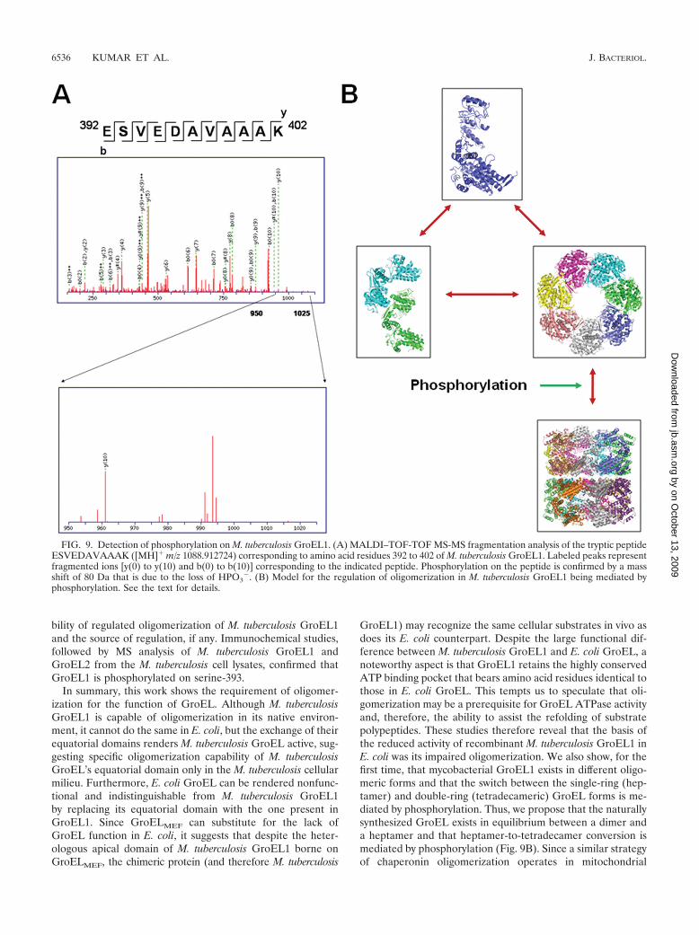
bility of regulated oligomerization of M. tuberculosis GroEL1and the source of regulation, if any. Immunochemical studies,followed by MS analysis of M. tuberculosis GroEL1 andGroEL2 from the M. tuberculosis cell lysates, confirmed thatGroEL1 is phosphorylated on serine-393.
In summary, this work shows the requirement of oligomer-ization for the function of GroEL. Although M. tuberculosisGroEL1 is capable of oligomerization in its native environ-ment, it cannot do the same in E. coli, but the exchange of theirequatorial domains renders M. tuberculosis GroEL active, sug-gesting specific oligomerization capability of M. tuberculosisGroEL’s equatorial domain only in the M. tuberculosis cellularmilieu. Furthermore, E. coli GroEL can be rendered nonfunc-tional and indistinguishable from M. tuberculosis GroEL1by replacing its equatorial domain with the one present inGroEL1. Since GroELMEF can substitute for the lack ofGroEL function in E. coli, it suggests that despite the heter-ologous apical domain of M. tuberculosis GroEL1 borne onGroELMEF, the chimeric protein (and therefore M. tuberculosis
GroEL1) may recognize the same cellular substrates in vivo asdoes its E. coli counterpart. Despite the large functional dif-ference between M. tuberculosis GroEL1 and E. coli GroEL, anoteworthy aspect is that GroEL1 retains the highly conservedATP binding pocket that bears amino acid residues identical tothose in E. coli GroEL. This tempts us to speculate that oli-gomerization may be a prerequisite for GroEL ATPase activityand, therefore, the ability to assist the refolding of substratepolypeptides. These studies therefore reveal that the basis ofthe reduced activity of recombinant M. tuberculosis GroEL1 inE. coli was its impaired oligomerization. We also show, for thefirst time, that mycobacterial GroEL1 exists in different oligo-meric forms and that the switch between the single-ring (hep-tamer) and double-ring (tetradecameric) GroEL forms is me-diated by phosphorylation. Thus, we propose that the naturallysynthesized GroEL exists in equilibrium between a dimer anda heptamer and that heptamer-to-tetradecamer conversion ismediated by phosphorylation (Fig. 9B). Since a similar strategyof chaperonin oligomerization operates in mitochondrial
FIG. 9. Detection of phosphorylation on M. tuberculosis GroEL1. (A) MALDI–TOF-TOF MS-MS fragmentation analysis of the tryptic peptideESVEDAVAAAK ([MH]� m/z 1088.912724) corresponding to amino acid residues 392 to 402 of M. tuberculosis GroEL1. Labeled peaks representfragmented ions [y(0) to y(10) and b(0) to b(10)] corresponding to the indicated peptide. Phosphorylation on the peptide is confirmed by a massshift of 80 Da that is due to the loss of HPO3
�. (B) Model for the regulation of oligomerization in M. tuberculosis GroEL1 being mediated byphosphorylation. See the text for details.
6536 KUMAR ET AL. J. BACTERIOL.
by on October 13, 2009
jb.asm.org
Dow
nloaded from
GroEL, this study, in addition to probing M. tuberculosisGroEL biology, might also shed light on the evolution of mi-tochondria and chloroplasts, which are thought to be the bac-terial symbionts of the eukaryotes (1).
ACKNOWLEDGMENTS
We thank Arthur Horwich, Jon Beckwith, Anthony R. M. Coates,Stewart Cole, Alan Fersht, Costa Georgopoulos, and Millicent Mas-ters, as well as NBRP, E. coli strain (NIG, Japan), for providing strains,plasmids and antibodies. We are grateful for materials supplied underNIH-NIAID contract no. HHSN266200400091C, Tuberculosis Vac-cine Testing and Research Materials, awarded to Colorado State Uni-versity, Fort Collins, CO. We thank Arthur Horwich, Peter Lund,Ranjan Sen, Nasreen Z. Ehtesham, T. Ramakrishna Murti, and VijayaGopal for helpful discussions and support in spectroscopic studies andThe Centre for Genomic Applications, New Delhi, for assistance withthe MS studies. We thank Kshama Goyal for help in sequence analysisand the generation of Fig. 3.
This work was supported by grants from the Wellcome Trust, UnitedKingdom, and the Department of Biotechnology, India. C.M.S.K. andG.K. were supported by fellowships from the Council for Scientific andIndustrial Research, India, and A.A.S. was supported by a fellowshipfrom the Department of Science and Technology, India. S.C.M. is aWellcome Trust International Senior Research Fellow. The authorsdeclare no financial conflict of interest.
Author contributions. C.M.S.K. conducted all the molecular biolog-ical, biochemical and genetic experiments. C.M.S.K. and A.A.S. de-signed the genetic experiments with E. coli, and C.M.S.K. and C.V.S.performed the gene-shuffling studies. C.M.S.K., G.K., A.K.T., andS.C.M. designed the experiments with M. tuberculosis, and C.M.S.K.and G.K. conducted these. Overall supervision and design was byS.C.M., A.K.T., and A.A.S.
REFERENCES
1. Allen, J. F. 2003. Why chloroplasts and mitochondria contain genomes.Comp. Funct. Genomics 4:31–36.
2. Barreiro, C., E. Gonzalez-Lavado, S. Brand, A. Tauch, and J. F. Martin.2005. Heat shock proteome analysis of wild-type Corynebacterium glutami-cum ATCC 13032 and a spontaneous mutant lacking GroEL1, a dispensablechaperone. J. Bacteriol. 187:884–889.
3. Bonifacino, J. S., E. C. Dell’Angelica, and T. A. Springer. 1988. Currentprotocols in immunology. John Wiley & Sons, New York, NY.
4. Braig, K., Z. Otwinowski, R. Hegde, D. C. Boisvert, A. Joachimiak, A. L.Horwich, and P. B. Sigler. 1994. The crystal structure of the bacterial chap-eronin GroEL at 2.8 Å. Nature 371:578–586.
5. Buchner, J., H. Grallert, and U. Jakob. 1998. Analysis of chaperone functionusing citrate synthase as nonnative substrate protein. Methods Enzymol.290:323–338.
6. Buckle, A. M., R. Zahn, and A. R. Fersht. 1997. A structural model forGroEL polypeptide recognition. Proc. Natl. Acad. Sci. USA 94:3571–3575.
7. Bukau, B., and A. L. Horwich. 1998. The Hsp70 and Hsp60 chaperonemachines. Cell 92:351–366.
8. Chatellier, J., F. Hill, N. W. Foster, P. Goloubinoff, and A. R. Fersht. 2000.From minichaperone to GroEL 3: properties of an active single-ring mutantof GroEL. J. Mol. Biol. 304:897–910.
9. Chaudhuri, T. K., G. W. Farr, W. A. Fenton, S. Rospert, and A. L. Horwich.2001. GroEL/GroES-mediated folding of a protein too large to be encapsu-lated. Cell 107:235–246.
10. Chen, D., J. Song, D. T. Chuang, W. Chiu, and S. J. Ludtke. 2006. Anexpanded conformation of single-ring GroEL-GroES complex encapsulatesan 86 kDa substrate. Structure 14:1711–1722.
11. Chen, L., and P. B. Sigler. 1999. The crystal structure of a GroEL/peptidecomplex: plasticity as a basis for substrate diversity. Cell 99:757–768.
12. Reference deleted.13. Cole, S. T., R. Brosch, J. Parkhill, T. Garnier, C. Churcher, D. Harris, S. V.
Gordon, K. Eiglmeier, S. Gas, C. E. Barry III, F. Tekaia, K. Badcock, D.Basham, D. Brown, T. Chillingworth, R. Connor, R. Davies, K. Devlin, T.Feltwell, S. Gentles, N. Hamlin, S. Holroyd, T. Hornsby, K. Jagels, B. G.Barrell, et al. 1998. Deciphering the biology of Mycobacterium tuberculosisfrom the complete genome sequence. Nature 393:537–544.
14. Dickson, R., C. Weiss, R. J. Howard, S. P. Alldrich, R. J. Ellis, G. H.Lorimer, A. Azem, and P. V. Viitanen. 2000. Reconstitution of higher plantchloroplast chaperonin 60 tetradecamers active in protein folding J. Biol.Chem. 275:11829–11835.
15. Ewalt, K. L., J. P. Hendrick, W. A. Houry, and F. U. Hartl. 1997. In vivo
observation of polypeptide flux through the bacterial chaperonin system. Cell90:491–500.
16. Farr, G. W., W. A. Fenton, and A. L. Horwich. 2007. Perturbed ATPaseactivity and not “close confinement” of substrate in the cis cavity affects ratesof folding by tail-multiplied GroEL. Proc. Natl. Acad. Sci. USA 104:5342–5347.
17. Fayet, O., T. Ziegelhoffer, and C. Georgopoulos. 1989. The groES and groELheat shock gene products of Escherichia coli are essential for bacterialgrowth at all temperatures. J. Bacteriol. 171:1379–1385.
18. Fischer, H. M., M. Babst, T. Kaspar, G. Acuna, F. Arigoni, and H. Hennecke.1993. One member of a groESL-like chaperonin multigene family of Brady-rhizobium japonicum is co-regulated with the symbiotic nitrogen fixationgenes. EMBO J. 12:2901–2912.
19. Galan, A., B. Sot, O. Llorca, J. L. Carrascosa, J. M. Valpuesta, and A. Muga.2001. Excluded volume effects on the refolding and assembly of an oligo-meric protein. GroEL, a case study. J. Biol. Chem. 276:957–964.
20. Georgopoulos, C. P., and B. Hohn. 1978. Identification of a host proteinnecessary for bacteriophage morphogenesis (the groE gene product). Proc.Natl. Acad. Sci. USA 75:131–135.
21. Goyal, K., R. Qamra, and S. C. Mande. 2006. Multiple gene duplication andrapid evolution in the groEL gene: functional implications. J. Mol. Evol.63:781–787.
22. Guzman, L.-M., D. Belin, M. J. Carson, and J. Beckwith. 1995. Tight regu-lation, modulation, and high-level expression by vectors containing thearabinose PBAD promoter. J. Bacteriol. 177:4121–4130.
23. Haslbeck, M., T. Franzmann, D. Weinfurtner, and J. Buchner. 2005. Somelike it hot: the structure and function of small heat-shock proteins. Nat.Struct. Mol. Biol. 12:842–846.
24. Hayer-Hartl, M. K., J. Martin, and F. U. Hartl. 1995. Asymmetrical inter-action of GroEL and GroES in the ATPase cycle of assisted protein folding.Science 269:836–841.
25. Henkel, R. D., J. L. VandeBerg, and R. A. Walsh. 1988. A microassay forATPase. Anal. Biochem. 169:312–318.
26. Horwich, A. L., K. B. Low, W. A. Fenton, I. N. Hirshfield, and K. Furtak.1993. Folding in vivo of bacterial cytoplasmic proteins: role of GroEL. Cell74:909–917.
27. Houry, W. A., D. Frishman, C. Eckerskorn, F. Lottspeich, and F. U. Hartl.1999. Identification of in vivo substrates of the chaperonin GroEL. Nature402:147–154.
28. Hu, Y., B. Henderson, P. A. Lund, P. Tormay, M. T. Ahmed, S. S. Gurcha,G. S. Besra, and A. R. M. Coates. 2008. A Mycobacterium tuberculosis mutantlacking the groEL homologue cpn60.1 is viable but fails to induce an inflam-matory response in animal models of infection. Infect. Immun. 76:1535–1546.
29. Huang, Y. S., and D. T. Chuang. 1999. Mechanisms for GroEL/GroES-mediated folding of a large 86-kDa fusion polypeptide in vitro. J. Biol. Chem.274:10405–10412.
30. Karunakaran, K. P., Y. Noguchi, T. D. Read, A. Cherkasov, J. Kwee, C.Shen, C. C. Nelson, and R. C. Brunham. 2003. Molecular analysis of themultiple GroEL proteins of chlamydiae. J. Bacteriol. 185:1958–1966.
31. Kerner, M. J., D. J. Naylor, Y. Ishihama, T. Maier, H. C. Chang, A. P. Stines,C. Georgopoulos, D. Frishman, M. Hayer-Hartl, M. Mann, and F. U. Hartl.2005. Proteome-wide analysis of chaperonin-dependent protein folding inEscherichia coli. Cell 122:209–220.
32. Kipnis, Y., N. Papo, G. Haran, and A. Horovitz. 2007. Concerted ATP-induced allosteric transitions in GroEL facilitate release of protein substratedomains in an all-or-none manner. Proc. Natl. Acad. Sci. USA 104:3119–3124.
33. Kusmierczyk, A. R., and J. Martin. 2001. Assembly of chaperonin com-plexes. Mol. Biotechnol. 19:141–153.
34. Levy-Rimler, G., P. Viitanen, C. Weiss, R. Sharkia, A. Greenberg, A. Niv, A.Lustig, Y. Delarea, and A. Azem. 2001. Type I chaperonins: not all arecreated equal. Eur. J. Biochem. 268:3465–3472.
35. Lin, Z., and H. S. Rye. 2004. Expansion and compression of a protein foldingintermediate by GroEL. Mol. Cell 16:23–34.
36. McLennan, N., and M. Masters. 1998. GroE is vital for cell-wall synthesis.Nature 392:159.
37. Mendoza, J. A., P. Dulin, and T. Warren. 2000. The lower hydrolysis of ATPby the stress protein GroEL is a major factor responsible for the diminishedchaperonin activity at low temperature. Cryobiology 41:319–323.
38. Narayan, A., P. Sachdeva, K. Sharma, A. K. Saini, A. K. Tyagi, and Y. Singh.2007. Serine threonine protein kinases of mycobacterial genus: phylogeny tofunction. Physiol. Genomics 29:66–75.
39. Ojha, A., M. Anand, A. Bhatt, L. Kremer, W. R. Jacobs, and G. F. Hatfull.2005. GroEL1: a dedicated chaperone involved in mycolic acid biosynthesisduring biofilm formation in mycobacteria. Cell 123:861–873.
40. Qamra, R., and S. C. Mande. 2004. Crystal structure of the 65-kDa heatshock protein, chaperonin 60.2 of Mycobacterium tuberculosis. J. Bacteriol.186:8105–8113.
41. Qamra, R., V. Srinivas, and S. C. Mande. 2004. Mycobacterium tuberculosisGroEL homologues unusually exist as lower oligomers and retain the abilityto suppress aggregation of substrate proteins. J. Mol. Biol. 342:605–617.
VOL. 191, 2009 MULTIPLE FORMS OF MYCOBACTERIAL GroEL1 6537
by on October 13, 2009
jb.asm.org
Dow
nloaded from
42. Richardson, A., S. J. Landry, and C. Georgopoulos. 1998. The ins and outsof a molecular chaperone machine. Trends Biochem. Sci. 23:138–143.
43. Roseman, A. M., S. Chen, H. White, K. Braig, and H. R. Saibil. 1996. Thechaperonin ATPase cycle: mechanism of allosteric switching and movementsof substrate-binding domains in GroEL. Cell 87:241–251.
44. Rosenkrands, I., and P. Andersen. 2001. Preparation of culture filtrate pro-teins from Mycobacterium tuberculosis, p. 205–216. In T. Parish and N. G.Stoker (ed.), Methods in molecular medicine: Mycobacterium tuberculosisprotocols, vol. 54. Humana Press, Inc., Totowa, NJ.
45. Rye, H. S., A. M. Roseman, S. Chen, K. Furtak, W. A. Fenton, H. R. Saibil,and A. L. Horwich. 1999. GroEL-GroES cycling: ATP and nonnativepolypeptide direct alternation of folding-active rings. Cell 97:325–338.
46. Sakikawa, C., H. Taguchi, Y. Makino, and M. Yoshida. 1999. On the max-imum size of proteins to stay and fold in the cavity of GroEL underneathGroES. J. Biol. Chem. 274:21251–21256.
47. Sambrook, J., E. F. Fritsch, and T. Maniatis. 1989. Molecular cloning: alaboratory manual, 2nd ed. Cold Spring Harbor Laboratory Press, ColdSpring Harbor, NY.
48. Sigler, P. B., Z. Xu, H. S. Rye, S. G. Burston, W. A. Fenton, and A. L.Horwich. 1998. Structure and function in GroEL-mediated protein folding.Annu. Rev. Biochem. 67:581–608.
49. Stan, G., B. R. Brooks, G. H. Lorimer, and D. Thirumalai. 2006. Residues insubstrate proteins that interact with GroEL in the capture process are buriedin the native state. Proc. Natl. Acad. Sci. USA 103:4433–4438.
50. Steede, N. K., S. L. Temkin, and S. J. Landry. 2000. Assay of chaperonin-assisted refolding of citrate synthase, p. 133–138. In C. Schneider (ed.),Methods in molecular biology: chaperonin protocols, vol. 140. HumanaPress, Totowa, NJ.
51. Stemmer, W. P. 1994. DNA shuffling by random fragmentation and reas-sembly: in vitro recombination for molecular evolution. Proc. Natl. Acad.Sci. USA 91:10747–10751.
52. Tang, Y., H. Chang, A. Roeben, D. Wischnewski, N. Wischnewski, M. J.Kerner, F. U. Hartl, and M. Hayer-Hartl. 2006. Structural features of theGroEL-GroES nano-cage required for rapid folding of encapsulated protein.Cell 125:903–914.
53. Thirumalai, D., and G. H. Lorimer. 2001. Chaperonin-mediated proteinfolding. Annu. Rev. Biophys. Biomol. Struct. 30:245–269.
54. Tilly, K., and C. Georgopoulos. 1982. Evidence that the two Escherichia coligroE morphogenetic gene products interact in vivo. J. Bacteriol. 149:1082–1088.
55. Todd, M. J., P. V. Viitanen, and G. H. Lorimer. 1994. Dynamics of the
chaperonin ATPase cycle: implications for facilitated protein folding. Sci-ence 265:659–666.
56. Ueno, T., H. Taguchi, H. Tadakuma, M. Yoshida, and T. Funatsu. 2004.GroEL mediates protein folding with a two successive timer mechanism.Mol. Cell 14:423–434.
57. Viitanen, P. V., T. H. Lubben, J. Reed, P. Goloubinoff, D. P. O’Keefe, andG. H. Lorimer. 1990. Chaperonin-facilitated refolding of ribulosebisphos-phate carboxylase and ATP hydrolysis by chaperonin 60 (groEL) are K�
dependent. Biochemistry 29:5665–5671.58. Wang, J. D., C. Herman, K. A. Tipton, C. A. Gross, and J. S. Weissman.
2002. Directed evolution of substrate-optimized GroEL/S chaperonins. Cell111:1027–1039.
59. Wang, J. D., M. D. Michelitsch, and J. S. Weissman. 1998. GroEL-GroES-mediated protein folding requires an intact central cavity. Proc. Natl. Acad.Sci. USA 95:12163–12168.
60. Warrens, A. N., M. D. Jones, and R. I. Lechler. 1997. Splicing by overlapextension by PCR using asymmetric amplification: an improved techniquefor the generation of hybrid proteins of immunological interest. Gene 186:29–35.
61. Wehenkel, A., M. Bellinzoni, M. Grana, R. Duran, A. Villarino, P. Fernan-dez, G. Andre-Leroux, P. England, H. Takiff, C. Cervenansky, S. T. Cole, andP. M. Alzari. 2008. Mycobacterial Ser/Thr protein kinases and phosphatases:physiological roles and therapeutic potential. Biochim. Biophys. Acta 1784:193–202.
62. Weissman, J. S., C. M. Hohl, O. Kovalenko, Y. Kashi, S. Chen, K. Braig,H. R. Saibil, W. A. Fenton, and A. L. Horwich. 1995. Mechanism of GroELaction: productive release of polypeptide from a sequestered position underGroES. Cell 83:577–587.
63. Xu, Z., A. L. Horwich, and P. B. Sigler. 1997. The crystal structure of theasymmetric GroEL–GroES–(ADP)7 chaperonin complex. Nature 388:741–750.
64. Yokokawa, M., C. Wada, T. Ando, N. Sakai, A. Yagi, S. H. Yoshimura, andK. Takeyasu. 2006. Fast-scanning atomic force microscopy reveals the ATP/ADP-dependent conformational changes of GroEL. EMBO J. 25:4567–4576.
65. Zhang, C. C., L. Gonzalez, and V. Phalip. 1998. Survey, analysis and geneticorganization of genes encoding eukaryotic-like signaling proteins on a cya-nobacterial genome. Nucleic Acids Res. 26:3619–3625.
66. Zhao, H., and F. H. Arnold. 1997. Optimization of DNA shuffling for highfidelity recombination. Nucleic Acids Res. 25:1307–1308.
6538 KUMAR ET AL. J. BACTERIOL.
by on October 13, 2009
jb.asm.org
Dow
nloaded from
Related Documents